Aberrant modulation of a delayed rectifier potassium channel by glutamate in Alzheimer's disease

Localization and age-dependent expression of the inward recti¢er K�
channel subunit Kir 5.1 in a mammalian reproductive system
Lorena Salvatorea;b, Maria Cristina D'Adamoa;b, Roman Polishchuka;b;c, Mario Salmonad,Mauro Pessiaa;b;*
aIstituto di Ricerche Farmacologiche `Mario Negri', Consorzio Mario Negri Sud, 66030 Santa Maria Imbaro (Chieti), ItalybDepartment of Molecular Pharmacology and Pathology, DBCO, 66030 Santa Maria Imbaro (Chieti), Italy
cUnit of Morphology, DBCO, 66030 Santa Maria Imbaro (Chieti), ItalydIstituto di Ricerche Farmacologiche `Mario Negri', Via Eritrea 62, Milan, Italy
Received 10 February 1999
Abstract Kir 5.1 is a member of the inward rectifier potassiumchannel superfamily which does not form functional channelswhen expressed by itself in Xenopus laevis oocytes. rt-PCRreveals high levels of Kir 5.1 mRNA expression in testis but thefunction of this channel remains unknown. To determine the cell-specific expression of this channel in the testis we raised apolyclonal antibody against an external epitope of Kir 5.1 andtested its specificity in Xenopus oocytes expressing several clonedKir subunits. Strong immunoreactivity for Kir 5.1 was found inseminiferous tubules of rat testis and, particularly, in spermato-gonia, primary and secondary spermatocytes, spermatids and inthe head and body of spermatozoa. The intensity of Kir 5.1immunofluorescence, quantified using laser scanning microscopy,increased with age at every stage in the development of spermfrom spermatogonia and reached a peak in 60-day-old rats. Incontrast, the immunofluorescence decreased in 90-day-oldanimals and was detected mostly in spermatozoa. The resultsdemonstrate that Kir 5.1 expression in the testis is localised tocells involved in spermatogenesis, showing a temporal pattern ofexpression during sexual maturity.z 1999 Federation of European Biochemical Societies.
Key words: Potassium channel; Spermatozoon;Immunohistochemistry; Immuno£uorescence; Testis ;Confocal microscopy
1. Introduction
Virtually every cell type expresses inwardly rectifying potas-sium channels (Kir) which maintain the resting membranepotential of the cell near the K� reversal potential (EK), reg-ulate their excitability and K� homoeostasis [1^8]. The degreeof inward recti¢cation varies amongst the many cloned mem-bers of this potassium channel superfamily [9^14]. Generally,the inward £ow of potassium ions is greater at potentialsmore negative than the EK, but at more positive potentialsthe outward £ow is inhibited and the membrane potential isfree to change [15]. The rectifying nature of the conductance isdue to a voltage-dependent block of the intracellular mouth ofthe pore by cytoplasmic polyamines and Mg2� ions [16^21].Like voltage-gated potassium channels Kir channels are tet-ramers which may be comprised of homomeric as well as
heteromeric subunits [22^26]. Kir 5.1 appears to require thecoassembly of additional inward recti¢er potassium channelsubunits to produce functional channels in Xenopus oocytes.To date, Kir 4.1 is the only cloned subunit which has beenshown to co-assemble speci¢cally with Kir 5.1 in a heterolo-gous expression system [27]. Interestingly, the heteromericchannels formed display macroscopic current kinetics and sin-gle channel properties dependent upon the relative positionsoccupied by the two types of subunits [27]. Kir 5.1 mRNAdistribution, analysed by rt-PCR, indicates that this subunit isexpressed in the rat testis [13], but the cellular localisation ofKir 5.1 protein remains unknown.
Here we show that Kir 5.1 subunits are expressed in amarkedly age-dependent fashion in all proliferating and di¡er-entiating cells of rat testis. Spermatogenesis represents a majorreproductive function tightly regulated by several hormonaland cellular events. Thus, the identi¢cation and functionalcharacterisation of proteins implicated in sperm maturation,activation and sperm-egg interaction are of fundamental im-portance. Several ion channels such as voltage-dependent cal-cium channels [28^31], cyclic nucleotide-gated channels [32]and pH-sensitive potassium channels [33] have been clonedand described in testis where they may have an importantrole in spermatogenesis and reproduction.
2. Materials and methods
2.1. Molecular biologyThe subunits were concatenated as dimers and the in vitro mRNAs
were generated and microinjected in Xenopus oocytes as described[27].
2.2. ImmunohistochemistryA portion of the extracellular loop linking the ¢rst transmembrane
domain to the pore region of Kir 5.1 subunit was chosen as a immu-nogen peptide (see boxed sequence shown in Fig. 1) and synthesised invitro (kindly provided by Dr M. Salmona). The peptide was adsorbedto a poly-L-lysine network in order to increase its immunogenicitybefore subcutaneous injection in rabbits. The polyclonal antiserumwas puri¢ed by ion exchange chromatography through DEAE-Sepha-cell (Pharmacia). The IgG fraction thus obtained was a¤nity-puri¢edusing the antigenic peptide coupled to CNBr-activated Sepharose 4B(Fluka Chemie AG, Buchs, Switzerland). The ¢nal concentration ofthe antibody was obtained by determining the absorbance of theeluate at 280 nm.
The presence and speci¢city of the anti-Kir 5.1 subunit IgGs havebeen tested by means of dot-blot analysis [34]. Brie£y, the rabbitantiserum, obtained 15^20 days after the second immunisation, spe-ci¢cally reacted with immunogen peptide in a concentration-depend-ent fashion. In contrast, no reactivity of the rabbit antiserum obtainedbefore immunisation was observed. After an incubation step with ablocking solution (Tris-saline bu¡er containing 3% bovine serum al-
0014-5793 / 99 / $20.00 ß 1999 Federation of European Biochemical Societies. All rights reserved.PII: S 0 0 1 4 - 5 7 9 3 ( 9 9 ) 0 0 4 2 0 - 2
*Corresponding author at address a. Fax: (39) (872) 578240.E-mail: [email protected]
FEBS 21888 19-4-99 Cyaan Magenta Geel Zwart
FEBS 21888FEBS Letters 449 (1999) 146^152

bumin (BSA), pH 7.4), di¡erent concentrations of the immunogenpeptide (10, 5, 1, 0.5 Wg/ml) and BSA (5, 2.5, 1 Wg/ml) were incubatedwith three di¡erent dilutions of the rabbit serum (1/100, 1/200, 1/500in Tris-saline bu¡er) and with the rabbit serum before immunisation(1/100 in Tris-saline bu¡er) as a negative control. The sheet of nitro-cellulose was then incubated with a secondary goat anti-rabbit IgGperoxidase-conjugated (1/500 in Tris-saline bu¡er) and ¢nally, withthe reaction bu¡er (50% chloronaphthol in 20 ml cold methanolwith 100 ml Tris-saline bu¡er). The development of the colorimetricreaction was blocked by several washes in Tris-saline bu¡er. Aftereach incubation step, the sheet of nitrocellulose was washed threetimes with Tris-saline bu¡er containing 0.025% Tween 20. Tris-HCl,BSA, Tween 20, chloronaphthol, were purchased by Sigma ChemicalCo. (St. Louis, MO, USA). Nitrocellulose transfer membrane waspurchased by Schleicher and Schuell Inc. (Keene, NH, USA). Goatanti-rabbit IgG peroxidase-conjugated antibody was purchased fromCalbiochem Co. (La Jolla, CA, USA). Rat tissues and Xenopus oo-cytes were sectioned and immunostained using the avidin-biotin com-plex (ABC method) as previously described [35]. Brie£y, maleSprague-Dawley rats were deeply anaesthetised with Nembutal (40mg/kg i.p.); tissues were washed and ¢xed by intracardiac perfusionwith sodium phosphate bu¡er (PBS 0.1 M, pH 7.4) and with 10%bu¡ered formalin, respectively. Testes from rat of di¡erent ages weredissected, post-¢xed in 10% formalin for 24 h at room temperature,dehydrated and para¤n-embedded. Xenopus oocyte dissection andinjections were performed as described [36]. Tissue blocks were cutwith a microtome (Ernst Leitz GmbH, Austria) and 5 Wm sectionswere pre-treated with 0.3% H2O2 in ethanol at 4³C for 15 min toinhibit endogenous peroxidase activity and washed with PBS (0.1M, pH 7.4) containing 0.05% Tween 20. Sections were incubatedwith 50% normal goat serum (NGS) for 10 min at 37³C, to reducenon-speci¢c immunostaining, then with anti-Kir 5.1 polyclonal anti-body (0.15 Wg/ml) in 1% NGS at 4³C overnight. Detection of anti-Kir5.1 antibody was accomplished using a Vectastain Elite ABC kit(Vector Laboratories, Burlingame, CA, USA). Sections were incu-bated with an anti-rabbit IgG biotin-conjugated (Calbiochem)(1:100) in 50% NGS for 30 min at room temperature and ¢nallyincubated with the ABC reagent for 30 min at room temperature.After each incubation step, sections were carefully washed with PBS(0.1 M, pH 7.4). The localisation of anti-Kir 5.1 was visualised using3,5P-diaminobenzidine (DAB) (0.04% in 0.1 M PBS) containing(0.33x) H2O2, and tissues were counterstained with methyleneblue. Negative control sections were processed in the same way usingthe anti-Kir 5.1 antibody pre-adsorbed with an excess of the antigenicpeptide or 1% NGS, instead of the anti-Kir 5.1 antibody. The specif-icity of the anti-Kir 5.1 antibody was tested on sections of formalin-¢xed, para¤n-embedded Xenopus oocytes injected with mRNAs en-coding: Kir 5.1, Kir 4.1, Kir 3.2 and dimeric Kir 3.1-3.4, Kir 4.1-5.1constructs. Uninjected Xenopus oocytes were used as negative con-trols. Photomicrographs were taken using a Zeiss Axiophot micro-scope (Carl Zeiss Inc., Oberkochen, Germany).
2.3. Immuno£uorescenceTissue sections were obtained as described above. For immuno-
£uorescence analysis, samples were ¢rst incubated with NH4Cl (50mM in 0.1 M PBS) for 15 min at room temperature to reduce auto-£uorescence of tissues, then with a blocking solution containing 0.5%BSA, 0.05% saponin in 0.1 M PBS for 30 min at room temperature.Tissues were incubated with anti-Kir 5.1 (0.15 Wg/ml) in blocking
solution, for 1 h at room temperature, carefully washed with 0.1 MPBS and incubated with Cy3-labelled anti-rabbit IgG (Sigma) (1:400)in blocking solution for 1 h at room temperature in darkness.
2.4. Laser scanning confocal microscopy (LSCM)After immunostaining, sections were mounted in Mowiol 4-88 (Cal-
biochem, La Jolla, CA, USA), and imaged using an INSIGHT PLUS
laser scanning confocal microscope system (Meridian, Oketos, MI,USA) equipped with an Olympus IMT-2 inverted microscope. Fivegroups of optical Z-section serial slices from each experiment weretaken with 0.3 Wm Z-steps from the top to the bottom of the speci-men. Fluorescent images were recorded using a Dage CCD camera,and stored directly on computer using a frame grabber and INSIGHT-IQ software. The integrated £uorescence of each image in the stackwas calculated using the Z-quantitation part of INSIGHT-IQ soft-ware. The optical slice with highest intensity from each series wasselected as the most representative for morphometric analysis. UsingINSIGHT-IQ software, the average £uorescence of selected tissue siteswas calculated.
Fig. 1. Deduced amino acid sequence alignment of the indicated Kir channels. The boxed region shows the amino acid sequence chosen to syn-thesise the immunogenic peptide. The proposed transmembrane domain (TM1), extracellular loop (L) and the pore region (P-region) are over-lined; dashes represent gaps introduced to optimise the alignment.
Fig. 2. Heteromeric Kir 4.1-5.1 channel expression in Xenopus oo-cytes and block by barium and cesium ions. Representative currenttraces recorded from oocytes expressing Kir 4.1-5.1 channels before(A, D) and after the application of 30 WM barium (B) or 300 WMcesium (E). Currents were evoked in 90 mM extracellular potassiumby voltage commands from +50 to 3120 mV, every 10 mV decre-ments, from a holding potential of 310 mV. Current-voltage rela-tionships were obtained from the shown traces by plotting thesteady-state current and the instantaneous currents as a function ofmembrane potential before and after the application of Ba2� (C), orCs� (F) (open circles: steady-state currents; closed circles: instanta-neous currents in control conditions; open diamonds: steady-statecurrents; closed diamonds: instantaneous currents after the applica-tion of the blocking cations).
FEBS 21888 19-4-99 Cyaan Magenta Geel Zwart
L. Salvatore et al./FEBS Letters 449 (1999) 146^152 147
3. Results
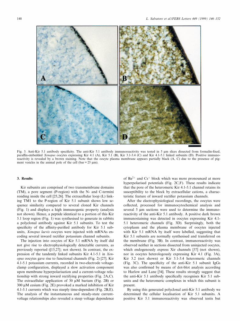
Kir subunits are comprised of two transmembrane domains(TM), a pore segment (P-region) with the N- and C-terminiresiding inside the cell [25,26]. The extracellular loop (L) link-ing TM1 to the P-region of Kir 5.1 subunit shows low se-quence similarity compared to several cloned Kir channels(Fig. 1) and displays a high immunogenic property (analysisnot shown). Hence, a peptide identical to a portion of this Kir5.1 loop region (Fig. 1) was synthesised to generate in rabbitsa polyclonal antibody against Kir 5.1 subunits. To test thespeci¢city of the a¤nity-puri¢ed antibody for Kir 5.1 sub-units, Xenopus laevis oocytes were injected with mRNAs en-coding several inward recti¢er potassium channel subunits.
The injection into oocytes of Kir 5.1 mRNA by itself didnot give rise to electrophysiologically detectable currents, aspreviously reported ([13,27]; not shown). In contrast, the ex-pression of the tandemly linked subunits Kir 4.1-5.1 in Xen-opus oocytes gave rise to functional channels (Fig. 2) [27]. Kir4.1-5.1 potassium currents, recorded in two-electrode voltage-clamp con¢guration, displayed a slow activation componentupon membrane hyperpolarisation and a current-voltage rela-tionship with strong inward rectifying properties (Fig. 2A,C).The extracellular application of 30 WM barium (Fig. 2B) or300 WM cesium (Fig. 2E) provoked a marked inhibition of Kir4.1-5.1 currents which was steeply time-dependent (Fig. 2B,E).The analysis of the instantaneous and steady-state current-voltage relationships also revealed a steep voltage dependence
of Ba2� and Cs� block which was more pronounced at morehyperpolarised potentials (Fig. 2C,F). These results indicatethat the pore of the heteromeric Kir 4.1-5.1 channel retains itssusceptibility to the block by extracellular cations, a charac-teristic feature of inward recti¢er potassium channels.
After the electrophysiological recordings, the oocytes werecollected, processed for immunocytochemical analysis andseveral 5 Wm sections were used to determine the immuno-reactivity of the anti-Kir 5.1 antibody. A positive dark brownimmunostaining was detected in oocytes expressing Kir 4.1-5.1 heteromeric channels (Fig. 3D). Surprisingly, both thecytoplasm and the plasma membrane of oocytes injectedwith Kir 5.1 mRNA by itself were labelled, suggesting thatKir 5.1 subunits are normally synthesised and transferred onthe membrane (Fig. 3B). In contrast, immunoreactivity wasobserved neither in sections dissected from uninjected oocytes,which endogenously express Xir channels [37] (not shown),nor in oocytes heterologously expressing Kir 4.1 (Fig. 3A),Kir 3.2 (not shown) or Kir 3.1-3.4 heteromeric channels(Fig. 3C). The speci¢city of the anti-Kir 5.1 subunit IgGswas also con¢rmed by means of dot-blot analysis accordingto Harlow and Lane [34]. These results strongly suggest thatthe anti-Kir 5.1 antibody speci¢cally recognises Kir 5.1 sub-units and the heteromeric complexes in which this subunit ispresent.
By using this generated polyclonal anti-Kir 5.1 antibody wedetermined the cellular localisation of Kir 5.1 subunits. Apositive Kir 5.1 immunoreactivity was observed testis but
Fig. 3. Anti-Kir 5.1 antibody speci¢city. The anti-Kir 5.1 antibody immunoreactivity was tested in 5 Wm slices dissected from formalin-¢xed,para¤n-embedded Xenopus oocytes expressing Kir 4.1 (A), Kir 5.1 (B), Kir 3.1-3.4 (C) and Kir 4.1-5.1 linked subunits (D). Positive immuno-reactivity is revealed by a brown staining. Note that the oocyte plasma membrane appears partially black (A, C) due to the presence of pig-ment vesicles in the animal pole of the cell (bar = 25 Wm).
FEBS 21888 19-4-99 Cyaan Magenta Geel Zwart
L. Salvatore et al./FEBS Letters 449 (1999) 146^152148

not in the ovary (not shown). In 60-day-old rats testis Kir 5.1subunits were localised in the seminiferous tubules and, par-ticularly, in spermatogonia, primary and secondary spermato-cytes, spermatids and in the head and body of spermatozoa(Figs. 4 and 5). Neither Sertoli cells nor the interstitial Leydigcells were stained by the anti-Kir 5.1 antibody (Figs. 5 and 6).
To examine the developmental pro¢le of Kir 5.1 proteinappearance in rat testis tissues were dissected from animalsat postnatal day 10 (P10), P20, P30, P60 and P90. Negligiblepositive immunoreactivity was detected in P10 and P20 rattestis (Fig. 5A). In contrast, a signi¢cant labelling was di¡er-entially detected in the seminiferous tubules of P30, P60 and
P90 rats (Fig. 5B^D). Each experiment was repeated threetimes on specimens collected from three animals of di¡erentages and gave similar results.
In order to quantify the Kir 5.1-speci¢c signals at di¡erentdevelopmental stages, the £uorescent images of several serialslices were recorded and analyzed by LSCM. Bright rings ofimmuno£uorescence were observed in all developing cells ofthe seminiferous tubules suggesting that Kir 5.1 protein ismostly localised on the plasma membrane (Fig. 6B^D). More-over, the morphometric analysis of specimens from P10 toP90 revealed that Kir 5.1 protein appearance in testis in-creased with age and reached a peak in 60-day-old rats (Fig.6E). In contrast, the immuno£uorescence decreased in 90-day-old animals to levels comparable to P30 and was mostly lo-calised in spermatozoa (Fig. 6D,E). These results stronglysuggest that Kir5.1 subunits follow a speci¢c developmentalpattern of expression related to spermatogenesis and to thesexual maturity of the animal.
4. Discussion
A polyclonal antibody raised in rabbits by using a distincthighly immunogenic peptide speci¢cally recognises Kir 5.1subunits and the heteromeric channels comprised of Kir 4.1and Kir 5.1 subunits. Interestingly, experiments performedwith Xenopus oocytes injected with Kir 5.1 mRNA by itselfreveal the ability of these cells to synthesise and to properlytranslocate the Kir 5.1 protein to the plasma membrane.However, no channel activity could be recorded electrophy-siologically from oocytes expressing Kir 5.1 alone. These ob-servations suggest that Kir 5.1 subunits may form homomericchannels in the plasma membrane but require an additional,still unknown component(s) other than Kir 4.1, capable ofactivating the channel. On the other hand, tandemly linkedKir 4.1-5.1 subunits give rise to potassium currents whichdisplay strong inward rectifying properties and an instantane-ous component followed by a slowly activating phase, whosekinetics are conferred by the C-terminal region of Kir 5.1subunits [27,38]. Barium ions block in a time- and voltage-dependent fashion preferably the slowly activating componentof Kir 4.1-5.1 currents implying that these ions act as openchannel pore blockers. In contrast, cesium ions equally blockboth components suggesting a more complex interaction withthe ion conducting pore.
We focused our interest on the immunolocalisation analysisof rat testis and showed that Kir 5.1 subunits can be detectedin spermatogonia, primary and secondary spermatocytes,spermatids and in the head and body of spermatozoa butnot in Sertoli cells or Leydig cells. These observations stronglysuggest that inward recti¢er potassium channels comprised ofKir 5.1 subunits may regulate important physiological func-tions such as the resting membrane potential, excitability andK� homeostasis of cells related to spermatogenesis. Indeed, ithas been shown that an increase in extracellular potassiumconcentrations causes a reduction of sperm motility [39].This observation suggests that Kir channels, by setting theresting membrane potential of sperm near the K� reversalpotential, may play an important role in regulating the free-swimming properties of spermatozoa. Moreover, the G pro-tein-gated Girk2 channel, a member of the inward recti¢erpotassium channel superfamily, has been reported to be ex-pressed in testis and to play a pivotal role in spermatogenesis.
Fig. 4. Detailed localisation of Kir 5.1 subunits in seminiferous tu-bules of 60-day-old rat testis. Positive ABC immunoperoxidasestaining showing the localisation of Kir 5.1 subunits (A) in sperma-togonia (arrowhead), primary spermatocytes (arrow) and (B) in thehead and body of mature sperm. C: Kir 5.1 subunits were also de-tected in secondary spermatocytes (arrows), spermatids (closed ar-rowhead) and spermatozoa (open arrowhead). Bar = 10 Wm.
FEBS 21888 19-4-99 Cyaan Magenta Geel Zwart
L. Salvatore et al./FEBS Letters 449 (1999) 146^152 149

In fact, a genetic mutation in the coding sequence of the Girk2channel disrupts the selectivity ¢lter of these channels, blocksneuronal di¡erentiation and causes male sterility in weavermice [40^42].
Kir 4.1 subunits have been detected in rat testis by rt-PCRanalysis, but the cellular localisation of these subunits remainsunknown [13]. Our previous and present results demonstratethat Kir 5.1 does not form functional channels by itself butrequires the speci¢c co-assembly of Kir 4.1 subunits [27]. Ifboth subunits are expressed in the same cells, it would beplausible to hypothesise the formation of heteromeric chan-nels comprised of Kir 5.1 and Kir 4.1 subunits in rat testis.Therefore, if both Kir 5.1 and Kir 4.1 subunits show similarpatterns of expression in rat testis they may form heteromericchannels which are expected to possess electrophysiologicalproperties and cation-blocking features similar to Kir4.1-5.1dimers. Alternatively, spermatozoa and germinal cells mayexpress, as yet unidenti¢ed proteins, capable of activatingKir 5.1 homomeric channels.
Both the ABC immunoperoxidase staining and the immu-no£uorescence LSCM analysis showed that Kir 5.1 proteinmay be detected on the plasma membrane and to a lesserextent in the cytoplasm of cells located in the seminiferoustubules. Moreover, the expression of Kir 5.1 subunits isstrongly related to the development of a mammalian species,being highest during the sexual maturity of the animal. How-ever, the role of this temporally de¢ned pattern of Kir 5.1
expression in sperm motility or male fertility remains to bedetermined.
Acknowledgements: We thank Antonio De Blasi and Stephen Tuckerfor reading and commenting on the manuscript. We are grateful toJohn Adelman for the generous gifts of Kir 4.1 and Kir 5.1 clones andto Simone Valentino, Cosmo Rossi, Nicola Celli, Antonio Tamburro,Alexander Mironov for fruitful discussion, insightful suggestions andinvaluable technical assistance. We thank the Animal Care Unit ofCMNS and the ¢nancial support of the Italian National ResearchCouncil (Convenzione CNR ^ Mario Negri Sud). M.C. D'Adamoand Roman Polishchuk are fellows of the Alfredo Leonardi Fundand G.L. Pfei¡er Foundation.
References
[1] Katz, B. (1949) Arch. Sci. Physiol. 2, 285^289.[2] Standen, N.B. and Stan¢eld, P.R. (1978) J. Physiol. 280, 169^
191.[3] Sakmann, B. and Trube, G. (1984) J. Physiol. 347, 659^683.[4] Mihara, S., North, R.A. and Surprenant, A. (1987) J. Physiol.
390, 335^355.[5] McKinney, L.C. and Gallin, E.K. (1988) J. Membr. Biol. 103,
41^53.[6] Barres, B.A. (1991) Curr. Opin. Neurobiol. 1, 354^359.[7] Lewis, D.L., Ikeda, S.R., Aryee, D. and Joho, R.H. (1991) FEBS
Lett. 290, 17^21.[8] Ito, M., Inanobe, A., Horio, Y., Hibino, H., Isomoto, S., Ito, H.,
Mori, K., Tonosaki, A., Tomoike, H. and Kurachi, Y. (1996)FEBS Lett. 388, 11^15.
[9] Kubo, Y., Baldwin, T.J., Jan, Y.N. and Jan, L.Y. (1993) Nature362, 127^133.
Fig. 5. Age-dependent localisation of Kir 5.1 subunits. A: 5 Wm cross-section of a P10 seminiferous tubule showing a very low immunoreactiv-ity of the anti-Kir 5.1 antibody. Positive immunostaining can be observed in sections dissected from P30 (B), P60 (C), and P90 (D) rat testis.Kir 5.1 subunits are localised in all stages of spermatogenesis and are indicated in B: primary (arrows) and secondary spermatocytes (closed ar-rowheads); in C: primary (arrows) and secondary spermatocytes (open arrowheads); in D: spermatozoa (*). Note that Sertoli cells (closed ar-rowhead in C) are not positively stained. Bar = 25 Wm.
FEBS 21888 19-4-99 Cyaan Magenta Geel Zwart
L. Salvatore et al./FEBS Letters 449 (1999) 146^152150

[10] Kubo, Y., Reuveny, E., Slesinger, P.A., Jan, Y.N. and Jan, L.Y.(1993) Nature 364, 802^806.
[11] Ho, K., Nichols, C.G., Lederer, W.J., Lytton, J., Vassilev,P.M., Kanazirska, M.V. and Hebert, S.C. (1993) Nature 362,31^38.
[12] Morishige, K.-I., Takahashi, N., Findlay, I., Koyama, H., Zanel-li, J.S., Peterson, C., Jenkins, N.A., Copeland, N.G., Mori, N.and Kurachi, Y. (1993) FEBS Lett. 336, 375^380.
[13] Bond, C.T., Pessia, M., Xia, X.-M., Lagrutta, A., Kavanaugh,M.P. and Adelmann, J.P. (1994) Receptors Channels 2, 183^191.
[14] Inagaki, N., Tsuura, Y., Namba, N., Masuda, K., Gonoi, T.,Horie, M., Seino, Y., Mizuta, M. and Seino, S. (1995) J. Biol.Chem. 270, 5691^5694.
[15] Hille, B. (1992) in: Ionic Channels in Excitable Membranes, 2ndedn., pp. 127^130, Sinauer Associates, Sunderland, MA.
[16] Matsuda, H., Saigusa, A. and Irisawa, H. (1987) Nature 325,156^159.
[17] Lu, Z. and Mackinnon, R. (1994) Nature 371, 243^245.[18] Stan¢eld, P.R., Davies, N.W., Shelton, P.A., Khan, I.A., Bram-
mar, W.J., Standen, N.B. and Conley, E.C. (1994) J. Physiol.475, 1^7.
[19] Wible, B.A., Taglialatela, M., Ficker, E. and Brown, A.M. (1994)Nature 371, 246^249.
[20] Fakler, B., Brandle, U., Bond, C., Glowatzki, E., Konig, C.,Adelman, J.P., Zenner, H.-P. and Ruppersberg, J.P. (1994)FEBS Lett. 356, 199^203.
Fig. 6. Estimation of Kir 5.1 age-dependent expression by LSCM analysis. Kir 5.1 immuno£uorescence was extremely low in P10 (A) or P20sections (not shown) while it becomes detectable in P30 (B) cells located within the seminiferous tubules. The highest intensity of labelling wasdetected in P60 sections (C) and decreased to lower levels in P90 specimens (D). Arrows indicate that Kir 5.1 signal is mostly localised on plas-ma membrane and no labelling can be detected in interstitial cells (see asterisks in C and D). E: Morphometric analysis showing the averageintensity of immuno£uorescence, expressed in arbitrary units, as a function of days after birth (mean þ S.E.M.; n = 3).
FEBS 21888 19-4-99 Cyaan Magenta Geel Zwart
L. Salvatore et al./FEBS Letters 449 (1999) 146^152 151

[21] Lopatin, A.N., Makhina, E.N. and Nichols, C.G. (1994) Nature372, 366^369.
[22] Krapivinsky, G., Gordon, E.A., Wickman, K., Velimirovic̈, B.,Krapivinsky, L. and Clapham, D.E. (1995) Nature 374, 135^141.
[23] Glowatzky, E., Fakler, G., Brandle, U., Rexhausen, U., Zenner,H.P., Ruppersberg, J.P. and Fakler, B. (1995) Proc. R. Soc.Lond. B Biol. Sci. 261, 251^261.
[24] Yang, J., Jan, Y.N. and Jan, L.Y. (1995) Neuron 15, 1441^1447.[25] Doyle, D.A., Cabral, J.M., Pfuetzner, R.A., Kuo, A., Gulbis,
J.M., Cohen, S.L., Chait, B.T. and MacKinnon, R. (1998) Sci-ence 280, 69^77.
[26] McKinnon, R., Cohen, S.L., Kuo, A., Lee, A. and Chait, B.T.(1998) Science 280, 106^109.
[27] Pessia, M., Tucker, S.J., Lee, K., Bond, C.T. and Adelman, J.P.(1996) EMBO J. 15, 2980^2987.
[28] Florman, H.M. (1994) Dev. Biol. 165, 152^164.[29] Arnoult, C., Cardullo, R.A., Lemons, J.R. and Florman, H.M.
(1996) Proc. Natl. Acad. Sci. USA 93, 13004^13009.[30] Lievano, A., Santi, C.M., Serrano, C.J., Trevino, C.L., Bellve,
A.R., Hernandez-Cruz, A. and Darszon, A. (1996) FEBS Lett.338, 150^154.
[31] Santi, C.M., Darszon, A. and Hernandez-Cruz, A. (1996) Am. J.Physiol. 271, C1583^C1593.
[32] Weyand, I., Godde, M., Frings, S., Weiner, J., Muller, F., Al-tenhofen, W., Hatt, H. and Kaupp, U.B. (1994) Nature 368, 859^863.
[33] Schreiber, M., Wei, A., Yuan, A., Gaut, J., Saito, M. and Salk-o¡, L. (1998) J. Biol. Chem. 273, 3509^3516.
[34] Harlow, E. and Lane, D. (1988) Antibodies. A Laboratory Man-ual, Cold Spring Harbor Laboratory, Cold Spring Harbor, NY.
[35] Bini, A., Mesa-Tejada, R., Fenoglio Jr., J.J., Kudrik, B. andKaplan, K.L. (1989) Lab. Invest. 60, 814^821.
[36] D'Adamo, M.C., Liu, Z., Adelman, J.P., Maylie, J. and Pessia,M. (1998) EMBO J. 17, 1200^1207.
[37] Hedin, K.E., Lim, N.F. and Clapham, D.E. (1996) Neuron 16,423^429.
[38] Lagrutta, A.A., Bond, C.T., Xia, X.M., Pessia, M., Tucker, S.and Adelman, J.P. (1996) Jpn. Heart J. 37, 651^660.
[39] Detweiler, C. and Thomas, P. (1998) J. Exp. Zool. 281, 139^148.[40] Patil, N., Cox, D.R., Bhat, D., Faham, M., Myers, R.M. and
Peterson, A.S. (1995) Nature Genet. 11, 126^129.[41] Slesinger, P.A., Patil, N., Liao, J., Jan, Y.N., Jan, L.Y. and Cox,
D.R. (1996) Neuron 16, 321^331.[42] Tucker, S.J., Pessia, M., Moorhouse, A.J., Gribble, F., Ashcroft,
F.M., Maylie, J. and Adelman, J.P. (1996) FEBS Lett. 390, 253^257.
FEBS 21888 19-4-99 Cyaan Magenta Geel Zwart
L. Salvatore et al./FEBS Letters 449 (1999) 146^152152
Copyright © 2022 FDOKUMEN


![Inward transport of [3H]-1-methyl-4-phenylpyridinium in rat isolated hepatocytes: putative involvement of a P-glycoprotein transporter](https://static.fdokumen.com/doc/165x107/631a3442d5372c006e0363a6/inward-transport-of-3h-1-methyl-4-phenylpyridinium-in-rat-isolated-hepatocytes.jpg)