Living in Three Dimensions: 3D Nanostructured Environments for Cell Culture and Regenerative...
13
REVIEW © Copyright 2006 by Humana Press Inc. All rights of any nature whatsoever reserved. 1085-9195/(Online)1559-0283/06/45:215–227/$30.00 Cell Biochemistry and Biophysics 215 Volume 45, 2006 INTRODUCTION Efforts in the practice of tissue culture have, for the most part, been limited by the use of two-dimensional (2D) surfaces that lack the requisite three-dimensional (3D) physical cues present in the extracellular matrix (ECM) and the basement membrane (BM) within tis- sues. Recent advances have been directed toward pro- viding biochemical as well as physical cues that approximate the complex nano- and micro-environ- ments within the ECM and BM (1–4). This work has been pursued so as to provide biologically mimetic model systems for the delineation of specific pathways associated with proliferation, differentiation, metabo- lism, aging, and disease, as well as in the practice of tis- sue engineering and regenerative medicine (4–7). It is through efforts to engineer new tissue (5–9), and from research that compares cell growth on different culture surfaces (10–13), that the relevancy of 2D culture sur- faces in producing physiological outcomes has been brought into question (14). In this review, we will first discuss properties of the ECM and BM that are critical for regulation of cellular growth and function. We will then apply these properties to the development of 3D Living in Three Dimensions 3D Nanostructured Environments for Cell Culture and Regenerative Medicine Melvin Schindler, 1 Alam Nur-E-Kamal, 2 Ijaz Ahmed, 2 Jabeen Kamal, 2 Hsing-Yin Liu, 2 Nathan Amor, 2 Abdul S. Ponery, 2 David P. Crockett, 3 Timothy H. Grafe, 4 H. Young Chung, 4 Thom Weik, 4 Elizabeth Jones, 4 and Sally Meiners 2,* 1 Department of Biochemistry and Molecular Biology, Michigan State University, East Lansing, MI 48824; 2 Department of Pharmacology and 3 Dept. of Neurosciences and Cell Biology, UMDNJ-Robert Wood Johnson Medical School, 675 Hoes Lane, Piscataway, NJ 08854; 4 Donaldson Co., Inc., P.O. Box 1299, Minneapolis, MN 55440 Abstract Research focused on deciphering the biochemical mechanisms that regulate cell proliferation and function has largely depended on the use of tissue culture methods in which cells are grown on two-dimensional (2D) plastic or glass surfaces. However, the flat surface of the tissue culture plate represents a poor topological approximation of the more complex three-dimensional (3D) architecture of the extracellular matrix (ECM) and the basement membrane (BM), a structurally compact form of the ECM. Recent work has provided strong evi- dence that the highly porous nanotopography that results from the 3D associations of ECM and BM nanofibrils is essential for the reproduction of physiological patterns of cell adherence, cytoskeletal organization, migration, signal transduction, morphogenesis, and differentiation in cell culture. In vitro approximations of these nano- structured surfaces are therefore desirable for more physiologically mimetic model systems to study both nor- mal and abnormal functions of cells, tissues, and organs. In addition, the development of 3D culture environments is imperative to achieve more accurate cell-based assays of drug sensitivity, high-throughput drug discovery assays, and in vivo and ex vivo growth of tissues for applications in regenerative medicine. Index Entries: Extracellular matrix; basement membrane; 2D; 3D; nanofibrillar; tissue culture; regenerative medicine. * Author to whom all correspondence and reprint requests should be addressed. E-mail: [email protected]
-
Upload
independent -
Category
Documents
-
view
3 -
download
0
Transcript of Living in Three Dimensions: 3D Nanostructured Environments for Cell Culture and Regenerative...
REVIEW
© Copyright 2006 by Humana Press Inc.All rights of any nature whatsoever reserved.1085-9195/(Online)1559-0283/06/45:215–227/$30.00
Cell Biochemistry and Biophysics 215 Volume 45, 2006
INTRODUCTION
Efforts in the practice of tissue culture have, for themost part, been limited by the use of two-dimensional(2D) surfaces that lack the requisite three-dimensional(3D) physical cues present in the extracellular matrix(ECM) and the basement membrane (BM) within tis-sues. Recent advances have been directed toward pro-viding biochemical as well as physical cues thatapproximate the complex nano- and micro-environ-
ments within the ECM and BM (1–4). This work hasbeen pursued so as to provide biologically mimeticmodel systems for the delineation of specific pathwaysassociated with proliferation, differentiation, metabo-lism, aging, and disease, as well as in the practice of tis-sue engineering and regenerative medicine (4–7). It isthrough efforts to engineer new tissue (5–9), and fromresearch that compares cell growth on different culturesurfaces (10–13), that the relevancy of 2D culture sur-faces in producing physiological outcomes has beenbrought into question (14). In this review, we will firstdiscuss properties of the ECM and BM that are criticalfor regulation of cellular growth and function. We willthen apply these properties to the development of 3D
Living in Three Dimensions
3D Nanostructured Environments for Cell Culture and Regenerative Medicine
Melvin Schindler,1 Alam Nur-E-Kamal,2 Ijaz Ahmed,2 Jabeen Kamal,2
Hsing-Yin Liu,2 Nathan Amor,2 Abdul S. Ponery,2 David P. Crockett,3
Timothy H. Grafe,4 H. Young Chung,4 Thom Weik,4
Elizabeth Jones,4 and Sally Meiners2,*
1Department of Biochemistry and Molecular Biology, Michigan State University, East Lansing, MI 48824; 2Department of Pharmacology and 3Dept. of Neurosciences and Cell Biology, UMDNJ-Robert Wood Johnson Medical School,
675 Hoes Lane, Piscataway, NJ 08854; 4Donaldson Co., Inc., P.O. Box 1299, Minneapolis, MN 55440
Abstract
Research focused on deciphering the biochemical mechanisms that regulate cell proliferation and functionhas largely depended on the use of tissue culture methods in which cells are grown on two-dimensional (2D)plastic or glass surfaces. However, the flat surface of the tissue culture plate represents a poor topologicalapproximation of the more complex three-dimensional (3D) architecture of the extracellular matrix (ECM) andthe basement membrane (BM), a structurally compact form of the ECM. Recent work has provided strong evi-dence that the highly porous nanotopography that results from the 3D associations of ECM and BM nanofibrilsis essential for the reproduction of physiological patterns of cell adherence, cytoskeletal organization, migration,signal transduction, morphogenesis, and differentiation in cell culture. In vitro approximations of these nano-structured surfaces are therefore desirable for more physiologically mimetic model systems to study both nor-mal and abnormal functions of cells, tissues, and organs. In addition, the development of 3D cultureenvironments is imperative to achieve more accurate cell-based assays of drug sensitivity, high-throughput drugdiscovery assays, and in vivo and ex vivo growth of tissues for applications in regenerative medicine.
Index Entries: Extracellular matrix; basement membrane; 2D; 3D; nanofibrillar; tissue culture; regenerativemedicine.
* Author to whom all correspondence and reprint requestsshould be addressed. E-mail: [email protected]
model systems for more physiologically relevant modelsystems for cell culture and, ultimately, for tissue repair.
THE ECM AND BM
The ECM is a complex mixture of macromoleculesthat self-assemble into nanofibrillar supramolecular net-works that fill the extracellular space between cells(15–17). The ECM has three major functions: to providea scaffolding to physically support cells and provide ten-sile strength, to provide a surface for cell adhesion andmigration, and to provide a reservoir of growth factorsand chemical cues that can regulate cellular differentia-tion and metabolic function (15–18). Structurally, theECM functions to connect, bind, integrate, and supportcells. Chemically, the ECM regulates cellular growth viatwo major classes of macromolecules, the glycosamino-glycans, which are predominantly linked to proteins toform proteoglycans, and the fibrous proteins (14–17).
The proteoglycans can be further subdivided intosecreted- and membrane-bound forms. Chondroitinsulfate, heparan sulfate, keratan sulfate, and hyaluronicacid are secreted into the extracellular environment,whereas syndecan-1 is retained at the surface of the celland anchored in the plasma membrane to form an inte-gral transmembrane protein (15). The fibrous proteins,in turn, can be subdivided into three groups: structuralproteins, such as collagen type I and elastin, adhesiveproteins, such as fibronectin; and de-adhesive proteins,such as tenascin-C and thrombospondin. The propor-tion and types of proteoglycans and fibrous proteinspresent in the ECM differ according to the tissue inquestion. Moreover, many ECM molecules form super-families whose isoforms have a diverse range of chemi-cal and physical properties (19,20), adding to thephysical and chemical complexity of the ECM.
The BM is a specialized, compact form of ECM local-ized to the basolateral surface of epithelium, endothe-lium, peripheral nerve axons, fat cells, and muscle cells(15). BMs are involved in embryonic development (theyfirst occur at the eight cell stage), wound healing, tissueremodeling, and tumor metastasis (15,18,19). As a gen-eral principle, BMs provide structural support for overly-ing adherent cells and divide tissues into compartments,separating, for example, the epithelium from the stroma.In addition, BMs function as active barriers for infiltra-tion and cell migration. As with the ECM, the BM alsoserves as a reservoir in which a variety of growth factorscan be stored. The growth factors may be releasedthrough the action of specific extracellular proteases andcan, in the unbound form, elicit specific mitogenic or sur-vival responses (15).
The BM is predominantly comprised of macromole-cules that can self-assemble into dense, sheet-like struc-
tures 50–100 nm in thickness. Major componentsinclude collagen type IV, laminins, heparan-sulfate pro-teoglycans, perlecan, and nidogen/entactin. Minorcomponents include agrin, SPARC/BM-40/osteopon-tin, fibulin-1 and -2, and collagen types IV, XV, XVIII,and XIX (15). As for the ECM, the proportion and typesof BM components differs depending on the tissue.
Physically, the ECM and BM are both represented byan assembly of nanofibrils that is organized into either a3D nanofibrillar lattice (ECM) or a denser 3D nanofibril-lar mesh (BM) (11,15–19,21). Examples demonstratingthe nanofibrillar nature of the ECM and the BM areshown in Fig. 1. Functionally, the ECM and BM are piv-otal regulators of cell adhesion, migration, morphogene-sis, apoptosis, proliferation, and differentiation (22–25).The normal functioning and structure of the ECM andBM are significantly changed during wounding, inflam-mation, and tumorigenesis. For example, several nor-mally stabile ECM components are secreted by tumorcells during the growth of primary cancers, resulting inchanges in the physical and chemical environment sur-rounding the tumor (15,17). For aggressive cancers,these changes can promote the migration of cells fromthe primary tumor into the bloodstream.
Matrix Design Principles for Biomimetic Cell Culture Matrices
The following physical and chemical properties havebeen identified as key parameters in achieving structuralas well as functional fidelity of the ECM and BM: dimen-sionality (2D vs 3D) (26,27), compliance/rigidity (26–29),fibrillarity (26,28), fiber alignment (20), and chemistry(peptide recognition motifs) (30,31). As for the propor-tion and types of macromolecules that comprise theECM and BM, these parameters can vary widely in dif-ferent tissues. To quote from Katz et al., “matricesderived from different cells or tissues may each have aunique composition-topography-rigidity ‘signature’ thatinduces specific cellular interactions, differentiation,morphogenesis, polarity, repair, survival, and migra-tion” (27).
DIMENSIONALITY
3D Matrix AdhesionsIn an effort to obtain information concerning the
composition and function of adhesions between fibrob-lasts and ECM that occur in vivo, Cukierman et al. (26)cultured fibroblasts on 3D matrices derived from eitherdetergent-extracted mouse embryo sections or naturallydeposited ECMs secreted by NIH-3T3 fibroblasts thatwere denuded of cells (26,28,32). Although the twoforms of matrix differed in chemical composition, cellu-
216 Schindler et al.
Cell Biochemistry and Biophysics Volume 45, 2006

lar responses to the 3D surfaces were similar. This sug-gests that the ECM architecture is as important as chem-istry in regulating cellular phenotype.
The authors described a type of adhesion, which theytermed a 3D-matrix adhesion, whose composition andstructure differed significantly from the focal and fibril-
lar adhesions previously observed on 2D substrates(26,28,33). These adhesions differed in their content ofα5β1 and αvβ3 integrins, paxillin, cytoskeletal compo-nents (in particular, an absence of stress fibers andenhanced cortical actin was observed), and the degreeof tyrosine phosphorylation of focal adhesion kinase
Living in Three Dimensions 217
Cell Biochemistry and Biophysics Volume 45, 2006
Fig. 1. Composite of fibrillar extracellular matrices and basement membranes. (A) Scanning electron micrograph ofDescemet’s membrane side of the substantia propria. Bar, 2 µm. (Reprinted from ref. 117, with permission layer Wiley-Liss,Inc.) (B) Scanning electron micrograph of epithelial cells that have been isolated from tadpole epidermis and placed on acollagenous substratum. The substratum comprises the acellular dermis of the tadpole, which consists of 20 orthogonal lay-ers of collagen fibrils. The epithelial cells sit on top of the orthogonal lattice. Bar, 5 µm. (Reprinted from ref. 62, with per-mission from Elsevier.) (C) 3D confocal backscatter microscopy of migrating T cell in a 3D collagen matrix. Black lines(overlay) indicate transmigrated path. Bar, 5 µm. Reprinted from ref. 118, with permission from the American Society ofHematology. (D) AFM image of human sclera. This overview shows scleral collagen fibrils with a variable diameter. Bar, 5µm. (Reprinted from ref. 63, with permission from Springer-Verlag.) (E) Scanning electron micrograph of fibroblasts adher-ing to collagen IV polymers. Fibroblasts were seeded onto glass cover slips coated with collagen polymers for an adhesionperiod of 60 min. Bar, 10 µm. (Reprinted from ref. 59, with permission from Elsevier.) (F) Multiphoton intravital image ofcarcinoma cells moving extracellular matrix fibers in a mammary tumor. Bar, 5 µm. (Reprinted from from ref. 44, with per-mission from Nature Publishing Group.)
(FAK) (34). Structurally, cells cultured on the 3D matri-ces had an elongated morphology more characteristic offibroblasts in vivo. Functionally, the cells demonstrateda 6-fold increase in attachment, a 2-fold increase in therate of proliferation, and 1.5-fold increase in migrationrate in comparison to cells cultured on 2D culture sur-faces coated with laminin-1 or collagen IV, or in com-parison to cells cultured in 3D collagen gels. Theenhanced attachment, proliferation, and migrationcould be inhibited by using a function-blocking anti-body against α5 integrin, with a concomitant loss of 3Dmatrix adhesions and a reversion to the focal and fibril-lar adhesions observed on 2D substrates. Flattening the3D matrices through application of a weight also pre-vented fibroblasts from forming 3D-matrix formations(26). Taken together, these data suggest that the 3D pre-sentation of α5 integrin recognition motifs in fibronectinwas essential for the induction of 3D-matrix adhesions,as well as for the significant changes in fibroblast phe-notype and function (26).
Drug SensitivityTumor cells evade chemotherapy by acquiring resis-
tance to apoptosis (24). In one study, HMT-3522 MECcells (a model of metastatic breast cancer cells) were cul-tured on 2D surfaces and within 3D collagen type I gels(24). Culture within 3D gels induced drug resistance totreatment with paclitaxel and the topoisomerase IIinhibitor etoposide. Furthermore, MCF-7 human breastcancer cells displayed a decrease in sensitivity to tamox-ifen when cultured on a 3D chitosan scaffold (35). Inanother study, MCF-7 and osteosarcoma cells culturedon a soluble form of BM extracted from an Engelbreth-Holm-Swarm tumor developed an increased resistanceto Adriamycin in comparison to cells cultured on 2Dplastic surfaces (36). These outcomes support an emerg-ing view that associations between cancer cells and theirlocal ECM/BM may lead to decreased sensitivity tochemotherapeutic drugs (37,38). As such, these resultshave a significant bearing on the design of cancer thera-pies involving chemotherapeutics and on all currentefforts to perform high-throughput drug screening withcell-based assays (39).
3D vs 2D MigrationCell migration is an essential aspect of embryogene-
sis, tumor formation (metastasis and intravasation),inflammation, and wound repair (40,41). Two modes oftumor cell migration have been demonstrated in a 3Dmatrix composed of matrigel or collagen type I that aredifferentially regulated by Rho GTPases (42). The first isa rounded or “amoeboid” mode of cell migration thathas not been observed on 2D surfaces. This form ofmigration requires signaling through the Rho GTPase
RhoA and its effector, Rho kinase and does not requirepericellular proteolysis. Hence, a pliable ECM thattumor cells can squeeze through likely favors therounded tumor cell morphology. The second form oftumor migration is an elongated mode of cell movementdriven by Rac, which is independent of Rho activity andinvolves proteolysis of the surrounding matrix (42).
Collagen Fibril TransportCollagen remodeling by migrating fibroblasts plays a
crucial role in reorganizing tissue structures duringwound repair, development, and regulation of cellgrowth. A seminal article by Meshel et al. (43) demon-strated that myosin IIB was localized to the cell cortex infibroblast lamellipodia extending along 3D nanofibrilscomposed of collagen type I. The lamellipodia boundand retracted the fibrils in a “hand-over-hand” cycleinvolving α2β1 integrin. This “hand-over-hand” fibertranslocation was a result of the recycling of myosin IIBfrom the cell interior into protruding structures at thecell periphery. Myosin IIB was not required for themovement of collagen beads on 2D surfaces, demon-strating the importance of the fibrillar geometry of thecollagen fibrils (43). Most provocatively, the movementof collagen type I fibers could be initiated when onlypart of the cell surface contacted a single fibril. Thuschanges in cellular behavior and function are apparentlyachieved by the cell mechanically sensing the curvatureof the fibril. However, how this sensing occurs remainsto be determined. Significantly, cellular contacts with 3Dnanofibrillar surfaces may be similarly critical for themigration and collagen remodeling observed for tumorcells undergoing metastasis or intravasation in vivo (44)and for migration of muscle cell precursors from thesomite into the forelimb (45). These studies provide sup-port for the importance of dimensionality and fibrillargeometry in cellular migration.
Compliance/RigidityThe mechanical properties of the ECM and BM as
defined by the rigidity or compliance of the matrix havebeen shown to be significant parameters in regulatingcellular morphology, cytoskeletal organization, and cel-lular activities such as migration (26,27,29,46–50). In thiscontext, the rigidity of a typical plastic cell culture sur-face is more than 1 GPa, whereas the range for collagenI gels used in cell culture studies varies between 1 and100 kPa (29,47,48). By comparison, reports of the elasticmodulus of the ECM and BM can vary from kPa forECM to MPa for BM (51–53). These numbers clearlydemonstrate that the ECM and BM in the tissues canhave a rather large dynamic range of material mechani-cal properties. That such changes are physiologicallyimportant regulators of cellular physiology can be
218 Schindler et al.
Cell Biochemistry and Biophysics Volume 45, 2006
observed in experiments demonstrating structural andfunctional differences for fibroblasts (13,26,29,47),epithelial cells (23,34,54), and endothelial cells (55)cultured on “unrestrained and unloaded” or “strainedand loaded” collagen type I gels whose rigidity variesbetween 1 and 100 kPa.
In a series of experiments utilizing “soft” and “hard”2D substrates coated with polyacrylamide, Pelham andWang (47) demonstrated a 6-fold increase in the migra-tion of NIH 3T3 fibroblasts (0.1–0.6 µm/min) for a 12-fold decrease in the Young’s modulus and a 16-folddecrease in the “stiffness” (approx 7.3 × 10⁻⁷ N/µm vsapprox 4.6 × 10⁻⁸ N/µm) of the polyacrylamide. Theauthors also observed that lamellpodial ruffling fornormal rat kidney cells was approximately sixfoldhigher on the “softest” polyacrylamide surface testedin comparison to lamellpodial ruffling on the “hardest”surface (47). Both cell types demonstrated a loss of focaladhesions and stress fibers on the “soft” polyacry-lamide substrate. Hence the decreased rigidity of thesubstratum is associated with enhanced cellular motil-ity. However, a limitation of this work is that differ-ences between compression and tension, both of whichare components of compliance, cannot readily be madeon 2D surfaces. On the other hand, they can readily bedistinguished in 3D. For example, work with 3D mod-els of the mammary gland have demonstrated thatmalignant transformation of the breast is associatedwith significant changes in gland tension that includeelevated compression forces, high tensional resistancestresses, and increases in the rigidity of the associatedextracellular matrix (56). The suggestion was made thatmalignant transformation of the breast may be linkedto perturbations in the tensional homeostasis of breasttissue resulting from physico-chemical changes in theassociated ECM. Other examples of regulation of cellu-lar phenotype and function by compliance can befound in the enhanced formation of tubules or acinarstructures of T47D and MCF-10A cells in “soft” floating3D collagen type I gels (54) and enhanced neurite elon-gation for neurons cultured on “soft” polyacrylamidesubstrates (57).
FibrillarityCells cultured in collagen type I gels and on 2D sur-
faces coated with collagen type I have demonstratedtheir ability to acquire tissue-like phenotypes and dif-ferentiated functions (54,58,59). A number of reportshave clearly shown that an important aspect of theECM and BM in general and collagen in particularfor producing more in vivo-like structures and func-tions for cells is the fibrillar organization (59–61).Interestingly, fibroblasts cultured on nanofibrillar colla-gens demonstrated changes in integrin organization
and focal adhesion structure and composition that aresimilar to those observed for 3D matrix adhesions(26,59,60). Experiments with fibroblasts, focusing onthe supramolecular organization of the collagens, haveshown significant differences in integrin clustering(60), organization of the actin network (59,60), localiza-tion of talin, vinculin and phosphotyrosine-containingproteins (60), GTPase-mediated signaling (60), and dif-ferences in the activation of p70S6 kinase, an effector inmTOR-mediated regulation of cell growth (61) for cellscultured on collagen monomers in comparison to cellscultured on collagen polymers (fibrillar). Fibroblastscultured on polymerized collagen demonstrated mor-phological changes more consistent with an in vivophenotype (59–61). The observed differences in inte-grin organization for cells cultured on fibrillar collagensuggest that the fibrillar nature of collagen is itself asignal to regulate integrin collagen interactions.
Oriented Fiber Alignment
ECM FIBRIL ALIGNMENT
Collagen assembled as thin filaments in an orthogo-nal lattice (20,51,62,63) is an important structural aspectof the cornea. Without this type of fibril alignment, itwould be difficult to maintain optical transparency.Moreover, mechanical strength in the tendon is a resultof thick collagen fibrils in parallel arrays (64), whichhave a higher rigidity than fibers in a random array (65).In the ECM of tissues, thick arrays of collagen type I fib-rils are observed integrated with the fibronectin matrix(27,33). These results suggest that fiber alignment is crit-ical for specific physical properties that include cornealtransparency and physical strength.
ECM fiber alignment appears to be critical for thenormal and pathological function of other tissues andcells as well. For example, an aligned network offibronectin containing ECM fibrils has been suggestedto guide the migration of presumptive mesodermalcells from the blastopore toward the animal pole in theamphibian embryo (66). Furthermore, as mesenchymalcells move through the ECM, their polarized move-ment results in the reorganization of collagen fibrils atthe back end of the cell from randomly organized toarrays of oriented fibrils (17). A similar phenomenonwas observed for tumor cells in vivo (44). The cellsreorganize some of surrounding collagen fibrils intooriented collagen tracks. In vivo imaging of cellsundergoing metastasis from the primary tumor showtumor cells migrating along these oriented collagenfibrils (44). These results suggest that mesenchymalcells and tumor cells can induce tracks and pathwayswith the appropriate substratum compliance forguided migration.
Living in Three Dimensions 219
Cell Biochemistry and Biophysics Volume 45, 2006
TOPOGRAPHIC GUIDANCE
A number of investigations have employed culturesurfaces modified with oriented micro- or nano-sizedgrooves designed to mimic aligned ECM fibrils.Fibroblasts plated onto such surfaces adjust theirshape, orientation, and direction of movement to con-form to the alignment of the machined components(67,68). These dynamic changes resulted from realign-ments of the actin and tubulin based networks of thecytoskeleton (69,70). Topographic or contact-mediatedeffects of machined surfaces were also observed formigrating sheets of epithelial cells, which demon-strated enhanced migration along the microgroovesand decreased migration across the grooves (71).Similar patterns of guidance were reported formacrophages (72) and oligodendrocytes (73) culturedon surfaces containing oriented nanogrooves. Othertypes of oriented substrates have been developed forpotential applications in tissue engineering. For exam-ple, surfaces incorporating neuroactive peptidesderived from laminin-1 deposited in patterns guidedneurite outgrowth from hippocampal neurons, withneurons preferentially adhering to and extending neu-rites on the peptide-coated regions (74). Furthermore,perfectly aligned electrospun polylactic acid nano/microfibers supported neural stem cell differentiationand facilitated guided neurite outgrowth, which wasnot observed on 2D surfaces or on 3D nano/microfib-rillar surfaces comprised of a random array of nanofibers(75). Such aligned nano/microfibrillar surfaces are ofparticular interest for therapies aimed at encouragingdirected axonal regeneration, for example, after spinalcord injury.
Chemical Cues
SIGNALING MOTIFS
Two-way signaling between the ECM and the cell ispredominantly mediated by integrin-ECM interactions(26,27,33,76–79). These interactions are controlled by themechanical properties of the ECM (26,33,34,47,48,54,78)as well as by peptide recognition motifs within the pri-mary sequence of the molecules comprising the ECM(30,31,77,79,80). The availability of these motifs andtheir density can act to regulate specific cell functions,and, in some cases, cause apoptosis (49,50,79,81). Theimportance of integrin/matrix interactions was clearlyshown in studies demonstrating that function blockingantibodies to integrin subunits prevented the develop-ment of a 3D phenotype for fibroblasts cultured on 3Dmatrices (α5 integrin subunit) (26) and prevented thepattern of 3D collagen remodeling observed for fibrob-lasts interacting with collagen I fibrils (α2 integrin sub-unit) (43).
SIGNALING THROUGH RHO GTPASES
The cytoskeleton integrates and propagates physi-cal and chemical changes from the ECM to the cell,leading to alterations in cell morphology, cell–cellinteractions, and function (33,34,76,77). This regula-tion is achieved through the selective activation ofmembers of the Rho family of small GTPases (82–84).Rho, Rac, and Cdc42 in conjunction with their down-stream effectors (there are 60 identified activators andinactivators of GTPase activity (85)) regulate celladhesion, cell migration, cell polarity, endocytosis,vesicle trafficking, cell cycle progression, embryogen-esis, differentiation, oncogenesis, wound repair, andgene transcription (84). The 2D culture surfaces pre-dominantly activate Rho, which through its effectorRho kinase induces the assembly of focal adhesionsand stress fibers in addition to cell entry into the cellcycle (34,54). However, stress fibers, focal adhesions,and the concomitant flat morphology observed forcells grown on 2D surfaces are rarely seen in vivo andhave been suggested to be a “wound” response to the2D tissue culture environment (58,82). Migration stud-ies within 3D environments (collagen type I (40,42)and transverse slices from the forelimb region of chickembryos (45)) have suggested that activation of Racpromotes the protrusive activity associated withmigration (86) and is involved in regulating morpho-genesis (87). As described previously, differential reg-ulation of migration modes on 2D surfaces vs 3Denvironments apparently occurs through a balancebetween Rho and Rac/Cdc42 activation (88–91).
INTEGRINS AND RHO GTPASES
Considering the role of integrins in the activation ofthe Rho GTPases (76–78,82), it is likely that the 3D pre-sentation of integrin attachment sites, the compli-ance/rigidity of the matrix with which the integrins areassociated, and the fibrillar organization (random vsaligned) of the matrix components are important cues toregulate the “outside/in” signaling that differentiallyinfluences specific patterns of Rho GTPase activation(84,85,88). This type of signaling is complemented andmade more so by the “inside/out” interactions that canphysically reorganize the ECM (33,46) and by the secre-tion of ECM molecules capable of providing differentfunctional cues. An example of such “inside/out” regu-lation can be found in the observation that changes inthe mechanical forces generated by the cell cytoskeletoncan alter the organization and assembly state offibronectin fibrils through the exposure of thefibronectin type III modules that are normally buriedwithin the soluble molecule (92). The reorganizedfibronectin can then promote altered “outside/in” inter-actions through integrin binding.
220 Schindler et al.
Cell Biochemistry and Biophysics Volume 45, 2006
3D NANOSTRUCTURED MODELS FOR CELL CULTURE
All of these examples emphasize the critical role ofdimensionality on cellular phenotype and function. Avariety of approaches and materials have been employedin an effort to mimic the aforementioned physical andchemical properties of the ECM and BM to provide moreappropriate substrates for the investigation of cell physi-ology in vitro as well as scaffolds for the repair of tissuein vivo. The 3D model systems for cell culture can bedivided into two broad categories: 3D gels and 3D sur-faces. A 3D gel either partially or completely surroundscells, whereas a 3D surface interacts with cells uniquelyat their basal surface.
Hydrogels
NATURAL PRODUCT-BASED HYDROGELS
The most widely used biologically derived materialsfor providing the appropriate nanostructured environ-ments whose physical form can be easily modified for 3Dcell culture are collagen type I (31,46,54,58) and matrigel,a BM-like biomatrix derived from the Engelbreth-Holm-Swarm mouse sarcoma cell line (21–23). Work from anumber of laboratories has demonstrated that the interac-tion of epithelial and endothelial cells with collagen typeI gels or matrigel is sufficient to induce a halt in prolifera-tion and the induction of morphogenesis and differentia-tion (21–24,54,55,61). The advantage of using collagentype I is that it is found in the ECM and BM of many celltypes, where it provides the necessary mechanical prop-erties as well as the appropriate chemical recognitionmotifs to interact with other ECM proteins and integrinsfor cell attachment, migration, proliferation, and differen-tiation. Cells cultured in collagen gels and on fibrillar col-lagen surfaces have demonstrated their ability to acquiretissue-like phenotypes and differentiated function (10,34,93,94). Although most types of collagen have becomeavailable, its use bears the caveats that different collagenpreparations can have different mechanical and chemicalproperties as a result of changes in the extent of polymer-ization (59–61). Moreover, different collagens can havedifferent assortments of covalently bound growth factors,which cannot be extracted without denaturing the struc-tural collagen (95). Collagens are also readily digested bycollagenases and matrix metalloproteases (96), which canbe either an advantage or a disadvantage, depending onthe application. For example, growth cones migratingthrough ECM use matrix metalloproteases to digest theECM (97), making collagen a good model system to studysome aspects of axon growth.
Matrigel, a secretion composed of laminin-1, collagenIV, and growth factors, has been successfully employed
in a number of efforts to promote morphogenesis anddifferentiation (23). It has also been used as a graftingmaterial in models of spinal cord injury (98). This suc-cess is tempered by the fact that as a biological material,it exhibits considerable batch-to-batch variation, and asa tumor secretion, it contains a number of ill-definedgrowth factors whose concentrations have been shownto vary with preparation. For this reason, efforts havebeen made to provide more defined 3D systems thatincorporate biological molecules.
SYNTHETIC SELF-ASSEMBLING
PEPTIDE HYDROGELS
A new class of peptide-based biomaterials has beendeveloped that is capable of self-assembling into stablenanofibrillar hydrogels (99–103). In general, these self-assembling peptides are composed of amino acidsequences containing alternating hydrophobic andhydrophilic side groups with sequences of chargedamino acid residues to include alternating positive andnegative charges (99–103). Self-assembling peptidesform stable β-sheet structures when dissolved in deion-ized water. Exposure to an electrolyte solution initiatesassembly into interweaving nanofibers. The resultingnanofiber structure (approx 10 nm) is almost threeorders of magnitude smaller than most polymermicrofibers that have been used for cell culture and pre-sents a 3D gel for cell interactions (103). In addition,peptide recognition motifs may be incorporated into thesequence of amino acids to promote specific cell matrixinteractions that influence cell differentiation and tissueformation (99,100).
3D Nanofibrillar Surfaces
NANOFIBRILLAR SURFACES PRODUCED
BY PHASE SEPARATION
Cukierman et al. (26) used natural ECMs denuded ofcells to provide a 3D surface for cell culture. Althoughthis study yielded many pivotal results, natural ECMsdenuded of cells cannot be shaped into a chosen 3Ddesign with defined parameters that can be varied tooptimize cellular growth. Therefore, recent investiga-tions have focused on designing synthetic 3D surfacesfor cell culture, such as the micro- and nano-groovedsurfaces described previously. An additional type ofnanostructured synthetic 3D surface for cell culture isprovided by nanofibrous foam materials. Nanofibrousfoam materials are prepared by phase separation, atechnique formerly employed for the manufacture ofporous polymer membranes (104). This process occursin five steps: polymer dissolution, phase separationwith gelation, solvent extraction from the gel withwater, freezing, and, finally, freeze-drying. The resulting
Living in Three Dimensions 221
Cell Biochemistry and Biophysics Volume 45, 2006
highly porous structures that have a 3D architecturesimilar to that of native collagen type I. A particularadvantage of these materials is that they have beenshown to absorb higher amounts of protein in compari-son to 2D surfaces as a consequence of their high sur-face to volume ratio, which can contribute to cellularfunctions such as cell attachment (104).
NANOFIBRILLAR SURFACES PRODUCED
BY ELECTROSPINNING
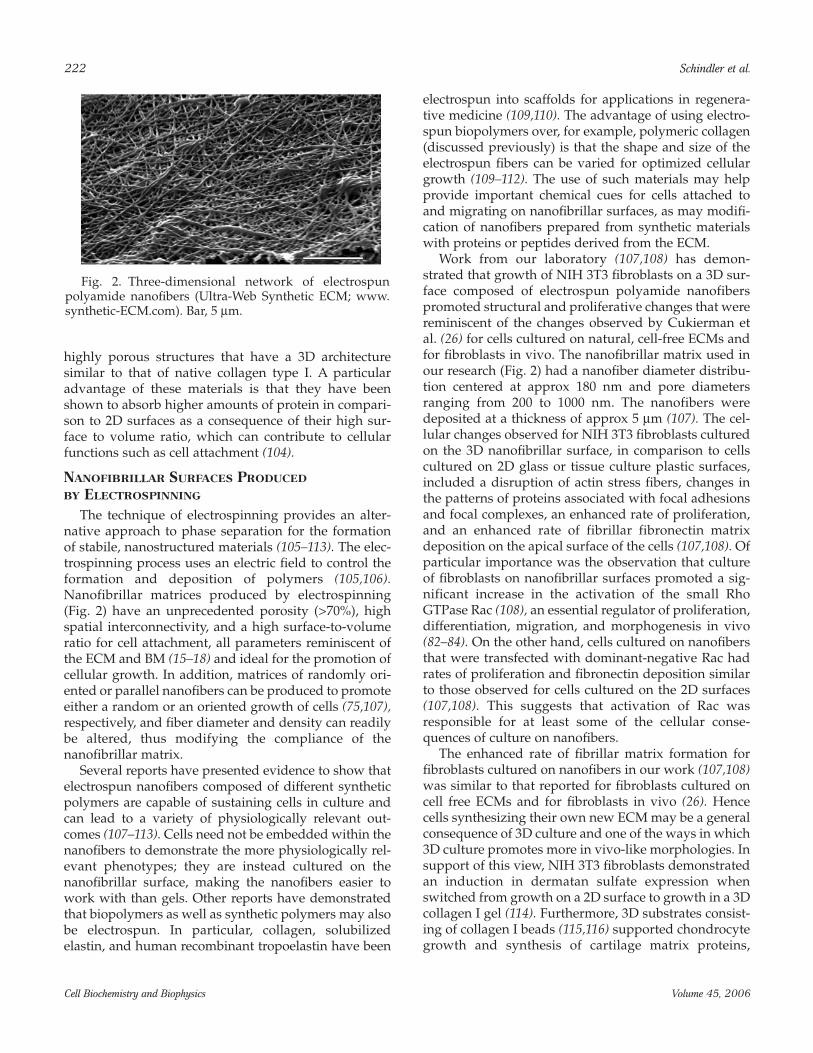
The technique of electrospinning provides an alter-native approach to phase separation for the formationof stabile, nanostructured materials (105–113). The elec-trospinning process uses an electric field to control theformation and deposition of polymers (105,106).Nanofibrillar matrices produced by electrospinning(Fig. 2) have an unprecedented porosity (>70%), highspatial interconnectivity, and a high surface-to-volumeratio for cell attachment, all parameters reminiscent ofthe ECM and BM (15–18) and ideal for the promotion ofcellular growth. In addition, matrices of randomly ori-ented or parallel nanofibers can be produced to promoteeither a random or an oriented growth of cells (75,107),respectively, and fiber diameter and density can readilybe altered, thus modifying the compliance of thenanofibrillar matrix.
Several reports have presented evidence to show thatelectrospun nanofibers composed of different syntheticpolymers are capable of sustaining cells in culture andcan lead to a variety of physiologically relevant out-comes (107–113). Cells need not be embedded within thenanofibers to demonstrate the more physiologically rel-evant phenotypes; they are instead cultured on thenanofibrillar surface, making the nanofibers easier towork with than gels. Other reports have demonstratedthat biopolymers as well as synthetic polymers may alsobe electrospun. In particular, collagen, solubilizedelastin, and human recombinant tropoelastin have been
electrospun into scaffolds for applications in regenera-tive medicine (109,110). The advantage of using electro-spun biopolymers over, for example, polymeric collagen(discussed previously) is that the shape and size of theelectrospun fibers can be varied for optimized cellulargrowth (109–112). The use of such materials may helpprovide important chemical cues for cells attached toand migrating on nanofibrillar surfaces, as may modifi-cation of nanofibers prepared from synthetic materialswith proteins or peptides derived from the ECM.
Work from our laboratory (107,108) has demon-strated that growth of NIH 3T3 fibroblasts on a 3D sur-face composed of electrospun polyamide nanofiberspromoted structural and proliferative changes that werereminiscent of the changes observed by Cukierman etal. (26) for cells cultured on natural, cell-free ECMs andfor fibroblasts in vivo. The nanofibrillar matrix used inour research (Fig. 2) had a nanofiber diameter distribu-tion centered at approx 180 nm and pore diametersranging from 200 to 1000 nm. The nanofibers weredeposited at a thickness of approx 5 µm (107). The cel-lular changes observed for NIH 3T3 fibroblasts culturedon the 3D nanofibrillar surface, in comparison to cellscultured on 2D glass or tissue culture plastic surfaces,included a disruption of actin stress fibers, changes inthe patterns of proteins associated with focal adhesionsand focal complexes, an enhanced rate of proliferation,and an enhanced rate of fibrillar fibronectin matrixdeposition on the apical surface of the cells (107,108). Ofparticular importance was the observation that cultureof fibroblasts on nanofibrillar surfaces promoted a sig-nificant increase in the activation of the small RhoGTPase Rac (108), an essential regulator of proliferation,differentiation, migration, and morphogenesis in vivo(82–84). On the other hand, cells cultured on nanofibersthat were transfected with dominant-negative Rac hadrates of proliferation and fibronectin deposition similarto those observed for cells cultured on the 2D surfaces(107,108). This suggests that activation of Rac wasresponsible for at least some of the cellular conse-quences of culture on nanofibers.
The enhanced rate of fibrillar matrix formation forfibroblasts cultured on nanofibers in our work (107,108)was similar to that reported for fibroblasts cultured oncell free ECMs and for fibroblasts in vivo (26). Hencecells synthesizing their own new ECM may be a generalconsequence of 3D culture and one of the ways in which3D culture promotes more in vivo-like morphologies. Insupport of this view, NIH 3T3 fibroblasts demonstratedan induction in dermatan sulfate expression whenswitched from growth on a 2D surface to growth in a 3Dcollagen I gel (114). Furthermore, 3D substrates consist-ing of collagen I beads (115,116) supported chondrocytegrowth and synthesis of cartilage matrix proteins,
222 Schindler et al.
Cell Biochemistry and Biophysics Volume 45, 2006
Fig. 2. Three-dimensional network of electrospunpolyamide nanofibers (Ultra-Web Synthetic ECM; www.synthetic-ECM.com). Bar, 5 µm.

whereas chondrocytes grown on 2D tissue culture flaskslost their ability to produce the appropriate cartilageECM. Thus regulation of the synthesis of new ECMcomponents may be dependent upon the architecture ofthe preexisting ECM.
We have recently found that fibroblasts incorporatemyosin IIB into lamellipodia extensions when culturedon the electrospun polyamide nanofibers (submittedmanuscript) in a manner that is characteristic of the 3Ddependent collagen remodeling phenotype discussedpreviously (43). As noted previously, the 3D remodelingphenotype involves the movement of myosin IIB intoand out of lamellipodia extensions during an extensionand retraction cycle as the cell pulls along 3D collagenfibrils (43). This type of myosin IIB relocation may be animportant component of the “amoeboid” migrationobserved for cells derived from highly invasive tumors,where cells squeeze between and likely pull on fibrils ofcollagen type I (40,44). Therefore, although the diame-ters of the pores (200–1000 nm) in the 3D nanofibrillarsurface used in our work were too small to permit cellmigration through the nanofibers, and the polyamidecomposition of the nanofibers is resistant to proteolysis,migration of cells on top of these surfaces may reflectthe “amoeboid” type of tumor cell movement associ-ated with 3D fiber networks in vivo.
3D Nanostructured Surfaces: Hydrogels vs 3D Nanostructured Surfaces
In assessing the benefits of a particular 3D nanos-tructured model, the following should be considered.For mesenchymal cells (e.g., fibroblasts, chondroblasts,osteoblasts, tumor cells undergoing elongating cellmigration) that use proteolysis for migration, a sur-rounding layer provided by a hydrogel would likely bemost representative of the 3D environment experiencedby these cells in vivo. Nonetheless, we and others(26,107,108) have demonstrated that physiologically rel-evant phenotypes can be obtained for fibroblasts usingnanostructured surfaces. In contrast, epithelial cells,endothelial cells, and muscle cells maintain a specificpolarity in tissues and in general are only exposed to theECM through basolateral interactions with the base-ment membrane. In attempting to reconstruct the tissueand its concomitant polarity, it may be more physiolog-ically relevant to use 3D nanostructured surfaces forthese latter cell types. Another significant advantage of3D nanostructured substrates is that large numbers ofcells can be grown on their surfaces for biochemicalanalyses. Cells cultured on 3D nanostructured surfacesare also more physically accessible for extraction andexperimental manipulation than are cells culturedwithin a natural or synthetic hydrogel. In addition, theyare more accessible for most imaging techniques, such
as optical and scanning force microscopy, and electro-physiological approaches, such as patch clamping.
CONCLUSIONS
It has become increasingly clear that it will be neces-sary to more accurately model the physical and chemi-cal characteristics of the growth environment of theECM and BM to promote more physiological relevantstructure and function for cells in vitro. A key feature ofthe ECM and BM environment is the nanofibrillar archi-tecture that assembles into a 3D compliant and highlyporous network. Current methods to produce syntheticenvironments that can provide the requisite structuraland chemical information hold great promise for appli-cations in cell culture and regenerative medicine.
ACKNOWLEDGMENTS
This manuscript is dedicated to the memory of LynnElizabeth Meiners. Research described within thisreview was supported by National Institutes of HealthGrant R01 NS40394 and New Jersey Commission onSpinal Cord Research Grant 04-3034 SCR-E-O to S.M.and a grant from The Center for Plant Products andTechnology (MSU), East Lansing, MI, to M.S.
REFERENCES
1. Berthiaume, F., Moghe, P.V., Toner, M., and Yarmush,M.L. (1996) Effect of extracellular matrix topology on cellstructure, function, and physiological responsiveness:hepatocytes cultured in a sandwich configuration. FASEBJ. 10, 1471–1484.
2. Knedlitschek, G., Schneider, F., Gottwald, E., Schaller, T.,Eschbach, E., and Weibezahn, K.F. (1999) A tissue-likeculture system using microstructures: influence of extra-cellular matrix material on cell adhesion and aggrega-tion. J. Biomech. Eng. 121, 35–39.
3. Ertel, S. I., Chilkoti, A., Horbett, T. A., and Ratner, B. D.(1991) Endothelial cell growth on oxygen-containing filmsdeposited by radio-frequency plasmas; the role of surfacecarbonyl groups. Biomater. Sci. Polym. Ed. 3, 163–183.
4. Hojo, M., Inokuchi, S., Kidokoro, M., et al. (2003)Induction of vascular endothelial growth factor by fibrinas a dermal substrate for cultured skin substitute. Plast.Reconstr. Surg. 111, 1638–1645.
5. Kim, B. S., Nikolovski, J., Bonadio, J., Smiley, E., andMooney, D. J. (1999) Engineered smooth muscle tissues:regulating cell phenotype with the scaffold. Exper. Cell.Res. 251, 318–328.
6. Sakiyama, S. E., Schense, J. C., and Hubbell, J. A. (1999)Incorporation of heparin-binding peptides into fibringels enhances neurite extension: an example of designermatrices in tissue engineering. FASEB J. 13, 2214–2224.
Living in Three Dimensions 223
Cell Biochemistry and Biophysics Volume 45, 2006
7. Lutolf, M. P., Lauer-Fields, J. L., Schmoekel, H. G.,et al.(2003) Synthetic matrix metalloproteinase-sensitivehydrogels for the conduction of tissue regeneration:Engineering cell-invasion characteristics. Proc. Natl. Acad.Sci. USA 100, 5413–5418.
8. Bottaro, D. P., Liebmann-Vinson, A., and Heidaran, M. A.(2002) Molecular signaling in bioengineered tissuemicroenvironments. Ann. N. Y. Acad. Sci. 961, 143–153.
9. Alsberg, E., Anderson, K. W., Albeiruti, A., Rowley, J. A.,and Mooney, D. J. (2002) Engineering growing tissues.Proc. Natl. Acad. Sci. USA 99, 12025–12030.
10. Stegman, J. P. and Nerem, R. M. (2003) Altered responseof vascular smooth muscle cells to exogenous biochemi-cal stimulation in two- and three-dimensional culture.Exp. Cell Res. 283, 146–155.
11. Walpita, D. and Hay, E. (2002) Studying actin-dependentprocesses in tissue culture. Nat. Rev. Mol. Rev. Mol. CellBiol. 3, 137–141.
12. Mueller-Klieser, W. (1997) Three dimensional cell cul-tures: from molecular mechanisms to clinical applica-tions. Am. J. Physiol. (Cell Physiol.). 42, C1109–C1123.
13. Grinnell, F., Ho, C.-H. Tamariz, E., Lee, D. J., and Skuta,G. (2003) Dendritic fibroblasts in three-dimensional col-lagen matrices. Mol. Biol. Cell 14, 384–395.
14. Abbott, A. (2003) Cell culture: biology’s new dimension.Nature 424, 870–872.
15. Kalluri, R. (2003) Basement membranes: structure, assem-bly and role in tumour angiogenesis. Nat. Rev. Cancer 3,422–433.
16. Ashkenas, J., Muschler, J., and Bissell, M. J. (1996) Theextracellular matrix in epithelial biology: shared mole-cules and common themes in distant phyla. Dev. Biol. 180,433–444.
17. Hay, E. D. (2005) The mesenchymal cell, its role in theembryo, and the remarkable signaling mechanisms thatcreate it. Dev. Dyn. 233, 706–720.
18. Boudreau, N. J. (2003) Organized living: from cell sur-faces to basement membranes. Sci. STKE 196, pe34.
19. Miner, J. H. and Yurchenco, P. D. (2004) Laminin func-tions in tissue morphogenesis. Annu. Rev. Cell Dev. Biol.20, 255–284.
20. Michelacci, Y. M. (2003) Collagens and proteoglycans ofthe corneal extracellular matrix. Braz. J. Med. Biol. Res. 36,1037–1046
21. Abrams, G. A., Goodman, S. L., Nealy, P. F., Franco, M.,and Murphy, C. J. (2000) Nanoscale topography of thebasement membrane underlying the corneal epitheliumof the Rhesus Macaque. Cell Tissue Res. 299, 39–46.
22. Petersen, O. W., Ronnov-Jessen, L., Howlett, A. R., andBissell, M. J. (1992) Interaction with basement membraneserves to rapidly distinguish growth and differentiationpattern of normal and malignant human breast epithelialcells. Proc. Natl. Acad. Sci. USA 89, 9064–9068.
23. Schmeichel, K. L. and Bissell, M. J. (2003) Modeling tis-sue-specific signaling and organ function in three dimen-sions. J. Cell. Sci. 116, 2377–2388.
24. Weaver, V. M., Lelievre, S., Lakins, J. N., et al. (2002) β-4integrin-dependent formation of polarized three-dimen-sional architecture confers resistance to apoptosis in nor-
mal and malignant mammary epithelium. Cancer Cell 2,205–216.
25. Kleinman, H. K., Philp, D., and Hoffman, M. P. (2003)Role of the extracellular matrix in morphogenesis. Curr.Op. Biotech. 14, 526–532.
26. Cukierman, E., Pankov, R., Stevens, D. R., and Yamada,K. M. (2001) Taking cell-matrix adhesions to the thirddimension. Science 294, 1708–1712.
27. Katz, B. Z., Zamir, E., Bershadsky, A., Kam, Z., Yamada, K.M., and Geiger, B. (2000) Physical state of the extracellularmatrix regulates the structure and molecular compositionof cell-matrix adhesions. Mol. Biol. Cell 11, 1047–1060.
28. Cukierman, E., Pankov, R., and Yamada, K. M. (2002) Cellinteractions with three-dimensional matrices. Curr. Opin.Cell. Biol. 14, 633–639.
29. Wang, H. B., Dembo, M., Hanks, S. K., and Wang, Y-L.(2001) Focal adhesion kinase is involved in mechanosens-ing during fibroblast migration. Proc. Natl. Acad. Sc.i USA98, 11295–11300.
30. Meiners, S. and Mercado, M. L. (2003) Functional peptidesequences derived from extracellular matrix glycopro-teins and their receptors: strategies to improve neuronalregeneration. Mol. Neurobiol. 27, 177–196.
31. Shin, H., Jo, S., and Mikos, A. G. (2003) Biomimetic mate-rials for tissue engineering. Biomaterials 24, 4353–4364.
32. Vlodavsky I. (1999) Preparation of extracellular matricesproduced by cultured corneal endothelial and PF-HR9endodermal cells, in Current Protocols in Cell Biology, Vol. 1(Bonifacino, J., Dasso, M., Harford, J., Lippincott-Schwartz,J., and Yamada, K. M., eds), John Wiley & Sons, New York,pp. 10.14.11–10.14.14.
33. Zamir, E. and Geiger, B. (2001) Molecular complexity anddynamics of cell-matrix adhesions. J. Cell Sci. 14, 3583–3590.
34. Wozniak, M. A., Modzelewska, K., Kwong, L., and Keely,P. (2004) Focal adhesion regulation of cell behavior.Biochim. Biophys. Acta. 1692, 103–119.
35. Dhiman, H. K., Ray, A. R., and Panda, A. K. (2005) Three-dimensional chitosan scaffold-based MCF-7 cell culturefor the determination of the cytotoxicity of tamoxifen.Biomaterials 26, 979–986.
36. Pogany, G., Timar, F., Olah, J., et al. (2001) Role of thebasement membrane in tumor cell dormancy and cyto-toxic resistance. Oncology 60, 274–281.
37. Shain, K. H. and Dalton, W. S. (2001) Cell adhesion is akey determinant in de novo multidrug resistance (MDR):new targets for the prevention of acquired MDR. Mol.Cancer Ther. 1, 69–78.
38. Buttery, R. C., Rintoul, R. C., and Sethi, T. (2004) Smallcell lung cancer: the importance of the extracellularmatrix. Int. J. Biochem. Cell Biol. 36, 1154–1160.
39. Balis, F. M. (2002) Evolution of anticancer drug discoveryand the role of cell-based screening. J. Natl. Cancer Inst. 94,78–79.
40. Friedl, P. (2004) Prespecification and plasticity: shiftingmechanisms of cell migration. Curr. Op. Cell. Biol. 16, 14–23.
41. Lauffenburger, D. A. and Horwitz, A. F. (1996) Cell migra-tion: a physically integrated molecular process. Cell 84,359–369.
224 Schindler et al.
Cell Biochemistry and Biophysics Volume 45, 2006
42. Sahai, E. and Marshall, C. J. (2003) Differing modes oftumour cell invasion have distinct requirements forRho/ROCK signaling and extracellular proteolysis. Nat.Cell Biol. 5, 711–719.
43. Meshel, A. S., Wei, Q., Adelstein, R. S., and Sheetz, M. P.(2005) Basic mechanism of three-dimensional collagenfibre transport by fibroblasts. Nat. Cell Biol. 7, 157–164
44. Condeelis, J. and Segall, J. E. (2003) Intravital imaging ofcell movement in tumours. Nat. Rev. Cancer 3, 921–930.
45. Knight, B., Laukaitis, C., Akhtar, N., Hotchin, N. A.,Edlund, M., and Horwitz, A. R. (2000) Visualizing mus-cle cell migration in situ. Curr. Biol. 10, 576–585.
46. Tamariz , E. and Grinnell, F. (2002) Modulation of fibrob-last morphology and adhesion during collagen matrixremodeling. Mol. Biol. Cell 13, 3915–3929.
47. Pelham, R. J. and Wang, Y-L. (1997) Cell locomotion andfocal adhesions are regulated by substrate flexibility.Proc. Nat. Acad. Sci. USA 94, 13661–13665.
48. Wang, Y.-K., Wang, Y.-H., Wang, C.-Z., et al. (2003)Rigidity of collagen fibrils controls collagen gel-induceddown-regulation of focal adhesion complex proteinsmediated by α2β1 integrin. J. Biol. Chem. 278, 21886–21892.
49. Semler, E. J., Lancin, P. A., Dasgupta, A., and Moghe, P. V.(2005) Engineering hepatocellular morphogenesis andfunction via ligand-presenting hydrogels with gradedmechanical compliance. Biotechnol. Bioeng. 89, 296–307.
50. Engler, A., Bacakova, L., Newman, C., Hategan, A.,Griffin, M., and Discher, D. (2004) Substrate complianceversus ligand density in cell on gel responses. Biophys. J.86, 617–628.
51. Danielson, C.C. (2004) Tensile mechanical and creepproperties of Descemet’s membrane and lens capsule.Exper. Eye. Res. 79, 343–350.
52. Chen, C. S., Yannas, I. V., and Spector, M. (1995) Porestrain behaviour of collagen-glycosaminoglycan ana-logues of extracellular matrix. Biomaterials 16, 777–783.
53. Codd, S. L., Lambert, R. K., Alley, M. R., Pack, R. J. (1994)Tensile stiffness of ovine tracheal wall. J. Appl. Physiol. 76,2627–2635.
54. Wozniak, M. A., Desai, R., Solski, P. A., Der, C. J., andKeely, P. J. (2003) ROCK-generated contractility regulatesbreast epithelial cell differentiation in response to thephysical properties of a three-dimensional collagenmatrix. J. Cell Biol. 163, 583–595.
55. Deroanne, C. F., Lapiere, C. M., and Nusgens, B. V. (2001)In vitro tubulogenesis of endothelial cells by relaxation ofthe coupling extracellular matrix-cytoskeleton. Cardiovasc.Res. 49, 647–658.
56. Paszek, M. J. and Weaver, V. M. (2004) The tensionmounts: mechanics meets morphogenesis and malig-nancy. J. Mamm. Gland Biol. Neoplasia 9, 325–342.
57. Gunn, J. W., Turner, S. D., and Mann, B. K. (2005)Adhesive and mechanical properties of hydrogels influ-ence neurite extension. J. Biomed. Mater. Res. 72A, 91–97.
58. Grinnell, F. (2003) Fibroblast biology in three-dimen-sional collagen matrices. Trends Cell. Biol. 13, 264–269.
59. Mercier, I., Lechaire, J-P., Desmouliere, A., Gaill, F., andAumailley, M. (1996) Interactions of human skin fibrob-lasts with monomeric or fibrillar collagens induce differ-
ent organization of the cytoskeleton. Exp. Cell Res. 225,245–256.
60. Sato, K., Hattori, S., Irie, S., and Kawashima, S. (2003)Spike formation by fibroblasts adhering to fibrillar colla-gen I gel. Cell. Struc. Func. 28, 229–241.
61. Koyama, H., Raines, E. W., Bornfeldt, K. E., Roberts, J. M.,and Ross, R. (1996) Fibrillar collagen inhibits arterialsmooth muscle proliferation through regulation of cdk2inhibitors. Cell 87, 1069–1078.
62. Overton, J. (1977) Response of epithelial and mesenchy-mal cells to culture on basement lamella observed byscanning microscopy. Exp. Cell Res. 105, 313–323.
63. Meller, D., Peters, K., and Meller, K. (1997) Humancornea and sclera studied by atomic force microscopy.Cell Tiss. Res. 288, 111–118.
64. Sasaki, N. and Odajima, S. (1996) Elongation mechanismof collagen fibrils and force-strain relations of tendon ateach level of structural hierarchy. J. Biomech. 29,1131–1136.
65. Lee, C. H., Shin, H. J., Cho, I. H., et al. (2005) Nanofiberalignment and direction of mechanical strain affect theECM production of human AACL fibroblast. Biomaterials26, 1261–1270.
66. Nakatsuji, N. and Johnson, K. E. (1984) Experimentalmanipulation of a contact guidance system in amphibiangastrulation by mechanical tension. Nature 307, 453–455.
67. Oakley, C., Jaeger, N. A. F., and Brunette, D. M. (1997)Sensitivity of fibroblasts and their cytoskeletons to sub-stratum topographies: topographic guidance and topo-graphic compensation by micromachined grooves ofdifferent dimensions. Exp. Cell Res. 234, 413–424.
68. Teixeira, A. I., Abrams, G. A., Bertics, P. J., Murphy, C. J.,and Nealey, P. F. (2003) Epithelial contact guidance onwell-defined micro- and nanostructured substrates. J.Cell Sci. 116, 1881–1892.
69. Lehnert, D., Wehrle-Haller, B., David, C., et al. (2003) Cellbehaviour on micropatterned substrata: limits of extra-cellular matrix geometry for spreading and adhesion. J.Cell Sci. 117, 41–52.
70. Dalby, M. J., Riehle, M. O., Sutherland, D. S., Agheli, H.,and Curtis, A. S. G. (2004) Changes in fibroblast mor-phology in response to nano-columns produced by col-loidal lithography. Biomaterials 25, 5415–5422.
71. Dalton, B. A., Walboomers, X. F., Dziegielewski, M.,et al.(2001) Modulation of epithelial tissue and cell migrationby microgrooves. J. Biomed. Mater. Res. 56, 195–207.
72. Wojciak-Stothard, B., Curtis, A., Monaghan, W.,MacDonald, K., and Wilkinson, C. (1996) Guidance andactivation of murine macrophages by nanometric scaletopography. Exp. Cell Res. 223, 426–435.
73. Webb, A., Clark, P., Skepper, J., Compston, A., and Wood,A. (1995) Guidance of oligodendrocytes and their progen-itors by substratum topography. J. Cell Sci. 108, 2747–2760.
74. Saneinejad, S. and Shoichet, M. S. (2000) Patternedpoly(chlorotrifluoroethylene) guides primary nerve celladhesion and neurite outgrowth. J. Biomed. Mater. Res. 50,465–474.
75. Yang, F., Murugan, R., Wang, S., and Ramakrishna, S. (2005)Electrospinning of nano/micro scale poly(l-lactic acid)
Living in Three Dimensions 225
Cell Biochemistry and Biophysics Volume 45, 2006
aligned fibers and their potential in neural tissue engineer-ing. Biomaterials 26, 2603–2610.
76. Geiger, B., Bershadsky, A., Pankov, R., and Yamada, K. M.(2001) Transmembrane cross-talk between the extracellu-lar matrix-cytoskeleton. Nat. Rev. Mol. Cell Biol. 2, 793–805.
77. Hynes, R. O. (1999) The dynamic dialogue between cellsand matrices: implications of fibronectin’s elasticity. Proc.Natl. Acad. Sci. USA 96, 2588–2590.
78. Katsumi, A., Orr, A. W., Tzima, E., and Schwartz, M. A.(2004) Integrins in mechanotransduction. J. Biol. Chem.279, 12001–12004.
79. Maheshwari, G., Brown, G., Lauffenburger, D. A., Wells,A., and Griffith, L. G. (2000) Cell adhesion and motilitydepend on nanoscale RGD clustering. J. Cell Sci. 113,1677–1686.
80. Kato, M. and Mrksich, M. (2004) The synergy peptidePHSRN and the adhesion peptide RGD mediate celladhesion through a common mechanism. Biochem 43,15811–15821.
81. Wang, H-B., Dembo, M., and Wang Y-L. (2000) Substrateflexibility regulates growth and apoptosis of normal butnot transformed cells. Am. J. Physiol. Cell Physiol. 279,C1345–C1350.
82. Burridge, K. and Wennerberg, K. (2004) Rho and Rac takecenter stage. Cell 116, 167–179.
83. Nobes, C. D. and Hall, A. (1995) Rho, Rac, and Cdc42GTPases regulate the assembly of multimolecular focalcomplexes associated with actin stress fibers, lamellipo-dia, and filopodia. Cell 81, 53–62.
84. Etienne-Manneville, S. and Hall, A. (2002) Rho GTPasesin cell biology, Nature 420, 629–635.
85. Bishop, A. L. and Hall, A. (2000) Rho GTPases and theireffector proteins. Biochem. J. 348, 241–255.
86. DeMali, K. A., Burridge, K. (2003) Coupling membraneprotrusion and cell adhesion. J. Cell Sci. 116, 2389–2397.
87. Connolly, J. O., Simpson, N., Hewlett, L., and Hall A.(2002) Rac regulates endothelial morphogenesis and cap-illary assembly. Mol. Biol. Cell 13, 2474–2485.
88. Sander, E., ten Klooster, J. P., van Delft, S., van derKammen, R. A., and Collard, J. G. (1999) Rac downregu-lates Rho activity: reciprocal balance between bothGTPases determines cellular morphology and migratorybehavior. J. Cell Biol. 147, 1009–1021.
89. Zhou, H. and Kramer, R. H. (2004) Integrin engagementdifferentially modulates epithelial cell motility byRhoA/ROCK and PAK1. J. Biol. Chem. 205, 10624–10635.
90. Tsuji, T., Ishizaki, T., Okamoto, M., et al. (2002) ROCKand mDiaA1 antagonize in Rho-dependent Rac activa-tion in Swiss 3T3 fibroblasts. J. Cell Biol. 157, 819–830.
91. Watanabe, N., Kato, T., Fujita, A., Ishizaki, T., andNarumiya, S. (1999) Cooperation between mDia1 andROCK in Rho-induced actin reorganization. Nat. CellBiol. 1, 136–143.
92. Baneyx, G., Baugh, L., and Vogel, V. (2002) Fibronectinextension and unfolding within cell matrix fibrils con-trolled by cytoskeletal tension. Proc. Natl. Acad. Sci. USA99, 5139–5143.
93. Kale, S., Biermann, S., Edwards, C., Tarnowski, C.,Morris, M., and Long, M. W. (2000) Three-dimensional
cellular development is essential for ex vivo formation ofhuman bone. Nat. Biotech. 18, 954–958.
94. Li, S., Lao, J., Chen, B. P. C, et al. (2003) Genomic analysisof smooth muscle cells in 3-dimensional collagen matrix.FASEB J. 17, 97–99.
95. Hanssen, E., Reinboth, B., and Gibson, M. A. (2003)Covalent and non-covalent interactions of betaig-h3 withcollagen IV. Bet ig-h3 is covalently attached to the amino-terminal region of collagen IV in tissue microfibrils. J.Biol. Chem. 278, 24334–24441.
96. Hubbell, J. A. (2003) Materials as morphogenetic guidesin tissue engineering. Curr. Opin. Biotech. 14, 551–558.
97. Szklarcyzk, A., Lapinkska, J., Rylski, M., McKay, R. D., andKaczmarek, L. (2002) Matrix metalloproteinase-9 under-goes expression and activation during dendritic remodel-ing in adult hippocampus. J. Neurosci. 22, 920–930.
98. Lemons, M. L., Sandy, J. D., Anderson, D. K., andHowland, D. R. (2003) Intact aggregan and chondroitinsulfate-depleted aggrecan core glycoprotein inhibit axongrowth in the adult rat spinal cord. Exp. Neurol. 184,981–990.
99. Genove, E., Shen, C., Zhang, S., and Semino, C. E. (2005)The effect of functionalized self-assembling peptide scaf-folds on human aortic endothelial cell function.Biomaterials 26, 3341–3351.
100. Silva, G. A., Czeisler, C., Niece, K. L., Harrington, D.,Kessler, J., and Stupp, S. I. (2004) Selective differentiatonof neuronal progenitor cells by high epitope densitynanofibers. Science 303, 1352–1355.
101. Zhang, S., Holmes, T. C., DiPersio, C. M., Hynes, R. O.,Su, X., and Rich, A. (1995) Self-complementary oligopep-tide matrices support mammalian cell attachment.Biomaterials 16, 1385–1393.
102. Ryadnov, M. G. and Woolfson, D. N. (2003) Engineeringthe morphology of a self-assembling protein fibre. Nat.Mater. 2, 329–332.
103. Zhang, S. (2003) Fabrication of novel biomaterialsthrough molecular self-assembly. Nat. Biotech. 21,1171–1177.
104. Smith, L. A. and Ma, P. X. (2004) Nan-fibrous scaffolds fortissue engineering. Colloids Surfaces B: Biointerfaces 39,125–131.
105. Chung, H. Y., Hal, J. R. B., Gogins, M. A., Crofoot, D. G.,and Weik, T. M. (2004) Polymer, polymer microfiber,polymer nanofiber and applications including filterstructures. US Patent No. 6,743,273 B2
106. Doshi, J. and Reneker, G. L. (1995) Electrospinningprocess and applications of electrospun fibers. J. Electrost.35, 151–160.
107. Schindler, M., Ahmed, I., Nur-E-Kamal, A., et al. (2005)Synthetic nanofibrillar matrix promotes in vivo-like orga-nization and morphogenesis for cells in culture.Biomaterials, 26, 5624–5631.
108. Nur-E-Kamal, A., Ahmed, I., Kamal, J., Schindler, M., andMeiners, S. (2005) Three dimensional nanofibrillar sur-faces induce activation of Rac. Biochem. Biophys. Res.Commun. 331, 428–34.
109. Li, W. J., Danielson, K. G., Alexander, P. G., and Tuan, R.S. (2003) Biological response of chondrocytes cultured in
226 Schindler et al.
Cell Biochemistry and Biophysics Volume 45, 2006
three-dimensional nanofibrous poly(epsilon-caprolac-tone) scaffolds. J. Biomed. Mater. Res. 67A, 1105–1114.
110. Yoshimoto, H., Shin, Y. M., Terai, H., and Vacanti, J. P.(2003) A biodegradable nanofiber scaffold by electrospin-ning and its potential for bone tissue engineering.Biomaterials 12, 2077–2082.
111. Boland, E. D., Matthews, J. A., Pawlowski, K. J., Simpson,D. G., Wnek, G. E., and Bowlin, G. L. (2004)Electrospinning collagen and elastin: preliminary vascu-lar tissue engineering. Front. Biosci. 9, 1422–1432.
112. Li, M., Mondrinos, M. J., Gandhi, M. R., Ko, F. K., Weiss,A. S., and Lelkes, P. I. (2005) Electrospun protein fibers asmatrices for tissue engineering. Biomaterials 26, 5999–6008.
113. Stankus, J. J., Guan, J., and Wagner, W. R. (2004)Fabrication of biodegradable elastomeric scaffolds withsub-micron morphologies. J. Biomed. Mater. Res. 70A,603–614.
114. Lee, P. H., Trowbridge, J. M., Taylor, K. R., Morhenn, V. B.,and Gallo, R. L. (2004) Dermatan sulfate proteoglycanand glycosaminoglycan synthesis is induced in fibrob-
lasts by transfer to a three-dimensional extracellularenvironment. J. Biol. Chem. 279, 48640–48646.
115. Frondoza, C., Sohrabi, A., and Hungerford, D. (1996)Human chondrocytes proliferate and produce matrixcomponents in microcarrier suspension culture.Biomaterials 17, 879–888.
116. Overstreet, M., Sohrabi, A., Polotsky, A., Hungerford, D.S., and Frondoza, C. (2003) Collagen microcarrier spinnerculture promotes osteoblast proliferation and synthesisof matrix proteins. In Vitro Cell. Dev. Biol. Anim. 39,228–234.
117. Hayashi, S., Osawa, T., and Tohyama, K. (2002) Com-parative observations on corneas, with special referenceto Bowman’s layer and Descemet’s membrane in mam-mals and amphibians. J. Morphol. 254, 247–258.
118. Wolf, K., Muller, R., Borgmann, S., Brocker, E.-B., andFriedl, P. (2003) Amoeboid shape change and contactguidance: T-lymphocyte crawling through fibrillar colla-gen is independent of matrix remodeling by MMPs andother proteases. Blood 102, 3262–3269.
Living in Three Dimensions 227
Cell Biochemistry and Biophysics Volume 45, 2006