Left–right asymmetry and congenital cardiac defects: Getting to the heart of the matter in...
20
Review Left–right asymmetry and congenital cardiac defects: Getting to the heart of the matter in vertebrate left–right axis determination Ann F. Ramsdell * Department of Cell and Developmental Biology and Anatomy, School of Medicine and Program in Women’s Studies, College of Arts and Sciences, University of South Carolina, Columbia, SC 29208, USA Department of Cell Biology and Anatomy and Cardiovascular Developmental Biology Center, Medical University of South Carolina, Charleston, SC 29425, USA Received for publication 9 June 2005, revised 21 July 2005, accepted 26 July 2005 Abstract Cellular and molecular left – right differences that are present in the mesodermal heart fields suggest that the heart is lateralized from its inception. Left – right asymmetry persists as the heart fields coalesce to form the primary heart tube, and overt, morphological asymmetry first becomes evident when the heart tube undergoes looping morphogenesis. Thereafter, chamber formation, differentiation of the inflow and outflow tracts, and position of the heart relative to the midline are additional features of heart development that exhibit left – right differences. Observations made in human clinical studies and in animal models of laterality disease suggest that all of these features of cardiac development are influenced by the embryonic left – right body axis. When errors in left – right axis determination happen, they almost always are associated with complex congenital heart malformations. The purpose of this review is to highlight what is presently known about cardiac development and upstream processes of left – right axis determination, and to consider how perturbation of the left – right body plan might ultimately result in particular types of congenital heart defects. D 2005 Elsevier Inc. All rights reserved. Keywords: Cardiac development; Congenital heart defect; Heterotaxy; Left – right asymmetry; Situs inversus Introduction The significant morbidity and mortality of laterality disease almost always are attributed to complex, congenital heart defects (CHDs). This prevalence indicates that the developing heart is extremely susceptible to disturbances in embryonic left –right patterning. In attempt to define the cellular and molecular mechanisms that underlie cardiac asymmetry, most of the focus in the field has been on identifying genes and cell – cell signaling interactions that establish and maintain global left –right asymmetry of the vertebrate body plan. While this line of investigation is certainly critical to unraveling the issue, equally important is to understand how global left – right axial patterning intersects with morphogenetic processes of heart development. In this review, I discuss how heart development necessarily invokes three different types of asymmetric pattern and summarize the upstream molecules and inductive signaling processes that are central to current models of vertebrate left – right axis determination. In addition, I propose how different types of CHDs might arise when left – right axis defects impede normal development of one or more of the three types of asymmetric pattern in the heart. Overview of left – right axis determination Left – right development of the heart (and other organs) is critically dependent upon upstream pathways that impose asymmetry onto what is initially a bilaterally symmetric body plan. These early-acting pathways are collectively known as ‘‘left–right axis determination’’ and involve not only the breaking of bilateral symmetry, but also equally important, the directional orientation of asymmetry relative to the anteroposterior and dorsoventral body axes. Deviations in left–right axis determination during embryogenesis result in a 0012-1606/$ - see front matter D 2005 Elsevier Inc. All rights reserved. doi:10.1016/j.ydbio.2005.07.038 * Department of Cell and Developmental Biology and Anatomy, School of Medicine and Program in Women’s Studies, College of Arts and Sciences, University of South Carolina, Columbia, SC 29208, USA. Fax: +1 843 792 0664. E-mail address: [email protected]. Developmental Biology 288 (2005) 1 – 20 www.elsevier.com/locate/ydbio
Transcript of Left–right asymmetry and congenital cardiac defects: Getting to the heart of the matter in...

lsevier.com/locate/ydbio
Developmental Biology
Review
Left–right asymmetry and congenital cardiac defects: Getting to
the heart of the matter in vertebrate left–right axis determination
Ann F. Ramsdell *
Department of Cell and Developmental Biology and Anatomy, School of Medicine and Program in Women’s Studies,
College of Arts and Sciences, University of South Carolina, Columbia, SC 29208, USA
Department of Cell Biology and Anatomy and Cardiovascular Developmental Biology Center,
Medical University of South Carolina, Charleston, SC 29425, USA
Received for publication 9 June 2005, revised 21 July 2005, accepted 26 July 2005
Abstract
Cellular and molecular left– right differences that are present in the mesodermal heart fields suggest that the heart is lateralized from its
inception. Left– right asymmetry persists as the heart fields coalesce to form the primary heart tube, and overt, morphological asymmetry first
becomes evident when the heart tube undergoes looping morphogenesis. Thereafter, chamber formation, differentiation of the inflow and outflow
tracts, and position of the heart relative to the midline are additional features of heart development that exhibit left–right differences. Observations
made in human clinical studies and in animal models of laterality disease suggest that all of these features of cardiac development are influenced
by the embryonic left– right body axis. When errors in left– right axis determination happen, they almost always are associated with complex
congenital heart malformations. The purpose of this review is to highlight what is presently known about cardiac development and upstream
processes of left– right axis determination, and to consider how perturbation of the left– right body plan might ultimately result in particular types
of congenital heart defects.
D 2005 Elsevier Inc. All rights reserved.
Keywords: Cardiac development; Congenital heart defect; Heterotaxy; Left– right asymmetry; Situs inversus
Introduction
The significant morbidity and mortality of laterality disease
almost always are attributed to complex, congenital heart
defects (CHDs). This prevalence indicates that the developing
heart is extremely susceptible to disturbances in embryonic
left–right patterning. In attempt to define the cellular and
molecular mechanisms that underlie cardiac asymmetry, most
of the focus in the field has been on identifying genes and cell–
cell signaling interactions that establish and maintain global
left–right asymmetry of the vertebrate body plan. While this
line of investigation is certainly critical to unraveling the issue,
equally important is to understand how global left–right axial
patterning intersects with morphogenetic processes of heart
0012-1606/$ - see front matter D 2005 Elsevier Inc. All rights reserved.
doi:10.1016/j.ydbio.2005.07.038
* Department of Cell and Developmental Biology and Anatomy, School of
Medicine and Program in Women’s Studies, College of Arts and Sciences,
University of South Carolina, Columbia, SC 29208, USA. Fax: +1 843 792
0664.
E-mail address: [email protected].
development. In this review, I discuss how heart development
necessarily invokes three different types of asymmetric pattern
and summarize the upstream molecules and inductive signaling
processes that are central to current models of vertebrate left–
right axis determination. In addition, I propose how different
types of CHDs might arise when left–right axis defects impede
normal development of one or more of the three types of
asymmetric pattern in the heart.
Overview of left–right axis determination
Left–right development of the heart (and other organs) is
critically dependent upon upstream pathways that impose
asymmetry onto what is initially a bilaterally symmetric body
plan. These early-acting pathways are collectively known as
‘‘left–right axis determination’’ and involve not only the
breaking of bilateral symmetry, but also equally important,
the directional orientation of asymmetry relative to the
anteroposterior and dorsoventral body axes. Deviations in
left–right axis determination during embryogenesis result in a
288 (2005) 1 – 20
www.e

Fig. 1. Endpoints of left – right axial pattern. Ventral view of an MF-20
immunostained chick heart showing dextral (rightward) looping (A) and ventral
view of a Xenopus tadpole showing counterclockwise gut coiling (B). Nkx2.5
expression in spleen tissue detected by whole mount in situ hybridization in
Xenopus embryos (C–E, arrows). Although Nkx2.5 expression is initially
symmetric (C), it gradually becomes restricted to the left side only (D, E).
Normal asymmetry in left and right mouse lungs (F, ventral view) and
symmetric (abnormal) lobulation in an FGF8 null mouse (G, ventral view).
Left (L) and right (R) sides are indicated for all panels. See text for details.
Panel A courtesy of A. Wessels (Van den Hoff et al., 1999); panel B, A.
Ramsdell unpublished; panels C–E courtesy of P. Krieg (Patterson et al.,
2000); panels F and G courtesy of G. Martin (Meyers and Martin, 1999).
A.F. Ramsdell / Developmental Biology 288 (2005) 1–202
wide spectrum of abnormal laterality phenotypes that are
generally classified as either situs inversus or situs ambiguus.
Situs inversus is a condition in which the left–right axis is
reversed in alignment with the other two body axes, resulting in
a mirror image of normal body and organ situs (situs solitus).
Because of the concordant inversion of the body plan, one
common misperception is that situs inversus is not linked with
a higher than normal incidence of CHDs. However, the
estimated incidence of CHDs in situs inversus patients is
significantly higher than that in patients with situs solitus (i.e.,
¨3% vs. ¨0.08%) (Ferencz et al., 1985; Nugent et al., 1994;
Sternick et al., 2004). In addition, the risk for developing
laterality disease, and hence complex CHDs, is greatly
increased for progeny of individuals with situs inversus (Burn,
1991; Gebbia et al., 1997).
Situs ambiguus, also termed heterotaxy, is a much broader
category that refers to any combination of discordant normal
and abnormal left–right asymmetries that cannot be strictly
classified as situs solitus or situs inversus. Complex CHDs
almost always are present in individuals with situs ambiguus
with estimates reaching at or greater than 90% (Nugent et al.,
1994). Failure to establish asymmetry or errors in relay of axial
patterning information during development can cause situs
ambiguus, such that asymmetry in structure and placement of
organs still develops, albeit stochastically, due to the lack of
definitive positional information. Situs ambiguus also includes
isomerism, a condition in which normally lateralized organs
instead develop left or right symmetry. Cardiac defects
typically occurring with situs ambiguus include, but are not
limited to, atrial septal defects (ASDs), ventricular septal
defects (VSDs), transposition (or corrected transposition) of the
great arteries (TGA), double outlet right ventricle (DORV),
anomalous venous return, and aortic arch (AA) anomalies
(reviewed by Bowers et al., 1996).
Once the body plan is established and positional information
has been relayed throughout the embryo, three different
endpoints of left–right axial pattern are possible. The first is
directionally oriented looping that occurs in organs that
essentially begin as a tube (e.g., heart or gut) (Figs. 1A, B).
The series of bending and rotational movements in looping
morphogenesis are necessary to establish structural asymmetry
within organs as well as to establish proper organ placement
within the body. A second endpoint of left–right pattern is
unilateral regression and/or persistence of structure. One
example of this is during spleen development, in which two
organ fields are initially present to either side of the midline,
but under normal circumstances, only the left-side tissue
completes differentiation (Patterson et al., 2000) (Figs. 1C–
E). Third, as exemplified by left–right differences in lobulation
of the lungs and liver, some organs that first appear symmetric
go on to develop structural features that show ‘‘handedness’’ to
their asymmetry (Figs. 1F, G). This third endpoint is called
lateralization of structure, and it is preceded by cellular and
molecular left–right differences that are present even before
morphological asymmetry can be observed. As detailed below,
because the vertebrate heart must acquire all three endpoints of
left–right pattern during its formation, it is especially prone to
developing defects if any aspect of left–right axis determina-
tion is compromised.
Cardiac left–right development
Lateralization of the heart fields and the primary heart tube
Mesoderm cells appear to become specified to a cardiac
lineage quite early in development, either just prior to or during
their migration at gastrulation (Antin et al., 1994; Yatskievych
et al., 1997). As these cells gastrulate, they migrate anteriorly
and spread laterally to the left and right of the embryonic
midline (i.e., the primitive streak in chick or mouse, and the
dorsal midline in fish and frog), where they form two paired
fields of cardiac-fated mesoderm called the primary heart fields
(Fig. 2A). Lineage analysis of gastrulating cells in the chick
embryo has shown that very few cells cross the midline when
migrating through the primitive streak (Levy and Khaner,
1998). As discussed below, because left–right positional
information is present in the embryo during gastrulation stages,
this suggests that left and right mesoderm cells, including those
cells destined for the cardiac lineage, may be exposed to

Fig. 2. Overview of vertebrate heart development. Schematic view of the primary heart fields depicted in the chick embryo (A). As development proceeds, the
bilateral heart fields coalesce to form the heart tube, which begins the looping process as elongation of the tube continues. Prior to septation and completion of
looping, the heart tube (B) is comprised of a common atrium (A), atrioventricular canal (AVC), trabeculated left and right ventricle (VEN), outflow tract (OFT), and
aortic sac (AS). Endocardial cushions (yellow) form in the AVC and OFT regions, and line the inner curvature (stippled area). The myocardium of the inner curvature
is removed during remodeling (C) to allow completion of septal alignments and looping. This is accomplished by migration of myocardial cells into underlying
cushions, the latter which also become cellularized with mesenchyme cells derived from an epithelial–mesenchymal transformation of the endocardium in these
regions. Concomitant with these processes, the aorticopulmonary septum (APS), sinoatrial fold (SAF), and interatrial septum (IAS) form (C). Transformation of the
arterial pole into the adult (human) arterial pattern is depicted in panel D. The distal end of the OFT, called the truncus arteriosus, and the AS connect to the paired
dorsal aortae via a series of paired aortic arches (numbered and distinguished by colors). Structures derived from the different arches are color-coded to indicate
origin and dashed lines indicate regression. Transformation of the venous pole into the adult (human) venous pattern is depicted in panel E. Different veins are color-
coded to indicate contribution to different regions of the vena cava and dashed lines indicate regression. The paired cardinal veins drain into the sinus venosus of the
heart, and the right anterior cardinal and right common cardinal veins eventually contribute to the superior vena cava. The paired subcardinal veins form an
anastomosis and eventually contribute to part of the inferior vena cava (IVC). The IVC also derives from a portion of the right, but not left, supracardinal vein located
caudal to the kidneys as well as the right subsupracardinal vein. Panels B and C were redrawn and modified from Mjaatvedt et al. (1999).
A.F. Ramsdell / Developmental Biology 288 (2005) 1–20 3
different left–right patterning signals as they become arranged
in the newly formed mesodermal germ layer.
The earliest indication of cardiac molecular asymmetry is
observed after gastrulation, once cardiac cells are residents of
the primary heart fields. In the chick, there are genes and
proteins that are expressed either by only one heart field, or
genes that are expressed asymmetrically by both heart fields,
with expression being higher in one field compared to the
other. Three proteins that show relative asymmetry in the
primary heart fields are components of the extracellular matrix
and include fibrillin-2 (Rongish et al., 1998; Smith et al.,
1997), which is predominant on the right, and hLAMP-1
(Smith et al., 1997), and flectin (Tsuda et al., 1996), which are
predominant on the left. Pitx2c, a bicoid-related transcription
factor, is detected in cells only in the left, but not right, heart
field (Campione et al., 2001; St Amand et al., 1998). Flectin
and Pitx2c continue to be expressed asymmetrically as cells
become incorporated into the primary heart tube.
The asymmetric gene expression in the primary heart tube is
consistent with much earlier studies that demonstrated cellular
differences between the left and right heart fields. For example,
cardiomyocyte differentiation (Patten and Kramer, 1933) and
striated myofibril formation (Lindner, 1960) happen slightly
earlier in the right heart field compared to the left. The right
cardiogenic fold appears slightly more advanced than the left
(Stalsberg and DeHaan, 1969), and when cardia bifida is
A.F. Ramsdell / Developmental Biology 288 (2005) 1–204
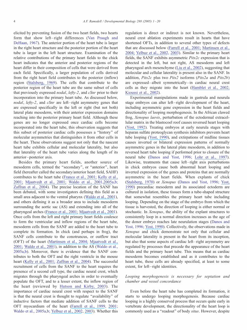
elicited by preventing fusion of the two heart fields, two hearts
form that show left– right differences (Van Praagh and
DeHaan, 1967). The anterior portion of the heart tube is larger
in the right heart structure and the posterior portion of the heart
tube is larger in the left heart structure. Examination of the
relative contributions of the primary heart fields to the chick
heart indicates that the anterior and posterior regions of the
heart differ in their composition of cells that were derived from
each field. Specifically, a larger population of cells derived
from the right heart field contributes to the posterior (inflow)
region (Stalsberg, 1969). The cells that contribute to the
posterior region of the heart tube are the same subset of cells
that previously expressed nodal, lefty-2, and cSnr prior to their
incorporation into the primary heart tube. As discussed below,
nodal, lefty-2, and cSnr are left–right asymmetry genes that
are expressed specifically in the left or right (but not both)
lateral plate mesoderm, with their anterior expression domains
reaching into the posterior primary heart field. Although these
genes are no longer expressed once cardiac cells become
incorporated into the heart tube, this observation suggests that
this subset of posterior cardiac cells possesses a ‘‘history’’ of
molecular asymmetries that distinguishes it from other cells in
the heart. These observations suggest not only that the nascent
heart tube exhibits cellular and molecular laterality, but also
that laterality of the heart tube varies along the length of its
anterior–posterior axis.
Besides the primary heart fields, another source of
mesoderm cells, termed the ‘‘secondary’’, or ‘‘anterior’’, heart
field (hereafter called the secondary/anterior heart field, SAHF)
contributes to the heart tube (Franco et al., 2001; Kelly et al.,
2001; Mjaatvedt et al., 2001; Waldo et al., 2001,2005a,b;
Zaffran et al., 2004). The precise location of the SANF has
been debated, with some investigators defining this field as a
small area adjacent to the ventral pharynx (Waldo et al., 2001),
and others defining it as a broader area to include mesoderm
surrounding the aortic sac (AS) and extending into all of the
pharyngeal arches (Franco et al., 2001; Mjaatvedt et al., 2001).
Once cells from the left and right primary heart fields coalesce
to form the ventricular and inflow regions of the heart tube,
mesoderm cells from the SANF are added to the heart tube to
complete its formation. In chick (and perhaps in frog), the
SANF cells contribute to the conotruncus, or outflow tract
(OFT) of the heart (Martinsen et al., 2004; Mjaatvedt et al.,
2001; Waldo et al., 2001), in addition to the AS (Waldo et al.,
2005a,b). Moreover, there is evidence that the SANF con-
tributes to both the OFT and the right ventricle in the mouse
heart (Kelly et al., 2001; Zaffran et al., 2004). The successful
recruitment of cells from the SANF to the heart requires the
presence of a second cell type, the cardiac neural crest, which
migrates through the pharyngeal arches in order to eventually
populate the OFT, and to a lesser extent, the inflow region of
the heart (reviewed by Hutson and Kirby, 2003). The
importance of cardiac neural crest with respect to the SANF
is that the neural crest is thought to regulate ‘‘availability’’ of
inductive factors that mediate addition of SANF cells to the
OFT myocardium of the heart (Farrell et al., 1999, 2001;
Waldo et al., 2005a,b; Yelbuz et al., 2002, 2003). Whether this
regulation is direct or indirect is not known. Nevertheless,
neural crest ablation experiments result in hearts that have
shortened OFTs, in addition to several other types of defects
that are discussed below (Farrell et al., 2001; Martinsen et al.,
2004; Yelbuz et al., 2002, 2003). Similar to the primary heart
fields, the SANF exhibits asymmetric Pitx2c expression that is
detected in the left, but not right, AS mesoderm and left
pharyngeal arch mesenchyme (Liu et al., 2002), suggesting that
molecular and cellular laterality is present also in the SANF. In
addition, Pitx2c plus two Pitx2 isoforms (Pitx2a and Pitx2b)
are expressed–albeit symmetrically–in cardiac neural crest
cells as they migrate into the heart (Hamblet et al., 2002;
Kioussi et al., 2002).
Experimental manipulations made in gastrula and neurula
stage embryos can alter left–right development of the heart,
including asymmetric gene expression in the heart fields and
directionality of looping morphogenesis. In the African clawed
frog, Xenopus laevis, perturbation of the ectodermal extracel-
lular matrix in the blastocoel roof causes reversed heart looping
(Yost, 1992). Treating embryos at early neurula stages with
heparan sulfate proteoglycan synthesis inhibitors prevents heart
tube looping (Yost, 1990), and extirpations of midline tissues
causes inverted or bilateral expression patterns of normally
asymmetric genes in the lateral plate mesoderm, in addition to
reversed heart tube looping if performed prior to closure of the
neural tube (Danos and Yost, 1996; Lohr et al., 1997).
Likewise, treatments that cause left–right axis perturbations
in chick embryos cause both abnormal heart looping and
inverted expression of the genes and proteins that are normally
asymmetric in the heart fields. When explants of chick
(Stalsberg, 1970) or Xenopus (Danos and Yost, 1996; Yost,
1990) precardiac mesoderm and its associated ectoderm are
cultured in isolation, these tissues form a tube-shaped structure
that somewhat resembles the primary heart tube including
looping. Depending on the stage of the embryo from which the
tissue is harvested, the direction of looping is either normal or
stochastic. In Xenopus, the ability of the explant structures to
consistently loop in a normal direction increases as the age of
the donor embryo reaches late neurulation stages (Danos and
Yost, 1996; Yost, 1990). Collectively, the observations made in
Xenopus and chick demonstrate not only that cellular and
molecular laterality is present in the heart from its inception,
but also that some aspects of cardiac left–right asymmetry are
regulated by processes that precede the appearance of the heart
fields and the primary heart tube. This means that as cardiac
mesoderm becomes established and as it contributes to the
heart tube, these cells are already specified, at least to some
extent, for left–right identities.
Looping morphogenesis is necessary for septation and
chamber and vessel concordance
Even before the heart tube has completed its formation, it
starts to undergo looping morphogenesis. Because cardiac
looping is a highly conserved process that occurs quite early in
vertebrate development, the directionality of the heart loop is
commonly used as a ‘‘readout’’ of body situs. However, despite

Fig. 3. Congenital heart defects that are frequently associated with laterality
disease. A schematic depiction of a normal heart is shown in panel A. Red and
blue indicate oxygenated and deoxygenated blood, respectively, and purple
shading in panels B–G indicates mixing of oxygenated and deoxygenated
blood. A ventricular septal defect (VSD) is shown in panel B. An atrial septal
defect is shown in panel C. Double-inlet left ventricle (DILV) is shown in panel
D. Double-outlet right ventricle (DORV) is shown in panel E. Transposition of
the great arteries (TGA) is shown in panel F. Persistent truncus arteriosus (PTA)
is shown in panel G. All hearts are drawn in ventral view.
A.F. Ramsdell / Developmental Biology 288 (2005) 1–20 5
it being one of the most widely recognized aspects of cardiac
asymmetry, it is often less well appreciated that looping is a
complex process involving both bending and rotational move-
ments (reviewed by Manner, 2000). As the heart tube continues
to elongate, it first develops a dextral loop (‘‘d’’ loop) that
results from the coordinated activities of ventral bending and
rightward rotation. Dextral looping occurs concomitantly with
increased growth at the outer vs. inner curvature, a process that
accompanies chamber formation and that ultimately causes the
heart tube to take on a ‘‘C’’-shaped appearance (Christoffels et
al., 2000; Rumyantsev, 1977; Thompson et al., 1990). Under
the influence of the left–right body axis, the dextral loop
normally orients to the right side of the embryonic midline,
aligning the primordial cardiac chambers to face the outer
curvature (Christoffels et al., 2000), and it is this aspect of
looping that most investigators equate with ‘‘rightward’’
looping. Thereafter, the dextrally looped heart transitions to
an ‘‘S’’ shape, a process that shifts the ventricular bend caudally
toward the atria. The final phase of looping morphogenesis is
characterized by the ‘‘wedging’’ of the distal OFT toward the
right atrium. At this point of development, the heart has lost its
tubular character, and the anterior (arterial) and posterior
(venous) poles of the heart are brought together in close
proximity. Current models of the ‘‘biomechanics’’ of looping
suggest that forces that are present on both the left and right
sides of the heart tube drive looping morphogenesis. Impor-
tantly, the left- and right-side forces are thought not to be
equivalent and are thought to differ at the cranial vs. caudal
aspects of the heart, such that the cranial portion of the tube
undergoes a rightward rotation and the caudal portion of the
tube undergoes a leftward rotation (Manner, 2004; Voronov et
al., 2004). The opposing polarity of rotations at the two ends of
the tube implies that asymmetry genes that control looping may
be expressed in opposite left–right patterns at the distal ends of
the heart tube (Manner, 2004; Voronov et al., 2004).
It should be emphasized that both the process of looping per
se and the directionality of the loop are important for normal
heart development. Because most cardiac structures arise from
cells derived from more than one area of the heart tube, the
significant outcome of looping is to rearrange regions of the
heart tube so they are appropriately positioned for proper
formation and alignment of chambers, valves, and septa (Fig.
2B). Thus, although left–right differences in the ventricles are
established by anteroposterior, rather than left–right patterning
processes (Franco et al., 2001; Thomas et al., 1998), the
directionality of looping determines whether the morphological
left ventricle underlies the left atrium, and the morphological
right ventricle beneath the right atrium. When the topological
situs of the ventricles is correct, the heart is said to have atrio-
ventricular (A-V) concordance.
In addition to A-V relations, looping also affects septation of
the heart. As looping occurs, cushion tissues (progenitor
valvuloseptal tissues) that have formed in the atrioventricular
canal (AVC) and the OFT regions of the heart are brought
together at the inner curvature of the looped heart tube. This
repositioning allows two important processes to occur (Fig. 2C).
First, it facilitates septation by bringing AV and OFT cushion
tissues together for formation of the atrioventricular septum
(AVS) and the OFT septum. If either end of the heart tube is
delayed or somehow impaired in its looping, then this would
cause cushion tissues to be out of alignment at the inner curvature
of the heart, increasing the chance for septal defects to occur
(Figs. 3B, G). Second, remodeling of the myocardium at the
inner curvature is necessary in order for the OFTand the AVC to
merge to form the future mitroaortic continuity. One potential
mechanism bywhich this remodeling occurs is through a process
termed myocardialization, in which the OFT cushion tissues at
the level of the inner curvature become invaded by overlying
myocardial cells (van den Hoff et al., 1999). Abnormal
myocardialization in hearts of trisomy 16 and other mouse
mutant models is correlated with incomplete looping and failure
to properly remodel the inner curvature, suggesting that
myocardialization plays an important role in facilitating the final

A.F. Ramsdell / Developmental Biology 288 (2005) 1–206
aspects of looping morphogenesis (Waller et al., 2000; Bartram
et al., 2001; Boot et al., 2004). One probable outcome of
myocardialization, therefore, is that it allows completion of
wedging, which in turn, is necessary to establish properly aligned
inflow and outflow tracts. In addition, this process also creates
the muscular outflow septum below the level of the valves.
Differentiation of the inflow tract (IFT) and OFT occurs
during wedging. In the IFT, the common AVC must become
divided into left and right components by an AVS that shifts
rightward during the final phase of looping to become
positioned directly atop the ventricular septum. This results
in alignment of the AVC with the ventricles. If the IFT does not
fully undergo this rightward shift, a condition known as
double-inlet left ventricle (DILV) persists (Fig. 3C). DILV
results in persistence of blood flowing into the left ventricle
from both the left and right atriums.
Meanwhile, at the OFT region of the heart, the conotruncus
shifts leftward and simultaneously twists 180- to become
positioned atop the AVS. This repositioning of the conotruncus
serves two purposes. First, it brings proximal conal cushion
tissues into alignment with AV and ventricular cushions so that
septation can finish. Second, the rotational movement of the
conotruncus is necessary to position the future base of the aorta
and pulmonary artery (which form from the distal conotruncus
and the AS) with the appropriate ventricles. Failure to correctly
align the conotruncus during looping morphogenesis can result
in double-outlet right ventricle (DORV), a condition in which
the right ventricle communicates with both the aorta and the
pulmonary artery and the left ventricle has no outlet (Fig. 3D).
This is different from the type of defect that would occur if the
rotational aspect of conotruncal wedging was disturbed.
Defects specifically in its rotational component would misalign
the base of the aorta and the pulmonary artery with the left and
right ventricles, resulting in a condition called transposition of
the great arteries (TGA) (Fig. 3E).
In instances where normal conotruncal wedging and rotation
happen, an outflow defect known as persistent truncus arterious
(PTA) still can occur (Fig. 3F). The base of the aorta and the
pulmonary artery form from the distal portion of the conotruncus
plus the AS. Both the AS and the conotruncus must become
divided in order to separate systemic and pulmonary blood flow
through this region. When this septation process does not occur
normally, PTA is the result. It has been appreciated for many
years that aorticopulmonary septation requires the population of
this region by the cardiac neural crest that pass through the
pharyngeal arches to form the APS, which divides the AS
(Nishibatake et al., 1987). Specifically, neural crest arising from
between the fourth and sixth pharyngeal arches extends into the
AS and ultimately merges with the fused OFT cushion tissues.
Perturbation of this process in vivo, by neural crest cell ablation,
results in PTA (Nishibatake et al., 1987).
Lateralization and differentiation of the inflow tract and AV
canal
Unlike the ventricles, which form in ‘‘series’’, the atria derive
from a common progenitor, the common atrium, which must
become divided into two chambers with distinct, left–right
features (Anderson, 1992; Min et al., 2000). In Xenopus, this
process of division may be related to differences in atrial cells
that derive from the left vs. right primary heart field (Gormley
and Nascone-Yoder, 2003). Once differentiated, the morpho-
logical right atrium contains pectinate muscles in its atrial
appendage and receives the IVC, and the morphological left
atrium contains a trabeculated appendage that lacks pectinated
muscle and receives the pulmonary vein. Failure to achieve one
or the other of these lateralized differences during division of
the common atrium is the basis of atrial isomerism.
Studies of the interatrial septum (IAS) suggest that the left–
right differences that arise during lateralization of the common
atrium also can be related to the existence of two different cell
populations that are present in this region. The IAS is derived, in
large part, from a myocardial infolding of the left atrial wall,
followed by transformation of some IAS cells to mesenchyme
(Wessels et al., 2000). Myocardial cells of the IAS share
common characteristics, such as creatine kinase B and Pitx2c
expression with cells in the left, but not right, atrial wall,
indicating a molecular asymmetry that is present in the common
atrium prior to its differentiation (Franco and Campione, 2003;
Liu et al., 2002; Wessels et al., 2000). Because the IAS
originates from the cell population specified for ‘‘leftness’’, in
instances where left-side signaling pathways are impaired (e.g.,
right isomerism), it would be predicted that structures derived
from the ‘‘left’’ cell population will not form. Consistent with
this prediction, there is a significant deficiency, if not complete
absence, of the IAS in hearts of many individuals afflicted with
right atrial isomerism (Bowers et al., 1996).
Whether lateralized processes can influence other aspects of
cardiac septation has not been investigated; however, several
features of cardiac cushion formation (progenitor valvuloseptal
tissue) in the AVC region suggest that this is possible. Despite
their dorsal and ventral anatomic positions in the looped heart,
the original superior (dorsal) and inferior (ventral) AV
endocardial cushions form from the original left and right
sides of the AVC (Lamers and Moorman, 2002; Moreno-
Rodriguez et al., 1997). Consistent with their initial left–right
origins, the inferior and superior cushions exhibit distinct
properties throughout septation morphogenesis, including
differences in temporal proliferative rates, spatial distribution,
and absolute amounts of mesenchymal tissue formed (Lamers
and Moorman, 2002; Moreno-Rodriguez et al., 1997). Addi-
tionally, myocardium in the AV canal exhibits left, but not
right, side Pitx2c expression (Campione et al., 2001). These
types of cellular and molecular left–right differences suggest
that similar to the common atrium, the AVC region also is
lateralized. Laterality disturbances in the AVC would be
predicted to affect endocardial cushion tissue formation,
ultimately increasing risk for valvuloseptal defects. By analogy
to the AVC and the common atrium, it is tempting to speculate
that the AS and conotruncus similarly develop a cellular and
molecular laterality that influences their subsequent division
into the base of the outlet vessels. If so, this could represent a
process that, if affected by abnormalities in left–right pattern,
could result in OFT defects such as PTA and/or TGA.

A.F. Ramsdell / Developmental Biology 288 (2005) 1–20 7
Asymmetric tissue regression patterns the aortic arches and
venous return
In addition to lateralization and looping, the third type of
asymmetry that occurs during normal heart development is
unilateral regression of blood vessels that connect with the
inflow and outflow portions of the heart. This feature is the
basis for the complex patterned regression and persistence of
the six pairs of aortic arch (AA) arteries and the aorta (Davies
and Guest, 2003). Initially, the AA arteries develop as a series
of six bilaterally paired vessels that connect with the paired
dorsal aortae (Fig. 2D). During AA artery remodeling, the first
two pairs of AA arteries regress into capillary beds. The third
pair of AA arteries persists and eventually becomes the paired
common carotid arteries. In contrast to the symmetric fates of
AAs 1–3, the left artery of the fourth pair of AA arteries
contributes to the aortic arch (in mammals), and the right artery
contributes to the right subclavian artery. The fifth pair of AA
arteries completely regresses or fails to form, and the sixth pair
of AA arteries persists only on the left side to contribute to the
pulmonary artery and the truncus arteriosus. At the inflow
region of the heart, similar mechanisms of regression/persis-
tence operate to pattern the paired posterior cardinal, sub-
cardinal, and supracardinal veins, which ultimately give rise to
the right-side inferior vena cava (Fasouliotis et al., 2002) (Fig.
2E). Aberrant regression/persistence patterns in the AA arteries
and the cardinal veins therefore can result in many types of AA
anomalies or abnormal venous return, depending on which
particular vessels are affected (Ruscazio et al., 1998).
Left–right patterning events upstream of cardiac
development
Because cardiac laterality defects nearly always occur in
conjunction with laterality defects in one or more other organs,
this indicates that the causative perturbation is one that occurs
early in development and that precedes organogenesis. Awidely
held view of left–right development is that it proceeds as a three
‘‘step’’ process. The first is left–right axis determination, which
establishes initial asymmetry in the embryo that is in correct
alignment with the other two body axes. Thereafter, left–right
positional information emanating from this axis must propagate
throughout the embryo over a wide range of developmental
stages, so that this information becomes relayed to each
emerging cell and tissue type. Finally, once organogenesis
begins, cells then must interpret and respond to the global left–
right ‘‘blueprint’’ in order to translate this information into
anatomical asymmetries. Errors in any of these three steps can
result in cardiac (and other) laterality defects. The nature of the
upstream signaling molecules that convey left–right patterning
information in each of the three ‘‘steps’’ remained for the most
part unknown until the pivotal 1995 discovery that sonic
hedgehog and activin can function as left–right asymmetry
genes in the chick (Levin et al., 1995). Since this time, the field
of vertebrate left–right development has rapidly grown to
recognize dozens more genes, as well as many types of cell–cell
signaling interactions, that act in concert to establish left–right
asymmetries in the embryo. Molecules and inductive signaling
processes that are central to current models of vertebrate left–
right development are discussed below.
Breaking bilateral symmetry: models of left – right axis
determination
In Xenopus, there is a patch of tissue located in the dorsal part
of the blastopore, called the dorsal lip, that is capable of initiating
gastrulation and directing complete secondary axis formation
when transplanted to a ventral region in a host embryo. Because
of the unique inductive properties of this tissue, the dorsal lip is
historically known as the ‘‘organizer’’. Functionally equivalent
structures exist in mammals (the node), avians (Hensen’s node),
and zebrafish (shield). Implicit in the patterning properties
observed in grafted organizer/node tissue is that the organizer/
node is a source of positional information for the different cell
types that it induces. Direct evidence for node involvement in
left–right development was first derived from studies in chick
and mouse, in which a series of grafting and extirpation
experiments indicated that the node is both necessary and
sufficient to direct left–right asymmetry of the body plan
(Davidson et al., 1999; Pagan-Westphal and Tabin, 1998). In
chick, the node is not the first source of left–right asymmetry
information; but rather, tissues adjacent to the node impart left–
right pattern that is in turn relayed by the node to surrounding
tissues as development proceeds (Pagan-Westphal and Tabin,
1998). In the mouse embryo, node ablation during late
gastrulation results in embryos with normal anterior–posterior
and dorsoventral development, but abnormal left–right devel-
opment, highlighting its important function in propagating left–
right patterning information (Davidson et al., 1999).
The earliest molecular aspect of left–right axis determina-
tion that is clearly conserved among all vertebrates is the
asymmetric, left-side expression of nodal. Nodal is a TGFhfamily member that is expressed in the left half of the node,
followed by the onset of a wide domain of expression in the
left, but not right, lateral plate mesoderm. Normal, left-side
only nodal expression is required for normal left–right
development in all species thus far examined (Collignon et
al., 1996; Hyatt et al., 1996; Levin et al., 1995; Lohr et al.,
1997; Lowe et al., 1996; Rebagliati et al., 1998b; Sampath et
al., 1997). Bilateral, absent, or right-side nodal expression
patterns are observed in iv/iv mice, a classic animal model of
heterotaxy (Collignon et al., 1996; Lowe et al., 1996). In inv/
inv mice, which exhibit situs inversus, inverted (right-side)
nodal expression is observed (Collignon et al., 1996).
Consistent with these findings, direct perturbation of nodal
expression in mouse results in situs ambiguus, indicating that
left– right development of the heart and visceral organs
requires restricted, left-side nodal activity (Brennan et al.,
2002). Studies of nodal homologs and components of the nodal
signaling pathway in Xenopus and zebrafish have corroborated
its central role in establishing vertebrate left–right asymmetries
(Ahmad et al., 2004; Hashimoto et al., 2004; Lohr et al., 1997,
1998; Rebagliati et al., 1998a,b; Sampath et al., 1997; Schier,
2003; Schier and Shen, 2000).

A.F. Ramsdell / Developmental Biology 288 (2005) 1–208
Experimental evidence derived from mouse, chick, and
Xenopus has led to a number of models that seek to explain the
steps of left–right axis determination that operate upstream of
nodal expression. In the mouse, ‘‘nodal flow’’, a mechanism
that involves ciliary function in node cells, is the prevailing
model. Null mutations in genes that are necessary for cilia
formation and/or function (e.g., iv (a.k.a. left–right dynein), inv,
Kif3-A, Kif3-B, HFH-4, RFX3, D2LIC) result in pronounced
laterality defects (Bonnafe et al., 2004; Brody et al., 2000; Chen
et al., 1998; Marszalek et al., 1999; Morgan et al., 1988; Nonaka
et al., 1998; Rana et al., 2004; Supp et al., 1997; Watanabe et al.,
2003). A role for ciliary genes was not necessarily unexpected,
since much earlier studies of Kartegener’s syndrome (a situs
inversus phenotype) had revealed an association between
human laterality defects and ultrastructural defects in cilia
(reviewed by Palmblad et al., 1984). Stunning experiments
performed with video microscopy have shown that motile cilia
present in the center of the mouse node propagate directional
fluid flow, and furthermore, that this flow is abnormal in several
mouse models bearing null mutations in ciliary motor proteins
(Nonaka et al., 1998, 2002; Okada et al., 1999; Watanabe et al.,
2003). One widely held interpretation is that nodal flow directs
left–right axis formation by causing asymmetric accumulation
of a diffusible morphogen that, in turn, launches widespread
asymmetric gene expression of factors such as nodal. In support
of this model, it has been shown that nodal cilia in mouse,
rabbit, zebrafish, and medakafish exhibit a posterior tilt that is
thought to result in much deeper ciliary contact with nodal fluid
in the right-to-left portion of ciliary rotational beating, a
phenomenon that in turn could account for asymmetric
Fig. 4. Nodal flow models proposed for mouse and zebrafish. In mouse (A), there ar
express left – right dynein (lrd) (green) and polycystin-2 (red), propagate directional
but not left – right dynein. In response to the directional fluid flow, Ca2+ levels beco
regulate left-side nodal expression (B). In zebrafish (C), dorsal forerunner cells expr
right defects, as do mutations in genes that are necessary for these cells to form K
develop cilia that propagate directional fluid flow, which in turn, is proposed to
mesoderm. Panels A and B courtesy of M. Brueckner (McGrath et al., 2003) and p
deposition of components swept by nodal flow (Kramer-Zucker
et al., 2005; Okada et al., 2005). Direct evidence for the ability
of nodal cilia to generate an asymmetric gradient was
demonstrated by the introduction of fluorescently conjugated
dextran particles (comparable to protein ¨40 kDa in size),
which were found to distribute preferentially on the left side of
the rabbit node (Okada et al., 2005), and FGF, SHH, and
retinoic acid are factors that are proposed to be involved in
generating the morphogen gradient in the mouse node (Tanaka
et al., 2005). However, as detailed elsewhere (Hornstein and
Tabin, 2005; Levin, 2004; Wagner and Yost, 2000), there are
some inconsistencies between predictions of the nodal flow
morphogen model and the nature of the laterality defects present
in mice null for genes that are needed for ciliary formation or
function. With the discovery of a second population of nodal
cilia, the so-called ‘‘mechanosensory cilia’’, an alternative
model has been put forth. As reported by Brueckner and
colleagues (McGrath et al., 2003), the mouse node also contains
non-motile cilia that are located on its periphery and that detect
flow generated by the central, motile cilia (Fig. 4A). The net
result of this detection is a transient spike in intracellular Ca2+
levels in cells to the left of the node, which in turn culminates in
left-side nodal expression (Fig. 4B). In this alternative, ‘‘two-
cilia’’ model, defects in either or both types of cilia would cause
defective left–right axis determination. Consistent with this,
mice null for polycystin-2 –a gene that is mutated in polycystic
kidney disease (Mochizuki et al., 1996) and that also is
expressed by the non-motile, sensory cilia in the node (McGrath
et al., 2003)–exhibit abnormal left–right development in
addition to renal and pancreatic cysts (Pennekamp et al.,
e two populations of cilia located in the node. The centrally located cilia, which
fluid flow that is detected by peripherally located cilia that express polycystin-2
me elevated in cells located to the left of the node, which in turn is proposed to
ess lrd. Genetic mutations (oep, sur, ntl) that inhibit lrd expression cause left –
uppfer’s vesicle (ntl, spt). Once organized into Kuppfer’s vesicle, these cells
regulate asymmetric gene expression (nodal, lefty, pitx2) in the lateral plate
anel C courtesy of H. J. Yost (Essner et al., 2005).

A.F. Ramsdell / Developmental Biology 288 (2005) 1–20 9
2002). Exactly how left–right differences in Ca2+ signaling
regulate nodal asymmetry still needs to be defined, and it has
been suggested that decreased levels on the right side of the
node might function to repress nodal expression on this side
(McGrath et al., 2003). The recent findings that inversin (the
protein encoded by inv) blocks canonical Wnt signaling and
that fluid flow can cause increased levels of inversin in ciliated
cells suggest that important roles for these two factors in ciliary
function and ultimately, regulation of nodal asymmetry, might
also be found (Simons et al., 2005).
As details unfolded with the nodal flow model in the mouse,
efforts weremade to determine whether this mechanism operates
in other vertebrates. ‘‘Nodal’’ cilia were soon discovered in
chick, frog, and zebrafish (Essner et al., 2002). Functional
studies in zebrafish indicate that similar to the nodal cilia models
in mouse, directional fluid flow is a critical aspect of left–right
asymmetry determination in this species (Essner et al., 2005;
Kramer-Zucker et al., 2005). In the zebrafish, a small population
of cells known as dorsal forerunner cells migrate at the leading
edge of the shield (‘‘node’’). Near the end of gastrulation, these
cells involute and form a structure of undefined function called
Kuppfer’s vesicle. Defects in dorsal forerunner cell migration or
interference with Kuppfer’s vesicle formation causes left–right
defects (Amack and Yost, 2004) and ciliated cells in Kuppfer’s
vesicle express LRD, which is necessary for their ability to
generate directional fluid flow in this region (Essner et al., 2005)
(Fig. 4C). As in mouse, it is proposed that unidirectional fluid
flow (in Kuppfer’s vesicle) regulates asymmetric nodal expres-
sion in lateral tissue via directed accumulation of an unknown,
left-side determinant (Essner et al., 2005).
Fig. 5. Models of axis initiation in Xenopus and chick. (A) A summary of process
Beginning with the one-cell stage embryo, cortical rotation affects formation of bo
proteins and physiological processes become restricted in expression and/or activity
mesoderm is regulated by left-side Vg1 signaling, and an opposing, right-side B
functional relationships but do not necessarily imply direct interactions; question ma
for details. (B) A summary of processes and genes that function upstream of left-side
node drives a transient, left-side increase in extracellular calcium, which in turn ac
hedgehog expression, is required for left-side nodal expression; however, the relation
side and right-side pathways, as well as midline influences, reinforce/repress nodal e
but do not necessarily imply direct interactions. See text and references cited withi
In Xenopus, there is abundant evidence that molecular
asymmetry is established at stages of development that precede
detection of ‘‘nodal’’ cilia (Fig. 5A). In the cleavage stage
Xenopus embryo, two molecular asymmetries are present: a
fusicoccin receptor, called the 14-3-3E protein, is expressed in
right, but not left, blastomeres at the 2–4 cell stage (Bunney et
al., 2003), and an H+/K+-ATPase pump is transiently asym-
metrically expressed in the right, but not left, ventral cell of the
4-cell stage embryo (Levin et al., 2002). Whether there is
functional overlap between these two proteins is not clear (14-3-
3 proteins control a variety of H+ pumps and ion channels in
other systems); but, regardless, assays employing inhibitors of
either the 14-3-3E protein or the H+/K+-ATPase pump result in
situs ambiguus as well as abnormal nodal expression patterns
(Bunney et al., 2003; Levin et al., 2002). The asymmetric
expression of the H+/K+-ATPase pump results in differential
left– right pH and voltage gradients; these gradients are
proposed to set up left-side accumulation of small molecule
morphogen(s)–serotonin is a recently identified candidate
(Fukumoto et al., 2005)–via unidirectional movement through
gap junctions, which have been shown to be necessary for
normal left–right patterning in Xenopus (Levin and Mercola,
1998). One possible role for motor proteins such as lrd, inv, and
Kif3-B is that they function as cytoplasmic transporters to
localize proteins and mRNAs involved in establishing left–
right asymmetry, e.g., the 14-3-3E protein and the H+/K+-
ATPase (Levin, 2004). Implicit in this model is that the
cytoskeleton is oriented in alignment with the future left–right
axis—a phenomenon that is possible given that left–right organ
reversals have been linked with disturbances in the microtubule-
es and genes that function upstream of left-side nodal expression in Xenopus.
th the dorsoventral and left – right axes. During subsequent cleavages, various
to the left or right side of the embryo. Nodal expression in the left lateral plate
MP signaling pathway represses right-side nodal expression. Arrows denote
rks indicate possible functional interactions. See text and references cited within
nodal expression in chick. Asymmetric depolarization of cells to the left of the
tivates left-side notch signaling. Notch signaling, in addition to left-side sonic
ship between these two pathways, if any, is not defined. A cascade of both left-
xpression in the lateral plate mesoderm. Arrows denote functional relationships
n for details.

A.F. Ramsdell / Developmental Biology 288 (2005) 1–2010
dependent, cortical rotation of the first cell cycle in Xenopus
(Yost, 1991). Although much remains to be tested, it is clear that
molecular asymmetry in Xenopus is established at least within
the first few cell cycles, well before the formation of the
Organizer and the appearance of ‘‘nodal’’ cilia. Thus, if cilia do
play a role in left–right development in Xenopus, it is very
likely that they participate in relaying left–right patterning cues,
rather than initiating the left–right axis.
As in Xenopus, studies in chick do not indicate a role for
nodal flow in establishing left–right asymmetry beyond the
circumstantial identification of cilia present in Hensen’s node.
In fact, a previous study implicated an earlier source of left–
right asymmetry signals in the embryo by showing that tissues
lateral to the node inductively interact with the node to specify
its left–right identity (Pagan-Westphal and Tabin, 1998).
Although the nature of the signal(s) that induce node left–
right identity is not known, they probably are involved in
regulating pH and voltage gradients, which as in Xenopus, are
required to set up early asymmetry in chick (Fig. 5B). In the
chick, there are distinct boundaries of gap junctional commu-
nication surrounding the node as well as asymmetric activities
in the H+/K+-ATPase pump that result in left-side depolariza-
tion of cells to the left of the node (Levin et al., 2002). The H+/
K+-ATPase driven depolarization causes a transient spike in
left-side extracellular Ca2+ levels (as opposed to intracellular
Ca2+ levels that are regulated by nodal flow), which
preferentially activates left-side notch signaling (Raya et al.,
2004). The latter occurs through modulating the affinity of
notch for its ligands, Dll1 and Srr1, an interaction that is
temporally influenced by locally expressed waves of lunatic
fringe (Przemeck et al., 2003; Raya et al., 2004). Notch
signaling is required for asymmetric nodal expression (Raya et
al., 2004); whether this regulation is direct or through
activation of other factors that are clearly involved in mediating
nodal expression (e.g., activin, sonic hedgehog) remains to be
determined. It is alternatively possible that notch acts in a
parallel pathway to these other factors in controlling nodal
expression. In mouse, notch-mediated induction of nodal
expression is direct, although how this interaction relates to
other processes of left–right axis determination in this
species, particularly nodal flow, is not known (Krebs et al.,
2003; Raya et al., 2003). In zebrafish, both notch signaling
and H+/K+-ATPase activity function in left–right development
prior to the formation of Kuppfer’s vesicle (and hence,
asymmetric nodal expression), suggesting that the role of nodal
cilia in this species is to relay previously established asymmetric
pattern (Kawakami et al., 2005).
In addition to asymmetric H+/K+-ATPase activity in
Xenopus, there is abundant evidence that left–right develop-
ment in the frog also requires TGFh-related signaling mediated
by a pathway comprised of Vg1, ALK4 (a type I Vg1 receptor),
and syndecan-2 (a co-factor that facilitates Vg1-ALK4 signal-
ing) (Chen et al., 2004; Hyatt and Yost, 1998; Kramer and Yost,
2002) (Fig. 5A). Ectopic activation of this pathway, either
through overexpression of mature Vg1 ligand or constitutively
active ALK4, results in a population of embryos exhibiting
predominately right-side or bilateral nodal expression and situs
inversus. Interruption of this pathway, either through interfer-
ence with Vg1-ALK4 signaling or syndecan expression or
function, causes abnormal nodal expression and situs ambiguus
(Chen et al., 2004; Kramer and Yost, 2002; Kramer et al., 2002;
Ramsdell and Yost, 1999). Elegant studies performed with
temporally regulated dominant-negative forms of syndecan-2
pinpointed the necessity of this pathway during gastrulation
(Kramer and Yost, 2002), which is the same developmental
window in which ALK4 expression is detected in the organizer
of the frog embryo (Chen et al., 2005). Consistent with these
findings, syndecan-2 is asymmetrically expressed to the right
side of Hensen’s node (Fukumoto and Levin, 2005), suggesting
conservation of TGFh signaling function downstream of the
initial symmetry-breaking events. Since the discovery of a role
for Vg1 and components of its signaling pathway in mediating
left–right axis determination in Xenopus, it has been unclear
though, how this pathway might be mechanistically linked to
the other processes upstream of nodal expression. The recent
discovery that ALK4 signaling in Xenopus can induce
expression of notch and its ligands, delta-1 and delta-2 (Abe
et al., 2004), raises the possibility that the Vg1-ALK4 pathway
could function to link asymmetric pH and voltage gradients
with notch pathway activation. Because null mutations in the
mouse Vg1 orthologue, Gdf1, illustrate that this pathway also is
necessary for left–right axis determination in this species (Wall
et al., 2000), studies aimed at coupling the Vg1/Gdf1 pathway
with activation of notch signaling are logical and potentially
very interesting directions for future investigation.
Maintenance of asymmetric nodal expression and relay of
left–right positional information to developing tissues
Once established, nodal expression in the perinodal area is
believed to activate left-side nodal expression in the lateral
plate mesoderm, and the latter domain of nodal asymmetry is
maintained by complex interactions among a number of
positive- and negative-acting regulators (Fig. 5B). This
regulatory network is best characterized in chick, where sonic
hedgehog induction of nodal expression initiates an autoregu-
latory loop that also requires the action of BMPs and caronte, a
member of the Cerberus/DAN family of BMP antagonists
(Monsoro-Burq and Le Douarin, 2000, 2001; Piedra and Ros,
2002; Rodriguez Esteban et al., 1999; Yokouchi et al., 1999).
BMP2 expression in the lateral plate mesoderm is symmetric
(Rodriguez Esteban et al., 1999; Yokouchi et al., 1999), and its
role in regulating nodal expression is to induce CFC-cripto, a
co-factor that is necessary for cellular responsiveness to nodal
(Fischer et al., 2002; Fujiwara et al., 2002; Schlange et al.,
2002). Caronte also is induced by BMP2; while the function of
caronte in this cascade is not certain, it might be to limit
(indirectly) the extent of nodal autoinduction (Piedra and Ros,
2002). Two important targets of nodal expression include
Pitx2, a bicoid-related transcription factor that is discussed in
detail below, and the homeobox gene NKX 3.2 (also termed
BapX1. In chick, NKX 3.2 is detected in the left, but not right,
lateral plate mesoderm, where its expression is positively
regulated by nodal BMP2 (Schlange et al., 2002; Schneider et

A.F. Ramsdell / Developmental Biology 288 (2005) 1–20 11
al., 1999). Despite its inverted expression pattern in mouse
(Schneider et al., 1999), null mutations nevertheless are
associated with defects in laterality of the spleen and pancreas
(Hecksher-Sorensen et al., 2004). In addition to nodal, lefty-1
and lefty-2 are two other TGFh family members which are
involved in relay of left–right pattern and which are expressed
in the left half of the prospective floor plate, and the left lateral
plate mesoderm, respectively (Bisgrove et al., 1999; Meno et
al., 1996, 1997, 1998). Both lefty proteins function as nodal
antagonists to prevent the spread of left-side signals to the
opposite side of the embryo (Branford and Yost, 2004; Cheng
et al., 2000; Meno et al., 1998; Thisse and Thisse, 1999). As
observed for nodal, lefty-2 expression is similarly altered in iv/
iv and inv/inv mice, and a number of experimental perturba-
tions of nodal activity also cause abnormal lefty-1 and lefty-2
expression patterns (Meno et al., 1996, 1997). On the right side
of the node, a series of signaling pathways is necessary for
repression of right-side nodal expression and for induction of
cSnr, a transcription factor that is normally restricted to the
right lateral plate mesoderm (Isaac et al., 1997). Beginning
with right-side activin signaling, which is necessary for BMP4
expression, BMP4 in turn induces FGF8 and PCL2 expression,
the latter which functions to represses right-side sonic
hedgehog (Boettger et al., 1999; Wang et al., 2004). BMP2
signaling also is active on the right side of the embryo, where it
cooperatively functions with the FGF8 pathway to induce cSnr
expression (Boettger et al., 1999).
The importance of BMP, FGF, and nodal antagonists in
maintaining left-side specific nodal expression is suggested by
functionality of these pathways in other vertebrate species. In
mouse, BMP4 is required for left-side nodal expression
(Fujiwara et al., 2002) and null mutations in Cerl-2, which
encodes a novel nodal antagonist, result in situs ambiguus
(Marques et al., 2004). In zebrafish, functional inhibition of the
nodal antagonist charon leads to bilateral nodal-related gene
expression and reversed heart looping (Hashimoto et al., 2004).
Null mutations in mouse ACRVRI (also termed ALK2), which
encodes a type I BMP receptor, result in situs ambiguus and
bilateral nodal expression, the latter probably due to loss of
lefty-1 expression in the midline (Kishigami et al., 2004). Gain-
and loss-of-function studies of ALK2 in Xenopus also indicate
that this pathway is necessary for normal left–right develop-
ment of the heart and visceral organs (Ramsdell and Yost,
1999). It should be emphasized that even though many of the
same signaling pathways that are active in chick, frog, and fish
are employed in mouse, this species differs from other
vertebrates in that some of its left–right signaling molecules
either act on the contralateral side of the embryo (e.g., FGF8 is
a left-side determinant in mouse), or are expressed in a
symmetric fashion while nevertheless being involved in left–
right patterning (e.g., sonic hedgehog) (Meyers and Martin,
1999). Other genes that affect nodal expression and heart and
visceral organ asymmetries in mouse include rotatin (Faisst et
al., 2002), cited2 (Bamforth et al., 2004), PKD2 (Pennekamp
et al., 2002), and BPI/PLUNC (Hou et al., 2004).
In conjunction with left- and right-side regulatory networks,
gene expression in the midline itself is important in regulating
asymmetric nodal expression. In addition to lefty-1, another
important midline gene is Zic3, a member of the GLI
transcription factor family that is normally present in midline
mesoderm and that is induced by activin/Vg1 signaling in
Xenopus (Kitaguchi et al., 2000, 2002; Nagai et al., 1997;
Purandare et al., 2002). Misexpression of either wild type or
deletion mutant forms of Zic3 in Xenopus embryos causes situs
ambiguus and predominantly bilateral or right-side nodal
expression (Kitaguchi et al., 2000). In mouse, Zic3 null
mutations result in a failure to maintain asymmetric nodal
expression in the node and situs ambiguus (Purandare et al.,
2002). Zic3 is thought to function in left–right development by
controlling formation of midline tissues such as the notochord.
One of the main consequences of maintaining a midline
barrier, via midline gene expression such as Zic3 or lefty-1, is
that cell populations positioned on opposite sides of the
midline propagate left–right patterning information indepen-
dently of one another. Clues to how the midline operates on a
cellular level to prevent left–right signals from reaching the
contralateral side come from notable studies in which cell death
in the primitive streak (Kelly et al., 2002) and a novel
population of ventral foregut cells derived from Hensen’s node
(Kirby et al., 2003) are shown to contribute to left–right
patterning activity. Further investigation of these two newly
identified midline cell populations and how they interface with
left–right signaling cascades should greatly increase our
understanding of the specific mechanisms employed at the
embryonic midline.
It should be noted that concomitant with the development of
left–right asymmetry, there are paired tissues located to either
side of the midline (e.g., presomitic mesoderm) that must
undergo symmetric morphogenesis. Exactly how this coordina-
tion occurs, and particularly how these tissues remain refractory
in response to opposing left- and right-side lateralizing
influences, has been a longstanding question. A series of very
recent studies demonstrate that, in chick, mouse, and zebrafish,
somite formation is labile to left–right signaling pathways, but
that under normal conditions, retinoic acid mediates synchro-
nized left and right side somitogenesis by preventing presomitic
mesoderm from responding to left–right asymmetric signaling
(Kawakami et al., 2005; Vermot et al., 2005; Vermot and
Pourquie, 2005).
‘‘Translating’’ left–right axis information into anatomical
asymmetry
The nodal Y Pitx2c pathway
The connection between asymmetric nodal expression in
the lateral plate mesoderm and cardiac asymmetry is the
regulation of downstream genes that have direct roles in heart
development. One such gene that is induced to be expressed in
the left lateral plate mesoderm by nodal and that is required for
normal heart development is Pitx2c (Fig. 6). Pitx2c is one of
three Pitx2 isoforms (Pitx2a, Pitx2b, and Pitx2c) that belong to
the bicoid group of paired homeobox transcription factors.
Pitx2 is a homolog of the human RIEG gene, whose mutation

Fig. 6. Conserved, left-side Pitx2c expression in the lateral plate mesoderm. Whole-mount in situ hybridization reveals left-side Pitx2c expression (arrowhead) in the
lateral plate mesoderm in mouse (A, ventral view), chick (B), tadpole (anterior to left and dorsal to top), and zebrafish embryo (D). Panel A courtesy of P. Tam
(Davidson et al., 1999), panels B and C courtesy of M. Blum (Campione et al., 1999).
A.F. Ramsdell / Developmental Biology 288 (2005) 1–2012
results in Rieger syndrome, a haploinsufficiency condition
characterized by dental hypoplasia, craniofacial dysmorphism,
umbilical stump protrusions, and ocular anomalies (Amendt et
al., 2000). Less frequently, defects in cardiac, limb, and
pituitary development are observed (Amendt et al., 2000).
Once induced by nodal (Logan et al., 1998; Long et al.,
2003; Piedra et al., 1998; Shiratori et al., 2001; Yoshioka et al.,
1998), Pitx2c expression in the left lateral plate mesoderm is
maintained by nkx2.5 (Shiratori et al., 2001). Similar to nodal,
a combination of right side only, bilateral, and absent
expression Pitx2c patterns is observed in experimentally
induced cases of situs ambiguus in mouse, chick, and frog
(Bamforth et al., 2004; Bisgrove et al., 2000; Boettger et al.,
1999; Branford et al., 2000; Chen et al., 2004; Piedra et al.,
1998; Schlange et al., 2001; St Amand et al., 1998; Yoshioka et
al., 1998). The observation that null mutations in many
different left–right axis genes result in a similar defective
left–right phenotype can be attributed to the downstream
effects of these genes on the nodal Y Pitx2c pathway. For
example, null mutations that result in absent Pitx2c expression
include cryptic, FGF8, gdf1, and nodal and the predominant
phenotype in these mice is right isomerism. Null mutations that
cause bilateral Pitx2c expression include lefty-2 and ACRIa/
ALK2 and these mice predominantly exhibit left isomerism.
Midline genes that are necessary for left–right development
also converge at the nodal Y Pitx2c pathway; shh and lefty-1
are two examples that cause abnormal nodal (and hence Pitx2c)
expression in null mutants. Mice that are null for all three Pitx2
isoforms (Pitx2abc) exhibit defective body wall closure, gut
malrotation, right pulmonary isomerism, and the spectrum of
cardiac defects that are usually associated with heterotaxy,
including DORV, TGA, common AV canal, ASD/VSD, and
anomalous venous return (Gage et al., 1999; Kitamura et al.,
1999; Liu et al., 2001, 2002). Right atrial isomerism, PTA, and
aortic arch anomalies such as right aortic arch or double aortic
arch are also sometimes present. Interestingly, despite the
numerous cardiac defects caused by Pitx2abc null mutation, all
of these can be rescued with hypomorphic Pitx2c alleles except
right atrial isomerism, which requires a higher dosage of Pitx2c
expression/activity (Liu et al., 2001). Consistent with these
findings, analysis of Pitx2c function in Xenopus has shown that
Pitx2c antisense oligonucleotides cause OFT, AVC, and atrial
septation defects (Dagle et al., 2003).
Pitx2c is implicated in regulating multiple aspects of cardiac
left–right development
Unlike nodal, Pitx2c is expressed during stages of heart
tube formation when it is detected exclusively in the left, but
not right, side of the developing heart (Campione et al., 1999;
Logan et al., 1998; Ryan et al., 1998; St Amand et al., 1998;
Yoshioka et al., 1998) (Fig. 7). Cardiac Pitx2c expression
persists through looping stages, where it is present in the entire
left side of the heart, spanning from the left atrium to the most
distal part of the outflow tract, reaching into the left side of the
SANF and the left pharyngeal arch mesenchyme (Liu et al.,
2002) (Fig. 7). In addition, Pitx2c plus two other Pitx2
isoforms (Pitx2a and Pitx2b) are expressed in migrating
cardiac neural crest (Hamblet et al., 2002; Kioussi et al.,
2002). During later stages, Pitx2c expression is observed in the
left atrioventricular canal, left atrium, interatrial septum, and
the left caval vein (Franco and Campione, 2003).
Initial studies showed that right side ectopic Pitx2c expres-
sion in chick and Xenopus causes reversed heart looping
(Campione et al., 1999; Logan et al., 1998; Piedra et al., 1998;
Ryan et al., 1998; Yoshioka et al., 1998). These findings,
coupled with the observations that Pitx2c is expressed by
components of other organs that undergo looping, suggested that
Pitx2c mediates looping morphogenesis (Campione et al., 1999;
Logan et al., 1998; Ryan et al., 1998). However, Pitx2abc and
Pitx2c null mutant mice do not exhibit reversed cardiac looping
(Gage et al., 1999; Kitamura et al., 1999), suggesting that Pitx2c
is dispensable for this aspect of the looping process. In Xenopus,
Pitx2c loss-of-function causes abnormal (though not necessarily
reversed) shifting of the OFT, confirming that although Pitx2c
can mediate later aspects of looping, it is not involved in
controlling initial directionality of looping. Errors in the leftward
shifting and/or rotation of the OFT are causatively linked with
DORV, TGA, and VSDs; therefore, the abnormal OFT looping
that is associated with impaired Pitx2c function could account
for these types of CHDs.
The mechanism by which Pitx2c regulates OFT looping is
undefined. During looping, the outlet region of the heart
undergoes lengthwise growth due to the addition of myocar-
dium derived from the SANF (Brand, 2003; Mjaatvedt et al.,
2001; Waldo et al., 2001). Thus, one possible function of
Pitx2c in OFT looping is that its expression in the SANF may

Fig. 7. Conserved, left-side Pitx2c expression during early stages of heart
development. Whole-mount in situ hybridization reveals left-side Pitx2c
expression (arrows) during initial formation of the heart tube in chick (A)
and in mouse (B). As the heart tube continues to elongate, Pitx2c expression
(arrows) is maintained in the left half of the chick (C) and mouse (D) heart.
During looping, Pitx2c expression is detected in the entire left half of the chick
heart, including the aortic sac (AS, arrow) (E) and in the entire left half of the
mouse heart (F), as seen along the inner curvature (asterisk). All panels ventral
views, with left (L) and right (R) indicated. Figures courtesy of M. Campione
and M. Blum (Campione et al., 2001).
A.F. Ramsdell / Developmental Biology 288 (2005) 1–20 13
somehow be involved in the recruitment of cells from the
SANF to the conotruncus. Alternatively, Pitx2c might mediate
OFT looping by affecting the cardiac neural crest. In Pitx2abc
null mice, the population of neural crest cells that contributes to
the heart appears decreased (Kioussi et al., 2002; Liu et al.,
2002). Although Pitx2 does not directly regulate migration of
neural crest cells, null mutations in Pitx2abc do reduce the
number of neural crest cells that accumulate in the OFT by
inhibiting proliferative expansion of this cell population
(Kioussi et al., 2002). This inhibition requires the upstream
activation a wnt/Dvl/h-catenin pathway that is responsible for
inducing Pitx2 expression (Kioussi et al., 2002). In other
mouse mutants (e.g., FGF8�/�, BMP4�/�), decreased
numbers of neural crest cells in the OFT have been associated
with defective OFT looping (Abu-Issa et al., 2002; Liu et al.,
2004). It has been proposed that the link between neural crest
cells and altered OFT looping is that neural crest cells
somehow regulate the factors that mediate the addition of
SANF cells to the outflow myocardium to the heart (Farrell et
al., 1999, 2001; Yelbuz et al., 2002, 2003). In turn, it has been
proposed that when cardiac neural crest cells are ablated, the
impaired recruitment of SANF cells to the OFT causes the OFT
to develop a shortened stature that impedes its ability to
normally loop (Farrell et al., 2001; Yelbuz et al., 2002, 2003).
Whether this model is correct is somewhat controversial
because it is difficult to reconcile that a shortened OFT can
simultaneously occur with PTA, a defect that is unequivocally
and causally linked to neural crest cell ablation. Thus, if the
function of Pitx2 in the neural crest is required for normal OFT
looping, it is alternatively possible that the neural crest plays a
role unrelated to SANF cell recruitment. Besides Pitx2, other
left–right asymmetry genes–ALK2, ALK4, and FGF8 –are
expressed in the cardiac neural crest, suggesting that neural
crest might be subject to left–right specification prior to or
during its migration (Abu-Issa et al., 2002; Chen et al., 2005;
Kaartinen et al., 2004; Kishigami et al., 2004). However,
whether expression of Pitx2 or other laterality genes by cardiac
neural crest cells is induced as part of the left–right signaling
cascade that specifies the midline remains to be determined.
With the availability of neural crest mutants in mouse and
zebrafish, as well as the ease of experimentally ablating neural
crest in chick and Xenopus, the different potential roles for
neural crest cells in cardiac looping could be tested using a
variety of molecular, embryological, and genetic approaches.
In addition to OFT looping, Pitx2c might play a role in
regulating septation in the IFT and OFT (Fig. 8). The
myocardium in the AVC exhibits left, but not right, side Pitx2c
expression (Campione et al., 2001), which is required for
migration of cells into cardiac cushions (Liu et al., 2002). The
origin of the Pitx2c expressing cells within the cushions is
probably the AV myocardium, which together with epicardial-
derived cells, is required to complete septation and valve
formation in the heart (Lamers and Moorman, 2002). Pitx2 also
appears to be required for migration of a small subset of
myocardial cells from the left to the right side of the OFT (Liu
et al., 2002); although the significance of this migratory
process is unknown, it does suggest that Pitx2 may be involved
in conferring cellular and molecular asymmetry to this portion
of the heart. With respect to the OFT, Pitx2abc null mice
exhibit PTA, a condition in which the conotruncus and AS are
not divided properly into the base of the aorta and pulmonary
artery by the APS (Kitamura et al., 1999; Lu et al., 1999; Liu et
al., 2001). This suggests that Pitx2 is necessary for septation of
the OFT. Finally, the expression of Pitx2c in cells that will
form the IAS suggests it too plays a direct role in atrial
septation, perhaps by affecting competency of cells to
transform to mesenchyme, as appears to occur in the AVC
region. Consistent with this idea, IAS defects are frequently
present in Pitx2c null mice (Liu et al., 2001).
Pitx2c and aortic arch anomalies
Aortic arch anomalies are frequently present in heterotaxy
patients, indicating that patterned regression and persistence of
vessels are governed by the left–right axis (Bowers et al., 1996).

Fig. 8. Conserved, left-side Pitx2c expression during later stages of heart
development. Whole-mount in situ hybridization reveals left-side Pitx2c
expression (arrows) during looping and septation stages of the heart in chick
and mouse. Ventral view of a HH stage 16 chick heart (A) and left lateral view
of an E10.0 mouse heart (B) showing expression in the left atrium (LA),
atrioventricular canal (AVC), ventricle (VEN), outflow tract (OFT), and aortic
sac (AS). Dorsal view of a HH stage 23 chick heart (C) and ventral view of an
E10.0 mouse heart (D) showing lack of Pitx2c expression in the right atrium
(RA), the right side of the OFT, and the original right side of the ventricular
chambers (positioned dorsally at this stage due to looping). Pitx2c is expressed
in the LA and the interatrial septum (IAS) as shown in the chick heart (E) and in
a cross-section of an E10.0 mouse heart (F). Figures courtesy of M. Campione
and M. Blum (Campione et al., 2001).
A.F. Ramsdell / Developmental Biology 288 (2005) 1–2014
However, virtually nothing is known about how this occurs, or
for that matter, even if vessels (including the endocardium of the
heart) truly possess any type of cellular or molecular asymmetry.
Despite their mesodermal origin, it is possible that blood vessels
themselves are not lateralized, but rather that they develop
asymmetric pattern in response to biomechanical feedback from
blood flow. An alternative, though not necessarily exclusive
possibility, is that vessels develop asymmetric pattern based on
signals derived from the tissues that induce their formation.
Consistent with this possibility, the assortment of pulmonary and
systemic venous anomalies that are frequently found in cases of
isomerism suggest that vascular endothelial cells are competent
to respond to the same set of left–right patterning signals,
regardless of which side of the midline they are located (Bowers
et al., 1996; Ruscazio et al., 1998). Moreover, vessel persistence
during vascular remodeling involves recruitment of cells that
interact with the endothelium to promote its survival and
differentiation. Failure to recruit support cells or disrupted
interaction between support cells and the endothelium would
result in vessel regression. Therefore, with respect to asymmetric
AA remodeling, laterality gene expression in pharyngeal arch
mesenchyme may be important. Pitx2c is expressed in the left,
but not right, pharyngeal arch mesenchyme (Liu et al., 2002),
and it has been suggested that Pitx2c null mutations could impair
recruitment, maintenance, or cross-talk between supporting cells
and endothelial cells in this region. Because Pitx2c is expressed
only by left-side AAs, the absence of Pitx2c expression would
be predicted to result in abnormal patterning on this side,
consistent with the right-side aortic arch, right-side ductus
arteriosus, and left inominate arteries that frequently occur in
Pitx2c null mice.
Genes in addition to Pitx2 that regulate cardiac left–right
morphogenesis
In addition to Pitx2c, BMP4 and flectin are two other genes
that play important roles in cardiac left–right morphogenesis. In
zebrafish and Xenopus, BMP4 is asymmetrically expressed in
the nascent heart tube (Chen et al., 1997), and transgenic studies
in Xenopus using cardiac-targeted inhibitors of BMP signaling
demonstrate that BMP4 is required for looping morphogenesis,
independent of Pitx2c function (Breckenridge et al., 2001).
Flectin is an extracellular matrix protein that is asymmetrically
expressed in chick, with predominant expression observed in the
left heart fields (primary and anterior), the left dorsal mesocar-
dium, and the left side of the heart tube during looping stages
(Linask et al., 2003; Tsuda et al., 1996, 1998). Ectopic Pitx2c
expression causes a rightward shift in flectin expression in the
heart fields and the dorsal mesocardium, coincident with
reversed or absent heart looping (Linask et al., 2002). Treatment
of embryos with CFC antisense oligonucleotides, which
likewise cause reversed or absent looping, also inverts the
asymmetry of flectin expression (Linask et al., 2003). However,
this treatment does not alter normal Pitx2c expression, indicat-
ing either that flectin is a downstream target of Pitx2c that is
directly mediating looping, or that flectin acts in a pathway
parallel to the nodal Y Pitx2 pathway. In addition to the role of
flectin in looping, these studies also showed that Pitx2c could
alter the position of the developing foregut relative to the
embryonic midline. As shown in these studies and in the
previously mentioned study of node-derived ventral midline
cells (Kirby et al., 2003), this finding is significant because the
orientation of heart looping is somehow regulated by cells of the
ventral floor of the foregut. Given that CFC antisense treatment
also causes a rightward shift of the ventral foregut relative to the
midline, this suggests that the ventral foregut may be a primary
tissue target in the relay of left–right axis signals.
Some ‘‘isolated’’ CHDs might actually be subtle laterality
defects
Although the majority of human laterality defects are
thought to be sporadic, it is clear that left–right defects also

A.F. Ramsdell / Developmental Biology 288 (2005) 1–20 15
can arise from environmental perturbations and from heritable
modes of transmission (Kuehl and Loffredo, 2002). Clinical
analyses of familial cases indicate three modes of inheritance
for laterality defects: X-linked, autosomal dominant (usually
with incomplete penetrance), and autosomal recessive (Bel-
mont et al., 2004; Kosaki and Casey, 1998). In many cases,
specific mutations have been mapped and include ZIC3,
ACVR2B, CRYPTIC/CFC1, LEFTYA, and NKX2.5 (Bamford
et al., 2000; Belmont et al., 2004; Gebbia et al., 1997; Kosaki
et al., 1999a,b). Because these genes also have been identified
or implicated in animal models of heterotaxy, this confirms that
our current models are (and presumably will continue to be) of
great value in understanding the etiology of human laterality
disease.
Observations made in animal models of heterotaxy on the
role of the midline in left–right development (reviewed by
Yost, 1998) have prompted investigators to look for clinical
correlations between human midline defects and laterality
disturbances (Goldstein et al., 1998; Morelli et al., 2001).
Intriguingly, in one of these studies, a significant association
was observed not only between laterality defects and midline
defects, but also between laterality defects and ‘‘isolated’’
cardiac defects (Morelli et al., 2001). The latter association
has been observed in both animal and human studies, leading
to the suggestion that some instances of isolated cardiac
defects may actually represent subtle, ‘‘forme fruste’’ laterality
defects (Goldmuntz et al., 2002). For example, in the cited-2
null mouse, which lacks Pitx2c expression in the left lateral
plate mesoderm and in the heart, embryos develop the cohort
of CHDs that is typical of heterotaxy (i.e., defects in OFT,
septal, and AA remodeling) in addition to abnormal body
turning, right atrial and pulmonary isomerism, and hypoplastic
spleen (Bamforth et al., 2004; Weninger et al., 2005).
Comparison of these mice with cited-2 null mutations made
on a mixed genetic background gave surprising results
(Bamforth et al., 2004). In the latter group, Pitx2c expression
was present in the left lateral plate mesoderm but absent or
reduced specifically in the OFT of the heart. These mice
exhibited DORV and VSD but laterality of other organs was
not affected. These results suggest that there are genetic
modifiers that can influence the cited-2 null phenotype by
directly or indirectly regulating Pitx2c expression. Moreover,
the occurrence of CHDs in the mixed background mutant
embryos suggests that some CHDs actually may result from
perturbations of left–right axis genes, even in instances of
otherwise normal body situs. As a direct test of this
hypothesis in a clinical setting, a population of patients with
TGA (but no overt laterality defects) was examined for
mutations in CFC1, a gene that is expressed in the OFT of the
embryonic mouse heart (Dono et al., 1993) and that is
required for cellular responsiveness to nodal signaling in the
lateral plate mesoderm, probably by functioning as a co-
receptor for nodal (Fischer et al., 2002; Fujiwara et al., 2002;
Schlange et al., 2002). A subset of patients afflicted with TGA
(but not laterality defects) was found to indeed harbor CFC1
mutations, indicating for the first time, a common genetic
etiology between human TGA and laterality disease (Gold-
muntz et al., 2002). Because many laterality genes identified in
animal models are known also to participate in processes of
cardiac morphogenesis, it is predicted that many more
seemingly ‘‘isolated’’ cardiac defects will be found in situs
solitus individuals that represent a mutation or other perturba-
tion in left–right axis gene(s).
Prospects
The past decade has brought immense progress in decon-
structing the complexities of left–right axis determination and
its relationship to cardiac development while at the same time
illuminating areas in which further investigation is much needed.
Studies in mouse, chick, frog, and zebrafish reveal that
establishing the left–right axis involves a number of intriguing
physiological processes that extend beyond the conventionally
studied, growth factor-mediated signaling pathways. Whether
processes of ‘‘nodal’’ flow, gap junctional communication, and
membrane voltage potential are unique for axis determination in
each species is an important but unresolved issue. Relatively
more is known about the role of the midline in left–right
patterning, but despite unraveling many of the complicated
genetic interactions, the specifics of which cell types are
engaged in relay of left–right signals are less well defined.
With regard to asymmetric morphogenesis, work on Pitx2c has
accelerated our understanding of the types of cardiac defects that
can arise from impaired function of a single laterality gene. Yet,
how this one gene actually regulates the three different endpoints
of cardiac left–right patterning (looping, regression/persistence,
and lateralization) is by no means definitively answered, and is
limited, in part, by our current understanding of fundamental,
morphogenetic processes that drive normal cardiac develop-
ment. Finally, there is exciting interplay between clinical
observations and experimental observations in animal models
of laterality disease, indicating that some seemingly ‘‘isolated’’
congenital cardiac defects might actually arise from disturbances
in left–right signaling pathway(s). Future progress in this latter
area is anticipated to be especially significant, as elucidating
common genetic etiologies will greatly advance our understand-
ing of the causes of many types of deleterious and life-
threatening congenital heart malformations.
Acknowledgments
Thanks to colleagues near and far for enjoyable discussions
of cardiac left–right development and to R. Markwald, D.
Bernanke, and C. Drake for comments on the manuscript.
Special thanks to Sue Tjepkema-Burrows for drawing the
schematic figures. Cardiac asymmetry research in the author’s
lab is supported by HL73270 (to A.F.R.) and a SC COBRE for
Cardiovascular Disease P20-RR-1634 (to Roger R. Markwald).
References
Abe, T., Furue, M., Myoishi, Y., Okamoto, T., Kondow, A., Asashima, M.,
2004. Activin-like signaling activates Notch signaling during mesodermal
induction. Int. J. Dev. Biol. 48, 327–332.

A.F. Ramsdell / Developmental Biology 288 (2005) 1–2016
Abu-Issa, R., Smyth, G., Smoak, I., Yamamura, K., Meyers, E.N., 2002. Fgf8 is
required for pharyngeal arch and cardiovascular development in the mouse.
Development 129, 4613–4625.
Ahmad, N., Long, S., Rebagliati, M., 2004. A southpaw joins the roster: the
role of the zebrafish nodal-related gene southpaw in cardiac LR asymmetry.
Trends Cardiovasc. Med. 14, 43–49.
Amack, J.D., Yost, H.J., 2004. The T box transcription factor no tail in ciliated
cells controls zebrafish left – right asymmetry. Curr. Biol. 14, 685–690.
Amendt, B.A., Semina, E.V., Alward, W.L., 2000. Rieger syndrome: a clinical,
molecular, and biochemical analysis. Cell. Mol. Life Sci. 57, 1652–1666.
Anderson, R.H., 1992. Understanding the nature of congenital division of the
atrial chambers. Br. Heart J. 68, 1–3.
Antin, P.B., Taylor, R.G., Yatskievych, T., 1994. Precardiac mesoderm is
specified during gastrulation in quail. Dev. Dyn. 200, 144–154.
Bamford, R.N., Roessler, E., Burdine, R.D., Saplakoglu, U., dela Cruz, J.,
Splitt, M., Goodship, J.A., Towbin, J., Bowers, P., Ferrero, G.B., Marino,
B., Schier, A.F., Shen, M.M., Muenke, M., Casey, B., 2000. Loss-of-
function mutations in the EGF-CFC gene CFC1 are associated with human
left – right laterality defects. Nat. Genet. 26, 365–369.
Bamforth, S.D., Braganca, J., Farthing, C.R., Schneider, J.E., Broadbent, C.,
Michell, A.C., Clarke, K., Neubauer, S., Norris, D., Brown, N.A.,
Anderson, R.H., Bhattacharya, S., 2004. Cited2 controls left – right
patterning and heart development through a Nodal-Pitx2c pathway. Nat.
Genet. 36, 1189–1196.
Bartram, U., Molin, D.G.M., Wisse, L.J., Mohamad, A., Sanford, L.P.,
Doetschman, T., Speer, C.P., Poelmann, R.E., Gittenberger-de Groot,
A.C., 2001. Double-outlet right ventricle and overriding tricuspid valve
reflect disturbances of looping, myocardialization, endocardial cushion
differentiation, and apoptosis in TGF-{beta}2-knockout mice. Circulation
103, 2745–2752.
Belmont, J.W., Mohapatra, B., Towbin, J.A., Ware, S.M., 2004. Molecular
genetics of heterotaxy syndromes. Curr. Opin. Cardiol. 19, 216–220.
Bisgrove, B., Essner, J., Yost, H.J., 1999. Regulation of midline development
by antagonism of lefty and nodal signaling. Development 126, 3253–3262.
Bisgrove, B.W., Essner, J.J., Yost, H.J., 2000. Multiple pathways in the midline
regulate concordant brain, heart and gut left – right asymmetry. Develop-
ment 127, 3567–3579.
Boettger, T., Wittler, L., Kessel, M., 1999. FGF8 functions in the specification
of the right body side of the chick. Curr. Biol. 9, 277–280.
Bonnafe, E., Touka, M., AitLounis, A., Baas, D., Barras, E., Ucla, C., Moreau,
A., Flamant, F., Dubruille, R., Couble, P., Collignon, J., Durand, B., Reith,
W., 2004. The transcription factor RFX3 directs nodal cilium development
and left – right asymmetry specification. Mol. Cell. Biol. 24, 4417–4427.
Boot, M.J., Steegers-Theunissen, R.P.M., Poelmann, R.E., van Iperen, L.,
Gittenberger-de Groot, A.C., 2004. Cardiac outflow tract malformations in
chick embryos exposed to homocysteine. Cardiovasc. Res. 64, 365.
Bowers, P.N., Brueckner, M., Yost, H.J., 1996. Laterality disturbances. Prog.
Pediatr. Cardiol. 6, 53–62.
Brand, T., 2003. Heart development: molecular insights into cardiac specifica-
tion and early morphogenesis. Dev. Biol. 258, 1–19.
Branford, W.W., Yost, H.J., 2004. Nodal signaling: CrypticLefty mechanism of
antagonism decoded. Curr. Biol. 14, R341–R343.
Branford, W.W., Essner, J.J., Yost, H.J., 2000. Regulation of gut and heart left –
right asymmetry by context-dependent interactions between Xenopus lefty
and BMP4 signaling. Dev. Biol. 223, 291–306.
Breckenridge, R.A., Mohun, T.J., Amaya, E., 2001. A role for BMP signalling
in heart looping morphogenesis in Xenopus. Dev. Biol. 232, 191–203.
Brennan, J., Norris, D.P., Robertson, E.J., 2002. Nodal activity in the node
governs left – right asymmetry. Genes Dev. 16, 2339–2344.
Brody, S.L., Yan, X.H., Wuerffel, M.K., Song, S.K., Shapiro, S.D., 2000.
Ciliogenesis and left – right axis defects in forkhead factor HFH-4-null
mice. Am. J. Respir. Cell Mol. Biol. 23, 45–51.
Bunney, T.D., De Boer, A.H., Levin, M., 2003. Fusicoccin signaling reveals 14-
3-3 protein function as a novel step in left – right patterning during
amphibian embryogenesis. Development 130, 4847–4858.
Burn, J., 1991. Disturbance of morphological laterality in humans. In: Bock,
G.R., Marsh, J. (Eds.), Ciba Found. Symp., vol. 162. Wiley, Chinchester,
pp. 282–296.
Campione, M., Steinbeisser, H., Schweickert, A., Deissler, K., van Bebber, F.,
Lowe, L.A., Nowotschin, S., Viebahn, C., Haffter, P., Kuehn, M.R., Blum,
M., 1999. The homeobox gene Pitx2: mediator of asymmetric left– right
signaling in vertebrate heart and gut looping. Development 126, 1225–1234.
Campione, M., Ros, M.A., Icardo, J.M., Piedra, E., Christoffels, V.M.,
Schweickert, A., Blum, M., Franco, D., Moorman, A.F., 2001. Pitx2
expression defines a left cardiac lineage of cells: evidence for atrial and
ventricular molecular isomerism in the iv/iv mice. Dev. Biol. 231, 252–264.
Chen, J.N., van Eeden, F.J., Warren, K.S., Chin, A., Nusslein-Volhard, C.,
Haffter, P., Fishman, M.C., 1997. Left– right pattern of cardiac BMP4 may
drive asymmetry of the heart in zebrafish. Development 124, 4373–4382.
Chen, J., Knowles, H.J., Hebert, J.L., Hackett, B.P., 1998. Mutation of the
mouse hepatocyte nuclear factor/forkhead homologue 4 gene results in an
absence of cilia and random left– right asymmetry. J. Clin. Invest. 102,
1077–1082.
Chen, Y., Mironova, E., Whitaker, L.L., Edwards, L., Yost, H.J., Ramsdell,
A.F., 2004. ALK4 functions as a receptor for multiple TGF beta-related
ligands to regulate left – right axis determination and mesoderm induction in
Xenopus. Dev. Biol. 268, 280–294.
Chen, Y., Whitaker, L.L., Ramsdell, A.F., 2005. Developmental analysis of
activin-like kinase receptor-4 (ALK4) expression in Xenopus laevis. Dev.
Dyn. 232, 393–398.
Cheng, A.M., Thisse, B., Thisse, C., Wright, C.V., 2000. The lefty-related
factor Xatv acts as a feedback inhibitor of nodal signaling in mesoderm
induction and L–R axis development in Xenopus. Development 127,
1049–1061.
Christoffels, V.M., Habets, P.E., Franco, D., Campione, M., de Jong, F.,
Lamers, W.H., Bao, Z.Z., Palmer, S., Biben, C., Harvey, R.P., Moorman,
A.F., 2000. Chamber formation and morphogenesis in the developing
mammalian heart. Dev. Biol. 223, 266–278.
Collignon, J., Varlet, I., Robertson, E.J., 1996. Relationship between asymmet-
ric nodal expression and the direction of embryonic turning. Nature 381,
155–158.
Dagle, J.M., Sabel, J.L., Littig, J.L., Sutherland, L.B., Kolker, S.J., Weeks,
D.L., 2003. Pitx2c attenuation results in cardiac defects and abnormalities
of intestinal orientation in developing Xenopus laevis. Dev. Biol. 262,
268–281.
Danos, M.C., Yost, H.J., 1996. Role of notochord in specification of cardiac
left – right orientation in zebrafish and Xenopus. Dev. Biol. 177, 96–103.
Davidson, B.P., Kinder, S.J., Steiner, K., Schoenwolf, G.C., Tam, P.P., 1999.
Impact of node ablation on the morphogenesis of the body axis and the
lateral asymmetry of the mouse embryo during early organogenesis. Dev.
Biol. 211, 11–26.
Davies, M., Guest, P.J., 2003. Developmental abnormalities of the great vessels
of the thorax and their embryological basis. Br. J. Radiol. 76, 491–502.
Dono, R., Scalera, L., Pacifico, F., Acampora, D., Persico, M.G., Simeone, A.,
1993. The murine cripto gene: expression during mesoderm induction and
early heart morphogenesis. Development 118, 1157–1168.
Essner, J.J., Vogan, K.J., Wagner, M.K., Tabin, C.J., Yost, H.J., Brueckner, M.,
2002. Conserved function for embryonic nodal cilia. Nature 418, 37–38.
Essner, J.J., Amack, J.D., Nyholm, M.K., Harris, E.B., Yost, H.J., 2005.
Kupffer’s vesicle is a ciliated organ of asymmetry in the zebrafish embryo
that initiates left – right development of the brain, heart and gut. Develop-
ment 132, 1247–1260.
Faisst, A.M., Alvarez-Bolado, G., Treichel, D., Gruss, P., 2002. Rotatin is a
novel gene required for axial rotation and left – right specification in mouse
embryos. Mech. Dev. 113, 15–28.
Farrell, M., Waldo, K., Li, Y.X., Kirby, M.L., 1999. A novel role for cardiac
neural crest in heart development. Trends Cardiovasc. Med. 9, 214–220.
Farrell, M.J., Burch, J.L., Wallis, K., Rowley, L., Kumiski, D., Stadt, H., Godt,
R.E., Creazzo, T.L., Kirby, M.L., 2001. FGF-8 in the ventral pharynx alters
development of myocardial calcium transients after neural crest ablation. J.
Clin. Invest. 107, 1509–1517.
Fasouliotis, S.J., Achiron, R., Kivilevitch, Z., Yagel, S., 2002. The human fetal
venous system: normal embryologic, anatomic, and physiologic character-
istics and developmental abnormalities. J. Ultrasound Med. 21, 1145–1158.
Ferencz, C., Rubin, J.D., McCarter, R.J., Brenner, J.I., Neill, C.A., Perry, L.W.,
Hepner, S.I., Downing, J.W., 1985. Congenital heart disease: prevalence at

A.F. Ramsdell / Developmental Biology 288 (2005) 1–20 17
livebirth. The Baltimore–Washington Infant Study. Am. J. Epidemiol. 121,
31–36.
Fischer, A., Viebahn, C., Blum, M., 2002. FGF8 acts as a right determinant
during establishment of the left – right axis in the rabbit. Curr. Biol. 12,
1807–1816.
Franco, D., Campione, M., 2003. The role of Pitx2 during cardiac development.
linking left – right signaling and congenital heart diseases. Trends Cardio-
vasc. Med. 13, 157–163.
Franco, D., Kelly, R., Moorman, A.F., Lamers, W.H., Buckingham, M.,
Brown, N.A., 2001. MLC3F transgene expression in iv mutant mice
reveals the importance of left – right signalling pathways for the
acquisition of left and right atrial but not ventricular compartment identity.
Dev. Dyn. 221, 206–215.
Fujiwara, T., Dehart, D.B., Sulik, K.K., Hogan, B.L., 2002. Distinct
requirements for extra-embryonic and embryonic bone morphogenetic
protein 4 in the formation of the node and primitive streak and coordination
of left – right asymmetry in the mouse. Development 129, 4685–4696.
Fukumoto, T., Levin, M., 2005. Asymmetric expression of Syndecan-2 in early
chick embryogenesis. Gene Expression Patterns 5, 525–528.
Fukumoto, T., Kema, I.P., Levin, M., 2005. Serotonin signaling is a very early
step in patterning of the left – right axis in chick and frog embryos. Curr.
Biol. 15, 794–803.
Gage, P.J., Suh, H., Camper, S.A., 1999. Dosage requirement of Pitx2 for
development of multiple organs. Development 126, 4643–4651.
Gebbia, M., Ferrero, G.B., Pilia, G., Bassi, M.T., Aylsworth, A., Penman-Splitt,
M., Bird, L.M., Bamforth, J.S., Burn, J., Schlessinger, D., Nelson, D.L.,
Casey, B., 1997. X-linked situs abnormalities result from mutations in
ZIC3. Nat. Genet. 17, 305–308.
Goldmuntz, E., Bamford, R., Karkera, J.D., dela Cruz, J., Roessler, E., Muenke,
M., 2002. CFC1 mutations in patients with transposition of the great arteries
and double-outlet right ventricle. Am. J. Hum. Genet. 70, 776–780.
Goldstein, A.M., Ticho, B.S., Fishman, M.C., 1998. Patterning the heart’s left –
right axis: from zebrafish to man. Dev. Genet. 22, 278–287.
Gormley, J.P., Nascone-Yoder, N.M., 2003. Left and right contributions to the
Xenopus heart: implications for asymmetric morphogenesis. Dev. Genes
Evol. 213, 390–398.
Hamblet, N.S., Lijam, N., Ruiz-Lozano, P., Wang, J., Yang, Y., Luo, Z., Mei,
L., Chien, K.R., Sussman, D.J., Wynshaw-Boris, A., 2002. Dishevelled 2 is
essential for cardiac outflow tract development, somite segmentation and
neural tube closure. Development 129, 5827–5838.
Hashimoto, H., Rebagliati, M., Ahmad, N., Muraoka, O., Kurokawa, T., Hibi,
M., Suzuki, T., 2004. The Cerberus/Dan-family protein Charon is a
negative regulator of Nodal signaling during left – right patterning in
zebrafish. Development 131, 1741–1753.
Hecksher-Sorensen, J., Watson, R.P., Lettice, L.A., Serup, P., Eley, L., De
Angelis, C., Ahlgren, U., Hill, R.E., 2004. The splanchnic mesodermal
plate directs spleen and pancreatic laterality, and is regulated by
Bapx1/Nkx3.2. Development 131, 4665–4675.
Hornstein, E., Tabin, C.J., 2005. Developmental biology: asymmetrical threat
averted. Nature 435, 155–156.
Hou, J., Yashiro, K., Okazaki, Y., Saijoh, Y., Hayashizaki, Y., Hamada, H., 2004.
Identification of a novel left – right asymmetrically expressed gene in the
mouse belonging to the BPI/PLUNC superfamily. Dev. Dyn. 229, 373–379.
Hutson, M.R., Kirby, M.L., 2003. Neural crest and cardiovascular development:
a 20-year perspective. Birth Defects Res., C Embryo Today 69, 2–13.
Hyatt, B.A., Yost, H.J., 1998. The left – right coordinator: the role of Vg1 in
organizing left – right axis formation. Cell 93, 37–46.
Hyatt, B.A., Lohr, J.L., Yost, H.J., 1996. Initiation of vertebrate left – right axis
formation by maternal Vg1. Nature 384, 62–65.
Isaac, A., Sargent, M.G., Cooke, J., 1997. Control of vertebrate left – right
asymmetry by a snail-related zinc finger gene. Science 275, 1301–1304.
Kaartinen, V., Dudas, M., Nagy, A., Sridurongrit, S., Lu, M.M., Epstein, J.A.,
2004. Cardiac outflow tract defects in mice lacking ALK2 in neural crest
cells. Development 131, 3481–3490.
Kawakami, Y., Raya, A., Raya, R.M., Rodriguez-Esteban, C., Belmonte, J.C.,
2005. Retinoic acid signalling links left – right asymmetric patterning and
bilaterally symmetric somitogenesis in the zebrafish embryo. Nature 435,
165–171.
Kelly, R.G., Brown, N.A., Buckingham, M.E., 2001. The arterial pole of the
mouse heart forms from Fgf10-expressing cells in pharyngeal mesoderm.
Dev. Cell 1, 435–440.
Kelly, K.A., Wei, Y., Mikawa, T., 2002. Cell death along the embryo midline
regulates left – right sidedness. Dev. Dyn. 224, 238–244.
Kioussi, C., Briata, P., Baek, S.H., Rose, D.W., Hamblet, N.S., Herman, T.,
Ohgi, K.A., Lin, C., Gleiberman, A., Wang, J., Brault, V., Ruiz-Lozano, P.,
Nguyen, H.D., Kemler, R., Glass, C.K., Wynshaw-Boris, A., Rosenfeld,
M.G., 2002. Identification of a Wnt/Dvl/beta-Catenin Y Pitx2 pathway
mediating cell-type-specific proliferation during development. Cell 111,
673–685.
Kirby, M.L., Lawson, A., Stadt, H.A., Kumiski, D.H., Wallis, K.T., McCraney,
E., Waldo, K.L., Li, Y.X., Schoenwolf, G.C., 2003. Hensen’s node gives
rise to the ventral midline of the foregut: implications for organizing head
and heart development. Dev. Biol. 253, 175–188.
Kishigami, S., Yoshikawa, S., Castranio, T., Okazaki, K., Furuta, Y., Mishina,
Y., 2004. BMP signaling through ACVRI is required for left – right
patterning in the early mouse embryo. Dev. Biol. 276, 185–193.
Kitaguchi, T., Nagai, T., Nakata, K., Aruga, J., Mikoshiba, K., 2000. Zic3 is
involved in the left – right specification of the Xenopus embryo. Develop-
ment 127, 4787–4795.
Kitaguchi, T., Mizugishi, K., Hatayama, M., Aruga, J., Mikoshiba, K., 2002.
Xenopus Brachyury regulates mesodermal expression of Zic3, a gene
controlling left – right asymmetry. Dev. Growth Differ. 44, 55–61.
Kitamura, K., Miura, H., Miyagawa-Tomita, S., Yanazawa, M., Katoh-Fukui,
Y., Suzuki, R., Ohuchi, H., Suehiro, A., Motegi, Y., Nakahara, Y., Kondo,
S., Yokoyama, M., 1999. Mouse Pitx2 deficiency leads to anomalies of the
ventral body wall, heart, extra- and periocular mesoderm and right
pulmonary isomerism. Development 126, 5749–5758.
Kosaki, K., Casey, B., 1998. Genetics of human left – right axis malformations.
Semin. Cell Dev. Biol. 9, 89–99.
Kosaki, K., Bassi, M.T., Kosaki, R., Lewin, M., Belmont, J., Schauer, G.,
Casey, B., 1999. Characterization and mutation analysis of human LEFTY
A and LEFTY B, homologues of murine genes implicated in left – right axis
development. Am. J. Hum. Genet. 64, 712–721.
Kosaki, R., Gebbia, M., Kosaki, K., Lewin, M., Bowers, P., Towbin, J.A.,
Casey, B., 1999. Left– right axis malformations associated with mutations
in ACVR2B, the gene for human activin receptor type IIB. Am. J. Med.
Genet. 82, 70–76.
Kramer, K.L., Yost, H.J., 2002. Ectodermal syndecan-2 mediates left – right
axis formation in migrating mesoderm as a cell-nonautonomous Vg1
cofactor. Dev. Cell 2, 115–124.
Kramer, K.L., Barnette, J.E., Yost, H.J., 2002. PKCgamma regulates syndecan-
2 inside-out signaling during Xenopus left – right development. Cell 111,
981–990.
Kramer-Zucker, A.G., Olale, F., Haycraft, C.J., Yoder, B.K., Schier, A.F.,
Drummond, I.A., 2005. Cilia-driven fluid flow in the zebrafish pronephros,
brain and Kupffer’s vesicle is required for normal organogenesis.
Development 132, 1907–1921.
Krebs, L.T., Iwai, N., Nonaka, S., Welsh, I.C., Lan, Y., Jiang, R., Saijoh, Y.,
O’Brien, T.P., Hamada, H., Gridley, T., 2003. Notch signaling regulates
left – right asymmetry determination by inducing Nodal expression. Genes
Dev. 17, 1207–1212.
Kuehl, K.S., Loffredo, C., 2002. Risk factors for heart disease associated with
abnormal sidedness. Teratology 66, 242–248.
Lamers, W.H., Moorman, A.F., 2002. Cardiac septation: a late contribution of
the embryonic primary myocardium to heart morphogenesis. Circ. Res. 91,
93–103.
Levin, M., 2004. The embryonic origins of left – right asymmetry. Crit. Rev.
Oral Biol. Med. 15, 197–206.
Levin, M., Mercola, M., 1998. Gap junctions are involved in the early
generation of left – right asymmetry. Dev. Biol. 203, 90–105.
Levin, M., Johnson, R.L., Stern, C.D., Kuehn, M., Tabin, C., 1995. A
molecular pathway determining left – right asymmetry in chick embryogen-
esis. Cell 82, 803–814.
Levin, M., Thorlin, T., Robinson, K.R., Nogi, T., Mercola, M., 2002.
Asymmetries in H+/K+-ATPase and cell membrane potentials comprise a
very early step in left – right patterning. Cell 111, 77–89.

A.F. Ramsdell / Developmental Biology 288 (2005) 1–2018
Levy, V., Khaner, O., 1998. Limited left – right cell migration across the midline
of the gastrulating avian embryo. Dev. Genet. 23, 175–184.
Linask, K.K., Yu, X., Chen, Y., Han, M.D., 2002. Directionality of heart
looping: effects of Pitx2c misexpression on flectin asymmetry and midline
structures. Dev. Biol. 246, 407–417.
Linask, K.K., Han, M.D., Linask, K.L., Schlange, T., Brand, T., 2003. Effects
of antisense misexpression of CFC on downstream flectin protein
expression during heart looping. Dev. Dyn. 228, 217–230.
Lindner, E., 1960. Myofibrils in the early development of chick embryo hearts
as observed with the electron microscope. Anat. Rec. 136, 234–235.
Liu, C., Liu, W., Lu, M.F., Brown, N.A., Martin, J.F., 2001. Regulation of
left – right asymmetry by thresholds of Pitx2c activity. Development 128,
2039–2048.
Liu, C., Liu, W., Palie, J., Lu, M.F., Brown, N.A., Martin, J.F., 2002. Pitx2c
patterns anterior myocardium and aortic arch vessels and is required for
local cell movement into atrioventricular cushions. Development 129,
5081–5091.
Liu, W., Selever, J., Wang, D., Lu, M.F., Moses, K.A., Schwartz, R.J., Martin,
J.F., 2004. Bmp4 signaling is required for outflow-tract septation and
branchial-arch artery remodeling. Proc. Natl. Acad. Sci. U. S. A. 101,
4489–4494.
Logan, M., Pagan-Westphal, S.M., Smith, D.M., Paganessi, L., Tabin, C.J.,
1998. The transcription factor Pitx2 mediates situs-specific morphogenesis
in response to left – right asymmetric signals. Cell 94, 307–317.
Lohr, J.L., Danos, M.C., Yost, H.J., 1997. Left– right asymmetry of a nodal-
related gene is regulated by dorsoanterior midline structures during
Xenopus development. Development 124, 1465–1472.
Lohr, J.L., Danos, M.C., Groth, T.W., Yost, H.J., 1998. Maintenance of
asymmetric nodal expression in Xenopus laevis. Dev. Genet. 23, 194–202.
Long, S., Ahmad, N., Rebagliati, M., 2003. The zebrafish nodal-related gene
southpaw is required for visceral and diencephalic left – right asymmetry.
Development 130, 2303–2316.
Lowe, L.A., Supp, D.M., Sampath, K., Yokoyama, T., Wright, C.V.E., Potter,
S.S., Overbeek, P., Kuehn, M.R., 1996. Conserved left – right asymmetry
of nodal expression and alterations in murine situs inversus. Nature 381,
158–161.
Lu, M.F., Pressman, C., Dyer, R., Johnson, R.L., Martin, J.F., 1999. Function of
Rieger syndrome gene in left – right asymmetry and craniofacial develop-
ment. Nature 401, 276–278.
Manner, J., 2000. Cardiac looping in the chick embryo: a morphological review
with special reference to terminological and biomechanical aspects of the
looping process. Anat. Rec. 259, 248–262.
Manner, J., 2004. On rotation, torsion, lateralization, and handedness of the
embryonic heart loop: new insights from a simulation model for the heart
loop of chick embryos. Anat. Rec., A Discov. Mol. Cell Evol. Biol. 278,
481–492.
Marques, S., Borges, A.C., Silva, A.C., Freitas, S., Cordenonsi, M., Belo, J.A.,
2004. The activity of the Nodal antagonist Cerl-2 in the mouse node is
required for correct L/R body axis. Genes Dev. 18, 2342–2347.
Marszalek, J.R., Ruiz-Lozano, P., Roberts, E., Chien, K.R., Goldstein, L.S.,
1999. Situs inversus and embryonic ciliary morphogenesis defects in
mouse mutants lacking the KIF3A subunit of kinesin-II. Proc. Natl. Acad.
Sci. U. S. A. 96, 5043–5048 (in process citation).
Martinsen, B.J., Frasier, A.J., Baker, C.V., Lohr, J.L., 2004. Cardiac neural crest
ablation alters Id2 gene expression in the developing heart. Dev. Biol. 272,
176–190.
McGrath, J., Somlo, S., Makova, S., Tian, X., Brueckner, M., 2003. Two
populations of node monocilia initiate left – right asymmetry in the mouse.
Cell 114, 61–73.
Meno, C., Saijoh, Y., Fujii, H., Ikeda, M., Yokoyama, T., Yokoyama, M.,
Toyoda, Y., Hamada, H., 1996. Left– right asymmetric expression of the
tgf-beta-family member lefty in mouse embryos. Nature 381, 151–155.
Meno, C., Ito, Y., Saijoh, Y., Matsuda, Y., Tashiro, K., Kuhara, S., Hamada,
H., 1997. Two closely-related left – right asymmetrically expressed genes,
lefty-1 and lefty-2: their distinct expression domains, chromosomal
linkage and direct neuralizing activity in Xenopus embryos. Genes Cells
2, 513–524.
Meno, C., Shimono, A., Saijoh, Y., Yashiro, K., Mochida, K., Ohishi, S., Noji,
S., Kondoh, H., Hamada, H., 1998. lefty-1 is required for left – right
determination as a regulator of lefty-2 and nodal. Cell 94, 287–297.
Meyers, E.N., Martin, G.R., 1999. Differences in left – right axis pathways in
mouse and chick: functions of FGF8 and SHH. Science 285, 403–406.
Min, J.Y., Kim, C.Y., Oh, M.H., Chun, Y.K., Suh, Y.L., Kang, I.S., Lee, H.J.,
Seo, J.W., 2000. Arrangement of the systemic and pulmonary venous
components of the atrial chambers in hearts with isomeric atrial
appendages. Cardiol. Young 10, 396–404.
Mjaatvedt, C.H., Yamamura, H., Wessels, A., Ramsdell, A., Turner, D.,
Markwald, R.R., 1999. Mechanisms of segmentation, septation, and
remodeling of the tubular heart: endocardial cushion fate and cardiac
looping. In: Harvey, R.P., Rosenthal, N. (Eds.), Heart Development.
Academic Press, San Diego.
Mjaatvedt, C.H., Nakakoka, T., Moreno-Rodriguez, R., Norris, R.A., Kern,
M.J., Eisenberg, C.A., Turner, D., Markwald, R.R., 2001. The outflow tract
of the heart is recruited from a novel heart-forming field. Dev. Biol. 238,
97–109.
Mochizuki, T., Wu, G., Hayashi, T., Xenophontos, S.L., Veldhuisen, B., Saris,
J.J., Reynolds, D.M., Cai, Y., Gabow, P.A., Pierides, A., Kimberling, W.J.,
Breuning, M.H., Deltas, C.C., Peters, D.J., Somlo, S., 1996. PKD2, a gene
for polycystic kidney disease that encodes an integral membrane protein.
Science 272, 1339–1342.
Monsoro-Burq, A.H., Le Douarin, N., 2000. Left– right asymmetry in
BMP4 signalling pathway during chick gastrulation. Mech. Dev. 97,
105–108.
Monsoro-Burq, A.H., Le Douarin, N., 2001. BMP4 plays a key role in left –
right patterning in chick embryos by maintaining sonic hedgehog
asymmetry. Mol. Cell 7, 789–799.
Morelli, S.H., Young, L., Reid, B., Ruttenberg, H., Bamshad, M.J., 2001.
Clinical analysis of families with heart, midline, and laterality defects. Am.
J. Med. Genet. 101, 388–392.
Moreno-Rodriguez, R.A., De la Cruz, M.V., Krug, E.L., 1997. Temporal and
spatial asymmetries in the initial distribution of mesenchyme cells in the
atrioventricular canal cushions of the developing chick heart. Anat. Rec.
248, 84–92.
Morgan, D., Turnpenny, L., Goodship, J., Dai, W., Majumder, K., Matthews,
L., Gardner, A., Schuste, r.G., Vien, L., Harrison, W., Elder, F., Penman-
Splitt, M., Overbeek, P., Strachan, T., 1988. Inversin, a novel gene in the
vertebrate left – right axis pathway, is partially deleted in the inv mouse.
Nat. Genet. 20, 149–156.
Nagai, T., Aruga, J., Takada, S., Gunther, T., Sporle, R., Schughart, K.,
Mikoshiba, K., 1997. The expression of the mouse Zic1, Zic2, and Zic3
gene suggests an essential role for Zic genes in body pattern formation.
Dev. Biol. 182, 299–313.
Nishibatake, M., Kirby, M.L., Van Mierop, L.H., 1987. Pathogenesis of
persistent truncus arteriosus and dextroposed aorta in the chick embryo after
neural crest ablation. Circulation 75, 255–264.
Nonaka, S., Tanaka, Y., Okada, Y., Takeda, S., Harada, A., Kanai, Y., Kido, M.,
Hirokawa, N., 1998. Randomization of left – right asymmetry due to loss of
nodal cilia generating leftward flow of extraembryonic fluid in mice lacking
KIF3B motor protein. Cell 95, 829–837.
Nonaka, S., Shiratori, H., Saijoh, Y., Hamada, H., 2002. Determination of left –
right patterning of the mouse embryo by artificial nodal flow. Nature 418,
96–99.
Nugent, E.W., Plauth, W.H., Edwards, J.E., et al., 1994. The Pathology,
Pathophysiology, Recognition, and Treatment of Congenital Heart Disease.
McGraw-Hill, New York.
Okada, Y., Nonaka, S., Tanaka, Y., Saijoh, Y., Hamada, H., Hirokawa, N., 1999.
Abnormal nodal flow precedes situs inversus in iv and inv mice. Mol. Cell
4, 459–468.
Okada, Y., Takeda, S., Tanaka, Y., Belmonte, J.C., Hirokawa, N., 2005.
Mechanism of nodal flow: a conserved symmetry breaking event in left –
right axis determination. Cell 121, 633–644.
Pagan-Westphal, S.M., Tabin, C.J., 1998. The transfer of left – right positional
information during chick embryogenesis. Cell 93, 25–35.
Palmblad, J., Mossberg, B., Afzelius, B.A., 1984. Ultrastructural, cellular, and
clinical features of the immotile-cilia syndrome. Annu. Rev. Med. 35,
481–492.

A.F. Ramsdell / Developmental Biology 288 (2005) 1–20 19
Patten, B.M., Kramer, T.C., 1933. The initiation of contraction in the embryonic
chick heart. Am. J. Anat. 53, 349–375.
Patterson, K.D., Drysdale, T.A., Krieg, P.A., 2000. Embryonic origins of spleen
asymmetry. Development 127, 167–175.
Pennekamp, P., Karcher, C., Fischer, A., Schweickert, A., Skryabin, B., Horst,
J., Blum, M., Dworniczak, B., 2002. The ion channel polycystin-2 is
required for left – right axis determination in mice. Curr. Biol. 12, 938–943.
Piedra, M.E., Ros, M.A., 2002. BMP signaling positively regulates Nodal
expression during left right specification in the chick embryo. Development
129, 3431–3440.
Piedra, M.E., Icardo, J.M., Albajar, M., Rodriguez-Rey, J.C., Ros, M.A., 1998.
Pitx2 participates in the late phase of the pathway controlling left – right
asymmetry. Cell 94, 319–324.
Przemeck, G.K., Heinzmann, U., Beckers, J., Hrabe de Angelis, M., 2003.
Node and midline defects are associated with left – right development in
Delta1 mutant embryos. Development 130, 3–13.
Purandare, S.M., Ware, S.M., Kwan, K.M., Gebbia, M., Bassi, M.T., Deng,
J.M., Vogel, H., Behringer, R.R., Belmont, J.W., Casey, B., 2002. A
complex syndrome of left – right axis, central nervous system and axial
skeleton defects in Zic3 mutant mice. Development 129, 2293–2302.
Ramsdell, A.F., Yost, H.J., 1999. Cardiac looping and the left – right axis:
antagonism of left-sided Vg1 activity by a right-sided ALK2-dependent
BMP pathway. Development 126, 5195–5205.
Rana, A.A., Barbera, J.P., Rodriguez, T.A., Lynch, D., Hirst, E., Smith, J.C.,
Beddington, R.S., 2004. Targeted deletion of the novel cytoplasmic dynein
mD2LIC disrupts the embryonic organiser, formation of the body axes and
specification of ventral cell fates. Development 131, 4999–5007.
Raya, A., Kawakami, Y., Rodriguez-Esteban, C., Buscher, D., Koth, C.M., Itoh,
T., Morita, M., Raya, R.M., Dubova, I., Bessa, J.G., De La Pompa, J.L.,
Belmonte, J.C., 2003. Notch activity induces Nodal expression and
mediates the establishment of left – right asymmetry in vertebrate embryos.
Genes Dev. 17, 1213–1218.
Raya, A., Kawakami, Y., Rodriguez-Esteban, C., Ibanes, M., Rasskin-Gutman,
D., Rodriguez-Leon, J., Buscher, D., Feijo, J.A., Izpisua Belmonte, J.C.,
2004. Notch activity acts as a sensor for extracellular calcium during
vertebrate left – right determination. Nature 427, 121–128.
Rebagliati, M.R., Toyama, R., Fricke, C., Haffter, P., Dawid, I.B., 1998a.
Zebrafish nodal-related genes are implicated in axial patterning and
establishing left – right asymmetry. Dev. Biol. 199, 261–272.
Rebagliati, M.R., Toyama, R., Haffter, P., Dawid, I.B., 1998b. Cyclops
encodes a nodal-related factor involved in midline signaling. Proc. Natl.
Acad. Sci. U. S. A. 95, 9932–9937.
Rodriguez Esteban, C., Capdevila, J., Economides, A.N., Pascual, J., Ortiz, A.,
Izpisua Belmonte, J.C., 1999. The novel Cer-like protein Caronte mediates
the establishment of embryonic left– right asymmetry. Nature 401, 243–251.
Rongish, B.J., Drake, C.J., Argraves, W.S., Little, C.D., 1998. Identification of
the developmental marker, JB3-antigen, as fibrillin-2 and its de novo
organization into embryonic microfibrous arrays. Dev. Dyn. 212, 461–471.
Rumyantsev, P.P., 1977. Interrelations of the proliferation and differentiation
processes during cardiac myogenesis and regeneration. Int. Rev. Cytol. 51,
186–273.
Ruscazio, M., Van Praagh, S., Marrass, A.R., Catani, G., Iliceto, S., Van
Praagh, R., 1998. Interrupted inferior vena cava in asplenia syndrome and a
review of the hereditary patterns of visceral situs abnormalities. Am. J.
Cardiol. 81, 111–116.
Ryan, A.K., Blumberg, B., Rodriguez-Esteban, C., Yonei-Tamura, S., Tamura,
K., Tsukui, T., de la Pena, J., Sabbagh, W., Greenwald, J., Choe, S., Norris,
D.P., Robertson, E.J., Evans, R.M., Rosenfeld, M.G., Izpisua Belmonte,
J.C., 1998. Pitx2 determines left – right asymmetry of internal organs in
vertebrates. Nature 394, 545–551.
Sampath, K., Cheng, A.M., Frisch, A., Wright, C.V., 1997. Functional
differences among Xenopus nodal-related genes in left – right axis determi-
nation. Development 124, 3293–3302.
Schier, A.F., 2003. Nodal signaling in vertebrate development. Annu. Rev. Cell
Dev. Biol. 19, 589–621.
Schier, A.F., Shen, M.M., 2000. Nodal signalling in vertebrate development.
Nature 403, 385–389.
Schlange, T., Schnipkoweit, I., Andree, B., Ebert, A., Zile, M.H., Arnold,
H.H., Brand, T., 2001. Chick CFC controls Lefty1 expression in the
embryonic midline and nodal expression in the lateral plate. Dev. Biol.
234, 376–389.
Schlange, T., Arnold, H.H., Brand, T., 2002. BMP2 is a positive regulator of
Nodal signaling during left – right axis formation in the chicken embryo.
Development 129, 3421–3429.
Schneider, A., Mijalski, T., Schlange, T., Dai, W., Overbeek, P., Arnold, H.H.,
Brand, T., 1999. The homeobox gene NKX3.2 is a target of left – right
signalling and is expressed on opposite sides in chick and mouse embryos.
Curr. Biol. 9, 911–914.
Shiratori, H., Sakuma, R., Watanabe, M., Hashiguchi, H., Mochida, K., Sakai,
Y., Nishino, J., Saijoh, Y., Whitman, M., Hamada, H., 2001. Two-step
regulation of left – right asymmetric expression of Pitx2: initiation by nodal
signaling and maintenance by Nkx2. Mol. Cell 7, 137–149.
Simons, M., Gloy, J., Ganner, A., Bullerkotte, A., Bashkurov, M., Kronig, C.,
Schermer, B., Benzing, T., Cabello, O.A., Jenny, A., Mlodzik, M., Polok,
B., Driever, W., Obara, T., Walz, G., 2005. Inversin, the gene product
mutated in nephronophthisis type II, functions as a molecular switch
between Wnt signaling pathways. Nat. Genet. 37, 537–543.
Smith, S.M., Dickman, E.D., Thompson, R.P., Sinning, A.R., Wunsch, A.M.,
Markwald, R.R., 1997. Retinoic acid directs cardiac laterality and the
expression of early markers of precardiac asymmetry. Dev. Biol. 182,
162–171.
St Amand, T.R., Ra, J., Zhang, Y., Hu, Y., Baber, S.I., Qiu, M., Chen, Y., 1998.
Cloning and expression pattern of chicken Pitx2: a new component in the
SHH signaling pathway controlling embryonic heart looping. Biochem.
Biophys. Res. Commun. 247, 100–105.
Stalsberg, H., 1969. The origin of heart asymmetry: right and left contributions
to the early chick embryo heart. Dev. Biol. 19, 109–127.
Stalsberg, H., 1970. Mechanism of dextral looping of the embryonic heart. Am.
J. Cardiol. 25, 265–271.
Stalsberg, H., DeHaan, R.L., 1969. The precardiac areas and formation of the
tubular heart in the chick embryo. Development, 128–159.
Sternick, E.B., Marcio Gerken, L., Max, R., Osvaldo Vrandecic, M., 2004.
Radiofrequency catheter ablation of an accessory pathway in a patient with
wolff–parkinson–white and Kartagener’s syndrome. Pacing Clin. Electro-
physiol. 27, 401–404.
Supp, D.M., Witte, D.P., Potter, S.S., Brueckner, M., 1997. Mutation of an
axonemal dynein affects left – right asymmetry in inversus viscerum mice.
Nature 389, 963–966.
Tanaka, Y., Okada, Y., Hirokawa, N., 2005. FGF-induced vesicular release of
Sonic hedgehog and retinoic acid in leftward nodal flow is critical for
leftright determination. Nature 435, 172–177.
Thisse, C., Thisse, B., 1999. Antivin, a novel and divergent member of the
TGFbeta superfamily, negatively regulates mesoderm induction. Develop-
ment 126, 229–240.
Thomas, T., Yamagishi, H., Overbeek, P.A., Olson, E.N., Srivastava, D., 1998.
The bHLH factors, dHAND and eHAND, specify pulmonary and systemic
cardiac ventricles independent of left – right sidedness. Dev. Biol. 196,
228–236.
Thompson, R.P., Lindroth, J.R., Wong, Y.M.M., 1990. Regional Differences in
DNA-Synthetic Activity in the Preseptation Myocardium of the Chick.
Futura Mount Kisco, NY.
Tsuda, T., Philp, N., Zile, M.H., Linask, K.K., 1996. Left– right asymmetric
localization of flectin in the extracellular matrix during heart looping. Dev.
Biol. 173, 39–50.
Tsuda, T., Majumder, K., Linask, K.K., 1998. Differential expression of flectin
in the extracellular matrix and left – right asymmetry in mouse embryonic
heart during looping stages. Dev. Genet. 23, 203–214.
van den Hoff, M.J.B., Moorman, A.F.M., Ruijter, J.M., Lamers, W.H.,
Bennington, R.W., Markwald, R.R., Wessels, A., 1999. Myocardialization
of the cardiac outflow tract. Dev. Biol. 212, 477–490.
Van Praagh, R., DeHaan, R.L., 1967. Morphogenesis of the heart:
mechanism of curvature. Annu. Rep. Dir.-Carnegie Inst. Wash., Dep.
Embryol., 536–537.
Vermot, J., Pourquie, O., 2005. Retinoic acid coordinates somitogenesis and
leftright patterning in vertebrate embryos. Nature 435, 215–220.
Vermot, J., Llamas, J.G., Fraulob, V., Niederreither, K., Chambon, P., Dolle, P.,

A.F. Ramsdell / Developmental Biology 288 (2005) 1–2020
2005. Retinoic acid controls the bilateral symmetry of somite formation in
the mouse embryo. Science 308, 563–566.
Voronov, D.A., Alford, P.W., Xu, G., Taber, L.A., 2004. The role of mechanical
forces in dextral rotation during cardiac looping in the chick embryo. Dev.
Biol. 272, 339–350.
Wagner, M.K., Yost, H.J., 2000. Left– right development: the roles of nodal
cilia. Curr. Biol. 10, R149–R151.
Waldo, K.L., Kumiski, D.H., Wallis, K.T., Stadt, H.A., Hutson, M.R., Platt,
D.H., Kirby, M.L., 2001. Conotruncal myocardium arises from a secondary
heart field. Development 128, 3179–3188.
Waldo, K.L., Hutson, M.R., Stadt, H.A., Zdanowicz, M., Zdanowicz, J., Kirby,
M.L., 2005a. Cardiac neural crest is necessary for normal addition of the
myocardium to the arterial pole from the secondary heart field. Dev. Biol.
281, 66–77.
Waldo, K.L., Hutson, M.R., Ward, C.C., Zdanowicz, M., Stadt, H.A., Kumiski,
D., Abu-Issa, R., Kirby, M.L., 2005b. Secondary heart field contributes
myocardium and smooth muscle to the arterial pole of the developing heart.
Dev. Biol. 281, 78–90.
Wall, N.A., Craig, E.J., Labosky, P.A., Kessler, D.S., 2000. Mesendoderm
induction and reversal of left – right pattern by mouse Gdf1, a Vg1-related
gene. Dev. Biol. 227, 495–509.
Waller, B.R, McQuinn, T., Phelps, A.L., Markwald, R.R., Lo, C.W., Thompson,
R.P., Wessels, A., 2000. Conotruncal anomalies in the trisomy 16 mouse: an
immunohistochemical analysis with emphasis on the involvement of the
neural crest. Anat. Rec. 260, 279–293.
Wang, S., Yu, X., Zhang, T., Zhang, X., Zhang, Z., Chen, Y., 2004. Chick Pcl2
regulates the left – right asymmetry by repressing Shh expression in
Hensen’s node. Development 131, 4381–4391.
Watanabe, D., Saijoh, Y., Nonaka, S., Sasaki, G., Ikawa, Y., Yokoyama,
T., Hamada, H., 2003. The left – right determinant Inversin is a
component of node monocilia and other 9 + 0 cilia. Development 130,
1725–1734.
Weninger, W.J., Floro, K.L., Bennett, M.B., Withington, S.L., Preis, J.I.,
Barbera, J.P.M., Mohun, T.J., Dunwoodie, S.L., 2005. Cited2 is required
both for heart morphogenesis and establishment of the left – right axis in
mouse development. Development 132, 1337–1348.
Wessels, A., Anderson, R.H., Markwald, R.R., Webb, S., Brown, N.A., Viragh,
S., Moorman, A.F., Lamers, W.H., 2000. Atrial development in the human
heart: an immunohistochemical study with emphasis on the role of
mesenchymal tissues. Anat. Rec. 259, 288–300.
Yatskievych, T.A., Ladd, A.N., Antin, P.B., 1997. Induction of cardiac
myogenesis in avian pregastrula epiblast: the role of the hypoblast and
activin. Development 124, 2561–2570.
Yelbuz, T.M., Waldo, K.L., Kumiski, D.H., Stadt, H.A., Wolfe, R.R., Leather-
bury, L., Kirby, M.L., 2002. Shortened outflow tract leads to altered cardiac
looping after neural crest ablation. Circulation 106, 504–510.
Yelbuz, T.M., Waldo, K.L., Zhang, X., Zdanowicz, M., Parker, J., Creazzo,
T.L., Johnson, G.A., Kirby, M.L., 2003. Myocardial volume and organiza-
tion are changed by failure of addition of secondary heart field myocardium
to the cardiac outflow tract. Dev. Dyn. 228, 152–160.
Yoshioka, H., Meno, C., Koshiba, K., Sugihara, M., Itoh, H., Ishimaru, Y.,
Inoue, T., Ohuchi, H., Semina, E.V., Murray, J.C., Hamada, H., Noji, S.,
1998. Pitx2, a bicoid-type homeobox gene, is involved in a lefty-signaling
pathway in determination of left – right asymmetry. Cell 94, 299–305.
Yokouchi, Y., Vogan, K.J., Pearse II, R.V., Tabin, C.J., 1999. Antagonistic
signaling by Caronte, a novel Cerberus-related gene, establishes left – right
asymmetric gene expression. Cell 98, 573–583.
Yost, H.J., 1990. Inhibition of proteoglycan synthesis eliminates left – right
asymmetry in Xenopus laevis cardiac looping. Development 110, 865–874.
Yost, H.J., 1991. Development of the left – right axis in amphibians. Ciba
Found. Symp. 162, 165–176.
Yost, H.J., 1992. Regulation of vertebrate left – right asymmetries by extracel-
lular matrix. Nature 357, 158–161.
Yost, H.J., 1998. The genetics of midline and cardiac laterality defects. Curr.
Opin. Cardiol. 13, 185–189.
Zaffran, S., Kelly, R.G., Meilhac, S.M., Buckingham, M.E., Brown, N.A.,
2004. Right ventricular myocardium derives from the anterior heart field.
Circ. Res. 95, 261–268.