The effects of ventilation on left-to-right shunt and regional ...
7
RESEARCH ARTICLE Open Access The effects of ventilation on left-to-right shunt and regional cerebral oxygen saturation: a self-controlled trial Peiyi Li 1,2 , Jun Zeng 2 , Wei Wei 2* and Jing Lin 2 Abstract Background: Increase of pulmonary vascular resistance (PVR) is an efficient method of modulating pulmonary and systemic blood flows (Qp/Qs) for patients with left-to-right (L-R) shunt, and is also closely associated with insufficient oxygen exchange for pulmonary hypoperfusion. So that it might be a preferred regime of maintaining arterial partial pressure of carbon dioxide tension (PaCO 2 ) within an optimal boundary via ventilation management in congenital heart disease (CHD) patients for the inconvenient measure of the PVR and Qp/Qs. However, the appropriate range of PaCO 2 and patient-specific mechanical ventilation settings remain controversial for CHD children with L-R shunt. Methods: Thirty-one pediatric patients with L-R shunt, 1–6 yr of age, were included in this observation study. Patients were ventilated with tidal volume (V T ) of 10, 8 and 6 ml/kg in sequence, and 15 min stabilization period for individual V T . The velocity time integral (VTI) of L-R shunt, pulmonary artery (PA) and descending aorta (DA) were measured with transesophageal echocardiography (TEE) after an initial 15 min stabilization period for each V T , with arterial blood gas analysis. Near-infrared spectroscopy sensor were positioned on the surface of the bilateral temporal artery to monitor the change in regional cerebral oxygen saturation (rScO 2 ). Results: PaCO 2 was 31.51 ± 0.65 mmHg at V T 10 ml/kg vs. 37.15 ± 0.75 mmHg at V T 8 ml/kg (P < 0.03), with 44.24 ± 0.99 mmHg at V T 6 ml/kg significantly higher than 37.15 ± 0.75 mmHg at V T 8 ml/kg. However, PaO 2 at a V T of 6 ml/kg was lower than that at a V T of 10 ml/kg (P = 0.05). Meanwhile, 72% (22/31) patients had PaCO 2 in the range of 40-50 mmHg at V T 6 ml/kg. VTI of L-R shunt and PA at V T 6 ml/kg were lower than that at V T of 8 and 10 ml/kg (P < 0.05). rScO 2 at a V T of 6 ml/kg was higher than that at a V T of 8 and 10 ml/kg (P < 0.05), with a significantly correlation between rScO 2 and PaCO 2 (r = 0.53). VTI of PA in patients with defect diameter > 10 mm was higher that that in patients with defect diameter ≤ 10 mm. Conclusions: Maintaining PaCO 2 in the boundary of 40-50 mmHg with V T 6 ml/kg might be a feasible ventilation regime to achieve better oxygenation for patients with L-R shunt. Continue raising PaCO 2 should be careful. Trail registration: Clinical Trial Registry of China (http://www.chictr.org.cn) identifier: ChiCTR-OOC-17011338, prospectively registered on May 9, 2017. Keywords: Arterial partial pressure of carbon dioxide tension, Left-to-right shunt, Pulmonary vascular resistance, Ventilation strategy © The Author(s). 2019 Open Access This article is distributed under the terms of the Creative Commons Attribution 4.0 International License (http://creativecommons.org/licenses/by/4.0/), which permits unrestricted use, distribution, and reproduction in any medium, provided you give appropriate credit to the original author(s) and the source, provide a link to the Creative Commons license, and indicate if changes were made. The Creative Commons Public Domain Dedication waiver (http://creativecommons.org/publicdomain/zero/1.0/) applies to the data made available in this article, unless otherwise stated. * Correspondence: [email protected] 2 Department of Anesthesiology, West China Hospital, Sichuan University, Guo Xue Xiang 37, Chengdu 610041, Sichuan, China Full list of author information is available at the end of the article Li et al. BMC Anesthesiology (2019) 19:178 https://doi.org/10.1186/s12871-019-0852-1
-
Upload
khangminh22 -
Category
Documents
-
view
3 -
download
0
Transcript of The effects of ventilation on left-to-right shunt and regional ...

RESEARCH ARTICLE Open Access
The effects of ventilation on left-to-rightshunt and regional cerebral oxygensaturation: a self-controlled trialPeiyi Li1,2, Jun Zeng2, Wei Wei2* and Jing Lin2
Abstract
Background: Increase of pulmonary vascular resistance (PVR) is an efficient method of modulating pulmonary andsystemic blood flows (Qp/Qs) for patients with left-to-right (L-R) shunt, and is also closely associated with insufficientoxygen exchange for pulmonary hypoperfusion. So that it might be a preferred regime of maintaining arterial partialpressure of carbon dioxide tension (PaCO2) within an optimal boundary via ventilation management in congenitalheart disease (CHD) patients for the inconvenient measure of the PVR and Qp/Qs. However, the appropriate range ofPaCO2 and patient-specific mechanical ventilation settings remain controversial for CHD children with L-R shunt.
Methods: Thirty-one pediatric patients with L-R shunt, 1–6 yr of age, were included in this observation study. Patientswere ventilated with tidal volume (VT) of 10, 8 and 6ml/kg in sequence, and 15min stabilization period for individualVT. The velocity time integral (VTI) of L-R shunt, pulmonary artery (PA) and descending aorta (DA) were measured withtransesophageal echocardiography (TEE) after an initial 15min stabilization period for each VT, with arterial blood gasanalysis. Near-infrared spectroscopy sensor were positioned on the surface of the bilateral temporal artery to monitorthe change in regional cerebral oxygen saturation (rScO2).
Results: PaCO2 was 31.51 ± 0.65mmHg at VT 10ml/kg vs. 37.15 ± 0.75mmHg at VT 8 ml/kg (P < 0.03), with 44.24 ± 0.99mmHg at VT 6 ml/kg significantly higher than 37.15 ± 0.75mmHg at VT 8 ml/kg. However, PaO2 at a VT of 6 ml/kg waslower than that at a VT of 10ml/kg (P = 0.05). Meanwhile, 72% (22/31) patients had PaCO2 in the range of 40-50 mmHgat VT 6 ml/kg. VTI of L-R shunt and PA at VT 6 ml/kg were lower than that at VT of 8 and 10ml/kg (P < 0.05). rScO2 at aVT of 6 ml/kg was higher than that at a VT of 8 and 10ml/kg (P < 0.05), with a significantly correlation between rScO2
and PaCO2 (r = 0.53). VTI of PA in patients with defect diameter > 10mm was higher that that in patients with defectdiameter≤ 10mm.
Conclusions: Maintaining PaCO2 in the boundary of 40-50 mmHg with VT 6 ml/kg might be a feasible ventilationregime to achieve better oxygenation for patients with L-R shunt. Continue raising PaCO2 should be careful.
Trail registration: Clinical Trial Registry of China (http://www.chictr.org.cn) identifier: ChiCTR-OOC-17011338,prospectively registered on May 9, 2017.
Keywords: Arterial partial pressure of carbon dioxide tension, Left-to-right shunt, Pulmonary vascular resistance,Ventilation strategy
© The Author(s). 2019 Open Access This article is distributed under the terms of the Creative Commons Attribution 4.0International License (http://creativecommons.org/licenses/by/4.0/), which permits unrestricted use, distribution, andreproduction in any medium, provided you give appropriate credit to the original author(s) and the source, provide a link tothe Creative Commons license, and indicate if changes were made. The Creative Commons Public Domain Dedication waiver(http://creativecommons.org/publicdomain/zero/1.0/) applies to the data made available in this article, unless otherwise stated.
* Correspondence: [email protected] of Anesthesiology, West China Hospital, Sichuan University, GuoXue Xiang 37, Chengdu 610041, Sichuan, ChinaFull list of author information is available at the end of the article
Li et al. BMC Anesthesiology (2019) 19:178 https://doi.org/10.1186/s12871-019-0852-1

BackgroundPulmonary oxygen exchange and cardiac output (CO) areclosely associated with adequate tissue oxygenation, whichcould be evaluated by the ratio of pulmonary and systemicblood flow (Qp/Qs) [1]. In congenital heart disease (CHD)children with left-to-right (L-R) shunt, the ratio of Qp/Qsoften more than one due to the steal of pulmonary bloodflow from systemic blood flow [2], and result in pulmon-ary hyperperfusion and poor systemic perfusion, whichwas associated with seriously complications, includingpulmonary hemorrhage and necrotizing enterocolitis [3].Increasing pulmonary vascular resistance (PVR) is a
double-blade sword, since it would not only augmentright ventricular afterload and lessen L-R shunt [4], butalso lead to the insufficient pulmonary oxygenation anddeteriorate tissue oxygenation [5]. Therefore, the key re-gime of achieving better oxygenation in patients with L-R shunt is to balance the PVR for a favorable ratio ofQp/Qs [6, 7]. However, precisely measuring PVR andQp/Qs is complicated and time-consuming in the oper-ating room. In Reddy’s study, increased PVR, CO and re-duced ratio of Qp/Qs through enhancing PaCO2 wastestified [8], similar clinical phenomenon was also ob-served after 4 % CO2 added to the fresh gas flow in a 6-year-old patient with a 4 mm Blalock-Taussing shunt[9]. These indicated that PaCO2 which could be nonin-vasively measured via blood gases is more likely to be anindicator of unstable PVR and Qp/Qs [10]. However, thedefinite PaCO2 level that would cause intended changein L-R shunt at patient with congenital heart lesions re-main unknown. Besides, adjusting PaCO2 by ventilationmanagement is preferred by anesthesiologists for itsavailable and handy, compare to adding CO2 to the freshgas. Whereas, how to maintain a favorable level ofPaCO2 by regulating mechanical ventilation parametersis still unclear, and largely derived from anesthesiolo-gist’s personal experience to alleviate this unequal Qp/Qs distribution in patients with L-R shunt.The aim of our study was to compare the VTI of L-R
shunt, PA and DA blood flow, cerebral oxygen satur-ation (rScO2) by interfering with common mechanicalventilation parameters for pediatrics: VT 10, 8 and 6ml/kg respectively. We attempted to find a perioperativeventilation strategy with a proper range of PaCO2 whichcould provide an appropriate Qp/Qs with optimal oxy-gen supply for children with L-R shunt.
MethodsPatientThis study was approved by Ethics Committee of WestChina Hospital of Sichuan University and then regis-tered on Clinical Trial Registry of China (ChiCTR-OOC-17011338). Eligible subjects were clinically stablechildren of ASA II-III and aged 1-6 years with a
diagnosis of L-R shunt confirmed by echocardiographyand scheduled for elective cardiac surgery. Written in-formed consent from parents or legal guardians was ob-tained. Children with pulmonary diseases, heart failureor severe arrhythmia were excluded. The case would becancelled if had one of the followings: difficult intub-ation, bronchial spasm and unfinished experiment beforecardiopulmonary bypass.A standardized anesthetic protocol was administered.
Electrocardiogram (ECG), peripheral oxygen saturation(SpO2) and mean blood pressure (MAP) were performedon arrival at the operating room. General anesthesia wasinduced with midazolam (0.2~0.3 mg/kg), sufentanil(1~1.5 μg/kg) and rocuronium (0.6~1 mg/kg). After intu-bated a tracheal tube, bilateral lung ventilations wereevaluated by auscultation. Then patients were meachani-cally ventilated with volume control mode (Aisys CS2,Datex-Ohmeda, WI, USA). Initial settings were VT 10ml/kg, ratio of inspiratory to expiratory at 1:2 and FiO2
at 0.6, with ventilator rate adjusted according to age (1to 3 years old: 20–25 rates/min, 3 to 6 years old: 16–20rates/min). After induction of anesthesia, a radial arterialcatheter was inserted for invasive arterial blood pressuremonitoring and gas sampling. During next procedure,end-tidal carbon dioxide partial pressure (EtCO2) andinvasive arterial pressure (IAP) were monitored continu-ously. In addition, two sensors of cerebral oximeter(EGOS-600, Enginmed, Suzhou, China) were placed bi-laterally on forehead to detect rScO2 [11].Anesthesia was maintained with 1~2% sevofurane,
additional rocuronium and sufentanil were given intra-venously when necessary. Moreover, all patients received10~15 ml/kg of crystalloid in the first hour. Core bodytemperature remained stable at 36° to 37 °C throughoutthe study.
Experiment protocolAfter intubation, all children were ventilated in threelevel of VT in the order of 10 ml/kg, 8 ml/kg and 6ml/kg. Each VT level was maintained for 15 min. TEE exam-inations and blood gas measurement were performedafter 15 min of stabilization ventilation at each VT level.Data of rScO2 was automatic recording every 3 s duringthe operation and downloaded to a storage disk for fur-ther analysis after surgery.
TEE measurementsTEE, regarding as a reliable and minimally invasive mon-itoring technology, had been used in cardiac surgery[12]. Descending aorta blood flow (DA) which occupiesalmost 70% of CO could reflect the change in systemicblood flow. The velocity time integral (VTI) of L-Rshunt, pulmonary artery (PA) and DA were measuredrespectively to represent blood flow assuming that the
Li et al. BMC Anesthesiology (2019) 19:178 Page 2 of 7

diameters of arteries changed subtly than arterioles byPaCO2 [13, 14].Detailed TEE measurements as follows: a TEE probe
was inserted after intubation, and connected to an ultra-sound system (Philips iE33, Bothell, WA, USA). M-line,color flow and spectral Doppler were used to measurethe VTI of blood flow, assuring the angel between beamM-line and flow smaller than 20°. Once 15min ventila-tion of VT 10 ml/kg completed, the L-R shunt and PAflows were obtained at optimal TEE views, such as mid-esophageal (ME) four chamber and ME right ventricleinflow-outflow. The blood flow of DA was acquired vialong-axis view of ascending aorta with the depth of TEEprobe kept at mid-esophageal (ME) [15]. After 15 min of
stabilization at following VT level, the identical TEEviews with VT 10ml/kg were scanned to acquire the VTIin each patient. All VTI of L-R shunt, PA and DA flowswere traced and averaged over three consecutive cardiaccycles (Fig. 1a, b). All echocardiography recordings wereobtained and analyzed by the same manipulator.
Statistical analysisA preliminary test of 5 patients revealed that at least 24children were needed to detect a significant differenceamong three VT assuming α = 0.05 and ß = 0.8. Datawere tested for distribution and presented as mean ± SD.Differences among three VT were analyzed usingRelated-Samples Wilcoxon Signed Rank test. The
Fig. 1 A 11-year-old patient diagnosed with VSD. VTI of PA were measured respectively at short axis of ascending aorta (a, b)
Li et al. BMC Anesthesiology (2019) 19:178 Page 3 of 7

differences of between-group were evaluated byStudent’s t-test. Besides, Spearman correlation test wasperformed to access correlations. P value of < 0.05 wasconsidered statistically significant. All statistical analyseswere performed with IBM SPSS Statistics 24 (SPSS, Inc.,IL, USA).
ResultsAll enrolled patients had completed and 93 TEE mea-surements was successfully obtained (Table 1). Therewas a statistically significant increase in PaCO2 andrScO2 as VT progressive decreased from 10ml/kg to 8ml/kg and 6ml/kg (Table 2). Low PaCO2 (below than30mmHg) occurred in 11/31 patients at VT 10 ml/kg,and 5/31 patients occurred high PaCO2 (above than 50mmHg) at VT 6ml/kg. In addition, the proportion ofPaCO2 between 40 and 50mmHg increased from 0% inVT 10 ml/kg to 34% in VT 8ml/kg, and 72% among theVT 6ml/kg.Figure 2 delineated a significantly stepwise decline in
VTI of L-R shunt (from 60.16 ± 6.0 cm to 50.35 ± 5.12cm and 43.44 ± 5.11 cm). Meanwhile, the correspondingVTI of PA markedly descended from 26.36 ± 1.68 cm to26.33 ± 1.82 cm and 23.23 ± 1.55 cm with VT 6ml/kgcompared to 10 and 8ml/kg. Nevertheless, no obviouschanges was found in the VTI of DA flow. Concurrently,the hypoventilation also resulted in a relatively slight butconsistent reduction in the ratio of VTI PA/VTI DA
(Table 2).The mean value of VTIPA/VTIDA was lower in the
group of PaCO2 higher than 40mmHg (1.85 vs 1.67),but without statistical significance. Meanwhile, therScO2 risen 2.78% significantly following PaCO2 higherthan 40mmHg (from 62.14 to 64.92%), with a significantlinear correlation between PaCO2 and rScO2 (r = 0.53)depicted in Fig. 3.Children with defect> 10mm were younger, accompan-
ied by higher VTI of PA flow and longer hospitalizationdays (Table 3). In addition, the decrease of L-R shuntcaused by rising PaCO2 was more striking in children
whose defect≤10mm rather than children with defect> 10mm, although without significant difference.
DiscussionIn the present study, hyperventilation with VT 10 ml/kgaggravated the VTI of L-R shunt and reduced rScO2 forpediatric patients with CHD, inversely, hypoventilationwith VT 6ml/kg evoked moderate hypercapnia indicatedwith mitigating the excessive L-R shunt and raisingrScO2.Hyperventilation has traditionally been preferred to
improve pulmonary oxygen exchange and reduce the in-cidence of perioperative desaturation. However, it maybe harmful for CHD children with L-R shunt as the fra-gile balance of Qp/Qs might be exacerbated by decreaseof PaCO2 [16, 17]. Seriously, the increase of Qp/Qs mayresult in imbalance of oxygen supply and postoperativemortality [18, 19]. CO2 could increase the extracellularconcentration of Ca2+, constrict pulmonary arteriolesand lead to an increase of PVR [20], which is likely to re-sult in a reduction in L-R shunt and redistribute theblood from PA to the systemic blood flow. In this study,we founded the VTI of L-R shunt decreased by 27.8% asPaCO2 increased from 31mmHg to 44 mmHg, mean-while the VTI of PA decreased by 11.9%, without signifi-cant increase in the DA blood flow and MAP. Thepossible explanation was that the DA blood flow repre-sented about 70% of CO, which may underestimate theslightly increase of systemic blood flow.Previous studies demonstrated that adjusting Qp/Qs
via ventilation strategies was an effective method in chil-dren with intra-cardiac shunt [21, 22]. We decreased theVT from 10ml/kg to 6ml/kg in this population, and theincrease of PaCO2 led to a decrease in VTIPA/VTIDA,Fajardo et al. also came to the same conclusion [7]. Des-pite the ventilation strategies of increased PVR differedin some way, but the ultimately outcomes of lower L-Rshunt was coincident. The decrease of VTIPA/VTIDA in
Table 1 Patients’ preoperative characteristics and operativedetails
Age (month) 33.52 ± 3.44
Weight (kg) 13.01 ± 0.79
Height (cm) 90.42 ± 4.0
Sex (Male/Female) 17/14
Type of defect (PDA/ASD/VSD) 1/5/25
Diameter of defect (mm) 9.81 ± 1.26
Length of surgery (hour) 3.51 ± 0.31
ASD Atrial septal defect, PDA Patent ductus arteriosus, VSD Ventricularseptal defect
Table 2 Hemodynamics and other parameters with VT 10, 8and 6ml/ kg
VT 10 ml/ kg VT 8 ml/ kg VT 6 ml/ kg
IAP (mmHg) 65.35 ± 2.15 62.39 ± 2.11 61.23 ± 2.52
HR (beats/min) 107.81 ± 3.45 105.58 ± 3.07 114.26 ± 3.01
VTI PA/VTI DA 1.83 ± 0.14 1.80 ± 0.15 1.71 ± 0.13
rScO2 (%) 67.23 ± 4.72 67.55 ± 4.54 69.34 ± 4.29*#
EtCO2 (mmHg) 30.87 ± 0.52 36.68 ± 0.69* 44.45 ± 1.06*#
PaCO2 (mmHg) 31.51 ± 0.65 37.15 ± 0.75* 44.24 ± 0.99*#
PaO2 (mmHg) 216.89 ± 10.72 214.89 ± 9.87 203.74 ± 8.97*
EtCO2 End-tidal carbon dioxide partial pressure, HR Heart rate, IAP Invasivearterial pressure, PaCO2 Arterial partial pressure of carbon dioxide tension,PaO2 Arterial oxygen pressure, rSCO2 Regional cerebral oxygen saturation, VTTidal volume*P < 0.05 Respect to VT 10 ml/kg; # P < 0.05 Respect to VT 8 ml/kg
Li et al. BMC Anesthesiology (2019) 19:178 Page 4 of 7

our results mainly attributed to the decreased L-R shuntand PA blood flow, which was also testified in anotherstudy, in which an increase in PaCO2 from 55mmHgeven 90mmHg incurred statistically significant reduc-tion in Qp/Qs [23]. Nevertheless, our modest but insig-nificant decrease of VTIPA/VTIDA was the consequent ofconfined fluctuate range of PaCO2 in our study: merely30 mmHg to 50 mmHg considering the clinical safety.Bradely et al.demonstrated that hypoventilation im-
proves cerebral blood flow velocity in infants with bidir-ectional superior cavopulmonary connection [24], andrSCO2 was associated with the change of cerebral bloodperfusion [25]. In this study, we found increase of rSCO2
in accordance of increase of PaCO2. One reason mightbe that dilated cerebrovascular induced by increasedPaCO2 [26, 27]. Another possible reason was increasedCO for decrease of L-R shunt and PA flow while DA re-main unchanged. It’s seems that the cerebrovasculardilation component was predominated contributor tothe increased rSCO2.Previous studies have suggested that hypocarbia alkal-
osis should be vigilant in children with elevated
pulmonary artery tension due to pulmonary vasodilation[28]. But it need be cautious that although hypercarbiacould induce a series of advantages, including mitigatedthe pulmonary over-circulation and dilated cerebral vascu-lar bed, as indicated by decreased L-R shunt, PA bloodflows and VTIPA/VTIDA and increased rSCO2 in ourstudy. Nonetheless, hypercarbia would also cause disad-vantage of decreased PaO2 by alveolar hypoventilation.Pervious study founded systemic oxygen saturation ini-tially improved as the Qp/Qs declined in animals modes,whereas decreased reversely after the Qp/Qs below than0.7 [29]. Moreover, the hypercarbic probably induced anincrease in heart rate, even arrhythmia and acute rightheart failure which might be undesirable for the adequateoxygen supply during perioperative. As our results mani-fested, PaO2 decreased from 216.89 to 203.74mmHg asPaCO2 increased, accompanied by a small rise in HR.Therefore maintaining the ratio of Qp/Qs near 1 both
Fig. 2 Changes in VTI of L-R shunt, PA and DA with VT 10, 8 and 6ml/kg. * P < 0.05 respect to VT 10ml/kg, # P < 0.05 respect to VT 8 ml/kg
Fig. 3 There was significant correlations between rScO2 and PaCO2
(r = 0.53, P = 0.03)
Table 3 Differences between different defect diameters
Defectdiameter≤ 10mm
Defect diameter> 10 mm
Number of patients 20 11
Age (month) 36.19 ± 3.89 23.82 ± 3.56*
Weight (kg) 12.95 ± 0.99 11.88 ± 1.37
Height (cm) 92.67 ± 3.89 84.18 ± 3.57
VTI of L-R shunt (cm) 58.02 ± 5.36 39.12 ± 10.34
VTI of PA (cm) 22.86 ± 6.26 30.54 ± 3.16*
VTI of DA (cm) 15.01 ± 5.68 18.86 ± 2.73
ΔVTI of L-R shunt (cm) 19.74 ± 4.14 12.44 ± 3.9
VTI PA/VTI DA 1.75 ± 0.67 1.93 ± 0.2
rSCO2 (%) 63.66 ± 2.26 61.89 ± 0.51
Hospitalization stay (day) 9.95 ± 5.01 16.55 ± 2.15*
ΔVTI which indicated the absolute decrease of L-R shunt, was calculated bythe absolute difference between the maximum and minimum of L-R shuntamong three VT* P < 0.05 respect to patients with defect diameter smaller than 10mm
Li et al. BMC Anesthesiology (2019) 19:178 Page 5 of 7
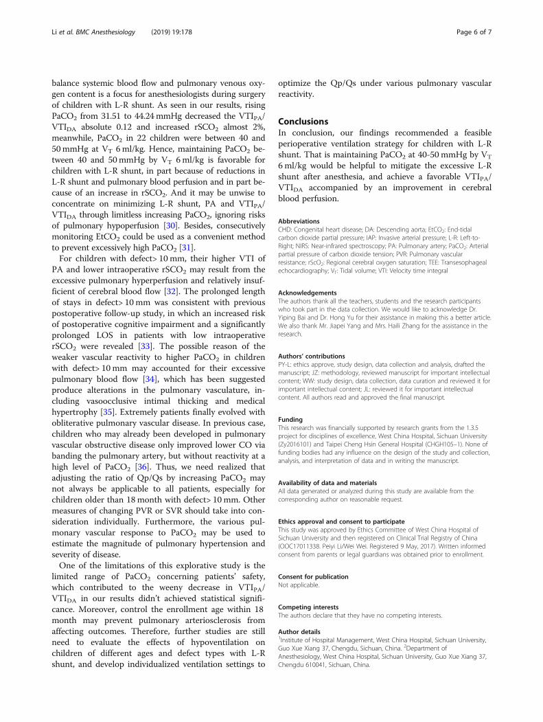
balance systemic blood flow and pulmonary venous oxy-gen content is a focus for anesthesiologists during surgeryof children with L-R shunt. As seen in our results, risingPaCO2 from 31.51 to 44.24mmHg decreased the VTIPA/VTIDA absolute 0.12 and increased rSCO2 almost 2%,meanwhile, PaCO2 in 22 children were between 40 and50mmHg at VT 6ml/kg. Hence, maintaining PaCO2 be-tween 40 and 50mmHg by VT 6ml/kg is favorable forchildren with L-R shunt, in part because of reductions inL-R shunt and pulmonary blood perfusion and in part be-cause of an increase in rSCO2. And it may be unwise toconcentrate on minimizing L-R shunt, PA and VTIPA/VTIDA through limitless increasing PaCO2, ignoring risksof pulmonary hypoperfusion [30]. Besides, consecutivelymonitoring EtCO2 could be used as a convenient methodto prevent excessively high PaCO2 [31].For children with defect> 10 mm, their higher VTI of
PA and lower intraoperative rSCO2 may result from theexcessive pulmonary hyperperfusion and relatively insuf-ficient of cerebral blood flow [32]. The prolonged lengthof stays in defect> 10mm was consistent with previouspostoperative follow-up study, in which an increased riskof postoperative cognitive impairment and a significantlyprolonged LOS in patients with low intraoperativerSCO2 were revealed [33]. The possible reason of theweaker vascular reactivity to higher PaCO2 in childrenwith defect> 10mm may accounted for their excessivepulmonary blood flow [34], which has been suggestedproduce alterations in the pulmonary vasculature, in-cluding vasoocclusive intimal thicking and medicalhypertrophy [35]. Extremely patients finally evolved withobliterative pulmonary vascular disease. In previous case,children who may already been developed in pulmonaryvascular obstructive disease only improved lower CO viabanding the pulmonary artery, but without reactivity at ahigh level of PaCO2 [36]. Thus, we need realized thatadjusting the ratio of Qp/Qs by increasing PaCO2 maynot always be applicable to all patients, especially forchildren older than 18month with defect> 10mm. Othermeasures of changing PVR or SVR should take into con-sideration individually. Furthermore, the various pul-monary vascular response to PaCO2 may be used toestimate the magnitude of pulmonary hypertension andseverity of disease.One of the limitations of this explorative study is the
limited range of PaCO2 concerning patients’ safety,which contributed to the weeny decrease in VTIPA/VTIDA in our results didn’t achieved statistical signifi-cance. Moreover, control the enrollment age within 18month may prevent pulmonary arteriosclerosis fromaffecting outcomes. Therefore, further studies are stillneed to evaluate the effects of hypoventilation onchildren of different ages and defect types with L-Rshunt, and develop individualized ventilation settings to
optimize the Qp/Qs under various pulmonary vascularreactivity.
ConclusionsIn conclusion, our findings recommended a feasibleperioperative ventilation strategy for children with L-Rshunt. That is maintaining PaCO2 at 40-50 mmHg by VT
6ml/kg would be helpful to mitigate the excessive L-Rshunt after anesthesia, and achieve a favorable VTIPA/VTIDA accompanied by an improvement in cerebralblood perfusion.
AbbreviationsCHD: Congenital heart disease; DA: Descending aorta; EtCO2: End-tidalcarbon dioxide partial pressure; IAP: Invasive arterial pressure; L-R: Left-to-Right; NIRS: Near-infrared spectroscopy; PA: Pulmonary artery; PaCO2: Arterialpartial pressure of carbon dioxide tension; PVR: Pulmonary vascularresistance; rScO2: Regional cerebral oxygen saturation; TEE: Transesophagealechocardiography; VT: Tidal volume; VTI: Velocity time integral
AcknowledgementsThe authors thank all the teachers, students and the research participantswho took part in the data collection. We would like to acknowledge Dr.Yiping Bai and Dr. Hong Yu for their assistance in making this a better article.We also thank Mr. Jiapei Yang and Mrs. Haili Zhang for the assistance in theresearch.
Authors’ contributionsPY-L: ethics approve, study design, data collection and analysis, drafted themanuscript; JZ: methodology, reviewed manuscript for important intellectualcontent; WW: study design, data collection, data curation and reviewed it forimportant intellectual content; JL: reviewed it for important intellectualcontent. All authors read and approved the final manuscript.
FundingThis research was financially supported by research grants from the 1.3.5project for disciplines of excellence, West China Hospital, Sichuan University(Zy2016101) and Taipei Cheng Hsin General Hospital (CHGH105–1). None offunding bodies had any influence on the design of the study and collection,analysis, and interpretation of data and in writing the manuscript.
Availability of data and materialsAll data generated or analyzed during this study are available from thecorresponding author on reasonable request.
Ethics approval and consent to participateThis study was approved by Ethics Committee of West China Hospital ofSichuan University and then registered on Clinical Trial Registry of China(OOC17011338. Peiyi Li/Wei Wei. Registered 9 May, 2017). Written informedconsent from parents or legal guardians was obtained prior to enrollment.
Consent for publicationNot applicable.
Competing interestsThe authors declare that they have no competing interests.
Author details1Institute of Hospital Management, West China Hospital, Sichuan University,Guo Xue Xiang 37, Chengdu, Sichuan, China. 2Department ofAnesthesiology, West China Hospital, Sichuan University, Guo Xue Xiang 37,Chengdu 610041, Sichuan, China.
Li et al. BMC Anesthesiology (2019) 19:178 Page 6 of 7

Received: 5 September 2018 Accepted: 20 September 2019
References1. Brienza N, Biancofiore G, Cavaliere F, Corcione A, De Gasperi A, De Rosa RC,
et al. Clinical guidelines for perioperative hemodynamic management ofnon cardiac surgical adult patients. Minerva Anestesiol. 2019. https://doi.org/10.23736/S0375-9393.19.13584-5.
2. Yamasaki Y, Kawanami S, Kamitani T, Sagiyama K, Sakamoto I, Hiasa KI, et al.Noninvasive quantification of left-to-right shunt by phase contrast magneticresonance imaging in secundum atrial septal defect: the effects of breathholding and comparison with invasive oximetry. Int J Cardiovasc Imaging.2018;34(6):931–7.
3. Kitterman JA, Edmunds LH Jr, Gregory GA, Heymann MA, Tooley WH,Rudolph AM. Patent ducts arteriosus in premature infants. Incidence,relation to pulmonary disease and management. N Engl J Med. 1972;287(10):473–7.
4. Wheller J, George BL, Mulder DG, Jarmakani JM. Diagnosis andmanagement of postoperative pulmonary hypertensive crisis. Circulation.1979;60(7):1640–4.
5. Liu W, Huang Q, Lin D, Zhao L, Ma J. Effect of lung protective ventilation oncoronary heart disease patients undergoing lung cancer resection. J ThoracDis. 2018;10(5):2760–70.
6. Pinsky MR. The hemodynamic consequences of mechanical ventilation: anevolving story. Intensive Care Med. 1997;23(5):493–503.
7. Fajardo MF, Claure N, Swaminathan S, Sattar S, Vasquez A, D'Ugard C, et al.Effect of positive end-expiratory pressure on ductal shunting and systemicblood flow in preterm infants with patent ductus arteriosus. Neonatology.2014;105(1):9–13.
8. Reddy VM, Liddicoat JR, Fineman JR, McElhinney DB, Klein JR, Hanley FL.Fetal model of single ventricle physiology: hemodynamic effects of oxygen,nitric oxide, carbon dioxide, and hypoxia in the early postnatal period. JThorac Cardiovasc Surg. 1996;112(2):437–49.
9. Jobes DR, Nicolson SC, Steven JM, Miller M, Jacobs ML, Norwood WI Jr.Carbon dioxide prevents pulmonary overcirculation in hypoplastic left heartsyndrome. Ann Thorac Surg. 1992;54(1):150–1.
10. Misra S, Koshy T, Mahaldar DA. Sudden decrease in end-tidal carbon-dioxidein a neonate undergoing surgery for type B interrupted aortic arch. AnnCard Anaesth. 2011;14(3):206–10.
11. Dix LM, van Bel F, Lemmers PM. Monitoring cerebral oxygenation inneonates: an update. Front Pediatr. 2017;5:46.
12. Schober P, Loer SA, Schwarte LA. Perioperative hemodynamicmonitoring with transesophageal Doppler technology. Anesth Analg.2009;109(2):340–53.
13. Nagi MM, Ward ME. Modulation of myogenic responsiveness by CO2 in ratdiaphragmatic arterioles: role of the endothelium. Am J Phys. 1997;272(3 Pt2):H1419–25.
14. Green JF, Schmidt ND. Mechanism of hyperpnea induced by changes inpulmonary blood flow. J Appl Physiol Respir Environ Exerc Physiol. 1984;56(5):1418–22.
15. Sreedhar R. Acyanotic congenital heart disease and transesophagealechocardiography. Ann Card Anaesth. 2017;20(Supplement):S36–s42.
16. Sousse LE, Herndon DN, Andersen CR, Ali A, Benjamin NC, Granchi T, et al.High tidal volume decreases adult respiratory distress syndrome, atelectasis,and ventilator days compared with low tidal volume in pediatric burnedpatients with inhalation injury. J Am Coll Surg. 2015;220(4):570–8.
17. Bradley SM, Simsic JM, Mulvihill DM. Hyperventilation impairs oxygenationafter bidirectional superior cavopulmonary connection. Circulation. 1998;98(19 Suppl):II372–6 discussion II6–7.
18. McElhinney DB, Marianeschi SM, Reddy VM. Additional pulmonary bloodflow with the bidirectional Glenn anastomosis: does it make a difference?Ann Thorac Surg. 1998;66(2):668–72.
19. Mainwaring RD, Lamberti JJ, Uzark K, Spicer RL, Cocalis MW, Moore JW.Effect of accessory pulmonary blood flow on survival after the bidirectionalGlenn procedure. Circulation. 1999;100(19 Suppl):Ii151–6.
20. Horimoto M. Measurements of blood flow velocity in pulmonarymicrovessels with laser-Doppler microscope and investigation of severalfactors affecting the blood flow velocity (author's transl). Hokkaido IgakyZasshi. 1981;56(5):507–18.
21. Lellouche F, Delorme M, Bussieres J, Ouattara A. Perioperativeventilatory strategies in cardiac surgery. Best Pract Res Clin Anaesthesiol.2015;29(3):381–95.
22. White FN, Hicks JW, Ishimatsu A. Relationship between respiratory state andintracardiac shunts in turtles. Am J Phys. 1989;256(1 Pt 2):R240–7.
23. Tabbutt S, Ramamoorthy C, Montenegro LM, Durning SM, Kurth CD, StevenJM, et al. Impact of inspired gas mixtures on preoperative infants withhypoplastic left heart syndrome during controlled ventilation. Circulation.2001;104(12 Suppl 1):I159–64.
24. Bradley SM, Simsic JM, Mulvihill DM. Hypoventilation improves oxygenationafter bidirectional superior cavopulmonary connection. J Thorac CardiovascSurg. 2003;126(4):1033–9.
25. Yagi Y, Yamamoto M, Saito H, Mori T, Morimoto Y, Oyasu T, et al. Changesof cerebral oxygenation in sequential Glenn and Fontan procedures in thesame children. Pediatr Cardiol. 2017;38(6):1215–9.
26. Hoskote A, Li J, Hickey C, Erickson S, Van Arsdell G, Stephens D, et al. Theeffects of carbon dioxide on oxygenation and systemic, cerebral, andpulmonary vascular hemodynamics after the bidirectional superiorcavopulmonary anastomosis. J Am Coll Cardiol. 2004;44(7):1501–9.
27. Park CS, Kwak JG, Lee C, Lee CH, Lee SK, Kim YL. Near-infrared spectroscopyas a possible device for continuous monitoring of arterial carbon dioxidetension during cardiac surgery. Perfusion. 2011;26(6):524–8.
28. Schuller JL, Bovill JG, Nijveld A. End-tidal carbon dioxide concentration as anindicator of pulmonary blood flow during closed heart surgery in children.A report of two cases. Br J Anaesth. 1985;57(12):1257–9.
29. Barnea O, Austin EH, Richman B, Santamore WP. Balancing the circulation:theoretic optimization of pulmonary/systemic flow ratio in hypoplastic leftheart syndrome. J Am Coll Cardiol. 1994;24(5):1376–81.
30. Li J, Zhang G, Holtby H, Bissonnette B, Wang G, Redington AN, et al. Carbondioxide--a complex gas in a complex circulation: its effects on systemichemodynamics and oxygen transport, cerebral, and splanchnic circulationin neonates after the Norwood procedure. J Thorac Cardiovasc Surg. 2008;136(5):1207–14.
31. Smolinsky AK, Shinfeld A, Paret G, Bar-El Y, Glauber V, Shabtai EL, et al. End-tidal CO2 levels are a reliable indicator of band tightness in pulmonaryartery banding. Ann Thorac Surg. 1995;60(6 Suppl):S523–4.
32. Matthews IL, Bjornstad PG, Kaldestad RH, Heiberg L, Thaulow E, Gronn M.The impact of shunt size on lung function in infants with univentricularheart physiology. Pediatr Crit Care Med. 2009;10(1):60–5.
33. Meng L, Xiao J, Gudelunas K, Yu Z, Zhong Z, Hu X. Association ofintraoperative cerebral and muscular tissue oxygen saturation withpostoperative complications and length of hospital stay after major spinesurgery: an observational study. Br J Anaesth. 2017;118(4):551–62.
34. Dorrington KL, Talbot NP. Human pulmonary vascular responses to hypoxiaand hypercapnia. Pflugers Arch. 2004;449(1):1–15.
35. Heath D, Edwards JE. The pathology of hypertensive pulmonary vasculardisease; a description of six grades of structural changes in the pulmonaryarteries with special reference to congenital cardiac septal defects.Circulation. 1958;18(4 Part 1):533–47.
36. Wong RS, Baum VC, Sangwan S. Truncus arteriosus: recognition and therapyof intraoperative cardiac ischemia. Anesthesiology. 1991;74(2):378–80.
Publisher’s NoteSpringer Nature remains neutral with regard to jurisdictional claims inpublished maps and institutional affiliations.
Li et al. BMC Anesthesiology (2019) 19:178 Page 7 of 7