The Non-Aligned Movement in the Cold War: Between Policy and Rhetoric
Upload
independentCategory
view
1download
0
Labeling strategies for 13C-detected aligned-sample solid-stateNMR of proteins
Fabian V. Fillip, Neeraj Sinha, Lena Jairam, Joel Bradley, and Stanley J. Opella*Department of Chemistry and Biochemistry, University of California San Diego, 9500 Gilman Drive,La Jolla, California 92093-0307 and Cambridge Isotope Laboratories, 50 Frontage Road, Andover,Massachusetts 01810
Abstract13C-detected solid-state NMR experiments have substantially higher sensitivity than thecorresponding 15N-detected experiments on stationary, aligned samples of isotopically labeledproteins. Several methods for tailoring the isotopic labeling are described that result in spatiallyisolated 13C sites so that dipole-dipole couplings among the 13C are minimized, thus eliminating theneed for homonuclear 13C-13C decoupling in either indirect or direct dimensions of one- or multi-dimensional NMR experiments that employ 13C detection. The optimal percentage for randomfractional 13C labeling is between 25% and 35%. Specifically labeled glycerol and glucose can beused at the carbon sources to tailor the isotopic labeling, and the choice depends on the resonancesof interest for a particular study. For investigations of the protein backbone, growth of the bacteriaon 2-13C-glucose containing media was found to be most effective.
Keywords13C labeling; PISEMA; solid-state NMR; tailored isotopic labeling; triple-resonance
IntroductionThe majority of aligned-sample solid-state NMR studies on proteins immobilized insupramolecular complexes, such as virus particles or membranes, have relied on 1H/15Ndouble-resonance experiments (1). There are several advantages to this approach, includingthe relative ease and low cost of labeling all nitrogen sites in proteins obtained by expressionin bacteria (2). Solid-state NMR experiments on uniformly 15N labeled proteins arestraightforward because they have no nitrogen atoms directly bonded to other nitrogen atomsin either backbone or side chain sites. As a result, there is no need to implementhomonuclear 15N decoupling at any stage in the pulse sequences, including during the directacquisition of 15N signals, and the requisite heteronuclear decoupling is accomplished byirradiation of the 1H resonances. It is feasible to make accurate measurements of 1H chemicalshift, 15N chemical shift, and 1H-15N heteronuclear dipolar coupling frequencies for individualsites, as well as to detect 1H-1H and weak 15N-15N homonuclear couplings using a wide varietyof multidimensional NMR experiments.
*corresponding author: [email protected]'s Disclaimer: This is a PDF file of an unedited manuscript that has been accepted for publication. As a service to our customerswe are providing this early version of the manuscript. The manuscript will undergo copyediting, typesetting, and review of the resultingproof before it is published in its final citable form. Please note that during the production process errors may be discovered which couldaffect the content, and all legal disclaimers that apply to the journal pertain.
NIH Public AccessAuthor ManuscriptJ Magn Reson. Author manuscript; available in PMC 2010 December 1.
Published in final edited form as:J Magn Reson. 2009 December ; 201(2): 121–130. doi:10.1016/j.jmr.2009.08.012.
NIH
-PA Author Manuscript
NIH
-PA Author Manuscript
NIH
-PA Author Manuscript

However, there are two disadvantages to the 1H/15N double-resonance approach: it is restrictedto the amide nitrogen sites in the polypeptide backbone and the few nitrogen-containing sidechain sites, and the direct detection of 15N signals has low sensitivity because of its lowgyromagnetic ratio. Both of these issues can be addressed by implementing 1H/13C/15N triple-resonance experiments on proteins labeled with both 13C and 15N. The sensitivity can beimproved by detecting 13C signals because its gyromagnetic ratio is about 2.5 times larger thanthat of 15N, and there is the opportunity to obtain spectroscopic data from nearly all backboneand side chain sites. The design of triple-resonance experiments for stationary samples isconsiderably different than that for magic angle spinning (MAS) experiments. In this articlewe describe progress towards the development of isotopic labeling schemes compatible withdirect detection of 13C signals in 1H/13C/15N triple-resonance experiments on stationaryaligned samples (3–10) by examining a wide range of 13C labeling approaches (11) over arange of fractions of dilutions, ranging from 15% to 100% of all sites in the proteins, and2-13C-glucose in addition to the complementarily labeled glycerols for labeling throughmetabolic pathways. Magnetically aligned filamentous bacteriophage particles are used as thesamples to demonstrate the influence of the labeling patterns on solid-state NMR spectra.
In single-contact spin-lock cross-polarization experiments on single crystals of peptides andaligned samples of proteins, we have observed a four-fold improvement in the signal to noiseratio when 13C magnetization is detected compared to 15N magnetization for individual labeledsites in the same samples under equivalent experimental conditions (7). As is the case for 15Nlabeling of proteins expressed in bacteria, 100% uniform labeling of all carbons siteswith 13C is straightforward when completely labeled carbon sources, such as 13C6 glucoseor 13C3 glycerol, are used in the growth media. However, in protein samples where all of thecarbon sites are labeled with 13C, the homonuclear dipole-dipole couplings among the densenetwork of bonded and nearby 13C nuclei present significant complications in solid-state NMRof stationary samples. Indeed, one of the principal advantages of high-speed magic anglespinning solid-state NMR experiments is that the homonuclear dipole-dipole couplings amongthe 13C sites are greatly attenuated. In contrast, in stationary samples, the 13C homonuclearcouplings must be dealt with in order to obtain high-resolution spectra and to realize thesensitivity advantages of 13C-detection.
There are two approaches to dealing with the homonuclear 13C-13C dipole-dipole couplingsin stationary aligned samples. The first is to apply multiple-pulse homonuclear decouplingsequences to the 13C nuclei (12). We have demonstrated the efficacy of this approach in theindirect dimensions of multidimensional experiments that enable 13C chemical shiftfrequencies to be measured; however, these experiments were performed with 15N-detectionin the direct dimension (4). The detection of signals in the windows of multiple-pulse sequencesrarely leads to optimal sensitivity because of the filtering limitations associated with the shortperiods of time available for sampling the signals. The second approach is to tailor the patternof isotopic labeling so that the 13C labeled sites of interest are sufficiently isolated fromother 13C nuclei to eliminate the need for homonuclear decoupling.
The isotopic labeling schemes described in this article generate samples with diluted, spatiallyisolated 13C sites so that dipole-dipole couplings among the 13C are minimized, thuseliminating the need for homonuclear 13C-13C decoupling in either indirect or directdimensions of one- or multi-dimensional NMR experiments that employ 13C detection. Theisotopic precursors utilized for tailored labeling of proteins expressed in bacteria range fromspecifically labeled two-, three-, or six-carbon molecules to random fractionally labeled growthmedia prepared from algae grown in the presence of a defined mixtures of 12C and 13C carbondioxide. The coupling among 13C nuclei is avoided either as a result of the alternate site patternof metabolic incorporation (13–15) or because of the low statistical probability of two 13Cnuclei being bonded to each other. Here, results from uniformly 13C labeling and metabolically
Fillip et al. Page 2
J Magn Reson. Author manuscript; available in PMC 2010 December 1.
NIH
-PA Author Manuscript
NIH
-PA Author Manuscript
NIH
-PA Author Manuscript
tailored 13Cα labeling based on [2-13C]-glucose, [2-13C]-glycerol, or [1, 3-13C]-glycerol arecompared to fractional 13C labeling. Two-dimensional projections of triple-resonance solution-state NMR spectra are used to characterize the labeling patterns. The filamentousbacteriophages fd, which infects Escherichia coli, and Pf1, which infects Pseudomonasaeruginosa, are used for the experimental demonstrations. The structural forms of the majorcoat proteins are immobilized and aligned along with the virus particles by the magnetic fieldfor solid-state NMR experiments, and the membrane-bound forms of the same proteins aresolubilized in detergent micelles for solution NMR experiments. The bacteriophage samplesprovide direct insights into the spectroscopic effects of the 13C labeling schemes at differentprotein backbone sites in solid-state NMR experiments., They have been used incomplementary magic angle spinning studies (16,17).
ResultsFilamentous bacteriophage samples were obtained from bacterial cultures grown on algal-based media containing various fractions of 13C labeled nutrients, or on minimal mediasupplemented with [2-13C]-glucose, [2-13C]-glycerol, or [1,3-13C]-glycerol. In this article, ourmain focus is on the 1H-13Cα sites that are uniformly distributed throughout the proteinbackbone, separated from each other by three bonds, and have one directly bonded hydrogen(except for glycine and proline). At the intersection of peptide planes, the associated 1Hchemical shift, 13C chemical shift, and 1H-13C dipolar couplings provide valuable structuralconstraints, and enable direct comparisons of sensitivity and resolution between 15N-and 13C- detected experiments.
In the absence of homonuclear decoupling, only proteins labeled such that the 13Cα sites areisotopically isolated from other 13C, i.e. bonded to 12Cβ and 12CO (Figure 1A), will yield solid-state NMR spectra with high resolution and sensitivity in stationary samples. The presenceof 13C in either or both adjacent carbon sites (Figure 1B) will result in broadened signals dueto the presence of unresolved 13C-13C dipolar couplings. The probability of having anisolated 13Cα site can be calculated from the individual probabilities of having 13Cα, 12Cβ,and 12CO present in the same polypeptide with random 13C labeling at various fractions. Asshown in Figure 1D, the maximum probability occurs near a labeling ratio p =⅓. Because themaximum is broad and both natural abundance 13C and the enrichment of 13C in otherproximate sites are potential complications, we prepared custom algal media with 13Cpercentages of 15%, 25%, 35%, and 45%, as marked by arrows in Figure 1D, in order toexperimentally determine the optimal isotopic composition for solid-state NMR spectroscopyon stationary aligned samples of proteins.
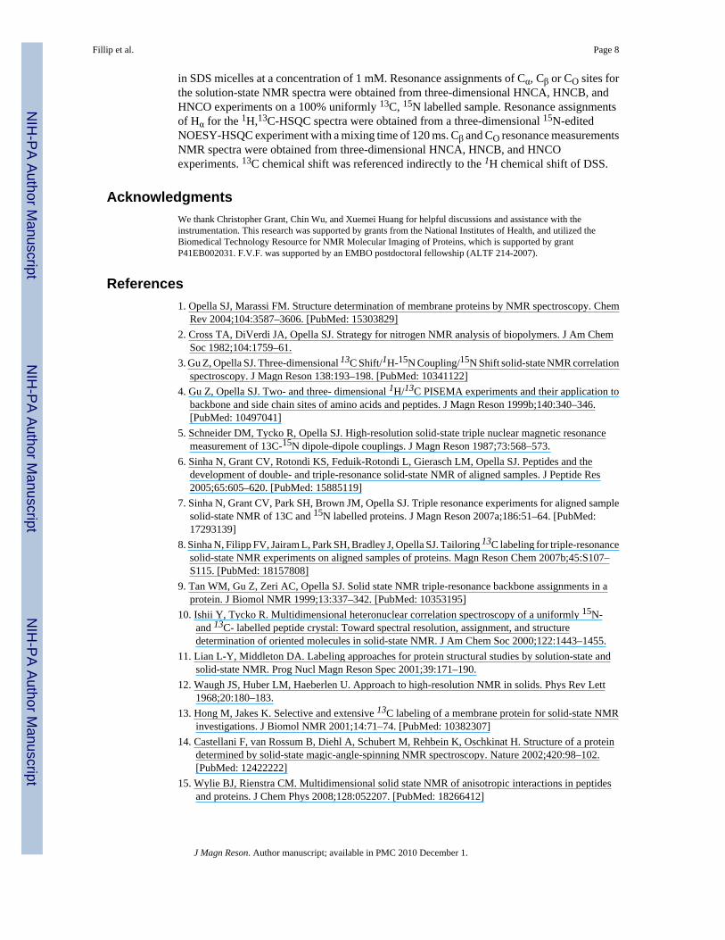
The isotope labeling patterns of the protein samples were analyzed using solution NMR spectra.In general, the proteins were uniformly labeled to the predicted extent in all carbon sites inboth E. coli and P. aeruginosa when grown on the algal-based media. One-dimensional 1H-decoupled, 13C solution NMR spectra report on the 13C labeling at all sites, including thecarbonyl and aromatic ring carbons that are not directly bonded to hydrogens. However, onlya detailed analysis of individual sites reveals whether labeled sites are isotopically isolated.Since E. coli is the host for fd bacteriophage, its major coat protein represents the labeling thatoccurs in this bacterium. Solution NMR spectra of the membrane-bound form of fd coat proteinin micelles are shown in Figure 2. The comparison of the spectrum obtained on a 25% uniformlylabeled sample (Figure 2A) and that from a sample labeled from media containing 2-13C-glucose (Figure 2B) demonstrates that there is a greater extent of 13C labeling in the Cα region(45 ppm – 65 ppm) and reduced labeling in the aliphatic (0 ppm – 50 ppm), aromatic (110 ppm– 170 ppm), and carbonyl (165 ppm – 185 ppm) regions in the 2-13C-glucose labeled sample.
Fillip et al. Page 3
J Magn Reson. Author manuscript; available in PMC 2010 December 1.
NIH
-PA Author Manuscript
NIH
-PA Author Manuscript
NIH
-PA Author Manuscript

A combination of two-dimensional projections from three-dimensional heteronuclear solutionNMR spectra is used to quantify the 13C enrichment at the Cα, Cβ, and CO sites of the proteins.The choice of experiments utilized for this purpose depends on the availability of resonanceassignments as well as which sites are of interest. The labeling at Cα and CO sites can beassessed by comparison of peak intensities in 13C-edited 1H/15N correlation spectra of thevarious 13C labeled samples at equal concentrations, and this requires only backbone amideproton assignments (Figures 2C and 3). If 1H, 13C assignments are available, 1H/13C correlationspectra, e.g. two-dimensional 1H/13C HMQC or 1H/13C projections of three-dimensionalHNCA, HNCB, and HNCO spectra can be used to elucidate the 13C labeling of the protein.The 13C labeling results for E.coli grown on [2-13C] glucose containing media are summarizedin Figure 4 for the Cα and CO sites of fd coat protein. 13C is enriched in the Cα sites of allresidues to level that is > 18%, except for leucine, and some approach 70%. The datasummarized in Figure 4B show that the CO sites of isoleucine, leucine, proline, threonine, andtyrosine have enrichment levels > 18%, but that most amino acids undergo minimal labelingof their CO sites.
The coat protein of Pf1 bacteriophage reflects the metabolic pathways of its host organism P.aeruginosa, which are known to differ from those of E. coli (16,17). The labeling patterns ofproteins from P. aeruginosa were assessed using the same solution NMR approach used forE. coli. All of the proteins are 100% uniformly labeled with 15N in all nitrogen sites, and thisprovides a spectroscopic reference for the 13C resonance intensities. Column A in Figure 5contains the 15N-edited 1H NMR spectra of all of the samples of the membrane-bound formof Pf1 coat protein in micelles that are directly comparable to the 13C NMR spectra in ColumnB. The corresponding one-dimensional solid-state 13C NMR spectra of the structural form ofPf1 coat protein in magnetically aligned bacteriophage particles are shown in Column C; thesespectra are analyzed in three regions, with the broad band of resonances near 200 ppm fromthe carbonyl carbons, the intensity between 40 ppm and 80 ppm from the Cα sites, and theintensity between about 10 ppm and 40 ppm from other aliphatic carbons especially methylgroups. Although highly overlapped, the relative intensities of the signals provide a guide tothe sensitivity in 13C-detected solid-state NMR experiments. Notably, the lowest signal to noiseratio is observed for the sample with the highest degree of 13C labeling (Figure 5D) becauseof the broadening effects of the homonuclear 13C dipolar couplings.
The one-dimensional solution 13C NMR spectra of the fractionally 13C labeled samples havepeak intensities that are homogeneously scaled according to the 13C dilution ratio for all carbonregions compared to 100% uniform 13C labeling (Column B in Figure 5D- F ). Many signalsof carbonyl, aromatic, and aliphatic carbons are resolved is the [45 %-13C] labeled protein,however, at 25% labeling the majority of signals are close to the level of noise under comparableexperimental conditions, showing the direct effect of isotopic dilution when homonucleardipolar couplings are not a factor. The 13C NMR spectra in Column B of Figure 5 indicate thedifferences in the labeling from media containing [2-13C]-glucose, [2-13C]-glycerol, and[1,3-13C]-glycerol. Compared to uniform labeling, the [2-13C]-glycerol sample has reducedintensity in the aliphatic, aromatic, and carbonyl regions of the spectrum, whereas the[1,3-13C]-glycerol sample shows a complementary labeling pattern with diminished intensityin the alpha carbon resonance region. The overall level of isotopic labeling in the [2-13C]-glycerol and [2-13C]-glucose samples is similar, however, the spectral patterns are different,for example the strong aromatic resonances in the spectrum of [2-13C]-glycerol are missing inthe spectrum of the [2-13C]-glucose sample.
The extent of labeling of the Cα, Cβ, and CO sites differ only slightly from the expected averagevalue for the various amino acids as indicated by the data in Figure 6A. In contrast to the evendistributions of the random fractional labeling, the samples labeled by incorporation of
Fillip et al. Page 4
J Magn Reson. Author manuscript; available in PMC 2010 December 1.
NIH
-PA Author Manuscript
NIH
-PA Author Manuscript
NIH
-PA Author Manuscript

[2-13C]-glucose have quite different levels of 13C enrichment in different sites (Figures 6B and6C).
In the case of [2-13C]-glucose labeling in P.aeruginosa, at least 14 amino acids haveisolated 13Cα labels. The enrichment ratio between the [2-13C]-glucose and a uniformly 13Clabeled sample at the Cα position of glycine, leucine, glutamine, arginine, serine and tyrosineis less than 20 %. Glycine, serine and tyrosine are highly enriched in E. coli [3], however, dueto the Entner-Doudoroff metabolic pathway in P.aeruginosa these amino acids are omitted inthe 13C labeling [6]. With the exception of glycine, leucine, glutamine, arginine, serine, andtyrosine, in total 14 of 20 amino acids in P.aeruginosa, and in E.coli all amino acids exceptfrom leucine provide isolated 13Cα labels (Figure 7) For [2-13C]-glycerol as labeling precursorin P.aeruginosa only leucine, glutamine, and arginine are not enriched above 20 % at the Cαsite. Isoleucine and valine have significant 13C enrichment at the adjacent Cβ, and will beaffected by 13C-13C dipolar coupling. A set of 15 of the 20 amino acids in proteins obtainedfrom P.aeruginosa grown on [2-13C]-glycerol containing media is found to be suitablefor 13C solid-state NMR experiments (Figure 4C).
The effects of tailoring the 13C labeling are readily observed in 13C NMR spectra. As notedabove, because of the strong influence of the 13C-13C homonuclear dipole-dipole couplingsthere is not a simple relationship between the extent or type of 13C labeling and the resolutionand sensitivity. The spectra of the random fractional 13C labeled samples have higher signal-to-noise ratios than that of a comparable uniformly 100% 13C labeled sample. The metabolicCα labeling schemes based on [2-13C]-glucose and [2-13C]-glycerol show strong signals in theone-dimensional solid-state NMR spectra, compared to the broad, poorly resolved spectra fromsamples with uniform 100% 13C labeling. In contrast to the random fractionally labeledsamples, the samples labeled with [2-13C]-glucose and [2-13C]-glycerol have relatively highoverall labeling of the alpha carbons and significantly lower levels of labeling of carbonyl andaliphatic side chain carbons. The [1,3-13C]-glycerol sample has more extensive labeling ofcarbonyl and side chain methyl carbons, and the spectra have better resolution in these regions.
The effects of tailoring 13C labeling can also be observed in 15N NMR spectra. Although theeffects are more subtle than those in the directly detected 13C NMR spectra, they are apparentin comparisons of 15N NMR spectra obtained with and without heteronuclear 13C decoupling.This is illustrated with the spectra in Figure 8. The spectra in the left column are all very similar;since they were obtained with 1H and 13C decoupling, the effects of the 13C-15N dipolarcouplings are not seen. In contrast, the spectra in the right column were obtained on the samesamples with only 1H decoupling. The broadening effects of nearby 13C labeled sites onthe 15N amide backbone resonances can be observed to vary among the labeling schemes.
Two-dimensional 1H-13C PISEMA spectra of aligned bacteriophage samples prepared withthe various 13C labeling schemes are compared in Figure 9. In the 100% uniformly 13C labeledsample, the strong network of 13C-13C homonuclear couplings at all sites interferes with theexperiment and, with the exception of the methyl carbon region between 10 ppm and 40 ppm,there is essentially no intensity observable in the displayed spectral region, which encompassesall of the aliphatic carbon sites in the protein. In contrast, all of the samples with tailored 13Clabeling yield resolved spectra. The spectra from Pf1 coat protein obtained from bacteria grownon media containing [2-13C]-glycerol or [1,3-13C]-glycerol are complementary, due to thespecific metabolic labeling pattern of the glycerol precursors. There is notably more intensityfrom aliphatic side chain carbons in the spectrum obtained from the [1,3-13C]-glycerol due toits labeling pattern. Although the total amount of 13C in the protein is reduced, there is a netgain in signal-to-noise ratio in all of the one-dimensional spectra. Although less than half ofthe carbon sites are labeled in the case of [45 %-13C] phage, the observed signal-to-noise ratiois about twice that observed for the 100% labeled phage. As expected, the signal-to-noise ratio
Fillip et al. Page 5
J Magn Reson. Author manuscript; available in PMC 2010 December 1.
NIH
-PA Author Manuscript
NIH
-PA Author Manuscript
NIH
-PA Author Manuscript

decreases with decreasing 13C content, however, even [15 %-13C] phage gives spectra withbetter signal-to-noise ratios than those from a 100% uniformly 13C labeled sample. The gainin signal-to-noise ratio is greater in the case of proteins obtained from media containingspecifically labeled glucose or glycerol.
The analysis of the two-dimensional solid-state NMR spectra reveals different line shapebehavior for the 13C chemical shift and the 1H-13C dipolar coupling dimensions. For all of thesamples the full width at half height is approximately 250 Hz to 300 Hz in the 13C chemicalshift dimension, except with [45 %-13C] labeling where the samples have somewhat broaderlines of 400 Hz. For the 1H-13C dipolar coupling dimension, the different dilutions of the 13Caffect the line width of the two-dimensional spectra. For the spectra of the random fractionallabeled samples the full width at half height decreases with increasing 13C dilution from 1300Hz for [45 %-13C] phage to 700 Hz for [15 %-13C] phage. For the metabolically tailored 13Clabeling schemes the full width at half height of the two dimensional spectra is between 1350Hz and 900 Hz, with the [1,3-13C]-glycerol and [2-13C]-glucose labeled samples providingspectra with somewhat better resolution than those labeled with [2-13C]-glycerol.
Although there is a reduction in signal-to-noise with decreasing 12C to 13C isotope ratio.However, this is compensated by improvement in the line shape in the 1H-13C dipolar couplingdimension whereas the 13C chemical shift dimension is hardly affected. For the two differentCα labeling precursors, [2-13C]-glycerol and [2-13C]-glucose, the later shows a better line shapein both dimensions of the two dimensional spectra. The [1,3-13C]-glycerol sample yieldsslightly better line shapes for the Cα sites.
DiscussionUniform isotopic labeling of proteins has been an integral part of the experimental design fromthe beginning of the field (18,19), and 100% uniform labeling with 13C and 15N is widely usedin triple-resonance solution NMR experiments as well as MAS solid-state NMR experiments.Because of the natural separation of nitrogen sites in proteins, there is no need to vary the extentof 15N labeling because of through-space of through-bond couplings. However, all carbons arebonded to at least one other carbon and in most cases two other carbons, resulting in densenetworks of homonuclear scalar and/or dipole-dipole couplings that can interfere with bothsolution NMR and solid-state NMR experiments. The two basic strategies for 13C labeling thatevaluated in the context of aligned-sample solid-state NMR experiments with the data inFigures 8 and 9 were previously used for solution NMR and magic angle spinning solid-stateNMR. In solution NMR, the preparation of protein samples that were uniformlyfractionally 13C labeled enabled J couplings to be used to assist in making resonanceassignments (20) and to simplify relaxation pathways (21). Biosynthetic incorporation ofspecifically 13C labeled metabolic precursors, including glycerol (22), glucose (23), andpyruvate (24), were also used to facilitate 13C relaxation studies and triple-resonanceexperiments solution NMR. Increased resolution in magic angle spinning solid-state NMRexperiments has resulted primarily from the use of samples with complementary labelingpatterns obtained from growth on media containing [1-13C]-glycerol and [2,3-13C]-glycerol(13–15).
The tailored labeling from the labeled glycerol improves the resolution that is possible withmagic angle spinning of 100% uniformly 13C labeled samples. In contrast, in aligned-samplesolid-state NMR, in order to obtain high-resolution spectra and realize the sensitivity gainsfeasible with 13C-detection, the homonuclear dipole-dipole couplings have to be dealt witheither by multiple-pulse homonuclear decoupling or isotopic labeling. As described here, anapproach applicable to proteins obtained by expression in bacteria is to tailor the 13C labelingto label sites of interest, for example the alpha carbons, while adjacent carbon sites are
Fillip et al. Page 6
J Magn Reson. Author manuscript; available in PMC 2010 December 1.
NIH
-PA Author Manuscript
NIH
-PA Author Manuscript
NIH
-PA Author Manuscript

unlabeled. In this continuation of our development of this approach, several complementarylabeling schemes were tested for two different bacteria. The 13C dilution from randomfractional labeling media had predictable outcomes for both tested expression systems.However, the implementation of metabolic precursors for labeling depends on the organism’sspecific metabolic pathways, and requires experimental verification. The choice of labelingstrategy is an integral part of the experimental design, which contrasts with the situation formost solution NMR and magic angle spinning solid-state NMR studies that can beaccomplished with only one or a few labeled sample prepared according to the previouslydemonstrated labeling schemes.
For aligned-sample solid-state NMR of proteins, the optimal percentage for uniformfractional 13C labeling is between 25% and 35%. Specifically labeled glycerol and glucose canbe used at the carbon sources to tailor the isotopic labeling, and the choice depends on theresonances of interest for a particular study. For investigations of the protein backbone,2-13C-glucose was found to be most effective. In the case of P. aeruginosa there are 14 of the20 amino acids with isolated Cα position in the protein backbone and for E. coli. 19 aminoacids have 13Cα sites.
MethodsSample Preparation
The 100% Uniformly 13C and 15N labelled samples were obtained in the conventional way byusing a minimal salts media with 15N labelled ammonium sulfate and 13C6 labeled glucose asthe sole nitrogen and carbon sources. Uniform fractional 13C labeling was performed bygrowing bacteria on standard BioExpress Cell Growth Media prepared with the statedpercentages of 13C. The metabolically 13C labelled samples were obtained by supplementingthe using [2-13C]-glycerol, [2-13C]-glycerol, [1,3-13C]-glycerol, or as the sole carbon source.All of the isotopically labelled compounds and media described in this article are fromCambridge Isotope Laboratories (www.isotope.com).
The 50 mg/ml solutions of aligned bacteriophage particles were prepared as describedpreviously (23). The coat proteins subunits were separated from the phage particles andsolubilized in SDS for the solution NMR analysis (24).
NMR spectroscopyThe solid-state NMR experiments were performed on a Varian Inova spectrometerwith 1H, 13C, and 15N frequencies of 500.125 MHz, 125.76 MHz, and 50.68 MHz respectively.A home-built triple resonance probe with a single 5 mm solenoid coil was used and the RFpower levels were adjusted to generated 50 kHz RF fields on all three channels. PISEMA wasutilized for the separated local field experiments because its cycle time was short enough toaccommodate the large frequencies of 1H-13C dipolar couplings. The 1H carrier frequency wasset to the resonance of water at 4.7 ppm; the 13C and 15N carrier frequencies were set to 100ppm on their respective scales. Solid samples of adamantane and ammonium sulfate served asexternal chemical shift references for 13C and 15N, respectively. The two-dimensional solid-state NMR spectra were acquired with 64 points in t1 and 512 complex points in t2. Theexperimental data were zero filled in t1 to 2K and in t2 to 4K data points and multiplied by asine bell window function before Fourier transformation in each dimension. The recycle delaywas 6 s.
The solution NMR experiments were performed on a Bruker Avance 800 MHz spectrometerwith 1H, 13C, and 15N frequencies of 800.034 MHz, 201.203 MHz, and 81.076 MHzrespectively. For these experiments, the coat proteins of the bacteriophages were solubilized
Fillip et al. Page 7
J Magn Reson. Author manuscript; available in PMC 2010 December 1.
NIH
-PA Author Manuscript
NIH
-PA Author Manuscript
NIH
-PA Author Manuscript
in SDS micelles at a concentration of 1 mM. Resonance assignments of Cα, Cβ or CO sites forthe solution-state NMR spectra were obtained from three-dimensional HNCA, HNCB, andHNCO experiments on a 100% uniformly 13C, 15N labelled sample. Resonance assignmentsof Hα for the 1H,13C-HSQC spectra were obtained from a three-dimensional 15N-editedNOESY-HSQC experiment with a mixing time of 120 ms. Cβ and CO resonance measurementsNMR spectra were obtained from three-dimensional HNCA, HNCB, and HNCOexperiments. 13C chemical shift was referenced indirectly to the 1H chemical shift of DSS.
AcknowledgmentsWe thank Christopher Grant, Chin Wu, and Xuemei Huang for helpful discussions and assistance with theinstrumentation. This research was supported by grants from the National Institutes of Health, and utilized theBiomedical Technology Resource for NMR Molecular Imaging of Proteins, which is supported by grantP41EB002031. F.V.F. was supported by an EMBO postdoctoral fellowship (ALTF 214-2007).
References1. Opella SJ, Marassi FM. Structure determination of membrane proteins by NMR spectroscopy. Chem
Rev 2004;104:3587–3606. [PubMed: 15303829]2. Cross TA, DiVerdi JA, Opella SJ. Strategy for nitrogen NMR analysis of biopolymers. J Am Chem
Soc 1982;104:1759–61.3. Gu Z, Opella SJ. Three-dimensional 13C Shift/1H-15N Coupling/15N Shift solid-state NMR correlation
spectroscopy. J Magn Reson 138:193–198. [PubMed: 10341122]4. Gu Z, Opella SJ. Two- and three- dimensional 1H/13C PISEMA experiments and their application to
backbone and side chain sites of amino acids and peptides. J Magn Reson 1999b;140:340–346.[PubMed: 10497041]
5. Schneider DM, Tycko R, Opella SJ. High-resolution solid-state triple nuclear magnetic resonancemeasurement of 13C-15N dipole-dipole couplings. J Magn Reson 1987;73:568–573.
6. Sinha N, Grant CV, Rotondi KS, Feduik-Rotondi L, Gierasch LM, Opella SJ. Peptides and thedevelopment of double- and triple-resonance solid-state NMR of aligned samples. J Peptide Res2005;65:605–620. [PubMed: 15885119]
7. Sinha N, Grant CV, Park SH, Brown JM, Opella SJ. Triple resonance experiments for aligned samplesolid-state NMR of 13C and 15N labelled proteins. J Magn Reson 2007a;186:51–64. [PubMed:17293139]
8. Sinha N, Filipp FV, Jairam L, Park SH, Bradley J, Opella SJ. Tailoring 13C labeling for triple-resonancesolid-state NMR experiments on aligned samples of proteins. Magn Reson Chem 2007b;45:S107–S115. [PubMed: 18157808]
9. Tan WM, Gu Z, Zeri AC, Opella SJ. Solid state NMR triple-resonance backbone assignments in aprotein. J Biomol NMR 1999;13:337–342. [PubMed: 10353195]
10. Ishii Y, Tycko R. Multidimensional heteronuclear correlation spectroscopy of a uniformly 15N-and 13C- labelled peptide crystal: Toward spectral resolution, assignment, and structuredetermination of oriented molecules in solid-state NMR. J Am Chem Soc 2000;122:1443–1455.
11. Lian L-Y, Middleton DA. Labeling approaches for protein structural studies by solution-state andsolid-state NMR. Prog Nucl Magn Reson Spec 2001;39:171–190.
12. Waugh JS, Huber LM, Haeberlen U. Approach to high-resolution NMR in solids. Phys Rev Lett1968;20:180–183.
13. Hong M, Jakes K. Selective and extensive 13C labeling of a membrane protein for solid-state NMRinvestigations. J Biomol NMR 2001;14:71–74. [PubMed: 10382307]
14. Castellani F, van Rossum B, Diehl A, Schubert M, Rehbein K, Oschkinat H. Structure of a proteindetermined by solid-state magic-angle-spinning NMR spectroscopy. Nature 2002;420:98–102.[PubMed: 12422222]
15. Wylie BJ, Rienstra CM. Multidimensional solid state NMR of anisotropic interactions in peptidesand proteins. J Chem Phys 2008;128:052207. [PubMed: 18266412]
Fillip et al. Page 8
J Magn Reson. Author manuscript; available in PMC 2010 December 1.
NIH
-PA Author Manuscript
NIH
-PA Author Manuscript
NIH
-PA Author Manuscript

16. Goldbourt A, Gross BJ, Day LA, McDermott AE. Filamentous phage studied by magic-angle spinningNMR: Resonance assignment and secondary structure of the coat protein in Pf1. J Am Chem Soc2007;129:2338–2344. [PubMed: 17279748]
17. Goldbourt A, Day LA, McDermott AE. Assignment of congested NMR spectra: Carbonyl backboneenrichment via the Entner-Doudoroff pathway. J Magn Reson 2007;189:157–165. [PubMed:17900951]
18. Lian LY, Middleton DA. labeling approaches for protein structural studies by solution-state and solid-state NMR’, 2001. Prog Nucl Magn Reson Spectrosc 2001;39:171–190.
19. Hoogstraten CG, Johnson JE. Metabolic labeling: taking advantabe of bacerial pathways to preparespectroscopically useful isotope patterns in proteins and nucleic acids. Concepts in Magn Reson PartA 2008;32A:34–55.
20. Oh BH, Westler WM, Darba P, Markley JL. Protein carbon-13 spin systems by a single two-dimensional nuclear magnetic resonance experiment. Science 1988;240:908–911. [PubMed:3129784]
21. Venters RA, Calderone TL, Spicer LD, Fierke CA. Uniform 13C isotope labeling of proteins withsodium acetate for NMR studies: application to human carbonic anhydrase II. Biochemistry1991;30:4491–4494. [PubMed: 1902380]
22. Lemaster DM, Kushaln DM. Dynamical mapping of E.coli thioredoxin via 13C NMR relaxationanalysis. J Am Chem Soc 1996;118:9255–9264.
23. Lundstrom P, Teilum K, Carstensen T, Bezsonova I, Wisner S, Hansen DF, Religa TL, Akke M, KayLE. Fractional 13C enrichment of isolated carbons using [1-13C]- or [2–13C]-glucose facilitates theaccurate measurement of dynamics at backbone Ca and side-chain methyl positions in proteins. JBiomol NMR 2007;38:199–212. [PubMed: 17554498]
24. Guo C, Geng C, Tugarinov V. Selective backbone labeling of proteins using [1,2–13C2]-pyruvate ascarbon source. J Biomol NMR 2009;44:167–173. [PubMed: 19468838]
25. Thiriot DS, Nevzorov AA, Zagyanskiy L, Wu CH, Opella SJ. Structure of the coat protein in Pf1bacteriophage determined by solid-state NMR spectroscopy. J Mol Biol 2004;341:869–879.[PubMed: 15288792]
26. McDonnell PA, Shon K, Kim Y, Opella SJ. fd Coat protein structure in membrane environments. JMol Biol 1993;233:447–463. [PubMed: 8411155]
Fillip et al. Page 9
J Magn Reson. Author manuscript; available in PMC 2010 December 1.
NIH
-PA Author Manuscript
NIH
-PA Author Manuscript
NIH
-PA Author Manuscript

Figure 1.Analysis of fractional uniform 13C labeling at the Cα sites in the polypeptide backbone. A.Schematic chemical structure of an isolated 13Cα site bonded only to 12C that would contributea high resolution NMR signal. B. Schematic chemical structure of a non-isolated 13Cα sitebonded to 13Cβ and/or 13CO sites that would contribute a broadened or undetectable NMRsignal. C. Schematic chemical structure of an unlabeled 12Cα site that would not contribute asignal. D. The probability (p) of occurrence of an isolated 13 sitesCαthat would contribute highresolution NMR signals is predicted by the function f(p) = p(1-p)2. The arrows denote theexperimentally tested labeling ratios of 15%, 25%, 35%, and 45%.
Fillip et al. Page 10
J Magn Reson. Author manuscript; available in PMC 2010 December 1.
NIH
-PA Author Manuscript
NIH
-PA Author Manuscript
NIH
-PA Author Manuscript
Figure 2.Analysis of 13C labeling patterns of proteins obtained from E. coli by solution NMR of fd coatprotein in micelles. A. and B. are one-dimensional direct-detected 13C NMR spectra obtainedby direct excitation. A. The protein was obtained from bacteria grown on 25% uniformly 13Clabeled media. B. The protein was obtained from bacteria grown on 2–13C glucose-containingmedia. C. Overlay of two-dimensional 13CO-edited 1H/15N correlation spectra of the proteinswhose spectra are shown in panels A. and B. Grey indicates 25% uniformly labeled with 13Cand black indicates labeled by growth on [2-13C]-glucose containing media.
Fillip et al. Page 11
J Magn Reson. Author manuscript; available in PMC 2010 December 1.
NIH
-PA Author Manuscript
NIH
-PA Author Manuscript
NIH
-PA Author Manuscript
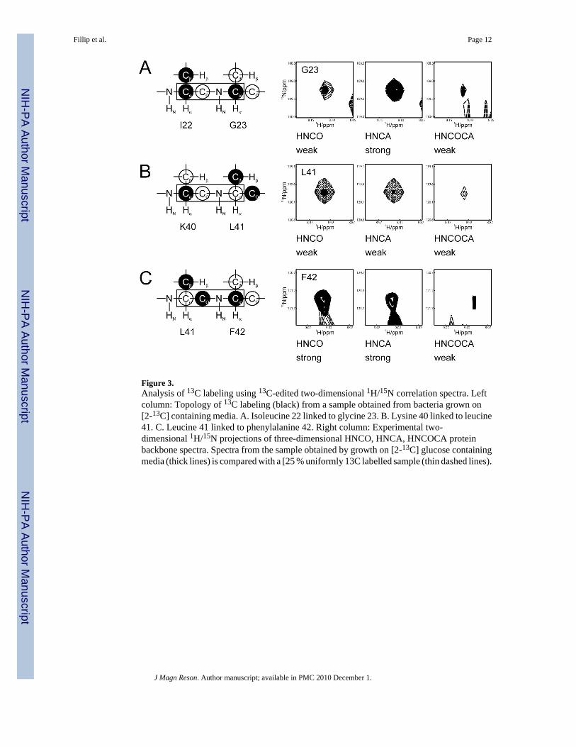
Figure 3.Analysis of 13C labeling using 13C-edited two-dimensional 1H/15N correlation spectra. Leftcolumn: Topology of 13C labeling (black) from a sample obtained from bacteria grown on[2-13C] containing media. A. Isoleucine 22 linked to glycine 23. B. Lysine 40 linked to leucine41. C. Leucine 41 linked to phenylalanine 42. Right column: Experimental two-dimensional 1H/15N projections of three-dimensional HNCO, HNCA, HNCOCA proteinbackbone spectra. Spectra from the sample obtained by growth on [2-13C] glucose containingmedia (thick lines) is compared with a [25 % uniformly 13C labelled sample (thin dashed lines).
Fillip et al. Page 12
J Magn Reson. Author manuscript; available in PMC 2010 December 1.
NIH
-PA Author Manuscript
NIH
-PA Author Manuscript
NIH
-PA Author Manuscript

Figure 4.Experimentally observed 13C labeling of fd coat protein obtained from E. coli grown on[2-13C]-glucose-containing media as measured from intensities in 1H-13C projections ofHNCO and HNCA spectra. A. Ca sites. B. CO sites. The dashed lines mark 18% labeling, whichis the level used to designate those sites that are likely to provide observable signals in solid-state NMR experiments.
Fillip et al. Page 13
J Magn Reson. Author manuscript; available in PMC 2010 December 1.
NIH
-PA Author Manuscript
NIH
-PA Author Manuscript
NIH
-PA Author Manuscript

Figure 5.Comparison of NMR spectra of 100% uniformly 15N labelled and fractional uniformly 13Clabelled samples of Pf1 coat protein obtained from P. aeruginosa. Column A: 15N-edited 1Hsolution NMR spectra of the protein in micelles. Column B: Direct-detected solution 13C NMRspectra obtained by direct-excitation of the protein in micelles. Column C: 13C-detected cross-polarization solid-state NMR spectra obtained with both {1H} and {15N} decoupling duringacquisition. D. 100% uniformly 13C labelled. E. 45% uniformly 13C labelled. F. 25%uniformly 13C labelled. G. From growth on 2–13C glycerol-containing media. H. From growthon 1, 3 13C glycerol-containing media. I. From growth on 2–13C-glucose containing media
Fillip et al. Page 14
J Magn Reson. Author manuscript; available in PMC 2010 December 1.
NIH
-PA Author Manuscript
NIH
-PA Author Manuscript
NIH
-PA Author Manuscript
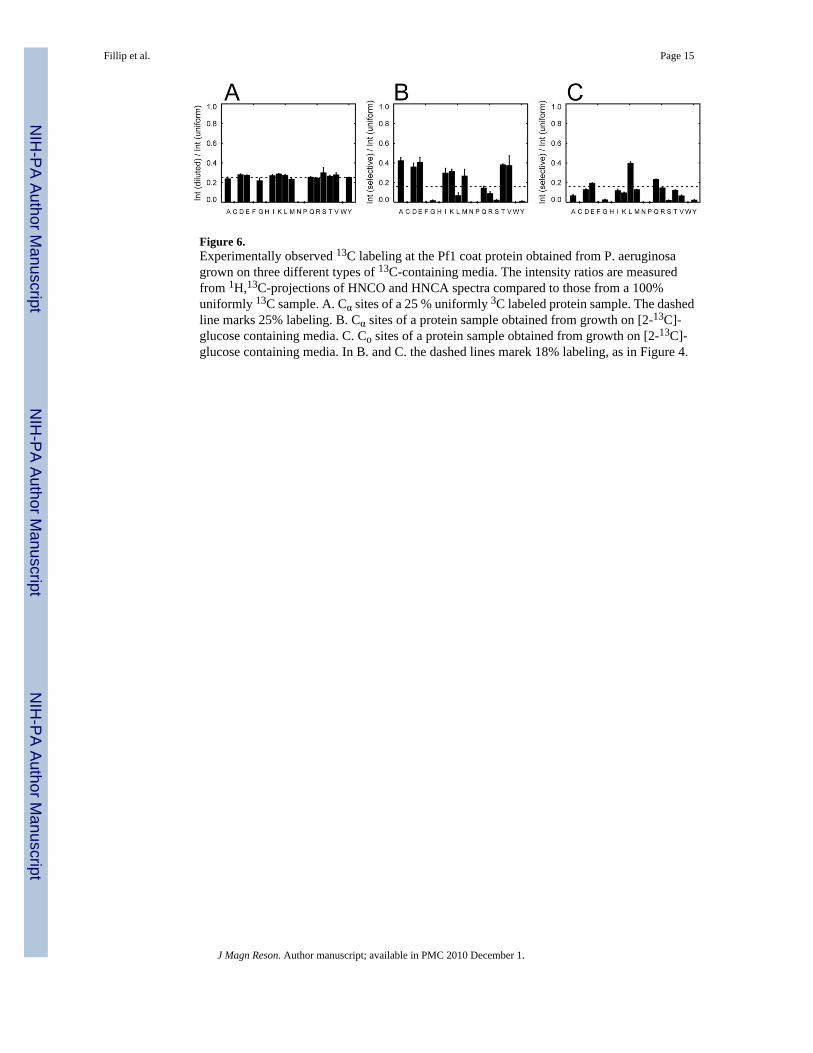
Figure 6.Experimentally observed 13C labeling at the Pf1 coat protein obtained from P. aeruginosagrown on three different types of 13C-containing media. The intensity ratios are measuredfrom 1H,13C-projections of HNCO and HNCA spectra compared to those from a 100%uniformly 13C sample. A. Cα sites of a 25 % uniformly 3C labeled protein sample. The dashedline marks 25% labeling. B. Cα sites of a protein sample obtained from growth on [2-13C]-glucose containing media. C. Co sites of a protein sample obtained from growth on [2-13C]-glucose containing media. In B. and C. the dashed lines marek 18% labeling, as in Figure 4.
Fillip et al. Page 15
J Magn Reson. Author manuscript; available in PMC 2010 December 1.
NIH
-PA Author Manuscript
NIH
-PA Author Manuscript
NIH
-PA Author Manuscript

Figure 7.Amino acids with isolated Cα sites in the polypeptide backbone with > 18% labeling. Theamino acids present in Pf1 coat protein are marked by asterisks; for those amino acids that donot occur in the protein, the labeling anticipated from the Entner-Doudoroff pathway isincluded for completeness. The presence or absence of significant 13C labeling in the Cα, Cβ,and CO sites of the specified amino acids is designated in the horizontal rows. If the labelingat a Cα site is > 18% and the labeling at the bonded Cβ and CO sites is < 18%, then the Cα siteis marked as + isolated. A few marginal cases are marked +/−. A. Protein obtained from E.coli grown on media containing [2-13C]-glucose. B. Protein obtained from P. aeruginosagrown on media containing [2-13C]-glucose. C. Protein obtained from P.aeruginosa grown onmedia containing [2-13C]-glycerol.
Fillip et al. Page 16
J Magn Reson. Author manuscript; available in PMC 2010 December 1.
NIH
-PA Author Manuscript
NIH
-PA Author Manuscript
NIH
-PA Author Manuscript

Figure 8.Comparison of one-dimensional solid-state 15N NMR NMR spectra of 100% uniformly 15Nlabelled and fractional uniformly 13C labelled samples of Pf1 coat protein obtained from P.aeruginosa. A., C., E., and G. were obtained with both 1H and 13C decoupling. B., D., F., andH. were obtained with only 1H decoupling. A. and B. 45% uniformly 13C labelled. C. and D.25% uniformly 13C labelled. E. and F. Protein obtained from bacteria grown on 1,3 -13Cglycerol-containing media. G. and H. Protein obtained from bacteria grown on I, 2-13C glucose-containing media.
Fillip et al. Page 17
J Magn Reson. Author manuscript; available in PMC 2010 December 1.
NIH
-PA Author Manuscript
NIH
-PA Author Manuscript
NIH
-PA Author Manuscript

Figure 9.Comparison of two-dimensional 1H-13C PISEMA spectra of aligned Pf1 bacteriophagesamples. A. 100% uniformly 13C labelled. B. - E. Fractional uniformly 13C labelled at theindicated percentages. F. From bacteria grown on [2-13C] glycerol-containing media. G. Frombacteria grown on [1,3-13C] glycerol-containing media. H. From bacteria grown on [2-13C]glucose-containing media.
Fillip et al. Page 18
J Magn Reson. Author manuscript; available in PMC 2010 December 1.
NIH
-PA Author Manuscript
NIH
-PA Author Manuscript
NIH
-PA Author Manuscript

Figure 10.Comparison of single-to-noise ratios and resonance linewidths in the 1H-13C PISEMA spectraof aligned Pf1 bacteriophage. The error bars represent the estimated uncertainty in themeasurements made on the experimental spectra in Figure 9. A. Signal-to-noise ratios of theone-dimensional solid-state NMR spectra. B. Full width at half height of the 13C chemical shiftdimension. C. Full width at half height of 1H-13C dipolar coupling dimension. Left panels: Thepercentages listed on the bottom correspond to the extent of fractional uniform 13C labeling.Right panels: 13C labeling from bacteria grown in media containing D. [2-13C] glycerol. E.[1,3-13C] glycerol. F. [2-13C] glucose.
Fillip et al. Page 19
J Magn Reson. Author manuscript; available in PMC 2010 December 1.
NIH
-PA Author Manuscript
NIH
-PA Author Manuscript
NIH
-PA Author Manuscript
Copyright © 2022 FDOKUMEN


![Conversion of Hyperpolarized [1-13C]Pyruvate in Breast ...](https://static.fdokumen.com/doc/165x107/6328a69be491bcb36c0bdd22/conversion-of-hyperpolarized-1-13cpyruvate-in-breast-.jpg)