
SYNTHESIS OF RING-13C-LABELLED AND ^RING ...
170
INIS-mf--11370 SYNTHESIS OF RING- 13 C-LABELLED AND ^RING-DEMETMYLATED RETINALS Investigations on 13 C-Labelled and Chemically modified . ' ' '"•• (bacterio)rhodopsins ,. ' . .i.M I ( o
-
Upload
khangminh22 -
Category
Documents
-
view
2 -
download
0
Transcript of SYNTHESIS OF RING-13C-LABELLED AND ^RING ...

IN IS -m f - - 11370
SYNTHESIS OF RING-13C-LABELLED AND^RING-DEMETMYLATED RETINALS
Investigations on 13C-Labelled and Chemically modified .' ' '"•• (bacterio)rhodopsins ,. ' .
.i.M I ( o

SYNTHESIS OF RING-13C-LABELLED AND RING-DEMETHYIATED RETINALS
Investigations on ^C-Labelled and Chemically modified
(Bacterio)rhodops ins

SYNTHESIS OF RING-13C-LABELLED AND RING-DEMETHYLATED RETINALS
Investigations on 13C-Labelled and Chemically modified
(bacterio)rhodopsins
Proefschrift
ter verkrijging van de graad van Doctor
aan de Rijksuniversiteit te Leiden,
op gezag van de Rector Magnificus Dr. J.J.M. Beenakker,
Hoogleraar in de faculteit der Wiskunde en Natuurwetenschappen,
volgens besluit van het College van Dekanen
te verdedigen op woensdag 13 januari 1988
te klokke 15.15 uur
door
JACOBUS MARINÜS LEO COURTIN
geboren te Heinkenszand (thans Borsele) in 1958

PROMOTIE-COMMISSIE:
PROMOTOR: Prof. Dr. J. Lugtenburg
REFERENT: Prof. Dr. J. Cornelisse
OVERIGE LEDEN: Prof. Dr. J.H. van Boom
Prof. Dr. A. van der Gen
Prof. Dr. J. Reedijk
This investigation was supported by the Netherlands Foundation
for Chemical Research (SON) with financial aid from the
Netherlands Organization for the Advancement of Pure Research.

Y rrvtin modelerUUt naajcdoichttnis a<vn
Uooor-

CONTENTS
CHAPTER 1 GENERAL INTRODUCTION1-1 Rhodopsin and bacteriorhodopsin as photo- 1
sensitive pigments1-2 Structure of rhodopsin 11-3 Opsin shift 41-4 Photochemistry and function of bovine 5
rhodopsin1-5 Structure of bacteriorhodopsin 71-6 Photochemistry and function of bacterio- 10
rhodopsin1-7 Purpose of this investigation 12
CHAPTER 2 SYNTHESIS OF RING-DEMETHYLATED RETINALS2-1 Introduction 192-2 A general synthetic scheme for the demethyl- 22
retinals la-e2-3 Synthesis of l-cyclohexene- (2a) and 24
2-methyl-l-cyclohexenecarboxaldehyde (2b)2-4 Synthesis of 6-methyl- (2c), 2,6-dimethyl- 25
(2d) and 6,6-dimethyl-l-cyclohexenecarbox-aldehyde (2e)
2-5 Preparation of retinals la-e 282-6 Discussion 34
CHAPTER 3 SYNTHESIS OF RING-13C-LABELLED RETINALS3-1 Introduction 413-2 Development of efficient synthetic schemes 42
for ring-13C-labelled retinals3-3 Synthesis of (l-13C)retinal (if) and 46
(16,17-13C2)retinal (lm)3-4 Synthesis of (4-13C)retinal (li) 473-5 Synthesis of (5- 1 3C)- (lj) and (18- 1 3C)- 48
retinal (In)3-6 Synthesis of (6- 1 3C)- (Ik) and (7- 1 3C)- 50
retinal (11)3-7 Attempted synthesis of (2- 1 3C)- (lg) and 51
(3-13C)retinal (lh)3-8 Discussion 52
CHAPTER 4 SPECTROSCOPIC CHARACTERIZATION OF RETINALS4-1 Introduction 574-2 Mass spectrometry 574-3 UV-VIS spectroscopy 614-4 1H-NMR spectroscopy 6 34-5 13C-NMR spectroscopy 694-6 Concluding remarks 7 3

CHAPTER 5
5-15-2
5-3
5-45-55-65-7
CHAPTER 6
CHAPTER
6-16-2
6-3
6-4
6-5
77-17-2
APPENDIX I
APPENDIX II
MODIFIED BACTERIORHODOPSINS AND MODEL PROTO-NATED SCHIFF BASESIntroduction 75Preparation and UV-VIS spectroscopy of 77protonated Schiff basesFormation and kinetics of the ring- 7 8-demethylated bacteriorhodopsin analoguesLight-dark adaptation 80Determination and study of the opsin shift 81Proton-pump action 81Conclusion 84
SOLID-STATE 13C-NMR SPECTROSCOPY OF RETINOIDSAND (BACTERIO)RHODOPSINS.Introduction 87Solid-state MASS 1 3C NMR of retinal and 89retinoic acidSolid-state 13C-NMR spectroscopy of 1 3C- 9 7-labelled bacteriorhodopsinsSolid-state 13C-NMR spectroscopy of 13C- 10 3-labelled rhodopsinsConcluding remarks 106
EXPERIMENTAL DETAILSRetinal synthesis 117Bacteriorhodopsin 137
SOLID-STATE 13C NMR DETECTION OF A PERTURBED 1396-S-TRANS CHROMOPHORE IN BACTERIORHODOPSING.S. Harbison, S.O. Smith, J.A. Pardoen,J.M.L. Courtin, J. Lugtenburg, J. Herzfeld,R.A. Mathies and R.G. GriffinBiochemistry 24, 6955 (1985)
LOW-TEMPERATURE SOLID-STATE 1 3C NMR STUDIESOF THE RETINAL CHROMOPHORE IN RHODOPSINS.O. Smith, I. Palings, V. Copie,D.P. Raleigh, J. Courtin, J.A. Pardoen,J. Lugtenburg, R.A. Mathies and R.G. GriffinJ. Am. Chem. Soc. 26, 1606 (1987)
149
SUMMARYSAMENVATTINGCURRICULUM VITAELIST OF PUBLICATIONSNAWOORD
155159163165167
Fig. 1-2 has been reproduced with permission from theauthors, E.A, Dratz and P.A. Hargrave, and with fullacknowledgement of Elsevier Biomedical Press B.v. Fig. 1-6has been reproduced with permission from Prof. H.G. Khorana.Appendices I and II have been reprinted with permission fromthe American Chemical Society. We gratefully acknowledge thegroup of Dr. R.G. Griffin for the solid-state *3C-NMR spectrarepresented in Chapter 6.

Chapter 1
GENERAL INTRODUCTION
1-1: Rhodopsin and bacteriorhodopsin as photosensitive
pigments1"5
In nature virtually all life forms are dependent on the
energy provided by daily sunlight. For efficient capture and
conversion of this energy into chemical energy they use
various types of pigments. A special group of pigments
contains a retinylidene chromophore. Up to date many of these
retinoid proteins are known, but by far the most studied are
the visual retinoid pigments (rhodopsins) in vertebrates^ and
invertebrates7 (such as arthropods and molluscs) and bac-
teriorhodopsin**, one of the four retinoid pigments present in
the archaebacterium Halobacterium halobium9'10. The other
three pigments are halorhodopsin (light-activated chloride
ion pump)11 and the sensory rhodopsins I and II ("visual pig-
ments" of the bacterium)12'13'14.
The human retina has two types of photoreceptor cells. The
cone cells (3 million in total), which enable color vision,
each cell containing one of three visual pigments (blue: \max
440 nm,green: \ m a x 535 nm, red: \jnax 575 nm). The rod cells
(100 million in total), used for dim-light vision, contain
another retinoid pigment (**max 4 9 8 nm) 1 5' 1 6.
For reasons of availability, bovine rhodopsin (visual pigment
from cattle rod cells) has been studied most extensively as a
representative of the vertebrate rhodopsins.
Although rhodopsin and bacteriorhodopsin have many similari-
ties, they do possess important functional differences and
probably bear no close evolutionary relationship in view of
their low homology in the amino-acid sequence of the peptide
chain17.
1-2: Structure of rhodopsin
Vertebrate rhodopsin forms the principal integral membrane
protein embedded in the disk membranes in the rod outer-
-segment cells and is present in large amounts as monomers

(about 30 million per rod cell)18'19. Rhodopsin contains an
11-cis retinylidene chromophore, which is covalently linked
to the e-amino group of Lys 296 of the peptide chain via a
protonated Schiff base (Fig. 1-1).
OD
300 500\(nm)Fig. 1-1: Structure of the chromophore in rhodopsin and the
absorption spectrum of rhodopsin (\max 4 9 8 ntn).
The primary structure of bovine rhodopsin (41,000 D) has been
elucidated by both amino-acid sequencing5'17'20 and gene
sequencing5. In total, 348 amino acids are constituting the
peptide chain. Dichroism studies have indicated a large
a-helical content (50-60%), the helices having a roughly
perpendicular orientation to the plane of the disk membrane.
Prompted by the above knowledge, a reasonable estimate of the
secondary structure of bovine rhodopsin has been proposed
(Fig. 1-2)18. In this model, the peptide chain consists if 7
trans-membrane a-helices (with mostly hydrophobic residues)
separated by extra-membraneous regions, which contain
primarily hydrophylic amino acids. The carboxyl terminus is
exposed to the rod-cell cytoplasm, while the amino terminus
is located at the intra-discal surface. According to optical
dichroism the ll-cis retinylidene chromophore is situated at
the hydrophobic core and orientated nearly parallel to the
plane of the membrane (inclination angle some 16-230)4'18.
Recently, an attempt to predict the tertiary structure of
ovine rhodopsin has been made, allowing the functional site
to be studied in more detail4.
Gene sequencing of the human rhodopsins has revealed that the
rod visual pigment comprises 348 amino-acid residues, differ-
ing with the equally long amino-acid sequence of bovine
rhodopsin in (only) 23 residues21'22.

Fig. 1-2: Secondary structure of bovine rhodopsin.Upper part of the membrane is directed to the cytoplasma, while thebottom part represents the intradiscal surface.

1-3: Opsin shift
The colour of retinoid pigments varies over a considerable
spectral range. Ai visual pigments (containing the chromo-
phore depicted in Fig. 1-1), for example, have absorption
maxima ranging from ca. 4 20 nm to 580 nm23*24, whereas the
\ m a x value for an 11-ds retinylidene protonated Schiff base
(SBH+) is ca. 440 nm. Evidently, the apoproteins regulate the
absorption maxima of the various visual pigments. Kropf and
Hubbard originally proposed that the shift in \ m a x between
the free and protein-bound SBH+ involves charged or dipolar
amino-acid residues in the retinal binding site25. Based on
calculations and experiments with dihydroretinals, Honig,
Nakanishi et al., who named this shift the "opsin shift"
(expressed in cm"1), developed a model for rhodopsin (Xmax
498 nm) to account for its opsin shift of 2650 cm"1
[(l/440-l/498)xl07]26. This so called "external point charge
model" includes an extra negative charge located in the
neighbourhood of C12, C13 and C14 of the ll-cis SBH+, in
addition to the Schiff-base counterion. The combined effects
of these charges on the rhodopsin chromophore cause a smaller
energy difference between the ground and excited state,
resulting in the red shift of 2650 cm"1. This theoretical
model for rhodopsin has been corroborated by UV-VIS data
obtained for various model SBH+ in solution, having an extra
non-conjugated negative charge somewhere along the polyene
chain, in addition to the Schiff-base counterion27. Finally,
the conformational identity of the C6-C7 bond will be of
influence on the absorption maximum. Retinal in solution
preferentially occurs in a twisted 6-s-cis (torsional angle
40-60°C)28"30, being more stable than the planar 6-s-trans
conformation by only 10 kj-Mol"1 31. Changing of conformation
from 6 s-cis to 6 s-trans results in a planarization and
better conjugation of the C5=C6 bond with the rest of the
polyene chain, effecting a concomitant bathochromic shift32.

i-4: Photochemistry and function of bovine rhodopsin
As shown by its absorption spectrum (\max 498 nm;
e 40,000 M'-^-cm"1) bovine rhodopsin (hereafter referred to as
rhodopsin) absorbs green light most efficiently (Fig. 1-1)33.
By irradiation with visible light rhodopsin is bleached. Via
a cascade of intermediates the pigment dissociates into free
all- trans retinal (̂ rnax 3 8 0 n m) anc* the colourless apoproteinnncHn IVin 7 t \ ' ' - '" RHODOPSINo p s i n ( F i g . 1 - 3 ) . R E T I N A ( _ „ u 9 6 n i n | , v., ^
* ~\-.~\_ ~ • ~
OPSIN BATHORHODOPSINGit - Cons |
RETINAL t - : "~\
4
META I I • 3P.0-" L U M I - - - - -
Fig. 1-3: Photocycle of bovine rhodopsin. W V photochemicalreactions.——* thermal reactions.
The quantum efficiency of this process has the high value of
0.6734. -phe primary photoproduct, bathorhodopsin, is formed
within 6 psec. and has a half-life of 30 ns35,36. ̂ he concom-
itant energy uptake is clearly expressed by the increased
ground-state energy (ca. 32 kcal-mol"1 higher than that of
rhodopsin)37'38. Thermal relaxation takes place along the
sequence of intermediates following from bathorhodopsin.
It is generally accepted that the fourth intermediate meta II
rhodopsin initiates an enzymatic cascade in the rod cell,
which results in a reduced sodium conductance of its plasma
membrane4'16'39. First, light-activated rhodopsin (M II)
interacts with the enzyme transducin, which in turn activates
phosphodiesterase. Phosphodiesterase is capable of hydrolys-
ing cyclic GMP (guanosine monophosphate), which forms the
transmitter between the events in the disk membrane (capture
of light) and that in the plasma membrane (closure of the
sodium channels) of the rod outer segment. This enzymatic
process is characterized by a large amplification factor (105
cGMP molecules are hydrolyzed per bleached rhodopsin). In
this way, capture of one single photon by rhodopsin leads, to

a graded hyperpolarization of the rod neuron, after which the
impuls is passed on to the cognitive area of the central
nervous system via the optic nerve4'6»l5,33.
In the pigment epithelium, all-trans retinal is converted
enzymatically, via retinol, into 11-cis retinal4^, with
regeneration of the bleached opsin with 11-cis retinal to
give rhodopsin, the photosensitivity of the rod and cone
cells is maintained.
in vitro it is possible to obtain rhodopsin analogues by
incubating synthetic 11-cis retinal analogues. The rhodopsin
binding site also tolerates 9-cis retinal, which provides the
unnatural isorhodopsin. This artificial 9-cis pigment is also
formed after irradiation of bathorhodopsin, the primary
photoproduct of rhodopsin. Below -140°C (77 K), irradiation
of Rh or isorhodopsin leads to the same photostationary state
(Fig. 1-4)35. The composition depends on the wavelength of
the light used. BATHORHODOPSIN
RHODOPSIN Ii98nm) ISORHODOPSIN (i90nm!
11-cis \ /^9-cisRETINAL \ / RETINAL
OPSIN
Pig. 1-4: Photostationary state of rhodopsin, bathorhodopsinand isorhodopsin, obtained by irradiation of rho-dopsin or isorhodopsin at 77 K. Rhodopsin andisorhodopsin can be regenerated from opsin with11-cis and 9-cis retinal, respectively."VW photo-chemical reactions. — — • thermal reactions.
While there is basic agreement on the number of intermediates
in the photocycle (Fig. 1-3), their structure is not yet com-
pletely understood. Only recently, advanced spectroscopic
techniques have given more insight in the molecular structure
of several intermediates.
With the aid of resonance Raman (RR)41~45 and Fourier trans-
form (FT) IR studies46"48 spectroscopic information about the

chromophore in rhodopsin, isorhodopsin and bathorhodopsin has
been obtained. From the similarities between the spectra of
model SBH+ and rhodopsin, isorhodopsin and bathorhodopsin it
is already inferred that rhodopsin has an almost unperturbed
ll-cis, 12-s-trans chromophore, that i:sorhodopsin contains an
almost unperturbed 9-cis chromophore and that bathorhodopsin
possesses an all-trans chromophore with considerable confor-
mational distortion. Further details of the chromophores have
been provided by spectroscopic studies on isotopically label-
led pigments. It has been found, for example,that the Schiff-
base linkage is protonated and has the anti configuration in
each of these pigments. Thus, with these techniques it is
possible to give detailed structural information about the
relatively small chromophore in the active site of a protein.
1-5: Structure of bacteriorhodopsin
Bacteriorhodopsin (bR)2, the major membrane protein of the
extremely halophilic archaebacterium Halobacterium halobium,
was discovered in 1971 and owes its name to its analogy with
the visual pigment49'50. It is arranged in distinct crystal-
line patches, consisting of a two-dimensional hexagonal
lattice of bR trimers and accompanying lipids (so called
"purple membrane"). The purple membrane constitutes up to 50%
of the cell membrane and contains bR as single protein.
Under aerobic conditions, the prokaryotic bacterium shows
normal oxygen-dependent respiratory processes to provide its
metabolic energy. Only under anaerobic conditions and in
light,it starts to synthesize the purple membrane (containing
bR56e: ^max 5 6 8 nm' e 63,000 M'^-'cm"1). BR568 possesses a
pure all-trans retinylidene chromophore, which is attached to
the protein via a protonated anti Schiff-base linkage with
the e-amino group of Lys 216 (Fig. 1-5).
BR568 functions as a ligth-driven proton pump, creating a
proton gradient across the cell membrane, which can be used
by the membrane ATPase to synthesize ATP from ADP and
inorganic phosphate, thereby storing chemical energy for life
processes51. In this way, Halobacterium halobium employs a
simple form of photosynthesis without using chlorophyll.

8
0.6-i
OD
0.4-
02-
0.0.500 550 600 650 700
X(nm)
Fig. 1-5: Structure of the chromophore in bR568 an(3
absorption spectrum of b
As in the case of rhodopsin, the colour of bacteriorhodopsin
(purple, *>max ^ ® nm' ^s a resul't °f the interaction of the
protonated retinylidene Schiff-base chromophore with its
protein environment in the binding site. Taking into account
the absorption maximum for a free all-trans SBH+ (440 nm), it
means that the opsin shift for bR558 is 5100 cm"1
[l/440-l/568)xl07]. To explain this shift, Nakanishi et al .52
have proposed an external point charge model analogous to
that for rhodopsin. The model assumes the presence of an
extra negative protein charge near the cyclohexene ring of
the chromophore, in addition to the Schiff-base counterion.
Recently, Khorana et al. have provided evidence for the
crucial role of amino-acid residues Trp 182 and Trp 189 in
determining the colour of bR53. It is assumed that these
tryptophan residues have a strong interaction with the
chromophore in the binding site.
Further, the geometry of the C6-C7 single bond in the bR
chromophore will affect the \max value of the protein. It is
expected that a planar 6-s-trans conformation (in contrast to
the preferred twisted 6-s-cis conformation for retinoids, in
solution28"30) will cause a bathochromic shift, due to the
larger extent of conjugation in the polyene system32.
The primary structure of bR (MW 26,000 D) has been establis-
hed by amino-acid sequencing54»55 and by gene sequencing5^
and found to consist of 248 amino-acid residues (> 70%
hydrophobic).
Integration of results of cross-linking experiments and the

primary structure of bR has provided a model for the secon-
dary structure57 (Fig. 1-6). The C-terminus is directed
towards the cytoplasmic side and the N-terminus is located at
the external surface of the cell membrane. The hydrophobic
regions of the peptide chain are arranged as trans-membrane
a-helices, while the regions with a more hydrophylic nature
are ly.tng in the connecting ' bridges' between the a-helices.
Recently, the bacterioopsin gene has been cloned and expres-
sed in E-coli, which enables more detailed structure-function
studies by using site-specific mutagenesis5'58""64.
Electron-diffraction studies with resolutions of 3.5-7 A
have shown that the peptide chain is disposed as 7 anti-
-parallel membrane-spanning a-helices, which are approxi-
mately perpendicular to the membrane plane64'65.
INSIDEGlu A | 0 Pro
Lyi
AipSw Pro
Vol AspGly >» Ala »
M«l Lyi-Gly - i
Ltu
Tyf
Gly
MiL«u
Thr
Gly
ThrGly
L * u Alo
Trp" • Tip
GluPro
—ArgGly •Thr
IKGin
Ala<Glu
TyrAla
Gly
•-ProLtu tr Asn
Thr GinMil GluVol Gly
Pro _ GlyFT4
Fig. 1-6: Secondary structure of bacteriorhodopsin.
Although attempts have been made to correlate the spatial
arrangement of the a-helices with their order in the amino-
-acid sequence with the aid of neutron diffraction, a defi-
nitive answer is still awaited66. The same technique applied
to bR, regenerated with (partially) deuterated retinals, has
given more information about the exact location (resolution
8.7 A) of the chromophore in the hydrophobic core of the pig-
ment67"69. Recent polarized FT-IR difference studies not only
corroborated the fact that the chromophore axis is oriented

10
at an angle of about 22° to the membrane plane (earlier found
with dichroism studies), but also indicated that its polyene
plane is perpendicular to the plane of the membrane (in both
bR and its first photoproduct K ) 7 0 .
1-6: Photochemistry and function of bactsriorhodopsin
Light absorbed by bRsgg initiates a photocycle (see Fig.
1-7), which effects proton extrusion through the cell mem-
brane. Consequently, a rise in the membrane potential from
100 mv (resting potential) up to about 300 mV is effected
(inside negative)4'15. The proton gradient is used by the
bacterial ATPase to synthesize ATP from ADP and inorganic
phosphate. The quantum efficiency of the photocycle has been
determined by Oesterhelt efc al, and was found to be 0.671.
The bRs6e-photocycle depicted in Fig. 1-772 represents the
major intermediates73, which have been established unambig-
uously. In the dark, bR568 thermally isomerizes partially
into bR548- The equilibrium mixture (dark-adapted bR)
consists of DR548 and bRsgg in a ca. 2/1 ratio. BR548 is
converted into bRsgs b v light.
Excitation of bRsgg starts the photocycle depicted in Fig.
1-7. The primary photointermediate of this cycle, K525, is
formed from bRsss within ca. 10 ps. Its potential energy is
ca. 16 kcal- mol"1 higher than that of bR56874- At 77 K a
photostationary mixture of bR558 and K625 can be obtained75.
At room temperature K decays with a half-time of 2 micro-
seconds to the first thermal intermediate L550. Going from
L550 to M412 a proton is released at the cell exterior and
during the transition from M412 to O640 proton uptake from
the cytoplasmic side occurs. After a period of many debates
concerning the stoichiometry of the proton translocation,
evidence has recently been provided (using very sensitive pH
indicators) that under neutral conditions only one H+ per bR
photocycle is pumped across the membrane76'77. Although the
time course of proton release and re-uptake is nearly
identical with the rise and decay time of M4i2' respectively,
further studies will be needed to reveal how the Schiff base
proton is linked to this proton-transporting mechanism.

11
10 ms
10 ps
Pig. 1-7: Photocycle of sss"WV» photochemicalreactions. '» thermalreactions.
During the photocycle the chromophore remains attached to the
peptide chain. Detachment of the chromophore can be effected
by hydroxylamine treatment of bR under illumination78. In the
dark, bR is stable to hydroxylamine treatment, but under
illumination it reacts. As a result, the Schiff-base bond is
broken and the retinylidene chromophore is released from the
active site as retinaloxime. Another way to obtain the apo-
protein is to isolate it from retinal-deficient mutants of
Halobacterium halobium {e.g. JW 5).
It is possible to regenerate the native bR by incubating
bacterioopsin with unmodified retinal. Also chemically or
isotopically modified retinals can be incubated to provide
the corresponding bR analogues.
More detailed information about the configuration and the
conformation, charge distribution and specific interactions
of the chromophore in bR has been obtained from collaborative
spectroscopic studies with the groups of Prof. Dr. R.A.
Mathies (University of California, Berkeley; RR spectros-
copy)79. Prof. Dr. K.J. Rothschild (University of Boston,
Boston; FT-IR)80, Dr. R.G. Griffin (Francis Bitter Natl.
Magnet Lab., Cambridge, USA; solid-state NMR)81, Dr. W.J. de
Grip and Prof. Dr. W.S. Veeman (University of Nijmegen; NMR
and FT-IR). The complete vibrational analysis of the chromo-
phores in bRsse and bR54g have been recently accomplished
with the aid of RR spectroscopy of isotopically-labelled
and bR548 analogues82'83.

12
1-7: Purpose of this investigation
As described above, the first structural features of the
chromophore in rhodopsin and bacteriorhodopsin have been
obtained with the aid of advanced spectroscopic techniques
(RR, FT-IR). In order to obtain the detailed structural
information contained in the vibrational spectra, a full
vibrational analysis is required. For this purpose, the
vibrational spectra of many 2H- and 13C-isotopomers have to
be measured. A great number of isotopomeric pigments has
already been measured. RR studies on ^pj-labelled Rh analo-
gues, for example, showed that the primary photoproduct
bathorhodopsin has considerable torsion in the central part
of the chromophore (CIO to C13)44. Using 2H- and 13C-label-
led pigments to localize characteristic vibrations with RR,
also specific structural elements, such as C=C configuration
and C-C conformation have been elaborated^.
This combination of non-invasive, non-perturbing spectros-
copic techniques and isotopomeric chromophores, is the most
potent method for studying the structure and interactions of
the chromophore in the binding site of the various pigments.
Isotopic substitution is an isomorphous replacement, which
does not introduce any changes in steric and electronic
factors.
Recently, solid-state magic-angle sample-spinning (MASS) 13C
NMR has emerged as a powerful and non-perturbing tool for the
study of bR analogues containing a specific 13C label in the
chromophoric polyene chain***»85, These studies have been
performed by the group of Dr. R.G. Griffin in collaboration
with the group of Prof. Dr. R.A. Mathies and the Leiden group
and have been applied to bR analogues with a 13C label in the
chain part from C8 up to C15 (see Fig. 1-5). In this way,
independent evidence has been provided for the configuration
of the Schiff-base moiety in bRsgs a n d bR54885- BR548 has
been found to contain a protonated 13-cis,15-syn chromophore,
whereas bR568 has a protonated 13-trans,15-anti configura-
tion. Thus, the bR548 to bR558 transition involves a double
cis-trans isomerization.
These results demonstrate the applicability of solid-state

13
13C NMR spectroscopy to investigate structural, steric and
electronic features of the chromophore in the active site of
the protein. Solid-state 13C NMR spectroscopy is even more
discriminative, because spatial information about each single
carbon atom in the chromophore can be obtained, in contrast
to vibrational spectroscopy where always more than one atom
is involved. Therefore, it offers an ideal opportunity for
the study of the structure and interactions of the chromo-
phore in the various retinoid pigments.
The aim of this study is to prepare the missing
retinals with the 13C label on positions 1, 2, 3, 4, 5, 6, 7,
16, 17 and 18, and to investigate the in situ structure of
the cyclohexene part of the chromophore in rhodopsin, iso-
rhodopsin and bacteriorhodopsin and to get information about
the nature of its intimate contacts with the peptide chain.
Solid-state 13C NMR techniques, applied to pigments with a
specifically 13C-labelled chromophore (especially at posi-
tions 1, 4, 5, 6, 7 and 18), are expected to offer more
information about the assumed external point charge near the
ring system in bacteriorhodopsin. Further, information about
possible chirality of the chromophore, induced by the chiral
protein in bacteriorhodopsin, rhodopsin and isorhodopsin will
be provided by the chemical-shift values of C16 and C17 (C16
and C17 are isochronous in free retinal in solution). Probing
the positions 2 and 3 might give information about the
inversion rate of the cyclohexene ring. Finally, the 13C NMR
technique may be indispensible for the determination of the
conformation around the C6-C7 single bond in the pigments
(retinal in solution preferentially exists in the twisted 6-
-s-cis conformation; torsional angle 40-60°).
Another approach to investigate the interactions at the
cyclohexene part of the bR chromophore is the method of
chemical modification. The study of 5-demethyl-bR by Muradin
et al .86 and Oesterhelt et al .87 has shown the importance of
this methyl group. For a complete study of the influence of
all of the ring-methyl groups, we also need the rest of
the ring-demethylated retinals. In Chapter 2, the synthesis

14
of these four specifically ring-demethylated retinals,
1-demethyl-, 1,1'-didemethyl-, 1,5-didemethyl- and 1,1',5-
-tridemethylretinal, is described. The all-trans isomers of
these retinals can be incubated with bo to give the
corresponding demethylated bR analogues. In this Chapter,
also a two-step CIQ+CIO procedure for the synthesis of
retinal, starting from |3-cyclocitral, is elaborated.
In Chapter 3, efficient synthetic schemes are described for
the preparation of the eight required mono-l3C-labelled
retinals (at positions 1, 2, 3, 4, 5, 6, 7 and 18) and of one
di-l^c-labelled retinal (at positions 16 and 17), based on
simple 13C-labelled starting materials. Since we have chosen
to use 90% 13C-enriched starting materials, the retinals all
contain around 90% 13C at the enriched position. A method is
described to obtain the required isomers, all-trans and 13-
-cis (for the preparation of bR analogues), 9-cis (isorhodop-
sin analogues) and 11-cis (rhodopsin analogues).
In Chapter 4, the spectroscopic characterization (Mass, UV-
-VIS, ^H NMR and 13C NMR spectroscopy) of the ring-demethyla-
ted bR analogues and the specifically ring-13C-labelled
retinals is presented. For the 13C-labelled retinals a list
of 13C-1H and 13C-13C coupling constants is given.
In Chapter 5, the UV-VIS spectroscopic characteristics of
model protonated Schiff bases and ring-modified bacterio-
rhodopsins are presented. A study of the opsin shift, light-
-dark adaptation and proton-pump efficiencies (when incor-
porated in phospholipid vesicles) of the bR analogues is
described.
In Chapter 6, results from solid-state 13C-NMR spectroscopic
studies of the various ring-13C-labelled bacteriorhodopsins
and rhodopsins are discussed. These results were obtained in
collaboration with the research groups of Dr. R.G. Griffin at
MIT, Cambridge, Prof. J. Herzfeld at Brandeis University,
Prof. R.A. Mathies at the University of California, Berkeley
(see also Appendices I-II) and Prof. W.S. Veeman and Dr. W.J.
de Grip at the University of Nijmegen. Structural implica-
tions of these results for the chromophore in bR
bR54Q and M412) and rhodopsin are commented upon.
Experimental details are presented in Chapter 7.

LITERATURE
15
1. "Methods in Enzymology", vols. 81 and 88, L. Packer (Ed.),Acad. Press, N.Y., 1982 (review).
2. w. stoeckenius and R.A. Bogomolni, Annu. Rev. Biochem.52, 587 (1982) (rev.).
3. R.R. Birge, Annu. Rev. Biophys. Bioeng. 10,315(1981)(rev.).4. J.B.C. Findlay and D.J.C. Pappin, Biochem. J. 238, 625
(1986) (rev.).5. H.G. Khorana, Ann. New York Acad. Sci., 272 (1986) (rev.).6 Y. Koutalos and T.G. Ebrey, Photochem. Photobiol. 44,
809 (1987) (rev.).7. M. Tsuda, Photochem. Photobiol. 45, 915 (1987) (review),8. W. Stoeckenius and R.A. Bogomolni, Annu. Rev. Biochem.
52, 587 (1982) (rev.).9. P. Scherrer, K. McGinnis and R.A. Bogomolni, Proc. Natl.
Acad. Sci. USA 84, 402 (1987).10. W. Stoeckenius, TIBS, 483 (1985).11. J. Tittor, A. Blanck, D. Oesterhelt in "Retinal Proteins",
Y. Ovchinnikov (Ed.), VNU Sci. press, Holland, in press.12. E.K. Wolff, R.A. Bogomolni, P. Scherrer, B. Hess and W.
Stoeckenius, Proc. Natl. Acad. Sci. USA 83, 7272 (1986).13. J.L. Spudich, D.A. McCain, K. Nakanishi, M. Okabe, N.
Shimizu, H. Rodman, B. Honig and R.A. Bogomolni, Biophys.J. 49, 479 (1986).
14. H. ontani, T. Kobayashi and M. Tsuda, Photochem. Photobiol.13, 203 (1986).
15. H. Schichi, "Biochemistry of Vision", Acad. Press, N.Y.,1983.
16. L. Stryer, Sci. Am., 257, 32 (1987).17. P.A. Hargrave, J.H. McDowell, D.R. Curtis, J. Wang, E.
Juszczak, S.L. Fong, J.K.M. Rao and P. Argos, Biophys.Struct. Mech. 9, 235 (1983).
18. E.A. Dratz and P.A. Hargrave, TIBS 8, 128 (1983).19. P.S. Zurer, C&EN Washington, 18 november, 24 (1983).°.0. Y. Ovchinnikov, N.G. Abdulaev, M.E. Feigina, I.D. Artama-
nov, A.S. Zolotarev, M.B. Kostina, A.S. Bogachuk, A.I.Moroshnikov, V.I. Martinov and A.B. Kudel, Bioorg. Khim. 8,1011 (1982).
21. J. Nathans and D.S. Hogness, Proc. Natl. Acad. Sci. USA81, 4851 (1984).
22. J. Nathans and D.S. Hogness, Science 232, 193 (1986).23. P. Llebman, "Biochemistry and Physiology of Visual
Pigments Symposium", 299, Springer, N.Y., 1973.24. H.J.A. Dartnall and J.N. Lythgoe, Vision Res. 5, 81 (1965).25. A. Kropf and R. Hubbard, Ann. N.Y. Acad. Sci. 74,266(1958).26. B. Honig, U. Dinur, K. Nakanishi, V. Balogh-Nair, M.A.
Gawinowicz, M.A. Arnaboldi and M.G. Motto, J. Am. Chem.Soc. 101, 7084 (1979).
27. M. Sheves, K. Nakanishi and B. Honig, ibid. 101,7086(1979).28. P.K. Das and R.S. Becker, J. Phys. Chem. 82, 2081 (1978).29. R.R. Birge, D.F. Biocan and L.M. Hubbard, J. Am. Chem.
Soc. 104, 1196 (1982).30. B. Honig, A.D. Greenberg, B.D. Sykes, M. Karplus, Proc.
Natl. Acad. Sci. USA 68, 1289 (1971).

16
B. Pullman and H.J. Berthod, J. Mol. Struct. 6,
Dlnur and T.G. Ebrey,
Sporn, A.B. Roberts,Press, N.Y. (1984).
D.S.
(1981).
31. J. Langlet,139 (1970).
32. B. Honig, A.D. Greenberg, U.Biochemistry 15, 4593 (1976)
33. "The Retinoids", vol. 2, M.B.Goodman (Eds.), Ch. 10, Acad.
34. T. Suzuki and R.H. Callender, Biophys. J. 34, 26135. T. Yoshizawa and G. Wald, Nature 197, 1279 (1963).36. G.E. Busch, M.L. Applebury, A.A. Lamola and P.M.
Rentzepis, Proc. Natl. Acad. Sci. USA 69, 2802 (1972).37. A. Cooper, Nature 282, 531 (1979).38. G.A. Schick, T.M. Cooper, R.A. Holloway, L.P. Murray and
R.R. Birge, Biochemistry 26, 2556 (1987).39. L. Stryer, Biopolymers 24, 29 (1985).40. C D . Bridges and R.A. Alvarez, Science 236, 1678 (1987).41. A.R. Oseroff and R.H. Callender, Biochemistry 13, 4243
(1974).42. R.H. Callender, A. Doukas, R. Crouch and K. Nakanishi,
Biochemistry 15, 1621 (1976).43. G. Eyring and R. Mathies, Proc. Natl. Acad. Sci. USA 76,
33 (1979).44. G. Eyring, B. Curry, A. Broek, J. Lugtenburg and R.
Mathies, Biochemistry 21, 384 (1982).45. R.A. Mathies, S.O. Smith and I. Palings, in "Biological
Applications of Raman Spectrometry", vol. 2, T.G. Spiro(Ed.), 59, Wiley and Sons Inc., N.Y., 1987.
46. K. Rothschild, H.A. Cantore and H. Marrero, Science 219,1333 (1983).
47. F. Siebert, W. Mantele and K. Gerwert, Eur. J. Biochem.136, 119 (1983).
48. K.A. Bagley, V. Balogh-Nair, A.A. Croteau, G. Dollinger,T.G. Ebrey, L. Eisenstein, M.K. Hong, K. Nakanishi andJ. Vittitow, Biochemistry 24, 6055 (1985).
49. D. Oesterhelt and W. Stoeckenius, Nature New Biol. 233,149 (1971).
50. W. Stoeckenius, Speculations about the evolution ofHalobacteria and of chemiosmotic mechanisms, in "Light--transducing mechanisms", D.w. Deamer (Ed.)Press, N.Y. (1978).
51. D. Oesterhelt and B. Hess, Eur. J. Biochem.52. K. Nakanishi, V. Balogh-Nair, M. Arnaboldi, K. Tsujimoto
and B. Honig, J. Am. Chem. Soc. 102, 7945 (1980).53. N.R. Hackett, L.J. Stern, B.H. Chao, K.A. Cronis and H.G.
Khorana, J. Biol. Chem. 262, 9277 (1987).54. H.G. Khorana, G.E. Gerber, W.C. Herlihy, C.P. Gray, R.J.
Anderegg, K.F. Nihei and K. Biemann, Proc. Natl. Acad.Sci. USA 76, 5045 (1979).
55. Y.A. Ovchinnikov, N.G. Abdulaev, M.Y. Feigina, A.v.Kiselov, N.A. Lobanov and I.v. Nazimov, Bioorg. Chem. 4,1573 (1978).
56. R. Dunn, J.M. McCoy, M. Simsek, A. Majumdar, S.H. Chang,U.L. Raj Bhandary and H.G. Khorana, Proc. Natl. Acad.Sci. USA 78, 6744 (1981).
57. K.S. Huang, R. Radhakrishnan, H. Bayley and H.G. Khorana,J. Biol. Chem. 257, 13616 (1982).
58. Y.A. Ovchinnikov, Pure Appl. Chem. 58, 725 (1986).59. M.C. Betlach, D. Leong and H.W. Boyer, Syst. Appl.
Microbiol. 7, 83 (1986).
127, Acad.
37, 316 (1973)

17
60.
61.
62.
63.
64.65.66.
67.
68.
69.
70.
71.
72.
73.
74.75.
76.77.
78.
79.
80.
81.
82.
83.
84.
85.
86.
87.
R.J. Dunn, N.R. Hackett, J.M. McCoy, B.H. Chao, K. Kimuraand H.G. Khorana, J. Biol. Chem. 262, 9246 (1987).S.S. Karnik, M. Nassal, T. Doi, E. Jay, v. sgaramella andH.G. Khorana, ibiuom, p. 9255.M. Nassal, T. Mogi, S.S. Karnik and H.G. Khorana, ibidem,p. 9264.M.S. Braiman, L.J. Stern, B.H. Chao and H.G. Khorana,ibidem, p. 9271.P.N.T. unwin and R. Henderson, J. Mol. Biol.94, 425 (1975)S.B. Hayward and R.M. Stroud, J. Mol. Biol. 151, 491(1981)J. Trewhella, J.L. Popot, G. zaccal and D.M. Engelman,EMBO J. 5, 3045 (1986).J.S. Jubb, D.L. Worcester, H.L. Crespi and G. Zaccai,EMBO J. 3, 1455 (1984).F. Seiff, I. Wallat, P. Ermann and M.P. Heyn, Proc.Natl. Acad. Sci. USA 82, 3227 (1985).F. Seiff, J. Westerhausen, I. Wallat and M.P. Heyn,ibidem, 83, 7746 (1986).T.N. Earnest, P. Roepe, M.S. Braiman, J. Gillespie andK.J. Rothschild, Biochemistry 25, 7793 (1986).D. Oesterhelt, P. Hegemann and J. Tittor, EMBO J. 4, 2351(1985).S.O. Smith, A.B. Myers, J.A. Pardoen, C. Winkel, P.P.J.Mulder, J. Lugtenburg and R. Mathies, Proc. Natl. Acad.Sci. USA 81, 2055 (1984).R.H. Lozier, R.A. Bogomolni and w. Stoeckenius, Biophys.J. 15, 955 (1975).R.R. Birge and T.M. Cooper, ibidem 42, 62 (1983).W. Stoeckenius, R.H. Lozier and R.A. Bogomolni, Biochim.Biophys. Acta 505, 215 (1979).S. Grzesiek and N.A. Dencher, FEBS Lett. 208, 337 (1986).L.A. Drachev, A.D. Kaulen, V.P. Skulachev and V.v.Zorina, ibidem. 209, 316 (1986).D. Oesterhelt, L. Schuhmann and H257 (1974).S.O. Smith, J. Lugtenburg and R.ABiol. 85, 95 (1985) (review).K.J. Rothschild, P. Roepe, J. Lugtenburg and J.A.Pardoen, Biochemistry 23, 6103 (1984).G.S. Harbison, J. Herzfeld and R.G. Griffin, Biochemistry22, 1 (1983).S.O. Smith, M.S. Braiman, A.B. Myers, J.A. Pardoen,J.M.L. Courtin, C. Winkel, J. Lugtenburg and R.A.Mathies, J. Am. Chem. Soc. 109, 3108 (1987).S.O. Smith, J.A.Pardoen, J. Lugtenburg and R.AJ. Phys. Chem. 91, 804 (1987).G.S. Harbison, S.O. Smith, J.A. Pardoen, P.P.JJ. Lugtenburg, J. Herzfeld, R. Mathies and R.GBiochemistry 23, 2662 (1984).G.S. Harbison, S.O. Smith, J.A. Pardoen, CLugtenburg, J. Herzfeld, R. Mathies and R.G. Griffin,Proc. Natl. Acad. Sci. USA 81, 1706 (1984).M. Muradin-Szweykowska, L.J.P. van Amsterdam, L.J.M.Rodenburg, J. Lugtenburg, R.L. van der Bend and K. van DamFEBS Lett. 180 (1983).W. Gartner, P. Towner, H. Hopf and D. Oesterhelt,Biochemistry 22, 2637 (1983).
Gruber, FEBS Lett. 44,
Mathies, J. Membrane
Mathies,
Mulder,Griffin,
Winkel, J.

19
Chapter 2
SYNTHESIS OF RING-DEMETHYLATEP RETINALS
2-1: Introduction.
Retinoids occur mainly in a twisted 6-s-cis conformation. In
solution retinal (1) and its Schiff base, in the neutral (SB)
and protonated form (SBH+), show a preference for the non-
-planar 6-s-cis conformation (Fig. 2-1)1"4.
Fig. 2-1: Structure of all-trans retinal 1, its Schiff base(SB) and protonated Schiff base (SBH+) in solutionhaving the prevalent twisted 6-s-cis conformation;torsional angle 40-60°.
Almost all crystalline retinoids occur in the stable twisted
6-s-cis conformation. There are exceptions, however, which
exist in the planar 6~s-trans conformation. These are, for
example, monoclinic all-trans retinoic acid5 and 13-cis
retinal6. The latter has a 1/1 ratio of 6-s-cis and 6-s-trans
molecules in its unit cell. Unmodified retinal (1 in the 6-s-
-cis conformation. Fig. 2-1) absorbs at 380 nm, whereas it
has been found that an all-trans 6-s-trans locked retinal
analogue absorbs at 400 nm^, i.e. a bathochromic shift of
1300 cm"1 [=(l/380-l/400)xl07]. Comparison of the absorption
maxima of the corresponding protonated Schiff bases leads to
a red-shift of 1200 cm"1. It can therefore be concluded that
the conformational transition from twisted 6 s-cis to planar
6 s-trans (see also Fig. 2-2) is accompanied by a bathochro-
mic shift of 1200-1300 cm"1, as expected on the basis of the
better conjugation in the planar 6-s-trans form. Bacterio-
rhodopsin (bR), which contains an all-trans retinylidene pro-
tonated Schiff-base group as chromophore absorbs at 568 nm.

20
i.e. a red-shift of 5100 cm-1 [ = (l/440-l/568)xl07 ] with
respect to the free retinal protonated Schiff base (SBH+ in
the 6-s-cis conformation, Fig. 2-1). This difference in
absorption maximum is evidently caused by the interaction of
the peptide chain with the chromophore in the binding site.
Binding of the retinal in the active site of the protein may
force the chromophore in either the planar 6-s-trans confor-
mation or the more or less twisted 6-s-cis conformation (cf.
Fig. 2-2). If the protein forces the chromophore in a planar
6-s-trans conformation, this would account for about 1200
cm"1 of the total shift. Nakanishi et al., who dubbed the
red-shift of 5100 crrr1 the 'opsin shift' of bR, have tried to
rationalize this shift by assuming two negative point charges
in the protein8. The counterion of the protonated Schiff base
is in close proximity to the Schiff-base nitrogen atom. The
other, so called external point charge, is assumed to be
close to the cyclohexene ring (see paragraphs 1-3 and 1-5).
It is to be expected that the methyl groups in the six-
-membered ring of retinal have a profound influence on the
conformational equilibrium around the C6-C7 single bond in
the chromophore of bR (and rhodopsin).
6 s - c i s CH3 6s-trans
Fig. 2-2: Conformational equilibrium between 6-s-cis and 6-s--trans retinal. The twisted 6-s-cis conformation(torsional angle 40-60°) is energetically favouredin solution.
In the 6-s-cis conformation the retinal molecule cannot
attain a planar structure, due to the severe steric inter-
action of the 5-CH3 group with H8 (Fig. 2-2). To minimize
this repulsion it adopts a torsional angle of 40-60° around
the C6-C7 single bond. In the planar 6-s-trans conformation
there is a strong steric interaction between at least one of
the CH3 groups on Cl with H8. In solution this leads to an

21
equilibrium containing mainly the twisted 6-s-cis form {ca.
72%)9. Due to its electron-donating properties, the 5-CH3
group is expected to have an influence on the electron
delocalization of the positively-charged protonated Schiff-
-base structure in bR. Earlier studies on 5-demethylretinal
showed a large influence of the 5-CH3 group on the binding
with bacterioopsin and on the proton-pump action of the 5-de-
methyl-bacteriorhodopsin analogue1^»l:L .
In order to get information about the influence of all of the
ring-methyl groups on the bR formation, opsin shift and its
bioenergetic function (these experiments will be discussed in
Chapter 5), we have chosen to use chemically modified reti-
nals, to obtain the corresponding bR analogues. The study of
such bR and rhodopsin analogues is a common method to obtain
information about the function of the chromophore12'13,14_
For our purpose we need the following five demethylated reti-
nals: l,l',5-tridemethylretinal (la), 1,1'-didemethylretinal
(lb), 1,5-didemethylretinal (lc), 1-demethylretinal (Id) and
5-demethylretinal (le) (see Pig. 2-3). Incubation of these
retinals with bacterioopsin will provide the corresponding bR
analogues*.
Fig. 2-3: The structure of the five possible ring-demethyl-ated retinals la-e.
*The properties of 5-demethyl-bacteriorhodopsin10'11 and1,1' ,5-tridemethyl-bacteriorhodopsin,15 together withsomewhat different preparations of the required 5-demethyl-retinal16 and 1,1 • ,5-tridemethylretinal,15 have beendescribed earlier.

22
2-2: A general synthetic scheme for demethylretinals la-e.
For the synthesis of the demethylated retinals la-e we liked
to have one general synthetic scheme. Because la-e all have
the same chain part, it is most convenient to base these
syntheses on the appropriate demethylated (3-cyclocitral
derivatives.
p-cyclocitral 2 Cs-phosphono-nitrile 3
In the literature a simple four-step method for a ten-carbon
extension of 1-cyclohexenecarboxaldehyde (i.e.: totally de-
methylated (3-cyclocitral 2a) to obtain 1,1' ,5-tridemethyl-
retinal la has been described15. According to this method
1-cyclohexenecarboxaldehyde is subjected to a Horner-Emmons
reaction with Cs-phosphono-nitrile 317-1^. The resulting
Ci2-nitrile i-s reduced with diisobutylaluminium hydride
(Dibal)20 to give the corresponding Ci2~aldehyde. Horner-
-Emmons coupling of this product with the same phosphonate 3,
followed by Dibal reduction, provides 1,1',5-tridemethyl-
retinal la in 64% overall yield. The same method has been
used successfully in our group before in the synthesis of
open-chain retinal analogues21. Encouraged by these good
results, we decided to investigate the general applicability
of this four-step process. It is known that Horner-Emmons
reactions are sensitive to steric hindrance22. j n the cases
described above, steric hindrance will be small or negligible
and we therefore tested the method on the synthon with the
largest steric hindrance, 2 (|3-cyclocitral: the precursor for
retinal).

23
Thus, 2 was submitted twice to the Horner-Emmons reaction
with C5-phosphonate 3 and Dibal reduction (Scheme 2-1).
In this way, retinal (1) is prepared in 64% overall yield (as
a mixture of all-E, 9-Z and 13-Z isomers, containing predo-
minantly the all-E isomer) based on 2. Evidently, this
convenient method of preparing retinal does not suffer from
possible steric hindrance, and we thus felt confident that
the required demethylretinals la-e could also be synthesized
via the same route, using the appropriate cyclohexenecarbox-
aldehydes 2a-e. Furthermore, this method may also be very
useful for the synthesis of ring-13c-labelled retinals, which
we need for our studies on 13C-labelled rhodopsins and13C-labelled bacteriorhodopsins (see Chapter 3).
Ri Ri
2a-e
0II
i)(EtO)2P
2) Dibal
CN NaH
i. a-e
D(EtO)2 P
2) Dibal
Ri
.NaH
1 a-e
Scheme 2-1: Pour-step synthesis of ring-demethylated retinalsla-e
1 Ri, Ri, R2 = CH3la Ri, Ri, R2 = Hlb Rl7 Ri = H, R2 = CH3lc Rlr R2 = H, R| = CH3Id Rx, R2 = CH3, Ri = Hle RX, Rf = CH3, R2 = H
retinal1,1',5-tridemethylretinal1,1'-didemethylretinal1,5 -didemethylretinal1-demethylretinal5-demethylretinal
*The cyclohexenecarboxaldehydes 2a-e are numbered accordingto the IUPAC Nomenclature. For the retinals and intermediateCi5-aldehydes 4a-e, the IUPAC retinoid numbering is used
23.

24
2-3: Synthesis of l-cyclohexene- (2a) and 2-methyl-l-cyclo-
hexenecarboxaldehyde (2b).
The starting compounds for 1,1•,5-tridemethyl- (la) and 1,1'-
-didemethylretinal (lb) are 1-cyclohexenecarboxaldehyde 2a
and 2-methyl-l-cyclohexenecarboxaldehyde 2b, respectively.
The method to prepare 2a and 2b, starting with a Claisen-
-Wislicenus24 reaction of cyclohexanone, is analogous to a
literature procedure^r applied to (3-ionone in a commercial
synthesis of vitamin A. Also a saturated aliphatic ketone was
converted into its corresponding dimethylacetal in a similar
way26. jn modified form, this procedure turned out to be
convenient for our purpose (see Scheme 2-2).
0 CTNa"I . 0
1) CH3C-CI21 MeOH
H OH n _
Scheme 2-2: Synthesis of l-cyclohexenecarboxaldehyde 2a and2-methyl-1-cyclohexenecarboxaldehyde 2b.
Cyclohexanone is converted into 7 in a one-pot, three-step
reaction. The 2-formylation of cyclohexanone to the 2-
-formylate anion with base is a well-known high-yield pro-
cess^7. At 0°C we treated the produced sodium salt 6 with
acetyl chloride and subsequently with methanol. Work-up gives

25
7 in 84% yield, based on cyclohexanone. This acetal 7 forms
an ideal synthon for our purpose, because of its double func-
tionality. While the aldehyde group is protected, the
remaining ketone function can be reacted in several ways.
Reduction of 7 with lithium aluminium hydride (LiAlH4),
giving the secondary alcohol 8a, followed by acid-catalysed
dehydration and deprotection, leads to l-cyclohexenecarbox-
aldehyde (2a). When the reaction is performed with methyl-
lithium, the required 2-methyl-l-cyclohexenecarboxaldehyde 2b
is obtained. In this way, both 2a and 2b are easily prepared
from cyclohexanone in an overall yield of 57% and 45%,
respectively. We realise that, according to this scheme, a
wide range of 2-alkylated and 2-arylated l-cyclohexene-
carboxaldehydes, which are otherwise not easily accessible,
can be made available.
2-4: Synthesis of 6-methyl- (2c), 2,6-dimethyl- (2d) and
6,6-dimethyl-l-cyclohexenecarboxaldehyde (2e).
The title compounds form the ideal precursors for the
synthesis of 1,5-didemethyl- (lc), 1-demethyl- (Id) and
5-demethylretinal (le), respectively.
ce°2c 2d 2e
Because the aldehydes 2c, 2d and 2e cannot be prepared
according to the procedure described above, we had to find
another method. The most convenient way is to start with the
appropriately substituted cyclohexanones, which are easily
prepared or even commercially available. Next, we have to
find a method, by which not only a one-carbon extension is
accomplished, but also a double bond is introduced, we have
chosen an a,(3-unsaturated nitrile as our first objective,
which can easily be converted with Dibal into the required
a, (3-unsaturated cyclohexenecarboxaldehydes. For this purpose.

26
the well-known cyanosilylation procedure for ketones28-30^
combined with the POCl3-pyridine mediated removal of the
trimethylsiloxy group28 seems the most appropriate.
(CH3I3SICN l
catalyst
A
Scheme 2-3: Preparation of a,|3-unsaturated nitriles by cyano-silylation of ketones and subsequent POCl3/pyri-dine treatment.
Scheme 2-3 shows the general procedure to convert a ketone A
into an ct,|3-unsaturated nitrile C. The intermediate tri-
methylsiloxy nitrile B is prepared by acid- or
anion-catalysed reaction of A with trimethylsilylcyanide
(10), which is commercially available. 10 can easily be
prepared as well, according to Scheme 2-4. For this purpose,
a solution of KCN in N-methyl-2-pyrrolidone is treated with
commercial chlorotrimethylsilane in the presence of potassium
iodide31.
(CH3)jSiCI*KCN
Scheme 2-4: Preparation of trimethylsilylcyanide (10).
The process depicted in Scheme 2-3 can be carried out in a
one-pot reaction, though isolation of the intermediate
trimethylsiloxy nitrile B gives better results. Both Lewis
acids (e.g. Znl2 and AICI3) and anionic reagents (e.g.
KCN/18-crown-6 ether complex) can be used as catalyst. When
this general method is applied to the appropriate cyclohexa-
none derivatives, we obtain the a,(3-unsaturated cyclohexene-
nitriles llc-e, which are converted into the required
cyclohexenecarboxaldehydes 2c-e by Dibal reduction. In Scheme
2-5 the synthesis of 2c (the precursor for 1,5-didemethyl-
retinal lc) is depicted.

27
10. SiCN
a"—:—LJ . -+- -t-2)POCI3.pyr r - ^ ^ - C N ^ - ^. oc I
lib 2t>
Scheme 2-5: Synthesis of 6-methyl-l-cyclohexenecarboxaldehyde2c.
The starting compound is 2-methylcyclohexanone (9), which is
reacted with 10 in the presence of Znl2- The intermediate
siloxy nitrile is treated directly with POCI3 and pyridine,
to give the nitriles lib and lie in a 1/3 ratio28. The
nitriles are converted by Dibal reduction into the required
1-cyclohexenecarboxaldehydes 2b and 2c. The yield of the
aldehydes 2b and 2c (2b/2c = 1/3) is 51%, starting from 9.
For the synthesis of 2d and 2e we have used the cyanosilyla-
tion reaction with CN~ catalysis (Scheme 2-6).
12(CH3)3SiCN
KCN/18-crown-612
•° (CH3)3 SiCN
KCN/18-crown-6
U ' 15 11e 2e
Scheme 2-6: Synthesis of 2,6-dimethyl- (2d) and 6,6-dimethyl--1-cyclohexenecarboxaldehyde (2e).

28
When 2,6-dimethylcyclohexanone (12) is treated with 10 and a
catalytic amount of KCN/l8-crown-6 ether complex32'33, the
corresponding siloxy nitrile 13 is isolated in 90% yield.
Subsequent reaction of 13 with phosphoryl chloride and
pyridine provides the cyclohexenecarbonitrile lid, which is
converted very efficiently into the required aldehyde 2d.
In this way, 2d has been prepared in 36% overall yield,
starting from 12. The precursor for 2e is 2,2-dimethylcyclo-
hexanone (14)34. When 14 is subjected to the above reaction
sequence, we obtain 48% overall yield of 6,6-dimethyl-l-
-cyclohexenecarboxaldehyde 2e.
2-5: Preparation of retinals la-e
Now that the appropriate (3-cyclocitral derivatives 2a-e can
be synthesized efficiently (according to Schemes 2-2, 2-5 and
2-6), the corresponding ring-demethylated retinals la-e can
be easily prepared. This is accomplished via the four-step
sequence from Scheme 2-1. In this way, 1,1',5-tridemethyl-
retinal (la) and 1,1'-didemethylretinal (lb) are synthesized
in 36% and 29% overall yield, respectively, based on cyclo-
hexanone. For the preparation of 1,5-didemethylretinal (lc), ,
we have used the 1/3 mixture of the cyclohexenecarbox-
aldehydes 2b and 2c, obtained via the reactions in Scheme
2-5. Therefore, the required 1,5-didemethylretinal lc is
obtained as a mixture together with 1,1'-didemethylretinal lb
(lb/lc = 1/3). By high performance liquid chromatography
(HPLC) lc is easily obtained from this mixture in an overall
yield of 22%, based on 2-methylcyclohexanone.
The remaining two retinals, viz. l-demethylretinal (Id) and
5-demethylretinal (le), are synthesized in 23% and 31%
overall yield, based on the starting ketones 12 and 14,
respectively.
The all-trans isomers of the retinals la-e are obtained in
pure form from the synthetic mixtures, using HPLC. In Fig. - .-
2-4 the HPLC trace of a typical isomeric mixture, as obtained I
in the synthesis of 1,1•-didemethylretinal lb, is shown.
29
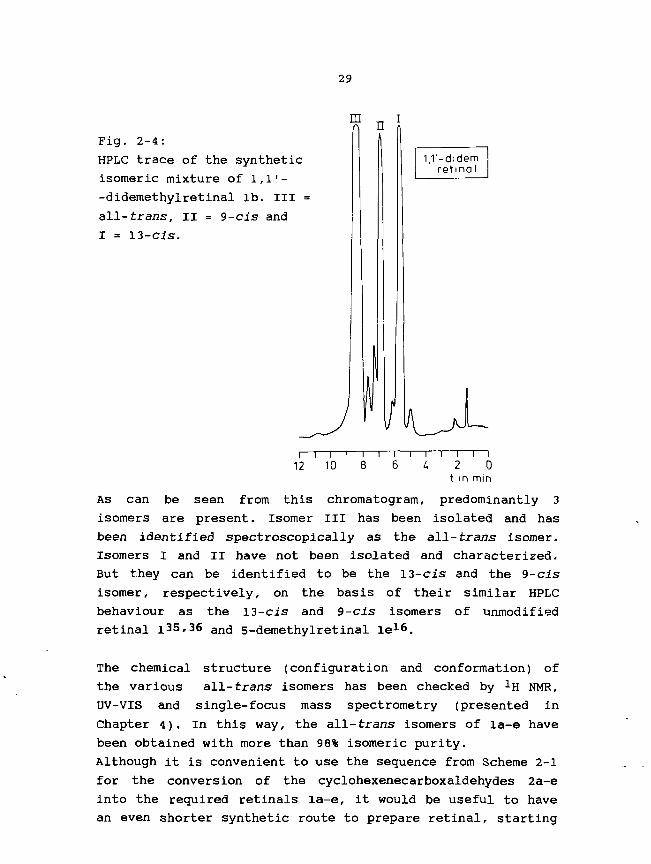
mFig. 2-4:
HPLC trace of the synthetic
isomeric mixture of 1,1'-
-didemethylretinal lb. Ill =
all-trans, II = 9-cis and
I = 13-cis.
1,1'-didem.retinal
112
~\—i—i—r10 E
—I—i—I1 6 U
T 1—2
t in
T 10
min
As can be seen from this chromatogram, predominantly 3
isomers are present. Isomer III has been isolated and has
been identified spectroscopically as the all-trans isomer.
Isomers I and II have not been isolated and characterized.
But they can be identified to be the 13-cis and the 9-cis
isomer, respectively, on the basis of their similar HPLC
behaviour as the 13-cis and 9-cis isomers of unmodified
retinal i35,36 an(j 5_demethylretinal le16.
The chemical structure (configuration and conformation) of
the various all-trans isomers has been checked by -̂H NMR,
UV-VTS and single-focus mass spectrometry (presented in
Chapter 4). In this way, the all-trans isomers of la-e have
been obtained with more than 98% isomeric purity.
Although it is convenient to use the sequence from Scheme 2-1
for the conversion of the cyclohexenecarboxaldehydes 2a-e
into the required retinals la-e, it would be useful to have
an even shorter synthetic route to prepare retinal, starting

30
from the easily accessible |3-cyclocitral37~41. Especially,
with regeird to the synthesis of ring-13C-labelled retinals,
it is important to have a short and efficient procedure, due
to the usually small amounts of valuable 13C-labelled
starting materials. Therefore, we have studied the possi-
bility to accomplish the Cio~extension of |3-cyclocitral to
retinal in only two steps. In view of the good results with
the Horner-Emmons reagent 3, which contains a nitrile
function, we chose for a CiQ-phosphono-nitrile (19A) as a
first approach. 19A can be prepared according to Scheme 2-7.
0 . 3
NBS.AIBN^CN
'0 NaH16 17
Scheme 2-7: Synthesis of Cin-phosphono-nitrile 19A.
when commercial 3-methylcrotonaldehyde 16 is subjected to a
Horner-Emmons reaction with 3, the Cin-nitrile 17 is ob-
tained, which can be converted into the CiQ-bromo-nitrile
(18) by NBS bromination (initiator AIBN: azodiisobutyro-
nitrile). Arbusow reaction with triethyl phosphite provides
the required Cin-phosphono-nitrile 19A. The potential of this
Cin-synthon in the retinal synthesis was tested by investi-
gation of its coupling reaction with (3-cyclocitral (2 in
Scheme 2-8).

31
19A
Scheme 2-8: Attempted coupling of 19A with (3-cyclocitral 2.
However, when the anion of 19A was reacted with 2, no
coupling occurred(independently of type of base and solvent),
even under forcing conditions (reflux and excess of 19A).
Similar phenomena have been reported for the reaction of a
CiQ-phosphono-carboxylic ester with another sterically
hindered cyclocitral derivative,viz. 5,6-methano-5,6-dihydro-
retinal and have been ascribed to the longer conjugated
system of this Cio-phosphonate42. This seems a plausible
explanation, however it is contradicted by literature data on
other conjugated Horner-Emmons reagentia. For example, when a
long-chain polyene-phosphonate, such as retinylphosphonate,
is coupled with retinal, the expected Horner-Emmons reaction
takes place efficiently to give P-carotene (Scheme 2-9)43.
Moreover, one of the commercial syntheses of p-carotene is
based on the analogous reaction with the retinyltriphenyl-
phosphonium ylide44.
0
•PIOEt), + r e l i n o l «°O"e . p . c a r o t e n e
, P y r (61%)
Scheme 2-9: Horner-Emmons coupling of retinylphosphonate withretinal.
Therefore, the low reactivity of the Cio-phosphonate 19A must
be at least partially due to the nitrile group. Probably, the
electron-withdrawing character of the nitrile group causes a
significant reduction of the effective negative charge on the

32
C-a atom (a with respect to the phosphonate group), preven-
ting an efficient nucleophilic attack on the carbonyl
function of (3-cyclocitral. It is expected that resonance
structures carrying the negative charge near or on the
nitrile group (III, IV and V in Fig. 2-5), will contribute to
the resonance stabilization of the carbanion to a greater
extent.
oII
lEtOljP. CN.
Fig. 2-5: Resonance structures for the anion of 19A.
If the lack of reactivity of 19A is indeed caused by the
electron-withdrawing nitrile function, replacement of this
group by another (less electronegative) functionality,
providing a Cio-phosPnonate of a more "polyene type"22, will
give better results. Our aim is to prepare an aldehyde
(retinal). Therefore, we confine ourselves to functionalities
that serve as an aldehyde-protecting group. To this end, the
easily removable imino or acetal functions seem appropriate.
Successful Horner-Emmons coupling of these compounds with
P-cyclocitral and subsequent acidic hydrolysis, would give
rttinal in a one-pot reaction. The preparation of such a
Cxo-PnosPhono-imine (19B) and CiQ-phosphono-acetal (19C),
starting from the corresponding nitrile 18, is depicted in
Scheme 2-10.

33
i P.Of.3
?0
HCIOCH3I3
Scheme 2-10: Synthesis of CiQ-phosphono-imine 19B andCiQ-phosphono-acetal 19C, starting from thecorresponding nitrile 18-
The starting compound 18, which is also a precursor for 19A,
is reduced with Dibal to give the bromoaldehyde 20. Arbusow
reaction of 20 with triethyl phosphite yields the Cio~
-phosphono-aldehyde 21, which is reacted with t-butylamine or
triethylorthoformate, to provide the required CiQ-phospho-
nates 19B and 19C, respectively.
Also the Horner-Emmons reaction of these compounds with
(3-cyclocitral was investigated. Horner-Emmons coupling of 19B
with (3-cyclocitral and subsequent acidic hydrolysis provided
the retinal in 20% yield. Horner-Emmons reaction of 19C with
P-cyclocitral, followed by acidic hydrolysis, results in 50%
yield of retinal.
Prom these facts it follows, that the yield of retinal
increases with diminishing electronegativity of the end-
-group, in agreement with our expectations. The best results
were obtained with the phosphono-acetal 19C, which gives 50%
overall yield of retinal, after coupling with P-cyclocitral
and subsequent one-pot hydrolysis. This yield is still lower

34
than that of the known four-step sequence in Scheme 2-1
(providing retinal in 64%). However, the yield of retinal via
coupling with 19C may be better than 50% {or 64%), because
this coupling has not yet been optimized.
2-6: Discussion.
We have found efficient syntheses of ring-demethylated
retinals la-e in overall yield of 22% to 36%, via the appro-
priate cyclocitral precursors 2a-e. The ten-carbon extension
of 2a-e is accomplished via repeated coupling with one and
the same Cs-synthon. This Cs-phosphono-nitrile 3 has often
been used by our group4^ and others^, 20,46 #It can be easily prepared from chloroacetonitrile (Scheme
2-1). Arbusow reaction of this reagent with triethyl
phosphite provides diethylphosphono-acetonitrile 22 1 7' 4 7,
which is also a useful synthon in retinal chemistry. Horner-
-Emmons reaction of this C2~phosphonate with commercial
chloroacetone provides an allylic chloronitrile, which is
converted into the required Cs-phosphono-nitrile 3, by an
Arbusow reaction with triethyl phosphite.
C|-CH2CN
Scheme 2-11: Synthesis of diethylphosphono-acetonitrile 22and diethyl(3-cyano-2-methyl-2-propenyl)phosphonate 3.
We have described two efficient and complementary methods to
prepare all of the required cyclohexenecarboxaldehydes 2a-e,
starting from the appropriate cyclohexanones. 2a And 2b have
been prepared according to the formylation method (Scheme
2-2), while the other cyclohexene aldehydes 2c, 2<S and 2e are
easily obtained via the cyanosilylation reaction (Schemes 2-5
and 2-6).

35
The reactions in Scheme 2-2 represent an efficient method of
preparing the otherwise poorly accessible l-cyclohexene-
carboxaldehyde 2a and its 2-methyl derivative 2b in two steps
starting from cyclohexanone. We expect that the scope of this
method for preparing 2-alkyl- and 2-aryl-l-cyclohexenecarbox-
aldehydes is very wide, on account of the ready availability
of the various alkyl- and aryl-metal reagents.
The method for preparation of 7 is analogous to a literature
procedure for the conversion of (3-ionone into the dimethyl
acetal of 3-oxo-5-(2,6,6-trimethyl-l-cyclohexenyl)-4-pent-
enal25 and the conversion of a saturated aliphatic ketone
into its corresponding dimethoxymethyl derivative26. But our
version, using NaH instead of sodium metal, is a significant
improvement in terms of reaction time and work-up. The pre-
paration of 2a with 26% overall yield via a difficult proce-
dure starting from the commercially unavailable l-chloro-2-
-cyclohexene has been described in the literature16'48. Due
to its versatility and higher yield our procedure is the
method of choice for the preparation of compound 2a and its
analogues. For the synthesis of 2-methyl-l-cyclohexene-
carboxaldehyde (2b) a route similar to our procedure, but
more cumbersome, has been described before4**. According to
this route, cyclohexanone was converted into 2b in 3 steps in
a yield comparable to that of our method. Reduction of the
intermediately isolated l-oxo-2-(isopropoxymethyleneJcyclo-
hexane 7a (an a,|3-unsaturated keto-enolether: see Fig. 2-6)
required low temperatures to prevent attack at the (3-position
by the nucleophilic reagent
7a
Fig. 2-6: Two possible reaction sites for nucleophilic attackat the a,(3-unsaturated ketone 7a (indicated byarrows).

36
The use of the saturated ketone 7 excludes this side reaction
completely, and moreover, we only need 2 steps to synthesize
2b starting from cyclohexanone.
The cyanosilylation of a ketone (Scheme 2-3) proves to be a
convenient method to prepare the important and commercially
not available cyclohexenecarboxaldehydes 2c, 2d and 2e.
Although this reaction works equally well with acid and anion
catalysis, the latter reaction is preferred, because it is
mostly difficult to maintain the quality of the appropriate
Lewis acids, due to their very hygroscopic character. And
under basic conditions there is no chance of formation of
volatile and toxic hydrogen cyanide.
Especially the KCN/18-crown-6 ether complex32'33, which can
easily be prepared by mixing equimolar amounts of potassium
cyanide and 18-crown-6 ether, gives good results. Further-
more, the use of trimethylsiloxy nltriles as intermediates
means an advantage over the commonly used cyanohydrins,
because they are more stable towards hydrolysis than the
corresponding cyanohydrins30.
According to the above method, 2c can be prepared in 3 steps
and in good yield. Only one alternative (four-step) synthesis
of 2c has been published previously50, without much experi-
mental details. Finally, we have described an alternative
novel "Cio+Cio" procedure to prepare retinal in 50% overall
yield, starting from (3-cyclocitral 2 and the CiQ-phosphono-
acetal 19C. Because of its time-saving character (two-step
one-pot method), this procedure could be attractive for the
synthesis of ring-13C-labelled retinals (see Chapter 3). For
this purpose, it then becomes necessary to improve the yield
of the reaction to about 64% or more (i.e. the overall yield
of the four-step sequence in Scheme 2-1). A number of other
Cig-phosphonates and CiQ-Wittig reagents, used in retinal
synthesis, have been desribed in the literature.
Fig. 2-7: Example of a Cio-PnosPnorane ester, used in retinalsynthesis.

37
For example, the reaction of a phosphorane ester (Fig. 2-7)
with benzaldehyde has been reported to proceed in 70%
yield51. Since phosphoranes are less reactive than their
corresponding phosphonates52, the good reactivity of thisc10~P n o sP n o r a n e ester contrasts with our findings with the
Cxo-phosphonate 19C. However, an ester group (in the phospho-
rane) and a nitrile group (in 19C) are known to be of differ-
ent electronegativity. The stronger electron-withdrawing
capacity of the nitrile group, compared with the ester group,
is illustrated by the op values53. The a-g value for a CN
group is 0.70, whereas it is 0.30 for a COOR group. Also the
absence of steric hindrance in the case of benzaldehyde,
compared with (3-cyclocitral, may be of importance. Further
support for this argument is provided by the negligible reac-
tivity of the phosphonate analogue of 23 towards the
sterically hindered l,2-methano-l,2-dihydro-|3-cyclocitral42.
On these grounds, it is expected that Horner-Emmons reaction
of (3-cyclocitral (which is also sterically hindered) with
this phosphonate ester will likewise give negative results.
Moreover, phosphonate reagents with a carboxylic ester end-
-group are not attractive to be used in the synthesis of
ring-13C-labelled retinals. They all suffer from the fact,
that, after the coupling reaction, two extra steps are needed
to obtain retinal.
In conclusion, it is clear that the phosphono-acetal 19C is a
promising synthon for the preparation of ring-13C-labelled
retinals, starting from the corresponding i3C-labelled |3-
-cyclocitrals. For this purpose, its Horner-Emmons reaction
with p-cyclocitral has to be optimized.

38
LITERATURE
1. P.K. Das and R.S. Becker, J. Phys. Chem. 82, 2081 (1978).2. K. Muellen, H. Schmickler, B. Frei and H.R. Wolf,
Tetrahedron Lett. 27, 477 (1986).3. R.R. Birge, D.F. Biocan and L.M. Hubbard, J. Am. Chem.
Soc. 104, 1196 (1982).4. B. Honig, A.D. Greenberg, B.D. Sykes, M. Karplus, Proc.
Natl. Acad. Sci. USA 68, 1289 (1971).5. C.H. Stam and C.H. McGillivary, Acta Crystallogr., Sect.
B, B16, 62 (1963).6. C.J. Simmons, R.S.H. Liu, M. Denny and K. Seff, Acta
Crystallogr., Sect. B, B37, 2197 (1981).7. R. van der Steen, P.L. Biesheuvel, R.A. Mathies and
J. Lugtenburg, J. Am. Chem. Soc. 108, 6410 (1986).8. K. Nakanishi, V. Balogh-Nair, M. Arnaboldi-Tanis,
K. Tsujimoto and B. Honig, J. Am. Chem. Soc. 102, 7945(1980).
9. G.S. Harbison, P.P.J. Mulder, H. Pardoen, J. Lugtenburg,J. Herzfeld, and R.G. Griffin, J. Am. Chem. Soc. 107,4809 (1985).
10. W. Gaertner, P. Towner, H. Hopf and D. Oesterhelt, Bio-chemistry 22, 2637 (1983).
11. M. Muradin-Szweykowska, L.J.P. van Amsterdam,L.J.M. Rodenburg, J. Lugtenburg, R.L. van der Bend andK. van Dam, FEBS Lett. 154, 180 (1983).
12. B.I. Mitsner, A.A. Khodonov, E.N. Zvonkova, R.P. Evstig-neeva, Bioorgan. Khim. 12, 5 (1986) (review).
13. V. Balogh-Nair and K. Nakanishi in "Methods in Enzym-ology". Vol. 88, L. Packer (Ed.), 496, N.Y., 1982.
14. A. Albeck, N. Friedman, M. Sheves and M. Ottolenghi, J.Am. Chem. Soc. 108, 4614 (1986).
15. M. Sheves, N. Friedman, V. Rosenbach and M. Ottolenghi,FEBS Lett. 166, 245 (1984).
16. M.B. Spijker-Assink, c. winkel, G.S. Baldwin and J.Lugtenburg, to appear in Reel. Trav. Chim. Pays-Bas(1988).
17. R.W. Dugger and C.H. Heathcock, Synth. Commun. 10, 509(1980) .
18. K. Fujiwara, H. Takahashi and M. Ohta, Bull. Chem. Soc.Jpn. 35, 1743 (1962).
19. H. Pommer, W. Stilz, BASF Aktiengesellschaft, Ger. Pat.1,116,652 (9 nov. 1961) [C.A. 57, P 2267d (1962)].
20. R.S.H. Liu, H. Matsumoto, A. Asato, M. Denny,Y. Schichida, T. Yoshizawa and F.W. Dahlguist, J. Am.Chem. Soc. 103, 7195 (1981).
21. M. Muradin-Szweykowska, J.A. Pardoen, D. Dobbelstein,L.J.P. v. Amsterdam, J. Lugtenburg, Eur. J. Biochem.140, 173 (1984)
22. J. Boutagy, R. Thomas, Chem. Rev. 74, 87 (1974).23. IUPAC-IUB Joint Commission on Biochemical Nomenclature,
Eur. J. Biochem. 129, 1 (1982).24. 0. Bayer in "Methoden der Organischen Chemie", 4° Ed.,
Bd 7/1, 44, Thieme, Stuttgart (1954).25. G.J.M. Nicolaux, E.A. Gray, J. Matet, R.L.H. Mauge,
C.M.T. Sandevoir and A.J.A. Wasmer, Fr. Patent,N° 1.243.824 CO7C (1960); C.A. 57 (1962), 16671 h.

39
26.
27.28.29.
30.
31.
32.
33.34.
35.
36.37.
38.
39.
40.
41.
42.
43.
44.
45.
46.
47.48.49.
50.
51.
52.
53.
M. Baumann and W. Hoffmann, Liebigs Ann. Chem., 743(1979) .C. Ainsworth, Org. Synth., Coll. Vol. 4, 536 (1963).M. Oda, A. Yamamuro, T. Watabe, Chem. Lett., 1427 (1979).F. Duboudin, P. Cazeau, F. Moullnes and O. Laporte,Synthesis 212 (1982).D.A. Evans, L.K. Truesdale and G.L. Carroll, J. Chem.
(Chem. Commun.), 55 (1973).Reetz and I. Chatziiosifidis, Synthesis (Commun.),(1982).Evans, J.M. Hoffman, L.K. Truesdale, J. Am. Chem.95, 5822 (1973).Greene, Tetrahedron Lett., 1793 (1972).King and J.G. Topliss, J. Chem. Soc. (C),
(1957).R.S.H. Liu in 'Methods in Enzymology1
506, Academic Press, N.Y., 1982.R.S.H. Liu and A.E. Asato, Tetrahedron 40,
SocM.T330D.ASocR.NF.E 919
88, L. Packer (Ed.)
1931 (1984)R.S.H. Liu,V. Ramamurthy, G. Tustin, C.C. Yau and
Tetrahedron 31, 193 (1975).R.N. Gedye, P.C. Arora and K. Deck, Can. J. Chem. 49,1764 (1971).L. Colombi, A. Bosshard, H. Schinz and C.F.
265 (1951).Seidel, Helv.
Chem. Soc., 1154
A.Chim. Acta 34,H.B. Henbest, B.L. Shaw and G. Woods, J.(1952).S. Yamada, M. Shibasaki, S. Terashima, Tetrahedron Lett.,377 (1973).M.I. Dawson, P.D. Hobbs, R.L.S. Chan and W.R. chao,J. Med. Chem. 24, 1214 (1981).J.D. Surmatis and R. Thommen, J. Org. Chem. 34, 559(1969).
DBP 1068709 (1958) BASF;911 (1960).Leiden University, I.C.G.
H. Pommer and W. Sarnecki,H. Pommer, Angew. Chem. 72J.A. Pardoen, Ph.D. ThesisPrinting, Dordrecht, 1986.K. Eiter, H. Oediger and E1,110,633; C.A.W. Stilz and H.E.A. Braude and E.A. Evans,K.E. Harding and R.C. Ligon(1974).
Truscheit, Ger. Pat.56, 3522 C (1962).Pommer, C.A. 56, 11,422 (1962).
J. Chem. S o c , 3334 (1955).Synth. Commun. 4, 297
Goff and A.S. Waggoner,A. Kropf, B.P. wittenberger, S.P.Exp. Eye Res. 17, 591 (1973).H. Bayley, R. Radhakrishnan, K.S. Huang and H. Khorana,
Chem. 256, 3797 (1981).Klink and W. Hoffmann, Chem. Ber.W. 96, 3133
J. Biol.L. Horner,(1963).J. March, Advanced Organic Chemistry, 3° Ed., 244, Wileyand Sons, N.Y. (1985).
41
Chapter 3
SYNTHESIS OF RING-13C-LABELLED RETINALS
3-1: Introduction
For investigation of the ring moiety of the chromophore in
bacteriorhodopsin (bR), rhodopsin (Rh) and isorhodopsin with
solid-state 13C NMR spectroscopy (see Chapter 6), we need the
appropriate 13C-labelled retinals to prepare the correspon-
ding 13C-labelled pigments. The chain part of retinal (from
C8 up to and including C15) has been labelled with 13C
earlier, by our group1. The remaining ten positions, 1, 2, 3,
4, 5, 6, 7, 16/17 and 18, which occur in the (3-cyclocitryli-
dene part of retinal, are the objective to be labelled with13C (see Fig. 3-1).
Fig. 3-1: Structure of all-trans retinal (l), includingpositions to be labelled with 13C (*).
For bacteriorhodopsin, positions 1, 4, 5, 6, 7 and 18 are all
important to give a decisive answer, as to the possible
external point-charge near the cyclohexene ring (see para-
graph 1-5)2. The chemical-shift parameters of C5 are expected
to give definitive information about the conformation around
the C6-C7 single bond of the chromophores in bR, Rh and
isorhodopsin. The Ti values of the methyl carbons 16, 17 and
18 will probe this conformation to a further extent. The
chemical-shift values of C16 and C17 will provide information
about the possible chirality of the chromophore, induced by
the chiral peptide chain. Finally, the chemical-shift
characteristics of C2 and C3 will be informative about the
inversion rate of the cyclohexene ring in these pigments.
In order to observe distinctive signals of the chromophore in
the 13C NMR spectra of the pigments, we require a certain
extent of 13C-enrichment. We are restricted to the use of
simple commercially available 13C-labelled starting mate-

42
rials, which contain either 90 or 99 at.% 1 3C. For financial
reasons we have chosen for starting materials with 90%
enrichment, which is amply sufficient for the 13C NMR
studies. For the synthesis of (1-13C)- and (16,17-13C2)-
retinal we use (2-13C)- and (1,3-13C2)-acetone, respectively.
For the attempted synthesis of (2-13C)- and (3-13C)retinal
the appropriate starting materials are (1-13C)- and (2-13C)-
-acetonitrile, respectively. (4-13C)retinal is prepared from
(2-13C)-acetonitrile. K13CN is used for the synthesis of (5-
-13C)retinal. 13CH3I is applied in the synthesis of (18-13C)-
retinal. (2-13C)- and (l-13C)-acetonitrile are the precursors
for (6-13C)- and (7-13C)retinal, respectively. Taking into
account the high costs of these 13C-labelled compounds and
the synthetic difficulties encountered in labelling the ring
positions, it is clear that our strategy requires very effi-
cient schemes to end up with as much 13C-labelled material as
possible. In the next paragraph, we discuss the general syn-
thetic method for the 13C-labelled retinals. The syntheses of
the corresponding ^3C-labelled starting compounds are de-
scribed in paragraphs 3-3 to 3-6. In paragraph 3-7, we
discuss the preliminary results, concerning the attempted
synthesis of (2-13C)retinal and (3-13C)retinal.
3-2: Development of efficient synthetic schemes for ring-13C-
-labelled retinals.
We initially considered the successful four-step sequence
used for the synthesis of the ring-demethylated retinals la-e
("c10+c5+c5" method in Scheme 2-1). This method provides
retinal in 64% overall yield, based on the easily accessible
(3-cyclocitral (2). Once optimized, also the "CIO+CJLO" Proce-
dure (see par. 2-5) would be attractive. Thus, our next
objective is to synthesize the appropriate 13C-labelled (3-
-cyclocitrals. The large-scale preparation of (3-cyclocitral
2, starting from the open-chain citral 23 is well-known and
has been described in the literature (Scheme 3-1)4~8.
Citral (23), when converted into the citralanil, cyclises in
concentrated sulfuric acid to produce usually a mixture of a-

43
and p-cyclocitral after hydrolytic work-up. under basic
conditions, the a-cyclocitral (a-2) can easily be isomerized
into the required (3-cyclocitral (P-2)3~9. For this large-
-scale preparation(ca. 15-50g), the overall yield of (3-cyclo-
citral has been reported to be at most about 65%. We turned
our attention to the small-scale synthesis (ca. lg or less).
In many cases, we succeeded to get about 90% overall yield of
ctral 23 citrolaml o-2 fi-2
CH30H
Scheme 3-1: Synthesis of (3-cyclocitral 2 from citral 23.
P-cydocitral. However, sometimes the cyclisation on this
small scale failed for unknown reasons. Because we could not
risk losing a batch of 13C-labelled cyclocitral, we had to
decide to discontinue the "C10+C5+C5" procedure.
As the next approach we have chosen to base our synthetic
schemes on the Ci3~synthon P-ionone (24), which has been
prepared by cyclisation of pseudo-ionone, on large and even
industrial scale under various conditions10"16. One of the
versions, using a homogeneous system for the cyclisation
procedure, seemed the most appropriate for our purpose11. We
have carefully examined the applicability of this procedure
for our small-scale experiments. The cyclisation of pseudo-
-ionone has been performed in a homogeneous system of concen-
trated sulfuric acid and nitromethane with ice-bath cooling
(Scheme 3-2). In this way, the extent of polymerization is
substantially reduced, due to the proper dilution of pseudo-
-ionone. Moreover, at a temperature of 0-10°C exclusively P-
-ionone is formed, with only a trace amount of a-ionone. We
have succeeded in optimizing our small-scale procedure to
give P-ionone in 75% overall yield. In contrast with the poor
reproducibility of the citral-cyclisation reaction, this
quick and simple method to cyclise pseudo-ionone is reliable

44
and always provides 75% of (3-ionone. Thus, this method is the
method of choice for the preparation of the corresponding
ring-13c-labelled (3-ionones from the appropriate 13C-labelled
pseudo-ionones.
pseudo- lonone
Scheme 3-2: Efficient cyclisation of pseudo-ionone intoP-ionone 24 in 75%
Also on small scale, P-ionone (24) can be converted into
retinal (1), according to the well-documented four-step
sequence, depicted in Scheme 3-317.
0 22II ~
2U-n
2) Dibali. f-n
2) Dibal1 f-n
Scheme 3-3: Four-step synthesis of retinal (l) starting from(3-ionone (24).
At first, P-ionone (24) is treated with the C2-phosphono-
-nitrile 2218, followed by a diisobutylaluminium hydride
(Dibal) reduction19"21 to give (3-ionylideneacetaldehyde 25.
Compound 25 is then subjected to another Horner-Emmons
coupling with Cs-phosphono-nitrile 3 2 2 and subsequently
treated with Dibal to yield the required retinal 1 (all-£",
13-Z and 9-Z) in 64% overall yield. The preparation of the
essential phosphonate reagents 3 and 22 has been discussed in
Chapter 2 (Scheme 2-11)18'22.
As can be seen, application of the procedure in Scheme 3-3 to

45
the appropriate 13C-labelled (3-ionones, provides the required
(l-13C)retinal (If), (2-i3C)retinal (lg), (3-13C)retinal
(lh), (4-13C)retinal (li), (5-13C)retinal (1j), (6-13C)-
retinal (Ik), (7-13C)retinal (ll), (16,17-13C2)retinal (1m)
and (18-13C)retinal (in). The required cis isomers of these
retinals are obtained by irradiation of the HPLC-isolated
all-trans isomers in an appropriate solvent^,24# <i>ne photo-
chemistry of retinals is highly dependent on the nature of
the solvent (methanol, ethanol and acetonitrile as polar
solvents and n-hexane as non-polar solvent)24-26, j n polar
solvents, where the l(n,n*) state is the lowest excited state
and intersystem crossing seems to be negligible, we are
dealing with the 1(TT,IT*) or !Ag*~ state. Under these condi-
tions the mono-cis isomers are the principal contributors to
the photostationary state (the contribution of each di-cis
isomer being ca. 1 % ) . Irradiation of all-trans retinal in
acetonitrile results in a steady-state mixture, in which the
ll-cis isomer (of essence to rhodopsin research) reaches 35%
of the combined isomer composition. In non-polar solvents,
the 1(n,iT*) state is the lowest excited state and here the
intersystem crossing to the triplet state is important. The
photoisomerization largely proceeds via the triplet state.
The photostationary state differs considerably from that
found in polar media, the main isomers being all-trans, 9-cis
and 13-cis, without traces of 7- and ll-cis or of di-cis
isomers. The total amount of cis isomers is much smaller than
in polar media. The presence of even traces of water or other
polar molecules affects the photostationary state in n-hexane
by forming hydrogen-bond complexes with retinal, in which the1(n,Ti*) state is at higher energy level than the 1(TT,TT*)
state27. In conclusion, it is evident that to obtain the re-
quired cis isomers, the irradiation of the all-trans retinals
should be performed in a polar solvent (CH3CN, ethanol).

46
3-3: Synthesis of (1-13C)retinal (If) and (16,17-13C2)retinal
(lm).
In Scheme 3-4 is indicated how 13C-labelled acetone can be
converted in a six-step sequence into the labelled (3-ionones
24f and 24m. The easily available phosphonium salt 27 (Scheme
3-4) is treated successively with n-BuLi and labelled acetone
26f or 26m, effecting a five-carbon extension of the labelled
acetone. Deprotection28 gives the labelled ketones 28f and
28m (80-88% yield). Horner-Emmons reaction of 28f and 28m
with C2~phosphonate 22 followed by a Dibal reduction gives
the 13C-labelled citrals 23f and 23m in about 85% yield.
Aldol condensation of 23f and 23m with 2N NaOH in acetone
quantitatively gives the open-chain pseudo-ionones29*30.
These are cyclised in concentrated sulfuric acid, as de-
scribed above, to provide the p-ionones 24f and 24m in 75%
yield. This means that the sequence of Scheme 3-4 gives the13C-labelled p-ionones 24f and 24m in 33% overall yield,
based on the labelled acetones 26f and 26m, respectively. Two
subsequent Horner-Emmons reactions and Dibal reductions lead
to If and lm in 20% overall yield, based on the 13C-labelled
acetones•
tn 11*01
\ . \ 0 0 „ n-BuU21 SiO2
26f.m 27 28f.m
22 ^
? ^ ^ .2NNoOH2] D.bal kJ^\ 2) 9 6 % H2SO t
23'.m
24f.m
Scheme 3-4: Synthesis of (1-13C)- (24f) and (16,17-13C2)-P-ionone (24m).
In Scheme 3-5, it is depicted how commercially available
5-chloro-2-pentanone 29 is converted into the necessary
Wittig salt 27.

47
HO OH , 1 ,—,11 v_v .H' S (TO2)kl
29 30 27
Scheme 3-5: Synthesis of the phosphonium salt 27.
The keto function is protected as ketal with ethylene glycol
and a catalytic amount of acid31. SN2 reaction of the ketal
chloride with KI in toluene (+ 18-crown-6 ether) yields the
protected iodide 3O32. Reaction of 30 with triphenylphos-
phine in toluene provides the required Wittig reagent 27 in
about 65% overall yield based on the chloro-ketone 29.
3-4: Synthesis of (4-13C)retinal (li)
13C-labelling of position 4 (or 5) in retinal can be effected
according to Scheme 3-6.
31 a. b
n ~
^j 2)Dibol
28..J 2~3'J
Scheme 3-6: Synthesis of (4-13C)-citral (23i)[and (5-13C)-citral (23j)]
The anion of (2-13C)-acetonitrile 31b33'34 reacts with the
allylic bromide 32 (which is commercially available or can be
easily made from 2-methyl-3-buten-2-ol and 47% aqueous HBr/35
to yield 60% of the labelled l-cyano-4-methyl-3-pentene 33i.

48
Treatment of 33i with excess of methyllithium36 gives
6-methyl-5-hepten-2-one 28i in 70% yield. The conversion of
28i into retinal has been described above (Schemes 3-3 and
3-4). Thus, thrice repeated Horner-Emmons reaction followed
by a Dibal reduction results in an overall yield of 17% of
(4-13C)retinal (li) in 10 steps starting from (2-13C)-aceto-
nitrile 31b. The reactions depicted in Scheme 3-6 also
provide (5-13C)retinal (lj) in the same yield, based on
(I-13C)-acetonitrile 31a. However, for the preparation of
(5-13C)retinal (lj) we have found a more efficient synthetic
procedure based on the less expensive K13CN. This method is
described in the next paragraph.
3-5: Synthesis of (5-13C)retinal (lj ) and (18-13C)retinal
(in)
For the synthesis of (5-13C)- (1j) and (18-13C)retinal (In),
the unsaturated tosylate 34 is the starting compound (its
synthesis is presented in Scheme 3-8). In Scheme 3-7 we show
how the required 13C-labelled |3-ionones 24 j and 24n can be
obtained from 13C-labelled potassium cyanide and
respectively.
K' 3CN •
34
D - : S . ^ ^ ^ .LDA X . , , j v 2 N N Q O H
2) H30* " \ ^ 2! 96% HjSOi
Scheme 3-7: Route to prepare (5-13C)- (24j) and (18-13C)--(3-ionone (24n) from K13CN and 13CH3l, respec-tively.

49
The tosylate 34 undergoes an SN2 reaction with K13CN to
afford the nitrile 35j37. Reaction of 35j with an excess of
methyllithium gives the unsaturated methylketone 36j.
Peterson-type olefination of 36j with the tert-butylimine of
(trimethylsilyl)acetaldehyde (37)38, yields the isocitral
38j. Aldol condensation of 38j with acetone gives the
isopseudo-ionone. Sulfuric-acid mediated cyclisation provides
(5-13C)-|3-ionone (24 j ) in 40% overall yield, based on K13CN.
For the preparation of (18-13C)retinal In, we treated
unlabelled nitrile 35 with 13C-labelled methylmagnesium
iodide (prepared from 13C-labelled iodomethane and magnesium
powder), giving the methyl-13C-labelled unsaturated ketone
36n. In the same way, 13C-labelled methyllithium (from13C-labelled iodomethane and lithium) could be used. Because
now the Grignard reagent possesses the 13C label, only a
slight excess (1.1 eq.) is used. For this reason and probably
also due to the small scale of the reaction, only 10% yield
of 36n has been obtained, based on the starting 13CH3i. The
methylketone 36n is converted into |3-ionone 24n analogously
to the conversion of 36j into 24j. in this way (18-13C)-|3-
-ionone (24n) is obtained in 6% overall yield, based on13CH3l. The 13C-labelled |3-ionones 24j and 24n are then
converted into (5-13C)retinal (lj) and (18-13C)retinal (in)
in 64% yield, via the four-step sequence illustrated in
Scheme 3-3.
In Scheme 3-8 we present the method used to synthesize
tosylate 34, which is necessary for efficient introduction of
the 13C label at position 5 of retinal. Commercial 5-hydroxy-
-2-pentanone (39) is first esterified with acetic anhydride
to give the acetate. The resulting acetoxy ketone is olefi-
nated with triphenylmethylenephosphorane to yield the
unsaturated acetate 4.0, and subsequently hydrolysed to
provide the corresponding unsaturated alcohol. When this
alcohol is treated with p-toluenesulphonic acid chloride in
pyridine the required tosylate is obtained39.
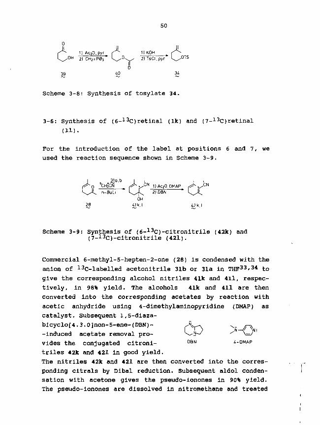
50
I1) KOH
° H 2) CH2 = P03 k / ° X ^ 2)TsCl.pyr
F0
39 40 34
Scheme 3-8: Synthesis of tosylate 34.
3-6: Synthesis of (6-13C)retinal (Ik) and (7-13C)retinal
(11).
For the introduction of the label at positions 6 and 7, we
used the reaction sequence shown in Scheme 3-9.
31a.b^ C N 1)AC;0,DMAP
2JD8NOH
28 41k.I 42k,I
Scheme 3-9: Synthesis of (6-13C)-citronitrile (42k) and(7-13C)-citronitrile (421).
Commercial 6-methyl-5-hepten-2-one (28) is condensed with the
anion of 13C-labelled acetonitrile 31b or 31a in THF33'34 to
give the corresponding alcohol nitriles 41k and 411, respec-
tively, in 98% yield. The alcohols 41k and 411 are then
converted into the corresponding acetates by reaction with
acetic anhydride using 4-dimethylaminopyridine (DMAP) as
catalyst. Subsequent 1,5-diaza-
bicyclo[4.3.0]non-5-ene-(DBN)-
-induced acetate removal pro-
vides the conjugated citroni- DBN 4-DMAP
triles 42k and 421 in good yield.
The nitriles 42k and 421 are then converted into the corres-
ponding citrals by Dibal reduction. Subsequent aldol conden-
sation with acetone gives the pseudo-ionones in 90% yield.
The pseudo-ionones are dissolved in nitromethane and treated
Cp

51
with concentrated sulphuric acid at 0°C to yield the 13C-
-labelled |3-ionones 24k and 241. The |3-ionones (24k,24l> are
converted via the known four-step procedure (Scheme 3-3) into
(6-13C)retinal (lk) and (7-13C)retinal (11) in 37% overall
yield based on the corresponding 13C-labeJ.led acetonitriles
31b and 3la, respectively.
3-7: Attempted synthesis of (2-13C)- (lg) and (3-13C)retinal
(lh).
In this paragraph we present the strategy for 13C-labelling
of positions 2 and 3 in the retinal molecule. The most
convenient way is to label both positions, starting from only
one Cs-synthon, which can be converted into citral in a few
steps. A good candidate for this C5 building-block would be
the dimethyl acetal of 4-bromo-3-methyl-2-butenal 43 (for
preparation see Chapter 7).
>CH3°CN • if^O- n-BuL, . f* f<0- „
31a,b 43 44g.h
Scheme 3-10: Introduction of 13C at positions 2 (=g) and 3(=h) via substitution by the anion of 31a and31b.
We have found that the bromide in 43 can be substituted
satisfactorily by reaction with the anion of (l-13C)-aceto-
nitrile (31a) and (2-13C)-acetonitrile (31b), respectively,
while the protecting acetal group is kept intact. For
introduction of the tri-substituted double bond present in
citral, via a Wittig reaction, 44 has to be converted into
the aldehyde 45 first. Although Dibal reduction of 44 seems
suitable, the desired aliphatic aldehyde 45 is not obtained.
Instead, accompanying reduction of the acetal group takes
place. We then decided to replace the acetal group by a
protected alcohol function, thus making geraniol (49) our
first target molecule. The choice of the t-butyldimethylsilyl

52
(TBDMS) protecting group proves to be convenient, provided
iodide is used as leaving group (the combination of the TBDMS
group and the bromide did not give a proper substitution
reaction). After these exploring experiments, we now propose
a synthetic scheme for the synthesis of (2-13C)- and (3-13C)-
-(3-ionone (Scheme 3-11).
LM3LIN * T I I -,. p. "~j *
i-^^^x^ 2) Uibal
31a,b Lb ^7g,h
1) x ^ V ~ ' f | ( ^ ^ O H _1] DOQ iorMnO?)21 TBAF ''Ok 0
2; .K 2NNaOH
£ 3) 96% H2b0t
Scheme 3-11: Proposed synthetic route to synthesize (2-13C)(24g) and (3-13C)-|3-ionone (24h).
The anion of 31a or 31b is treated with the fcerfc-butyldi-
methylsilyl ether of 4-iodo-3-methyl-2-buten-l-ol (46: for
its preparation see Scheme 3-12) to give both the correspon-
ding 13C-labelled nitriles. Dibal reduction of these nitriles
would provide the 13C-labelled primary aldehydes 47g and 47h,
which can be treated with isopropylidenetriphenylphosphorane
(48)40 , followed by tetrabutylammonium-fluoride-(TBAF)-
-induced desilylation41, to give the 13C-labelled geraniols
49g and 49g. These geraniols can be oxidized with 2,3-
-dichloro-5,6-dicyano-l,4-benzoguinone (DDQ)4^ Or MnO2
equally well, to provide the corresponding citrals, which can
be converted into the 13C-labelled (3-ionones 24g and 24h,
according to the usual procedure (see Scheme 3-4).
Nearly all of the reactions in Scheme 3-11 have been experi-
mentally worked out, using the equivalent unlabelled rea-
gents, and found to proceed successfully, except for the
Dibal reduction. The substitution of 46 with the anion of
unlabelled acetonitrile provides the siloxy nitrile in about
60% yield (after column chromatography). The iodide 46 can
easily be made from commercially available ethyl 3-methyl-2-
-butenoate 50 (Scheme 3-12).

53
.OEt 11 NBS n r I 1) TBDMSCl.DiPEA0 2) Dibal ^ ^ " " " ^ O H 2) N ° I
50 51
Scheme 3-12: Synthesis of iodide 46.
After brominatlon and Dibal reduction of 50, the bromo-
alcohol 51 is obtained. Protection of 51 with the aid of
tert-butyldimethylsilyl chloride (TBDMSC1) in the presence of
diisopropylethylamine (DIPEA)4^ and subsequent iodide substi-
tution42, leads to the required iodide 46. Although the
crucial substitution reaction of 46 can be performed success-
fully and most of the other reactions have proven to be
convenient as well, some steps still have to be studied in
more detail. Especially, the Dibal reduction of the siloxy
nitrile, to produce 47, turned out to be not successful. Lack
of time prevented us from exploring Scheme 3-11 further.
3-8: Discussion.
with the present synthesized seven ring-13C-labelled retinals
If and li-n, specific 13C-labelling at each position (with
the exception of 2 and 3) in the retinal skeleton (20 carbon
atoms) has been achieved. The ten positions in the chain part
of retinal (from C8 up to and including C15) have been label-
led earlier1.
The retinals If and li-n, having the required chemical purity
(> 99%) and the expected isotopic enrichment (ca. 90% 13C:
the same as the 13c-labelled starting materials), have been
synthesized using the methods illustrated in Schemes 3-3 to
3-9. In our syntheses we obtain about 60% of the all-trans
isomer. Only for (6-13C)- and (7-13C)retinal an alternative
synthesis has been described earlier43, based on the commer-
cially available 13C-labelled ethylbromoacetate, which is 2.5
times more expensive than 13c-labelled acetonitrile.
In general, the 13C-labelled starting compounds are used very
efficiently. The yield of if, Ik, 11 and lm is about 35%

54
based on (2-13C)-acetone, (2-13C)-acetonitrile, (1-13C)-
-acetonitrile and (1,3-13C2)-acetone, respectively. The yield
of li is 17% based on (2-13C)-acetonitrile and that of lj is
26% based on K13CN. However, (18-13C)retinal in is prepared
in only 4% yield, based on the starting 13CH3l, due to the
low yield (10%) of the 35 to 36n conversion.
We have found that the Cs-bromoacetal 43 is not appropriate
for the introduction of the 13C label at retinal positions 2
and 3 (see paragraph 3-7). Although the substitution reaction
with the anion of 31a/31b takes place properly, the subse-
quent Dibal reduction of the product nitriles 44g/44h did not
provide the expected aldehydes 45g/45h. Instead, it turned
out that the acetal function is unstable towards Dibal reduc-
tion, yielding a methyl ether by hydride substitution. This
unexpected behaviour has probably to do with the allylic
character of the acetal group in our Cs-synthon 43. The TBDMS
function has been found to be an ideal alcohol-protecting
group44, because it is stable towards nucleophiles (e.g.
~CH2CN), Dibal reduction and mild acidic conditions (pH 6),
and because it can be easily removed41. Use of the iodo-t-
-butyldimethylsilyl ether 46 for the substitution reaction
with 13C-labelled acetonitrile (Scheme 3-11) provided the
siloxy nitriles in good yield. We have found that the
cyclisation of pseudo-ionone can best be achieved by dissol-
ving in nitromethane and dropwise adding to a mixture of
nitromethane and excess of sulfuric acid at 0°C. In this way,
the pseudo-ionone remains reasonably diluted and is in good
contact with the acid, diminishing the risk of polymeriza-
tion, and overcoming the problems encountered with most of
the other cone, sulfuric-acid/organic-solvent systems (not
mixable, not easily uniformly cooled).
In conclusion, it can be said that the retinal positions 1,
4, 5, 6, 7, 16/17 and 18 have been labelled with 1 3C,
according to straightforward procedures, based on simple
commercially available 13C-labelled starting materials. The
efficiency is high, except for position 18. For the enrich-
ment of the remaining positions 2 and 3, we have carried out
several reactions (paragraph 3-7), which lay an important
basis for the synthesis of (2-13C)- and (3-13C)retinal.

55
LITERATURE
1.
2.
3.
4.
5.
6.
7.
8.
9.10.11.
12.
13.
14.
15.
16.
17.
18.19.
20.
21.
22.23.
24.25.26.27.
28.
29.
J.A. Pardoen, Ph.D. Thesis , Leiden University, ICGPrinting, Dordrecht, 1986.K. Nakanishi, V. Balogh-Nair, M. Arnaboldi-Tanis, K.Tsujimoto and B. Honig, J. Am. Chem. Soc. 102, 7945(1980).R. Rowan, III, and B.D. Sykes, J. Am. Chem. Soc. 96, 7000(1974).V. Ramamurthy, G. Tustin, C.C. Yau and R.S.H. Liu,Tetrahedron 31, 193 (1975).R.N. Gedye, P.C. Arora and K. Deck, Can. J. Chem. 49,1764 (1971).L. Colombi, A.Chim. Acta 34,
Seidel, Helv.
Terashima, Tetrahedron
38, 546 (1946)
Bosshard, H. Schinz and C.F.265 (1951).
H.B. Henbest, B.L. Shaw and G. Woods, J. Chem. Soc., 1154(1952).S. Yamada, M. Shibasaki and S.Lett., 377 (1973).H. Koster, Chem. Ber. 77, 555 (1944)E. Earl Royals, Industr. Eng. Chem.V.A. Smit, A.V. Semenovskii, V.M. Medvedeva and V.F.Kucherov, Dokl. Akad. Nauk SSSR 124, 1080 (1959).Th.j. de Boer, H.A. van 't Hof and J.u. Veenland, Reel.Trav. Chim. Pays-Bas 83, 689 (1964).I.G. Mursakulov, A.V. Semenovski, W.A. Smit and V.F.Kucherov, Tetrahedron 23, 1621 (1967).N.V. Philips Gloeilampenfabrieken, Neth. Appl.67.00.757; C.A. 70 (1969), 29117 z.C. Breant and J. Poizat (Societe des Usines ChimiquesRhone-Poulenc), Fr. Patent N° 1.523.361 ; C.A. 72 (1970)32077 h.P.H. van Leeuwen (N.V. Philips Gloeilampenfabrieken),Ger. Offen. 1.917.132 ; ibidem, 32080 d.J.A. Pardoen, P.P.J. Mulder, E.M.M. van den Berg and J.Lugtenburg, Can. J. Chem. 63, 1431 (1985).References 17 and 47 in Chapter 2.N.V. Philips Gloeilampenfabrieken, Brit. Pat. 811,527 ;C.A. 53, 17177 (1959).G.I. Samovkhvalov, L.I. Zakhavakin, L.P. Davydova andI.M. Khorlina, Dokl. Akad. Nauk SSSR 126, 1013 (1959).R.S.H. Liu, H. Matsumoto, A. Asato, M. Denny, Y.Schichida, T. Yoshizawa and F.W. Dahlquist, J. Am. Chem.Soc. 103, 7195 (1981).References 17 to 19 in Chapter 2.R.S.H. Liu and A.E. Asato, in "Methods in Enzymology",Vol. 88, L. Packer (Ed.), 506, Acad. Press, N.Y., 1982.R.S.H. Liu and A.E. Asato, Tetrahedron 40, 1931 (1984).R.R. Birge, Annu. Rev. Biophys. Bioenerg. 10, 315 (1981).M. Ottolenghi, Adv. Photochem. 12, 97 (1980).T. Takamura, P.K. Das, G. Hug and R.S. Becker, J. Am.Chem. Soc. 98, 7099 (1976).F. Huet, A. Lechevallier, M. Pellet and J.M. Conia,Synthesis (Commun.), 63 (1978).N.L. Wendler, H.L. Slates, N.R. Trenner and M. Tishler,J. Am. Chem. Soc. 73, 719 (1951).

56
30. H.O. Huisman, A. Smit, P.H. van Leeuwen and J.H. van Rij,Reel. Trav. Chim. Pays-Bas 75, 977 (1956).
31. R.K. Mttller, H. Mayer, K. Noack, J.J. Daly, J.D. Tauberand S. Liaaen-Jensen, Helv. Chim. Acta 61, 2881 (1978).
32. J. Royer and H.P. Husson, J. Org. Chem. 50, 670 (1985).33. G. Gainelli, G. Cardillo, M. Contento, P. Graselli and
A.U. Ronchi, Gazz. Chim. Ital. 103, 117 (1962).34. K. Eiter, E. Truscheit, H. Henecka and R. Lorenz, Ger.
Pat. 1.124.941 (1962); C.A. 57, 7323 b (1962).35. L.F. Fieser, J. Am. Chem. Soc. 49, 857 (1927).36. A.M. van Leusen and D. van Leusen, Synth. Commun. 8, 39 7
(1978).37. R.A. Smiley and C. Arnold, J. Org. Chem. 25, 257 (1978).38. E.J. Corey, D. Enders and M.G. Bock, Tetrahedron Lett.,
7 (1976).39. C.S. Marveland and V.C. Sekera, J. Am. Chem. Soc. 55, 345
(1933).40. H. Schulz and I. Sprung, Angew. Chem. 81, 258 (1969).41. E.J. Corey and A. venkateswarlu, J. Am. Chem. Soc. 94,
6190 (1972).42. D.D. Burn, V. Petrow and G.O. Weston, Tetrahedron Lett.,
14 (1960).43. G.D. Mateescu, W.G. Copan, D.D. Muccio, D.v. Waterhouse
and E.w. Abrahamson, "Proc. Int. Symp. Synth, and Appl.of Isot. Label. Compds.", W.P. Duncan and A.B. Susan(Eds.), 123, Elsevier, Amsterdam, 1983.
44. T.N. Greene, "Protective groups in Organic Chemistry",Wiley and Sons, N.Y. (1981).

57
Chapter 4
SPECTROSCOPIC CHARACTERIZATION OF RETINALS
4-1: Introduction
The all-trans isomers of the five ring-demethylated retinals
la-e and the seven ring-13C-labelled retinals if and li-n
(Chapter 2 and 3) have been characterized by UV-VIS spectros-
copy, mass spectrometry and high-field in NMR and 13C NMR
spectroscopy. The UV-VIS data provide information about the
extent of conjugation and thus about the planarity of the
retinal molecule. The correct molecular formula of the
retinals can be confirmed via the parent peak in the corres-
ponding mass spectra. Mass spectrometry also determines the
percentage of 13C in the 13c-labelled retinals. This percen-
tage can be inferred from their *H NMR spectra as well.1H NMR spectrometry, in particular, confirms the right
structural formula for all of the retinals and unambiguously
establishes the position of the 13C atom in the 13C-labelled
retinals via the observed 13C-1H splitting pattern. It is
possible from the ^H NMR spectrum to obtain the 1J(13C-1H),2J(13C-1H) and 3J(13C-1H) values. The position of the 13C
label is independently ascertained by 13C NMR spectrometry.
This technique also provides the additional 1J( 1 3C- 1 3C),2J(13C-13C) and 3J(13C-13C) values for the coupling of the
carbon at the enriched position with the neighbouring 13C
atoms. The results obtained from the UV-VIS, mass, -̂H NMR and13C NMR spectra of the retinals are presented and discussed.
4-2: Mass spectrometry
Ring-demethylated retinals
Single-focus mass spectra (electron impact) of the ring-deme-
thylated retinals la-e have been obtained. For each of these
retinals the m/z value of the parent peak (M+i) is in
agreement with the value calculated from the corresponding
molecular formula (Table 4-1). Furthermore, the M+> peak,

58
being the base peak, directly indicates the number of methyl
groups lacking in the retinal molecule under observation.
Compound
1lalblcIdle
State ofdernethylation
retinal1,1',5-tridemethyl-
l,l'-didemethyl-1,5 -didemethyl-
1-demethyl-5-demethyl-
Molecularformula
C20H28OC17H22Oc18H24OC18H24OC19H26OCi9H26O
M+-(m/2)
284242256256270270
Table 4-1: Parent peak (M+-) values of ring-demethylatedretinals la-e, as obtained from their single--focus mass spectra.
A typical fragmentation pattern, as for instance for la, is
shown in Fig. 4-1. This spectrum shows distinct peaks origi-
nating from characteristic fragmentation processes. Loss of a
chain-methyl group from the molecular ion, for example,
causes a peak at m/z 227 (M+>-15). Loss of a CHO fragment
results in a peak at m/z 213 (M+--29). Similar phenomena are
also observed in the mass spectra of the other retinals lb-e.
100-, 242
Vfc-tridemethyI retinal80-
60-
40-
20-
45
157
140
171
h i.lil185
199 213 227
160 180 200 220 240 260
100-
80-
60-
40-
20-0-
91105
28 7955 67
.1 .. 1
20 40 60
95
.Jill
131
119
80 100 120 140Fig. 4-1: Mass spectrum of 1,1',5-tridemethylretinal la
(El, 70 eV).

59
13C-labelled retinals
The 13C-labelled retinals If and li-n have also been analysed
with single-focus mass spectrometry (electron impact). The
molecular ion (M+i) peak, obtained from the mass spectrum of
retinal l, turns out to be the base peak*. For unlabelled
retinal the M-l peak intensity is less than 0.1% of that of
the M+- peak under the measurement conditions (70 ev, probe
temp. ca. 50°C)2. Consequently, the level of specific13C-enrichment in our mono-13C-labelled retinals (If,In and
li-ll) and di-13C-labelled retinal (lm) can easily be obtain-
ed from the mass spectra by determination of the M+-/M-1 or
the M+-/M-2 ratio, respectively, after correction of the m/z
peaks for the natural abundance cf i 3C, 2H, 17O and 18O in
the rest of the molecule. Also the peak-intensity ratios
(M+-/M-1 and M+-/M-2, resp.), in excess of that measured for
a control sample of unlabelled retinal, may serve as a
measure for the 13C-isotope incorporation. In Fig. 4-2 the
single-focus mass spectrum of (7-13C)retinal 11, is shown.
30-
50-
0
(7-13C) retinal
i .....1 \
256. 1.
270
1
285
1 1 1
200 220 240 260 280 300 320 340
100-1
50-
0.
41 95
55 69
81
120106
.,il Illl. ,,l
174134 147 162
l L .II III..,..,I40 60 80 100 120 140 160 180
Fig. 4-2: Single-focus mass spectrum of (7-13C)retinal 11(El, 70 eV).

60
The much higher intensity of the line at m/z 285 (M+- peak)
relative to that at m/z 284 (M-l peak) is in agreement with
the mono-13C-enrichment of the retinal molecule. Accurate
determination of the M+-/M-1 intensity ratio leads to a
value of 90% -^C-incorporation for ll, in accordance with our
expectations on the basis of the atomic percentage of 13C in
the starting material [(l-13C)-acetonitrile].
The fragmentation patterns of the retinals If and li-n, which
have not been studied in detail, are virtually similar to
that of unlabelled retinal. The fragmentation pattern of
retinal has been discussed earlier1.
The results obtained from the mass spectra of the retinals
are presented in Table 4-2. For (5-13C)retinal (1 j) also a
double-focus mass spectrum has been obtained (Mass calcd. for12C19
13CH28O: 285.2174; found: 285.2164).
Retinal % 1 3C M+- peak
If (i-13C) 94 285li (4-13C) 88 285lj (5-13C) 85 285Ik (6-13C) 95 28511 (7-13C) 90 285lm (16,17-13C2) 79* 286in (18-13C) 92 285
Table 4-2: 13C-enrichment in retinals If and li-n, asdetermined by single-focus mass spectrometry.*Statistically this means that both C16 and C17contain 89 at.% 1 3C.
As is seen from Table 4-2, the 13C-enrichment in each of the
mono 13C-labelled retinals if, in and li-1 is around 90%,
i.e. the same as in the starting materials used in the
syntheses.

61
4-3: UV-VIS spectroscopy.
Ring-demethylated retinals
The \max values of the all-trans isomers of la-e determined
in methanol are presented in Table 4-3, together with the
structure of the ring-demethylated retinals in question.
Compound State of demethylation
1 Ri, Ri, R2 = CH3la Rx, Ri, R2 = Hlb R]_, Ri = H, R2 = CH31C Rlf R2 = H, Ri = CH3Id Rlr R2 = CH3, Ri = Hle R1# Ri = CH3, R2 = H
retinal 3801,1',5-trldemethyl- 3871,1'-didemethyl- 4011,5 -didemethyl- 3921-demethyl- 3965-demethyl- 388
Table 4-3: \,maxvalues of the all-trans retinals la-e.
obtained in MeOH.
As is seen in Table 4-3, the demethylated retinals la-e have
higher \max values than the parent retinal 1. Especially the
\ m a x values of Id and lb, lacking one and two methyl groups
at position 1, respectively, have increased with about 20 nm.
This can be rationalized as follows. In retinal (1), the
prevalent conformation around the C6-C7 single bond is
twisted 6 s-cis (torsional angle 40-60°)3~6, diminishing the
conjugation of the C5-C6 bond with the rest of the polyene
chain considerably.
Ring-demethylation, however, allows conformations with
smaller dihedral angle around the C6-C7 bond, thus leading to
a better conjugation between the C5-C6 double bond and the
rest of the polyene chain, with the ensuing higher \max value
(bathochromic shift). The electron-donating methyl
group, when attached to the polyene system, is known to lead

62
to a somewhat higher Xmax value7. The absence of the 5-CH3
group in retinal should weaken the above mentioned batho-
chromic effect of the planarization. Table 4-3 shows that the
\ m a x values of 5-demethylated retinals (la, lc and le) are
indeed lower than those of lb and Id, but still show a
bathochromic shift compared with retinal.
In conclusion, the UV-VIS results for la-e are strongly in
favour of a planar conformation around the C6-C7 bond. The
isomeric character of this single bond will be discussed on
the basis of the !H NMR data {paragraph 4-4).
13C-labelled retinals
The absorption maxima of the all-trans isomers of the Re-
labelled retinals If and li-n have been determined in n-
-hexane and ethanol. Since the introduction of a l^c isotope
has no influence on the electronic and steric properties of
the retinal molecule, the Xmax values (368 nm in hexane and
383 nm in ethanol) are identical with that of unlabelled
all-trans retinal**.
The same equivalence holds for the cis isomers, which are
obtained by irradiating the all-trans isomer in an appro-
priate solvent9~13.
The spectroscopic characteristics of the predominant retinal
isomers are given in Table 4-4 (reproduced from ref. 9).
Isomer ethanol(nm) 6 (M~1cm-1)
n-hexane(nm) e (
All-trans7-cis9-cis11-cis13-cis
383377373379.5375
42,88438,00036,01024,94035,500
368359363365363
48,00044,10037,66026,36038,770
Table 4-4: Absorption maxima and molar absorptivities (e) ofvarious isomers of retinal in ethanol and n-hexane

63
4-4: NMR spectroscopy.
Ring-demethylated retinals
The 200 or 300 MHz ^H NMR spectra of the all-trans isomers of
the ring-demethylated retinals la-e (see Table 4-3 for
structure) allow an unambiguous assignment of the structure
of the corresponding retinal molecules. The chemical shifts
and J(1H-1H) values are presented in Table 4-5.
Chemical
shift(ppm)
HIH2H3H4H5H7H8H10HllHI 2HI 4H15
I-CH35-CH39-CH 3
I3-CH3
3J values
1
1.461.622.03
—6.346.166.197.146.375.9710.111.041.722.032.33
(HZ)
la
2.191 1.6-J 1.8
2.195.906.426.256.247.136.365.9710.10
——
2.012.32
Retinal(analogue)
lb
2.21a
1 1.6-2.'l4a
—6.926.296.247.146.365.9810.10
—1.862.042.33
lc
11.5-
2.185.836.296.286.247.146.»75.9710.111.12
—2.012.33
id
11.4-
r92.0-2
—6.796.316.257.156.375.9710.101.081.832.052.33
le
* 2.115.916.386.476.227.126.375.98
10.111.09
—2.002.33
7.0 6.9J(4,5)J(7,8)J(10,ll)J(ll,12)J(14,15)
Table 4-5:
—16.111.415.08.1
XH NMR
4.216.311.415.18.1
data of
—16.311.515.18.1
all-trans
4.2n.d.11.615.08.1
—16.111.315.08.2
retinals I 1 4
4.215.811.715.08.1
» 1 5 and(in CDCI3; 300 MHz for la-lc, 200 MHz for Id andle). — : not applicable, n.d.: not determined.The 3J(H7-H8) value for lc can only be determinedby simulation (higher order spectrum).a6Hl and 6H4 of lb might be interchanged.

64
For a more detailed study of the structure (configuration and
conformation), two different parts in the retinal molecules
have to be considered, viz. the ring moiety and the chain
part (see Table 4-3). Looking at the -̂H NMR data of the
retinals la-e, one can see that the 6 values and the coupling
constants for the tail-end (from C9 up to and including C15)
of the analogues are virtually identical with those of unmod-
ified retinal (1), indicating the nearly planar all-trans
configuration of the chain part. In contrast, the 1H NMR data
for the ring part (up to C8) of the retinal analogues show
considerable differences, as is to be expected on the basis
of the different ring-substitution pattern and altered C6-C7
conformation. These differences can clearly be illustrated by
comparison of the !H NMR spectra of unmodified retinal 1 and
m7 8
•/ /-J
O-CH,
1-CH;
9-CH,
Jj IL
4-CH.
2-CH23-CH?
5-CH,
2-CH,3-CH,
5-CH3
3-CH
1-CHj
1-(CH3lj
'22-CH,
Pig. 4-3: Comparison of the olefinic and saturated regionsof the 300-MHz XH NMR spectra of: I, unmodifiedretinal 1; II, 1-demethylretinal Id and III,1,1',5-tridemethylretinal la.

65
e.g. 1-demethylretinal id and 1,1' , 5-tridemethylretinal la.
In Fig. 4-3 the saturated and olefinic regions are plotted.
We will discuss the !H NMR spectrum of 1,1',5-tridemethyl-
retinal la first. The lack of the two methyl groups at the
C-l position is clearly established by the total absence of
the 1-(CH3)2 signal (at 1.04 ppm in unmodified retinal 1).
Due to the unsubstituted C-l position, another allylic CH2
resonance is appearing, which overlaps with the 4-CH2 signal.
Together, the I-CH2 and 4-CH2 group cause the triplet-liko
multiplet in the region from 2.1 to 2.2 ppm. The multiplets
of the 2-CH2 and 3-CH2 group are strongly overlapping,
compared with the resolved situation in retinal 1.
In the olefinic region, only the signals of the protons near
the cyclohexene ring are expected to be affected by the
demethylation. Due to the lack of the methyl group at the C-5
position, la shows an extra triplet, at 5.90 ppm (J 4.2 Hz),
belonging to H-5. The 6 values of H7 and H8 have shifted
downfield for about 0.1 ppm, compared with retinal 1mm*
(spectrum I). Another remarkable feature is the sharpened
doublet observed for H7. In retinal the doublet is consider-
ably broadened by homo-allylic coupling with the cyclohexene-
ring methyl groups.
In the 1H NMR spectrum of 1-demethylretinal Id (spectrum II),
the presence of only one methyl group at C-l is clearly
indicated by the splitting of the I-CH3 signal (at 1.08 ppm)
into a doublet (JHH value ca. 7.0 Hz). The 2-CH2 and 3-CH2
multiplets are strongly overlapping and are at lower field,
compared with retinal. The 5-CH3 and the 4-CH2 signals have
shifted downfield as well.
In the olefinic region the downfield shift (0.45 ppm) of H7
is most pronounced. Just as in la, the H7 signal is sharpened
compared with that of retinal. Therefore, it is concluded
that the altered orientation of the methyl groups in the
cyclohexene ring with respect to H7 makes efficient homo-
-allylic coupling impossible.
For the other ring-demethylated retinals, the same sort of
differences are observed (see also Table 4-5). However, the
fluctuation of the 6H7 values for the various retinals
remains the most striking. For example, the 6H7 value varies

66
between 6.29 ppm (for lc) and 6.92 ppm (for lb), while it is
6.34 ppm for retinal (1). This demonstrates that H7 is very
sensitive to conformational changes around the C6-C7 bond.
For lb the high 6 value of H7 (6.92 ppm), combined with its
high \max value (401 nm, see Table 4-3), clearly points to a
planar 6-s-trans conformation. Independent support for the
6-s-trans conformation is provided by recent 1H NMR data of
8,16-methanoretinal (obtained in the Leiden group)16. The 6H7
value (6.77 ppm) of this 6-s-trans locked retinal analogue
agrees very well with that of lb (and Id). The retinal
analogues all have higher \max values (387 nm to 401 nm)
than retinal (\max 380 nm) (see Table 4-3), indicating the
large contribution of the planar 6-s-trans conformation. For
la (lacking all ring-methyl groups), this has been checked by
NOE studies. Irradiation of proton H5 leads to a significant
NOE on H7. In the case of 1-demethylretinal Id, NOE experi-
ments likewise support the 6-s-trans conformation. The
5-demethyl analogue le has also been examined for NOE.
Irradiation of H5 gives an enhanced intensity of both the H7
and the H8 signal, indicating that in solution at least, the
molecules occur largely in the 6-s-trans conformation.
Ring-13C-labelled retinals
300-MHz 1H NMR spectra have been obtained for the all-trans13C-labelled retinals If and li-n (see Fig. 4-4 for struc-
ture) .
Fig. 4-4: Structure of retinal, also indicating the variouspositions (*) of the 13C label: 1-13C (If);4-13C (li); 5-13C (lj); 6-13C (Ik); 7-13C (11);16,17-13C2 (lm) and 18-13C (in).
The chemical-shift values of If and li-n are identical with
those of unlabelled all-trans retinal 1 (see Table 4-5 for lH
NMR data of 1). Further, in addition to the usual 1H-1H
couplings also one-bond, two-bond and three-bond 13C-1H
couplings are observed in the 1H MMR, due to -̂H coupling with

67
at the enriched position. The resulting multiplicities of
the proton signals establish the position of the 1 3 C label
unambiguously. Moreover, the 1 3 C percentage in the retinals
can be determined from the ratio of the integrals of the
centerband and coupling-sideband signals. Within measuring
accuracy, these percentages are identical with those found by
mass spectrometry (around 90 at.% 1 3 C ) . In Fig. 4-5 the high-
-field regions of the iH NMR spectra of (16,17- 1 3C 2)retinal
lm and unmodified retinal 1 are presented.
II
5-CHq
3-CH2-CH,
1.5 to
Fig. 4-5: 300 MHz ^-H NMR high-field region of: I, unmodifiedretinal 1 and II, (16,17-13C2)retinal lm. *H 2O.
If we compare the high-field region of lm with that of l,
substantial differences are observed. The splitting of the
1-(CH3)2 signal, which is a singlet in unlabelled retinal 1,
clearly shows the effect of the 13C-labelling at positions 16
and 17 (89% at each position). At its resonance value (1.04

68
ppm) only a small singlet is observed, due to the remainder
of unlabelled retinal (11% x 11% =1.2%). Further, in 19.6%
[2x(89% x 11%)] of the molecules, the 1 3C label will be
located at only one of the two methyl groups. As a conse-
quence, two doublets are observed, both centered around 1.04
ppm. One doublet is caused by the protons, which are three
bonds distant from the 1 3C label, and has a 3J(13C-1H) value
of 4.8 Hz. The other doublet originates from the protons
directly bonded to the 1 3C atom, having a 1J(13C-1H) value of
125.3 Hz. The main part of the molecules (79.2%) is doubly
labelled, giving rise to both the one-bond and three-bond
carbon-proton couplings. The result is a double doublet with1J(13C-1H)=125.3 Hz and 3J(13C-1H)=4.8 Hz, centered around
1.04 ppm. Finally, the multiplet splitting of 2-CH2 has
become more complex, due to the three-bond 13C-1H couplings
with C16 and C17. Similar differences are observed for the
other 13C-iabelled retinals. In the case of (18-13C)retinal
In, e.g., the original 5-CH3 singlet (Fig. 4-3, spectrum I)
has split into a doublet [1J(13C-1H)=125.6 Hz], due to 92%13C-incorporation at position 18. The 8% of unlabelled mole-
cules gives the singlet at the normal position (1.72 ppm).
A list of the 13C-1H coupling constants obtained, is presen-
ted in Table 4-6.
lj (13 C_1 H ) 2j (13 C_1 H ) 3j (13 C_1 H )
C4C7C16C17C18
- H4- H7- H16- H17- HZ8
125152.125.125.125.
3336
ClClClC4C5C5C7
- H2- H16- H17- H3- H4- H18- H8
4.0a
3.43.44.5a
6.56.23.8
ClC4C4C5C5C6C6C6C6C6
C16/17C16C17C18
- H3- H2- H18- H3- H7- H2- H4- H8- H16/17- H18- H2- H17- HI 6- H4
5.0a
6.0a
4.42.24.83.35.54.53.64.94.5a(t)4.84.83.7
Table 4-6: J C H values (Hz) determined in all-trans retinal asobtained from if and li-n. Signs not determined,t: triplet. adetermlned by 2D J-resolved *H NMR.

69
As is seen from Table 4-6, the saturated 1J(13C-1H) values
are about 125 Hz, a value expected for one-bond proton
couplings to an sp3 carbon. The value for 1J(C7-H7) is much
larger (152.3 Hz), in agreement with that of other olefinic
chain positions in retinal2- This higher velue is related to
the larger s-character of the sp2 carbon atom (C7) involved.
Further, the small magnitude of the 2J(13C-1H) and 3J(13C-1H)
coupling constants is in good agreement with those known for
sp2- and sp3-bonded hydrogens in hydrocarbon compounds17""20
and with those reported earlier for the chain part of
retinal3'21'22. In order to determine some of the 2J(13C-1H)
and 3J( 1 3C-1H) values, we have used the proton 2D J-resolved
NMR technique.
4-5: 13C NMR spectroscopy.
Noise-decoupled 13C NMR spectra of the 13C-labelled retinals
If and li-n have been measured. The chemical-shift values are
identical to those given in the literature23'24. Additional-
ly, these 50-MHz and 75-MHz 13C NMR spectra show a very
strong singlet at the expected position. In Fig. 4-6 the 13C
NMR spectra of (5-13C)- and (18-13C)retinal are presented,
clearly showing these strong absorptions at 130.3 ppm and
21.7 ppm, respectively. Due to the 90 at.% 13C-enrichment,
only a short data accumulation is required to observe these
signals.
Besides the enriched position, all other carbon positions
contain an amount of 1.1% 13C (natural abundance), which
enables detection of these carbons after longer data accu-
mulation. On the average. If and li-n have 13C pairs at a
level of 1.0% (90% x 1.1%). Therefore, the 13C-13C couplings
of the labelled position with a neighbouring carbon can be
determined from the multiplicity of the (much weaker) signals
of the other positions in the retinal skeleton.

70
III
II
1S0 100PPM
Pig. 4-6: 13C NMR spectra obtained from all-trans (5-13C)(III) and (18-13C)retinal (II) and all-transretinal with natural abundance 1 3C (I).
The 1J(13c-13C), 2j(13c_13C) and 3J(13c-13C) values, obtained
from the 13c-labelled retinals, are given in Table 4-7.

71
ClClClClC3C4C5C5C6C7
(13C--
- C2- C6- C16- C17- C4- C5- C6- C18- C7- C8
L3C)
33.740.635.235.233.740.676.443.956.471.1
2j
C2C4C4
(13C-13C)
- C4 br.- C6 br.- C18 2.9
3j
ClClClC3C3C3C4C7C7
(130-1
- C4- C8- C18- C6- C16- C17- C7- C18- C19
<2.0<2.02.93.1
<2.0<2.04.02.73.1
Table 4-7: 13c-13c NMR coupling constants (Hz) in all-transretinal as obtained from If and li-n. Signs notdetermined, br., broadening.
The 1J(13c-13C) values, in particular, relate well with the
bond character of the bond between the two carbon
atoms17'18'20. The effect of conjugation and alkyl sub-
stitution on the !j(13c-13c) values of a double-bond system
is clearly demonstrated in Fig. 4-7.
Fig. 4-7: 1J(13C-13C) values (Hz) for three different doublebonds.
The di-substituted double bond in 2-methyl-2-propene A has a1J(13c-13C) value of 72.6 Hz. With the introduction of
another methyl group (2-methyl-2-butene B), the 1J(13c-13C)
value increases to 74.7 Hz. In the case of a conjugated
double bond (butadiene C), this value decreases to 68.6 Hz,
reflecting the diminished bond order of the double bond. As a
consequence of the conjugation, the formal single bond in
butadiene has a 1J(13C-13C) value of 53.7 Hz, i.e. much
higher than that of a single bond attached to only one double

72
bond (1JcC ca • 40 Hz).
In the conjugated polyene chain of retinal, the 1J(13C-13C)
values for double-bond carbons are around 70 Hz and around 55
Hz for a formal single bond2. As can be seen in Table 4-7,
the values obtained for the C7-C8 double bond (71.1 Hz) and
for the C6-C7 single bond nicely agree with the above-
-mentioned general values. The C5-C6 double bond however, has
a value of 7 6.4 Hz. This value, which is higher than the
general value (70 Hz) obtained for the other double bonds in
the polyene chain, can be explained by the higher it-bond
order of the C5-C6 double bond. And this in turn must be
caused by the torsional angle (40-60°) around the C6-C7
single bond, leading to a diminished conjugation of the C5=C6
bond with the rest of the polyene chain.
Generally, the ^-J^^C-^^C) values are decreasing going from
bonds with more s-character (sp2-sp2) to bonds with the least
s-character (sp3-sp3), reflecting the alteration of hybridi-
zation and bond length. The values presented in Table 4-7 are
in full agreement with this trend. For example, the1J(13C-13C) values for an sp2-sp3 bond (e.g. C1-C6 and C4-C5)
in the cyclohexene ring are around 40 Hz, while the sp3-sp3
bonds (e.g. C1-C4) have values around 35 Hz. All these values
are in good agreement with those previously reported for
small systems such as butadiene and 1-methyl-cyclo-
hexenel7,18,20,25-27.
The 2J(13C-13C) and 3J(13C-13C) values are much smaller than
the 1J(13C-13C) values. The 3J(13C-13C) values are usually
larger than the 2J(13C-13C) values. These general features
are also reflected in Table 4-7.
The 3J(C-C) values along a conjugated pathway are known to
depend on the u-bond order of the associated bonds18# giving
a lower value with a lower ii-bond order. Thus by comparison
of the 3J(C-C) values in similar bond systems, important
conformational information can be gathered. In the case of
retinal, it is possible to compare two similar bond systems,
each containing 2 single and 2 double bonds. These are:
"C5=C6-C7=C8-C9" (System I) and "C9=C1O-C11=C12-C13" (System
II) (see Pig. 4-4 for numbering). Comparison of several3J(13C-13C) values, obtained via comparable coupling pathways
73
in the two systems (see Table 4-7 and ref. 2), leads to
important observations. So, 3J(C5-C8) is not observed and3J(C6-C9) is only 5.4 Hz, whereas 3J(C9-C12) is 9.4 Hz and3J(C1O-C13) is 7.5 Hz. This again demonstrates the effect of
the twisted C6-C7 single bond in retinal3-6^ causing a dimin-
ished n-bond order in System I.
4-6: Concluding remarks
The molecular mass and characteristic molecular fragments of
the demethylretinals la-e have been ascertained by mass
spectrometry. -̂H NMR spectroscopy (+ NOE experiments), in
combination with UV-VIS spectroscopy, has established the
planar 6-s-trans conformation as the prevalent conformation
for la-e.
The percentage of specific 13C-enrichment of the ring-13C-
-labelled retinals if and li-n has been determined success-
fully by both mass spectrometry and !H NMR. The -̂H NMR
spectroscopic splitting patterns directly revealed the13C-labelled position and have provided the 1J(13C-1H),2J(13C-1H) and 3J(13C-1H) values for the retinal molecule.
The position of the 13C label has unambiguously been esta-
blished by 13C NMR spectroscopy, which also has provided the1J( 1 3C- 1 3C), 2J(13C-13C) and 3J(13C-13C) values. Both the
J(13C-1H) and J(13C-13C) values are in accordance with the
structural characteristics of the ring moiety in retinal.

74
LITERATURE
and M. Karplus,(1971).
1. R.L. Lin, G.R. Waller, E.D. Mitchell, K.S. Yang andE.C. Nelson, Anal. Biochemistry 35, 435 (1970).
2. J.A. Pardoen, Ph.D. Thesis, Leiden University, ICGPrinting, Dordrecht, 1986.
3. P.K. Das and R.S. Becker, J. Phys. Chem. 82, 2081 (1978).4. K. Muellen, H. Schmickler, B. Frei and H.R. Wolf, Tetra-
hedron Lett., 477 (1986).5. R.R. Birge, D.F. Biocan and L.M. Hubbard, J. Am. Chem.
Soc. 104, 1196 (1982).6. B. Honig, A.D. Greenberg, B.D. Sykes
Proc. Natl. Acad. Sci. USA 68, 12897. R.B. Woodward, J. Am. Chem. Soc. 64, 72 (1942).8. R. Hubbard, P.K. Brown and D. Bownds, in "Methods in
Enzymology", Vol. 18, 628.9. R.S.H. Liu and A.E. Asato, ibidem, Vol. 88, 506 (1982).10. R.S.H. and A.E. Asato, Tetrahedron 40, 1931 (1984).11. R.R. Birge, Annu. Rev. Biophys. Bioenerg. 10, 315 (1981).12. M. Ottolenghi, Adv. Photochem. 12, 97 (1980).13. T. Takamura, P.K. Das, G. Hug and R.S. Becker, J. Am.
Chem. Soc. 98, 7099 (1976).14. D.J. Patel, Nature 221, 825 (1969).15. G. Englert, in "Carotenoid Chemistry and Biochemistry",
G. Britton and T.W. Goodwin (Eds.), 107, Pergamon Press,N.Y., 1982.
16. R. van der Steen, P.L. Biesheuvel, R.A. Mathies andJ. Lugtenburg, J. Am. Chem. Soc. 108, 6410 (1986).
17. H.O. Kalinowski, S. Berger and S. Braun, "i^C-NMRSpektroskopie", Georg Thieme Verlag, Stuttgart (1984).
18. J.L. Marshall, in "Methods in Stereochemical Analysis",Vol.2, Ed. A.P. Marchand, Verlag Chemie Int., DeerfieldBeach, Florida (1983).
19. A.S. Perlin, in "Isotopes in Organic Chemistry", Vol. 3,E. Buncel and C.C. Lee (Eds.), 171, Elseviers Publ.Comp., Amsterdam (1977).
20. J.B. Stothers, "Carbon-13 NMR Spectroscopy", Acad. Press,N.Y. (1972).
21. J.A. Pardoen, H.A. Neijenesch, P.P.J. Mulder and J.Lugtenburg, Reel. Trav. Chim. Pays-Bas 102, 341 (1983).
22. J.A. Pardoen, C. Winkel, P.P.J. Mulder and J. Lugtenburg,ibidem 103, 135 (1984).
23. G. Englert, Helv. Chim. Acta 58, 2367 (1975).24. R. Rowan, III and B.D.J. Sykes, J. Am. Chem. Soc. 96,
7000 (1974).25. J.L. Marshall and D.E. Mflller, Org. Magn. Reson. 6, 395
(1974).26. V. Wray, Prog. Nucl. Magn. Reson. Spectrosc. 13, 177
(1979) .27. G. Becher, W. Luttke and G. Schrunraf, Angew. Chem. Int.
Ed. Engl. 12, 339 (1973).

75
Chapter 5
MODIFIED BACTERIORHODOPSINS AND MODEL PROTONATED SCHIFF BASES
5-1: Introduction
Bacteriorhodopsin (bR), the light-energy converting protein
of the purple membrane of the halophilic micro-organism
Halobacterium halobium, acts as a light-driven proton pump,
thereby creating a trans-membrane proton gradient. This
provides the energy for the formation of ATP from ADP and
inorganic phosphate (Pi) (see Fig. 5-1J1.
ATPase ATP
hvbR celtnembrane
Fig. 5-1: Schematic representation of the simple photosyn-thesis of Halobacterium halobium.
Bacteriorhodopsin exists in an active light-adapted form
bR568'* ^max 5 6 8 run). The chromophore of bRsse is a protonated
all-trans retinylidene group, connected via a Schiff base to
Lys 2162*3. In the dark, bRsss thermally iscmerizes to dark-
-adapted bR. Dark-adapted bR consists of a ca. 2/1 mixture of
bR548 and bRsgs- bR548, with its chromophore in the 13-cis,
15-syn configuration, photoisomerizes to bRsgS/ upon exposure
to light2"5.
As indicated by their subscripts, bR54Q and bRsgg have their
absorption maxima at 548 nm and 568 nm, respectively, whereas
a free retinylidene protonated Schiff base in methanol has

76
its maximum at 440 nm. It is clear that binding to the active
site of the protein is accompanied by a drastic change in the
\ m a x value. The difference, in wavenumbers (era"1), between
the (^max)"1 value of the protein and that of the free
protonated Schiff base (SBH+) has been called the "opsin
shift" by Nakanishi and coworkers6. For bR5s8 the opsin shift
is 5100 cm"1 [(1/440 - 1/568) x 10 7].
In solution, retinal (1), its Schiff base and protonated
Schiff base occur mainly in a twisted S-s-cis conformation
(torsional angle 40-60°, see Fig. 5-2) 7~ 1 0.
Fig. 5-2: Structure of all-trans retinal 1, its Schiff base(SB) and protonated Schiff base (SBH+) in solution,having the prevalent twisted 6-s-cis conformation;torsional angle 40-60°.
We have found (Chapter 4) that the various ring-demethylated
retinals la-e (for preparation: see Chapter 2) occur mainly
in a (nearly) planar 6-s-trans conformation (see Fig. 5-3).
Further, the weakly electron-donating 5-methyl group is
expected to exert an influence on the charge density at C5,
the terminal position of the essential pentaenal system.
la R]_, Ri, R2 = Hlb R]., Ri = H, R2 = CH3
1C Rx, R2 = H, Ri = CH3Id Rx, R2 = CH3, Ri = Hle Rx, Ri = CH3, R2 = H
1,1',5-tridemethylretinal1,1'-didemethylretinal1,5 -didemethylretinal1-demethylretinal5-demethylretinal
Fig. 5-3: Various ring-demethylated retinals la-e, in thepreferential planar 6-s-trans conformation, insolution.

77
According to recent 13C NMR experiments (see Chapter 6) the
chromophores in bl?548 and bRsgs also occur in a planar 6-s-
-trans conformation and the positive charge on C5 is higher
than in model protonated Schiff bases, due to a negative
charge in the protein part, close to C511.
It seems promising to investigate the influence of the 1-,
I1- and 5-methyl groups in bacteriorhodopsin on the bR
formation, opsin shift, light-dark adaptation and the
proton-pump action. For this purpose, the various ring-de-
methylated retinals have been incubated with bacterioopsin
(bO) to provide the corresponding bacteriorhodopsin analogues
1,1',5-tridemethyl-bR (= bRa), 1,1'-didemethyl-bR (= bRfc) ,
1,5-didemethyl-bR (= bRc) and 1-demethyl-bR (= bR^). The
properties of 5-demethyl-bR (=bRe) have been reported by the
Leiden group and other groups before^, 13.
For a proper study of the modified bacteriorhodopsins, we
first have to prepare and study the corresponding protein-
-free chromophores (SBH+).
5-2: Preparation and UV-VIS spectroscopy of protonated Schiff
bases.
Treatment of the retinals la-e with a small excess of n-
-butylamine and concentrated HCl in methanol results in the
almost instantaneous formation of the SBH+. The \ m a x values
of the SBH+ in methanol are given in Table 5-1.
All-trans retinalanalogue
1lalblcIdle
\na>Aldehyde
380387401392396388
. (nm)SBH+
440458474465470453
Table 5-1: ^max values of retinals la-e and their correspon-ding protonated n-butyl Schiff bases (SBH+),measured in MeOH.

78
As is seen from Table 5-1, the SBH+ of the analogues la-e
have all higher Xmax values than that of of unmodified
retinal 1. This bathochromic effect is similar to that
observed for the retinal series (2nd column). We have shown
that this effect originates from the planarity of the retinal
analogues, which is brought about by the ring-demethylation
(Chapter 4). The SBH+ of la-e then must also occur mainly in
the planar 6-s-trans conformation, leading to a better
conjugation of the C5=C6 bond with the rest of the polyene
chain.
Similarly, it is found that the absence of the weakly
electron-donating 5-methyl group reduces the bathochromic
effect (see SBH+'s of la, lc and le) even more than in the
free aldehydes. This phenomenon probably originates from the
increased partial positive charge on C5 in the excited state
of the SBH+, compared with the retinals, making the 5-methyl
group more important for stabilization14'15.
5-3: Formation and kinetics of the ring-demethylated
bacteriorhodopsin analogues.
When the retinal analogues la-e are incibated at room temper-
ature with bacterioopsin (bO), they all form the correspon-
ding bR analogues very rapidly. This rapid binding is illus-
trated in Fig. 5-4, which shows the complete formation of
1-demethyl-bR (bR<3) within 5 minutes.
oo
02-
«to Mnml
Fig. 5-4: Formation of 1-demethyl-bR (=bR<-|):1, directly after addition of Id to bO;2, after 5 min.; 3, after 15 min.; 4, after 1 day.

79
In order to follow the incubation of bO with the retinals in
more detail, the binding was carried out at 1.0°C. Under
these conditions the retinal analogues first form a complex
with bO (the complexes of la-e have a broad absorption
spectrum with \ m a x at about 480 nm, while for retinal, this
\ m a x value of the complex lies at 430 nm)16,17, These com-
plexes are then converted in a slower step into the bR
analogues. These phenomena are all very similar to those
observed for the binding of retinal to bO17. The rate of
formation of the bR analogues can be determined from the
absorption increase at their \ m a x values as a function of
time. We have found that some of the analogues bind even
faster to bo than does retinal itself; bRa and bR<j form about
1.5 times faster. For bRe, the formation is some 3 times morerapidl2,13.
After treatment of the different bR analogues with excess of
retinal 1, no absorption increase at 568 nm (Xmax bR^A) is
detected. This indicates that each of the chromophore ana-
logues occupies the same binding site as does retinal in
natural bR, and further that the Schiff-base binding is
stable towards displacement by retinal. For illustration, the
absorption spectra of several bR analogues are shown in Fig.
5-5.
08-
O D J
0.6-1
0.4-
0.2-
500 X[nm|
Fig. 5-5: Electronic absorption spectra of unmodifiedbacteriorhodopsin (1), l,l'-didemethyl-bR (2),1-demethyl-bK (3), 5-demethyl-bR (4) and 1,1',5--tridemethyl-bR (5).

80
As can be seen by comparison of the various Xjnax regions, the
5-demethylated bR analogues (curves 4 and 5) have a ca. 20 nm
lower \max value than the bR analogues containing the
5-methyl group (spectra 1, 2 and 3, see also Table 5-2). The
same trend has already been observed for the 5-demethylated
retinal analogues and corresponding SBH+, again clearly
showing the distinctive influence of the 5-methyl group.
5-4: Light-dark adaptation
When the light-adapted (LA) form of the bR analogues is kept
in the dark for a few hours at room temperature, the dark-
-adapted pigment is obtained. Illumination of this DA form
with visible light results in the reformation of the LA form.
As can be seen from Table 5-2, the LA forms have always
higher \max values (with a concomitant increase of the ab-
sorption in the r̂nax region) than the corresponding DA forms.
These effects correspond with those found for native bR. It
is to be expected that, just as in bRDA, the DA form of the
bR analogues consists of a mixture of all-trans and 13-cis,
15-syn chromophores4'5.
bRanalog.
bRbRabRbbRcbR<jbRo
lax(nm)~
LA
568544564549566548
DA
558539553537558540
0.(cm
LA
510035003400330036003800
S.~1)DA
480033003000290034003600
proton-pumpactivity
(nmol H+/mg bR)
18610617974
184130
relativeefficiency*
(%)
1005796409970
Table 5-2: Results of bR-formation and proton-pumpexperiments. The \max values of the bR analogues[the light-adapted form (LA) as well as the dark--adapted form (DA)] are measured in H2O.The opsin shift (O.S.) is defined as :<\maxSBH+)-l - ( W b R ) - 1 (in cm-1)e.g.: (440 .10~7)-l - (568 .10"7)-1 - 5100 cm"1
*bR Efficiency 100%, non-incorporated bO effi-ciency 0%.

81
5-5: Determination and study of the opsin shift
Now that the \ m a x values of the SBH+ and the corresponding bR
analogues have been determined, the opsin shifts of the bR
analogues can be calculated. The values of the opsin shift
(of the LA and DA form) are presented in Table 5-2, together
with that of native bR. The opsin shifts of the bRLA analo-
gues (3300-3800 cm"1) are all considerably lower than the
opsin shift of native bRLA (5100 cm"1).
In unmodified retinal l, the conformation is twisted 6 s-cis
(torsional angle 40-60°)7*10^ diminishing the conjugation of
the C5=C6 bond considerably. It has been found, using
solid-state NMR, that the conformation of the chromophore in
bR around the C6-C7 bond is planar s-trans^-1. Van der Steen
et al. have studied 8,16-methanoretinal, which is locked in
the planar 6-s-trans conformation1**. The opsin shift for the
LA form of its corresponding bR analogue amounts to 3800
cm"1, a value much lower than for native bR (5100 cm"1). Due
to its constrained structure the chromophore now must remain
planar upon binding to bO instead of going from twisted 6 s-
-cis to planar 6 s-trans as in bR. The opsin shifts of LA
forms of the bR analogues bRa_e are also 3800 cm"1 or some-
what lower. This means that in all these cases binding does
not involve change in conformation. The same reasoning holds
for the DA forms of bRa_e, which also have a substantially
lower opsin shift (2900-3600 cm"1) compared with that of
native bRDA (4800 cm"1).
5-6: Proton-pump action
When illuminated with visible light, bR, in the cell membrane
of Halobacterium halobium, pumps protons from the cytoplasmic
side to the exterior of the cell. The trans-membrane proton
gradient thus created is used by the enzyme ATPase to form
ATP from ADP and inorganic phosphate. To test their proton-
-pump efficiency the bR analogues were incorporated into
soybean-phospholipid vesicles and irradiated with blue-
-filtered light (>500nm) from a tungsten lamp. The proton

82
efflux was measured using a sensitive pH electrode (see also
Chapter 7 ) 1 9 .
In Fig. 5-6 the experimental recordings are shown for
1,5-didemethyl-bR (=bRc) in comparison with bR and bO.
Fig. 5-6: The light-driven proton transport in phospholipidvesicles (4 mg in 0.2 ml) containing 0.2 mg of:bacteriorhodopsin (I), 1,5-didemethyl-bR (II) andbacterioopsin (III); (1) light on, (2) light off.Horizontal scale: time (1 min.), vertical scale:pH change by addition of 5 nmol H+.
Upon illumination of bR (trace I), protons are pumped into
the vesicles and the pH of the external medium increases
rapidly, leading to the final maximum uptake of 186 nmol
H+/mg bR (which is put at 100%). when the light is turned
off, the initial trans-membrane potential is slowly restored
by protons 'leaking1 back to the external medium. The bRcanalogue shows a similar photoinduced behaviour (trace II).
The maximum uptake, however, amounts to 74 nmol H+/mg bRc,
which is only 40% of the value found for bR. For control, the
experiment is also performed with non-incorporated
bacterioopsin (trace III). As expected, no photodynamic
behaviour is observed in this case.
A list of the relative proton-pump efficiencies of the bR
analogues, compared with that of native bR, is given in Table
5-2. The indispensibility of the 5-methyl group in bR, bRb
and bRd for high proton-pump action (95-100%) is very
strikingly demonstrated. bR analogues lacking the 5-methyl
83
group, have proton-pump efficiencies of 7 0% (bRe) or even
less (bRa and bRc).
Van der Bend has found that a close relationship exists
between the light-driven proton-pump activity and the ATP-
-synthesis activity, when bR and ATPase are co-reconstituted
in soybean phospholipid vesicles (liposomes)20. The
co-reconstitution was first achieved by Racker and
Stoeckenius, who were also able to measure light-driven
ATP-synthesis activity, which implies that the proteins were
functionally coupled1. This is expected from the chemiosmotic
hypothesis of Mitchell, according to which it should be
possible to couple two or more proton-pumping systems in
liposomes in a functional way21.
The involvement of bR's chromophore in this energy-coupled
process can be assessed by using modified chromophores. To
this end, van der Bend has studied the energy coupling
between our demethylated bR analogues and ATPase20. It turned
out, that those bR analogues having a relatively low proton-
-pump action also show little ATP-synthesis activity, compa-
red with the unmodified bR/ATPase system. Thus, 1-demethyl-
ated bR analogues (bR)-, and bR(j) showed equal ATP-synthesis
activities as bR, whereas the 5-demethylated bR analogues
(bRa, bRc and bRe) have a substantially diminished activity.

84
5-7: Conclusions
We have found that ring-demethylated retinals la-e can
successfully be incorporated in bacterioopsin, providing the
corresponding bacteriorhodopsin analogues bRa_e. Because
these bR analogues show the same phenomena as observed for
native bR, they form an ideal probe for the investigation of
the influence of the chromophore on the bioenergetic function
of bR.
The relatively high \max values of the model protonated
Schiff bases of la-e indicated a planar C6-C7 conformation,
similar to the situation in the retinals (Chapter 2).
Unmodified retinal 1, however, occurs in a twisted 6-s-cis
conformation (torsional angle 40-60°)7~10. Recently, it has
been found that bR contains a planar 6-s-trans chromophore
(see also Chapter 6 ) 1 1 . The opsin-shift values for the bR
analogues (3300-3800 cm" 1), being 1300-1800 cm"1 lower than
that of native bR (5100 cm" 1), point to a planar conformation
in these pigments as well. This finding is in agreement with
the results found for a 6-s-trans locked retinal analogue,
which showed that the transition from the twisted 6-s-cis
conformation (retinal in solution) to the planar 6-s-trans
conformation (chromophore in bR) contributes ca. 1300 cm"1 to
the total opsin shift of bR1". The retinals studied can
attain a planar conformation much more readily than retinal,
which seems to be associated with the more rapid binding of
the retinal analogues (of. refs. 16,22).
Finally, this study shows that absence of the 5-CH3 group
causes significantly lower Vmax values of the corresponding
5-demethylated retinals, SBH+ and bR analogues and also
effects substantially diminished proton-pump activity and
coupled ATP-synthesis. The importance of the 5-CH3 group
also appears from the lack of proton-pump action of a
5-CF3~bR23 analogue and the drastically diminished proton
translocation by the 5-methoxy- and 5-ethyl-bR analogues24.
Recent solid-state 1 3C NMR studies have demonstrated the
existence of a negative protein charge near C5 1 1.
Thus, undoubtedly the 5-CH3 group plays a very special role
in the bioenergetic functioning of bR.

85
LITERATURE
Becker, J. Phys. Chem. 82, 2081 (1978).B. Frei and H.R. Wolf,
J. Am. Chem.
1. E. Racker and W. Stoeckenius, J. Biol. Chem. 249, 662(1974).
2. W. Stoeckenius and R.A. Bogomolni, Annu. Rev. Biochem.52, 587 (1982).
3. R.R. Birge, Annu. Rev. Biophys. Bioeng. 10, 315 (1981).4. G.S. Harbison, S.O. Smith, J.A. Pardoen, C. Winkel,
J. Lugtenburg, J. Herzfeld, R. Mathies and R.G. Griffin,Proc. Natl. Acad. Sci. USA 81, 1706 (1984).
5. S.O. Smith, A.B. Myers, J.A. Pardoen, C. Winkel, P.P.J.Mulder, J. Lugtenburg and R.A. Mathies, ibidem 81, 2055(1984).
6. K. Nakanishi, V. Balogh-Nair, M. Arnaboldi-Tanis,K. Tsujimoto and B. Honig, J. Am. chem. Soc. 102, 9745(1980) .
7. P.K. Das and R.S.8. K. Muellen, H. Schmickler,
Tetrahedron Lett. 27, 477 (1986).9. R.R. Birge, D.F. Biocan and L.M. Hubbard,
Soc. 104, 1196 (1982).10. B. Honig, A.D. Greenberg and B.D. Sykes, M. Karplus,
Proc. Natl. Acad. Sci. USA 68, 1289 (1971).11. G.S. Harbison, S.O. Smith, J.A. Pardoen, J.M.L. Courtin,
J. Lugtenburg, J. Herzfeld, R.A. Mathies and R.G.Griffin, Biochemistry 24, 6955 (1985).
12. M. Muradin-Szweykowska, L.J.P. van Amsterdam, L.J.M.Rodenburg, J. Lugtenburg, R.L. van der Bend and K. vanDam, FEBS Lett. 154, 180 (1983).
13. w. Gartner, P. Towner, H. Hopf and D. Oesterhelt, Bio-chemistry 22, 2637 (1983).
14. K. Nakachi, K. Ishikawa and H. Suzuki, J. Phys. Soc. Jpn,50, 617 (1981).
15. K. Ishikawa, Y. Maeda, T. Yoshihara and H. Suzuki, Bull.Sci. Eng. Res. Lab. Waseda University 101, 12 (1982).
16. T. Schreckenbach, B. walckhoff and D. Oesterhelt, Eur. J.Biochem. 76, 499 (1977).
17. M. Muradin-Szweykowska, Ph.D. Thesis, Leiden University,NKB Offset, Leiden (1984).
18. R. van der Steen, P.L. Biesheuvel, R.A. Mathies and J.Lugtenburg, J. Am. Chem. Soc. 108, 6410 (1986).
19. M. Muradin-Szweykowska, A.D. Broek, J. Lugtenburg, R.L.van der Bend and P.W.M. van Dijck, Reel. Trav. Chim.Pays-Bas 102, 42 (1983).
20. R.L. van der Bend, Ph.D.Elinkwijk B.V., Utrecht
21. P. Mitchell, Biol. Rev.22. M. Akthar, L. Jallo and A.H. Johnson, J.
Chem. Commun., 44 (1982).23. V.Y. Rao, F. Derguini, K. Nakanishi, T. Taguchi, A.
Hosoda, Y. Hanzawa, C M . Pande and R.H. Callender, J. Am.Chem. Soc. 108, 6077 (1986).
24. E. KSlling, D. Oesterhelt, H. Hopf and N. Krause, Angew.Chem. Int. Ed. Eng. 26, 580 (1987).
Thesis, University of Amsterdam,(1985).41, 4455 (1966).
Chem. Soc,

87
Chapter 6
SOLID-STATE 13C NMR SPECTROSCOPY OF RETINOIDS AND (BACTERIO)-
RHODOPSIN
6-1: Introduction.
Nowadays, high-resolution NMR spectroscopy in solution is the
most-used spectroscopic technique with the most potential
power in the chemical laboratory. Solution NMR spectroscopy
has the advantage that all kinds of magnetic interactions are
averaged to zero, because of the rapid tumbling of the mole-
cules. The NMR spectrum, therefore, displays sharp signals at
the isotropic chemical-shift positions which are a function
of scalar coupling constants and relaxation times.
However, bacteriorhodopsin (bR) occurs in trimers which, in
turn, form large patches of purple membrane. Because of their
size (50-60 A thick; 0.5-1 ym in diameter), the patches
tumble slowly and do not yield high-resolution spectra with
"solution" NMR techniques. Rhodopsin (Rh) occurs in the disk
membranes in the rod outer segments. Although the Rh molecule
shows more mobility, it still has a small rotational correla-
tion time and likewise no high-resolution solution NMR
spectra of the protein are obtained1. Attempts to solubilize
the protein in detergents have inevitably led to problems of
protein stability and to NMR spectra of poor quality2»3# p o r
these reasons, solid-state techniques are the method of
choice for securing high-resolution NMR spectra of bR and Rh.
Since 1983, our group is collaborating with the groups of Dr.
R.G. Griffin (National Magnet Lab., MIT, Cambridge) and Prof.
Dr. R.A. Mathies (University of California, Berkeley) in the
field of solid-state magic-angle sample spinning (MASS) 13C
NMR studies of l3C-labelled (bacterio)rhodopsins. In coope-
ration with the groups of Prof. Dr. W.S. Veeman and Dr. W.J.
de Grip (University of Nijmegen), the high-resolution solid-
-state 13C NMR spectra of 5-13C- and 12-13C-Rhodopsin have
been measured.

88
The introduction of isotopes is an isomorphous replacement
which does not lead to changes in either steric or electronic
properties of the molecule. Only the amount of the isotopomer
in question has increased relative to the natural-abundant
isotopomeric distribution. The NMR technique has the advan-
tage of being non-invasive and fully non-destructive.
The MASS 13C NMR spectra of bR labelled at positions 8 to 15,
19 and 20, obtained earlier (13C-retinals prepared by J.A.
Pardoen) , provided the first in situ 13C NMR data of the
chromophore embedded in a protein4'5. it has been established
that dark-adapted bR consists of a ca. 2/1 mixture of bR548
(l3-cis,15-syn chromophore) and ^553 (all-trans chromo-
phore), which are not interconverting on the NMR time scale4.
In this way, new and otherwise unattainable information about
the chromophore has been obtained.
In this chapter, we present the results of our joint studies
on bacteriorhodopsins, 13C-labelled (90 at.% 13c) at the
chromophoric carbon positions 1, 4, 5, 6, 7, 16/17 and 18
(see Fig. 6-1) and rhodopsins labelled at C5, C6 and C7.
Fig. 6-1: 13C-labelled positions in retinal discussed inthis chapter.
For bacteriorhodopsin, it is expected that positions 1, 4, 5,
6, 7, 16, 17 and 18 are relevant for the study of the inter-
action between the cyclohexene ring and the putative external
point charge near the ring*>.
In solution, retinal occurs mainly in the favourable non-
-planar 6-s-cis conformation7"10. Upon binding to the various
apoproteins, however, the conformation around the 6-7 single
bond may deviate considerably from the 6-s-cis conformation,
due to specific interactions with the peptide chain.
The 13c-chemical-shift parameters of C5 are expected to give
definitive information about the conformation around the 6-7

89
single bond of the chromophore in bR, Rh and isorhodopsin.
The Ti values (spin-lattice relaxation) of the methyl carbons
C16, C17 and C18 will give additional information about the
conformation around the C6-C7 bond in the chromophore.
The chemical-shift values of C16 and C17 may provide infor-
mation about the possible chirality of the chromophore,
induced by the chiral peptide chain11. In solution, the
retinal carbons C16 and C17 are isochronous, indicating fast
exchange on the NMR time scale12.
Finally, the chemical-shift characteristics of C2 and C3 will
inform on the ring-flipping rate of the cyclohexene ring.
Thus, it is expected that the solid-state 13C NMR investiga-
tions of specifically ring-13C-labelled (bacterio) rhodopsins
will release new and important information.
in paragraph 6-2, the MASS 13C NMR technique and its appli-
cation to simple retinoids are discussed.
The results obtained for the 13C-labelled bacteriorhodopsins
are presented in paragraph 6-3.
In paragraph 6-4, the very first solid-state MASS 13C NMR
data of 13C-labelled rhodopsins are discussed.
The implications of the 13C NMR data for the structure and
environment of the chromophore in the binding site of bac-
teriorhodopsin and rhodopsin are reviewed in paragraph 6-5.
6-2: Solid-state MASS 13C NMR of retinal and retinoic acid.
In solids, the molecules show very restricted or almost no
mobility. Now, the ^^H and 13C-1H dipolar interactions, are
dominating the whole spectrum. The ^H-^H dipolar interactions
are the largest and cause a spectral range of ca. 100 times
that in the liquid phase. Because 13C is a 'dilute1 nucleus
(only 1.1% natural-abundant 13C at every carbon position),13C-13C dipolar interactions are negligible and not relevant
for this case. Further, broadening also originates from the
chemical-shift anisotropy. By a combination of MASS and pulse
techniques, however, high-resolution solid-state NMR spectra
can be obtained. The line-broadening interactions and the
90
effects of 'the MASS technique13"15 will be exemplified by
polycrystalline all-trans retinal15.
Polycrystalline retinal measured with 13C NMR spectroscopy
(without decoupling) in the static case, displays one broad
absorption, covering the complete 13C chemical-shift range.
This clearly demonstrates the dramatic effect which the di-
polar interactions and chemical-shift anisotropy exert on the
spectrum. With the sample rotated about the axis inclined at
an angle 0 to the magnetic field direction, the dipolar
interactions are modified according to the following relation
HD „ (3cos26 - 1)
where Hp represents the total of dipolar interactions and e
is the inclination of the sample axis to the magnetic-field
vector. A similar relation holds for the chemical-shift an-
isotropy. By a proper choice of the inclination angle of the
sample axis and fast rotation (4-15 kHz) about this axis, the
interactions will be averaged to zero (the isotropic value).
As can be calculated from the above correlation, this avera-
ging will occur at 6 54°44' (=54,7 3°), i.e. the magic angle
[cos2(54°44')=l/3; 3cos2(54°44')-l=0]. Under these conditions
it has also become possible to remove the scalar ^C-1!! coup-
lings by ^-decoupling techniques.
When polycrystalline retinal is measured under the above MASS
conditions, a resolved spectrum (spectrum B, Fig. 6-2) is
obtained, which shows sharp lines at the isotropic chemical-
-shift values and a set of rotational sidebands separated by
the spinning frequency, for each of the 11 different sp2
carbon atoms. These sidebands, being a remainder of the
chemical-shift anisotropy, can only be completely removed by
spinning at a rate which exceeds the total range of the
chemical-shift anisotropy ( > 15 kHz under the usual condi-
tions). Owing to the much smaller chemical-shift-anisotropy
range of the 9 different sp3 carbons, their anisotropy is
completely averaged at the employed spinning rate (5 kHz),
resulting in the sharp lines between 0 and 50 ppm.

91
A. SOLUTION 16
15
200 150 100 50
Fig. 6-2: XH noise-decoupled 100 MHz 13C NMR spectra of all--trans retinal in solution (A), MASS spectrum inthe crystalline state (B), TOSS spectrum (C).Rotation frequency 5 kHz, *CDCl3.
Recently, a new technique has become available, which effects
total suppression of sidebands (TOSS)16. Application of this
TOSS technique to crystalline retinal, yields a spectrum (C:
Fig. 6-2), which is equivalent to the solution spectrum (A),

92
showing only the isotropic carbon resonances.
In the solid state C19 and C20 have nearly identical isotro-
pic chemical-shift values (Oi), resulting in one large signal
(spectrum C), while, in solution, the small difference be-
tween the values provides the two smaller, very close lying
singlets (spectrum A). The o^ values for C16 and C17 show the
opposite effect, being different in the solid state and equal
in solution. Further, the signals of C3 (20.4 ppm) and C2
(40.2 ppm) are considerably broadened in the solid state
(spectrum C) compared with the corresponding signals in the
solution spectrum (A). The cause for this feature is probably
found in the rate of ring flipping, which is not in the fast
exchange limit under the employed conditions. Indications for
this assumption have been obtained by warming the sample to
ca. 45°C, where the C2 and C3 signals became sharper.
Although the spinning sidebands complicate the 13C NMR
spectra, they contain useful "three dimensional" information
about the spin system. From the relative intensities of the
sidebands and the centerband at different spinning frequen-
cies, the principal values of the chemical-shift tensor (the
orthogonal tensor elements on, 022 an<3 033) are obtained17.
In highly symmetrical molecules, the relative orientation of
these tensor elements is determined by the molecular symme-
try. The permutation of the tensor elements with the mole-
cular axes enables a discussion of the tensor-element values
in terms of structural and electronic features for a given
compound1&.
If the relevant molecule can be obtained as a single crystal
of convenient size, the sample can be orientated in three
independent orthogonal ways with respect to the external
magnetic-field vector. Then, only one single line, directly
corresponding to one of the tensor elements o^, 022 an^ °33'
is observed in the MASS 13C NMR spectra recorded for each of
the orientations. For ethene, its D2jj symmetry is sufficient
to specify the shielding axes as parallel to the three C2-
-rotation axes of this molecule. On the basis of low-tempe-
rature (20 K) solid-state l^C NMR measurements on doubly 13c-
labelled ethene (to trace the orientation of 022) and theore-
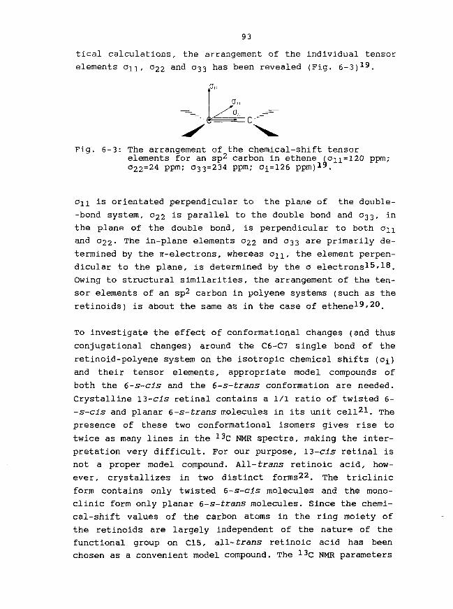
93
tical calculations, the arrangement of the individual tensor
elements OJI, 022 and 033 has been revealed (Fig. 6-3J1^.
Fig. 6-3: The arrangement of the chemical-shift tensorelements for an sp2 carbon in ethene (o11=l20 ppm;022=24 ppm; 033=234 ppm; 0^=126 ppm)1^.
is orientated perpendicular to the plane of the double-
-bond system, 022 is parallel to the double bond and 033, in
the plane of the double bond, is perpendicular to both an
and O22- The in-plane elements 022 and 033 are primarily de-
termined by the 7r-electrons, whereas an, the element perpen-
dicular to the plane, is determined by the o electrons1^'1^.
Owing to structural similarities, the arrangement of the ten-
sor elements of an sp2 carbon in polyene systems (such as the
retinoids) is about the same as in the case of ethene19'20.
To investigate the effect of conformational changes (and thus
conjugational changes) around the C6-C7 single bond of the
retinoid-polyene system on the isotropic chemical shifts (Oi)
and their tensor elements, appropriate model compounds of
both the 6-s-cis and the 6-s-trans conformation are needed.
Crystalline 13-cis retinal contains a l/l ratio of twisted 6-
-s-cis and planar 6-s-trans molecules in its unit cell21. The
presence of these two conformational isomers gives rise to
twice as many lines in the 13C NMR spectra, making the inter-
pretation very difficult. For our purpose, 13-cis retinal is
not a proper model compound. All-trans retinoic acid, how-
ever, crystallizes in two distinct forms22. T n e triclinic
form contains only twisted 6-s-cis molecules and the mono-
clinic form only planar 6-s-trans molecules. Since the chemi-
cal-shift values of the carbon atoms in the ring moiety of
the retinoids are largely independent of the nature of the
functional group on C15, all-trans retinoic acid has been
chosen as a convenient model compound. The 13C NMR parameters

94
of carbon C5 (see Fig. 6-1 for numbering) have been obtained
for both polycrystalline forms and are given in Fig. 6-415.
Comparison of the tensor elements of the C5 resonance in
these crystalline forms, reveals that the o n values of
twisted 6-s-cis (on=28 ppm) and planar 6-s-trans (on =
27 ppm) retinoic acid are identical within measuring
accuracy. The same is true for the o22 values (141 and 143
ppm, respectively). Tensor element 033, however, shows a
remarkable large downfield shift of 20 ppm, going from the
twisted 6-s-cis (033=217 ppm) to the planar 6-s-trans (033=
237 ppm) form. Thus, 033 proves to be a unique indicator of
conformational changes in a retinoid.
o i l Lo L X? 6-s-transo ^^6c
"*~ 6-s-cis s ^~
200 100ppm(TMS)
Fig. 6-4: Effect of the conjugation on the isotropic chemicalshift (Ojj and its tensor elements (on, 022 and033) of carbon C5 in planar 6-s-trans and twisted6-s-cis retinoic acid.
As a result of the down-shifted 033 element, the C5 isotropic
chemical-shift value [01=1/3(011+022+033)] displays likewise
a downfield shift of 7 ppm, going from twisted 6-s-cis (O£=
128.8 ppm) to planar 6-s-trans (Oi=135.9 ppm) retinoic acid.
Earlier, 13C NMR results on crystalline retinal and several
derivatives have shown that o n is the only element indica-
tive of the so-called gamma-effect (steric effect)15. That

95
o22 is an important measure for the Tr-electron charge distri-
bution, was demonstrated by 13C NMR studies on crystalline
ratinoic acid and other compounds15*18.
The Ti values of the methyl groups of bcth conformational
forms of retinoic acid have been obtained23. For twisted 6-s-
-cis retinoic acid, Ti(C18) is 2.00 s and for carbons C16 and
C17 (chemically identical, but conformationally distinct) two
different Ti values, 0.30 s and 0.25 s, have been determined.
In planar 6-s-trans retinoic acid Ti(C18) is 25.6 s, Ti(C16)
and C17 are 1.05 s and 1.32 s. In general, T^ values of
methyl carbons are related to the rotational rate of the
methyl group in question14*24. Rapid rotation of the methyl
group results in large Tj and slow rotation in small Tx
values. The much larger Ti(C18) value for the 6-s-trans
conformation (25.6 s), which is also found for other
retinoids23, is in agreement with the structural consi-
derations as encountered in retinoids (cf. Fig. 6-5)14'24.
In the twisted 6-s-cis form of the retinoid, the 5-CH3 group
(i.e. C18) experiences considerable interaction with the
nearby proton H8, causing a relatively fast relaxation
[T1(C18)=2.00 s], while in the 6-s-trans form, the 5-CH3
group can rotate more freely, effecting a long relaxation
time [T1(C18)=25.6 s]. Due to their mutual interaction, the
methyl groups at Cl (i.e. C16 and C17) have small Ti values.
These groups probably experience somewhat more steric inter-
action in the twisted 6-s-cis (with H7) than in the planar 6-
-s-trans conformation, explaining the somewhat larger T±
values in the planar 6-s-trans form.
,10-60°
,-OH^ 0
6 s - c i s CH3 6s-trons
Pig. 6-5: Illustration of the severe steric hindrance betweenthe 5-CH3 group and proton H8 in the case of 6-s--cis retinoic acid, which is absent in 6-s-transretinoic acid.

96
6-3: Solid-state 13C NMR spectroscopy of 1^c-labelled
bacteriorhodop. sins.
The individual elements of the chemical shielding tensor
contain much more information about the chemical structure
than the isotropic shift alone. So, detailed information on
steric interactions, conformation and charge distribution can
be obtained from the tensor elements13'15'18. It is expected
then, that solid-state MASS 13C NMR spectra of ring-13C-
-labelled bacteriorhodcpsins and rhodopsins will reveal fully
new information concerning the chromophoric-ring part in
these pigments.
In Fig. 6-6 the MASS 13C NME spectra of 5-, 6- and 7-13C-bR
are shown, together with that of native bR.
In the bottom spectrum, representing the ^3C NMR spectrum of
unlabelled native bR, three different types of natural-abund-
ant (1.1% 13C) signals can be distinguished. At about 175
ppm, the signals of the ca. 250 peptide carbonyls (C) are
present, accompanied by their spinning side-bands (*). Around
130 ppm, the resonances of ca. 90 aromatic carbons (A) with
their sidebands (*) are found. The signals below 75 ppm ori-
ginate from the saturated carbons (S). Since the total reso-
nance region of the saturated carbons {ca. 800 in total) lies
between 10 and 75 ppm, the spectra of Fig. 6-6 represent only
a small part.
in the light of the numerous carbon atoms in the protein it
is evident that the signals of the tiny chromophore (20
carbon atoms), cannot be discerned at all. With a specific13C-enrichment at a level of 90 at.%, the signal intensities
of the enriched carbon positions increase ca. 83 times
(90%/l.l%) and they are readily distinguished from the
natural-abundant background spectrum. Such is clearly demon-
strated in the 13C NMR spectra of 5-, 6- and 7-13C-bR, each
of which shows the signal of the 13C-labelled carbon at its
isotropic position with accompanying sidebands.
The signals for 6-13C and 7-13C are split into two (insets in
spectra of 6- and 7-13C-bR), reflecting the presence of a 2/1
mixture of bR molecules with l3-cis,l5-syn and all-trans

97
Notive BR
200 100-ppm
Fig. 6-6: 79.9 MHz MASS 13C NMR spectra of dark-adapted bR:5-13C-bR, 6-13C-bR, 7-13c_bR a n d native bR, resp.S, resonances from saturated carbons;A, aromatic resonances; C, carbonyl resonances

98
chromophoric configuration in the dark-adapted sample. This
feature is not resolved for the C5-resonance in 5-13C-bR.
The observed isotropic chemical-shift values of C6 (0^=135.4/
134.9 ppm) and C7 (0^=129.5/130.7 ppm) are very close to the
corresponding values in the planar 6-s-trans retinoic acid
model compound, where Oi(C6)=137.6 ppm and o^(C7)=127.8 ppm.
The results for CS are summarized in Fig. 6-7, which shows
the tensor elements and the isotropic chemical-shift values.
13c-5-retinyl-bR
1 IO CO
6-s-trans
6-S-CIS CM ^ 0000
200 100pprn(TMS)
Fig. 6-7: Chemical-shield ing tensor elements (o^, 022 and033) of the C5 resonance of 5-13C-bR and those oftwisted 6-s-cis and planar 6-s-trans retinoic acid.Oi is the isotropic chemical shift.
Consideration of the C5-tensor elements reveals that
which is indicative for steric interactions (paragraph 6-2),
is the same {ca. 27 ppm) in all three systems. The values of
033 in 5-13C-bR and in planar 6-s-trans retinoic acid, how-
ever, are identical, strongly arguing that the C6-C7 bond is
s-trans in bacteriorhodopsin. 022 in 5-13C-bR (022=170 ppm)

99
has shifted downfield as much as ca. 27 ppm, compared with
022 in planar 6-s-trans retinoic acid (022=14 3 ppm) and twis-
ted 6-s-cls retinoic acid (022=141 ppm). Since this tensor
element is the unique indicator of charge effects15'18, this
shift must be caused by an increase in Dositive electric
charge on C5, compared with the model compounds. Presumably,
this increase of positive charge on C5 in the chromophore is
induced by a close negative protein charge, which repulses
the n electrons.
For 18-13C-bR the Tj value of C18 is found to be 16±2 s,
which is much closer to planar 6-s-trans [Ti(C18)=25.6 s]
than to twisted 6-cis retinoic acid [T1(C18)=2.00 s]. For
16,17-13C2-bR, the Hi values have been determined to be ca.
1.2 s, i.e. close to the planar 6-s-trans [T1(C16/C17)=l.O5/
1.32 s] and differing considerably from the twisted 6-s-cis
model compound [Tx(C16/C17)=0.30/0.25 s]. The T^ values of
the methyl carbons provide independent support for the infer-
red planar 6-s-trans conformation of the chromophore in bR.
The MASS spectra of the bR analogues 13C-labelled at the sp3
carbons l, 4, 16, 17 and 18 are presented in Fig. 6-8.
Comparison of the spectra of the 13C-bR analogues with the
native spectrum (E) clearly shows the resonances for Cl, C4,
C18 and C16/C17 at 34.5, 34.6, 22.0 and 29.8/28.4 ppm (inset
in spectrum D). These o^ values are all within 1.5 ppm of the
corresponding values in planar 6-s-trans retinoic acid. The
most unusual feature of the four 13C-bR spectra is the rela-
tively high intensity of the Cl resonance which most likely
is to be attributed to the absence of dipolar broadening.
In the MASS 13C NMR spectrum of l8-13C-bR (spectrum C), the
C18 resonance is difficultly discerned from the bulk of the
natural-abundant protein sp3-carbon signals. Most of these
resonances, however, have T^ values below 2 s, while Ti(Cl8)
is 16±2 s (i.e. 100 times larger than for the protein methyl

A 1-l3CbR
75 50 25 0CHEMICAL SHIFT (ppm)
Fig. 6-8: High-field region of the MASS 13C NMR spectra ofl-r3C-bR (A), 4-13C-bR (B), 18-13C-bR (C),16,17-13C2-bR (D) and native bR (E).

101
carbons). Application of a T^-filter (with a delay time of
2 s) to the MASS 13C NMR measurement, will leave only the
carbon resonances with Ti > 2 s. The effect of the T]_ filter
on the 13C NMR spectra of native bR and 18-13C-bR is seen in
Fig. 6-923. T
T, filter
Native
I3C-I8
70 fO
ppmFig. 6-9: High-field region of the T^-filtered MASS 13C
NMR spectra of native bR and l8-13C-bR.Delay time 2 s.
The upper part shows the effect of the Tl-filter experiment
on the spectrum of native bR. Because most of the saturated
peptide-chain carbons have a T]_ < 2 s, the applied T^ filter
effects a disappearance of these signals. Similarly, the T]_-
-filtered spectrum of 18-13C-bR is reproduced at the bottom
part. This spectrum reveals the C18 peak at 22.0 ppm, which
now has become very pronounced compared with the normal MASS
spectrum (Fig. 6-8, spectrum C; Tx(C18)=16±2 s).
Until now, all of the solid-state 13C NMR spectra of the 13C-
-bR analogues have been measured on dark-adapted samples,
showing two (un)resolved resonances for the labelled position
(due to the ca. 2/1 mixture of bR548 and DR563). Recently,

102
also spectra of fully light-adapted (bRsge) have been
obtained (below -20°C in order to block the thermal conver-
sion into bR54g), which display only one resonance for the13C-labelled position. Their o^ values correspond to those
assigned to DR56Q in the dark-adapted mixture, based on the
relative intensities. In the few cases where the discrimi-
nation is more difficult, the measurements on pure bR568
allow an unambiguous assignment.
Also MASS 13C NMR spectra of the photointermediate M412 could
be obtained25. Considering its short lifetime at room tempe-
rature (half-time l ms), it is necessary to stabilize the
sample by cooling to -40°C. The spectrum of 5-13C-M4i2 i s
shown in Fig. 6-10.
NATIVE
DIFFERENCE
200 100T0
CHEMICAL SHIFT (ppm!
Fig. 6-10: MASS 13C NMR difference spectrum of 5-13C-M4i2,resulting from subtraction of the 5-13C-labelledand native M412 spectra. Temp. -40°C, pH 10.

103
The central band of the C5 resonance (at 139.4 ppm) can
clearly be distinguished. Very recently, it has also become
possible to adjust the sample-spinning rate with very high
precision, benefitting accurate subtraction of the native and
the labelled pigment spectra21*, in this way, the 13C-NMR
difference spectrum of 5-13C-M4i2 (bottom spectrum) has been
obtained by subtraction of the 5-13C-M4i2 anc^ native M412
spectra. As is seen, all 'non-labelled' resonances are
effectively substracted to zero, leaving only the signals due
to the enriched position. In addition to the C5 centerband
(at 139.4 ppm), now also the spinning sidebands are better
visible, which enables an analysis of the C5 tensor elements.
From the Oi(C5) value (139.4 ppm) in 5-13C-M412' which is
even downfield of the OJ(C5) value (135.9 ppm) of 6-s-trans
retinoic acid, it is concluded that the chromophore in M412
has a planar 6-s-trans conformation.
6-4: 13C NMR spectroscopy of 13C-labelled rhodopsins27'28.
Rhodopsin (Rh), occurring as monomeric units in the disk
membranes of the rod-outer segments, shows considerable
rotational diffusion (rotational correlation time: ca. 20 ys)
at room temperature. This characteristic is a serious draw-
-back for the solution and solid-state 13C NMR studies on
rhodopsin. In solid-state 13C NMR studies on rhodopsin, the
loss of intensity of the 13C signals, due to the high protein
mobility, can be avoided by performing the measurements at
low temperature (-20 to -30°C). The protein motion is now
•frozen", and high-resolution solid-state 13C NMR spectra of
rhodopsin can be obtained. Yet, another problem is still
present. Owing to the high level of unsaturation in natural
rhodopsin lipids2^, the olefinic lipid signals obscure the
olefinic (120-130 ppm) chromophore resonances even in the
case of 13C-enrichment. This problem can be tackled by
exchange of the unsaturated lipids for saturated lipids28 or
detergents27. A convenient detergent is Ammonyx-LO (mixture
of dodecyl- and tetradecylamine oxide), which leaves the
protein intact and shows no resonances above 100 ppm.
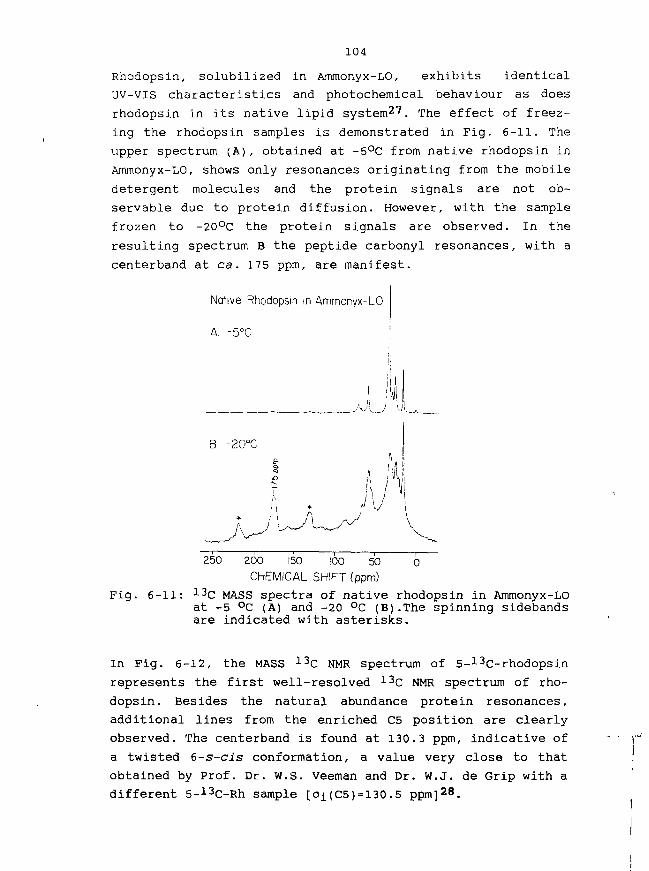
104
Rhodopsin, solubilized in Ammonyx-LO, exhibits identical
UV-VIS characteristics and photochemical behaviour as does
rhodopsin in its native lipid system27. The effect of freez-
ing the rhodopsin samples is demonstrated in Fig. 6-11. The
upper spectrum (A), obtained at -5°C from native rhodopsin in
Ammonyx-LO, shows only resonances originating from the mobile
detergent molecules and the protein signals are not ob-
servable due to protein diffusion. However, with the sample
frozen to -20°C the protein signals are observed. In the
resulting spectrum B the peptide carbonyl resonances, with a
centerband at ca . 175 ppm, are manifest.
Native Rhodopsin in Ammonyx-LO
A. -5°C
250 0200 150 100 50
CHEMICAL SHIFT (ppm)
Fig. 6-11: 13C MASS spectra of native rhodopsin in Ammonyx-LOat -5 °C (A) and -20 °C (B).The spinning sidebandsare indicated with asterisks.
In Fig. 6-12, the MASS 13C NMR spectrum of 5-13C-rhodopsin
represents the first well-resolved 13C NMR spectrum of rho-
dopsin. Besides the natural abundance protein resonances,
additional lines from the enriched C5 position are clearly
observed. The centerband is found at 130.3 ppm, indicative of
a twisted 6-s-cis conformation, a value very close to that
obtained by Prof. Dr. W.S. Veeman and Dr. W.J. de Grip with a
different 5-13C-Rh sample [oi(C5)=l30.5 ppm]28.

105
l3C-5 Rhodopsin
250 200 150 100 50 0
CHEMICAL SHIFT(ppm)
Fig. 6-12: 13C MASS spectrum of 5-13C-rhodopsin at -30 °C.Rotational sidebands are marked with asterisks.
Recently, also the 13C MASS spectra of 6- and 7-13C-rhodop-
sin have been obtained.
In order to discuss the implications of the results for the
Rh chromophoric structure, the 13C NMR data have to be
compared with those of appropriate model compounds. In view
of the 11-cis geometry of the Rh chromophore, ll-cis-
-retinylidenebutylimmcnium chloride (SBH+Cl", in solution)
makes a convenient model compound. Comparison of the o j
values for C5 (Oi=130.3 ppm), C6 (0^=137.7 ppm) and C7 (o±=
132.3 ppm) in rhodopsin with those of this model compound30,
Oi(C5)=130.3 ppm, Oi(C6)=l37.2 ppm and oi(C7)= 132.2 ppm,
learns that rhodopsin and the ll-cis SBH+ model compound
have nearly identical values. This demonstrates that the
chromophore in rhodopsin is not perturbed in the C5 to C7
region. Further, the oi(C5) in rhodopsin is closest to that
in twisted 6-s-cis retinoic acid, arguing that the Rh
chromophore exists in a twisted 6-s-cis conformation. The
chemical-shift tensor elements of C5 are in full support of
this conclusion. The C5 tensor elements of rhodopsin and of
6-s-cis and 6-s-trans retinoic acid are presented in Table
6-1. In all three compounds, the o n values are about 27 ppm,
indicating that there is no difference in steric perturbation
exerted on C5. The various 022 values, identical within

237217210 + 6
143141134+10
272826 + 3
106
Compound 033 022 03.1
6-s-trans ret. acid6-s-cis ret. acid5-13C-rhodops in
Table 6-1: Chemical-shift tensor-element values of C5 inrhodopsin, planar 6-s-trans and twisted 6-s-cisretinoic acid.
measuring accuracy, imply that there is no difference in
charge distribution at C5. The 033 values, however, are
considerably different. 6-s-cls retinoic acid (033=217 ppm)
has a 20 ppm lower 033 value than the 6-s-trans form (033 =
237 ppm), reflecting the effect of the transition from the
planar 6-s-trans to the twisted 6-s-cis conformation. In
rhodopsin, the 033 value is even below that of the 6-s-cis
model compound, confirming the 6-s-cis conformation of its
retinylidene chromophore.
6-5: Concluding remarks.
High-resolution solid-state NMR spectroscopy has proved to be
a very potent non-perturbing technique for the study of iso-
topically-labelled biomolecules. The atomic nucleus is used
as a probe to monitor the chemical-shift parameters and
relaxation time at each atomic position in the molecule. By
virtue of the remaining chemical-shift anisotropy (sidebands)
in solid-state NMR measurements, the three principal values
(°ll/ °22 a n d 033) of the chemical-shift tensor of a specific
atom can be obtained, next to its isotropic value
[01=1/3(011+022+033)]• Much more detailed information is to
be had from these tensor elements than from a solution NMR
spectrum, only yielding the isotropic chemical-shift value.
So, information of the configuration, conformation and charge
distribution in the chromophore and of its specific interac-
tions with the peptide chain in the active site of retinold
pigments could be evaluated in greater detail. It turns out

107
that many of the interactions of the chromophore are aniso-
tropic. Steric effects (the so-called gamma-effects) are
uniquely reflected in the an values. The effects of conju-
gation are observed in the 03.3, and the 022 ar© indicative of
the charge distribution.
At this moment, MASS 13C NMR data are available for every
sp2-carbon position in the bacteriorhodopsin chromophore.
These data enable us to compare the isotropic chemical-shift
values (o-[) of the chromophoric sp2 carbons in bR with those
of an appropriate 6-s-trans model compound. In the solid
state, some retinoids, such as 13-cis retinal^l, monoclinic
all-trans retinoic acid^2, retinylidenemethylimine and
-butylimine occur in the stable planar 6-s-trans conforma-
tion. These compounds served as first reasonable options of
model compounds for the solid-state 13C NMR study of bR.
Only very recently, the first example of a planar 6-s-trans
SBH+ model compound (all-tra^s retinylidene-terfr-butyl-
immonium perchlorate: SBH+ClC>4 ) has been described in the
literature31. The fcert-butyl group in this model, compared
with a linear alkyl group, as present in native bR, exhibits
no effect on the o^ values of the chromophoric carbons Cl up
to C12. Only the Oi(C13) {ca. 4 ppm downfield and Oi(C15)
{ca. 4 ppm upfield) values have slightly changed. The
difference between the o^ values of the bR55g and the model
compound sp2 carbons is illustrated in Fig. 6-13.
The most pronounced differences between the o± of DR568 and
those of the SBH+CIOJ salt are found at C5, C6, C7 and C8,
the C5 resonance in bR5g9 being ca. 8 ppm down-shifted. It is
apparent that the effective positive charge on C5 in bRsge
has increased. The only source of this effect is the elec-
tronic interaction between the peptide chain and the chromo-
phore near C5. This feature fully supports our conclusion of
a negative protein charge in close proximity to C5 {cf. para-
graph 6-3). A negative charge near the cyclohexene-ring
moiety has been proposed earlier by Nakanishi et al.6.

108
Fig. 6-13: Plot of the a3C NMR chemical-shift differencesbetween bRssg and solid all-trans retinylidene--teri-butylimmonium perchlorate (SBH+C1C>4, 6-s--trans) as a function of chain position.
Due to the conjugation of the C5=C6 bond with the rest of the
polyene chain, a negative protein charge close to C5 might
have a shielding effect on carbons C6 and C8, and a deshield-
ing effect on the odd-numbered carbon atoms C7 (and C9). The
shielding effect on C6 and C8 is clearly inferred from their
negative chemical-shift difference (cf. Fig. 6-13). However,
no deshielding effect on C7, but even a shielding of about
5 ppm is observed. A possible downfield shift of C7, induced
by the negative charge near C5, may be over-compensated by a
positive protein charge near C7. Indications of such a posi-
tive charge close to C7 have been found later in studies of
dihydrobacteriorhodopsin analogues32'33.
For the other sp2-carbons in the chain the compensating
effects of the positive and negative charge, lead to only
small Oi differences (cf. Fig. 6-13).

109
The oi differences found for the sp3 carbons Cl, C4 and
C16/C17, are all relatively small (< 2 ppm), arguing that the
negative charge is closely associated with C5 and that the
assumed positive charge is not close to Cl as has been
proposed by Spudich et al .34.
Further, some other remarkable features may be noted. o^(Cl6)
and Oi(C17) have been found to be identical (both 29.9 ppm)
in the planar 6-s-trans model compound, in line with the
presence of a mirror plane31. For 16,17-13C2-bR568» however,
the two different values (29.8 and 28.4 ppm) indicate a
different protein microenvironment at either side of the
cyclohexene ring. Finally, it has been observed in retinoid
model compounds that Oi(C2) strongly depends on the confor-
mational identity of the C6-C7 bond, i.e. near 40 ppm in the
6-s-cis and near 45 ppm in the 6-s-trans conformation23/31.
Examination of the 2-13C-bR analogue with MASS 13C NMR
spectroscopy should yield additional information.
Summation of the above data, results in the representation of
the bRsga chromophore as depicted in Fig. 6-13. This chromo-
phore has a planar all-trans, 6-s-trans geometry. In addition
to the Schiff-base counterion, a dipolar pair of charges is
present near the cyclohexene ring: the negative protein
charge closely associated with C5 and the positive protein
charge located near C7.
It is clear that the structural elements of the bR chromo-
phore determine the opsin shift. On the basis of the evidence
from studies of several bR analogues, it is concluded that
the total opsin shift (5100 cm"1) is composed of two distinct
contributions. Part of this shift, 1300 cm"1, can be accoun-
ted for by the transition from a twisted 6-s-cis (free
retinal) to a planar 6-s-trans conformation (in bR). The
remaining ca. 3800 cm"1 can be ascribed to differences in the
tail end of the chromophore. Van der Steen et al. were the
first who recognized these aspects, in an earlier study of a
6-s-trans locked bR analogue35. Independent support has been
obtained later from the studies of the 7,8-dihydro-bR ana-
logue3^ and from our studies of demethyl-bR analogues
(Chapter 5). In all of these cases no change in the C6-C7
conformation upon binding of the retinal analogue to bac-

110
terioopsin has been observed.15N NMR data of e-15N-lysine bR36 and thorough investigations
of the dependence of 15N Schiff-base chemical shifts on
hydrogen-bond strength37'38, indicate that the Schiff-base
nitrogen-proton bond is stronger in bRsgg than in SBH+. This
corresponds with the weaker hydrogen bond between this proton
and its protein counterion3^, leading to a shorter N-H bond
and a larger positive charge in the conjugated polyene
system. The ensuing better delocalization of the positive
charge is responsible for the 3800 cm"1 bathochromic contri-
bution to the \max value of bR. This better delocalization
also gives somewhat more double-bond character to the C-C
single bonds along the conjugated chain, which explains the
higher stretching frequencies of these bonds in bR than in
the SBH+40.
Furthermore, the dipolar pair of charges apparently does not
contribute to the opsin shift, presumably due to the counter-
balancing effect of the negative charge close to C5 and the
positive charge near C7. The first experimental indications
for this assessment are based on the ca. 3500 cm"1 opsin
shift for a 7,8-dihydro-bR analogue, where the influence of
the protein charges on the polyene system near C5 and C7 is
totally absent3^.
With the 13C NMR data of the 13C-labelled rhodopsin analogues
under consideration27 and the recent data of the other sp2
carbons41, their 13C NMR parameters can be evaluated. In Fig.
6-14 the differences in the o± values between the sp2 carbons
in the 13C-labelled rhodopsins and in the model compound, 11-
-cis retinylidenebutylimmonium chloride (6-s-cis SBH+Cl", in
solution, ref. 30) are given.
An analysis of the chemical-shift tensor elements of C5,
demonstrates the unperturbed character of the Rh chromophore
and its twisted 6-s-cis conformation, implying that a
possible change in conformation around the C6-C7 bond does
not contribute to the 2650 cm"1 opsin shift.
In the meantime, the o± values of all sp2 carbons of the
conjugated chain of the Rh chromophore have been determined.
Interestingly, most of these correspond within 2 ppm with the
values of the model. The only exceptions are Cll (difference

H I
-4.1 ppm) , C12 (-3.1 ppm) and C13 (-6.2 ppm) , indicating a
peptide-chain induced perturbation, localized in the region
Cll to C13.
Fig. 6-14: protein-induced perturbation of the isotropic 13cchemical shift in rhodopsin as a function ofposition, relative to 11-cis SBH+Cl" (6-s-cis,in solution).
Since the chemical-shift tensor elements for Cll, C12 and C13
have not yet been determined, we lack information about the
nature of the perturbation, such as electric charge, twisting
of bonds or steric effects.
The Oi values of C14 in Rh and in the 11-cis SBH+ model are
virtually identical (difference +0.1 ppm). Consequently, the
proposal of Nakanishi et al.*2, placing the external point
charge near C12 and C14, is not correct.
The almost identical C-C bond stretches along the conjugated
chain in the Rh chromophore and in the model SBH+ indicate
that the better delocalization of the positive charge, as
happens in bR, does not contribute to the ops in shift of Rh
either43.
Therefore, the nature of the interactions in the Cll to C13
region needs to be investigated in more detail in order to
reveal the mechanism responsible for the opsin shift in
visual pigments. A solid-state ^c-NMR study of

112
isorhodopsins may shed light on this mechanism, since RR data
on isorhodopsin have shown that the C10-C11 and C14-C15
stretching frequencies are substantially higher than in the
corresponding model. Also the chemical-shift parameters of
CIO, Cll, C14 and CIS are expected to be different.
The solid-state 13C NMR investigations of bacteriorhodopsin
and rhodopsin with 13C-labelled chromophores has given unique
information about the structure (configuration, conformation
and charge distribution) of the chromophore and about its
specific interactions with the peptide chain in the active
site of the protein.
Using this technique at lower temperature where the photo-
intermediates are stable, has given structural information
about the chromophore in the thermally labile form bR56e and
in M412' o n e °f the intermediates in the bR photocycle. It is
expected that, using low-temperature studies, structural
information about the chromophore in the primary photo-
products bathorhodopsin and K, and in the thermal interme-
diates will be obtained.
Other photochemically active retinoid pigments, such as halo-
rhodopsin, the sensory rhodopsins I and II, and the rod and
cone visual pigment of different animals may similarly be
studied.
Solid-state NMR is not restricted to the study of coloured
biomolecules. All systems displaying an interaction between a
small molecule (ligand) and a protein can be investigated.
Examples are hormone-receptor systems, the ligands in taste
and smell receptors, et cetera.
From growing Halobacterium halobium on 13C-, 15N- and 1 70-
-enriched amino acids, bacteriorhodopsins with isotopically-
-labelled peptide chains can be harvested. Solid-state NMR
spectroscopy will help and elucidate the structure of bR and
its photoproducts, approaching the peptide part in the
binding site. NMR studies on bR, specifically labelled in
both the chromophore and the peptide chain, will enable the
distance between a specific position in the chromophore and a

113
certain amino acid in its neighbourhood to be estimated.
The results discussed show the exquisite applicability of the
solid-state MMR technique to the study of biological molecu-
les. They are among the first studies undertaken in this
field and they show the promising potential of the technique
for the investigation of a wide range of (bio)molecules.
The joint application of specific isotope labelling and
advanced non-invasive and non-perturbing physical techniques
to study the function of biological systems has a tremendous
power. Since the desired specifically-labelled molecules,
among these ligands and amino acids, can only be obtained via
total synthesis, synthetic organic chemistry will continue to
play an essential role in these studies.

114
LITERATURE
1.
2.
3.
4.
5.
6.
7.8.
9.
10.
11.
12.
13.
14.
15.
16.
17.
18.
19.
20.
21.
22.
23.
24.
F. Millet, P.A. Hargrave and M.A. Raftery, Biochemistry12, 359 (1973).G.D. Mateescu, W.G. Copan, D.D. Muccio, D.v. Waterhousand E.W. Abranamson, in "Proc. Int. Symp. Synth, andAppl. of Isot. Label. Compds.", W.P. Duncan and A.B.Susan (Eds.), 123, Elsevier, Amsterdam, 1983.A. Yamaguchi, T. Unemoto and A. Ikegami, Photochem.
511 (1981).S.O. Smith, J.A. Pardoen, C. Winkel, J.Herzfeld, R.A. Mathies and R.G. Griffin,
Proc. Natl. Acad. Sci. USA 81, 1706 (1984).G.S. Harbison, S.O. Smith, J.A. Pardoen, P.P.J.
Photobiol. 33,G.S. Harbison,Lugtenburg, J.
Mulder,J. Lugtenburg, J. Herzfeld, R.A. Mathies and R.G.Griffin, Biochemistry 23, 2662 (1984).K. Nakanishi, V. Balogh-Nair, M. Arnaboldi, K. Tsujimotoand B. Honig, J. Am. Chem. Soc. 102, 7945 (1980).P.K. Das and R.S. Becker, J. Phys. Chem. 82, 2081 (1978).K. Muellen, H. Schmickler, B. Frei and H.R. Wolf,Tetrahedron Lett. 27, 477 (1986).R.R. Birge, D.F. Biocan and L.M. Hubbard, J. Am. Chem.Soc. 104, 1196 (1982).B. Honig, A.D. Greenberg, B.D. Sykes, M. Karplus, Proc.Natl. Acad. Sci. USA 68, 1289 (1971).K. Hiraki, T. Hamanaka, K. Yoshihara and Y. Kito,Biochim. Biophys. Acta 891, 177 (1987).R. Rowan, III, B.D. Sykes, J. Am. Chem. Soc. 96, 7000(1974).C.A. Fyfe, "Solid State NMR for Chemists", C.F.C. Press,Guelph, Ontario, Canada, 1983.H.O. Kalinowski, S. Berger and S. Braun, nl3C-NMR--Spektroskopie", G. Thieme Verlag, Stuttgart, 1984.G.S. Harbison, P.P.J. Mulder, H. Pardoen, J. Lugtenburg,J. Herzfeld and R.G. Griffin, J. Am. Chem. Soc. 107, 4809(1985).W.T. Dixon, J. Schaefer, M.D. Sefcik, E.O. Stejskal andR.A. McKay, J. Magn. Reson. 49, 341 (1982).J. Herzfeld and A.E. Berger, J. Chem. Phys. 73, 6021(1980).J. Facelli, D.M.152 (1987).K.W. Zilm, R.T. Conlin, D.M. Grant and J.Chem. Soc. 102, 6672 (1980).
Griffin and J.S. waugh, J. Chem. Phys.
Grant and J. Michl, Ace. Chem. Res. 20,
Michl, J. Am.Soc.
E.K. Wolff, R.G.67, 2387 (1977).C.J. Simmons, R.S.H. Liu, M. Denny and K. Seff, ActaCrystallogr., Sect. B, B37, 2197 (1981).C.H. Stam and C.H. McGillivary, Acta Crystallogr., Sect.B, B16, 62 (1963).G.S. Harbison, S.O. Smith, J.A. Pardoen, J.M.L. Courtin,J. Lugtenburg, J. Herzfeld, R.A. Mathies and R.G.Griffin, Biochemistry 24, 6955 (1985).A.S. Perlin, in "isotopes in Organic Chemistry", E.Buncel and C.C. Lee (Eds.), 171, Elseviers Publ. Comp.,Amsterdam, 1977.

115
25.
26.27.
28.
29.
30.
31.
32.
33.
34.
35.
36.
37.
38.
39.
40.
41.
42.
43.
J. Lugtenburg and W.J. de9 (1987).
White and E.A. Dratz,(1979).
S.O. Smith, J. Courtin, E. van den Berg, C. Winkel, J.Lugtenburg, J. Herzfeld and R.G. Griffin, submitted.H. de Groot et al., submitted.S.O. Smith, I. Palings, V. Copie, D.P. Raleigh, J.M.L.Courtin, J.A. Pardoen, J. Lugtenburg. R.A. Mathies andR.G. Griffin, Biochemistry 26, 1606 (1987).L.C.P.J. Mollevanger, A.P.M. Kentgens, J.A. Pardoen,J.M.L, Courtin, W.S. Veeman,Grip, Eur. J. Biochem. 163,G.P. Miljanich, L.A. Sklar, D.L.Biochim. Biophys. Acta 552, 294J.W. Shrivsr, G.D. Mateescu and E.W. Abrahamson,Biochemistry 18, 4785 (1979).R.F. Childs, G.S. Shaw and R.E. Wasylishen, J. Am. Chem.Soc. 109, 5362 (1987).J. Lugtenburg, M. Muradin-Szweykowska, C. Heeremans, J.A.Pardoen, G.S. Harbison, J. Herzfeld, R.G. Griffin, S.O.Smith and R.A. Mathies, J. Am. Chem. Soc. 108, 3104(1986; .H. Rodr.an, B. Honig, B. Nakanishi, M. Okabe, N. Shimizu,J.L. Spudich aid D.A. McCain, Biophys. J. 49, 210a(1986).J.L. Spudich, D.A. McCain, K. Nakanishi, M. Okabe, N.Shimizu, H. Rodman, B. Honig and R.A. Bogomolni, Biophys.J. 49, 479 (1987).R. van der Steen, P.L. Biesheuvel,Lugtenburg, J. Am. Chem. Soc. 108,
J.
R.A. Mathies and J.6410 (1986).
Herzfeld and R.G. Griffin, BiochemistryG.S. Harbison22, 1 (1983).G.S. Harbison, S.O. Smith, J.E. Roberts, J.A. Pardoen,P.P.J. Mulder, C. Winkel, J.M.L. Courtin, R.A. Mathies,J. Herzfeld and R.G. Griffin, in "NMR in Biology andMedicine", S. Chien and C. Ho (Eds.), 33, Raven Press,N.Y., 1986.G.S. Harbison, J.E. Roberts, J. Herzfeld and R.G. GriffinBiochemistry, submitted.P.E. Blatz, J.H. Mohler and H.v. Navangul, Biochemistry11, 848 (1972).S.O. Smith, M.S. Braiman, A.B. Myers, J.A. Pardoen,J.M.L. Courtin, C. Winkel, J. Lugtenburg and R.A.Mathies, J. Am. Chem. Soc. 109, 3108 (1987).S.O. Smith, K. Friedlander, I. Palings, J. Courtin, H. deGroot, R.A. Mathies and R.G. Griffin, submitted.B. Honig, U. Dinur, K. Nakanishi, V. Balogh-Nair, M.A.Gawinowicz, M. Arnaboldi, M.G. Motto, J. Am. Chem. Soc.101, 7984 (1979).I. Palings, J.A. Pardoen, E. van den Berg, C. Winkel, J.Lugtenburg and R.A. Mathies, Biochemistry 26, 2544(1987) .

117
Chapter 7
EXPERIMENTAL DETAILS
7-1: Retinal synthesis*.
General
All experiments were carried out in a nitrogen atmosphere andthose with polyenes, in addition, under dim red light.Distilled dry solvents were used. Pet. ether refers to low--boiling petroleum ether 40-60°C. Unless otherwise stated,purification was performed by flash chromatography1 (Mercksilica gel 60, 230-400 Mesh) using ether/pet, ether mixtures.TLC analyses were performed on Schleicher and Schuell F1500/-LS254 silica-gel plates using ether/pet, ether mixtures.Evaporation of the solids was carried out in vacuo (10 Torr).The ^H NMR spectra were recorded on a Bruker WM-300, a JeolNM FX-200 or a Jeol PS-100 spectrometer (in CDCI3) withtetramethylsilane (TMS: 0 ppm) as internal standard. The 13CNMR spectra were recorded on the Bruker WM-300 (at 75.5 MHz)or on the Jeol NM FX-200 (at 50 MHz) spectrometer (in CDCI3,internal TMS: 0 ppm). Jco J C H an& JHH refer to J(13C-13C),J(13C-1H) and J(1H-1H), respectively. The signs were notdetermined. The mass spectra were recorded using an AEI MS902. IR spectra were obtained using a Pye-Unicam SP 3-200 andthe UV-VIS spectra using a Cary 219 spectrophotometer. HPLCseparations were performed using a Dupont 830 equipped with aDupont spectrophotometer (360 nm) and a 25 cm x 22.5 mmZorbax Sil column. Elution was effected using 10% ether/pen-tane at a flow rate of 20 ml/min.Spectral signal designations for the (intermediate) compoundswith less than 13 carbon atoms (e.g. cyclohexenecarboxalde-hydes 2a-e) are based on the IUPAC nomenclature. The designa-tions for the retinoid-like compounds, containing 13 carbonatoms (e.g. |3-ionone 24) or more, follow the IUPAC retinoidnumbering system^.For each series of isotopomers (different by exchange of 1%Cfor 13C at a specific position), only the NMR spectra of thefirst mentioned compound have been completely assigned. Forthe following isotopomers, restriction has been made to thespectral changes with respect to the analogously unlabelledcompound.3-Acetyl-l-propanol (39: 5-hydroxy-2-pentanone) was purchasedfrom Aldrich. 5-Chloro-2-pentanone (29) and chloroacetonewere purchased from Janssen Chimica. Chloroacetonitrile and3-methylcrotonaldehyde(16: 3-methyl-2-butenal) were purchasedfrom Merck. 3,3-Dimethyl-allylbromide (32: l-bromo-3-methyl--2-butene) was purchased from Fluka AG. (2-13C)- and (1,3-13C2)-acetone, (l-13C)- and (2-13C)-acetonitrile, (13C)iodo-methane and potassium(13C)cyanide (all with ca. 90 at.% 13C)were purchased from Merck Sharp and Dohme Isotopes, Canada.
*Most of the references to the synthetic procedures are givenin chapters 2 and 3.

118
Preparation of demethylretinals la-e
2-(Dimethoxymethyl)cyclohexanone (7)
2.27 g of NaH (55% dispersion in oil, 52 mmol) were washedthree times with pet. ether and suspended in 25 ml of dryether. After this suspension had been cooled to 0°C, 5 g (51mmol) of cyclohexanone 5 and 7.56 g (102 mmol) of ethylformate, dissolved in 25 ml ether, were added over a periodof 10 min, under thorough stirring. Upon warming to roomtemperature, hydrogen evolution started and a yellow-orangeprecipitate (6) formed. After H2 liberation had ceased, themixture was refluxed for 25 minutes. After cooling to 0°C,2.3 ml (39 mmol) of ethanol were added. Upon addition of 8 g(102 mmol) of freshly distilled acetyl chloride in 20 mlether, the yellow-orange precipitate disappeared and a newgrey precipi-tate (NaCl) was formed. Next, 47 ml (1.17 mmol)of methanol were added in 15 m.in. The mixture was stirred for2 h at room temperature. Work-up was accomplished by pouringthe mixture into 200 ml of 5% NaHCC>3 solution. After extrac-tion with ether, the organic layers were washed with brine,dried over NaoC03 and evaporated to give 7.4 g (84%) of 7 asa brown oil. rH NMR (100 MHz): 6 1.4-2.0 (m, 6H, 3-CH2, 4-CH2and 5-CH2), 2.4 (m, 2H, 6-CH2), 2.64 (m, 1H, 2-H), 3.44 (s,6H, 2 x OCH3), 4.72 [d, JHH 6 H z' 1H, -CH(O-)2]. IR (film):1710 cm"1 (C=0 stretch), 1050-1100 cm"1 (C-0 stretch).
2-(Dimethoxymethyl)cyclohexanol (8a)
4 g (23.3 mmol) of compound 7 in 50 ml ether were addedslowly to a cooled (ice-bath) and stirred suspension of 0.9 g(22.5 mmol) L1AIH4 in 10 ml ether. After removal of the ice--bath, the mixture was stirred for 30 min at room tempera-ture. After completion, a little ice together with asaturated NH4CI solution (pH 4-5) were added slowly. Thismixture was extracted with 50 ml ether (3x). The ether layerswere washed with brine, dried over NaCC>3, filtered andevaporated to give 3.6 g (90%) of 8a. lH NMR (100 MHz): 61.0-2.0 (m, 9H, 2-H, 3-CH2, 4-CH2, 5-CH2 and 6-CH2), 3.46 and3.37 (2 X S, 6H, 2 x OCH3), 3.52 (m, 1H, 1-H), 4.1 (bs, 1H,OH), 4.3[d, JHH 6 Hz, 1H, -CH(O-)2]- IR (film): 3450 cnr1
(0-H stretch), 1100 cm"1 (C-0 stretch).
2-(Dimethoxymethyl)-l-methylcyclohexanol (8b)
To a cooled (0°C) and stirred solution of 19 mmol CH3Li (12ml 1.55 M solution in ether; Aldrich) 2 g of 7, dissolved in25 ml ether were added over a period of 20 min. After theaddition, stirring was continued for 75 min at room tempera-ture. The mixture was then cooled to 0°C and 200 ml of 0.1 NHC1 were added slowly. After extraction with ether (3x), theether layers were washed with water, dried over Na2CO3 andevaporated to yield 1.56 g (69%) of 8b. According to NMR andIR data, in some instances a considerable amount of the 2--hydroxy-2-methyl-cyclohexanecarboxaldehyde had alreadyformed.*H NMR (100 MHZ): 6 1.34 (S, 3H, I-CH3), 1.0-2.0 (m, 9H, 2-H,3-CH2, 4-CH2, 5-CH2 and 6-CH2), 3.38 and 3.25 (2 x S, 6H,

119
2 x OCH3). IR (film): 3450 cm"1 (O-H stretch), 1050-1100 cm"1
(C-0 stretch).
1-Cyclohexenecarboxaldehyde (2a)
3.5 g (20.1 mmol) of 8a were dissolved in 35 ml of acetoneand heated to reflux. 0.6 ml of 1 N HCl were then added via asyringe and the mixture was refluxed for about 2% h. Aftercooling, the mixture was extracted with ether. The etherlayer was washed with brine (2x), dried over MgSC>4 andevaporated after filtration to give 2.5 g of crude aldehyde2a (> 100%). Purification by column chromatography (silicagel; pet. ether/ether = 85/15) yielded 1.1 g (50%) of 2a.^H NMR (100 MHz): 6 1.7 (m, 4H, 4-CH2 and 5-CH2), 2.3 (m, 4H,3-CH2 and 6-CH2), 6.8 (m, 1H, 2-H), 9.5 (s, 1H, aldehyde-H).
2-Methyl-l-cyclohexenecarboxaldehyde (2b)
1.56 g of 8b was dissolved in 25 ml of acetone and heated toreflux. To the refluxing mixture, 0.4 ml of 1 N HCl was addedslowly via a syringe. The refluxing was continued for afurther 2 h. After cooling, 50 ml of water were added.Extraction was performed with pet. ether (4x). The pet. etherlayers were washed with water, dried over MgSC>4 and distilledin vacuo. The yield consisted of 843 mg (82%) of 2b as a red--brown oil. !H NMR (100 MHz): 6 1.6 (m, 4H, 4-CH2 and 5-CH2),2.16 (S, 3H, 2-CH3), 2.16 (m, 4H, 3-CH2 and 6-CH2), 10.06 (S,1H, aldehyde-H).
6-Methyl-l-cyclohexenecarbonitrile (lie)
For this one-pot, two-step procedure care has to be takenthat glassware, solvents and reagents are dried thoroughly. Amixture of 2-methylcyclohexanone (0.19 g; 1.7 mmol), commer-cial trimethylsilyl cyanide (10; 0.20 g; 2.0 mmol; Merck) andzinc iodide (ca. 13 mg) in benzene (l ml) was stirred at roomtemperature until completion of the addition reaction (over-night; TLC analysis). Pyridine (2.7 ml) and freshly distilledPOCI3 (5 mmol) were added and the whole mixture was heated toreflux for about 10 h. The cooled dark solution was thenpoured into ice-cold hydrochloric acid. After extraction, thesolvent was removed and the product was purified using silicagel column chromatography. The yield of nitrile (llb/llc=l/3)was 128 mg (60%). This mixture of lib and lie was not sepa-rated until the retinal stage, when the all-trans isomers ofthe two compounds could easily be separated with HPLC. -̂H NMRof lie (100 MHz): 6 1.19 (d, JHH 6 Hz, 3H, I-CH3), 1.4-2.7(m, 7H, 3-CH2, 4-CH2, 5-CH2 and 6-H), 6.54 (m, 1H, 2-H).
6-Methyl-l-cyclohexenecarboxaldehyde (2c)
A stirred solution of 128 mg (1 mmol) of nitrile (lib + lie)in pet. ether was cooled to -60°C. 1.5 ml of a solution (1.5mmol) of diisobutylaluminium hydride (Dibal) was then addedciropwise. After completion (1-2 h at 0°C), 2 g of silicagel,moistened with 0.4 ml of water and slurried in ether/pet.ether (l/l), were added slowly and the mixture was stirred at

120
0°C for ca. 1 h. After drying (MgS04), the solids werefiltered off and washed with ether. Evaporation of thesolvents yielded 109 mg (85%) of 2c and 2b as a 3/1 mixture.1H NMR of 2c (100 MHz): S 1.3 (d, Jjm 7.5 Hz, 3H, 6-CH3),6.59 (t, JHH 4 Hz, 1H, 2-H), 9.9 (s, 1H, aldehyde-H).
2,6-Dimethyl-l-cyclohexenecarbonitril (lid)
For this reaction, the same precautions have to be taken asfor the preparation of lie to guarantee absolutely dry con-ditions. Commercial 2,6-dimethylcyclohexanone 12 (mixture ofisomers; 0.5 g; 4 mmol) was dissolved in 2 ml of benzene.With the aid of a syringe, 0.52 g (5.2 mmol) trimethylsilylcyanide 10 was added, followed by a catalytic amount ofpotassium/l8-crown-6 ether complex. This mixture was stirredover night at room temperature in a closed argon-filledroundbottom flask. Then 5 ml of pentane were added and themixture was poured into ice-water and extracted with pentane.After washing the pentane layers with saturated sodiumbisulfite solution and drying over sodium sulfate 0.8 g (89%)of 13 was isolated on evaporation of the solvent. This silylether (0.8 g; 3.6 mmol) was dissolved in pyridine (5.6 ml)and refluxed for 7 h in the presence of freshly distilledPOCI3 (10.7 mmol). The same work-up procedure as for lieyielded 0.24 g (50%) of lid. !H NMR (100 MHz): 6 1.19 (d, JHH7.0 Hz, 3H, 6-CH3), 1.4-2.0 (m, 4H, 4-CH2 and 5-CH2), 2.02(s, 3H, 2-CH3), 1.9-2.4 (m, 3H, 1-H and 3-CH2).
2,6-Dimethyl-l-cyclohexenecarboxaldehyde (2d)
The nitrile lid (0.24 g; 1.78 mmol) was dissolved in pet.ether and cooled to -60 °C. Treatment with 2.6 ml of a 1 MDibal solution in hexane (2.6 mmol) and hydrolysis, aftercompletion, with 1.0 ml of water adsorbed on 4.2 g of silicagel as a suspension in ether/pet, ether (l/l), yielded 0.2 g(80%) Of 2d. 2H NMR(100 MHz): 6 1.26(d, JHH 7 Hz, 3H, 6-CH3),1.6 (m, 4H, 4-CH2 and 5-CH2), 2.0-2.3 (m, 3H, 1-H and 3-CH2),2.13 (s, 3H, 2-CH3), 10.04 (s, 1H, aldehyde-H).
6,6-Dimethyl-l-cyclohexenecarbonitrile (lie)
The procedure for preparing lie is analogous to that for lid.The quantitatively obtained cyanosilyl ether 15, yielded0.16 g (50%) of lie, after refluxing (22 h) in pyridine withPOCI3, starting from 0.3 g (2.4 mmol) 2,2-dimethylcyclohexa-none (14)3. 2H NMR (100 MHz): 6 1.19 [d, 6H, 6-(CH3)2], 1.4-1.8 (m, 4H, 4-CH2 and 5-CH2), 2.13 (m, 2H, 3-CH2), 6.42 (t,
4.0 Hz, 1H, 2-H).
6,6-Dimethyl-l-cyclohexenecarboxaldehyde (2e)
Synthetic procedure the same as for 2d. Treatment of 0.16 g(1.2 mmol) of lie with 1.7 ml Dibal (1 M) and subsequenthydrolysis yielded 95% of 2e. -̂H NMR(100 MHz): 6 1.20 [s, 6H,6-(CH3)2], 1.4-1.8 (m, 4H, 4-CH2 and 5-CH2)'
2-3 (m< 2H' 3"-CH2), 6.56(t, JHH 4.0 HZ, 1H, 2-H), 9.23(S, 1H, aldehyde-H).

121
General synthetic procedure for 4a-e4'5
Sodium hydride (NaH, 1.25 eq, 55% in mineral oil) was washedthree times with dry pet. ether to remove the mineral oil andsuspended in dry THF. A solution of diethyl{3-cyano-2-methyl--2-propenyl)phosphonate 3̂ -8 (1.3 eq) in dry THF was addeddropwise and stirred for 30 min at room temperature. Aftercooling to 0°C, a solution of 1 eq of the cyclohexenecarbox-aldehyde (2a-e: for preparation of 4c, the synthetical 1/3mixture of 2b/2c was used) in dry THF was added dropwise andthe mixture was stirred and warmed to room temperature. After2 h, the solution was poured into half-saturated NH4CI andextracted with ether. The organic layers were successivelywashed with 10% NaOH and brine, dried (MgSO4), filteredthrough a layer of silica gel and evaporated to give 80% ofthe cyclohexenecarbonitriles 4a-e as a 2-E/Z mixture. Below,only the •'•H NMR data for the 2-E,4.-E isomers are given. Thespectroscopic assignments are in accordance with the IUPACretinoid numbering system^, whereas the compounds are indi-cated with their common IUPAC systematic names.
(2E,AE)-5-(1-Cyclohexenyl)-3-methyl-2,4-pentadienal (4a).*H NMR (100 MHz): 6 1.72 (m, 4H, 2-CH2 and 3-CH2), 2.24 (m,4H, I-CH2 and 4-CH2), 2.32 (S, 3H, 9-CH3), 5.96 (d, Jj^ 8 Hz,1H, 10-H), 6.24 (d, JHH 16 Hz, 1H, 8-H), 6.76 (d, JHJJ 16 Hz,1H, 7-H), 10.08 (d, JHH 8 Hz, 1H, 11-H).
( 2E, AE) - 3-Methyl-5- (2-methyl-l-cyclohexenyl) -2,4-pentadienal(4b). lH NMR (100 MHz): 6 1.65 (m, 4H, 2-CH2 and 3-CH2), 1.88(S, 3H, 5-CH3), 2.14 (m, 4H, 1-CH2 and 4-CH2), 2.30 (S, 3H,9-CH3), 5.94 (d, JHH 8 Hz, 1H' 10-H), 6.20 (d, JHH 1 6 HZ, 1H,8-H), 7.20 (d, JHH 16 Hz, 1H, 7-H), 10.04 (d, JHH 8 H z' 1H'11-H).
(2E, AE) - 3-Methyl-5-(6-methyl-l-cyclohexenyl)-2,4-pentadienal(4c). lH NMR (100 MHz): 6 1.1 (d, JHH 7 Hz, 3H, I-CH3), 2.3(s, 3H, 9-CH3), 5.92 (bs, 1H, 5-H) , 5.96 (d, JHH 8 Hz, 1H,10-H), 6.26 (d, JUH 16 Hz, 1H, 8-H), 6.58 (d, JHH 16 Hz, 1H,7-H), 10.10 (d, JHH 8 Hz, 1H, 11-H).
( 2E,AE)-5- (2, 6-Dimethyl-l-cyclohexenyl)-3-methyl-2,4-penta-dienal (4d). J-H NMR(100 MHz): 6 1.09(d, JHH 7 Hz, 3H, I-CH3),1.4-1.9 (m, 5H, 1-H, 2-CH2 and 3-CH2), 1.88 (s, 3H, 5-CH3),2.15(m, 2H, 4-CH2), 2.36 (s, 3H, 9-CH3), 5.92 (d, JHH 8.0 Hz,1H, 10-H), 6.24 (d, JHH 16 Hz, 1H, 8-H), 7.07 (d, JJJH 16 Hz,1H, 7-H), 10.04 (d, J HH 8.0 Hz, 1H, 11-H).
( 2E,AE)-5- ( 6 , 6 -Dimethyl -l-cyclohexenyl)-3-methyl-2 ,4-penta-dienal (4e). ̂ H NMR (100 MHz): 6 1.12 [S, 6H, 1-(CH3)2], 1.4-1.8 (m, 4H, 2-CH2 and 3-CH2), 2.1 (m, 2H, 4-CH2), 2.30 (S,3H, 9-CH3), 5.8-6.1 (m, 2H, 5-H and 10-H), 6.48 (d, JHH notdetermined, 1H, 8-H), 6.60 (d, JHH not determined, 1H, 7-H),10.0 (d, JHH 8 Hz, 1H, 11-H).

122
General synthetic procedure for retinals la-e and theircorresponding protonated Schiff bases
The method to prepare the retinals la-e is analogous to thatused for 4a-e. Thus, application of the procedure to thestarting compounds 4a-e (2-E/Z mixture) yielded 80% or moreof the demethylretinals la-e (as a mixture of all-E, 9-2 and13-Z isomers: predominantly all-E). Since the synthesis of lcstarted from the synthetical 1/3 mixture of 4b/4c, lc had tobe isolated from the obtained lb/lc mixture (1/3) by HPLC.The spectroscopic characterization of the all-trans isomersof la-e has been presented in Chapter 4.The protonated n-butyl Schiff bases of retinal(analogues)were prepared on cuvet-scale by addition of excess (a fewdrops) of n-butylamine, followed by one drop of cone. HCl to2 ml of a 10~4-10~5 M solution of the retinal (analogue) inmethanol. The Vnax values were directly determined.
(1E,3E,5E)-l-Cyano-2,6-dimethyl-l,3,5-heptatriene (17)
Commercial 3-methyl-2-butenal was subjected to a Horner--Emmons reaction with Cs-phosphonate 3, analogously to theprocedure for 4a-e. The yield of 17 was 85%. -̂H NMR (100MHz): 6 1.90 [bs, 6h, 6-CH3(E) and 6-CH3(Z)], 2.20 (s, 1H,1-H), 5.92(d, JHH 11.4 Hz, 1H, 5-H) , 6.16 (d, JJJH 16.0 Hz,1H, 3-H), 6.80 (dd, JHH 16.0 Hz, J^j 11.4 Hz, 1H, 4-H).
(IE, 3E, 5E) -7-Bromo-l-cyano-2,6-dimethyl-l,3,5-heptatriene(18)
To a solution of 2,85 g (19.3 mmol) of 17 in 10 ml of hexane,3.7 g (20.7 iranol) of N-bromosuccinimide (NBS) and a traceamount of radical initiator [dibenzoylperoxide or azodiiso-butyronitrile (AIBN)] were added. After 60% conversion (5 hreflux), the mixture was worked up. The crude product (3.69 g90%) was purified twice by column chromatography (5% ether/-pet. ether) to remove unwanted mono- and dibromides. So, 18was obtained in ca. 30% yield. l-H NMR (100 MHz): 6 2.02 (s,3H, 6-CH3), 2.24 (S, 3H, 2-CH3), 4.07 (S, 2H, 7-CH2), 5.24(s, 1H, 1-H), 6,24 (d, JHH 11.4 Hz, 1H, 5-H), 6.30 (d, JHH16.0 Hz, 1H, 3-H), 6.74 (dd, JHH 16.0 Hz, JHH 11.4 Hz, 1H,4-H).
Diethy 1 [ ( 2E,AE, 6E) -7-cyano-2,6-dimethyl-2,4,6-heptatrienyl)]phosphonate (19A)
18 Was subjected to an Arbusow reaction by heating at ca.120°C for 3 h in the presence of 1.1 eq of triethylphosphite. Purification of 19A was achieved via columnchromatography (silica, dichloromethane). After elution ofthe first fractions (containing less polar starting compoundand byproducts), the C^g-phosphonate ISA was gathered byeluting with 5% MeOH/dichloromethane. The yield, afterevaporation of the solvents, was 75%. 1H NMR (100 MHz): 61.30 [bt, JHH 7 Hz, 6H, (CH3-C-O}2], 2.03 (d,
4JpH 5 Hz, 3H,2-CH3), 2.18 (S, 3H, 6-CH3), 2.70 (d, 2JpH 24 Hz, 2H, 1-CH2),4.09 [dd, 3J P H 8 Hz, JHH 7 Hz, 4H, fCH2-O*2]. 5.23 (S, 1H,7-H), 6.04 (dd, JHH 11-4 Hz, 3 J p H 5 H z , 1H, 3-H), 6.24 (dd,J 16.0 HZ, 6JpH 2.6 HZ, 1H, 5-H), 6.7 (m, 1H, 4-H).

123
(2E,4E,6J3)-8-Bromo-3,7-dimethyl-2,4,6-octatrienal (20)
A solution of 3.3 g (14.6 mmol) of 18 in 100 ml of ether wascooled to -S0°C and treated with 1.4 eq (20.5 mmol) of Dibal(l M in ether). The mixture was allowed to warm up to 0°Cover a period of 3 h. At -40°C, the mixture was brought to pH4 with 1 N HCl under vigorous stirring. Further hydrolysiswas performed at 0°C for ca. & h. Then, the mixture wasneutralized at 0°C by adding triethylamine. Most of the saltswere removed with a separatory funnel. The water layer wasextracted with ether and the ether layers were dried oversodium sulphate and filtered through a combined layer ofhyflo and silica gel. The product was stored as a solution inether, because it decomposed in concentrated form and at roomtemperature. For 1H NMR spectroscqpy a small amount wasconcentrated and directly measured. % NMR (100 MHz): 6 1.03(bs, 3H, 7-CH3), 2.33 (bs, 3H, 3-CH3), 4.06 (S, 2H, 8-CH2),5.96 (d, JHH 8.0 Hz, 1H, 2-H), 6.1-6.5 (m, 2H, 4-H and 6-H),7.0-7.3 (m, 1H, 5-H), 10.06 (d, JHH 8.0 Hz, 1H, 1-H).
Diethylf ( 2E, AE,6E) -7-carboxy 1-2 , 6-dimethyl-2,4,6-heptatri-enyl]phosphonate (21)The ether solution of 20 was further diluted with 30 mlacetonitrile and 2.4 g (14.5 mmol) of triethyl phosphite wereadded. Most of the ether was then evaporated in vacuo. Theremaining mixture was heated at ca. 80°C for 3 h (in thepresence of a trace amount of hydroquinone). The crudemixture was concentrated and purified by columnchromatography (silica gel, ether) to give 1.0 g (25% basedon 19A) of phosphonate 21. XH NMR (100 MHz): 6 1.34 [t, Jmi 7Hz, 6H, (CH3-C-Oi2]' 2.07 (d,
4JpH 5 Hz, 3H, 2-CH3), 2.33 (S,3H, 6-CH3), 2.70 (d, 2J P H 24 HZ, 2H, 1-CH2), 4.09 [dd, 3J P H8 HZ, JHH 7 HZ, 4H, fCH2-O*2], 5.92 (d, JHH 8.0 Hz, 1H, 7-H),6.0-6.2 (m, 1H, 3-H), 6.26 (dd, JHH 16.0 Hz, 6J P H 2.5 Hz, 1H,5-H), 6.92 (ddd, JHH 16.0 Hz, JHH 11«4 H Z , 5Jpn 2.0 Hz, 1H,4-H), 10.01 (d, JHH 8.0 Hz, 1H, 8-H).
tert-Butylimine of 21 (19B)
A solution of 0.4 g (1.4 mmol) of 21 in 20 ml of ether wastreated with 0.11 g {1.47 mmol) of tert-butylamine andstirred overnight. Then, the mixture was concentrated, etherwas added and the organic layers washed with brine, dried(K2CO3), filtered through hyflo and evaporated to yield 19Bin 90%. In some instances also the enamine form was present(see also refs. 9 and 10). lH NMR (100 MHz): 6 1.34 (bt, JHH7 Hz, 6H, (CH3-C-O}2], 2.02 (d,
4JpH 5 Hz, 3H, 2-CH3), 2.10fs, 3H, 6-CH3), 2.68 (d, 2J P H 24 Hz, 2H, 2-CH2), 4.11 [dd,3jpH 8 HZ, JHH 7 Hz, 4H, fCH2-O+2], 5.8-6.8 (m, 4H, 3-H, 4-H,5-H and 7-H), 8.30 (d, JHH 8 Hz, 8-H).
Dimethyl acetal of 21 (19C)
A solution of 0.4 g (1.4 mmol) of 21 in 30 ml of methanol wastreated with 0.5 g (5.6 mmol) of triethylorthoformate in thepresence of a trace amount of p-toluenesulphonic acid. Afterstirring for ca. 1 day, the mixture was brought to pH 8 withtriethylamine. After addition of brine, the mixture was

124
extracted with ether and the organic layers were dried(K2CO3/Na2CO3), filtered through hyflo and evaporated to give19C nearly quantitatively. 2H NMR{100 MHz): 6 1.36 [bt, JHH 7Hz, 6H, (CH3-C-CH2]' l-92(s, 3H, 6-CH3), 2.65 (d, 2J P H 24 Hz,2H, 1-CH2), 4.10(dd,
3J P H 8.0 Hz, JHH 7 Hz, 4H, fCH2-O}2],4.95 (S, 6H, 2XOCH3), 5.12 (d, JHH 7 Hz, 1H, 6-H), 5.44 (bd,JHH 7 Hz, 1H, 7-H), 5.8-6.6 (m, 3H, 3-H, 4-H and 5-H).
Coupling of (3-cyclocitral (P-2) with 19C (and 19B)
A solution of 0.053 g (0.35 mmol) (3-cyclocitral (p-2) and0.23 g (0.7 mmol) of phosphonate 19C [or 0.24 g (0.7 mmol) of19B] in 10 ml THF was cooled to -60°C. At this temperature,0.7 mmol (for 19B it is better to use 1.4 mmol) of an ice--cold, freshly prepared lithium diisopropylamide solution inTHF was added dropwise via a syringe. Upon addition, a deepred colour directly appeared. The mixture was slowly warmedto room temperature (in ca. 2 h) and stirred for a furtherlV4 h. Hydrolysis was accomplished by adding water and 1 N HC1to make pH 3-4 and additional stirring for H h. The mixturewas extracted with ether (3x), and the ether layers werewashed with 10% NaOH (3x) and with brine (2x), dried (MgSO4),filtered through a thin layer of silica gel and evaporated togive crude retinal 1 (89% in the case of 19C). Columnchromatography (silica gel, 5% ether/pet, ether) providedpurified retinal 1. The yield was 50 mg (50%) and 20 mg (20%)starting from 19C and 19B, respectively. The spectroscopicdata of retinal 1 are presented in Chapter 4.
Preparation of p-cyclocitral (2,6,6-trimethyl-l-cyclohexene-carboxaldehyde: P-2) by cyclisation of citral 23.
At room temperature, in argon atmosphere and with vigorousstirring, 1.0 g (6.6 mmol) of citral (23) in 2 ml of etherwas added dropwise to 0.61 g (6.6 mmol) of aniline in 1 ml ofether. Stirring was continued for a period of l h, afterwhich a small amount of molecular sieves were added to removethe water formed, while the mixture was occasionally shaken.After lA h, the mixture was filtered and the residue rinsedwith 3 ml of ether. The yellow filtrate (total volume ca.6 ml) was added dropwise to 8 ml (22 eq) of 96% H2SO4 at-20/-18°C. During the addition the temperature did not exceed-15°C and the deep red mixture was stirred for ca. 3 h at-20/-18°C. After completion (TLC analysis), the mixture waspoured into 100 g of ice. 50 ml of ether were added and thehydrolysis was performed over a period of 1M h under argonatmosphere. Work-up was accomplished by ether extraction,washing the ether layers with saturated NaHCC>3 (3x), with H20(2x) and with brine (3x), drying (MgSC>4) and evaporating thesolvent to yield 95% of cyclocitral 2 (mainly a-2).The a-cyclocitral (a-2) was totally converted into 0-cyclo-citral (P-2) by treating the product mixture at 0°C with 8 mlof 5% KOH/MeOH solution and stirring for 1 h. Then, 15 ml ofbrine were added. Extraction with ether, drying (MgSC>4) andconcentration yielded 0.90 g (90%) of p-2. *H NMR (100 MHz):6 1.21 [S, 6H, 6-(CH3)2], 1.3-1.8 (m, 4H, 4-CH2 and 5-CH2),2.11 (S, 3H, 2-CH3)f, 2.22 (bt, JHH 6 Hz, 2H, 3-CH2), 10.04(s, 1H, aldehyde-H).

125
Preparation of 13C-labelled retinals If and li-n
2-(3-iodopropyl)-2-methyl-l,3-dioxolane (30)
(a) To 15 g (0.124 mol; leq) of 5-chloro-2-pentanone 29 wereadded 11.6 g (0.187 mol;1.5 eq) of distilled glycol, 15.8 g(0.149 mol,-1.2 eq) of trimethylorthoformate and a catalyticamount of p-toluenesulfonic acid monohydrate. This mixturewas refluxed at 80°C over a period of 3 h. After completionthe reaction mixture was neutralized with triethylamine. Thenwater was added and extraction performed with ether (3x). Theether layers were washed with brine (3x), dried over MgSC>4,filtered and evaporated to give 18.6 g (91%) of the desiredchloro-ketal. XH NMR (100 MHz): 6 1.32 (s, 3H, 2-CH3), 1.6 -2.1 (m, 4H, 1-CH2 and 2-CH2), 3.56 (t, JHH 6 Hz, 2H, 3-CH2),3.92 (S, 4H, 2X CH2-diox°lane)•(b) In 120 ml of dry and distilled toluene 18.5 g (0.112 mol;1 eq) of the chloro-ketal, 22.3 g (134 mol; 1.2 eq) of KI(dried in vacuo at 100°C overnight) and 1.34 g (5 mmol;0.05 eq) of l8-crown-6 ether were dissolved. The solution wasrefluxed for 19 hours. (N.B. : when acetonitrile was used assolvent part of the ketal opened). Then, the reaction mixturewas filtered off over a layer of silica gel (230-400 Mesh)and the residue washed thoroughly with a 1/1 mixture ofCH2CI2 and pet. ether. After evaporation of the solvent somesalts remained in the liquid product. Even more saltsprecipitated when a portion of pet. ether was added. Allthese solids were filtered off as above. After evaporation28 g (97%) of the iodo-ketal 30 was obtained as a darkliquid. l-H NMR (100 MHz): 6 1.31 (s, 3H, 2-CH3), 1.6-2.1 (m,4H, 1-CH2 and 2-CH2), 3-23 (t, JHH 6 Hz, 2H, 3-CH2), 3.92 (S,4H, 2x CH2-dioxolane).
4,4-Ethylenedioxypentylphosphoniumiodide (27)
9.7 g (37.9 mmol; 1 eq) of 30 and 10 g (38.2 mmol; 1.01 eq)of triphenylphosphine were dissolved in 150 ml of dry anddistilled toluene and refluxed for 18 h. Mostly, a substan-tial part of the solid product was not uniformely dispersed.To achieve this, ether was added after most of the toluenewas removed by decanting and the mixture was stirredmagnetically for a few hours. Then, the stirring was stoppedto let the solid settle, the ether was decanted and the sameprocedure was repeated. The dispersed solid was filtered offand washed with ether (to remove excess triphenylphosphine).After drying the yield was 14.6 g (74%) of phosphonium salt27, of a quality good enough to be used for the next step.Otherwise the phosphonium salt could be recrystallized fromCH2Cl?/ethylacetate (1/1). 1H NMR (100 MHz): 6 1.14 (s, 3H,5-CH3T, 1.6-2.4 (m, 4H, 2-CH2 and 3-CH2), 3.6-4.0 (m, 2H,1-CH2), 3.87 (s, 4H, 2x CH2-dioxolane) , 7.6-8.0 (m, 15H,3xC6H5).
(l-13C)-l-Cyano-4-methyl-3-pentene (33i)
0.5 g (12.2 mmol, 1 eq) of 2-13C-labelled acetonitrile 31bwas dissolved in 15 ml of THF. The solution was cooled to-60°C. Then, 8 ml (12.8 mmol, 1.05 eq) of n-BuLi (1.6 M

126
solution in n-hexane) were added at this temperature. AfterU , 2.18 g of the allylic bromide 18 (dissolved in 10 ml ofTHF) were added at the same temperature. After ca. 1 h thereaction was complete. Work-up was accomplished by addingsaturated NH4CI and extraction with ether. The ether layerswere washed with brine, dried over MgSO4 and evaporated togive the crude nitrile 3 3i. After column chromatography theyield was 0.79 g (61%) of purified 331. lH NMR (100 MHz): 61.66 [S, 3H, 4-CH3(E)], 1.73 [S, 3H, 4-CH3(Z)], 2.32 (m, 2H,2-CH2), 2.37 (dt, -LJCH 128 Hz, JHH 8.0 Hz, 2H, 1-CH2), 5.12(m, 1H, 3-H). 13C NMR (of the unlabelled compd.; 50 MHz)*: 617.64 (S, 1-C), 17.78 [s, 6-C(Z)], 24.12 (S, 2-C), 25.62 [S,5-C(E)], 119.69 (s, C=N), 120.36 (s, 3-C), 135.34 (s, 4-C).
4-Methyl-4-pentenyl acetate (40)
(a) Commercially available 5-hydroxy-2-pentanone (39) wasdistilled prior to use. To a stirred solution of 22 g (0.21mol) of acetic acid anhydride and 18 g (0.22 mol, 1.1 eq) ofpyridine in toluene (250 ml) 20,5 g (0.20 mol, 1 eq) of thealcohol 39 was added at room temperature and stirring wascontinued overnight. After completion of the reaction (GCcontrol), the mixture was washed with dilute l N hydrochloricacid (2x), 1 N sodium hydroxide (lx), water and brine. Dryingover magnesium sulphate and evaporation yielded 23 g (80%) of5-acetoxy-2-pentanone. XH NMR (100 MHz): 6 1.98 (dt, JHH 7-5
Hz, 2H, 4-CH2), 2.04 [S, 3H, CH3(CO)O-], 2.16 (S, 3H, I-CH3),2.50 (t, JHH 7.5 HZ, 2H, 3-CH2), 4.04 (t, JHH 7.5 Hz, 2H,5-CH2). IR (film): 1750 cm"
1 and 1725 cm"1 (2x C=O stretch),1250 cm"1 (C-0 stretch).(b) 48 g of triphenylmethylphosphonium iodide (116 mmol, 1.24eq) were dissolved in 100 ml of dry THF in a 500-ml three--necked, round-bottom flask. After cooling to -30°C, 73 ml(1.24 eq) of n-BuLi (1.6 M in n-hexane) was added. Onstirring for 1 h at room temperature, the reaction mixtureturned into a deep red clear solution. The solution of theylide was then cooled to -30°C and 13.5 g (116 mmol, 1.24 eq)of the above acetate, dissolved in 75 ml dry THF, were addeddropwise with vigorous stirring. Immediately, precipitationof the triphenylphosphine oxide commenced. The mixture wasallowed to warm to room temperature. After stirring for l hat this temperature, the reaction was complete and themixture was worked up. Quenching with a saturated solution ofammonium chloride and extraction with pet. ether yielded aclear yellow organic phase. The organic layer was washed withwater, dried over MgSC>4, filtered over a short column ofsilica gel (40-60 \im) and concentrated. This yielded 10.9 g(82%) Of 40. XH NMR (100 MHz): 6 1.72 (dt, JHH 7.5 Hz, 2H, 2-CH 2), 1.72 (S, 3H, 4-CH3), 2.03 [S, 3H, CH3(CO)O-], 2.05 (bt,JHH 7.5 HZ, 2H, 3-CH2), 4.03 (t, J m 7.5 Hz, 2H, 1-CH2), 4.66(s, 2H, 2x 5-H). IR(film): 1750 cnr* (C=0 stretch), 1655 cm"1
(C=C stretch), 1246 cm"1 (C-0 stretch).
*The Sf^c) values were assigned with the use of the INEPTtechnique and literature data*1.

127
4-Methyl-4-pentenyl tosylate (34)
(a) 4.0 g (28 mmol) of the acetate 40 were hydrolysed in 56ml of 10% KOH/MeOH. After 1 h, 60 ml of saturated NH4C1 wasadded and the mixture was brought to pH 7 with concentratedHC1. Extraction with ether, drying over MgSC>4 and concentra-tion yielded 2.37 g (85%) of 4-methyl-4-penten-l-ol.XH NMR (100 MHz): 6 1.74 (dt, JJJH 7.5 Hz, 2H, 2-CH2), 1.75(s, 3H, 4-CH3), 2.10 (bt, JHH 7.5 Hz, 3-CH2), 2.30 (bs, 1H,O-H), 3.62 (t, JHH 7.5 Hz, 2H, 1-CH2), 4.70 (bs, 2H, 2x 5-H) .IR (film): 335C cm"1 (0-H stretch), 1655 cm"1 (C=C stretch),1060 cm"1 (C-0 stretch).(b) 2.2 g (22 mmol) of the above alcohol was dissolved in6.9 g (113 mmol) pyridine, after which 4.2 g purified tosylchloride (p-toluenesulphonyl chloride) was added in portionsat 10°C. During the additions, the temperature did not exceed20°C. When the reaction had finished (TLC control), 50 ml of3 N HCl were added and the mixture was extracted with ether.The ether layers were neutralized with saturated sodiumbicarbonate, dried over K2CO3 and concentrated to yield 3.9 g(79%) of tosylate 34. Occasionally, the tosylate contains aresidue of tosyl chloride which can be removed by columnchromatography over neutral AI2O3, using 20% ether/pet, etheras eluent. J-H NMR (100 MHz): 6 1.68 (s, 3H, 4-CH3), 1.80 (dt,JHH 7.5 HZ, 2H, 2-CH2), 2.00 (bt, JHH 7.5 HZ, 2H, 3-CH2),2.46 (S, 3H, TOS-CH3), 4.00 (t, JHH 7.0 Hz, 2H, 1-CH2), 4.56[S, 1H, 5-H(Z)], 4.67 [S, 1H, 5-H(£)] , 7.32 [d, JHH 8.0 Hz,2H, 2x Hm(Tos)], 7.76 [d, JRH 8.0 Hz, 2H, 2x Ho(Tos)].IR (film): 1645 cm"1 (C=C stretch), 1360 cm"1 [asynun. S(=O)2stretch], 1285 cm"1 and 1275 cm"1 [symm. S(=O)2 stretch].
l-[(13C)Cyano]-4-methyl-4-pentene (35])
For the formation of the nitrile 35j from the tosylate 34,all reagents and glassware had to be dried thoroughly. Sodiumidode and 13C-labelled potassium cyanide were dried undervacuum (200 Torr) at 100°C for 6 h. 0.83 g (12.8 mmol,1.1 eq) of K13CN (92 at.% 13C) and a catalytic amount of Nalwere dissolved in 15 ml of dry, degassed dimethylsulfoxide.While keeping the mixture under argon, a solution of 2.95 g(11.6 mmol, l eq) tosylate 34 in 15 ml dimethylsulfoxide wasadded dropwise at ambient temperature. Work-up was achievedby pouring the mixture into 40 ml of ice-water and extractingwith ether (3x). The combined ether layers were washedthoroughly with 10 ml portions of brine (4x) and dried overMgSC>4. After evaporation, a quantitative yield of 13C-label-led nitrile 35j was obtained. According to the same procedureusing unlabelled KCN, the unlabelled nitrile 35 was prepared.2H NMR (100 MHz): 6 1.76 (S, 3H, 4-CH3) , 1.81 (dt, J^j 7.5Hz, 2H, 2-CH2), 2.19 (bt, JHH 7.5 Hz, 2H, 3-CH2), 2.36 (dt,2JCH 9.4 HZ, JHH 7.5 Hz, 2H, 1-CH2), 4.76 [S, 1H, 5-H(Z)],4.81 [s, 1H, 5-H(B)]. IR (of the unlabelled compd., film):2250 cm"1 (C=N stretch), 1655 cm"1 (C=C stretch).

128
Dimethyl acetal of (2E)-4-Bromo-3-methyl-2-pentenal (43)12'13
(a) One of the starting compounds is methoxymethyldiphenyl-phosphinoxide. This phosphinoxide was prepared according to anew procedure developed by P. de Wit (Leiden University). Forthis purpose, 1.15 eq of formaldehyde (35% in H2O) wasdissolved in THF (1 ml/mmol) and l eq of chlorodiphenyl-phosphine was slowly added. After noting at 60°C for 4 h,the mixture was carefully neutralized '.lth saturated NaHCC^,extracted with dichloromethane (3x). The organic layers werewashed with water and brine and dried (MgS04>. Recrystallisa-tion in toluene yielded 70% of hydroxymethyldiphenylphosphin-oxide (m.p. >.13O°C; lit. 135-137°C). Treatment of thishydroxy compound with 1 eq of NaH and CH3I provided thereciired methoxymethyldiphenylphosphinoxide in 95%. At -60°Ct. lution of 1.2 eq of this phosphinoxide in THF was treatedwiv,n 1.15 eq of n-BuLi to give the deep red coloured ylide.Then, methacrolein in THF was added dropwise at -50°C. Duringwarming to room temperature, the mixture turned orange. Atroom temperature, 1.5 eq of KOtBu were added and stirring wascontinued for 3 h at 30°C. The mixture was quenched withsaturated NH4CI and extracted with pentane. The organiclayers were washed with water and brine, dried (K2CO3),filtered and partly concentrated in vacuo at ambienttemperature. Further micro-distillation (at reduced pressure)yielded the required l-methoxy-3-methyl-l,3-butadiene in 35%yield (b.p. 38-40°C, 30 mm Hg). This enol ether can also beprepared from methacrolein and methoxymethyltriphenylphos-phorane in the same yield14. 1H NMR (100 MHz): 6 1.82 (s, 3H,3-CH3), 3.56 (S, 3H, OCH3), 4.68 [S, 1H, 4-H(£)], 4.75 [S,1H, 4-H(Z)], 5.61 (d, JHH 13.0 Hz, 1H, 1-H) , 6.55 (d, JHH13.0 Hz, 1H, 2-H).(b) To a solution of 2.84 g (29 mmol) of the above methoxy-butadiene in 20 ml of MeOH under nitrogen, 5.16 g (29 mmol)of NBS were added in portions, while the temperature did notexceed 0°C. After stirring for 3 h, pet. ether was added andthe precipitate of succinimide was twice filtered off througha layer of hyflo (mixed with a little Na2CC>3), washed with30% ether/pet, ether and the organic layers were evaporatedto yield 4.54 g (75%) of bromo-acetal 43. In view of itsrelative instability, 43 is better prepared freshly for eachsubsequent reaction. XH NMR (100 MHz): 6 1.87 (s, 3H, 3-CH3), 3.28 (S, 6H, 2x OCH3), 3.88 (S, 2H, 4-CH2), 4.96 (d,J 6-0 Hz, 1H, 1-H), 5.58 (d, JJJH 6.0 Hz, 1H, 2-H).
Dimethyl acetal of 5-cyano-3-methyl-2-pentenal (44)
0.25 g (6.1 mmol) of acetonitrile was dissolved in 10 ml ofTHF and cooled to -60°C. At this temperature 3.8 ml of n-BuLi(1.6 M in n-hexane) were added via a syringe. After stirringfor % h, the mixture was cooled to -80°C and 1.4 g of bromide43 in THF were added dropwise. The mixture was stirred over aperiod of 1 h, below -50°C. Then, H2O was added and themixture was extracted with ether (3x), the ether layerswashed with brine, dried (Na2CC>3), filtered and evaporated togive 1.04 g (93%) of the crude nitrile 44 as a 1-E/z mixture.rH NMR (100 MHz) of the 2-E isomer: 6 1.76 (s, 3H, 3-CH3),2.4 (m, 4H, 4-CH2
a n d 5-CH2), 3.17 (S, 6H, 2x OCH3), 4.90 (d.

129
J HH 6-0 Hz, 1H, 1-H), 5.24 (m, 1H, 2-H). IR (film): 1070 and
1130 cm"1 (C-0 stretch); 1670 cnr1 (C=C stretch); 2240 cm"1
(CsN stretch) .
4-Bromo-3-methyl-2-buten-l-ol (51)
(a) 5.25 g (41 mmol) of commercial ethyl 3-methyl-2-butenoate(50) were dissolved in 50 ml of hexane and subjected toradical bromination with 8.4 g (47 mmol) of NBS, according tothe procedure used for 18, and to refluxing overnight. Theusual work-up gave 7.3 g (86%) of the crude product. Columnchromatography (silica gel, 2% ether/pet, ether) provided5.6 g (66%) of purified bromo-ester (as a 2-E/Z mixture).XH NMR (100 MHz) of the 2-E isomer: 6 1.30 (t, JHH 6.0 Hz,3H, CH3-C-O), 2.30 (s, 3H, 3-CH3), 3.96 (s, 2H, 4-CH2), 4.18(<?' JHH 6-° Hz' 2H' -CH20-), 5.96 (bs, 1H, 2-H). IR (film):1645 cm-1 (C=C stretch); 1715 cnr1 (C=0 stretch).(b) To 5.4 g (26 mmol) of the above bromo-ester in 50 mlether, 54.4 ml of diisobutylaluminium hydride (Dibal; 1 M inether) were added via a syringe at -70/-80°C. After lA h,water was added and the mixture acidified to pH 4-5, understirring. Extraction with ether, drying (MgSQ4), filteringthrough a layer of silica gel and concentration yielded 3,4 g(80%) of the bromo-alcohol 51 {2-E/Z mixture). XH NMR (100MHz) of the 2-E isomer: 6 1.81 (s, 3H, 3-CH3), 3.76 (bs, 1H,O-H) , 3.92 (S, 2H, 4-CH2), 4.06 (d, JHH 7.0 Hz, 2H, 1-CH2),5.68 (bt, JJQJ 7.0 Hz, 1H, 2-H).
tert-Butyldimethylsilyl ether of 4-iodo-3-methyl-2-buten-l-ol(46)
(a) At room temperature 1.65 g (10 mmol) of 51 were added toa solution of 1.61 g (12.5 mmol) diisopropylethylamine(DIPEA) and 1.57 g (12 mmol) of terfc-butyldimethylsilylchloride (TBDMSCl) in 10 ml of dichloromethane. Afterstirring overnight, water was added and the mixture wasextracted with ether. The organic layers were washed withwater (2x), with brine (3x), filtered through a silica layerand evaporated to give the crude product. Column chromato-graphy (silica, pet. ether) yielded 2.15 g (77%) of thebromo-silyl ether (2-E/Z). XH NMR (100 MHz) of the 2-Eisomer: & 0.04 [s, 6H, Si(CH3)2]/ 0.90 [s, SH, C(CH3)3], 1.72(S, 3H, 3-CH3), 3.96 (S, 2H, 4-CH2), 4.13 (d, JHH 6.0 Hz, 2H,1-CH2), 5.52 (bt, JHH 6.0 HZ, 1H, 2-H).(b) The bromo-silyl ether (1.0 g, 3.6 mmol) and 2.7 g (17.9mmol) of Nal were dissolved in 10 ml of acetone and stirredat room temperature overnight. Work-up was performed byaddition of water and extraction with ether. The ether layerswere washed with Na2S2C>3 solution, with water and with brine,dried and evaporated. The crude iodo-silyl ether 46 waspurified by column chromatography (silica gel, pet. ether) togive a yield of 0.85 g (70%) {2-E/Z). XH NMR (100 MHz) of the2-E isomer: 5 0.04 [s, 6H, Si(CH3)2], 0.90 [s, 9H, C(CH3)3],1.88 (S, 3H, 3-CH3), 3.85 (S, 2H, 4-CH2), 4.07 (d, JHH 6.0Hz, 2H, 1-CH2), 5.70 (bt, JHH 6.0 Hz, 1H, 2-H).

130
SN2 reaction of the acetonitrile anion with 46
Acetonitrile anion was prepared at -75°C by adding 0.09 ml(1.7 mmol) n-BuLi to 0.07 g (1.7 mmol) of acetonitrile inTHF. After 15 min, iodide 46 (0.85 g, 2.5 mmol) was dropwiseadded at -80°C. The mixture was warmed slowly to roomtemperature. After 3 h, water was added and extraction wasperformed with ether. The ether layers were washed with water(3x) and brine (3x, and dried (MgS04) to give the crudenitrile. Via column chromatography (silica gel, 10% ether/-pet. ether) 0.25 g (62%) of pure nitrile 46 was obtained(2-E/Z). !H NMR (100 MHz) of the 2-E isomer: 6 0.08 [s, 6H,Si(CH3)2], 0.93 [S, 9H, C(CH3)3], 1.70 (S, 3H, 3-CH3), 2.4(m, 4H, 4-CH2 and 5-CH2), 4.17 (d, JJOJ 6.0 Hz, 2H, 1-CH2),5.4 (bt, JHH 6.0 Hz, 1H, 2-H].
(6-13C)-6-Methyl-5-hepten-2-one (28f)
(a) 2.68 g (5.17 mmol; 1.22 eq) of phosphonium salt 27 wasdispersed in 20 ml of THF. At -45°C 3.2 ml of a 1.6 Msolution of n-BuLi (5.09 mmol; l.2eq) was added via asyringe. Upon warming and stirring for 1.5 hours at roomtemperature the mixture coloured dark orange/red. Then, thesolution was cooled to -30°C and 0.25 g of (2-13C)-acetone26f in 5 ml of THF was added and the mixture was stirred atroom temperature for 20 h. Work-up was accomplished by adding30 ml of saturated NH4CI and extraction with pet. ether. Theorganic layers were washed with H2O (2x) and brine (4x).After drying over MgSC>4 and filtering over a thin layer ofsilica, the yield was 0.64 g (88%) of 4-13C-labelled 2-(4--methyl-5-pentenyl)-2-methyl-l,3-dioxolane. XH NMR (100 MHz):6 1.32 (S, 3H, 2-CH3), 1.6-1.9 (m, 2H, 1-CH2) 1.61 [d, 2J C H6.0 Hz, 3H, 4-CH3(Z)], 1.68 [d, 2 J Q H 6-° Hz,3H,4-CH3(K)] ,1.9-2.3 (m, 2H, 2-CH2), 3.89 (s, 4H, 2x CH2-dioxolane), 5.08(bt, 1H, 3-H).(b) To 7.5 g of silica gel (Merck 60; 230-400 Mesh) 1.5 ml of15% H2SO4 was added and thoroughly adsorbed. Then 7 ml ofCH2C12 and 0.64 g of 4-
13C labeled ketal dissolved in 4 ml ofCH2CI2 were added subsequently. The mixture was stirredovernight at room temperature under argon. Then 0.05 g ofNaHCO3 was added and after 5 min the solids were filtered offand the residue was rinsed 10 times with CH2CI0. The yieldwas 0.45 g (i.e. 84% starting from 26f) of 6-13C-labelledketone 28f. -̂H NMR (100 MHz): 6 1.63 [d, 2 J Q H 6«° H z' 3 H'6-CH3(Z)], 1.69 [d, 2J C H 6.0 Hz, 3H, 6-CH3(E)], 2,0-2.6 (m,4H, 3-CH2 and 4-CH2), 2.14 [S, 3H, -C(O)CH3], 5.08 (bt, 1H,5-H) .
(6-13CH3)-(7-13C)-5-hepten-2-one (28m)
The procedure used was analogous to that for 28f. So, 0.25 g0.25 g of (1,3-13C2)-acetone was converted into 0.42 g (80%overall yield) of 6-(13CH3Jo-labelled 28m. *H NMR (100 MHz):6 1.63 [dd, ijcn 124 Hz, ^JcH 4-0 H z' 3H- 6-CH3(Z)], 1.69[dd, 1JCH 124 Hz, 3JCH 4.0 Hz, 3H, 6-CH3(E)], 2.13 [s, 3H,-C(O)CH3], 2.0-2.6 (m, 4H, 3-CH2 and 4-CH2), 5.08 (m, 1H,5-H).

131
(2-13C)-6-Methyl-6-hepten-2-one (36j)
The nitrile 35j (0.5g, 4.6 mmol) was dissolved in 10 ml ofether and cooled to 0°C. A solution of methyllithium (8.5 ml,1.4 M in ether, Aldrich; 11.9 mmol) was added dropwise to thenitrile solution. The mixture was stirred for Yi h at roomtemperature. After the reaction was complete (in about. % h),the mixture was hydrolysed in the following way. At 0°C,consecutively, 15 ml of water and 15 ml 6 N HC1 werecarefully added to the mixture and stirring was continued atroom temperature. After 1 h, solid NaHCC>3 was added forneutralization. After extraction with ether and drying overMgSO4, evaporation of the ether yielded 0.9 g (67%) crudemethyl ketone 36j. XU NMR (100 MHz): 6 1.71 (s, 3H, 6-CH3),1.72 (m, 2H, 4-CH2), 2.00 (t, JHH 7 •5 Hz' 2H' 5-CH2) , 2.14(d, 2J C H 6.5 Hz, 3H, I-CH3), 2.42 (dt,
2JCH 9.0 Hz, JJJH 7.5Hz, 2H, 3-CH2), 4.72 (bs, 2H, 2x 7-H). IR (of the unlabelledcompd., film): 1720 cm"1 (C=O stretch), 1655 cm"1 (C=Cstretch).
(l-13C)-6-Methyl-6-hepten-2-one (36n)
For the preparation of l8-13C-labelled retinal In, the 13Clabel was introduced using the following procedure. A 5 mlround-bottom flask, fitted with a reflux condenser andseptum, containing 146 mg (5.84 mmol) of powdered magnesium,was dried under argon flow. 2 ml of dry ether and 0.4 ml(887 mg, 6.25 mmol) of 13C-labelled iodomethane were theninjected. After initiation, the reaction was controlled byoccasional cooling. When all the magnesium had reacted, theGrignard reagent was cooled to 0°C and 450 mg (4.13 mmol) ofunlabelled nitrile 35 was carefully injected. After stirringfor 5% h at 30-35°C, 3 ml of water-saturated ether was addedand the mixture poured into 5 ml of a saturated solution ofNH4CI. Extraction with ether, drying and evaporation, yielded94 mg (10%) of l-13C-labelled methylketone 36n, based on 13C--labelled CH3I. 1H NMR (100 MHz): 6 1.71 (s, 3H, 6-CH3), 1.72( 2H CH 200 (t J
7 5 H 2 H 5 C H ) 214 (d3 ( 3
(m, 2H, 4-CH2), 2.00 (t, JHH 7-5 H z' 2H' 5-CH2), 2.14 (d,IJCH 126 HZ, 3H, I-CH3), 2.42 (t, JHH 7.5 Hz, 2H, 3-CH2),
bCH
4.72 (bs, 2H, 2x 7-H).
(3-13C)-6-Methyl-5-hepten-2-one (28i)
The procedure was analogous to that for 36j, but this timethe nitrile (33i) was added dropwise to MeLi at 3°C. Themixture was worked up after 3 h stirring at 3°C and filteredover silica. In this way, 0.79 g (7.2 mmol) of 33i wasconverted into 0.64 g (70%) of 28i. 2H NMR (100 MHz): 6 1.63[S, 3H, 6-CH3(Z)], 1.69 [s, 3H, 6-CH3(S)], 2.13 [s, 3H,C(O)CH3], 2.26-2.50 (m, 2H, 4-CH2), 2.45 (dt, -^CH 126 Hz,J HH 8 Hz, 2H, 3-CH2), 5.08(t, JRH
7 H z' 1H' 5-H).
l-Cyano-2,6-dimethyl-5-hepten-2-ol
To a stirred solution of 0.5 g (12.2 mmol) of the appropriate13C-labelled acetonitrile in 40 ml of freshly distilled THF,7.8 ml of a 1.6 M solution of n-BuLi in hexane (1.1 eg) wasadded at -60°C. After 15 min, a solution of 1.55 g (12.3

132
mmol) of the ketone 28 in 10 ml of THF was added dropwise tothe cream-like mixture. After stirring for 1 h at 0°C, anequal volume of water was added to the gelatinous mixture.Extraction was carried out with ether (3x). The combinedorganic layers were washed with saturated NH4CI and brineand, after drying over MgSO4, evaporated to yield 2.0 3 g(98%) of a yellowish oil which was used without furtherpurification.Nitrile 41k. XH NMR (100 MHz): 6 1.36 (d, 3J C H 4.5 Hz, 3H,2-CH3), 1.66 [s, 3H, 6-CH3(2)], 1.72 [s, 3H, 6-CH3(£)], 1.8-2.2 (m, 4H, 3-CH2 and 4-CH2), 2.50 (d, 1Jcii 133.5 Hz, 2H,1-CH2), 2.70 (bs, 1H, O-H), 5.03 (bt, JHH 6.8 Hz, 1H, 5-H).Nitrile 411. -̂H NMR (100 MHz): 6 1.36 (s, 3H, 2-CH3), 1.66[s, 3H, 6-CH3(Z)], 1.72 [s, 3H, 6-CH3(E)], 1.8-2.2 (m, 4H,3-CH2 and 4-CH2), 2.50 (d, 2J C H 10.0 Hz, 2H, 1-CH2), 2.70(bs, 1H, 0-H), 5.03 (bt, JHH 6.8 Hz, 1H, 5-H).
l-Cyano-2,6-dimethyl-l,5-heptadiene (42k,1)
(a) 2.03 g (12.1 mmol) of the hydroxynitrile 41k,1 and 1.5 g(12.1 mmol) 4-(dimethylamino)pyridine (DMAP) were dissolvedin toluene (40 ml). 2.5 g (24.2 mmol) of distilled aceticacid anhydride were added to the stirred mixture at roomtemperature. A white precipitate formed. After 1 h, work-upwas accomplished by adding 20 ml of brine and extracting withether. The organic layer was washed with dilute HCl,neutralized, dried and evaporated to yield, quantitatively,the acetate ester of 41k,1.Acetate ester of 41k. 1H NMR(100 MHz):6 1.56 (d, 3J C H 1.0 Hz,3H, 2-CH3), 1.60 [S, 3H, 6-CH3(Z)], 1.68 [S, 3H, 6-CH3(£)],2.04 [s, 3H, CH3(CO)O-], 1.2-2.2 (m, 4H, 3-CH2 and 5-CH2),2.84 (dd, J C H 136.0 Hz, JHH 17.3 Hz, 1H, Ix 1-H), 3.08 (dd,1J C H 136.0 Hz, JHH 17.3 Hz, 1H, Ix 1-H), 5.1 (bs, 1H, 6-H).Acetate ester of 411. -̂H NMR(100 MHz): 6 1.56 (s, 3H, 2-CH3),1.60 [s, 3H, 6-CH3(Z)], 1.68 [s, 3H, 6-CH3 (£?)], 2.04 [s, 3H,CH3(O)O-], 1.2-2.2 (m, 4H, 3-CH2 and 4-CH2), 2.84 (dd, JHH17.3 Hz, 2J C H 4.5 Hz, 1H, lx 1-H), 3.08 (dd, JHH 17.3 Hz,2J C H 4.5 Hz, 1H, lx 1-H), 5.10 (bs, 1H, 5-H).(b) The acetate ester of 41k,1 (2.3 g, 11 mmol) and 2 ml (16mmol) diazabicylononeen (DBN) were refluxed in toluene (3 ml)for 3 h (only 50% deacetylation was achieved after standingat room temperature for 64 h) . For work-up, 15 ml of brinewere added. After extraction with toluene, the combinedorganic layers were washed with dilute HCl to remove excessDBN and, after neutralization and drying, evaporated to yield1.6 g (97%) of the cyanide 42k,1 as a 2/1 2-B/Z mixture,which was used after purification.2-E isomer of 42k. *H NMR(100 MH Z ) : 6 1.60[s, 3H, 6-CH3(Z)],1.68 [S, 3H, 6-CH3(S)], 2.04 (d,
3J C H 6.0 Hz, 3H, 2-CH3), 2.2(m, 3JCH
3-° Hz' 4H> 3~CH2 a n d 4-CH2), 4.9-5.2 (bt, 1H, 5-H),
5.15 (d, J-JCH 172.5 Hz, 1H, 1-H).2-E isomer of 421. l-H NMR(100 MHz): 6 1.60[s, 3H, 6-CH3(Z)],1.68 [S, 3H, 6-CH3(£)], 2.04 (d,
4J C H <1 Hz, 3H, 2-CH3), 2.2(m, 4H, 3-CH2 and 4-CH2), 4.95-5.2 (m, 2H, 1-H and 5-H).

133
Isocitral (3,7-dimethyl-2,7-octadienal) (38j,n)
7.3 ml (11.6 mmol, 1.6 eq) of n-BuLi (1.6 M solution inhexane) were added dropwise to a stirred solution of 1.0 g(11.6 mmol, 1.6 eq) of diisopropylamine in 7 ml of dry THF at0°C. After 30 min, a solution of 1.7 g (9.9 mmol, 1.4 eq)(trimethylsilyl)acetaldehyde fcert-butylimine in 5 ml of dryTHF was added dropwise and the mixture stirred for a further15 min. After cooling to -60°C, a solution of 0.9 g (7.1mmol, 1 eq) of 35j ,n in 5 ml dry THF was added dropwise andthe mixture warmed to 0°C over 2 h. 10 ml H2O were thenadded, followed by sufficient formic acid to achieve pH 3.After stirring for 1 h, brine was added, the layers wereseparated and the aqueous layer extracted with ether. Theorganic layer was washed with saturated NaHCC>3, followed bybrine, then dried (K2CO3) and concentrated. The yield was0.87 g (80%) of 38j,n as a 2-E/Z mixture.2-E isomer of 38j . 1H NMR (100 MHz): 6 1.70 (m, 2H, 5-CH2),1.72 (s, 3H, 7-CH3), 2.02 (t, 2H, 6-CH2), 2.16 (d, 2J C H 6.0Hz, 3H, 3-CH3), 2.18 (td, 2H, 4-CH2), 4.64 [s, 1H, 8-H(Z)],4.69 [S, 1H, 8-H(£)], 5.80 (d, JHH 8.0 Hz, 1H, 2-H], 9.84 (d,JHH 8.0 Hz, 1H, 1-H).2-E isomer of 38n. lB NMR (100 MHz): 6 1.66 (dt, JHH 7.5 Hz,2H, 5-CH2), 1.72 (s, 3H, 7-CH3), 2.04 (t, JHH 7.5 Hz, 2H,6-CH2), 2.18 (dd, 1Jrn 129 Hz, 4J H H 1.3 Hz, 3H, 3-CH3), 2.22(td, JHH 8.0 Hz, JJCH 4.0 Hz, 2H, 4-CH2), 4.69 [s, 1H,8-H(Z)], 4.74 [s, 1H, 8-H(£)], 5.89 (dd, 3JCH 8.0 Hz, JHH
8-°Hz, 1H, 2-H), 10.0 (d, JHH 8.0 Hz, 1H, 1-H).
Citral (3,7-dimethyl-2,6-octadienal) (23i,f,m)
General procedure.At 0-10°C 0.89 g (4.96 mmol) C2-phosphonate 22 in 10 ml ofTHF was added dropwise to a solution of 0.21 g (4.8 mmol)pet. ether-washed NaH (55% dispersion in oil). After stirringfor 1 h at room temperature, the mixture was cooled on anice-bath and 0.45 g (3.54 mmol) of 28i,f,m dissolved in 10 mlTHF was added. Further stirring at room temperature over aperiod of l h, led to complete conversion of the startingcompound 28i,f,m (TLC analysis: dinitrophenylhydrazine detec-tion). Work-up was accomplished by adding water, extractionwith pet. ether (3x) and washing the organic layer with 2 NNaOH and brine, respectively. Drying over MgSO4, filtrationthrough a thin layer of silica and evaporation yielded 0.45 g(85%) of the required 13C-labelled nitrile.With a syringe 4.5 ml of a 1 M Dibal solution in hexane wasadded to a solution of 0.45 g (3.0 mmol) of the 13C-labellednitrile in 15 ml of pet. ether at -60°C. After stirring for1.5 h (while the mixture warmed up to -1O°C), hydrolysis waseffected at -30°C by adding a slurry of 7.2 g silica gel(with 1.44 ml of water adsorbed) in 20 ml of pet. ether/ether(1/1). Then, the suspension was stirred for 1 h at 0°C. Atthe end, MgSO4 was added and the solids were filtered off andwashed with ether (3x). The filtrate was evaporated to give aquantitative yield of citral 23i,f,m (as a 2-E/Z mixture).So, the overall yield of 13C-labelled citral 23i,f,m was 85 %based on 28i,f,m.

134
2-E isomer of 28f. 1H NMR (100 MHz): 6 1.62 [d, 2J C H 6.0 Hz,3H, 7-CH3(Z)], 1.70 [d, 2J C H 6.0 Hz, 3H, 7-CH3(E)], 2.1-2.4(m, 7H, 4-CH2, 5-CH2 and 3-CH3), 4.95-5.2 (m, 1H, 6-H), 5.86(d, JHH 8 Hz, 1H, 2-H), 9.95 (d,JHH 8 Hz, 1H, 1-H).2-E isomer of 28m. The 6 and JHH values are the same as for28m. The occurring JQH values, caused by the 13C label aregiven here. 1.62 [dd, 1JCH 124 Hz, 3 J C H 4.0 H Z , 3H, 7--CH3(Z)], 1.70 [dd, ! j C H 124 Hz, 3J C H 4.0 Hz, 3H, 7-CH3(E)],4.9 (m, 1H, 6-H).2-E isomer of 28i. The 6 and Jj^ values the same as for 28f.JCH values are presented.2.18 (d, 3JCH 4 HZ, 3H, 3-CH3), 2.24(dt, 1JCH 124 Hz, JHH 8 Hz, 2H, 4-CH2), 5.86 (JHH 8 Hz,
3J C H6 Hz, 1H, 2-H).
Citral (23k,1)
Dibal reduction of the nitrile (42k,l) was performedaccording to the same procedure as used for the Dibalreduction of the starting 13C-labelled nitriles for 23i,f,m.In this way, 1.6 g (10.7 mmol) of 42k, 1 was converted into1.5 g (91%) of the crude citral 23k,1 (2-E/Z mixture).2-E isomer of 23k. XH NMR(100 MHz): 6 1.62 [s, 3H, 7-CH3(Z)],1.70 [s, 3H, 7-CH3(£)], 2.1 - 2.4 (m, 7H, 4-CH2, 5-CH2 and3-CH3), 4.95-5.2 (m, 1H, 6-H), 5.87 (dd, 3-JCH 159 Hz, JHH 7-5Hz, 1H, 2-H), 9.92 idd, 2J C H 24.0 Hz, JHH 7.5 Hz, 1H, 1-H).2-E isomer of 231. % NMR(100 MHz): 6 1.62 [s, 3H, 7-CH3(Z)],1.70 [s, 3H, 7-CH3 (£)], 2.1-2.4 (m, 7H, 4-CH2, 5-CH2 and3-CH3), 4.95-5.2 (m, 1H, 6-H), 5.87 (d, JHH 7.5 Hz, 1H, 2-H),9.80 (dd, ijcH 168.8 Hz, JHH 7.5 Hz, 1H, 1-H).
P-Ionone [(2E)-4-(2,6,6-trimethyl-1-cyclohexenyl)-3-buten-2--one] (24f, 24i-n)
General procedure.(a) Aldol condensation of (iso)citral with acetone to give(iso)pseudo-ionone was performed in the following way. 1.5 g(9.8 mmol) of (iso)citral were dissolved in 37 ml of acetonep.a.. 11 ml of 2N NaOH was then added and the solutionstirred for 2% h at room temperature (the exact v/v ratio ofthe acetone and 2 N NaOH is important). Work-up was performedby successively adding 50 ml of pet. ether and 50 ml ofsaturated NH4CI. Extraction with pet. ether, drying andevaporation resulted in a quantitative yield of (iso)pseudo--ionone as a S-E/Z mixture. This crude product was directlyused for the subsequent cyclisation to (3-ionone 24.(b) With stirring on an ice-bath, 1,88 g (9.8 mmol, 1 eq) of(iso)pseudo-ionone in 19.3 ml of nitromethane was added to19.3 ml (349 mmol, 30 eq) of 96% H2SO4. The temperatureremained below ca. 10°C, while the solution turned reddish--brown. Stirring was continued for 15 min on an ice-bath. Themixture was poured into ice-water (270 ml) and neutralizedwith a stoichiometric solution of NaOH. Extraction with ether(4x), washing with brine and concentration yielded 1.42 g(75%) of |3-ionone 2.4. The IUPAC retinoid numbering system wasused starting with (3-ionone 24.(l-13C)-|3-Ionone 24f. XH NMR (300 MHz): 6 1.07 (d, 2J C H 3.7HZ, 6H, 1-(CH3)2>, 1.46-1.51 (m, 2H, 2-CH2), 1.60-1.66 (m,2H, 3-CH2), 1.76 (S, 3H, 5-CH3), 2.07 (t, JHH 6.0 Hz, 2H,

135
4-CH2), 2.29 (S, 3H, 9-CH3), 6.11 (d, JHH 16.4 Hz, 1H, 8-H),7.27 (d, JHH 16.4 Hz, 1H, 7-H).
13C NMR (75 MHz): 6 18.9 (s)C3, 21.7 (d, 3J C C 3.0 Hz) C18, 27.1 (s) C19, 28.8 (d,
1JCC35.9 Hz) C16/C17, 33.5 (d, 3JQC 2-l H z) C 4' 34-° (s) C 1/ 9°%
enriched signal, 39.7 (d, lj c c 35.0 Hz) C2, 131.5 (d, 2J C C
1.5 Hz) C8, 135.9 (d, !jCc 41-8 H Z ) c6' 136.1 (s) C5, 143.1
(d, 2J C C 2.2 Hz) C7, 198.7 (s) C9.(16,17--r3C2)-|3-Ionone 24m. 1H NMR (300 MHz): 6 and JHH valuesthe same as for 24f. Relevant JCH values: 1.07 (dd, 1JCH125.5 Hz, 3J C H 4.7 Hz, 6H, 1-(CH3)2).
13C NMR (75 MHz): 6values are the same as for 24f. Relevant J^p values are: 28.8(s) C16/C17, 90% enriched signal, 34.0 (t, JJCC 36.7 Hz) Cl,39.7 (s, broadened) C2.(4-13C)-P-Ionone 24i. 1H NMR (200 MHz): 6 and JHH values arethe same as for 24f. The important JCH values are: 1,4-1.7(m, 4H, 2-CH2 and 3-CH2), 1.70(d, JCH 4.0 Hz, 3H 5-CH3) ,1.99 (dt, 1JCH 124 Hz, JHH 6.0 Hz, 2H, 4-CH2).
13C NMR(50MHz): 6 values are identical to those of 24f. Occurring JQQvalues: 18.7 (d, ! j c c 33.7 Hz) C3, 21.6 (d,
2J C C 2.9 Hz) C18,3 3.4 (s) C4, 90% enriched signal, 135.9 (d, 1J C C 40.6 Hz) C5,143.0 (d, 3J C C 4.4 HZ) C7.(6-13C)-|3-Ionone 24k. lB NMR (100 MHz): 6 and JHH values:24f. Occurring JCH values: 1.07 [d, 3JCH 3 < 7 Hz^ 6H< 1~~(CH3)2], 1.51 (broad signal, 2H, 2-CH2), 1.76 (d, 3 J c H 4.9Hz, 3H, 5-CH3), 2.07 (bdt, JHH 6.0 Hz, 3J C H 5.5 Hz, 2H, 4--CH2), 6.11(dd, JHH 16.0 Hz, ^JcH 4-l H z' 1H' 8"H) 1 3 c NMR
(25 MHz): 6 values identical to 24f. Occurring JQQ values:18.9 (d, 3J C C 3.1 Hz) C3, 34.0 (d,
1J C C 43.0 Hz) Cl, 135.9(s) C6: 90% enriched signal, 136.0 (d, lj c c 72.5 Hz) C5,143.0 (d, 1JCC 54.2 Hz) C7, 198 (d, 3J C C 4.5 Hz) C9.(7-13C)-(3-Ionone 241. XH NMR (100 MHz): 6 and J^j valuesidentical to those of 24f. Relevant JCH values: 1.51 (broadsignal, 2H, 2-CH2), 7.28 (d,
2 J C H 149.8 Hz, JHH 16.0 Hz, 1H,7-H). 13C NMR (25 MHz): 6 values the same as for 24f.Occurring Jcc values: 21.7 (d, 3J C C 4.0 Hz) C18, 3 3.5 (d,3J C C 4.0 Hz) C4, 131.5 (d, l-Jcc
68-6 H z) C 8 • 135.9 (d, ijcc54.2 Hz) C6, 14 3.1 (s) C7: 90% enriched signal.(5-13C)-(3-Ionone 24j . lH NMR (100 MHz): 6 and JHH values thesame as for 24f. Relevant JCH values: 1.51(broad signal, 2H,2-CH2), 1.76 (d,
2 J C H 6.3 Hz, 3H, 5-CHi,) , 2.07 (bdt, JHH 6.0Hz, 2H, 4-CH2), 7.28 (dd, JHH 16.0 Hz, ̂ JQH 4.7 Hz, 1H, 7-H).13C NMR (25 MHz): 6 values identical to 24f, the occurringJCc values: 21.7 (d, !jcc 42.7 Hz) C18, 33.5 (d, ijcc 39-7Hz) C4, 39.7 (d, 3JCC 4.0 Hz) C2, 135.9 (d,
lJcc 12.5 Hz) C6,136.0 (s) C5: 92% enriched signal.(5-13CH3)-(3-Ionone 24n. ^ NMR (100 MHz): 6 and JHH valuesidentical to 24f. Additional JCH value: 1.76 (d, 1 J C H 126 Hz,3H, 5-CH3).
(2£?,4E)-P-Ionylideneacetaldehyde (4f, 4i-n)5
The (3-ionones 24f and 24i-n were subjected to the sameprocedure as used for the synthesis of 23f,m. Thus, after aHorner-Emmons reaction with C2-phosphonate 22 and a subse-quent Dibal reduction the (3-ionylideneacetaldehydes 4f and4i-n were obtained in 80% overall yield (9-E/Z mixture).(9-E) (l-13C)-(3-Ionylideneacetaldehyde 4f. 1H NMR (100 MHz): 61.06 (d, 2JCH 3-7 Hz, 6H, 1-(CH3)2), 1.4-1.7 (m, 4H, 2-CH2

136
and 3-CH2), 1.72 (s, 3H, 5-CH3), 2.06 (bt, J"HH 6-° Hz' 2H'4~CH2), 2.30 (S, 3H, 9-CH3), 5.98 (d, JHH 8.0 Hz, 1H, 10-H),6.16 (d, JHH 16.0 Hz, 1H, 8-H), 6.70 (d, JHH 16.0 Hz, 1H,7-H), 10.01 (d, JHH 8.0 Hz, 1H, 11-H).(9-E)(16,17-i3C2)-f3-Ionylideneacetaldehyde lm. 1H NMR (100MHz): 6 and JJJH values are the same as for 4f. Relevant JQJJvalues: 1.06[dd, lj C H 1
24 Hz, 2J C H 4.5 Hz, 6H, 1-(CH3)2].(9-E) (4-13C)-|3-Ionylideneacetaldehyde 4i. :H NMR (100 MHz): 6and JHH values are identical to those of 4f. Relevant JQHvalues: 1.72 (d, 2J C H 4 Hz, 3H, 5-CH3), 2.06 (bdt, ̂ -JCH 126Hz, JHH 6.0 Hz, 2H, 4-CH2).(9-E)(7-13C)-|3-Ionylideneacetaldehyde 41. XH NMR (100 MHz): 6and JHH values are identical to 4f. Relevant JQJJ values: 6.16(dd, JHH 16.0 Hz,
2J C H 3.5 Hz, 1H, 8-H), 6.70 (dd, ijcjj 153Hz, JHH 16.5 Hz, 1H, 7-H).(9-E)(5-13C)-|3-Ionylideneacetaldehyde 4j. XH NMR (100 MHz): 6and J HH values identical to those of 4f. Additional JCHvalues: 1.72 (d, 2J C H 6.0 Hz, 3H, 5-CH3), 2.06(m, 2H, 4-CH2).(9-E)(5-13CH3)-|3-lonylideneacetaldehyde 4n.
XH NMR (100 MHz):6 and JJJH values identical to those of 4f. Relevant JQHvalues: 1.72(d, ^-JCH 126 Hz, 3H, 5-CH3). 2.06(m, 2H, 4-CH2).6.56 (bd, JHH 165 Hz,
3J C H 3.0 Hz, 1H, 7-H).
Preparation of retinal (isomers) (lf,li-n)4'5
The (3-ionylideneacetaldehydes 4f and 4i-n were subjected tothe same procedure as used for la-e. So, the l3C-labelledretinals were obtained in 80% yield as isomeric mixtures (13--Z, 9-Z and predominantly all-S). The all-trans (all-i?)isomers were isolated from the mixture by HPLC separation andsubsequently dissolved in p.a. CH3CN (ca . 10~4 M solution)and irradiated at room temperature for 2 h with a 100 WTungsten lamp (Philips, \em >300 nm and \max
90° nm) 1 6.The obtained photostationary state mixture contains the all--trans, 7-, 9-, 11- and 13-cis isomers (11-cis present at alevel of 35%), which were separated by HPLC.The spectroscopic characterization of the all-trans isomersis presented in Chapter 4.
7-2: BACTERIORHODOPSIN*
Halobacterium halobium (low carotenoid strain R1S9) was grownin a medium containing (per 1 1 of distilled water): 250 g ofNaCl, 20 g of MgSO4.7H2O, 2 g of KC1, 3 g of trisodium ci-trate. 2H2O, 0.2 g of CaCl2, a trace amount of FeCl2 and MnCl2(l ml of the stock solution to 1 1 medium; stock solution:0.358 g FeCl2-4H2O and 0.0218 g of MnCl2.4H2O in 100 ml ofH20) and 10 g of bacteriological peptone (Oxoid) according tothe literature-*-7. The temperature of 37°C was maintainedduring the bacterium's growth.
*Additional references to the procedures described are givenin Chapter 5. Proton-pump experiments were performed incollaboration with Dr. R.L. van der Bend and Prof. dr. K. vanDam (University of Amsterdam).

137
Isolation of the purple membrane and preparation of bacterio-rhodopsin. The purple membrane was isolated using publishedmethods17'1**. Bleached purple membrane containingbacteriorhodopsin was prepared as described in theliterature1^ using a Tungsten lamp (100 W).
Binding experiments and kinetic experiments. An amount of(modified) retinal was used such that no free retinal(analogue) was left after the binding with bacterioopsin hadgone to completion. The experiments were performed accordingto the literature 19,20,21 using a Cary 219 spectrophoto-meter. For kinetic experiments an excess of all-trans retinalanalogue was incubated with bacterioopsin; after the completeregeneration no free bacterioopsin was present. Theexperiments were carried out at 1.0°C. The absorptionincrease was followed spectroscopically at ^max of therespective (modified) bacteriorhodopsin. The regeneration wasfollowed till no further increase of absorption was observedon a Cary 219 spectrophotometer using l cm path lengthcuvettes^O,21_
Light-dark adaptation. The regenerated pigments were kept inthe dark for 24 h at room temperature and then irradiated for5 min with visible light (Tungsten lamp, 100 W) and againkept in the dark until the original situation was reached.The course of the light-dark adaptation was followed in l cmpath length cuvettes on a Cary 219 spectrophotometer20'21.
Incorporation of bacteriorhodopsin (analogues) intophospholipid vesicles and measuring of the light-drivenproton-pump action. Soy-bean phospholipids (AssociatedConcentrates) were purified according to the literature22 byDr. R.L. van der Bend (University of Amsterdam). 40 mg of thepurified soy-bean phospholipids were dried in a round-bottomflask an a rotary film evaporator. (Modified) bacterio-rhodopsins were prepared from 1 mg protein/ml solution ofbacterioopsin in distilled water and about 2-fold molarexcess of (modified) retinal. The regeneration of the pigmentwas followed spectroscopically until the regeneration wascomplete. 2 ml of bacteriorhodopsin (analogue) (cone. 1mg/ml) in 150 mM KCl and 2 mM EDTA (pH 7) were added andshaken with glass beads until all the phospholipids wereremoved from the glass wall of the flask. The mixture wasthen sonified with water-bath cooling (10°C) and in argonatmosphere, using a MSE probe-type ultrasonifier (probediameter 9 mm, frequency 21 kHz, amplitude 4 pm) for 45 min(15 s of sonification, followed by 45 s without sonifica-tion). The pH changes were measured using a Radiometer glasselectrode connected to a Vibron Electrometer 33B-2. The pHchanges of the surrounding medium were recorded on aRikadenki recorder. The electrode vessel (volume 4 ml),equipped with a magnetic stirrer, was thermostatted at 25°C.It contained 3.7 ml of 150 mM KCl and 0.2 mM EDTA (pH 7) inH2O and 200 pi of bacteriorhodopsin (analogue) incorporatedinto phospholipid vesicles. The samples were illuminatedusing a Tungsten lamp (21 V, 6.1 A) and a blue glass filter(\ >500 nm). The pH changes were calibrated by addition of lpi of lOmM oxalic acid.
138
LITERATURE
1. Vv.C. Still, M. Kahn, J. Mitra, J. Org. Chem. 43, 2923(1978) .
2. IUPAC-IUB Joint Commission on Biochemical Nomenclature,Eur. J. Biochem. 129, 1 (1982).
3. F.E. King, T.J. King, J.G. Topliss, J. Chem. Soc. (C),919 (1957).
4. M. Muradin-Szweykowska, J.A. Pardoen, D. Dobbelstein,L.J.p. v. Amsterdam and J. Lugtenburg, Eur. J. Biochem.140, 173 (1984).
5. J.A. Pardoen, P.P.J. Mulder, E.M.M. van den Berg and J.Lugtenburg, Can. J. Chem. 63, 1431 (1985).
6. R.W. Dugger and C.H. Heathcock, Synth. Commun. 10, 509(1980) .
7. K. Fujlwara, H. Takahashi and M. Ohta, Bull. Soc. Chem.Jpn. 35, 1743 (1962).
8. H. Pommer and W. Stilz, BASF Aktiengesellschaft, Ger.Pat. 1,116,652 (9 nov. 1961) [C.A. 57, P 2267d (1962)].
9. W. Nagata and Y. Hayase, Tetrahedron Lett., 4359 (1968).10. W. Nagata and Y. Hayase, J. Chem. Soc. (C), 460 (1969).11. G. Englert, Helv. Chim. Acta 58, 2367 (1975); J.B.
Stothers, "Carbon-13 NMR Spectroscopy", Acad. Press,N.Y., 1972, Ch. 5 and 8.
12. S.M. Makin, Russ. Chem. Rev. 38, 237 (1669).13. S.M. Makin, J. Gen. Chem. USSR 32, 3105 (1962).14. T.R. Kelly, Tetrahedron Lett., 1387 (1978).15. E.J. Corey and A. Venkateswarlu, J. Am. Chem. Soc. 94,
6190 (1972).16. L.O. Bjorn, "Light and Life", 215, Hodder and stoughton,
London, 1976.17. D. Oesterhelt and W. Stoeckenius, Methods Enzymol. 31,
667 (1974).18. M. Braiman and R. Mathies, Biochemistry 19, 5421 (1980).19. D. Oesterhelt and L. Schumann, FEBS Letters 44, 262
(1974).20. M. Muradin-Szweykowska, A.D. Broek, J. Lugtenburg, R.L.
van der Bend and P.W.M. van Dijck, Reel. Trav. chim.Pays-Bas 102, 426 (1983).
21. M. Muradin-Szweykowska, L.J.P. van Amsterdam, L.J.M.Rodenburg, J. Lugtenburg, R.L. van der Bend and K. vanDam, FEBS Letters 154, 180 (1983).
22. Y. Kagawa and R. Racker, J. Biol. Chem. 246, 5477 (1971).

139
Appendix I
SOLID-STATE 13C NMR DETECTION OF A PERTURBED 6-S-TRANSCHROMOPHORE IN BACTERIORHODOPSIN
Biochemistry 24, 6955 (1985)

141
CopyrightH.'prinlrd from Bio.hrniislry. 19X5. :'/, «9.V..
ins.'. h> the American Chemical Society and reprinted by permission iif the copyright owner.
Solid-State I3C NMR Detection of a Perturbed 6-s-trans Chromophore inBacteriorhodopsin*
0 S Harbison.1 S O. Smith.1 J. A. Pardoen. J. M 1.. Courlin. J. Luglenburg. J. Hcrzfcld.*' R. A. Mathics.5
and R.G. GrifrtnJ
Department o) Physiology and Biophysics. Harranl Medical School. Boston. Massachusetts l)!llf. Department of Chenmtn.l'niier\il\ of California. Berkeley. California W7.VI, Department of Chemistry. Ri/ksumrersitrll te Uitlen. !MI R.4 Leiden.
The \elhertands, anil Francis Bt'ter Satitmal Magttt'l Laboratory. Massachmein Inslilule of JWh-iolotrl. Cambridge.Massachusetts 02IW
Received March 19. IMf
ABSTRACT: Solid-stale "C magic angle sample spinning NMR speciroscopy has been used 10 study theiononc ring portion of the chromophorc of bacleriorhodopsin. Spectra were obtained from fully hydratcdsamples regenerated with relinals "C labeled at positions C-5, C-6. C-7, C-8. and C-18 and from lyophilizedsamples regenerated with retinals labeled at C-9 and C-13. C-15-labeled samples were studied in bothlyophilized and hydrated forms. Three independent NMR parameters (the downfield element of the C-5chemical shift lensor. the C-8 isotropic chemical shift, and the C-18 longitudinal relaxation time) indicatethai Ihe chromophore has a 6-s-trans conformation in the protein, in contrast to the 6-s-cis conformationthai is energetically favored for rctinoids in solution. We also observe an additional 27 ppm downfield shiftin the middle clement of the C-5 shift tensor, which provides support for (he existence of a negatively chargedprolcin residue near C-5 Evidence for a positive charge near C-7, possibly the counterion for Ihe negativecharge, is also discussed. On the basis of these results, we present a new model for the retinal binding site,which has important implications for ihe mechanism of the "opsin shift" observed ii. bacteriorhodopsin.
-f\ major class of biological pigments contain the polyenealdehyde retinal. Schiff base linked to a surrounding proteinvia a lysinc residue (Wald. 1967; Schreckenbach et al.. 1977).These rhodopsins include both signal transducers (rhodopsins,iodopsin. porphyropsin, slow rhodopsin) and energy transducers(bacteriorhodopsin, haiorhodopsin), found in mammals, in-vertebrates, and bacteria. Rhodopsins absorb over a widerange of wavelengths. The pigments of human color visionabsorb at 447, 540, and 577 nm (MacNichol, 1964) whilevertebrate rhodopsins absorb as low as 417 nm (Fager &Fager. 1982) and as high as 620 nm (Liebman. 1973).Bactcriorhodopsin absorbs close to (he upper limit at 568 nm.In contrast, simple protonatcd retinal Schiff bases absorb al440-450 nm and unproionaled ones al 390 nm. Thus, thespectral properties of the rhodopsins reflect the sensitivity ofthe chromophore to the environment provided by the sur-rounding apoprotein. In addition to shifting the absorptionlo an advantageous range, intimate interactions between thechromophore and the protein presumably serve to channelabsorbed energy according lo the specific functions of theparticular pigment.
The mechanisms thai have been proposed to account for theability of a protein to significantly perturb the absorptionmaximum of a protonatcd Schiff base divide roughly into twoclasses—conformational and electrostatic. A red shift induced
* This research was supported by the National Institutes of Health(GM-23316, GM-23289. EY-02051. and RR-00995). the NationalScience Foundation (CHE-81 16042 and DMR-8211416). the Nether-lands Foundations for Chcmicjl Research ISON). and the NetherlandsOrgani?auon for the Advancement of Pure Research (ZWO). R.A.M.is a recipient of an N1H Research Career Development Award. Thiswork was first presented at the 29th Annual Meeting of the BiophysicalSociety. Baltimore. MD. February 1985 (Harbison et al.. ]985a)
'Harvard Medical School.* University of California. Bcrkclcv.' Rijksumivcrsitcrt te Leiden.1 Massachusetts Institute of Technology.
by twisting about double bonds (Yoshizawa & Wald. 1963)has been observed in bianthrones (Korenstein et al.. 1973) butis unlikely in rhodopsins because it requires a thermodynam-ically unfavorable chromophore configuration and because noevidence for it is seen by vibrational speciroscopy. On the otherhand, twisting about single bonds is. on both theoretical andexperimental grounds, an eminently resonablc means by whichthe Anux1 of a retinal-containing pigment might be shifted. Ifa single bond in a conjugated chain is rotated significantlyaway from a torsion angle of 0 or 180°. it acts as a break inthe conjugation, terminating or partially terminating the chainat the point of Ihe break. Since Ihe *„„ is dependent on theeffective number of conjugated bonds in the chain (Salem.1967). it is clear that a purely s-trans chain will have anabsorption to the red of a chain with one or more twisted singlebonds. This is particularly relevant for retinoids. where stericconstraints make twisting about single bonds favorable.Specifically, the 6-7 single bond is found to be s-cis and twisted40-70° out of plane in the majority of rettnoid crystalstructures [reviewed in Simmons ct al. (1981)] and to a pre-dominant extent in solution (Honig el al., 1971). This wascalculated to cause a blue shift from a purely s-trans con-formation (Honig el al., 1976), an effect that may be greatly-amplified by other factors (see below).
An electrostatic perturbation of the chromophore by chargedprotein residues may also drastically affect the Xma, of rho-dopsins. The first such charge that must be considered is theSchiff base counterion, presumably a negatively charged aminoacid side chain of the protein. In theory, moving this fromclose contact (3 A) to infinity can shift the Xm< of a protonatedretinal Schiff base from 440 to 600 nm (Blatz el al.. 1972).However, it has been pointed out that such a removal is
1 Abbreviations: bR. bacteriorhodopsin; MASS, magic angle samplespinning: CP, cross-polarization: PM. purple membrane: X^,, absorptionmaximum.
0006-2960/85/04 24-6955S01.50/0 © 1985 American Chemical Society
1 4 2
6956 B JOC H h M 1 S I RY
thermodynamically not feasible, and it is unlikely [hat thecountcrion could be more than 4 A or so away in the groundstate (Honig e( af., I976i. This distance could, however, redshift (he absorption maximum by approximately 30 nm (Blat?etdL J972). We have already shown that the Schiff base "Nchemical shifts of bRSft]( and bRMM arc much closer to thoseof model compounds in which the interaction between coun-tcrions and N H* is relatively weak (iodide salt) lhan thosewhere it is strong (e.g.. chloride) (Harbison ct al., 1983). Thus,it is reasonable to suppose a weakly hydrogen-bonded coun-terion.
A more potent means of red shifting the Xmai would be toplace another negative charge adjacent to the conjugated chainin the neighborhood of ihe /j-iononc ring. This would have(he effect of stabilizing the strongly dipolar excited state(Mathies & Strycr, 1976) and thus lowering the excitationenergy. Such a red-shift mechanism was first proposed byKropf & Hubbard (1958) for rhodopsin, and Honig, Naka-nishi. and co-workers have more recently presented evidencefor such a charge in bacteriorhodopsin (Nakanishi et a)., 1980;Kakitani et al., 1983). By replacing the reiinal moiely withmodified chromophores containing either breaks in the con-jugaiion {Nakanishi et al.. 1980) or a pair of nitrogens at eitherend of the conjugated chain (Derguini et al.. 1983), theyinferred that the binding site contains an approximately sym-metrical external charge distribution with one negative chargenear the counterion and another interacting with the polyenein the vicinity ofC-5. An additional factor discussed by Honiget al. (!976) is that a point charge interacting with C-5.coupled with a 6-s-trans conformation, could have a synergisticeffect on the red shift. Specifically, a perturbation of C-5 ismost efficiently coupled to the remainder of the chain whenthe tf-ionone ring and the polyene chain are coplanar. Thus,to understand ihe red-shifted ^ of rhodopsins in general andbacteriorhodopsin in particular, it is desirable to know boththe conformation and the local charge environment of theretinal Schiff base. This requires a spectroscopic probe thatis sensitive to both.
Previously, vibrational spectroscopies—Raman and Fouri-er-transform infrared—have been the major tools for inves-tigating the in situ retinal structure in bacteriorhodopsin(Smith et al.. 1985; Bagley et al., 1982; Rothschild et al.,1982). More recently, we have demonstrated that magic anglesample spinning (MASS) NMR experiments can also providedetailed structural information. Using I5N solid-state NMRof [<-(iNlLys bR, we confirmed the protonation of the Schiffbase linkage in dark-adapted bR and reported that its I5Nchemical shift was consislent with weak hydrogen bonding tothe counterion (Harbison et al.. 1983). Subsequently, 13CMASS experiments on bacleriorhodopsin regenerated withspecifically IJC-enriched retinals showed that the configurationof the Schiff base end of the chromophore was a mixture of13-cis. 15-syn and all-trans. 15-anti tsomers (Harbison et al.,1984a) while the geometry of the central part of bR chro-mophore was very similar to that of retinal Schiff bases insolution (Harbison et al.. 1984b). These results were obtainedwith lyophilized purple membrane and have since been con-firmed in many cases with hydratcd samples.
In this paper we report the 13C MASS NMR spectra of bRlabeled on the olefinic carbons C-5 through C-9, C-13, andC-15 and the methyl group C-18. Our results demonstratethat the configuration about the 6-7 bond of retina) is s-lrans.Furthermore, the results are consisieni with the presence ofan external negative charge interacting with C-5 and suggestanother positive charge close to C-7. C-8. and C- (9. The latter
H A R B I S O N H T A L .
DECOUPLE
MUUKt I Pulse sequences used in this work <a) standard cro.svpolarizalfon/Dipback sequence used for obtaining spectra of ' 'C-5-.IlC-6-. nC-9-,and l3C-l3-labeled bR; (b) sequence used lo obtain7,-nitered spectra of "C-7-, "C-8-. and 1JC-18-labelcd bR. as wellas for 7", measuremcnis. Except for inversion-recovery spectra, thephase of the first low-frequency pulse is phase-cycled lo eliminatesignals from relaxed "C.
could be the counterion lo C-5 and might also play a significantrote in determining the bR absorption maximum.
MATERIALS AND METHODSl3C-Labeled retinals were synthesized and incorporated into
white membrane as described elsewhere (Smith et al., 1983).Reconstituted PM was lyophilized at 0.1 Torr and packedunder ambient humidity into cylindrical alumina rotors (DolyScientific, Columbia, SC). Hydrated PM samples were ob-tained by adding 50 wt % water to the lyophilized materialsand mixing mechanically until homogeneous. Samples re-generated with "C-5-. l3C-6-, 13C-7-, l3C-8-, and 1JC-18-la-beled retinals were studied in fully hydratcd form, while theliC-9- and uC-13-labeled PM samples were studied in lyo-philized form. IJC-l5-labeIed PM was studied in both hy-drated and lyophiltzed forms. IJC MASS spectra were ob-tained at rotor speeds between 3.1 and 4.5 kHz, with a I3Cfrequency of 79.9 MHz.
The pulse sequences used in this work are shown in Figure1. Figure la is the standard cross-polarization with fiipbacksequence (Tegenfeldt & Haeberlen, 1979) used to obtainspectra of 13C-5-, IJC-6-, tJC-9-, and l3C-I3-labcled retinalPM. Figure 1b shows the sequence used for Tx measurementsand for 7", filtering. This sequence was also employed forcross-polarization inversion-recovery experiments.
The r, filter, which is baf«i on an idea initially proposedby Torchia (1978), operates as follows. After cross-polari-zation, the "C magnetization along y has intensity AfCP -*CPI J W7C)M B . where -yH and yc are the respective gyro-magnetic ratios of the proton and carbon-13 nuclei, eCP is thecross-polarization efficiency (a dimensionJess factor, typically0.75-0.8), and A/B is the equilibrium Bohznwnn magnetizationof I3C. If we then apply an x pulse to I3C. the magnetizationis now directed along Z, resulting in a Z magnetization of-MCP. Sampled with a final x pulse, this relates to anequilibrium value of MB by 7\ processes, approximately ac-cording to
W, = -A/Cp*>-T/r' + MB
On the other hand, if after cross-polarization we apply an xpulse, the initial magnetization is MCP and after time T be-comes

143
I VC N M R OF B A C T E R 1 O R H O D O P S I N
"c-5 :
i J
• = C - 6 •
|. ,v
V 0 1 . 2 4 . \ o . 2 4 . 1 9 k * 6 9 5 7
l3C-7
1 • < .
10 ppm
l sC-9 • l 3C-8 <
I3C-I3
200 ICC
CHEMICAL SHIFT'.ppm/
FIGURE 2: Downfield region of ihe "C MASS spectra of bR labeledon the quaternary retinylic carbons. The centerbands in all cases arearrowed. The inset shows the centerband of IJC-fi-labeled bR with30-Hz resolution enhancement. Spectra of IJC-5 and l3C-6 wereobtained from fully hydraled samples while the nC-9 and )3C-I3samples were lyophilized.
sampled with an x pulse. If we phase cycle the acquisitionto subtract these two experiments, the net magnetization M= M2 - A/j becomes
which declines monotonically to zero at r » Tx. Thus, bysetting r much greater than 7", of rapidly relaxing residues,we can filter them out, retaining only those with T, > T. Themagnetization is placed alternately along Z and 2 by phasecycling the first low-frequency 90° pulse. A minor additionis the x proton pulse after the cross-polarization period. Thisreturns the proton magnetization to Z, preventing 'H satu-ration by dephasing of xy magnetization during the decay timeand thus eliminating transient Overhauser effects that maydistort the Tx measurements.
Chemical shift tensors of l3C-5-labeled retinal PM werecalculated from several spectra taken at different spinningspeeds by the method of Herzfcld & Berger (1980). with thecomputer program described in that publication. Errors inthe tensor components arc estimated to be ± 2 ppm. Spectraof mode] compounds were assigned as described elsewhere(Harbison et al., 1985b). r,'s were measured by obtainingMASS spectra of these compounds by pulse sequence 1 b, withdelay times between 62.5 ms and 64 s. The intensities decaynearly exponentially (Naito el al., 1983) from those obtainedby cross-polarization to zero at infinite delay time. Logarithmsof these intensities were fitted by a linear least-squares pro-gram; the time constant of this decay is 7",.
RESULTS
Figure 2 shows the downfield region of the MASS spectrumof bR labeled on the nonprotonated olefinic carbons ofretinal—C-5, C-6, C-9, and C-13. These were obtained bystandard techniques. Spectra of C-9 and C-13 clearly exhibittwo centerbands due to the coexisting 13-cis and all-transisomcrs in dark-adapted bR. The spectrum of "C-6-labcledbR is not obviously split; however, the resolution-enhancedinset (line broadening = -30 Hz) reveals a small splitting of~ 0 . 5 ppm. Only a single line is observed from l3C-5-labcled
200 100
CHEMICAL SHIFT (ppm)FIGURE 3: Downfield region of the IJC MASS spectra of bR labeledon C-7 and C-8. obtained with the Tj-filtered cxpecrimeni with a delaytime rD of 2 s. Samples for both spectra were fully hydratcd.
CP T, finer
"C-,8
70 10 70 K)
CHEMICAL SHIFT (ppm)
FIGURE 4: Upfield (aliphatic) region of the "C MASS spectra ofunlabeled and 'K-18-labeled bR (fully hydrated). Spectra on the]ef] were taken with standard cross-polarization. The labeled retinylicresonance (arrowed) is visible but highly overlapped with natural-abundance carbon. The 7|-filtered experiment (right) with rD = 2s shows the "C-18 much more clearly.
bR. As noted previously, the IJC-5- and '-'C-6-labcled PMsamples were fully hydrated whereas C-9 and C-13 werelyophilized. Thus, the traces shown in Figure 2 permit a directassessment of the effects of hydration on the I3C spectra.Figure 3 shows the same region of the "C-7-Jabeled andl3C-8-labeled bR spectra. These were obtained by the T ,filtering" experiment discussed in the previous section, witha 2-s time delay to allow the underlying aromatic natural-abundance carbon magnetization to decay. Again, two linesare observed.
The utility of the Tt filtering experiment is more clearlydemonstrated in Figure 4, which shows the upfield region ofnative and uC-I8-labeled bR with and without the technique.The cross-polarization spectra on the left barely betray thepresence of an extra line due to the labeled retinylic carbon.However, with the pulse sequence of Figure I b and ;i 2-s delay,native PM shows no strong upfield resonances, while thel3C-l8-)abeled bR signal is still strong.
Figure 5 shows an inversion-recovery sequence of "C-6-I a be led bR, which confirms the short T\"s {— 1 SO ms) of thealiphatic signals and the tong relaxation limes of (he C-tr (~~25s), C-6 ( ~ 2 5 s), and C = O { ~ 5 0 s) magnetization.
Table I gives the isotropic chemical shifts of the l3C-labeledbRs studied in this work and compares them with those ofprotonatcd Schiff bases in the solid and in solution. Table II

1 4 4
6 9 5 8 B I O C H E M I S T R Y H A R B I S O N FT A I..
Table 1. Chemical Shifts of the 13-Cis and All-Trans Isomers of bR.Compared «ith Those of afi-trans-RclmylidcncbuiyhmmonmrnChloride, in ihe Solid Slate and in Solution
NRBH*C]solid1*
NRB1I-C1solution'
( -50-60-7C-fiC-9C-10C- l l0-12O i l0-140-15C-I 8C-I 9C 2 0
144.K135 4129.5132.714641.1.1.0119.11.14 .11690122 0lt>3 222011.31.1.1
I44.K"134.9"-'1.10.7"131.6"148.4"129 7»115 4 'I24.2*]fi5.yJ
110.5'160.4'J22 0"II .1"113'
128 7138 8128.81408142.11.15 01.18.9135.0161.8122.5167.023.314.014.0
.11 837.432 0.16.94V129.5.17 433 6M . I20 163 621 913 2M l
"This work. "Harbison el j | . (1984b) 'Harbison ct al (1984a)''Harbison el al (J9X5b). 'Shriver ct al. (1976). 'Assignment tobRMg or bR5(,g is not known.
Table II: Chemical Shifts of "C-.S-Labclcd and "C-S-Ubeled bR.Compared wilh Those of Various 6-c-m- and 6-.w runs-RetinalDerivativcsJ
compound "c-a6-s-cis
o//-rran,(-retinal
Inclinic idinoic acid.V-relrnyJideneetbjliminc.Vrelinylidcnebulylimmonium bromideli-carotenerctinyl acetate
6-s-trans1 H-cis-rclinal (6-s-trans conformation)monoclinic retinoic acid.V-rctinylidcnemclhylimine["Clretinylbacteriorhodopsin
"Harbison et al. (1985b).
28 5
28827.229.428.7275
36.835.934.644. K
118,2
118 9139.1140,61.19 11400
<133 0130 913.141.! 1 6. 1.12 7
compares the nC-5 and "C-8 chemical shifts in bR with thoseof several crystalline 6-s-ci.s- and 6-s-rran.s-retinaI derivatives.Table III gives the methyl group "C 7,'s for a scries ofcrystalline retina) derivatives and compares them with thoseof "C-18-labcled bR. Finally, Table IV gives the shift aniso-tropies of tJC-5- and IJC-8-labeted retinal bacteriorhodopsin,obtained by the method of Herzfcld & Berger (1980).
It was mentioned above that two of the PM samples (l3C-9and 1:!C-13) were studied in lyophilizcd form and that 1JC-15was examined in both lyopftilized and hydratcd stales.Chronologically, l3C-9 and '3C-13 were examined first whenwe thought it was difficult to spin fully hydrated membranes.
I
200 100 C
H(JI RI- 5: Cross-polarization inversion-recovery speclra of hydrated1 'C-o-labeled bR. illustrating the short 7~, of the narrow aliphatic aminoiicjd side-chain signals {10-50 ppml and the much longer 7", of therigid n (—60 ppml.carbonyl (cenlerband al - 175 ppm), and rclinylic(cmterband at 135 ppm) carbons
During our studies of "C-15, methods were developed forcircumventing this problem, and thus, the remaining fivesamples were examined in fully hydratcd form. Results shownin Figure 2 illustrate that there are only minor differencesbetween spectra of hydratcd and lyophilized samples and thesehave to do exclusively with the line widths. For example, inthe region around 50 ppm somewhat better resolution is ap-parent in the "C-5 and "C-6 spectra because they are hy-drated. Otherwise, the shape of. for instance, the C=Qcentcrbands and sidebands is roughly the same. Identicalresults—a decrease of the line widths on hydralion—have beenobserved for lyophilized and hydrated '-'C-lS-labeled PM andfor O-'*N]Lys-Iabcled PM where the Schiff base lines and theline due to the six Lys -NH,+"s narrow on hydration (Harbisonet al.. 1985b). These latter samples permit us to assess theeffect of hydration on retinal chemical shifts, and as expected,we have observed none. Specifically, the I3C-15-labeled andthe lt-^N]Lys-labcled bR spectra both show lines due to thecoexistence of 13-cis and all-trans isomers, and Ihe chemicalshifts of both componcnis are unchanged by lyophilbation(hydration). Similar results for hydraled and )yophili/edproteins have been observed in niCd spectra of Cd-substitutcdmetalloproteins. parvalbumin and concanavalin A (Marchcttiet al., 1985). In these two cases, the —1-kH? line widthsobserved in Jyophilizcd samples narrow by a factor of 4 uponhydration. Apparently, in both bR and the Cd-substitutedmetalloprotcins. lyophilization freezes in a certain amount of
Table 111: "C 7, of MOI8-Ljbelcd bR (Obtained I sing ihc Pulse Sequence Ib) Compared with Methyl Group 7",*s of a Series of b-s-cis- and6-.v-/raris-Rc!inal Derivatives
compound
6-vcl:.^-carotene[ncltnic rclinoic acidVrentulidcneevclotieiOcniine''•i-iotnlidenekela/ini.'*
6-vtransmonoclinic icuntiic acid.Vri'HUYliiJi'ncim'lh.YliTninc
"C-IK-labclcd bR
"C-16 and C-I7 arc chcmicall) identical .tnd ometric unit
16"
0.250.300 150.11.0 28
1 051 05
nh conform.!!!
17"
0.250250 IK0 28. 0
1 .1215 3.1
onjllv di>linct
18
0 412 001 6.1. 2.01
21 2 50. .1 69
25 6.11 7
1(>6
Their designation ih purely
19
24.221 91.1.5VI6HJ. 16.92
18322 1
arbitrary *Tw<
20
25 425515 12
26.419 9
;i molecules per asynv

1 4 5
3 C N M R O F BACTfcR I O R I I O D O P S I N V O I . M . M ) 24 . IV ! - ' 6959
Table IV- Chemical Shield. njRelinul Bacleriurhodopsiiis11
nf "C-^-and "C-X-I.abclcd 'V r) rehnyl hR
"C-K-tabele170 ± 1ll\ + 2
21 ± 344 ± 6
Jppm from triniclhvKilanc.
local disorder. The rehydraiion process relaxes this heterog-eneiiv. and the lines in the MASS spectra narrow.
DISCUSSION
Conformation of the 6-s Bond in Bacteriorhvdopsin. Theconformation about the 6-7 bond joining the tf-ionone ringto the olcfinic chain in reiinals and rhodopsins has long beenthe subject of attention. Early theoretical calculations andproton NMR studies (Honig et al., 1971) suggested a pre-dominantly 6-s-cis conformation in solution. X-ray diffractionstudies of crystalline retinals and carotenoids confirmed anatural bias toward 6-s-cis conformers in the solid slate, witha smaller representation of 6-s-lrans compounds (Simmons etal., 1981). The conformation about the 6-s bond of crystallinerelinoids may also be studied with solid-slate NMR, as we haverecently shown (Harbison el aJ.. 1985b). The evidence re-garding the conformation in u native pigment is indirect. Theability of the planar synthetic chromophores phenyjretinal and"naphthylreiinaP (7-miphihyl-4-methy)heptairien-]-al) lo bindto bleached bR has been used to infer a planar binding sitein bR (Bayley et al., 1981; Akhtar el al.. 198^), and energeticconsiderations dictate thai this be of a 6-s-trans geometry. Inaddition, the optical spectrum of bacteriorhodopsin-boundretinal has been interpreted to indicate a planar conformation(Schreckenbach et al., 1977).
Our case for a 6-.v-/r«/is-retinal chromophorc in native bRrests on three entirely independent NMR parameters, eachof which individually would be highly suggestive and whichtogether convincingly demonstrate a planar 6-s-trans confor-mation. These are as follows:
{A) Chemical Shift Tensor and Chemical Shifts o/xiC-5-Labeled bR. In our recent study of L1C chemical shifts andshift tensors in crystalline retinal derivatives, we showed thaithe l lC chemical shifts at the C-5 position differ profoundlyin 6-s-cis and 6-s-trans derivatives (Harbison et al., 1985b).The monoclinic modification of retinoic acid (Stam, 1972) andone of the two molecules in the unit cell of 13-m-reiinal(Simmons ct al., 1981). both known by X-ray diffraction tobe 6-s-trans. shows "C-5 chemical shifts of 136 and 137 ppm.respectively. These shifts are 7-9 ppm downfield of crystalline6-s-cis derivatives [/̂ -carotene (Sterling. 1964), a//-/ran.*-rctinal(Hamanaka el a]., 1972). iriclinic retinoic acid (Stam &McGillavry, 1963). rctinyl acetate (Obcrhansli et al., 1974)]and 5-6 ppm downfield of solulion C-5 chemical shifts. Ad-ditionally, two of the seven Schiff bases studied also showedshifts in the range 134-136 ppm. in contrast to the others,which had typical 6-s-cis shifts. These data arc summarizedin Table II. Thus, a strongly downficld shifted C-5 resonanceis indicative of a 6-s-trans conformation and vice versa. Thisphenomenon is quite different from the •>• effect, noted onisomcri/.ation about double bonds, in thai it does not appearto be steric in origin. This distinction is manifested in thedistribution of the isotropic chemical shift change between thethree principal values of the shielding tensor. Cis-transisomeri/auons. as we (Harbison ct ul., 1985b) and others(Mchring el al.. 1983; Terao et al.. 19H4) have noticed, affectprimarily the upfield (^[() tensor element, while in contrastwe showed that the differences between the nC-5 shift of
IT Mi
I u.L Rl- f>: Chemical shielding tensor d e m e n t s of hvdnitcd ' ' ( - 5 -labeled bR. obtained b,, Ihc method of Her/feld &• Berber i WHO).compared with those of '^C-^ carbons al n-i-i/v- uricl inic) ;md d-s-rrans- (monoclinic) rctinoic :icid The position of the anion. A .is arbitrary. The 6-s-cis conformer is shown in ;i skewed conform.tiion
(t-s-ciS' a n d 6-.f-miff.f-re t ino ic a c i d s res ides p r i m a r i K in nt,
It is obvious that the chemical shift of MC-5-hbclcd bR(144.8 ppm) is much closer to the 6-s-trans derivatives studiedthan to the 6-s-cis; this suggests the former conformation ispresent in bR and also clearly illustrates that there is a strongadditional downficld shift from even 6-^-irans model com-pounds (discussed below). The shift tensor further illuminatesthis phenomenon. Figure 6 shows Ihe nC'-5 shielding tensorsof 6-.wf.v-relinoic acid, 6-.wra/j.v-retinoic acid, and ' 'C-5-la-belcd bR. (Since the carboxyl group is 10 bonds away fromC-5. the use of rctinoic acids rather than protonaied Schiffbases as model compounds is of little consequence.) an isnearly identical in all three compounds, showing little variationin steric effects at C-5. o2: in bR is strongly downficld-shiftcdfrom both retinoic acids, a phenomenon that will be discussedbelow, dj, (the element primarily affected by the 6-7 torsionangle) of "C-5-labeled bR is 20 ppm downfield from the 6-s-ciscompound at a chemical shift essential!} identical with thatof t>-s-!rans-Tctino\c acid. Thus, the chemical shifts of l X -5-bR support a 6-s-trans conformation for the chroniophore.
(B) Chemical Shift of ' xC-8-Labeled bR. The other ' 'Cchemical shift affected by 6-s-cis to 6-s-irans isomcrizationis that of C-fc*. which in retinoic acid and in unprotonated Schiffbases is upfield-shiftcd by 8.0 and 5.7 ppm. respectively.Because of overlap of the !1C-K lines with of her lines in thespectrum, it was nol possible to obtain a shift tensor for a6-s-trans rctinoid, and thus, the origin of the shift is less- clear.However, it is reasonable to assume it to be at least parlK stericin view of the close (—2.0 A) contacts between the C-K protonand the C-16 and C-17 methyls observed by X-ray diffractionin the structure of 6-*-trans-retinoic acid. These contacts arenot present in 6-s-cis structures. Again, in bacteriorhodopsinthe 13C-8 chemical shift is much closer to those of 6-s-transderivatives iban to6-s-cis compounds (Table II). Additionally,the shielding tensor of "C-8-labelcd bR shows an upficld-shifted (Tn dement (Table IV) from those observed for 6-.s-ciscompounds, which are typically 60-70 ppm (Harbison et al.,1985b). Since IT,, appears to be primarily affected by siericinlcractions, this again leads to an inference that bR possessesIhe more stcrically hindered 6-s-trans structure
(O n C 7', ofiyC-tS-tabelvd fiavWriorhodopsin. As shownin Figure 4 the rclinal line in "C-IK-labeled bR is severelyoverlapped with the signals from natural abundance methylsof the protein amino acid side chains. Since these methyl

1 4 5
6960 B I O C H E M I S T R Y
groups have short longitudinal relaxation limes (as seen in theinversion-recovery spectra in Figure 5), of the order of I 50ms, we applied the pulse sequence of Figure 1 b, which is phasecycled in such a manner to constitute an effective Tt filter,cancelling the signal from rapidly relaxing carbons but re-taining that from slowly relaxing residues. As is seen in Figure4, applying the pulse sequence with a delay time of 2 s to anunlabeled sample of PM resulted in removal of almost all ofthe sharp resonances in the 10-45 ppm range, while conservingthe intensity of the essentially immobile a-carbons. In I3C-18-labeled bR, an additional line is visible even in the ordinaryMASS spectrum. However, its presence is graphically obviousin the T,, = 2 s 7",-filtered spectrum, being the only intenseresonance remaining in the aliphatic methyl region. Byplotting the log residual intensity of the signal against time,we obtain a 7"j of 16 ± 2 s, approximately 100 limes longerthan the amino acid side-chain methyls.
Methyl group relaxation in the solid is usually dominatedby the modulation of the 13C-'H dipole-dipole interaction byhops about the C3 symmetry axis. The resulting 7*i has beenapproximated by Naito et al. (1983) under conditions of magicangle spinning. Their expression, at our field (7.4 T), leadsto correlation times of 2 X I0"10 or 7 X 10"9 s for 3-fold hopsof the bulk protein methyls and 3 X 10~12 s for the retinall3C-18 methyl.2
In Table III we compare the measured Tt of 13C-l8-labeledretinal PM with those of the methyl groups of a series of retinalderivatives. It is apparent that the T,'s divide into two classes.Those of methyls attached to the conjugated chain (C-19 andC-20) are uniformly long, while those attached to C-l (O16and C-l7) are rather short and, in some cases, close to the 7*L
minimum. The r , 's of the C-l8 depend strongly on theconformation about the C-6-C-7 bond, being long where thebond is planar 6-s-lrans (and where there is a strong stericinteraction with the proton on C-7, similar to those experiencedby the C-19 and C-20 methyls) and short where the systemis skewed 6-s-cis and where C-l8 is out of the plane of thehydrogen atoms of the conjugated system. As is seen fromthe table, 13C-18-labeIed bR has a long 7\, in the range of the6-s-trans derivatives. Furthermore, because in bR there existmany more fast relaxing methyls with chemical shifts similarto those of 13C-18 to act as relaxation sinks for spin-diffusion,the measurement of the bR IJC-18 T, is likely to be an un-derestimate of the "true", i.e.. t 3C-'H dipolar, 7*,. Thus, therelaxation time of the 13C-18 methyl in bR also points to a6-s-trans linkage.
Charge Distribution in the Vicinity o/nC-5. Since l3C-5-labeled bR is strongly down field-shifted even from 6-s-transderivatives, it is apparent that its chemical shift cannot beexplained solely by a 6-s-trans conformation, and anotherorigin must be postulated for the additional ~ 9 ppm shift.This shift is rather singular in that it is not accompanied bya similar perturbation of the directly bonded I3C-6 carbon andthus appears to be a highly localized effect at C-5.3 The shiftis far too large to be due to ring currents. Nor, for example,can it be due to some hypothetical transient covalent linkageto C-5, since this would inevitably eliminate the sp2 hybrid-ization and produce a significant upfield shift. The phenom-
1 "C retaliation times, in general, correspond with two possible cor-relation times for 3-fold methyl hops. Deuterium NMR spectroscopy ofdcuteraicd retinal derivatives has been used to discriminate between thetwo possibilities (G. S. Harbison. J. Hcrzfeld, and R. G. Griffin, un-published data).
1 Studies of the "C spectra of other positions in the tf-iononc ring<IJC-l through "C-4 and "C-16 and "C-171 are in progress.
H A R B I S O N t - T A L .
bR548
X--.*' bR568
FIGURE 7: Structure of the retinal chromophores in bR548 and bR>6()derived from this and previous work. Note thai these are drawn inperspective in order to emphasize the approximate locations of thecharges. The configuration about the C-!3=C-14 bond has beenshown to be trans in bRtbg and cis in bRS4g by Raman (Braiman &Mathies, 1982). chemical extraction (Pcttei et al.. 1977), and "CNMR (Harbison et al.. 1984a) experiments. Solid-state NMR(Harbison et al.. 1984a) and Raman (Smith et al.. 1984) resultsindicate that the C-1S=N bond is anti in bRS6h and syn in bRS4g.Results presented here demonstrate that boib b R w and b R ^ containperturbed 6-s-trans conformers.
enon cannot be steric: first, because strong sterically inducedshift changes are not generally experienced by quaternarycarbons (Harbison et al., 1985b); second, because no changeis observed in the au element of the !3C-5-labeled bR shieldingtensor (which would be expected to change in such a case);third, because a downfield shift would be induced only by reliefof a previously strong steric interaction and no such interactionis apparent in the structure of a 6-s-trans retinoid.
It appears that the only reasonable explanation of our ex-perimental observations is to place a negative charge adjacentto C-5 as is illustrated schematically in Figure 7. Here, theanion. A", repels electrons from that part of the conjugatedchain and thus increases the positive charge density. Thisdecreased shielding present at C-5 then leads to the observeddownfield chemical shift. This sort of interaction has also bee.ihypothesized by Honig and Nakanishi (Nakanishi et al., 1980)to explain the optical spectra of bR. The l3C-5 chemical shifttensor data provide support for the presence and location ofthis negative charge and, in addition, further constrain theexact form of any molecular model. In particular, a featureof any successful model must be that the charge affects pri-marily the <722 element of the shift tensor.
ft is not easy to correlate the magnitude of this inductiveeffect with those observed in simple molecules since one canseldom introduce a pure "external charge" without otherwisedisturbing the geometry of the system. However, the size ofthe downfield shift is not unreasonable given the much greaterchanges in chemical shift induced elsewhere in the chain, byperturbations that have a comparable effect on Xmil. Forexample, protonation of the Schiff base (Shriver et al., 1976)brings about a change in excitation energy comparable \o thatprovided by the postulated external charge and leads to a ~20ppm change in the chemical shift of 13C-I3. It is thus an-ticipated that the observed phenomenon will be shown to beconsistent on theoretical grounds with the absorption maximumchanges also induced by the point charge.
It is noteworthy that the 13-cis and all-trans isomers indark-adapted bR give rise to a single C-5 resonance and thusdiffer in chemical shift by no more than 0.5 ppm. There is,in fact, a consistent decrease in chemical shift differences

1 4 7
C NMR OF BACTER1ORHODOPSIN
between the two isomers from C-10 to C-5, such that the twolines are barely resolvable in l3C-7 and 13C-6 bR and coincidentin uC-5-bR. The fact that bRMB and bR5M possess identical9 ppm perturbations at C-5 suggests that the spatial rela-tionship between ionone ring and point charge is identical inboth species. Since the point of attachment of the Schiff baseis also identical, bR568 must compensate in some way for theshorter chromophore of bR54B. This may be accomplishedeither by twisting the polymethylene chain of lysine-216, bytwisting the point charge residue, or by bodily displacing thelysine-216 and the helix carrying the putative point chargerelative to each other.
Charge Distribution about the Conjugated Chain. Almostas remarkable as the charge-induced shift at C-5 is the fadthat the shift is not manifested elsewhere in the chain. Usuallya perturbation at a single position in a conjugated system isnot localized in its effects but rather affects the chemical shiftcarbons quite distant from the perturbation. For example,protonation of the retinal Schiff base, which brings about a20 ppm shift at C-13, shifts C- l l by 10 ppm, O 9 by 7 ppm,and C-7 by 3 ppm (Shriver et al., 1976; Harbison et al.,1985b). A corresponding, although proportionally smaller,effect might be expected at C-7 from the point charge at C-5.On the contrary, however, C-7 in bR is somewhat upfield-shifted from protonated Schiff bases in solution and is slighlydownfield from the solid. C-9 in bR also manifests a similarchemical shift in bR as in protonated Schiff bases.
One possible explanation for this anomalously ordinarychemical shift in C-7 is that the downfield change induced bythe point charge at C-5 is being offset by an opposite, butsomewhat weaker, perturbation acting at C-7. This would takethe form of a positively charged amino acid side chain,probably an arginine. This is depicted schematically in Figure7 where the positively charged side chain is represented as B+.There are several attractive features to this idea. First, it hasbeen argued on thermodynamic grounds that the point charge,being an internal charge in a hydrophobic region of a protein,must be associated with a counterion. The expected ion-counterion distance of 2.5-3 A corresponds nicely with thedistance between C-5 and C-7. Second, we have previouslynoted (Harbison et al., 1984b) an upfield shift at C-I9, whichmight be due to the same counterion, particularly if it is aguanidinium group whose positive charge may be distributedover three nitrogens. Finally, recent studies on bacterio-rhodopsin containing dihydroderivatives of retinal indicate thatthere is a larger opsin shift in the 7,8-dihydroderivative (3500cm'*) than in 5,6-dihydro-bR (2300 cm"1), which supports theplacement of a positive charge near C-7 (J. Lugtenburg andM. Muradin-Szweykowska, unpublished results).
Opsin Shift. Both the occurrence of an ion pair near theionone ring and the presence of a 6-s-trans conformation inthe chromophore greatly influence the absorption maximumin bR. It is generally assumed that retinal-protonated Schiffbases in solution, with A^, ~ 440 nm, have a 6-s-cis structurewith a C-6-C-7 torsion angle of 40-70° (Honig et al., 1971).Honig et al. (1976) calculated that a 6-s-cis (twisted) -»6-s-trans (planar) conversion produces an ~ 800-1500-cm 'shift in Xnan. Thus, planarization of the chromophore in bRcould account for nearly one-third of (he opsin shift. Muchof the remaining ~3600-4300-cm'1 shift presumably arisesfrom interactions of the 6-s-lrans con former with proteincharges al the two ends of the chromophorc. The dipolar pairof charges near Cs—C7 will interact electrostatically less ef-fectively with the chromophore than a single negative chargeand attenuate the opsin shift. The observation of a large opsin
V O L . 2 4 , N O . 2 - 1 , I V 8 5 6 9 6 1
shift in the 7,8-dihydro derivative, in which both the iononering charges and the C-6-C-7 conformation are isolated fromthe conjugated 7r system, suggests that weak hydrogen bondingof the Schiff base to its protein counterion is predominantlyresponsible for the opsin shift.
The detection of a 6-s-trans conformation may shed lighton some anomalous results of previous studies, using modifiedor photoaffinily-labeled chromophores. For example, Shcveset al. (1984) noted an equilibrium between two species, onewith a "normal" opsin shift and one with a blue-shifted Ama,.on reconstituting bR with retinals substituted at the 4-position.One possible explanation for this finding is that some of themodified bR is being forced into an unnatural 6-s-cis con-formation by substitution on the ring, thus displacing C-5 fromits usual place near the point charge. Thus, we suggest thatany siudies conducted with bR's modified on or near the iononering should be carefully examined to ensure that they do notforce the chromophore into an unnatural 6-s-cis conformation,which, in the case of photoaffinity labels, could displace theactive residue several angstroms from its position in the 6-s-trans derivative. The anomalously low \mAX obtained by Huanget al. (1982) using (diazirinophenyl)rctinal, for example, maybe a result of an unnatural 6-s conformation in that derivative.
CONCLUSIONS
The absorption maximum of bR occurs at the extreme redend of the range of X ^ values displayed by retinal-containingpigments. As such, it is not surprising that the opsin shift inbR is a result of a combination of several influences of theprotein on the polyene chain. In this study we have shownthat retinal in bacleriorhodopsin is 6-s-trans and verified theexistence of a perturbation acting at C-5, two effects that actsynergislicalJy to decrease the excitation energy and increaseihe Xma, (Honig el al., 1976). We have also found evidencefor an additional charge at C-7, which could be the counterionfor the negative charge and might also play a role in thedetermination of the color in bR. Finally, previously published(Harbison et ah, 1983) and recently obtained I5N NMR data(Harbison, Roberts, Herzfeld, and Griffin, unpublished results)support the hypothesis of a weakly hydrogen-bonded Schiffbase counterion, which would also tend to increase the Amav
Thus, three different mechanisms are of importance in thedetermination of the absorption maximum of baclerio-rhodopsin, and some or all of these may also play a role in Xra;,regulation of other retinal-containing pigments.
This work also completes the spectroscopic determinationof the structure of a significant part of the "active site" of amembrane protein. The in situ configuration of the bR polyenechain about C-13=C-14 was first obtained by Raman spec-troscopy (Braiman & Malbies, 1982) and agreed with chem-ical extraction results (Pettei et al., 1977). that about C-15=Nby " C solid-state NMR (Harbison et al., 1984a) and byRaman (Smith et al., 1984), and the conformation aboutC-6-C-7 by I3C solid-state NMR (this work). 1JC NMR datapresented here, together wiih studies of model compounds(Harbison et al., 1985b), also rule out unusual configurationsaboul other bonds in the chain. These structural aspects ofthe retinal chromophorc in bR arc summarized in Figure 7.The success of this endeavor points the way to spcclroscopiccharacterizations of other proteins and in particular emphasizesthe usefulness of solid-state NMR in determining structuresof biological entities thai arc intractable by other techniques.
ACKNOWLEDOMHNTS
We gratefully acknowledge the assistance in retinal synthesisof Gcrald't Lam ("C-6 and 13C-7), Ellen van den Berg (13C-8

148
6962 BICK H I M K T R V
and ' 'C-9), Patrick Mulder ("C-9 and "C-13). jnd ToonPelcrs ("C-18). We also lhank Drs. I-:. Menger. .1 I- Roberts,and K. T. Glejnic/ak for valuable assistance al \ariuus slavesof ihis research and V l.awlhers lor preparation ol themanusenpt.
Registry No fi-wnj/n-Retm.ii. IIh-.M-4
Rt l I:R!-Nt TS
Akhlar, M.. Jallo. I... & John.,™. I I . M. (19X21 J. (hcm.Sot..Chetn. Co/n'nun.. 44 46.
Bagley. K.. Dollingcr. (.>.. iiisenslem. I... Singh. A k.. &Z i m a n y i . L . ( 1 9 X 2 1 P m r . S a i l . A c a d S f i I S . - I ' » .4972-4976.
Bavlcv. H.. Radhakrishnan. R.. Huang. K. S.. & Miorana.H. G. (19X1) J. Binl. Chen,. 256. 3797 3X01
Blatz. P.. Mohler. J.. & Navangul. I I . (197:) BiochemistryII. 848-855.
Braiman. M.. & Mathies. R. (19X21 Proc. Sutl. .-Kail SetUS.A. ~">. 403-407.
Dcrguini. I'.. CaUwcll. C. (i . Motto. M. G.. Balogh-Nair. V .& Nukamshi. K. (I9K3) J. Am. Chem. Sue. 105, 646-64K.
Fager. I.. Y.. & lager, R. S. (1982) .Methods t:«;nm,l. SI,160-166.
Hamanaka. T.. Mitsui. T.. Ashida. T.. & Kakudo. M (1972)Ada Cryslulhigr.. Seel. II. B2S. 214-222.
Harb i son , G . S.. H c r / f c l d . J . . & G r i f f i n . R. 0. I I9S3I Bi,>-
chemittry 22. 1 -5.Harbison. G. S.. Smith. S. O.. I'ardoen, J. A.. VVinkel. C .
l.uglenburg, J.. Hcrrfeld. J., Malhies. R.. & Orirfin. R. G.(1984a) Proc. .Wall. Acad. Sci. l.S.A. HI. 1706-1709.
Harbison. G. S., Smith. S. O., Pardocn. J. A.. Mulder. P. P.J.. Lugtenburg. J.. Hcrcfeld. J.. Malhres. R.. & firil 'fin. R.G. (1984b) Biochemistry 2i. 2662-2667.
Harbison. G. S.. llcrzfeld. J.. Smith. S.. Mathies. R.. Pardwn.H.. Luglenburg. J.. & Griff in. R. G. (1985a) Bi,,/i/n>. J.47. 92a.
Harbison. G. S., Mulder. P. P. J.. Pardocn. .1. A., l.uglenburg.J.. Her/fcld. J.. & Griffin. R. G (l«)K5bl / Am. Chem. Stir.107. 4809-4816.
Hcr/feld. J.. & Bcrger. A. G. (1980) J. Chem. Pins. 73.6021-6030.
Honig. B.. Hudson. B.Sykes. B. I).. &. Karplus. M. (1971)Proc. Sail. Acad. Sci. U.S.A. 6K. 1289-1293.
Honig. B.. Grccnbcrg. A., Dinur. U.. & Ebrcy. T. G. (1976)Biochemistry 15. 4593-4599.
Huang. K. S.. Radhakrishnan. R.. Bayley. H.. & Khorana.H. G. (1982) J. Biul. Chem. 257. 13616-13623.
Kakitani.T.. Kakitani. H., Honig. B.. & Nnkanishi. K. (1983)./. Am. Chem. Soc. 105. 648-650.
Kornslein. R.. Muszkal, K. A.. & Sharaly-O/eri. J. (1973)J. Am. Chem. Soc. 15. 6 1 7 7 - 6 1 8 1 .
Kropf. A.. <$; Hubbard. R. (1958) Ann. A.) ' . . - I I W . Sci. 74.266-280.
f] 1 N K 1 S O S I I \ 1 .
Licbman. l>. (1973) Biochemistry and Physiology of UsualPigments Simfnf.uuf'1. pp 299 305. Springer. New Yurk.
MacNichol. I-.. I . (1964) Set. Am 211. 4H- 56.M.irchelti. [>. S.. Tllis. P. I ) . & Br\anl. R ( i . 119X51 Bui-
chemistry (submitted for publicaiiontMathies. R. & Slryer. 1.. (1976) I'roc. Sail. Acud. Sci L ..V A.
"J . 2169- 2173.Meliring. M.. Weber. H.. Mullcr. W.. & -Aegner. <>. (19X3)
Solid Stale Cumniun. 45. 1079- I0N2.Nailn. A.. Ganapaihy. S . Akasaka. K.. & McDowell. C . A.
(1983) J. Mann. Reson. 54. 226 235.Nakanishi. K.. IJaJogh-Nair. V.. Arnaboldi. M.. Tsujimoto.
K . & Honig. I). (19X0) J. Am Chem S<ic l<>2. 7945-7947.Oberhansli. W. 1-:.. Wagner. H. P.. & Islcr. O. 11974) Ada
C rystiil'ngr.. Sect. 11 Struct. Crvnullogr. Crvsl. ChemH.HI. 161 166.
1'ettei. M. J.. Yudd. A. P.. Nakanishi. K.. Hensclman. R.. &Sloeekenius. V.\ (1977) Biochemistry 16. 1955-1959.
Rolhsehild. K.. & Marrero. H. (1982) Proc. Sail Acad. Si,.U.S.A. 7V. 4045-4049.
Salem. I.. (1967| in "The Molecular Orbital Theory of Con-jugated .Systems". W. A. Benjamin. New York.
Schreckcnbach. T.. Walckhoff. B.. & Oesterhelt. U. (1977)Kur. .1 Biochem. 7(i. 499 571.
Slieves. M.. Ba.isov, I'.. Friedman. N.. Ottolenghi. M.. Fein-man-Weinherg. R.. Roscnbach. V.. & r.hrcnbcrg. l i . (1984)J. Am. Chem. Soc. 1(16. 2435 2437.
.Sliriver, J.. Abrahamson. I:. W.. & Maieescu. G. D. 11976)J. Am. Chem. Soc. VS. 2407-2409.
Simmons. C. I . , l.iu. R.S. M.. Dcnncy. M.. & Seff. K. (19X1)Ada Crvstallo^r.. Sect. B. Struct. Cryual!i>i>r Crysl.Chem. BJ7. 2197-2205.
Smilh. S. O.. Pardoen. .1. A.. Mulder. P. P. J.. Curry. B..I.ugtenburg. J.. & Mathics. R. ( I W 3 I Biochemistry 22.6141-6148.
Smith. S. O.. Myers. A. B.. Pardoen. .1. \ . . Winkcl. C ,Mulder. P. P. j . . I ugtenburc. J.. & Mathics. R. (I9S4)Pn:. Sail. Acad. Sci. ILS.A. HI. 2055-2059.
Smith. S. O.. Lugtenburg. J., & Mathies. R. (I9S5)./. Memhr.Binl. .15. 95-109.
Stam. C. H. (1972) Ada Cryslallonr.. .Sect. B: Struct.Crystallogr. Crysl. Chem. B2H. 2936-2945.
Stam. ('. H.. & McGillavry. C. M. (1963) Ada Cnsl. 16.62-68.
Sterling. C. 11964) Ada Crysl. 17. 1224-1228.regcnfeldl. J.. & Hacbcrlcn. L!. (1979) J. Mann. Reson. .16.
453-457.Tcrao. T.. Maeda. S.. Yamabc. T.. Akagi. K . & Shirakawa.
I I . (19X4) Chem. Phys. Lett. 11)3. 347.Toreh ia . D. A . (1978) J. Magi,, fieson. 30. 613 -616 .
Wald. 0 11967) Science [Washington. /).('.) /*. '. 230-242.Yoshi/awa. T . & Wald. G. (1963) Nature (London) l</7.
1279-1286.

149
Appendix II
LOW-TEMPERATURE SOLID-STATE l3C NMR STUDIES OF THE RETINALCHROMOPHORE IN RHODOPSIN
J. Am. Chem. Soc. 26, 1606 (1987)
KrprinU'd from BiiK-lu-mistri, 1»*7. 2H, J«IHi.Copyright tf'1 1'JST by ihe American Chemical Society and reprinted by permission of xhv copyright owner.
Low-Ternperature Solid-State I3C NMR Studies of the Retinal Chromophore inRhodopsin*
Steven O. Smith,1 llona Palings,^ Valeric Copie.'1 Daniel P. Raleigh.1- Jacques Courtin.* Johannes A. Pardoen, *Johan Lugtcnburg.' Richard A. Mathics,8 and Robert G. Griffin*1
Francis Bitter National Magnet Laboratory and Department of Chemistry, Massachusetts Institute of technology. Cambridge.Massachusetts 02139. Department of Chemistry, t'nirersity of California. Berkeley. California V4?2O. and Department of
Chemistry. Leiden L'nuersiiy. 2300 R.i Leidvn. The Netherlands
Received August 12. IW6; Reused Manuscript Received Sovvnibi'r 14. WS6
ABSTRACT: Magic angle sample spinning (MASS) I3C NMR spectra have been obtained of bovine rhodopsinregenerated with retinal prosthetic groups isotopically enriched with 13C al C-5 and C-14. In order to observethe L1C retinal chromophorc resonances, it was necessary to employ low temperatures (-1 ? -*• -35 °C) torestrict rotational diffusion of the protein. The isotropic chemical shift and principal values of the chemicalshift tensor of the l3C-5 label indicate that the retinal chromophore is in the twisted 6-s-cis conformationin rhodopsin, in contrast to the planar 6-s-trans conformation found in bactcriorhodopsin. The 13C-I4 isotropicshift and shift tensor principal values show that the Schiff base C—N bond is ami. Furthermore, the I3C-I4chemical shift (121.2 ppm) is within the range of values (120-123 ppm) exhibited by protonaied (C~Nanti) Schiff base model compounds, indicating thai the C = N linkage is protonated. Our results are discussedwith regard to the mechanism of wavelength regulation in rhodopsin.
Tiejic visual pigment rhodopsin found in vertebrate rod cellscontains the protonated Schiff base (PSB)' of 1 l-<7.wctinal
' Th i s research was supported by ihe National Institutes of Health{GM-232Sy. KY-O2O51. and RR-OOW), the National Science foun-dation (DMR-K21141b). the Netherlands lnundulions for ChemicalResearch (SON). ;ind the Nvihcrkind* Organization for Ihe Advance-ment of Pure Research ( / W O ) S.O.S wa* supported b\ an NIHpostdoctoral fellowship <fiM-]O5()2-O2l. and f> P R . vms supporicd h>an NSF predocloral fellowship
'f-raneis Hitler Sational Magnet l.uboratot>. Massachuseits Insiituieuf Technulog)
' Universil) of Ci l i fornu. Berkele>'Dcpurtmenl nf ChfiniMrj. Masvic"insetts Inslitulc «>f UvhimlonN1 l.eidcn t University
(Figure 1) as its phoiorcactive chromophore [for reviews, seeOttolcnghi (1980) and Birge (1981)]. Absorption of lightproduces an I I-eis --* 11-trans isomerization of the retinal PSBthat then dissociates (bleaches) from the protein as all-/rtfrt.s-retinul (Wald, 1968). This photochemical reactionchannels light energy into the protein, selling into motion a
1 Abbrviijuiiiit bR. b.uienorhod.ipsin: 1)1 PC, dilaiirovl-ph.isphaiitlylch.iline. DMPt", dimyriMovlphosphuiidvlchulinc; h'DTA.eihylenedi.imineletra.icci.itc; MASS, magic angle sample spinning;MOF'S, .'-(.V-nmrplinliniilpnipanesiilfonii: acid; NMR. nuclear magncficresnnufK'e; ppm, parts per millinn; PSB. prottmated Schiff basn; VJc4Si,
h I h b
(K)06-29ftO/87/O426-16O6$OI.5O/0 c |9«7 American Chemical Socict>

150
•' C N M R OF- R H O D O P S I N
HGX'Rt 1 Struclure of ] l-c/s-retinal protonated Schiff base inrhodopsin The Cb~d bond is shown in the s-cis conformation, andthe C = N bond is in the artti configuration. Carbon atoms specificallylabeled with "C in th's study are marked with asterisks.
cascade of events leading 10 rod cell excitation (Stryer. 1986).One of the intriguing aspects of rhodopsin photochemistry
involves the mechanism by which the apoprotein (opsin)regulates the absorption maximum (Xmax) of the pigment. The\iu* f°r t n e ' I-m-retinal PSB model compounds is —440 nm,while it ranges from ~420 to 580 nm in various rhodopsinpigments (Liebman, 1973). Kropf and Hubbard (1958) or-iginally proposed thai this shift in \mx between the free andprotein-bound retinal PSB, referred lo as the "opsin shift"(Honig el al., 1979), involves charged amino acids in Ihe retinalbinding site. Perhaps the most important charged proteinresidue is the counterion associated with the prolonated Schiffbase nitrogen, while a second protein charge is believed to lieabove the plane of ihe retinal chain, ~3 A from C-12 andC-14. The evidence in support of this latter perturbation isderived from the oplical absorption spectra of a series of di-hydrorelinal rhodopsin derivatives (Arnaboldi et al., 1979).On the basis of results of two-photon spectroscopy of locked1 i-m-rhodopsin, Birge et al. (1985) have recently proposeda refinement of this model in which the carboxyl group of anaspariate or glutamate residue serves as the counterion to theprotonaled Schiff base nitrogen. Concurrently, this negativelycharged protein residue interacts electrostatically with the C-12to C-14 region of the polyene chain. In addition to proteincharges, another possible contribution to the opsin shift iscon forma tional distortion of the retinal chromophore. Forinstance, the Cb-C7 single bond is in the s-cis conformationand twisted 40-70° out of the plane of the retinal chain in mostretinal model compounds (Simmons et al., 1981; Honig et al..1971). However, absorption studies on 6-s-cis- and 6-s-trans- retina Is (van der Steen et al., 1986) and electroniccalculations (Honig et al., 1976) indicate that a 25-35-nm redshift occurs when the twisted 6-s-cis isomer converts to theplanar 6-s-trans conformation. A ~30-nm (1450 cm"1) redshift due to protein-induced 6-s-cis - • 6-s-trans isomerizationof the retinal would represent a significant fraction of the totalopsin shift observed in rhodopsin (2650 cm"1).
Solid-state !3C NMR provides a new approach for exam-ining both the structure and protein environment of the retinalchromophore in membrane proteins such as rhodopsin.High-resolution solid-state 13C NMR spectra are most readilyobserved by using MASS techniques (Andrew et al., 1958;Lowe, 1959,-Schacfer & Stejskal, 1976). In particular, MASSincreases spectral resolution by resolving broad-shift anisotropypowder patterns (Pines et al., 1973) inio sharp centerbandsat the isotropic chemical shift and rotational sidebands spacedat the spinning frequency (Lippmaa et al., 1976; Maricq &Waugh. 1979; Haberkorn et al., 1978). The principal valuesof the chemical shift tensor can be extracted from the relativeintensities of the rotational sidebands (Herzfeld & Berger,1980). Our previous solid-state NMR studies of l3C-labclcdretinal in bR have shown that the isotropic shift and principalvalues of ihe chemical shift tensor can be used to determine
V O L . 2 6 , N O . 6 , 1 9 8 ? 1 6 0 7
the C,3=C14, C=N, and C6-C7 geometries of the protein-bound chromophore, in addition to providing information oncharged amino acids in the vicinity of the retinal binding site(Harbison et al., 1984a,b, 1985a). The success of these ex-periments on bR has provided the impetus to extend thesemethods to studies of ihe visual pigment rhodopsin.
There have been several previous attempts to study the l3CNMR spectra of rhodopsin. The first solution 13C NMRspectra of rhodopsin in rod outer segment membranes exhibitedonly resonances due to membrane lipids (Millet et al., 1973).Presumably, the protein resonances were broadened beyonddetection because of the low rotational correlation time ofrhodopstn, a characteristic observation for membrane proteins.In addition, solution i3C NMR spectra have been obtained ofrhodopsin regenerated with specifically labeled [14-'JC]retinaI(Shriver et al.. 1977). The I3C-14 resonance was assigned toa line al 130.8 ppm, much closer in frequency to the 13C-14chemical shift in unprotonated Schiff bases and retinaldehydes(-129-130 ppm) than to PSB model compounds (-120-123ppm). On the basis of this observation, Shriver et al. proposedthat rhodopsin contained an unprotonated Schiff base indisagreement with previous resonance Raman studies (Oseroff& Callender, 1974; Mathies et a)., J976). The 130.8 ppmresonance has also been interpolated as arising from a pro-tonated Schiff base interacting with a negative protein charge(Honig et al., 1979). However, it has been suggested that theline observed by Shriver and co-workers arises from free (moremobile) all-trans-retinal released from bleached rhodopsin(Yamaguchi et al., 1981). Specifically, the I3C-14 signal fromfree retinal occurs at 129.0 ppm, which is very close to theresonance observed by Shriver et al. These studies illustratethe problems associated with solution NMR studies of largedetergent-solubilized proteins.
More recently, there has been an attempt to obtain solid-state NMR spectra of rhodopsin (Sefcik et al., 1984). How-ever, this too was unsuccessful for a slightly different reason.Jn this case, rhodopsin was dispersed in DLPC, and the dif-fusion of the protein in the lipid bilayer probably preventedobservation of its resonances. Cone (1972) showed thatrhodopsin undergoes rotational diffusion in rod outer segmentmembranes with a correlation time (TC) of ~20 n%. Motionon this time scale in MASS experiments can interfere with'H-I3C decoupling during data acquisition and can reducecross-polarization efficiency by reducing the proton T\p of theprotein (Rothwell & Waugh, 1981; Pines et al., 1973). Motioncomparable to 1/«R, where u>R is the rotational frequency ofthe sample, can also prevent complete narrowing of proteinresonances by MASS (Suwelack et aL, 1980; Schmidt et al.,1986). Recent MASS NMR spectra of bR containing Re-labeled leucine in DM PC vesicles showed a similar loss ofprotein signal due to rotational diffusion with a rc —2—!0 y&(Lewis et al., 1985). These problems can be eliminated byworking at low temperatures, where protein motion can bequenched.
In this paper, we present I3C MASS NMR spectra of nativerhodopsin and rhodopsin regenerated with [5-IJCJ- and [14->3C]retinal using low temperature to restrict rotational dif-fusion of the protein. The 13C~5 label is used to determine theconformation of the Q-C7 bond in rhodopsin, while the I3C-I4label is used to determine the C=N configuration and providesinformation on the protonation state of the Schiff base andon the protein charge proposed near C-14. These resultsillustrate the methodology necessary to obtain high-qualityspectra of rhodopsin labeled with I3C at a single site on theretinal chromophore and introduce a new structural technique

1 5 1
1608 B I O C H E M I S T R Y
for studying the function of retinal in rhodopsin.
MATCRIALS AND METHODS
The synthesis of the [5-13C]- and [ 14-13C]retinals has beendescribed by Pardoen et al. (1984) and Lugtenberg (1985).The procedures for obtaining rhodopsin containing l3C-labeledretinal have been presented by Palings et al. (1986). Briefly,the rod outer segments from ^100 bovine retinas were purifiedby sucrose density-gradient centrifugalion. The yield wastypically 10-15 nmol of rhodopsin/retina ((„,„ = 40000 cm'1
MH), corresponding to ~0.5 mg of protein/retina. The iso-lated rod outer segments were bleached in 100 mM phosphatebuffer containing 10 mM hydroxylamine hydrochloride andthen washed to remove excess hydroxylaminc. Pigments wereregenerated in 100 mM phosphate buffer for 90 min at roomtemperature with the [5-13C]- and [14-13C]-I1-m-retinals andwere subsequently dissolved in 3% Ammonyx-LO (OnyxChemical Co., Jersey City, NJ) and purified by hydroxyl-apatile chromatography (Applebury et ai., 1974).
The purified pigment was concentrated with Amicon cen-triflo membrane cones (CF25) (Danvers, MA) to "~2 mL.Excess water was removed from the pigment concentrate byblowing dry N2 over the solution at —0 °C. The resultingmoist gel (~0.4 mL) was then loaded into the NMR rotorand placed into the NMR probe. All of the procedures werecarried out under dim red light. Absorption spectra wereobtained after the NMR experiments to verify that bleachingof the rhodopsin had not occurred; specifically the 280/498-nmratio was ~2.0, indicating a protein-bound chromophore.
The NMR spectra were obtained in Ammonyx-LO deter-gent for two reasons. First, rhodopsin requires the presenceof detergent or membrane Iipids for stability, and the use ofdetergent is most convenient because the column purificationis performed with detergent-solubilized material. Second,Ammonyx-LO, which is a mixture of dodecyidimethyl- andtetradecylamine oxide, exhibits no 13C resonances above 100ppm in the region where the retinal chain resonances are found.Ammonyx-LO has been used extensively in rhodopsin prep-arations and does not perturb the absorption spectrum, Ramanspectrum (Mathies el al., 1976), or photochemistry (Appleburyet al., 1974). X-ray (Sardet et al., 1976) and neutron dif-fraction (Yeager, 1975) studies show that one rhodopsinmolecule associates with — 150—250 detergent molecules insolution. However, in our case the detergent-rhodopsin ratioand the effect of Ammonyx-LO on rhodopsin motion have notbeen fully characterized in the hydrated gels used for theNMR experiments. For example, we have observed that theNMR spectrum changes from lipid dominated to proteindominated as the temperature is lowered. In the preparationsused in the experiments presented here, this transition occurredat about -15 °C, but in subsequent experiments this transitionhas occurred as low as -28 °C.
13C NMR spectra were obtained at rotor speeds between2.5 and 4.5 kHz with aluminum oxide rotors (Doty Scientific,Columbia, SC). The I3C and 'H frequencies were 79.9 and318 MHz, respectively. Standard cross-polarization (Pineset al., 1973) from 'H to I3C spin systems was employed toincrease the 13C sensitivity and shorten the effective I3C T]and was accomplished with fields of 25 and 100 G, respectively,and a mixing lime of 2 ms. The 'H 90° pulse length was ~*3.0(is. Typically, 10000-20000 transients (total acquisition time= 6-12 h) were accumulated for each spectrum. All chemicalshifts were referenced to external Me4Si, and no correctionwas made for bulk susceptibility effects which are expectedto be small. !3C shift tensor elements were determined bymeasuring the relative intensities of the rotational sidebands
JlB -20°C
1I. /
250 200 150 100 50 OCHEMICAL SHIFT (ppm)
HGURE 2: I3C MASS spectra of native rhodopsin in Ammonyx-LOat -5 °C (A) and -20 °C (B). The spinning speed in (A) is 2.8 kHzand in (B) is 3.6 kHz. In (B) the protein carbonyl resonance is at175 ppm and rotational sidebands spaced at the sample spinningfrequency are marked with asterisks.
and centcrband in the MASS spectra and then fitting theintensities with a computer program based on the calculationsof Herzfeld and Berger (1980). The samples were maintainedbetween -35 and -15 °C with cooled N\ as the spinning gas,and the temperatures were monitored in the spinning chamberby a platinum resistance thermometer.
RESULTS
Figure 2 presents the spectrum of native rhodopsin in Am-monyx-LO detergent al -5 and -20 °C. The spectrum inFigure 2A is due almost entirely to the detergent, which doesnot exhibit NMR lines above 100 ppm, while the spectrumin Figure 2B has contributions from both the detergent andrhodopsin. The protein contribution is most apparent in thecarbonyl centerband at 175 ppm and its associated rotationalsidebands, which are marked by asterisks. The absence ofprotein signals in the higher temperature spectrum results fromdiffusion of the rhodopsin molecule. A similar loss in theprotein signal has been observed in both rhodopsin/DLPC(Sefcik et al., 1984) and bacteriorhodopsin/DMPC (Lewis etai., 1985) vesicles when the lipid is in the Ln phase. Loweringthe temperature of the sample serves to restrict protein dif-fusion and results in an observable protein spectrum.
Figure 3A presents the spectrum of rhodopsin regeneratedwith 5-13C-labeled 11-m-retinal obtained at a spinning speedof 4.5 kHz. Natural abundance protein lines are observed asdiscussed above. The centerband from the [5-l3C]retinal labelis indicated at 130.3 ppm, and its rotational sidebands aremarked with asterisks. The low-frequency shoulder at 128.2ppm most likely results from natural abundance aromaticresonances and not from all-trans-rctinal released frombleached rhodopsin. Bleaching the sample completely toall-trans~Tctina\ and opsin resulted in loss of 1 he sharp 130.3line and produced a broad band of low intensity at — 128 ppm.The disappearance of the sharp [l3C]retinal resonance uponbleaching was also observed in several other solid-state spectraof rhodopsin, arguing that lhe bleached retinal binds or as-sociates with the protein/detergent in heterogeneous envi-ronments. This is in dramatic contrast to solution I3C NMRof rhodopsin where ihc native retinal exhibits broadened NMR

1 5 2
Y NVIR OP RHOOUPSI.N V O L . 2 6 , N O . 6, 1 9 H 7 1609
L - !<4 HhL,fiorsin
?00 iM 100 bOCHLMIC&L SHirT l .ppm)
resonances and free (blenched) retinal produces sharp reso-nances.
Figure 3B presents the spectrum of [14-13C)retinal-labeledrhodopsin obtained at a spinning speed of 3.0 kHz. The nC-14cenlerband is observed at 121.2 ppm and is flanked by a setof rotational sidebands. The spinning speeds used in Figure3 were selected to move the rotational sidebands associatedwith the protein earbonyl resonance away from the retinalresonances.
DISCUSSION
The [5-l3C'l- and [14-l3C]retinal-labeled rhodopsin spectrademonstrate that l3C-iabeling of a single site in the retina)chromophore in rhodopsin can be used to examine the structureof the protein-bound chromophore in a manner analogous tothat used in bR. In this case, il was necessary to employlow-temperature methods to restrict protein diffusion in orderto observe the protein and retinal resonances. Such motioncan reduce the proton r lp, so that cross-polarization cannotoccur efficiently, and can also interfere with lH-l3C dipolarcoupling and with the MASS averaging process (Sefcik et al.,1984; Lewis et a!., 1985; Rothwell & Waugh, 1981; Schmidtet al., 1986). This or some similar approach will probably bemandatory in solid-state NMR studies of membrane proteinsor other biomolecules where motion on the microsecond timescale is present.
Conformation about the Q-C7 Single Bond in Rhodopsin.The structure of the Cft-C7 bond in rhodopsin's retinal chro-mophore is potentially important in determining the mecha-nism of the red shift in the pigment's visible absorption band.Calculations (Honig et al., 1976) and model compound studies(van der Steen et al., 1986) have shown that an ~25-35-nmred shift occurs between the twisted 6-s-cis and planar 6-s-transconformations. l3C-Labeling at C-5 of the retinal can be usedto distinguish between the two conformations. Table I liststhe isotropic chemical shifts reported for 6-s-cis- and 6-J-rran.s-retina I derivatives. Isomerization (6-s-cis —* 6-s-irans)results in a 3-10 ppm change in the l3C-5 chemical shift. The13C-5 resonance in rhodopsin is observed at 130.3 ppm, withinthe range of the 6-s-cis- retina I compounds, and 1.4 ppm belowthe reported value for the 11-cis PSB in solution.2 Further
Table 1: "C Isotrnpic Chemical Shifts.rnd RhudopMn in ppm from Mt!45i
compound
b-s-cis-retinal derivatives0"1"b-s-trans-retinal derivative*.0
M-ris PSB (6-s-cis)1'rhodopsin
compound
all-trans PSB (C=N arm)"11-cih PSB ( C - S anil)'bRMll(C.-"N syn)u
rhodopsin
"Harbison ct al. (1985b) *Shrivcr(1979). ''Harbison el al. (1984b),
of Rciinal Model Compounds
" C o chemical shift
1 2ft 7 - 1 3 1 . 7
1.14.6-136.81 31.7130.3
"C-14 chemical shift
no-123121.3110.5121.2
ct al (197d). 'Shrivcr et al.
Table II: Chemical Shift Tensor Values for Retinal ModelCompounds and Rhodopsin in ppm from Me^St
compound (TJJ n:2 "11
|5-IJCf-6-s-/ran5-relinoic acid0'J5-"CJ-6-i-C7j-rc[inoic acid"-*|5-IJCJrhodopsin()4-nC]bR<4B<C=Ns>n)'14-"C all-trans PSB (C=N anti>*"J l4-liC)rhodopsin
237 143 27217 14] 28210 ± 6 134 ± 1 0 26 ± 3d
187 110 35193 123 45185 ± 6 127 ± 3 47 ± 2'
"Harbison ei al. (1985a) *Harbison el al (1985b) fHarbi»oneial. (|y84b). rf Errors represent differences between spectra obtained al2.5. 3.0, and 4.5 kHz. 'Errors represent differences in spectra obtainedat 2.6, 3.0. and 3.6 kHz.
evidence for a 6-s-cis conformation is provided by the analysisof the anisotropic components of the chemical shift. TableII presents the principal values of the 13C-5 shift tensor for6-s-cis- and 6-.w/wre-retinoic acid and for rhodopsin. Thepredominant effect of isomerization in the retinoic acids is a20 ppm shift of the a3i tensor element from 217 to 237 ppm(Harbison ct al., 1985a,b). In rhodopsin. the aJ3 element isbelow that of the 6-s-cis model compound, consistent with theidea that the retina) in rhodopsin has a 6-s-cis conformation.Additional support for this conclusion comes from studies onthe 9-cis pigment isorhodopsin regenerated with a locked 6-s-cischromophore in which the regenerated isorhodopsin exhibitedan opsin shift similar lo that of the native pigment (Ito et al.,1985).
Configuration of (he C=NH Schiff Base Bond in Rho-dopsin. Determining the structure of the C==N bond inrhodopsin is important in defining the orientation of the Schiffbase proton and testing models for charge separation in therhodopsin ~* bathorhodopsin transition. A C=N s>n chro-mophore would give rise to very different geometry changesin the primary photoisomerization compared to a C=N antistructure. Recent resonance Raman (Palings et al., 1985) andFourier transform infrared (Bagley et al., 1985) studies havesuggested that the C=N bond is anti in rhodopsin on the basisof the absence of coupling between the C|4-Cl5 stretching andNH rocking coordinates in the vibrationat spectrum. Solid-state NMR provides an independent test of these results.
The isotropic shift and principal values of the °C-]4chemical shift tensor are sensitive to the configuration aboutthe C=N bond. In particular, steric interaction between theC-14 proton and the protons on the e-carbon oflysine resultsin an upfield shift of the 13C-14 resonance, a 7-effect, when
2 Solid-state spectra of the ll-cts PSB have aol been reported.However, the chemical shifts of carbon atoms removed from the site ofisomcrizaiion (C 1 l=C l I ) should be similar to those in the all-trans PSB.Specifically, the "C-5 and "C-14 chemical shifts are expected to be closeto the solid-state values for the all-trans {6-s-cis. C = N ami) PSB of128.7 and ^-122 ppm, respectively.

153
1 6 1 0 B I ( ) ( H I M 1 S T It V
the C-=N bond is in the syn configuration. Solid-Mule NMR.studies on prmonalcd kclimines (Harbison el at.. )984b) in-dicate that an upfield shift of ^5-6 ppm occurs upon anti *syn isomeri/ation. 1»\ble I lists the | lC-l4 isotropic shifts forthe all-Iran!. PSB in solution and in the solid-state and for the11 -cis I*SH in solution/ The C'=N configuration in each caseis ;rnti, and the chemical shift values lie within a range of —.?[>pm. The 1JC-14 isotropic shift in bacteriorhodopsin's bRMK
intermediate, which has been shown to contain a C=N synbond, is 10 ppm upficld from the C=N anti position (Harbisonci al., 19S4b). The isotropic shift of 121.2 ppm in rhodopsinindicates an ami configuration for the O=N bond. Thisassignment is supported by the chemical shift tensor elements(Table II). which are much closer to the all-trans (C=N ami)PSB than to bRMS. the only retinal PSB known to have aO=N syn geometry. These results are in agreement with theprevious vibralional studies (Palings et al.. 1985; Bagley el al..1985).
Implications for the Mechanism of the Opsin Shift inRhodopsin. The results on |5-I3C]- and [14-13C)retinal-labeledrhodopsin provide information on three specific mechanismsfor red .shifting the visible absorption band in visual pigments.First, the "C-.S chemical shift values indicate that the C6-C'bond is twisted s-cis. the same conformation as the retinal PSBmode! compounds used for the opsin shift comparisons. Thisis in contrast to bacteriorhodopsin. which contains a planar6-s-trans chromophore (Harbison et al., 1985a). Twistingabout the C6~C\ bond breaks the conjugation of the 7r-electronsof the retinal chain with the C5=Cf, double bond and blueshifts the retinal absorption band. Although a strictly planar6-s-cis conformation is prevented by steric interaction betweenihcC-18 methyl group and the C-8 hydrogen, the value (orrange of values) of the Cfi-C7 dihedral angle in rhodopsin hasnot been determined. This is important since the A^, of theretinal PSB is sensitive to Cft-CT twist (Birge el al.. 1982) andprotein binding may change the C,,-C-i dihedral angle relativeto the retinal model compounds and contribute to the opsinshift. However, such an effect is expected to be less significantthan formation of a planar 6-s-trans conformation as foundin bacteriorhodopsin.
Second, the isotropic chemical shift of 13C-14 at 121.2 ppmis very similar to the retinal proionated Schiff base modelcompounds. This observation argues that Shriver et al. (1977)observed only a//-/ra/w-retinat released from bleached rho-dopsin in their solution NMR experiments. The 121.2 ppmchemical shift observed in the solid-stale spectrum agrees withthe resonance Raman conclusion that the Schiff base is pro-tonatcd.
Third, interestingly the lJC-14 chemical shift shows noevidence of perturbation by the protein.3 fn the calculationsof Honig et al. (1979). which account for the large opsin shiftin 11,12-dihydrorhodopsin. it was necessary to place a negativecharge ~-3 A from C-14. Such a charge would be expectedto deshield the C-14 position and generate a downficld changein the "C-14 chemical shift, although the magnitude of thischange is difficult lo estimate. An analogous negative proteincharge in bacteriorhodopsin proposed to be ~3.5 A from C-5generated an -^9 ppm shift of the l3C-5 resonance (Harbisonet al., 1985a). It will now be important to examine the 13Cchemical shifts at all positions along the conjugated chain ofthe chromophore. particularly the "C-12 and I3C-13 chemicalshifts, in order to determine the location and influence of
1 The vibralional frequency of the C M - C | j stretching mode is at thesame position in rhinlopsin as ihe ll-cis PSB. also arguing against aprotein perturbation localized near C-14 (Palings et a\ , 1986)
S M I T H I£T A I..
protein charges in (he retinal binding site of rhodopsin.
Cost I I'SIONSPrevious attempts to obtain solid-state NMR spectra of
rhodopsin were unsuccessful because of problems associatedwith protein diffusion. In ihis study, we have circumventedthese difficulties by rciiricii/ig protein motion uiih low tem-peratures. Specifically, the | lC NMR spectra presented hereillustrate that high-quality spectra can be obtained with MASStechniques on frozen samples and thus introduce a new methodfor investigating structural features of rhodopsin and other highmolecular weight membrane proteins. The isotropic and an-isolropic shifts of |5-I3C]- and [l4-"C]rctinal-labeled rho-dopsin demonstrate that the C6-C7 bond is in the twisted s-cisconformation and that the C=N bond is in the anti config-uration and proionated. Finally, the [14-l3C]retinal chemicalshift in rhodopsin is very similar to that observed in model11-cis PSB's. Observation of this unperturbed chemical shiftprovides the impetus to examine the I3C shifts of other positionsalong the olefinic chain to further elucidate the mechanismof the opsin shift in this protein.
A C K NO W L E DO M ENTS
We thank J. Roberts for construction of the low-temperatureMASS probe, K. Beshah and P. Allen for technical assistance,and A. Lawthers for preparation of the manuscript.
Registry No. 11-m-Retinal. 564-87-4.
REFERENCES
Andrews, E. R., Bradbury, D., & Eades, R. G. (1958) Nature(London) 182, 1659-1661.
Applcbury, M. L., Zuckerman, D., Lamola, A. A., & Jovin,T. (1974) Biochemistry 13, 3448-3458.
Arnaboldi, M.. Motto, M., Tsujimoto. K., Balogh-Nair. V.,& Nakanishi, K. (1979) J. Am. Chem. Soc. 101,7082-7084.
Bagley, K. A., Balogh-Nair. V., Crotcau, A. A., Dollinger, G..Ebrey, T. G.. Eisenstein, L.. Hong. M. K., Nakanishi. K..& Vi'ttitow, J, (1985) Biochemistry 24, 6055-6071.
Birge, R. R. (1981) Annu. Rev. Bhphys. Bioeng. 10. 315-354.Birge, R. R., Bocian, D. F., & Hubbard. L. M. (1982) J. Am.
Chem. Soc. 104, 1196-1207.Birge, R. R., Murray, L. P., Pierce, B. M.. Akita, H., Bal-
ogh-Nair, V., Findsen, L. A., & Nakanishi, K. (1985) Proc.Nail. Acad. Sci. V.S.A. 82, 4117-4121.
Cone, R. A. (1972) Nature (London), New Biol. 236, 39-43.Haberkorn. R. A.. Herzfeld, J., & Griffin, R. G. (1978) J.
Am. Chem. Soc. 100, 1296-1298.Harbison, G. S., Smith, S. O., Pardoen, J. A., Mulder, P. P.
J., Lugtenburg, J., Herzfeld, J., Malhies. R., & Griffin, R.G. (1984a) Biochemistry 23, 2662-2667.
Harbison. G. S., Smith, S. O.. Pardoen, J. A., Mulder. P. P.J., Lugtenburg, J., Herzfeld, J., Mathies, R-, & Griffin. R.G. (1984b) Proc. Natl. Acad. Sci. V.S.A. 81, 1706-1709.
Harbison, G. S., Smith, S. O., Pardoen, J. A., Courlin, J. M.L.. Lugtenburg, J., Herzfeld, J., Mathies, R. A., & Griffin,R. G. (1985a) Biochemistry 24, 6955-6962.
Harbison, G. S., Mulder, P. P. J., Pardoen, J. A., Lugtenburg,J., Herzfeld, J., & Griffin, R. G. (1985b) J. Am. Chem. Soc.107, 4809-4816.
Herzfeld, J., & Berger, A. E. (1980) J. Chem. Phys. 73,6021-6030.
Honig, B.. Hudson, B., Sykes, B. D., & Karplus, M. (1971)Proc. Natl. Acad. Sci. U.S.A. 68, 1289-1293.
Honig, B., Grcenberg, A. D., Dinur, U., & Ebrey, T. G. (1976)Biochemistry 15, 4593-4599.

1 5 4
Honig, B., Dinur, U., Nakanishi, K.. Balogh-Nair, V., Gaw-inowicz. M. A.. Arnaboldi. M.. & Mono, M G. (1979)./.Am. Chem. Soc. 101. 7084-7086.
Ilo, M., Hiroshima. T.. Tsukida. K.. Schichida. V.. & Yosh-izawa, T. (1985) J. Chem. Soc.. Chem. Commun..1443-1444.
Kropf, A., & Hubbard, R. (1958) Ann. N.Y. Acad. Sci. 74.266-280.
Lewis, B. A., Harbison, G. S., Hcr7fcld, J.. & Griffin, R. G.(1985) Biochemistry 24, 4671-4679.
Liebman, P. (1973) Biochemistry and Physiology of VisualPigments Symposium, pp 299-305, Springer, New York.
Lippmaa, E.. Alia. M.. & Tuherm. T. (1976) Proceeding ofthe XlXth Ampere Congress, pp 113-118, GroupcmentAmpere, Heidelberg and Geneva.
Lowe, I. J. (1959) Phys. Rn: Lett. 2. 285.Lugtenburg, J. (1985) Pure Appl. Chem. 57. 753-762.Maricq, M. M., & Waugh, J. S. (1979) J. Chem. Phys. 70.
3300-3316.Mathies, R. A.. Oseroff. A. R., & Slryer, L. (1976) Proc. Natl.
Acad. Sci. U.S.A. 73. 1-5.Millet. F., Hargrave, P. A.. & Raftery, M. A. (1973) Bio-
chemistry 12, 3591-3593.Oseroff, A., & Callender, R. H. (1974) Biochemistry IS,
4243-4248.Oltolenghi. M. (1980) Adi: Pholochem. 12. 97-200.Palings, I.. Mathies, R. A., Pardoen. J. A., Winkel, C, &
Lugtcnburg, J. (1985) Biophys. J. 47. 358a.Palings, 1., Pardoen, J. A., van den Berg, E., Winkel, C ,
Lugtenburg, J.. & Malhies, R. A. (1987) Biochemistry (inpress).
1611
Pardoen, J. A.. Winkel, C. Mulder, P P J., & Lugtenburg.J. (1984) Reel. J. R. Nelh. Chem. Soc 103. 135-141
Pines, A.. Gibby. M. C . & Waugh. J S. (1973) J. ChemPhvs. 59. 569-590.
Rothwell. W. P.. & Waugh. J. S. (1981) J Chem. Phvs 74.2721-2732.
Sardet. C Tardicu. A. & l.uz/ati. V (1976) J. M,,l. Biol.105. 383-407.
Schaefcr. J.. & Stcjskal, F. O. (1976) J Am. Chem. Soc. Vfi,1031.
Schmidt, A.. Smith. S. O., Raleigh. D P.. Roberts. J. F..Griffin. R. G.. & Vega. S. (1986) J. Chem. Phvs. « .4248-4253,
Sefcik. M. D., Schaefcr. J., Stejskal, E. O.. McKay. R. A..Ellena.J. F., Dodd, S. W.. & Brown. M F. (1983) Biochem.Biophys Res. Commun. 114. 1048-1055.
Shriver, J.. Abrahamson, E. W.. & Mateescu. G. D. (1976)J. Am. Chem. Soc. 9g. 2407-2409.
Shriver, J., Mateescu, G., Fager, R., Torchia, D.. & Abra-hamson. E. W. (1977) Nature (London) 270. 271-273.
Shriver, J. W.. Mateescu.G. D., & Abrahamson. E. W (1979)Biochemistry 1H. 4785-4792.
Slryer. L. (1986) Annu. Rec. .Xeurosci. 9. 87-119.Suwclack. D., Rothwell. W. P.. & Waugh, J. S. (1980) J.
Chem. Phys. 73. 2559-2569.van der Steen. R., Biesheuvel. P. 1... Malhies. R. A.. &
Lugtenburg. J. (1986) J. Am. Chem. Soc. 108. 6410-6411.Wald. G. (1968) Science (Washington, DC) 162. 230-242.Yamaguchi, A.. Unemoto, T., & Ikegami. A. (1981) Pholo-
chem. Photobiol. 33. 511-516.Yeager. M. J. (1975) Brookhatvn Symp. Biol. No. 27. 3-36.

SUMMARY
An important class of biomolecules contains a retinyli-
dene (derived from vitamin A aldehyde) prosthetic group. This
class includes visual pigments (rhodopsins) and bacterio-
rhodopsin (the purple-coloured membrane pigment from Halo-
bacterium haloblum). From these pigments, the natural
chromophore can be removed to yield the free apoprotelns.
Incubation of the apoproteins with chemically or isotopically
modified retinals, provides the corresponding rhodopsins and
bacteriorhodopsins with chemically or isotopically modified
chromophores.
Since bacteriorhodopsin (bR) and rhodopsin (Rh) owe their
function to initial changes in the chromophoric structure by
the action of light, it is called for to investigate the
configuration, conformation and charge distribution of the
chromophore and its interactions with the peptide chain in bR
and Rh and their photointermediates.
Building on the work of Dr. M. Muradin-Szweykowska and Dr.
J.A. Pardoen, concerning the polyene chain, the object of
this study especially is the ring-part of the chromophore in
these pigments. It has been suggested earlier that the ring
moiety might play a special role in determining the ops in
shift in bR and Rh. The opsin shift (O.S.) is defined as the
difference (expressed in cm"1) between the Xntax values of a
model protonated Schiff base (SBH+) and the pigment. It is a
measure of the interaction of the peptide chain with the
chromophore, which causes the red-shift of the \max values ofD R (\nax 5 6 8 nm' O.S. 5100 cm"1) and Rh (\max
4 9 8 nm' O.S.
2650 cm"1) compared with the \max 440 nm of the appropriate
SBH+ model. Upon binding of retinal to the apoprotein, the
C6-C7 conformation may deviate from the energetically
favoured twisted 6-s-cis conformation (free retinal in
solution, torsional angle 40-60°). This will have an effect
on the conjugation and on the colour of the protein. Further,
the presence of (an) extra negative protein charge(s) in the
proximity of the polyene system will stabilize the excited
state of the pigment and contribute to the observed red-
-shift.

156
We have used bR analogues with chemically modified chromo-
phores to study the influence of the ring-methyl groups on
the properties of bR. To this end, all possible ring-
-demethylated retinals have been synthesized.
In Chapter 2, efficient syntheses are described for 1,1,5-
-tridemethyl-, 1,1-didemethyl-, 1,5-didemethyl-, 1-demethyl-
and 5-demethylretinal, starting from the appropriate cyclo-
hexanone derivatives. In each of these sequences, the inter-
mediately obtained demethylcyclocitral is converted into the
corresponding demethylretinal with the same well-proven,
four-step procedure in high yield (64%). A time-saving alter-
native two-step, one-pot procedure, using a Cin-phosphono-
acetal, giving a 50% yield of retinal (not yet optimized),
has also been investigated.
The all-trans isomers of the demethylretinals have been iso-
lated from the isomeric product mixture by HPLC and charac-
terized with mass spectrometry, uv-vis and 1H NMR spectros-
copy (Chapter 4). All of the demethylretinals, in solution,
preferentially occur in a planar 6-s-trans conformation,
contrary to retinal itself which predominantly exists in the
twisted 6-s-cis conformation.
In Chapter 5, it is described how the all-trans demethyl-SBH+
model compounds have been prepared from the all- trans deme-
thylretinals and how these retinals have been incubated with
bacterioopsin to obtain the demethyl-bR analogues, with the
same properties as bR. The formation, the light-dark adapta-
tion, the stability towards displacement of the chromophore
by retinal and the opsin shift of these bR analogues has been
investigated with QV-VIS spectroscopy. The rate of formation
of l-demethyl- and 1,1,5-tridemethyl-bR is about 1.5 times
higher, and that of 5-demethyl-bR even 3 times higher than
that of unmodified bR. The bR analogues show substantially
lower opsin shifts (3300 - 3800 cm"1) than does native bR
(5100 cm"1). These values are about equal to that obtained
for the 7,8-dihydro-bR analogue (3500 cm"1; no influence of
the C6-C7 conformation on O.S.), arguing that there is no
change of conformation around the C6-C7 bond upon binding of
the demethylretinals to bacterioopsin. Together with the
faster formation rate of some of the analogues, this suggests

157
a 'planar' binding site in bR. In collaboration with Dr. R.
van der Bend from the group of Prof. Dr. K. van Dam (Univer-
sity of Amsterdam), the proton-pump efficiencies of the bR
analogues, incorporated in phospholipid vesicles, have been
determined. The importance of the 5-CH3 group for efficient
bio-energetic functioning of bR has been demonstrated by the
substantially lowered proton-pump and coupled ATP-synthesis
activities of the bR analogues lacking this methyl group.
In Chapter 3, the efficient syntheses of 13C-retinals,
mono-labelled at positions 1, 4, 5, 6, 1 and 18, and di-
-labelled at positions 16 and 17, is described. From the iso-
meric product mixtures, the all-trans isomers have been
isolated with HPLC. The 11-cis and 9-cis isomers have been
obtained by HPLC separation of the photostationary mixtures,
produced by irradiation of the all-trans isomers with visible
light. In Chapter 4, the spectroscopic characterization of
the 13C-retinals with mass spectrometry, UV-VIS, XH and 13C
NMR spectroscopy, is described. The position of the 13C label
and the level of enrichment (about 90 at.% 1 3C, i.e. 83 times
the natural-abundant level) have been ascertained without
ambiguities. The 13C-1H and 13C-13C coupling constants have
been determined.
The all-trans isomers of the 13C-retinals are necessary for
the preparation of specifically 13C-labelled bR. The 11-cis
isomers provide the specifically 13C-labelled Rh. The 13C-bR
and Rh pigments have been used for a solid-state 13C NMR
investigation of the structure (configuration, conformation)
and the charge distribution of the chromophore in the binding
site and its interactions with the peptide chain (Chapter 6,
Appendices I and II). These studies have been performed in
collaboration with the groups of Dr. R.G. Griffin (Francis
Bitter Natl. Magn. Lab., MIT, Cambridge, Massachusetts, USA),
Prof. Dr. J. Herzfeld (Brandeis University, Waltham, Massa-
chusetts) and Prof. Dr. R.A. Mathies (University of Califor-
nia, Berkeley, USA). In collaboration with Prof. Dr. W.S.
Veeman and Dr. W.J. de Grip (University of Nijmegen, The
Netherlands), corroborating solid-state NMR results have been
obtained for differently prepared samples of S-l^c-Rh and
12-13c-Rh.

158
With the aid of the solid-state magic-angle sample-spinning
(MASS) 13C NMR technique, high-resolution 13C NMR spectra can
be obtained from 13C-bR and Rh pigments.
From the 13C NMR data of the 13C-bR, compared with those of
the model compound, it follows that the bR chromophore has a
planar 6-s-trans conformation, contrary to the energetically-
-favoured twisted 6-s-cis conformation (in the free chromo-
phore). Moreover, a negative protein charge is closely asso-
ciated with C5 and presumably a positive protein charge is
present near C7, which counteracts the effects of the nega-
tive charge. These conclusions (Chapter 6, Appendix I) are
primarily based on the isotropic chemical shifts (Oi) for
each of the studied carbon positions, on the chemical-shift
tensor elements (on, 022 and 033) of C5 and on the spin-
-lattice relaxation times (Ti) for the ring-methyl groups.
In contrast to crystalline bR, the Rh pigment, in the rod-
-outer-segment disks, has a considerable mobility at room
temperature, precluding high-resolution 13C NMR spectra of
the 13C-labelled Rh. Therefore, frozen samples (-20 to -30°C)
have been used, in order to exclude the protein diffusion.
Until now, 13C-Rh pigments, mono-labelled at the chromophoric
positions 5 to 15 and 20 have been measured with MASS 13C NMR
(Chapter 6, Appendix II). The 13C NMR data of 13c-Rh, com-
pared with that of the model compound, indicate that the Rh
chromophore has an almost unperturbed twisted 6-s-cis confor-
mation, where only the o^ values of Cll, C12 and C13 are
different. Thus, this part of the chromophore is slightly
perturbed by the peptide chain. Because the o^, 022 a n d °33
of these positions have not yet been determined, information
about the nature of this perturbation is lacking.
In addition to the above studies on thermally stable bR and
Rh pigments, also low-temperature 13C NMR spectra were
obtained from the mefcastable intermediates bR^^B and M4J2-
The data for bRsgs and M412 establish that their chromophores
both have a planar 6-s-trans conformation.
Chapter 6 is concluded by discussing the contribution of our
work to the better understanding of the structure of the
chromophore and its interactions with the peptide chain in
the binding site of bR and Rh.
159
SAMENVATTING
Een belangrijke klasse van biomoleculen bevat een reti-
nylideen (afgeleid van retinal) prosthetische groep. Hier-
onder vallen de visuele pigmenten (rhodopsines) en bacterio-
rhodopsine (het paars gekleurde pigment van Halobacterium
halobium). Door verwijdering van de natuurlijke chromofoor
kunnen de vrije apo-eiwitten verkregen worden. Incubatie van
deze apo-eiwitten met chemisch of isotoop gemodificeerde
retinallen, levert de bacteriorhodopsines en rhodopsines met
chemisch of isotoop gemodificeerde chromoforen.
Daar bacteriorhodopsine (bR) en rhodopsine (Rh) hun func-
tie in eerste instantie ontlenen aan structuurveranderingen
van de chromofoor door licht, is het belangrijk om de confi-
guratie, de conformatie en de ladingsverdeling van de chromo-
foor en de interacties met de peptideketen te onderzoeken.
Voortbouwend op het werk van Dr. M. Muradin-Szweykowska en
Dr. J.A. Pardoen, betreffende de polyeenstaart van de chromo-
foor, zijn wij vooral geïnteresseerd in het ring-gedeelte van
de chromofoor in bR en Rh. Het is reeds eerder voorgesteld
dat het ring-gedeelte een speciale rol zou kunnen hebben bij
het tot stand komen van de opsine shift in bR en Rh. De
opsine shift (O.S.) wordt gedefinieerd als het verschil (uit-
gedrukt in cm"1) tussen de \ m a x waarden van een geprotoneerde
Schiff se base model (SBH+) en het pigment. Het is een maat
voor de interactie van de peptideketen met de chromofoor,
welke verantwoordelijk is voor de rood-verschuiving in de
\ m a x waarden van bR (Kmax 568 nm; O.S. 5100 cm"1) en Rh (Xjnax
498 nm; O.S. 2650 cm"1), vergeleken met het geschikte SBH+
model (\max 4*0 nm). Door binding van retinal aan het apo-
-eiwit, kan de C6-C7 conformatie gaan afwijken van de ener-
getisch gunstige getordeerde 6-s-cls conformatie (vrij
retinal in oplossing; torsiehoek 40-60°). Dit zal van in-
vloed zijn op de conjugatie in de chromofoor en op de kleur
van het eiwit. Verder is te verwachten, dat de aanwezigheid
van (een) extra negatieve lading(en), in de nabijheid van het
polyeensysteem, de aangeslagen toestand van het pigment zal
stabiliseren en aan de rood-verschuiving zal bijdragen.
Wij hebben gebruik gemaakt van bR analoga met chemisch
160
gemodificeerde chromoforen, om de invloed van de ring-methyl-
groepen op de eigenschappen van bR te kunnen bestuderen.
Daartoe zijn alle mogelijke ring-gedemethyleerde retinallen
gesynthetiseerd.
In Hoofdstuk 2 worden efficiënte syntheseroutes beschreven
voor 1,1,5-tridemethyl-, 1,1-didemethyl-, 1,5-didemethyl-,
1-demethyl- en 5-demethylretinal, uitgaande v? i de geschikte
cyclohexanonderivaten. In elk van deze routos wordt het
intermediaire demethylcyclocitral met hoge opbrengst (64%)
omgezet in het corresponderende demethylretinal met behulp
van eenzelfde reeds bekende, vier-staps procedure. Ook is een
tijdbesparende alternatieve twee-staps, een-pots procedure
onderzocht. Gebruik makend van een CiQ-fosfono-acetaal, werd
50% aan retinal verkregen (nog niet geoptimaliseerd).
De all-trans isomeren van de demethylretinallen zijn uit de
isomere produktmengsels geïsoleerd met behulp van HPLC en
gekarakteriseerd met massaspectrometrie, UV-vis en XH NMR
spectroscopie {Hoofdstuk 4). Het is vastgesteld dat al deze
demethylretinallen, in oplossing, bij voorkeur een vlakke 6-
-s-trans conformatie aannemen, in tegenstelling tot retinal
zelf, dat hoofdzakelijk in de getordeerde 6-s-cis conformatie
voor komt.
In Hoofdstuk 5 wordt beschreven, hoe de all-trans demethyl-
-SBH+ modelverbindingen zijn bereid uit de demethylretinallen
en hoe deze retinallen geïncubeerd zijn met bacterioöpsine,
om de corresponderende demethyl-bR analoga te verkrijgen, met
dezelfde eigenschappen als ongemodificeerd bR. De vorming, de
licht-donker adaptatie, de stabiliteit ten opzichte van ver-
dringing van de chromofoor door retinal en de opslne shift
van deze bR analoga is onderzocht met UV-VIS spectroscopie.
De vormingssnelheid van 1-demethyl- en 1,1,5-tridemethyl-bR
is ongeveer 1.5 maal hoger, en die van 5-demethyl-bR zelfs 3
maal hoger dan die van ongemodificeerd bR. voor de bR analoga
werden aanzienlijk lagere opsine shifts (3300 - 3800 cnr1 )
gevonden dan voor het ongemodificeerde bR (5100 cm" 1). Deze
waarden komen goed overeen met de waarde, verkregen voor het
7,8-dihydro-bR analoog (3500 cm"1; geen invloed van de C6-C7
conformatie op de O.S.). Dit betekent dat bij binding van de
demethyl-retinallen aan het bacterioöpsine geen verandering
161
van de C6-C7 conformatie optreedt. Tezamen met de hogere
vormingssnelheid van enkele analoga, suggereert dit * een
'vlakke' blndingsplaats in bR. In samenwerking met Dr. R. van
der Bend, uit de groep van Prof. K. van Dam (Universiteit van
Amsterdam), is de mate van proton-pomp werking van deze bR
analoga, geïncorporeerd in fosfolipide vesicles, bepaald. Het
belang van de 5-CH3 groep voor de efficiënte bioenergetische
werking van bR, is aangetoond door de drastisch verlaagde
proton-pomp werking en daaraan gekoppelde ATP-synthese acti-
viteit van de bR analoga die deze methylgroep missen.
In Hoofdstuk 3 wordt de efficiënte totaalsynthese van13C-retinallen, enkelvoudig gelabeld op posities 1, 4, 5, 6,
7 en 18 en dubbel gelabeld op posities 16 en 17, beschreven.
Uit de isomere produktmengsels zijn de all-trans isomeren met
behulp van HPLC geïsoleerd. De 11-cis en 9-cis isomeren zijn
verkregen door HPLC scheiding van de fotostationaire meng-
sels, die gevormd worden door bestraling van de all-trans
isomeren met zichtbaar licht.
In Hoofdstuk 4 wordt de karakterisatie van de 13C-retinallen,
met behulp van massaspectrometrie^H NMR en 13C NMR spectros-
copie, beschreven. De positie van het 13C label en de verrij-
kingsgraad (ongeveer 90 at.% 1 3C, d.w.z. 83 maal de natuur-
lijke hoeveelheid) zijn ondubbelzinnig vastgesteld. Tevens
zijn de 13C-1H en 13C-13C koppelingskonstanten bepaald.
De all-trans isomeren van de 13C-retinallen zijn nodig voor
de bereiding van de specifiek 13C-gelabelde bR. De ll-cis
isomeren leveren de specifiek 13C-gelabelde Rh. De 13C-bR en
Rh pigmenten zijn vervolgens onderworpen aan een vaste-stof13C NMR onderzoek naar de structuur (configuratie, conforma-
tie) en ladingsverdeling van de chromofoor in de actieve
plaats en naar zijn interacties met de peptideketen (Hoofd-
stuk 6, Appendices I en II). Dit onderzoek is verricht in
samenwer-king met de groepen van Dr. R.G. Griffin (Francis
Bitter Natl. Magn. Lab., MIT, Cambridge, Massachusetts, USA),
Prof. Dr. J. Herzfeld (Brandeis University, Waltham, MA, USA)
en Prof.Dr. R.A. Mathies (University of California, Berkeley,
USA). In samenwerking met Prof. Dr. W.S. Veeman en Dr. W.J.
de Grip (Universiteit van Nijmegen), zijn identieke resulta-
ten verkregen uit vaste-stof NMR metingen aan iets anders

162
bereide samples van 5- en l2-13C-Rh.
Met behulp van de vaste-stof magic angle sample spinning
(MASS) 13C NMR techniek, kunnen van de 13C-bR en Rh pigmenten13C NMR spectra met zeer goede resolutie verkregen worden.
Uit de vergelijking van de 13C NMR gegevens van 13C-bR en het
model volgt dat de bR chromofoor een vlakke 6-s-trans confor-
matie heeft, in tegenstelling tot de energetisch gunstige,
getordeerde 6-s-cis conformatie. Bovendien is er een
negatieve eiwitlading dicht bij C5 en waarschijnlijk een
positieve lading in de buurt van C7, die de effecten van de
negatieve lading gedeeltelijk compenseert. Deze conclusies
{Hoofdstuk 6, Appendix I) zijn voornamelijk gebaseerd op de
isotrope chemische verschuiving (o^) van ieder bestudeerd
koolstofatoom, op de desbetreffende tensorelementen (on, o22
en 033) van C5 en op de spin-rooster relaxatietijden (T^) van
de ring-methylgroepen.
In tegenstelling tot kristallijn bR, heeft Rh, in de disk-
membranen van de staafjes, een behoorlijke mobiliteit, waar-
door bij kamertemperatuur geen hoge-resolutie 13C NMR spectra
verkregen kunnen worden. Om de eiwitdiffusie uit te sluiten,
zijn gekoelde samples (-20/-30°C) gebruikt. Tot nu toe zijn13C-Rh pigmenten met een label op posities 5 tot en met 15,
en 20, gemeten met MASS 13C NMR (Hoofdstuk 6, Appendix II).
De 13C NMR gegevens van deze ^3C-Rh tonen aan dat de Rh chro-
mofoor een bijna onverstoorde getordeerde 6-s-cis conformatie
heeft. Alleen de o^ waarden van Cil, C12 en C13 verschillen
iets van die in het model. Dit betekent dat dit deel van de
chromofoor lichtelijk verstoord wordt door de peptideketen.
De aard van deze verstoring is nog niet bekend, omdat de o\i,
o22 and 033 waarden van deze posities niet bepaald zijn.
In aanvulling op de bovenstaande studies aan de thermisch
stabiele bR en Rh pigmenten, zijn ook 13C NMR spectra voor de
metastabiele intermedia!ren bRsgg e n M4i2 bij lage tempera-
tuur verkregen. De chromofoor van zowel bRsgß als M412 blijkt
een vlakke 6-s-trans conformatie te bezitten.
In Hoofdstuk 6 wordt de betekenis van deze studie voor een
beter inzicht in de structuur van de chromofoor en in zijn
interacties met de peptideketen in de bindingsplaats van bR
en Rh besproken.

CURRICULUM VITAE
Van 1970 tot 1976 volgde ik de Atheneum B opleiding aan het
Sint Willibrordscollege te Goes. Dit betekende een eerste
kennismaking met het vak scheikunde, die werd geïntensiveerd
door de overstap naar de Rijksuniversiteit te Leiden in 1976.
De propedeuse werd in een jaar gehaald en het kandidaats-
examen SI organisch werd afgelegd in juni 1979. De reeds
aanwezige belangstelling voor de praktijk-gerichte chemie
kreeg vorm in het hoofdvak Organische Chemie in de groep van
Prof. Dr. J. Lugtenburg en het eerste bijvak Chemische
Technologie in Delft bij de groep van Prof. Drs. P.J. van den
Berg. Tevens werd aandacht besteed aan de maatschappelijke
aspecten van de chemie in het tweede bijvak Chemie en
Samenleving onder begeleiding van Prof. Dr. A. Rip. Na het
afsluiten van de doctoraalfase in november 1982, werd in
februari 1983 een start gemaakt met het hier beschreven
promotie-onderzoek. Dit gebeurde in dienst van de Nederlandse
Organisatie voor Zuiver Wetenschappelijk Onderzoek (ZWO) en
met financiële steun van de stichting Scheikundig Onderzoek
Nederland (SON), onder de dagelijkse begeleiding van Prof.
Dr. J. Lugtenburg.
In 1983 en 1984 werd geassisteerd bij het eerstejaarsprac-
ticum. Financiële steun van SON maakte deelname aan twee
internationale congressen mogelijk: in juli 1983, het NATO
Advanced Study Institute on Spectroscopy of Biological
Molecules te Acquafredda di Maratea in Italië; in augustus
1984, het 7t*1 Carotenoïd Symposium te München.
De firma BASF (Duitsland) bood mij de gelegenheid, gedurende
3 weken (juli 1985), zeer intensief kennis te maken met
diverse facetten van het bedrijf in Ludwigshafen am Rhein.
Met ingang van 18 januari 1988 treed ik in dienst van Sigma
Coatings te uithoorn.

LIST OF PUBLICATIONS
1. A.D. Broek, J.M.L. Courtin, J.R. Mellema, J. Lugtenburg,K. Nicolay, K. Dijkstra and R. Kaptein, Org. Magn. Reson.19, 105 (1982): "Structural assignment of isomericchain-demethylated retinals by -̂H NMR spectroscopy".
2. A.D. Broek, M. Muradin-Szweykowska, J.M.L. Courtin and J.Lugtenburg, Reel. Trav. Chim. Pays-Bas 102, 46 (1983):"Preparation of 11,14-epoxy-bridged and isomeric chain--demethylated retinals. 13-demethyl-ll,14-epoxy-, 9-denne-thyl-, 13-demethyl- and 9,13-bisdemethylretinals".
3. J.M.L. Courtin, G.K. 't Lam, A.J.M. Peters and J.Lugtenburg, Reel. Trav. Chim. Pays-Bas 104, 281 (1985):"Synthesis of 5-, 6-, 7- and l8-mono-13C-labelledretinals".
4. J.M.L. Courtin, J.B. Shadid, J. van Haveren, K. vanHaeringen, R. Gebhard and J. Lugtenburg, to bepublished: "Synthesis of 1-, 2-, 3- and 4-mono- andÏ6,17-di-13C-labelled retinals".
5. G.S. Harbison, S.O. Smith, J.A. Pardoen, J.M.L. Courtin,J. Lugtenburg, J. Herzfeld, R.A. Mathies and R.G.Griffin, Biochemistry 24, 6955 (1985): "Solid-state 13CNMR detection of a perturbed 6-s-trans chromophore inbacteriorhodopsin".
6. G.S. Harbison, S.O. Smith, J.E. Roberts, J.A. Pardoen,P.P.J. Mulder, C. Winkel, J.M.L. Courtin, R.A. Mathies,J. Lugtenburg, J. Herzfeld and R.G. Griffin, in "NMR inBiology and Medicine", Eds. Shu Chien and Chien Ho, p.33, Raven Press (N.Y.), 1986. "Solid-state i 3C- and l5N-NMR investigations of the retinal protonated Schiff basechromophore in bacteriorhodopsin".
7. J.M.L. Courtin, L. Verhagen, P.L. Biesheuvel, R.L. vander Bend, K. van Dam and J. Lugtenburg, Reel. Trav. Chim.Pays- Bas 106, 112 (1987): "Bacteriorhodopsin. Theinfluence of the cyclohexene-ring methyls".
8. S.O. Smith, M.S. Braiman, A.B. Myers, J.A. Pardoen,J.M.L. Courtin, C. Winkel, J. Lugtenburg and R.A.Mathies, J. Am. Chem. Soc. 109, 3108 (1987): "Vibrationalanalysis of the all-trans-retinal chromophore inlight-adapted bacteriorhodopsin".
9. L.C.P.J. Mollevanger, A.P.M. Kentgens, J.A. Pardoen,J.M.L. Courtin, W.S. Veeman, J. Lugtenburg and W.J. deGrip, Eur. J. Biochem. 163, 9 (1987): "High resolutionsolid-state 13C NMR study of carbons C-5 and C-12 of thechromophore of bovine rhodopsin".

166
10. S.O. Smith, I. Palings, V. Copie, D.P. Raleigh, J.M.L.Courtin, J.A. Pardoen, J. Lugtenburg, R.A. Mathies andR.G. Griffin, Biochemistry 26, 1606 (1987): "Low-tempe-rature solid state 13C NMR studies of the retinalchromophore in rhodopsin".
11. R.A. Mathies, S.O. Smith,' G.S. Harbison, J.M.L. Courtin,J. Herzfeld, R.G. Griffin, and J. Lugtenburg, to bepublished in the Proceedings of the 2n" Int. Conferenceon retinal proteins, July 22-25, 1986, Irkutsk, USSR, VNUScience Press, Utrecht, The Netherlands: "Determinationof the chromophore structure and photochemistry inrhodopsins with resonance Raman, NMR and chemicalanalogues".

NAWOORD
Op de eerste plaats verdienen mijn ouders een bijzonderevermelding, daar zij mij de gelegenheid boden eenUniversitaire opleiding te volgen.Het voorliggende proefschrift is niet enkel en alleen hetresultaat van het enthousiasme, de inspanning en het door-zettingsvermogen van de auteur. Maar, zoals wetenschapmensenwerk is, komt ook een proefschrift pas ter wereld,indien ieders individuele capaciteiten optimaal benut worden.
Mijn interesse voor het onderzoek op het gebied van hetgezichtsproces en de fotochemische processen in Halobacteriumhalobium, kreeg handen en voeten in de bioorganische foto-chemie (BOF) groep. In deze groep heerst een stimulerendesfeer, mede door de samenwerking met collega-promovendi enstudenten. Hans Pardoen, Marinka Muradin-Szweykowska enMarijke Spijker-Assink zijn elk bereid geweest een deel vanhun kennis en ervaring aan mij over te dragen. De onderzoeks-projecten van Peter Biesheuvel, Leen Verhagen, Toon Peters,Gerard 't Lam, Kees van Haeringen, Jacco van Haveren en JafarShadid hebben een wezenlijke bijdrage geleverd aan het totstand komen van dit proefschrift. Verder wil ik hier graagReija Arnold en Tiny van Houwelingen noemen, die nuttig ver-kennend onderzoek hebben verricht.
During my Ph.D. research, I have appreciated the pleasantcollaboration with the members of the research groups of Dr.R .G. Griffin (Francis Bitter Natl. Magn. Lab., Cambridge,Massachusetts, USA) and Prof. Dr. J. Herzfeld (BrandeisUniversity, Waltham, MA, USA), who measured the solid-state13C NMR spectra of the 13C-labelled bacteriorhodopsins andrhodopsins, and with the members of the group of Prof. Dr.R.A. Mathies (University of California, Berkeley, USA), whoperformed the Raman vibrational analyses of these pigments.Ook was er een plezierige samenwerking met de groepen vanProf. Dr. W.S. Veeman en Dr. W.J. de Grip (Universiteit vanNijmegen), op het gebied van vaste-stof NMR spectroscopie aantwee *3C-gelabelde rhodopsines.De medewerkers van de groep van Prof. Dr. K. van Dam (Univer-siteit van Amsterdam), met name Rob van der Bend, ben ik zeererkentelijk voor het verzorgen van de apparatuur en de assis-tentie bij het meten van de proton-pomp werking van debacteriorhodopsine analoga.Voor de analyse van de gesynthetiseerde retinallen kon eenberoep worden gedaan op de medewerking en deskundigheid vanCees Erkelens en Fons Lefeber (NMR faciliteit), Ben van Vlieten Jos van Brussel (UV-VIS en IR spectroscopie) en J.J. vanHoute (Massaspectrometrie).De afbeeldingen en figuren voor het onderzoek en dit proef-schrift werden verzorgd door de heren J.J. Pot, A.G. Huigen,W.E. van den Bosch en C.W. Pouw. Verder ben ik de diverseondersteunende diensten binnen het Gorlaeus laboratoriumerkentelijk voor de door hen verleende assistentie.Bij de perfectionering van het manuscript werd een belang-rijke bijdrage geleverd door Mevr. S. Amadio. De enthousias-te steun van Ineke van der Hoef, Rob van der Steen, CarlaBoonman en mijn huisgenoten heb ik als zeer plezierig enstimulerend ervaren.

STELLINGEN
De door Silvestrini et al. gepostuleerde hypothese, datde redox-activiteit van Pseudomonas aeruginosa azurinepH afhankelijk is, wordt tegengesproken door de pH onaf-hankelijkheid van de electronen uitwisselingssnelheid vanhet eiwit met zichzelf.
-M.C. Silvestrini, M. Brunori en V.M. Darley-Usmar, J.Inorg. Biochem. 14, 327 (1981).
-C.M. Groeneveld en G.W. Canters, Eur. J. Biochem. 153,559 (1985).
-C. M. Groeneveld, S. Dahlin, B. Reinhammar en G. W.Canters, J. Am. Chem. Soc. 109, 3247 (1987).
2. De opmerking dat "The dioxolane ring is inert to powerfulnucleophiles, including organometallic reagents and thehydride reducing agents" is misleidend.
-Advanced Organic Chemistry, 2° editie, deel B, F.A. Careyen R. Sundberg, 549, Plenum Press, N.Y., 1983.
-Dit proefschrift.
3. De door Morrison et al. voorgestelde aryl-activering vande fotochemische splitsing van de koolstof-chloor band in1-chlorobenzonorborneen, wordt niet door de gepresenteer-de gegevens onderbouwd.
-H. Morrison, T.V. Singh en B. Maxwell, J. Org. Chem. 51,3707 (1986). •
4. Met het oog op neutronendiffractie studies, valt het tebetreuren dat Bestmann en Ermann niet opgemerkt hebben,dat via hun syntheseroute het aantal deuteriumatomen inretinal eenvoudig met drie verhoogd kan worden.
-H.J. Bestmann en P. Ermann, Ann. Chem., 2061 (1985).
5. Het door elkaar gebruiken van oude en nieuwe nomenclatuurvan pyreen door Kulpe en Seidel, is op zijn minst verwar-rend.
-S. Kulpe en J. Seidel, Monatshefte für Chemie 117, 295(1986).

6. Pentaenolethers zijn veel minder gevoelig voor zuren dandoor Gupta en medewerkers verondersteld werd.
-I. Gupta, K. Suzuki, W.R. Bruce, J.J. Krepinsky en P.Yates, Science 225, 521 (1984).-P.P. de Wit, M. van der Steeg en A. van der Gen, Tetrahe-dron Lett. 27, 6263 (1986).
7. Het is opmerkelijk dat Ellis-Davies et al. bij bestralingvan ortho-gesubstitueerde (methyl/methoxy) derivaten van5-phenyl-l-penteen, de -in het algemeen gunstige- intra-moleculaire 2,6-meta-fotocycloadditie niet waarnemen.
-G. Ellis-Davies, A. Gilbert, P. Heath, J.C. Lane, J.V.Warrington en D.L. Westover, J. Chem. Soc, Perkin II,1833 (1984).
-H. Neijenesch, R. de Ruiter, H. Ridderikhoff en J.Cornelisse, 11° IUPAC symposium on photochemistry, 1986.
-W. Ferrée Jr., J.B. Grutzner en H. Morrison, J. Am. Chem.Soc. 93, 5502 (1971).
Voor medisch-biologische toepassingen van de kernspin-resonantie spectroscopie is het van groot belang datkwantitatieve methoden worden ontwikkeld voor het metenvan in-vivo metaboliet concentraties.
-G.K. Radda, Soc. Magn. Res. in Medicine, Lecture Abstr.281 (1987).
-J.P. Idström, V. Harikara Subramanian, B. Chance, T.Schersten en A.C. Bylund-Fellenius, Am. J. Physiol. 248,H40 (1985).
9. De vaste oeververbinding over de Westerschelde zal geenechte oplossing vormen voor het overbruggen van dezenatuurlijke barrière.
10. In-vitro chemische reacties hebben dikwijls een aanzien-lijk effect op de in-vivo reacties van de onderzoeker envice-versa.
J.M.L. Courtin, Leiden13 januari, 1988.


![Conversion of Hyperpolarized [1-13C]Pyruvate in Breast ...](https://static.fdokumen.com/doc/165x107/6328a69be491bcb36c0bdd22/conversion-of-hyperpolarized-1-13cpyruvate-in-breast-.jpg)