Mouse JunD negatively regulates fibroblast growth and antagonizes transformation by ras
Upload
independentCategory
view
4download
0
of October 5, 2015.This information is current as
and Bcl-2LBcl-xEnhances Cell Proliferation and Expression ofEndogenous IL-15 in Autocrine Fashion Functional IL-15 Receptor Complex:Rheumatoid Arthritis Patients Express Fibroblast-Like Synoviocytes from
Wlodzimierz MaslinskiZiólkowska, Sylvie Ferrari-Lacraz, Terry B. Strom andJanicka, Magdalena Chorazy, Jacek Kowalczewski, Maria Mariola Kurowska, Weronika Rudnicka, Ewa Kontny, Iwona
http://www.jimmunol.org/content/169/4/1760doi: 10.4049/jimmunol.169.4.1760
2002; 169:1760-1767; ;J Immunol
Referenceshttp://www.jimmunol.org/content/169/4/1760.full#ref-list-1
, 24 of which you can access for free at: cites 47 articlesThis article
Subscriptionshttp://jimmunol.org/subscriptions
is online at: The Journal of ImmunologyInformation about subscribing to
Permissionshttp://www.aai.org/ji/copyright.htmlSubmit copyright permission requests at:
Email Alertshttp://jimmunol.org/cgi/alerts/etocReceive free email-alerts when new articles cite this article. Sign up at:
Print ISSN: 0022-1767 Online ISSN: 1550-6606. Immunologists All rights reserved.Copyright © 2002 by The American Association of9650 Rockville Pike, Bethesda, MD 20814-3994.The American Association of Immunologists, Inc.,
is published twice each month byThe Journal of Immunology
by guest on October 5, 2015
http://ww
w.jim
munol.org/
Dow
nloaded from
by guest on October 5, 2015
http://ww
w.jim
munol.org/
Dow
nloaded from

Fibroblast-Like Synoviocytes from Rheumatoid ArthritisPatients Express Functional IL-15 Receptor Complex:Endogenous IL-15 in Autocrine Fashion Enhances CellProliferation and Expression of Bcl-xL and Bcl-21
Mariola Kurowska,* Weronika Rudnicka,* Ewa Kontny,* Iwona Janicka,*Magdalena Chorazy,* Jacek Kowalczewski,† Maria Ziołkowska,* Sylvie Ferrari-Lacraz, 2‡
Terry B. Strom,‡ and Włodzimierz Maslinski3*§
The hallmarks of rheumatoid arthritis (RA) are leukocytic infiltration of the synovium and expansiveness of fibroblast-likesynoviocytes (FLS). The abnormal proliferation of FLS and their resistance to apoptosis is mediated, at least in part, by presentin RA joints proinflammatory cytokines and growth factors. Because IL-15 exerts properties of antiapoptotic and growth factors,and is produced by RA FLS, we hypothesized that IL-15 participates in RA FLS activation. To test this hypothesis, we firstexamined whether RA FLS express chains required for high affinity functional IL-15R. Indeed, RA FLS express IL-15R� atmRNA and protein levels. Moreover, we confirmed the presence of IL-2R� and common�-chains. Interestingly, TNF-� or IL-1 �triggered significant elevation of IL-15R� chain at mRNA and protein levels. Next, we investigated the effects of exogenous orendogenous IL-15 on Bcl-2 and Bcl-xL expression, FLS proliferation, and apoptosis. Exogenous IL-15 enhanced RA FLS prolif-eration and increased the level of mRNA-encoding Bcl-xL. To test the role of endogenous IL-15 in the activation of RA FLS, anIL-15 mutant/Fc�2a protein exerting properties of specific antagonist to the IL-15R� chain was used. We found that blockingIL-15 biological activities using this protein substantially reduced endogenous expression of Bcl-2 and Bcl-xL, and RA FLSproliferation that was reflected by increased apoptosis. Thus, we have demonstrated that a distinctive phenotype of RA FLS, i.e.,persistent activation, proliferation, and resistance to apoptosis, is related to the autocrine activation of IL-15Rs by FLS-derivedIL-15. The Journal of Immunology, 2002, 169: 1760–1767.
T he hallmark of rheumatoid arthritis (RA)4 is leukocyticinfiltration of the synovium (1). This inflammatory pro-cess stimulates proliferation of apoptosis-resistant fibro-
blast-like synoviocytes (FLS). As a direct consequence, pannusformation and joint destruction result (2–5). Intraarticular expres-sion of proinflammatory cytokines, especially TNF-� and IL-1�,play a key role in the pathogenesis of RA (6). IL-15, first identifiedas a T cell growth factor (7, 8), is also expressed within the dis-eased joint.
There are two IL-15 isoforms that differ in their signal sequence,translation efficiency, localization within cells, and tissues (9). Theisoform bearing a 48 aa-long signal sequence is localized to theplasma membrane or may be secreted. The other IL-15 isoform,containing a shorter (21 aa) signal sequence, is found within thecytoplasm or nucleus (9–11). The high-affinity IL-15R is hetero-trimeric. The complex contains� and common�-chain (�c) sub-units that also serve as essential components of the IL-2R. Thethird receptor component is the IL-15-specific IL-15R� chain. Theshared IL-2/15� and�c are responsible for transducing IL-15- andIL-2-triggered intracellular signals. The specificity for IL-15 vsIL-2 binding is provided by unique cytokine-specific, private�-chain receptor subunits. Monomeric IL-15R�, but not IL-2R�,chains provide a cytokine-specific high-affinity (Kd � 10�11 M)binding site. Expression of both IL-2 and IL-2R� are restricted toactivated T cells, while IL-15 and IL-15R� transcripts have abroader tissue distribution (12, 13).
McInnes et al. (14, 15) suggested that IL-15 may play a primaryrole in the development of RA. High levels of this cytokine arepresent in synovial fluid, synovial membrane, and serum isolatedfrom RA patients. IL-15 may increase the number of cells involvedin inflammatory reaction in the joint by directly: 1) stimulatingmigration of neutrophils and T lymphocytes into the joint (14, 16,17), 2) protecting these cells from apoptosis (18, 19), and 3) trig-gering the proliferation of memory CD8� and CD4� and naiveCD8� T cells (20–22), and/or indirectly by inducing expression ofproinflammatory cytokines TNF-� (15, 23), IL-1� (23), IL-17(24), and IL-8 (25, 26), as well as inflammation-inciting free rad-icals (27). IL-15 may also participate in local bone destruction in
*Department of Pathophysiology and Immunology, and†Clinic of Orthopaedy, In-stitute of Rheumatology, Warsaw, Poland;‡Division of Immunology, Department ofMedicine, Beth Israel Deaconess Medical Center, Harvard Medical School, Boston,MA 02215; and§Department of Experimental and Clinical Physiology, Medical Uni-versity of Warsaw, Warsaw, Poland
Received for publication November 7, 2001. Accepted for publication June 11, 2002.
The costs of publication of this article were defrayed in part by the payment of pagecharges. This article must therefore be hereby markedadvertisement in accordancewith 18 U.S.C. Section 1734 solely to indicate this fact.1 This work was supported by Grant 6PO5A 118 20 from the State Committee forScientific Research of Poland (to M.K. and W.M.), National Institutes of Health GrantRO1 AI42298 (to T.B.S.), and Swiss National Science Foundation (to S.F.-L.). Pre-sented in part at Annual European Congress of Rheumatology, European LeagueAgainst Rheumatism, Prague, Czech Republic, June 2001; and 11th InternationalCongress of Immunology, Stockholm, Sweden, July 2001.2 Current address: Division of Immunology and Allergy, University Hospital, Ge-neva, Switzerland.3 Address correspondence and reprint requests to Dr. Włodzimierz Maslinski, De-partment of Pathophysiology and Immunology, Institute of Rheumatology, Spartan-ska 1, 02-637 Warsaw, Poland. E-mail address: [email protected] Abbreviations used in this paper: RA, rheumatoid arthritis; FLS, fibroblast-like sy-noviocytes; bFGF, basic fibroblast growth factor;�c, common�-chain; MFI, meanfluorescence intensity.
The Journal of Immunology
Copyright © 2002 by The American Association of Immunologists, Inc. 0022-1767/02/$02.00
by guest on October 5, 2015
http://ww
w.jim
munol.org/
Dow
nloaded from

RA (28). The role of IL-15 in these processes is supported byevidence showing that administration of soluble IL-15R� preventscollagen-induced arthritis in mice and effectively reduces inflam-mation, synovial hyperplasia, and adjacent bone erosion (29).
FLS are believed to actively contribute to the pathogenesis ofRA. By aggressive invasion into the cartilage and production ofmetalloproteinases, these cells directly contribute to the destruc-tion of joint tissue. Moreover, FLS are sources of many factorsinvolved in perpetuation of inflammation: IL-8, IL-6, GM-CSF,PGE2 (3), autocrine growth factor-basic fibroblast growth factor(bFGF; Ref. 30), and angiogenic factor-vascular endothelialgrowth factor (31). In addition, the phenotype of RA FLS is similarto transformed cells as the phenotype notable for anchorage inde-pendent growth, spontaneous expression of protooncogenes,c-myc, c-fos, mutated p53, and expression of antiapoptotic proteinsBcl-2, Bcl-xL, and sentrin-1 (2–5, 32, 33). Moreover, FLS are im-portant sources of IL-15 in RA joints. Indeed, IL-15 expressed byRA FLS may be responsible for intraarticular T cell activation andexpansion (34). The aim of this study was to test the hypothesisthat IL-15 is critical to FLS activation and survival.
Materials and MethodsPatients
A group of 32 patients who fulfilled the American College of Rheumatol-ogy (Atlanta, GA) criteria for the diagnosis of RA and were undergoingknee synovectomy or joint replacement surgery as a normal part of clinicalcare were included in this study. Subjects were 26 females, 6 males, age21–78 years, mean � SEM 55.2 � 2.4, and duration of disease was 1.5–40years, mean � SEM 14.4 � 1.8.
Cells
Synovial fibroblast cell lines were prepared from synovial samples ob-tained from RA patients as described previously (35). FLS were used forexperiments after three to seven passages.
Expression of mRNA-encoding IL-15, IL-15R�, IL-2/IL-15R�,IL-2/IL-15R�, Bcl-2, Bcl-xL, and GAPDH
For the analysis of IL-15, IL-15R�, IL-2/IL-15R�, and IL-2/IL-15R�mRNA expression, culture flasks (Nunc, Roskilde, Denmark) were seededwith 0.6–1.5 � 105 synoviocytes/ml for 2–3 days. Then, fresh mediumsupplemented with rIL-1� (1 ng/ml), rTNF-� (10 ng/ml), rbFGF (1/5 ng/ml; all from R&D Systems, Abingdon, U.K.), or rIL-2 (250 U/ml; Gen-zyme Genetics, Framingham, MA) was added, and cells were incubated foran additional 4 h. To test whether exogenous IL-15 modulates the expres-sion of mRNA-encoding Bcl-2 and Bcl-xL, cells were incubated withrIL-15 (25 ng/ml; R&D Systems) in medium containing a low concentra-tion of FCS (0.5%). To determine the role of endogenous IL-15 on theexpression of mRNA-encoding Bcl-2 and Bcl-xL directly after seeding,cells were incubated for 24 h in medium containing 2% FCS supplementedwith: 1) IL-15DM/Fc�2a (murine Fc�2a; 5–10 �g/ml; Ref. 36) or IL-15DM/Fc�1 (human Fc�1), a high-affinity receptor site-specific antagonistfor IL-15R�-chain protein (both from Cardion Pharmaceuticals, Boston,MA); 2) relevant isotype controls, murine IgG2a or human IgG1, respec-
tively (both Abs from R&D Systems); 3) goat anti-human IL-15 neutral-izing Ab (5 �g/ml); or 4) control total goat IgG (both Abs from R&DSystems). After 24 h, one-third of the culture medium was discarded andfresh medium with supplementary proteins were added for another 24 h.Total RNA extraction, cDNA template preparation, and the RT-PCR weredone as previously described (37). Quantitative competitive RT-PCR wasused to measure bcl-2, bcl-xL, and GAPDH mRNA expression. In thistechnique, cellular RNA-derived cDNA was coamplified with the internalcontrol using the pair of primers common for both templates as previouslyreported (38). Products of competitive bcl-2, bcl-xL, and GAPDH PCRs,separated on 2% agarose gel, were densitometrically scanned using Kodak1D Image analysis software (Eastman Kodak, Rochester, NY). For everysample, the ratio of tested to internal control product was calculated. Next,each sample was normalized to GAPDH level. The expression of IL-15,IL-15R� isoforms, IL-2R�, and �c were measured in simple RT-PCR andanalyzed as described above. The rate of amplification of the particular
FIGURE 1. Expression of IL-15 in resting and TNF-�- or IL-1�-stim-ulated RA FLS. A and B, FLS from RA patients were incubated withTNF-� (10 ng/ml) or IL-1� (1 ng/ml) or medium alone (lane C) for 4 hfollowed by total RNA extraction. A, The RT-PCR was performed using apair of primers recognizing sequences present in cDNA encoding bothIL-15 isoforms: 528 bp-long cytosolic and 409 bp-long secretory/mem-brane-bound protein. B, The level of GAPDH mRNA was used as a con-trol. Representative RT-PCR products of three independent experimentswith similar result are shown. C and D, The membrane and cytosolic pro-tein fractions were isolated from the RA FLS as described in Materials andMethods. Proteins (15 �g/lane) were separated on 15% SDS-PAGE. Rep-resentative Western blots for membrane (C) and cytosolic (D) protein offour (for TNF-�) and one (for IL-1�) with similar results are shown.
Table I. Sequences of used primers and expected product sizes
Target Primers Sequences Sense/Antisense (5�–3�)Sizes Isoform 1/Isoform 2 (bp)
IL-15 ccgtggctttgagtaatga/gggaatgtaacagaatctg 409/528IL-15R� ggaattcatcacgtgccctccccccatg/cgggatcctcaagtggtgtcgctgtggccctg 444/543IL-2R� acctcttgggcatctgcagc/cgtctccaggcagatccatt 531IL-2R� ccagaagtgcagccactatc/tcactccaatgctgagcact 420bcl-xL ggagctggtggttgactttc/tccacaaaagtatcccagccg 560Internal control for bcl-xL ggagctggtggttgactttccagaagggactgaatcggaga/tccacaaaagtatcccagccg 505bcl-2 ggtgccacctgtggtccacctg/cttcacttgtggcccagatagg 459Internal control for bcl-2 ggtgccacctgtggtccacctggacgctttgccacggtggtgg/cttcacttgtggcccagatagg 384GAPDH tggtatcgtggaaggactcatgac/atgccagtgagcttcccgttcagc 190Internal control for GAPDH tggtatcgtggaaggactcatgacgggaaactgtggcgtgatgg/atgccagtgagcttcccgttcagc 141
1761The Journal of Immunology
by guest on October 5, 2015
http://ww
w.jim
munol.org/
Dow
nloaded from

product was within exponential range. The fine details of this system areshown in Table I.
Cell proliferation assay
Proliferation of FLS was assessed by incorporation of tritiated thymidine.For the assay, 96-well flat-bottom culture plates (Nunc) were seeded with5 � 103 FLS in 0.2 ml of culture medium containing 0.5% FCS for 72 h.Then, the culture medium was removed and replaced for 24 h with freshmedium alone or medium supplemented with 25 ng/ml IL-15 (R&D Sys-tems). To determine the contribution of endogenous IL-15 in the FLS pro-liferation, cells were incubated with murine IgG2a (2 �g/ml) in mediumwith 2% FCS for 24 h. Then, medium was replaced with fresh, supple-mented with mouse IgG2a (0.5; 2 �g/ml) or antagonist of IL-15R� (IL-15DM/Fc�2a) (0.5; 2 �g/ml; Ref. 36), and FLS were cultured for an ad-ditional 72 h. [3H]TdR (2 �Ci/ml; Amersham Pharmacia Biotech, LittleChalfont, U.K.) was added 18 h before the termination of the cell culture,and radioactivity of the samples were measured as described (35).
Western blotting
To assess spontaneous, or TNF-� (10 ng/ml) or IL-1� (1 ng/ml) triggeredexpression of IL-15 protein, synoviocytes (1 � 105/ml) were cultured inmedium containing 5% FCS at 37°C for 48 h. Expression of IL-15 wasanalyzed in cytosolic and membrane protein fractions according to themethod previously described (39). Proteins were separated on 15% SDS-PAGE and transferred onto polyvinylidene difluoride membranes (Bio-Rad, Hercules, CA). Membranes were blocked with 5% nonfat milk andincubated with rabbit anti-human IL-15 Ab (2 �g/ml; PeproTech, London,U.K.) overnight at 4°C. The bands were visualized by application of per-oxidase-conjugated goat anti-rabbit IgG (dilution, 1/2000; Sigma-Aldrich,St. Louis, MO) and ECL system (Amersham Pharmacia Biotech).
Flow cytometric analysis
For the analysis of IL-15R� and IL-15 surface expression, cells were stim-ulated with TNF-� (10 ng/ml), IL-1� (1 ng/ml), bFGF (1/5 ng/ml), or IL-2(250 U) for 24 and 48 h. To determine the role of endogenous IL-15 on theexpression of Bcl-2, Bcl-xL proteins, and in apoptosis, FLS were culturedwith IL-15DM/Fc�1, goat anti-human IL-15, or relevant isotype control asdescribed above for 48 and 72 h. FLS were dissociated from culture flasksusing trypsin/EDTA treatment. To estimate the surface expression of IL-15R� and IL-15, cells (0.15–0.3 � 106) were washed first in PBS (without
Mg2�/Ca2�) buffer containing 1% BSA and 0.06% NaN3, and then withglycine buffer (0.1 M, pH 4.5), followed by incubation with mouse IgG1(for IL-15) or IgG2a (for IL-15R�) (1 �g per sample; both IgG from R&DSystems). For detection of IL-15 or IL-15R�, FLS were incubated withmouse anti-human IL-15 (0.5 �g/ml) (M111; Genzyme Genetics) or IL-15R� antagonist (IL-15DM/Fc�2a; 50 ng), or isotype-matched negativecontrol Ab (R&D Systems). Next, cells were stained with PE-conjugatedgoat anti-mouse IgG (5 �l/sample; DAKO, Glostrup, Denmark). To con-firm binding specificity of IL-15DM/Fc�2a to IL-15R�, cells were incu-bated with recombinant human IL-15 (150 ng/sample) for 15 min beforetheir incubation with IL-15R� antagonist. For intracellular Bcl-2 andBcl-xL staining, cell aliquots were permeabilized using Cytofix/Cytopermkit (BD Biosciences, Mountain View, CA) according to the manufacturer’sprotocol. Permeabilized FLS were incubated with specific mAb, FITC-labeled anti-Bcl-2 Ab (5 �l/sample; DAKO), or Ab against Bcl-xL (0.5�g/sample; BD Transduction Laboratories, Lexington, KY) or relevantcontrol Ab (DAKO). For Bcl-xL, a secondary PE-conjugated goat anti-mouse IgG (5 �l/sample; DAKO) was used. For apoptotic cell death anal-ysis, FLS were stained with FITC-conjugated annexin V and propidiumiodide according to manufacturer’s instruction (Roche Diagnostic Systems,Mannheim, Germany). All analyses were done using FACSCalibur andCellQuest software (BD Biosciences).
Statistical analysis
Data are expressed as mean � SEM. Where appropriate, the differentgroups were tested for statistical significance using paired two-tailed Stu-dent’s t tests. Values of p � 0.05 were considered to be statisticallysignificant.
ResultsRegulation of IL-15 expression by proinflammatory cytokines inRA FLS
RA synoviocytes, especially when stimulated with TNF-� or IL-1�, secrete IL-15 (34). However, it is not known whether FLSexpress only one or both known IL-15 isoforms: secretory/mem-brane and cytosolic/nucleus (9), and whether IL-15 protein ispresent on the FLS surface. To determine the expression ofmRNA-encoding IL-15 isoforms in FLS by RT-PCR analysis, the
FIGURE 2. The effects of cytokines on the expression of mRNA-encoding IL-15R� isoforms. Analysis of IL-15R� mRNA expression in FLS: with exon3 (A) or lacking exon 3 (B). FLS from RA patients were stimulated with TNF-� (10 ng/ml; n � 3) or IL-1� (1 ng/ml; n � 5), or bFGF (1/5 ng/ml; n �2) or IL-2 (250 U; n � 2) for 4 h. Total RNA was isolated, and RT-PCRs for IL-15R� and GAPDH were performed. Data were densitometrically scanned.For every sample, the expression of IL-15R� was normalized based on GAPDH level. Values are the mean (percentage of nonstimulated control) � SEM;�, p � 0.05; ��, p � 0.02 vs control (paired two-tailed Student’s t test). C, The representative results of RT-PCR products (selected from A and B) areshown. Lane C, Resting control cells.
1762 IL-15 ACTIVATES FLS
by guest on October 5, 2015
http://ww
w.jim
munol.org/
Dow
nloaded from

set of primers recognizing specific sequences present in both IL-15isoforms was used. Resting RA FLS express both IL-15 isoforms(3/3 RA FLS lines). Interestingly, the expression of mRNA-en-coding secretory/membrane-bound IL-15 increased after a 4-hstimulation with TNF-� or IL-1� (450 and 312%, respectively;p � 0.05) (Fig. 1A). In contrast, FLS stimulation with bFGF, aknown synoviocyte growth factor (30), exerted no effect on IL-15isoforms mRNA expression (data not shown).
Because posttranscriptional regulation of IL-15 expression ismore important (9), the expression of IL-15 protein in the cytosolicand membrane protein fractions isolated from cultured FLS wasexamined for IL-15 expression by Western blotting technique. Asreported previously for other cells (9), in FLS, several bands rep-resenting different levels of IL-15 glycosylation were detected.Similar amounts of highly glycosylated IL-15 protein were presentin both cytosolic and membrane fractions of nonstimulated RAFLS lines (Fig. 1, C and D). However, upon TNF-� stimulation, asignificant increase in IL-15 expression was detected in the mem-brane (257%; p � 0.05), but not in cytosolic fraction (Fig. 1, C andD). In contrast, IL-1� did not affect the IL-15 level in either proteinfraction. These data were further confirmed by flow cytometricanalysis of surface-expressed IL-15 after extensive washing of re-ceptor-bound IL-15 with low pH glycine buffer. Stimulation withTNF-�, but not with IL-1�, results in the increase of surface-ex-pressed IL-15 (data not shown). Neither bFGF nor IL-2 enhancedsurface-expressed IL-15 (data not shown). Thus, the TNF-� up-regulated membrane-bound/secretory isoform of IL-15 is likely todominate the pool of IL-15 produced by FLS.
RA FLS express all receptor chains required for IL-15-triggeredsignal transduction (IL-15R�, IL-2/15R�, and IL-2/15R�)
To determine the expression of mRNA encoding for IL-15R�, theset of primers recognizing extracellular domains of IL-15R� iso-forms was used (13). Using these primers, PCR yields two IL-15R� chain-related products: 444 and 543 bp, lacking and con-taining exon 3, respectively. Each of the FLS samples isolatedfrom nine RA patients constitutively expressed mRNA for bothIL-15R� chain isoforms (Fig. 2). Moreover, RA FLS expressedboth IL-2R� and �c encoding mRNA (Fig. 3). Therefore, FLSexpress mRNA, encoding all three chains required for the forma-tion of high affinity functional IL-15R complex. Surface-expressedIL-15R� chain was further analyzed using previously describedmutated IL-15 fused to the Fc fragment of murine IgG2a, IL-15DM/Fc�2a (36), and flow cytometry. Roughly 50% of non-stimulated FLS express IL-15R� (Fig. 4).
TNF-� and IL-1�, but not bFGF or IL-2, enhance theexpression of IL-15R� in RA FLS
As shown in Fig. 2, IL-1� and TNF-� significantly enhanced theexpression of mRNA encoding for both IL-15R� isoforms in cul-tured FLS. In contrast, bFGF or IL-2 exerted no stimulatory effecton the IL-15R� mRNA level. Moreover, TNF-�, and to a lesserextent IL-1�, enhance surface-expressed IL-15R�. Both percent-age of IL-15R� positive (Fig. 4A) and IL-15R� chain density(judged by changes of mean fluorescence intensity (MFI); Fig. 4E)were elevated. The specificity of IL-15R� detection was confirmedby preincubation of FLS with 3-fold excess of recombinant humanIL-15 for 15 min before staining with IL-15 DM/Fc�2a. As ex-pected, this procedure resulted in complete blockade of detectionof surface-expressed IL-15R� (Fig. 4, C and D).
IL-15 enhances RA FLS proliferation
To determine whether IL-15R complex expressed upon FLS isfunctional, cells were stimulated with IL-15 in medium containing
FIGURE 3. Expression of mRNA encoding IL-2R� and �c in RA FLS.The comparison of IL-2R� (A) or IL-2R�c (B) mRNA expression in threeRA FLS lines and PBMC from a healthy blood donor. Results are shownas RT-PCR products. Twenty microliters (FLS) and 5 �l (PBMC) of cDNAobtained from 1 �g total RNA were used as template. Lanes 1-3, FLS linesfrom three different RA patients.
FIGURE 4. Surface expression of IL-15R� on resting and cytokinesstimulated RA FLS. FLS were stimulated with cytokines as described inFig. 2, but for 48 h. Cells were harvested by trypsin/EDTA treatment,incubated with IL-15DM/Fc�2a or mouse IgG2a, and stained with PE-labeled goat anti-mouse IgG. IL-15R� expression in nontreated (A and B)or TNF-� or IL-1� (A) or IL-2 or bFGF-stimulated cells (B). Representa-tive histograms of three independent experiments with similar results areshown. C and D, Recombinant human IL-15 inhibits binding of IL-15DM/Fc�2a to TNF-�-stimulated RA FLS. C, Cells were stained as describedabove. D, Cells were incubated with recombinant human IL-15 (150 ng) onice for 15 min before staining with IL-15DM/Fc�2a. E, The comparison ofMFI. Data are expressed as the mean of MFI � SEM. ��, p � 0.008 and�, p � 0.03 vs isotype control, #, p � 0.03 vs spontaneous expression ofIL-15R� (paired two-tailed Student’s t test).
1763The Journal of Immunology
by guest on October 5, 2015
http://ww
w.jim
munol.org/
Dow
nloaded from

low FCS (0.5%). As shown in Fig. 5A, provision of IL-15 signif-icantly enhanced proliferation of quiescent FLS (176% of unstimu-lated control).
Proliferation of cultured RA FLS depends in part on theautocrine IL-15/IL-15R activation
Nonstimulated FLS express IL-2R� and �c. Expression of bothIL-15 and IL-15R� is enhanced in the presence of TNF-� (Figs. 1,2, and 4). Thus, it is possible that endogenously produced IL-15, inan autocrine fashion, stimulates IL-15Rs providing signals for pro-liferation and/or preventing cell apoptosis. This hypothesis wastested using IL-15DM/Fc�2a (36). This protein, exerting proper-ties of high affinity stereo-specific IL-15R antagonist, inhibitedFLS proliferation in a dose-dependent manner, while isotypematching control IgG2a Ab did not (Fig. 5B). Therefore, our re-sults suggest that FLS-derived IL-15 binds to and activates IL-15Rs expressed on these cells. An IL-15 autocrine pathway helpsto drive RA FLS cell proliferation.
Endogenous IL-15 is responsible for enhanced expression ofBcl-xL and Bcl-2 in RA FLS
IL-15, a growth factor, also protects many cell types from apopto-tic death (18, 19, 40). To determine whether IL-15 elicitsantiapoptotic effects upon RA FLS, the effect of IL-15 on the ex-
pression of mRNA encoding antiapoptotic proteins Bcl-2 andBcl-xL were examined. As expected, FLS express mRNA encod-ing for both Bcl-xL and Bcl-2 (Fig. 6). Exogenously added IL-15enhanced bcl-xL gene expression in RA FLS (Fig. 6, B and C);however, in contrast to PBMC (data not shown), IL-15 did notelevate bcl-2 gene expression (Fig. 6, A and C). These resultsraised the possibility that levels of mRNA-encoding antiapoptoticproteins present in FLS depend on the autocrine IL-15 network. Totest this hypothesis, RA FLS were incubated for 48 h in the pres-ence of IL-15DM/Fc�2a protein or control mouse IgG2a Ab. Asillustrated in Fig. 7, provision of the IL-15R� antagonist, in con-trast to control IgG2a, significantly inhibited spontaneous expres-sion of mRNA-encoding Bcl-xL and Bcl-2. Similar results wereobtained in experiments, where goat anti-human IL-15 neutralizingAb was used to block biological activities of endogenous IL-15(Fig. 8). The expression of Bcl-xL and Bcl-2 proteins were con-firmed using specific Abs and flow cytometric analysis. Indeed,FLS express both tested proteins (Fig. 9). As predicted fromchanges at mRNA levels, FLS cultured in the presence of IL-15Rantagonist exert diminished levels of Bcl-2 and Bcl-xL (Fig. 9).Similar data were obtained when cells were cultured in the pres-ence of neutralizing anti-IL-15 Abs (data not shown).
FIGURE 5. IL-15 stimulates FLS proliferation. A, Exogenous IL-15stimulates FLS proliferation. FLS from six RA patients were cultured for72 h in medium with low FCS (0.5%). Then, the culture medium wasreplaced for 24 h with fresh medium or fresh medium supplemented withIL-15 (25 ng/ml). [3H]TdR was added simultaneously with IL-15. Data areexpressed as mean (percentage of nonstimulated control) � SEM; �, p �0.02 compared with medium alone (paired two-tailed Student’s t test). B,IL-15R� antagonist inhibits FLS proliferation. RA FLS were incubatedwith murine IgG2a (2 �g/ml) in medium with 2% FCS for 24 h, thenmedium was replaced with fresh, supplemented with mouse IgG2a (0.5; 2�g/ml) or IL-15DM/Fc�2a (0.5; 2 �g/ml). Cells were cultured for 72 h.[3H]TdR was added 18 h before culture termination. All data are the av-erage of triplicate samples, and the error bars represent the sample SEM.Similar results were obtained in two independent experiments. #, p � 0.03vs nontreated cells (medium); �, p � 0.04 vs IgG2a (0.5 �g/ml) treatedcells; ��, p � 0.009 vs IgG2a (2 �g/ml) treated cells (paired two-tailedStudent’s t test).
FIGURE 6. Exogenous IL-15 triggers significant elevation of mRNAencoding Bcl-xL, but fails to raise the level of bcl-2 in FLS. RA FLS wereincubated with IL-15 (25 ng/ml) in medium containing a low level of FCS(0.5%) for 4 h. Total RNA was isolated and competitive RT-PCRs forbcl-2, Bcl-xL, and GAPDH were performed. Data were densitometricallyscanned. For every sample, the ratio of tested to internal control productwas calculated and normalized to GAPDH level. Data are expressed as themean (percentage of nonstimulated control) � SEM of three for bcl-2 (A)and four for Bcl-xL (B) independent experiments. �, p � 0.02 vs control;ns � not statistically significant (paired two-tailed Student’s t test). C, Therepresentative results of competitive RT-PCR products (selected from Aand B) are shown. The position and size of internal controls and cDNAderived from cellular mRNA are marked.
1764 IL-15 ACTIVATES FLS
by guest on October 5, 2015
http://ww
w.jim
munol.org/
Dow
nloaded from

Endogenous IL-15 prevents apoptosis of RA FLS
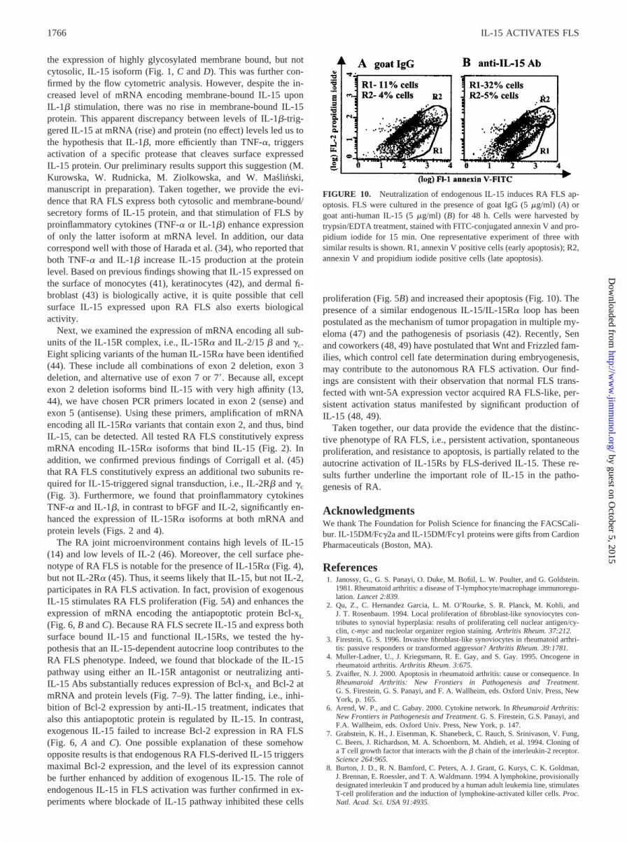
In the next set of experiments, we tested whether autocrine IL-15-triggered elevation of antiapoptotic proteins is reflected by the re-sistance of FLS to apoptosis. Indeed, enhanced apoptosis (32%) ofFLS cultured in the presence of neutralizing anti-IL-15 Abs (Fig.10B) was observed in comparison with FLS cultured in the pres-ence of isotype matching control IgG (11%; Fig. 10A). Similardata were obtained when IL-15R� antagonist was used (data notshown). These results indicate that endogenously produced IL-15contributes to the antiapoptotic status of RA FLS.
DiscussionWe have now tested the hypothesis that IL-15 participates in theactivation of RA FLS. First, we confirmed recent a report by
Harada et al. (34) that RA FLS spontaneously produce IL-15, andthat IL-15 expression is further enhanced by TNF-� and IL-1�.Next, we expanded their findings by analyzing the expression ofthe two known IL-15 isoforms (9, 11) in RA FLS. In resting RAFLS, both secretory/membrane-bound and cytosolic/nucleus IL-15isoforms are present at mRNA and protein levels (Fig. 1). It wasmost interesting to note that TNF-� and IL-1� treatment signifi-cantly elevated mRNA levels encoding the secretory/membrane-bound isoform (Fig. 1, A and B). In keeping with this observation,we determined at the protein level that TNF-� stimulation raised
FIGURE 7. IL-15R� antagonist inhibits bcl-xL and bcl-2 mRNA expression in RA FLS. RA FLS were cultured in medium alone or supplemented withIL-15DM/Fc�2a (10 �g/ml) or mouse IgG2a (10 �g/ml) for 48 h. The evaluation of mRNA expression for bcl-2, bcl-xL, and GAPDH was performed asdescribed in Fig. 6. Data for bcl-2 (A) and bcl-xL (C) are expressed as the mean (percentage of IgG2a-treated control) � SEM of four experiments. ��,p � 0.0002; �, p � 0.04; ns, not statistically significant (paired two-tailed Student’s t test). B and D, The representative results of RT-PCR products (selectedfrom A and C, respectively) are shown. The position and size of internal control and cDNA derived from cellular mRNA are marked.
FIGURE 8. IL-15 neutralizing Ab inhibits bcl-xL and bcl-2 mRNA ex-pression in RA FLS. RA FLS were cultured in medium alone or supple-mented with goat anti-human IL-15 (5 �g/ml) or goat IgG (5 �g/ml) for48 h. Total RNA was isolated and competitive RT-PCRs for bcl-2, bcl-xL,and GAPDH, in the presence of internal controls, were performed. TheRT-PCR products from one representative experiment of two with similarresults are shown.
FIGURE 9. IL-15R� antagonist decreases Bcl-2 and Bcl-xL protein ex-pression in RA FLS. FLS were cultured in the presence of IL-15DM/Fc�1(10 �g/ml) or human IgG1 (10 �g/ml) for 48 h. Cells were harvested bytrypsin/EDTA treatment, fixed, permeabilized, and stained with anti-Bcl-2or anti-Bcl-xL Ab or appropriate isotype control followed by flow cyto-metric analysis. One representative experiment of three for Bcl-2 (A) andtwo for Bcl-xL (B) with similar results are shown.
1765The Journal of Immunology
by guest on October 5, 2015
http://ww
w.jim
munol.org/
Dow
nloaded from
the expression of highly glycosylated membrane bound, but notcytosolic, IL-15 isoform (Fig. 1, C and D). This was further con-firmed by the flow cytometric analysis. However, despite the in-creased level of mRNA encoding membrane-bound IL-15 uponIL-1� stimulation, there was no rise in membrane-bound IL-15protein. This apparent discrepancy between levels of IL-1�-trig-gered IL-15 at mRNA (rise) and protein (no effect) levels led us tothe hypothesis that IL-1�, more efficiently than TNF-�, triggersactivation of a specific protease that cleaves surface expressedIL-15 protein. Our preliminary results support this suggestion (M.Kurowska, W. Rudnicka, M. Ziolkowska, and W. Maslinski,manuscript in preparation). Taken together, we provide the evi-dence that RA FLS express both cytosolic and membrane-bound/secretory forms of IL-15 protein, and that stimulation of FLS byproinflammatory cytokines (TNF-� or IL-1�) enhance expressionof only the latter isoform at mRNA level. In addition, our datacorrespond well with those of Harada et al. (34), who reported thatboth TNF-� and IL-1� increase IL-15 production at the proteinlevel. Based on previous findings showing that IL-15 expressed onthe surface of monocytes (41), keratinocytes (42), and dermal fi-broblast (43) is biologically active, it is quite possible that cellsurface IL-15 expressed upon RA FLS also exerts biologicalactivity.
Next, we examined the expression of mRNA encoding all sub-units of the IL-15R complex, i.e., IL-15R� and IL-2/15 � and �c.Eight splicing variants of the human IL-15R� have been identified(44). These include all combinations of exon 2 deletion, exon 3deletion, and alternative use of exon 7 or 7�. Because all, exceptexon 2 deletion isoforms bind IL-15 with very high affinity (13,44), we have chosen PCR primers located in exon 2 (sense) andexon 5 (antisense). Using these primers, amplification of mRNAencoding all IL-15R� variants that contain exon 2, and thus, bindIL-15, can be detected. All tested RA FLS constitutively expressmRNA encoding IL-15R� isoforms that bind IL-15 (Fig. 2). Inaddition, we confirmed previous findings of Corrigall et al. (45)that RA FLS constitutively express an additional two subunits re-quired for IL-15-triggered signal transduction, i.e., IL-2R� and �c
(Fig. 3). Furthermore, we found that proinflammatory cytokinesTNF-� and IL-1�, in contrast to bFGF and IL-2, significantly en-hanced the expression of IL-15R� isoforms at both mRNA andprotein levels (Figs. 2 and 4).
The RA joint microenvironment contains high levels of IL-15(14) and low levels of IL-2 (46). Moreover, the cell surface phe-notype of RA FLS is notable for the presence of IL-15R� (Fig. 4),but not IL-2R� (45). Thus, it seems likely that IL-15, but not IL-2,participates in RA FLS activation. In fact, provision of exogenousIL-15 stimulates RA FLS proliferation (Fig. 5A) and enhances theexpression of mRNA encoding the antiapoptotic protein Bcl-xL
(Fig. 6, B and C). Because RA FLS secrete IL-15 and express bothsurface bound IL-15 and functional IL-15Rs, we tested the hy-pothesis that an IL-15-dependent autocrine loop contributes to theRA FLS phenotype. Indeed, we found that blockade of the IL-15pathway using either an IL-15R antagonist or neutralizing anti-IL-15 Abs substantially reduces expression of Bcl-xL and Bcl-2 atmRNA and protein levels (Fig. 7–9). The latter finding, i.e., inhi-bition of Bcl-2 expression by anti-IL-15 treatment, indicates thatalso this antiapoptotic protein is regulated by IL-15. In contrast,exogenous IL-15 failed to increase Bcl-2 expression in RA FLS(Fig. 6, A and C). One possible explanation of these somehowopposite results is that endogenous RA FLS-derived IL-15 triggersmaximal Bcl-2 expression, and the level of its expression cannotbe further enhanced by addition of exogenous IL-15. The role ofendogenous IL-15 in FLS activation was further confirmed in ex-periments where blockade of IL-15 pathway inhibited these cells
proliferation (Fig. 5B) and increased their apoptosis (Fig. 10). Thepresence of a similar endogenous IL-15/IL-15R� loop has beenpostulated as the mechanism of tumor propagation in multiple my-eloma (47) and the pathogenesis of psoriasis (42). Recently, Senand coworkers (48, 49) have postulated that Wnt and Frizzled fam-ilies, which control cell fate determination during embryogenesis,may contribute to the autonomous RA FLS activation. Our find-ings are consistent with their observation that normal FLS trans-fected with wnt-5A expression vector acquired RA FLS-like, per-sistent activation status manifested by significant production ofIL-15 (48, 49).
Taken together, our data provide the evidence that the distinc-tive phenotype of RA FLS, i.e., persistent activation, spontaneousproliferation, and resistance to apoptosis, is partially related to theautocrine activation of IL-15Rs by FLS-derived IL-15. These re-sults further underline the important role of IL-15 in the patho-genesis of RA.
AcknowledgmentsWe thank The Foundation for Polish Science for financing the FACSCali-bur. IL-15DM/Fc�2a and IL-15DM/Fc�1 proteins were gifts from CardionPharmaceuticals (Boston, MA).
References1. Janossy, G., G. S. Panayi, O. Duke, M. Bofiil, L. W. Poulter, and G. Goldstein.
1981. Rheumatoid arthritis: a disease of T-lymphocyte/macrophage immunoregu-lation. Lancet 2:839.
2. Qu, Z., C. Hernandez Garcia, L. M. O’Rourke, S. R. Planck, M. Kohli, andJ. T. Rosenbaum. 1994. Local proliferation of fibroblast-like synoviocytes con-tributes to synovial hyperplasia: results of proliferating cell nuclear antigen/cy-clin, c-myc and nucleolar organizer region staining. Arthritis Rheum. 37:212.
3. Firestein, G. S. 1996. Invasive fibroblast-like synoviocytes in rheumatoid arthri-tis: passive responders or transformed aggressor? Arthritis Rheum. 39:1781.
4. Muller-Ladner, U., J. Kriegsmann, R. E. Gay, and S. Gay. 1995. Oncogene inrheumatoid arthritis. Arthritis Rheum. 3:675.
5. Zvaifler, N. J. 2000. Apoptosis in rheumatoid arthritis: cause or consequence. InRheumaroid Arthritis: New Frontiers in Pathogenesis and Treatment.G. S. Firestein, G. S. Panayi, and F. A. Wallheim, eds. Oxford Univ. Press, NewYork, p. 165.
6. Arend, W. P., and C. Gabay. 2000. Cytokine network. In Rheumaroid Arthritis:New Frontiers in Pathogenesis and Treatment. G. S. Firestein, G.S. Panayi, andF.A. Wallheim, eds. Oxford Univ. Press, New York, p. 147.
7. Grabstein, K. H., J. Eisenman, K. Shanebeck, C. Rauch, S. Srinivason, V. Fung,C. Beers, J. Richardson, M. A. Schoenborn, M. Ahdieh, et al. 1994. Cloning ofa T cell growth factor that interacts with the � chain of the interleukin-2 receptor.Science 264:965.
8. Burton, J. D., R. N. Bamford, C. Peters, A. J. Grant, G. Kurys, C. K. Goldman,J. Brennan, E. Roessler, and T. A. Waldmann. 1994. A lymphokine, provisionallydesignated interleukin T and produced by a human adult leukemia line, stimulatesT-cell proliferation and the induction of lymphokine-activated killer cells. Proc.Natl. Acad. Sci. USA 91:4935.
FIGURE 10. Neutralization of endogenous IL-15 induces RA FLS ap-optosis. FLS were cultured in the presence of goat IgG (5 �g/ml) (A) orgoat anti-human IL-15 (5 �g/ml) (B) for 48 h. Cells were harvested bytrypsin/EDTA treatment, stained with FITC-conjugated annexin V and pro-pidium iodide for 15 min. One representative experiment of three withsimilar results is shown. R1, annexin V positive cells (early apoptosis); R2,annexin V and propidium iodide positive cells (late apoptosis).
1766 IL-15 ACTIVATES FLS
by guest on October 5, 2015
http://ww
w.jim
munol.org/
Dow
nloaded from

9. Tagaya, Y., G. Kurys, T. Thies, J. M. L. Osi, N. Azimi, J. A. Hanover,R. N. Bamford, and T. A Waldmann. 1997. Generation of secretable and non-secretable interleukin 15 isoforms though alternate usage of signal peptides. Proc.Natl. Acad. Sci. USA 94:14444.
10. Gaggero, A., B. Azzoarone, C. Andrei, A. Mishal, R. Meazza, E. Zappia,A. Rubertelli, and S. Ferrini. 1999. Differential intracellular trafficking, secretion,and endosomal localization of two IL-15 isoforms. Eur. J. Immunol. 29:1265.
11. Onu, A., T. Pohl, H. Krause, and S. Bulfone-Paus. 1997. Regulation of IL-15secretion via the leader peptide of two IL-15 isoforms. J. Immunol. 158:255.
12. Giri, J. G., M. Ahdieh, J. Eisenman, K. Shanebeck, K. Grabstein, S. Kumaki,A. Namen, L. S. Park, D. Cosman, and D. M. Anderson. 1994. Utilization of the� and � chains of the IL-2 receptor by the novel cytokine IL-15. EMBO J.13:2822.
13. Anderson, D. M., S. Kumaki, M. Ahdieh, J. Bertles, M. Tometsko, A. Loomis,J. Giri, N. G. Copeland, D. J. Gilbert, N. A. Jenkins, et al. 1995. Functionalcharacterization of the human interleukin-15 receptor � chain and close linkageof IL-15R� and IL-2R� genes. J. Biol. Chem. 270:29862.
14. McInnes, I. B., J. Al-Mughales, M. Field, B. P. Leung, F. P. Huang, R. Dixon,R. D. Sturrock, P. C. Wilkinsin and F. Y. Liew. 1998. The role of interleukin-15in T cell migration and activation in rheumatoid arthritis. Nat. Med. 2:175.
15. McInnes, I. B., B. P. Leung, R. D. Sturrock, M. Field, and F. Y. Liew. 1997.Interleukin-15 mediates T-cell dependent regulation of tumor necrosis factor-�production in rheumatoid arthritis. Nat. Med. 3:189.
16. Wilkinson, P. C., and F. Y. Liew. 1995. Chemoattraction of human blood Tlymphocytes by IL-15. J. Exp. Med. 181:1255.
17. McInnes, I. B., and F. Y. Liew. 1998. Interleukin 15: a proinflammatory role inrheumatoid arthritis synovitis. Immunol. Today 19:75.
18. Giarard, D., M. E. Paquet, R. Paquin, and A. D. Beaulieu. 1996. Differentialeffects of interleukin-15 (IL-15) and IL-2 on human neutrophils: modulation ofphagocytosis, cytoskeleton rearrangement, gene expression, and apoptosis by IL-15. Blood 88:3176.
19. Akbar, A. N., N. J. Borthwick, R. G. Wickremasinghe, P. Panayoitidis, D. Pilling,M. Bofill, S. Krajewski, J. C. Reed, and M. Salmon. 1996. Interleukin-2 receptorcommon � chain signalling cytokines regulate activated T cell apoptosis in re-sponse to growth factor withdrawal: selective induction of antiapoptotic (bcl-2,bcl-xL) but not proapoptotic (bax, bcl-xS) gene expression. Eur. J. Immunol.26:294.
20. Kanegane, H., and G. Tosato. 1996. Activation of naive and memory T cells byinterleukin-15. Blood 88:230.
21. Zhang, X., S. Sun, I. Hwang, D. F. Tough, and J. Sprent. 1998. Potent andselective stimulation of memory-phenotype CD8� T cells in vivo by IL-15. Im-munity 8:591.
22. Lodolce, J. P., D. L. Boone, S. Chai, R. E. Swain, T. Dassopoulos, S. Trettin, andA. Ma. 1998. IL-15 receptor maintains lymphoid homeostasis by supporting lym-phocyte homing and proliferation. Immunity 9:669.
23. Alleva, D. G., S. B. Kaser, M. A. Monroy, M. J. Fenton, and D. I. Beller. 1997.IL-15 functions as a potent autocrine regulator of macrophage proinflammatorycytokine production: evidence for differential receptor subunit utilization associ-ated with stimulation or inhibition. J. Immunol. 159:2941.
24. Ziolkowska, M., A. Koc, G. Luszczykiewicz, K. Ksiezopolska-Pietrzak,E. Klimczak, H. Chwalinska-Sadowska, and W. Maslinski. 2000. High levels ofIL-17 in rheumatoid arthritis patients: IL-15 triggers in vitro IL-17 production viacyclosporin A-sensitive mechanism. J. Immunol. 164:2832.
25. McDonalds, P. P., M. P. Russo, S. Ferrini, and M. A. Cassatella. 1998. Interleu-kin-15 (IL-15) induces NF-�B activation and IL-8 production. Blood 92:4828.
26. Badolato, R., A. Negro Ponzi, M. Millesimo, L. D. Notarangelo, and T. Musso.1997. Interleukin-15 (IL-15) induces IL-8 and monocyte chemotactic protein-1production in human monocytes. Blood 90:2804.
27. Wojtecka-Lukasik, E., W. Maslinski, and S. Maslinski. 1997. Stimulation of thehuman neutrophil respiratory burst by IL-15. J. Physiol. Pharmacol. 48:66.
28. Ogata, Y., A. Kukita, T. Kukita, M. Komine, A. Miyahara, S. Miyazaki, andO. Kohashi. 1999. A novel role of IL-15 in the development of osteoclast: in-ability to replace its activity with IL-2. J. Immunol. 162:2754.
29. Ruchatz, H., B. P. Leung, X. Wei, I. B. McInnes, and F. Y. Liew. 1998. SolubleIL-15 receptor �-chain administration prevents murine collagen-induced arthritis:a role for IL-15 in development of antigen-induced immunopathology. J. Immu-nol. 160:5654.
30. Melnyk, V. O., G. D. Shipley, M. D. Sternfeld, L. Sherman, and J. T. Rosenbaum.1990. Synoviocytes synthesize, bind, and respond to basic fibroblast growth fac-tor. Arthritis Rheum. 33:493.
31. Jackson, J. R., J. A. Minton, M. L. Ho, N. Wei, and J. D. Winkler. 1997. Ex-pression of vascular endothelial growth factor in synovial fibroblasts is inducedby hypoxia and interleukin 1�. J. Rheumatol. 24:1253.
32. Sugiyama, M., T. Tsukazaki, A. Yonekura, S. Matsuzaki, S. Yamashita, andK. Iwasaki. 1995. Localization of apoptosis and expression of apoptosis relatedproteins in the synovium of patients with rheumatoid arthritis. Ann. Rheum. Dis.55:442.
33. Franz, J. K., T. Pap, K. M. Hummel, M. Nawrath, W. K. Aicher, Y. Shigeyama,U. Muller-Ladner, R. E. Gay, and S. Gay. 2000. Expression of sentrin, a novelantiapoptotic molecule, at sites of synovial invasion in rheumatoid arthritis. Ar-thritis Rheum. 43:599.
34. Harada, S., M. Yamamura, H. Okamoto, Y. Morita, M. Kawashima, T. Aita, andH. Makino. 1999. Production of interleukin-7 and interleukin-15 by fibroblast-like synoviocytes. Arthritis Rheum. 42:1508.
35. Kontny, E., A. Grabowska, J. Kowalczewski, M. Kurowska, I. Janicka,J. Marcinkiewicz, and W. Maslinski. 1999. Taurine chloramine inhibition of cellproliferation and cytokine production by rheumatoid arthritis fibroblast-like sy-noviocytes. Arthritis Rheum. 42:2552.
36. Kim, Y., W. Maslinski, X. X. Zheng, G. H. Tesch, V. R. Kelley, and T. B. Strom.1998. Targeting the IL-15 receptor with an antagonist IL-15/Fc protein blocksdelayed type hypersensitivity. J. Immunol. 160:5742.
37. Kontny, E., K. Szczepanska, J. Kowalczewski, M. Kurowska, I. Janicka,J. Marcinkiewicz, and W. Maslinski. 2000. The mechanism of taurine chloramineinhibition of cytokine (interleukin-6, interleukin-8) production by rheumatoidarthritis fibroblast-like synoviocytes. Arthritis Rheum. 43:2169.
38. Lipman, M., A. Stevens, and T. B. Strom. 1994. Heightened intragraft CTL geneexpression in acutely rejecting renal allografts. J. Immunol. 152:5120.
39. Kontny, E., M. Ziolkowska, A. Ryzewska, and W. Maslinski. 1999. Protein ki-nase C-dependent pathway is critical for the production of pro-inflammatory cy-tokines (TNF-�, IL-1�, IL-6). Cytokine 11:839.
40. Bulfone-Paus, S., D. Ungureanu, T. Pohl, G. Linder, R. Paus, R. Ruckert,H. Krause, and U. Kunzendorf. 1997. Interleukin-15 protects from lethal apo-ptosis in vivo. Nat. Med. 3:1124.
41. Musso, T., L. Calosso, M. Zucca, M. Millesimo, D. Ravarino, M. Giovarelli,F. Malavasi, A. N. Ponzi, R. Paus, and S. Bulfone-Paus. 1999. Human monocytesconstitutively express membrane bound, biologically active and IFN-� upregu-lated interleukin-15. Blood 93:3531.
42. Ruckert, R., K. Asadullah, M. Seifert, V. M. Budagian, R. Arnold, C. Trombotto,R. Paus, and S. Bulfone-Paus. 2000. Inhibition of keratinocyte apoptosis by IL-15: a new parameter in the pathogenesis of psoriasis. J. Immunol. 165:2240.
43. Rappl, G., A. Kapsokefalou, C. Heuser, M. Rosslev, S. Ugurel, W. Tilgen,U. Reinhold, and H. Abken. 2001. Dermal fibroblast sustain proliferation of ac-tivated T cells via membrane-bound interleukin-15 upon long-term stimulationwith tumor necrosis factor-�. J. Invest. Dermatol. 116:102.
44. Dubois, S., F. Magrangeas, P. Lehours, S. Raher, J. Bernard, O. Boisteau,S. Leroy, S. Minvielle, A. Godard, and Y. Jacques. 1999. Natural splicing of exon2 of human interleukin-15 receptor �-chain mRNA results in shortened form withdistinct pattern of expression. J. Biol. Chem. 274:26978.
45. Corrigall, V. M., M. Arastu, S. Khan, C. Shah, M. Fife, T. Smeets, P. P. Tak, andG. S. Panayi. 2001. Functional IL-2 receptor � (CD 122) and � (CD 132) chainsare expressed by fibroblast-like synoviocytes: activation by IL-2 stimulatesmonocyte chemoattractant protein-1 production. J. Immunol. 166:4141.
46. Firestein, G. S., W. D. Xu, K. Townsend, D. Broide, J. M. Alvaro-Gracia,A. Glasebrook, and N. J. Zvaifler. 1988. Cytokine in chronic inflammatory ar-thritis: failure to detect T cell lymphokines (interleukin-2 and interleukin-3) andpresence of macrophage colony-stimulating factor (CSF-1) and a novel mast cellgrowth factor in rheumatoid synovitis. J. Exp. Med. 168:1573.
47. Tinhofer, I., I. Marschitz, T. Henn, A. Egle, and R. Greil. 2000. Expression offunctional interleukin-15 receptor and autocrine production of interleukin-15 asmechanism of tumor propagation in multiple myeloma. Blood 95:610.
48. Sen, M., K. Lauterbach, H. El-Gabalawy, G. S. Firestein, M. Corr, andD. A. Carson. 2000. Expression and function of wingless and frizzled homologsin rheumatoid arthritis. Proc. Natl. Acad. Sci. USA 97:2791.
49. Sen, M., M. Chamorro, J. Reifert, M. Corr, and D. A. Carson. 2001. Blockade ofWnt-5A/Frizzled 5 signaling inhibits rheumatoid synoviocyte activation. ArthritisRheum. 44:772.
1767The Journal of Immunology
by guest on October 5, 2015
http://ww
w.jim
munol.org/
Dow
nloaded from
Copyright © 2022 FDOKUMEN