Bcl-B Expression in Human Epithelial and Nonepithelial Malignancies
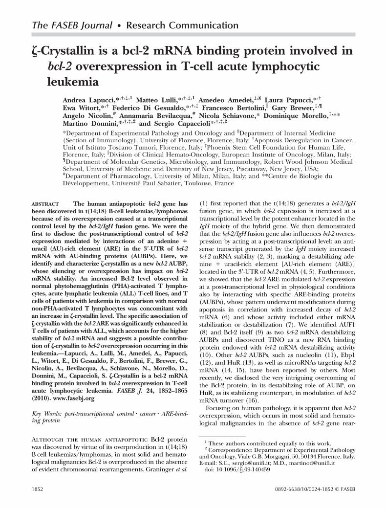
The FASEB Journal • Research Communication
�-Crystallin is a bcl-2 mRNA binding protein involved inbcl-2 overexpression in T-cell acute lymphocyticleukemia
Andrea Lapucci,*,†,‡,1 Matteo Lulli,*,†,‡,1 Amedeo Amedei,‡,§ Laura Papucci,*,†
Ewa Witort,*,† Federico Di Gesualdo,*,†,‡ Francesco Bertolini,� Gary Brewer,‡,¶
Angelo Nicolin,# Annamaria Bevilacqua,# Nicola Schiavone,* Dominique Morello,‡,**Martino Donnini,*,†,‡,2 and Sergio Capaccioli*,†,‡,2
*Department of Experimental Pathology and Oncology and §Department of Internal Medicine(Section of Immunology), University of Florence, Florence, Italy; †Apoptosis Deregulation in Cancer,Unit of Istituto Toscano Tumori, Florence, Italy; ‡Phoenix Stem Cell Foundation for Human Life,Florence, Italy; �Division of Clinical Hemato-Oncology, European Institute of Oncology, Milan, Italy;¶Department of Molecular Genetics, Microbiology, and Immunology, Robert Wood Johnson MedicalSchool, University of Medicine and Dentistry of New Jersey, Piscataway, New Jersey, USA;#Department of Pharmacology, University of Milan, Milan, Italy; and **Centre de Biologie duDeveloppement, Universite Paul Sabatier, Toulouse, France
ABSTRACT The human antiapoptotic bcl-2 gene hasbeen discovered in t(14;18) B-cell leukemias/lymphomasbecause of its overexpression caused at a transcriptionalcontrol level by the bcl-2/IgH fusion gene. We were thefirst to disclose the post-transcriptional control of bcl-2expression mediated by interactions of an adenine �uracil (AU)-rich element (ARE) in the 3�-UTR of bcl-2mRNA with AU-binding proteins (AUBPs). Here, weidentify and characterize �-crystallin as a new bcl-2 AUBP,whose silencing or overexpression has impact on bcl-2mRNA stability. An increased Bcl-2 level observed innormal phytohemagglutinin (PHA)-activated T lympho-cytes, acute lymphatic leukemia (ALL) T-cell lines, and Tcells of patients with leukemia in comparison with normalnon-PHA-activated T lymphocytes was concomitant withan increase in �-crystallin level. The specific association of�-crystallin with the bcl-2 ARE was significantly enhanced inT cells of patients with ALL, which accounts for the higherstability of bcl-2 mRNA and suggests a possible contribu-tion of �-crystallin to bcl-2 overexpression occurring in thisleukemia.—Lapucci, A., Lulli, M., Amedei, A., Papucci,L., Witort, E., Di Gesualdo, F., Bertolini, F., Brewer, G.,Nicolin, A., Bevilacqua, A., Schiavone, N., Morello, D.,Donnini, M., Capaccioli, S. �-Crystallin is a bcl-2 mRNAbinding protein involved in bcl-2 overexpression in T-cellacute lymphocytic leukemia. FASEB J. 24, 1852–1865(2010). www.fasebj.org
Key Words: post-transcriptional control � cancer � ARE-bind-ing protein
Although the human antiapoptotic Bcl-2 proteinwas discovered by virtue of its overproduction in t(14;18)B-cell leukemias/lymphomas, in most solid and hemato-logical malignancies Bcl-2 is overproduced in the absenceof evident chromosomal rearrangements. Graninger et al.
(1) first reported that the t(14;18) generates a bcl-2/IgHfusion gene, in which bcl-2 expression is increased at atranscriptional level by the potent enhancer located in theIgH moiety of the hybrid gene. We then demonstratedthat the bcl-2/IgH fusion gene also influences bcl-2 overex-pression by acting at a post-transcriptional level: an anti-sense transcript generated by the IgH moiety increasedbcl-2 mRNA stability (2, 3), masking a destabilizing ade-nine � uracil-rich element [AU-rich element (ARE)]located in the 3�-UTR of bcl-2 mRNA (4, 5). Furthermore,we showed that the bcl-2 ARE modulated bcl-2 expressionat a post-transcriptional level in physiological conditionsalso by interacting with specific ARE-binding proteins(AUBPs), whose pattern underwent modifications duringapoptosis in correlation with increased decay of bcl-2mRNA (6) and whose activity included either mRNAstabilization or destabilization (7). We identified AUF1(8) and Bcl-2 itself (9) as two bcl-2 mRNA destabilizingAUBPs and discovered TINO as a new RNA bindingprotein endowed with bcl-2 mRNA destabilizing activity(10). Other bcl-2 AUBPs, such as nucleolin (11), Ebp1(12), and HuR (13), as well as microRNAs targeting bcl-2mRNA (14, 15), have been reported by others. Mostrecently, we disclosed the very intriguing overcoming ofthe Bcl-2 protein, in its destabilizing role of AUBP, onHuR, as its stabilizing counterpart, in modulation of bcl-2mRNA turnover (16).
Focusing on human pathology, it is apparent that bcl-2overexpression, which occurs in most solid and hemato-logical malignancies in the absence of bcl-2 gene rear-
1 These authors contributed equally to this work.2 Correspondence: Department of Experimental Pathology
and Oncology, Viale G.B. Morgagni, 50, 50134 Florence, Italy.E-mail: S.C., [email protected]; M.D., [email protected]
doi: 10.1096/fj.09-140459
1852 0892-6638/10/0024-1852 © FASEB

rangements (17–19), could be due to impaired ARE/AUBP interaction-based bcl-2 post-transcriptional control.Here, we identify �-crystallin as a new bcl-2 AUBP endowedwith bcl-2 mRNA stabilizing activity and responsible, ifoverproduced, for enhanced bcl-2 mRNA stability in T-cellacute lymphocytic leukemia (ALL).
ALLs are the most common childhood cancers, withpeak prevalence between the ages of 2 and 5 yr (20, 21).Although more than 80% of pediatric patients with ALLare cured, the current intensive therapeutic regimenshave toxic side effects (22) and the prognosis for the20–30% of pediatric patients who have a relapse or whoseALL is refractory to conventional therapies remains dis-mal (23). Indeed, relapsed ALL is the fifth most commonpediatric malignancy and the first cause of pediatriccancer mortality (24), patients with T-cell ALL beingmore prone to early initial relapse and inferior outcomethan patients with B-cell ALL (25). Based on previousconsiderations, advances in the understanding of thepathogenesis of ALLs as well as in the identification of theprecise molecular events that take place in their genesissuggest that drugs specifically targeting the genetic alter-ations could revolutionize management of this disease(26). In this scenario, we propose that deregulated �-crys-tallin could be included among these candidate therapeu-tic targets.
�-Crystallin is a highly conserved protein (27) endowedwith pleiotropic functions. First discovered as a structuralprotein in the lens of guinea pigs (28), camelids (29, 30),and tree frogs (31), it was also found in plants (32) andyeast (33). �-Crystallin also has oxidoreductase activity (34,35) and has been identified as a novel NADPH:quinonereductase (36, 37). More recently, Tang and Curthoys(38) and Ibrahim et al. (39) identified �-crystallin as anRNA-binding protein, able to stabilize rat glutaminasemRNA in conditions of metabolic acidosis by binding to apH response element (pH-RE) in substitution of thedestabilizing AUBP, namely AUF1. In addition, Schroederet al. (40) found that �-crystallin also stabilized ratglutamate dehydrogenase mRNA by binding to peculiarmotifs of its 3�-UTR that are highly homologous to theglutaminase mRNA pH-RE. Moreover, Fernandez et al.(41) have recently reported that �-crystallin is a highlyevolutionarily conserved AUBP, being able, besides func-tioning as an NADPH-dependent ortho-quinone reduc-tase, to specifically bind to a synthetic A(UUUA) pentam-eric probe both in humans and in yeast. Last, Porte et al.(27) have further characterized the RNA binding proper-ties of �-crystallin, demonstrating that NADPH efficientlycompeted against the A(UUUA) pentameric probe forbinding to human �-crystallin, which suggests that theNADPH-binding site is involved in the binding of �-crys-tallin to RNA.
MATERIALS AND METHODS
Cell cultures and transfections
The human ALL Jurkat (clone E6–1), ALL MOLT-4, andlymphocytic lymphoma SUP-T1 T-cell lines and the embry-
onic kidney HEK 293 cell line (European Collection of CellCultures, Porton Down, UK), were cultured according to thesupplier’s maintenance protocols.
For silencing of �-crystallin by siRNA, Jurkat cells or HEK 293cells were electrophorated or transfected using Lipofectamine2000 reagent (Invitrogen, Carlsbad, CA, USA), respectively, witha combination of two siRNAs (Qiagen, Hilden, Germany) spe-cific for the �-crystallin or one siRNA (Qiagen) specific for thegreen fluorescent protein (GFP) (200 nM final concentration). Thesequences of �-crystallin targeted by the siRNAs were: 5�-CAAGCCTACTTACCTTTATAA-3� and 5�-CAGAGGTACTATT-GAAATAAA-3�. The GFP sequence targeted by the siRNA was5�-CGGCAAGCTGACCCTGAAGTTCAT-3�.
For transient transfection of recombinant �-crystallin, a1016-nucleotide segment corresponding to the entire openreading frame of �-crystallin was PCR-amplified using 5�-GCGAAGCTTATGGCGACTGGACAGAAGTTG-3� and 5�-GCCTCGAGTAAGAGAAGAATCATTTTACCAGTAGCC-3� asforward and reverse primers, respectively, and cloned into theHindIII and XhoI sites of the pQE-TriSystem vector (Qiagen) toobtain the pQE-TriSystem/HIS-Tag CRYZ expression vector.HEK 293 cells (8�105) were transfected with 4 �g of pQE-TriSystem/HIS-Tag CRYZ or the empty vector, using Lipo-fectamine 2000 reagent (Invitrogen), and analyzed for expres-sion after 48 h by immunoblotting, using anti-HIS (C-term) mAb(Invitrogen).
For chimeric reporter assays, a DNA fragment containingthe bcl-2 ARE was cloned into the XbaI cloning site at the 3�end of the hRlucP gene coding for Renilla reniformis luciferaseof the pGL4.71P plasmid to produce the pGL4.71P bcl2 AREplasmid, as described previously (42). Forty-eight hours afterthe first �-crystallin transfection or silencing of HEK293 cells,the aliquots of 2 � 105 cells were plated in 24-well dishes andwere cotransfected for the second time with 500 ng of thepGL4.71P or pGLA4.71P bcl2 ARE constructs and 500 ng ofthe pGL3-P firefly luciferase reporter gene plasmid.
Blood samples and isolation of T lymphocytes
Peripheral blood samples for preparation of CD3� T lympho-cytes were obtained from 40 healthy middle-aged donors ofboth sexes and from 4 patients with CD3� ALL. All patientswere thoroughly informed about this study and gave writtenconsent for the investigation. The samples were kept smallaccording to the guidelines of the Italian ethics committee.The T lymphocytes were isolated from peripheral bloodmononuclear cells, which had been fractionated, cultured,and activated, when required, with 1% v/v phytohemoagglu-tinin (PHA) (Life Technologies, Inc., Grand Island, NY,USA), as described previously (43). ALL T lymphocytes wereisolated as described above and stored at �80°C until use.
Characterization of CD3�ALL T cells
Leukemia samples were characterized on the basis of theirphenotype, and the absence of evident chromosomal rear-rangement was ascertained.
Preparation of cytosolic fractions
Cells were pelleted at 100 g for 5 min at 4°C, washed withice-cold PBS, and lysed in 100 �l of cytosolic lysis buffer (10mM HEPES, 10 mM KCl, 1 mM EDTA, 1 mM EGTA, 1 mMDTT, 1 mM MgCl2, 5% glycerol, 0.25 mM Pefabloc, 2 �Mleupeptin, 0.3 �M aprotinin, and 0.1 mM sodium orthovana-date). After incubation on ice for 10 min, cells were lysed withNonidet P-40 (final concentration 0.25%), vortexed for 10 s,and than centrifuged for 5 min at 16,000 g. Cytoplasmic
1853�-CRYSTALLIN IMPACT ON bcl-2 OVEREXPRESSION

supernatants were saved as a cytosolic fraction (44). Proteinconcentrations were determined using the Qubit fluorimeter(Invitrogen).
UV-cross-linking assays
Aliquots of cytoplasmic proteins (�50 �g) from nonactivatedor PHA-activated human T lymphocytes or Jurkat cells wereincubated in a total volume of 10 �l with the 32P-labeled bcl-2ARE probe (109 cpm/�g, 5�105 cpm) as described previ-ously (6) and analyzed by a Cyclone Storage Phosphor System(PerkinElmer Life and Analytical Sciences, Waltham, MA,USA).
Isoelectrofocusing and bidimensional electrophoresis
Samples, resuspended in the rehydration buffer, were appliedto 7-cm immobilized pH gradient strips (pH 3–10 NL; Invitro-gen), incubated at room temperature overnight and thenfocused in a ZOOM IPGRunner Minicell System (Invitrogen),as recommended by the manufacturer. Focused proteins wereseparated by bidimensional SDS-PAGE on NuPAGE 4–12%Bis-Tris gels (Invitrogen), and the resulting spots were stainedwith the SilverQuest Silver staining kit (Invitrogen).
Protein identification by mass spectrometry
The gel spot was excised and processed using the Montagein-Gel Digest kit (Millipore, Billerica, MA, USA), according tothe manufacturer’s instructions. The digested eluate was ana-lyzed by mass spectrometry on an Ultraflex TOF/TOF instru-ment (Bruker Daltronics, Bremen, Germany), according to themanufacturer’s instructions. The resulting mass picks were ana-lyzed by Mascot Peptide Mass Fingerprint software (MatrixScience Ltd., London, UK) and then searched for in the MSDBdatabase (http://www.matrixscience.com/search_form_select.html).
In vitro transcription
Radiolabeled RNA probes were generated by in vitro transcrip-tion using a MAXIscript T7 kit (Ambion, Austin, TX, USA) andeither pCRII/bcl-2 ARE (10) or the pCRII/TA vector (Invitro-gen) as a negative control, using plasmids as templates. Tran-scription reactions were performed for 1 h at 37°C according tothe manufacturer’s instructions: 100 U of T7 polymerase; 500�M concentrations each of ATP, CTP, and GTP; 12.5 �M UTP;200 �Ci of [32P]UTP (800 Ci/mmol; Amersham Biosciences,Piscataway, NJ, USA); and 1 �g of plasmid template linearized bydigestion with SmaI for pCRII/bcl-2 ARE or HindIII for pCRII/TAcontrol, in a total volume of 20 �l. Unincorporated nucleo-tides were removed using the MEGAclear column kit (Am-bion). Biotinylated RNA probes were obtained by in vitrotranscription with the MEGAscript (Ambion). A 20-�l volumeof the final reaction contained 2 �l of 10� transcriptionbuffer and 2 �l each of 75 mM ATP, GTP, and UTP. It alsocontained 1 �l of 75 mM CTP and 7.5 �l of 10 mMbiotinylated CTP (Invitrogen), 10 �g of linearized pCRII/bcl-2 ARE or pCRII/TA templates, and 2 �l of T7 MEGAscriptenzyme mix. After an overnight incubation at 37°C, the tran-scription reaction was stopped by the addition of 2 �l ofRNase-free DNase (DNA-free; Ambion); unincorporated nucle-otides were removed. The RNA concentration was determinedspectrophotometrically at 260 nm.
In vitro translation
�-crystallin cDNA (Geneservice Ltd., Cambridge, UK) wasinserted into the plasmid pCMV-SPORT6 (Image Clone6067125). On MluI linearization, the �-crystallin-harboringplasmid was incubated with the SP6 TNT Coupled Reticulo-cyte Lysate System (Promega, Madison, WI, USA) in thepresence of cold methionine, according to the manufacturer’sinstructions.
RNA binding assays of bcl-2 ARE probe to �-crystallin inJurkat cell and PHA-activated T-lymphocyte extracts
RNA binding assays were performed essentially according toCok et al. (44). In brief, cytoplasmic proteins (4 mg/400 �l)were incubated with 120–150 �g of biotinylated RNA probesin the presence of 20 U of Recombinant RNasin Inhibitor(Promega) for 2 h with constant rotation and then weretreated with 600 �l of streptavidin-agarose beads (Invitro-gen). The mixture was prewashed 3 times in 1 ml of bindingbuffer (10 mM HEPES, pH 7; 6.5 mM MgCl2; 40 mM KCl; 1mM DTT; 5% glycerol; and 5 mg/ml heparin), for anadditional 2 h with constant rotation. Bound proteins werewashed 4 times with 1 ml of binding buffer, eluted from thebiotinylated RNA with 200 �l of 1 M NaCl in binding buffer,and collected with the ReadyPrep 2-D Cleanup kit (Bio-RadLaboratories, Hercules, CA, USA). Samples were then resus-pended in 150 �l of rehydration buffer (8 M urea; 0.15mg/ml; 2% CHAPS; 0.5% carrier ampholytes, pH 7–10;0.0002% bromphenol blue; and 20 mM DTT). All incuba-tions were performed at room temperature. Results werefrom either four healthy donors or four inoculations of Jurkatcells.
RNA electromobility shift assay (REMSA)
Mobility shift assays were performed using the radiolabeledbcl-2 ARE or the pCRII/TA riboprobes (6). Samples wereanalyzed using the Cyclone Storage Phosphor System.
Intracellular localization
Cells (2�105) were immobilized onto glass cover slips (15�15mm), washed twice with 1 ml of cold PBS, and fixed for 20min in 3.7% paraformaldehyde in PBS. After 3 washes for 2min with PBS, cells were permeabilized with 1 ml of 0.25%Triton X-100 in PBS for 5 min at room temperature andwashed 3 times for 5 min in PBS. All of the followingtreatments were performed in the dark. After staining of thenuclei with Hoechst 33258 (Sigma-Aldrich, St. Louis, MO,USA) in PBS for 30 min at 37°C, the cells were washed 3 timesfor 5 min with 1 ml of PBS at room temperature, incubated in1 ml of blocking buffer (3% bovine serum albumin and 0.1%Triton X-100 in PBS) for 1 h at room temperature, and thenincubated overnight at 4°C with the primary rabbit polyclonalanti-�-crystallin Ab (1:250) in blocking buffer (kindly pro-vided by J. Samuel Zigler, Jr., National Eye Institute, Be-thesda, MD, USA). The following day, the cells were washed3 times for 15 min in washing buffer (0.1% Triton X-100 inPBS), incubated with the secondary Cy-3 conjugated anti-rabbit antibody (Chemicon International, Inc., Temecula,CA, USA) (1:800) for 60 min at room temperature, andwashed 3 times with 1 ml of washing buffer for 5 min at roomtemperature. They were then dried, mounted onto glassslides, and examined with confocal microscopy using a NikonEclipse TE2000-U (Nikon, Tokyo, Japan). Confocal images(1024�768 pixels) were obtained using a �63 objective lens.
1854 Vol. 24 June 2010 LAPUCCI ET AL.The FASEB Journal � www.fasebj.org

Cytofluorimetric analysis of levels of Bcl-2 and �-crystallinproteins
Nonactivated or PHA-activated T lymphocytes and Jurkat cellswere stained with anti-CD3 fluorescent mAb (BD, FranklinLakes, NJ, USA) and then with anti-Bcl-2 mAb (UpstateBiotechnology, Charlottesville, VA, USA) or rabbit polyclonalanti-�-crystallin Ab. In brief, 106 cells were incubated with theanti-CD3 mAb at �4°C for 30 min, washed with PBS contain-ing 1% bovine serum albumin and 0.1% sodium azide, andthen fixed and permeabilized with a permeabilizing solution(BD) for 10 min. Cells were incubated with mouse anti-Bcl-2or rabbit anti-�-crystallin Ab or the isotype control mAb (BDBiosciences Pharmingen, San Diego, CA, USA) at �4°C for 30min. They were then washed with PBS, which contained 0.5%saponin and 0.1% sodium azide, stained with goat anti-mouseIgG-FITC (BD) or goat anti-rabbit IgG-FITC (MolecularProbes, Eugene, OR, USA) at �4°C for 15 min, and analyzedon a BDLSRII cytofluorimeter using Diva software (BD) with104 events for each sample acquired.
In vivo cross-linking and immunoprecipitation (IP)/RT-PCR
Protein extracts (2 mg) of the Jurkat cell line were cross-linked in vivo with formaldehyde (45) and precleared with 30�l of a 50% slurry of rProtein G-Agarose beads (Invitrogen),250 mM NaCl, and 400/U RNasin for 3 h at 4°C and thenimmunoprecipitated with 30 �l of a 50% slurry of rProtein GAgarose beads, anti-�-crystallin Ab, or preimmune serum(1:100) and incubated overnight at 4°C. After extensivewashes, one-half of the beads were used for immunoblotanalysis with the anti-�-crystallin Ab. For the IP/RT-PCR, theother half was used for subsequent RNA purification using anRNeasy Kit (Qiagen) and treated with DNA-free. Purified
RNA was retro-transcribed by the iScript cDNA Synthesis kit(Bio-Rad Laboratories, Hercules, CA, USA), and cDNA wasamplified with primers for bcl-2, glutaminase, and �-actindescribed below in the Real-Time PCR section.
Immunoblot analysis
The cytoplasmic proteins (50 �g) were separated by NuPAGE12% Bis-Tris Gel and electroblotted (Trans-Blot Semi-Dryapparatus; Bio-Rad) onto Protran nitrocellulose transfermembranes (Schleicher & Schuell, Dassel, Germany). Thefollowing antibodies were used: rabbit-polyclonal �-crystallin,AUF1, and �-actin (Santa Cruz Technology, Inc., Santa Cruz,CA, USA) and monoclonal Bcl-2 and -tubulin (Sigma-Aldrich). The secondary antibodies included goat anti-mouseIRDye 800CW and goat anti-rabbit IRDye 800CW (Li-CorBiosciences, Lincoln, NE, USA). The protein bands wereanalyzed by the Odyssey Infrared Imaging System (Li-Cor)using the software for protein quantification.
Real-time PCR
Total RNA was isolated from nonactivated or PHA-activated Tlymphocytes, cultured ALL T cells, ALL T cells obtained frompatients with leukemia (�5�106 cells/sample), and HEK 293cells with an RNeasy mini kit (Qiagen), treated with RNase-freeDNase, and analyzed spectroscopically. One microgram of RNAwas retrotranscribed using iScript (Bio-Rad) and amplified withspecific primers: For bcl-2, forward 5�-TCAGCTATTTACTGC-CAAAG-3� and reverse 5�-GATTTCCAAAGACAGGAG-3�; forglutaminase, forward 5�-CCTAGATGGCACCTCCTTTGG-3� andreverse 5�-TGCTACCTGTCTCCATGGCTTG-3�; for 18S, for-ward 5�-CGGCTACCACATCCAAGGAA-3� and reverse 5�-GCT-
Figure 1. Different patterns of bcl-2 AUBP complexes in T lymphocytes andJurkat cells and identification of �-crystallin as a new bcl-2 AUBP. A) Cytoplas-mic extracts (50 �g) were UV-cross-linked to the bcl-2 ARE radiolabeled probeand analyzed using the Cyclone Storage Phosphor System. B) Western blotanalysis of cytoplasmic extracts (50 �g) challenged with anti-Bcl-2 mAb.-Tubulin was used as the loading control. All results are representative of �3independent experiments. C) Two typical silver-stained bidimensional electro-pherograms of bcl-2 AUBPs from PHA-activated T lymphocytes (left) or Jurkatcells (right) obtained from RNA binding assays. Insets: magnified views ofcorresponding boxed areas.
1855�-CRYSTALLIN IMPACT ON bcl-2 OVEREXPRESSION

GGAATTACCGCGGCT-3� ; for � -actin, forward 5� -GAAACTACCTTCAACTCCATCATG-3� and reverse 5�-AGGAGGAGCAATGATCTTGATC-3�; for R. reniformis luciferase,forward 5�-GTCGAGACCATGCTCCCAAGCA-3� and reverse 5�-TTGCGGACAATCTGGACGACGT-3�; and for firefly luciferase,forward 5�-CTGAATTGGAATCCATCTTGCTCCAACAC-3� andreverse 5�-TTCGTCCACAAACACAACTCCTCCG-3�. All prim-ers were purchased from Eurofins MWG Operon (Ebersberg,Germany). Real-time PCR assays were performed using theRotor-Gene 3000 cycler system (Corbett Research, Sydney, NSW,Australia).
mRNA decay assay
Forty-eight hours after �-crystallin transient transfection orsilencing, or 24 h after transient transfection of chimericreporters, cells were treated with actinomycin D at a finalconcentration of 5 �g/ml to block transcription and har-vested at various time points. Total RNA was extracted, andthe real-time PCR assays were performed as described above.The quantification of bcl-2 mRNA and heterogenous nuclearRNA (hnRNA) for the bcl-2 mRNA stability assessment wasperformed by PCR amplification, according to the strategydescribed by Otake et al. (46), using the same primers andamplification conditions. PCR amplification of 18S rRNA and�-actin RNA were used as the normalizer. In the chimeric
reporter assay, R. reniformis and firefly luciferase quantificationdata were normalized on the expression of the housekeepinggene 18S, and then the R. reniformis data were normalized ontransfection efficiency by using firefly luciferase mRNA levels.The data from the mRNA decay assays are reported as apercentage of remaining RNA after actinomycin D addition.
Binding of bcl-2 ARE to �-crystallin in cytoplasmic extractsof nonactivated or PHA-activated T lymphocytes, ALL T-celllines, and ALL T cells from patients with leukemia
bcl-2 ARE radiolabeled probe and cytoplasmic extracts ob-tained from either nonactivated or PHA-activated T lympho-cytes from 4 healthy donors or the 3 ALL T-cell lines or theALL T lymphocytes from 4 patients with leukemia wereprepared as described above. Aliquots of cytoplasmic frac-tions containing 100 �g of proteins were precleared withrProtein G Agarose beads in cytosolic lysis buffer containing150 mM KCl. The cytosolic fractions were then incubated for3.5 h at 4°C with 0.25 nM bcl-2 ARE and either 2 �g ofanti-�-crystallin antibody or preimmune serum (control anti-body). The immunocomplexes were precipitated with proteinG, washed twice with cytosolic lysis buffer, and then analyzedby liquid scintillation. Results are the means se of 3independent experiments (46). The specificity of binding ofour anti-�-crystallin antibody was confirmed by the fact that
Figure 2. Validations of �-crystallin association with the bcl-2 ARE. A) SDS-PAGE ofRNA binding assays of cytoplasmic extracts from ALL Jurkat T cells challenged withthe �-crystallin antibody. Lane 1, molecular weight marker; lane 2, cytoplasmicextract; lane 3, cytoplasmic proteins bound to the biotinylated bcl-2 ARE riboprobe;and lane 4, cytoplasmic proteins bound to pCRII/TA riboprobe. The result is aparadigm of 5 experiments. B) REMSA obtained by incubating increasing amounts of in vitro synthesized �-crystallin withthe radiolabeled bcl-2 ARE (left) or pCRII/TA probes (right). Asterisks indicate the maximum volume of a mixturecontaining the in vitro synthesized luciferase and either the bcl-2 ARE or the pCRII/TA probes. The result is a paradigm of3 independent experiments. C) �-Crystallin/bcl-2 mRNA complex in Jurkat cells evaluated by IP/RT-PCR. Immunoblotanalyses (above): lane 1, protein extracts; lane 2, immunoprecipitated �-crystallin; and lane 3, immunoprecipitation withpreimmune serum. Agarose gel electrophoresis (below) shows that the bands corresponding to bcl-2 and glutaminase (asa positive control) mRNAs in the immunoprecipitated complex (output) are restricted to the lane corresponding to the�-crystallin antibody. Amplification of the housekeeping gene �-actin, bound at low levels with the immunoprecipitatedcomplexes, shows equal loading of immunoprecipitation samples.
TABLE 1. Summary of tryptic peptides of human �-crystallin protein sequenced by MALDI-TOF mass spectrometry
�-Crystallin protein Observed mass (m/z) Sequence Amino acid residues
Peptide 1 1054.575 DLSLLSHGGR CRYZ: 233-242Peptide 2 1221.657 VFEFGGPEVLK CRYZ: 13-23Peptide 3 1468.791 ESSIIGVTLFSSTK CRYZ: 263-276Peptide 4 1527.805 QGAAIGIPYFTAYR CRYZ: 125-138Peptide 5 1614.814 VHACGVNPVETYIR CRYZ: 42-55Peptide 6 1633.860 IVLQNGAHEVFNHR CRYZ: 188-201Peptide 7 2180.131 AGESVLVHGASGGVGLAACQ IAR CRYZ: 148-170Peptide 8 2551.260 VFTSSTISGGYAEYALAADH TVYK CRYZ: 93-116
1856 Vol. 24 June 2010 LAPUCCI ET AL.The FASEB Journal � www.fasebj.org

�4-fold higher radioactivity was recovered from all immuno-precipitates with the anti-�-crystallin antibody compared withthose with preimmune serum.
Statistical analysis
Statistical evaluation of the data was performed with a 2-tailedStudent’s t test using STATA 9.0 analysis software(StataCorpLP, College Station, TX, USA). Differences were consideredstatistically significant when P � 0.05.
RESULTS
Identification of �-crystallin as a new cytoplasmic bcl-2AUBP
We used the bcl-2 overexpressing ALL Jurkat T-cell linevs. normal nonactivated or PHA-activated T lympho-
cytes as the first cellular experimental model. As shownin Fig. 1, we analyzed the bcl-2 AUBP complex pattern,following an RNA-protein UV-cross-linking assay car-ried out with the synthetic bcl-2 ARE as probe in 3cellular cytoplasmic extracts. Western blot analysis ofBcl-2 protein levels was also performed. RNA bindingassays using an bcl-2 ARE-biotin-streptavidin affinitymatrix were performed to reveal the binding pattern ofsingle bcl-2 AUBPs in Jurkat cells and PHA-activated Tlymphocytes. Figure 1A shows qualitative/quantitativedifferences in bcl-2 AUBP complexes, mostly rangingfrom 25 to 70 kDa, in nonactivated or PHA-activated Tlymphocytes and Jurkat cells. Figure 1B shows that thisphenomenon was accompanied by a significantlyhigher amount of Bcl-2 protein in the PHA-activated Tlymphocytes (6.50.7, P�0.005) and Jurkat cells(9.30.3, P�0.005) compared with nonactivated Tlymphocytes. To identify specific bcl-2 AUBPs that un-
Figure 3. Immunolocalization of �-crystallin. Images of nonactivated or PHA-activated T lymphocytes from healthy donor andJurkat cells obtained through confocal microscopy are shown. Merged images are representative of 6 independent experiments.
1857�-CRYSTALLIN IMPACT ON bcl-2 OVEREXPRESSION

dergo quantitative or qualitative changes accountingfor bcl-2 AUBP pattern modifications, the cytoplasmicextracts from PHA-activated T lymphocytes and Jurkatcells were analyzed by RNA binding assays to capturethe bcl-2 AUBPs. The captured proteins were separatedby bidimensional SDS-PAGE. Figure 1C shows twotypical silver-stained bidimensional SDS-PAGE samplesof bcl-2 AUBPs from PHA-activated T lymphocytes (left)or Jurkat cells (right). Among the spots specificallydetected in Jurkat cell extracts, we chose the one withan apparent molecular mass of �35 kDa and pI of�8 –9 for further mass spectrometry analysis. Eightpeptides, corresponding to the m/z indicated inTable 1, were analyzed by Mascot Peptide Mass Finger-print software in the MSDB database. This resulted in amatch with the amino acid sequence of the human�-crystallin/NADPH:quinone reductase (�-crystallin gene;GenBank accession number NM_001889). Sequenceanalysis by matrix-assisted laser desorption ionization(MALDI)-time of flight (TOF)/TOF of two peptidesconfirmed the identity of �-crystallin (data notshown).
The association of �-crystallin with the bcl-2 ARE wasevaluated both in vitro and in vivo (Fig. 2). First, weisolated the AUBPs bound to the biotin/streptavidinconjugated bcl-2 ARE or pCRII/TA (negative control)probes using RNA binding assays and confirmed thepresence of �-crystallin as a 37- to 38-kDa proteinexclusively bound to the bcl-2 ARE probe (Fig. 2A).
Then we analyzed the �-crystallin binding to the bcl-2ARE by REMSA in vitro. It clearly demonstrated the shiftof the bcl-2 ARE but not the pCRII/TA probe by �-crystallin in a dose-dependent manner (Fig. 2B). Next,the association of �-crystallin with the bcl-2 ARE wasvalidated in vivo by IP/RT-PCR experiments performedwith protein extracts from Jurkat cells using a �-crystal-lin antibody or preimmune serum. RT-PCR analysis of�-crystallin/RNA immunocomplexes detected copre-cipitation of bcl-2 mRNA with �-crystallin protein(Fig. 2C).
The cellular localization of �-crystallin by confocalimmunohistochemistry clearly indicated that �-crystal-lin is a cytoplasmic protein both in nonactivated orPHA-activated T lymphocytes and Jurkat cells (Fig. 3).
�-Crystallin increases in PHA-activated T lymphocytesto reach levels comparable to those of ALL T-celllines compared with nonactivated T lymphocytes andcorrelates with Bcl-2 protein levels
Besides Jurkat, two other bcl-2-overexpressing ALLT-cell lines, MOLT-4 and SUP-T1, were included inthe analysis of �-crystallin expression (Fig. 4A). PHA-activated T lymphocytes displayed higher levels of�-crystallin compared with nonactivated T lympho-cytes and reached levels similar to those observed inthe 3 ALL T-cell lines. Moreover, the enhancementof Bcl-2 protein levels induced by PHA activation was
Figure 4. �-Crystallin and Bcl-2 levels in nonactivated or PHA-activated T lymphocytes and in ALL T-cell lines. A) �-Crystallinimmunoblot of cytoplasmic extracts from nonactivated or PHA-activated T lymphocytes derived from a healthy donor and ALLT-cell lines (Jurkat, MOLT-4, and SUP-T1). B) �-Crystallin and Bcl-2 immunoblots of cytoplasmic extracts of nonactivated (�)or PHA-activated (�) T lymphocytes derived from 4 representative healthy donors (D.A to D.D) and from Jurkat cells (Jc).-Tubulin served as an internal control. C) Box and whisker graphs showing the percentage of �-crystallin-expressing (top) andBcl-2-expressing cells (bottom) among nonactivated or PHA-activated T lymphocytes and Jurkat cells, evaluated by FACSanalysis. Boxes contain the middle 50% of the data. Horizontal bars indicate means se of 30 observations (P�0.00005 forPHA-activated T lymphocytes or Jurkat cells vs. nonactivated T lymphocytes; Student’s t test).
1858 Vol. 24 June 2010 LAPUCCI ET AL.The FASEB Journal � www.fasebj.org

Figure 5. Bcl-2 ARE-dependent ef-fects of �-crystallin silencing andoverexpression on bcl-2 mRNA half-life and Bcl-2 protein levels. A) Leftpanel: immunoblot analysis of �-crys-tallin or GFP silencing in Jurkatcells. Middle panel: Bcl-2 mRNA de-cay after �-crystallin or GFP silencingin Jurkat cells, analyzed followingtranscriptional block using actino-mycin D. Right panel: effects of�-crystallin or GFP silencing on Bcl-2protein levels in Jurkat cells. Datarepresent means se of 4 indepen-dent experiments. B) Left panel:immunoblot analysis of ectopi-cally expressed HIS-Tag �-crystal-lin in transfected HEK 293 cells.Middle panel: Bcl-2 mRNA decay af-ter �-crystallin overexpression in HEK293 cells was analyzed followingtranscriptional block using actino-mycin D. Right panel: effects of�-crystallin overexpression in HEK293 cells on Bcl-2 protein levels.Data represent means se of 4independent experiments. C) Sche-matic representation of luciferaseconstruct carrying (pGL4.71P Rluc-bcl2 ARE) or not carrying (pGL4.71PRluc) the bcl2 ARE. Shaded bars indi-cate Renilla luciferase gene; open barindicates region of the bcl2 ARE.
Transcription was under the control of the simian virus 40 promoter (hatched bars) and the poly(A) site (solid bars).D) Rluc-bcl-2 ARE (top) or Rluc (bottom) mRNA decay after �-crystallin or GFP silencing in HEK 293 cells, analyzed aftertranscriptional block using actinomycin D. E) Rluc-bcl-2 ARE (top) or Rluc (bottom) mRNA decay after �-crystallinoverexpression in HEK 293 cells, analyzed following transcriptional block using actinomycin D. Data are means seof 3 independent experiments. Horizontal dotted line in each graph indicates mRNA half-life. *P � 0.005.
1859�-CRYSTALLIN IMPACT ON bcl-2 OVEREXPRESSION

concomitant with that of �-crystallin and reached thesame values as those observed in Jurkat cells (Fig.2B). Results of flow cytometry showed that, analogi-cally, the PHA activation concomitantly increased thepercentage of �-crystallin and Bcl-2-expressing cells com-pared with nonactivated T lymphocytes (from 36.28.41to 90.33.74% for �-crystallin and from 59.65.00 to92.72.89% for Bcl-2, P�0.00005), reaching the constitu-tive levels of Jurkat cells (90.00.9% for the �-crystallinand 87.260.69% for Bcl-2 (P�0.00005) (Fig. 2C). Thesedata indicate a correlation between Bcl-2 and �-crystallinprotein levels.
�-Crystallin is endowed with bcl-2 mRNA stabilizingactivity in a bcl-2 ARE-dependent manner
The hypothesis that �-crystallin controls bcl-2 expressionat the post-transcriptional level by affecting bcl-2 mRNAstability was confirmed by a causal relationship betweenthe two events (Fig. 5). First, we verified the possibleconsequence of �-crystallin silencing on bcl-2 mRNAdecay. The silencing of �-crystallin, but not of GFP, withspecific siRNAs in Jurkat cells led to a dramatic reduc-tion (more than 75%) (Fig. 5A) in the �-crystallinprotein level, concomitant with a significant decrease inthe bcl-2 mRNA half-life (from 5 to 3 h, P�0.005) andBcl-2 protein level (130.52%, P�0.005). Second, wetested the impact of �-crystallin overexpression in HEK293 cells transfected with HIS-Tag �-crystallin on bcl-2mRNA stability (Fig. 5B) and noted the significantincrease in the bcl-2 mRNA half-life (from 7.5 to 10.5 h,P�0.005) and Bcl-2 protein level (120.67%, P�0.005). The difference in the basal bcl-2 mRNA half-lifebetween the Jurkat (5 h) and HEK 293 (7.5 h) cells wasfully justified by the different gene expression patternthat characterizes the two different cell types.
Finally, two symmetrical clear-cut affirmations as-sured that the stabilizing effect of �-crystallin on bcl-2mRNA is bcl-2 ARE-dependent and excluded the possi-bility that the alteration in bcl-2 mRNA stability isconsequent to the toxicity of �-crystallin exogenousmodulations. The bcl-2 ARE regulative effects on mRNAstability were studied using the bcl-2 ARE-luciferasereporter constructs (Fig. 5C). Figure 5D shows thatsilencing of �-crystallin, but not of GFP, in HEK 293 cellsreduced the bcl-2 ARE-bearing Renilla luciferase re-porter (Rluc-bcl-2 ARE) mRNA from 7.2 to 5 h (P�0.005). Symmetrically, Fig. 5E shows that �-crystallinoverexpression in HEK 293 cells increased the Rluc-bcl-2ARE mRNA half-life from 6.5 to 13.5 h (P�0.005). Asexpected, �-crystallin exogenous modulation did nothave any effect on the half-life of hRluc devoid of bcl-2ARE, used as a negative control. Moreover, in the sameexperimental model we observed a significant de-crease (604%, P�0.0005) or increase (1153.4%,P�0.0005) of Rluc-bcl-2 ARE with respect to Rluc mRNAsteady states by �-crystallin silencing or overexpression,respectively. The same modulative effects were alsoobserved in a luciferase reporter protein expressionassay (data not show).
Association of �-crystallin with the bcl-2 ARE isstronger in Jurkat cells than in PHA-activated Tlymphocytes and results in increased bcl-2 mRNAstability
Although �-crystallin protein levels in Jurkat cells arecomparable to those of PHA-activated T lymphocytesfrom healthy donors (Fig. 4), detection of the bcl-2ARE-associated �-crystallin by silver staining was re-
Figure 6. Differential association of �-crystallin with the bcl-2ARE probe in PHA-activated T lymphocytes and Jurkat cellsand relative levels of bcl-2 mRNA decay. A) �-Crystallin immu-noblots of cytoplasmic extracts from PHA-activated T lympho-cytes and Jurkat cells, before (input) and after (output) RNAbinding assays using the bcl-2 ARE probe. -Tubulin was usedas the input protein loading control. Quantitative data of therelative binding of �-crystallin to the bcl-2 ARE probe arereported as the ratio of bound �-crystallin (output) to total�-crystallin (input) expressed as means se of 4 experiments.B) Ratio of the bcl-2 mRNA/hnRNA levels in PHA-activated Tlymphocytes and Jurkat cells determined by real-time PCRand normalized to the 18S rRNA. Results are means se of4 experiments. *P � 0.001.
1860 Vol. 24 June 2010 LAPUCCI ET AL.The FASEB Journal � www.fasebj.org

stricted to Jurkat cells (Fig. 1C). This prompted us toinvestigate the association of �-crystallin with bcl-2 AREin leukemia cells compared with that in normal lym-phocytes. The levels of �-crystallin in cytoplasmic ex-tracts were compared before (input) and after (output)RNA binding to a biotinylated bcl-2 ARE probe (Fig. 6A).Association in Jurkat cells was 3.4-fold greater than inthe PHA-activated T lymphocytes (P�0.001). This is inkeeping with the 2.4-fold increase in bcl-2 mRNA stabil-ity (Fig. 6B), measured as the ratio of bcl-2 mRNA andbcl-2 hnRNA expression (37) scored by real-time PCR.
Bcl-2 overexpression in ALL T cells from patientswith leukemia is accompanied by the stabilization ofbcl-2 mRNA consequent to increased association of�-crystallin with its ARE
We then evaluated the possibility that the increasedassociation of �-crystallin with bcl-2 ARE observed incultured Jurkat cells also occurs in spontaneous T-cell
ALL and possibly accounts for the pathogenetic mech-anism of this malignancy. We extended our analyses toALL T cells obtained from four patients, compared withnonactivated or PHA-activated T lymphocytes (Fig. 7). Asshown in Fig. 7A, the levels of Bcl-2 protein (above) inALL T cells from patients were �9-fold higher thanthose in nonactivated T lymphocytes but also, althoughto a much lower extent, were significantly higher thanthose in PHA-activated T lymphocytes, which con-firmed in spontaneous leukemias the trend observed inthe 3 ALL cultured T-cell lines. On the other hand(below), �-crystallin levels in ALL T cells from patientswere �5-fold higher than those in nonactivated Tlymphocytes but did not differ from those scored inPHA-activated T lymphocytes.
Although the ratios of Bcl-2 and �-crystallin to �-actinprotein levels plotted in Fig. 7B (r�0.769, P�0.001)indicated that they were significantly correlated, it isunclear why ALL T cells from patients had higher levelsof Bcl-2 compared with PHA-activated T lymphocytes
Figure 7. Bcl-2 and �-crystallin proteinlevels and their correlation in normaland leukemic T lymphocytes. A) Pro-tein levels of Bcl-2 (left) and �-crystal-lin (right) in nonactivated or PHA-activated T lymphocytes obtained from5 healthy donors; the Jurkat, MOLT-4,and SUP-T1 ALL T-cell lines; and ALLT cells from 4 patients with leukemia.Results are means se. *P � 0.05,**P � 0.001 vs. nonactivated T lym-phocytes; §P � 0.005 vs. PHA-activatedT lymphocytes. B) Correlation between�-crystallin/�-actin and Bcl-2/�-actinprotein levels calculated by STATAsoftware analysis, using results of 3different experiments for each point(P�0.001).
1861�-CRYSTALLIN IMPACT ON bcl-2 OVEREXPRESSION

despite having the same levels of �-crystallin. A possibleexplanation for this discrepancy was the lower decay ofbcl-2 mRNA in ALL T cells compared with that innormal and both nonactivated and PHA-activated Tlymphocytes, already observed in Jurkat cells (Fig. 6B).Results of bcl-2 mRNA decay shown in Fig. 8A, indicat-ing that the ratio of bcl-2 mRNA to hnRNA levels in ALLT cells from patients was 3.3 0.01-fold higher than thatin PHA-activated T lymphocytes (P�0.001), confirmedthis hypothesis. In turn, an obvious explanation for thisevidence could be the stronger association of �-crystallinwith the bcl-2 ARE in ALL T cells. This hypothesis wasconfirmed by the results of RNA binding assays obtainedby using a bcl-2 ARE radiolabeled probe (Fig. 8B), indicat-ing that the association of �-crystallin with the probe was2.3-fold higher in extracts obtained from patients withleukemia (43916 cpm) than in those obtained in nor-mal PHA-activated T lymphocytes (19516 cpm).
On the whole, our results indicate that the increasedlevel of �-crystallin as well as its stronger association withthe bcl-2 ARE in ALL T cells enhances bcl-2 mRNAstability and may thereby be involved in bcl-2 overex-pression typical of this malignancy.
DISCUSSION
T-cell ALL is a rare but aggressive malignancy thatrepresents 15% of childhood and 25% of adult ALLs(47). A high percentage of T-cell ALLs do not containobvious chromosomal translocations (48). Neverthe-less, cytogenetically cryptic abnormalities that can leadeither to oncogene activation or to the loss of tumorsuppressor genes have been frequently described (49).
One of these abnormalities involves bcl-2, which plays afundamental role during T lymphocyte development(50) and whose overexpression in T-cell ALLs contrib-utes to the accumulation of apoptosis-defective che-moresistant T lymphocytes (18, 51–53). Nevertheless,the mechanism underlying bcl-2 overexpression in T-cell ALLs has not yet been fully unraveled.
We have discovered that bcl-2 expression can also becontrolled at a post-transcriptional level through coop-eration between an ARE located in the 3�-UTR of itsmRNA (4) and multiple ARE-binding proteins, whoseexpression pattern undergoes modifications during ap-optosis (6). We also identified 3 regulatory bcl-2 AUBPs:AUF1, Bcl-2 itself, and Tino (8–10). Nucleolin andEbp1 have been added to the family of bcl-2 AUBPs byothers and have been demonstrated to be involved inbcl-2 overexpression in the acute promyelocytic leuke-mia HL-60 cell line and B-cell chronic lymphocyticleukemias (CLLs) (11, 12, 46). Recently, Cimmino et al.(14) demonstrated that two microRNAs, miR-15 andmiR-16, also bind to and destabilize bcl-2 mRNA and areresponsible, if deleted, for the onset of CLL.
Here, we used 3 leukemia T-cell lines characterizedby high Bcl-2 levels in the absence of obvious bcl-2 generearrangements (54) to shed further light on bcl-2post-transcriptional control and its role in leukemias/lymphomas. We first revealed quantitative and qualita-tive differences in cytoplasmic bcl-2 AUBP complexesfrom Jurkat cells compared with nonactivated or PHA-activated T lymphocytes from healthy donors. Bidimen-sional electrophoresis of bcl-2 AUBPs from PHA-activatedT lymphocytes and Jurkat cells confirmed differentialpatterns of bcl-2 AUBPs. We hypothesized that bcl-2 over-expression is caused by alterations of specific AUBPs.
Figure 8. Bcl-2 decay and differential association of �-crystallin with the bcl-2 ARE probe in our T-cell types. A) Ratio of bcl-2mRNA/hnRNA levels obtained from the ALL T cells of a patient with leukemia and all our T-cell types indicated in Fig. 7,determined by real-time PCR and normalized to the 18S rRNA. Results are means se of 4 experiments. *P � 0.001 vs.nonactivated T lymphocytes; §P � 0.001 vs. PHA-activated T lymphocytes. B) Counts per minute of 32P-labeled bcl-2ARE-�-crystallin complexes recovered after �-crystallin immunoprecipitation. Columns represent means se of 3 independentexperiments. *P � 0.005 vs. nonactivated T lymphocytes; §P � 0.001 vs. PHA-activated T lymphocytes.
1862 Vol. 24 June 2010 LAPUCCI ET AL.The FASEB Journal � www.fasebj.org

Among the differentially expressed proteins, we iden-tified the �-crystallin/NADPH:quinone reductase as anew bcl-2 AUBP. The presence of �-crystallin in aribonucleoprotein complex containing bcl-2 ARE wasconfirmed both in vitro by RNA binding and REMSAassays and in vivo by IP/RT-PCR. Our immunolocaliza-tion analyses ascertained that �-crystallin was an exclu-sively cytoplasmic protein. Next, we explored its impacton bcl-2 expression. Both negative and positive modu-lation of expression in Jurkat cells and HEK 293 cellsrevealed that �-crystallin is endowed with bcl-2 AREdependent-stabilizing activity on bcl-2 mRNA. Althoughlevels of �-crystallin in PHA-activated T lymphocytes arecomparable, its association with the bcl-2 ARE in leuke-mia cells is stronger than that in normal T lymphocytes.This accounts for the higher bcl-2 mRNA stability inT-cell ALLs, which results in the increased Bcl-2 proteinlevel. Similarly, the binding to bcl-2 ARE of another bcl-2AUBP, nucleolin, has been reported to be �5-foldgreater in CLL B cells than in normal B lymphocytes(46). The authors explained this result as being conse-quent to the 6.5-fold greater cytoplasmic level ofnucleolin in CLL B cells, which indicates that bcl-2mRNA binding of nucleolin undergoes quantitativealterations in these cells. In our system, the �-crystallinassociation with the bcl-2 ARE was much greater in ALLT cells from patients with leukemia and in the 3leukemia T-cell lines than in normal nonactivated orPHA-activated T lymphocytes. However, cytoplasmiclevels of �-crystallin did not differ in normal PHA-activated T lymphocytes and leukemia T cells, indicat-ing that the varying concentration-independent bind-ing of �-crystallin to the bcl-2 ARE in leukemia T cellsmight depend on other parameters. Therefore, wepropose a schematic model to explain the differentialinteraction of �-crystallin with the bcl-2 ARE in normal Tlymphocytes and ALL T cells (Fig. 9). In this model, wepropose two alternative mechanisms. In the first, qual-itative modifications undergone by �-crystallin in ALL Tcells respect to normal T lymphocytes increase itsbinding to bcl-2 mRNA, consequently altering the bcl-2AUBP pattern. In the second, modifications of the bcl-2AUBP pattern in ALL T cells could favor the �-crystallininteraction with the bcl-2 ARE.
Other AUBPs, including AUF-1, Tino, nucleolin,HuR, and Bcl-2 itself and two microRNAs, are known to
bind to bcl-2 mRNA to modulate its turnover in differ-ent cell types and to be involved in bcl-2 overexpressionin human leukemias (8–14, 16). There is also thepossibility that their simultaneous expression in onecell type in different physiological conditions is not anaxiom, implying that their interactions and possiblederegulation pathways in human diseases are highlycomplex. Furthermore, because the binding site of onebcl-2 AUBP could either overlap or not overlap that ofanother, possible interactions among different AUBPscompared with the bcl-2 ARE and their resulting effectson bcl-2 mRNA fate are unpredictable a priori.
The involvement of �-crystallin in bcl-2 overexpres-sion in ALL T cells might constitute a significantmolecular pathogenetic mechanism of the disease. Thepossibility that �-crystallin contributing to bcl-2 mRNAstabilization observed in T-cell ALLs could also occur inother hematological malignancies or even in solidtumors and its relationships with other AUBPs in bcl-2post-transcriptional control are now under investiga-tion in our laboratory.
The authors thank Professors Vieri Boddi and RiccardoCaldini for help with the statistical analysis, Dr. J. SamuelZigler, Jr. (National Eye Institute, Bethesda, MD, USA) forthe contribution of the anti-�-crystallin antibody, and Pro-fessor Mary Forrest and Dr. Carolyn Demsey for accurateediting of the article. This work was supported by grantsfrom Programmi di Ricerca di Interesse Nazionale (to S.C.and A.N.), the Associazione Italiana per la Ricerca sulCancro (to S.C.), the Ente Cassa di Risparmio di Firenze(to S.C.), the Fondazione Cassa di Risparmio di Lucca (toS.C.), and the U.S. National Institutes of Health (R01CA052443 to G.B.).
REFERENCES
1. Graninger, W. B., Seto, M., Boutain, B., Goldman, P., andKorsmeyer, S. J. (1987) Expression of Bcl-2 and Bcl-2-Ig fusiontranscripts in normal and neoplastic cells. J. Clin. Invest. 80,1512–1515
2. Capaccioli, S., Quattrone, A., Schiavone, N., Calastretti, A.,Copreni, E., Bevilacqua, A., Canti, G., Gong, L., Morelli, S., andNicolin, A. (1996) A bcl-2/IgH antisense transcript deregulatesbcl-2 gene expression in human follicular lymphoma t(14;18)cell lines. Oncogene 13, 105–115
3. Morelli, S., Delia, D., Capaccioli, S., Quattrone, A., Schiavone,N., Bevilacqua, A., Tomasini, S., and Nicolin, A. (1997) The
Figure 9. Proposed model of bcl-2 mRNA and �-crystallin differential interaction innormal T lymphocytes and ALL T cells. See text for details.
1863�-CRYSTALLIN IMPACT ON bcl-2 OVEREXPRESSION

antisense bcl-2-IgH transcript is an optimal target for syntheticoligonucleotides. Proc. Natl. Acad. Sci. U. S. A. 94, 8150–8155
4. Schiavone, N., Rosini, P., Quattrone, A., Donnini, M., Lapucci,A., Citti, L., Bevilacqua, A., Nicolin, A., and Capaccioli, S. (2000)A conserved AU-rich element in the 3� untranslated region ofbcl-2 mRNA is endowed with destabilizing function that isinvolved in bcl-2 down-regulation during apoptosis. FASEB J. 4,174–184
5. Bevilacqua, A., Ceriani, M. C., Capaccioli, S., and Nicolin, A.(2003) Post-transcriptional regulation of gene expression bydegradation of messenger RNAs. J. Cell. Physiol. 195, 356–372
6. Donnini, M., Lapucci, A., Papucci, L., Witort, E., Tempestini,A., Brewer, G., Bevilacqua, A., Nicolin, Capaccioli, S., andSchiavone, N. (2001) Apoptosis is associated with early mod-ifications of bcl-2 mRNA AU-binding proteins. Biochem. Bio-phys. Res. Commun. 287, 1063–1069
7. Barreau, C., Paillard, L., and Osborne, H. O. (2006) AU-richelements and associated factors: are there unifying principles?Nucleic Acids Res. 33, 7138–7150
8. Lapucci, A., Donnini, M., Papucci, L., Witort, E., Tempestini, A.,Bevilacqua, A., Nicolin, A., Brewer, G., Schiavone, N., andCapaccioli, S. (2002) AUF1 is a bcl-2 ARE-binding proteininvolved in bcl-2 mRNA destabilization during apoptosis. J. Biol.Chem. 277, 16139–16146
9. Bevilacqua, A., Ceriani, M. C., Canti, G., Asnaghi, L., Gherzi, R.,Brewer, G., Papucci, L., Schiavone, N., Capaccioli, S., andNicolin, A. (2003) Bcl-2 protein is required for the ARE-dependent degradation of its own messenger. J. Biol. Chem. 278,23451–23459
10. Donnini, M., Lapucci, A., Papucci, L., Witort, E., Jacquier, A.,Brewer, G., Nicolin, A., Capaccioli, S., and Schiavone, N. (2004)Identification of Tino: a new evolutionarily conserved bcl-2AU-rich element RNA binding protein. J. Biol. Chem. 279,20154–20166
11. Sengupta, T. K., Bandyopadhyay, S., Fernandes, D. J., andSpicer, E. K. (2004) Identification of nucleolin as an AU-richelement binding protein involved in bcl-2 mRNA stabilization.J. Biol. Chem. 279, 10855–10863
12. Bose, S. K., Sengupta, T. K., Bandyopadhyay, S., and Spicer,E. K. (2006) Identification of Ebp1 as a component of cytoplas-mic bcl-2 mRNP (messenger ribonucleoprotein particle) com-plexes. Biochem. J. 96, 99–107
13. Ishimaru, D., Ramalingam, S., Sengupta, T. K., Bandyopadhyay,S., Dellis, S., Tholanikunnel, B. G., Fernandes, D. J., and Spicer,E. K. (2009) Regulation of Bcl-2 expression by HuR in HL60leukemia cells and A431 carcinoma cells. Mol. Cancer Res. 7,1354–1366
14. Cimmino, A., Calin, G. A., Fabbri, M., Iorio, M. V., Ferracin, M.,Shimizu, M., Wojcik, S. E., Aqeilan, R. I., Zupo, S., Dono, M.,Rassenti, L., Alder, H., Volinia, S., Liu, C. G., Kipps, T. J.,Negrini, M., and Croce, C. M. (2005) miR-15 and miR-16 induceapoptosis by targeting BCL2. Proc. Natl. Acad. Sci. U. S. A. 102,13944–13949
15. Bommer, G. T., Gerin, I., Feng, Y., Kaczorowski, A. J., Kuick, R.,Love, R. E., Zhai, Y., Giordano, T. J., Qin, Z. S., Moore, B. B.,MacDougald, O. A., Cho, K. R., and Fearon, E. R. (2007)p53-mediated activation of miRNA34 candidate tumor-suppres-sor genes. Curr. Biol. 17, 1298–1307
16. Ghisolfi, L., Calastretti, A., Franzi, S., Canti, G., Donnini, M.,Capaccioli, S., Nicolin, A., and Bevilacqua, A. (2009) B celllymphoma (Bcl)-2 protein is the major determinant in bcl-2adenine-uridine-rich element turnover overcoming HuR activ-ity. J. Biol. Chem. 284, 20946–20955
17. Bakhshi, A., Jensen, J. P., Goldman, Wright, P. J. J., McBride,O. W., Epstein, A. L., and Korsmeyer, S. J. (1985) Cloning thechromosomal breakpoint of t(14;18) human lymphomas: clus-tering around JH on chromosome 14 and near a transcriptionalunit on 18. Cell 41, 899–906
18. Steube, K. G., Jadau, A., Teepe, D., and Drexler, H. G. (1995)Expression of bcl-2 mRNA and protein in leukemia-lymphomacell lines. Leukemia 9, 1841–1846
19. Robertson, L. E., Plunkett, W., McConnell, K., Keating, M. J.,and McDonnell, T. J. (1996) Bcl-2 expression in chronic lym-phocytic leukemia and its correlation with the induction ofapoptosis and clinical outcome. Leukemia 10, 456–459
20. Pui, C. H., Relling, M. V., and Downing, J. R. (2004) Acutelymphoblastic leukemia. N. Engl. J. Med. 350, 1535–1548
21. Pui, C. H., and Evans, W. E. (2006) Treatment of acutelymphoblastic leukemia. N. Engl. J. Med. 354, 166–178
22. Valerie, I., Brown, V. I., Seif, A. E., Reid, G. S. D., Teachey, D. T.,and Grupp, S. A. (2008) Novel molecular and cellular therapeu-tic targets in acute lymphoblastic leukemia and lymphoprolif-erative disease. Immunol. Res. 42, 84–105
23. Gaynon, P. S., Qu, R. P., Chappell, R. J., Willoughby, M. L.,Tubergen, D. G., Steinherz, P. G., and Trigg, M. E. (1998)Survival after relapse in childhood acute lymphoblastic leuke-mia: impact of site and time to first relapse—the Children’sCancer Group Experience. Cancer 82, 1387–1395
24. Chessells, J. M. Relapsed lymphoblastic leukaemia in children: acontinuing challenge. Br. J. Haematol. 102, 423–433
25. Borgmann, A., von Stackelberg, A., Hartmann, R., Ebell, W.,Klingebiel, T., Peters, C., and Henze, G. (2003) Unrelateddonor stem cell transplantation compared with chemotherapyfor children with acute lymphoblastic leukemia in a secondremission: a matched-pair analysis. Blood 101, 3835–3839
26. Pui, C. H., Robison, L. L., and Look, A. T. (2008) Acutelymphoblastic leukaemia. Lancet 371, 1030–1043
27. Porte, S., Crosas, E., Yakovtseva, E., Biosca, J. A., Farres, J.,Fernandez, M. R., and Pares, X. (2009) MDR quinone oxi-doreductases: the human and yeast zeta-crystallins. Chem. Biol.Interact. 178, 288–294
28. Huang, Q. L., Russell, P., Stone, S. H., and Zigler, J. S., Jr. (1987)�-Crystallin, a novel lens protein from the guinea pig. Curr. EyeRes. 6, 725–732
29. Garland. D., Rao, P. V., Del Corso, A., Mura, U., and Zigler, J. S.,Jr. (1991) �-Crystallin is a major protein in the lens of Camelusdromedarius. Arch. Biochem. Biophys. 285, 134–136
30. Duhaiman, A. S., Rabbani, N., AlJafari, A. A., and Alhomida,A. S. (1995) Purification and characterization of �-crystallinfrom the camel lens. Biochem. Biophys. Res. Commun. 215, 632–640
31. Fujii, Y., Kimoto, H., Ishikawa, K., Watanabe, K., Yokota, Y.,Nakai, N., and Taketo, A. (2001) Taxon-specific �-crystallin inJapanese tree frog (Hyla japonica) lens. J. Biol. Chem. 276,28134–28139
32. Mano, J., Yoon, H., Asada, K., Babiychuk, E., Inze, D., andMikami, B. (2000) Crystallization and preliminary X-ray crystal-lographic analysis of NADPH: azodicarbonyl/quinone oxi-doreductase, a plant �-crystallin. Biochim. Biophys. Acta 1480,374–376
33. Kranthi, B. V., Balasubramanian, N., and Rangarajan, P. N.(2006) Isolation of a single-stranded DNA-binding protein fromthe methylotrophic yeast, Pichia pastoris and its identification as� crystallin. Nucleic Acids Res. 34, 4060–4068
34. Rodokanaki, A., Holmes, R. K., and Borras, T. (1989) �-Crystal-lin, a novel protein from the guinea pig lens is related to alcoholdehydrogenases. Gene 78, 215–224
35. Rao, P. V., Krishna, C. M., and Zigler, J. S., Jr. (1992) Identifi-cation and characterization of the enzymatic activity of �-crystal-lin from guinea pig lens. A novel NADPH:quinone oxidoreduc-tase. J. Biol. Chem. 267, 96–102
36. Rao, P. V., and Zigler, J. S., Jr. (1992) Purification and charac-terization of �-crystallin/quinone reductase from guinea pigliver. Biochim. Biophys. Acta 1117, 315–320
37. Gonzalez, P., Rao, P. V., and Zigler, J. S., Jr. (1993) Molecularcloning and sequencing of �-crystallin/quinone reductasecDNA from human liver. Biochem. Biophys. Res. Commun. 1191,902–907
38. Tang, A., and Curthoys, N. P. (2001) Identification of �-crystal-lin/NADPH:quinone reductase as a renal glutaminase mRNApH response element-binding protein. J. Biol. Chem. 276, 21375–21380
39. Ibrahim, H., Lee, Y. J., and Curthoys, N. P. (2008) Renalresponse to metabolic acidosis: role of mRNA stabilization.Kidney Int. 73, 11–18
40. Schroeder, J. M., Liu, W., and Curthoys, N. P. (2003) pH-responsive stabilization of glutamate dehydrogenase mRNAin LLC-PK1-F� cells. Am. J. Physiol. Renal Physiol. 285, F258–F265
41. Fernandez, M. R., Porte, S., Crosas, E., Barbera, N., Farres, J.,Biosca, J. A., and Pares, X. (2007) Human and yeast �-crystallinsbind AU-rich elements in RNA. Cell. Mol. Life Sci. 64, 1419–1427
1864 Vol. 24 June 2010 LAPUCCI ET AL.The FASEB Journal � www.fasebj.org

42. Bevilacqua, A., Ghisolfi, L., Franzi, S., Maresca, G., Gherzi, R.,Capaccioli, S., Nicolin, A., and Canti, G. (2007) Stabilization ofcellular mRNAs and up-regulation of proteins by oligoribonu-cleotides homologous to the Bcl2 adenine-uridine rich elementmotif. Mol. Pharmacol. 71, 531–538
43. Amedei, A., Romagnani, C., Benagiano, M., Azzurri, A., Fomia, F.,Torrente, F., Plebani, A., D’Elios, M. M., and Del Prete, G. (2001)Preferential Th1 profile of T helper cell responses in X-linked(Bruton’s) agammaglobulinemia. Eur. J. Immunol. 31, 1927–1934
44. Cok, S. J., Acton, S. J., Sexton, A. E., and Morrison, A. R. (2004)Identification of RNA-binding proteins in RAW 264.7 cells thatrecognize a lipopolysaccharide-responsive element in the 3-un-translated region of the murine cyclooxygenase-2 mRNA. J. Biol.Chem. 279, 8196–8205
45. Niranjanakumari, S., Lasda, E., Brazas, R., and Garcia-Blanco, M. A.(2002) Reversible cross-linking combined with immunoprecipita-tion to study RNA-protein interactions in vivo. Methods 26, 182–190
46. Otake, Y., Soundararajan, S., Sengupta, T. K., Kio, E. A., Smith,J. C., Pineda-Roman, M., Stuart, R. K., Spicer, E. K., andFernandez, D. J. (2007) Over-expression of nucleolin in chroniclymphocytic leukemia cells induces stabilization of bcl-2 mRNA.Blood 109, 9069–9075
47. Ferrando, A. A., Neuberg, D. S., Staunton, J., Loh, M. L., Huard, C.,Raimondi, S. C., Behm, F. G., Pui, C. H., Downing, J. R., Gilliland,D. G., Lander, E. S., Golub, T. R., and Look, A. T. (2002) Geneexpression signatures define novel oncogenic pathways in T cellacute lymphoblastic leukemia. Cancer Cell 1, 75–87
48. Harrison. C. J., and Foroni, L. (2002) Cytogenetics and molec-ular genetics of acute lymphoblastic leukemia. Rev. Clin. Exp.Hematol. 6, 91–113
49. Graux, C., Cools, J., Michaux, L., Vandenberghe, P., andHagemeijer, A. (2006) Cytogenetics and molecular genetics ofT-cell acute lymphoblastic leukemia: from thymocyte to lympho-blast. Leukemia 20, 1496–1510
50. Rich, B. E., Campos-Torres, J., Tepper, R. I., Moreadith, R. W.,and Leder, P. (1993) Cutaneous lymphoproliferation and lym-phomas in interleukin 7 transgenic mice. J. Exp. Med. 177,305–316
51. Karawajew, L., Ruppert, V., Wuchter, C., Kosser, A., Schrappe,M., Dorken, B., and Ludwig, W. D. (2000) Inhibition of in vitrospontaneous apoptosis by IL-7 correlates with bcl-2 up-regula-tion, cortical/mature immunophenotype, and better early cy-toreduction of childhood T-cell acute lymphoblastic leukemia.Blood 96, 297–306
52. Barata, J. T., Cardoso, A. A., Nadler, L. M., and Boussiotis, V. A.(2001) Interleukin-7 promotes survival and cell cycle progres-sion of T-cell acute lymphoblastic leukemia cells by down-regulating the cyclin-dependent kinase inhibitor p27(kip1).Blood 98, 1524–1531
53. Gala, J. L., Vermylen, C., Cornu, G., Ferrant, A., Michaux, J. L.,Philippe, M., and Martiat, P. (1994) High expression of bcl-2 isthe rule in acute lymphoblastic leukemia, except in Burkittsubtype at presentation, and is not correlated with the progno-sis. Ann. Hematol. 69, 17–24
54. Abraham, R. T., and Weiss, A. (2004) Jurkat T cells anddevelopment of the T-cell receptor signaling paradigm. Nat.Rev. Immunol. 4, 301–308
Received for publication July 6, 2009.Accepted for publication December 23, 2009.
1865�-CRYSTALLIN IMPACT ON bcl-2 OVEREXPRESSION
Copyright © 2022 FDOKUMEN