Identification of PML Oncogenic Domains (PODs) in Human Megakaryocytes
Upload
independentCategory
view
0download
0
Knock-in of Oncogenic Kras Does Not Transform Mouse
Somatic Cells But Triggers a Transcriptional Response
that Classifies Human Cancers
Sabrina Arena,1Claudio Isella,
2Miriam Martini,
1Ario de Marco,
3Enzo Medico,
2
and Alberto Bardelli1,3
1Laboratory of Molecular Genetics and 2Laboratory of Functional Genomics, The Oncogenomics Center, Institute for Cancer Research andTreatment (IRCC), University of Turin Medical School, Candiolo, Turin, Italy; and 3FIRC Institute of Molecular Oncology, Milan, Italy
Abstract
KRAS mutations are present at a high frequency in humancancers. The development of therapies targeting mutatedKRAS requires cellular and animal preclinical models. Weexploited adeno-associated virus–mediated homologous re-combination to insert the Kras G12D allele in the genomeof mouse somatic cells. Heterozygous mutant cells displayeda constitutively active Kras protein, marked morphologicchanges, increased proliferation and motility but were nottransformed. On the contrary, mouse cells in which weoverexpressed the corresponding Kras cDNA were readilytransformed. The levels of Kras activation in knock-in cellswere comparable with those present in human cancer cellscarrying the corresponding mutation. Kras-mutated cells werecompared with their wild-type counterparts by gene expressionprofiling, leading to the definition of a ‘‘mutated Kras-KIsignature’’ of 345 genes. This signature was capable ofclassifying mouse and human cancers according to theirKRAS mutational status, with an accuracy similar to or betterthan published Ras signatures. The isogenic cells that wehave developed recapitulate the oncogenic activation of KRASoccurring in cancer and represent new models for studyingKras-mediated transformation. Our results have implicationsfor the identification of human tumors in which the oncogenicKRAS transcriptional response is activated and suggest newstrategies to build mouse models of tumor progression. [CancerRes 2007;67(18):8468–76]
Introduction
The protein encoded by the KRAS gene is a prototypic smallGTPase that acts as a molecular switch, transducing signalsinitiated at the cell surface from receptor and non–receptortyrosine kinases to the nucleus.
KRAS works by cycling between active GTP-bound and inactiveGDP-bound conformations (Ras-GTP and Ras-GDP). The com-peting activities of guanosine nucleotide exchange factors andGTPase-activating proteins regulate RAS-GTP levels (1).
According to its GTP status, KRAS activates multiple down-stream effectors including the mitogenic Raf/MEK/ERK pathway,and the survival-invasive phosphatidylinositol-3-kinase pathway(reviewed in ref. 2).
Mutations of the KRAS gene are among the most common geneticalterations in human cancer occurring in f50% of colorectalcancers, 75% to 100% of pancreatic cancers, 50% of cholangiocarci-nomas, and almost 50% of lung adenocarcinomas (2, 3). Interestingly,activation of the Ras/Raf/Mek/Erk pathway seems also present incancer lacking KRAS mutations (4). This suggests that themutational profiling of the KRAS gene might not identify the entirespectrum of tumors in which the Ras pathway is active.
Oncogenic activation of KRAS is often associated with poorprognosis and chemoresistance of the respective malignancies(5, 6). Multiple studies indicate that oncogenic KRAS is requiredfor the proliferation, adhesion, motility, and invasion of cancercells (reviewed in ref. 2). RAS mutations are also associated witha number of human syndromes, such as Costello, Noonan, andcranio-facial-cutaneous disorders (reviewed in ref. 7). In some cases(especially in the Noonan and Costello syndromes), the affectedindividuals have a higher risk of cancer, suggesting that germ lineRAS mutations may increase the susceptibility to tumors (7).
Given their importance in human tumorigenesis, a number ofcellular and animal models have been developed to study KRASoncogenic alleles (8, 9). These included plasmid- and viral-mediated ectopic expression of mutated KRAS cDNAs in a varietyof human and mouse cells of stromal and epithelial origin.Although the latter have been undoubtedly effective in dissectingthe oncogenic properties of mutated KRAS alleles, they oftenresulted in the overexpression of the corresponding mutatedproteins producing misleading results. For example, it has beenclearly shown that ‘‘dominant transformation by mutated humanRAS genes requires more than 100 times higher expression than isobserved in cancers’’ (10).
Animal models carrying mutated Kras alleles have also beendeveloped. These included elegant genetic experiments in which‘‘latent’’ Kras mutations were inserted in the genome of mouseembryonic stem cells and ‘‘activated’’ in somatic tissues (11, 12).Mouse models of Kras mutations have been instrumental inunderstanding the molecular mechanisms of Kras-mediatedtumorigenesis. In addition, they have been used to derive mutatedKRAS-specific transcriptional signatures (13). Because Kras onco-genic mutations are incompatible with embryonic development,the oncogenic alleles had to be engineered to be activated orexpressed only in adult mouse tissues (11, 12).
To overcome the limitations of the abovementioned models, wehave exploited the use of somatic homologous recombination tobuild mouse tumor progression models closely recapitulating the
Note: Supplementary data for this article are available at Cancer Research Online(http://cancerres.aacrjournals.org/).Requests for reprints: Alberto Bardelli, Institute for Cancer Research and
Treatment (IRCC), Laboratory of Molecular Genetics, University of Turin MedicalSchool, Strada Provinciale 142, km 3.95, I-10060 Candiolo, Turin, Italy. Phone: 39-993-3602; Fax: 39-993-3225; E-mail: [email protected].
I2007 American Association for Cancer Research.doi:10.1158/0008-5472.CAN-07-1126
Cancer Res 2007; 67: (18). September 15, 2007 8468 www.aacrjournals.org
Research Article
Research. on December 2, 2014. © 2007 American Association for Cancercancerres.aacrjournals.org Downloaded from
genetic alterations found in human tumors. Our approach is basedon the distinct properties of adeno-associated viruses (AAV)promoting homologous recombination in mammalian somaticcells. This strategy allows the introduction of point mutations ordeletions in a given genomic locus, thus allowing its expressionunder physiologic conditions similar to the situation found inhuman disease.
As a test case, we used AAV-mediated homologous recombina-tion to modify the genome of mouse somatic cells by introducingKras G12D point mutation. We present evidence that this strategysuccessfully generates mutant Kras mouse cell models that can beexploited to evaluate the biological and the oncogenic properties ofthe corresponding mutations. Furthermore, we show that theisogenic wild-type and mutant cells can be used to identify amutated Kras signature that classifies human tumors on the basisof their KRAS genetic status.
Materials and Methods
Cell culture and reagents. MLP29 have been previously described
(14) and cultured following standard procedures. Geneticin (G418) was
purchased from Life Technologies. Antibodies used for immunoblottingincluded anti-AKT, anti–phospho-AKT S473, anti–mitogen-activated pro-
tein kinase (MAPK), and anti–phospho-MAPK (Cell Signaling Technology);
the anti-actin antibody was purchased from Sigma.Construction of the targeting vector and identification of recom-
binant clones. Details on the experimental strategies used to generate the
Kras knock-in cells are available in the Supplementary Methods.
Construction of the lentiviral vector expressing mKRAS WT andG12D. Full-length cDNAs coding for mKras WT and mKras G12D wereamplified from cDNA extracted from MLP29 KI G12D and then cloned inpRRLsin.PPT.CMV.MCS MM.WPRE. The lentivirus production, cell infec-tion, and transduction procedures have been described elsewhere (15).
RNA extraction and reverse transcription-PCR analysis. To verify theexpression of the mutated alleles in the targeted MLP29 clones, total RNAwas extracted using RNeasy Mini/Midi Kit (Qiagen). Retrotranscription wascarried out with Moloney murine leukemia virus reverse-transcriptaseRNase H minus (Promega). Expression analysis of the mutated mKras allelewas done by PCR with the following primers: FW, 5¶-gcctgctgaaaatgactgag-3¶; RV, 5¶-tgctgaggtctcaatgaacg-3¶. For expression profiling, the RNAextraction was done using Trizol plus RNA purification kit (Invitrogen LifeTechnology). RNA qualitative and quantitative assessment was done usingthe Bioanalyzer 2100 (Agilent Technologies).RAS activation assay. Details on the experimental strategies used to
assess the amount of RAS-GTP in cellular lysates are available in theSupplementary Methods.Sequence analysis. cDNA was prepared from MLP WT and MLP KI ras
G12D cells as described above. Reverse transcription-PCR was carried outwith the following primers (FW, 5¶-GCCTGCTGAAAATGACTGAG-3¶; RV, 5¶-TGCTGAGGTCTCAATGAACG-3¶). Purified PCR products were sequenced
using the reverse primer 5¶-TCCAAGAGACAGGTTTCTCCA-3¶. Sequencingand mutational analysis was carried out as previously described (16).
Morphologic analysis. Representative pictures of MLP29 clones were
takenwith ImageReady software (Adobe) using amicroscope (DMIL; Leica) anda 20 � 0.30 objective (Leica) equipped with a digital camera (DFC320; Leica).
Cell viability, wound healing, motility, and anchorage independenceassays. Details on the experimental strategies used to assess the biological
properties of the targeted cells are available in the Supplementary Methods.
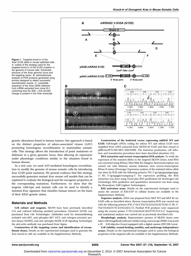
Figure 1. Targeted knock-in of theKras G12D allele in mouse epithelial cells.A, outline of the strategy used for thetargeted knock-in of the G12D mutation inthe genome of mouse cells. Schematicstructure of the Kras genomic locus andthe targeting vector. B, electrophoresisanalysis of PCR products generated usingprimers designed to detect successfulrecombination events. C, nucleotidesequence of the Kras cDNA obtainedfrom mRNA extracted from clone KI-1confirming that the 35G > G/A (G12D)change is present in the Kras transcript.
Knock-in of Oncogenic Kras in Mouse Somatic Cells
www.aacrjournals.org 8469 Cancer Res 2007; 67: (18). September 15, 2007
Research. on December 2, 2014. © 2007 American Association for Cancercancerres.aacrjournals.org Downloaded from
Results
Construction of the AAV Kras KI vector. AAV is a human,replication-defective parvovirus. The wild-type genome possessestwo open reading frames, termed rep and cap, flanked by twoinverted terminal repeats. We built a recombinant AAV, in whichboth of the open reading frames were deleted and replaced withexogenous mouse Kras sequences. The inverted terminal repeats,necessary for the packaging of the vector, were the only elementsmaintained from the wild-type virus. The homologous recombina-tion cassette cloned within the inverted terminal repeats consistedof two 1 kb sequences (‘‘homology arms’’), one of which containedthe mutated Kras exon. The selectable marker neomycin trans-ferase gene was introduced between the homology arms. The Neocassette was flanked by two LoxP sites to allow Cre recombinase–mediated excision of the Neo cassette from the targeted cells’genome (Fig. 1A). The regions of homology of the Kras locus wereamplified from genomic DNA obtained from the target mousesomatic cells (MLP29 cells; see below). The Kras G12D mutationwas introduced in the exon containing homology arm by standardsite-directed mutagenesis (Fig. 1A). Details on the construction ofinfective AAV carrying the Kras homology arms are reported inMaterials and Methods.Knock-in of the Kras G12D mutation in mouse liver
progenitor cells. As a recipient cellular model to test ourknock-in approach, we selected mouse liver progenitor cells(MLP29). MLP29 are somatic mouse cells previously derived fromembryonic liver (14). MLP29 display a number of features makingthem appealing for genetic and biological manipulation. They canbe propagated indefinitely in vitro but are not oncogenic.Furthermore, they can be used to assess cellular phenotypesincluding growth factor–dependent proliferation, motility, and‘‘invasive growth’’ (14). Their transcriptional profile has beenpreviously ascertained, highlighting genes involved in cancerprogression (17).KRAS is mutated at a very high frequency in cholangiocarcinomas
which originate from liver biliary ducts (3). MLP29 cells are bipotentprogenitors expressing markers of both hepatocytic and bile ductlineage, which makes them an appealing model to study KRAS-mediated carcinogenesis (14). We also verified that the Krassequence is wild-type in MLP29 cells, confirming that they are asuitable model for genetic manipulation of the correspondinggenomic locus.
MLP29 cells were infected with the recombinant AAV-Kras-G12Dvector described above and selected for 21 days in the presence ofneomycin until single resistant colonies appeared. Clones in whichhomologous recombination had occurred were identified by PCRanalyses on the corresponding genomic DNA as described inFig. 1B . Targeting frequencies were 1:300. Correct targeting of theKras point mutation into the corresponding genomic locus wasverified by sequencing at the DNA level (data not shown) and at themRNA level (Fig. 1C). PCR-positive clones displayed a heterozygousG > A nucleotide change corresponding to a glycine to glutamicacid modification at codon 12 of the Kras coding sequence(Fig. 1C).
Two independently identified clones (denominated KI-1 andKI-2) carrying the Kras G12D mutation were amplified andsubjected to further analysis. Interestingly, one of the clones(referred to as NEO+) that scored positively at the PCR screening,did not display the G > A nucleotide change. This indicates that thehomologous recombination had involved the NEO cassette but only
a portion of the 5¶ homology arm (Supplementary Fig. S1). Wereasoned that clone NEO+ differs from clones KI-1 and KI-2 onlyfor the Kras mutation, and would therefore represent the idealisogenic control for the mutated clones. The NEO+ clone wastherefore amplified and used throughout the study.
To exclude the possibility that the Neo resistance cassette mightaffect the expression of the mutated allele purified recombinant TAT-CRE protein was used to excise the Neo cassette using the flankingloxP sites (Supplementary Fig. S2A). Allele-specific quantitative PCRwas then used to assess the expression of the mutant G12D allele(Supplementary Fig. S2B). These experiments showed that thepresence of the Neo cassette did not negatively affect the expressionof the mutated allele. The ‘‘cre-out’’ cells were included as controls insome of the biochemical and biological experiments (see below).Kras knock-in cells display distinct biochemical and
biological features. The Kras protein cycles between the activeGTP-bound and the inactive GDP-bound conformations. The G12Doncogenic mutation is thought to lock Kras into the GTP-bound
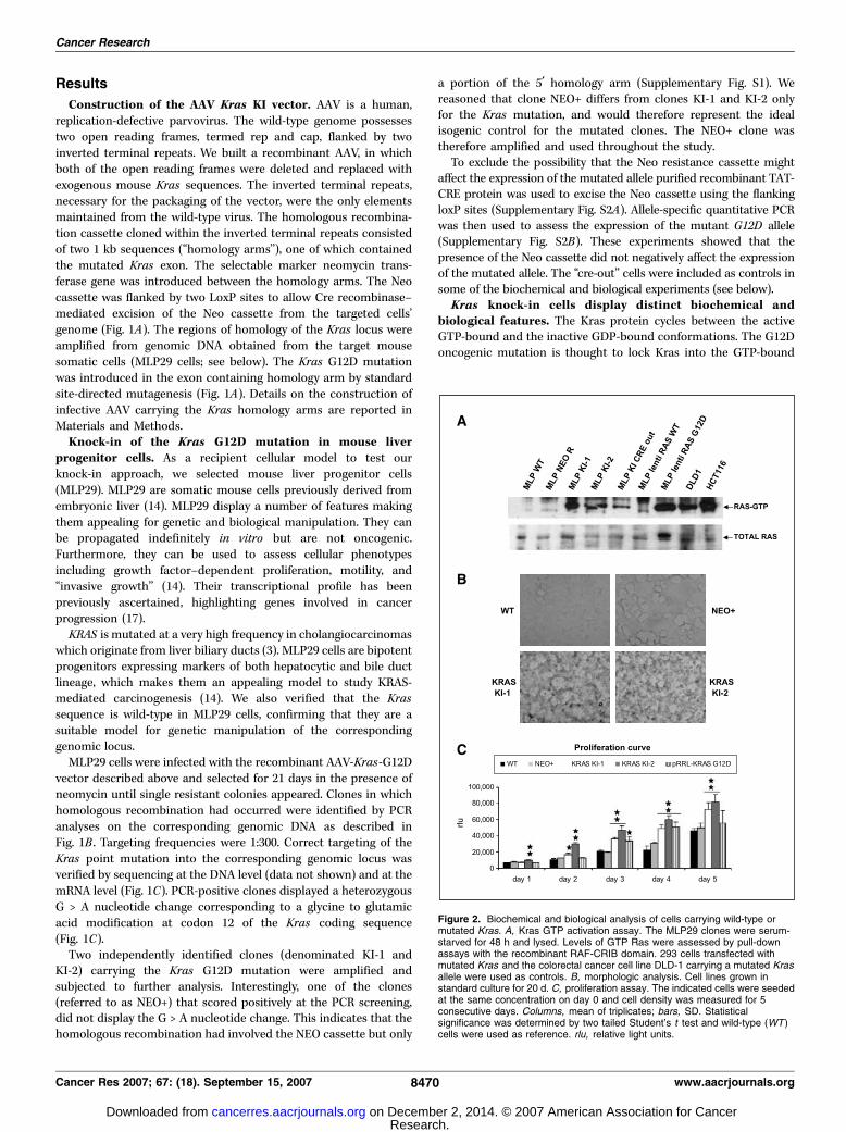
Figure 2. Biochemical and biological analysis of cells carrying wild-type ormutated Kras. A, Kras GTP activation assay. The MLP29 clones were serum-starved for 48 h and lysed. Levels of GTP Ras were assessed by pull-downassays with the recombinant RAF-CRIB domain. 293 cells transfected withmutated Kras and the colorectal cancer cell line DLD-1 carrying a mutated Krasallele were used as controls. B, morphologic analysis. Cell lines grown instandard culture for 20 d. C, proliferation assay. The indicated cells were seededat the same concentration on day 0 and cell density was measured for 5consecutive days. Columns, mean of triplicates; bars, SD. Statisticalsignificance was determined by two tailed Student’s t test and wild-type (WT )cells were used as reference. rlu, relative light units.
Cancer Research
Cancer Res 2007; 67: (18). September 15, 2007 8470 www.aacrjournals.org
Research. on December 2, 2014. © 2007 American Association for Cancercancerres.aacrjournals.org Downloaded from
conformation by reducing its constitutive GTPase activity, andthus, constitutively activating Ras signaling. To assess whether theknock-in of the Kras G12D mutation would activate the Krasprotein in MLP29 cells, we did a pull-down assay using therecombinant CRIB domain of BRAF, which is known to bind Rasonly in its GTP-bound conformation. Figure 2A shows that bothmutant clones (KI-1 and KI-2) display constitutively active Kras,whereas in the matched wild-type cells and in the perfectly isogenicNEO+ clone, Kras is inactive.
Next, we compared the levels of Kras activation in the KI cloneswith those present in naturally occurring colorectal cancer cellscarrying the corresponding mutations. The levels of Kras activationin mouse KI cells were comparable with those present in humancancer cells (Fig. 2A). Interestingly, when we ectopically expressed(by lentiviral-mediated transduction) a mutated Kras G12D cDNA,the targeted cells displayed levels of GTP-RAS higher than thosepresent in the knock-in or in the cancer cells (Fig. 2A). Overall,these data show that the knock-in system directly phenocopies theoncogenic activation of KRAS present in human tumors.
We then did biochemical analysis to assess activation of the Kraspathway in WT and knock-in cells. To this end, we measuredactivation of MAPK and AKT using anti–phospho-specific anti-bodies (Supplementary Fig. S3). The results indicate that knock-inof the G12D allele does not affect or even reduce the activation ofthe RAS downstream signaling pathways. These data are inaccordance with previous work by Guerra et al. and Tuvesonet al., which also reported that knock-in of mutated Kras in mousecells does not increase (but can actually reduce) the levels of MAPKactivation (12, 18).
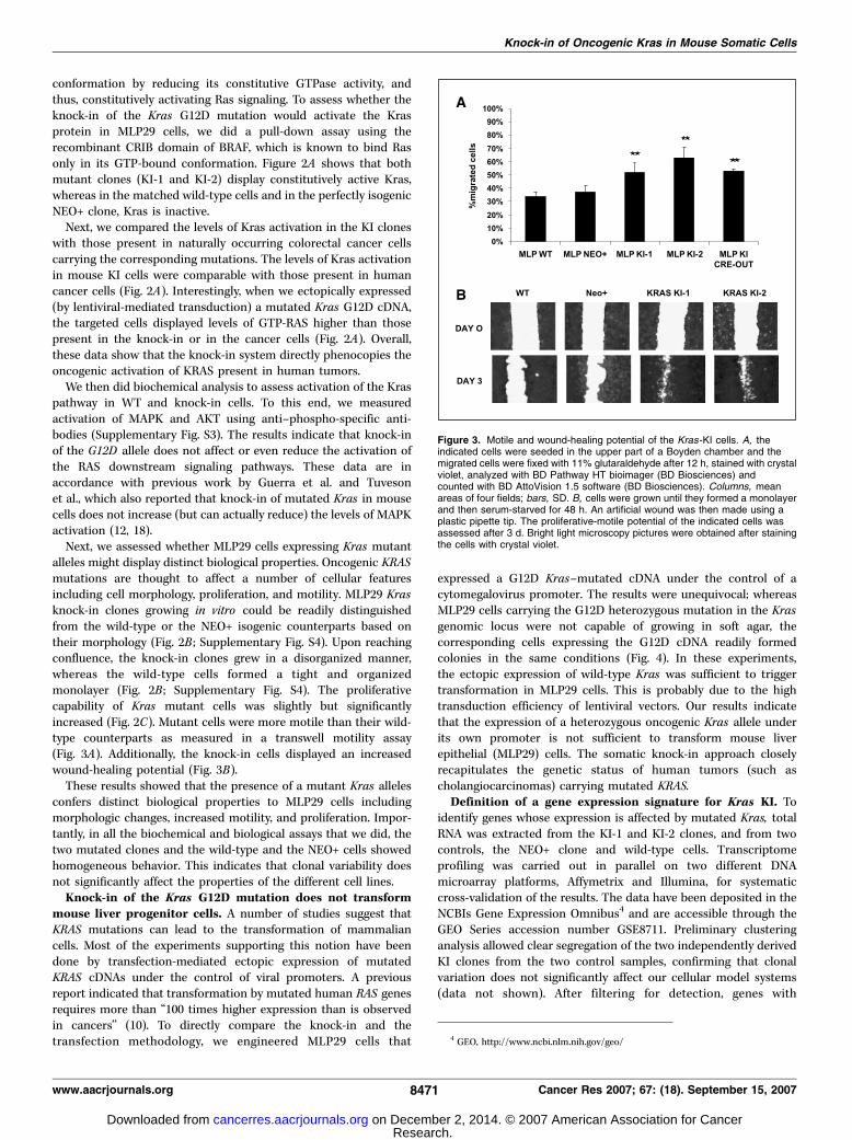
Next, we assessed whether MLP29 cells expressing Kras mutantalleles might display distinct biological properties. Oncogenic KRASmutations are thought to affect a number of cellular featuresincluding cell morphology, proliferation, and motility. MLP29 Krasknock-in clones growing in vitro could be readily distinguishedfrom the wild-type or the NEO+ isogenic counterparts based ontheir morphology (Fig. 2B ; Supplementary Fig. S4). Upon reachingconfluence, the knock-in clones grew in a disorganized manner,whereas the wild-type cells formed a tight and organizedmonolayer (Fig. 2B ; Supplementary Fig. S4). The proliferativecapability of Kras mutant cells was slightly but significantlyincreased (Fig. 2C). Mutant cells were more motile than their wild-type counterparts as measured in a transwell motility assay(Fig. 3A). Additionally, the knock-in cells displayed an increasedwound-healing potential (Fig. 3B).
These results showed that the presence of a mutant Kras allelesconfers distinct biological properties to MLP29 cells includingmorphologic changes, increased motility, and proliferation. Impor-tantly, in all the biochemical and biological assays that we did, thetwo mutated clones and the wild-type and the NEO+ cells showedhomogeneous behavior. This indicates that clonal variability doesnot significantly affect the properties of the different cell lines.Knock-in of the Kras G12D mutation does not transform
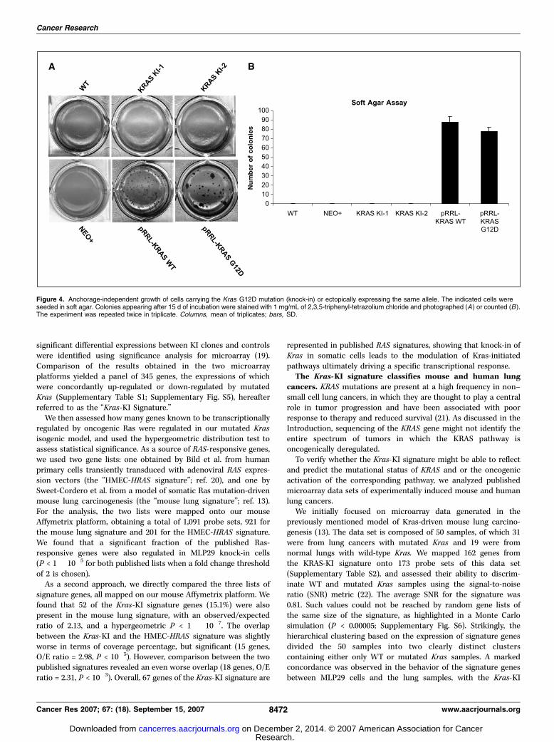
mouse liver progenitor cells. A number of studies suggest thatKRAS mutations can lead to the transformation of mammaliancells. Most of the experiments supporting this notion have beendone by transfection-mediated ectopic expression of mutatedKRAS cDNAs under the control of viral promoters. A previousreport indicated that transformation by mutated human RAS genesrequires more than ‘‘100 times higher expression than is observedin cancers’’ (10). To directly compare the knock-in and thetransfection methodology, we engineered MLP29 cells that
expressed a G12D Kras–mutated cDNA under the control of acytomegalovirus promoter. The results were unequivocal; whereasMLP29 cells carrying the G12D heterozygous mutation in the Krasgenomic locus were not capable of growing in soft agar, thecorresponding cells expressing the G12D cDNA readily formedcolonies in the same conditions (Fig. 4). In these experiments,the ectopic expression of wild-type Kras was sufficient to triggertransformation in MLP29 cells. This is probably due to the hightransduction efficiency of lentiviral vectors. Our results indicatethat the expression of a heterozygous oncogenic Kras allele underits own promoter is not sufficient to transform mouse liverepithelial (MLP29) cells. The somatic knock-in approach closelyrecapitulates the genetic status of human tumors (such ascholangiocarcinomas) carrying mutated KRAS.Definition of a gene expression signature for Kras KI. To
identify genes whose expression is affected by mutated Kras, totalRNA was extracted from the KI-1 and KI-2 clones, and from twocontrols, the NEO+ clone and wild-type cells. Transcriptomeprofiling was carried out in parallel on two different DNAmicroarray platforms, Affymetrix and Illumina, for systematiccross-validation of the results. The data have been deposited in theNCBIs Gene Expression Omnibus4 and are accessible through theGEO Series accession number GSE8711. Preliminary clusteringanalysis allowed clear segregation of the two independently derivedKI clones from the two control samples, confirming that clonalvariation does not significantly affect our cellular model systems(data not shown). After filtering for detection, genes with
Figure 3. Motile and wound-healing potential of the Kras-KI cells. A, theindicated cells were seeded in the upper part of a Boyden chamber and themigrated cells were fixed with 11% glutaraldehyde after 12 h, stained with crystalviolet, analyzed with BD Pathway HT bioimager (BD Biosciences) andcounted with BD AttoVision 1.5 software (BD Biosciences). Columns, meanareas of four fields; bars, SD. B, cells were grown until they formed a monolayerand then serum-starved for 48 h. An artificial wound was then made using aplastic pipette tip. The proliferative-motile potential of the indicated cells wasassessed after 3 d. Bright light microscopy pictures were obtained after stainingthe cells with crystal violet.
4 GEO, http://www.ncbi.nlm.nih.gov/geo/
Knock-in of Oncogenic Kras in Mouse Somatic Cells
www.aacrjournals.org 8471 Cancer Res 2007; 67: (18). September 15, 2007
Research. on December 2, 2014. © 2007 American Association for Cancercancerres.aacrjournals.org Downloaded from
significant differential expressions between KI clones and controlswere identified using significance analysis for microarray (19).Comparison of the results obtained in the two microarrayplatforms yielded a panel of 345 genes, the expressions of whichwere concordantly up-regulated or down-regulated by mutatedKras (Supplementary Table S1; Supplementary Fig. S5), hereafterreferred to as the ‘‘Kras-KI Signature.’’
We then assessed how many genes known to be transcriptionallyregulated by oncogenic Ras were regulated in our mutated Krasisogenic model, and used the hypergeometric distribution test toassess statistical significance. As a source of RAS-responsive genes,we used two gene lists: one obtained by Bild et al. from humanprimary cells transiently transduced with adenoviral RAS expres-sion vectors (the ‘‘HMEC-HRAS signature’’; ref. 20), and one bySweet-Cordero et al. from a model of somatic Ras mutation-drivenmouse lung carcinogenesis (the ‘‘mouse lung signature’’; ref. 13).For the analysis, the two lists were mapped onto our mouseAffymetrix platform, obtaining a total of 1,091 probe sets, 921 forthe mouse lung signature and 201 for the HMEC-HRAS signature.We found that a significant fraction of the published Ras-responsive genes were also regulated in MLP29 knock-in cells(P < 1 � 10�5 for both published lists when a fold change thresholdof 2 is chosen).
As a second approach, we directly compared the three lists ofsignature genes, all mapped on our mouse Affymetrix platform. Wefound that 52 of the Kras-KI signature genes (15.1%) were alsopresent in the mouse lung signature, with an observed/expectedratio of 2.13, and a hypergeometric P < 1 � 10�7. The overlapbetween the Kras-KI and the HMEC-HRAS signature was slightlyworse in terms of coverage percentage, but significant (15 genes,O/E ratio = 2.98, P < 10�5). However, comparison between the twopublished signatures revealed an even worse overlap (18 genes, O/Eratio = 2.31, P < 10�3). Overall, 67 genes of the Kras-KI signature are
represented in published RAS signatures, showing that knock-in ofKras in somatic cells leads to the modulation of Kras-initiatedpathways ultimately driving a specific transcriptional response.The Kras-KI signature classifies mouse and human lung
cancers. KRAS mutations are present at a high frequency in non–small cell lung cancers, in which they are thought to play a centralrole in tumor progression and have been associated with poorresponse to therapy and reduced survival (21). As discussed in theIntroduction, sequencing of the KRAS gene might not identify theentire spectrum of tumors in which the KRAS pathway isoncogenically deregulated.
To verify whether the Kras-KI signature might be able to reflectand predict the mutational status of KRAS and or the oncogenicactivation of the corresponding pathway, we analyzed publishedmicroarray data sets of experimentally induced mouse and humanlung cancers.
We initially focused on microarray data generated in thepreviously mentioned model of Kras-driven mouse lung carcino-genesis (13). The data set is composed of 50 samples, of which 31were from lung cancers with mutated Kras and 19 were fromnormal lungs with wild-type Kras. We mapped 162 genes fromthe KRAS-KI signature onto 173 probe sets of this data set(Supplementary Table S2), and assessed their ability to discrim-inate WT and mutated Kras samples using the signal-to-noiseratio (SNR) metric (22). The average SNR for the signature was0.81. Such values could not be reached by random gene lists ofthe same size of the signature, as highlighted in a Monte Carlosimulation (P < 0.00005; Supplementary Fig. S6). Strikingly, thehierarchical clustering based on the expression of signature genesdivided the 50 samples into two clearly distinct clusterscontaining either only WT or mutated Kras samples. A markedconcordance was observed in the behavior of the signature genesbetween MLP29 cells and the lung samples, with the Kras-KI
Figure 4. Anchorage-independent growth of cells carrying the Kras G12D mutation (knock-in) or ectopically expressing the same allele. The indicated cells wereseeded in soft agar. Colonies appearing after 15 d of incubation were stained with 1 mg/mL of 2,3,5-triphenyl-tetrazolium chloride and photographed (A) or counted (B ).The experiment was repeated twice in triplicate. Columns, mean of triplicates; bars, SD.
Cancer Research
Cancer Res 2007; 67: (18). September 15, 2007 8472 www.aacrjournals.org
Research. on December 2, 2014. © 2007 American Association for Cancercancerres.aacrjournals.org Downloaded from
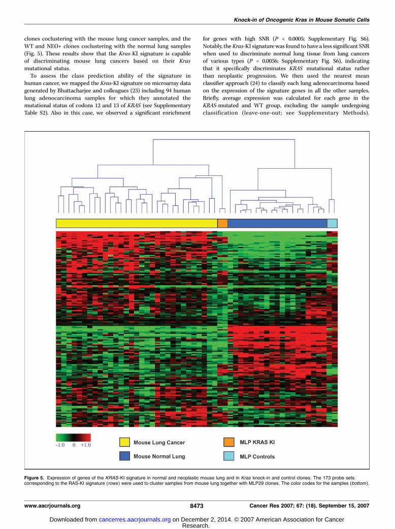
clones coclustering with the mouse lung cancer samples, and theWT and NEO+ clones coclustering with the normal lung samples(Fig. 5). These results show that the Kras-KI signature is capableof discriminating mouse lung cancers based on their Krasmutational status.
To assess the class prediction ability of the signature inhuman cancer, we mapped the Kras-KI signature on microarray datagenerated by Bhattacharjee and colleagues (23) including 94 humanlung adenocarcinoma samples for which they annotated themutational status of codons 12 and 13 of KRAS (see SupplementaryTable S2). Also in this case, we observed a significant enrichment
for genes with high SNR (P < 0.0005; Supplementary Fig. S6).Notably, theKras-KI signature was found to have a less significant SNRwhen used to discriminate normal lung tissue from lung cancersof various types (P = 0.0056; Supplementary Fig. S6), indicatingthat it specifically discriminates KRAS mutational status ratherthan neoplastic progression. We then used the nearest meanclassifier approach (24) to classify each lung adenocarcinoma basedon the expression of the signature genes in all the other samples.Briefly, average expression was calculated for each gene in theKRAS-mutated and WT group, excluding the sample undergoingclassification (leave-one-out; see Supplementary Methods).
Figure 5. Expression of genes of the KRAS -KI signature in normal and neoplastic mouse lung and in Kras knock-in and control clones. The 173 probe setscorresponding to the RAS-KI signature (rows ) were used to cluster samples from mouse lung together with MLP29 clones. The color codes for the samples (bottom ).
Knock-in of Oncogenic Kras in Mouse Somatic Cells
www.aacrjournals.org 8473 Cancer Res 2007; 67: (18). September 15, 2007
Research. on December 2, 2014. © 2007 American Association for Cancercancerres.aacrjournals.org Downloaded from

For comparison, we adopted the same classification procedure usinggenes from the two previously mentioned Ras signatures obtained inthe mouse lung model (mouse lung signature; ref. 13) and in HMECs(HMEC-HRAS signature; ref. 20), respectively. The results of thisclassification are presented in Table 1, and show that all threesignatures have a very similar performance. These data suggest that,albeit nontransforming, knock-in of mutated Kras in mouse cellsgenerates a transcriptional signature, the capability of which inpredicting the presence of KRAS mutations in human lung cancer iscomparable to those previously published.
Discussion
Omics technologies and the availability of the human genomesequence have allowed unprecedented progress in the identifica-tion of genes mutated in human cancers, leading to the detectionof hundreds of cancer-associated alleles (16, 25, 26). Comparedwith the genomic discovery stage, the functional validation phaseof the corresponding alleles is now lagging behind. For example,hundreds of point mutations affecting kinase genes have beenrecently described (16, 25, 26), but their biochemical and biologicalproperties are largely unknown.
Mammalian cell lines and mouse strains have been widely usedas model systems to functionally characterize cancer allelescarrying point mutations or deletions. In the first case, the cDNAcorresponding to the mutated alleles are ectopically expressed bymeans of plasmid transfection or retroviral infection in human ormouse cell lines upon selection with markers. The derivative cellsare then used to assess the biochemical and biological properties ofthe mutated cDNA with a variety of standardized assays. Classicexamples of this approach are the focus-forming assay, whichmeasures loss of contact inhibition, and the soft agar growth
assay, which evaluates the anchorage-independent potential ofneoplastic cells.
These studies have yielded remarkable results but are typicallyhampered by at least three issues. First, the expression is achievedby transient or stable transfection of cDNAs often resulting in theoverexpression of the target allele at levels that do not accuratelyrecapitulate what happens in human cancers. Second, theexpression of the mutated cDNA is mainly achieved under thecontrol of non–endogenous viral constitutively active promoterssuch as SV40, cytomegalovirus, and long terminal repeats (27). As aresult, the mutated alleles cannot be appropriately modulated inthe target cells. Finally, functional experiments have often beendone using cells of stromal origin such as embryonal or 3T3 mousefibroblasts. Stromal cells are not the primary targets of oncogenictransformation in the large majority of solid cancers.
Another approach has been to use homologous recombinationto introduce the point mutations in a locus-specific fashion intothe genome of mouse embryonic stem cells. This strategy worksvery efficiently and allows the generation of mouse strains carryingspecific cancer alleles. Very often, however, mutated dominantoncogenes (such as Kras) cannot be transmitted through themouse germ line (28) because the resulting embryos are not viable.Complex genetic strategies (such as the use of tissue-specific CRE-LOX systems, or latent alleles) have been devised to allow theexpression of the mutated alleles only in adult mouse somatictissues (12, 18). These have been used for a variety of experimentalapproaches including the demonstration that oncogenic KRASalleles can immortalize primary fibroblasts (12, 18).
To overcome these limitations, we exploited a novel approach tointroduce cancer alleles in the genome of mouse epithelial somaticcells. Similar to most mammalian somatic cells, most adult mousecells display a low frequency of homologous recombination thathas thus far limited their use for genetic and functional analysis. Toovercome this problem, we used a shuttle vector based on thebackbone of AAVs. The latter has been found to increase the rate oftargeted homologous recombination in human and mouse cells(29, 30).
As a model system to test our strategy, we chose the KrasG12D mutation, which is commonly found in human tumors suchas colorectal, lung and pancreatic cancers.5 Mutated KRASproteins are, at least in theory, good therapeutic targets becausethey act as central switches in cellular processes leading tooncogenesis (2). Despite many efforts, the development ofinhibitors directly targeting KRAS has thus far failed, apparentlydue to difficulties in targeting the intrinsic RAS GTPase activityor the lack of specificity of farnesyltransferase inhibitors (31). Thedevelopment of cellular and animal models that more closelyrecapitulate human diseases is a prerequisite to identifyingmolecules inhibiting the Kras signaling pathway rather than Krasitself.
AAV-mediated knock-in of the Kras G12D mutation wasattempted in mouse liver progenitor cells (MLP29), a somaticimmortal epithelial cell line previously established in ourlaboratory that is not transformed and does not form tumors inimmunocompromised mice.
Clones displaying correct targeting of the Kras genomic locuswere identified at a frequency of 1:300. Knock-in cells displayed
Table 1. Classification of human lung cancer samples bythe KRAS-KI, mouse lung, and HMEC signatures
KRAS mutational status Wild-type Mutated Total
Knock-in signature class predictionWild-type 31 5 36
Mutated 29 29 58
Total 60 34 94
Error (%) 48.3 14.7 36.1HMEC signature class prediction
Wild-type 33 5 38
Mutated 27 29 56
Total 60 34 94Error (%) 45.0 14.7 34.0
Murine lung signature class prediction
Wild-type 33 8 41Mutated 27 26 53
Total 60 34 94
Error (%) 45.0 23.5 37.2
NOTE: Nearest-mean classification with leave-one-out was used toclassify 94 adenocarcinoma samples according to the Kras-KI, mouse
lung, and HMEC signatures expression. The results of the classifica-
tion are compared with the results of KRAS sequencing at positions 12and 13.
5 http://www.sanger.ac.uk/genetics/CGP/cosmic/
Cancer Research
Cancer Res 2007; 67: (18). September 15, 2007 8474 www.aacrjournals.org
Research. on December 2, 2014. © 2007 American Association for Cancercancerres.aacrjournals.org Downloaded from
constitutively active Kras protein, marked morphologic changesand increased proliferation and motility. Interestingly, these cellswere not transformed as measured by the soft agar assay.Importantly, targeted cells did not show oncogene-inducedsenescence and were not transformed in contrast with resultspreviously reported for mouse cells overexpressing a mutatedHRAS cDNA (32). We confirmed that in our cell models,overexpression of mutated Kras cDNA led to full transformation,indicating that the levels of Kras activation are critical for thisphenotype. Amplification and/or overexpression of mutated KRASis very uncommon in human cancers, suggesting that the levels ofKRAS activation relevant for tumor progression are those achievedby the presence of point mutations in a heterozygous state. Inline with this, the levels of Kras activation (as measured by a GTPload assay) in the knock-in cells were highly comparable withthose found in naturally occurring tumor cells carrying thecorresponding heterozygous mutations.
Our results are the first to show the direct introduction of anoncogenic mutation (such as the Kras G12D allele) in the genomeof mouse epithelial somatic cells. Notably, the biochemical andtransforming properties of our knock-in cell models are remarkablysimilar to those obtained from cells derived from mice carryinganalogous Kras alleles (12, 18). We conclude that the isogenic cellsthat we have developed recapitulate the oncogenic activation ofKRAS occurring in human tumors. To further characterize andvalidate our cellular models, we did transcriptional profiling of thewild-type and knock-in cells using two independent microarrayplatforms that resulted in a distinct Kras KI signature of 345 genes.The signature was then tested for its ability to classify mouse andhuman cancers in which the mutational status of KRAS was alsoknown. The signature unequivocally distinguished mouse lungsamples carrying the G12D mutation from the correspondingnormal tissues. When applied to a large cohort of human non–small cell lung tumors for which the transcriptional profile wasavailable, the signature was able to correctly predict the wild-typestatus of the KRAS locus in most cases. Interestingly, a largefraction of cases displaying a wild-type KRAS were instead definedas ‘‘mutant’’ by the signature. We therefore used two additional Ras
signatures, obtained in mouse and human models alongside withthat obtained in the mouse KI cells to compare their performance,and the results were very similar.
There are two possible interpretations for these results, (a) thesignatures might have misclassified the wild-type samples due topoor specificity or (b) the signatures detected the functional statusof KRAS hyperactivation, possibly deriving from mutations at KRAScodons other than 12 and 13 or involving activation of signalingmolecules acting in the same pathway. The latter hypothesis isconsistent with the notion that molecules acting upstream ordownstream of Ras could also be deregulated in cancer (2). How-ever, further investigations are required to address this issue.
In conclusion, we have developed a new cellular model closelyrecapitulating the KRAS genetic lesions occurring in humancancers. Our findings support the concept that KRAS mutations,in the absence of overexpression, promote distinct biologicaleffects and drive a specific transcriptional response, but are notsufficient to trigger full transformation in epithelial cells.
Given the knock-in efficiency that we observed, our work pointsto new experimental strategies to build tumor progression modelsby ‘‘in vivo delivering’’ of oncogenic mutations using AAV-mediatedhomologous recombination in mouse somatic cells. Consideringthe recent successes and the unique phenotypes seen with mosaicconditional knockout mouse models of cancer (33, 34), we alsoenvision that orthotopic transplantation of genetically manipulatedsomatic murine cells would provide a physiologically relevantmodel of human carcinogenesis.
Acknowledgments
Received 3/26/2007; revised 6/7/2007; accepted 7/12/2007.Grant support: Italian Association for Cancer Research, Italian Ministry of Health,
Italian Ministry of University and Research, Regione Piemonte, Compagnia di S. PaoloFoundation, Association for International Cancer Research (AICR-UK), EU FP6contract MCSC 037297 and TRANSlational and Functional Onco-Genomics (TRANS-FOG) CEE n. 503438.
The costs of publication of this article were defrayed in part by the payment of pagecharges. This article must therefore be hereby marked advertisement in accordancewith 18 U.S.C. Section 1734 solely to indicate this fact.
We thank Drs. Chris Torrance for helpful discussions, Luca Cardone for criticalreading the manuscript, and Carlo Zanon for support with the DNA sequencing.
References1. Vetter IR, Wittinghofer A. The guanine nucleotide-binding switch in three dimensions. Science 2001;294:1299–304.2. Malumbres M, Barbacid M. RAS oncogenes: the first 30years. Nat Rev Cancer 2003;3:459–65.3. Ohashi K, Tstsumi M, Nakajima Y, Nakano H, KonishiY. Ki-ras point mutations and proliferation activity inbiliary tract carcinomas. Br J Cancer 1996;74:930–5.4. Schubbert S, Bollag G, Shannon K. Deregulated Rassignaling in developmental disorders: new tricks for anold dog. Curr Opin Genet Dev 2007;17:15–22.5. Perona R, Sanchez-Perez I. Control of oncogenesis andcancer therapy resistance. Br J Cancer 2004;90:573–7.6. Bernhard EJ, Stanbridge EJ, Gupta S, et al. Directevidence for the contribution of activated N-ras and K-ras oncogenes to increased intrinsic radiation resistancein human tumor cell lines. Cancer Res 2000;60:6597–600.7. Schubbert S, Shannon K, Bollag G. Hyperactive Ras indevelopmental disorders and cancer. Nat Rev Cancer2007;7:295–308.8. Lundberg AS, Randell SH, Stewart SA, et al.Immortalization and transformation of primary humanairway epithelial cells by gene transfer. Oncogene 2002;21:4577–86.
9. Vitale-Cross L, Amornphimoltham P, Fisher G,Molinolo AA, Gutkind JS. Conditional expression ofK-ras in an epithelial compartment that includes thestem cells is sufficient to promote squamous cellcarcinogenesis. Cancer Res 2004;64:8804–7.10. Hua VY, Wang WK, Duesberg PH. Dominanttransformation by mutated human ras genes in vitrorequires more than 100 times higher expression than isobserved in cancers. Proc Natl Acad Sci U S A 1997;94:9614–9.11. Johnson L, Mercer K, Greenbaum D, et al. Somaticactivation of the K-ras oncogene causes early onset lungcancer in mice. Nature 2001;410:1111–6.12. Guerra C, Mijimolle N, Dhawahir A, et al. Tumorinduction by an endogenous K-ras oncogene is highlydependent on cellular context. Cancer Cell 2003;4:111–20.13. Sweet-Cordero A, Mukherjee S, Subramanian A, et al.An oncogenic KRAS2 expression signature identified bycross-species gene-expression analysis. Nat Genet 2005;37:48–55.14. Medico E, Mongiovi AM, Huff J, et al. The tyrosinekinase receptors Ron and Sea control ‘‘scattering’’ andmorphogenesis of liver progenitor cells in vitro . Mol BiolCell 1996;7:495–504.15. Vigna E, Naldini L. Lentiviral vectors: excellent
tools for experimental gene transfer and promisingcandidates for gene therapy. J Gene Med 2000;2:308–16.16. Bardelli A, Parsons DW, Silliman N, et al. Mutationalanalysis of the tyrosine kinome in colorectal cancers.Science 2003;300:949.17. Medico E, Gentile A, Lo Celso C, et al. Osteopontin isan autocrine mediator of hepatocyte growth factor-induced invasive growth. Cancer Res 2001;61:5861–8.18. Tuveson DA, Shaw AT, Willis NA, et al. Endogenousoncogenic K-ras(G12D) stimulates proliferation andwidespread neoplastic and developmental defects.Cancer Cell 2004;5:375–87.19. Tusher VG, Tibshirani R, Chu G. Significance analysisof microarrays applied to the ionizing radiationresponse. Proc Natl Acad Sci U S A 2001;98:5116–21.20. Bild AH, Yao G, Chang JT, et al. Oncogenic pathwaysignatures in human cancers as a guide to targetedtherapies. Nature 2006;439:353–7.21. Aviel-Ronen S, Blackhall FH, Shepherd FA, Tsao MS.K-ras mutations in non-small-cell lung carcinoma: areview. Clin Lung Cancer 2006;8:30–8.22. Golub TR, Slonim DK, Tamayo P, et al. Molecularclassification of cancer: class discovery and classprediction by gene expression monitoring. Science1999;286:531–7.
Knock-in of Oncogenic Kras in Mouse Somatic Cells
www.aacrjournals.org 8475 Cancer Res 2007; 67: (18). September 15, 2007
Research. on December 2, 2014. © 2007 American Association for Cancercancerres.aacrjournals.org Downloaded from

Cancer Research
Cancer Res 2007; 67: (18). September 15, 2007 8476 www.aacrjournals.org
23. Bhattacharjee A, Richards WG, Staunton J, et al.Classification of human lung carcinomas by mRNAexpression profiling reveals distinct adenocarcinomasubclasses. Proc Natl Acad Sci U S A 2001;98:13790–5.24. Wessels LF, Reinders MJ, Hart AA, et al. A protocol forbuilding and evaluating predictors of disease state basedon microarray data. Bioinformatics 2005;21:3755–62.25. Sjoblom T, Jones S, Wood LD, et al. The consensuscoding sequences of human breast and colorectalcancers. Science 2006;314:268–74.26. Greenman C, Stephens P, Smith R, et al. Patterns ofsomatic mutation in human cancer genomes. Nature2007;446:153–8.
27. Hahn WC, Counter CM, Lundberg AS, BeijersbergenRL, Brooks MW, Weinberg RA. Creation of humantumour cells with defined genetic elements. Nature1999;400:464–8.28. Kuhn R, Schwenk F. Advances in gene targetingmethods. Curr Opin Immunol 1997;9:183–8.29. Russell DW, Hirata RK, Inoue N. Validation ofAAV-mediated gene targeting. Nat Biotechnol 2002;20:658.30. Kohli M, Rago C, Lengauer C, Kinzler KW, VogelsteinB. Facile methods for generating human somatic cellgene knockouts using recombinant adeno-associatedviruses. Nucleic Acids Res 2004;32:e3.
31. Downward J. Targeting RAS signalling pathways incancer therapy. Nat Rev Cancer 2003;3:11–22.32. Serrano M, Lin AW, McCurrach ME, Beach D, LoweSW. Oncogenic ras provokes premature cell senescenceassociated with accumulation of p53 and p16INK4a. Cell1997;88:593–602.33. Muzumdar MD, Luo L, Zong H. Modeling sporadicloss of heterozygosity in mice by using mosaic analysiswith double markers (MADM). Proc Natl Acad Sci U S A2007;104:4495–500.34. Wang W, Warren M, Bradley A. Induced mitoticrecombination of p53 in vivo . Proc Natl Acad Sci U S A2007;104:4501–5.
Research. on December 2, 2014. © 2007 American Association for Cancercancerres.aacrjournals.org Downloaded from
2007;67:8468-8476. Cancer Res Sabrina Arena, Claudio Isella, Miriam Martini, et al. Classifies Human CancersSomatic Cells But Triggers a Transcriptional Response that
Does Not Transform MouseKrasKnock-in of Oncogenic
Updated version
http://cancerres.aacrjournals.org/content/67/18/8468
Access the most recent version of this article at:
Material
Supplementary
http://cancerres.aacrjournals.org/content/suppl/2007/09/17/67.18.8468.DC1.html
Access the most recent supplemental material at:
Cited Articles
http://cancerres.aacrjournals.org/content/67/18/8468.full.html#ref-list-1
This article cites by 34 articles, 15 of which you can access for free at:
Citing articles
http://cancerres.aacrjournals.org/content/67/18/8468.full.html#related-urls
This article has been cited by 5 HighWire-hosted articles. Access the articles at:
E-mail alerts related to this article or journal.Sign up to receive free email-alerts
Subscriptions
Reprints and
To order reprints of this article or to subscribe to the journal, contact the AACR Publications
Permissions
To request permission to re-use all or part of this article, contact the AACR Publications
Research. on December 2, 2014. © 2007 American Association for Cancercancerres.aacrjournals.org Downloaded from
Copyright © 2022 FDOKUMEN