Kitasatospora saccharophila sp. nov. and Kitasatospora kazusensis sp. nov., isolated from soil and...
8
Introduction The genus Kitasatospora was first proposed by Ōmura et al. (1982) for an actinomycete with the unique characteristic of containing both meso- and LL-diamino- pimelic acid (DAP) as diamino acids in the cell wall. Aerial spores on solid culture and submerged spores in liquid culture contain LL-DAP, while mycelium in both cultures contain primarily meso-DAP (Ōmura et al., 1981, 1982; Takahashi et al., 1984). In 1992, members of the genus Kitasatospora were transferred to the ge- nus Streptomyces on the basis of rRNA-targeted oligo- nucleotide probes and phenotypic properties (Wel- lington et al., 1992), and later the genus Kitasatospora J. Gen. Appl. Microbiol., 55, 19‒26 (2009) A polyphasic study was undertaken to establish the taxonomic positions of two isolates, SK15 T and SK60 T , from soil samples that were found to have morphological and chemical properties consistent with Kitasatospora strains. Phylogenetic analysis based on the 16S rRNA gene se- quence revealed that strains SK15 T and SK60 T form novel evolutionary lineages within the radia- tion of the genus Kitasatospora and share the highest 16S rRNA gene sequences similarities with their closest relatives, Kitasatospora setae IAM 15325 T (97.8%) and Kitasatospora mediocidica IAM 15162 T (97.5%), respectively. However, the results of DNA-DNA hybridization experiment and phenotypic data demonstrated that strains SK15 T and SK60 T are distinct from their closest phylogenetic neighbors and other Kitasatospora species. For chemotaxonomic characteristics, the cell-wall peptidoglycan of strains contained both meso- and LL-diaminopimelic acids as the diamino acids, the predominant quinone system was MK-9(H 6 ) and MK-9(H 8 ), whole-cell hydrol- ysates were rich in galactose, mannose and ribose, and the major fatty acids were C 16:0 , anteiso- C 15:0 , iso-C 15:0 and iso-C 16:0 . On the basis of both phenotypic and phylogenetic evidence, strains SK15 T and SK60 T were assigned to represent two novel species of the genus Kitasatospora, for which the names Kitasatospora saccharophila sp. nov. (type strain SK15 T =JCM 14559 T =KCTC 19566 T ) and Kitasatospora kazusanensis sp. nov. (type strain SK60 T =JCM 14560 T =KCTC 19565 T ) are proposed. It is also proposed that Streptomyces atroaurantiacus should be transferred to the genus Kitasatospora as Kitasatospora atroaurantiaca comb. nov. (type strain NBRC 14327 T =DSM 41649 T ). Key Words—Kitasatospora atroaurantiaca comb. nov.; Kitasatospora kazusanensis sp. nov.; Kitasa- tospora saccharophila sp. nov. Full Paper Kitasatospora saccharophila sp. nov. and Kitasatospora kazusanensis sp. nov., isolated from soil and transfer of Streptomyces atroaurantiacus to the genus Kitasatospora as Kitasatospora atroaurantiaca comb. nov. Bing Li, 1 Keiko Furihata, 1 Takuji Kudo, 2 and Akira Yokota 1,* 1 Institute of Molecular and Cellular Biosciences, The University of Tokyo, Bunkyo-ku, Tokyo 113‒0032, Japan 2 Japan Collection of Microorganisms, RIKEN Bioresource Center, Wako, Saitama 351‒0198, Japan (Received September 3, 2008; Accepted October 30, 2008) * Address reprint requests to: Dr. Akira Yokota, Institute of Molecular and Cellular Biosciences, The University of Tokyo, Yayoi 1‒1‒1, Bunkyo-ku, Tokyo 113‒0032, Japan. Tel: +81‒3‒5841‒7827 Fax: +81‒3‒5841‒8490 E-mail: [email protected]
Transcript of Kitasatospora saccharophila sp. nov. and Kitasatospora kazusensis sp. nov., isolated from soil and...

Introduction
The genus Kitasatospora was fi rst proposed by Ōmura et al. (1982) for an actinomycete with the unique
characteristic of containing both meso- and LL-diamino-pimelic acid (DAP) as diamino acids in the cell wall. Aerial spores on solid culture and submerged spores in liquid culture contain LL-DAP, while mycelium in both cultures contain primarily meso-DAP (Ōmura et al., 1981, 1982; Takahashi et al., 1984). In 1992, members of the genus Kitasatospora were transferred to the ge-nus Streptomyces on the basis of rRNA-targeted oligo-nucleotide probes and phenotypic properties (Wel-lington et al., 1992), and later the genus Kitasatospora
J. Gen. Appl. Microbiol., 55, 19‒26 (2009)
A polyphasic study was undertaken to establish the taxonomic positions of two isolates, SK15T and SK60T, from soil samples that were found to have morphological and chemical properties consistent with Kitasatospora strains. Phylogenetic analysis based on the 16S rRNA gene se-quence revealed that strains SK15T and SK60T form novel evolutionary lineages within the radia-tion of the genus Kitasatospora and share the highest 16S rRNA gene sequences similarities with their closest relatives, Kitasatospora setae IAM 15325T (97.8%) and Kitasatospora mediocidica IAM 15162T (97.5%), respectively. However, the results of DNA-DNA hybridization experiment and phenotypic data demonstrated that strains SK15T and SK60T are distinct from their closest phylogenetic neighbors and other Kitasatospora species. For chemotaxonomic characteristics, the cell-wall peptidoglycan of strains contained both meso- and LL-diaminopimelic acids as the diamino acids, the predominant quinone system was MK-9(H6) and MK-9(H8), whole-cell hydrol-ysates were rich in galactose, mannose and ribose, and the major fatty acids were C16:0, anteiso-C15:0, iso-C15:0 and iso-C16:0. On the basis of both phenotypic and phylogenetic evidence, strains SK15T and SK60T were assigned to represent two novel species of the genus Kitasatospora, for which the names Kitasatospora saccharophila sp. nov. (type strain SK15T=JCM 14559T=KCTC 19566T) and Kitasatospora kazusanensis sp. nov. (type strain SK60T=JCM 14560T=KCTC 19565T) are proposed. It is also proposed that Streptomyces atroaurantiacus should be transferred to the genus Kitasatospora as Kitasatospora atroaurantiaca comb. nov. (type strain NBRC 14327T=DSM 41649T).
Key Words—Kitasatospora atroaurantiaca comb. nov.; Kitasatospora kazusanensis sp. nov.; Kitasa-
tospora saccharophila sp. nov.
Full Paper
Kitasatospora saccharophila sp. nov. and Kitasatospora kazusanensis sp. nov., isolated from soil and transfer of Streptomyces atroaurantiacus to
the genus Kitasatospora as Kitasatospora atroaurantiaca comb. nov.
Bing Li,1 Keiko Furihata,1 Takuji Kudo,2 and Akira Yokota1,*
1 Institute of Molecular and Cellular Biosciences, The University of Tokyo, Bunkyo-ku, Tokyo 113‒0032, Japan2 Japan Collection of Microorganisms, RIKEN Bioresource Center, Wako, Saitama 351‒0198, Japan
(Received September 3, 2008; Accepted October 30, 2008)
* Address reprint requests to: Dr. Akira Yokota, Institute of Molecular and Cellular Biosciences, The University of Tokyo, Yayoi 1‒1‒1, Bunkyo-ku, Tokyo 113‒0032, Japan. Tel: +81‒3‒5841‒7827 Fax: +81‒3‒5841‒8490 E-mail: [email protected]
20 Vol. 55LI et al.
was re-established by phylogenetic study based on 16S rRNA gene sequence data (Zhang et al., 1997). The twenty species of Kitasatospora have been validly published at the time of writing with recently described species K. sampliensis (Mayilraj et al., 2006), K. viridis (Liu et al., 2005), and K. putterlickiae, K. arboriphila, K.
gansuensis, K. nipponensis, K. paranensis, and K. ter-
estris (Groth et al., 2003, 2004). The aim of the present investigation was to determine the taxonomic posi-tions of two organisms presumptively assigned to the genus Kitasatospora, strains SK15T and SK60T, using a combined polyphasic taxonomic approach. The study was also carried out to clarify the taxonomic po-sition of Streptomyces atroaurantiacus.
Materials and Methods
Actinomycete strains and isolation. Actinomycete strains SK15T and SK60T were isolated from the soils collected in Nagano City, Japan, and Kazusa of Chiba Prefecture, Japan, by using humic acid-vitamin agar (Hayakawa and Nonomura, 1987) and were main-tained on the ISP 2 medium slant at 4°C and as a glyc-erol suspension (10%, v/v) at -80°C. Morphological and biochemical tests. Morpho-logical features were observed by light and electron microscopes (model BX60, Olympus; S-2400, Hitachi, Japan). Cells were grown on oatmeal agar medium for 3 weeks at 27°C. The dimensions of the spores were measured, and samples for scanning electron micros-copy were prepared by fi xing the agar block into 1% OsO4 in 0.1 M phosphate buffer (pH 7.2) at room tem-perature for 1 h. The samples were dehydrated in a graded series of ethanol and dried by critical-point drying and then placed onto a stub bearing adhesive and spatter-coated with gold under a vacuum. For cul-tural and phenotypic characteristics, the International Streptomyces Project (ISP) media recommended by Shirling and Gottlieb (1966) were used to investigate cultural and physiological characteristics. Cultures were observed after incubation on ISP 2, ISP 3, ISP 4 and ISP 5 medium (Difco) at 27°C for 2 weeks. Colors were determined according to the Color Harmony
Manual (Taylor et al., 1958). The growth ranges for temperature and pH were tested on ISP 2 medium. Carbon-source utilization tests were determined by adding a concentration of 1% (w/v) of carbohydrate solution to ISP 9 medium (Shirling and Gottleib, 1966), and results were recorded after 7 days incubation. The
tolerance of salinity was determined by inoculating the strain into ISP 2 medium supplemented with 0‒12% (w/v) NaCl at 1% intervals. Liquefaction of gelatin, hy-drogen sulfi de production, and nitrate reduction were examined as described by Lányi (1987), and hydroly-sis of starch and peptonization of milk were deter-mined according to Cowan and Steel (1965). API ZYM strips (bioMérieux) were used to determine the en-zyme activities of 48-h-old cultures grown in liquid ISP 2 medium according to the manufacturer’s instruc-tions. Chemotaxonomic tests. Freeze-dried cells for chemo-taxonomic studies were obtained upon incubation of the organism in trypticase soy broth (TSB, BBL) me-dium on a rotary shaker at 27°C for 4 days. For cellular fatty acid analysis, the strain was grown on trypticase soy agar (TSA, BBL) at 27°C for 3 days, and fatty acid methyl esters were prepared and identifi ed using the Microbial Identifi cation System as described by Xie and Yokota (2003). The amino acid composition and isomers of diaminopimelic acid in the cell walls were examined by two-dimensional TLC (Tokyo Kasei Co., Ltd., Tokyo, Japan), as described by Harper and Davis (1979), and by HPLC according to procedures de-scribed by Yokota et al. (1993). The alkaline metha-nolysis procedure was used to detect mycolic acids (Minnikin et al., 1980), and whole-cell sugars were analyzed by TLC (Yokota and Takashima, 2001). The respiratory quinone was extracted and analyzed by the method of Komagata and Suzuki (1987). G+C content of DNA and DNA-DNA hybridization.
DNA was prepared according to the method of Mar-mur (1961) from cells grown on TSBA medium, and the G+C content of DNA was determined by using the HPLC method of Mesbah et al. (1989). DNA-DNA hy-bridization was performed by the photobiotin-labeled probes in microplate wells as described by Ezaki et al. (1989), using a multi-well plate reader (CytoFluoR; PerSeptive Biosystems). 16S rRNA gene sequencing and phylogenetic analy-
sis. An approximately 1,500-bp fragment of the 16S rRNA gene was amplifi ed from the extracted DNA us-ing bacterial universal primers specifi c to the 16S rRNA gene, primer 8F, 5′-AGAGTTTGATCCTGGCTCAG-3′ [positions 8‒27, according to the Escherichia coli numbering system of Brosius et al. (1978)], and primer 1510R, 5′-GGCTACCTTGTTACGTA-3′ (positions 1510‒1527). PCR products were purifi ed by the PEG precipitation method (Kusukawa et al., 1990). Ampli-
2009 21Two new species and a new combination of Kitasatospora
cons were directly sequenced using the BigDye Termi-nator v1.1 Cycle Sequencing kit (Applied Biosystems). The 16S rRNA gene sequences of the strains were compared with the sequences obtained from GenBank (National Centre for Biotechnology Information). The sequences were aligned with the CLUSTAL X software package (Thompson et al., 1997), and evolutionary distances and the Knuc value (Kimura, 1980) were gen-erated. Alignment gaps and ambiguous bases were not taken into consideration in the calculations. A phy-logenetic tree based on a comparison of 1,320 bases was constructed using the neighbor-joining method (Saitou and Nei, 1987). The topology of the tree was evaluated by using the bootstrap resampling method of Felsenstein (1985) with 1,000 replicates, whereas similarity values were calculated using MEGA3 (Kumar et al., 2004).
Results and Discussion
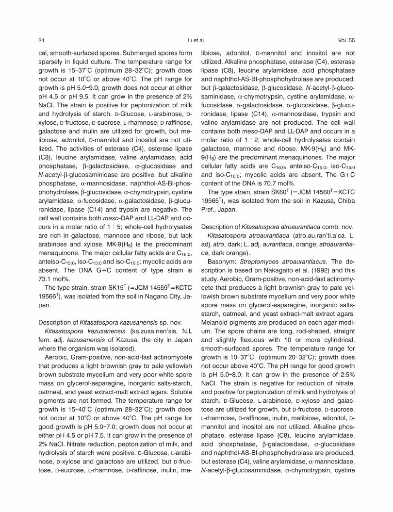
The phenotypic and phylogenetic characteristics of strains SK15T, SK60T and S. atroaurantiacus NBRC 14327T are consistent with their assignments to the genus Kitasatospora (Ōmura et al., 1982; Zhang et al., 1997). The organism formed an extensively branched substrate and aerial mycelium with smooth-surfaced
spores (Fig. 1). The colors of the aerial and substrate mycelium on different media of three strains are given in Table 1. The almost complete 16S rRNA gene se-quence of strains SK15T and SK60T were determined
Table 1. Growth and culture characteristics of the strains on ISP media.
CharacteristicIsolateSK15T
IsolateSK60T
K. atroaurantiaca
NBRC 14327T
ISP 2 medium Growth Moderate Moderate Moderate Aerial mycelium Ashes None Very poor, white Reverse color Brownish grey Pale yellowish brown Brown Soluble pigment None None Yellowish orangeISP 3 medium Growth Moderate Moderate Moderate Aerial mycelium Ashes Very poor, white White Reverse color Pale yellow Pale yellowish brown Light brown Soluble pigment None None Dull yellowish orangeISP 4 medium Growth Moderate Abundant Moderate Aerial mycelium Ashes Very poor, white White Reverse color Light brownish grey Light brownish grey Brown Soluble pigment None None Yellowish orangeISP 5 medium Growth Thin Moderate Moderate Aerial mycelium Ashes~grey Very poor, white White Reverse color Brownish white Pale yellowish brown Dark brown Soluble pigment None None Pale yellowish orange
Fig. 1. Scanning electron micrographs of the spore chains of strains (a) SK15T and (b) Kitasatospora atroaurantiaca NBRC 14327T grown on ISP 3 agar medium for 21 days at 27°C, and light micrograph of strain SK60T (c), grown on oatmeal agar medium for 21 days at 27°C. Bar, 5 μm.
22 Vol. 55LI et al.
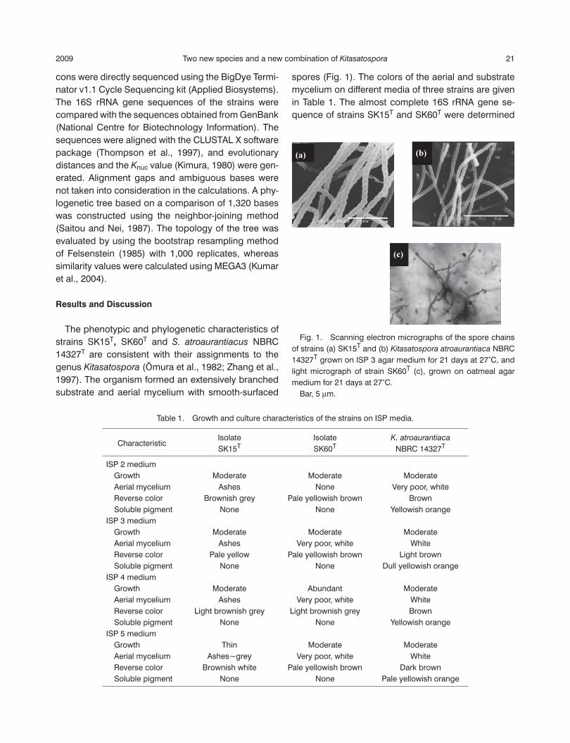
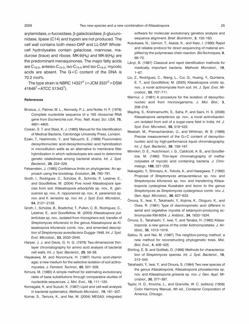
and have been deposited in the DDBJ database. Phy-logenetic analysis of the 16S rRNA gene sequence re-vealed that both isolates and S. atroaurantiacus NBRC 14327T fell within the evolutionary radiation occupied by the genus Kitasatospora and formed monophyletic clades with the members of the genus Kitasatospora (Fig. 2). Strains SK15T and SK60T shared the highest 16S rRNA gene sequence similarities with Kitasatospo-
ra setae IAM 15325T (97.8%) and Kitasatospora medio-
cidica IAM 15162T (97.5%), respectively. However, the DNA-DNA hybridization experiment demonstrated that strain SK15T was distinct from K. setae with 52% rela-tedness, and that between SK60T and K. mediocidica was 22%. Meanwhile, in the cluster of strain S. atroau-
rantiacus NBRC 14327T and Kitasatospora gansuensis
IAM 15269T, the level of DNA relatedness between them was 34%. All the values were below the 70% cut-off point recommended for the delineation of bacterial species by Wayne et al. (1987). These results indicate
that strains SK15T, SK60T, and S. atroaurantiacus
NBRC 14327T represent separate species from other species of the genus Kitasatospora. Chemotaxonomic analysis based on previous stud-ies of strain S. atroaurantiacus NBRC 14327T (Naka-gaito et al., 1992; Zhang et al., 1997) and this study of strains SK15T and SK60T further supported the phylo-genetic results. Cell wall analysis by HPLC and TLC indicated that the strains contained predominant amounts of LL-DAP and relatively small amounts of the meso-DAP, including glutamic acid, glycine and ala-nine in the cell wall. Whole-cell hydrolysates were rich in galactose, mannose and ribose, but no arabinose or xylose was detected. The predominant fatty acids were C16:0, anteiso-C15:0, iso-C15:0 and iso-C16:0. MK-9(H6) and MK-9(H8) were detected as predominant menaquinones. No mycolic acid was detected. The DNA G+C content of the strains was above 70 mol%. These results are consistent with the properties of the
Fig. 2. The 16S rRNA gene sequence-based phylogenetic tree generated by using the neighbor-joining method shows the relationships of strains SK15T, SK60T and Kitasatospora
atroaurantiaca NBRC 14327T in the genus Kitasatospora. Numbers at the nodes indicate the percentages of occurrence in 1,000 bootstrapped trees; only values greater than 50% are shown.
2009 23Two new species and a new combination of Kitasatospora
genus Kitasatospora. Moreover, strains SK15T, SK60T
and S. atroaurantiacus NBRC14327T could be distin-guished from their closest relatives and the other spe-cies of the genus Kitasatospora by various phenotypic data, as shown in Table 2, and other characteristics are given in the species description. It can therefore be concluded from the phenotypic, chemotaxonomic, and phylogenetic characteristics that strains SK15T and SK60T form two novel Kitasa-
tospora species for which the names Kitasatospora
saccharophila sp. nov. and Kitasatospora kazusanen-
sis sp. nov. are proposed. In addition, Streptomyces
atroaurantiacus NBRC 14327T formed a monophyletic clade with the members of the genus Kitasatospora in the phylogenetic tree, and phenotypic characteristics
also placed this strain in the genus Kitasatospora. It should therefore be transferred to the genus Kitasa-
tospora as Kitasatospora atroaurantiaca comb. nov.
Description of Kitasatospora saccharophila sp. nov. Kitasatospora saccharophila (sac.cha.ro.phi’la. Gr. n. saccharon sugar; Gr. adj. philos loving; N.L fem. adj. saccharophila sugar-loving). Aerobic, Gram-positive, non-acid-fast actinomycete that produces a brownish white to pale yellow sub-strate mycelium and ashes to gray spore mass on glycerol-asparagine, inorganic salts-starch, oatmeal and yeast extract-malt extract agars. Soluble pigments are not formed; the spore chains are long, cocci-shaped, straight and fl exuous with 20 or more cylindri-
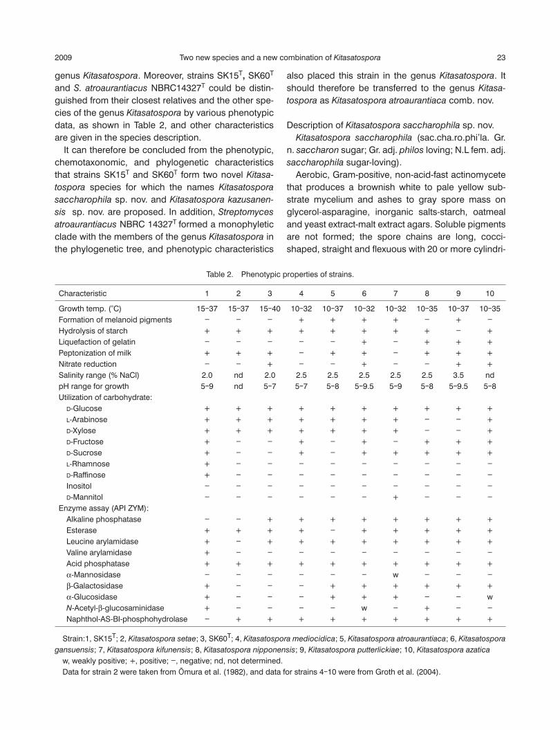
Table 2. Phenotypic properties of strains.
Characteristic 1 2 3 4 5 6 7 8 9 10
Growth temp. (°C) 15‒37 15‒37 15‒40 10‒32 10‒37 10‒32 10‒32 10‒35 10‒37 10‒35Formation of melanoid pigments - - - + + + + - + -Hydrolysis of starch + + + + + + + + - +Liquefaction of gelatin - - - - - + - + + +Peptonization of milk + + + - + + - + + +Nitrate reduction - - + - - + - - + +Salinity range (% NaCl) 2.0 nd 2.0 2.5 2.5 2.5 2.5 2.5 3.5 ndpH range for growth 5‒9 nd 5‒7 5‒7 5‒8 5‒9.5 5‒9 5‒8 5‒9.5 5‒8Utilization of carbohydrate: D-Glucose + + + + + + + + + + L-Arabinose + + + + + + + - - + D-Xylose + + + + + + + - - + D-Fructose + - - + - + - + + + D-Sucrose + - - + - + + + + + L-Rhamnose + - - - - - - - - - D-Raffi nose + - - - - - - - - - Inositol - - - - - - - - - - D-Mannitol - - - - - - + - - -Enzyme assay (API ZYM): Alkaline phosphatase - - + + + + + + + + Esterase + + + + - + + + + + Leucine arylamidase + - + + + + + + + + Valine arylamidase + - - - - - - - - - Acid phosphatase + + + + + + + + + + α-Mannosidase - - - - - - w - - - β-Galactosidase + - - - + + + + + + α-Glucosidase + - - - + + + - - w N-Acetyl-β-glucosaminidase + - - - - w - + - - Naphthol-AS-BI-phosphohydrolase - + + + + + + + + +
Strain:1, SK15T; 2, Kitasatospora setae; 3, SK60T; 4, Kitasatospora mediocidica; 5, Kitasatospora atroaurantiaca; 6, Kitasatospora
gansuensis; 7, Kitasatospora kifunensis; 8, Kitasatospora nipponensis; 9, Kitasatospora putterlickiae; 10, Kitasatospora azatica
w, weakly positive; +, positive; -, negative; nd, not determined. Data for strain 2 were taken from Ōmura et al. (1982), and data for strains 4‒10 were from Groth et al. (2004).
24 Vol. 55LI et al.
cal, smooth-surfaced spores. Submerged spores form sparsely in liquid culture. The temperature range for growth is 15‒37°C (optimum 28‒32°C); growth does not occur at 10°C or above 40°C. The pH range for growth is pH 5.0‒9.0; growth does not occur at either pH 4.5 or pH 9.5. It can grow in the presence of 2% NaCl. The strain is positive for peptonization of milk and hydrolysis of starch. D-Glucose, L-arabinose, D-xylose, D-fructose, D-sucrose, L-rhamnose, D-raffi nose, galactose and inulin are utilized for growth, but me-libiose, adonitol, D-mannitol and inositol are not uti-lized. The activities of esterase (C4), esterase lipase (C8), leucine arylamidase, valine arylamidase, acid phosphatase, β-galactosidase, α-glucosidase and N-acetyl-β-glucosaminidase are positive, but alkaline phosphatase, α-mannosidase, naphthol-AS-BI-phos-phohydrolase, β-glucosidase, α-chymotrypsin, cystine arylamidase, α-fucosidase, α-galactosidase, β-glucu-ronidase, lipase (C14) and trypsin are negative. The cell wall contains both meso-DAP and LL-DAP and oc-curs in a molar ratio of 1:5; whole-cell hydrolysates are rich in galactose, mannose and ribose, but lack arabinose and xylose. MK-9(H6) is the predominant menaquinone. The major cellular fatty acids are C16:0, anteiso-C15:0, iso-C15:0 and iso-C16:0; mycolic acids are absent. The DNA G+C content of type strain is 73.1 mol%. The type strain, strain SK15T (=JCM 14559T=KCTC 19566T), was isolated from the soil in Nagano City, Ja-pan.
Description of Kitasatospora kazusanensis sp. nov. Kitasatospora kazusanensis (ka.zusa.nen’sis. N.L fem. adj. kazusanensis of Kazusa, the city in Japan where the organism was isolated). Aerobic, Gram-positive, non-acid-fast actinomycete that produces a light brownish gray to pale yellowish brown substrate mycelium and very poor white spore mass on glycerol-asparagine, inorganic salts-starch, oatmeal, and yeast extract-malt extract agars. Soluble pigments are not formed. The temperature range for growth is 15‒40°C (optimum 28‒32°C); growth does not occur at 10°C or above 40°C. The pH range for good growth is pH 5.0‒7.0; growth does not occur at either pH 4.5 or pH 7.5. It can grow in the presence of 2% NaCl. Nitrate reduction, peptonization of milk, and hydrolysis of starch were positive. D-Glucose, L-arabi-nose, D-xylose and galactose are utilized, but D-fruc-tose, D-sucrose, L-rhamnose, D-raffi nose, inulin, me-
libiose, adonitol, D-mannitol and inositol are not utilized. Alkaline phosphatase, esterase (C4), esterase lipase (C8), leucine arylamidase, acid phosphatase and naphthol-AS-BI-phosphohydrolase are produced, but β-galactosidase, β-glucosidase, N-acetyl-β-gluco-saminidase, α-chymotrypsin, cystine arylamidase, α-
fucosidase, α-galactosidase, α-glucosidase, β-glu cu-ronidase, lipase (C14), α-mannosidase, trypsin and valine arylamidase are not produced. The cell wall contains both meso-DAP and LL-DAP and occurs in a molar ratio of 1:2; whole-cell hydrolysates contain galactose, mannose and ribose. MK-9(H6) and MK-9(H8) are the predominant menaquinones. The major cellular fatty acids are C16:0, anteiso-C15:0, iso-C15:0
and iso-C16:0; mycolic acids are absent. The G+C content of the DNA is 70.7 mol%. The type strain, strain SK60T (=JCM 14560T=KCTC 19565T), was isolated from the soil in Kazusa, Chiba Pref., Japan.
Description of Kitasatospora atroaurantiaca comb. nov. Kitasatospora atroaurantiaca (atro.au.ran’ti.a’ca. L. adj. atro, dark; L. adj. aurantiaca, orange; atroaurantia-
ca, dark orange). Basonym: Streptomyces atroaurantiacus. The de-scription is based on Nakagaito et al. (1992) and this study. Aerobic, Gram-positive, non-acid-fast actinomy-cete that produces a light brownish gray to pale yel-lowish brown substrate mycelium and very poor white spore mass on glycerol-asparagine, inorganic salts-starch, oatmeal, and yeast extract-malt extract agars. Melanoid pigments are produced on each agar medi-um. The spore chains are long, rod-shaped, straight and slightly fl exuous with 10 or more cylindrical, smooth-surfaced spores. The temperature range for growth is 10‒37°C (optimum 20‒32°C); growth does not occur above 40°C. The pH range for good growth is pH 5.0‒8.0; it can grow in the presence of 2.5% NaCl. The strain is negative for reduction of nitrate, and positive for peptonization of milk and hydrolysis of starch. D-Glucose, L-arabinose, D-xylose and galac-tose are utilized for growth, but D-fructose, D-sucrose, L-rhamnose, D-raffi nose, inulin, melibiose, adonitol, D-mannitol and inositol are not utilized. Alkaline phos-phatase, esterase lipase (C8), leucine arylamidase, acid phosphatase, β-galactosidase, α-glucosidase and naphthol-AS-BI-phosphohydrolase are produced, but esterase (C4), valine arylamidase, α-mannosidase, N-acetyl-β-glucosaminidase, α-chymotrypsin, cystine
2009 25Two new species and a new combination of Kitasatospora
arylamidase, α-fucosidase, β-galactosidase, β-glucuro-nidase, lipase (C14) and trypsin are not produced. The cell wall contains both meso-DAP and LL-DAP. Whole-cell hydrolysates contain galactose, mannose, ma-durose (trace) and ribose. MK-9(H6) and MK-9(H8) are the predominant menaquinones. The major fatty acids are C16:0, anteiso-C15:0, iso-C15:0 and iso-C16:0; mycolic acids are absent. The G+C content of the DNA is 70.2 mol%. The type strain is NBRC 14327T (=JCM 3337T=DSM 41649T=ATCC 51343T).
References
Brosius, J., Palmer, M. L., Kennedy, P. J., and Noller, H. F. (1978) Complete nucleotide sequence of a 16S ribosomal RNA gene from Escherichia coli. Proc. Natl. Acad. Sci. USA, 75, 4801‒4805.
Cowan, S. T. and Steel, K. J. (1965) Manual for the Identifi cation of Medical Bacteria, Cambridge University Press, London.
Ezaki, T., Hashimoto, Y., and Yabuuchi, E. (1989) Fluorometric deoxyribonucleic acid-deoxyribonucleic acid hybridization in microdilution wells as an alternative to membrane fi lter hybridization in which radioisotopes are used to determine genetic relatedness among bacterial strains. Int. J. Syst.
Bacteriol., 39, 224‒229.Felsenstein, J. (1985) Confi dence limits on phylogenies: An ap-
proach using the bootstrap. Evolution, 39, 783‒791.Groth, I., Rodríguez, C., Schütze, B., Schmitz, P., Leistner, E.,
and Goodfellow, M. (2004) Five novel Kitasatospora spe-cies from soil: Kitasatospora arboriphila sp. nov., K. gan-
suensis sp. nov., K. nipponensis sp. nov., K. paranensis sp. nov. and K. terrestris sp. nov. Int. J. Syst. Evol. Microbiol., 54, 2121‒2129.
Groth, I., Schütze, B., Boettcher, T., Pullen, C. B., Rodriguez, C., Leistner, E., and Goodfellow, M. (2003) Kitasatospora put-
terlickiae sp. nov., isolated from rhizosphere soil, transfer of Streptomyces kifunensis to the genus Kitasatospora as Ki-
tasatospora kifunensis comb. nov., and emended descrip-tion of Streptomyces aureofaciens Duggar 1948. Int. J. Syst.
Evol. Microbiol., 53, 2033‒2040.Harper, J. J. and Davis, G. H. G. (1979) Two-dimensional thin-
layer chromatography for amino acid analysis of bacterial cell walls. Int. J. Syst. Bacteriol., 29, 56‒58.
Hayakawa, M. and Nonomura, H. (1987) Humic acid-vitamin agar, a new medium for the selective isolation of soil actino-mycetes. J. Ferment. Technol., 65, 501‒509.
Kimura, M. (1980) A simple method for estimating evolutionary rates of base substitutions through comparative studies of nucleotide sequences. J. Mol. Evol., 16, 111‒120.
Komagata, K. and Suzuki, K. (1987) Lipid and cell-wall analysis in bacterial systematics. Methods Microbiol., 19, 161‒207.
Kumar, S., Tamura, K., and Nei, M. (2004) MEGA3: integrated
software for molecular evolutionary genetics analysis and sequence alignment. Brief. Bioinform., 5, 150‒163.
Kusukawa, N., Uemori, T., Asada, K., and Kato, I. (1990) Rapid and reliable protocol for direct sequencing of material am-plifi ed by the polymerase chain reaction. BioTechniques, 9, 66‒72.
Lányi, B. (1987) Classical and rapid identifi cation methods for medically important bacteria. Methods Microbiol., 19, 1‒67.
Liu, Z., Rodríguez, C., Wang, L., Cui, Q., Huang, Y., Quintana, E. T., and Goodfellow, M. (2005) Kitasatospora viridis sp. nov., a novel actinomycete from soil. Int. J. Syst. Evol. Mi-
crobiol., 55, 707‒711.Marmur, J. (1961) A procedure for the isolation of deoxyribo-
nucleic acid from microorganisms. J. Mol. Biol., 3, 208‒218.
Mayilraj, S., Krishnamurthi, S., Saha, P., and Saini, H. S. (2006) Kitasatospora sampliensis sp. nov., a novel actinobacteri-um isolated from soil of a sugar-cane fi eld in India. Int. J.
Syst. Evol. Microbiol., 56, 519‒522.Mesbah, M., Premachandran, U., and Whitman, W. B. (1989)
Precise measurement of the G+C content of deoxyribo-nucleic acid by high-performance liquid chromatography. Int. J. Syst. Bacteriol., 39, 159‒167.
Minnikin, D. E., Hutchinson, I. G., Caldicott, A. B., and Goodfel-low, M. (1980) Thin-layer chromatography of metha-nolysates of mycolic acid containing bacteria. J. Chro-
matogr., 188, 221‒233.Nakagaito, Y., Shimazu, A., Yokota, A., and Hasegawa, T. (1992)
Proposal of Streptomyces atroaurantiacus sp. nov. and Streptomyces kifunensis sp. nov. and transferring Kitasa-
tosporia cystarginea Kusakabe and Isono to the genus Streptomyces as Streptomyces cystargineus comb. nov. J.
Gen. Appl. Microbiol., 38, 627‒633.Ōmura, S., Iwai, Y., Takahashi, Y., Kojima, K., Otoguro, K., and
Oiwa, R. (1981) Type of diaminopimelic acid different in aerial and vegetative mycelia of setamycin-producing ac-tinomycete KM-6054. J. Antibiot., 34, 1633‒1634.
Ōmura, S., Takahashi, Y., Iwai, Y., and Tanaka, H. (1982) Kitasa-
tosporia, a new genus of the order Actinomycetales. J. An-
tibiot., 35, 1013‒1019.Saitou, N. and Nei, M. (1987) The neighbor-joining method: a
new method for reconstructing phylogenetic trees. Mol.
Biol. Evol., 4, 406‒425.Shirling, E. B. and Gottlieb, D. (1966) Methods for characteriza-
tion of Streptomyces species. Int. J. Syst. Bacteriol., 16, 313‒340.
Takahashi, Y., Iwai, Y., and Omura, S. (1984) Two new species of the genus Kitasatosporia, Kitasatosporia phosalacinea sp. nov. and Kitasatosporia griseola sp. nov. J. Gen. Appl. Mi-
crobiol., 30, 377‒387.Taylor, H. D., Knoche, L., and Granville, W. C. (editors) (1958)
Color Harmony Manual, 4th ed., Container Corporation of America, Chicago.
26 Vol. 55LI et al.
Thompson, J. D., Gibson, T. J., Plewniak, F., Jeanmougin, F., and Higgins, D. G. (1997) The CLUSTAL X windows inter-face: Flexible strategies for multiple sequence alignment aided by quality analysis tools. Nucleic Acids Res., 25, 4876‒4882.
Wayne, L. G., Brenner, D. J., Colwell, R. R., Grimont, P. A. D., Kandler, O., Krichevsky, M. I., Moore, L. H., Moore, W. E. C., Murray, R. G. E., Stackebrandt, E., Starr, M. P., and Trüper, H. G. (1987) International Committee on Systematic Bacte-riology. Report of the ad hoc committee on reconciliation of approaches to bacterial systematics. Int. J. Syst. Bacteriol.,
37, 463‒464.Wellington, E. M. H., Stackebrandt, E., Sanders, D., Wolstrup,
J., and Jorgensen, N. O. G. (1992) Taxonomic status of Ki-
tasatosporia, and proposed unifi cation with Streptomyces on the basis of phenotypic and 16S rRNA analysis and emendation of Streptomyces Waksman and Henrici 1943,
339AL. Int. J. Syst. Bacteriol., 42, 156‒160.Xie, C.-H. and Yokota, A. (2003) Phylogenetic analyses of Lam-
propedia hyalina based on the 16S rRNA gene sequence. J. Gen. Appl. Microbiol., 49, 345‒349.
Yokota, A. and Takashima, M. (2001) Cell wall. In Classifi cation and Identifi cation of Microorganisms, ed. by Suzuki, K., Hiraishi, A., and Yokota, A., Springer-Verlag, Tokyo, pp. 97‒117 (in Japanese).
Yokota, A., Tamura, T., Nishii, T., and Hasegawa, T. (1993) Kine-
ococcus aurantiacus gen. nov., sp. nov., a new aerobic gram-positive, motile coccus with meso-diaminopimelic acid and arabinogalactan in the cell wall. Int. J. Syst. Bacte-
riol., 43, 52‒57.Zhang, Z., Wang, Y., and Ruan, J. (1997) A proposal to revive
the genus Kitasatospora (Omura, Takahashi, Iwai, and Tanaka 1982). Int. J. Syst. Bacteriol., 47, 1048‒1054.