Sphingomonas cynarae sp. nov., the producer of an unusual type of sphingan

INTERNATIONAL JOURNAL OF SYSTEMATIC BACTERIOLOGY, Apr. 1996, p. 429-436
Copyright 0 1996, International Union of Microbiological Societies 0020-77 13/96/$04.00 + 0
Vol. 46, No. 2
Tsukarnurella pulmonis sp. nov. A. F. YASSIN,'* F. A. RAINEY,' H. BRZEZINKA,3 J. BURGHARDT,2 M. RIFAI,4 P. SEIFERT,'
K. FELDMANN,4 AND K. P. SCHAAL' Institut fur Medizinische Mikrobiologie und Imrnunologie der Universitat Bonn, 53127 Bonn, Deutsche Sammlung von
Mikroorganismen und Zellkulturen GmbH, 381 24 Braunschweig, Institut fur Rechtsrnedizin der Universitat Bonn, 531 11 Bonn, Institut fur Laboratoriurnsdiagnostik im Zentralkrankenhaus Gauting, 32131 Gauting, and
A l f i e d - h p p Labor, Universitiits-Augenklinik, 0-531 05 Bonn, ' Germany
Chemotaxonomic and 16s ribosomal DNA sequence analyses of an isolate from the sputum of a patient with a mycobacterial lung infection clearly delineated a new species of the genus Tsukarnurella. This new species can be defined on the basis of genotypic and phenotypic data. The name Tsukamurella pulmonis sp. nov. is proposed for this organism; the type strain is IMMIB D-1321T (= DSM 44142T). This isolate shows 44.2 and 36.2% DNA relatedness to Tsukamurella paurometabola DSM 20162T (T = type strain) and Tsukamurella inchonensis DSM 44067T, respectively.
The taxonomic history of the genus Tsukamurella, particu- larly that of Tsukamurellapaurometabola, was recently outlined in detail (36). In a numerical taxonomic study, Goodfellow et al. (7) proposed the name Tsukamurella wratislaviensis for a group of isolates that clustered to form a new center of vari- ation most closely associated with T. paurometabola. These strains had chemical, enzymic, nutritional, and tolerance prop- erties consistent with their assignment to the genus Tsuka- murella and formed a DNA homology group that also cor- responded to the group formed by T. paurometabola. Recent- ly, Yassin et al. (36) described Tsukamurella inchonensis. This species has chemotaxonomic and phylogenetic properties consistent with its assignment to the genus Tsukamurella. A lipid analysis of T. inchonensis and i? paurometabola showed that both of these species have two mycolates that correspond to a - and a'-mycolates which on pyrolysis produce C20:,, C20:o, C22:1, and C,,:, fatty acids as main cleavage products (some strains of T. inchonensis produce only C22:o). The fatty acid profile comprises saturated, unsaturated, and 10-methyl branched fatty acids. Unsaturated MK-8, MK-9, and MK-10 menaquinones are present, and MK-9 is the predominant com- ponent. The 16s ribosomal DNA (rDNA) sequences of T. inchonensis and T. paurometabola show a high level of similar- ity (99.3%).
Tsukamura and Kawakami (29) first drew attention to the pathogenic potential of Tsukarrzurella strains in humans when they isolated T. paurometabola seven times from the sputa of a patient with tuberculosis-like disease. A case of fetal meningi- tis (22), a case of severe gangrenous tendosynovitis with mul- tiple subcutaneous abscesses (28), and a case of bacteremia (24) have also been described. T. inchonensis was isolated four times from blood cultures of a patient who had ingested hy- drochloric acid (36). This species was also isolated from ne- crotic material from a patient with lung carcinoma. The simi- larity of Tsukamurella strains to other more common pathogens expected in immunocompromised patients, such as Rhodococ- cus, Nocardia, Gordona, and Mycobacterium strains, may have resulted previously in inadequate diagnosis and thus in under- rating the incidence of Tsukamurella infections. Clinicians should therefore be aware of the pathogenic potential of Tsuka-
* Corresponding author. Mailing address: Institut fur Medizinische Mikrobiologie und Immunologie der Universitat Bonn, Sigmund- Freud-Strasse 25, 53127 Bonn, Germany.
murella spp., especially when they are treating AIDS patients and similarly immunocompromised persons.
In this paper we describe a new Tsukamurella species which was isolated from the sputum of a 92-year-old woman suffering from pulmonary tuberculosis. Mycobacterium tuberculosis and Mycobacterium fortuitum were first isolated from the patient. During a second hospitalization (2 years later) Mycobacterium gordonae was recovered, and during a third hospitalization (1 year later) the species described in this paper was isolat- ed and erroneously identified as M. fortuitum. However, at- tempts to amplify the genomic DNA of this organism with two different universal primers for mycobacteria, one having the sequence 5 'GAGTTTGATCCTGGCTCAGGA3' (primer 283 of E. Bottger) and the other having the sequence 5'TGCA CACAGGCCACAAGGGA3' (Amplicor; Hoffman-LaRoche), were unsuccessful, and therefore the taxonomic status of the organism was questioned. Applying routine chemotaxonomic methods, we found that the organism appeared to be close- ly related to members of the genus Tsukamurella. This was then confinned by further taxonomic and phylogenetic investiga- tions.
MATERIALS AND METHODS Bacterial strains. Strain IMMIB D-1321T (T = type strain) was isolated from
the sputum of a 92-year-old woman who suffered from pulmonary tuberculosis and possibly an additional mycobacterial infection. The source of T. inchonensis DSM 44067T has been described previously (36). T. paurometabola DSM 20162T was obtained from the German Collection of Microorganisms and Cell Cultures.
Isolation method. Strain IMMIB D-l32lT was isolated from the sputum after decontamination with N-acetyl-L-cysteine (14, 26). The specimen was concen- trated by centrifugation at 3,000 X g for 20 min and was cultured on Loewen- stein-Jensen medium.
Morphology and pigmentation. Strain IMMIB D-1321T was grown on yeast extract-malt extract agar (International Streptomyces Project [ISP] medium 2), oatmeal agar (ISP medium 3), and inorganic salts-starch agar (ISP medium 4) as described by Shirling and Gottlieb (25) and was examined for pigmentation, production of aerial hyphae, and other morphological characteristics. Cultures were grown for 4 weeks and were observed weekly. Air-dried smears from brain heart infusion (BHI) agar cultures were stained by the Gram and Ziehl-Neelsen methods in order to determine the Gram reaction and acid fastness, respectively. The micromorphology of the organism was determined by using a culture grown at 37°C for 4 days on ISP medium 2. Electron micrographs of the preparation were taken with a Zeiss digital scanning electron microscope (model DSM 950).
Physiological characteristics. The ability of the organism to grow at various temperatures (24, 31, 37, and 45°C) was determined after 1 week of incubation on Loewenstein-Jensen medium slants and BHI agar. Peptone-yeast extract-iron agar (ISP medium 6) and tyrosine agar (ISP medium 7) as described by Shirling and Gottlieb (25) were used to determine melanoid pigment production. The following properties were determined as described previously: the niacin test was performed by using test strips (Difco Laboratories, Detroit, Mich.) (12, 32);
429

430 YASSIN ET AL. INT. J. SYST. BACTERIOL.
FIG. 1. Phase-contrast micrographs of cells of strain IMMIB D-1321T stained with lactophenol cotton blue, showing cells that cluster together either pole to pole or pole to side (A) to simulate a branched structure (B). A slide culture was grown on BHI agar for 6 h at 36 % 1°C.
nitrate reductase activity was determined by the method of Kubica and David (13); catalase activity was determined by the method of Kubica and Pool (15); arylsulfatase activity (after 3 days) was determined by the method of Kubica and Ridgon (16); P-glucosidase and P-galactosidase activities were determined by the method of Tsukamura (27); Tween 80 hydrolysis was determined by the method of Wayne et al. (31); growth on MacConkey agar was determined by the method of Pattyn and Portaels (21); resistance to NaCl and p-nitrobenzoic acid was determined by the methods of Wayne et al. (31) and Tsukamura and Tsukamura (30); acetamidase, allantoinase, benzamidase, nicotinamidase, pyrazinamidase, succinamidase, and urease activities were determined by the method of Bonicke (2); and tests to determine resistance to streptomycin (4.0 and 8.0 pg), isoniazid (0.25 and 1.0 pg), ethambutol (8.0, 16.0, and 32.0 pg), rifampin (16.0 and 32.0 pg), p-aminosalicylic acid (16 and 32.0 pg), protion-amide (16.0 and 32.0 Fg), capreomycin (16.0 and 32.0 pg), and cycloserine (16.0 and 32.0 pg) were per- formed by using Loewenstein-Jensen medium slants supplemented with these antibiotics (Biotest). Tests to determine decomposition of adenine, guanine, hypoxanthine, xanthine, tyrosine, elastin, keratin, and testosterone were per- formed by the method of Gordon and Smith (10); the test to determine esculin decomposition was performed by the method of Gordon (8); and casein and gelatin hydrolysis tests were performed by the method of Gordon and Mihm (9). The urea decomposition test was performed by using urea agar base (catalog no. CM 53; Oxoid) after 2.2% urea was added. In tests to determine utilization of substrates as carbon sources and as simultaneous carbon and nitrogen sources we
used the media described by Yassin et al. (36). Sensitivity to lysozyme was determined as described previously (34).
Cell chemistry. The strains which we studied were cultivated at 37°C in shake flasks (120 rpm) containing BHI (Difco) broth for 1 week. After we checked for purity at maximum growth, the organisms were killed with formaldehyde ( l%, vol/vol), harvested by centrifugation, washed with distilled water, and freeze- dried. Analyses of whole-cell hydrolysates for amino acids and sugars were performed by the methods of Becker et al. (1) and Lechevalier (17). Acid methanolysis, alkaline methanolysis, conversion to fert-butyldimethylsilyl (TBDMS) ethers, one-dimensional thin-layer chromatography, two-dimensional thin-layer chromatography, and pyrolysis gas chromatography were performed as previously described by Yassin et al. (33). Menaquinones were extracted and purified by the method of Collins et al. (4) and were identified by using a Finnigan Mat 212 mass spectrometer. Phospholipids were extracted, purified, and identified as described previously (35).
DNA isolation and characterization. DNA was isolated by chromatography on hydroxyapatite by using the method of Cashion et al. (3). Guanine-plus-cytosine (G+C) Contents were determined by high-performance liquid chromatography (20). DNA-DNA hybridization studies were carried out by using the thermal renaturation method as described previously (34).
16s rDNA isolation and sequence determination. Genomic DNA extraction and PCR-mediated amplification of 16s rDNA genes were performed as de- scribed previously (23). Purified PCR products were directly sequenced by using

VOL. 46, 1996 TSUKAMURELU PULMONIS SP. NOV. 43 1
FIG. 2. Scanning electron micrographs of strain IMMIB D-1321T, showing long rod-shaped cells (A) that fragment into three parts (B), which separate and grow as independent cells (C). The culture was grown on ISP medium 2 for 4 days at 36 * 1°C.
a Tuq DyeDeoxy terminator cycle sequencing kit (Applied Biosystems, Foster City, Calif.) according to the protocol provided by the manufacturer. An Applied Biosystems model 373A DNA sequencer was used to electrophorese the se- quencing reaction mixtures.
The 16s rDNA sequence determined in this study was manually aligned with previously published sequences available from public databases. Evolutionary distances calculated by the method of Jukes and Cantor (11) were used to construct a phylogenetic tree by the least-squares method of De Soete (6).

432 YASSIN ET AL. INT. J. SYST. BACTERIOL.
TABLE 1. Differential physiological characteristics of strain IMMIB D-1321T, T. inchonensis DSM 44067T,
and T. puurometubolu DSM 20162T
Strain T. incho- T. pauro- Characteristic IMMIB nensis metabola
D-1321T DSM 44067T DSM 20162T
Hydrolysis of Adenine Hypoxanthine Tyrosine Xanthine Casein Urea Esculin Gelatin Elastin Guanine Keratin Testosterone Tween 80
Utilization of the following compounds as sole sources of carbon and energy
Arabinose Xylose Glucose Galactose Rhamnose Lactose Maltose Raffinose Trehalose Sucrose Cellobiose Melezitose Adonitol Erythritol Inositol Mannitol D-Sorbitol Acetate Benzoate Citrate Gluconate Lactate rn-Hydroxybenzoate p-Hydroxybenzoate Adipiate Isoamyl alcohol 2,3-Butandiol 1 ,ZPropandiol Paraffin
Utilization of the following compounds as sole sources of carbon and nitrogen
Acetamide Alanine Gelatin Proline Serine
Growth at: 24°C 31°C 37°C 45°C
without crystal violet Growth on MacConkey agar
Niacin production Enzymatic activities
Acetamidase
Continued
TABLE 1-Continued
Strain T. incho- T. puuro- Characteristic IMMIB nerisis metaahoku
D-1321T DSM 44067T DSM 20162=
Allantoinase Arylsulfatase (3 days) Benzamidase Ca talase P-Glucosidase P-Galactosidase Nicotinamidase Nitrate reductase Pyrazinamidase Succinamidase Urease Phosphatase
Streptomycin Isoniazid Ethambutol Rifampin p-Aminosalicylic acid Protionamide Capreomycin Cycloserine
Growth in the presence of
+ -
-
+ + + + -
+ -
+ + + + + + + + + +
+ - -
+ + + + -
+ -
+ + + + + + + + + +
+ ND" + + + +
-
-
+ ND +
ND
+ + + + + + + +
" ND, not determined.
Nucleotide sequence accession number. The 16s rDNA sequence of strain IMMIB D-132IT is available from the EMBL data library under accession number X92981.
RESULTS AND DISCUSSION
Colonial morphology. Strain IMMIB D-1321T grew well on ISP medium 2 and ISP medium 3; the colonies were cream colored and rough. On ISP medium 4, growth was weak and the colonies were leathery. No diffusible pigments were ob- served. At 24 to 37°C growth occurred within 1 to 2 days on Loewenstein-Jensen medium and on BHI agar. Dilute inocula on BHI agar yielded large, eugonic, cream-colored, rough col- onies.
Micromorphology. The cells of strain IMMIB D-1321T were rod shaped, gram positive, and slightly acid-alcohol fast. Dur- ing growth the cells were primarily long rods (Fig. 2A) which stuck together, either pole to pole or pole to side (Fig. lA), to give an arrangement that looked like branched hyphae (Fig. 1B). In a later stage of growth these rods appeared to fragment into three parts (Fig. 2B) which separated (Fig. 2C) and started growing again. Spores, capsules, true branching, and aerial hyphae were not observed.
Physiological characteristics. The physiological properties of strain IMMIB D-1321T are shown in Table 1. This strain had acetamidase, allantoinase, catalase, P-glucosidase, P-galactosi- dase, nicotinamidase, pyrazinamidase, urease, and phosphatase activities; arylsulfatase, benzamidase, nitrate reductase, and succinamidase activities were not observed. Strain IMMIB D-1321T did not produce niacin but tolerated 5% NaCl and p-nitrobenzoic acid. This strain grew in the presence of the antituberculous drugs streptomycin, isoniazid, ethambutol, ri- fampin, paminosalicylic acid, protionamide, capreomycin, and cycloserine.
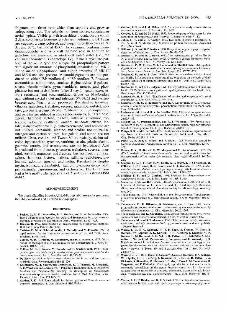
Lipid analysis. The results of one-dimensional thin-layer chromatography and two-dimensional thin-layer chromatogra- phy of whole-cell acid and alkaline methanolysates of strain IMMIB D-1321T are shown in Fig. 3. Two-dimensional thin- layer chromatography resulted in more precise resolution of

VOL. 46, 1996
Z paummetabola
M. aichiense
TSUKAMURELLA PULMONIS SP. NOV. 433
FIG. 3. (a and c) One-dimensional thin-layer chromatography and two-dimensional thin-layer chromatography of whole-cell alkaline (a) and acid (c) methanoly- sates of strain IMMIB D-1321T. Triple development in the first direction was with petroleum ether (bp 60 to 8O0C)-acetone (95: 5, vol/vol), and a single development in the second direction was with toluene-acetone (97: 3, vol/vol). Abbreviations: a, a-mycolate; a', a',-mycolate; F, nonhydroxylated fatty acid methyl esters. (b) TBDMS ethers of alkaline methanolysates. A single development in petroleun ether (bp 60 to 8O0C)-toluene (70:30, vol/vol) was used. c1 and a' refer to TBDMS ethers of the different types of mycolic acid methyl esters.
the components into two mycolate spots in addition to the nonhydroxylated fatty acid spot (spot F). One-dimensional thin-layer chromatography of the TBDMS ethers of the aika- line methanolysates resulted in identification of the two myco- late spots as a- and a'-mycolates by their Rfvalues (Fig. 3).
Pyrolysis gas chromatography of the pure a-mycolates that were isolated by preparative thin-layer chromatography from strain IMMIB D-1321T released C20:0, C20:1, C22:0, and C22:1 fatty acid methyl esters as main pyrolytic cleavage products. Gas chromatographic analyses of the nonhydroxylated fatty acid methyl esters revealed the presence of hexadecanoate, octadecanoate, and tuberculostearic acid (10-methyl octade- canoate) as major fatty acid methyl esters. No 2-methyl branched-chain fatty acids were observed.
A polar lipid analysis showed that the phospholipid type of strain IMMIB D-1321T is type PI1 sensu Lechevalier et al. (18); i.e., phosphatidylethanolamine is the characteristic phospho- lipid. Other phospholipids, including phosphatidylinositol, phosphatidylinositol mannoside, and diphosphatidylglycerol, were also detected. Strain IMMIB D-1321T contains menaqui- nones MK-8, MK-9, and MK-10; MK-9 is the major compo- nent.
DNA characteristics. The G+C content of the DNA of strain IMMIB D-1321T is 69.8 mol%. The levels of relatedness (binding rates) between strain IMMIB D-1321T DNA and the DNAs of T. paurometabola DSM 20162T and T. inchonensis DSM 44067T are 44.2 and 36.3%, respectively (as determined by the optical method).
The almost complete 16s rDNA sequence of strain IMMIB D-1321T was determined. A phylogenetic analysis showed that this strain is most closely related to the mycolic acid-containing actinomycetes and, more specifically, to the genus Tsuka- murella (Fig. 4). The 16s rDNA sequence of strain IMMIB D-1321T is very similar to the 16s rDNA sequences of T. paurometabola DSM 20162T and T. inchonensis DSM 44067T (levels of similarity, 99.3 and 99.4%, respectively) (Table 2). However, 16s rDNA sequence data showed that T. wratisla- viensis falls outside the genus Tsukamurella and is a Rhodococ- cus species (unpublished data).
The physiological and chemotaxonomic characteristics of isolate IMMIB D-1321T are compared with T. inchonensis DSM 44067T and T. paurometabola DSM 20162T characteris-
tics in Tables 1 and 3, respectively. Chemotaxonomically, strain IMMIB D-1321T, T. inchonensis DSM 44067T, and T. pauro- metabola DSM 20162T are similar (Table 3). All three strains contain galactose and arabinose as characteristic whole-cell sugars in addition to meso-diaminopimelic acid as a wall diamino acid (i.e., they are wall chemotype IV organisms); all contain two mycolic acids corresponding to a- and a'-myco- lates which on pyrolysis release C20:1, C20:0, C22:1, and C2;:0 fatty acids as mycolic acid cleavage products; the fatty acid profiles of all three consist of saturated, unsaturated, and 10- methyl branched fatty acids; and all contain unsaturated MK-8, MK-9, and MK-10 menaquinones, with MK-9 as the major
N. ofiridircaviarum
N. asteroides
R. equi
R. globerulus
R. rhodochmus
r- G. amrae
G. bronchialis
G. rubmpertincra
I 7 C. xerosis
I C. glufamicum
5%
FIG. 4. Phylogenetic dendrogram showing the position of strain IMMIB D-1321T within the mycolic acid-containing group. Scale bar = 5 nucleotide substitutions per 100 nucleotides. The sequences used in the analysis are the sequences of the type strains of the species included. Abbreviations: N., Nocar- dia; R. , Rhodococcus; G., Gordona; T., Tsukaniurella; M., Mycobacteriurn; D., Dietzia; C. ., Corynebacterium.

434 YASSIN ET AL. INT. J. SYST. BACTERIOL.
TABLE 2. Levels of 16s rDNA similarity between T. pulmonis and related taxa
% 16s rDNA similarity
Organism
Tsukamurella inchonensis Tsukamurella paurometabola Gordona bronchialis Gordona rubropertincta Gordona sputi Gordona amarae Rhodococcus rhodochrous Rhodococcus globerulus Rhodococcus equi Nocardia asteroides Nocardia otitidiscaviaium Mycobacterium aichiense Mycobacterium tuberculosis Mycobacterium terrae D. maris Coiynebacterium xerosis Corynebacterium glutamicum
99.4 99.3 99.3 93.2 93.3 93.4 93.1 93.1 93.3 98.5 93.0 93.2 93.4 97.4 96.9 94.1 94.4 94.6 97.1 97.1 96.4 94.1 94.4 94.5 94.4 94.4 93.9 94.1 94.8 94.7 94.8 93.8 93.4 92.7 93.4 95.1 94.8 94.7 94.9 94.4 94.3 93.2 94.2 96.2 96.5 94.4 94.6 94.7 93.5 93.4 92.7 94.0 94.0 95.2 96.3 94.4 94.7 94.6 94.8 94.6 93.6 94.5 95.2 95.9 97.3 98.0 94.2 94.5 94.6 93.7 93.2 93.3 94.0 93.9 94.0 93.7 94.1 94.1 92.4 92.2 92.3 92.7 91.8 91.2 91.9 91.9 92.1 92.3 92.7 91.9 95.5 93.7 94.1 94.1 93.2 92.7 92.7 93.3 93.4 93.2 93.1 93.5 93.1 97.3 96.8 95.2 95.1 95.3 93.1 92.8 92.8 93.8 94.6 94.9 94.2 93.3 93.4 93.7 91.9 93.1 92.2 92.3 92.3 92.2 91.4 91.1 91.2 92.4 91.9 91.9 90.7 91.5 91.7 92.2 92.2 93.4 90.4 90.4 90.5 90.0 89.9 89.4 89.4 91.4 90.7 90.7 89.1 89.8 89.6 89.1 89.5 91.2 93.5
component. These chemotaxonomic similarities are supported by the high levels of 16s rDNA similarity observed with isolate IMMIB D-1321T, T. inchonensis DSM 44067T, and T. pauro- metabolu DSM 20162T (Table 2).
In contrast to the chemotaxonomic similarities of isolate IMMIB D-1321T, T. inchonensis DSM 44067T and T. pauro- metabolu DSM 20162T, the results of our physiological tests (Table 1) revealed clear differences between strain IMMIB D- 1321T and the other two strains. Physiologically, isolate IMMIB D-1321T differs from T. inchonensis DSM 44067T by (i) its inability to hydrolyze hypoxanthin, (ii) its inability to utilize maltose, cellobiose, melezitose, inositol, and citrate as carbon sources, and (iii) its inability to grow at 45°C. Strain IMMIB D-1321T differs from T. puurometabola DSM 20162T by (i) its ability to utilize mannitol, sorbitol, isoamyl alcohol, and 1,2-propandiol as carbon sources and (ii) its ability to utilize acetamide as a simultaneous carbon and nitrogen source. These physiological differences are supported by the low levels of hybridization between strain IMMIB D-1321T
DNA and the DNAs of T. inchonensis DSM 44067T (36.3%) and T. puurometabola DSM 20162T (44.2%).
Thus, we propose that strain IMMIB D-1321T should be placed in a new species of the genus Tsukamurella. This strain has been deposited in the German Collection of Microorgan- isms and Cell Cultures as strain DSM 44142T. The type strain was isolated from the sputum of a 92-year-old woman with cured pulmonary tuberculosis who later developed signs of pulmonary infection again. The patient recovered from the infection only after antibiotics that were active against the Tsukamurella strain in question had been administered. Strain IMMIB D-1321T therefore appears to be pathogenic and should be placed in risk group 2 according to the German System. A description of this species is given below.
Description of Tsukamurella pulmonis sp. nov. Tsukamurella pulmonis (pul. mo' nis. L. gen. masc. n.pulmonis, of the lung, referring to the organ from which the bacterium was isolated). Cells are aerobic, gram-positive, and slightly acid-alcohol-fast bacilli. Most cells are long rods that, at a later stage of growth,
TABLE 3. Chemotaxonomic characteristics of strain IMMIB D-1321T, T. inchonensis DSM 44067T, and T. paurometabola DSM 20162T
Strain Fatty position pyrolosis products acids"
Wall Wall Phospho- Mycolic Wall sugars chemo- lipid Menaquinones" acid com- Mycolic acid diamino
acid type" typeb
7'. pulmonis IMMIB D-1321T meso-DIZPe Galactose, arabinose IV PI1 MK-10, MK-9, MK-8 a, a' 7'. inchonensis DSM 44067T meso-DAP Galactose, arabinose IV NDf MK-10, MK-9, MK-8 a, a' 7'. paurometabola DSM 20162= meso-DAP Galactose, arabinose IV ND MK-9, MK-8, MK-7g a, a'
'' Wall chemotype as described by Lechevalier and Lechevalier (19). ' Phospholipid type as described by Lechevalier et al. (18). ' MK-10 is a menaquinone with 10 isoprene units.
' meso-DAP, meso-diaminopimelic acid. S, straight chain; U, monounsaturated; T, tuberculostearic acid.
ND, not determined. Data from reference 5.
VOL. 46, 1996 TSUKAMURELLA PULMONIS SP. NOV. 435
fragment into three parts which then separate and grow as independent rods. The cells do not form spores, capsules, or aerial hyphae. Visible growth from dilute inocula occurs within 2 days; colonies on Loewenstein-Jensen medium and BHI agar are eugonic, creamy colored, and rough. Growth occurs at 24, 31, and 37”C, but not at 45°C. The organism contains meso- diaminopimelic acid as a wall diamino acid in addition to galactose and arabinose in whole-cell hydrolysates (i.e., the cell wall chemotype is chemotype IV). It has a mycolate pat- tern of the a, a’ type and a type PI1 phospholipid pattern with significant amounts of phosphatidylethanolamine. MK-9 is the major menaquinone, and minor amounts of MK-10 and MK-8 are also present. Melanoid pigments are not pro- duced on either ISP medium 6 or ISP medium 7. Produces acetamidase, allantoinase, catalase, P-glucosidase, P-galacto- sidase, nicotinamidase, pyrazinamidase, urease, and phos- phatase but not arylsulfatase (after 3 days), benzamidase, ni- trate reductase, and succinamidase. Grows on MacConkey agar without crystal violet and tolerates 5% NaCl and p-nitro- benzoic acid. Niacin is not produced. Resistant to lysozyme. Glucose, galactose, trehalose, sucrose, mannitol, sorbitol, ace- tate, gluconate, isoamyl alcohol, 2,3-butandiol, 1,2-propandiol, and paraffin are utilized as sole carbon sources, but arabinose, xylose, rhamnose, lactose, maltose, raffinose, cellobiose, me- lezitose, adonitol, erythritol, inositol, benzoate, citrate, lac- tate, m-hydroxybenzoate, p-hydroxybenzoate, and adipate are not utilized. Acetamide, alanine, and proline are utilized as nitrogen and carbon sources, but gelatin and serine are not utilized. Urea, esculin, and Tween 80 are hydrolyzed, but ad- enine, hypoxanthine, tyrosine, xanthine, casein, gelatin, elastin, guanine, keratin, and testosterone are not hydrolyzed. Acid is produced from glucose, galactose, trehalose, sucrose, man- nitol, sorbitol, mannose, and fructose, but not from arabinose, xylose, rhamnose, lactose, maltose, raffinose, cellobiose, me- lezitose, adonitol, inositol, and inulin. Resistant to strepto- mycin, isoniazid, ethambutol, rifampin, p-aminosalicylic acid, protionamide, capreomycin, and cycloserine. The G + C con- tent is 69.8 mol%. The type strain of T. pulmonis is strain DSM 44142.
ACKNOWLEDGMENT
We thank Claudine Strack (Alfried-Krupp laboratory) for preparing the phase-contrast and electron micrographs.
1.
2.
3.
4.
5.
6.
7.
8.
REFERENCES
Becker, B., M. P. Lechevalier, R. E. Gordon, and H. A. Lechevalier. 1964. Rapid differentiation between Nocurdiu and Streptonzyces by paper chroma- tography of whole cell hydrolysates. Appl. Microbiol. 12:421-423. Bonicke, R. 1962. Identification of mycobactcria by biochemical methods. Bull. Int. Union Tuberc. 32:13-86. Cashion, P., M. A. Holder-Franklin, J. McCully, and M. Franklin. 1977. A rapid method for the base ratio determination of bacterial DNA. Anal. Biochem. 81:461-466. Collins, M. D., T. Pirouz, M. Goodfellow, and D. E. Minnikin. 1977. Distri- bution of menaquinones in actinomycetes and corynebacteria. J. Gen. Mi- crobiol. 100221-230. Collins, M. D., J. Smida, M. Dorsch, and E. Stackebrandt. 1988. Tsuka- murellu gen. nov. harboring Colynebucterium paurometubolurn and Rliodo- coccus uuruntiucus. Int. J. Syst. Bacteriol. 38385-391. De Soete, G. 1983. A least squares algorithm for fitting additive trees to proximity data. Psychometrika 48:621-626. Goodfellow, M., J. Zakrzewska-CzeMinska, E. G. Thomas, M. Mordarski, A. C. Ward, and A. L. James. 1991. Polyphasic taxonomic study of the genera Gordona and Tsirkarnurellu including the description of Tsukaniirrella wrutislaviensis sp. nov. Zentralbl. Bakteriol. Int. J . Med. Microbiol. Virol. Parasitol. Infect. Dis. 275162-178. Gordon, R. E. 1966. Some criteria for the recognition of Nocurdin niadzrrue (Vincent) Blanchard. J. Gen. Microbiol. 45355-364.
9.
10.
11.
12.
13.
14.
15.
16.
17.
18.
19.
20.
21.
22.
23.
24.
25.
26.
27.
28.
29.
30.
31.
32.
Gordon, R. E., and J. M. Mihm. 1957. A comparative study of somc strains received as nocardiae. J. Bacteriol. 73: 15-27. Gordon, R. E., and M. M. Smith. 1955. Proposed group of characters for the separation of Streptoniyces and Nocurdiu. J. Bacteriol. 69:147-150. Jukes, T. H., and C. R. Cantor. 1969. Evolution of protcin molecules, p. 21-132. In H. N. Munro (ed.), Mammalian protein metabolism. Academic Press, New York. Kilburn, J. O., and G. P. Kubica. 1968. Reagent impregnated paper strips for detection of niacin. Am. J. Clin. Pathol. 50:530-532. Kubica, G. P., and H. L. David. 1980. The mycobacteria, p. 1693-1730. In A. C. Sonnenwirth and L. Jarett (ed.), Gradwohl’s clinical laboratory meth- ods and diagnosis. The C. V. Mosby Co.. St. Louis. Kubica, G. P., W. E. Dye, M. L. Cohn, and G. Middlebrook. 1963. Sputum digestion and decontamination with N-acetyl-L-cysteine-sodium hydroxide for culture of mycobacteria. Am. Rev. Respir. Dis. 87775-779. Kubica, G. P., and G. L. Pool. 1960. Studies on the calalase activity of acid fast bacilli. I . An attempt to subgroup these organisms on the basis of their catalasc activities at different temperatures and pH. Am. Rev. Respir. Dis.
Kubica, G. P., and A. L. Ridgon. 1961. The arylsulfatase activity of acid-fast bacilli. 111. Preliminary investigation of rapidly growing acid-fast bacilli. Am. Rev. Respir. Dis. 83:737-740. Lechevalier, M. P. 1968. Identification of aerobic actinomycetes of clinical importance. J. Lab. Clin. Med. 71934-944. Lechevalier, M. P., C. de Bievere, and H. A. Lechevalier. 1977. Chcmotax- onomy of aerobic actinomycetes: phospholipid composition. Biochem. Syst.
Lechevalier, M. P., and H. A. Lechevalier. 1970. Chemical composition as a criterion in the classification of aerobic actinomycetes. Int . J . Syst. Bacteriol. 20:435443. Mesbah, M., U. Premachandran, and W. B. Whitman. 1989. Precise mca- surement of the G + C content of deoxyribonucleic acid by high-performance liquid chromatography. Int. J. Syst. Bacteriol. 39:159-167. Pattyn, S. R., and F. Portaels. 1972. Identification and clinical significance of mycobacteria. Zentralbl. Bakteriol. Parasitenkd. Infektionskr. Hyg. Abt. 1 Orig. Reihe A 219:114-140. Prinz, G., E. Ban, S. Fekete, and Z. Szabo. 1985. Meningitis caused by Gordonu acrrantiuca (Rhodococc~rs uuruntiucus). J. Clin. Microbiol. 22:472- 474. Rainey, F. A., M. Dorsch, H. W. Morgan, and E. Stackebrandt. 1992. 16s rDNA analysis of Spirochrietu thennophila: position and amplifications for the systematics of the order Spirochuetu/es. Syst. Appl. Microbiol. 16224- 226. Shapiro, C. L., R. F. Haft, N. M. Gantz, G. V. Doern, J. C. Christenson, R. O’Brien, J. C. Overall, B. A. Brown, and R. J. Wallace, Jr. 1992. Tsuka- rniirella yaurornetubolum: a novel pathogen causing catheter-related bactc- remia in patients with cancer. Clin. Infect. Dis. 14:200-203. Shirling, E. B., and D. Gottlieb. 1966. Methods for characterization of Streptoniyces species. Int. J . Syst. Bacteriol. 1 6 3 13-340. Sommers, H. M., and R. C. Good. 1985. Mycobacteriun~, p. 216-248. In E. H. Lennette. A. Balows, W. J. Hauslcr, Jr., and H. J. Shadomy (ed.), Manual of clinical microbiology, 4th ed. American Society for Microbiology, Washing- ton, D.C. Tsukamura, M. 1974. Differentiation of the “Mycobacterium” r~zodochrous- group from nocardiae by P-galactosidase activity. J. Gen. Microbiol. 80553- 555. Tsukamura, M., K. Hikosaka, K. Nishimura, and S. Hara. 1988. Severe progressive subcutaneous abscesses and necrotizing tendosynovitis caused by Rhodococcus uuruntiucus. J. Clin. Microbiol. 26201-205. Tsukamura, M., and K. Kawakami. 1982. Lung infection caused by Gordonu aurantiuca (Rhodococcus aurantiacus). J. Clin. Microbiol. 16:604-607. Tsukamura, M., and S. Tsukamura. 1965. Differentiation of Mycohucteriuin tirherc~ilusis and Mvcobucten’rrrn hovis by p-nitrobenzoic acid susceptibility. Tubercle 4554-65. Wayne, L. G., H. C. Engbaek, H. W. B. Engel, S. Froman, W. Gross, J. Hawkins, W. Kappler, A. G. Karlson, H. H. Kleeberg, I. Krasnow, G. P. Kubica, C. McDurmont, E. E. Nel, S. R. Pattyn, K. H. Schroder, S. Sho- Walter, I. Tarnock, M. Tsukarnura, B. Vergman, and E. Wolinsky. 1974. Highly reproducible techniques for use in systematic bacteriology in the genus Mycohncten‘uni: tests for pigment, urease, resistance to sodium chlo- ride, hydrolysis of Tween 80, and P-galactosidase. I n t J. Syst. Bacteriol.
Wayne, L. G., H. W. B. Engel, C. Grassi, W. Gross, J. Hawkins, P. A. Jenkins, W. Kappler, H. H. Kleeberg, I. Krasnow, E. E. Nel, S. R. Pattyn, P. A.
81~387-391.
Ecol. 5~249-260.
24412-419.
Richards, S. Showalter, M. iosarek, I. Szabo, I. Tarnok, M. Tsukamura, B. Vergmann, and E. Wolinsky. 1976. Highly reproducible techniques for use in systematic bacteriology in the genus Mycobactenurn: test for niacin and catalase and for resistance to isoniazid. thiophene, 2-carboxylic acid hydra- zide, hydroxylamine, and p-nitrobcnzoate. Int. J. Syst. Bacteriol. 263 1 1- 318.
33. Yassin, A. F., C. Binder, and K. P. Schaal. 1993. Identification of mycobac- terial isolates by thin-layer and capillary gas-liquid chromatography under

436 YASSIN ET AL. INT. J. SYST. BACTERIOL.
diagnostic routine conditions. Zentralbl. Bakteriol. Int. J. Med. Microbiol. Virol. Parasitol. Infect. Dis. 27834-48.
34. Yassin, -4. F., E. A. Galinski, A. Wohlfarth, K.-D. Jahnke, K. P. Schaal, and H. G. Triiper. 1993. A new actinomycete species, Nocardiopsis lucentensis sp. nov. Int. J. Syst. Bacteriol. 43:266-271.
acid and polar lipid composition of the genus Amycolatopsis: application of fast atom bombardment-mass spectrometry to structure analysis of underi- vatized phospholipids. Int. J. Syst. Bacteriol. 43:414-420.
36. Yassin, A. F., F. A. Rainey, H. Bnezinka, J. Burghardt, H. L. Lee, and K. P. Schaal. 1995. Tsukamurella inchonensis sp. nov. Int. J. Syst. Bacteriol. 45:
35. Yassin, A. F., B. Haggenei, H. Budzikiewicz, and K. P. Schaal. 1993. Fatty 522-527.
Copyright © 2022 FDOKUMEN