Sphingomonas cynarae sp. nov., the producer of an unusual type of sphingan
8
Sphingomonas cynarae sp. nov., a proteobacterium that produces an unusual type of sphingan Adelfia Tala `, 1 Marcello Lenucci, 1 Antonio Gaballo, 2 3 Miriana Durante, 3 Salvatore M. Tredici, 1 Danisha A. Debowles, 4 Graziano Pizzolante, 1 Carlo Marcuccio, 1 Elisabetta Carata, 1 Gabriella Piro, 1 Nicholas C. Carpita, 4 Giovanni Mita 3 and Pietro Alifano 1 Correspondence Pietro Alifano [email protected] 1 Dipartimento di Scienze e Tecnologie Biologiche ed Ambientali, Universita ` del Salento, Via Monteroni, 73100 Lecce, Italy 2 CNR – Institute of Biomembranes and Bioenergetics (IBBE), Via G. Amendola, 165/A, 70126 Bari, Italy 3 CNR – Institute of Sciences of Food Production (ISPA), Operative Unit of Lecce, via Provinciale Lecce-Monteroni, 73100 Lecce, Italy 4 Department of Botany and Plant Pathology, Purdue University, 915 West State Street, West Lafayette, IN 47907-2054, USA Strain SPC-1 T was isolated from the phyllosphere of Cynara cardunculus L. var. sylvestris (Lamk) Fiori (wild cardoon), a Mediterranean native plant considered to be the wild ancestor of the globe artichoke and cultivated cardoon. This Gram-stain-negative, catalase-positive, oxidase-negative, non-spore-forming, rod-shaped and non-motile strain secreted copious amounts of an exopolysaccharide, formed slimy, viscous, orange-pigmented colonies and grew optimally at around pH 6.0–6.5 and 26–30 6C in the presence of 0–0.5 % NaCl. Phylogenetic analysis based on comparisons of 16S rRNA gene sequences demonstrated that SPC-1 T clustered together with species of the genus Sphingomonas sensu stricto. The G+C content of the DNA (66.1 mol%), the presence of Q-10 as the predominant ubiquinone, sym-homospermidine as the predominant polyamine, 2-hydroxymyristic acid (C 14 : 0 2-OH) as the major hydroxylated fatty acid, the absence of 3-hydroxy fatty acids and the presence of sphingoglycolipid supported this taxonomic position. 16S rRNA gene sequence analysis showed that SPC-1 T was most closely related to Sphingomonas hankookensis ODN7 T , Sphingomonas insulae DS-28 T and Sphingomonas panni C52 T (98.19, 97.91 and 97.11 % sequence similarities, respectively). However, DNA–DNA hybridization analysis did not reveal any relatedness at the species level. Further differences were apparent in biochemical traits, and fatty acid, quinone and polyamine profiles leading us to conclude that strain SPC-1 T represents a novel species of the genus Sphingomonas, for which the name Sphingomonas cynarae sp. nov. is proposed; the type strain is SPC-1 T (5JCM 17498 T 5ITEM 13494 T ). A component analysis of the exopolysaccharide suggested that it represents a novel type of sphingan containing glucose, rhamnose, mannose and galactose, while glucuronic acid, which is commonly found in sphingans, was not detected. Sphingomonads are a group of alphaproteobacteria that are widely distributed in nature, commonly isolated from many land and water habitats, as well as from plant phyllosphere and rhizosphere, clinical specimens, and other sources. Besides their ecological role, these micro-organisms have a great potential for biotechnological applications in the degradation, bioremediation and wastewater treatment of xenobiotic pollutants, as bacterial antagonists of phyto- pathogenic fungi and for the production of industrially useful polymers and enzymes (Fialho et al., 2008; Lal et al., 3Present address: Dipartimento di Scienze e Tecnologie Biologiche ed Ambientali, Universita ` del Salento, Via Monteroni, 73100 Lecce, Italy. Abbreviations: EPS, exopolysaccharide; FAME, fatty acid methyl ester; HPAEC-PAD, high-performance anion-exchange chromatography with pulsed amperometric detection; TFA, trifluoroacetic acid. The GenBank/EMBL/DDBJ accession number for the 16S rRNA gene sequence of strain SPC-1 T is HQ439186. Two supplementary tables, five supplementary figures and two supplementary files are available with the online version of this paper. International Journal of Systematic and Evolutionary Microbiology (2013), 63, 72–79 DOI 10.1099/ijs.0.032060-0 72 032060 G 2013 IUMS Printed in Great Britain
-
Upload
independent -
Category
Documents
-
view
3 -
download
0
Transcript of Sphingomonas cynarae sp. nov., the producer of an unusual type of sphingan

Sphingomonas cynarae sp. nov., a proteobacteriumthat produces an unusual type of sphingan
Adelfia Tala,1 Marcello Lenucci,1 Antonio Gaballo,23 Miriana Durante,3
Salvatore M. Tredici,1 Danisha A. Debowles,4 Graziano Pizzolante,1
Carlo Marcuccio,1 Elisabetta Carata,1 Gabriella Piro,1 Nicholas C. Carpita,4
Giovanni Mita3 and Pietro Alifano1
Correspondence
Pietro Alifano
1Dipartimento di Scienze e Tecnologie Biologiche ed Ambientali, Universita del Salento,Via Monteroni, 73100 Lecce, Italy
2CNR – Institute of Biomembranes and Bioenergetics (IBBE), Via G. Amendola, 165/A,70126 Bari, Italy
3CNR – Institute of Sciences of Food Production (ISPA), Operative Unit of Lecce,via Provinciale Lecce-Monteroni, 73100 Lecce, Italy
4Department of Botany and Plant Pathology, Purdue University, 915 West State Street,West Lafayette, IN 47907-2054, USA
Strain SPC-1T was isolated from the phyllosphere of Cynara cardunculus L. var. sylvestris (Lamk)Fiori (wild cardoon), a Mediterranean native plant considered to be the wild ancestor of the globeartichoke and cultivated cardoon. This Gram-stain-negative, catalase-positive, oxidase-negative,non-spore-forming, rod-shaped and non-motile strain secreted copious amounts of anexopolysaccharide, formed slimy, viscous, orange-pigmented colonies and grew optimally ataround pH 6.0–6.5 and 26–30 6C in the presence of 0–0.5% NaCl. Phylogenetic analysisbased on comparisons of 16S rRNA gene sequences demonstrated that SPC-1T clusteredtogether with species of the genus Sphingomonas sensu stricto. The G+C content of the DNA(66.1 mol%), the presence of Q-10 as the predominant ubiquinone, sym-homospermidine as thepredominant polyamine, 2-hydroxymyristic acid (C14 : 0 2-OH) as the major hydroxylated fatty acid,the absence of 3-hydroxy fatty acids and the presence of sphingoglycolipid supported thistaxonomic position. 16S rRNA gene sequence analysis showed that SPC-1T was most closelyrelated to Sphingomonas hankookensis ODN7T, Sphingomonas insulae DS-28T andSphingomonas panni C52T (98.19, 97.91 and 97.11% sequence similarities, respectively).However, DNA–DNA hybridization analysis did not reveal any relatedness at the species level.Further differences were apparent in biochemical traits, and fatty acid, quinone and polyamineprofiles leading us to conclude that strain SPC-1T represents a novel species of the genusSphingomonas, for which the name Sphingomonas cynarae sp. nov. is proposed; the type strainis SPC-1T (5JCM 17498T5ITEM 13494T). A component analysis of the exopolysaccharidesuggested that it represents a novel type of sphingan containing glucose, rhamnose, mannoseand galactose, while glucuronic acid, which is commonly found in sphingans, was not detected.
Sphingomonads are a group of alphaproteobacteria thatare widely distributed in nature, commonly isolated frommany land and water habitats, as well as from plantphyllosphere and rhizosphere, clinical specimens, and othersources. Besides their ecological role, these micro-organismshave a great potential for biotechnological applications inthe degradation, bioremediation and wastewater treatmentof xenobiotic pollutants, as bacterial antagonists of phyto-pathogenic fungi and for the production of industriallyuseful polymers and enzymes (Fialho et al., 2008; Lal et al.,
3Present address: Dipartimento di Scienze e Tecnologie Biologiche edAmbientali, Universita del Salento, Via Monteroni, 73100 Lecce, Italy.
Abbreviations: EPS, exopolysaccharide; FAME, fatty acid methyl ester;HPAEC-PAD, high-performance anion-exchange chromatography withpulsed amperometric detection; TFA, trifluoroacetic acid.
The GenBank/EMBL/DDBJ accession number for the 16S rRNA genesequence of strain SPC-1T is HQ439186.
Two supplementary tables, five supplementary figures and twosupplementary files are available with the online version of this paper.
International Journal of Systematic and Evolutionary Microbiology (2013), 63, 72–79 DOI 10.1099/ijs.0.032060-0
72 032060 G 2013 IUMS Printed in Great Britain

2006; Nilgiriwala et al., 2008; Stolz, 2009; White et al., 1996).Here, we report the taxonomic characterization of theorange-pigmented strain SPC-1T, which was preliminarilyidentified as a member of the genus Sphingomonas. Thestrain was isolated from the phyllosphere of Cynaracardunculus L. var. sylvestris (Lamk) Fiori (wild cardoon),a non-domesticated robust perennial plant characterized bya rosette of large spiny leaves and branched flowering stemsconsidered to be the wild ancestor of the globe artichoke andcultivated cardoon. In addition, evidence is provided thatstrain SPC-1T secretes copious amounts of an unusual typeof sphingan that lacks uronic acids.
Strain SPC-1T was isolated in pure culture by means of thestandard dilution plating technique on trypticase soy agar(TSA). Single orange-pigmented colonies were visible aftercultivation at 28 uC for 48 h. The type strains of threerecognized Sphingomonas species were used as referencestrains for DNA–DNA hybridization and phenotypiccharacterization: Sphingomonas insulae DSM 21792T (ori-ginal strain designation: DS-28T), Sphingomonas panniDSM 15761T (original strain designation: C52T) andSphingomonas hankookensis DSM 23329T (original straindesignation: ODN7T). These strains were obtained fromthe DSMZ, Braunschweig, Germany. Morphological,physiological and biochemical characteristics of strainSPC-1T with respect to reference strains, which were testedin parallel, were investigated using routine cultivationat 28 uC on TSA or trypticase soy broth (TSB). Cellmorphology and motility were examined by light micro-scopy. Gram staining was done using the bioMerieux Gramstain kit according to the manufacturer’s instructions.Temperature tolerance (4–40 uC) was examined on TSA.Salt tolerance was tested on TSA supplemented withvarious NaCl concentrations (0–5%); for this, the TSA wasprepared according to the formula of the Difco medium,except that NaCl was excluded from the medium formula.The pH range for growth was determined on TSA that wasadjusted to various pH values (pH 4.5–10.5 at intervals of0.5 pH units). The pH was adjusted prior to sterilization bythe addition of HCl or Na2CO3. Catalase and oxidaseactivities were determined by using the ID colour Catalase(ID-ASE; bioMerieux) and the Oxidase reagent kits(bioMerieux), respectively. Utilization of various sub-strates, enzyme activities, and other physiological andbiochemical properties were tested by using the API 20E,API 20NE and API 50CH systems (bioMerieux); utilizationof various substrates was determined by inoculating theAPI 50CH strip with bacteria suspended in AUX medium(bioMerieux). Results were recorded after 72 h incubationat 28 uC under aerobic conditions.
High-molecular-mass genomic DNA extraction from bac-teria grown in TSB to late exponential phase was carried outas described previously (Stabili et al., 2008). The 16S rRNAgene was amplified and sequenced using the primer pair 59-AGAGTTTGATCATGGCTCCAG-39 and 59-ACGGCTAC-CTTGTTACGACTT-39. PCR conditions and nucleotidesequencing procedures were as reported previously
(Vigliotta et al., 2007; Stabili et al., 2008). The sequencewas compared with those of closely related referenceorganisms using the EzTaxon service (Kim et al., 2012).Multiple sequence alignments were performed with CLUSTAL
W (version 2.1) (Thompson et al., 1994) at the KyotoUniversity Bioinformatic Center (http://www.genome.jp/tools/clustalw/) using the following default settings: weightmatrix IUB (for DNA), gap open penalty 15, gap extensionpenalty 6.66. Almost complete 16S rRNA gene sequencesfrom type strains of members of the genus Sphingomonaswere used (Table S1 and File S1, available in IJSEM Online).The CLUSTAL W output file (File S2) was utilized to inferevolutionary trees with the PHYLO_WIN package (Galtier et al.,1996) according to the neighbour-joining (Saitou & Nei,1987), maximum-parsimony (Sober, 1983) and maximum-likelihood (Felsenstein, 1981) methods. Evolutionary dis-tances were calculated with the neighbour-joining methodaccording to the algorithm of Kimura’s two-parametermodel (Kimura, 1980). Bootstrap resampling (1000 repli-cates each) was used to assess robustness of phylogenetictrees (Brown, 1994).
The DNA G+C content was determined using an HPLCsystem (Agilent 1100 Series HPLC) equipped with aPhenomenex-luna 5m C18 (2) 100 A column (25064.6 mm).A gradient elution mode was used according to Gehrke &Kuo (1990). The elution buffer was as follows: eluent A(2.50% methanol in 0.01 M NH4H2PO4; pH 5.3), eluent B(20.0% methanol in NH4H2PO4; pH 5.1), eluent C (35%acetonitrile in 0.01 M NH4H2PO4; pH 4.9) at flow rate of1 ml min21 at 25 uC. Each nucleoside was detected by its UVabsorbance at 270 nm. DNA was hydrolysed by nuclease P1(Sigma) and the resultant nucleotides were treated withalkaline phosphatase (2.4 units ml21) and then analysed byreversed-phase HPLC.
DNA–DNA hybridization experiments were carried out byusing the membrane filter method described by Ezaki et al.(1989) with modifications (Stabili et al., 2008). Total DNAs(5 mg) from the different bacterial strains were restrictedwith HaeIII, serially diluted in a buffer containing 26 SSC(l6 SSC is 0.15 M NaCl plus 0.015 M sodium citrate) and50% (v/v) formamide, heated at 95 uC for 5 min andimmobilized onto positively charged nylon filters by slowfiltration in a slot-blot apparatus (Minifold I Slot-BlotSystem; Sigma-Aldrich) in duplicate. The filters were driedat room temperature and directly used for hybridization.DNA probes were obtained by labelling HaeIII-restrictedgenomic DNAs with DIG-High Prime mix (Roche)according to manufacturer’s instructions. Pre-hybridiza-tion was carried out in a buffer containing 56 SSC, 56Denhardt’s solution, 0.1% SDS, 50 mM sodium phosphatebuffer (pH 6.5), 50% (v/v) formamide, 500 mg denaturedsalmon sperm DNA ml21. The hybridization buffer wassimilar to the pre-hybridization solution but containing100 ng digoxigenin-labelled DNA per millilitre in place ofsalmon sperm DNA. Pre-hybridization and hybridizationsteps were carried out at 47 uC. This temperature representsstringent conditions for strain SPC-1T, for which the
Sphingomonas cynarae sp. nov.
http://ijs.sgmjournals.org 73

optimal renaturation temperature (44.7 uC) is calculated as[(0.516DNA G+C content)+47]236 (Gillis et al., 1970),where 36 uC is the correction for the presence of 50%formamide (McConaughy et al., 1969). A DNA G+Ccontent of 66.1 mol% was determined for strain SPC-1T (seebelow). After hybridization, filters were washed three timeswith a solution containing 26 SSC, 0.2% SDS at 47 uC. Thefilters were then subjected to immunological detection,according to the manufacturer’s instructions. Semi-quant-itative analysis of the hybridization signals was performed bydensitometry using a Scanmaster 3 (Howtek), a high-performance desktop flatbed colour scanner equipped withan RFLPrint (Pdi) software package.
For analysis of respiratory quinones, polyamine pattern,polar lipids and fatty acid methyl esters (FAMEs), strain SPC-1T and reference strains were grown to late exponential phase(to 1.5 at O.D. 600 nm) in 2 l Erlenmeyer flasks eachcontaining 500 ml TSB at 28 uC on a rotary shaker at200 r.p.m. in the dark. To perform respiratory quinoneanalysis, isoprenoid quinones were extracted from 100 mglyophilized cell material with chloroform/methanol (2 : 1, v/v)and analysed using reversed-phase HPLC and a Phenomenex-luna 5m C18 (2) 100 A column (25064.6 mm) according tothe method of Moss & Guerrant (1983) with modifications.Mobile phases consisted of methanol (A), isopropanol (B)and water (C). The isocratic elution was as follows: 0 min,75% A, 20% B and 5% C; 0 to 7 min, 75% A, 25% B; 7 to32 min, 35% A, 65% B; 32 to 35 min, 65% A, 20% B and 5%C. The column was re-equilibrated for 10 min between runs.The flow rate was 1.0ml min21 and the column temperaturewas maintained at 25uC. The injection volume was 10 ml.Absorbance was registered by diode array at 290 nm.
Lipids were extracted using the modified method of Bligh& Dyer (1959) with some modifications. Lyophilizedpowder (100 mg) was mixed with a total of 114 ml solventadded in this sequence: chloroform, methanol, waterto achieve a final chloroform/methanol/water ratio of1 : 2 : 0.8 (by vol.). Samples were shaken for 15 s afteraddition of each solvent, and incubated overnight at 4uC.After centrifugation at 65006g for 10 min, the supernat-ant was transferred into a separating funnel, and phaseseparation of the biomass-solvent mixtures was achieved byadding chloroform and water to obtain a final chloroform/methanol/water ratio of 2 : 2 : 1.8 (by vol.). After settling,the bottom phase was collected. A portion of the total lipidextract was trans-esterified according to Eguchi et al.(2001) at 80 uC for 1 h using a solution of methanol/hydrochloric acid/chloroform (10 : 1 : 1). After the additionof 1 ml water, the mixture was extracted twice with 3 mlhexane/chloroform 4 : 1 (v/v) to obtain FAMEs, whichwere analysed using GC-MS. The GC-MS system consistedof a Shimadzu GC-17A ver. 3.0, with MS QP5050A.Compounds were separated on a DB-5 capillary column(30 m length, 0.25 mm ID and 0.25 mm thickness). TheGC parameters were as follows. The temperature of thecolumn was 80 uC after injection, then programmed at10 uC?min21 to 150 uC, at 5 uC?min21 to 250 uC and
maintained at that temperature for 15 min. Split injectionwas conducted with a split ratio of 50 : 1, the flow-rate was1.0 ml?min21, carrier gas used was 99.999% pure helium,the injector temperature was 250 uC and the column inletpressure was 74 kPa. The MS detection conditions were asfollows. Interface temperature was set to 250 uC; ionizationmode, EI+; electron energy, 70 eV; scanning method ofacquisition, ranging from 30 to 450, for mass/charge (m/z)was optimized. Spectrum data were collected at 0.5 sintervals. Solvent cut time was set at 2 min and 45 minretention time, which was sufficient for separation of all thefatty acids. Compounds were identified by using onlineNIST98-library spectra and published MS data. Moreover,bacterial FAME mix and PUFA-3 (from menhaden oil)authentic standards (both from Sigma-Aldrich) were usedto confirm MS data and to discriminate between C18 : 1v7cand C18 : 1v9c. Polar lipids were extracted according to theprocedures described by Minnikin et al. (1984), resolved bytwo-dimensional TLC [first dimension, chloroform/meth-anol/water (65 : 25 : 4, by vol.); second dimension, chlo-roform/acetic acid/methanol/water (80 : 15 : 12 : 4, by vol.)]and detected by spraying with 5% ethanolic molybdopho-sphoric acid followed by charring at 180uC.
Polyamines were extracted and derivatized as described byScherer & Kneifel (1983) with some modifications. In brief,40 mg freeze-dried samples were hydrolysed in 1 ml of0.2 M HClO4 at 100 uC for 30 min with shaking once after15 min. Internal standard 1,8-diaminooctane (360 nmol per40 mg cells) was added before heating and the samples werecentrifuged (45006g for 10 min). Supernatant samples(0.2 ml) were incubated with 300 ml Na2CO3 solution(100 mg ml21) and 800 ml dansyl chloride (7.5 mg ml21
in acetone). Proline solution (100 ml of 50 mg ml21
solution) was added to bind the excess dansyl chlorideduring incubation at 60 uC for 10 min. After cooling at 5 uC,polyamines were extracted with 100 ml toluene. Toluene wasremoved under a slight stream of N2. The volume wasadjusted to 100 ml with acetonitrile and 20 ml was loadedinto the HPLC system equipped with a Phenomenex-luna5m C18 (2) 100 A column (25064.6 mm). The mobilephase was: eluent A (70 mM acetic acid, 25 mM triethyla-mine, pH 4.82), eluent B (80% acetonitrile), eluent C(methanol). A gradient elution mode was used according tothe method of Shaw et al. (2010), at a flow rate of 1.2 mlmin21 at 35 uC. Detection was carried out at 254 nm. Toquantify spermidine and spermine, an internal standard anda calibration curve (0.05–50 mM) were used. The presenceof sym-homospermidine was confirmed by comparingHPLC profiles obtained with S. panni.
For exopolysaccharide (EPS) production, strain SPC-1T
was cultivated for 5 days at 28 uC with rotary shaking(250 r.p.m.) in 300 ml baffled Erlenmeyer flasks with 50 mlmedium containing 20 g glucose l21, 10 g peptone l21, 10 gyeast extract l21 and 5 g NaCl l21. The secreted EPS materialwas isolated from liquid culture as described previously byWest & Strohfus (1998) and Nampoothiria et al. (2003).Total sugar determination by the phenol-sulphuric acid
A. Tala and others
74 International Journal of Systematic and Evolutionary Microbiology 63
method (DuBois et al., 1956) indicated that 30–40% of thedry weight of this material comprised carbohydrates. Uronicacids were not detectable by the Blumenkrantz & Asboe-Hansen (1973) method. The dried material was hydrolysedwith 2.0 M trifluoroacetic acid (TFA) in sealed tubes at120 uC for 90 min. TFA-resistant material was precipitatedby centrifugation at 88006g, for 10 min. The supernatant(hydrolysate) was dried in vacuo, dissolved in 1 ml distilledwater and analysed by high-performance anion-exchangechromatography with pulsed amperometric detection(HPAEC-PAD) as described previously by Lenucci et al.(2008).
Strain SPC-1T was isolated serendipitously during investi-gations aimed to establish an in vitro propagation system forwild cardoon (Fig. S1). Morphological, physiological andbiochemical characteristics of strain SPC-1T are given in thespecies description and shown in Table 1. The analysis of thenear-complete (1426 nt) 16S rRNA gene sequence indicatedthat the closest relatives of the orange-pigmented strainSPC-1T were two members of the genus Sphingomonas,MK01 and PB163 (accession nos GQ339888.1 andGQ339895.1, respectively) isolated from a ginseng field andstream sediment, respectively (both showing 98.70%sequence similarity). Among the type strains of species withvalidly published names, S. hankookensisODN7T (Yoon et al.,2009), S. insulae DS-28T (Yoon et al., 2008) and S. panniC52T (Busse et al., 2005) showed the highest sequencesimilarities (98.19, 97.91 and 97.11%, respectively). Thisfinding was supported by phylogenetic analysis thatconfirmed that strain SPC-1T belonged to the genusSphingomonas sensu stricto (Takeuchi et al., 2001). In theneighbour-joining phylogenetic tree, it grouped with S.insulae DS-28T, S. hankookensis ODN7T and S. panni C52T
(Fig. 1). The relationship between SPC-1T, S. hankookensisODN7T and S. panni C52T was also maintained in treesconstructed via the maximum-parsimony and maximum-likelihood algorithms.
The predominant isoprenoid quinone detected in strainSPC-1T was ubiquinone-10 (Q-10) at a peak area ratio of83%; minor amounts of Q-9 were also detected (Table 2,Fig. S2). This predominant quinone is typical for themembers of the genus Sphingomonas (Yabuuchi et al., 1990;Takeuchi et al., 1995, 2001; Lee et al., 2001; Buonaurio et al.,2002; Ohta et al., 2004; Busse et al., 2005; Yoon et al., 2008).The polyamine pattern showed the predominance of sym-homospermidine, the key characteristic of Sphingomonassensu stricto (Busse et al., 1999; Takeuchi et al., 2001), withminor amounts of spermine and spermidine (Table 3, Fig.S3). Quantitative differences in both quinone and poly-amine profiles between strain SPC-1T and phylogeneticallyrelated members of the genus Sphingomonas could beobserved (Tables 2 and 3, Figs S2 and S3).
The fatty acid profile of strain SPC-1T was composed ofthe following: unsaturated fatty acids C16 : 1v7c (19.4%),C18 : 1v7c (40.2%), C18 : 1v9c (0.5%) and C22 : 1v9 (2.4%);straight chain fatty acids C12 : 0 (0.5%), C14 : 0 (0.8%), C16 : 0
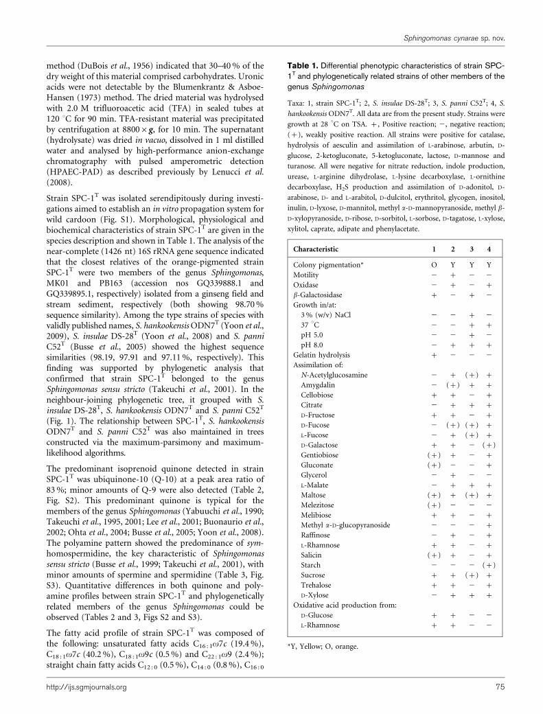
Table 1. Differential phenotypic characteristics of strain SPC-1T and phylogenetically related strains of other members of thegenus Sphingomonas
Taxa: 1, strain SPC-1T; 2, S. insulae DS-28T; 3, S. panni C52T; 4, S.
hankookensis ODN7T. All data are from the present study. Strains were
growth at 28 uC on TSA. +, Positive reaction; 2, negative reaction;
(+), weakly positive reaction. All strains were positive for catalase,
hydrolysis of aesculin and assimilation of L-arabinose, arbutin, D-
glucose, 2-ketogluconate, 5-ketogluconate, lactose, D-mannose and
turanose. All were negative for nitrate reduction, indole production,
urease, L-arginine dihydrolase, L-lysine decarboxylase, L-ornithine
decarboxylase, H2S production and assimilation of D-adonitol, D-
arabinose, D- and L-arabitol, D-dulcitol, erythritol, glycogen, inositol,
inulin, D-lyxose, D-mannitol, methyl a-D-mannopyranoside, methyl b-
D-xylopyranoside, D-ribose, D-sorbitol, L-sorbose, D-tagatose, L-xylose,
xylitol, caprate, adipate and phenylacetate.
Characteristic 1 2 3 4
Colony pigmentation* O Y Y Y
Motility 2 + 2 2Oxidase 2 + 2 +
b-Galactosidase + 2 + 2Growth in/at:
3% (w/v) NaCl 2 2 + 237 uC 2 2 + +
pH 5.0 2 2 + 2pH 8.0 2 + + +
Gelatin hydrolysis + 2 2 2Assimilation of:
N-Acetylglucosamine 2 + (+) +
Amygdalin 2 (+) + +
Cellobiose + + 2 +
Citrate 2 + + +
D-Fructose + + 2 +
D-Fucose 2 (+) (+) +
L-Fucose 2 + (+) +
D-Galactose + + 2 (+)
Gentiobiose (+) + 2 +
Gluconate (+) 2 2 +
Glycerol 2 + 2 2L-Malate 2 + + +
Maltose (+) + (+) +
Melezitose (+) 2 2 2Melibiose + + 2 +
Methyl a-D-glucopyranoside 2 2 2 +
Raffinose 2 + 2 +
L-Rhamnose + + 2 +
Salicin (+) + 2 +
Starch 2 2 2 (+)
Sucrose + + (+) +
Trehalose + + 2 +
D-Xylose 2 + + +
Oxidative acid production from:
D-Glucose + + 2 2L-Rhamnose + + 2 2
*Y, Yellow; O, orange.
Sphingomonas cynarae sp. nov.
http://ijs.sgmjournals.org 75

(18.4%), C18 : 0 (8.1%); hydroxy fatty acid C14 : 0 2-OH(5.4%); and C17 : 0 cyclo (2.0%) (Table 4, Fig. S4). The fattyacid profile confirmed the results of phylogenetic analysis.The profile was characterized by predominance of C18 : 1v7cand high levels of C16 : 0, which are typical of the majority ofmembers of the alphaproteobacteria, while the presence of 2-hydroxymyristic acid (C14 : 0 2-OH) as the major hydro-xylated fatty acid, and the absence of 3-hydroxy fattyacids are important markers of members of the familySphingomonadaceae (Busse et al., 1999, 2003; Takeuchi &Hiraishi, 2001; Takeuchi et al., 2001). Quantitative differ-ences in the fatty acid profiles between strain SPC-1T and
2‘
‘ ’
’
Fig. 1. Neighbour-joining phylogenetic tree based on 16S rRNA gene sequences showing the positions of Sphingomonascynarae sp. nov. SPC-1T with respect to other members of the genus Sphingomonas. Bootstrap values (expressed aspercentages of 1000 replicates) of .50% are shown at branch points. Filled circles and ‘X’ indicate that the correspondingnodes were also recovered in trees generated with maximum-parsimony and maximum-likelihood algorithms, respectively.Rhodospirillum rubrum ATCC 11170T was used as an outgroup. Bar, 0.01 substitutions per nucleotide position.
Table 2. Isoprenoid quinones in strain SPC-1T and phylogen-etically related strains of other members of the genusSphingomonas
Strain Q-9
(peak area, %)
Q-10
(peak area, %)
SPC-1T 17.0 83.0
S. insulae DS-28T 9.7 90.3
S. panni C52T 20.2 79.8
S. hankookensis ODN7T 8.9 91.1
A. Tala and others
76 International Journal of Systematic and Evolutionary Microbiology 63

phylogenetically related members of the genus Sphingomonascould be also observed (Table 4). Major polar lipidsidentified in strain SPC-1T were sphingoglycolipid, phos-phatidylcholine, phosphatidylglycerol, phosphatidylethano-lamine and diphosphatidylglycerol. Small amounts ofphosphatidylmonomethylethanolamine and several uniden-tified glyco- and phospholipids were also detected (Fig. S5).The DNA G+C content of strain SPC-1T was 66.1 mol%.
As SPC-1T, S. insulae DS-28T, S. hankookensis ODN7T andS. panni C52T shared high (.97%) 16S rRNA genesequence similarity, quantitative DNA–DNA hybridizationwas used to discriminate between these taxa. Analysis ofDNA relatedness between the pairs SPC-1T/S. insulae DS-28T (26.5%, reciprocal 29.6%), SPC-1T/S. hankookensisODN7T (19.0%, reciprocal 23.0%) and SPC-1T/S. panniC52T (20.9%, reciprocal 23.1%) did not reveal anyrelatedness at the species level. This finding was consistentwith the results of phenotypic analysis (Table 1). Many
differential phenotypic properties could be observedbetween strain SPC-1T and its close relatives S. insulaeDS-28T, S. hankookensis ODN7T and S. panni C52T, whichwere tested in parallel (Table 1). The highest degree ofsimilarity in terms of physiological characteristics (about73%) was found between SPC-1T and S. insulae DS-28T.
In liquid medium, strain SPC-1T produced a considerableamount of water-soluble EPS. Analytical methods showedthat SPC-1T EPS glycosyl composition is largely differentfrom that reported for sphingans (Fialho et al., 2008) andfrom that we obtained from Phytagel (Sigma-Aldrich), acommercial gellan we used for comparison. HPAEC-PADanalyses revealed mannose as the most abundant sugar witha molar ratio of 42.7±4.7%, followed by rhamnose(27.5±3.9 mol%), glucose (23.9±3.3 mol%) and galactose(5.9±1.1 mol%) (Table S2). Interestingly, glucuronic acid,the sugar conferring the anionic charge to the sphinganbackbone, and galacturonic acid were not detected indic-ating a substantial structural diversity of the isolated SPC-1T
EPS with respect to most sphingans produced by strains ofthe genus Sphingomonas (Fialho et al., 2008; White et al.,1996). To our knowledge, glucuronic acid is absent only inthe EPS produced by S. paucimobilis P4, in which thetrisaccharide structure D-glucose–D-glucose–L-rhamnose isa repeating unit structure (Lobas et al., 1994) and in the EPSproduced by Sphingomonas sp. CS101 in which glucose,mannose, fucose and rhamnose were detected in a molarratio of 2.1 : 1.1 : 1.0 : 0.1 (Seo et al., 2004).
The phylogenetic distinctiveness, together with DNA–DNArelatedness data and differential phenotypic properties, wassufficient to allocate strain SPC-1T to a species that isdistinct from recognized Sphingomonas species (Wayneet al., 1987; Stackebrandt & Goebel, 1994; Tindall et al.,2010). Therefore, on the basis of the presented data, strainSPC-1T is considered to represent a novel species of thegenus Sphingomonas, for which the name Sphingomonascynarae sp. nov. is proposed.
Description of Sphingomonas cynarae sp. nov.
Sphingomonas cynarae [cy.na9ra.e. N.L. gen. n. cynarae ofCynara, referring to the source of isolation Cynaracardunculus L. var. sylvestris (Lamk) Fiori (wild cardoon)].
Cells are Gram-stain-negative, non-spore-forming rods withrounded poles and are 0.4–0.561.5–3.0 mm in size. No
Table 3. Polyamines in strain SPC-1T and phylogenetically related strains of other membersof the genus Sphingomonas
Polyamine concentrations are given as mmol (g dry weight)21.
Strain Spermidine sym-Homospermidine Spermine
SPC-1T 0.84 15.88 2.71
S. insulae DS-28T 18.63 11.81 1.04
S. panni C52T 0.70 30.62 1.62
S. hankookensis ODN7T 1.88 42.40 1.98
Table 4. Fatty acids in strain SPC-1T and phylogeneticallyrelated strains of other members of the genus Sphingomonas
Taxa: 1, strain SPC-1T; 2, S. insulae DS-28T; 3, S. panni C52T; 4, S.
hankookensis ODN7T. Values are percentages of total fatty acids.
Components representing less than 0.5% in all strains were omitted.
ND, Not detected.
Fatty acid 1 2 3 4
Saturated
C12 : 0 0.5 ND 0.6 0.5
C14 : 0 0.8 3.0 2.1 2.3
C16 : 0 18.4 31.0 22.9 19.8
C18 : 0 8.1 5.7 3.4 4.5
Unsaturated
C16 : 1v7c 19.4 5.6 17.7 18.8
C16 : 1v5c ND 0.6 1.1 1.6
C18 : 1v7c 40.2 40.0 42.1 42.7
C18 : 1v9c 0.5 0.7 0.6 ND
C22 : 1v9c 2.4 0.4 1.2 2.5
Cyclopropane
C17 : 0 cyclo 2.0 1.5 1.2 0.7
C19 : 0 cyclo ND ND 0.5 ND
Hydroxy
C14 : 0 2-OH 5.4 10.9 5.9 6.2
Sphingomonas cynarae sp. nov.
http://ijs.sgmjournals.org 77

motility is observed by light microscopy. Colonies on TSAare convex, smooth, bright orange-pigmented and 1.5–2.0 mm in diameter after 5 days of incubation at 28 uC.Good growth occurs on TSA. Optimal temperature forgrowth is 28 uC. Growth does not occur at 4 uC or 37 uC.Optimal pH for growth is around 6.0–6.5. Poor growthoccurs above and below this range. Growth occurs in thepresence of 0–0.5% (w/v) NaCl. Anaerobic growth does notoccur on TSA or nutrient agar supplemented with nitrate.Oxidase-negative, catalase-positive. L-Arabinose, arbutin,cellobiose, D-fructose, D-galactose, gentiobiose (weakly), D-glucose, 2-ketogluconate, 5-ketogluconate, lactose, maltose(weakly), D-mannose, melibiose, melezitose (weakly), L-rhamnose, sucrose, trehalose, turanose, potassium gluconate(weakly) and salicin (weakly) are utilized as sole carbon andenergy sources, but N-acetylglucosamine, D-adonitol, D-arabinose, D- and L-arabitol, amygdalin, D-dulcitol, erythri-tol, D- and L-fucose, glycerol, glycogen, inositol, inulin, D-lyxose, D-mannitol, methyl a-D-glucopyranoside, methyl a-D-mannopyranoside, methyl b-D-xylopyranoside raffinose,D-ribose, D-sorbitol, L-sorbose, starch, D-tagatose, D- and L-xylose, xylitol, trisodium citrate, capric acid, adipic acid,malic acid and phenylacetic acid are not. Negative for indoleproduction, L-arginine dihydrolase, L-lysine decarboxylase,L-ornithine decarboxylase and urease activities, and nitratereduction. Positive for gelatin and aesculin hydrolysis,ONPG and p-nitrophenyl a-D-galactopyranoside. H2S isnot produced. Susceptible to gentamicin (10 mg), cipro-floxacin (5 mg), cefuroxime (30 mg) and weakly to nitrofur-antoin (100 mg), but not to chloramphenicol (30 mg) oramoxicillin/clavulanic acid (26.25/3.75 mg). The predom-inant ubiquinone is Q-10. The predominant polyamine issym-homospermidine. The major fatty acids (.10% of totalfatty acids) are C18 : 1v7c, C16 : 1v7c and C16 : 0. Major polarlipids are sphingoglycolipid, phosphatidylcholine, phospha-tidylglycerol, phosphatidylethanolamine and diphosphati-dylglycerol.
The type strain, SPC-1T (5JCM 17498T5ITEM 13494T),was isolated from the phyllosphere of Cynara cardunculus L.var. sylvestris (Lamk) Fiori (wild cardoon), a Mediterraneannative plant that is considered to be the wild ancestor of theglobe artichoke and cultivated cardoon. The DNA G+Ccontent of the type strain is 66.1 mol% (determined byHPLC).
Acknowledgements
This work was partially supported by grants from Italian MIUR toP. A. (Project PRIN-COFIN 2008).
References
Bligh, E. G. & Dyer, W. J. (1959). A rapid method of total lipidextraction and purification. Can J Biochem Physiol 37, 911–917.
Blumenkrantz, N. & Asboe-Hansen, G. (1973). New method forquantitative determination of uronic acids. Anal Biochem 54, 484–489.
Brown, J. K. (1994). Bootstrap hypothesis tests for evolutionarytrees and other dendrograms. Proc Natl Acad Sci U S A 91, 12293–12297.
Buonaurio, R., Stravato, V. M., Kosako, Y., Fujiwara, N., Naka, T.,Kobayashi, K., Cappelli, C. & Yabuuchi, E. (2002). Sphingomonasmelonis sp. nov., a novel pathogen that causes brown spots on yellowSpanish melon fruits. Int J Syst Evol Microbiol 52, 2081–2087.
Busse, H.-J., Kampfer, P. & Denner, E. B. (1999). Chemotaxonomiccharacterisation of Sphingomonas. J Ind Microbiol Biotechnol 23, 242–251.
Busse, H.-J., Denner, E. B. M., Buczolits, S., Salkinoja-Salonen, M.,Bennasar, A. & Kampfer, P. (2003). Sphingomonas aurantiaca sp.nov., Sphingomonas aerolata sp. nov. and Sphingomonas faeni sp. nov.,air- and dustborne and Antarctic, orange-pigmented, psychrotolerantbacteria, and emended description of the genus Sphingomonas. Int JSyst Evol Microbiol 53, 1253–1260.
Busse, H.-J., Hauser, E. & Kampfer, P. (2005). Description of twonovel species, Sphingomonas abaci sp. nov. and Sphingomonas pannisp. nov. Int J Syst Evol Microbiol 55, 2565–2569.
DuBois, M., Gilles, K. A., Hamilton, J. K., Rebers, P. A. & Smith, F.(1956). Colorimetric method for determination of sugars and relatedsubstances. Anal Chem 28, 350–356.
Eguchi, M., Ostrowski, M., Fegatella, F., Bowman, J., Nichols, D.,Nishino, T. & Cavicchioli, R. (2001). Sphingomonas alaskensis strainAFO1, an abundant oligotrophic ultramicrobacterium from theNorth Pacific. Appl Environ Microbiol 67, 4945–4954.
Ezaki, T., Hashimoto, Y. & Yabuuchi, E. (1989). Fluorometricdeoxyribonucleic acid-deoxyribonucleic acid hybridization in micro-dilution wells as an alternative to membrane filter hybridization inwhich radioisotopes are used to determine the genetic relatednessamong bacterial strains. Int J Syst Bacteriol 39, 224–229.
Felsenstein, J. (1981). Evolutionary trees from DNA sequences: amaximum likelihood approach. J Mol Evol 17, 368–376.
Fialho, A. M., Moreira, L. M., Granja, A. T., Popescu, A. O., Hoffmann, K.& Sa-Correia, I. (2008). Occurrence, production, and applications ofgellan: current state and perspectives. Appl Microbiol Biotechnol 79,889–900.
Galtier, N., Gouy, M. & Gautier, C. (1996). SEAVIEW and PHYLO_WIN:two graphic tools for sequence alignment and molecular phylogeny.Comput Appl Biosci 12, 543–548.
Gehrke, C. W. & Kuo, K. C. T. (editors) (1990). Chromatography andModification of Nucleosides. Amsterdam: Elsevier.
Gillis, M., De Ley, J. & De Cleene, M. (1970). The determination ofmolecular weight of bacterial genome DNA from renaturation rates.Eur J Biochem 12, 143–153.
Kim, O.-S., Cho, Y.-J., Lee, K., Yoon, S.-H., Kim, M., Na, H., Park, S.-C.,Jeon, Y. S., Lee, J.-H., Yi, H., Won, S. & Chun, J. (2012). IntroducingEzTaxon-e: a prokaryotic 16S rRNA gene sequence database withphylotypes that represent uncultured species. Int J Syst Evol Microbiol62, 716–721.
Kimura, M. (1980). A simple method for estimating evolutionary ratesof base substitutions through comparative studies of nucleotidesequences. J Mol Evol 16, 111–120.
Lal, R., Dogra, C., Malhotra, S., Sharma, P. & Pal, R. (2006). Diversity,distribution and divergence of lin genes in hexachlorocyclohexane-degrading sphingomonads. Trends Biotechnol 24, 121–130.
Lee, J.-S., Shin, Y. K., Yoon, J.-H., Takeuchi, M., Pyun, Y.-R. & Park,Y.-H. (2001). Sphingomonas aquatilis sp. nov., Sphingomonas koreensissp. nov., and Sphingomonas taejonensis sp. nov., yellow-pigmentedbacteria isolated from natural mineral water. Int J Syst Evol Microbiol51, 1491–1498.
A. Tala and others
78 International Journal of Systematic and Evolutionary Microbiology 63

Lenucci, M. S., Leucci, M. R., Piro, G. & Dalessandro, G. (2008).Variability in the content of soluble sugars and cell wall polysacchar-ides in red-ripe cherry and high-pigment tomato cultivars. J Sci FoodAgric 88, 1837–1844.
Lobas, D., Nimtz, M., Wray, V., Schumpe, A., Proppe, C. & Deckwer,W. D. (1994). Structure and physical properties of the extracellularpolysaccharide PS-P4 produced by Sphingomonas paucimobilis P4(DSM 6418). Carbohydr Res 251, 303–313.
McConaughy, B. L., Laird, C. D. & McCarthy, B. J. (1969). Nucleic acidreassociation in formamide. Biochemistry 8, 3289–3295.
Minnikin, D. E., O’Donnell, A. G., Goodfellow, M., Alderson, G.,Athalye, M., Schaal, A. & Parlett, J. H. (1984). An integratedprocedure for the extraction of bacterial isoprenoid quinines andpolar lipids. J Microbiol Methods 2, 233–241.
Moss, C. W. & Guerrant, G. O. (1983). Separation of bacterialubiquinones by reverse-phase high-pressure liquid chromatography.J Clin Microbiol 18, 15–17.
Nampoothiria, K. M., Singhania, R. R., Sabarinatha, C. & Pandey, A.(2003). Fermentative production of gellan using Sphingomonaspaucimobilis. Process Biochem 38, 1513–1519.
Nilgiriwala, K. S., Alahari, A., Rao, A. S. & Apte, S. K. (2008). Cloningand overexpression of alkaline phosphatase PhoK from Sphingomonassp. strain BSAR-1 for bioprecipitation of uranium from alkalinesolutions. Appl Environ Microbiol 74, 5516–5523.
Ohta, H., Hattori, R., Ushiba, Y., Mitsui, H., Ito, M., Watanabe, H.,Tonosaki, A. & Hattori, T. (2004). Sphingomonas oligophenolica sp.nov., a halo- and organo-sensitive oligotrophic bacterium from paddysoil that degrades phenolic acids at low concentrations. Int J Syst EvolMicrobiol 54, 2185–2190.
Saitou, N. & Nei, M. (1987). The neighbor-joining method: a newmethod for reconstructing phylogenetic trees.Mol Biol Evol 4, 406–425.
Scherer, P. & Kneifel, H. (1983). Distribution of polyamines inmethanogenic bacteria. J Bacteriol 154, 1315–1322.
Seo, E. J., Yoo, S. H., Oh, K. W., Cha, J., Lee, H. G. & Park, C. S.(2004). Isolation of an exopolysaccharide-producing bacterium,Sphingomonas sp. CS101, which forms an unusual type of sphingan.Biosci Biotechnol Biochem 68, 1146–1148.
Shaw, F. L., Elliott, K. A., Kinch, L. N., Fuell, C., Phillips, M. A. &Michael, A. J. (2010). Evolution and multifarious horizontal transferof an alternative biosynthetic pathway for the alternative polyaminesym-homospermidine. J Biol Chem 285, 14711–14723.
Sober, E. (1983). Parsimony in systematics: philosophical issues.Annu Rev Ecol Syst 14, 335–357.
Stabili, L., Gravili, C., Tredici, S. M., Piraino, S., Tala, A., Boero, F. &Alifano, P. (2008). Epibiotic Vibrio luminous bacteria isolated fromsome hydrozoa and bryozoa species. Microb Ecol 56, 625–636.
Stackebrandt, E. & Goebel, B. M. (1994). Taxonomic note: a place forDNA–DNA reassociation and 16S rRNA sequence analysis in the presentspecies definition in bacteriology. Int J Syst Bacteriol 44, 846–849.
Stolz, A. (2009). Molecular characteristics of xenobiotic-degradingsphingomonads. Appl Microbiol Biotechnol 81, 793–811.
Takeuchi, M. & Hiraishi, A. (2001). Taxonomic significance of 2-hydroxy fatty acid profiles of the species in the genus Sphingomonasand related taxa. IFO Res Commun 20, 72–82.
Takeuchi, M., Sakane, T., Yanagi, M., Yamasato, K., Hamana, K. &Yokota, A. (1995). Taxonomic study of bacteria isolated from plants:proposal of Sphingomonas rosa sp. nov., Sphingomonas pruni sp. nov.,Sphingomonas asaccharolytica sp. nov., and Sphingomonas mali sp.nov. Int J Syst Bacteriol 45, 334–341.
Takeuchi, M., Hamana, K. & Hiraishi, A. (2001). Proposal of the genusSphingomonas sensu stricto and three new genera, Sphingobium,Novosphingobium and Sphingopyxis, on the basis of phylogenetic andchemotaxonomic analyses. Int J Syst Evol Microbiol 51, 1405–1417.
Thompson, J. D., Higgins, D. G. & Gibson, T. J. (1994). CLUSTAL W:improving the sensitivity of progressive multiple sequence alignmentthrough sequence weighting, position-specific gap penalties andweight matrix choice. Nucleic Acids Res 22, 4673–4680.
Tindall, B. J., Rossello-Mora, R., Busse, H.-J., Ludwig, W. & Kampfer, P.(2010). Notes on the characterization of prokaryote strains fortaxonomic purposes. Int J Syst Evol Microbiol 60, 249–266.
Vigliotta, G., Nutricati, E., Carata, E., Tredici, S. M., De Stefano, M.,Pontieri, P., Massardo, D. R., Prati, M. V., De Bellis, L. & Alifano, P.(2007). Clonothrix fusca Roze 1896, a filamentous, sheathed,methanotrophic gamma-proteobacterium. Appl Environ Microbiol73, 3556–3565.
Wayne, L. G., Brenner, D. J., Colwell, R. R., Grimont, P. A. D., Kandler, O.,Krichevsky, M. I., Moore, L. H., Moore, W. E. C., Murray, R. G. E. & otherauthors (1987). International committee on systematic bacteriology.report of the ad hoc committee on reconciliation of approaches tobacterial systematics. Int J Syst Bacteriol 37, 463–464.
West, T. P. & Strohfus, B. (1998). Effect of carbon source onexopolysaccharide production by Sphingomonas paucimobilis ATCC31461. Microbiol Res 153, 322–327.
White, D. C., Sutton, S. D. & Ringelberg, D. B. (1996). The genusSphingomonas: physiology and ecology. Curr Opin Biotechnol 7, 301–306.
Yabuuchi, E., Yano, I., Oyaizu, H., Hashimoto, Y., Ezaki, T. &Yamamoto, H. (1990). Proposals of Sphingomonas paucimobilis gen.nov. and comb. nov., Sphingomonas parapaucimobilis sp. nov.,Sphingomonas yanoikuyae sp. nov., Sphingomonas adhaesiva sp.nov., Sphingomonas capsulata comb. nov., and two genospecies ofthe genus Sphingomonas. Microbiol Immunol 34, 99–119.
Yoon, J. H., Kang, S. J., Lee, S. Y. & Oh, T. K. (2008). Sphingomonasinsulae sp. nov., isolated from soil. Int J Syst Evol Microbiol 58, 231–236.
Yoon, J. H., Park, S., Kang, S. J., Kim, W. & Oh, T. K. (2009).Sphingomonas hankookensis sp. nov., isolated from wastewater. Int JSyst Evol Microbiol 59, 2788–2793.
Sphingomonas cynarae sp. nov.
http://ijs.sgmjournals.org 79