Kinnaird_Adam_S_201803_PhD.pdf - ERA
227
Metabolic Reprogramming and Epigenetic Regulation in Renal Cell Carcinoma by Adam Stewart Kinnaird A thesis submitted in partial fulfillment of the requirements for the degree of Doctor of Philosophy in Experimental Medicine Department of Medicine University of Alberta © Adam Stewart Kinnaird, 2018
-
Upload
khangminh22 -
Category
Documents
-
view
0 -
download
0
Transcript of Kinnaird_Adam_S_201803_PhD.pdf - ERA
Metabolic Reprogramming and Epigenetic Regulation in Renal Cell Carcinoma
by
Adam Stewart Kinnaird
A thesis submitted in partial fulfillment of the requirements for the degree of
Doctor of Philosophy
in
Experimental Medicine
Department of Medicine University of Alberta
© Adam Stewart Kinnaird, 2018
ii
Abstract
Renal cell carcinoma comprises approximately 2-3% of all malignancies,
with the majority, approximately 70%, being clear cell renal cell carcinoma
(ccRCC). ccRCC features many cancer hallmarks such as suppressed
mitochondrial glucose oxidation, apoptosis-resistance, angiogenesis, immune
evasion, uninhibited proliferation, and invasion and metastasis, thought to be
driven by sporadic mutations or epigenetic silencing of the tumor suppressor
gene, von Hippel Lindau (VHL). VHL loss leads to up-regulation of its target
protein, hypoxia inducible factor (HIF), even in normoxia. HIF controls many
downstream targets in order to prepare the cell for hypoxia, with the net effect
being suppression of mitochondrial glucose oxidation, up-regulation of glycolysis,
angiogenesis and apoptosis-resistance. Unchecked, as in VHL-deficiency, this
becomes a critical step in tumorigenesis. We show that reversing the suppression
of mitochondrial glucose oxidation in ccRCC using dichloroacetate, an inhibitor
of the HIF target gene mitochondrial pyruvate dehydrogenase kinase (PDK),
which inhibits a major producer of acetyl-CoA, the Pyruvate Dehydrogenase
Complex (PDC), results in increased production of mitochondrial acetyl-CoA via
PDC, reduced proliferation and angiogenesis, and induces apoptosis in animal
models. We also show that mitochondrial PDC dynamically translocates to the
nucleus in multiple types of cancer cells, including ccRCC, in response to growth
factor signaling, to produce acetyl-CoA in the nucleus. This local production of
acetyl-CoA by nuclear PDC is used to acetylate core histones involved in S-phase
progression and cellular proliferation. Next, we show that VHL, which has been
iii
described as a multipurpose adapter protein, directly binds the tumor suppressor
p53, preventing its activation, promoter binding and expression of its target genes.
This process is independent of the VHL-HIF axis and results in an attenuation of
p53 inducing therapies. Finally, we study Myocyte Enhancer Factor 2A (Mef2A),
a transcription factor with ties to multiple hallmarks of cancer, like mitochondrial
suppression, invasion and immune evasion. We show that Mef2A expression is
up-regulated in patient tumors compared to adjacent normal kidney parenchyma
and that nuclear Mef2A levels correlate with larger tumor size. Mef2A activity is
increased in VHL-deficient ccRCC cells due to HIF-mediated growth factor
signaling and it induces expression of the pro-tumor chemokine, CCL20.
Inhibition of Mef2A results in reduced ccRCC tumor growth in vivo and may
provide mechanistic insight into immune evasion as immune checkpoint
inhibitors develop into the first line therapy in metastatic ccRCC.
iv
Preface Chapter 1 of this thesis has been previously published as Kinnaird, A. and Michelakis, E. Metabolic Modulation of Cancer: A new frontier with great translational potential. (2015) Journal of Molecular Medicine 93(2):127-42 Chapter 2 of this thesis has been previously published as Kinnaird A, Dromparis P, Saleme B, Gurtu V, Watson K, Paulin R, Zervopoulos S, Stenson T, Sutendra G, Pink DB, Carmine-Simmen K, Moore R, Lewis JD and Michelakis ED. Metabolic Modulation of Clear-cell Renal Cell Carcinoma with Dichloroacetate, an Inhibitor of Pyruvate Dehydrogenase Kinase. (2016) European Urology 69(4):734-44 Chapter 3 of this thesis has been previously published as Sutendra G, Kinnaird A, Dromparis P, Paulin R, Stenson TH, Haromy A, Hashimoto K, Zhang N, Flaim E and Michelakis ED. A functional nuclear pyruvate dehydrogenase complex is important for a mitochondria-independent generation of acetyl-CoA and histone acetylation. (2014) Cell 158(1):84-97 Chapter 4 and 5 have not yet been published but are in preparation for submission to peer-reviewed journals at this time.

v
Acknowledgements
First, I would like to acknowledge my supervisor, Professor Evangelos Michelakis, for
his mentorship, dedication to his trainees, and creation of a ‘sky is the limit’ environment
for exploring new ideas and hypotheses. I have learned much from his advice and look
forward to continuing to work together as clinician scientists. I must also acknowledge
my committee members, Dr. Ronald Moore and Dr. Jayan Nagendran, for their
leadership and insight throughout my studies. Most importantly, I would like to thank my
wife, Dr. Erin Bristow and my parents, Helga and David Kinnaird, for their love, support
and genuine excitement for me to pursue a career in science and surgery. Furthermore, I
must thank Dr. Peter Dromparis and Dr. Gopinath Sutendra for their friendship, guidance
and laying the ground work from which I started this endeavor. Finally, I would like to
acknowledge Dr. Vikram Gurtu, Dr. Aristeidis Boukouris, Sotirios Zervopoulos and
Bruno Saleme for the laughter, the friendship and the hard work together, without which
this would not have been possible.
vi
Table of Contents
Chapter One
Title page Page 1
Introduction Page 2
1.1.0 Cancer’s abnormal metabolism and its molecular consequences Page 3
1.2.0 The cause of mitochondrial suppression in cancer Page 7
1.2.1 Growth factors Page 8
1.2.2 Oncogenes Page 8
1.2.3 Regulation of factors that control mitochondrial homeostasis Page 9
1.2.4 Enzymatic mutations Page 10
1.3.0 Mitochondria-targeting cancer therapies Page 11
1.3.1 Glucose Transport and Phosphorylation Page 11
1.3.2 Pyruvate Kinase M2 (PKM2) Activation Page 12
1.3.3 Pyruvate Dehydrogenase Kinase (PDK) Inhibition Page 14
1.3.4 Lactate Dehydrogenase A (LDHA) Inhibition Page 18
1.3.5 Mutant Isocitrate Dehydrogenase (IDH) Inhibition Page 19
1.4.0 Cancer Stem Cells Page 20
1.5.0 Glucose oxidation and histone acetylation Page 21
Tables and Figures
Table 1-1 Page 23
Fig. 1-1 Page 24
Fig. 1-2 Page 26
Fig. 1-3 Page 28
Fig. 1-4 Page 29
Fig. 1-5 Page 30
References Page 31
Chapter Two
Title page Page 44
Abstract Page 45
Introduction Page 47
Results
2.1 Pyruvate Dehydrogenase Kinase is up-regulated in ccRCC Page 49
2.2 DCA increases PDH activity and Mitochondrial Function Page 49
vii
2.3 DCA inhibits ccRCC HIF activity and decreases angiogenesis in vitro Page 50
2.4 HIF is essential for DCA’s effects on ccRCC Page 52
2.5 DCA reduces angiogenesis and tumor growth in vivo in ccRCC Page 54
Discussion Page 56
Materials and Methods Page 60
Figures
Fig. 2-1 Page 65
Fig. 2-2 Page 66
Fig. 2-3 Page 67
Fig. 2-4 Page 69
Fig. 2-5 Page 70
Fig. 2-6 Page 71
Fig. 2-7 Page 72
Fig. 2-8 Page 73
Fig. 2-9 Page 74
Fig. 2-10 Page 76
Fig. 2-11 Page 77
Fig. 2-12 Page 79
Fig. 2-13 Page 80
References Page 81
Chapter Three
Title page Page 85
Abstract Page 86
Introduction Page 87
Results
3.1 All Components of PDC are Present in the Nucleus Page 89
3.2 Nuclear PDC is Functional and Can Generate Acetyl-CoA from Pyruvate Page 90
3.3 Nuclear PDC is Important for Histone Acetylation Page 92
3.4 Pyruvate Dehydrogenase Kinase is Not Present in the Nucleus Page 93
3.5 PDC Translocates from the Mitochondria to the Nucleus During S-phase Page 94
3.6 Signals Increasing Nuclear PDC Levels Page 96
3.7 Nuclear PDC is Important for S-phase Entry and Cell Cycle Progression Page 98
viii
Discussion Page 101
Materials and Methods Page 104
Figures
Fig. 3-1 Page 112
Fig. 3-2 Page 113
Fig. 3-3 Page 115
Fig. 3-4 Page 117
Fig. 3-5 Page 119
Fig. 3-6 Page 120
Fig. 3-7 Page 121
Fig. 3-8 Page 123
Fig. 3-9 Page 124
Fig. 3-10 Page 126
Fig. 3-11 Page 128
Fig. 3-12 Page 130
Fig. 3-13 Page 133
Fig. 3-14 Page 135
References Page 137
Chapter Four
Title page Page 141
Abstract Page 142
Introduction Page 143
Results
4.1 VHL inhibits p53 binding to the promoters and expression of its target
genes, p21 and PUMA, in response to a p53 inducing stimulus Page 145
4.2 VHL inhibits p53-dependent target gene p21 expression, promoting
cell proliferation Page 146
4.3 VHL inhibits p21 independent of HIF and does not require prolyl
hydroxylation Page 147
4.4 VHL inhibits induction of p53-dependent apoptosis and attenuates
the response to anthracycline chemotherapy in vivo Page 148
Discussion Page 150
Materials and Methods Page 152
ix
Figures
Fig. 4-1 Page 155
Fig. 4-2 Page 156
Fig. 4-3 Page 157
Fig. 4-4 Page 158
Fig. 4-5 Page 159
Fig. 4-6 Page 160
Fig. 4-7 Page 161
Fig. 4-8 Page 162
Fig. 4-9 Page 163
References Page 164
Chapter Five
Title page Page 167
Abstract Page 168
Introduction Page 170
Results
5.1 Mef2A expression is up-regulated in renal tumor tissue Page 172
5.2 Mef2A regulates expression of CCL20 in kidney tumor cells Page 172
5.3 Loss of Mef2A inhibits proliferation, migration and invasion
and promotes mitochondrial activity and apoptosis of ccRCC cells Page 173
5.4 VHL-deficiency increases Mef2A transcriptional activity Page 173
5.5 Mef2A knockdown reduces tumor growth in-vivo Page 174
Discussion Page 175
Materials and Methods Page 177
Figures
Fig. 5-1 Page 179
Fig. 5-2 Page 180
Fig. 5-3 Page 181
Fig. 5-4 Page 182
Fig. 5-5 Page 183
Fig. 5-6 Page 184
Fig. 5-7 Page 185
Tables

x
Table 5-1 Page 185
References Page 186
Chapter Six
Title page Page 189
6.1 Discussion and Conclusions Page 190
6.2 Future Directions Page 193
Bibliography Page 195
xi
List of Abbreviations 2-HG – 2-hydroxyglutarate !"m – Mitochondrial Membrane Potential #KG – Alpha-ketoglutarate ACL – ATP-Citrate Lyase AML – Acute myeloid leukemia ADP – Adenosine diphosphate ATP – Adenosine triphosphate ccRCC – Clear Cell Renal Cell Carcinoma CCL20 – Chemokine ligand 20 CCR6 – Chemokine receptor 6 CoA – Coenzyme A CRISPR – Clustered regularly interspaced short palindromic repeats CS – Citrate Synthase DCA – Dichloroacetate DNA – Deoxyribonucleic acid EGF – Epidermal growth factor EGFP – Enhanced Green Fluorescent Protein EGFR – Epidermal growth factor receptor ETC – Electron transport chain FADH – Flavin adenine dinucleotide FDG – Fluorinated Deoxyglucose G6PD – Glucose-6-Phosphate Dehydrogenase GBM – Glioblastoma multiforme GLUT – Glucose transporter GLY – Glycolysis GO – Glucose Oxidation HIF – Hypoxia Inducible Factor HK – Hexokinase HSF1 – Heat shock factor 1 HSP70 – Heat shock protein 70 IDH – Isocitrate Dehydrogenase LDH – Lactate Dehydrogenase LCSC – Lung cancer stem cell PET – Positron Emission Tomography PDH – Pyruvate Dehydrogenase PDC – Pyruvate Dehydrogenase Complex PDK – Pyruvate Dehydrogenase Kinase PHD – Prolyl hydroxylase PKM2 – Pyruvate Kinase isoform 2 PPP – Pentose Phosphate Pathway MAPK – Mitogen-activated protein kinase MDM2 – Mouse double minute 2 homolog MEF – Mouse embryonic fibroblast Mef2A – Myocyte Enhancer Factor 2A MLS – Mitochondrial localization sequence MPP – Mitochondrial processing peptidase mROS – Mitochondrial reactive oxygen species MnSOD – Manganese Superoxide Dismutase NADH – Nicotinamide adenine dinucleotide
xii
NADPH – Nicotinamide adenine dinucleotide phosphate NFAT – Nuclear Factor of Activated T-cells PDGF – Platelet derived growth factor PGC1# – Peroxisome proliferator-activated receptor gamma coactivator 1 alpha PT – Proximal Tubules PUMA – p53 Upregulated Modulator of Apoptosis SAEC – Small Airway Epithelial Cells SCS – Succinyl-CoA Synthetase SDF-1 – Stromal cell-derived factor 1 SDH – Succinate Dehydrogenase shRNA – Short hairpin ribonucleic acid siRNA – Short interfering ribonucleic acid SIRT3 – Sirtuin 3 TACE – Transarterial Chemoembolization TCGA – The Cancer Genome Atlas TGF# – Transforming growth factor alpha TIGAR – Tp53-induced glycolysis and apoptosis regulator TMRM – Tetramethylrhodamine VEGF – Vascular endothelial growth factor VHL – von Hippel Lindau

! 1!
Chapter One
Metabolic modulation of cancer: A new frontier with
great translational potential
! 2!
Introduction
The majority of chemotherapeutic drugs for cancer inhibit pathways fundamental
to the life of all cells, leading to adverse effects on healthy tissues [1]. In addition,
traditional drug development in cancer has been focusing on a single molecular
pathway, following the “one gene - one drug” approach. It became apparent
however that most tumors are characterized by multiple molecular abnormalities
so that when the therapies are offered even in combination of 2-3 drugs, the tumor
eventually relapses. It is a rather rare example to find therapies that are effective
and not toxic. This requires the identification of molecular abnormalities that are
not only critical for the life of cancer – but not normal cells – but are also the
dominant or the only molecular abnormality within the tumor. For example, this
can be seen in certain leukemias or tumors where a mutation in the majority of
cancer cells not only dominates their molecular phenotype but is also critical for
their survival, explaining the great success of agents such as Gleevec for Chronic
Myelogenous Leukemia [2] or Herceptin for certain breast cancers [3]. Yet, most
cancers, like glioblastoma for example, are characterized by several cellular
phenotypes within each tumor and yet, in each of these cell types there are several
genetic and molecular abnormalities [4], obviously not susceptible to a single or
even a combination of 2-3 drugs. Is it possible to identify a common denominator
across all of these abnormalities that is not only critical for the survival of cancer
cells but is also not present in normal cells? And to push the envelop even further,
is it possible that this common denominator is also present in cancer stem cells, so
that if targeted, tumor relapse would be limited as well [5]? In other words, is
there an Achilles’ heel for such complex and molecularly plastic tumors? Recent
work over the past 10 years suggests that such a common denominator, unique to
cancer cells, may exist and be no other than the unique metabolism of cancer
cells, first identified by Otto Warburg, more than 90 years ago [6]. Unfortunately,
the work of Warburg, who was awarded the Nobel Prize for his work on
metabolism, did not translate in cancer therapies for many decades as it was
wrongly assumed that the unique cancer metabolism was a consequence, not a
! 3!
cause or contributing factor in cancer. We now know that if not causal, like in
certain examples of mutations in key metabolic enzymes, this metabolic
remodeling is a strong contributing or promoting factor as it promotes
proliferation, resistance to apoptosis and tumor angiogenesis, facilitating tumor
growth and metastasis. Even better, tumor metabolism is typically different than
that of normal cells. The development of ‘metabolic modulators’ over the past 10
years has already entered early-phase trials. Although it sounds ‘too good to be
true’ that metabolism is the long-sought Achilles’ heel for cancer, a new field has
already opened, which includes new therapies and metabolism-based imaging
such as FDG-PET. Metabolic Oncology is an exciting frontier in the battle against
cancer and this review focuses on the rationale for the development of metabolic
modulators, concentrating on those that hold promise or already have entered
human trials.
1.1.0 Cancer’s abnormal metabolism and its molecular consequences.
The cornerstone of metabolism in the cell is the mitochondrion. Traditional
biochemistry taught us that mitochondria’s main job is to generate heat and
energy (ATP). We now know that although the most efficient means for ATP
production lie within mitochondria, ATP can be adequately produced outside of
mitochondria, through cytoplasmic glycolysis, at amounts enough to support the
energy-hungry cancer cells. We also now know that mitochondria do much more:
they produce diffusible mediators that can regulate multiple molecular pathways
in the cell and even the nucleus. They can also induce apoptosis, an intriguing fact
since the “provider of life” (i.e. ATP) can also be the provider of death for the
cell. It was perhaps this “paradox” that did not allow mitochondria to be seen as
targets for pro-apoptotic strategies until recently. Therefore, suppression of
mitochondrial function can suppress apoptosis and alter cellular signaling toward
a pro-proliferative phenotype as we discuss below. It is thus not a surprise that
cancer cells have suppressed mitochondrial function.
Normally, cells metabolize glucose to pyruvate in the cytoplasm by
glycolysis; then this is converted by pyruvate dehydrogenase (PDH) into acetyl-
! 4!
CoA, which enters the mitochondrial Krebs’ cycle, where it is oxidized to
eventually produce the electron donors NADH and FADH2. These donate
electrons down a redox-gradient in the electron transport chain (ETC), while
protons are pumped out of the mitochondria and mitochondria-derived reactive
oxygen species (mROS; mostly the negatively charged superoxide) are generated,
creating a membrane potential (!"m) across the mitochondrial inner membrane
(Figure 1-1). This process uses oxygen as the final electron acceptor in Complex
IV of the ETC (forming water), and uses the stored energy of the !"m to produce
and release ATP. As protons re-enter the inner membrane, they release energy
used to pump ATP out in the cytoplasm and bring ADP in. A similar process is
followed in the oxidation of fatty acids, forming acetyl-CoA, which is also
oxidized in the Krebs’ cycle producing the same electron donors. Thus
mitochondria process fuel (carbohydrates, lipids, oxygen) to produce energy and
have evolved to be important fuel sensors. When fuel supply is ample, the growth
and differentiation of tissues can be kept under control by the coordinated
induction of apoptosis, producing effective cell population control. In contrast,
when fuel is limited, mitochondria suppress apoptosis in an attempt to preserve
life under stressed conditions. It is here that mitochondria can be “fooled” by the
cancer cells.
Let’s say, for example, that oxygen is limited. This de-facto inhibits
oxidative phosphorylation in the mitochondria. Immediately the mitochondria
sense the lack of fuel and ignite a series of mechanisms for the cell to: a) seek
alternate means of ATP production and b) suppress apoptosis since stress may be
imminent:
a) The expression of glucose transporters is increased and more glucose
enters the cell. This can be achieved by the activation of HIF1α directly by
hypoxia but -remarkably- the mitochondrial suppression can also activate HIF1α.
This is because alpha-ketoglutarate (αKG), a Krebs’ product that is a required co-
factor for the prolyl-hydroxylases that de-stabilize HIF1α, is decreased [7].
Independent of its stabilization, the HIF1α transcription machinery, in part driven
by the redox-sensitive p53, may also be activated when the diffusible mROS
! 5!
decrease (for example H2O2 from superoxide’s dismutation from manganese
superoxide dismutase) [8-11]. Activated HIF1α not only increases the expression
of glucose transporters but also increases the transcription of almost all the
glycolytic enzymes in the cytoplasm. Thus, with more glucose and activated
glycolysis, more ATP can be produced. Normally the efficiency of glycolysis for
ATP is less than that of mitochondria (each mol of glucose produces 36 mol of
ATP in mitochondrial glucose oxidation but only 2 mol of ATP in glycolysis), but
as glycolysis is enhanced the cell may eventually compensate for the ATP that is
missing from the inhibited mitochondria.
b) The next thing that happens is that mitochondria hyperpolarize (increased
!"m) [12, 13]. The pro-apoptotic factors stored inside mitochondria (like
cytochrome c or apoptosis inducing factor) are unable to leak out, making cancer
cells resistant to mitochondria-dependent apoptosis. This is because these factors
leak through the mitochondrial transition pore (MTP); a mega-channel that, being
voltage- and redox-sensitive, tends to close while membrane potential is high and
mROS are low [14]. The mechanism for mitochondrial hyperpolarization is not
clear although it has been known since the 1980s to be a feature of most cancers
[15]. One potential mechanism is that glycolysis leads to a GSK-3#-driven
translocation of the cytoplasmic hexokinase II (HK2) to the outer mitochondrial
membrane, where it binds and inhibits the major channel that releases negatively
charged ions, i.e. the voltage-dependent anion channel (VDAC; a critical
component of the MTP), leading to a build up of anions inside the mitochondria
[16, 17]. (Figure 1-1) Another mechanism may be that the enhanced production of
ATP in the cytoplasm due to the enhanced glycolysis, causes a decrease in the
ATP/ADP gradient in the microdomains around the outer mitochondrial
membrane, decreasing the function of ATP synthase and thus preventing the re-
uptake of the positively charged H+ ions back to the mitochondria [16]. Therefore,
mitochondrial suppression supports a state of apoptosis-resistance, while adequate
production of ATP is maintained. Interestingly, HIF1α also induces pyruvate
dehydrogenase kinase (PDK), inhibiting PDH, and thus decreasing the production
of acetyl-CoA entering the Krebs’ cycle.

! 6!
Let’s now see what would happen if PDH were to be inhibited by other
means, not by the hypoxia-mediated activation of HIF1α. Immediately, the
mitochondria will sense a lack of fuel entering them since the supply of acetyl-
CoA will decrease (despite the fact that the supply of glucose and oxygen to the
cell remains normal). They will then ignite the same process described above,
leading to resistance to apoptosis. One can follow the same logic and realize that
inhibition of any critical enzyme used in glucose or fatty acid oxidation may have
the same consequences, “fooling the mitochondria” and inhibiting oxidative
phosphorylation, as if there was lack of fuel. This mechanism can be used by
cancer cells to inhibit apoptosis, a sine qua non of cancer. At the same time, there
are three additional advantages that the cancer cells gain by this “inappropriate”
mitochondrial suppression:
First, the cancer cell can now use pyruvate, that is not oxidized in the
Krebs’ cycle, for biomass generation, as the tumor grows [18]. In other words, the
unused pyruvate helps a dividing cell create the amino acids, nucleotides, and
lipids needed to replicate. Specifically, unused pyruvate can be transaminated to
produce amino acids. Similarly, unused pyruvate can be metabolized and shunted
into the pentose phosphate pathway to produce both nucleotides and NADPH,
which is required to synthesize lipids [18].
Second, as in the case of decreased production of αKG which contributes
to HIF1α activation, there are other downstream signals from mitochondria, like
decreased mROS, dysregulation of cytoplasmic calcium or induction of
chaperones that via the retrograde pathway (a series of mechanisms that
mitochondria induce under stress to send signals to the nucleus) directly or
indirectly activate other pro-proliferative master transcription factors (like
Nuclear Factor of Activated T-cells (NFAT) for example) [12, 19-21]. Like
HIF1α, NFAT can suppress several aspects of mitochondrial function, enhancing
the proliferative potential of cancer cells and driving positive reinforcing feedback
loops (proliferative signals ! mitochondrial suppression ! proliferative signals).
Third, this mitochondrial remodeling promotes angiogenesis, not
surprising since this mitochondrial remodeling “mimics” hypoxia, a well-known
! 7!
driver of angiogenesis. While hypoxia and HIF1α activation induce mitochondrial
suppression [22] the reverse is also true [10], establishing a powerful feedback
loop that sustains angiogenesis even in the absence of hypoxia. The majority of
solid tumors experience hypoxia during early tumor development as it occurs
several cell layers away from capillaries (~150µm) [23]. This leads to an initial
primary hypoxia-driven activation of HIF1α and angiogenic signaling to form
new blood vessels to supply the growing tumor with nutrients. However, HIF1α
stabilization is also regulated by several diffusible mitochondria-derived factors,
like αKG and mROS as discussed above [23]. These lead to the inhibition of the
degradation of HIF1α through VHL-dependent ubiquitination or activation of its
transcriptional activity, respectively [24, 25]. Thus as the newly formed blood
vessels bring in more oxygen, limiting hypoxia, the mitochondrial suppression
that takes place in the tumor, now causes a primary normoxic activation of HIF1α,
sustaining angiogenesis as the tumor continues to grow [10, 22]. Furthermore, by
reducing pyruvate into lactic acid during glycolysis, cancer cells increase
extracellular acidosis. This potentiates breakdown of the extracellular matrix and
allows penetration of the cancer through the basement membrane, thereby driving
metastasis [26].
Thus mitochondrial suppression promotes suppressed apoptosis, increased
proliferation, angiogenesis and metastatic potential and so can be seen as a critical
hub of cancer signaling. While hypoxia is a physiologic mechanism for global
mitochondrial suppression, we will now discuss a variety of prominent cancer
mechanisms that all lead to mitochondrial suppression, in a sense, mimicking
hypoxia.
1.2.0 The cause of mitochondrial suppression in cancer
Mitochondria are well-known “integrators” of multiple signals [12]. Thus
mitochondrial suppression can occur through four overarching mechanisms in
cancer: (1) growth and transcription factor signaling or (2) off-target effects of
specific oncogenes that can suppress specific enzymes or ETC complexes; (3)
suppression of factors regulating mitochondrial homeostasis and (4) mutations in
! 8!
key metabolic enzymes. It is important to note that more than one of these factors
may be present within any given tumor.
1.2.1 Growth Factors: One of the pleiotropic effects of growth factors up-
regulated in cancer, such as epidermal growth factor (EGF) and fibroblast growth
factor (FGF), is the regulation of the flux of pyruvate into the mitochondria by the
gate-keeping enzyme PDH. EGF and FGF increase the activity of Pyruvate
Dehydrogenase Kinase (PDK) [27] which phosphorylates and inhibits PDH, thus
preventing the entry of pyruvate into the Krebs’ cycle and suppressing glucose
oxidation (GO) [28]. Furthermore, EGF signaling has recently been show to also
directly inhibit PDH function, through a PDK-independent tyrosine
phosphorylation of its E1 subunit [29]. Furthermore, PDK is a target gene of
HIF1α, which is up-regulated in most solid tumors [23, 30]. HIF1α works at
multiple levels to shift the balance of metabolism from GO toward glycolysis,
including up-regulation of glucose transporters and glycolytic enzymes. HIF1α
also increases the translation of the EGF receptor, thereby suppressing
mitochondrial function by both up-regulating and activating PDK [31]. There are
four PDK isoenzymes with variable tissue expression and some, like PDK 4, are
inducible in conditions of metabolic stress [32]. Thus it is reasonable to assume
that other, not yet identified, “fuel sensing” mechanisms exist in inducing the
transcription of one or more PDKs in tumors, in addition to increasing PDK
activity through tyrosine kinase signaling.
1.2.2 Oncogenes: Mitochondrial function is also suppressed by many oncogenes,
with p53 and c-MYC being prime examples. The Cancer Genome Atlas (TGCA)
has identified p53 to be the most commonly mutated gene in cancer [33]. p53
inhibits glycolysis by inducing the expression of Tp53-induced glycolysis and
apoptosis regulator (TIGAR) as well as by reducing the expression of glycolytic
enzymes, like phophoglycerate mutase [34, 35]. Furthermore, p53 enhances GO
by increasing the expression of cytochrome c oxidase subunits of the ETC [36].
Loss of p53 function causes up-regulation of both the glycolytic enzyme
! 9!
Hexokinase II (HKII) and PDK [37, 38]. Overall, loss of p53 function suppresses
mitochondria and shifts the cell to a more glycolytic phenotype. It is important to
note that post-translational modifications like acetylation, a process intimately
linked to mitochondrial function, are also known to regulate p53’s function in
cancer and in the absence of mutations [39]. Like p53, c-MYC increases the
expression of multiple glycolytic enzymes including HKII, phosphofructokinase,
GAPDH and Enolase A, as well as glucose transporters and Lactate
Dehydrogenase (LDH) [40-42]. On top of driving glycolysis, up-regulation of
LDH shifts the flux of pyruvate away from GO, thereby further suppressing
mitochondrial function.
1.2.3 Regulation of factors that control mitochondrial homeostasis: Global
mitochondrial function can be regulated through inhibition of mitochondria-
specific factors like Sirtuin 3 (Sirt3; the main de-acetylase in the mitochondria)
and Uncoupling Protein-2 (UCP-2; a putative mitochondria calcium transporter).
Inhibition of Sirt3 and UCP-2 suppresses mitochondrial function by increasing
protein acetylation and decreasing mitochondrial calcium, respectively. When
Sirt-3 is deficient, the acetylation of the majority of proteins involved in oxidative
metabolism, including Krebs’ enzymes and ETC complexes, increases; this post-
translational modification typically leads to the inhibition of enzymatic function
[43, 44]. Overall, loss of Sirt3 activity can cause up to 50% reduction in
mitochondria-derived ATP and respiration. Thus it is not surprising that loss of
Sirt3 function has been shown to promote the Warburg Effect and cancer. In fact,
embryonic fibroblasts lacking Sirt3 require activation of only one other oncogene
to transform into cancer [45], in contrast to wild-type cells that require at least
two. Moreover, mice deficient in Sirt3 spontaneously develop breast cancer, and
many human cancers are deficient in Sirt3 [45-47]. These observations have led
investigators to propose that Sirt3 fulfills the criteria to be an oncogene [45].
Similarly, loss of UCP-2 function, which despite its name is a weak
uncoupler but good mediator of calcium entry into the mitochondria, promotes
proliferation [20] in part due to a reduction in mitochondrial calcium, which
! 10!
decreases activity of many calcium-dependent mitochondrial enzymes such as
PDH, isocitrate dehydrogenase, and α-ketoglutarate dehydrogenase [48, 49]. Mice
deficient in UCP-2 are more prone to develop colon cancer than wild-type UCP-2
littermates when exposed to a carcinogen, demonstrating a predisposition to
oncogenic transformation in these glycolytic animals [50]. Similarly to Sirt3,
UCP2 was recently proposed to be a tumor-suppressing factor [20].
Another way that mitochondrial function can be suppressed globally is
disturbance of the way mitochondria form networks in the cell (mitochondrial
fission and fusion) or the way these organelles are “recycled” (mitophagy) [51].
Since these mitochondrial properties are closely linked to cell cycle progression
and cell division, it is not surprising that there is early evidence that they may be
involved in carcinogenesis [52, 53]. Yet, the importance of these essential
functions for many dividing healthy cells may limit their therapeutic potential.
1.2.4 Enzymatic mutations: Mutations in Krebs’ cycle enzymes lead to
suppressed mitochondrial function. Three examples of this are fumarase,
succinate dehydrogenase (SDH) and isocitrate dehydrogenase (IDH). Mutations
in fumarase have been identified in almost all cases of the tumor susceptibility
syndrome, Hereditary Leiomyomatosis and renal cell carcinoma [54]. Similarly,
SDH mutations have been linked to the development of pheochromocytoma,
paraganglioma, and renal cell carcinoma [55, 56]. Loss of Fumarase or SDH
function leads to an accumulation of intracellular fumarate or succinate,
respectively. Each of these Krebs’ metabolites has been shown to stabilize HIF1α
by inhibiting the prolyl-hydroxylases required for HIF1α degradation [57-59].
This leads to a high level of HIF1α activity even in the presence of oxygen. As a
result these tumors are very vascular. Similarly, a powerful pseudohypoxic
environment is created in tumors housing IDH mutations due to gain-of-function
activity. Two independent cancer genome sequencing projects found that
missense mutations in patients with glioblastoma multiforme and acute
myelogenous leukemia caused substitutions of a homologous arginine in the
active site of the enzyme at R132 and R172 in IDH1 and IDH2, respectively [60,
! 11!
61]. These mutations cause a gain-of-function, neomorphic activity, in which the
mutant IDH-mediated reaction yields 2-hydroxyglutarate (2-HG) rather than the
normal product, αKG [62, 63]. Nearly structurally identical to αKG, 2-HG may
competitively inhibit the over 60 αKG-dependent dioxygenase enzymes in
humans by binding to the αKG binding pocket in the enzyme’s active site [7].
These αKG-dependent enzymes (and thus 2-HG targets) are involved in a broad
range of biological processes including HIF1α degradation, collagen synthesis
and histone methylation [64]. 2-HG also alters HIF1α degradation by inhibiting
HIF prolyl-hydroxylases. As a result, cells producing 2-HG, due to an IDH
mutation, are locked into a state of pseudohypoxia and mitochondrial suppression.
Similar to renal cell carcinoma, glioblastomas are also particularly vascular
tumors.
Like p53, many of the Krebs’ enzymes, including those discussed above,
can be (at least partially) inhibited via posttranslational modifications like
acetylation, even in the absence of mutations and this has also been shown to be
present in several cancers [65, 66].
1.3.0 Mitochondria-targeting cancer therapies
We will now follow the sequential metabolism of glucose in cancer cells to
highlight several cancer-specific metabolic targets that have been explored
(Figure 1-2), focusing on the translational potential of these discoveries.
1.3.1 Glucose Transport and Phosphorylation: Since glycolysis is up-regulated
in many cancers, it may appear logical to attempt to inhibit it at its early stage.
Glycolysis starts by glucose entry into the cell, through glucose transporters
(GLUTs), followed immediately by phosphorylation by Hexokinase (HK),
required to “trap” glucose intracellularly. Logically, pharmacological inhibition of
these two proteins may make sense as both are up-regulated in many cancers in
order to increase glucose uptake and glycolysis and compensate for the loss of
GO. However, being so proximal in the metabolic pathway and ubiquitous in all
cells, inhibition of GLUTs and HK, while effective in some pre-clinical studies,
! 12!
has suffered set backs when tested in early phase human trials [67, 68]. For
example, inhibition of GLUTs led to brain toxicity as neurons rely mostly on the
use of carbohydrate metabolism [69]. Similarly, HK inhibition led to severe
hepatic toxicity, an organ heavily involved in both catabolism and anabolism of
glucose [69]. In fact, more than half of the clinical trials using the GLUT and HK
inhibitors, 2-Deoxyglucose and Lonidamine, were terminated prematurely [70].
Nevertheless, the HK inhibitor, 3-bromopyruvate, which showed good effect in a
xenotransplant model, was used in a patient with fibrolamellar hepatocellular
carcinoma [71, 72]. While this patient survived the duration of therapy, with few
reported serious systemic side effects, the drug was delivered directly to the
tumor-related artery by Transarterial Chemo Embolization (TACE) [73], perhaps
limiting toxicity. Inhibiting glycolysis follows a more traditional “cytotoxic”
pathway as inhibition of glycolysis causes nonspecific necrosis; in fact
suppressing glycolysis will unavoidably further suppress mitochondrial function
as it deprives mitochondria from the primary fuel in most tissues, limiting the
therapeutic potential of this strategy. This is in contrast to the metabolic
modulators discussed below that target the “coupling” of glycolysis to GO,
actually enhancing mitochondrial function, allowing mitochondria to operate their
intrinsic apoptotic machinery (an energy consuming function) or normalize their
downstream signaling. While it is easy to criticize retrospectively, the
investigators of these early clinical studies should be given credit as the first that
attempted to target a metabolic process in human cancer, contributing to our re-
examination of Warburg’s “forgotten” theory.
1.3.2 Pyruvate Kinase M2 (PKM2) Activation: Pyruvate stands at a crossroads
of metabolic fates: it is the product of cytoplasmic glycolysis, the product of
cytosolic malate oxidation (to make anabolic NADPH), a precursor for amino
acid production through transamination, the substrate of PDH versus LDH to
either make acetyl-CoA which drives the mitochondrial Krebs’ cycle, or lactate
and complete glycolysis, respectively [69, 74, 75]. Pyruvate kinase (PK) is the last
enzymatic step in glycolysis, catalyzing the reaction of phosphoenolpyruvate to
! 13!
pyruvate, and consists of four isoforms [76]. Two of these isoforms, M1 (PKM1)
and M2 (PKM2), are encoded by alternative splicing of the PKM2 (15q23) gene
[77, 78]. Enzymatically, PKM1 is the highly active version and is found in normal
tissues requiring large amounts of glucose-derived ATP, like skeletal muscle or
brain [79, 80]. In contrast, the less active PKM2 is expressed in most tissues
during development and has been found in many cancer cell lines [80-82]. Indeed,
preferential expression of PKM2 over PKM1 is associated with mitochondrial
suppression and enhanced tumorigenesis [80, 83, 84]. The enzymatic activity of
PKM2 is regulated by several factors including glycolytic intermediates, tyrosine
phosphorylation and acetylation [85-87]. Therefore, even though PKM2
expression in increased in cancer, its overall activity may be decreased [80].
Furthermore, it is dynamic since in nutrient-abundant states PKM2 forms active
tetramers that function similarly to the more active PKM1 [80, 88]. The
importance of this dynamic regulation of PKM2 has recently been explored in
cancer [89]. Israelson et al. found, that mice conditionally deficient in PKM2, had
a more accelerated tumor growth and mortality than their wild-type littermates.
Furthermore, the ratio of the inactive over active form of PKM2 was found to tip
the balance towards cancer growth. In contrast to the earlier belief that it was the
switch from one isozyme to the other that promotes cancer, this work showed that
it is the overall suppression of PKM1/2 activity as a whole (and thus the
suppressed GO) that promotes cancer (Figure 1-3A). Indeed, over-expressing
PKM1 in cancer cell lines lead to reduced tumor growth in xenotransplant models
[83]. Thus, while the early discovery of an isozyme “specific for cancer” would
have triggered efforts to inhibit it, it appears that it is PKM2 activators that may
hold promise as cancer therapies. Several small molecules have been developed,
such as TEPP-46, DASA-58 and ML265, which bind PKM2 at the subunit
interaction interface to promote formation of enzymatically active tetramers [90].
This leads to a constitutively active enzyme with over 200% enhanced activity
[83]. In vitro, treatment with these small molecule activators decreased the
intermediates necessary for biomass generation, reduced lactate production and
lipid synthesis and lead to smaller and slower growing tumors in vivo [83, 91].
! 14!
Despite these promising results, pharmacologic activation of PKM2 may
not induce cytotoxic changes in cancer. While populations of cancer cells
expressing PKM2 do not proliferate, they continue to persist in a non-
proliferative, perhaps senescent state [89]. Therefore, once a tumor has been
detected, treatment with PKM2 activators may only limit further growth and
would need to be combined with another agent or treatment modality to debulk
the tumor by increasing cytotoxicity or by inducing apoptosis.
1.3.3 Pyruvate Dehydrogenase Kinase (PDK) Inhibition: PDK phosphorylates
and inhibits the E1a subunit of PDH [28]. The net result of inhibited PDH is an
increase in the ratio of glycolysis over GO (Figure 1-3B), with all the subsequent
downstream signaling events that were discussed earlier. It is possible that
increased expression and activity of PDK (via HIF1α or tyrosine kinase signaling
as discussed earlier) is enough to induce the Warburg Effect and be the dominant
mechanism in certain cancers [13, 92] or other proliferative diseases, like
pulmonary arterial hypertension, characterized by a proliferative vascular
remodeling and mitochondrial suppression [12, 93]. For example, PDK is
significantly more increased in glioblastoma tumors compared to healthy brain
tissues from the same patient [94], although this has not been systematically
studied in cancer yet. There is strong evidence that PDK inhibition decreases
cancer growth in vitro and in vivo in a variety of tumors as discussed below.
Dichloroacetate is an orally available small molecule inhibitor of PDK
(structurally resembling pyruvate) that can reach most tissues and cross the blood
brain barrier. DCA inhibits PDK at concentrations of 10-250 µM, while it is more
active against some of the four PDK isoforms (i.e. the ubiquitously expressed
PDK2) compared to others [95, 96]. DCA’s mechanism of action is quite specific
as it is mimicked by molecular PDK knockdown; in addition, DCA has no
additional effects in cells with effective PDK knockdown [94, 97].
Originally, DCA was pioneered by Dr. Stacpoole’s group at the University
of Florida to limit lactic acidosis in children with congenital mitochondrial
diseases (for example, deficiencies of PDH or other mitochondrial enzymes) and
! 15!
over the past 40 years it has been explored in a number of disease states
associated with lactic acidosis or with a primary mitochondrial suppression,
including: diabetes, malaria, pulmonary arterial hypertension, lactic acidosis,
heart failure, and exercise tolerance in chronic respiratory disease [98-106]. In
2007, we described DCA’s pro-apoptotic and anti-proliferative effects due to
normalization of mitochondrial function in a variety or cancers (non-small cell
lung cancer, breast cancer, glioblastoma) in vitro and in xenotransplant models in
vivo (Figure 1-4A) [97]. DCA, a generic drug, cannot be patented, creating the
potential for financial barriers in its development as a cancer treatment. Yet, a
number of investigators have shown interest since and have described similar
effects in a variety of tumors. Some examples include prostate cancer [107], colon
cancer [108-110], gastric cancer [111], endometrial cancer [112], glioblastoma
[113], neuroblastoma [114], T-cell lymphoma [115], non-Hodgin’s lymphoma
[116], fibrosarcoma [117], and metastatic breast cancer [118] (Table 1). In this
last study, DCA reduced lung metastases by 58% in a highly metastatic breast
cancer model [118].
DCA appears to not have significant effects in normal cells, perhaps
because of low levels of PDK activity in healthy tissues. Generally speaking,
normal cells need active mitochondria with active PDH and keep the levels of its
inhibitor (PDK) low. For example, DCA normalized the high ΔΨm of non-small
cell lung cancer, glioblastoma and breast cancer cell lines without altering the
ΔΨm of each cancer’s non-malignant tissue analogue, i.e. small airway epithelial
cells, mammary epithelial cells or healthy brain tissues [10, 94, 97].
In addition to the induction of apoptosis and inhibition of proliferation
DCA can exhibit effects apparent only in vivo, as it suppresses angiogenesis by
reversing the pseudohypoxic state caused by activated PDK. By promoting the
decarboxylation of pyruvate into acetyl-CoA, DCA drives the Krebs’ cycle to
produce αKG, as well as NADH and FADH2 to be used in the ETC [10]. This
leads to increased mROS, which increases activity of redox sensitive tumor
suppressors like p53 [10, 13, 94, 115]. Together, increased p53 activity and
increased levels of αKG prevent the stabilization of HIF1α as well as reduce
! 16!
HIF1α transcriptional activity and the expression of downstream HIF1α targets
[10, 119]. DCA reduces the levels of angiogenic signaling molecules such as
VEGF and SDF-1 and prevents neovascularization both in vitro and in
xenotransplant tumor models [10]. In addition, inhibition of PDK using RNA
interference has recently been shown to promote oncogene-induced senescence in
melanoma in vitro and in vivo, providing another mechanism through which DCA
may be exerting antitumor effects [120].
DCA has had success in early-phase small clinical trials for GBM and
recurrent brain tumors [94, 121]. Compared to healthy brain tissue removed
during surgery for epilepsy, tumors from 49 patients with GBM exhibited
significantly higher levels of PDK and hyperpolarized ΔΨm. Treatment of 5
patients with DCA (for which brain tissue was removed at the time of debulking
surgery at baseline as well as after DCA treatment, allowing direct pre-post
comparisons) caused mitochondrial depolarization, increased rates of apoptosis,
activated p53, reduced proliferation and inhibited HIF1α activity and tumor
vascularity [94]. Despite the very advanced stage of their disease, some patients
showed evidence of tumor regression whereas others remained clinically stable
for >18 months (Figure 1-4B). No patient developed hematologic, hepatic, renal,
or cardiac toxicity. Peripheral neuropathy developed in some patients but reversed
at a lower dose of DCA. Similar results, supporting the safety of the drug and the
need for phase II trials in glioblastoma, were confirmed by another Phase I trial
performed by an independent group, demonstrating clinically stable disease with
no significant side effects beyond peripheral neuropathy, which when it occurs is
dose-dependent and reversible [121]. Recently, the University of Florida group
published their experience with DCA in children that were treated with DCA in
their group continuously from 9.7 to 16.5 years at a dose of 12.5mg/kg twice a
day (i.e. higher or equal to the doses used in the glioblastoma trials in humans).
They reported no hematological, electrolyte, renal, or hepatic toxicity, with the
only toxicity being a reversible and dose-dependent peripheral neuropathy
(treated with dose reduction or only temporary discontinuation of DCA) [122].
! 17!
Although the initial half-life of DCA is very short (i.e. approximately 2
hours) [96], the drug inhibits its own metabolism until it reaches a plateau, and
thus therapeutic concentrations can be achieved in plasma with time (for example,
at a dosing regime of 6.25mg/kg twice a day for three months) [94]. Nevertheless,
this may take more than 3 months. Thus DCA may not be a good choice as a
monotherapy in advanced and rapidly proliferating tumors. Like the PKM2
targeting drugs, it is not cytotoxic and could be seen as a drug that “sensitizes” the
tumors to apoptosis, perhaps best used as a part of a combination therapy with a
more cytotoxic drug at the early stages of treatment.
Another fact that may support DCA’s sustained effects in the long-term, is
its ability to inhibit HIF1α. For example, the early effectiveness of VEGF
inhibitors is limited by the eventual escape of the tumor, which, having sustained
HIF1α activity can continue to generate alternate pro-angiogenic factors. Thus,
metabolic modulators like DCA that have the ability to inhibit the normoxic
activation of HIF1α, may offer much more sustained and effective inhibition of
angiogenesis [10]. Indeed, synergy between DCA and VEGF inhibitors was
recently shown in glioblastoma cancer models [113].
The anti-cancer efficacy of DCA increases when combined with other
agents. Similar to the synergy between DCA and VEGF inhibitors or DCA and
temozolomide, a combination agent, called mitaplatin, combines DCA with
cisplatin [123]. This unique drug is synthesized by adding two DCA moieties (one
onto each end) of a cisplatin core. Once this drug enters a cancer cell, it is reduced
to release one molecule of free cisplatin and two molecules of free DCA. The
result is the combined effect of DCA on mitochondria (increasing cytochrome c
release and apoptosis) as well as cisplatin mediated DNA crosslinking. Mitaplatin
exceeded the anticancer efficacy of cisplatin alone in a variety of cancer cell lines
[123]. DCA has also been used in combination with 5-Fluorouracil (5-FU) to re-
sensitize hypoxic gastric cancer cells that developed resistance to 5-FU
monotherapy [111]. Similarly, the combination of DCA with tamoxifen and
omeprazole exhibited more potent antitumor activity than those agents alone
[117]. Furthermore, other therapeutic modalities, such as external beam radiation
! 18!
have been found to be efficacious in combination with metabolic modulators.
DCA sensitized prostate cancer cells (which were previously resistant due to
overexpression of BCL-2), to radiation therapy [107]. Furthermore, in
combination with etoposide or irradiation, DCA decreases the apoptosis resistance
seen in gliomas compared to treatment with either of these agents alone [124].
These examples suggest that mitochondrial activation may be effective in
combination strategies for several tumor types.
DCA’s proven ability to increase the GO/glycolysis ratio in the treated
tumors and its ability to decrease HIF1α activity and thus reverse the up-
regulation of glucose transporters, suggest that metabolic imaging, like FDG-PET
maybe used to track its effects in vivo, a very desirable tool in drug development
(Figure 1-4C). Tumor cells, expressing a relative abundance of glucose
transporters and glycolytic enzymes take up much more fluorinated deoxyglucose
(a metabolite that once up-taken remains trapped intra-cellularly allowing its
imaging) than surrounding non-cancerous tissue. In theory, one of the first signs
of DCA’s effectiveness in vivo maybe its ability to decrease the FDG uptake
under FDG-PET imaging, a possibility that although has been documented in
animal models [10], should be systematically pursued in future clinical trials.
1.3.4 Lactate Dehydrogenase A (LDHA) Inhibition: Suppressed mitochondria
in cancer cells force pyruvate to be reduced to lactate in order to allow glycolysis
to continue. This is achieved by Lactate Dehydrogenase (LDH), a tetrameric
enzyme that facilitates the recycling of NAD+ from NADH by reducing pyruvate
to lactate in the cytoplasm. There are five isoforms of LDH made from differing
subunit combinations of the products from two genes, LDHB and LDHA: LDHB
expresses a constitutively active form, LDH-H (heart); LDHA is a HIF1α
responsive gene that transcribes a more efficient enzyme, LDH-M (muscle) [74,
125] (Figure 1-3C). In highly glycolytic tumors, the isoform made exclusively
from four subunits of LDH-M, known as LDH5, predominates [126]. Similarly,
tumors epigenetically silence the LDHB gene through hypermethylation of its
promoter region, thereby further shifting the ratio of LDH towards LDH5 [127].
! 19!
Indeed, the increased expression of this highly active tetramer is a marker of poor
prognosis in multiple malignancies [126, 128, 129]. In tissues where LDH activity
is enhanced, its inhibition will facilitate pyruvate’s entry into the mitochondria
(assuming that PDH is active), increasing GO and preventing the shift of
pyruvate’s metabolism into anabolic precursors.
Indeed, inhibition of LDH5 with short hairpin RNA enhanced respiration
and reduced ΔΨm. LDHA knockdown reduced cancer growth rates in vitro and in
vivo in animal models [130]. This work led to the development of a small
molecule inhibitor of LDH, FX11, which was shown to be effective in animal
models of lymphoma and pancreatic cancer [131]. Another class of LDH5
competitive inhibitors, N-hydroxy-2-carboxy-substituted indoles, called NHI-1
and NHI-2, has been developed [132, 133]. These more specific and efficient
LDH5 inhibitors decrease lactate production and reduce proliferation in multiple
cancer cells lines.
Recently, 13C labeled magnetic resonance spectroscopy has been adapted
to follow dynamic metabolic conversions in vivo [134]. This imaging biomarker
assesses real-time changes in intracellular metabolism such as decreased
reduction of pyruvate to lactate in response to drugs like LDH inhibitors or DCA
[135, 136].
1.3.5 Mutant Isocitrate Dehydrogenase (IDH) Inhibition: As discussed above,
mutant IDH leads to pseudo-hypoxic signaling, due to the production of 2-HG
(Figure 1-3D). Recently, pharmacological inhibition of IDH has been explored.
Several small molecule inhibitors that specifically inhibit the mutant form of IDH
have been developed [137, 138]. Discovered through high-throughput screening,
AGI-5198 inhibits the production of 2-HG by mutant IDH1 while AGI-6780
inhibits 2-HG production by mutant IDH2. This inhibition appears highly specific
for the mutant isoform as AGI-5198 impairs only the growth of IDH1 mutant but
not IDH1 wild-type glioma xenotransplant tumors [138].
2-HG producing tumors cause exponentially higher levels of this
metabolite in the patient’s circulation [63]. Fathi et al. have taken advantage of
! 20!
this unique cancer metabolite, to non-invasively diagnose and subsequently
follow response to treatment in AML by detecting the levels of 2-HG in patients’
serum and urine samples before and during therapy [139]. While IDH inhibitors
have yet to be tested in humans, this powerful biomarker will facilitate IDH
inhibitor development in clinical trials.
1.4.0 Cancer Stem Cells: Mitochondrial metabolism also determines stem cell
fate. Temporally, a switch to glycolysis precedes expression of stem cell markers
and subsequent entry into a pluripotent state [140]. Conversely, an increase in
mitochondrial glucose oxidation is necessary for initiating stem cell
differentiation [141]. Normal stem cells, similar to cancer cells, exhibit increased
LDHA, HKII, PDK and phosphorylated PDH, compared to differentiated
progeny, leading to mitochondrial suppression [142]. This mitochondrial
suppression is further exacerbated in hypoxic tumor cells and cancer stem cells.
For example, when lung cancer stem cells (LCSCs) are directly compared to
differentiated lung cancer cells, LCSCs demonstrate lower oxygen consumption,
mROS, mitochondrial numbers and ATP levels, as well as higher mitochondrial
membrane potential [143]. Similarly, when studied in parallel, healthy brain
tissue, glioblastoma cancer cells, and glioblastoma putative cancer stem cells
exhibit a graded increase in mitochondrial suppression, with the highest levels of
!"m seen in the cancer stem cells [94]. Mechanistically, overwhelming
mitochondrial suppression in cancer stem cells provides significant resistance to
apoptosis, potentially contributing the cancer stem cells’ resistance to apoptosis
and conventional chemotherapeutics.
Activating mitochondria unlocks apoptosis resistance in cancer stem cells.
For example, tumor biopsies from GBM patients treated with DCA found
induction of apoptosis in glioblastoma putative cancer stem cells, particularly
when used in combination with temozolomide in vivo and in vitro [94]. Similarly,
DCA reduces cancer stem cell viability in embryonal cancer stem cells when the
substrate for PDH, pyruvate, is available [144].
Despite the induction of apoptosis in cancer stem cells, there is evidence
! 21!
that DCA exhibits specificity for cancer stem cells and not healthy stem cells. For
example, patients treated with long-term (up to 16 years) DCA did not suffer any
hematological side-effects, suggesting lack of effects on bone marrow stem cells
[122]. Yet, it is possible that more potent mitochondrial activators may have this
problem, an important issue that has to be addressed in the future with long-term
studies. On the other hand, metabolic disturbances may affect the ability of stem
cells to differentiate and, in the case of IDH mutations in leukemic cells, there is
evidence that they may impair the ability of hematopoietic stem cells to
differentiate, resulting in leukemias that mimic a difficult to treat, hematopoietic
stem cell phenotype [145].
1.5.0 Glucose oxidation and histone acetylation: Histone acetylation has
received a lot of attention in cancer research [146]. As the source of the acetyl
group is acetyl-CoA (a prime mitochondrial product) it is possible that the
mitochondria suppression discussed herein may actually also impact epigenetic
mechanisms. Isolated nuclei exposed to acetyl-CoA exhibit increased histone
acetylation [147]. Intriguingly, the acetyl-CoA molecule is extremely unstable
and has to be used in the organelle that is produced. In other words, acetyl-CoA
cannot simply leak out of mitochondria and enter the nucleus. Recent work
however has shown two mechanisms by which mitochondria can regulate histone
acetylation:
a) PDH activity promotes citrate production in the Krebs’ cycle, which can
diffuse out of the mitochondria and into the nucleus to be used as a substrate to
acetylate histones by the enzyme ATP-citrate lyase, which is present both in the
cytoplasm and the nucleus [148]. Thus, a primary inhibition of PDH will also
result eventually in a suppression of citrate production and histone acetylation,
unless citrate can be replenished by alternate pathway like the reductive glutamine
pathway, which can produce citrate in the cytoplasm from the amino acid
glutamine [149].
b) We recently described an alternate way of nuclear production of acetyl-CoA by
showing that PDH can actually translocate into the nucleus in a cell-cycle
! 22!
dependent manner [150]. Interestingly, several subunits of the PDH complex, E1a
and E2, had previously been shown to be present in the nucleus of leukemic T
cells, although at the time their presence was not linked to the main function of
PDH, i.e. production of acetyl-CoA [151]. We found that nuclear PDH (which
included all subunits of the complex), although in small amounts, is functional,
providing a source of acetyl-CoA to be used to acetylate specific histone residues
involved in cell-cycle progression [150]. Intriguingly, PDK does not follow PDH
in the nucleus (potentially being displaced from its binding site on the E2 subunit
by HSP70, which then transports a ‘PDK-free’ HSP70-PDH complex to the
nucleus) suggesting that nuclear PDH will be immune to DCA and perhaps
represent a potential “escape” mechanism to DCA treatment (Figure 1-5) [150].
The fact that translocation of such a large enzyme like PDH takes place
between mitochondria and the nucleus is not as surprising as one may first think.
For example, while mitochondrial PDH has been classically thought to be
localized in the mitochondrial matrix, it has also been shown to move through the
mitochondrial inner membranes and remain functional in the outer mitochondrial
membrane [27]. This is in keeping with our finding that the chaperone HSP70
may be involved in its nuclear translocation since it may easily reach PDH on the
outer mitochondrial membrane [150]. The role of nuclear PDH is not entirely
clear, but suggests that it may provide a critical regulatory mechanism for gene
expression by mediating a shift from heterochromatin to euchromatin thereby
facilitating transcription factor binding. Identifying a mechanism by which this
translocation is blocked, may represent a new means of cancer therapy, merging
the fields of metabolism and epigenetic regulation therapeutics.
! 23!
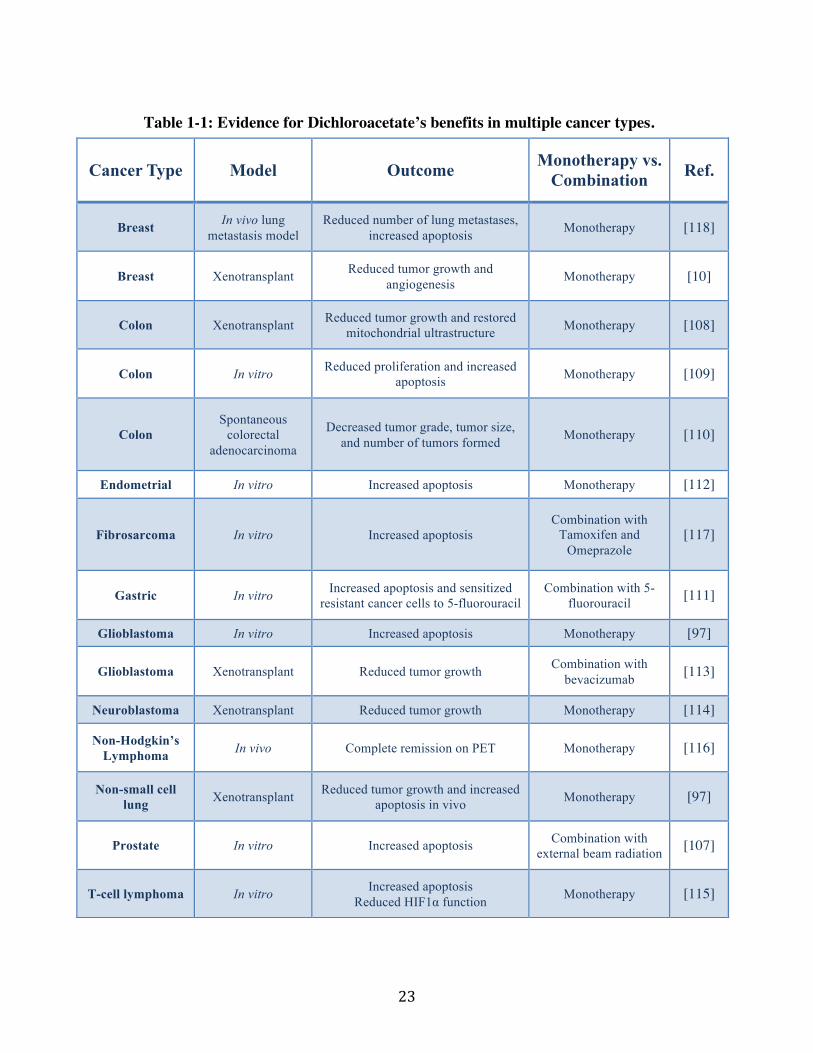
Table 1-1: Evidence for Dichloroacetate’s benefits in multiple cancer types.
Cancer Type Model Outcome Monotherapy vs. Combination Ref.
Breast In vivo lung metastasis model
Reduced number of lung metastases, increased apoptosis Monotherapy [118]
Breast Xenotransplant Reduced tumor growth and angiogenesis Monotherapy [10]
Colon Xenotransplant Reduced tumor growth and restored mitochondrial ultrastructure Monotherapy [108]
Colon In vitro Reduced proliferation and increased apoptosis Monotherapy [109]
Colon Spontaneous
colorectal adenocarcinoma
Decreased tumor grade, tumor size, and number of tumors formed Monotherapy [110]
Endometrial In vitro Increased apoptosis Monotherapy [112]
Fibrosarcoma In vitro Increased apoptosis Combination with
Tamoxifen and Omeprazole
[117]
Gastric In vitro Increased apoptosis and sensitized resistant cancer cells to 5-fluorouracil
Combination with 5-fluorouracil [111]
Glioblastoma In vitro Increased apoptosis Monotherapy [97]
Glioblastoma Xenotransplant Reduced tumor growth Combination with bevacizumab [113]
Neuroblastoma Xenotransplant Reduced tumor growth Monotherapy [114]
Non-Hodgkin’s Lymphoma In vivo Complete remission on PET Monotherapy [116]
Non-small cell lung Xenotransplant Reduced tumor growth and increased
apoptosis in vivo Monotherapy [97]
Prostate In vitro Increased apoptosis Combination with external beam radiation [107]
T-cell lymphoma In vitro Increased apoptosis!Reduced HIF1α function! Monotherapy [115]
"$!
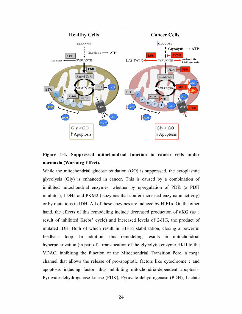
Figure 1-1. Suppressed mitochondrial function in cancer cells under
normoxia (Warburg Effect).
While the mitochondrial glucose oxidation (GO) is suppressed, the cytoplasmic
glycolysis (Gly) is enhanced in cancer. This is caused by a combination of
inhibited mitochondrial enzymes, whether by upregulation of PDK (a PDH
inhibitor), LDH5 and PKM2 (isozymes that confer increased enzymatic activity)
or by mutations in IDH. All of these enzymes are induced by HIF1!. On the other
hand, the effects of this remodeling include decreased production of !KG (as a
result of inhibited Krebs’ cycle) and increased levels of 2-HG, the product of
mutated IDH. Both of which result in HIF1! stabilization, closing a powerful
feedback loop. In addition, this remodeling results in mitochondrial
hyperpolarization (in part of a translocation of the glycolytic enzyme HKII to the
VDAC, inhibiting the function of the Mitochondrial Transition Pore, a mega
channel that allows the release of pro-apoptotic factors like cytochrome c and
apoptosis inducing factor, thus inhibiting mitochondria-dependent apoptosis.
Pyruvate dehydrogenase kinase (PDK), Pyruvate dehydrogenase (PDH), Lactate

! 25!
dehydrogenase 5 (LDH5), Pyruvate kinase M2 (PKM2), Isocitrate dehydrogenase
(IDH), Hypoxia inducible factor 1α (HIF1α), Alpha-ketoglutarate (αKG), 2-
hydroxyglutarate (2-HG), Hexokinase II (HKII), Voltage-dependent anion
channel (VDAC).
! 26!
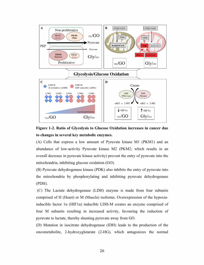
Figure 1-2. Ratio of Glycolysis to Glucose Oxidation increases in cancer due
to changes in several key metabolic enzymes.
(A) Cells that express a low amount of Pyruvate kinase M1 (PKM1) and an
abundance of low-activity Pyruvate kinase M2 (PKM2; which results in an
overall decrease in pyruvate kinase activity) prevent the entry of pyruvate into the
mitochondria, inhibiting glucose oxidation (GO).
(B) Pyruvate dehydrogenase kinase (PDK) also inhibits the entry of pyruvate into
the mitochondria by phosphorylating and inhibiting pyruvate dehydrogenase
(PDH).
(C) The Lactate dehydrogenase (LDH) enzyme is made from four subunits
comprised of H (Heart) or M (Muscle) isoforms. Overexpression of the hypoxia-
inducible factor 1α (HIF1α) inducible LDH-M creates an enzyme comprised of
four M subunits resulting in increased activity, favouring the reduction of
pyruvate to lactate, thereby shunting pyruvate away from GO.
(D) Mutation in isocitrate dehydrogenase (IDH) leads to the production of the
oncometabolite, 2-hydroxyglutarate (2-HG), which antagonizes the normal

! 27!
product, alpha-ketoglutarate (αKG), leading to suppressed GO via the
stabilization and accumulation HIF1α.
! 28!
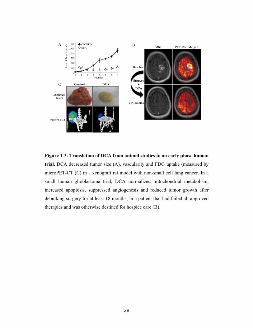
Figure 1-3. Translation of DCA from animal studies to an early phase human
trial. DCA decreased tumor size (A), vascularity and FDG uptake (measured by
microPET-CT (C) in a xenograft rat model with non-small cell lung cancer. In a
small human glioblastoma trial, DCA normalized mitochondrial metabolism,
increased apoptosis, suppressed angiogenesis and reduced tumor growth after
debulking surgery for at least 18 months, in a patient that had failed all approved
therapies and was otherwise destined for hospice care (B).
! 29!
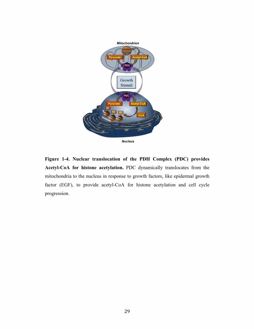
Figure 1-4. Nuclear translocation of the PDH Complex (PDC) provides
Acetyl-CoA for histone acetylation. PDC dynamically translocates from the
mitochondria to the nucleus in response to growth factors, like epidermal growth
factor (EGF), to provide acetyl-CoA for histone acetylation and cell cycle
progression.
! 30!
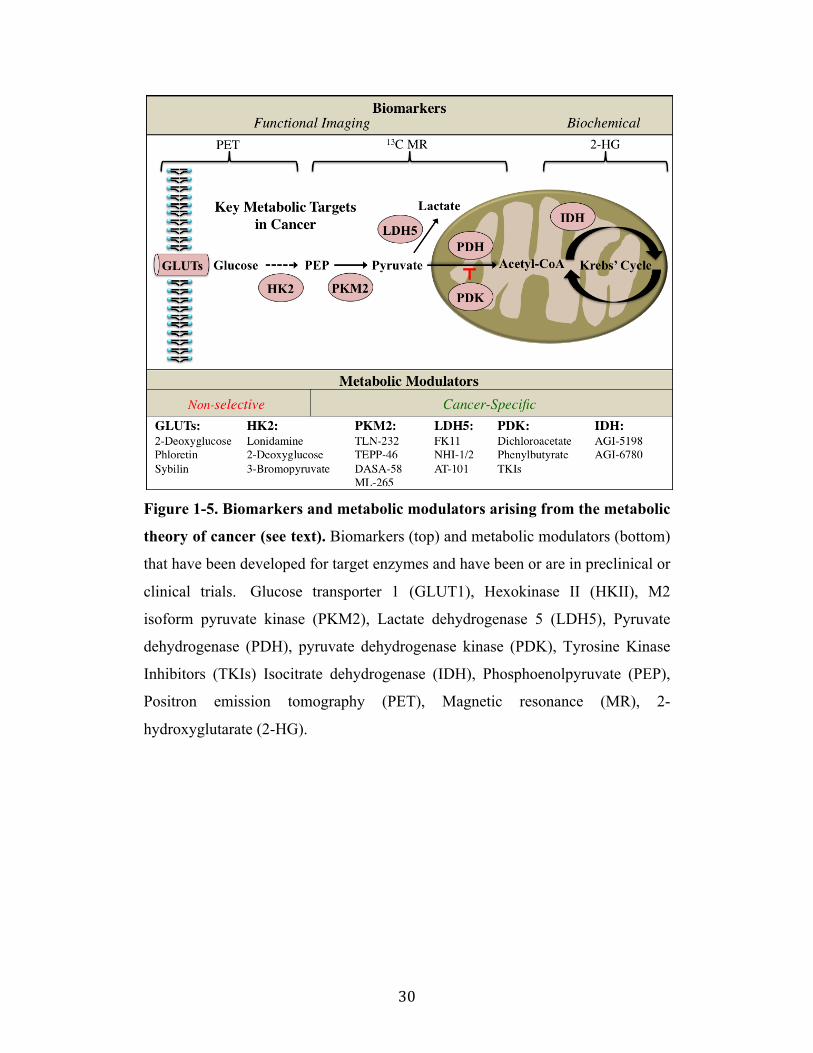
Figure 1-5. Biomarkers and metabolic modulators arising from the metabolic
theory of cancer (see text). Biomarkers (top) and metabolic modulators (bottom)
that have been developed for target enzymes and have been or are in preclinical or
clinical trials. Glucose transporter 1 (GLUT1), Hexokinase II (HKII), M2
isoform pyruvate kinase (PKM2), Lactate dehydrogenase 5 (LDH5), Pyruvate
dehydrogenase (PDH), pyruvate dehydrogenase kinase (PDK), Tyrosine Kinase
Inhibitors (TKIs) Isocitrate dehydrogenase (IDH), Phosphoenolpyruvate (PEP),
Positron emission tomography (PET), Magnetic resonance (MR), 2-
hydroxyglutarate (2-HG).
! 31!
References
1. Le Tourneau C, Lee JJ, Siu LL (2009) Dose escalation methods in phase I cancer clinical trials. J Natl Cancer Inst 101: 708-720. 2. Druker BJ, Talpaz M, Resta DJ, Peng B, Buchdunger E, Ford JM, Lydon NB, Kantarjian H, Capdeville R, Ohno-Jones S, Sawyers CL (2001) Efficacy and safety of a specific inhibitor of the BCR-ABL tyrosine kinase in chronic myeloid leukemia. N Engl J Med 344: 1031-1037. 3. Slamon DJ, Leyland-Jones B, Shak S, Fuchs H, Paton V, Bajamonde A, Fleming T, Eiermann W, Wolter J, Pegram M, Baselga J, Norton L (2001) Use of chemotherapy plus a monoclonal antibody against HER2 for metastatic breast cancer that overexpresses HER2. N Engl J Med 344: 783-792. 4. Wen PY, Kesari S (2008) Malignant gliomas in adults. N Engl J Med 359: 492-507. 5. Loureiro R, Mesquita KA, Oliveira PJ, Vega-Naredo I (2013) Mitochondria in cancer stem cells: a target for therapy. Recent Pat Endocr Metab Immune Drug Discov 7: 102-114. 6. Warburg O (1923) Metabolism of tumours. Biochem Zeitschr 142: 317-333. 7. Loenarz C, Schofield CJ (2008) Expanding chemical biology of 2-oxoglutarate oxygenases. Nat Chem Biol 4: 152-156. 8. Schmid T, Zhou J, Kohl R, Brune B (2004) p300 relieves p53-evoked transcriptional repression of hypoxia-inducible factor-1 (HIF-1). Biochem J 380: 289-295. 9. Vousden KH, Ryan KM (2009) p53 and metabolism. Nat Rev Cancer 9: 691-700. 10. Sutendra G, Dromparis P, Kinnaird A, Stenson TH, Haromy A, Parker JM, McMurtry MS, Michelakis ED (2012) Mitochondrial activation by inhibition of PDKII suppresses HIF1a signaling and angiogenesis in cancer. Oncogene 32: 1638-1650. 11. Maddocks OD, Vousden KH (2011) Metabolic regulation by p53. J Mol Med (Berl) 89: 237-245. 12. Dromparis P, Michelakis ED (2013) Mitochondria in vascular health and disease. Annu Rev Physiol 75: 95-126. 13. Sutendra G, Michelakis ED (2013) Pyruvate dehydrogenase kinase as a novel therapeutic target in oncology. Front Oncol 3: 38. 14. Zamzami N, Kroemer G (2001) The mitochondrion in apoptosis: how Pandora's box opens. Nature reviews Molecular cell biology 2: 67-71. 15. Chen LB (1988) Mitochondrial membrane potential in living cells. Annu Rev Cell Biol 4: 155-181. 16. Lemasters JJ, Holmuhamedov E (2006) Voltage-dependent anion channel (VDAC) as mitochondrial governator--thinking outside the box. Biochimica et biophysica acta 1762: 181-190. 17. Pastorino JG, Hoek JB, Shulga N (2005) Activation of glycogen synthase kinase 3beta disrupts the binding of hexokinase II to mitochondria by phosphorylating voltage-dependent anion channel and potentiates chemotherapy-induced cytotoxicity. Cancer Res 65: 10545-10554.
! 32!
18. Vander Heiden MG, Cantley LC, Thompson CB (2009) Understanding the Warburg effect: the metabolic requirements of cell proliferation. Science 324: 1029-1033. 19. Butow RA, Avadhani NG (2004) Mitochondrial signaling: the retrograde response. Mol Cell 14: 1-15. 20. Esteves P, Pecqueur C, Ransy C, Esnous C, Lenoir V, Bouillaud F, Bulteau AL, Lombes A, Prip-Buus C, Ricquier D, Alves-Guerra MC (2014) Mitochondrial retrograde signaling mediated by UCP2 inhibits cancer cell proliferation and tumorigenesis. Cancer Res 74: 3971-3982. 21. Wallace DC (2012) Mitochondria and cancer. Nat Rev Cancer 12: 685-698. 22. Semenza GL (2010) HIF-1: upstream and downstream of cancer metabolism. Current opinion in genetics & development 20: 51-56. 23. Denko NC (2008) Hypoxia, HIF1 and glucose metabolism in the solid tumour. Nat Rev Cancer 8: 705-713. 24. Yu F, White SB, Zhao Q, Lee FS (2001) HIF-1alpha binding to VHL is regulated by stimulus-sensitive proline hydroxylation. Proc Natl Acad Sci U S A 98: 9630-9635. 25. Ke Q, Costa M (2006) Hypoxia-inducible factor-1 (HIF-1). Molecular pharmacology 70: 1469-1480. 26. Gatenby RA, Gillies RJ (2004) Why do cancers have high aerobic glycolysis? Nat Rev Cancer 4: 891-899. 27. Hitosugi T, Fan J, Chung TW, Lythgoe K, Wang X, Xie J, Ge Q, Gu TL, Polakiewicz RD, Roesel JL, Chen GZ, Boggon TJ, Lonial S, Fu H, Khuri FR, Kang S, Chen J (2011) Tyrosine phosphorylation of mitochondrial pyruvate dehydrogenase kinase 1 is important for cancer metabolism. Mol Cell 44: 864-877. 28. Korotchkina LG, Patel MS (2001) Probing the mechanism of inactivation of human pyruvate dehydrogenase by phosphorylation of three sites. J Biol Chem 276: 5731-5738. 29. Fan J, Kang HB, Shan C, Elf S, Lin R, Xie J, Gu TL, Aguiar M, Lonning S, Chung TW, Arellano M, Khoury HJ, Shin DM, Khuri FR, Boggon TJ, Kang S, Chen J (2014) Tyr-301 phosphorylation inhibits pyruvate dehydrogenase by blocking substrate binding and promotes the Warburg effect. J Biol Chem 289: 26533-26541. 30. Kim JW, Tchernyshyov I, Semenza GL, Dang CV (2006) HIF-1-mediated expression of pyruvate dehydrogenase kinase: a metabolic switch required for cellular adaptation to hypoxia. Cell Metab 3: 177-185. 31. Franovic A, Gunaratnam L, Smith K, Robert I, Patten D, Lee S (2007) Translational up-regulation of the EGFR by tumor hypoxia provides a nonmutational explanation for its overexpression in human cancer. Proc Natl Acad Sci U S A 104: 13092-13097. 32. Wu P, Inskeep K, Bowker-Kinley MM, Popov KM, Harris RA (1999) Mechanism responsible for inactivation of skeletal muscle pyruvate dehydrogenase complex in starvation and diabetes. Diabetes 48: 1593-1599.
! 33!
33. Kandoth C, McLellan MD, Vandin F, Ye K, Niu B, Lu C, Xie M, Zhang Q, McMichael JF, Wyczalkowski MA, Leiserson MD, Miller CA, Welch JS, Walter MJ, Wendl MC, Ley TJ, Wilson RK, Raphael BJ, Ding L (2013) Mutational landscape and significance across 12 major cancer types. Nature 502: 333-339. 34. Bensaad K, Tsuruta A, Selak MA, Vidal MN, Nakano K, Bartrons R, Gottlieb E, Vousden KH (2006) TIGAR, a p53-inducible regulator of glycolysis and apoptosis. Cell 126: 107-120. 35. Kondoh H, Lleonart ME, Gil J, Wang J, Degan P, Peters G, Martinez D, Carnero A, Beach D (2005) Glycolytic enzymes can modulate cellular life span. Cancer Res 65: 177-185. 36. Matoba S, Kang JG, Patino WD, Wragg A, Boehm M, Gavrilova O, Hurley PJ, Bunz F, Hwang PM (2006) p53 regulates mitochondrial respiration. Science 312: 1650-1653. 37. Contractor T, Harris CR (2012) p53 negatively regulates transcription of the pyruvate dehydrogenase kinase Pdk2. Cancer Res 72: 560-567. 38. Mathupala SP, Heese C, Pedersen PL (1997) Glucose catabolism in cancer cells. The type II hexokinase promoter contains functionally active response elements for the tumor suppressor p53. J Biol Chem 272: 22776-22780. 39. Choudhary C, Weinert BT, Nishida Y, Verdin E, Mann M (2014) The growing landscape of lysine acetylation links metabolism and cell signalling. Nature reviews Molecular cell biology 15: 536-550. 40. Dang CV, Semenza GL (1999) Oncogenic alterations of metabolism. Trends in biochemical sciences 24: 68-72. 41. Shim H, Dolde C, Lewis BC, Wu CS, Dang G, Jungmann RA, Dalla-Favera R, Dang CV (1997) c-Myc transactivation of LDH-A: implications for tumor metabolism and growth. Proc Natl Acad Sci U S A 94: 6658-6663. 42. Kim JW, Zeller KI, Wang Y, Jegga AG, Aronow BJ, O'Donnell KA, Dang CV (2004) Evaluation of myc E-box phylogenetic footprints in glycolytic genes by chromatin immunoprecipitation assays. Mol Cell Biol 24: 5923-5936. 43. Nogueiras R, Habegger KM, Chaudhary N, Finan B, Banks AS, Dietrich MO, Horvath TL, Sinclair DA, Pfluger PT, Tschop MH (2012) Sirtuin 1 and sirtuin 3: physiological modulators of metabolism. Physiological reviews 92: 1479-1514. 44. He W, Newman JC, Wang MZ, Ho L, Verdin E (2012) Mitochondrial sirtuins: regulators of protein acylation and metabolism. Trends in endocrinology and metabolism: TEM 23: 467-476. 45. Kim HS, Patel K, Muldoon-Jacobs K, Bisht KS, Aykin-Burns N, Pennington JD, van der Meer R, Nguyen P, Savage J, Owens KM, Vassilopoulos A, Ozden O, Park SH, Singh KK, Abdulkadir SA, Spitz DR, Deng CX, Gius D (2010) SIRT3 is a mitochondria-localized tumor suppressor required for maintenance of mitochondrial integrity and metabolism during stress. Cancer Cell 17: 41-52. 46. Guarente L (2014) The Many Faces of Sirtuins: Sirtuins and the Warburg effect. Nature medicine 20: 24-25.
! 34!
47. Finley LW, Carracedo A, Lee J, Souza A, Egia A, Zhang J, Teruya-Feldstein J, Moreira PI, Cardoso SM, Clish CB, Pandolfi PP, Haigis MC (2011) SIRT3 opposes reprogramming of cancer cell metabolism through HIF1alpha destabilization. Cancer Cell 19: 416-428. 48. Denton RM (2009) Regulation of mitochondrial dehydrogenases by calcium ions. Biochimica et biophysica acta 1787: 1309-1316. 49. Dromparis P, Paulin R, Sutendra G, Qi AC, Bonnet S, Michelakis ED (2013) Uncoupling protein 2 deficiency mimics the effects of hypoxia and endoplasmic reticulum stress on mitochondria and triggers pseudohypoxic pulmonary vascular remodeling and pulmonary hypertension. Circ Res 113: 126-136. 50. Derdak Z, Fulop P, Sabo E, Tavares R, Berthiaume EP, Resnick MB, Paragh G, Wands JR, Baffy G (2006) Enhanced colon tumor induction in uncoupling protein-2 deficient mice is associated with NF-kappaB activation and oxidative stress. Carcinogenesis 27: 956-961. 51. Archer SL (2013) Mitochondrial dynamics--mitochondrial fission and fusion in human diseases. N Engl J Med 369: 2236-2251. 52. Mitra K, Wunder C, Roysam B, Lin G, Lippincott-Schwartz J (2009) A hyperfused mitochondrial state achieved at G1-S regulates cyclin E buildup and entry into S phase. Proc Natl Acad Sci U S A 106: 11960-11965. 53. Rehman J, Zhang HJ, Toth PT, Zhang Y, Marsboom G, Hong Z, Salgia R, Husain AN, Wietholt C, Archer SL (2012) Inhibition of mitochondrial fission prevents cell cycle progression in lung cancer. FASEB journal : official publication of the Federation of American Societies for Experimental Biology 26: 2175-2186. 54. Tomlinson IP, Alam NA, Rowan AJ, Barclay E, Jaeger EE, Kelsell D, Leigh I, Gorman P, Lamlum H, Rahman S, Roylance RR, Olpin S, Bevan S, Barker K, Hearle N, Houlston RS, Kiuru M, Lehtonen R, Karhu A, Vilkki S, Laiho P, Eklund C, Vierimaa O, Aittomaki K, Hietala M, Sistonen P, Paetau A, Salovaara R, Herva R, Launonen V, Aaltonen LA, Multiple Leiomyoma C (2002) Germline mutations in FH predispose to dominantly inherited uterine fibroids, skin leiomyomata and papillary renal cell cancer. Nature genetics 30: 406-410. 55. Neumann HP, Pawlu C, Peczkowska M, Bausch B, McWhinney SR, Muresan M, Buchta M, Franke G, Klisch J, Bley TA, Hoegerle S, Boedeker CC, Opocher G, Schipper J, Januszewicz A, Eng C, European-American Paraganglioma Study G (2004) Distinct clinical features of paraganglioma syndromes associated with SDHB and SDHD gene mutations. Jama 292: 943-951. 56. Baysal BE (2003) On the association of succinate dehydrogenase mutations with hereditary paraganglioma. Trends in endocrinology and metabolism: TEM 14: 453-459. 57. Isaacs JS, Jung YJ, Mole DR, Lee S, Torres-Cabala C, Chung YL, Merino M, Trepel J, Zbar B, Toro J, Ratcliffe PJ, Linehan WM, Neckers L (2005) HIF overexpression correlates with biallelic loss of fumarate hydratase in renal cancer: novel role of fumarate in regulation of HIF stability. Cancer Cell 8: 143-153.
! 35!
58. King A, Selak MA, Gottlieb E (2006) Succinate dehydrogenase and fumarate hydratase: linking mitochondrial dysfunction and cancer. Oncogene 25: 4675-4682. 59. Selak MA, Armour SM, MacKenzie ED, Boulahbel H, Watson DG, Mansfield KD, Pan Y, Simon MC, Thompson CB, Gottlieb E (2005) Succinate links TCA cycle dysfunction to oncogenesis by inhibiting HIF-alpha prolyl hydroxylase. Cancer Cell 7: 77-85. 60. Parsons DW, Jones S, Zhang X, Lin JC, Leary RJ, Angenendt P, Mankoo P, Carter H, Siu IM, Gallia GL, Olivi A, McLendon R, Rasheed BA, Keir S, Nikolskaya T, Nikolsky Y, Busam DA, Tekleab H, Diaz LA, Jr., Hartigan J, Smith DR, Strausberg RL, Marie SK, Shinjo SM, Yan H, Riggins GJ, Bigner DD, Karchin R, Papadopoulos N, Parmigiani G, Vogelstein B, Velculescu VE, Kinzler KW (2008) An integrated genomic analysis of human glioblastoma multiforme. Science 321: 1807-1812. 61. Mardis ER, Ding L, Dooling DJ, Larson DE, McLellan MD, Chen K, Koboldt DC, Fulton RS, Delehaunty KD, McGrath SD, Fulton LA, Locke DP, Magrini VJ, Abbott RM, Vickery TL, Reed JS, Robinson JS, Wylie T, Smith SM, Carmichael L, Eldred JM, Harris CC, Walker J, Peck JB, Du F, Dukes AF, Sanderson GE, Brummett AM, Clark E, McMichael JF, Meyer RJ, Schindler JK, Pohl CS, Wallis JW, Shi X, Lin L, Schmidt H, Tang Y, Haipek C, Wiechert ME, Ivy JV, Kalicki J, Elliott G, Ries RE, Payton JE, Westervelt P, Tomasson MH, Watson MA, Baty J, Heath S, Shannon WD, Nagarajan R, Link DC, Walter MJ, Graubert TA, DiPersio JF, Wilson RK, Ley TJ (2009) Recurring mutations found by sequencing an acute myeloid leukemia genome. N Engl J Med 361: 1058-1066. 62. Dang L, White DW, Gross S, Bennett BD, Bittinger MA, Driggers EM, Fantin VR, Jang HG, Jin S, Keenan MC, Marks KM, Prins RM, Ward PS, Yen KE, Liau LM, Rabinowitz JD, Cantley LC, Thompson CB, Vander Heiden MG, Su SM (2009) Cancer-associated IDH1 mutations produce 2-hydroxyglutarate. Nature 462: 739-744. 63. Ward PS, Patel J, Wise DR, Abdel-Wahab O, Bennett BD, Coller HA, Cross JR, Fantin VR, Hedvat CV, Perl AE, Rabinowitz JD, Carroll M, Su SM, Sharp KA, Levine RL, Thompson CB (2010) The common feature of leukemia-associated IDH1 and IDH2 mutations is a neomorphic enzyme activity converting alpha-ketoglutarate to 2-hydroxyglutarate. Cancer Cell 17: 225-234. 64. Rose NR, McDonough MA, King ON, Kawamura A, Schofield CJ (2011) Inhibition of 2-oxoglutarate dependent oxygenases. Chemical Society reviews 40: 4364-4397. 65. Guan KL, Xiong Y (2011) Regulation of intermediary metabolism by protein acetylation. Trends in biochemical sciences 36: 108-116. 66. Zhao S, Xu W, Jiang W, Yu W, Lin Y, Zhang T, Yao J, Zhou L, Zeng Y, Li H, Li Y, Shi J, An W, Hancock SM, He F, Qin L, Chin J, Yang P, Chen X, Lei Q, Xiong Y, Guan KL (2010) Regulation of cellular metabolism by protein lysine acetylation. Science 327: 1000-1004.
! 36!
67. Devi MA, Das NP (1993) In vitro effects of natural plant polyphenols on the proliferation of normal and abnormal human lymphocytes and their secretions of interleukin-2. Cancer Lett 69: 191-196. 68. Kobori M, Shinmoto H, Tsushida T, Shinohara K (1997) Phloretin-induced apoptosis in B16 melanoma 4A5 cells by inhibition of glucose transmembrane transport. Cancer Lett 119: 207-212. 69. Porporato PE, Dhup S, Dadhich RK, Copetti T, Sonveaux P (2011) Anticancer targets in the glycolytic metabolism of tumors: a comprehensive review. Front Pharmacol 2: 49. 70. Tennant DA, Duran RV, Gottlieb E (2010) Targeting metabolic transformation for cancer therapy. Nat Rev Cancer 10: 267-277. 71. Ko YH, Verhoeven HA, Lee MJ, Corbin DJ, Vogl TJ, Pedersen PL (2012) A translational study "case report" on the small molecule "energy blocker" 3-bromopyruvate (3BP) as a potent anticancer agent: from bench side to bedside. J Bioenerg Biomembr 44: 163-170. 72. Ko YH, Smith BL, Wang Y, Pomper MG, Rini DA, Torbenson MS, Hullihen J, Pedersen PL (2004) Advanced cancers: eradication in all cases using 3-bromopyruvate therapy to deplete ATP. Biochem Biophys Res Commun 324: 269-275. 73. Biolato M, Marrone G, Racco S, Di Stasi C, Miele L, Gasbarrini G, Landolfi R, Grieco A (2010) Transarterial chemoembolization (TACE) for unresectable HCC: a new life begins? Eur Rev Med Pharmacol Sci 14: 356-362. 74. DeBerardinis RJ, Mancuso A, Daikhin E, Nissim I, Yudkoff M, Wehrli S, Thompson CB (2007) Beyond aerobic glycolysis: transformed cells can engage in glutamine metabolism that exceeds the requirement for protein and nucleotide synthesis. Proc Natl Acad Sci U S A 104: 19345-19350. 75. Hutson SM, Sweatt AJ, Lanoue KF (2005) Branched-chain [corrected] amino acid metabolism: implications for establishing safe intakes. J Nutr 135: 1557S-1564S. 76. Gupta V, Bamezai RN (2010) Human pyruvate kinase M2: a multifunctional protein. Protein Sci 19: 2031-2044. 77. Takenaka M, Noguchi T, Sadahiro S, Hirai H, Yamada K, Matsuda T, Imai E, Tanaka T (1991) Isolation and characterization of the human pyruvate kinase M gene. Eur J Biochem 198: 101-106. 78. Noguchi T, Inoue H, Tanaka T (1986) The M1- and M2-type isozymes of rat pyruvate kinase are produced from the same gene by alternative RNA splicing. J Biol Chem 261: 13807-13812. 79. Munoz ME, Ponce E (2003) Pyruvate kinase: current status of regulatory and functional properties. Comp Biochem Physiol B Biochem Mol Biol 135: 197-218. 80. Wong N, De Melo J, Tang D (2013) PKM2, a Central Point of Regulation in Cancer Metabolism. Int J Cell Biol 2013: 242513. 81. Imamura K, Tanaka T (1972) Multimolecular forms of pyruvate kinase from rat and other mammalian tissues. I. Electrophoretic studies. J Biochem 71: 1043-1051.
! 37!
82. Mazurek S (2011) Pyruvate kinase type M2: a key regulator of the metabolic budget system in tumor cells. Int J Biochem Cell Biol 43: 969-980. 83. Anastasiou D, Yu Y, Israelsen WJ, Jiang JK, Boxer MB, Hong BS, Tempel W, Dimov S, Shen M, Jha A, Yang H, Mattaini KR, Metallo CM, Fiske BP, Courtney KD, Malstrom S, Khan TM, Kung C, Skoumbourdis AP, Veith H, Southall N, Walsh MJ, Brimacombe KR, Leister W, Lunt SY, Johnson ZR, Yen KE, Kunii K, Davidson SM, Christofk HR, Austin CP, Inglese J, Harris MH, Asara JM, Stephanopoulos G, Salituro FG, Jin S, Dang L, Auld DS, Park HW, Cantley LC, Thomas CJ, Vander Heiden MG (2012) Pyruvate kinase M2 activators promote tetramer formation and suppress tumorigenesis. Nat Chem Biol 8: 839-847. 84. Christofk HR, Vander Heiden MG, Harris MH, Ramanathan A, Gerszten RE, Wei R, Fleming MD, Schreiber SL, Cantley LC (2008) The M2 splice isoform of pyruvate kinase is important for cancer metabolism and tumour growth. Nature 452: 230-233. 85. Lv L, Li D, Zhao D, Lin R, Chu Y, Zhang H, Zha Z, Liu Y, Li Z, Xu Y, Wang G, Huang Y, Xiong Y, Guan KL, Lei QY (2011) Acetylation targets the M2 isoform of pyruvate kinase for degradation through chaperone-mediated autophagy and promotes tumor growth. Mol Cell 42: 719-730. 86. Hitosugi T, Kang S, Vander Heiden MG, Chung TW, Elf S, Lythgoe K, Dong S, Lonial S, Wang X, Chen GZ, Xie J, Gu TL, Polakiewicz RD, Roesel JL, Boggon TJ, Khuri FR, Gilliland DG, Cantley LC, Kaufman J, Chen J (2009) Tyrosine phosphorylation inhibits PKM2 to promote the Warburg effect and tumor growth. Sci Signal 2: ra73. 87. Christofk HR, Vander Heiden MG, Wu N, Asara JM, Cantley LC (2008) Pyruvate kinase M2 is a phosphotyrosine-binding protein. Nature 452: 181-186. 88. Ashizawa K, Willingham MC, Liang CM, Cheng SY (1991) In vivo regulation of monomer-tetramer conversion of pyruvate kinase subtype M2 by glucose is mediated via fructose 1,6-bisphosphate. J Biol Chem 266: 16842-16846. 89. Israelsen WJ, Dayton TL, Davidson SM, Fiske BP, Hosios AM, Bellinger G, Li J, Yu Y, Sasaki M, Horner JW, Burga LN, Xie J, Jurczak MJ, DePinho RA, Clish CB, Jacks T, Kibbey RG, Wulf GM, Di Vizio D, Mills GB, Cantley LC, Vander Heiden MG (2013) PKM2 isoform-specific deletion reveals a differential requirement for pyruvate kinase in tumor cells. Cell 155: 397-409. 90. Walsh MJ, Brimacombe KR, Anastasiou D, Yu Y, Israelsen WJ, Hong BS, Tempel W, Dimov S, Veith H, Yang H, Kung C, Yen KE, Dang L, Salituro F, Auld DS, Park HW, Vander Heiden MG, Thomas CJ, Shen M, Boxer MB (2010) ML265: A potent PKM2 activator induces tetramerization and reduces tumor formation and size in a mouse xenograft modelProbe Reports from the NIH Molecular Libraries Program, Bethesda (MD). 91. Parnell KM, Foulks JM, Nix RN, Clifford A, Bullough J, Luo B, Senina A, Vollmer D, Liu J, McCarthy V, Xu Y, Saunders M, Liu XH, Pearce S, Wright K, O'Reilly M, McCullar MV, Ho KK, Kanner SB (2013) Pharmacologic activation of PKM2 slows lung tumor xenograft growth. Mol Cancer Ther 12: 1453-1460.
! 38!
92. Michelakis ED, Webster L, Mackey JR (2008) Dichloroacetate (DCA) as a potential metabolic-targeting therapy for cancer. Br J Cancer 99: 989-994. 93. Sutendra G, Michelakis ED (2014) The metabolic basis of pulmonary arterial hypertension. Cell Metab 19: 558-573. 94. Michelakis ED, Sutendra G, Dromparis P, Webster L, Haromy A, Niven E, Maguire C, Gammer TL, Mackey JR, Fulton D, Abdulkarim B, McMurtry MS, Petruk KC (2010) Metabolic modulation of glioblastoma with dichloroacetate. Sci Transl Med 2: 31ra34. 95. Bowker-Kinley MM, Davis WI, Wu P, Harris RA, Popov KM (1998) Evidence for existence of tissue-specific regulation of the mammalian pyruvate dehydrogenase complex. Biochem J 329 ( Pt 1): 191-196. 96. Stacpoole PW (1989) The pharmacology of dichloroacetate. Metabolism 38: 1124-1144. 97. Bonnet S, Archer SL, Allalunis-Turner J, Haromy A, Beaulieu C, Thompson R, Lee CT, Lopaschuk GD, Puttagunta L, Harry G, Hashimoto K, Porter CJ, Andrade MA, Thebaud B, Michelakis ED (2007) A mitochondria-K+ channel axis is suppressed in cancer and its normalization promotes apoptosis and inhibits cancer growth. Cancer Cell 11: 37-51. 98. Stacpoole PW, Moore GW, Kornhauser DM (1978) Metabolic effects of dichloroacetate in patients with diabetes mellitus and hyperlipoproteinemia. N Engl J Med 298: 526-530. 99. Stacpoole PW, Wright EC, Baumgartner TG, Bersin RM, Buchalter S, Curry SH, Duncan CA, Harman EM, Henderson GN, Jenkinson S, et al. (1992) A controlled clinical trial of dichloroacetate for treatment of lactic acidosis in adults. The Dichloroacetate-Lactic Acidosis Study Group. N Engl J Med 327: 1564-1569. 100. Stacpoole PW, Kerr DS, Barnes C, Bunch ST, Carney PR, Fennell EM, Felitsyn NM, Gilmore RL, Greer M, Henderson GN, Hutson AD, Neiberger RE, O'Brien RG, Perkins LA, Quisling RG, Shroads AL, Shuster JJ, Silverstein JH, Theriaque DW, Valenstein E (2006) Controlled clinical trial of dichloroacetate for treatment of congenital lactic acidosis in children. Pediatrics 117: 1519-1531. 101. McMurtry MS, Bonnet S, Wu X, Dyck JR, Haromy A, Hashimoto K, Michelakis ED (2004) Dichloroacetate prevents and reverses pulmonary hypertension by inducing pulmonary artery smooth muscle cell apoptosis. Circ Res 95: 830-840. 102. Krishna S, Supanaranond W, Pukrittayakamee S, Kuile FT, Ruprah M, White NJ (1996) The disposition and effects of two doses of dichloroacetate in adults with severe falciparum malaria. Br J Clin Pharmacol 41: 29-34. 103. Bersin RM, Wolfe C, Kwasman M, Lau D, Klinski C, Tanaka K, Khorrami P, Henderson GN, de Marco T, Chatterjee K (1994) Improved hemodynamic function and mechanical efficiency in congestive heart failure with sodium dichloroacetate. J Am Coll Cardiol 23: 1617-1624. 104. Calvert LD, Shelley R, Singh SJ, Greenhaff PL, Bankart J, Morgan MD, Steiner MC (2008) Dichloroacetate enhances performance and reduces blood lactate during maximal cycle exercise in chronic obstructive pulmonary disease. Am J Respir Crit Care Med 177: 1090-1094.
! 39!
105. Stacpoole PW (1969) Review of the pharmacologic and therapeutic effects of diisopropylammonium dichloroacetate (DIPA). J Clin Pharmacol J New Drugs 9: 282-291. 106. Holloway PA, Knox K, Bajaj N, Chapman D, White NJ, O'Brien R, Stacpoole PW, Krishna S (1995) Plasmodium berghei infection: dichloroacetate improves survival in rats with lactic acidosis. Exp Parasitol 80: 624-632. 107. Cao W, Yacoub S, Shiverick KT, Namiki K, Sakai Y, Porvasnik S, Urbanek C, Rosser CJ (2008) Dichloroacetate (DCA) sensitizes both wild-type and over expressing Bcl-2 prostate cancer cells in vitro to radiation. Prostate 68: 1223-1231. 108. Sanchez-Arago M, Chamorro M, Cuezva JM (2010) Selection of cancer cells with repressed mitochondria triggers colon cancer progression. Carcinogenesis 31: 567-576. 109. Madhok BM, Yeluri S, Perry SL, Hughes TA, Jayne DG (2010) Dichloroacetate induces apoptosis and cell-cycle arrest in colorectal cancer cells. Br J Cancer 102: 1746-1752. 110. Sebastian C, Zwaans BM, Silberman DM, Gymrek M, Goren A, Zhong L, Ram O, Truelove J, Guimaraes AR, Toiber D, Cosentino C, Greenson JK, MacDonald AI, McGlynn L, Maxwell F, Edwards J, Giacosa S, Guccione E, Weissleder R, Bernstein BE, Regev A, Shiels PG, Lombard DB, Mostoslavsky R (2012) The histone deacetylase SIRT6 is a tumor suppressor that controls cancer metabolism. Cell 151: 1185-1199. 111. Xuan Y, Hur H, Ham IH, Yun J, Lee JY, Shim W, Bae Kim Y, Lee G, Han SU, Kwan Cho Y (2013) Dichloroacetate attenuates hypoxia-induced resistance to 5-fluorouracil in gastric cancer through the regulation of glucose metabolism. Exp Cell Res. 112. Wong JY, Huggins GS, Debidda M, Munshi NC, De Vivo I (2008) Dichloroacetate induces apoptosis in endometrial cancer cells. Gynecol Oncol 109: 394-402. 113. Kumar K, Wigfield S, Gee HE, Devlin CM, Singleton D, Li JL, Buffa F, Huffman M, Sinn AL, Silver J, Turley H, Leek R, Harris AL, Ivan M (2013) Dichloroacetate reverses the hypoxic adaptation to bevacizumab and enhances its antitumor effects in mouse xenografts. J Mol Med (Berl) 91: 749-758. 114. Vella S, Conti M, Tasso R, Cancedda R, Pagano A (2012) Dichloroacetate inhibits neuroblastoma growth by specifically acting against malignant undifferentiated cells. Int J Cancer 130: 1484-1493. 115. Kumar A, Kant S, Singh SM (2012) Novel molecular mechanisms of antitumor action of dichloroacetate against T cell lymphoma: Implication of altered glucose metabolism, pH homeostasis and cell survival regulation. Chem Biol Interact 199: 29-37. 116. Flavin DF (2010) Non-Hodgkin's Lymphoma Reversal with Dichloroacetate. J Oncol 2010. 117. Ishiguro T, Ishiguro R, Ishiguro M, Iwai S (2012) Co-treatment of dichloroacetate, omeprazole and tamoxifen exhibited synergistically antiproliferative effect on malignant tumors: in vivo experiments and a case report. Hepatogastroenterology 59: 994-996.
! 40!
118. Sun RC, Fadia M, Dahlstrom JE, Parish CR, Board PG, Blackburn AC (2010) Reversal of the glycolytic phenotype by dichloroacetate inhibits metastatic breast cancer cell growth in vitro and in vivo. Breast Cancer Res Treat 120: 253-260. 119. Kaluzova M, Kaluz S, Lerman MI, Stanbridge EJ (2004) DNA damage is a prerequisite for p53-mediated proteasomal degradation of HIF-1alpha in hypoxic cells and downregulation of the hypoxia marker carbonic anhydrase IX. Mol Cell Biol 24: 5757-5766. 120. Kaplon J, Zheng L, Meissl K, Chaneton B, Selivanov VA, Mackay G, van der Burg SH, Verdegaal EM, Cascante M, Shlomi T, Gottlieb E, Peeper DS (2013) A key role for mitochondrial gatekeeper pyruvate dehydrogenase in oncogene-induced senescence. Nature 498: 109-112. 121. Dunbar EM, Coats BS, Shroads AL, Langaee T, Lew A, Forder JR, Shuster JJ, Wagner DA, Stacpoole PW (2013) Phase 1 trial of dichloroacetate (DCA) in adults with recurrent malignant brain tumors. Invest New Drugs. 122. Abdelmalak M, Lew A, Ramezani R, Shroads AL, Coats BS, Langaee T, Shankar MN, Neiberger RE, Subramony SH, Stacpoole PW (2013) Long-term safety of dichloroacetate in congenital lactic acidosis. Mol Genet Metab 109: 139-143. 123. Dhar S, Lippard SJ (2009) Mitaplatin, a potent fusion of cisplatin and the orphan drug dichloroacetate. Proc Natl Acad Sci U S A 106: 22199-22204. 124. Morfouace M, Lalier L, Bahut M, Bonnamain V, Naveilhan P, Guette C, Oliver L, Gueguen N, Reynier P, Vallette FM (2012) Comparison of spheroids formed by rat glioma stem cells and neural stem cells reveals differences in glucose metabolism and promising therapeutic applications. J Biol Chem 287: 33664-33674. 125. Markert CL, Shaklee JB, Whitt GS (1975) Evolution of a gene. Multiple genes for LDH isozymes provide a model of the evolution of gene structure, function and regulation. Science 189: 102-114. 126. Koukourakis MI, Giatromanolaki A, Sivridis E, Bougioukas G, Didilis V, Gatter KC, Harris AL (2003) Lactate dehydrogenase-5 (LDH-5) overexpression in non-small-cell lung cancer tissues is linked to tumour hypoxia, angiogenic factor production and poor prognosis. Br J Cancer 89: 877-885. 127. Leiblich A, Cross SS, Catto JW, Phillips JT, Leung HY, Hamdy FC, Rehman I (2006) Lactate dehydrogenase-B is silenced by promoter hypermethylation in human prostate cancer. Oncogene 25: 2953-2960. 128. Koukourakis MI, Giatromanolaki A, Sivridis E, Gatter KC, Trarbach T, Folprecht G, Shi MM, Lebwohl D, Jalava T, Laurent D, Meinhardt G, Harris AL (2011) Prognostic and predictive role of lactate dehydrogenase 5 expression in colorectal cancer patients treated with PTK787/ZK 222584 (vatalanib) antiangiogenic therapy. Clin Cancer Res 17: 4892-4900. 129. Koukourakis MI, Giatromanolaki A, Winter S, Leek R, Sivridis E, Harris AL (2009) Lactate dehydrogenase 5 expression in squamous cell head and neck cancer relates to prognosis following radical or postoperative radiotherapy. Oncology 77: 285-292.
! 41!
130. Fantin VR, St-Pierre J, Leder P (2006) Attenuation of LDH-A expression uncovers a link between glycolysis, mitochondrial physiology, and tumor maintenance. Cancer Cell 9: 425-434. 131. Le A, Cooper CR, Gouw AM, Dinavahi R, Maitra A, Deck LM, Royer RE, Vander Jagt DL, Semenza GL, Dang CV (2010) Inhibition of lactate dehydrogenase A induces oxidative stress and inhibits tumor progression. Proc Natl Acad Sci U S A 107: 2037-2042. 132. Granchi C, Roy S, Giacomelli C, Macchia M, Tuccinardi T, Martinelli A, Lanza M, Betti L, Giannaccini G, Lucacchini A, Funel N, Leon LG, Giovannetti E, Peters GJ, Palchaudhuri R, Calvaresi EC, Hergenrother PJ, Minutolo F (2011) Discovery of N-hydroxyindole-based inhibitors of human lactate dehydrogenase isoform A (LDH-A) as starvation agents against cancer cells. J Med Chem 54: 1599-1612. 133. Maftouh M, Avan A, Sciarrillo R, Granchi C, Leon LG, Rani R, Funel N, Smid K, Honeywell R, Boggi U, Minutolo F, Peters GJ, Giovannetti E (2014) Synergistic interaction of novel lactate dehydrogenase inhibitors with gemcitabine against pancreatic cancer cells in hypoxia. Br J Cancer. 134. Golman K, Zandt RI, Lerche M, Pehrson R, Ardenkjaer-Larsen JH (2006) Metabolic imaging by hyperpolarized 13C magnetic resonance imaging for in vivo tumor diagnosis. Cancer Res 66: 10855-10860. 135. Dutta P, Le A, Vander Jagt DL, Tsukamoto T, Martinez GV, Dang CV, Gillies RJ (2013) Evaluation of LDH-A and glutaminase inhibition in vivo by hyperpolarized 13C-pyruvate magnetic resonance spectroscopy of tumors. Cancer Res 73: 4190-4195. 136. Hill DK, Orton MR, Mariotti E, Boult JK, Panek R, Jafar M, Parkes HG, Jamin Y, Miniotis MF, Al-Saffar NM, Beloueche-Babari M, Robinson SP, Leach MO, Chung YL, Eykyn TR (2013) Model free approach to kinetic analysis of real-time hyperpolarized 13C magnetic resonance spectroscopy data. PLoS One 8: e71996. 137. Wang F, Travins J, DeLaBarre B, Penard-Lacronique V, Schalm S, Hansen E, Straley K, Kernytsky A, Liu W, Gliser C, Yang H, Gross S, Artin E, Saada V, Mylonas E, Quivoron C, Popovici-Muller J, Saunders JO, Salituro FG, Yan S, Murray S, Wei W, Gao Y, Dang L, Dorsch M, Agresta S, Schenkein DP, Biller SA, Su SM, de Botton S, Yen KE (2013) Targeted inhibition of mutant IDH2 in leukemia cells induces cellular differentiation. Science 340: 622-626. 138. Rohle D, Popovici-Muller J, Palaskas N, Turcan S, Grommes C, Campos C, Tsoi J, Clark O, Oldrini B, Komisopoulou E, Kunii K, Pedraza A, Schalm S, Silverman L, Miller A, Wang F, Yang H, Chen Y, Kernytsky A, Rosenblum MK, Liu W, Biller SA, Su SM, Brennan CW, Chan TA, Graeber TG, Yen KE, Mellinghoff IK (2013) An inhibitor of mutant IDH1 delays growth and promotes differentiation of glioma cells. Science 340: 626-630. 139. Fathi AT, Sadrzadeh H, Borger DR, Ballen KK, Amrein PC, Attar EC, Foster J, Burke M, Lopez HU, Matulis CR, Edmonds KM, Iafrate AJ, Straley KS, Yen KE, Agresta S, Schenkein DP, Hill C, Emadi A, Neuberg DS, Stone RM, Chen YB (2012) Prospective serial evaluation of 2-hydroxyglutarate, during

! 42!
treatment of newly diagnosed acute myeloid leukemia, to assess disease activity and therapeutic response. Blood 120: 4649-4652. 140. Folmes CD, Nelson TJ, Martinez-Fernandez A, Arrell DK, Lindor JZ, Dzeja PP, Ikeda Y, Perez-Terzic C, Terzic A (2011) Somatic oxidative bioenergetics transitions into pluripotency-dependent glycolysis to facilitate nuclear reprogramming. Cell Metab 14: 264-271. 141. Xu X, Duan S, Yi F, Ocampo A, Liu GH, Izpisua Belmonte JC (2013) Mitochondrial regulation in pluripotent stem cells. Cell Metab 18: 325-332. 142. Varum S, Rodrigues AS, Moura MB, Momcilovic O, Easley CAt, Ramalho-Santos J, Van Houten B, Schatten G (2011) Energy metabolism in human pluripotent stem cells and their differentiated counterparts. PLoS One 6: e20914. 143. Ye XQ, Li Q, Wang GH, Sun FF, Huang GJ, Bian XW, Yu SC, Qian GS (2011) Mitochondrial and energy metabolism-related properties as novel indicators of lung cancer stem cells. Int J Cancer 129: 820-831. 144. Vega-Naredo I, Loureiro R, Mesquita KA, Barbosa IA, Tavares LC, Branco AF, Erickson JR, Holy J, Perkins EL, Carvalho RA, Oliveira PJ (2014) Mitochondrial metabolism directs stemness and differentiation in P19 embryonal carcinoma stem cells. Cell Death Differ 21: 1560-1574. 145. Figueroa ME, Abdel-Wahab O, Lu C, Ward PS, Patel J, Shih A, Li Y, Bhagwat N, Vasanthakumar A, Fernandez HF, Tallman MS, Sun Z, Wolniak K, Peeters JK, Liu W, Choe SE, Fantin VR, Paietta E, Lowenberg B, Licht JD, Godley LA, Delwel R, Valk PJ, Thompson CB, Levine RL, Melnick A (2010) Leukemic IDH1 and IDH2 mutations result in a hypermethylation phenotype, disrupt TET2 function, and impair hematopoietic differentiation. Cancer Cell 18: 553-567. 146. Minucci S, Pelicci PG (2006) Histone deacetylase inhibitors and the promise of epigenetic (and more) treatments for cancer. Nat Rev Cancer 6: 38-51. 147. Lee JV, Carrer A, Shah S, Snyder NW, Wei S, Venneti S, Worth AJ, Yuan ZF, Lim HW, Liu S, Jackson E, Aiello NM, Haas NB, Rebbeck TR, Judkins A, Won KJ, Chodosh LA, Garcia BA, Stanger BZ, Feldman MD, Blair IA, Wellen KE (2014) Akt-Dependent Metabolic Reprogramming Regulates Tumor Cell Histone Acetylation. Cell Metab. 148. Wellen KE, Hatzivassiliou G, Sachdeva UM, Bui TV, Cross JR, Thompson CB (2009) ATP-citrate lyase links cellular metabolism to histone acetylation. Science 324: 1076-1080. 149. Mullen AR, Wheaton WW, Jin ES, Chen PH, Sullivan LB, Cheng T, Yang Y, Linehan WM, Chandel NS, DeBerardinis RJ (2012) Reductive carboxylation supports growth in tumour cells with defective mitochondria. Nature 481: 385-388. 150. Sutendra G, Kinnaird A, Dromparis P, Paulin R, Stenson TH, Haromy A, Hashimoto K, Zhang N, Flaim E, Michelakis ED (2014) A Nuclear Pyruvate Dehydrogenase Complex Is Important for the Generation of Acetyl-CoA and Histone Acetylation. Cell 158: 84-97. 151. Chueh FY, Leong KF, Cronk RJ, Venkitachalam S, Pabich S, Yu CL (2011) Nuclear localization of pyruvate dehydrogenase complex-E2 (PDC-E2), a

! 43!
mitochondrial enzyme, and its role in signal transducer and activator of transcription 5 (STAT5)-dependent gene transcription. Cellular signalling 23: 1170-1178.
44"
!
!
Chapter Two
Metabolic Modulation of Clear Cell Renal Cell Carcinoma with Dichloroacetate, an Inhibitor of Pyruvate Dehydrogenase Kinase
!
!
45"
Abstract
Background: Clear cell renal cell carcinoma (ccRCC) exhibits suppressed mitochondrial
function and preferential use of glycolysis even in normoxia, promoting proliferation and
suppressing apoptosis. ccRCC’s resistance to therapy is driven by a constitutive HIF expression
(due to a genetic loss of VHL). In addition to promoting angiogenesis, HIF suppresses
mitochondrial function by inducing Pyruvate Dehydrogenase Kinase (PDK), a glucose
oxidation-gatekeeping mitochondrial enzyme.
Objective: To reverse the mitochondrial suppression of ccRCC using the PDK inhibitor
Dichloroacetate (DCA).
Design, setting, and participants: Radical nephrectomy specimens from patients with ccRCC
were assessed for PDK expression. The 786-O ccRCC line and two animal models (chicken in
ovo and murine xenografts) were used for mechanistic studies.
Outcome measurements and statistical analysis: Mitochondrial function, proliferation,
apoptosis, HIF transcriptional activity, angiogenesis and tumor size were measured in vitro and
in vivo. Independent samples t-test and ANOVA were used.
Results: PDK was elevated in ccRCC compared to normal kidney tissues from the same
patients, as well as in 786-O cells. DCA reactivated mitochondrial function [increased:
respiration, Krebs’ cycle metabolites like !-ketoglutarate -a cofactor of Factor Inhibiting HIF
(FIH)-, mitochondrial reactive oxygen species] increased p53 activity and apoptosis, while
decreasing proliferation in 786-O cells. DCA reduced HIF transcriptional activity in a FIH-
dependent manner, inhibiting angiogenesis in vitro. In vivo, DCA reduced tumor size and
angiogenesis in both animal models.
Conclusions: DCA can reverse the mitochondrial suppression of ccRCC and decrease HIF’s
46"
transcriptional activity, “bypassing” its constitutive expression. Its previous clinical use in
humans makes it an attractive candidate for translation to ccRCC patients.
Patient Summary: We show that an energy-boosting drug decreases tumor growth and tumor
blood vessels in animals carrying human kidney cancer cells. This generic drug has been used in
patients for other conditions and thus could be tested in kidney cancer that remains incurable.
47"
Introduction
Renal Cell Carcinoma (RCC) affects more than 270,000 patients annually worldwide but
remains deadly and resistant to radiation and chemotherapies (1). Current treatments with
vascular endothelial growth factor (VEGF), platelet-derived growth factor (PDGF), and
mammalian target of rapamycin (mTOR) antibodies and inhibitors, despite improving
progression free survival, rarely provide a durable response (2). The most common subtype of
RCC, clear cell (ccRCC), exhibits loss-of-function mutations or gene silencing of the von
Hippel-Lindau (VHL) factor in 70-90% of patients, which leads to elevated levels of hypoxia
inducible factor (HIF), even in normoxia, due to the inhibition of the prolyl-hydroxylase-
dependent proteasome machinery that normally destabilizes HIF (3-5). In addition to directly
promoting angiogenesis by the induction of several pro-angiogenic genes, HIF upregulates many
enzymes of cytoplasmic glycolysis and a suppression of mitochondrial glucose oxidation, in part
by up-regulation of its gate-keeping enzyme Pyruvate Dehydrogenase Kinase (PDK) (6-8). This
normoxic suppression of mitochondrial function, known as the Warburg Effect, promotes
suppression of mitochondrial-dependent apoptosis, as well as a shift of carbohydrates toward
biomass biosynthesis, in part via the pentose phosphate pathway (since they are no longer
oxidized in mitochondria) (9-11). In addition, this suppression leads to a decrease in the
production of Krebs’ cycle metabolites such as α-ketoglutarate (αKG), and mitochondrial
reactive oxygen species (mROS) generation from the electron transport chain. Both are diffusible
and their decrease can have secondary tumor promoting biologic effects, including the inhibition
of the redox-sensitive transcription factor p53, further promoting proliferation and resistance to
apoptosis (10, 12).
Thus, in ccRCC the activation of HIF underlies the strong pro-angiogenic and, largely
through mitochondrial suppression, the anti-apoptotic and pro-proliferative environment as well.
However, the presence of a genetic trigger for the stability of HIF (i.e. VHL loss) limits the
pharmacologic therapy options, although there are early efforts for the development of small
molecule HIF inhibitors (13, 14). The essentially constitutive activation of HIF in normoxia is a
48"
constant driver of the metabolic downstream effects and the angiogenic products, perhaps
explaining the long-term failure of essentially all ccRCC therapies.
While HIF does not get ubiquitinated in the absence of VHL, its transcriptional activity
can still be inhibited by other pathways. For example, Factor Inhibiting HIF (FIH) hydroxylates
HIF at a site different than the prolyl-hydroxylases (that promote its ubiquitination), preventing
its DNA binding onto “hypoxia response elements”. FIH is linked to mitochondrial function
since it requires αKG as a co-factor. Thus, the decrease in the levels of αKG would inhibit the
function of FIH, removing this potential “defense” to the activation of HIF. Another way to
inhibit HIF’s transcriptional activity may be the activation of p53 (which is inhibited by
suppressed mitochondria) (15), since they both compete for p300, a cofactor for the
transcriptional machinery of both p53 and HIF (16). A promising ccRCC therapy may be a
mitochondrial activator that, in addition to promoting apoptosis and suppressing non-HIF-driven
pathways, via an increase in αKG, or activation of p53, may also inhibit HIF function. Such a
therapy will be more attractive if it is a small molecule with an acceptable toxicity profile in
humans. The objective of this study was to reverse the mitochondrial suppression of ccRCC
using Dichloroacetate (DCA), a small molecule that has several of these properties. Our group
and others have previously shown that by inhibiting PDK, DCA is a potent mitochondrial
activator with anti-tumor activity in several solid tumors (10, 17-20). We report that by inhibiting
PDK, DCA activates pyruvate dehydrogenase (PDH) and thus increases the flux of pyruvate into
mitochondria in ccRCC. This leads to an increase in the production of Krebs’ cycle metabolites
such as αKG and mROS, activating p53 and inhibiting tumor growth in vitro and in vivo. We
also show that DCA inhibits HIF’s transcriptional activity and decreases angiogenesis in ccRCC,
and propose an αKG and FIH-dependent mechanism.
49"
Results
2.1 Pyruvate Dehydrogenase Kinase is up-regulated in ccRCC.
We compared PDK levels in ccRCC versus normal kidney cells at the tissue and cellular
level. We used radical nephrectomy surgical specimens that were handled in an identical manner
for both the tumor (ccRCC) and the non-cancer kidney tissue as part of a prospective tissue
registry. This allowed us to directly compare the levels of PDK in the tumor and normal tissue
from the same patient, minimizing potentially confounding differential effects on PDK levels.
The age, tumor grade and stage of the patients is shown in the inset of Figure 2-1A.We found a
significant increase in PDK1 levels in the tumor compared to normal tissues at both the protein
level (confocal immunohistochemistry) and the mRNA level (qRT-PCR). While there was not a
significant increase in the PDK2 isoform, there was also an increase in the PDK4 isoform (of the
four PDK isoforms, PDK3 expression is typically restricted in the testis) (Figures 3-1A and 3-
2A). Similarly, the commonly studied human 786-O ccRCC cell line, which lacks functional
VHL and shows a sustained expression of HIF (mostly HIF2!), expresses significantly more
PDK1 and 4 compared to a human proximal tubule cell line, i.e. the cell type from which ccRCC
is derived (Figures 3-1B and 3-2B).
2.2 DCA increases PDH activity and Mitochondrial Function.
We studied the effects of PDK inhibition by DCA in ccRCC cells (Figure 2-3A). DCA
decreased the PDK-mediated Serine 293 phosphorylation of PDH (Figure 2-3B), causing an
increase in PDH activity, measured by a standard immunocapture dipstick assay (Figure 2-3C).
Since activated PDH causes a shift to pyruvate metabolism away from lactate and toward acetyl-
CoA, which enters and drives the Krebs’ cycle, we found an increase in acetyl-CoA levels in the
DCA-treated cells. To ensure that the measured acetyl-CoA is only derived from pyruvate we
used mass spectrometry and measured the levels of 13C isotope-labeled acetyl-CoA in cells
grown in the presence of 13C isotope-labeled pyruvate (Figure 2-3D). Accordingly, we also
found a decrease in both pyruvate and lactate levels in the DCA treated cells (Figure 2-3E). The
predicted activation of the Krebs’ cycle was confirmed by increased levels of the Krebs’ cycle
50"
intermediates oxaloacetate, succinate and fumarate, measured by mass spectroscopy (Figure 2-
4A). More importantly, the overall increase in mitochondrial function was shown by an increase
in respiration of the DCA-treated cells (Figure 2-3F).
We have previously shown that cancer cells (glioblastoma, lung and breast cancer) have
increased mitochondrial membrane potential and decreased production of mROS, i.e superoxide,
when compared to normal matching epithelial cells (17). We used the mitochondria-specific
membrane potential and ROS-sensitive dyes TMRM and MitoSox and now showed that this is
also true in ccRCC cells, which exhibit increased mitochondrial membrane potential and
decreased mROS production compared to proximal tubule cells. DCA reversed the mitochondrial
suppression of ccRCC cells, decreasing the membrane potential and increasing mROS levels
(Figure 2-3G). Since an increase in mitochondrial membrane potential and a decrease in mROS
is thought to facilitate a state of resistance to mitochondrial driven apoptosis, these effects of
DCA may unlock cancer cells from apoptosis-resistance (11, 12). DCA may restore the
mitochondrial activity that is required for apoptosis (an energy-dependent process) and this is in
keeping with its lack of effects on normal cells, which already have normal mitochondrial
function (perhaps in part because of their much lower expression of PDK). Accordingly, DCA
did not alter the mitochondrial membrane potential nor mROS levels in the normal proximal
tubule cells, which have a lower mitochondrial potential and higher levels of mROS than the
ccRCC cells at baseline (Figure 2-3G).
We then studied 786-O ccRCC cells stably transfected with VHL (+VHL 786-O cells
(21)) and found that DCA does not alter mitochondrial function (i.e. respiration) in these cells,
which do not express HIF2α in normoxia (Figure 2-4B). The lack of DCA effects suggests that
its mitochondrial effects in 786-O cells depend largely on the expression of HIF. Thus, we
studied the mechanism by which DCA may modulate HIF function in ccRCC.
2.3 DCA inhibits ccRCC HIF activity and decreases angiogenesis in vitro.
As expected, DCA does not reduce the levels of HIF2! protein expression in ccRCC
cells, since the main regulatory mechanism, i.e. ubiquitination and degradation by the
51"
proteasome, requires VHL which is absent in VHL-deficient 786-O ccRCA cells (Figure 2-5A).
Despite this, DCA decreased the expression of the HIF responsive genes VEGF and PDGF
(Figure 2-5B) (22), suggesting that DCA may inhibit HIF’s transcriptional activity. This was
confirmed by a direct measurement of HIF’s transcriptional activity using a widely used dual
reporter luciferase assay (Figure 2-5B). There are two ways that HIF’s transcriptional activity
can be inhibited even in a state of constitutive HIF protein expression (Figure 2-5C):
a) Activation of p53: as p53 and HIF transcriptional machineries compete for the same cofactor
(i.e. p300), loss of p53 promotes HIF activation (16). The inhibition of p53 can actually be
caused by a suppression of mitochondria-driven redox signals, like a decrease in mROS
followed, in the presence of MnSOD, by a decrease in the more stable and diffusible mROS,
H2O, which is known to activate p53 (10, 15, 23).
b) Inhibition of FIH: this αKG-dependent hydroxylase, hydroxylates HIF and prevents its
binding to DNA (24). Thus, a lack of αKG due to the suppressed Krebs’ cycle activity may
suppress FIH further contributing to the enhanced HIF transcription in cancer, although this has
not been studied in ccRCC. Nevertheless, in ccRCC tissues, inhibited FIH is a strong
independent predictor of poor survival (25).
DCA increased the overall expression, nuclear localization, and transcriptional activity of
p53 in ccRCC cells, measured by immunoblot, confocal microscopy and a dual reporter
luciferase assay (Figure 2-6). In keeping with increased p53 activity, p21 (a downstream p53
target) was significantly increased by DCA treatment (Figure 2-6B). Furthermore, DCA
increased the co-localization coefficient of p53 with its cofactor p300, while reducing the co-
localization coefficient of HIF2α with p300 (Figure 2-7). The pro-carcinogenic effects of p53
inhibition are broad and include inhibition of mitochondrial complexes and activation of
glycolysis (23). They also promote a shift toward the pentose phosphate pathway, a metabolic
pathway that is critical to biomass biosynthesis, by directly inhibiting its first enzyme, Glucose-
6-Phosphate Dehydrogenase (G6PD) (26). Cells treated with DCA had significantly less 6-
phosphogluconolactone (the product of G6PD) as well as lower levels of other PPP intermediates

52"
including the nucleotide precursors Ribulose-5-Phosphate, Ribose-5-Phosphate and NADPH, a
co-factor used in many anabolic reactions (Figure 2-6C).
We then studied HIF transcriptional activity using a dual reporter luciferase assay in
ccRCC cells treated with FIH siRNA, which effectively decreased the expression of FIH mRNA
by more than 90% and increased HIF activity by more than 2.5 fold (Figure 2-8A). However in
the absence of FIH, DCA failed to inhibit HIF, suggesting a critical role of FIH in this
mechanism (Figure 2-8A). We speculate that DCA increased FIH activity by increasing the
levels of αKG, which we confirmed by measuring the production of αKG in ccRCC exposed to
DCA (Figure 2-8B). This increase in αKG is in keeping with the increase in other Krebs’ cycle
metabolites caused by DCA (Figure 2-4A). Our hypothesized model (Figure 2-5C) suggested a
dual mechanism for HIF inhibition by DCA. However, the complete inhibition of DCA’s effects
in the absence of FIH suggests that the primary mechanism is the increase in FIH activity, rather
than the activation of p53; although both mechanisms may play a role in different conditions.
Nevertheless, p53 activation by DCA may contribute to its overall antitumor effects besides HIF,
i.e. limiting proliferation and promoting apoptosis.
We then used an in vitro matrigel model for angiogenesis, where the supernatant from
ccRCC cells pretreated with DCA versus vehicle (water) is applied to microvascular endothelial
cells, and showed that DCA reduced paracrine angiogenic signaling as both metrics of
angiogenesis in this assay, complete structures and total tubule length, were significantly reduced
(Figure 2-8C). We have previously shown that DCA does not have any direct effects on normal
microvascular endothelial cells, suggesting that its effects are mediated by the decrease in the
production of pro-angiogenic factors in ccRCC due to HIF inhibition (Figure 2-5B) (10).
2.4 HIF is essential for DCA’s effects on ccRCC
Our data presented above emphasized the role of HIF on DCA’s effects. We then designed a
series of experiments to prove the hypothesis that HIF is indeed essential for the effects of DCA
that we studied so far. We used two strategies: a) an effective inhibition of HIF2! using siRNA,
which we hypothesized would prevent DCA’s effects; and b) a physiologic restoration of HIF by
53"
placing stably expressing VHL cells (that do not express much HIF in normoxia) into hypoxia.
Here we hypothesized that the restoration of HIF expression will restore the responsiveness to
DCA. Using these strategies we measured the effects of DCA on apoptosis, angiogenesis and
mitochondrial respiration.
To explore the essential role of HIF on the response of ccRCC to DCA we studied two
strategies, using: (1) HIF2α siRNA (vs. Scrambled) treated –VHL 786-O cells (these cells show
constitutive HIF activation due to lack of VHL); and (2) 786-O cells stably transfected with
functional VHL (+VHL 786-O cells: this is a rescue phenotype) in hypoxia (vs. normoxia). In
other words we used a condition where we artificially inhibited HIF2α (to achieve a ‘loss of
HIF’ state, hypothesizing loss of DCA’s effect on the cells lacking HIF), vs. a physiologic
induction of HIF (hypoxia) on 786-O cells in which we had genetically replenished VHL (to
achieve a ‘gain of HIF’ state); hypothesizing a gain of DCA’s effect on these cells, in which we
showed lack of significant DCA effects on respiration (Figure 2-4B). Compared to the –VHL
786-O scrambled RNA-treated cells, -VHL 786-O siHIF2α-treated cells express significantly
reduced HIF2α levels (both at the mRNA and protein level), as well as reduced GLUT1 and
PDK1 (HIF targets) (Figure 2-9A). In contrast, +VHL 786-O cells placed in hypoxia for 48
hours significantly upregulated HIF2α as well as both HIF targets compared to +VHL cells in
normoxia. In both of these strategies, the expression of PDK1 varied accordingly to the amount
of HIF (the higher the HIF, the higher the PDK expression) and allowed us to study whether the
response to DCA varies accordingly.
We next asked whether the effect of DCA on apoptosis (Figure 2-8B) and angiogenesis
(Figure 2-9C) depend on the expression of HIF. Following the above 2 strategies we showed that
DCA increased apoptosis and decreased angiogenesis in conditions where HIF2α and PDK1
levels are higher (i.e. –VHL scrambled RNA-treated 786-O cells and hypoxic +VHL 786-O
cells). In contrast, DCA does not alter apoptosis and angiogenesis in cells where HIF2α and
PDK1 levels are low (siHIF2α –VHL 786-O cells and normoxic +VHL 786-O cells). Similarly,
DCA increased respiration in +VHL 786-O cells exposed to hypoxia (high HIF state) while it did
not alter respirations in their normoxic controls (low HIF state) (Figures 3-4B and 3-9D).
54"
Taken together, these data suggest that the inhibitory effect of DCA on angiogenesis,
apoptosis, and mitochondrial respiration in ccRCC cells requires the presence of HIF activity.
2.5 DCA reduces angiogenesis and tumor growth in vivo in ccRCC
In order to test the effect of DCA on tumor angiogenesis and tumor growth in animals,
we used two models: the chick chorioallantoic membrane (CAM) in ovo xenograft and a
standard nude mouse xenotransplant model. Compared to the effects of oral DCA in mice, the
chick onplant model allows the examination of the direct effects of DCA as the drug is directly
applied on the tumor (rather than those of DCA metabolites or indirect effects on other tissues,
like circulating cells). It also allows a precise and quantitative assessment of angiogenesis as
tumor cells grow on gridded plugs that are placed on the CAM, allowing the direct measurement
of vessels in a standardized surface area under microscopy.
Using the in ovo xenograft model with ccRCC cells, we tested the effects of DCA on
tumor development, using prevention (DCA starting at the day of ccRCC injection for 4 days)
and reversal protocols (DCA given 4 days after tumor development and for 4 more days) (Figure
2-10A). In the prevention model, 45% of tumors grew when treated with DCA, compared to 75%
of tumors treated with vehicle injections (Figure 2-10B). Of the tumors that did grow in the
prevention protocol, those treated with DCA were significantly smaller (18.5 vs. 8.3 mg). In the
reversal protocol where DCA was given after measurable tumors were established, DCA reduced
the tumor weight (73.8 vs. 17.6 mg).
In a variation of the in ovo model, grids carrying wild-type 786-0 cells (which lack
functional VHL protein) versus 786-0 cells with a stable transfection of a VHL gene (carrying
functional VHL protein and lacking HIF; see Figure 2-4B) were attached in the CAM. DCA
versus vehicle (water) was applied directly on the grids once a day for two days and the grids
were explanted for assessment of angiogenesis on the third day. The wild type 786-0 cells
exhibited an angiogenic index of 69% compared to 35% in transgenic 786-O cells constitutively
expressing VHL (Figure 2-10C). DCA treatment reduced the angiogenic index of wild-type 786-
O cells to 44%.
55"
In the second model, nude mice were injected with 786-0 cells and tumors were allowed
to grow to a mean volume of 40mm3 prior to initiation of oral DCA treatment (in the drinking
water). After 4 weeks, mice receiving DCA had significantly smaller volume tumors (387.8 vs.
152.2 mm3) as well as significantly lower tumor weights (684.3 vs. 279.1 mg) (Figure 2-11A,B).
There were no observable side effects in the DCA treated animals (appetite, grooming, mobility)
and throughout the entire treatment course there were no differences in overall mouse weight
(Figure 2-11B). Excised tumor tissue stained for ki67 demonstrated a 3-fold reduction in the
number of proliferating tumor cells in DCA-treated animals as well as an increase in the number
of cells staining positive for TUNEL (Figure 2-11C). These effects on proliferation and apoptosis
were not due to the inhibition of angiogenesis alone but also due to direct effects of DCA on the
cancer cells, as DCA in cultured wild type 786-0 cells also increased apoptosis (% TUNEL
positive cells) and decreased proliferation (% ki67 positive cells) (Figure 2-12). In addition,
tumors from DCA-treated mouse xenografts stained less for VEGF and had significantly less
vascularity as measured by lectin staining (injected to the animals prior to sacrifice, thus staining
perfused vessels), using 3D stereotactically reconstructed images following stacked confocal
imaging (Figure 2-11C,D).
56"
Discussion
We showed that the small molecule DCA inhibits a gate-keeping enzyme for glucose
oxidation (i.e. PDK) and reverses the mitochondrial suppression in ccRCC, resulting in both
reduced angiogenesis and tumor growth in vitro and in vivo in two animal models. Our work
supports the emerging evidence that mitochondrial suppression contributes to the apoptosis-
resistance and proliferation potential of cancer cells. The novel aspect of our work is that the
strong metabolic remodeling in ccRCC (one of the most chemotherapy-resistant tumors) is
reversible, despite the fact that perhaps the main driver for this mitochondrial suppression is the
sustained expression of HIF due to lack of VHL. The fact that the lack of VHL is mostly based
on mutations, makes this metabolic remodeling challenging for pharmacological approaches. We
report that this sustained protein expression of HIF, can be “bypassed” by attacking its FIH-
dependent transcriptional activity instead. This may make DCA more attractive than
angiogenesis inhibitors that target products of HIF (like VEGF).
We, and others, have shown that DCA can reverse the mitochondrial suppression in
several tumors, unlocking the cancer cells from a state of resistance to mitochondria-dependent
apoptosis (17-20). The target of DCA (i.e. PDK) is up-regulated in ccRCC tumors (as well as in
a widely used ccRCC cell line, i.e. 786-O) compared to the normal surrounding kidney from the
same patient in radical nephrectomy specimens (27), a finding that we confirmed in our cohort of
ccRCC patients (Figure 2-1). Despite being a small molecule, DCA’s mechanism of action is
quite specific. It inhibits PDK by lodging at a specific site on the protein (confirmed by
crystallization models of DCA on the PDK protein), changing its configuration and preventing
the phosphorylation of PDH (28). DCA completely mimics effective inhibition of PDK by
siRNA, and DCA added to PDK siRNA does not have additional effects (10, 17). The DCA-
induced reactivation of mitochondria (Krebs’ cycle activity and glucose oxidation) leads to
mitochondrial depolarization (facilitating the opening of the voltage-dependent mitochondrial
transition pore from where pro-apoptotic mediators like cytochrome c or apoptosis inducing
factor leak to induce apoptosis) and an increase in mROS that can promote apoptosis as well
(Figure 2-3G) (29). Other mechanisms like a redox-dependent activation of p53 are now
57"
confirmed in RCC as well (Figure 2-6D,E), contributing to DCA’s pro-apoptotic and anti-
proliferative effects (Figures 3-9, 3-11C, and 3-12) (30). Although the precise role of p53 in
ccRCC remains controversial (31), there is evidence that in the majority of ccRCC tumors, p53 is
reversibly inhibited. While in other cancers p53 can be inhibited by mitochondrial suppression,
this has not been studied directly in ccRCC, but the trigger may not be loss of function mutations
(as in many tumors) since the majority of ccRCC tumors do not carry p53 mutations (32, 33).
Thus our work provides another mechanism that may contribute to this p53 suppression in
ccRCC, i.e. mitochondrial suppression. While p53 loss is known to contribute to mitochondrial
suppression, the presence of the opposite arm (i.e. mitochondrial suppression inhibiting p53)
suggests a potentially powerful feed forward loop that may be blocked by DCA.
Another effect of the DCA-induced activation of the Krebs’ cycle is the increase in the
production of diffusible metabolites, like αKG with well-described biologic effects (Figures 3-
4A and 3-8B). For example, αKG is a critical cofactor for FIH and in its absence FIH is
inhibited. Thus the increase in αKG may underlie the activation of FIH and explain the decrease
in HIF transcriptional activity despite the fact that the HIF protein levels remain unaltered
(Figures 3-5A,B). In summary, our finding that PDK inhibition and mitochondrial reactivation
may offer an alternate mechanism for the inhibition of HIF transcriptional activity, in addition to
several other pro-apoptotic and anti-proliferative signals, makes DCA a potential candidate for
translation in RCC patients (Figure 2-13).
DCA has been used for over 4 decades to treat mostly children with congenital
mitochondrial abnormalities (for example PDH deficiencies) (34, 35). Its toxicity profile is good
with the only significant toxicity being a non-demyelinating peripheral neuropathy that is dose-
dependent and reversible upon discontinuation of the drug (36). A small but mechanistic early-
phase trial of oral DCA in patients with glioblastoma showed the operation of the mechanism
described in animal models, in actual human tumors (18). In this trial we obtained glioblastoma
biopsies before and after treatment with DCA. Activation of PDH, mitochondrial depolarization,
increase in mROS, activation of apoptosis and decrease in proliferation were shown in the
tumors from DCA-treated patients. Importantly, when compared with non-cancer brain tissue
58"
(taken form epilepsy surgery) PDK expression was shown to be significantly higher in
glioblastoma, in a manner similar to our data provided here with ccRCC (Figure 2-1). In another
early phase dose-finding trial in patients with advanced and metastatic cancer (lung, breast,
colon) that had failed all standard previous therapies, we showed that a dose of 6.25mg/kg po tid
is well-tolerated (37). At that dose, the trough serum levels of DCA (despite some variability
among patients) reached the levels required for the pharmacologic inhibition of its target
enzyme. However, DCA has initially a short half-life (less than one hour) but it inhibits its own
metabolism requiring perhaps around 3 months before it reaches a plateau of sustainable trough
levels (18, 35, 37). The survival of the patients in this trial was very limited due to the advanced
stage of disease at the time of enrollment, not allowing perhaps enough exposure to DCA to
show a meaningful clinical response (although waterfall analysis suggested that the longer the
exposure to the drug the higher the trough levels, the stronger the clinical response (decrease in
tumor size) and the more the predicted decrease in the FDG-PET signal) (37).
DCA is not a cytotoxic drug, but perhaps can be seen as a “facilitator” of apoptosis,
making it an attractive component of combination treatments (12). For example, in such a
scheme, a cytotoxic drug may limit the tumor burden decreasing disease progression and thus
prolonging the exposure to DCA, which can contribute by facilitating the effectiveness of the
standard chemotherapy and by prevention of recurrence (38, 39). In the GBM trial, in vitro
addition of DCA to standard chemotherapy (temozolomide) showed promising pro-apoptotic
effects in patient-derived putative GBM cancer stem cells, suggesting more positive long-term
effects of the DCA therapy (18).
Such a combination therapy in ccRCC may include DCA plus tyrosine kinase inhibitors
(TKI) (22, 40). There is early evidence that this combination with DCA may prove effective in
overcoming resistance to TKIs in other tumor types (30). Interestingly, TKIs may also partially
inhibit PDK, as well as activate PDH by inhibiting tyrosine phosphorylation of these two
proteins (41, 42). It is important to note that the inhibition caused by these tyrosine phosphylated
sites is independent from DCA activity (which causes a serine phosphorylation in PDK). This
means that there is a possibility for a synergistic combination of TKIs with DCA to more
59"
completely activate PDH (and therefore inhibit HIF activity) while the TKIs themselves directly
antagonize downstream HIF targets and other anti-apoptotic signals in ccRCC. A challenge in
the translation of DCA to early-phase clinical trials with DCA is the fact that it is a generic drug,
with limited options for intellectual property protection. This makes the involvement of industry
challenging. However, there do remain options for the conduction of early-phase trials driven by
academic institutions or clinical networks supported by the National Cancer Institute in North
America or similar organizations.
In summary, our work suggests that, a) it is possible that metabolic modulators like DCA
can “bypass” the persisting activity of HIF that is genetically driven (and thus difficult to be
reversed pharmacologically) by targeting its transcriptional machinery in a FIH-dependent
manner and b) its in vivo beneficial effects along with the recent experience of its use as an oral
agent in patients with cancer, establishing a dose-limiting dose and proving that the mechanisms
in human tumors in vivo (glioblastoma) are identical to the ones described in preclinical models,
make its translation to humans with ccRCC attractive.
60"
Materials and Methods
Patient Samples: We received institutional Health Research Ethics Board approval
(Pro00051169) to obtain specimens from patients undergoing radical nephrectomy for RCC. All
specimens were formalin fixed, paraffin embedded, pathologist diagnosed ccRCC. In each case,
the pathologist microscopically examined and separated normal kidney sections from tumor
sections. All tissues were processed identically and sliced into 5μm sections. RNA was isolated
using the RNeasy FFPE Kit (Qiagen) or stained for confocal immunofluoresence using a
monoclonal PDK1 antibody (Abcam, ab110025).
Cell Culture and Reagents: The 786-O human ccRCC line was purchased from ATCC,
maintained in RMPI 1640 media (GIBCO, 10% FBS, 1% PSF). –VHL 786-O and +VHL 786-O
cell lines (stably infected with an empty vector or functional, Hemagglutinin-tagged VHL,
respectively) were a gift from Dr. Kaelin (Harvard Medical School). Human renal proximal
tubular cells were purchased from ScienCell and maintained in EpiCM media. DCA powder was
purchased from TCI America and diluted in sterile water adjusting the pH of the solution to 7.4.
Confocal imaging: Confocal microscopy was performed using a two-photon Zeiss LSM 510
NLO model (Carl Zeiss). Live cell TMRM (Life Technologies) and Mitosox (Invitrogen)
imaging as well as TUNEL Apoptosis Detection Kit (Millipore) were completed as previously
described (10). Co-localization coefficients were calculated by the Zen Software (Carl Zeiss) by
quantifying the degree of pixel overlap between the FITC and TRITC channels after samples
were stained in an identical manner. Antibodies used were: ki67 (Abcam, ab16667), p53 (Cell
Signaling, CS9282), VEGF (Abcam, ab46154), p300 (Abcam, ab14984) and HIF2α (Novus
Biologicals, NB100-122).
Oxygen Consumption Rate: An XFe 24 Analyzer (Seahorse Bioscience) was used to determine
oxygen consumption rate in 24 well plates. Results were normalized to protein concentration
determined by BCA (Thermo Scientific).
61"
PDH Activity Assay and α-Ketoglutarate Assay Kit: PDH activity was determined by an
immunocapture assay (MitoSciences) and α-Ketoglutarate levels measured using the BioVision
α-Ketoglutarate Assay Kit, as previously described (10).
Nude Mouse Xenografts: 6 week old male nu/nu mice (Charles River) were injected with ~3
million 786-O cells (in 250 µl PBS) in a 1:1 ratio with Matrigel using a previously published
protocol (43). Tumors were measured twice per week using electronic calipers and allowed to
grow to a volume of 40mm3 prior to initiation of DCA treatment. DCA was administered in the
drinking water as previously published (10).
Lectin Imaging: Anesthetized mice were injected intraperitoneally with 50µl of Heparin (1000
units/ml; American Pharmaceuticals Partners) followed by intravenous injection of 100µl (0.5µg)
FITC-conjugated Ricinus Communis Agglutinin I (Vector Laboratories) through the internal
jugular vein. Lectin was perfused for 15 minutes prior to sacrifice. Tumors were immediately
frozen in OCT, cut into 50µm sections and imaged in 3-dimensional z-stacks.
Matrigel Assay: Matrigel (BD Biosciences) was prepared as a 1:1: mixture with serum free,
supplemented Medium 131 (Cascade Biologics) and plated (300µl per well) on a 24-well dish, as
previously described (10). Once the matrigel solidified, human microvascular endothelial cells
were plated at 60,000 cells/well. Fully supplemented RMPI media or supernatant from cancer
cells (normalized to cell count) was then added to the well. After 5 hours pictures were taken and
analyzed for total tubule length and number of complete structures using Image-Pro software
(MediaCybernetics).
Mass spectroscopy: Cells were washed with ice-cold PBS and metabolites extracted by freeze-
thaw in 80% methanol. Samples were flow injected into a 4000 QTRAP mass spectrometer (AB
Sciex) measuring either enhanced product ion (EPI; IonSpray voltage of 4,500 V) or enhanced
62"
MS (EMS; IonSpray voltage of 5,500 V). In 13C labeled experiments, cells were exposed to 13C2-
pyruvate (Cambridge Isotope Laboratories) for 24hr before the experiment was terminated with
the addition of ice-cold PBS.
Immunoblotting: Immunoblotting with standard SDS-PAGE was performed as previously
described (17). Antibodies used were: PDK1 (Abcam, ab110025), PDK4 (Abcam, ab110336),
PDH (Santa Cruz Biotechnology, sc-377092), Phospho-Ser293 PDH (EMD Millipore, AP1062),
p21 (Abcam, ab7960), p53 (Cell Signaling, CS9282), HA-tag (Abcam, ab9134), GLUT1
(Abcam, ab115730), Tubulin (Sigma, T6199), HIF2α (Novus Biologicals, NB100-122), VHL
(BD Biosciences, BD556347), and Actin (Abcam, ab3280).
siRNA transfection and qRT-PCR: Cells were grown to 60% confluence in 6-well plates and
transfected using the RNAiMAX transfection reagent (Invitrogen). Experiments were carried out
72hrs post siRNA transfection. qRT-PCR was performed using gene specific primers from Life
Technologies, using an ABI Prism 7900 Sequence Detector (Applied Biosystems), as previously
described (10). 18S rRNA and Beta-2 Microglobulin were used as housekeeping genes.
HIF and p53 luciferase assays: HIF and p53 Cignal dual-luciferase reporter kits (SA
Biosciences) were used as previously described (10). 48hrs after transfection, activity was
assessed using a dual luciferase reporter assay kit (Promega). HIF and p53 activity was
normalized to the Renilla luminescence.
Chick chorioallantoic membrane (CAM) angiogenesis assay: The anti-angiogenic activity of
DCA was tested using an onplant assay in an ex ovo chicken embryo CAM model as previously
described (44-46). In brief, fertilized White Leghorn chicken eggs were received from the
University of Alberta Poultry research Centre (Edmonton, AB) and incubated in a humidified
environmental chamber at 38˚C, with regular rocking. At day 4, egg shells were removed and the
embryos cultured under shell-less conditions, in a covered plastic dish at 38˚C, 85% humidity.
63"
Onplants were assembled by overlaying two gridded nylon meshes and embedding them into 28
uL of 1.6 mg/mL collagen I (sc-136157, Santa Cruz Biotechnology). Collagen was then
supplemented with 786-O or 786-O+VHL +/- 0.5mM DCA diluted in water. Each onplant
contained 0.5x10⁶ cells. Onplants (4 per embryo) were placed on the CAM of day 10 shell less
embryos and incubated for an additional 72 hours, with at least 36 embryos used for each
experimental compound. Each onplant was covered with 20 uL of DCA or water once every
24hrs for a total of two treatments. At 72hrs, each onplant was imaged using a dissecting
microscope, after which groups were randomized and de novo angiogenesis was quantified
independently by two individuals. The percentage of grids that showed new blood vessels
compared to the total number of grids scored was considered the angiogenic index. Lectin was
injected intravenously for visualization of representative images.
Chick chorioallantoic membrane (CAM) In Ovo assay: Fertilized White Leghorn chicken eggs
were received from the University of Alberta Poultry research Centre (Edmonton, AB) and
incubated in a humidified environmental chamber at 38˚C, with regular rocking. At day 10 of
development, eggs were prepared for in ovo assays as previously described (47-49). Briefly,
following identification and marking of the chorioallantoic vein by candling, a small hole is first
drilled using a rotating cutting tool (Dremel) fitted with a silicone carbide grinding stone
(Dremel part #84922) into the air sac, then a secondary but smaller hole next to the location of
the chorioallantoic vein. Using the suction from an automatic pipette, the CAM is gently
detached from the eggshell membrane in the area of the second hole next to the chorioallantoic
vein. After the CAM is dropped, an opening of ~1cm2 is cut around the second hole used to
provide access to the surface of the dropped CAM. The holes located in the square near the
chorioallantoic vein and the air sac are sealed with a piece of laboratory tape. Next, using a
cotton-tipped applicator the CAM was abraded 3 times. Immediately after damaging the CAM,
25-30μl of a 786-0 cell suspension was placed onto the damaged area. 786-O cells were prepared
into a single cell suspension by trypsinization and 4 x 10⁶ 786-0 cells were applied to the abraded
area of each CAM by pipette. Xenografts were allowed to grow for 7 days (at 38˚C, 85%
64"
humidity). For DCA treated groups, 200 uL of 0.5mM DCA was applied directly each day, with
the control receiving a similar volume of PBS. At the end of the treatment period, embryos were
killed and tumors excised; excess CAM was removed and tumors then weighed.
Statistics: We used Independent-Samples T test when comparing two groups and one-way
ANOVA with Tukey post hoc analysis for comparison among 3 or more groups. To compare
categorical data we used Fisher’s Exact Test. All statistics were performed using SPSS Version
21 Software. p<0.05 was considered significant.
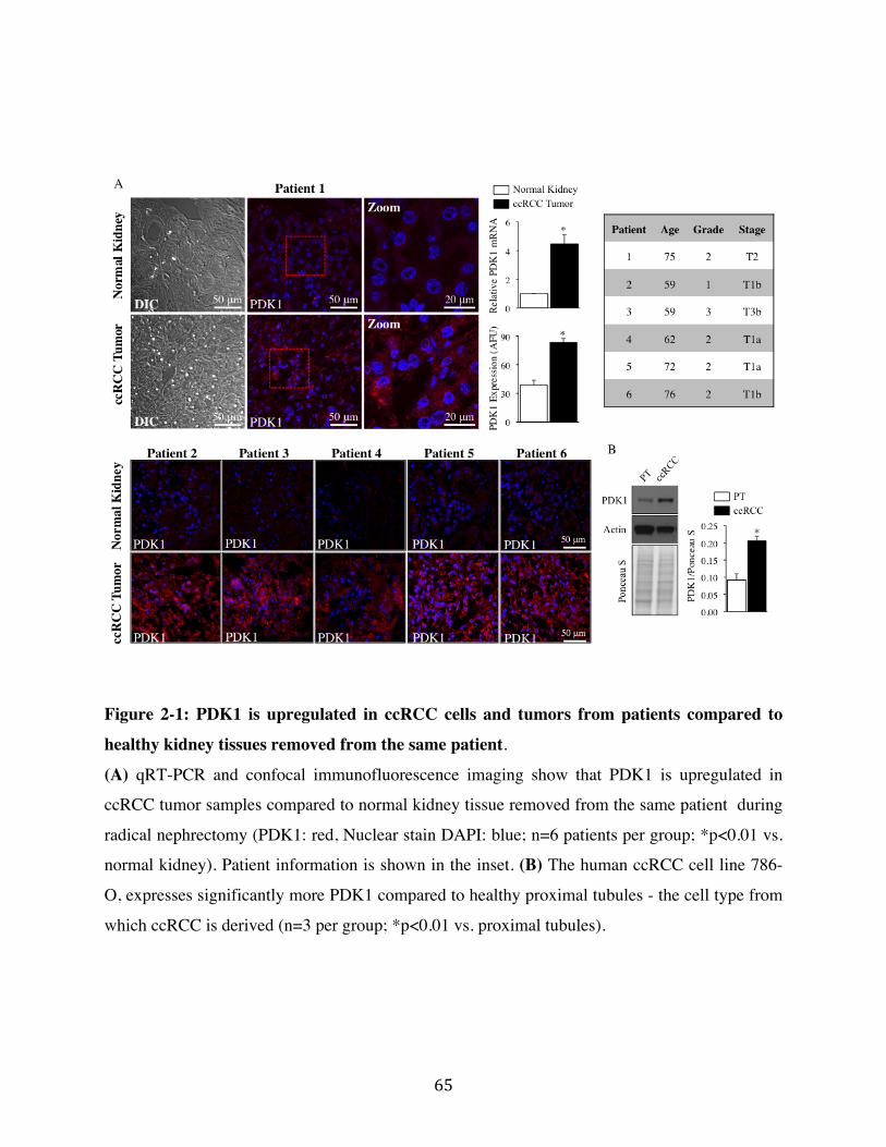
65"
Figure 2-1: PDK1 is upregulated in ccRCC cells and tumors from patients compared to
healthy kidney tissues removed from the same patient.
(A) qRT-PCR and confocal immunofluorescence imaging show that PDK1 is upregulated in
ccRCC tumor samples compared to normal kidney tissue removed from the same patient during
radical nephrectomy (PDK1: red, Nuclear stain DAPI: blue; n=6 patients per group; *p<0.01 vs.
normal kidney). Patient information is shown in the inset. (B) The human ccRCC cell line 786-
O, expresses significantly more PDK1 compared to healthy proximal tubules - the cell type from
which ccRCC is derived (n=3 per group; *p<0.01 vs. proximal tubules).
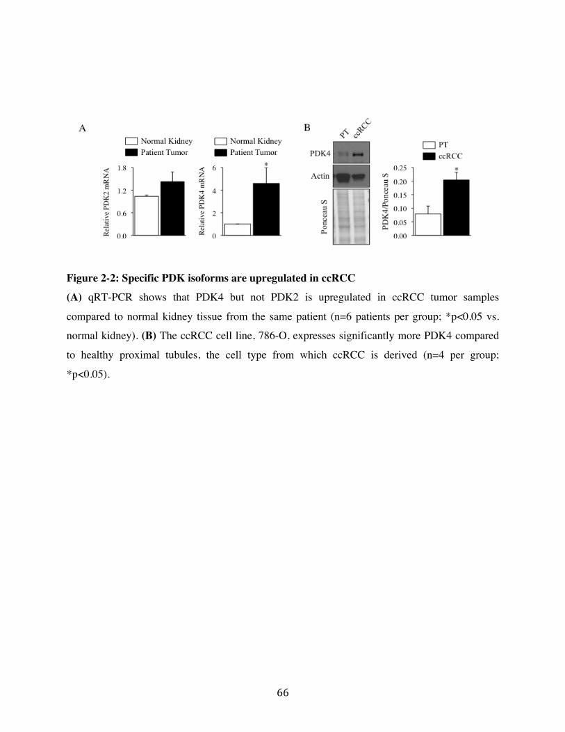
66"
Figure 2-2: Specific PDK isoforms are upregulated in ccRCC
(A) qRT-PCR shows that PDK4 but not PDK2 is upregulated in ccRCC tumor samples
compared to normal kidney tissue from the same patient (n=6 patients per group; *p<0.05 vs.
normal kidney). (B) The ccRCC cell line, 786-O, expresses significantly more PDK4 compared
to healthy proximal tubules, the cell type from which ccRCC is derived (n=4 per group;
*p<0.05).
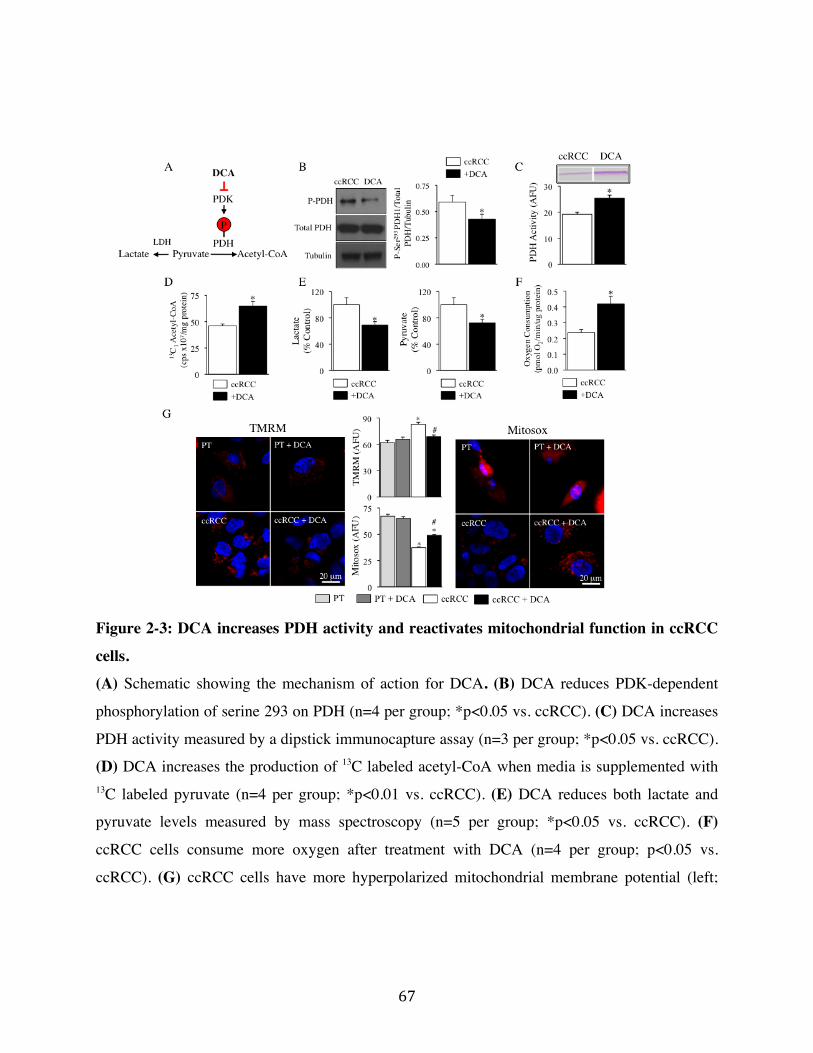
67"
Figure 2-3: DCA increases PDH activity and reactivates mitochondrial function in ccRCC
cells.
(A) Schematic showing the mechanism of action for DCA. (B) DCA reduces PDK-dependent
phosphorylation of serine 293 on PDH (n=4 per group; *p<0.05 vs. ccRCC). (C) DCA increases
PDH activity measured by a dipstick immunocapture assay (n=3 per group; *p<0.05 vs. ccRCC).
(D) DCA increases the production of 13C labeled acetyl-CoA when media is supplemented with 13C labeled pyruvate (n=4 per group; *p<0.01 vs. ccRCC). (E) DCA reduces both lactate and
pyruvate levels measured by mass spectroscopy (n=5 per group; *p<0.05 vs. ccRCC). (F)
ccRCC cells consume more oxygen after treatment with DCA (n=4 per group; p<0.05 vs.
ccRCC). (G) ccRCC cells have more hyperpolarized mitochondrial membrane potential (left;
68"
TMRM: red, nuclear stain Hoechst: blue) and a reduced production of mitochondrial reactive
oxygen species (mROS) (right; Mitosox: red, nuclear stain Hoechst: blue) compared to normal
proximal tubule cells at baseline. DCA treatment repolarizes the mitochondrial membrane
potential and increases mROS in ccRCC but not normal proximal tubule cells (PT) (n=100 cells
per group; *p<0.01 vs. PT, #p<0.01 vs. ccRCC).
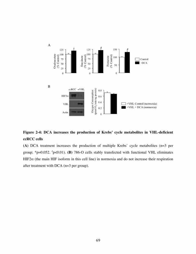
69"
Figure 2-4: DCA increases the production of Krebs’ cycle metabolites in VHL-deficient
ccRCC cells
(A) DCA treatment increases the production of multiple Krebs’ cycle metabolites (n=3 per
group; *p=0.052, #p<0.01). (B) 786-O cells stably transfected with functional VHL eliminates
HIF2α (the main HIF isoform in this cell line) in normoxia and do not increase their respiration
after treatment with DCA (n=3 per group).
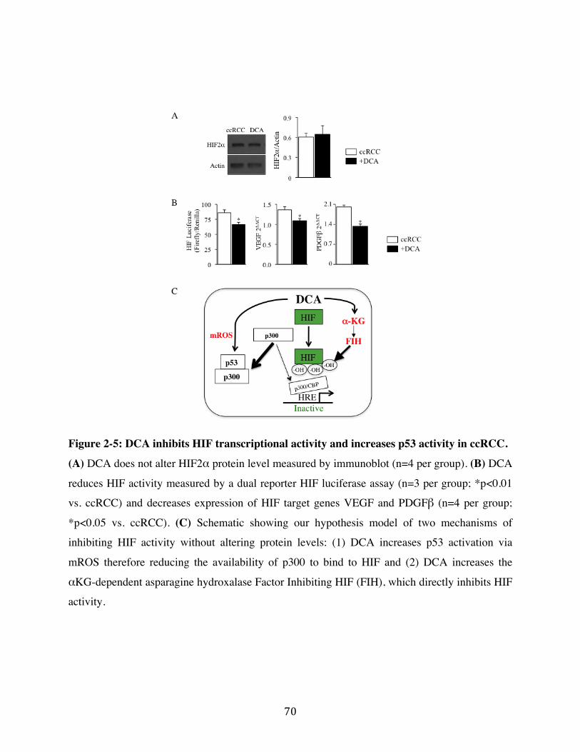
70"
Figure 2-5: DCA inhibits HIF transcriptional activity and increases p53 activity in ccRCC.
(A) DCA does not alter HIF2α protein level measured by immunoblot (n=4 per group). (B) DCA
reduces HIF activity measured by a dual reporter HIF luciferase assay (n=3 per group; *p<0.01
vs. ccRCC) and decreases expression of HIF target genes VEGF and PDGFβ (n=4 per group;
*p<0.05 vs. ccRCC). (C) Schematic showing our hypothesis model of two mechanisms of
inhibiting HIF activity without altering protein levels: (1) DCA increases p53 activation via
mROS therefore reducing the availability of p300 to bind to HIF and (2) DCA increases the
αKG-dependent asparagine hydroxalase Factor Inhibiting HIF (FIH), which directly inhibits HIF
activity.
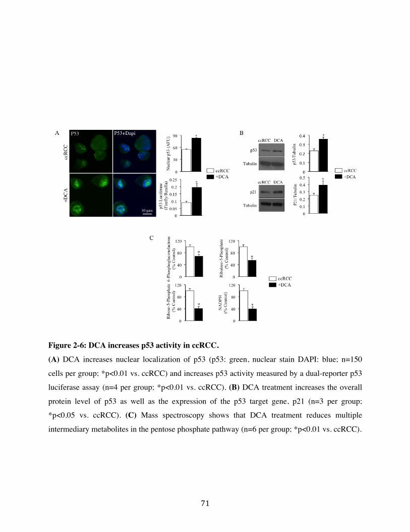
71"
Figure 2-6: DCA increases p53 activity in ccRCC.
(A) DCA increases nuclear localization of p53 (p53: green, nuclear stain DAPI: blue; n=150
cells per group; *p<0.01 vs. ccRCC) and increases p53 activity measured by a dual-reporter p53
luciferase assay (n=4 per group; *p<0.01 vs. ccRCC). (B) DCA treatment increases the overall
protein level of p53 as well as the expression of the p53 target gene, p21 (n=3 per group;
*p<0.05 vs. ccRCC). (C) Mass spectroscopy shows that DCA treatment reduces multiple
intermediary metabolites in the pentose phosphate pathway (n=6 per group; *p<0.01 vs. ccRCC).
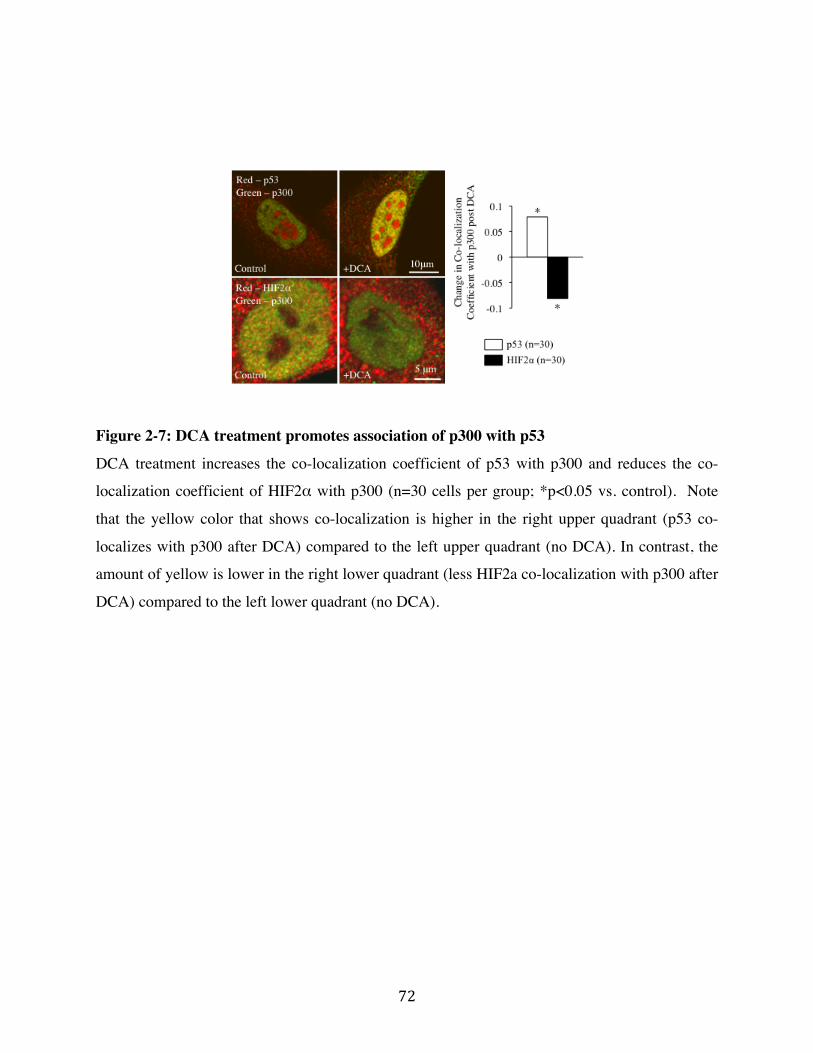
72"
Figure 2-7: DCA treatment promotes association of p300 with p53
DCA treatment increases the co-localization coefficient of p53 with p300 and reduces the co-
localization coefficient of HIF2α with p300 (n=30 cells per group; *p<0.05 vs. control). Note
that the yellow color that shows co-localization is higher in the right upper quadrant (p53 co-
localizes with p300 after DCA) compared to the left upper quadrant (no DCA). In contrast, the
amount of yellow is lower in the right lower quadrant (less HIF2a co-localization with p300 after
DCA) compared to the left lower quadrant (no DCA).
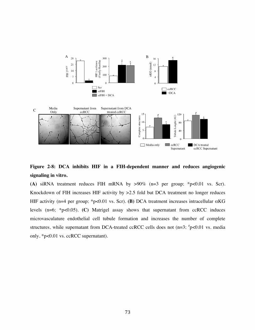
73"
Figure 2-8: DCA inhibits HIF in a FIH-dependent manner and reduces angiogenic
signaling in vitro.
(A) siRNA treatment reduces FIH mRNA by >90% (n=3 per group; *p<0.01 vs. Scr).
Knockdown of FIH increases HIF activity by >2.5 fold but DCA treatment no longer reduces
HIF activity (n=4 per group; *p<0.01 vs. Scr). (B) DCA treatment increases intracellular αKG
levels (n=6; *p<0.05). (C) Matrigel assay shows that supernatant from ccRCC induces
microvasculature endothelial cell tubule formation and increases the number of complete
structures, while supernatant from DCA-treated ccRCC cells does not (n=3; #p<0.01 vs. media
only, *p<0.01 vs. ccRCC supernatant).
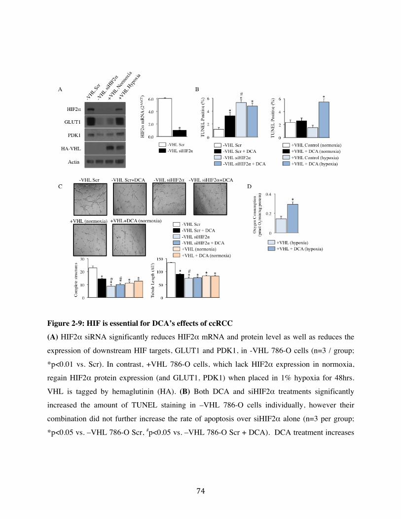
74"
Figure 2-9: HIF is essential for DCA’s effects of ccRCC
(A) HIF2α siRNA significantly reduces HIF2α mRNA and protein level as well as reduces the
expression of downstream HIF targets, GLUT1 and PDK1, in -VHL 786-O cells (n=3 / group;
*p<0.01 vs. Scr). In contrast, +VHL 786-O cells, which lack HIF2α expression in normoxia,
regain HIF2α protein expression (and GLUT1, PDK1) when placed in 1% hypoxia for 48hrs.
VHL is tagged by hemaglutinin (HA). (B) Both DCA and siHIF2α treatments significantly
increased the amount of TUNEL staining in –VHL 786-O cells individually, however their
combination did not further increase the rate of apoptosis over siHIF2α alone (n=3 per group;
*p<0.05 vs. –VHL 786-O Scr, #p<0.05 vs. –VHL 786-O Scr + DCA). DCA treatment increases
75"
TUNEL staining when +VHL 786-O cells are in hypoxia, but not normoxia (n=3 per group;
*p<0.05 vs. +VHL 786-O Control). (C) DCA treatment, HIF2α siRNA and +VHL 786-O cells
in normoxia reduce the number of complete structures and total tubule length compared to –VHL
786-O Scr (n=6 wells per group; *p<0.01 vs. –VHL 786-O Scr, #p<0.05 vs. –VHL 786-O Scr +
DCA). The addition of DCA to –VHL 786-O siHIF2α or normoxic +VHL 786-O cells does not
further reduce these angiogenesis indices. (D) DCA treatment increases respiration in hypoxic
+VHL 786-O cells (n=5 per group; *p<0.01 vs. +VHL 786-O (hypoxia)).
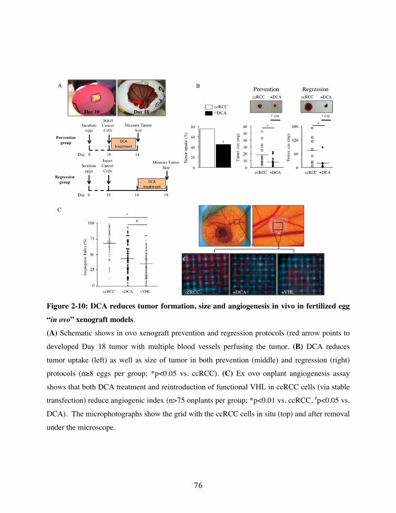
76"
Figure 2-10: DCA reduces tumor formation, size and angiogenesis in vivo in fertilized egg
“in ovo” xenograft models.
(A) Schematic shows in ovo xenograft prevention and regression protocols (red arrow points to
developed Day 18 tumor with multiple blood vessels perfusing the tumor. (B) DCA reduces
tumor uptake (left) as well as size of tumor in both prevention (middle) and regression (right)
protocols (n≥8 eggs per group; *p<0.05 vs. ccRCC). (C) Ex ovo onplant angiogenesis assay
shows that both DCA treatment and reintroduction of functional VHL in ccRCC cells (via stable
transfection) reduce angiogenic index (n>75 onplants per group; *p<0.01 vs. ccRCC, #p<0.05 vs.
DCA). The microphotographs show the grid with the ccRCC cells in situ (top) and after removal
under the microscope.
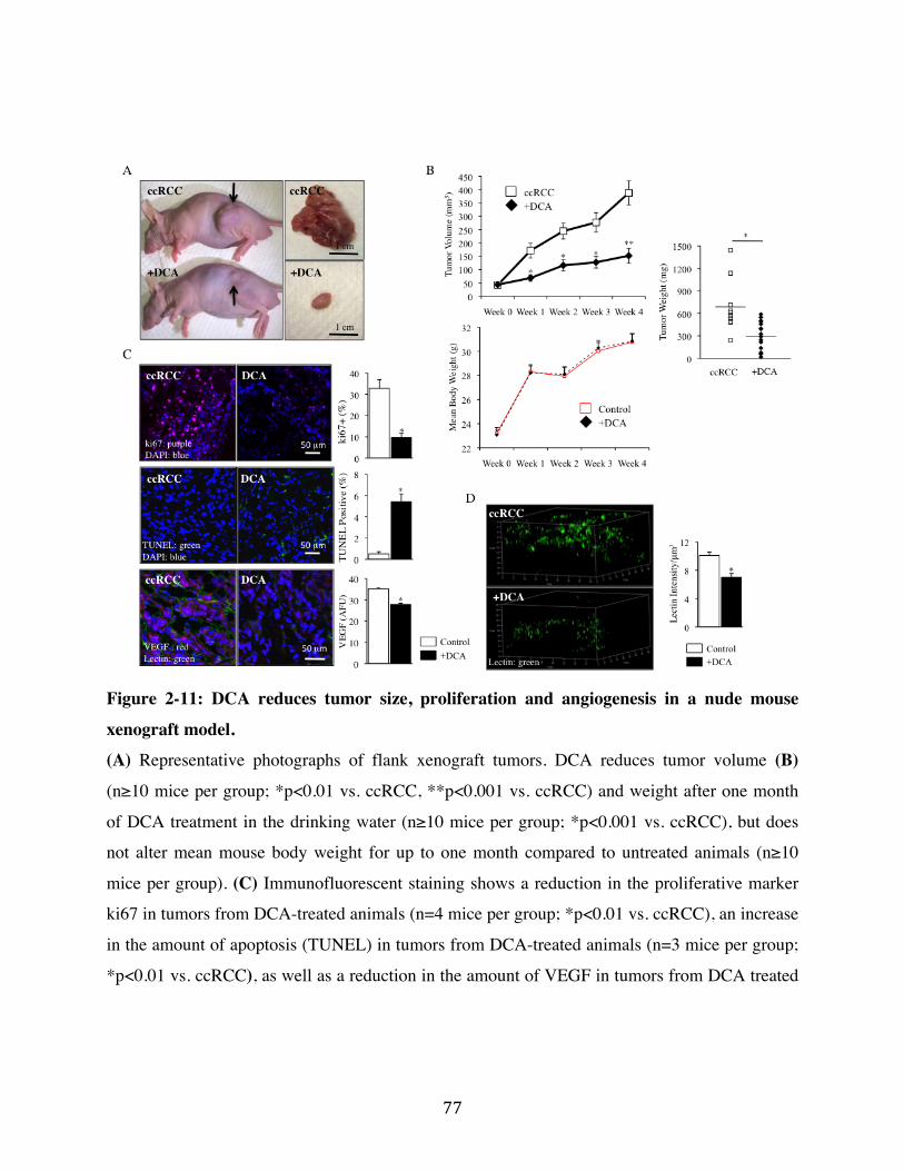
77"
Figure 2-11: DCA reduces tumor size, proliferation and angiogenesis in a nude mouse
xenograft model.
(A) Representative photographs of flank xenograft tumors. DCA reduces tumor volume (B)
(n≥10 mice per group; *p<0.01 vs. ccRCC, **p<0.001 vs. ccRCC) and weight after one month
of DCA treatment in the drinking water (n≥10 mice per group; *p<0.001 vs. ccRCC), but does
not alter mean mouse body weight for up to one month compared to untreated animals (n≥10
mice per group). (C) Immunofluorescent staining shows a reduction in the proliferative marker
ki67 in tumors from DCA-treated animals (n=4 mice per group; *p<0.01 vs. ccRCC), an increase
in the amount of apoptosis (TUNEL) in tumors from DCA-treated animals (n=3 mice per group;
*p<0.01 vs. ccRCC), as well as a reduction in the amount of VEGF in tumors from DCA treated

78"
animals (n=3 mice per group; *p<0.01 vs. ccRCC). (D) 3D stereotactic analysis of stacked
confocal imaging of lectin injected tumors shows reduced vascular density in tumors from DCA-
treated animals (n=3 mice per group; *p<0.01 vs. ccRCC).
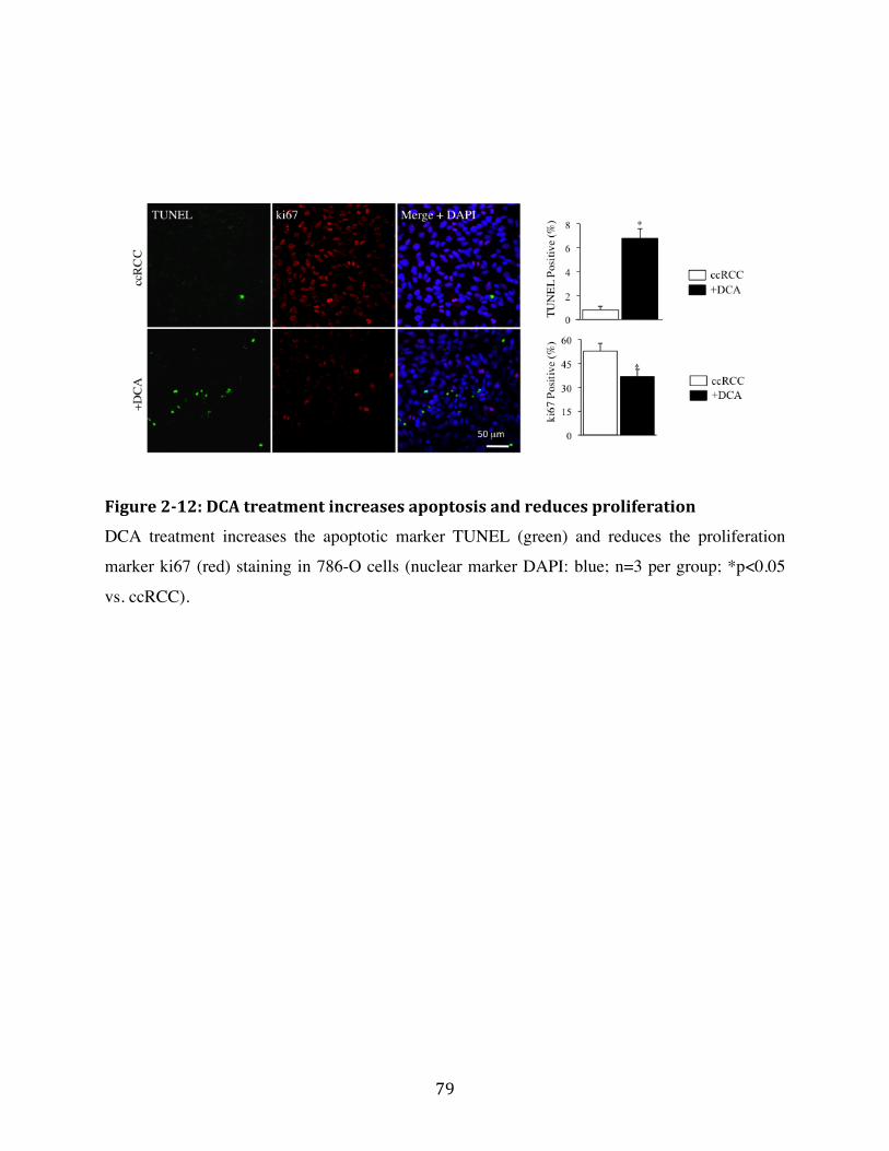
79"
"Figure!2)12:!DCA!treatment!increases!apoptosis!and!reduces!proliferation!
DCA treatment increases the apoptotic marker TUNEL (green) and reduces the proliferation
marker ki67 (red) staining in 786-O cells (nuclear marker DAPI: blue; n=3 per group; *p<0.05
vs. ccRCC).
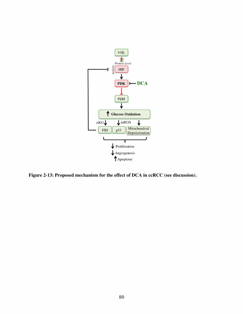
80"
Figure 2-13: Proposed mechanism for the effect of DCA in ccRCC (see discussion).
81"
References
1." Srinivasan" R," Ricketts" CJ," Sourbier" C," Linehan" WM." New" strategies" in" renal" cell"carcinoma:"targeting"the"genetic"and"metabolic"basis"of"disease."Clinical"cancer"research" :"an"official"journal"of"the"American"Association"for"Cancer"Research."2015;21(1):10P7."2." Coppin" C," Kollmannsberger" C," Le" L," Porzsolt" F," Wilt" TJ." Targeted" therapy" for"advanced"renal"cell"cancer"(RCC):"a"Cochrane"systematic"review"of"published"randomised"trials."BJU"international."2011;108(10):1556P63."3." Kim"WY,"Kaelin"WG."Role"of"VHL"gene"mutation"in"human"cancer."Journal"of"clinical"oncology":"official"journal"of"the"American"Society"of"Clinical"Oncology."2004;22(24):4991P5004."4." Moore"LE,"Nickerson"ML,"Brennan"P,"Toro" JR," Jaeger"E,"Rinsky" J,"et"al."Von"HippelPLindau" (VHL)" inactivation" in" sporadic" clear" cell" renal" cancer:" associations"with" germline"VHL"polymorphisms"and"etiologic"risk"factors."PLoS"genetics."2011;7(10):e1002312."5." Krieg"M,"Haas"R,"Brauch"H,"Acker"T,"Flamme"I,"Plate"KH."UpPregulation"of"hypoxiaPinducible"factors"HIFP1alpha"and"HIFP2alpha"under"normoxic"conditions"in"renal"carcinoma"cells" by" von" HippelPLindau" tumor" suppressor" gene" loss" of" function." Oncogene."2000;19(48):5435P43."6." Kim" JW," Tchernyshyov" I," Semenza" GL," Dang" CV." HIFP1Pmediated" expression" of"pyruvate" dehydrogenase" kinase:" a" metabolic" switch" required" for" cellular" adaptation" to"hypoxia."Cell"metabolism."2006;3(3):177P85."7." Papandreou"I,"Cairns"RA,"Fontana"L,"Lim"AL,"Denko"NC."HIFP1"mediates"adaptation"to" hypoxia" by" actively" downregulating" mitochondrial" oxygen" consumption." Cell"metabolism."2006;3(3):187P97."8." Sudarshan"S,"Karam"JA,"Brugarolas"J,"Thompson"RH,"Uzzo"R,"Rini"B,"et"al."Metabolism"of"kidney"cancer:"from"the"lab"to"clinical"practice."European"urology."2013;63(2):244P51."9." Vander"Heiden"MG," Cantley" LC," Thompson"CB."Understanding" the"Warburg" effect:"the"metabolic"requirements"of"cell"proliferation."Science."2009;324(5930):1029P33."10." Sutendra" G," Dromparis" P," Kinnaird" A," Stenson" TH," Haromy" A," Parker" JM," et" al."Mitochondrial" activation" by" inhibition" of" PDKII" suppresses" HIF1a" signaling" and"angiogenesis"in"cancer."Oncogene."2013;32(13):1638P50."11." Sutendra"G,"Michelakis"ED."Pyruvate"dehydrogenase"kinase"as" a"novel" therapeutic"target"in"oncology."Frontiers"in"oncology."2013;3:38."12." Kinnaird" A," Michelakis" ED." Metabolic" modulation" of" cancer:" a" new" frontier" with"great"translational"potential."J"Mol"Med"(Berl)."2015;93(2):127P42."13." Scheuermann"TH,"Li"Q,"Ma"HW,"Key"J,"Zhang"L,"Chen"R,"et"al."Allosteric"inhibition"of"hypoxia"inducible"factorP2"with"small"molecules."Nat"Chem"Biol."2013;9(4):271P6."14." Zimmer"M,"Ebert"BL,"Neil"C,"Brenner"K,"Papaioannou"I,"Melas"A,"et"al."SmallPmolecule"inhibitors"of"HIFP2a"translation"link"its"5'UTR"ironPresponsive"element"to"oxygen"sensing."Mol"Cell."2008;32(6):838P48."
82"
15." Compton" S," Kim" C," Griner" NB," Potluri" P," Scheffler" IE," Sen" S," et" al." Mitochondrial"
dysfunction" impairs" tumor" suppressor" p53" expression/function." J" Biol" Chem."
2011;286(23):20297P312."
16." Schmid" T," Zhou" J," Kohl" R," Brune" B." p300" relieves" p53Pevoked" transcriptional"
repression"of"hypoxiaPinducible"factorP1"(HIFP1)."Biochem"J."2004;380(Pt"1):289P95."
17." Bonnet"S,"Archer"SL,"AllalunisPTurner"J,"Haromy"A,"Beaulieu"C,"Thompson"R,"et"al."A"
mitochondriaPK+" channel" axis" is" suppressed" in" cancer" and" its" normalization" promotes"
apoptosis"and"inhibits"cancer"growth."Cancer"cell."2007;11(1):37P51."
18." Michelakis" ED," Sutendra" G," Dromparis" P," Webster" L," Haromy" A," Niven" E," et" al."
Metabolic"modulation"of"glioblastoma"with"dichloroacetate."Science"translational"medicine."
2010;2(31):31ra4."
19." Cao"W,"Yacoub"S,"Shiverick"KT,"Namiki"K,"Sakai"Y,"Porvasnik"S,"et"al."Dichloroacetate"
(DCA)"sensitizes"both"wildPtype"and"over"expressing"BclP2"prostate"cancer"cells"in"vitro"to"
radiation."The"Prostate."2008;68(11):1223P31."
20." Sun"RC,"Fadia"M,"Dahlstrom"JE,"Parish"CR,"Board"PG,"Blackburn"AC."Reversal"of"the"
glycolytic" phenotype" by" dichloroacetate" inhibits" metastatic" breast" cancer" cell" growth" in"
vitro"and"in"vivo."Breast"cancer"research"and"treatment."2010;120(1):253P60."
21." Li"L,"Zhang"L,"Zhang"X,"Yan"Q,"Minamishima"YA,"Olumi"AF,"et"al."HypoxiaPinducible"
factor" linked" to" differential" kidney" cancer" risk" seen" with" type" 2A" and" type" 2B" VHL"
mutations."Mol"Cell"Biol."2007;27(15):5381P92."
22." Rini" BI," Atkins" MB." Resistance" to" targeted" therapy" in" renalPcell" carcinoma." The"
lancet"oncology."2009;10(10):992P1000."
23." Vousden" KH," Ryan" KM." p53" and" metabolism." Nature" reviews" Cancer."
2009;9(10):691P700."
24." Keith"B,"Johnson"RS,"Simon"MC."HIF1alpha"and"HIF2alpha:"sibling"rivalry"in"hypoxic"
tumour"growth"and"progression."Nature"reviews"Cancer."2012;12(1):9P22."
25." Kroeze" SG," Vermaat" JS," van" Brussel" A," van"Melick" HH," Voest" EE," Jonges" TG," et" al."
Expression" of" nuclear" FIH" independently" predicts" overall" survival" of" clear" cell" renal" cell"
carcinoma"patients."Eur"J"Cancer."2010;46(18):3375P82."
26." Jiang"P,"Du"W,"Wang"X,"Mancuso"A,"Gao"X,"Wu"M,"et"al."p53"regulates"biosynthesis"
through" direct" inactivation" of" glucoseP6Pphosphate" dehydrogenase." Nature" cell" biology."
2011;13(3):310P6."
27." Lim"HY,"Yip"YM,"Chiong"E,"Tiong"HY,"Halliwell"B,"Esuvaranathan"K,"et"al."Metabolic"
signatures"of"renal"cell"carcinoma."Biochem"Biophys"Res"Commun."2015;460(4):938P43."
28." Kato"M,"Li"J,"Chuang"JL,"Chuang"DT."Distinct"structural"mechanisms"for"inhibition"of"
pyruvate" dehydrogenase" kinase" isoforms" by" AZD7545," dichloroacetate," and" radicicol."
Structure."2007;15(8):992P1004."
29." Kaplon"J,"Zheng"L,"Meissl"K,"Chaneton"B,"Selivanov"VA,"Mackay"G,"et"al."A"key"role"for"
mitochondrial" gatekeeper" pyruvate" dehydrogenase" in" oncogenePinduced" senescence."
Nature."2013;498(7452):109P12."
83"
30." Shen" YC," Ou" DL," Hsu" C," Lin" KL," Chang" CY," Lin" CY," et" al." Activating" oxidative"
phosphorylation" by" a" pyruvate" dehydrogenase" kinase" inhibitor" overcomes" sorafenib"
resistance"of"hepatocellular"carcinoma."Br"J"Cancer."2013;108(1):72P81."
31." Warburton" HE," Brady" M," Vlatkovic" N," Linehan" WM," Parsons" K," Boyd" MT." p53"
regulation"and"function"in"renal"cell"carcinoma."Cancer"research."2005;65(15):6498P503."
32." Gurova"KV,"Hill"JE,"Razorenova"OV,"Chumakov"PM,"Gudkov"AV."p53"pathway"in"renal"
cell"carcinoma"is"repressed"by"a"dominant"mechanism."Cancer"research."2004;64(6):1951P
8."
33." Roberts" AM," Watson" IR," Evans" AJ," Foster" DA," Irwin" MS," Ohh" M." Suppression" of"
hypoxiaPinducible" factor" 2alpha" restores" p53" activity" via" Hdm2" and" reverses"
chemoresistance"of"renal"carcinoma"cells."Cancer"research."2009;69(23):9056P64."
34." Stacpoole"PW,"Kerr"DS,"Barnes"C,"Bunch"ST,"Carney"PR,"Fennell"EM,"et"al."Controlled"
clinical" trial" of" dichloroacetate" for" treatment" of" congenital" lactic" acidosis" in" children."
Pediatrics."2006;117(5):1519P31."
35." Stacpoole" PW." The" pharmacology" of" dichloroacetate." Metabolism."
1989;38(11):1124P44."
36." Abdelmalak"M,"Lew"A,"Ramezani"R,"Shroads"AL,"Coats"BS,"Langaee"T,"et"al."LongPterm"
safety"of"dichloroacetate"in"congenital" lactic"acidosis."Mol"Genet"Metab."2013;109(2):139P
43."
37." Chu" QS," Sangha" R," Spratlin" J," L" JV," Mackey" JR," McEwan" AJ," et" al." A" phase" I" openP
labeled," singleParm," dosePescalation," study" of" dichloroacetate" (DCA)" in" patients" with"
advanced"solid"tumors."Invest"New"Drugs."2015;33(3):603P10."
38." Dhar" S," Lippard" SJ." Mitaplatin," a" potent" fusion" of" cisplatin" and" the" orphan" drug"
dichloroacetate."Proc"Natl"Acad"Sci"U"S"A."2009;106(52):22199P204."
39." Kumar" K,"Wigfield" S," Gee" HE," Devlin" CM," Singleton" D," Li" JL," et" al." Dichloroacetate"
reverses" the" hypoxic" adaptation" to" bevacizumab" and" enhances" its" antitumor" effects" in"
mouse"xenografts."J"Mol"Med"(Berl)."2013;91(6):749P58."
40." Buczek" M," Escudier" B," Bartnik" E," Szczylik" C," Czarnecka" A." Resistance" to" tyrosine"
kinase" inhibitors" in" clear" cell" renal" cell" carcinoma:" from" the" patient's" bed" to" molecular"
mechanisms."Biochimica"et"biophysica"acta."2014;1845(1):31P41."
41." Hitosugi" T," Fan" J," Chung" TW," Lythgoe" K," Wang" X," Xie" J," et" al." Tyrosine"
phosphorylation" of" mitochondrial" pyruvate" dehydrogenase" kinase" 1" is" important" for"
cancer"metabolism."Mol"Cell."2011;44(6):864P77."
42." Fan" J," Kang"HB," Shan" C," Elf" S," Lin" R," Xie" J," et" al." TyrP301" phosphorylation" inhibits"
pyruvate"dehydrogenase"by"blocking"substrate"binding"and"promotes"the"Warburg"effect."J"
Biol"Chem."2014;289(38):26533P41."
43." Mullen"P." The" use" of"Matrigel" to" facilitate" the" establishment" of" human" cancer" cell"
lines"as"xenografts."Methods"in"molecular"medicine."2004;88:287P92."
44." Quail"DF,"Walsh"LA,"Zhang"G,"Findlay"SD,"Moreno"J,"Fung"L,"et"al."Embryonic"protein"
nodal"promotes"breast"cancer"vascularization."Cancer"research."2012;72(15):3851P63."
84"
45." Pink"DB,"Schulte"W,"Parseghian"MH,"Zijlstra"A,"Lewis"JD."RealPtime"visualization"and"quantitation" of" vascular" permeability" in" vivo:" implications" for" drug" delivery." PLoS" One."2012;7(3):e33760."46." Leong"HS,"Steinmetz"NF,"Ablack"A,"Destito"G,"Zijlstra"A,"Stuhlmann"H,"et"al."Intravital"imaging" of" embryonic" and" tumor" neovasculature" using" viral" nanoparticles." Nat" Protoc."2010;5(8):1406P17."47." Kain"KH,"Miller" JW," JonesPParis" CR," Thomason"RT," Lewis" JD," Bader"DM," et" al." The"chick"embryo"as"an"expanding"experimental"model"for"cancer"and"cardiovascular"research."Dev"Dyn."2014;243(2):216P28."48." Goulet"B,"Kennette"W,"Ablack"A,"Postenka"CO,"Hague"MN,"Mymryk"JS,"et"al."Nuclear"localization" of"maspin" is" essential" for" its" inhibition" of" tumor" growth" and"metastasis." Lab"Invest."2011;91(8):1181P7."49." Palmer"TD," Lewis" J," Zijlstra"A."Quantitative" analysis" of" cancer"metastasis" using" an"avian"embryo"model."J"Vis"Exp."2011(51).""

! 85!
!
Chapter Three
A Nuclear Pyruvate Dehydrogenase Complex is
Important for the Generation of Acetyl-CoA and Histone
Acetylation
! 86!
Abstract
DNA transcription, replication and repair are regulated by histone acetylation, a
process that requires the generation of acetyl-CoA. Here we show that all the
subunits of the mitochondrial pyruvate dehydrogenase complex (PDC) are also
present and functional in the nucleus of mammalian cells. We found that
knockdown of nuclear PDC in isolated functional nuclei decreased the de-novo
synthesis of acetyl-CoA and acetylation of core histones. Nuclear PDC levels
increased in a cell-cycle dependent manner and in response to serum, epidermal
growth factor or mitochondrial stress; this was accompanied by a corresponding
decrease in mitochondrial PDC levels, suggesting a translocation from the
mitochondria to the nucleus. Inhibition of nuclear PDC decreased acetylation of
specific lysine residues on histones important for G1-S-phase progression and
expression of S phase markers. Dynamic translocation of mitochondrial PDC to
the nucleus provides a novel pathway for nuclear acetyl-CoA synthesis required
for histone acetylation and epigenetic regulation.
! 87!
Introduction
Epigenetic regulation of gene expression is essential for embryonic
development and differentiation, protection against viruses and progression of
cancer (Jaenisch and Bird, 2003). It includes modifications of core histones by
either methylation or acetylation. By neutralizing the positive charges of lysine
residues in histones, acetylation promotes the relaxation of DNA, necessary for
replication and transcription (Vogelauer et al., 2002). A recent high-resolution
mass spectrometry analysis identified a large number of nuclear proteins with
multiple acetylation sites, suggesting that the role of acetylation extends beyond
histones (Choudhary et al., 2009). In Eukaryotes, the biosynthesis of acetyl-CoA
is thought to occur in the sub-cellular compartment in which it is required,
because it is membrane impermeable and very unstable, due to the high-energy
thioester bond that joins the acetyl- and -CoA groups. Our understanding of how
the nucleus generates acetyl-CoA in metazoan cells is incomplete. Acetyl-CoA
synthetase (AceCS1) and ATP-citrate lyase (ACL) are both present in the nucleus
(Wellen et al., 2009). ACL is be the predominant pathway for nuclear acetyl-CoA
generation in mammalian cells since it utilizes glucose oxidation-derived
mitochondrial citrate as its substrate, as opposed to AceCS1 which uses acetate, a
fuel that is not an important energy source for mammalian cells (Wellen et al.,
2009). However, in cells with decreased citrate levels due to mitochondrial
suppression by Bcl-xL over-expression, levels of acetyl-CoA and N-! acetylated
proteins decreased in the cytoplasm with no apparent decrease in histone
acetylation (Yi et al., 2011). This suggested the presence of a yet unidentified
mitochondria-independent mechanism for histone acetylation in the nucleus.
Primitive protozoan cells that lack mitochondria, like Entamoeba
histolytica, utilize pyruvate:ferrodoxin oxidoreductase (PFO) to generate acetyl-
CoA (Rodriguez et al., 1998). Mammalian cells do not express PFO, but generate
acetyl-CoA in mitochondria with a similar complex of nucleus-encoded proteins
known as pyruvate dehydrogenase complex (PDC) (Horner et al., 1999). The
generation of acetyl-CoA from both PFO and PDC is dependent on the glycolysis-
! 88!
derived product pyruvate. The nuclear presence of several metabolic enzymes has
been observed as early as the 1960s, with many shown to interact with nuclear
proteins and DNA and “moonlight” to the nucleus in conditions of cellular stress
(Kim and Dang, 2005; Siebert and Humphrey, 1965; Yang et al., 2012; Yogev et
al., 2010). For example, the M2 isoform of the glycolytic enzyme pyruvate kinase
(PKM2) translocates to the nucleus and functions as a protein kinase,
phosphorylating nuclear proteins, like threonine-11 of histone 3 (H3) and
enhancing the acetylation of lysine 9 on H3 (H3K9) (Yang et al., 2012). The
protein kinase function of PKM2 utilizes the glycolytic intermediate
phosphoenolpyruvate (PEP) and not ATP as its phosphate donor (Vander Heiden
et al., 2010), resulting in the subsequent generation of pyruvate (Gao et al., 2012;
Yang et al., 2012). However, the fate of pyruvate in the nucleus and whether this
is related to the acetyl-CoA used in the H3K9 acetylation remain unknown.
Intact and functional PDC can translocate across mitochondrial
membranes from the matrix to the outer mitochondrial membrane (Hitosugi et al.,
2011). Thus it would be possible for a chaperone protein to bind PDC from the
outer mitochondrial membrane and bring it to the nucleus under conditions that
stimulate S-phase entry and cell cycle progression, where histone acetylation is
critical.
We provide evidence that supports the hypothesis that functional PDC can
translocate from the mitochondria to the nucleus during cell cycle progression,
generating a nuclear pool of acetyl-CoA from pyruvate, and increasing the
acetylation of core histones important for S-phase entry. The mitochondrial
pyruvate dehydrogenase kinase (PDK), which phosphorylates and inhibits
mitochondrial PDC was not detectable in the nucleus, suggesting that nuclear
PDC may be constitutively active and regulated differently than mitochondrial
PDC. The nuclear translocation of PDC was triggered by growth signals like
serum and epidermal growth factor, or mitochondrial inhibitors, like rotenone.
Nuclear PDC provides a novel means for the nucleus to generate acetyl-CoA in an
autonomous fashion. The implications of this work extend to many conditions
! 89!
where nuclear acetylation is altered, like development, cancer, neurodegeneration
or cardiovascular disease.
Results
3.1 All Components of PDC are Present in the Nucleus
PDC is comprised of subunits from three catalytic enzymes: pyruvate
dehydrogenase (E1), dihydrolipoamide transacetylase (E2) and dihydrolipoamide
dehydrogenase (E3) as well as the tethering protein, E3 binding protein (E3BP)
(Behal et al., 1993). We first identified the nuclear presence of PDC-E1 (!
subunit) in human spermatozoa, which compartmentalize the nucleus (in the
head) away from mitochondria (in the mid-section), allowing for more clear
visualization of nuclear versus mitochondrial PDC (Figure 3-2A). To confirm the
nuclear presence of PDC-E1 in a terminally differentiated primary cell line, we
isolated primary fibroblasts from human lungs and detected its nuclear presence
(Figure 3-1A). We then performed a more detailed assessment for the presence of
nuclear PDC using microscopy in two commercially available cell lines, normal
small airway epithelial cells (SAEC) and A549 cells (non-small cell lung cancer
cells). In intact SAEC and A549 cells co-stained with an antibody against PDC-
E1 and the mitochondrial marker MitoTracker Red, co-localization of the two
signals was evident in the cytoplasm as expected, but PDC-E1 was also evident
within the nucleus (marked by DAPI) without any mitochondrial signal (Figures
3-1B and 3-1C). For our imaging we generated 25 separate images in each intact
cell, systematically scanning in the z-axis at 0.2µm in depth for each image. We
used these stacked images to generate 3D videos that show the presence of PDC-
E1 throughout the nucleus (Supplementary Video 1 and 2). We selected specific
planes that “cut through the nucleus” from the z-stacked images, as shown in the
top image of Figure 3-1D. Then, in the XY axis, we quantified the nuclear PDC-
E1 and MitoTracker Red signals by measuring the fluorescence intensity. As we
move from left to right in the image plane, an area of mitochondria is followed by
an area where only the nucleus is present. PDC-E1 is highly detected in the
mitochondria and co-localizes with the MitoTracker Red signal. However, while
the MitoTracker Red signal intensity is absent in the nucleus, the PDC-E1 signal
! 90!
is present, though with lesser intensity than the mitochondrial PDC-E1 signal
(Figure 3-1D). Furthermore, nuclear PDC-E1 followed a similar signal pattern to
the nuclear protein histone 3, confirming the specificity of the nuclear PDC-E1
signal (Figure 3-2B). In addition, a clear nuclear signal of PDC-E1 was also
detected in the nucleus using electron microscopy (Figure 3-2C). These data show
that the nuclear PDC-E1 signal is not a “contamination” from overlapping
mitochondria.
To address the possibility that our PDC-E1 antibody non-specifically
binds to other nuclear proteins, we transfected A549 and SAEC with a plasmid
encoding for cloned human E1! subunit of PDC in frame with enhanced green
fluorescent protein (EGFP). Based on EGFP fluorescence we detected the
presence of the fused EGFP-E1 protein in mitochondria as well as the nucleus of
both cell types (Figure 3-1E and Figure 3-2D). We then identified the nuclear
presence for other subunits of the PDC complex, including PDC-E2, PDC-E3 and
the ancillary subunit PDC-E3BP in intact cells using immunofluorescence (Figure
3-1F-H). For all confocal and electron microscopy experiments a “secondary
antibody-only” control staining was performed, which in all experiments showed
no signal (Figure 3-2E).
3.2 Nuclear PDC is Functional and Can Generate Acetyl-CoA from Pyruvate
To assess if nuclear PDC is functional, we isolated nuclei using a nuclei-
specific high sucrose gradient centrifugation protocol. As even a small amount of
mitochondrial membranes could be a confounding factor, we took several steps to
ensure that our nuclei were not contaminated with any mitochondria. We showed
that nuclear membranes were intact by the lamin staining pattern and that our
nuclear preparations were free of mitochondrial membranes, as assessed by
nonyl-acridine orange (NAO) and MitoTracker Red imaging (Figures 3-3A and 3-
3B), as well as free of cytoplasmic, mitochondrial matrix and membrane proteins,
as assessed by immunoblots with antibodies against !-tubulin, citrate synthase
(CS), isocitrate dehydrogenase 2 (IDH2), succinyl-CoA synthetase (SCS) and
succinate dehydrogenase (SDH) (Figure 3-3C). These vigorous purity indices
! 91!
were used in all of our experiments with isolated nuclear preparations throughout
this work. In these pure nuclei preparations, we confirmed the presence of PDC-
E1 in nuclei from the EGFP-transfected cells, as well as from intact nuclei from
A549 and SAEC cells, using immunofluorescence; with absence of any detectable
signal in secondary-only antibody straining (Figure 3-4A, upper panels). In
addition to the ! subunit of the PDC-E1, all the subunits of the PDC complex
were present in isolated nuclei (Figure 3-4A, bottom panels). We then used
immunoblots and showed the nuclear presence of all PDC subunits in isolated
nuclei from A549 cells (Figure 3-3D) as well as a separate cancer cell line, 786-0
renal cell carcinoma cells, in the absence of cytoplasmic or mitochondrial
contamination (Figure 3-3E). As NAD(H) is abundant in the nucleus, our finding
that PDC-E2 appears to be lipoylated (implying the nuclear presence of lipoic
acid, not present in our media), suggests that nuclear PDC has all the necessary
co-factors to be functional (Figure 3-4B).
To study the function of nuclear PDC without interference from
mitochondrial PDC, we performed our initial experiments on isolated nuclei. We
detected many glycolytic intermediates including PEP, pyruvate and acetyl-CoA
using collision-induced dissociation mass spectrometry (Figure 3-3F). We then
designed an experiment to study whether nuclear PDC is functional in terms of
the de-novo biosynthesis of acetyl-CoA.
We first isolated intact and pure nuclei from either A549 or 786-0 cells
previously treated with scrambled vs. PDC-E1 siRNA. We obtained an efficient
knockdown of PDC-E1 resulting in no detectable immunoblot signal (Figure 3-
3G; top). This was in keeping with the absence of any PDC activity in nuclei
lacking PDC, measured by a standard dipstick assay that measures the NADH
produced by the immunocaptured enzyme (Figure 3-3G; bottom).
We then treated these nuclei with an isotope-labeled form of pyruvate
(13C2-pyruvate) for 8hrs and measured the production of labeled acetyl-CoA
(13C1-acetyl-CoA; Figure 3-3H). Since the only way to synthesize acetyl-CoA
from pyruvate is by PDC, the detection of labeled acetyl-CoA in our nuclear
preparations reflects the activity of nuclear PDC. We detected a significant
! 92!
decrease in the levels of 13C1-labeled acetyl-CoA in nuclei from PDC-E1 siRNA-
treated cells compared to those from scrambled siRNA, using two separate mass
spectrometry acquisition modes in A549 and 786-0 nuclei (Figure 3-3I and 3-4C).
We also detected increasing levels of labeled acetyl-CoA in response to
increasing levels of labeled pyruvate, suggesting the presence of non-limiting
amounts of functional PDC in the isolated nuclei (Figure 3-3I and 3-4C).
3.3 Nuclear PDC is Important for Histone Acetylation
We then assessed whether the acetyl-CoA generated from nuclear PDC is
important for histone acetylation. Since histone acetylation depends on glucose
and not free-fatty acid metabolism (Wellen et al., 2009), we first deprived the
A549 cells of glucose for 24 hours in order to synchronize acetylation, in the
presence of scrambled or PDC-E1 siRNA. Next, we isolated functional nuclei
from scrambled and PDC-E1 siRNA-treated cells and exposed them to pyruvate
(10mM) for 8hrs and measured histone acetylation (Figure 3-5A). Prior to the
addition of pyruvate to our isolated nuclei (time=0), no significant differences
were apparent in acetylated lysine levels at the molecular weight of histones
between scrambled and PDC-E1 siRNA groups (Figure 3-5B), confirming the
synchronization of acetylation. After exposure to pyruvate, nuclei from PDC-E1
siRNA-treated cells had decreased levels of all three acetylated (Ac) core histones
H2B, H3 and H4, compared to nuclei from scrambled siRNA-treated cells (Figure
3-5C).
On the other hand, inhibition of nuclear PDC did not appear to change the
acetylation of p53 and FOXO1 in these cancer cells, suggesting that the
acetylation of targets in the nucleus is regulated, perhaps by the availability or
proximity of acetyl-transferases or de-acetylases with acetyl-CoA generating
enzymes, like PDC (Figure 3-6). Alternatively, a rapid turnover of acetyl-groups
in histones, compared to those of other proteins, may also explain why the
inhibition of nuclear PDC preferentially showed a decrease in histone acetylation.
Since the role of ACL on nuclear acetylation has not been studied in A549
cells, we used ACL or PDC-E1 siRNA in order to study the relative importance of
! 93!
these two enzymes in nuclear acetylation. Extracted histones from either PDC-E1
or ACL siRNA-treated cells had decreased histone acetylation compared to
scrambled siRNA-treated cells (Figure 3-5D). Nevertheless, in these cells the
impact of PDC-E1 silencing on histone acetylation appears to be larger than that
of ACL-silencing. This may be explained by the fact that PDC is also involved in
the ACL-mediated production of nuclear acetyl-CoA, as mitochondrial PDC is
also an important regulator of citrate production (i.e. ACL’s substrate) via the
Krebs’ cycle. The effects of ACL and PDC-E1 silencing were not the same on the
acetylation of specific histones, suggesting that the source of acetyl-CoA may
play a role on which target is acetylated.
3.4 Pyruvate Dehydrogenase Kinase is Not Present in the Nucleus
Mitochondrial PDC-E1 is tonically inhibited by PDC kinases (PDKs) and
activated by PDC phosphatases (PDPs). PDKs phosphorylate serine-293 of the !
subunit of PDC-E1, resulting in its inactivation (Behal et al., 1993). While PDK I
and II are ubiquitously expressed, PDK III and IV are only expressed in the testis
and under starvation conditions in muscle, respectively (Bowker-Kinley et al.,
1998). Hypoxia-inducible factor 1! (HIF1!) is activated in many cancers and can
suppress mitochondrial PDC by inducing PDK expression (Kim et al., 2006). We
detected PDKI and II in mitochondria, but not in isolated nuclei of A549 (which
express both isoforms) and 786-O cancer cells, (which only express PDKI )
(Figure 3-7A and 3-7B). On the other hand, we detected the presence of PDPI
(but not PDPII) in nuclei of A549 and 786-O cells (Figure 3-7A and 3-7B). We
found higher levels of PDC-E1 serine-293 phosphorylation in A549 mitochondria
compared to normal cells (Figure 3-7C), in keeping with the finding that cancer
cells have higher levels of PDK (Michelakis et al., 2010). Furthermore, PDC-E1
phosphorylated serine-293 was present in mitochondria but not in nuclei (Figure
3-7A), in keeping with the absence of PDK. This suggested that in certain
conditions (like in cancer or hypoxia, where HIF1! is activated) while
mitochondrial PDC can be relatively inactive, nuclear PDC could remain
constitutively active.
! 94!
We then treated isolated nuclei from A549 and 786-O cancer cells with the
small molecule PDK inhibitor Dichloroacetate (DCA), which primarily inhibits
PDK I and II (Bonnet et al., 2007; Michelakis et al., 2010) and showed no
differences in Ac-H3 levels (Figure 3-7D). However, DCA treatment of whole
A549 cells resulted in both increased Ac-H3 (nuclear acetylation) and Ac-tubulin
(cytoplasmic acetylation) (Figure 3-7E, 3-8A and 3-8B). The increase in
acetylation by DCA in whole cells may be due to acetyl-CoA biosynthesis by
cytoplasmic and nuclear ACL, which produces acetyl-CoA using citrate as
substrate (Figure 3-7F). We confirmed the expected DCA-induced increase in
citrate, along with another Krebs’ cycle intermediate (succinate) and the expected
decrease in lactate (Figure 3-8C). Thus the differential effects of DCA [which has
known anti-cancer properties (Bonnet et al., 2007; Dhar and Lippard, 2009;
Michelakis et al., 2010)] between mitochondrial and nuclear PDC, reflect the
differential expression of PDK in the two cellular compartments and suggests that
factors that can increase or decrease PDK function (like HIF1! and DCA,
respectively) can be used to exploit the functional significance of nuclear PDC, an
idea that we explored later on.
3.5 PDC Translocates from the Mitochondria to the Nucleus During S-phase
To study whether histone acetylation occurs prior to DNA replication
during S-phase in our cells, we first synchronized cells to the G1 phase by serum
starvation for 24 hrs. We then introduced serum and serially measured Ac-H3 and
cyclin A. Ac-H3 levels increased first, followed by cyclin A (Figure 3-9A). By
isolating pure nuclei from these cells we also showed that nuclear PDC-E1 levels
followed a similar increase pattern to Ac-H3 and then began to decrease towards
baseline, but only after Ac-H3 levels peaked (Figure 3-10A). Furthermore, using
microscopy, we showed that both PDC-E1 and Ac-H3 were higher during late S-
phase compared to baseline in isolated nuclei (Figure 3-10B).
We then measured both nuclear and mitochondrial PDC-E1 levels upon
serum stimulation after cell-cycle synchronization, at the same time points. We
found that the increase in nuclear PDC-E1 levels during cell cycle progression
! 95!
was associated with a parallel decrease in mitochondrial PDC-E1 levels, before
both PDC fractions returned toward their baseline levels (Figure 3-9B). The
decrease in mitochondrial PDC was not due to enhanced degradation since PDC
levels remained unaltered in whole cells in response to increasing concentrations
of serum (Figure 3-10C), suggesting the decrease in mitochondrial PDC was due
to its translocation to the nucleus.
To measure the relative distribution of PDC in the mitochondria and
nucleus, we loaded on the same gel the same amount of protein from isolated
nuclei and mitochondria at baseline and 3hrs post-serum stimulation. Although
PDC-E1 levels were clearly higher in mitochondria than the nuclei, the percent
ratio of nuclear to mitochondrial PDC-E1 increased from 17% to 30% after 3
hours of serum stimulation (Figure 3-10D). A similar increase in the percent ratio
of nuclear to mitochondrial PDC in response to serum stimulation was also
observed with the E2 component using immunofluorescence, where we were able
to measure mitochondrial PDC (overlap with MitoRed) and nuclear PDC (overlap
with DAPI) within the same cell (Figure 3-9C).
We then performed a series of experiments to further support the fact that
nuclear PDC translocates from the mitochondria. To exclude the possibility that
the increase in nuclear PDC in response to serum stimulation is a newly translated
product from the endoplasmic reticulum (ER), we inhibited ribosomal translation
with cycloheximide (CHX) and measured nuclear PDC levels (see Figure 3-9D).
Serum stimulation increased nuclear PDC (E1 and E2) and this was not altered by
CHX, although the translation of c-myc [a protein with a short half-life,
previously shown to be decreased within 2 hrs of CHX treatment (Alarcon-Vargas
et al., 2002)] was decreased during this timeframe (Figure 3-9E).
Newly synthesized mitochondrial proteins from the ER contain an N-
terminus mitochondrial localization sequence (MLS) and upon entry into the
matrix, the MLS is cleaved by the mitochondrial processing peptidase (MPP; see
Figure 3-9D). The cleavage of these (otherwise destabilizing) sequences (~15-50
amino acids) of mitochondrial proteins is required to prevent degradation and
facilitate subsequent assembly of subunits (Chacinska et al., 2009). Only mature
! 96!
(cleaved MLS) PDC proteins are able to re-fold and form complexes in the
mitochondria. Therefore, we hypothesized that if nuclear PDC is directly
translocated to the nucleus after its translation in ribosomes, we should be able to
detect the precursor form in the nucleus. Using two separate siRNA transfection
approaches for the "-catalytic subunit of mitochondrial processing peptidase
(MPP), we were able to inhibit the mRNA levels by 65% and 90% respectively,
resulting in the accumulation of increasing levels of the precursor form of PDC-
E1 (Figure 3-9F, see arrowhead) along with the mature form. We then used MPP
siRNA-treated whole cells and probed for PDC-E1 on the same membrane with
isolated nuclei and mitochondrial protein from untreated cells. While the
precursor form was clearly seen in the MPP-silenced whole cells in the same gel,
only the mature form of PDC was detected in isolated nuclei and mitochondria
(Figure 3-9G; top). We also isolated nuclei from both scrambled and MPP
siRNA-treated cells and could not detect the presence of the precursor; only
mature PDC was present in the nucleus (Figure 3-9G; bottom). As MPP is a
mitochondria-specific protease, our data suggest that nuclear PDC is processed in
the mitochondria prior to translocation to the nucleus.
All of the subunits of PDC are translated in the ER and are transported
separately to the mitochondria, where they are processed prior to complex
formation. We hypothesized that if we inhibit only one of the subunits and thus
disturb the stoichiometry balance of the subunits within the complex, we may
affect the nuclear levels of the other subunits. Indeed, knockdown of only PDC-
E1 by siRNA resulted in decreased levels of all catalytic components of PDC in
the nucleus, without changing their overall protein expression in whole cell
preparations (Figure 3-10E). This suggests that PDC is translocated from the
mitochondria to the nucleus as a functional complex.
3.6 Signals Increasing Nuclear PDC Levels
We then studied signals that may trigger the nuclear translocation of PDC.
Epidermal growth factor (EGF) signaling is important for S-phase entry and cell
cycle progression (Kato et al., 1998) and has been shown to increase the nuclear
! 97!
translocation of PKM2 in cancer (Yang et al., 2012). We found that recombinant
human EGF (rhEGF) increased the nuclear levels of PDC components, along with
PKM2, in A549 cells (Figure 3-11A and 3-12A). The increase in nuclear levels
was through translocation since rhEGF did not change the total cellular
expression of PDC subunits, while it predictably increased tyrosine-204
phosphorylation of MAPK (Figure 3-11B). Similarly, gefitinib, an EGF receptor
inhibitor, decreased nuclear (Figure 3-11C), but not whole-cell PDC levels
(Figure 3-11D). We then used confocal imaging and studied the relative
distribution of PDC subunits within the same cell, in 786-0 cells. Similar to A549
cells, rhEGF increased the relative distribution of PDC-E1 and PDC-E2 from the
mitochondria to the nucleus (Figure 3-11E).
We speculated that a mitochondrial stressor might trigger translocation of
PDC from the mitochondria to the nucleus. We studied the electron transport
chain (ETC) complex I inhibitor rotenone, since inhibition of ETC complexes is a
well-known cause of mitochondrial stress (Durieux et al., 2011). We found that
rotenone caused a significant translocation of PDC into the nucleus over and
above serum and rhEGF (Figure 3-11E and 3-12B). Mitochondria adapt to stress
by initiating the mitochondria unfolded protein response (mtUPR), which results
in increased expression of mitochondrial import proteins, folding chaperones and
heat shock proteins, amplifying the communication with the nucleus (Zhao et al.,
2002). We speculated that a heat shock chaperone may be involved in the nuclear
translocation of PDC.
We studied heat shock protein 70 (Hsp70) based on evidence that: (1)
induction of Hsp70 is cell-cycle dependent, with its highest expression and
nuclear localization observed during S-phase (Milarski and Morimoto, 1986), (2)
Hsp70 is involved in the nuclear translocation of several proteins (Shi and
Thomas, 1992) and (3) Hsp70 binds to and activates mitochondrial PDC (Kiang et
al., 2006). We confirmed that nuclear levels of Hsp70 increase in a cell-cycle
dependent manner, similarly to the increase in nuclear PDC-E1 following serum
stimulation (Figure 3-12C). We performed immunoprecipitation studies on A549
cells and MRC-9 cells (fibroblasts) with Hsp70 and detected the presence of
! 98!
PDC-E1 and PDC-E2 (Figure 3-11F and 3-12D), suggesting that Hsp70 may bind
to these components of the complex. Sequence and structural analysis of potential
binding motifs for Hsp70 within the PDC complex showed a putative Hsp70
binding motif (i.e. hydrophobic peptide regions) (Mayer and Bukau, 2005) within
the PDK binding site on PDC (Figure 3-12E). PDK did not co-immunoprecipitate
with Hsp70 (Figure 3-11F and 3-12D), suggesting that Hsp70 and PDK may
compete for similar binding domains within PDC.
We pre-treated serum-starved A549 and 786-0 cells with either vehicle
(DMSO) or KNK437, an inhibitor of heat shock factor 1 (HSF1), which results in
decreased expression of inducible Hsp70 (Koishi et al., 2001), prior to serum
stimulation for 4hrs. KNK437 decreased mRNA expression (Figure 3-12F) and
nuclear levels of both Hsp70 and PDC-E1, compared to vehicle (Figure 3-11G
and S6G). Nuclear Hsp70 levels correlated positively with nuclear PDC-E1 levels
in both vehicle and KNK437-treated A549 cells (Figure 3-11H and 3-12H).
Similar to KNK437, a specific siRNA against Hsp70 also decreased nuclear levels
of both Hsp70 and PDC-E1 in response to serum stimulation (Figure 3-11I).
3.7 Nuclear PDC is Important for S-phase Entry and Cell Cycle Progression
Our data on the dynamic translocation of PDC from the mitochondria to
the nucleus suggest that it is the same enzymatic complex that is present in both
compartments, rather than perhaps a different variant. This makes the study of the
relative biological role of PDC on histone acetylation and cell cycle progression
in the two cellular compartments challenging, particularly in the intact cell
setting. We took advantage of PDK’s localization in mitochondria, but not the
nucleus, and designed experiments in an intact cell setting, in which by
subtracting the effects of selective mitochondrial PDC inhibition (by the induction
of PDK) from the effects of total cellular PDC inhibition (by PDC-E1 siRNA), we
could expose the role of nuclear PDC.
While PDK is already induced in cancer cells, we aimed to maximally
inhibit mitochondrial PDC by two methods: first, inducing endogenous PDK by
activation of HIF1! and second, increasing exogenous PDKI via transfection with
! 99!
a PDKI plasmid (see Figure 3-13A). With either method, mitochondrial PDC
should be maximally inhibited (condition 1). Then by inhibiting total cellular
PDC by siRNA gene silencing (condition 2), we could expose the effects of
nuclear PDC on histone acetylation and cell cycle progression by subtracting
condition 1 from condition 2 (Figure 3-13A).
For the first experiment, we infected A549 cells with an adenovirus
encoding for CA5 (AdCA5), a constitutively active mutant form of HIF1!
(mHIF1!) (Manalo et al., 2005), avoiding the confounding effects of hypoxia.
Compared to an adenovirus encoding for LacZ (AdLacZ), increased expression of
mHIF1! by AdCA5 (Figure 3-14A and 3-14B) resulted in a significant increase
in the expression of PDKI (Figure 3-13B) and phosphorylation of serine-293 on
mitochondrial, but not nuclear PDC-E1 (Figure 3-13C). Mutant HIF1! increased
mitochondrial membrane potential (Figure 3-14C), further supporting the
mitochondrial PDC suppression and its impact on mitochondrial function in our
model, as we have previously described (Bonnet et al., 2007). We then treated
AdCA5-infected cells with scrambled (condition 1) versus PDC-E1 siRNA
(condition 2). We also gave scrambled siRNA to AdLacZ-infected cells (control).
We synchronized all the groups to the G1 phase by serum starvation for 24hrs,
followed by re-introduction of serum, and 24 hours later we measured S-phase
markers and specific histone-3 acetylation sites that are involved in cell cycle
progression. AdCA5-infected cells treated with scrambled siRNA had increased
acetylation of H3K9 and H3K18, both important for S-phase progression (Cai et
al., 2011), increased G1-S-phase progression shown by increased levels of
phosphorylated retinoblastoma (P-Rb), as well as elongation-2-factor (E2F),
cyclin A and cyclin-dependent kinase 2 (Cdk2), markers for S-phase entry,
compared to AdLacZ-infected cells treated with scrambled siRNA (Figure 3-
13D). These effects (condition 1) are in agreement with the described effects of
HIF1! on proliferation (Semenza, 2010) and the recruitment of histone acetyl-
transferases (HATs) (Luo et al., 2011). In contrast, AdCA5-infected cells treated
with PDC-E1 siRNA (condition 2) had significantly decreased acetylation of
H3K9 and H3K18 and decreased levels of P-Rb, E2F, cyclin A and Cdk2,
! 100!
compared to AdCA5-infected cells treated with scrambled siRNA (condition 1)
(Figure 3-13D). Since both conditions had activated HIF1! and inhibited
mitochondrial PDC, subtraction of condition 2 (grey bars) from condition 1 (black
bars) in Figure 3-13D shows that nuclear PDC inhibition decreases Ac-H3K9 and
Ac-H3K18 (while total H3 levels remain relatively stable) as well as P-Rb and S-
phase regulators (E2F, cyclin A, Cdk2).
For the second experiment, transfection with a PDKI plasmid increased
the expression of PDKI (Figure 3-14D) and phosphorylation of PDC-E1 serine-
293 (Figure 3-13E), compared to the empty vector control. PDKI over-expression
did not change histone acetylation or cell cycle progression (Figure 3-13E,
compare white bars [control] to black bars [condition 1]), compatible with the fact
that mitochondrial PDC is significantly inhibited in these cancer cells at baseline
and further inhibition may not elicit any measurable effects. However, similar to
the first experiment, PDC siRNA (condition 2), decreased H3K9 and H3K18
acetylation and decreased P-Rb, E2F, cyclin A and Cdk2, compared to scrambled
siRNA controls (condition 1; Figure 3-13E). Thus, two different approaches of
inhibiting nuclear PDC support its role in S-phase progression.
! 101!
Discussion
Here we show for the first time that PDC is present and functional in the
nucleus. Nuclear PDC can generate acetyl-CoA utilized for histone acetylation
and S-phase entry, providing a novel link between metabolism and epigenetic or
cell cycle regulation. This newly discovered source of nuclear acetyl-CoA may
also be important in conditions where the availability of cytoplasmic citrate
(which can cross the nuclear membrane and produce acetyl-CoA through ACL) is
decreased due to suppressed production or shift toward lipid synthesis. Our work
suggests that nuclear PDC has a mitochondrial origin since it lacks the MLS
(which can only be cleaved in the mitochondria) and its nuclear increase in
response to serum is not affected by inhibition of ribosomal translation. In
addition to serum, nuclear PDC translocation increases under growth factors
(EGF) or mitochondrial inhibitors (rotenone), suggesting a potential role in
disease states with proliferative signals or mitochondrial dysfunction, like cancer.
It is intriguing that our data suggest translocation of PDC in the nucleus,
given the large size of this enzymatic complex. High-resolution electron
microscopy and structural analysis have recorded its size and diameter within the
range of 8-10MDa and 25-45nm, respectively (Sumegi et al., 1987; Zhou et al.,
2001b). However, there is evidence for size and conformational variability of
PDC with identification of complexes as small as 1MDa (Sumegi et al., 1987;
Zhou et al., 2001a). Although the stoichiometry of the individual components in
the complex (i.e. the relative amount of E1, E2, E3 subunits) varies, in keeping
with the reported variability in its size among tissues, only the interplay of all
subunits within a functional complex can produce acetyl-CoA. The fact that we
find de-novo production of acetyl-CoA in isolated nuclei in response to pyruvate,
suggests that a functional complex is present. The fact that we can decrease the
nuclear levels of all subunits by only silencing the gene for one subunit, suggests
that the complex travels as a whole from the mitochondria into the nucleus. PDC
was recently identified as an intact functional complex on the outer mitochondrial
membrane (Hitosugi et al., 2011), suggesting that it can translocate across
mitochondrial membranes, a more complex process than translocation across the
! 102!
nuclear membrane. Since the nuclear pore complex can accommodate the entry of
large complexes of similar diameter to PDC, like ribonucleoprotein complexes
(Lodish et al., 2000) or intact nucleocapsids of viruses (Pante and Kann, 2002), it
is possible that an intact PDC complex could translocate to the nucleus.
We provide evidence that Hsp70 may promote the nuclear translocation of
a constitutively active form of PDC. By competing with PDK for binding to PDC,
Hsp70 may allow PDC to remain active when translocated to the nucleus (Figure
3-13F). Kiang et al (Kiang et al., 2006) showed that Hsp70 binds to and activates
mitochondrial PDC, but did not show the mechanism of this activation. It may be
that by competing with PDK, Hsp70 removes this inhibitory kinase and thus
activates PDC. It is possible that EGF can promote nuclear acetylation by a
coordinated translocation of PKM2 and PDC, potentially by increasing Hsp70
levels (Milarski and Morimoto, 1986) (Figure 3-13F). This model may contribute
to the recently described effects of nuclear PKM2 on tumor growth via histone
acetylation, offering a source and mechanism for the nuclear acetyl-CoA
generation used for the acetylation of H3K9 (Yang et al., 2012). It is appealing to
consider that there is a mechanism by which several factors required for the
nuclear response to proliferative stimuli (for example PKM2 and PDC) can be
transferred simultaneously, increasing efficiency. Although our collective data
support the existence of the translocation model shown in Figure 3-13F, we
cannot rule out the possibility that individual subunits may be transported
independently in the nucleus, where they could potentially be assembled in an
intact complex with the help of a chaperone like Hsp70.
Tyrosine phosphorylation by growth factor signaling, including EGFR,
activates PDKI providing a mechanism for suppression of mitochondrial PDC in
cancer (Hitosugi et al., 2011), in addition to the induction of PDK expression by
HIF1! (Kim et al., 2006). Therefore, in a “double hit” manner, EGF stimulation
can activate PDK, suppressing mitochondrial PDC [which has been shown to
inhibit mitochondria-dependent apoptosis in cancer (Bonnet et al., 2007)] in
tandem with Hsp70-mediated nuclear translocation of PDC, promoting histone
acetylation and cell cycle progression (Figure 3-13F). The inhibition of PDK by
! 103!
siRNA, DCA or hybrid drugs [like mitoplatin, a drug that structurally combines
cisplatin with DCA molecules (Dhar and Lippard, 2009)] decreases cancer growth
in animal models (Bonnet et al., 2007) and a small human trial (Michelakis et al.,
2010), by activating glucose oxidation, reversing the Warburg effect and the
resistance to apoptosis in cancer cells. Our work now suggests that the nuclear
pool of PDC is “immune” to this strategy. It raises the possibility that in response
to these interventions cancer cells may “escape” by promoting a transfer of PDC
in the nucleus, where PDC may promote proliferation. It also suggests that anti-
cancer strategies, in which PDK inhibition is used, may perhaps be strengthened
by simultaneous inhibition of EGFR signaling or Hsp70 function. As PDC plays a
prominent role in many metabolic disorders, it will be important for scientists to
be aware that their efforts to target PDC may have direct and previously
unrecognized effects on nuclear biology.
Our work suggests an alternative pathway to ACL for the nucleus to
generate acetyl-CoA for histone acetylation. It is possible that in specific tissues
or disease states, the relative importance of ACL or nuclear PDC may be
different. ACL has been shown to be important for histone acetylation during
differentiation (Wellen et al., 2009), while our work suggests that histone
acetylation by nuclear PDC may be important for cell cycle progression. ACL and
PDC should be studied together when assessing histone acetylation and epigenetic
regulation in conditions that both differentiation and proliferation are taking
place, including development, cancer and other proliferative conditions or stem
cell biology.
! 104!
Materials and Methods
Cell culture
Human A549 non-small cell lung cancer cells, 786-O renal cell carcinoma and
MRC-9 fibroblasts were purchased from ATCC (Manassas, VA). A549 cells were
maintained on F12K medium, while 786-O cells in RPMI-1640 media and MRC-
9 cells in EMEN. Primary human fibroblasts were isolated from the lung of a
transplant patient in accordance with the Human Ethics Committee at the
University of Alberta and maintained in DMEN. Primary human fibroblasts were
isolated from the lung using an enzymatic cocktail containing papin (1mg/ml),
dithiothreitol (0.5mg/ml), collagenase (0.6mg/ml) and bovine serum albumin
(0.6mg/ml) (Sigma-Aldrich) as previously described (Sutendra et al., 2011).
Human small airway epithelial cells (SAECs) were purchased from ScienCell
(Carisbad, CA). SAECs were maintained in SAEpiCM, which was provided by
the company. Media for all cell lines were supplemented with 10% FBS (unless
stated otherwise) and 5% antibiotic and antimycotic (Invitrogen-Gibco Canada,
Burlington, Canada).
Confocal Microscopy
Confocal microscopy imaging was performed using a Zeiss LSM 510 NLO
confocal microscope with two-photon capability (Carl Zeiss Microscopy, Jena,
Germany). PDC-E1!, E2, PDKI, PDKII and Hsp70 antibodies were purchased
from Santa Cruz Biotechnology (Santa Cruz, CA) and used at a 1:50 dilution.
PDC-E1", E3, E1BP, lamin and PKM2 antibodies were purchased from Abcam
(Cambridge, MA) and used at a dilution of 1:100. Ac-H3 antibody (against all N-
terminal acetyl-lysine residues) was purchased from Cell Signaling Technology
and used at a dilution of 1:100. MitoTracker Red (250nM; Invitrogen-Gibco) and
Nonyl acridine orange (100nM; Invitrogen-Molecular Probes, Burlington,
Canada) were used as mitochondrial membrane markers. Quantification of both
the mitochondria and nuclear PDC signal was assessed using the Zeiss physiology
option software, which can quantify fluorescence units along the plane (line) of a
specified region, using the Profile setting. As depicted in Figure 3-1D, a plane that
! 105!
crosses through the mitochondria and nucleus was selected to directly compare
the intensity of signal between the two organelles and to also show the purity of
the nuclear signal for PDC as the signal for the mitochondrial marker Mitotracker
Red was not detected in the portion of the plane that crosses the nucleus. Image J
64 software was used to measure the nuclear (signal present within the DAPI
region of the cell) and mitochondrial (signal present within MitoRed region of the
same cell) PDC signal (using the integrated density parameters) in response to
serum stimulation, rhEGF or rotenone treatment. All images were scanned in mid-
plane of the cell in the z-axis as shown in Figure 3-1D, using a 100X NA 1.3 oil
objective lens at 2X zoom, allowing for an pixel size of 0.04µm x 0.04µm x
0.2µm. This allows for the clear visualization of PDC in the mid-plane of the
nucleus. Fluorophore conjugated secondary antibodies (DAKO, Carpinteria, CA
and Invitrogen, Molecular Probes, Eugene OR) and the nuclear stain DAPI
(Molecular Probes) were used for immunofluorescence imaging with specific
excitations of 488 nm (Fitc), 543 (Tritc), 633 (Far Red) and 750 nm two photon
(DAPI) and the corresponding emissions were detected with the following filter
sets. Fitc: BP 505-615, Tritc: BP 565-615, Far red: LP 650 and DAPI: BP 390-
465. Overlap was eliminated between the emissions of any secondary antibodies,
mitochondrial-specific dyes, EGFP and nuclear stains by imaging each channel
independently and sequentially with only one excitation wavelength active during
each scan.
Reconstructed Video: Twenty-five stacked images consisting of 0.2µm
sections in depth were acquired using confocal microscopy. The frame size was
2048x2048 pixels. These stacked images covering an area of 46.1µm (length) x
46.1µm (width) x 5µm (depth) generated the constructed video using the Zeiss 3D
for LSM software. All experiments included negative control secondary antibody-
only staining (Figure 3-2E, 3-4A and 3-12A), which in all presented experiments
showed no signal, supporting the specificity of the antibodies used.
! 106!
EGFP-PDC-E1 plasmid
EGFP-PDC-E1 plasmid was generated by OriGene Technologies (Rockville,
MD). Briefly, cDNA ORF clone of homo sapiens pyruvate dehydrogenase alpha 1
(PDHA1) (OriGene Technologies; Rockville, MD) was cloned into pEGFP-N1
vector (Clontech, Mountain View, CA) and transfected into A549 and SAECs
using Xfect transfection agent (Clontech).
Spermatozoa isolation
Human spermatozoa were isolated from semen by centrifugation for 10 minutes at
700 x g. Spermatozoa were then plated on slides coated with cell-tak (BD
Biosciences, Mississauga, Canada) before immunofluorescence staining and
confocal microscopy.
Immunogold electron microscopy
A549 cells were grown on coverslips, fixed in 3% freshly prepared
paraformaldehyde plus 0.05% glutaraldehyde and permeabilized with 0.05%
saponin. Following blocking, cells were incubated with a mouse monoclonal
antibody against PDC-E1! (Santa Cruz Biotechnlology) for one hour and then
with fluro-nanogold anti mouse Fab Alexa Fluor 488 (Nanoprobes, Yaphank, NY)
secondary antibody over night at 4°C. Cells were further fixed in 2.5%
glutaraldehyde in PBS plus 2% sucrose, and nanogold particles were gold-
enhanced (Gold Enhance, Nanoprobes). Cells were then dehydrated and
embedded in EMbed-812. Seventy-millimeter thin sections were prepared and
observed using a Philips 410 electron microscope (Philips Research, Briarcliff
Manor, NY). A Hitachi H-7000 (Hitachi High Technologies America,
Schaumburg, IL) Transmission Electron Microscopy (TEM) was used at 15,000x
magnification and 80kV, in order to image isolated nuclei and confirm their
purity.
! 107!
Functional nuclear isolation
Isolation of nuclei was performed using the commercially available nuclei
isolation kit: nuclei PURE prep from Sigma Aldrich. Briefly, adherent cells were
washed with PBS and scraped from the plate in the presence of lysis buffer. Cells
(in lysis media) were carefully placed on top of a 1.8M sucrose gradient and the
resulting suspension was centrifuged at 30,000 x g for 45 minutes in a pre-cooled
swinging bucket ultracentrifuge. Nuclei were collected as a white pellet at the
bottom of the centrifuge and washed with nuclei storage buffer (provided with the
kit). Purity of nuclei was assessed by immunocytochemistry and immunoblot. For
functional experiments, isolated nuclei were used immediately.
PDC Activity Assay
PDC activity was measured using the MitoProfile Dipstick Assay Kit
(MitoSciences, Eugene, OR). Protein (50µL of 1µg/µL) was collected from
isolated nuclei and placed in a 96-well dish and incubated with the dipstick
containing the PDC antibody. The enzyme complex is immunocaptured in its
native form and activity is visualized by coupling PDC-dependent production of
NADH to the reduction of NBT in the presence of excess diaphorase, forming an
insoluble intensely colored precipitate at the capture line. PDC activity was
measured by the intensity of band using a flat top scanner.
siRNA treatment
PDC-E1, ACL, Hsp70 and scrambled-siRNA (Ambion, Austin, TX) were
transfected at a final concentration of 20nM with CaCl2. MPP" was transfected at
a final concentration of 200nM with CaCl2. After 18 hrs, media was changed and
experiments were performed 48 hrs later.
Isolated nuclei experimental protocol for acetyl-CoA measurement and
histone acetylation
A549 or 786-0 cells transfected with scrambled or PDC-E1 siRNA were
maintained in glucose free media 24 hours prior to nuclear isolation. Immediately
! 108!
following nuclear isolation, isolated nuclei were incubated with 10mM pyruvate
in nuclear storage buffer (Sigma Aldrich) for 8 hrs at 37°C. The experiments were
terminated by the addition of ice-cold nuclear storage buffer and the nuclei were
centrifuged at 500 x g before they were washed with ice-cold storage buffer to
remove any excess pyruvate. Nuclei were then prepared for either metabolite
extraction and acetyl-CoA measurements by mass spectrometry or protein
extraction and histone acetylation by immunoblots.
Metabolite extraction: Nuclei and cells were re-suspended in 800µL of ice-cold
80% methanol and 20% ddH2O. Samples were vigorously vortexed and placed in
liquid N2 for 10 minutes to freeze. Samples were then thawed on ice for 10
minutes, before freeze-thaw cycle was repeated. Samples were centrifuged at
13,000 x g to pellet cell debris, lipids and proteins. Supernatant was evaporated
and resulting metabolites were re-suspended in HPLC-grade H2O. Metabolites
were normalized to protein concentration.
Mass spectrometry for 13C1-acetyl-CoA: Isolated nuclei from scrambled and
PDC-E1 siRNA-treated cells were exposed to13C2-pyruvate (Cambridge Isotope
Laboratories, Andover, MA) for 8 hrs, before the experiment was terminated with
the addition of ice-cold storage buffer. Metabolites were extracted and 30µL of
samples was diluted to 120µL with methanol and flow injected to the MS using
4000 QTRAP mass spectrometer (AB Sciex, Concord, Canada) with either
enhanced product ion (EPI; IonSpray voltage of -4500V) or enhanced MS (EMS;
IonSpray voltage of 5500V).
Mass spectrometry for glycolytic intermediates
Seven microliters of sample was injected using a 4000 QTRAP mass spectrometer
(AB Sciex) equipped with a UHPLC 1290 system (Agilent Technologies,
Mississauga, Canada) via SRM for all 9 glycolytic intermediates and acetyl-CoA.
Samples were delivered to the MS with mobile phases A (20mM NH4OH, 20mM
NH4Ac in 95%/5% H2O/CH3CN) and B (98% CH3CN, 2% H2O) via a 2.0mm i.d.
x 10cm HILIC Luna NH2 column (Phenomenex, Torrance, CA) at 250µl/min
using negative ion LC/MS/MS analytical run. The dwell time was 5ms per SRM
! 109!
transition, and collision energy was optimized for each SRM transition. Total
cycle time was 2.09s.
Histone extraction
Histone extraction was performed using the commercially available EpiQuik
Global Histone Acetylation Assay kit (Epigentek, Brooklyn, NY). Briefly isolated
nuclei and whole cells were lysed and proteins were precipitated with 25%
trichloracetic acid. Extracted histone pellets were dissolved in HPLC-grade
distilled water.
1H-Nuclear Magnetic Resonance (1H-NMR) 1H-NMR spectra was acquired on an 800-MHz Inova spectrometer (Agilent
formerly Varian Inc, Palo Alto, CA) equipped with a HCN Z-axis gradient cold-
probe. 1H-NMR spectra were acquired at 25°C using the first increment of a 2D-1H-1H-NOESY probe sequence, commonly referred to as the metnosey (ie. 1D-1H-NOESY). Spectra were collected with 128 transients and 8 steady state scans
using a 4 second acquisition time and a 990 millisecond presaturation, with
saturation during the 100 millisecond-mixing period.
Cycloheximide Experiments
786-0 cells were serum starved and pre-treated with either vehicle (DMSO) or
100µg/ml cycloheximide (Cell Signaling) for 24hrs prior to serum stimulation for
4 hrs in the presence of either vehicle or cycloheximide. After nuclear isolation,
protein was extracted and immunoblots were performed.
Adenoviral Infection
A549 cells were infected with AdCA5 or AdLacZ at a multiplicity of infection of
500 for 48 hrs allowing an infection rate of ~100% (Figure 3-14A) as previously
described (Manalo et al., 2005; Sutendra et al., 2013). After 48hrs cells were than
transfected with scrambled vs. PDC-E1 siRNA.
! 110!
Pyruvate dehydrogenase kinase I (PDKI) Transfection
PDKI plasmid was generated by OriGene Technologies (Rockville, MD) and
40µg of plasmid was transfected into A549 cells using Xfect transfection agent
(Clontech). After 48hrs cells were transfected with either scrambled or PDC-E1
siRNA.
Epidermal Growth Factor (EGF) Experiments
A549 cells were treated with vehicle (PBS) or 500ng/mL rhEGF (Sigma Aldrich)
or 10µM Gefitinib (Cayman Chemicals) for 24 hrs prior to nuclear isolation. After
nuclear isolation, nuclei were prepared for either immunocytochemistry or
immunoblots. 786-0 cells were treated with either vehicle (DMSO) or 5µM
rotenone (Sigma Aldrich) in the presence of rhEGF for 24hrs prior to fixation in
paraformaldehyde (4%), immunofluorescence staining and confocal imaging.
KNK437 Experiments
A549 or 786-0 cells were serum starved and pre-treated with either vehicle
(DMSO) or 100µM KNK437 (Santa Cruz Biotechnology) for 24hrs prior to
serum stimulation for 4 hrs in the presence of either vehicle or KNK437. After
nuclear isolation, protein was extracted and immunoblots were performed.
Immunoprecipitation
The Dynabeads co-immunoprecipitation kit (Invitrogen Canada) was used as per
manufacturer’s instructions. Immunoprecipitation was performed on conjugated
beads with Hsp70 (mouse host; Santa Cruz Biotechnology). Immunoblots were
then performed using a rabbit host to Hsp70 (abcam), PDC-E1 (abcam) and PDC-
E2 (abcam) and a goat host to PDK I (Santa Cruz Biotechnology).
Immunoblotting
Standard SDS-PAGE and immunoblotting was performed as previously described
(Bonnet et al., 2007) and with antibody dilutions as recommended by the
manufacturer. Where required, SDS-PAGE of purified histones as well as nuclear,
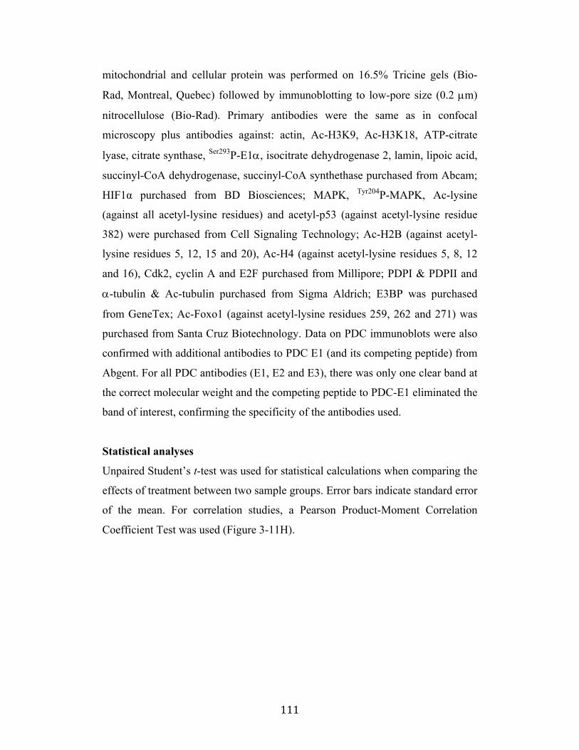
! 111!
mitochondrial and cellular protein was performed on 16.5% Tricine gels (Bio-
Rad, Montreal, Quebec) followed by immunoblotting to low-pore size (0.2 µm)
nitrocellulose (Bio-Rad). Primary antibodies were the same as in confocal
microscopy plus antibodies against: actin, Ac-H3K9, Ac-H3K18, ATP-citrate
lyase, citrate synthase, Ser293P-E1!, isocitrate dehydrogenase 2, lamin, lipoic acid,
succinyl-CoA dehydrogenase, succinyl-CoA synthethase purchased from Abcam;
HIF1α purchased from BD Biosciences; MAPK, Tyr204P-MAPK, Ac-lysine
(against all acetyl-lysine residues) and acetyl-p53 (against acetyl-lysine residue
382) were purchased from Cell Signaling Technology; Ac-H2B (against acetyl-
lysine residues 5, 12, 15 and 20), Ac-H4 (against acetyl-lysine residues 5, 8, 12
and 16), Cdk2, cyclin A and E2F purchased from Millipore; PDPI & PDPII and
!-tubulin & #c-tubulin purchased from Sigma Aldrich; E3BP was purchased
from GeneTex; Ac-Foxo1 (against acetyl-lysine residues 259, 262 and 271) was
purchased from Santa Cruz Biotechnology. Data on PDC immunoblots were also
confirmed with additional antibodies to PDC E1 (and its competing peptide) from
Abgent. For all PDC antibodies (E1, E2 and E3), there was only one clear band at
the correct molecular weight and the competing peptide to PDC-E1 eliminated the
band of interest, confirming the specificity of the antibodies used.
Statistical analyses
Unpaired Student’s t-test was used for statistical calculations when comparing the
effects of treatment between two sample groups. Error bars indicate standard error
of the mean. For correlation studies, a Pearson Product-Moment Correlation
Coefficient Test was used (Figure 3-11H).
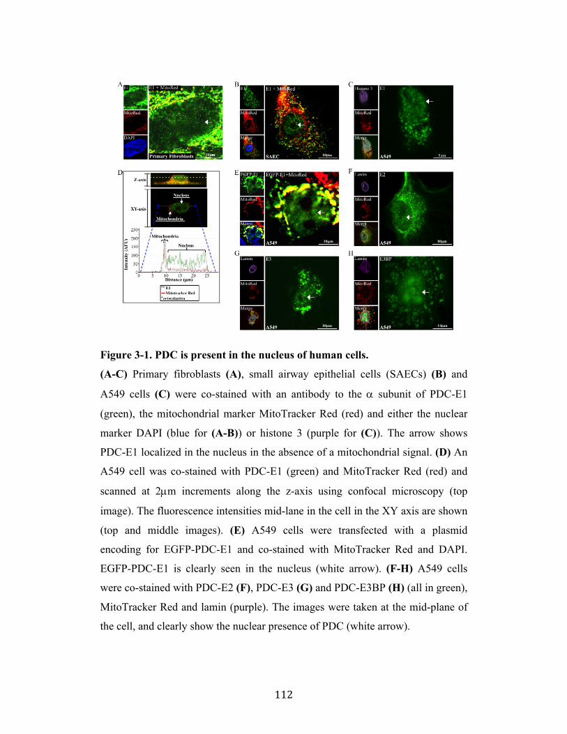
! 112!
Figure 3-1. PDC is present in the nucleus of human cells.
(A-C) Primary fibroblasts (A), small airway epithelial cells (SAECs) (B) and
A549 cells (C) were co-stained with an antibody to the ! subunit of PDC-E1
(green), the mitochondrial marker MitoTracker Red (red) and either the nuclear
marker DAPI (blue for (A-B)) or histone 3 (purple for (C)). The arrow shows
PDC-E1 localized in the nucleus in the absence of a mitochondrial signal. (D) An
A549 cell was co-stained with PDC-E1 (green) and MitoTracker Red (red) and
scanned at 2µm increments along the z-axis using confocal microscopy (top
image). The fluorescence intensities mid-lane in the cell in the XY axis are shown
(top and middle images). (E) A549 cells were transfected with a plasmid
encoding for EGFP-PDC-E1 and co-stained with MitoTracker Red and DAPI.
EGFP-PDC-E1 is clearly seen in the nucleus (white arrow). (F-H) A549 cells
were co-stained with PDC-E2 (F), PDC-E3 (G) and PDC-E3BP (H) (all in green),
MitoTracker Red and lamin (purple). The images were taken at the mid-plane of
the cell, and clearly show the nuclear presence of PDC (white arrow).
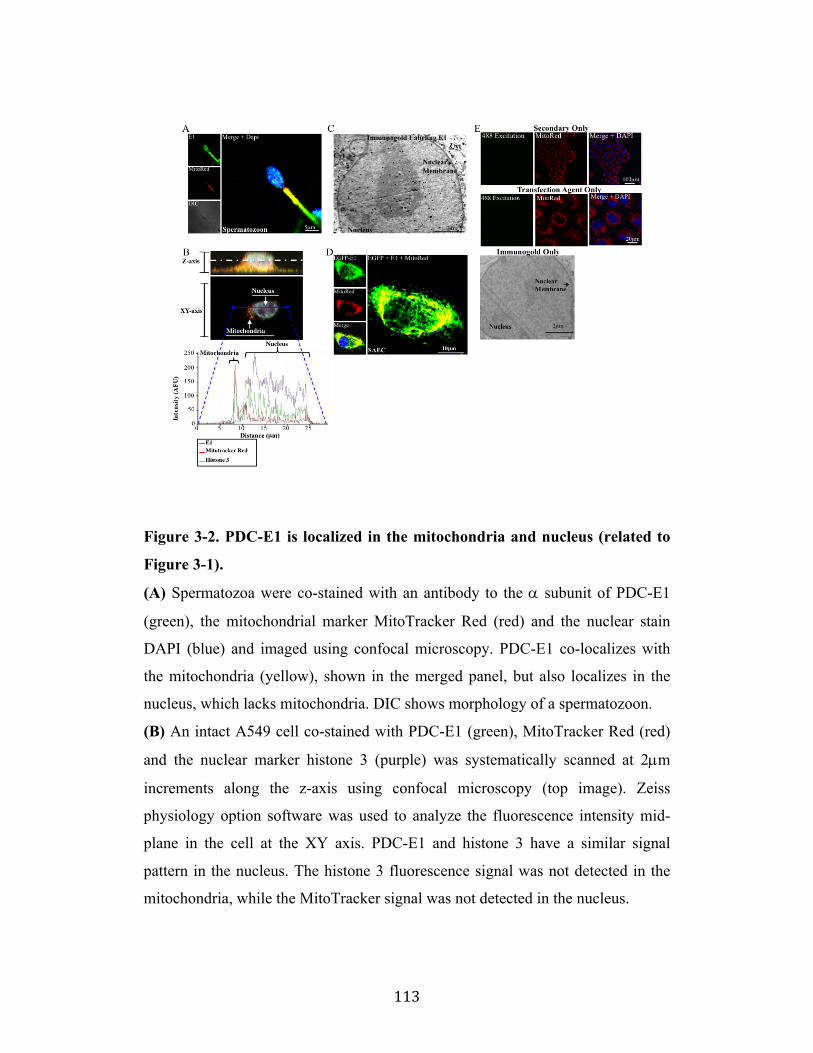
! 113!
Figure 3-2. PDC-E1 is localized in the mitochondria and nucleus (related to
Figure 3-1).
(A) Spermatozoa were co-stained with an antibody to the ! subunit of PDC-E1
(green), the mitochondrial marker MitoTracker Red (red) and the nuclear stain
DAPI (blue) and imaged using confocal microscopy. PDC-E1 co-localizes with
the mitochondria (yellow), shown in the merged panel, but also localizes in the
nucleus, which lacks mitochondria. DIC shows morphology of a spermatozoon.
(B) An intact A549 cell co-stained with PDC-E1 (green), MitoTracker Red (red)
and the nuclear marker histone 3 (purple) was systematically scanned at 2µm
increments along the z-axis using confocal microscopy (top image). Zeiss
physiology option software was used to analyze the fluorescence intensity mid-
plane in the cell at the XY axis. PDC-E1 and histone 3 have a similar signal
pattern in the nucleus. The histone 3 fluorescence signal was not detected in the
mitochondria, while the MitoTracker signal was not detected in the nucleus.
! 114!
(C) A549 cells were labeled using immunogold staining with an antibody against
the ! subunit of PDC-E1 (black dots) and imaged using transmission electron
microscopy. PDC-E1 was highly expressed in the nucleus within the nuclear
membrane (Cyt=cytosol). PDC-E1 presence in the cytoplasm and the nuclear
membrane is also compatible with the trafficking of PDC-E1 from the
mitochondria to the nucleus as discussed in the text. The negative control
(immunogold only) labeling is presented in Figure 3-2E.
(D) SAECs were transfected with a plasmid encoding for enhanced green
fluorescent protein in-frame with the ! subunit of PDC-E1 (EGFP-PDC; green)
and co-stained with MitoTracker Red (red) and the nuclear stain DAPI (blue).
EGFP-PDC-E1 co-localization with the mitochondria (yellow) is shown in the
merged panel (left). EGFP-PDC-E1 is also localized in the nucleus as shown by
the EGFP signal, obtained at a mid-plane level cutting through the nucleus.
(E) Secondary-only antibody to Fitc did not provide a fluorescence signal,
validating the specificity of the antibody used. MitoTracker Red (red) and the
nuclear stain DAPI (blue) are also shown (top). The EGFP signal detected was not
due to our transfection agent, as there was no detectable fluorescence signal with
our transfection agent-only control sample. MitoTracker (red) and the nuclear
stain DAPI (blue) are also shown (middle). Immunogold-only staining and
electron microscopy shows the specificity of our immunogold labeling.
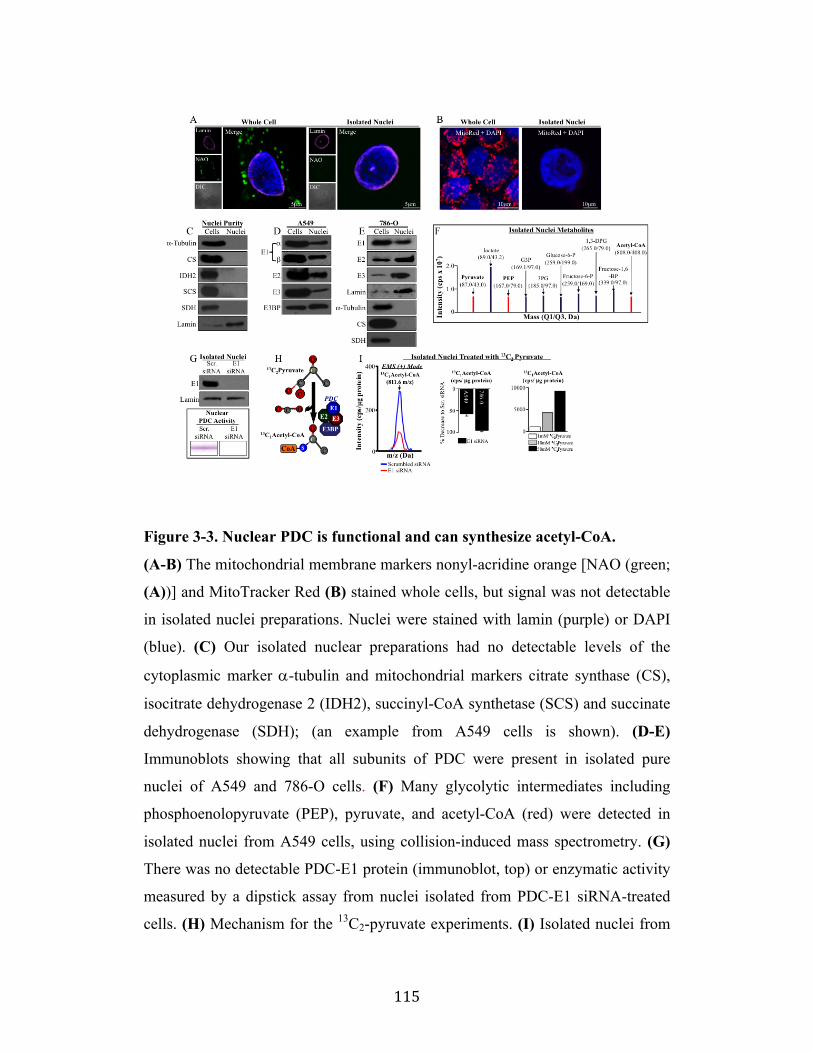
! 115!
Figure 3-3. Nuclear PDC is functional and can synthesize acetyl-CoA.
(A-B) The mitochondrial membrane markers nonyl-acridine orange [NAO (green;
(A))] and MitoTracker Red (B) stained whole cells, but signal was not detectable
in isolated nuclei preparations. Nuclei were stained with lamin (purple) or DAPI
(blue). (C) Our isolated nuclear preparations had no detectable levels of the
cytoplasmic marker !-tubulin and mitochondrial markers citrate synthase (CS),
isocitrate dehydrogenase 2 (IDH2), succinyl-CoA synthetase (SCS) and succinate
dehydrogenase (SDH); (an example from A549 cells is shown). (D-E)
Immunoblots showing that all subunits of PDC were present in isolated pure
nuclei of A549 and 786-O cells. (F) Many glycolytic intermediates including
phosphoenolopyruvate (PEP), pyruvate, and acetyl-CoA (red) were detected in
isolated nuclei from A549 cells, using collision-induced mass spectrometry. (G)
There was no detectable PDC-E1 protein (immunoblot, top) or enzymatic activity
measured by a dipstick assay from nuclei isolated from PDC-E1 siRNA-treated
cells. (H) Mechanism for the 13C2-pyruvate experiments. (I) Isolated nuclei from
! 116!
A549 or 786-0 cells previously treated with either PDC-E1 or scrambled siRNA
were incubated with 13C2-pyruvate for 8 hrs. The nuclei lacking PDC-E1 had
decreased levels of 13C1-acetyl-CoA compared to scrambled siRNA controls,
measured by mass spectrometry and normalized to protein concentration.
Increasing doses of labeled pyruvate showed a dose-dependent increase in labeled
acetyl-CoA.
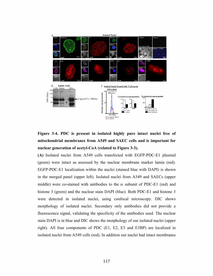
! 117!
Figure 3-4. PDC is present in isolated highly pure intact nuclei free of
mitochondrial membranes from A549 and SAEC cells and is important for
nuclear generation of acetyl-CoA (related to Figure 3-3).
(A) Isolated nuclei from A549 cells transfected with EGFP-PDC-E1 plasmid
(green) were intact as assessed by the nuclear membrane marker lamin (red).
EGFP-PDC-E1 localization within the nuclei (stained blue with DAPI) is shown
in the merged panel (upper left). Isolated nuclei from A549 and SAECs (upper
middle) were co-stained with antibodies to the ! subunit of PDC-E1 (red) and
histone 3 (green) and the nuclear stain DAPI (blue). Both PDC-E1 and histone 3
were detected in isolated nuclei, using confocal microscopy. DIC shows
morphology of isolated nuclei. Secondary only antibodies did not provide a
fluorescence signal, validating the specificity of the antibodies used. The nuclear
stain DAPI is in blue and DIC shows the morphology of our isolated nuclei (upper
right). All four components of PDC (E1, E2, E3 and E3BP) are localized in
isolated nuclei from A549 cells (red). In addition our nuclei had intact membranes
! 118!
as assessed by lamin (purple) and expressed histones (purple) in indicated panels
(bottom).
(B) Isolated nuclei contain a lipoylated protein at the molecular weight of E2 (~
70kDa) as assessed by an antibody to lipoic acid and immunoblots. The same
lipoylated protein is also highly detected in isolated mitochondria. The purity of
the nuclear samples are shown in Figure 3-3C.
(C) Nuclei from PDC-E1 siRNA-treated A549 and 786-0 cells had decreased
levels of 13C1-acetyl-CoA compared to scrambled siRNA controls, measured by
mass spectrometry (EPI mode) and normalized to protein concentration.
Furthermore, isolated nuclei had a dose-dependent increase of labeled acetyl-CoA
from labeled pyruvate using EPI mode mass spectrometry.
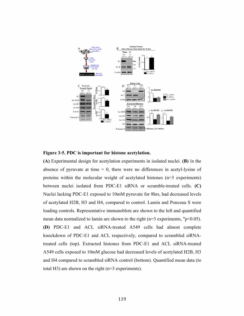
! 119!
Figure 3-5. PDC is important for histone acetylation.
(A) Experimental design for acetylation experiments in isolated nuclei. (B) In the
absence of pyruvate at time = 0, there were no differences in acetyl-lysine of
proteins within the molecular weight of acetylated histones (n=3 experiments)
between nuclei isolated from PDC-E1 siRNA or scramble-treated cells. (C)
Nuclei lacking PDC-E1 exposed to 10mM pyruvate for 8hrs, had decreased levels
of acetylated H2B, H3 and H4, compared to control. Lamin and Ponceau S were
loading controls. Representative immunoblots are shown to the left and quantified
mean data normalized to lamin are shown to the right (n=3 experiments, *p<0.05).
(D) PDC-E1 and ACL siRNA-treated A549 cells had almost complete
knockdown of PDC-E1 and ACL respectively, compared to scrambled siRNA-
treated cells (top). Extracted histones from PDC-E1 and ACL siRNA-treated
A549 cells exposed to 10mM glucose had decreased levels of acetylated H2B, H3
and H4 compared to scrambled siRNA control (bottom). Quantified mean data (to
total H3) are shown on the right (n=3 experiments).
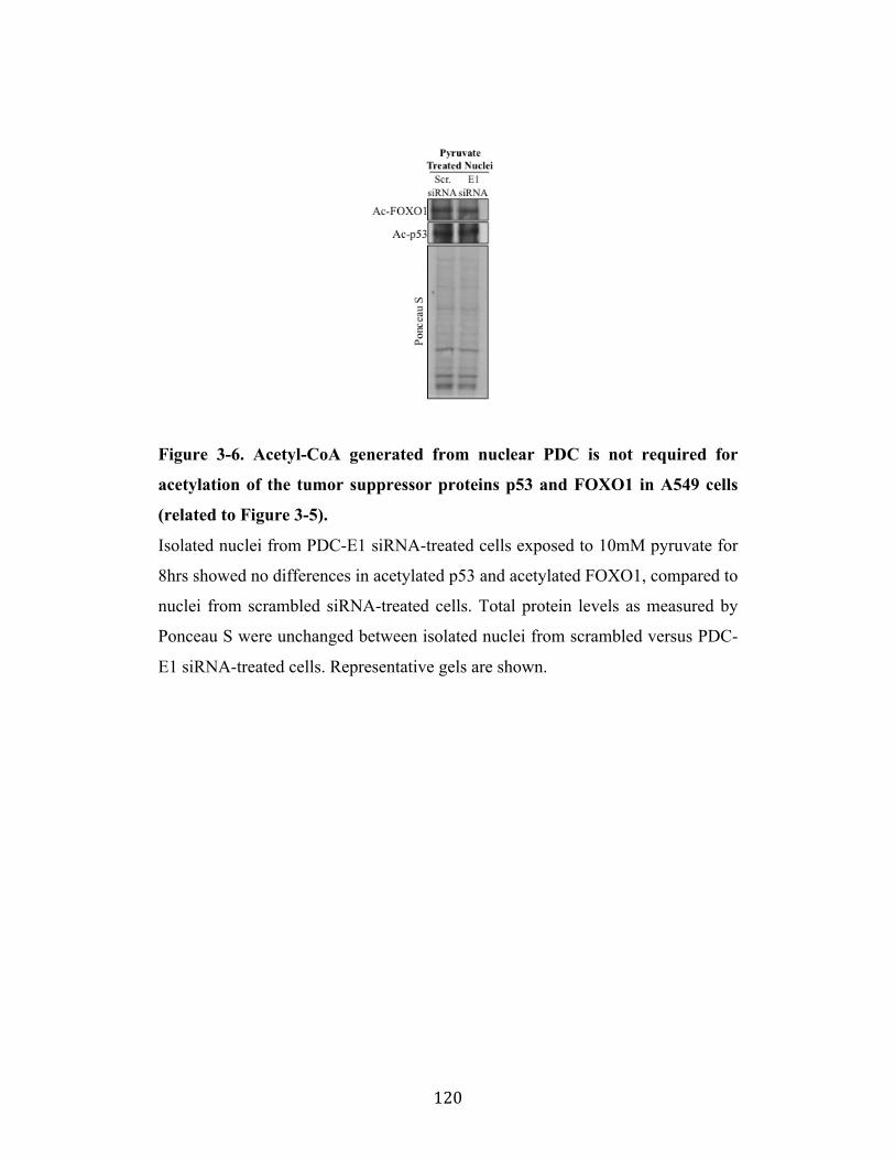
! 120!
Figure 3-6. Acetyl-CoA generated from nuclear PDC is not required for
acetylation of the tumor suppressor proteins p53 and FOXO1 in A549 cells
(related to Figure 3-5).
Isolated nuclei from PDC-E1 siRNA-treated cells exposed to 10mM pyruvate for
8hrs showed no differences in acetylated p53 and acetylated FOXO1, compared to
nuclei from scrambled siRNA-treated cells. Total protein levels as measured by
Ponceau S were unchanged between isolated nuclei from scrambled versus PDC-
E1 siRNA-treated cells. Representative gels are shown.

! 121!
Figure 3-7. Nuclear PDC is regulated differently than mitochondrial PDC.
(A) A549 cells have high levels of PDKI and PDKII in isolated mitochondria, but
no detectable levels in isolated nuclei. Phosphorylated PDC-E1 serine-293 was
present in isolated mitochondria (where PDK is present) but not in isolated nuclei
(where PDK is absent) and the ratio of P-E1/E1 is shown below. Confocal
microscopy shows the presence of PDKII (green) in the mitochondria as assessed
by co-localization (yellow) with MitoTracker Red in the merge panel, but not in
the nucleus, stained with DAPI (blue) (upper right). Immunoblot showing that
PDPI, but not PDPII, is present in isolated nuclei from A549 cells (bottom right).
(B) In 786-O cells PDKI (but not PDKII) is present in whole-cell preparations,
but not in isolated nuclei (left). The absence of PDK in the nucleus is confirmed
by confocal microscopy (top right). PDPI, but not PDPII is present in isolated
nuclei from 786-O cells (bottom right). (C) A549 cells had higher levels
phosphorylated serine-293 on PDC-E1 compared to isolated mitochondria from
normal SAECs (CS=citrate synthase). (D) Extracted histones from isolated A549
(left) and 786-O (right) nuclei exposed to DCA (5mM) had similar levels of
! 122!
acetylated H3 compared to vehicle-treated control cells. (E) In contrast, DCA
increased the acetylation of H3 (left) and tubulin (right) in whole A549 cells,
reflecting the activation of mitochondrial PDC. Representative immunoblots (top)
and quantified mean data normalized to total H3 (left) or tubulin (right) are shown
(n=4 experiments, *p<0.05). (F) Mechanism for DCA-mediated increase in
acetylation (see results section for discussion).
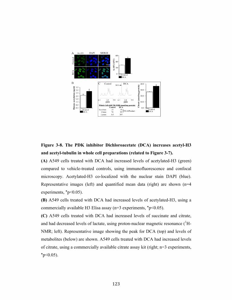
! 123!
Figure 3-8. The PDK inhibitor Dichloroacetate (DCA) increases acetyl-H3
and acetyl-tubulin in whole cell preparations (related to Figure 3-7).
(A) A549 cells treated with DCA had increased levels of acetylated-H3 (green)
compared to vehicle-treated controls, using immunofluorescence and confocal
microscopy. Acetylated-H3 co-localized with the nuclear stain DAPI (blue).
Representative images (left) and quantified mean data (right) are shown (n=4
experiments, *p<0.05).
(B) A549 cells treated with DCA had increased levels of acetylated-H3, using a
commercially available H3 Elisa assay (n=3 experiments, *p<0.05).
(C) A549 cells treated with DCA had increased levels of succinate and citrate,
and had decreased levels of lactate, using proton-nuclear magnetic resonance (1H-
NMR; left). Representative image showing the peak for DCA (top) and levels of
metabolites (below) are shown. A549 cells treated with DCA had increased levels
of citrate, using a commercially available citrate assay kit (right; n=3 experiments,
*p<0.05).
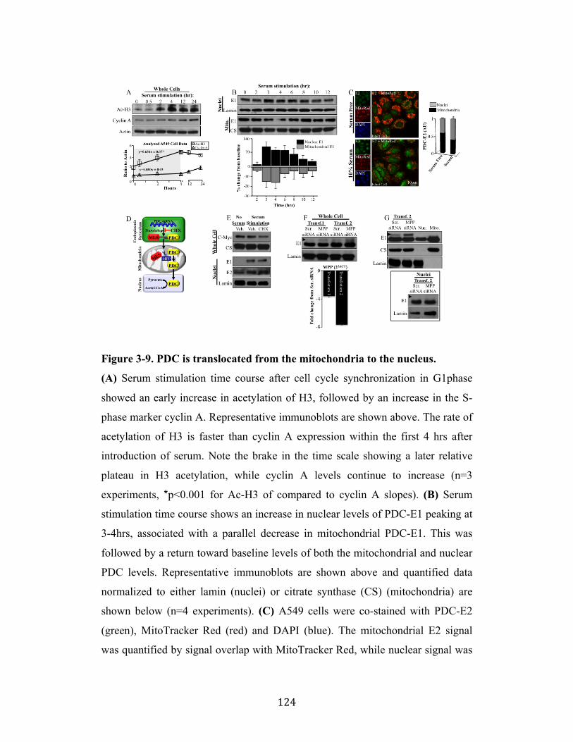
! 124!
Figure 3-9. PDC is translocated from the mitochondria to the nucleus.
(A) Serum stimulation time course after cell cycle synchronization in G1phase
showed an early increase in acetylation of H3, followed by an increase in the S-
phase marker cyclin A. Representative immunoblots are shown above. The rate of
acetylation of H3 is faster than cyclin A expression within the first 4 hrs after
introduction of serum. Note the brake in the time scale showing a later relative
plateau in H3 acetylation, while cyclin A levels continue to increase (n=3
experiments, *p<0.001 for Ac-H3 of compared to cyclin A slopes). (B) Serum
stimulation time course shows an increase in nuclear levels of PDC-E1 peaking at
3-4hrs, associated with a parallel decrease in mitochondrial PDC-E1. This was
followed by a return toward baseline levels of both the mitochondrial and nuclear
PDC levels. Representative immunoblots are shown above and quantified data
normalized to either lamin (nuclei) or citrate synthase (CS) (mitochondria) are
shown below (n=4 experiments). (C) A549 cells were co-stained with PDC-E2
(green), MitoTracker Red (red) and DAPI (blue). The mitochondrial E2 signal
was quantified by signal overlap with MitoTracker Red, while nuclear signal was
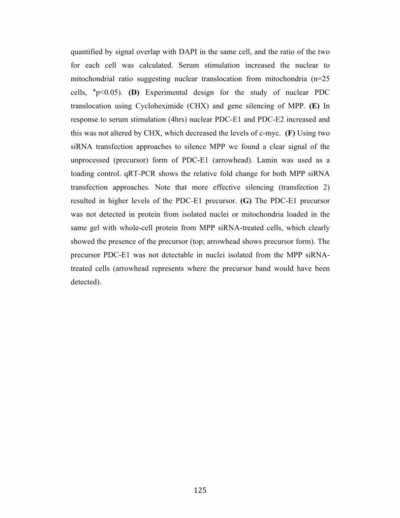
! 125!
quantified by signal overlap with DAPI in the same cell, and the ratio of the two
for each cell was calculated. Serum stimulation increased the nuclear to
mitochondrial ratio suggesting nuclear translocation from mitochondria (n=25
cells, *p<0.05). (D) Experimental design for the study of nuclear PDC
translocation using Cycloheximide (CHX) and gene silencing of MPP. (E) In
response to serum stimulation (4hrs) nuclear PDC-E1 and PDC-E2 increased and
this was not altered by CHX, which decreased the levels of c-myc. (F) Using two
siRNA transfection approaches to silence MPP we found a clear signal of the
unprocessed (precursor) form of PDC-E1 (arrowhead). Lamin was used as a
loading control. qRT-PCR shows the relative fold change for both MPP siRNA
transfection approaches. Note that more effective silencing (transfection 2)
resulted in higher levels of the PDC-E1 precursor. (G) The PDC-E1 precursor
was not detected in protein from isolated nuclei or mitochondria loaded in the
same gel with whole-cell protein from MPP siRNA-treated cells, which clearly
showed the presence of the precursor (top; arrowhead shows precursor form). The
precursor PDC-E1 was not detectable in nuclei isolated from the MPP siRNA-
treated cells (arrowhead represents where the precursor band would have been
detected).
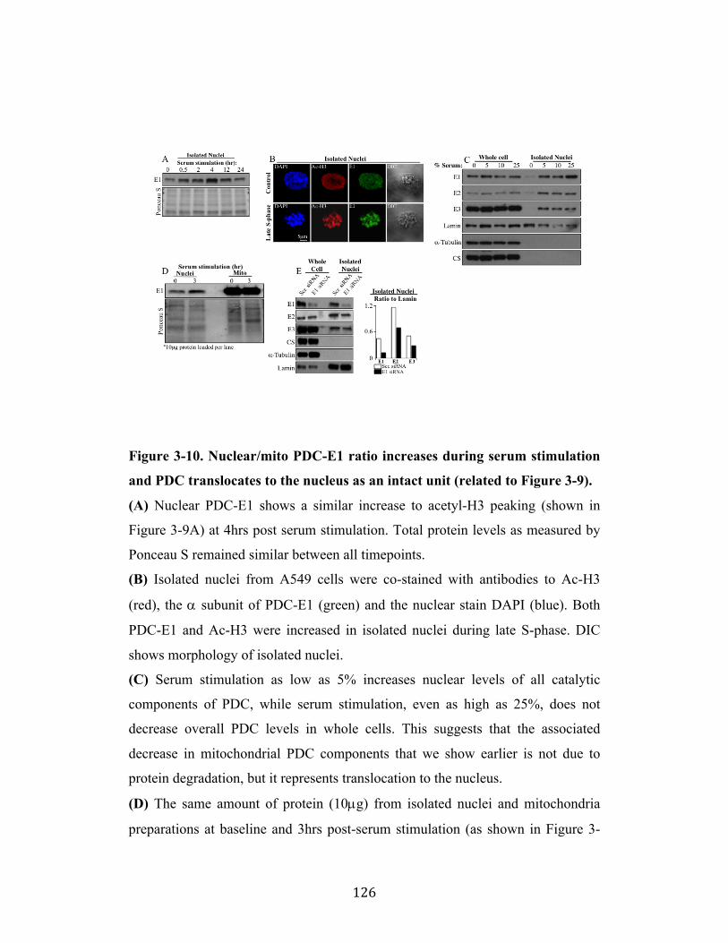
! 126!
Figure 3-10. Nuclear/mito PDC-E1 ratio increases during serum stimulation
and PDC translocates to the nucleus as an intact unit (related to Figure 3-9).
(A) Nuclear PDC-E1 shows a similar increase to acetyl-H3 peaking (shown in
Figure 3-9A) at 4hrs post serum stimulation. Total protein levels as measured by
Ponceau S remained similar between all timepoints.
(B) Isolated nuclei from A549 cells were co-stained with antibodies to Ac-H3
(red), the ! subunit of PDC-E1 (green) and the nuclear stain DAPI (blue). Both
PDC-E1 and Ac-H3 were increased in isolated nuclei during late S-phase. DIC
shows morphology of isolated nuclei.
(C) Serum stimulation as low as 5% increases nuclear levels of all catalytic
components of PDC, while serum stimulation, even as high as 25%, does not
decrease overall PDC levels in whole cells. This suggests that the associated
decrease in mitochondrial PDC components that we show earlier is not due to
protein degradation, but it represents translocation to the nucleus.
(D) The same amount of protein (10µg) from isolated nuclei and mitochondria
preparations at baseline and 3hrs post-serum stimulation (as shown in Figure 3-
! 127!
9B) was loaded on the same immunoblot. Total protein levels were similar
between the 0 and 3 hours groups, although there are apparent differences in the
bands between the nuclear and mitochondria on Ponceau S, reflecting the
different proteome in the two organelles.
(E) PDC translocates to the nucleus as an intact unit as knockdown of one specific
component of PDC (i.e. gene silencing of the E1 gene by siRNA) results in
decreased nuclear levels of other PDC components (E2 and E3), but does not alter
overall levels of these components in whole cells. Nuclear purity was shown by
absence of citrate synthase (CS) and tubulin in the nucleus. The ratios of PDC
components to lamin are shown on the right.
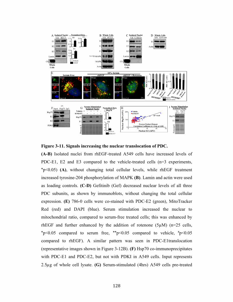
! 128!
Figure 3-11. Signals increasing the nuclear translocation of PDC.
(A-B) Isolated nuclei from rhEGF-treated A549 cells have increased levels of
PDC-E1, E2 and E3 compared to the vehicle-treated cells (n=3 experiments,
*p<0.05) (A), without changing total cellular levels, while rhEGF treatment
increased tyrosine-204 phosphorylation of MAPK (B). Lamin and actin were used
as loading controls. (C-D) Gefitinib (Gef) decreased nuclear levels of all three
PDC subunits, as shown by immunoblots, without changing the total cellular
expression. (E) 786-0 cells were co-stained with PDC-E2 (green), MitoTracker
Red (red) and DAPI (blue). Serum stimulation increased the nuclear to
mitochondrial ratio, compared to serum-free treated cells; this was enhanced by
rhEGF and further enhanced by the addition of rotenone (5µM) (n=25 cells,
*p<0.05 compared to serum free," **p<0.05 compared to vehicle, #p<0.05
compared to rhEGF). A similar pattern was seen in PDC-E1translocation
(representative images shown in Figure 3-12B). (F) Hsp70 co-immunoprecipitates
with PDC-E1 and PDC-E2, but not with PDKI in A549 cells. Input represents
2.5µg of whole cell lysate. (G) Serum-stimulated (4hrs) A549 cells pre-treated
! 129!
with KNK437 (100µM) show decreased nuclear levels of both Hsp70 and PDC-
E1 compared to vehicle (DMSO)-treated cells. Mean data are normalized to
Ponceau S (n=3 experiments, *p>0.05). (H) The same cells as in (G) were co-
stained with Hsp70 and PDC-E1, imaged with confocal microscopy and the
nuclear fluorescence intensity was measured. A Pearson product-moment
correlation plot showed that nuclear levels of Hsp70 and PDC-E1 correlate
positively (r=0.66; p<0.001; n=50 cells/group). Representative images are shown
in Figure 3-12H. (I) Serum stimulated (4hrs) A549 cells transfected with Hsp70
siRNA show decreased nuclear levels of both Hsp70 and PDC-E1, compared to
scrambled-transfected cells.
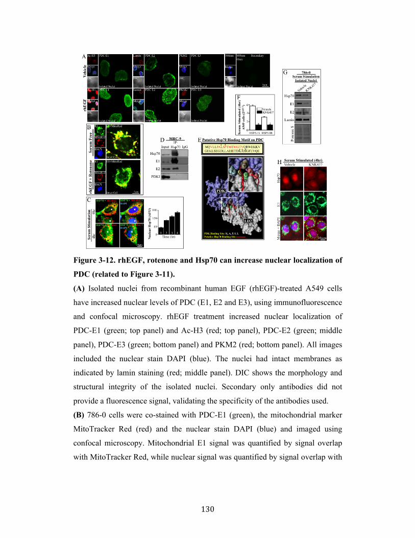
! 130!
Figure 3-12. rhEGF, rotenone and Hsp70 can increase nuclear localization of
PDC (related to Figure 3-11).
(A) Isolated nuclei from recombinant human EGF (rhEGF)-treated A549 cells
have increased nuclear levels of PDC (E1, E2 and E3), using immunofluorescence
and confocal microscopy. rhEGF treatment increased nuclear localization of
PDC-E1 (green; top panel) and Ac-H3 (red; top panel), PDC-E2 (green; middle
panel), PDC-E3 (green; bottom panel) and PKM2 (red; bottom panel). All images
included the nuclear stain DAPI (blue). The nuclei had intact membranes as
indicated by lamin staining (red; middle panel). DIC shows the morphology and
structural integrity of the isolated nuclei. Secondary only antibodies did not
provide a fluorescence signal, validating the specificity of the antibodies used.
(B) 786-0 cells were co-stained with PDC-E1 (green), the mitochondrial marker
MitoTracker Red (red) and the nuclear stain DAPI (blue) and imaged using
confocal microscopy. Mitochondrial E1 signal was quantified by signal overlap
with MitoTracker Red, while nuclear signal was quantified by signal overlap with
! 131!
DAPI, using the Image J 64 processing software. These are representative images
of the mean data shown in Figure 3-11E.
(C) Serum stimulation of A549 cells for 4hrs shows progressively increasing
nuclear Hsp70 levels compared to baseline control cells. Hsp70 is shown in green,
the mitochondrial marker MitoTracker Red is shown in red and the nuclear stain
DAPI is shown in blue. Black arrows show Hsp70 co-localization with
MitoTracker Red (yellow) and white arrows show nuclear Hsp70. Representative
images are shown on the left and quantified mean data for nuclear Hsp70 are
shown on the right (n=50 cells per group).
(D) Hsp70 co-immunoprecipitates with PDC-E1 and PDC-E2, but not with PDKI
in MRC-9 cells as shown by immunoblots. Input represents 2.5µg of whole cell
lysate.
(E) Sequence of residues 133-182 of PDC-E2 encoding for part of the lipoyl-
domain 2 (LD2), reveals putative Hsp70 binding motifs (red) and known PDK
interacting residues (highlighted by *). These include Leu 140 (L), Pro 142 (P),
Lys 173 (K), Ala 174 (A) and Ile 176 (I) of LD2 (Kato et al., 2005; Roche et al.,
2003) (top). Ribbon representation of putative Hsp70 binding motifs (red), shows
a potential binding region within the PDK binding site for LD2. PDK is shown in
purple and LD2 is shown in grey surface representation. Inset shows higher
magnification of putative Hsp70 and PDK binding sites on LD2 of PDC-E2. PDB
code 1Y8O (Kato et al., 2005) and the visual molecular dynamics program was
used to generate structural images.
(F) Serum stimulated (4hrs) A549 cells, previously serum starved and pre-treated
with 100µM of KNK437, show decreased mRNA levels of HSPA1A and
HSPA1B, the two genes responsible for inducible Hsp70, compared to vehicle
(DMSO)-treated cells (n=3 experiments, *p<0.05).
(G) Serum stimulated (4hrs) 786-0 cells previously serum starved and pre-treated
with 100µM of KNK437 (which inhibits induced Hsp70) show decreased nuclear
levels of Hsp70, PDC-E1 and PDC-E2 compared to vehicle (DMSO)-treated
cells, as shown by immunoblots. Lamin and Ponceau S show similar protein
loading between vehicle and KNK437 treated cells.
! 132!
(H) Serum stimulated (4hrs) A549 cells, previously serum starved and pre-treated
with 100µM of KNK437 show decreased nuclear levels of Hsp70 (red) and PDC-
E1 (green) compared to vehicle (DMSO)-treated cells as shown by
immunofluorescence and confocal microscopy. These are representative images
of the mean data shown in Figure 3-11H for a Pearson product-moment
correlation plot between nuclear Hsp70 and nuclear PDC-E1.
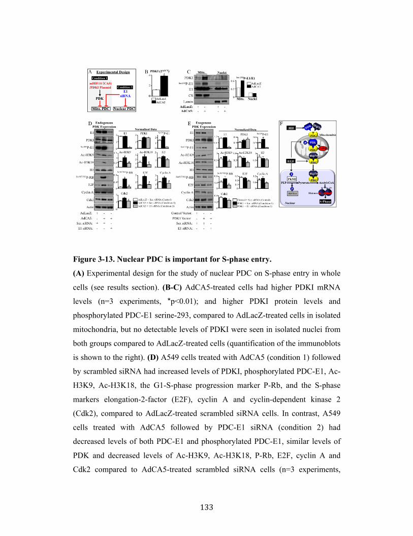
! 133!
Figure 3-13. Nuclear PDC is important for S-phase entry.
(A) Experimental design for the study of nuclear PDC on S-phase entry in whole
cells (see results section). (B-C) AdCA5-treated cells had higher PDKI mRNA
levels (n=3 experiments, *p<0.01); and higher PDKI protein levels and
phosphorylated PDC-E1 serine-293, compared to AdLacZ-treated cells in isolated
mitochondria, but no detectable levels of PDKI were seen in isolated nuclei from
both groups compared to AdLacZ-treated cells (quantification of the immunoblots
is shown to the right). (D) A549 cells treated with AdCA5 (condition 1) followed
by scrambled siRNA had increased levels of PDKI, phosphorylated PDC-E1, Ac-
H3K9, Ac-H3K18, the G1-S-phase progression marker P-Rb, and the S-phase
markers elongation-2-factor (E2F), cyclin A and cyclin-dependent kinase 2
(Cdk2), compared to AdLacZ-treated scrambled siRNA cells. In contrast, A549
cells treated with AdCA5 followed by PDC-E1 siRNA (condition 2) had
decreased levels of both PDC-E1 and phosphorylated PDC-E1, similar levels of
PDK and decreased levels of Ac-H3K9, Ac-H3K18, P-Rb, E2F, cyclin A and
Cdk2 compared to AdCA5-treated scrambled siRNA cells (n=3 experiments,
! 134!
*p<0.05 vs AdLacZ scr. siRNA, #p<0.05 vs AdCA5 scr. siRNA). (E) Transfection
with PDKI plasmid followed by PDC-E1 siRNA (condition 2) decreased the
levels of PDC-E1, phosphorylated PDC-E1, Ac-H3K9, Ac-H3K18, P-Rb, E2F,
cyclin A and Cdk2, compared to transfection with PDKI plasmid but treated with
scrambled siRNA (condition 1) (n=3 experiments, *p<0.05 vs scr. siRNA). (F)
Proposed model for the translocation of PDC from mitochondria to the nucleus
and its functional role (see discussion).
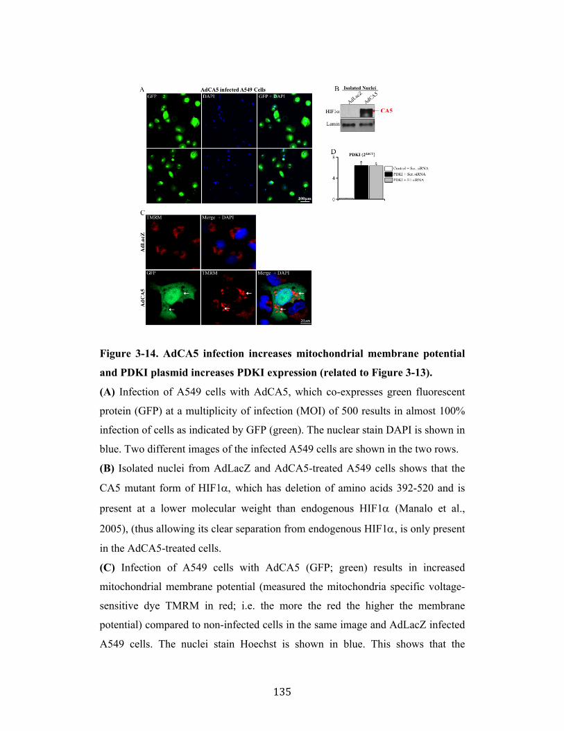
! 135!
Figure 3-14. AdCA5 infection increases mitochondrial membrane potential
and PDKI plasmid increases PDKI expression (related to Figure 3-13).
(A) Infection of A549 cells with AdCA5, which co-expresses green fluorescent
protein (GFP) at a multiplicity of infection (MOI) of 500 results in almost 100%
infection of cells as indicated by GFP (green). The nuclear stain DAPI is shown in
blue. Two different images of the infected A549 cells are shown in the two rows.
(B) Isolated nuclei from AdLacZ and AdCA5-treated A549 cells shows that the
CA5 mutant form of HIF1!, which has deletion of amino acids 392-520 and is
present at a lower molecular weight than endogenous HIF1! (Manalo et al.,
2005), (thus allowing its clear separation from endogenous HIF1!, is only present
in the AdCA5-treated cells.
(C) Infection of A549 cells with AdCA5 (GFP; green) results in increased
mitochondrial membrane potential (measured the mitochondria specific voltage-
sensitive dye TMRM in red; i.e. the more the red the higher the membrane
potential) compared to non-infected cells in the same image and AdLacZ infected
A549 cells. The nuclei stain Hoechst is shown in blue. This shows that the
! 136!
mutant, constitutively active HIF1! has the expected effects on mitochondria, as
we have previously published (see text).
(D) A549 cells transfected with a PDKI plasmid had higher PDKI mRNA levels
compared to empty vector transfected cells, measured by qRT-PCR. Treatment
with PDC-E1 siRNA of A549 cells transfected with the PDKI plasmid did not
alter PDKI mRNA levels compared to scrambled siRNA-treated cells (n=3
experiments, *p<0.01 compared to empty control vector and scrambled siRNA).
Supplementary Video 1 and 2. PDC E1 is present throughout the nucleus of
an intact cell (related to Figure 3-1).
Stacked images using confocal microscopy as represented in this video clearly
shows the presence of PDC-E1 (green) throughout the nucleus in two separate cell
lines (see text). PDC E1 is also localized in the mitochondria as assessed by the
yellow co-localization with the mitochondrial stain MitoTracker Red (red).
Videos are available on the CellPress website: www.cell.com
! 137!
References
Alarcon3Vargas,!D.,! Tansey,!W.P.,! and!Ronai,! Z.! (2002).! Regulation! of! c3myc!stability!by!selective!stress!conditions!and!by!MEKK1!requires!aa!1273189!of!c3myc.!Oncogene!21,!438434391.!Behal,!R.H.,!Buxton,!D.B.,!Robertson,!J.G.,!and!Olson,!M.S.!(1993).!Regulation!of!the!pyruvate!dehydrogenase!multienzyme!complex.!Annu!Rev!Nutr!13,!4973520.!Bonnet,! S.,! Archer,! S.L.,! Allalunis3Turner,! J.,! Haromy,! A.,! Beaulieu,! C.,!Thompson,! R.,! Lee,! C.T.,! Lopaschuk,! G.D.,! Puttagunta,! L.,! Harry,! G.,! et! al.!(2007).! A! mitochondria3K+! channel! axis! is! suppressed! in! cancer! and! its!normalization! promotes! apoptosis! and! inhibits! cancer! growth.! Cancer! Cell!11,!37351.!Bowker3Kinley,!M.M.,!Davis,!W.I.,!Wu,!P.,!Harris,!R.A.,!and!Popov,!K.M.!(1998).!Evidence! for! existence! of! tissue3specific! regulation! of! the! mammalian!pyruvate!dehydrogenase!complex.!Biochem!J!329!(!Pt!1),!1913196.!Cai,!L.,!Sutter,!B.M.,!Li,!B.,!and!Tu,!B.P.!(2011).!Acetyl3CoA!induces!cell!growth!and!proliferation!by!promoting! the!acetylation!of!histones!at!growth!genes.!Mol!Cell!42,!4263437.!Chacinska,! A.,! Koehler,! C.M.,! Milenkovic,! D.,! Lithgow,! T.,! and! Pfanner,! N.!(2009).!Importing!mitochondrial!proteins:!machineries!and!mechanisms.!Cell!138,!6283644.!Choudhary,! C.,! Kumar,! C.,! Gnad,! F.,!Nielsen,!M.L.,! Rehman,!M.,!Walther,! T.C.,!Olsen,!J.V.,!and!Mann,!M.!(2009).!Lysine!acetylation!targets!protein!complexes!and!co3regulates!major!cellular!functions.!Science!325,!8343840.!Dhar,!S.,!and!Lippard,!S.J.!(2009).!Mitaplatin,!a!potent!fusion!of!cisplatin!and!the!orphan!drug!dichloroacetate.!Proc!Natl!Acad!Sci!U!S!A!106,!22199322204.!Durieux,!J.,!Wolff,!S.,!and!Dillin,!A.!(2011).!The!cell3non3autonomous!nature!of!electron!transport!chain3mediated!longevity.!Cell!144,!79391.!Gao,!X.,!Wang,!H.,!Yang,! J.J.,!Liu,!X.,!and!Liu,!Z.R.!(2012).!Pyruvate!kinase!M2!regulates!gene!transcription!by!acting!as!a!protein!kinase.!Mol!Cell!45,!5983609.!Hitosugi,!T.,!Fan,!J.,!Chung,!T.W.,!Lythgoe,!K.,!Wang,!X.,!Xie,!J.,!Ge,!Q.,!Gu,!T.L.,!Polakiewicz,! R.D.,! Roesel,! J.L.,! et! al.! (2011).! Tyrosine! phosphorylation! of!mitochondrial! pyruvate! dehydrogenase! kinase! 1! is! important! for! cancer!metabolism.!Mol!Cell!44,!8643877.!Horner,!D.S.,!Hirt,!R.P.,!and!Embley,!T.M.!(1999).!A!single!eubacterial!origin!of!eukaryotic! pyruvate:! ferredoxin! oxidoreductase! genes:! implications! for! the!evolution!of!anaerobic!eukaryotes.!Mol!Biol!Evol!16,!128031291.!Jaenisch,! R.,! and! Bird,! A.! (2003).! Epigenetic! regulation! of! gene! expression:!how!the!genome!integrates!intrinsic!and!environmental!signals.!Nat!Genet!33!Suppl,!2453254.!Kato,!M.,!Chuang,!J.L.,!Tso,!S.C.,!Wynn,!R.M.,!and!Chuang,!D.T.!(2005).!Crystal!structure!of! pyruvate!dehydrogenase!kinase!3!bound! to! lipoyl! domain!2!of!human!pyruvate!dehydrogenase!complex.!EMBO!J!24,!176331774.!
! 138!
Kato,! Y.,! Tapping,! R.I.,! Huang,! S.,! Watson,! M.H.,! Ulevitch,! R.J.,! and! Lee,! J.D.!(1998).! Bmk1/Erk5! is! required! for! cell! proliferation! induced! by! epidermal!growth!factor.!Nature!395,!7133716.!Kiang,!J.G.,!Bowman,!P.D.,!Lu,!X.,!Li,!Y.,!Ding,!X.Z.,!Zhao,!B.,!Juang,!Y.T.,!Atkins,!J.L.,! and! Tsokos,! G.C.! (2006).! Geldanamycin! prevents! hemorrhage3induced!ATP! loss! by! overexpressing! inducible! HSP70! and! activating! pyruvate!dehydrogenase.!Am!J!Physiol!Gastrointest!Liver!Physiol!291,!G1173127.!Kim,! J.W.,! and! Dang,! C.V.! (2005).! Multifaceted! roles! of! glycolytic! enzymes.!Trends!Biochem!Sci!30,!1423150.!Kim,! J.W.,! Tchernyshyov,! I.,! Semenza,! G.L.,! and! Dang,! C.V.! (2006).! HIF313mediated!expression!of!pyruvate!dehydrogenase!kinase:!a!metabolic!switch!required!for!cellular!adaptation!to!hypoxia.!Cell!Metab!3,!1773185.!Koishi,!M.,!Yokota,!S.,!Mae,!T.,!Nishimura,!Y.,!Kanamori,!S.,!Horii,!N.,!Shibuya,!K.,!Sasai,!K.,!and!Hiraoka,!M.!(2001).!The!effects!of!KNK437,!a!novel!inhibitor!of! heat! shock! protein! synthesis,! on! the! acquisition! of! thermotolerance! in! a!murine!transplantable!tumor!in!vivo.!Clin!Cancer!Res!7,!2153219.!Lodish,!H.,!Berk,!A.,!Zipursky,!S.L.,!Matsudaira,!P.,!Baltimore,!D.,!and!Darnell,!J.!(2000).! Section! 11.4,! Signal3Mediated! Transport! through! Nuclear! Pore!Complexes.! In!Molecular! Cell! Biology,! 4th! Edition,!W.H.! Freeman,! ed.! (New!York:!W.H.!Freemand!and!Company).!Luo,!W.,!Hu,!H.,!Chang,!R.,!Zhong,!J.,!Knabel,!M.,!O'Meally,!R.,!Cole,!R.N.,!Pandey,!A.,! and! Semenza,! G.L.! (2011).! Pyruvate! kinase! M2! is! a! PHD33stimulated!coactivator!for!hypoxia3inducible!factor!1.!Cell!145,!7323744.!Manalo,!D.J.,! Rowan,!A.,! Lavoie,! T.,!Natarajan,! L.,! Kelly,! B.D.,! Ye,! S.Q.,! Garcia,!J.G.,! and! Semenza,! G.L.! (2005).! Transcriptional! regulation! of! vascular!endothelial!cell!responses!to!hypoxia!by!HIF31.!Blood!105,!6593669.!Mayer,!M.P.,!and!Bukau,!B.!(2005).!Hsp70!chaperones:!cellular!functions!and!molecular!mechanism.!Cell!Mol!Life!Sci!62,!6703684.!Michelakis,!E.D.,!Sutendra,!G.,!Dromparis,!P.,!Webster,!L.,!Haromy,!A.,!Niven,!E.,!Maguire,!C.,!Gammer,!T.L.,!Mackey,!J.R.,!Fulton,!D.,!et!al.!(2010).!Metabolic!modulation!of!glioblastoma!with!dichloroacetate.!Sci!Transl!Med!2,!31ra34.!Milarski,!K.L.,!and!Morimoto,!R.I.!(1986).!Expression!of!human!HSP70!during!the!synthetic!phase!of!the!cell!cycle.!Proc!Natl!Acad!Sci!U!S!A!83,!951739521.!Pante,! N.,! and! Kann,! M.! (2002).! Nuclear! pore! complex! is! able! to! transport!macromolecules!with!diameters!of!about!39!nm.!Mol!Biol!Cell!13,!4253434.!Roche,!T.E.,!Hiromasa,!Y.,!Turkan,!A.,!Gong,!X.,!Peng,!T.,!Yan,!X.,!Kasten,!S.A.,!Bao,!H.,!and!Dong,!J.!(2003).!Essential!roles!of!lipoyl!domains!in!the!activated!function! and! control! of! pyruvate! dehydrogenase! kinases! and! phosphatase!isoform!1.!Eur!J!Biochem!270,!105031056.!Rodriguez,!M.A.,!Garcia3Perez,!R.M.,!Mendoza,!L.,!Sanchez,!T.,!Guillen,!N.,!and!Orozco,!E.!(1998).!The!pyruvate:ferredoxin!oxidoreductase!enzyme!is!located!in! the! plasma! membrane! and! in! a! cytoplasmic! structure! in! Entamoeba.!Microb!Pathog!25,!1310.!Semenza,! G.L.! (2010).! Defining! the! role! of! hypoxia3inducible! factor! 1! in!cancer!biology!and!therapeutics.!Oncogene!29,!6253634.!
! 139!
Shi,! Y.,! and!Thomas,! J.O.! (1992).!The! transport! of! proteins! into! the!nucleus!requires! the! 703kilodalton! heat! shock! protein! or! its! cytosolic! cognate.!Mol!Cell!Biol!12,!218632192.!Siebert,! G.,! and! Humphrey,! G.B.! (1965).! Enzymology! of! the! nucleus.! Adv!Enzymol!Relat!Areas!Mol!Biol!27,!2393288.!Sumegi,!B.,!Liposits,!Z.,!Inman,!L.,!Paull,!W.K.,!and!Srere,!P.A.!(1987).!Electron!microscopic! study! on! the! size! of! pyruvate! dehydrogenase! complex! in! situ.!Eur!J!Biochem!169,!2233230.!Sutendra,! G.,! Dromparis,! P.,! Kinnaird,! A.,! Stenson,! T.H.,! Haromy,! A.,! Parker,!J.M.,!McMurtry,!M.S.,!and!Michelakis,!E.D.!(2013).!Mitochondrial!activation!by!inhibition! of! PDKII! suppresses!HIF1a! signaling! and! angiogenesis! in! cancer.!Oncogene!32,!163831650.!Sutendra,! G.,! Dromparis,! P.,! Wright,! P.,! Bonnet,! S.,! Haromy,! A.,! Hao,! Z.,!McMurtry,!M.S.,!Michalak,!M.,!Vance,!J.E.,!Sessa,!W.C.,!et!al.!(2011).!The!role!of!Nogo! and! the! mitochondria3endoplasmic! reticulum! unit! in! pulmonary!hypertension.!Sci!Transl!Med!3,!88ra55.!Vander! Heiden,! M.G.,! Locasale,! J.W.,! Swanson,! K.D.,! Sharfi,! H.,! Heffron,! G.J.,!Amador3Noguez,!D.,!Christofk,!H.R.,!Wagner,!G.,!Rabinowitz,!J.D.,!Asara,!J.M.,!et!al.! (2010).! Evidence! for! an! alternative! glycolytic! pathway! in! rapidly!proliferating!cells.!Science!329,!149231499.!Vogelauer,! M.,! Rubbi,! L.,! Lucas,! I.,! Brewer,! B.J.,! and! Grunstein,! M.! (2002).!Histone!acetylation!regulates!the!time!of!replication!origin!firing.!Mol!Cell!10,!122331233.!Wellen,! K.E.,! Hatzivassiliou,! G.,! Sachdeva,! U.M.,! Bui,! T.V.,! Cross,! J.R.,! and!Thompson,!C.B.!(2009).!ATP3citrate!lyase!links!cellular!metabolism!to!histone!acetylation.!Science!324,!107631080.!Yang,!W.,! Xia,! Y.,! Hawke,! D.,! Li,! X.,! Liang,! J.,! Xing,! D.,! Aldape,! K.,! Hunter,! T.,!Alfred!Yung,!W.K.,! and!Lu,!Z.! (2012).!PKM2!phosphorylates!histone!H3!and!promotes!gene!transcription!and!tumorigenesis.!Cell!150,!6853696.!Yi,! C.H.,! Pan,! H.,! Seebacher,! J.,! Jang,! I.H.,! Hyberts,! S.G.,! Heffron,! G.J.,! Vander!Heiden,!M.G.,!Yang,!R.,!Li,!F.,!Locasale,!J.W.,!et!al.!(2011).!Metabolic!regulation!of!protein!N3alpha3acetylation!by!Bcl3xL!promotes!cell!survival.!Cell!146,!6073620.!Yogev,!O.,!Singer,!E.,!Shaulian,!E.,!Goldberg,!M.,!Fox,!T.D.,!and!Pines,!O.!(2010).!Fumarase:! a! mitochondrial! metabolic! enzyme! and! a! cytosolic/nuclear!component!of!the!DNA!damage!response.!PLoS!biology!8,!e1000328.!Zhao,! Q.,! Wang,! J.,! Levichkin,! I.V.,! Stasinopoulos,! S.,! Ryan,! M.T.,! and!Hoogenraad,! N.J.! (2002).! A! mitochondrial! specific! stress! response! in!mammalian!cells.!EMBO!J!21,!441134419.!Zhou,!Z.H.,!Liao,!W.,!Cheng,!R.H.,!Lawson,! J.E.,!McCarthy,!D.B.,!Reed,!L.J.,! and!Stoops,! J.K.! (2001a).! Direct! evidence! for! the! size! and! conformational!variability! of! the! pyruvate! dehydrogenase! complex! revealed! by! three3dimensional! electron! microscopy.! The! "breathing"! core! and! its! functional!relationship!to!protein!dynamics.!J!Biol!Chem!276,!21704321713.!Zhou,!Z.H.,!McCarthy,!D.B.,!O'Connor,!C.M.,!Reed,!L.J.,!and!Stoops,!J.K.!(2001b).!The! remarkable! structural! and! functional! organization! of! the! eukaryotic!

! 140!
pyruvate! dehydrogenase! complexes.! Proc! Natl! Acad! Sci! U! S! A! 98,! 14802314807.!

! 141!
!
!
Chapter Four
VHL attenuates p53-dependent gene regulation
! 142!
Abstract
The von Hippel Lindau (VHL) tumour suppressor is known to bind and
degrade hypoxia inducible factors (HIF). Furthermore, it has been shown to
physically interact with approximately 60 different proteins, and loss of VHL
paradoxically results in increased cell proliferation and apoptosis-resistance. Hre,
we report that VHL directly interacts with p53, preventing its tetramerization,
promoter binding and expression of its target genes, p21 and PUMA, in response
to p53-inducing stimuli. This process is independent of prolyl-hydroxylation, HIF
activity and VHL mediated HIF degradation. Finally, we show that VHL-
deficiency results in an attenuation of p53-inducing therapies in multiple tumor
types, in vivo. In conclusion, this study suggests that VHL is a multifaceted
protein, balancing anti-tumor HIF degradation with pro-tumor p53 inhibition.
! 143!
Introduction
VHL is described as a tumor suppressor due to its ability to bind
hydroxylated hypoxia inducible factors (HIF) and target them for proteasomal
degradation (Gossage et al., 2015). Loss of VHL function through either
mutation, loss of heterozygosity or epigenetic silencing by promoter methylation
is an important event in renal cancer initiation, found in up to ~90% of clear cell
renal cell carcinomas (ccRCC) (Moore et al., 2011). However, while VHL-
deficiency is strongly associated with ccRCC and HIF activation, it has not been
correlated with tumor stage, grade or proliferative index (ki-67 staining)
(Gimenez-Bachs et al., 2006; Kondo et al., 2002; Schraml et al., 2002).
Furthermore, the impact on overall and cancer-specific survival is not definitive,
with some studies suggesting improved survival (Patard et al., 2008; Yao et al.,
2002) and others suggesting impaired survival (Kim et al., 2005). This is
discrepant from a systematic review which identified high nuclear levels of
HIF1! (i.e. VHL’s ‘target’) to be associated with a poor overall survival (Fan et
al., 2015).
A possible explanation for these discrepancies is that VHL has functions
that are independent of HIF. VHL has been shown to interact with several
proteins such as tubulin (Hergovich et al., 2003), RNA Polymerase II
(Kuznetsova et al., 2003), transcription factor Sp1 (Cohen et al., 1999;
Mukhopadhyay et al., 1997), and the tumor suppressor p53 (Lai et al., 2011).
However, these HIF-independent effects of VHL on tumor growth remain
unclear.
Recently, several studies have shown that VHL may in fact paradoxically
potentiate tumor growth. In order to determine the HIF-independent effects of
VHL on ccRCC growth, Yi et al. (2010) created VHL-deficient (-VHL) and wild-
type (+VHL) ccRCC lines that expressed similar levels of HIF; by genetically
silencing PHD2, which is the critical hydroxylase responsible for marking HIF for
recognition by VHL, HIF is no longer ubiquitinated and degraded by the
proteasome in +VHL cells (Berra et al., 2003). Surprisingly, when HIF is present
in both –VHL and +VHL cells, +VHL cells proliferate faster and grow larger
! 144!
tumors in xenograft models (Yi et al., 2010). Similarly, using the well-studied
RENCA renal cancer model, VHL-deficient RENCA cells (created using
CRISPR) have more HIF activation but paradoxically have an unexplained
reduction in cell proliferation compared to wild-type VHL RENCA cells
(Schokrpur et al., 2016). Finally, when VHL expression is inhibited, by
microRNA-101, the low VHL state induces cell cycle arrest and apoptosis (Liu et
al., 2016).
Therefore, as p53 is a critical regulator of proliferation and apoptosis and a
known VHL interaction partner, we hypothesized that VHL may be negatively
regulating p53-dependent gene expression (like p21 and the p53 up-regulated
modulator of apoptosis (PUMA)) and that conditions that induce p53, like
chemotherapy would have an attenuated effect in VHL-expressing cells.
! 145!
Results
4.1 VHL inhibits p53 binding to the promoters and expression of its target
genes, p21 and PUMA, in response to a p53 inducing stimulus
We confirm that VHL co-immunoprecipitates with p53 (Figure 4-1A), as
has been previously shown (Lai et al., 2011; Roe et al., 2006). However, in order
to understand the significance of this interaction we analyzed the effect of VHL
on p53 target genes, p21 and PUMA, using VHL-expressing (+VHL) and VHL-
deficient (-VHL) 786-O cells. Furthermore, we used doxorubicin, a clinically
utilized DNA damaging anthracycline chemotherapy, which induces p53 in order
to stop the cell cycle to repair or induce apoptosis, as a model to explore VHL-
dependent regulation of p53 stimulation. We show that VHL blunts the induction
of p53 target genes, p21 and PUMA, in response to doxorubicin treatment (Figure
4-1B). As DNA damage may have off target effects, we verified these results
using a second model of p53 induction, nutlin, which causes accumulation of p53
by inhibiting MDM2-dependent proteasomal degradation rather than DNA
damage (Figure 4-2A). Furthermore, using chromatin immunoprecipitation, we
show that VHL reduces p53 binding to the promoters of its target genes, p21 and
PUMA, in response to p53 stimulation (Figure 4-1C). p53 binds its targets’
promoters with varying affinity based on quaternary protein structure, with the
formation of p53 tetramers facilitating binding (Chene, 2001). Using
disuccinimidyl suberate to irreversibly crosslink intracellular proteins, we show
that VHL reduces the formation of p53 tetramers in response to p53 stimulation
(Figure 4-1D).
When we exclude p53 from the regulation of p21, by overexpressing an
exogenous FLAG-p21 protein driven from a CMV promoter (i.e. p53-independent
promoter) in +VHL cells, we find that p21 mRNA is restored to levels seen in –
VHL cells (Figure 4-2B). Critically, only endogenous (p53-dependent) p21
protein is reduced in +VHL cells, while exogenous FLAG-p21 (p53-independent)
protein is expressed at levels that are similar to p21 in –VHL cells (Figure 4-1E).
This suggests that VHL inhibits p53-dependent p21 mRNA transcription and does
not affect p21 at the protein level. Furthermore, we show that VHL does not co-
! 146!
immunoprecipitate with p21 protein, even when p21 levels are artificially
elevated in the cell by preventing its degradation using the proteasome inhibitor,
MG-132 (Figure 4-2C).
These results suggest that VHL inhibits p53-dependent mRNA
transcription by preventing the formation of p53 quaternary structures that have
higher binding affinity for target promoters.
4.2 VHL inhibits p53-dependent target gene p21 expression, promoting cell
proliferation
VHL-expressing 786-O cells (+VHL) express significantly less p21 than
VHL-deficient 786-O cells (-VHL) (Figure 4-3A and B). These findings are
replicated in a second model of VHL-deficiency with stable re-expression, in
RCC4 cells (Figure 4-3C) as well as in a loss-of-function model using CRISPR to
deplete VHL in A549 cells, which express wild-type VHL at baseline (Figure 4-
3D and Figure 4-4A and B). Furthermore, acute adenoviral reintroduction of full
length, wild-type VHL (AdVHL30) but not a mutated VHL (AdVHL"114-154),
which lacks Exon 2 (a region critical for binding other proteins), reduces p21
levels to those seen in stably VHL-expressing 786-O cells (Figure 4-3E). Taken
together, these four models suggest that VHL inhibits p21 expression, possibly
through interaction with another protein (i.e. p53).
As p21 is well-known to be regulated by p53, we sought to determine
whether this inhibition by VHL is specific to p53. To do this we knocked down
p53 using siRNA in -VHL and +VHL cells to show: 1. +VHL alone mimics p21
levels observed with p53 knockdown in –VHL and 2. p53 knockdown does not
further decrease p21 expression in the presence of VHL (Figure 4-3F). These
results suggest that VHL is inhibiting p53-dependent p21 expression.
Importantly, VHL did not reduce the expression or nuclear localization of
p53, suggesting its effect is not via p53 degradation or nuclear export (Figure 4-
4C). Furthermore, to ensure that this effect is relatively specific to a VHL-p53
axis, we performed a similar experiment, knocking down Sp1, which like p53 is a
transcription factor that can regulate p21 and that has been shown to interact with
! 147!
VHL (Abbas and Dutta, 2009; Cohen et al., 1999; Mukhopadhyay et al., 1997).
Unlike the changes observed with p53 knockdown, we show that loss of Sp1 does
not reduce p21 expression regardless of VHL status (Figure 4-4D).
Functionally, p21 is an important cell cycle inhibitor. Therefore, we
measured cell proliferation using two metrics, ki67 staining and cell number, and
show that +VHL cells, which express less p21, are more proliferative than –VHL
cells (Figure 4-3G and H).
4.3 VHL inhibits p21 independent of HIF and does not require prolyl
hydroxylation
As the canonical effect of VHL is HIF degradation, we sought to
determine if the inhibition of p21 expression by VHL is secondary to VHL-
mediated HIF degradation. In order to examine this possible relationship, we
performed a series of experiments that alter HIF expression or activity,
independent of VHL status, such as: HIF siRNA, hypoxia, pharmacological
inhibition of HIF degradation, knockdown of prolyl hydroxylases (that mark HIF
for degradation), and overexpression of a HIF isoform that cannot be recognized
by VHL.
We took advantage of 786-O cells, which are known to express only
HIF2! (and not HIF1!) protein in order to create HIF-deficient, –VHL and
+VHL cells using HIF2! siRNA (Figure 4-5A and B; note that VHL degrades
HIF2! protein and therefore while HIF2! mRNA is present, HIF2! protein is
not)(Hu et al., 2003). We show that although expression of the HIF target gene,
GLUT1, is reduced to similar levels in +VHL and –VHL cells treated with
siHIF2!, p21 mRNA is only reduced when VHL is present and not in HIF-
deficient cells lacking VHL (Figure 4-5A and B). Importantly, we show that
neither HIF2! knockdown nor VHL, reduced p53 expression in these cells
(Figure 4-6A).
Classically, VHL recognizes and binds hydroxylated proteins, like HIF.
Hydroxylation is an oxygen and !-ketoglutarate dependent enzymatic reaction
performed by prolyl hydroxylases (PHD), with PHD2 identified as the major
! 148!
regulator of HIF hydroxylation(Berra et al., 2003). We show that, neither hypoxia,
which induces HIF2!, nor infection with AdCA5 (an adenovirus encoding a
HIF1! mutant that cannot be hydroxylated) in +VHL cells restores p21
expression to levels measured in –VHL cells (Figure 4-5B). Similarly, treatment
with DMOG (a competitive inhibitor of !KG for PHD), restores HIF-driven
GLUT1 expression but not p21 expression in +VHL cells (Figure 4-5C).
Furthermore, siRNA knockdown of specific PHD isoforms (1, 2, and 3) shows
that while knockdown of PHD2 induces GLUT1 expression in +VHL cells, no
PHD knockdown restores p21 to levels seen in –VHL cells (Figure 4-5D and
Figure 4-6B). These data suggest that VHL-mediated inhibition of p21 expression
is independent of both the VHL-HIF interaction, HIF activity, and prolyl
hydroxylation.
4.4 VHL inhibits induction of p53-dependent apoptosis and attenuates the
response to anthracycline chemotherapy in vivo
The growth of tumors cells in vitro is a balance of proliferation and
apoptosis. While we have shown that VHL increases proliferation (Figure 4-3G
and H), we next sought to examine if VHL inhibits p53-dependent apoptosis. In
keeping with our results that demonstrate VHL-dependent inhibition of p53
activity, we show that while p53 protein is induced equally, regardless of VHL
status, in response to doxorubicin or nutlin treatment, expression of p53-
dependent target genes, p21 and PUMA, are not significantly increased in VHL-
expressing cells (Figure 4-7A and Figure 4-8A). Functionally, we show that VHL
dramatically reduces apoptosis in response to a p53-inducing stimulus, measured
using two markers of apoptosis (cleaved PARP and cleaved Caspase 3, Figure 4-
7A).
In vivo, tumor growth is dependent on proliferation and apoptosis as well
as other factors such as angiogenesis, which is largely regulated by HIF-driven
factors such as VEGF and PDGF; any perturbation of VHL will have significant
impact on tumor growth in vivo secondary to direct effects on HIF. In fact, it has
been previously shown that VHL-expressing renal tumor xenografts develop less
! 149!
frequently and grow more slowly than VHL-deficient renal tumor
xenografts(Iliopoulos et al., 1995). Therefore, we used an optimized xenograft
protocol, co-injecting matrigel to improve tumor establishment and yield (Mullen,
2004). We used two models of VHL-deficiency: 1. VHL rescue (786-O cells vs.
786-O cells stably expressing VHL) and 2. VHL depletion (A549 cells vs. A549
CRISPR VHL), injecting VHL-deficient tumors on the left flank of the animal
and VHL-expressing tumors on the right. We permitted these tumors to grow for
4 weeks and then initiated doxorubicin treatment for another 4 weeks (Figure 4-
8B). As hypothesized, we show that VHL-deficient tumors initially grow to be
larger than VHL-expressing tumors (Figure 4-7B), likely secondary to the effect
of VHL on HIF. However, after p53 induction using doxorubicin, we find that
VHL-deficient tumors are much more sensitive to treatment than VHL-expressing
tumors, measured by the ratio of the final tumor volume (i.e. after treatment) to
the pre-treatment tumor volume (Figure 4-7C). These results suggest that VHL is
attenuating the effect of anthracycline treatment on tumors in vivo.
! 150!
Discussion
In this paper, we show that VHL reduces binding of p53 at the promoters
of its target genes, p21 and PUMA, thereby attenuating production of these in
response to a p53 inducing stimulus (Figure 4-9). This results in increased
proliferation and reduced apoptosis in VHL-expressing cancer cells as well as a
relative insensitivity to treatment with p53-targeted therapy in vivo.
These findings suggest VHL straddles a delicate balance between anti-
tumor HIF degradation and pro-tumor p53 inhibition. This is critical for
understanding renal tumorigenesis as there is now mounting evidence that VHL-
deficiency alone is insufficient for renal cancer initiation (Albers et al., 2013). In
fact, several papers have found that normal kidney cells spontaneously form
tumors only when both sides of this axis, VHL and p53, have been knocked out,
but not with VHL knockout alone (Albers et al., 2013; Harlander et al., 2017).
Moreover, in another tissue type, VHL knockout mouse embryonic fibroblasts
(MEFs) and fibrosarcomas express more HIF targets and are more vascular, yet
proliferate more slowly and express more p21 than wild-type VHL MEFs and
fibrosarcomas (Mack et al., 2005). In keeping with this balancing act, Albers et al.
2013 found that VHL knockout lead to proliferation arrest that was abrogated if
simultaneous knockout of p53 was performed (Albers et al., 2013).
It must be noted, that while there is accumulating evidence, as discussed
above (Albers et al., 2013; Liu et al., 2016; Mack et al., 2005; Schokrpur et al.,
2016; Yi et al., 2010), which demonstrate an inhibitory effect of VHL on p53 and
proliferation, there is a publication that suggests VHL activates p53 through a
direct protein interaction (Roe et al., 2006). In this paper, different cell and tissue
types were used compared with our studies. Therefore, it is possible, as VHL has
been described as an adapter protein (Frew and Krek, 2008), which physically
interacts with at least 60 different proteins (Lai et al., 2011), that the inhibitory
interaction between VHL and p53 identified in our work requires binding of an
unidentified protein to the VHL-p53 complex and that this factor is not expressed
at a significant level in the cell types used by that study. Logically, it would make
sense that a process as critical as regulation of p53 would have several layers of
! 151!
regulation that may be cell type or tissue specific and may explain the differences
between the studies.
Importantly, this newly identified duality of VHL may potentially be
exploited clinically in a number of ways. For example, VHL-deficient tumors
such as ccRCC and pheochromocytomas may be sensitive to treatment with p53
inducers like doxorubicin or nutlin. Similarly, malignancies that have VHL may
be sensitized to p53 inducers by combining recently developed cell permeable
small molecules that inhibit VHL interaction with other proteins with specific HIF
inhibitors already in early phase clinical trial, in order to inhibit VHL-p53
interaction while mitigating undesirable induction of HIF (Chen et al., 2016; Frost
et al., 2016; Soares et al., 2017). Conversely, VHL may be induced in normal
tissues in order to prevent side effects of p53 inducing chemotherapies. For
example, a strategy to induce VHL in cardiac tissues (potentially using targeted
antagomirs to MiR-101), may prevent chemotherapy induced cardiomyopathy,
which is secondary to induction of p53 in the hearts of patients treated with
doxorubicin (Chatterjee et al., 2010).
We believe that this elucidation of a fundamental intracellular process,
between two critical proteins, may lead to novel therapies in cancer and vascular
diseases.
! 152!
Materials and Methods
All animal studies were approved and performed under the University of Alberta
Animal Care and Use Committee (ACUC).
Cell Culture: 786-O -/+VHL cells (generous gift from Dr. Kaelin, Harvard) (Li et
al., 2007), RCC4 -/+VHL cells (ATCC) and A549 cells (ATCC) were cultured in
RMPI-1640 (Gibco) or F12 media (Gibco) with 10% FBS, 1% PSF at 5% CO2.
Reagents: Doxorubicin (5µM, Cell Signaling), Nutlin (20µM, Cell Signaling),
DMOG (1mM, Sigma-Aldrich), and MG-132 (10µM, Sigma-Aldrich) were used
for in vitro experiments.
siRNA: RNAiMax (Invitrogen) was used to deliver gene specific siRNAs for p53,
Sp1, HIF2!, PHD1, PHD2, and PHD3 (Ambion) as previously
described(Kinnaird et al., 2016).
Plasmid Transfections: Cells were plated at 70% confluency 24hrs prior to
transfection in antibiotic-free media. Lipofectamine 2000 (Thermofisher) was
used to transfect 10µg of DNA per T25 flask. Cells were washed 6 hours later and
transfection was assayed 48 hours later. Human Flag-p21 plasmid was purchased
from Addgene (Plasmid 16240)
Adenoviral infection: AdLacZ and AdCA5 (generous gifts from Dr. Semenza,
Johns Hopkins) or AdGFP, AdVHL30, and AdVHL"114-154 (Vector Biolabs)
were infected into cells in serum-free media. Complete media was added after 6
hours and experiments performed 72 hours after infection.
CRISPR/Cas9 Genome Editing: Plasmids were made by Origene (KN208875)
with a guide plasmid against VHL along with Cas9 and a donor plasmid that had
turbo GFP (under endogenous promoter control) and puromycin resistance gene
under PGK promoter. Cells were transfected with plasmids and single cell sorted
into 96 well plates using flow cytometry to select the top 1% of tGFP-positive
cells. tGFP-positive cells were then grown in 1µg/ml Puromycin for 10 days.
Single cell clones were expanded and tested for genomic integration of the
plasmid by PCR as well as VHL expression via immunoblotting and qRT-PCR.
Confocal imaging/Immunofluorescence: Imaging was done using Zeiss LSM
710 confocal microscope. Cells on glass coverslips were fixed with 2% PFA at
! 153!
37˚C for 15 min, permeabilized with Triton X (0.5%) for 10-15 min, followed by
PBS washes and blocking in Image-iT FX Signal Enhancer (Thermofisher
I36933) for 30 min at room temperature. Primary antibodies were then added in
primary diluent (Dako) overnight at 4˚C. Alexa Fluor secondary antibodies
(Molecular Probes) were then added in secondary antibody diluent (Dako) at
1:1000 at 37˚C for 1hr in the dark. Slides were then washed in PBS for 20 min
and DAPI was added at room temperature for 10 min, prior to fixation in ProLong
Gold (Thermofisher) for 24hrs at room temperature.
Immunoblots: Cells were lysed in NP-40 (Santa Cruz) supplemented with
protease inhibitor cocktail, and PMSF for 30 min on ice with frequent vortexing.
Lysates were spun down at 10,000 RPM at 4˚C. Pierce BCA protein Assay
(Thermofisher) was used to measure concentrations and samples were prepared in
2x Laemmli’s (Sigma) and boiled for 7 min at 100˚C. Proteins were separated on
SDS-PAGE and transferred onto nitrocellulose membranes, blocked at room
temperature for 1h in 5% non-fat dry milk or 5% BSA in TBST (0.1% Tween-20)
and incubated with primary antibodies (Cell Signaling and Santa Cruz) overnight
at 4˚C. Membranes were then washed for 30’ in TBST and incubated with
horseradish-peroxidase-coupled secondary antibodies (Santa Cruz) at room
temperature for 1 hr and visualized with enhanced chemiluminescence (Pierce,
ThermoFisher).
RNA isolation and qRT PCR: RNA was isolated using Qiagen RNeasy Kit.
Taqman RNA-to-CT 1 step (Applied Biosystems 4392938) was used as
recommended for quantitative RT PCR. Primers were purchased from Ambion.
Co-immunoprecipitation: Co-Immunoprecipitations were done using
Dynabeads Co-IP kit (Thermofisher 14321D). Briefly, beads were conjugated
with glycerol-free antibody for 24hrs before protein extraction. Cells were rinsed
with ice cold PBS and trypsinized at room temperature followed by lysis with IP
buffer. 50mg equivalent of cell pellet volume were added to 1µg of antibody
conjugated to beads and incubated at 4˚C overnight. Beads were then washed,
eluted and boiled with 4x Laemmli’s buffer (BioRad).
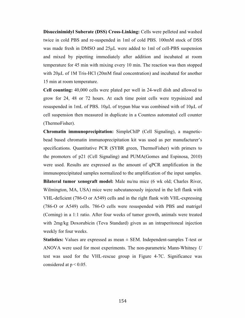
! 154!
Disuccinimidyl Suberate (DSS) Cross-Linking: Cells were pelleted and washed
twice in cold PBS and re-suspended in 1ml of cold PBS. 100mM stock of DSS
was made fresh in DMSO and 25µL were added to 1ml of cell-PBS suspension
and mixed by pipetting immediately after addition and incubated at room
temperature for 45 min with mixing every 10 min. The reaction was then stopped
with 20µL of 1M Tris-HCl (20mM final concentration) and incubated for another
15 min at room temperature.
Cell counting: 40,000 cells were plated per well in 24-well dish and allowed to
grow for 24, 48 or 72 hours. At each time point cells were trypsinized and
resuspended in 1mL of PBS. 10µL of trypan blue was combined with of 10µL of
cell suspension then measured in duplicate in a Countess automated cell counter
(ThermoFisher).
Chromatin immunoprecipitation: SimpleChIP (Cell Signaling), a magnetic-
bead based chromatin immunoprecipitation kit was used as per manufacturer’s
specifications. Quantitative PCR (SYBR green, ThermoFisher) with primers to
the promoters of p21 (Cell Signaling) and PUMA(Gomes and Espinosa, 2010)
were used. Results are expressed as the amount of qPCR amplification in the
immunoprecipitated samples normalized to the amplification of the input samples.
Bilateral tumor xenograft model: Male nu/nu mice (6 wk old; Charles River,
Wilmington, MA, USA) mice were subcutaneously injected in the left flank with
VHL-deficient (786-O or A549) cells and in the right flank with VHL-expressing
(786-O or A549) cells. 786-O cells were resuspended with PBS and matrigel
(Corning) in a 1:1 ratio. After four weeks of tumor growth, animals were treated
with 2mg/kg Doxorubicin (Teva Standard) given as an intraperitoneal injection
weekly for four weeks.
Statistics: Values are expressed as mean ± SEM. Independent-samples T-test or
ANOVA were used for most experiments. The non-parametric Mann-Whitney U
test was used for the VHL-rescue group in Figure 4-7C. Significance was
considered at p"<"0.05.
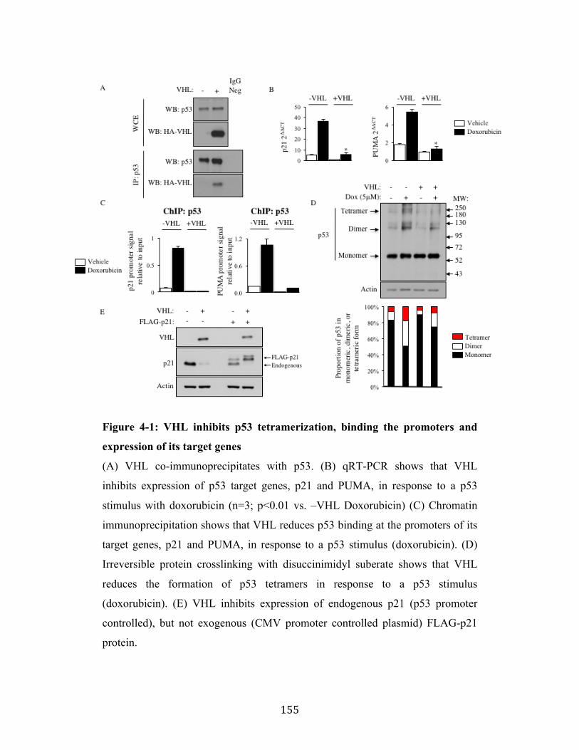
! 155!
Figure 4-1: VHL inhibits p53 tetramerization, binding the promoters and
expression of its target genes
(A) VHL co-immunoprecipitates with p53. (B) qRT-PCR shows that VHL
inhibits expression of p53 target genes, p21 and PUMA, in response to a p53
stimulus with doxorubicin (n=3; p<0.01 vs. –VHL Doxorubicin) (C) Chromatin
immunoprecipitation shows that VHL reduces p53 binding at the promoters of its
target genes, p21 and PUMA, in response to a p53 stimulus (doxorubicin). (D)
Irreversible protein crosslinking with disuccinimidyl suberate shows that VHL
reduces the formation of p53 tetramers in response to a p53 stimulus
(doxorubicin). (E) VHL inhibits expression of endogenous p21 (p53 promoter
controlled), but not exogenous (CMV promoter controlled plasmid) FLAG-p21
protein.
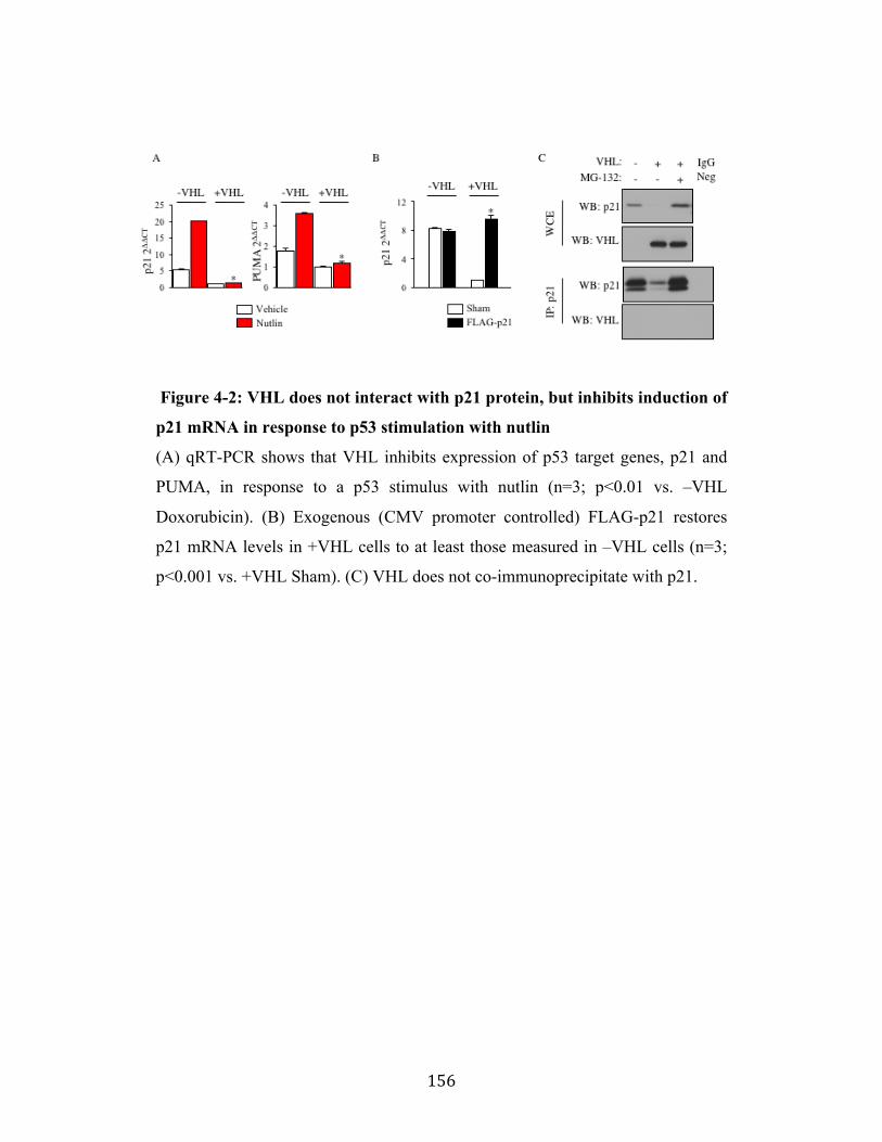
! 156!
Figure 4-2: VHL does not interact with p21 protein, but inhibits induction of
p21 mRNA in response to p53 stimulation with nutlin
(A) qRT-PCR shows that VHL inhibits expression of p53 target genes, p21 and
PUMA, in response to a p53 stimulus with nutlin (n=3; p<0.01 vs. –VHL
Doxorubicin). (B) Exogenous (CMV promoter controlled) FLAG-p21 restores
p21 mRNA levels in +VHL cells to at least those measured in –VHL cells (n=3;
p<0.001 vs. +VHL Sham). (C) VHL does not co-immunoprecipitate with p21.

! 157!
Figure 4-3: VHL inhibits p53-dependent target gene p21 expression,
promoting cell proliferation
(A) Confocal imaging shows +VHL cells express less p21 than –VHL cells
(VHL: green, p21: red, DAPI: blue, n=15 fields of view; *p<0.01 vs. –VHL). (B)
VHL reduces p21 protein expression in 786-O, (C) RCC4, and (D) A549 cancer
cells. (E) Adenoviral expression of wild-type, but not mutant VHL reduces p21
expression. (F) p53 knockdown mimics VHL expression’s reduction in p21 (n=3;
*p<0.01 vs. –VHL Scr, #p<0.01 vs. +VHL Scr). (G) More +VHL than -VHL cells
stain positive for ki67 (n=3; *p<0.05). (H) More +VHL than –VHL cells are
counted at 24, 48 and 72 hours after plating equal numbers of cells at time 0 hours
(n=4; *p<0.01 vs. -VHL).
! 158!
Figure 4-4: Validation of VHL expression in several models (A) Confirmation of CRISPR plasmid insertion into genomic DNA from A549
cells (left). VHL mRNA is depleted in VHL CRISPR A549 cells compared to
parental A549 cells (middle). p21 mRNA is increased in VHL CRISPR A549
cells compared to parental A549 cells (right). Confocal microscopy shows that
p53 has similar nuclear localization and increased expression in +VHL cells (n=
n=15 fields of view; *p<0.01 vs. -VHL). (C) Sp1 knockdown does not reduce p21
expression.
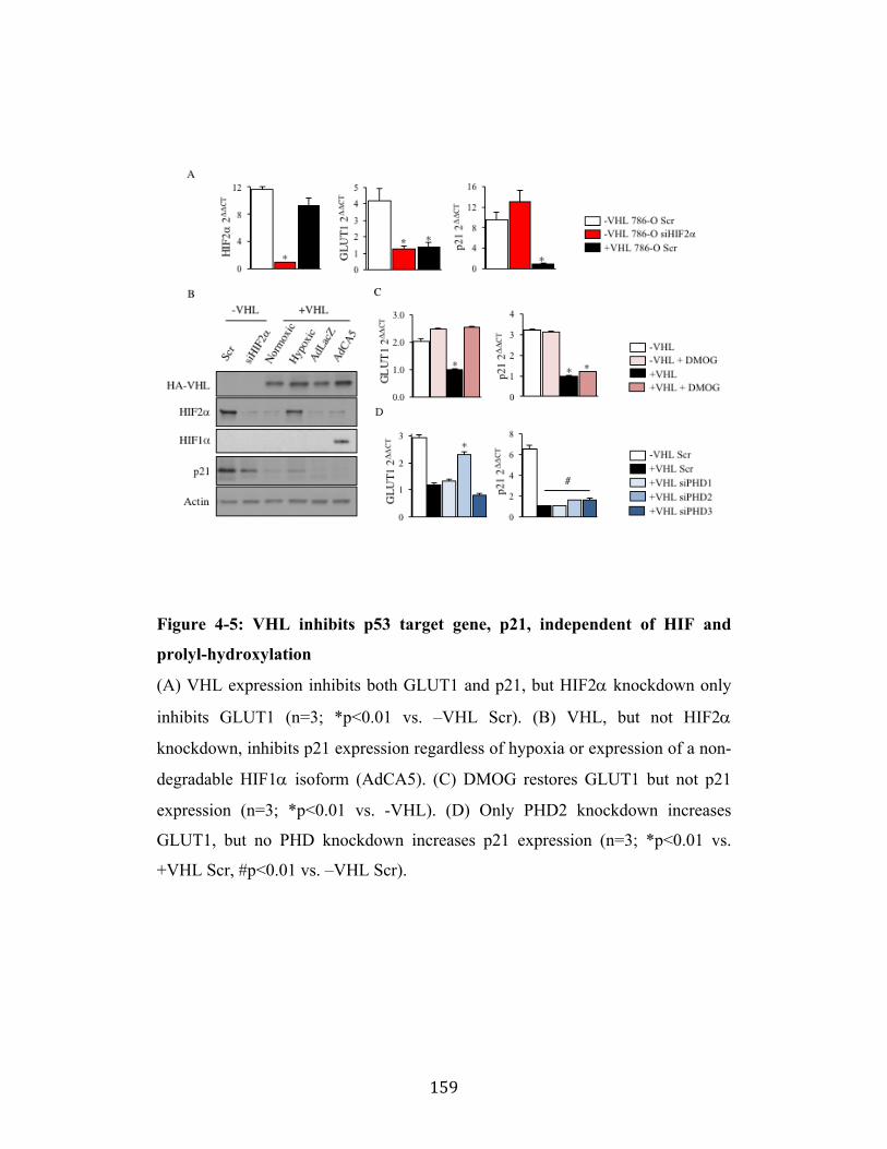
! 159!
Figure 4-5: VHL inhibits p53 target gene, p21, independent of HIF and
prolyl-hydroxylation
(A) VHL expression inhibits both GLUT1 and p21, but HIF2! knockdown only
inhibits GLUT1 (n=3; *p<0.01 vs. –VHL Scr). (B) VHL, but not HIF2!
knockdown, inhibits p21 expression regardless of hypoxia or expression of a non-
degradable HIF1! isoform (AdCA5). (C) DMOG restores GLUT1 but not p21
expression (n=3; *p<0.01 vs. -VHL). (D) Only PHD2 knockdown increases
GLUT1, but no PHD knockdown increases p21 expression (n=3; *p<0.01 vs.
+VHL Scr, #p<0.01 vs. –VHL Scr).
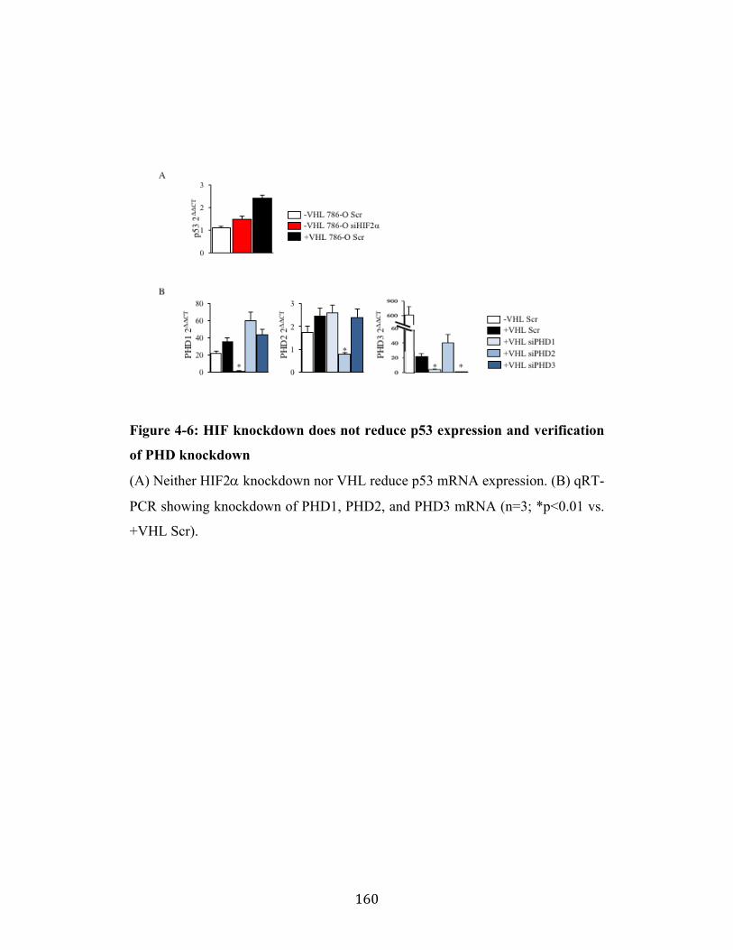
! 160!
Figure 4-6: HIF knockdown does not reduce p53 expression and verification
of PHD knockdown
(A) Neither HIF2! knockdown nor VHL reduce p53 mRNA expression. (B) qRT-
PCR showing knockdown of PHD1, PHD2, and PHD3 mRNA (n=3; *p<0.01 vs.
+VHL Scr).
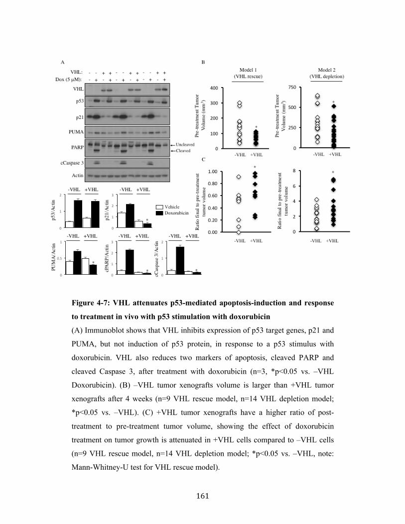
! 161!
Figure 4-7: VHL attenuates p53-mediated apoptosis-induction and response
to treatment in vivo with p53 stimulation with doxorubicin
(A) Immunoblot shows that VHL inhibits expression of p53 target genes, p21 and
PUMA, but not induction of p53 protein, in response to a p53 stimulus with
doxorubicin. VHL also reduces two markers of apoptosis, cleaved PARP and
cleaved Caspase 3, after treatment with doxorubicin (n=3, *p<0.05 vs. –VHL
Doxorubicin). (B) –VHL tumor xenografts volume is larger than +VHL tumor
xenografts after 4 weeks (n=9 VHL rescue model, n=14 VHL depletion model;
*p<0.05 vs. –VHL). (C) +VHL tumor xenografts have a higher ratio of post-
treatment to pre-treatment tumor volume, showing the effect of doxorubicin
treatment on tumor growth is attenuated in +VHL cells compared to –VHL cells
(n=9 VHL rescue model, n=14 VHL depletion model; *p<0.05 vs. –VHL, note:
Mann-Whitney-U test for VHL rescue model).
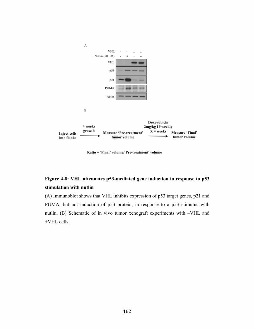
! 162!
Figure 4-8: VHL attenuates p53-mediated gene induction in response to p53
stimulation with nutlin
(A) Immunoblot shows that VHL inhibits expression of p53 target genes, p21 and
PUMA, but not induction of p53 protein, in response to a p53 stimulus with
nutlin. (B) Schematic of in vivo tumor xenograft experiments with –VHL and
+VHL cells.
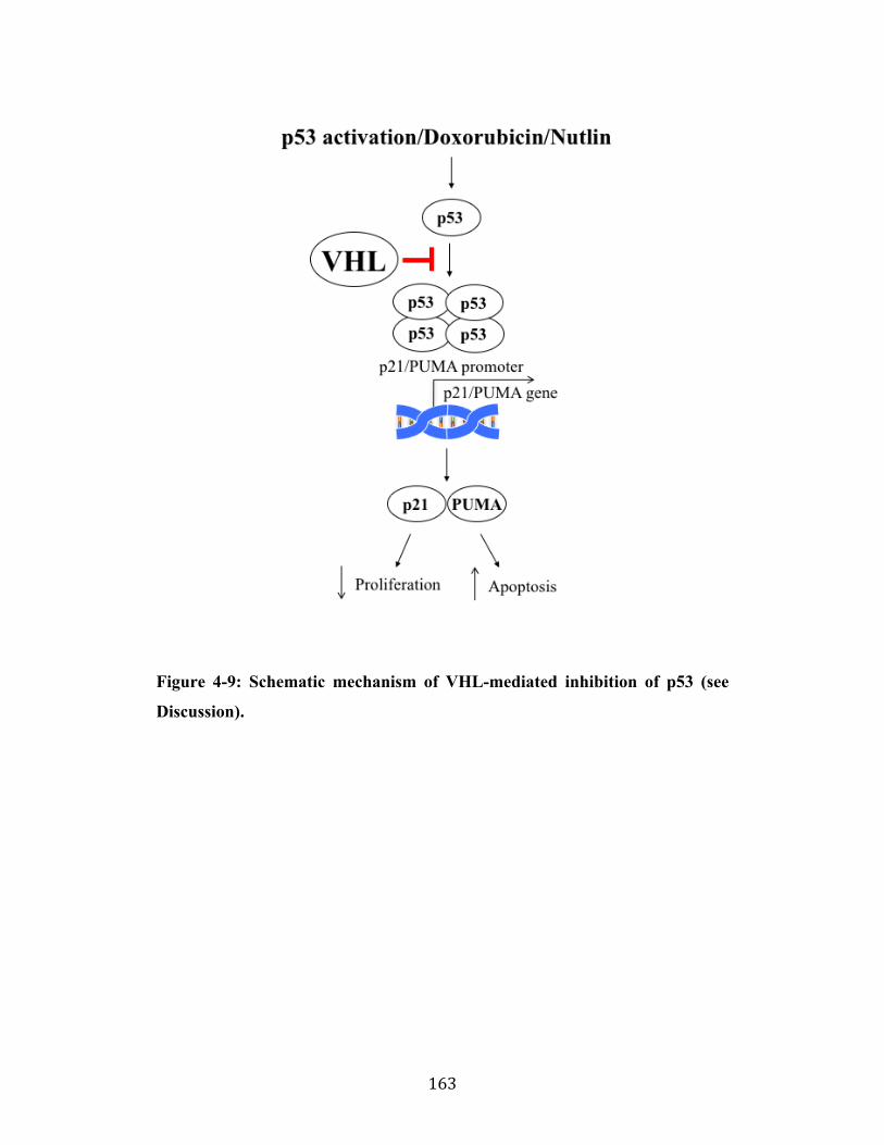
! 163!
Figure 4-9: Schematic mechanism of VHL-mediated inhibition of p53 (see
Discussion).
! 164!
References
Abbas,!T.,!and!Dutta,!A.!(2009).!p21!in!cancer:!intricate!networks!and!multiple!activities.!Nat!Rev!Cancer!9,!400I414.!Albers,!J.,!Rajski,!M.,!Schonenberger,!D.,!Harlander,!S.,!Schraml,!P.,!von!Teichman,!A.,!Georgiev,!S.,!Wild,!P.!J.,!Moch,!H.,!Krek,!W.,!and!Frew,!I.!J.!(2013).!Combined!mutation!of!Vhl!and!Trp53!causes!renal!cysts!and!tumours!in!mice.!EMBO!Mol!Med!5,!949I964.!Berra,!E.,!Benizri,!E.,!Ginouves,!A.,!Volmat,!V.,!Roux,!D.,!and!Pouyssegur,!J.!(2003).!HIF!prolylIhydroxylase!2!is!the!key!oxygen!sensor!setting!low!steadyIstate!levels!of!HIFI1alpha!in!normoxia.!EMBO!J!22,!4082I4090.!Chatterjee,!K.,!Zhang,!J.,!Honbo,!N.,!and!Karliner,!J.!S.!(2010).!Doxorubicin!cardiomyopathy.!Cardiology!115,!155I162.!Chen,!W.,!Hill,!H.,!Christie,!A.,!Kim,!M.!S.,!Holloman,!E.,!PaviaIJimenez,!A.,!Homayoun,!F.,!Ma,!Y.,!Patel,!N.,!Yell,!P.,!et!al.!(2016).!Targeting!renal!cell!carcinoma!with!a!HIFI2!antagonist.!Nature!539,!112I117.!Chene,!P.!(2001).!The!role!of!tetramerization!in!p53!function.!Oncogene!20,!2611I2617.!Cohen,!H.!T.,!Zhou,!M.,!Welsh,!A.!M.,!Zarghamee,!S.,!Scholz,!H.,!Mukhopadhyay,!D.,!Kishida,!T.,!Zbar,!B.,!Knebelmann,!B.,!and!Sukhatme,!V.!P.!(1999).!An!important!von!HippelILindau!tumor!suppressor!domain!mediates!Sp1Ibinding!and!selfIassociation.!Biochem!Biophys!Res!Commun!266,!43I50.!Fan,!Y.,!Li,!H.,!Ma,!X.,!Gao,!Y.,!Chen,!L.,!Li,!X.,!Bao,!X.,!Du,!Q.,!Zhang,!Y.,!and!Zhang,!X.!(2015).!Prognostic!Significance!of!HypoxiaIInducible!Factor!Expression!in!Renal!Cell!Carcinoma:!A!PRISMAIcompliant!Systematic!Review!and!MetaIAnalysis.!Medicine!(Baltimore)!94,!e1646.!Frew,!I.!J.,!and!Krek,!W.!(2008).!pVHL:!a!multipurpose!adaptor!protein.!Sci!Signal!1,!pe30.!Frost,!J.,!Galdeano,!C.,!Soares,!P.,!Gadd,!M.!S.,!Grzes,!K.!M.,!Ellis,!L.,!Epemolu,!O.,!Shimamura,!S.,!Bantscheff,!M.,!Grandi,!P.,!et!al.!(2016).!Potent!and!selective!chemical!probe!of!hypoxic!signalling!downstream!of!HIFIalpha!hydroxylation!via!VHL!inhibition.!Nat!Commun!7,!13312.!GimenezIBachs,!J.!M.,!SalinasISanchez,!A.!S.,!SanchezISanchez,!F.,!LorenzoIRomero,!J.!G.,!DonateIMoreno,!M.!J.,!PastorINavarro,!H.,!GarciaIOlmo,!D.!C.,!EscribanoIMartinez,!J.,!and!VirsedaIRodriguez,!J.!A.!(2006).!Determination!of!vhl!gene!mutations!in!sporadic!renal!cell!carcinoma.!Eur!Urol!49,!1051I1057.!Gomes,!N.!P.,!and!Espinosa,!J.!M.!(2010).!GeneIspecific!repression!of!the!p53!target!gene!PUMA!via!intragenic!CTCFICohesin!binding.!Genes!Dev!24,!1022I1034.!Gossage,!L.,!Eisen,!T.,!and!Maher,!E.!R.!(2015).!VHL,!the!story!of!a!tumour!suppressor!gene.!Nat!Rev!Cancer!15,!55I64.!Harlander,!S.,!Schonenberger,!D.,!Toussaint,!N.!C.,!Prummer,!M.,!Catalano,!A.,!Brandt,!L.,!Moch,!H.,!Wild,!P.!J.,!and!Frew,!I.!J.!(2017).!Combined!mutation!in!Vhl,!Trp53!and!Rb1!causes!clear!cell!renal!cell!carcinoma!in!mice.!Nat!Med!23,!869I877.!
! 165!
Hergovich,!A.,!Lisztwan,!J.,!Barry,!R.,!Ballschmieter,!P.,!and!Krek,!W.!(2003).!Regulation!of!microtubule!stability!by!the!von!HippelILindau!tumour!suppressor!protein!pVHL.!Nat!Cell!Biol!5,!64I70.!Hu,!C.!J.,!Wang,!L.!Y.,!Chodosh,!L.!A.,!Keith,!B.,!and!Simon,!M.!C.!(2003).!Differential!roles!of!hypoxiaIinducible!factor!1alpha!(HIFI1alpha)!and!HIFI2alpha!in!hypoxic!gene!regulation.!Mol!Cell!Biol!23,!9361I9374.!Iliopoulos,!O.,!Kibel,!A.,!Gray,!S.,!and!Kaelin,!W.!G.,!Jr.!(1995).!Tumour!suppression!by!the!human!von!HippelILindau!gene!product.!Nat!Med!1,!822I826.!Kim,!J.!H.,!Jung,!C.!W.,!Cho,!Y.!H.,!Lee,!J.,!Lee,!S.!H.,!Kim,!H.!Y.,!Park,!J.,!Park,!J.!O.,!Kim,!K.,!Kim,!W.!S.,!et!al.!(2005).!Somatic!VHL!alteration!and!its!impact!on!prognosis!in!patients!with!clear!cell!renal!cell!carcinoma.!Oncol!Rep!13,!859I864.!Kinnaird,!A.,!Dromparis,!P.,!Saleme,!B.,!Gurtu,!V.,!Watson,!K.,!Paulin,!R.,!Zervopoulos,!S.,!Stenson,!T.,!Sutendra,!G.,!Pink,!D.!B.,!et!al.!(2016).!Metabolic!Modulation!of!ClearIcell!Renal!Cell!Carcinoma!with!Dichloroacetate,!an!Inhibitor!of!Pyruvate!Dehydrogenase!Kinase.!Eur!Urol!69,!734I744.!Kondo,!K.,!Yao,!M.,!Yoshida,!M.,!Kishida,!T.,!Shuin,!T.,!Miura,!T.,!Moriyama,!M.,!Kobayashi,!K.,!Sakai,!N.,!Kaneko,!S.,!et!al.!(2002).!Comprehensive!mutational!analysis!of!the!VHL!gene!in!sporadic!renal!cell!carcinoma:!relationship!to!clinicopathological!parameters.!Genes!Chromosomes!Cancer!34,!58I68.!Kuznetsova,!A.!V.,!Meller,!J.,!Schnell,!P.!O.,!Nash,!J.!A.,!Ignacak,!M.!L.,!Sanchez,!Y.,!Conaway,!J.!W.,!Conaway,!R.!C.,!and!CzyzykIKrzeska,!M.!F.!(2003).!von!HippelILindau!protein!binds!hyperphosphorylated!large!subunit!of!RNA!polymerase!II!through!a!proline!hydroxylation!motif!and!targets!it!for!ubiquitination.!Proc!Natl!Acad!Sci!U!S!A!100,!2706I2711.!Lai,!Y.,!Song,!M.,!Hakala,!K.,!Weintraub,!S.!T.,!and!Shiio,!Y.!(2011).!Proteomic!dissection!of!the!von!HippelILindau!(VHL)!interactome.!J!Proteome!Res!10,!5175I5182.!Li,!L.,!Zhang,!L.,!Zhang,!X.,!Yan,!Q.,!Minamishima,!Y.!A.,!Olumi,!A.!F.,!Mao,!M.,!Bartz,!S.,!and!Kaelin,!W.!G.,!Jr.!(2007).!HypoxiaIinducible!factor!linked!to!differential!kidney!cancer!risk!seen!with!type!2A!and!type!2B!VHL!mutations.!Mol!Cell!Biol!27,!5381I5392.!Liu,!N.,!Xia,!W.!Y.,!Liu,!S.!S.,!Chen,!H.!Y.,!Sun,!L.,!Liu,!M.!Y.,!Li,!L.!F.,!Lu,!H.!M.,!Fu,!Y.!J.,!Wang,!P.,!et!al.!(2016).!MicroRNAI101!targets!von!HippelILindau!tumor!suppressor!(VHL)!to!induce!HIF1alpha!mediated!apoptosis!and!cell!cycle!arrest!in!normoxia!condition.!Sci!Rep!6,!20489.!Mack,!F.!A.,!Patel,!J.!H.,!Biju,!M.!P.,!Haase,!V.!H.,!and!Simon,!M.!C.!(2005).!Decreased!growth!of!VhlI/I!fibrosarcomas!is!associated!with!elevated!levels!of!cyclin!kinase!inhibitors!p21!and!p27.!Mol!Cell!Biol!25,!4565I4578.!Moore,!L.!E.,!Nickerson,!M.!L.,!Brennan,!P.,!Toro,!J.!R.,!Jaeger,!E.,!Rinsky,!J.,!Han,!S.!S.,!Zaridze,!D.,!Matveev,!V.,!Janout,!V.,!et!al.!(2011).!Von!HippelILindau!(VHL)!inactivation!in!sporadic!clear!cell!renal!cancer:!associations!with!germline!VHL!polymorphisms!and!etiologic!risk!factors.!PLoS!Genet!7,!e1002312.!
! 166!
Mukhopadhyay,!D.,!Knebelmann,!B.,!Cohen,!H.!T.,!Ananth,!S.,!and!Sukhatme,!V.!P.!(1997).!The!von!HippelILindau!tumor!suppressor!gene!product!interacts!with!Sp1!to!repress!vascular!endothelial!growth!factor!promoter!activity.!Mol!Cell!Biol!17,!5629I5639.!Mullen,!P.!(2004).!The!use!of!Matrigel!to!facilitate!the!establishment!of!human!cancer!cell!lines!as!xenografts.!Methods!Mol!Med!88,!287I292.!Patard,!J.!J.,!Fergelot,!P.,!Karakiewicz,!P.!I.,!Klatte,!T.,!Trinh,!Q.!D.,!RiouxILeclercq,!N.,!Said,!J.!W.,!Belldegrun,!A.!S.,!and!Pantuck,!A.!J.!(2008).!Low!CAIX!expression!and!absence!of!VHL!gene!mutation!are!associated!with!tumor!aggressiveness!and!poor!survival!of!clear!cell!renal!cell!carcinoma.!Int!J!Cancer!123,!395I400.!Roe,!J.!S.,!Kim,!H.,!Lee,!S.!M.,!Kim,!S.!T.,!Cho,!E.!J.,!and!Youn,!H.!D.!(2006).!p53!stabilization!and!transactivation!by!a!von!HippelILindau!protein.!Mol!Cell!22,!395I405.!Schokrpur,!S.,!Hu,!J.,!Moughon,!D.!L.,!Liu,!P.,!Lin,!L.!C.,!Hermann,!K.,!Mangul,!S.,!Guan,!W.,!Pellegrini,!M.,!Xu,!H.,!and!Wu,!L.!(2016).!CRISPRIMediated!VHL!Knockout!Generates!an!Improved!Model!for!Metastatic!Renal!Cell!Carcinoma.!Sci!Rep!6,!29032.!Schraml,!P.,!Struckmann,!K.,!Hatz,!F.,!Sonnet,!S.,!Kully,!C.,!Gasser,!T.,!Sauter,!G.,!Mihatsch,!M.!J.,!and!Moch,!H.!(2002).!VHL!mutations!and!their!correlation!with!tumour!cell!proliferation,!microvessel!density,!and!patient!prognosis!in!clear!cell!renal!cell!carcinoma.!J!Pathol!196,!186I193.!Soares,!P.,!Gadd,!M.!S.,!Frost,!J.,!Galdeano,!C.,!Ellis,!L.,!Epemolu,!O.,!Rocha,!S.,!Read,!K.!D.,!and!Ciulli,!A.!(2017).!GroupIBased!Optimization!of!Potent!and!CellIActive!Inhibitors!of!the!von!HippelILindau!(VHL)!E3!Ubiquitin!Ligase:!StructureIActivity!Relationships!Leading!to!the!Chemical!Probe!(2S,4R)I1I((S)I2I(1ICyanocyclopropanecarboxamido)I3,3Idimethylbutanoyl)I4Ihydroxy!INI(4I(4ImethylthiazolI5Iyl)benzyl)pyrrolidineI2Icarboxamide!(VH298).!J!Med!Chem.!Yao,!M.,!Yoshida,!M.,!Kishida,!T.,!Nakaigawa,!N.,!Baba,!M.,!Kobayashi,!K.,!Miura,!T.,!Moriyama,!M.,!Nagashima,!Y.,!Nakatani,!Y.,!et!al.!(2002).!VHL!tumor!suppressor!gene!alterations!associated!with!good!prognosis!in!sporadic!clearIcell!renal!carcinoma.!J!Natl!Cancer!Inst!94,!1569I1575.!Yi,!Y.,!Mikhaylova,!O.,!Mamedova,!A.,!Bastola,!P.,!Biesiada,!J.,!Alshaikh,!E.,!Levin,!L.,!Sheridan,!R.!M.,!Meller,!J.,!and!CzyzykIKrzeska,!M.!F.!(2010).!von!HippelILindauIdependent!patterns!of!RNA!polymerase!II!hydroxylation!in!human!renal!clear!cell!carcinomas.!Clin!Cancer!Res!16,!5142I5152.!
167$
!
!
!
!
Chapter Five
Myocyte Enhancer Factor 2A is up-regulated, increases CCL20 expression and promotes tumor growth in clear
cell renal cell carcinoma

168$
Abstract
Background: Advanced clear cell renal cell carcinoma (ccRCC) remains a deadly
disease driven by metabolic derangement, immune evasion and propensity for
invasion and metastasis. Myocyte Enhancer Factor 2A (Mef2A) is a growth factor
driven transcription factor involved in cell growth, metabolism, invasion,
immunomodulation and apoptosis-resistance.
Objective: We hypothesized that Mef2A is up-regulated in ccRCC, promoting
tumor growth.
Design, Setting, and Participants: Fifty-two pathologist reviewed ccRCC
tumors and surrounding normal kidney tissues (from the same patients) were
stained for Mef2A using immunofluorescent confocal microscopy. ccRCC and
proximal tubule cells lines as well as nude mouse xenografts were used for in-
vitro and in-vivo experiments, respectively.
Interventions: Mef2A was inhibited using RNA interference.
Outcomes: Unbiased GeneChip screening (confirmed by qRT-PCR) identified
Mef2A target genes. Proliferation (ki67, BrdU and phosphorylated
retinoblastoma), apoptosis (TUNEL), migration (scratch assay), invasion
(basement membrane extract) and xenograft tumor growth were measured using
standard techniques.
Results: Mef2A expression is higher in ccRCC tumors compared to the
surrounding normal kidney. Mef2A transcriptional activity is higher in ccRCC
cells vs. proximal tubule cells and in VHL-deficient (high growth factor) vs.
VHL-expressing (low growth factor) 786-O cells. The pro-tumorigenic, secretable
molecule Chemokine Ligand 20 (CCL20) is the most reduced target in Mef2A
knockdown. Mef2A knockdown reduces cell proliferation, migration, invasion,
and increases apoptosis in-vitro, as well as reducing tumor growth in a nude
mouse model. Mef2A levels correlate with tumor size in large tumors. Conclusions: Mef2A is up-regulated in ccRCC, drives expression of the pro-
tumorigenic chemokine CCL20 and is important for tumor growth.

169$
Patient Summary: In this report we identify the protein Mef2A to be important
for kidney cancer growth and find that it correlates with tumor size in large
kidney cancers. Mef2A may be involved in tumor cell growth and death as well as
local regulation of the immune system. Inhibition of Mef2A reduces tumor
growth in the dish as well as in an animal model.
170$
Introduction
More than 330,000 patients are diagnosed with renal cell carcinoma
(RCC) annually, with approximately 30% presenting with metastases at the time
of diagnosis(1, 2). Current first-line agents for advanced clear cell RCC (ccRCC),
such as tyrosine kinase inhibitors like sunitinib, only increase progression free
survival by 11 months(3). Therefore, we must link together several fundamental
mechanisms contributing to the pathogenesis of ccRCC, such as the recent
identification of local immune suppression and advancement of immune
checkpoint inhibitors, to find novel therapeutic targets to treat this deadly
cancer(4).
Deregulated cellular energetics, apoptosis-resistance, evading the immune
system and activating invasion and metastasis are critical hallmarks of cancer(5).
Identifying a mechanism that controls multiple hallmarks of cancer is a desirable
therapeutic target.
Myocyte enhancer factor 2A (Mef2A) is a master transcription factor
involved in cell survival, metabolism, apoptosis-resistance, immunomodulation,
and invasion(6-10), Mef2A has recently been identified in several solid organ
tumors (breast, liver, and lung), however its role in ccRCC has not yet been
described(6). Mef2A is activated by growth factor dependent phosphorylation
through the epidermal growth factor receptor (EGFR) signaling pathway(6, 11).
This signaling cascade is highly active in ccRCC due to loss of the von Hippel
Lindau (VHL) tumor suppressor, leading to accumulation of hypoxia inducible
factor (HIF) and overexpression of HIF target genes such as transforming growth
factor alpha (TGFα) (12-14). TGFα has been shown to be a potent ligand for
EGFR, strongly activating the EGFR signaling pathway in ccRCC and potentially
activating Mef2A(15).
Chemokine Ligand 20 (CCL20) is a pro-proliferation(16-18), pro-
migration(17-19) and pro-invasion(16, 19, 20) molecule that has been shown to
be secreted by several solid tumors including renal cell carcinoma(20-25). CCL20
exerts its physiological effect by binding to a highly specific G-coupled protein
receptor, CCR6(26).
171$
In this manuscript, we show that Mef2A is up-regulated and
transcriptionally activated in ccRCC. Furthermore, we use unbiased screening to
identify that Mef2A promotes the expression of the pro-proliferative and invasive
chemokine CCL20, as well as increases cell proliferation, apoptosis-resistance
and tumor growth.
172$
Results
5.1 Mef2A expression is up-regulated in renal tumor tissue
In order to determine the relative expression of Mef2A in ccRCC
compared to the normal kidney parenchyma in our patients, we obtained 52
ccRCC tumors and compared the amount of Mef2A expression in the tumors to
normal kidney tissues removed from the same patients at the time of their radical
nephrectomy. Nuclear Mef2A protein and mRNA expression were significantly
elevated in tumors compared to the normal kidney samples (Figure 5-1A-C). The
patient and tumor characteristics are outlined in Figure 5-2A.
Mef2A protein is expressed in ccRCC but not proximal tubule cells, which
are the healthy tissue type from which ccRCC is derived (Figure 5-1D). This
increase in Mef2A expression in cancer cells compared to the normal cell type
from which the cancer initiates was also found in several other cancer/normal
tissues pairings (non-small cell lung cancer vs. small airway epithelial cells and
melanoma vs. melanocytes; Figure 5-2B). Similarly, the transcriptional activity of
Mef2 is increased in ccRCC compared to proximal tubule cells as measured using
a promoter firefly luciferase assay (Figure 5-1E).
5.2 Mef2A regulates expression of CCL20 in kidney tumor cells
In order to determine the effect of Mef2A in ccRCC we performed a
GeneChip screening assay on a loss-of-function model using siRNA knockdown.
We achieved an ~90% knockdown of Mef2A at both the mRNA and protein
levels (Figure 5-3A). GeneChip screening demonstrated that Mef2A knockdown
resulted in a >6-fold reduction of CCL20 mRNA (Figure 5-3B; see Table 5-1 for
complete list of all genes with !1.5 fold change). Decreased CCL20 expression
was confirmed by primer specific qRT-PCR (Figure 5-3C). Similarly, we found
an increased expression of CCL20 mRNA in patient tumors (high Mef2A)
compared to normal kidney (low Mef2A) tissues (Figure 5-3D). As CCL20 is a
secretable chemokine, we measured CCL20 levels in the supernatant after Mef2A
knockdown. We found the concentration of CCL20 to be reduced in the
supernatant of ccRCC cells that had decreased Mef2A expression (Figure 5-3E).
173$
CCL20 is known to exert its pro-proliferative and pro-invasive effects by binding
to a specific G-coupled protein receptor, called CCR6, on several tissues such as
breast and immune cells. CCR6 has previously been identified in both ccRCC
tumor and infiltrating T regulatory cells within the tumors of patients with
ccRCC(24). We confirm that the CCR6 receptor is expressed in ccRCC cells and
at a high level compared to benign proximal tubule cells (Figure 5-3F).
5.3 Loss of Mef2A inhibits proliferation, migration and invasion and
promotes mitochondrial activity and apoptosis of ccRCC cells
We used three metrics to determine the effect of Mef2A on proliferation:
ki67, BrdU incorporation and phosphorylated retinoblastoma (p-Rb) protein
levels. Mef2A knockdown reduced ki67 staining (Figure 5-4A), BrdU
incorporation (Figure 5-4B) and p-Rb (Figure 5-4C) suggesting loss of Mef2A
inhibits proliferation. Mef2A knockdown increased mitochondrial activity
measured by a depolarized mitochondrial membrane potential as well as increased
mitochondrial reactive oxygen species (Figure 5-4D). This may be due to up-
regulation of a known Mef2 regulated gene, PGC1" and its downstream target,
the major mitochondrial deacetylase and activator Sirtuin 3 (Figure 5-4E) (27-29).
Mef2A knockdown increased staining of the apoptotic marker TUNEL in ccRCC
in keeping with its anti-apoptotic role previously described and the increase in
mitochondrial activity (Figure 5-4F)(10). Mef2A knockdown reduced tumor cell
migration as assessed using a standard scratch assay (Figure 5-4G). Similarly,
knockdown of Mef2A reduced the invasiveness of ccRCC cells measured using a
commercially available in-vitro invasion assay (Figure 5-4H).
5.4 VHL-deficiency increases Mef2A transcriptional activity
VHL-deficient 786-O ccRCC cells overexpress HIF2a in normoxia (Figure 5-5A).
This leads to an increased expression of the HIF2a target gene, Transforming
growth factor alpha (TGFα) (Figure 5-5B), and activation of the EGFR
phosphorylation pathway for which it has previously been shown to be a potent
ligand (Figure 5-5C)(15). Mef2A is significantly more phosphorylated at
174$
Threonine 312, which is a stimulatory phosphorylation that can be triggered by
EGFR signaling, in VHL-deficient compared to VHL-expressing ccRCC cells
(Figure 5-5D). Moreover, the transcriptional activity of Mef2 is significantly
higher in the VHL-deficient, hyperphosphorylated T312 Mef2A, ccRCC cells
(Figure 5-5E). The model of VHL deficiency leading to increased Mef2A activity
is outlined in Figure 5-5F.
5.5 Mef2A knockdown reduces tumor growth in-vivo
We developed a stable knockdown model of Mef2A using expression of a
short-hairpin RNA Mef2A plasmid (shMef2A) in order to assess the effect of
Mef2A inhibition on tumor growth in vivo (Figure 5-6A). Tumors initiated in 7/9
(78%) of animals injected with shScr and in 12/13 (92%) of animals injected with
shMef2A tumors suggesting that Mef2A is not critical for tumor establishment.
However, after 10 days, tumors with Mef2A knockdown failed to significantly
enlarge, while shScr control tumors continued to grow (Figure 5-6B). After 30
days, excised shMef2A tumors were significantly smaller in volume and weight
than shScr control tumors (Figure 5-6B and C). Stratification of human tumor
samples by size as either pT1 (early tumors, <7cm) or pT2 (established tumors,
>7cm) suggests that nuclear Mef2A expression is correlated with tumor size in
established (pT2) tumors (R=0.829; p=0.042; Figure 5-6D) but not in early (pT1)
tumors (Spearman correlation R=0.192; p=0.242; Figure 5-7). Inclusion of pT3
tumors that are >7cm, (i.e. pT2 by size) into the pT2 data also suggests a trend in
larger tumor size and nuclear Mef2A levels (R=0.506; p=0.078; Figure 5-6D).
175$
Discussion
We show that the expression and activity of Mef2A is increased in ccRCC
compared to normal kidney tissues. This leads to increased transcription and
secretion of the pro-proliferative chemokine CCL20. Inhibition of Mef2A reduces
CCL20 production and decreases cancer cell proliferation and tumor growth.
The molecular phenotype of ccRCC predisposes this malignancy to
Mef2A activation. VHL-deficiency leads to increased HIF activation, which
drives TGFα expression. TGFα activates the EGF receptor and downstream
cascade of protein kinases, which phosphorylate Mef2A threonine 312, increasing
transcriptional activity and subsequent activation of downstream targets like
CCL20 (Figure 5-6E). This is important because VHL is the most commonly
altered gene in ccRCC, being mutated or having its promoter hypermethylated in
up to 90% of all ccRCC tumors. Therefore, Mef2A may not only be up-regulated
in our patients, as we show, but potently activated in these tumors as well. These
findings are in keeping with results from The Cancer Genome Atlas and the
Human Protein Atlas (www.proteinatlas.org), which identify RCC as having the
highest median expression of Mef2A RNA across the 17 malignancies
studied(30).
We identified CCL20 as an important target gene of Mef2A using an
unbiased GeneChip screening assay and confirmed its reduction in Mef2A
knockdown cells at both the mRNA and secreted protein levels. CCL20 is a
chemokine secreted by both immune and tumor cells and exerts its effects by
binding in an autocrine and paracrine manner to the CCR6 G-coupled protein
receptor. There is evidence that CCL20/CCR6 promote tumorigenesis in two
manners: First by promoting proliferation, migration and invasion of the tumor
cells themselves as has been identified in several other tumor types such as breast,
lung, thyroid, colon and pancreatic cancers(17-20, 25). Secondly, CCL20/CCR6
mediates effector T-cell suppression by recruiting inhibitory T-regulatory cells to
the tumour microenvironment, thus locally suppressing the immune system(24,
31). Therefore, the overall effect of CCL20 is tumor tropism and immune
tolerance. This means that while we observe a significant reduction in tumour cell
176$
growth in the Mef2A knockdown ccRCC cells, we may not be capturing the
complete effect of Mef2A/CCL20 inhibition as we are missing the effects on
immune suppression. Indeed, the anti-tumor effects we observed in-vivo (in mice
lacking cell-mediated immunity) for Mef2A knockdown cells are likely limited to
the anti-proliferative effects, and may actually be more accentuated tumor
inhibition in our patients where immune suppression would also be targeted.
Furthermore, ccRCC has been identified as a cancer that hijacks the
immune system to increase its growth, and immune checkpoint inhibitors are
quickly becoming the new cornerstone of advanced ccRCC management(4).
Interestingly, we show that two other cancer types for which immune checkpoint
inhibitors have been approved, NSCLC and melanoma both have increased
expression of Mef2A compared to their normal tissues from which they develop.
Mef2A may be playing an important role in immune evasion as ~25% of the top
25 targets identified on GeneChip screening were immune activators such as
CCL20, tumor necrosis factor, and interleukin 8, which were all up-regulated in
Mef2A expressing compared to Mef2A inhibited ccRCC cells (Table 5-1).
Our work suggests that Mef2A functions as an important node in ccRCC
growth and that Mef2A/CCL20/CCR6 inhibition may represent a new therapeutic
target. While there are no specific Mef2A inhibitors available, the underlying
driving force of Mef2A activation in ccRCC (EGFR activation by TGFα) can be
pharmacologically targeted as an adjuvant to current therapies(32). Early attempts
at targeting CCL20 have been attempted both specifically with monoclonal
antibodies(33) and non-specifically using commonly prescribed agents such as
statins(34). Similarly, specific microRNAs that inhibit CCR6 have been
identified, which have been shown to inhibit tumor invasion and metastasis(35).
While we have identified a new pro-tumor pathway, further work will need to be
performed to inhibit the Mef2A/CCL20/CCR6 axis in ccRCC.
177$
Materials and Methods
Patient Samples: We received institutional Health Research Ethics Board
approval to obtain specimens from patients undergoing radical nephrectomy for
RCC. All specimens (tumor and surrounding normal tissue from each patient)
were formalin fixed, paraffin embedded and diagnosed by a pathologist as ccRCC
versus normal kidney tissue. All tissue was processed for mRNA isolation and
staining as previously described(36).
Cell Culture and Reagents: The 786-O human ccRCC line was purchased from
ATCC, maintained in RMPI 1640 media (GIBCO, 10% FBS, 1% PSF). –VHL
786-O and +VHL 786-O cell lines (stably infected with an empty vector or
functional, Hemagglutinin-tagged VHL, respectively) were a gift from Dr. Kaelin
(Harvard Medical School)(37). Human renal proximal tubular cells were
purchased from ScienCell and maintained in EpiCM media.
siRNA knockdown and qRT-PCR: Mef2A specific or scrambled siRNAs were
transfected using Lipofectamine RNAiMax reagent as previously described(36).
After 72 hours, RNA was isolated (Qiagen) and qRT-PCR using gene specific
primers and TaqMan reagents (ThermoFisher) were used in a QuantStudio 7 Flex
Real-time PCR system (ThermoFisher).
shRNA knockdown: 786-O cells were transfected with plasmid using the xfect
transfection reagent (Clontech). Stable integration of the plasmid was selected for
using puromycin (1ug/mL).
Luciferase: Cignal dual-luciferase (Mef2/Renilla) reporter kits (SA Biosciences)
were used as previously described(38). 48hrs after transfection, activity was
assessed using a dual luciferase reporter assay kit (Promega). Mef2 activity was
normalized to the Renilla luminescence.
Genechip: 200ng of total RNA was labeled using the Affymetrix GeneChip 3’
IVT PLUS Reagent kit. Poly-A controls were added to each sample at the
beginning of the labeling. 10ug of fragmented, biotin-modified aRNA was
hybridized to the Affymetrix PrimeView Human microarray in the Affymetrix
GeneChip Hybridization Oven 645 set at 45oC with 60rpm for 16 hours.
Hybridization controls were added to each sample before the arrays were
178$
hybridized. The arrays were washed and stained with Affymetrix GeneChip Wash
and Stain Kit on the Affymetrix GeneChip Fluidics Station 450. Arrays were
scanned on the Affymetrix GeneChip Scanner 3000 7G to generate the raw data
files.
Nude Mouse Xenografts: 6 week old male nu/nu mice (Charles River) were
injected with ~3 million cells (in 250μl PBS) in a 1:1 ratio with Matrigel and after
institutional animal ethics committee approval as previously described(36).
CCL20 ELISA: Supernatant was collected, centrifuged at 1500rpm for 5 minutes
to remove any remaining cellular debris and the CCL20 ELISA performed as per
the manufacturer’s specifications (Abcam). Concentrations were normalized to
milligrams of cell protein from the wells.
BrdU assay: Colorimetric BrdU ELISA assay was used as per the manufacturer’s
specifications (Roche). In summary cells were incubated with 20 μM of BrdU
solution for 2 hours and then fixed for 30 minutes. Cells were then incubated with
an anti-BrdU-POD solution for 90 minutes and were subsequently washed and
incubated with a substrate solution for 5 minutes before measuring the absorbance
at 370 nm and 492 nm.
Confocal microscopy was used for immunofluorescence and live cell (TMRM,
Mitosox) imaging as previously described(36).
Immunoblotting: Immunoblotting with standard SDS-PAGE was performed as
previously described(39).
Statistics: We used Paired and Independent-Samples T tests to compare two
samples when appropriate and Spearman’s correlation coefficient to determine
association between two variables. p<0.05 was considered significant.
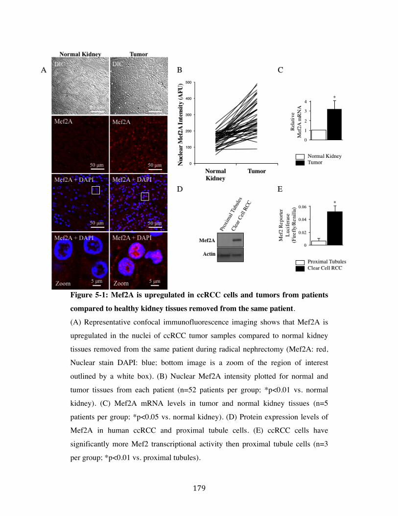
179$
Figure 5-1: Mef2A is upregulated in ccRCC cells and tumors from patients
compared to healthy kidney tissues removed from the same patient.
(A) Representative confocal immunofluorescence imaging shows that Mef2A is
upregulated in the nuclei of ccRCC tumor samples compared to normal kidney
tissues removed from the same patient during radical nephrectomy (Mef2A: red,
Nuclear stain DAPI: blue; bottom image is a zoom of the region of interest
outlined by a white box). (B) Nuclear Mef2A intensity plotted for normal and
tumor tissues from each patient (n=52 patients per group; *p<0.01 vs. normal
kidney). (C) Mef2A mRNA levels in tumor and normal kidney tissues (n=5
patients per group; *p<0.05 vs. normal kidney). (D) Protein expression levels of
Mef2A in human ccRCC and proximal tubule cells. (E) ccRCC cells have
significantly more Mef2 transcriptional activity then proximal tubule cells (n=3
per group; *p<0.01 vs. proximal tubules).
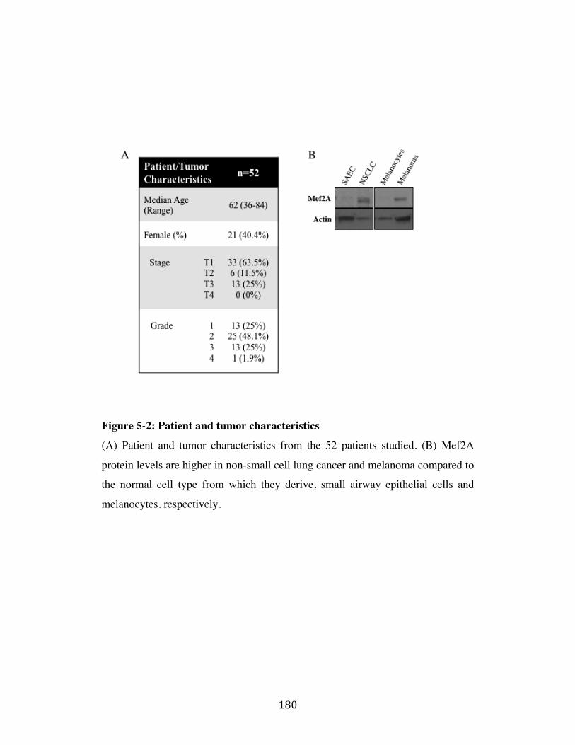
180$
Figure 5-2: Patient and tumor characteristics
(A) Patient and tumor characteristics from the 52 patients studied. (B) Mef2A
protein levels are higher in non-small cell lung cancer and melanoma compared to
the normal cell type from which they derive, small airway epithelial cells and
melanocytes, respectively.
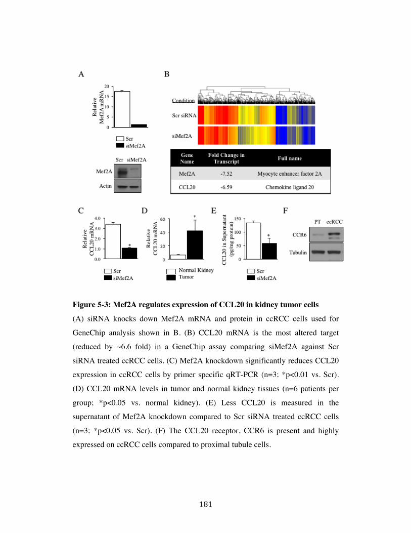
181$
Figure 5-3: Mef2A regulates expression of CCL20 in kidney tumor cells
(A) siRNA knocks down Mef2A mRNA and protein in ccRCC cells used for
GeneChip analysis shown in B. (B) CCL20 mRNA is the most altered target
(reduced by ~6.6 fold) in a GeneChip assay comparing siMef2A against Scr
siRNA treated ccRCC cells. (C) Mef2A knockdown significantly reduces CCL20
expression in ccRCC cells by primer specific qRT-PCR (n=3; *p<0.01 vs. Scr).
(D) CCL20 mRNA levels in tumor and normal kidney tissues (n=6 patients per
group; *p<0.05 vs. normal kidney). (E) Less CCL20 is measured in the
supernatant of Mef2A knockdown compared to Scr siRNA treated ccRCC cells
(n=3; *p<0.05 vs. Scr). (F) The CCL20 receptor, CCR6 is present and highly
expressed on ccRCC cells compared to proximal tubule cells.
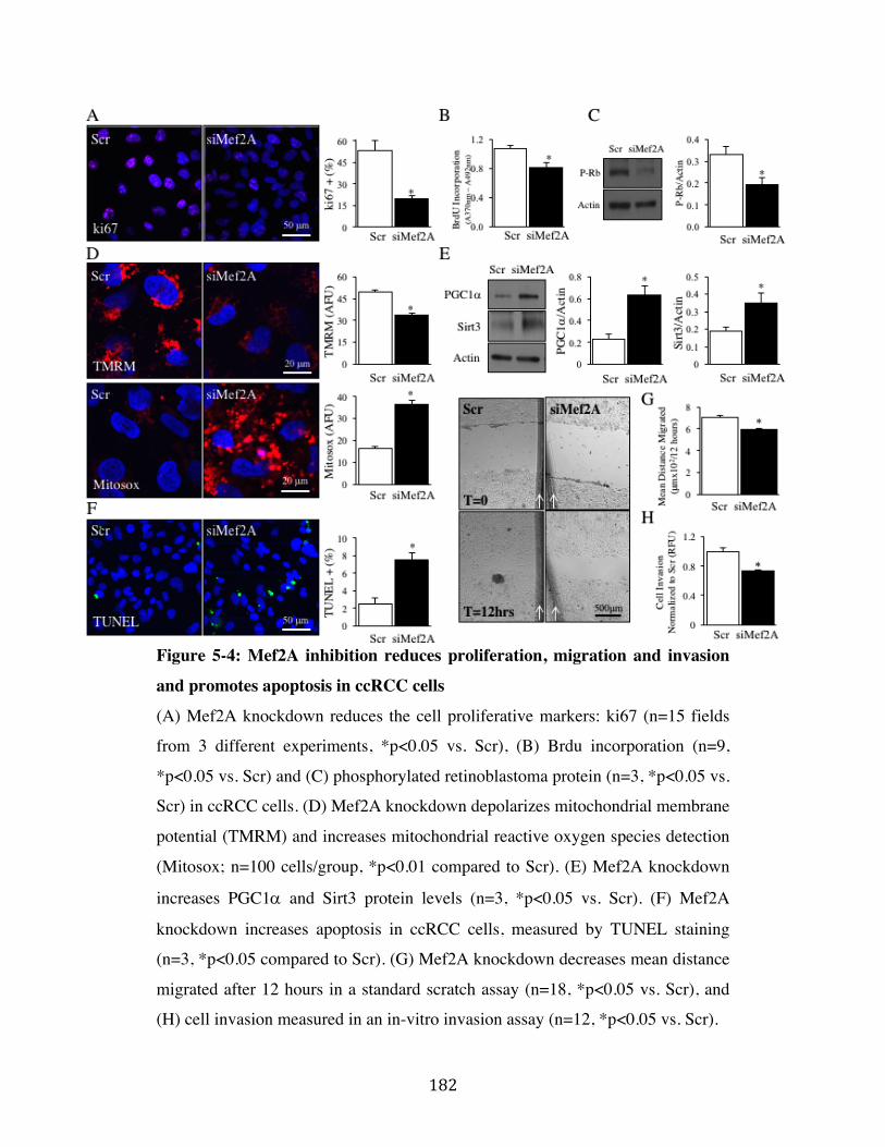
182$
Figure 5-4: Mef2A inhibition reduces proliferation, migration and invasion
and promotes apoptosis in ccRCC cells
(A) Mef2A knockdown reduces the cell proliferative markers: ki67 (n=15 fields
from 3 different experiments, *p<0.05 vs. Scr), (B) Brdu incorporation (n=9,
*p<0.05 vs. Scr) and (C) phosphorylated retinoblastoma protein (n=3, *p<0.05 vs.
Scr) in ccRCC cells. (D) Mef2A knockdown depolarizes mitochondrial membrane
potential (TMRM) and increases mitochondrial reactive oxygen species detection
(Mitosox; n=100 cells/group, *p<0.01 compared to Scr). (E) Mef2A knockdown
increases PGC1" and Sirt3 protein levels (n=3, *p<0.05 vs. Scr). (F) Mef2A
knockdown increases apoptosis in ccRCC cells, measured by TUNEL staining
(n=3, *p<0.05 compared to Scr). (G) Mef2A knockdown decreases mean distance
migrated after 12 hours in a standard scratch assay (n=18, *p<0.05 vs. Scr), and
(H) cell invasion measured in an in-vitro invasion assay (n=12, *p<0.05 vs. Scr).
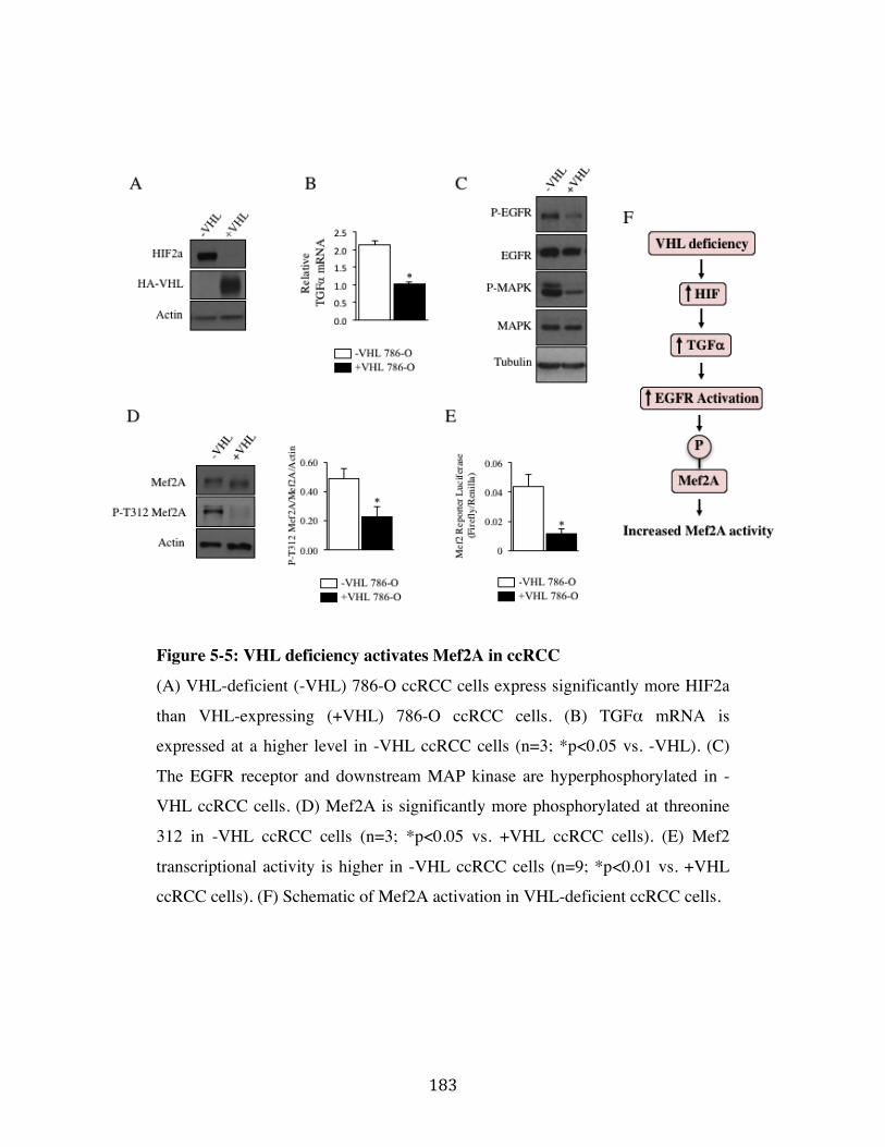
183$
Figure 5-5: VHL deficiency activates Mef2A in ccRCC
(A) VHL-deficient (-VHL) 786-O ccRCC cells express significantly more HIF2a
than VHL-expressing (+VHL) 786-O ccRCC cells. (B) TGFα mRNA is
expressed at a higher level in -VHL ccRCC cells (n=3; *p<0.05 vs. -VHL). (C)
The EGFR receptor and downstream MAP kinase are hyperphosphorylated in -
VHL ccRCC cells. (D) Mef2A is significantly more phosphorylated at threonine
312 in -VHL ccRCC cells (n=3; *p<0.05 vs. +VHL ccRCC cells). (E) Mef2
transcriptional activity is higher in -VHL ccRCC cells (n=9; *p<0.01 vs. +VHL
ccRCC cells). (F) Schematic of Mef2A activation in VHL-deficient ccRCC cells.
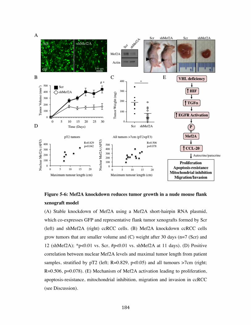
184$
Figure 5-6: Mef2A knockdown reduces tumor growth in a nude mouse flank
xenograft model
(A) Stable knockdown of Mef2A using a Mef2A short-hairpin RNA plasmid,
which co-expresses GFP and representative flank tumor xenografts formed by Scr
(left) and shMef2A (right) ccRCC cells. (B) Mef2A knockdown ccRCC cells
grow tumors that are smaller volume and (C) weight after 30 days (n=7 (Scr) and
12 (shMef2A); *p<0.01 vs. Scr, #p<0.01 vs. shMef2A at 11 days). (D) Positive
correlation between nuclear Mef2A levels and maximal tumor length from patient
samples, stratified by pT2 (left; R=0.829, p<0.05) and all tumours >7cm (right;
R=0.506, p=0.078). (E) Mechanism of Mef2A activation leading to proliferation,
apoptosis-resistance, mitochondrial inhibition, migration and invasion in ccRCC
(see Discussion).
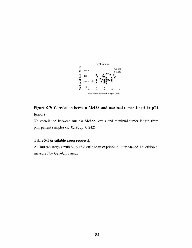
185$
Figure 5-7: Correlation between Mef2A and maximal tumor length in pT1
tumors
No correlation between nuclear Mef2A levels and maximal tumor length from
pT1 patient samples (R=0.192, p=0.242).
Table 5-1 (available upon request):
All mRNA targets with >1.5-fold change in expression after Mef2A knockdown,
measured by GeneChip assay.
186$
References
1.$ Ferlay$J,$Soerjomataram$I,$Dikshit$R,$Eser$S,$Mathers$C,$Rebelo$M,$et$al.$Cancer$incidence$and$mortality$worldwide:$sources,$methods$and$major$patterns$in$GLOBOCAN$2012.$Int$J$Cancer.$2015;136(5):E359U86.$2.$ Fisher$R,$Gore$M,$Larkin$J.$Current$and$future$systemic$treatments$for$renal$cell$carcinoma.$Semin$Cancer$Biol.$2013;23(1):38U45.$3.$ Motzer$RJ,$Hutson$TE,$Tomczak$P,$Michaelson$MD,$Bukowski$RM,$Rixe$O,$et$al.$Sunitinib$versus$interferon$alfa$in$metastatic$renalUcell$carcinoma.$N$Engl$J$Med.$2007;356(2):115U24.$4.$ Motzer$RJ,$Escudier$B,$McDermott$DF,$George$S,$Hammers$HJ,$Srinivas$S,$et$al.$Nivolumab$versus$Everolimus$in$Advanced$RenalUCell$Carcinoma.$N$Engl$J$Med.$2015;373(19):1803U13.$5.$ Hanahan$D,$Weinberg$RA.$Hallmarks$of$cancer:$the$next$generation.$Cell.$2011;144(5):646U74.$6.$ Pon$JR,$Marra$MA.$MEF2$transcription$factors:$developmental$regulators$and$emerging$cancer$genes.$Oncotarget.$2016;7(3):2297U312.$7.$ Yu$W,$Huang$C,$Wang$Q,$Huang$T,$Ding$Y,$Ma$C,$et$al.$MEF2$transcription$factors$promotes$EMT$and$invasiveness$of$hepatocellular$carcinoma$through$TGFUbeta1$autoregulation$circuitry.$Tumour$Biol.$2014;35(11):10943U51.$8.$ Clark$RI,$Tan$SW,$Pean$CB,$Roostalu$U,$Vivancos$V,$Bronda$K,$et$al.$MEF2$is$an$in$vivo$immuneUmetabolic$switch.$Cell.$2013;155(2):435U47.$9.$ Naya$FJ,$Black$BL,$Wu$H,$BasselUDuby$R,$Richardson$JA,$Hill$JA,$et$al.$Mitochondrial$deficiency$and$cardiac$sudden$death$in$mice$lacking$the$MEF2A$transcription$factor.$Nat$Med.$2002;8(11):1303U9.$10.$ Hashemi$S,$Salma$J,$Wales$S,$McDermott$JC.$ProUsurvival$function$of$MEF2$in$cardiomyocytes$is$enhanced$by$betaUblockers.$Cell$Death$Discov.$2015;1:15019.$11.$ Kato$Y,$Zhao$M,$Morikawa$A,$Sugiyama$T,$Chakravortty$D,$Koide$N,$et$al.$Big$mitogenUactivated$kinase$regulates$multiple$members$of$the$MEF2$protein$family.$J$Biol$Chem.$2000;275(24):18534U40.$12.$ Chen$W,$Hill$H,$Christie$A,$Kim$MS,$Holloman$E,$PaviaUJimenez$A,$et$al.$Targeting$renal$cell$carcinoma$with$a$HIFU2$antagonist.$Nature.$2016;539(7627):112U7.$13.$ Gossage$L,$Eisen$T,$Maher$ER.$VHL,$the$story$of$a$tumour$suppressor$gene.$Nat$Rev$Cancer.$2015;15(1):55U64.$14.$ Keith$B,$Johnson$RS,$Simon$MC.$HIF1alpha$and$HIF2alpha:$sibling$rivalry$in$hypoxic$tumour$growth$and$progression.$Nat$Rev$Cancer.$2011;12(1):9U22.$15.$ Gunaratnam$L,$Morley$M,$Franovic$A,$de$Paulsen$N,$Mekhail$K,$Parolin$DA,$et$al.$Hypoxia$inducible$factor$activates$the$transforming$growth$factorUalpha/epidermal$growth$factor$receptor$growth$stimulatory$pathway$in$VHL(U/U)$renal$cell$carcinoma$cells.$J$Biol$Chem.$2003;278(45):44966U74.$
187$
16.$ Beider$K,$Abraham$M,$Begin$M,$Wald$H,$Weiss$ID,$Wald$O,$et$al.$Interaction$between$CXCR4$and$CCL20$pathways$regulates$tumor$growth.$PLoS$One.$2009;4(4):e5125.$17.$ Marsigliante$S,$Vetrugno$C,$Muscella$A.$CCL20$induces$migration$and$proliferation$on$breast$epithelial$cells.$J$Cell$Physiol.$2013;228(9):1873U83.$18.$ Ghadjar$P,$Rubie$C,$Aebersold$DM,$Keilholz$U.$The$chemokine$CCL20$and$its$receptor$CCR6$in$human$malignancy$with$focus$on$colorectal$cancer.$Int$J$Cancer.$2009;125(4):741U5.$19.$ Zeng$W,$Chang$H,$Ma$M,$Li$Y.$CCL20/CCR6$promotes$the$invasion$and$migration$of$thyroid$cancer$cells$via$NFUkappa$B$signalingUinduced$MMPU3$production.$Exp$Mol$Pathol.$2014;97(1):184U90.$20.$ Kimsey$TF,$Campbell$AS,$Albo$D,$Wilson$M,$Wang$TN.$CoUlocalization$of$macrophage$inflammatory$proteinU3alpha$(MipU3alpha)$and$its$receptor,$CCR6,$promotes$pancreatic$cancer$cell$invasion.$Cancer$J.$2004;10(6):374U80.$21.$ Middel$P,$Brauneck$S,$Meyer$W,$Radzun$HJ.$ChemokineUmediated$distribution$of$dendritic$cell$subsets$in$renal$cell$carcinoma.$BMC$Cancer.$2010;10:578.$22.$ Rubie$C,$Frick$VO,$Ghadjar$P,$Wagner$M,$Grimm$H,$Vicinus$B,$et$al.$CCL20/CCR6$expression$profile$in$pancreatic$cancer.$J$Transl$Med.$2010;8:45.$23.$ Nandi$B,$Pai$C,$Huang$Q,$Prabhala$RH,$Munshi$NC,$Gold$JS.$CCR6,$the$sole$receptor$for$the$chemokine$CCL20,$promotes$spontaneous$intestinal$tumorigenesis.$PLoS$One.$2014;9(5):e97566.$24.$ Oldham$KA,$Parsonage$G,$Bhatt$RI,$Wallace$DM,$Deshmukh$N,$Chaudhri$S,$et$al.$T$lymphocyte$recruitment$into$renal$cell$carcinoma$tissue:$a$role$for$chemokine$receptors$CXCR3,$CXCR6,$CCR5,$and$CCR6.$Eur$Urol.$2012;61(2):385U94.$25.$ Wang$B,$Shi$L,$Sun$X,$Wang$L,$Wang$X,$Chen$C.$Production$of$CCL20$from$lung$cancer$cells$induces$the$cell$migration$and$proliferation$through$PI3K$pathway.$J$Cell$Mol$Med.$2016;20(5):920U9.$26.$ Schutyser$E,$Struyf$S,$Van$Damme$J.$The$CC$chemokine$CCL20$and$its$receptor$CCR6.$Cytokine$Growth$Factor$Rev.$2003;14(5):409U26.$27.$ Paulin$R,$Dromparis$P,$Sutendra$G,$Gurtu$V,$Zervopoulos$S,$Bowers$L,$et$al.$Sirtuin$3$deficiency$is$associated$with$inhibited$mitochondrial$function$and$pulmonary$arterial$hypertension$in$rodents$and$humans.$Cell$Metab.$2014;20(5):827U39.$28.$ Kong$X,$Wang$R,$Xue$Y,$Liu$X,$Zhang$H,$Chen$Y,$et$al.$Sirtuin$3,$a$new$target$of$PGCU1alpha,$plays$an$important$role$in$the$suppression$of$ROS$and$mitochondrial$biogenesis.$PLoS$One.$2010;5(7):e11707.$29.$ Czubryt$MP,$McAnally$J,$Fishman$GI,$Olson$EN.$Regulation$of$peroxisome$proliferatorUactivated$receptor$gamma$coactivator$1$alpha$(PGCU1$alpha$)$and$mitochondrial$function$by$MEF2$and$HDAC5.$Proc$Natl$Acad$Sci$U$S$A.$2003;100(4):1711U6.$30.$ Uhlen$M,$Zhang$C,$Lee$S,$Sjostedt$E,$Fagerberg$L,$Bidkhori$G,$et$al.$A$pathology$atlas$of$the$human$cancer$transcriptome.$Science.$2017;357(6352).$
188$
31.$ Xu$L,$Xu$W,$Qiu$S,$Xiong$S.$Enrichment$of$CCR6+Foxp3+$regulatory$T$cells$in$the$tumor$mass$correlates$with$impaired$CD8+$T$cell$function$and$poor$prognosis$of$breast$cancer.$Clin$Immunol.$2010;135(3):466U75.$32.$ Bukowski$RM,$Kabbinavar$FF,$Figlin$RA,$Flaherty$K,$Srinivas$S,$Vaishampayan$U,$et$al.$Randomized$phase$II$study$of$erlotinib$combined$with$bevacizumab$compared$with$bevacizumab$alone$in$metastatic$renal$cell$cancer.$J$Clin$Oncol.$2007;25(29):4536U41.$33.$ Kitagawa$Y,$Kikuchi$S,$Arita$Y,$Nishimura$M,$Mizuno$K,$Ogasawara$H,$et$al.$Inhibition$of$CCL20$increases$mortality$in$models$of$mouse$sepsis$with$intestinal$apoptosis.$Surgery.$2013;154(1):78U88.$34.$ Kim$TG,$Byamba$D,$Wu$WH,$Lee$MG.$Statins$inhibit$chemotactic$interaction$between$CCL20$and$CCR6$in$vitro:$possible$relevance$to$psoriasis$treatment.$Exp$Dermatol.$2011;20(10):855U7.$35.$ Ito$M,$Teshima$K,$Ikeda$S,$Kitadate$A,$Watanabe$A,$Nara$M,$et$al.$MicroRNAU150$inhibits$tumor$invasion$and$metastasis$by$targeting$the$chemokine$receptor$CCR6,$in$advanced$cutaneous$TUcell$lymphoma.$Blood.$2014;123(10):1499U511.$36.$ Kinnaird$A,$Dromparis$P,$Saleme$B,$Gurtu$V,$Watson$K,$Paulin$R,$et$al.$Metabolic$Modulation$of$ClearUcell$Renal$Cell$Carcinoma$with$Dichloroacetate,$an$Inhibitor$of$Pyruvate$Dehydrogenase$Kinase.$Eur$Urol.$2016;69(4):734U44.$37.$ Li$L,$Zhang$L,$Zhang$X,$Yan$Q,$Minamishima$YA,$Olumi$AF,$et$al.$HypoxiaUinducible$factor$linked$to$differential$kidney$cancer$risk$seen$with$type$2A$and$type$2B$VHL$mutations.$Mol$Cell$Biol.$2007;27(15):5381U92.$38.$ Sutendra$G,$Dromparis$P,$Kinnaird$A,$Stenson$TH,$Haromy$A,$Parker$JM,$et$al.$Mitochondrial$activation$by$inhibition$of$PDKII$suppresses$HIF1a$signaling$and$angiogenesis$in$cancer.$Oncogene.$2013;32(13):1638U50.$39.$ Sutendra$G,$Kinnaird$A,$Dromparis$P,$Paulin$R,$Stenson$TH,$Haromy$A,$et$al.$A$nuclear$pyruvate$dehydrogenase$complex$is$important$for$the$generation$of$acetylUCoA$and$histone$acetylation.$Cell.$2014;158(1):84U97.$

! 189!
Chapter Six
Overall Conclusions and Future Directions
! 190!
6.1 Discussion and Conclusions
Metabolic oncology is an exciting new field in cancer research, offering a
new window to cancer's molecular plasticity and promise for the development of
effective, cancer-selective therapies and novel biomarkers.
In Chapter One of this thesis, I synthesize the metabolic basis of
malignancy. It is based on the realization that cancer's unique metabolism (known
since Warburg's report in 1923) with suppression of mitochondrial glucose
oxidation and up-regulation of cytoplasmic glycolysis is not a secondary but a
primary event, offering many growth advantages to cancer cells. Several
mechanisms are discussed, including growth factor signaling, oncogenes, and
mutations, all contributing to a suppression of mitochondria, similar to what takes
place in hypoxia. This suppression leads to inhibition of mitochondria-driven
apoptosis, promotes proliferation, and enhances angiogenesis and metastatic
potential. Furthermore, most chromatin-modifying enzymes require substrates or
cofactors that are intermediates of cell metabolism. Such metabolites, and often the
enzymes that produce them, can transfer into the nucleus, directly linking
metabolism to nuclear transcription in cancer.
In Chapter Two of this dissertation, I explored the therapeutic effect of
reversing the suppression of mitochondrial glucose oxidation (GO) in clear cell
renal cell carcinoma (ccRCC) – a malignancy driven by metabolic derangement
due to constitutive HIF up-regulation secondary to von Hippel Lindau (VHL)
deficiency. Inhibition of Pyruvate dehydrogenase kinase (PDK) with
dichloroacetate (DCA) resulted in (1) increased production of mitochondrial acetyl-
CoA by PDK’s target, the Pyruvate dehydrogenase complex (PDC) (2) depolarized
mitochondrial membrane potential (3) increased production of mitochondria-
derived reactive oxygen species (4) increased production of Krebs’ cycle
intermediates, including !-ketoglutarate and (5) increased mitochondrial
respiration. This activation of mitochondrial GO leads to an inhibition of HIF
activity (and production of angiogenic signaling molecules), via the !-
ketoglutarate–dependent enzyme Factor Inhibiting HIF, without altering HIF
protein levels (as ccRCC cells are VHL-deficient). Furthermore, depolarized
! 191!
mitochondrial membrane potential lead to an induction of mitochondrial-driven
apoptosis while increased production of mitochondrial reactive oxygen species
increased the nuclear localization and activity of the redox sensitive tumor
suppressor, p53, to reduce cell proliferation. In vivo analysis using both ex ovo
chicken, and nude mouse flank, xenografts demonstrate DCA inhibits angiogenesis
and reduces tumor growth in animal models. This work supports the concept of
mitochondrial GO activation as a therapeutic strategy in ccRCC. Furthermore, it
identifies a method to suppress HIF activity in a malignancy that has lost the ability
to efficiently regulate its expression.
In Chapter Three of this thesis, we identify a novel link between cellular
metabolism and epigenetic regulation. We show that mitochondrial PDC (1)
dynamically translocates to the nucleus in response to growth stimuli (2) generates
acetyl-CoA from pyruvate locally, within the nucleus, and (3) this acetyl-CoA is
used to acetylate core histones involved in S-phase progression and cell
proliferation. This work provides key insights into the recent identification of
localized production of metabolites within the nuclear domain in which they are
used for epigenetic modification. The Histone Code has been discussed for nearly
two decades, with histone acetyltransferases, which add acetyl- groups representing
‘writers’ and histone deacetylases, which remove the acetyl- groups known as the
‘erasers’. However, it has not been until recently that the ‘ink’ for these writers has
been established. This paper identifies PDC as one of the three major producers of
this nuclear acetyl-CoA ‘ink’, along with ATP-Citrate Lyase (producing acetyl-
CoA from citrate) and Acetyl-CoA Synthetase 2 (producing acetyl-CoA from
acetate). Furthermore, this paper establishes a new link between carbohydrate
metabolism and cell proliferation and offers early mechanistic insight into
mitochondrial-nuclear communication with the transfer of a large enzymatic
complex between the two organelles.
In Chapter Four of this thesis, we identify a new function of VHL in cancer.
We show that VHL (1) physically interacts with p53 (2) prevents p53 binding to
the promoters and expression of its target genes, p21 and PUMA, in response to
p53-inducing stimuli (3) inhibits p53 independent of its effects on HIF and (4)
! 192!
attenuates p53-inducing therapies on tumor growth in animals. These results are
paradoxical as VHL is classically described as a tumor suppressor and VHL loss
has been suggested as a key event in ccRCC initiation. In fact, this work suggests
VHL to possess a Janus effect; eliciting anti-tumor HIF degradation, while
promoting pro-tumor p53 inhibition. This is in keeping with recent publications that
suggest VHL-deficiency alone is insufficient for tumorigenesis, however when
p53-deficiency is combined with VHL-deficiency, tumors initiate. Furthermore, the
identification of this balance provides a new treatment rationale that may be
exploited clinically, as discussed below, in 6.2.
In Chapter Five of this dissertation, explore the role of Myocyte Enhancer
Factor 2A (Mef2A) in ccRCC. Mef2A is a transcription factor that controls
mitochondrial metabolism, immune response and tumor invasion. We show that
Mef2A (1) expression and activity is up-regulated in ccRCC tumors and VHL-
deficient cells compared to normal kidney tissues (2) increases expression of
several immune factors including the pro-tumor chemokine CCL-20 (3)
knockdown reduces cell proliferation, migration and invasion as well as increases
mitochondrial activity and apoptosis, and reduces tumor growth in an animal model
and (4) nuclear levels correlate positively with larger tumor size in patients. This
work suggests that Mef2A is an important transcription factor for ccRCC growth.
In keeping with this, the Cancer Genome Atlas identifies RCC to express the
highest level of Mef2A amongst the 17 malignancies studied. While the effect of
Mef2A on cell metabolism, proliferation and apoptosis is important, a key finding
is the regulation of immune factors. Mef2A induces CCL-20, which has been
shown to recruit inhibitory T-regulatory cells in order to locally suppress immune
response in the tumor microenvironment. Therefore, Mef2A inhibition may
represent a new target in the setting of a tumor that has been recently described and
therapeutically targeted for its immune evasion using immune checkpoint
inhibitors.
! 193!
6.2 Future Directions
This work has multiple new and exciting implications for future basic science,
translational, and clinical research:
•! Activation of mitochondrial glucose oxidation using dichloroacetate has
now successfully been shown in early phase clinical trials in Glioblastoma
Multiforme and Pulmonary Arterial Hypertension. Our results may allow
for rapid translation to phase 2 and phase 3 randomized controlled clinical
trials of dichloroacetate in combination with tyrosine kinase inhibitors for
patients with advanced ccRCC. This combination will not only inhibit
tumor cell growth but also angiogenesis, thereby depleting the developing
tumors of nutrients and limiting metastatic potential. Based on this rationale
and the early success of dichloroacetate, companies are currently
developing more potent PDK inhibitors.
•! This work uncovers early insight into dynamic translocation of large,
mitochondrial protein complexes into the nucleus with the assistance of a
chaperone. This will be an important process to further explore as there is
accumulating evidence that at least 20 cytoplasmic or mitochondrial
metabolic enzymes, such as hexokinase, fumarase, isocitrate dehydrogenase
and carnitine acetyltransferase, ‘moonlight’ in the nucleus, either providing
local production of metabolites or performing newly discovered functions.
Alternative mechanisms for transfer of large mitochondrial proteins into the
nucleus that require further research include mitochondria-derived vesicles
and direct mitochondrial-nuclear contact points.
•! Our identification of VHL-mediated p53 inhibition opens the door to
accurately identifying susceptible tumors as well as new strategies to
sensitize or desensitize tissues to p53 inducing therapies. For example, a
personalized medicine approach for treatment with anthracyclines may be
taken based on the abundance of VHL expression in a tumor biopsy.
Alternatively, tumor biopsies from patients may be implanted into animal
models and sensitives to anthracyclines tested to determine efficacy for each
patient’s specific tumor prior to administering systemic therapy to the
! 194!
patient. Furthermore, malignancies that have high levels of VHL may be
sensitized to p53 inducers by combining recently developed cell permeable
small molecules that inhibit VHL interaction with other proteins.
Conversely, strategies to increase VHL expression may prevent side effects
of p53 inducing chemotherapies, like anthracycline induced cardiotoxicity.
•! This works suggests that Mef2A or CCL20 inhibition may represent new
therapeutic targets in ccRCC. Development of small molecule inhibitors of
these proteins or the CCL20 receptor, CCR6, may advance treatment of this
deadly cancer. Furthermore, as ccRCC known to evade the immune system
and immune checkpoint inhibitors are quickly becoming the new
cornerstone of advanced ccRCC management, it will be critical to further
explore the role of local CCL20 production on immune tolerance in the
tumor microenvironment.
! 195!
Bibliography Abbas, T., and A. Dutta. "P21 in Cancer: Intricate Networks and Multiple Activities." Nat Rev
Cancer 9, no. 6 (Jun 2009): 400-14. Abdelmalak, M., A. Lew, R. Ramezani, A. L. Shroads, B. S. Coats, T. Langaee, M. N. Shankar,
et al. "Long-Term Safety of Dichloroacetate in Congenital Lactic Acidosis." Mol Genet Metab 109, no. 2 (Jun 2013): 139-43.
Ahn, B. H., H. S. Kim, S. Song, I. H. Lee, J. Liu, A. Vassilopoulos, C. X. Deng, and T. Finkel. "A Role for the Mitochondrial Deacetylase Sirt3 in Regulating Energy Homeostasis." Proc Natl Acad Sci U S A 105, no. 38 (Sep 23 2008): 14447-52.
Alarcon-Vargas, D., W. P. Tansey, and Z. Ronai. "Regulation of C-Myc Stability by Selective Stress Conditions and by Mekk1 Requires Aa 127-189 of C-Myc." [In eng]. Oncogene 21, no. 28 (Jun 27 2002): 4384-91.
Albaugh, B. N., K. M. Arnold, and J. M. Denu. "Kat(Ching) Metabolism by the Tail: Insight into the Links between Lysine Acetyltransferases and Metabolism." Chembiochem 12, no. 2 (Jan 24 2011): 290-8.
Albers, J., M. Rajski, D. Schonenberger, S. Harlander, P. Schraml, A. von Teichman, S. Georgiev, et al. "Combined Mutation of Vhl and Trp53 Causes Renal Cysts and Tumours in Mice." EMBO Mol Med 5, no. 6 (Jun 2013): 949-64.
Albert, M., and K. Helin. "Histone Methyltransferases in Cancer." Semin Cell Dev Biol 21, no. 2 (Apr 2010): 209-20.
Bachman, K. E., B. H. Park, I. Rhee, H. Rajagopalan, J. G. Herman, S. B. Baylin, K. W. Kinzler, and B. Vogelstein. "Histone Modifications and Silencing Prior to DNA Methylation of a Tumor Suppressor Gene." Cancer Cell 3, no. 1 (Jan 2003): 89-95.
Ballantyne, C. M., M. H. Davidson, D. E. Macdougall, H. E. Bays, L. A. Dicarlo, N. L. Rosenberg, J. Margulies, and R. S. Newton. "Efficacy and Safety of a Novel Dual Modulator of Adenosine Triphosphate-Citrate Lyase and Adenosine Monophosphate-Activated Protein Kinase in Patients with Hypercholesterolemia: Results of a Multicenter, Randomized, Double-Blind, Placebo-Controlled, Parallel-Group Trial." J Am Coll Cardiol 62, no. 13 (Sep 24 2013): 1154-62.
Bantscheff, M., C. Hopf, M. M. Savitski, A. Dittmann, P. Grandi, A. M. Michon, J. Schlegl, et al. "Chemoproteomics Profiling of Hdac Inhibitors Reveals Selective Targeting of Hdac Complexes." Nat Biotechnol 29, no. 3 (Mar 2011): 255-65.
Behal, R. H., D. B. Buxton, J. G. Robertson, and M. S. Olson. "Regulation of the Pyruvate Dehydrogenase Multienzyme Complex." [In eng]. Annu Rev Nutr 13 (1993): 497-520.
Beider, K., M. Abraham, M. Begin, H. Wald, I. D. Weiss, O. Wald, E. Pikarsky, et al. "Interaction between Cxcr4 and Ccl20 Pathways Regulates Tumor Growth." PLoS One 4, no. 4 (2009): e5125.
Berra, E., E. Benizri, A. Ginouves, V. Volmat, D. Roux, and J. Pouyssegur. "Hif Prolyl-Hydroxylase 2 Is the Key Oxygen Sensor Setting Low Steady-State Levels of Hif-1alpha in Normoxia." EMBO J 22, no. 16 (Aug 15 2003): 4082-90.
Berwick, D. C., I. Hers, K. J. Heesom, S. K. Moule, and J. M. Tavare. "The Identification of Atp-Citrate Lyase as a Protein Kinase B (Akt) Substrate in Primary Adipocytes." J Biol Chem 277, no. 37 (Sep 13 2002): 33895-900.
Bharathi, S. S., Y. Zhang, A. W. Mohsen, R. Uppala, M. Balasubramani, E. Schreiber, G. Uechi, et al. "Sirtuin 3 (Sirt3) Protein Regulates Long-Chain Acyl-Coa Dehydrogenase by
! 196!
Deacetylating Conserved Lysines near the Active Site." J Biol Chem 288, no. 47 (Nov 22 2013): 33837-47.
Bonnet, S., S. L. Archer, J. Allalunis-Turner, A. Haromy, C. Beaulieu, R. Thompson, C. T. Lee, et al. "A Mitochondria-K+ Channel Axis Is Suppressed in Cancer and Its Normalization Promotes Apoptosis and Inhibits Cancer Growth." Cancer Cell 11, no. 1 (Jan 2007): 37-51.
Borodovsky, A., V. Salmasi, S. Turcan, A. W. Fabius, G. S. Baia, C. G. Eberhart, J. D. Weingart, et al. "5-Azacytidine Reduces Methylation, Promotes Differentiation and Induces Tumor Regression in a Patient-Derived Idh1 Mutant Glioma Xenograft." Oncotarget 4, no. 10 (Oct 2013): 1737-47.
Bowker-Kinley, M. M., W. I. Davis, P. Wu, R. A. Harris, and K. M. Popov. "Evidence for Existence of Tissue-Specific Regulation of the Mammalian Pyruvate Dehydrogenase Complex." [In eng]. Biochem J 329 ( Pt 1) (Jan 1 1998): 191-6.
Buczek, M., B. Escudier, E. Bartnik, C. Szczylik, and A. Czarnecka. "Resistance to Tyrosine Kinase Inhibitors in Clear Cell Renal Cell Carcinoma: From the Patient's Bed to Molecular Mechanisms." Biochim Biophys Acta 1845, no. 1 (Jan 2014): 31-41.
Bukowski, R. M., F. F. Kabbinavar, R. A. Figlin, K. Flaherty, S. Srinivas, U. Vaishampayan, H. A. Drabkin, et al. "Randomized Phase Ii Study of Erlotinib Combined with Bevacizumab Compared with Bevacizumab Alone in Metastatic Renal Cell Cancer." J Clin Oncol 25, no. 29 (Oct 10 2007): 4536-41.
Bungard, D., B. J. Fuerth, P. Y. Zeng, B. Faubert, N. L. Maas, B. Viollet, D. Carling, et al. "Signaling Kinase Ampk Activates Stress-Promoted Transcription Via Histone H2b Phosphorylation." Science 329, no. 5996 (Sep 3 2010): 1201-5.
Cahill, G. F., Jr. "Fuel Metabolism in Starvation." Annu Rev Nutr 26 (2006): 1-22. Cai, L., B. M. Sutter, B. Li, and B. P. Tu. "Acetyl-Coa Induces Cell Growth and Proliferation by
Promoting the Acetylation of Histones at Growth Genes." Mol Cell 42, no. 4 (May 20 2011): 426-37.
Cairns, R. A., and T. W. Mak. "Oncogenic Isocitrate Dehydrogenase Mutations: Mechanisms, Models, and Clinical Opportunities." Cancer Discov 3, no. 7 (Jul 2013): 730-41.
Cao, W., S. Yacoub, K. T. Shiverick, K. Namiki, Y. Sakai, S. Porvasnik, C. Urbanek, and C. J. Rosser. "Dichloroacetate (Dca) Sensitizes Both Wild-Type and over Expressing Bcl-2 Prostate Cancer Cells in Vitro to Radiation." Prostate 68, no. 11 (Aug 1 2008): 1223-31.
Carey, B. W., L. W. Finley, J. R. Cross, C. D. Allis, and C. B. Thompson. "Intracellular Alpha-Ketoglutarate Maintains the Pluripotency of Embryonic Stem Cells." Nature 518, no. 7539 (Feb 19 2015): 413-6.
Cederbaum, A. I. "Alcohol Metabolism." Clin Liver Dis 16, no. 4 (Nov 2012): 667-85. Chacinska, A., C. M. Koehler, D. Milenkovic, T. Lithgow, and N. Pfanner. "Importing
Mitochondrial Proteins: Machineries and Mechanisms." [In eng]. Cell 138, no. 4 (Aug 21 2009): 628-44.
Chatterjee, K., J. Zhang, N. Honbo, and J. S. Karliner. "Doxorubicin Cardiomyopathy." Cardiology 115, no. 2 (2010): 155-62.
Chen, L. B. "Mitochondrial Membrane Potential in Living Cells." Annu Rev Cell Biol 4 (1988): 155-81.
Chen, R., M. Xu, J. S. Nagati, R. T. Hogg, A. Das, R. D. Gerard, and J. A. Garcia. "The Acetate/Acss2 Switch Regulates Hif-2 Stress Signaling in the Tumor Cell Microenvironment." PLoS One 10, no. 2 (2015): e0116515.
! 197!
Chen, W., H. Hill, A. Christie, M. S. Kim, E. Holloman, A. Pavia-Jimenez, F. Homayoun, et al. "Targeting Renal Cell Carcinoma with a Hif-2 Antagonist." Nature 539, no. 7627 (Nov 03 2016): 112-17.
Chen, Y., W. Zhao, J. S. Yang, Z. Cheng, H. Luo, Z. Lu, M. Tan, W. Gu, and Y. Zhao. "Quantitative Acetylome Analysis Reveals the Roles of Sirt1 in Regulating Diverse Substrates and Cellular Pathways." Mol Cell Proteomics 11, no. 10 (Oct 2012): 1048-62.
Chene, P. "The Role of Tetramerization in P53 Function." Oncogene 20, no. 21 (May 10 2001): 2611-7.
Choudhary, C., C. Kumar, F. Gnad, M. L. Nielsen, M. Rehman, T. C. Walther, J. V. Olsen, and M. Mann. "Lysine Acetylation Targets Protein Complexes and Co-Regulates Major Cellular Functions." Science 325, no. 5942 (Aug 14 2009): 834-40.
Choudhary, C., B. T. Weinert, Y. Nishida, E. Verdin, and M. Mann. "The Growing Landscape of Lysine Acetylation Links Metabolism and Cell Signalling." Nat Rev Mol Cell Biol 15, no. 8 (Aug 2014): 536-50.
Chow, J. D., R. T. Lawrence, M. E. Healy, J. E. Dominy, J. A. Liao, D. S. Breen, F. L. Byrne, et al. "Genetic Inhibition of Hepatic Acetyl-Coa Carboxylase Activity Increases Liver Fat and Alters Global Protein Acetylation." Mol Metab 3, no. 4 (Jul 2014): 419-31.
Chu, Q. S., R. Sangha, J. Spratlin, J. Vos L, J. R. Mackey, A. J. McEwan, P. Venner, and E. D. Michelakis. "A Phase I Open-Labeled, Single-Arm, Dose-Escalation, Study of Dichloroacetate (Dca) in Patients with Advanced Solid Tumors." Invest New Drugs 33, no. 3 (Jun 2015): 603-10.
Cimen, H., M. J. Han, Y. Yang, Q. Tong, H. Koc, and E. C. Koc. "Regulation of Succinate Dehydrogenase Activity by Sirt3 in Mammalian Mitochondria." Biochemistry 49, no. 2 (Jan 19 2010): 304-11.
Clark, R. I., S. W. Tan, C. B. Pean, U. Roostalu, V. Vivancos, K. Bronda, M. Pilatova, et al. "Mef2 Is an in Vivo Immune-Metabolic Switch." Cell 155, no. 2 (Oct 10 2013): 435-47.
Cluntun, A. A., H. Huang, L. Dai, X. Liu, Y. Zhao, and J. W. Locasale. "The Rate of Glycolysis Quantitatively Mediates Specific Histone Acetylation Sites." Cancer Metab 3 (2015): 10.
Cohen, H. T., M. Zhou, A. M. Welsh, S. Zarghamee, H. Scholz, D. Mukhopadhyay, T. Kishida, et al. "An Important Von Hippel-Lindau Tumor Suppressor Domain Mediates Sp1-Binding and Self-Association." Biochem Biophys Res Commun 266, no. 1 (Dec 09 1999): 43-50.
Comerford, S. A., Z. Huang, X. Du, Y. Wang, L. Cai, A. K. Witkiewicz, H. Walters, et al. "Acetate Dependence of Tumors." Cell 159, no. 7 (Dec 18 2014): 1591-602.
Compton, S., C. Kim, N. B. Griner, P. Potluri, I. E. Scheffler, S. Sen, D. J. Jerry, S. Schneider, and N. Yadava. "Mitochondrial Dysfunction Impairs Tumor Suppressor P53 Expression/Function." J Biol Chem 286, no. 23 (Jun 10 2011): 20297-312.
Coppin, C., C. Kollmannsberger, L. Le, F. Porzsolt, and T. J. Wilt. "Targeted Therapy for Advanced Renal Cell Cancer (Rcc): A Cochrane Systematic Review of Published Randomised Trials." BJU Int 108, no. 10 (Nov 2011): 1556-63.
Cravo, M. L., A. G. Pinto, P. Chaves, J. A. Cruz, P. Lage, C. Nobre Leitao, and F. Costa Mira. "Effect of Folate Supplementation on DNA Methylation of Rectal Mucosa in Patients with Colonic Adenomas: Correlation with Nutrient Intake." Clin Nutr 17, no. 2 (Apr 1998): 45-9.
Czubryt, M. P., J. McAnally, G. I. Fishman, and E. N. Olson. "Regulation of Peroxisome Proliferator-Activated Receptor Gamma Coactivator 1 Alpha (Pgc-1 Alpha ) and
! 198!
Mitochondrial Function by Mef2 and Hdac5." Proc Natl Acad Sci U S A 100, no. 4 (Feb 18 2003): 1711-6.
Dang, C. V. "C-Myc Target Genes Involved in Cell Growth, Apoptosis, and Metabolism." Mol Cell Biol 19, no. 1 (Jan 1999): 1-11.
———. "Myc, Metabolism, Cell Growth, and Tumorigenesis." Cold Spring Harb Perspect Med 3, no. 8 (Aug 2013).
Dang, L., D. W. White, S. Gross, B. D. Bennett, M. A. Bittinger, E. M. Driggers, V. R. Fantin, et al. "Cancer-Associated Idh1 Mutations Produce 2-Hydroxyglutarate." Nature 462, no. 7274 (Dec 10 2009): 739-44.
Denisov, I. G., and S. G. Sligar. "A Novel Type of Allosteric Regulation: Functional Cooperativity in Monomeric Proteins." Arch Biochem Biophys 519, no. 2 (Mar 15 2012): 91-102.
Dhalluin, C., J. E. Carlson, L. Zeng, C. He, A. K. Aggarwal, and M. M. Zhou. "Structure and Ligand of a Histone Acetyltransferase Bromodomain." Nature 399, no. 6735 (Jun 3 1999): 491-6.
Dhar, S., and S. J. Lippard. "Mitaplatin, a Potent Fusion of Cisplatin and the Orphan Drug Dichloroacetate." Proc Natl Acad Sci U S A 106, no. 52 (Dec 29 2009): 22199-204.
Donohoe, D. R., L. B. Collins, A. Wali, R. Bigler, W. Sun, and S. J. Bultman. "The Warburg Effect Dictates the Mechanism of Butyrate-Mediated Histone Acetylation and Cell Proliferation." Mol Cell 48, no. 4 (Nov 30 2012): 612-26.
Dromparis, P., and E. D. Michelakis. "Mitochondria in Vascular Health and Disease." Annu Rev Physiol 75 (2013): 95-126.
Dunbar, E. M., B. S. Coats, A. L. Shroads, T. Langaee, A. Lew, J. R. Forder, J. J. Shuster, D. A. Wagner, and P. W. Stacpoole. "Phase 1 Trial of Dichloroacetate (Dca) in Adults with Recurrent Malignant Brain Tumors." Invest New Drugs 32, no. 3 (Jun 2014): 452-64.
Durieux, J., S. Wolff, and A. Dillin. "The Cell-Non-Autonomous Nature of Electron Transport Chain-Mediated Longevity." [In eng]. Cell 144, no. 1 (Jan 7 2011): 79-91.
Eckel-Mahan, K., and P. Sassone-Corsi. "Metabolism and the Circadian Clock Converge." Physiol Rev 93, no. 1 (Jan 2013): 107-35.
Edmunds, L. R., L. Sharma, A. Kang, J. Lu, J. Vockley, S. Basu, R. Uppala, et al. "C-Myc Programs Fatty Acid Metabolism and Dictates Acetyl-Coa Abundance and Fate." J Biol Chem 289, no. 36 (Sep 5 2014): 25382-92.
Eisenberg, T., S. Schroeder, A. Andryushkova, T. Pendl, V. Kuttner, A. Bhukel, G. Marino, et al. "Nucleocytosolic Depletion of the Energy Metabolite Acetyl-Coenzyme a Stimulates Autophagy and Prolongs Lifespan." Cell Metab 19, no. 3 (Mar 4 2014): 431-44.
Elsheikh, S. E., A. R. Green, E. A. Rakha, D. G. Powe, R. A. Ahmed, H. M. Collins, D. Soria, et al. "Global Histone Modifications in Breast Cancer Correlate with Tumor Phenotypes, Prognostic Factors, and Patient Outcome." Cancer Res 69, no. 9 (May 1 2009): 3802-9.
Esteller, M. "Epigenetics in Cancer." N Engl J Med 358, no. 11 (Mar 13 2008): 1148-59. Esteller, M., J. M. Silva, G. Dominguez, F. Bonilla, X. Matias-Guiu, E. Lerma, E. Bussaglia, et
al. "Promoter Hypermethylation and Brca1 Inactivation in Sporadic Breast and Ovarian Tumors." J Natl Cancer Inst 92, no. 7 (Apr 5 2000): 564-9.
Faiola, F., X. Liu, S. Lo, S. Pan, K. Zhang, E. Lymar, A. Farina, and E. Martinez. "Dual Regulation of C-Myc by P300 Via Acetylation-Dependent Control of Myc Protein Turnover and Coactivation of Myc-Induced Transcription." Mol Cell Biol 25, no. 23 (Dec 2005): 10220-34.
! 199!
Falkenberg, K. J., and R. W. Johnstone. "Histone Deacetylases and Their Inhibitors in Cancer, Neurological Diseases and Immune Disorders." Nat Rev Drug Discov 13, no. 9 (Sep 2014): 673-91.
Fan, J., H. B. Kang, C. Shan, S. Elf, R. Lin, J. Xie, T. L. Gu, et al. "Tyr-301 Phosphorylation Inhibits Pyruvate Dehydrogenase by Blocking Substrate Binding and Promotes the Warburg Effect." J Biol Chem 289, no. 38 (Sep 19 2014): 26533-41.
Fan, J., C. Shan, H. B. Kang, S. Elf, J. Xie, M. Tucker, T. L. Gu, et al. "Tyr Phosphorylation of Pdp1 Toggles Recruitment between Acat1 and Sirt3 to Regulate the Pyruvate Dehydrogenase Complex." Mol Cell 53, no. 4 (Feb 20 2014): 534-48.
Fan, Y., H. Li, X. Ma, Y. Gao, L. Chen, X. Li, X. Bao, et al. "Prognostic Significance of Hypoxia-Inducible Factor Expression in Renal Cell Carcinoma: A Prisma-Compliant Systematic Review and Meta-Analysis." Medicine (Baltimore) 94, no. 38 (Sep 2015): e1646.
Feinberg, A. P., and B. Vogelstein. "Hypomethylation Distinguishes Genes of Some Human Cancers from Their Normal Counterparts." Nature 301, no. 5895 (Jan 6 1983): 89-92.
Ferlay, J., I. Soerjomataram, R. Dikshit, S. Eser, C. Mathers, M. Rebelo, D. M. Parkin, D. Forman, and F. Bray. "Cancer Incidence and Mortality Worldwide: Sources, Methods and Major Patterns in Globocan 2012." Int J Cancer 136, no. 5 (Mar 1 2015): E359-86.
Figueroa, M. E., O. Abdel-Wahab, C. Lu, P. S. Ward, J. Patel, A. Shih, Y. Li, et al. "Leukemic Idh1 and Idh2 Mutations Result in a Hypermethylation Phenotype, Disrupt Tet2 Function, and Impair Hematopoietic Differentiation." Cancer Cell 18, no. 6 (Dec 14 2010): 553-67.
Filippov, S., S. L. Pinkosky, and R. S. Newton. "Ldl-Cholesterol Reduction in Patients with Hypercholesterolemia by Modulation of Adenosine Triphosphate-Citrate Lyase and Adenosine Monophosphate-Activated Protein Kinase." Curr Opin Lipidol 25, no. 4 (Aug 2014): 309-15.
Finley, L. W., A. Carracedo, J. Lee, A. Souza, A. Egia, J. Zhang, J. Teruya-Feldstein, et al. "Sirt3 Opposes Reprogramming of Cancer Cell Metabolism through Hif1alpha Destabilization." Cancer Cell 19, no. 3 (Mar 8 2011): 416-28.
Finley, L. W., W. Haas, V. Desquiret-Dumas, D. C. Wallace, V. Procaccio, S. P. Gygi, and M. C. Haigis. "Succinate Dehydrogenase Is a Direct Target of Sirtuin 3 Deacetylase Activity." PLoS One 6, no. 8 (2011): e23295.
Fisher, R., M. Gore, and J. Larkin. "Current and Future Systemic Treatments for Renal Cell Carcinoma." Semin Cancer Biol 23, no. 1 (Feb 2013): 38-45.
Flavahan, W. A., Y. Drier, B. B. Liau, S. M. Gillespie, A. S. Venteicher, A. O. Stemmer-Rachamimov, M. L. Suva, and B. E. Bernstein. "Insulator Dysfunction and Oncogene Activation in Idh Mutant Gliomas." Nature 529, no. 7584 (Jan 7 2016): 110-4.
Frew, I. J., and W. Krek. "Pvhl: A Multipurpose Adaptor Protein." Sci Signal 1, no. 24 (Jun 17 2008): pe30.
Frost, J., C. Galdeano, P. Soares, M. S. Gadd, K. M. Grzes, L. Ellis, O. Epemolu, et al. "Potent and Selective Chemical Probe of Hypoxic Signalling Downstream of Hif-Alpha Hydroxylation Via Vhl Inhibition." Nat Commun 7 (Nov 04 2016): 13312.
Gao, L., W. Chiou, H. Tang, X. Cheng, H. S. Camp, and D. J. Burns. "Simultaneous Quantification of Malonyl-Coa and Several Other Short-Chain Acyl-Coas in Animal Tissues by Ion-Pairing Reversed-Phase Hplc/Ms." J Chromatogr B Analyt Technol Biomed Life Sci 853, no. 1-2 (Jun 15 2007): 303-13.
! 200!
Gao, X., H. Wang, J. J. Yang, X. Liu, and Z. R. Liu. "Pyruvate Kinase M2 Regulates Gene Transcription by Acting as a Protein Kinase." [In eng]. Mol Cell 45, no. 5 (Mar 9 2012): 598-609.
Ghadjar, P., C. Rubie, D. M. Aebersold, and U. Keilholz. "The Chemokine Ccl20 and Its Receptor Ccr6 in Human Malignancy with Focus on Colorectal Cancer." Int J Cancer 125, no. 4 (Aug 15 2009): 741-5.
Gimenez-Bachs, J. M., A. S. Salinas-Sanchez, F. Sanchez-Sanchez, J. G. Lorenzo-Romero, M. J. Donate-Moreno, H. Pastor-Navarro, D. C. Garcia-Olmo, J. Escribano-Martinez, and J. A. Virseda-Rodriguez. "Determination of Vhl Gene Mutations in Sporadic Renal Cell Carcinoma." Eur Urol 49, no. 6 (Jun 2006): 1051-7.
Gomes, N. P., and J. M. Espinosa. "Gene-Specific Repression of the P53 Target Gene Puma Via Intragenic Ctcf-Cohesin Binding." Genes Dev 24, no. 10 (May 15 2010): 1022-34.
Gossage, L., T. Eisen, and E. R. Maher. "Vhl, the Story of a Tumour Suppressor Gene." Nat Rev Cancer 15, no. 1 (Jan 2015): 55-64.
Goulet, B., W. Kennette, A. Ablack, C. O. Postenka, M. N. Hague, J. S. Mymryk, A. B. Tuck, et al. "Nuclear Localization of Maspin Is Essential for Its Inhibition of Tumor Growth and Metastasis." Lab Invest 91, no. 8 (Aug 2011): 1181-7.
Greger, V., E. Passarge, W. Hopping, E. Messmer, and B. Horsthemke. "Epigenetic Changes May Contribute to the Formation and Spontaneous Regression of Retinoblastoma." Hum Genet 83, no. 2 (Sep 1989): 155-8.
Gunaratnam, L., M. Morley, A. Franovic, N. de Paulsen, K. Mekhail, D. A. Parolin, E. Nakamura, I. A. Lorimer, and S. Lee. "Hypoxia Inducible Factor Activates the Transforming Growth Factor-Alpha/Epidermal Growth Factor Receptor Growth Stimulatory Pathway in Vhl(-/-) Renal Cell Carcinoma Cells." J Biol Chem 278, no. 45 (Nov 07 2003): 44966-74.
Gurova, K. V., J. E. Hill, O. V. Razorenova, P. M. Chumakov, and A. V. Gudkov. "P53 Pathway in Renal Cell Carcinoma Is Repressed by a Dominant Mechanism." Cancer Res 64, no. 6 (Mar 15 2004): 1951-8.
Gutierrez, M. J., N. L. Rosenberg, D. E. Macdougall, J. C. Hanselman, J. R. Margulies, P. Strange, M. A. Milad, S. J. McBride, and R. S. Newton. "Efficacy and Safety of Etc-1002, a Novel Investigational Low-Density Lipoprotein-Cholesterol-Lowering Therapy for the Treatment of Patients with Hypercholesterolemia and Type 2 Diabetes Mellitus." Arterioscler Thromb Vasc Biol 34, no. 3 (Mar 2014): 676-83.
Hallows, W. C., S. Lee, and J. M. Denu. "Sirtuins Deacetylate and Activate Mammalian Acetyl-Coa Synthetases." Proc Natl Acad Sci U S A 103, no. 27 (Jul 5 2006): 10230-5.
Hanahan, D., and R. A. Weinberg. "Hallmarks of Cancer: The Next Generation." Cell 144, no. 5 (Mar 4 2011): 646-74.
Hardie, D. G., F. A. Ross, and S. A. Hawley. "Ampk: A Nutrient and Energy Sensor That Maintains Energy Homeostasis." Nat Rev Mol Cell Biol 13, no. 4 (Apr 2012): 251-62.
Harlander, S., D. Schonenberger, N. C. Toussaint, M. Prummer, A. Catalano, L. Brandt, H. Moch, P. J. Wild, and I. J. Frew. "Combined Mutation in Vhl, Trp53 and Rb1 Causes Clear Cell Renal Cell Carcinoma in Mice." Nat Med 23, no. 7 (Jul 2017): 869-77.
Hashemi, S., J. Salma, S. Wales, and J. C. McDermott. "Pro-Survival Function of Mef2 in Cardiomyocytes Is Enhanced by Beta-Blockers." Cell Death Discov 1 (2015): 15019.
! 201!
Hatzivassiliou, G., F. Zhao, D. E. Bauer, C. Andreadis, A. N. Shaw, D. Dhanak, S. R. Hingorani, D. A. Tuveson, and C. B. Thompson. "Atp Citrate Lyase Inhibition Can Suppress Tumor Cell Growth." Cancer Cell 8, no. 4 (Oct 2005): 311-21.
Henry, R. A., Y. M. Kuo, V. Bhattacharjee, T. J. Yen, and A. J. Andrews. "Changing the Selectivity of P300 by Acetyl-Coa Modulation of Histone Acetylation." ACS Chem Biol 10, no. 1 (Jan 16 2015): 146-56.
Hergovich, A., J. Lisztwan, R. Barry, P. Ballschmieter, and W. Krek. "Regulation of Microtubule Stability by the Von Hippel-Lindau Tumour Suppressor Protein Pvhl." Nat Cell Biol 5, no. 1 (Jan 2003): 64-70.
Herman, J. G., F. Latif, Y. Weng, M. I. Lerman, B. Zbar, S. Liu, D. Samid, et al. "Silencing of the Vhl Tumor-Suppressor Gene by DNA Methylation in Renal Carcinoma." Proc Natl Acad Sci U S A 91, no. 21 (Oct 11 1994): 9700-4.
Hirschey, M. D., T. Shimazu, E. Goetzman, E. Jing, B. Schwer, D. B. Lombard, C. A. Grueter, et al. "Sirt3 Regulates Mitochondrial Fatty-Acid Oxidation by Reversible Enzyme Deacetylation." Nature 464, no. 7285 (Mar 4 2010): 121-5.
Hitosugi, T., J. Fan, T. W. Chung, K. Lythgoe, X. Wang, J. Xie, Q. Ge, et al. "Tyrosine Phosphorylation of Mitochondrial Pyruvate Dehydrogenase Kinase 1 Is Important for Cancer Metabolism." Mol Cell 44, no. 6 (Dec 23 2011): 864-77.
Hnisz, D., A. S. Weintraub, D. S. Day, A. L. Valton, R. O. Bak, C. H. Li, J. Goldmann, et al. "Activation of Proto-Oncogenes by Disruption of Chromosome Neighborhoods." Science 351, no. 6280 (Mar 25 2016): 1454-8.
Hollebeke, J., P. Van Damme, and K. Gevaert. "N-Terminal Acetylation and Other Functions of Nalpha-Acetyltransferases." Biol Chem 393, no. 4 (Apr 2012): 291-8.
Horner, D. S., R. P. Hirt, and T. M. Embley. "A Single Eubacterial Origin of Eukaryotic Pyruvate: Ferredoxin Oxidoreductase Genes: Implications for the Evolution of Anaerobic Eukaryotes." [In eng]. Mol Biol Evol 16, no. 9 (Sep 1999): 1280-91.
Houtkooper, R. H., E. Pirinen, and J. Auwerx. "Sirtuins as Regulators of Metabolism and Healthspan." Nat Rev Mol Cell Biol 13, no. 4 (Apr 2012): 225-38.
Hu, C. J., L. Y. Wang, L. A. Chodosh, B. Keith, and M. C. Simon. "Differential Roles of Hypoxia-Inducible Factor 1alpha (Hif-1alpha) and Hif-2alpha in Hypoxic Gene Regulation." Mol Cell Biol 23, no. 24 (Dec 2003): 9361-74.
Huang, H., S. Lin, B. A. Garcia, and Y. Zhao. "Quantitative Proteomic Analysis of Histone Modifications." Chem Rev 115, no. 6 (Mar 25 2015): 2376-418.
Huang, H., B. R. Sabari, B. A. Garcia, C. D. Allis, and Y. Zhao. "Snapshot: Histone Modifications." Cell 159, no. 2 (Oct 9 2014): 458-58 e1.
I, H., E. Ko, Y. Kim, E. Y. Cho, J. Han, J. Park, K. Kim, D. H. Kim, and Y. M. Shim. "Association of Global Levels of Histone Modifications with Recurrence-Free Survival in Stage Iib and Iii Esophageal Squamous Cell Carcinomas." Cancer Epidemiol Biomarkers Prev 19, no. 2 (Feb 2010): 566-73.
Iliopoulos, O., A. Kibel, S. Gray, and W. G. Kaelin, Jr. "Tumour Suppression by the Human Von Hippel-Lindau Gene Product." Nat Med 1, no. 8 (Aug 1995): 822-6.
Intlekofer, A. M., R. G. Dematteo, S. Venneti, L. W. Finley, C. Lu, A. R. Judkins, A. S. Rustenburg, et al. "Hypoxia Induces Production of L-2-Hydroxyglutarate." Cell Metab 22, no. 2 (Aug 4 2015): 304-11.
Ito, M., K. Teshima, S. Ikeda, A. Kitadate, A. Watanabe, M. Nara, J. Yamashita, et al. "Microrna-150 Inhibits Tumor Invasion and Metastasis by Targeting the Chemokine
! 202!
Receptor Ccr6, in Advanced Cutaneous T-Cell Lymphoma." Blood 123, no. 10 (Mar 06 2014): 1499-511.
Izumi, H., R. Ohta, G. Nagatani, T. Ise, Y. Nakayama, M. Nomoto, and K. Kohno. "P300/Cbp-Associated Factor (P/Caf) Interacts with Nuclear Respiratory Factor-1 to Regulate the Udp-N-Acetyl-Alpha-D-Galactosamine: Polypeptide N-Acetylgalactosaminyltransferase-3 Gene." Biochem J 373, no. Pt 3 (Aug 1 2003): 713-22.
Jaenisch, R., and A. Bird. "Epigenetic Regulation of Gene Expression: How the Genome Integrates Intrinsic and Environmental Signals." [In eng]. Nat Genet 33 Suppl (Mar 2003): 245-54.
Jencks, W.P. Handbook of Biochemistry and Molecular Biology. 3rd Edition: Physical and Chemical Data ed. Vol. 1, Boca Raton, FL: CRC Press, 1976.
Jeong, S. M., C. Xiao, L. W. Finley, T. Lahusen, A. L. Souza, K. Pierce, Y. H. Li, et al. "Sirt4 Has Tumor-Suppressive Activity and Regulates the Cellular Metabolic Response to DNA Damage by Inhibiting Mitochondrial Glutamine Metabolism." Cancer Cell 23, no. 4 (Apr 15 2013): 450-63.
Ji, X., D. B. Dadon, B. E. Powell, Z. P. Fan, D. Borges-Rivera, S. Shachar, A. S. Weintraub, et al. "3d Chromosome Regulatory Landscape of Human Pluripotent Cells." Cell Stem Cell 18, no. 2 (Feb 4 2016): 262-75.
Jiang, P., W. Du, X. Wang, A. Mancuso, X. Gao, M. Wu, and X. Yang. "P53 Regulates Biosynthesis through Direct Inactivation of Glucose-6-Phosphate Dehydrogenase." Nat Cell Biol 13, no. 3 (Mar 2011): 310-6.
Jiang, Y., X. Qian, J. Shen, Y. Wang, X. Li, R. Liu, Y. Xia, et al. "Local Generation of Fumarate Promotes DNA Repair through Inhibition of Histone H3 Demethylation." Nat Cell Biol 17, no. 9 (Sep 2015): 1158-68.
Kadaveru, K., P. Protiva, E. J. Greenspan, Y. I. Kim, and D. W. Rosenberg. "Dietary Methyl Donor Depletion Protects against Intestinal Tumorigenesis in Apc(Min/+) Mice." Cancer Prev Res (Phila) 5, no. 7 (Jul 2012): 911-20.
Kain, K. H., J. W. Miller, C. R. Jones-Paris, R. T. Thomason, J. D. Lewis, D. M. Bader, J. V. Barnett, and A. Zijlstra. "The Chick Embryo as an Expanding Experimental Model for Cancer and Cardiovascular Research." Dev Dyn 243, no. 2 (Feb 2014): 216-28.
Kamphorst, J. J., M. K. Chung, J. Fan, and J. D. Rabinowitz. "Quantitative Analysis of Acetyl-Coa Production in Hypoxic Cancer Cells Reveals Substantial Contribution from Acetate." Cancer Metab 2 (2014): 23.
Kaplon, J., L. Zheng, K. Meissl, B. Chaneton, V. A. Selivanov, G. Mackay, S. H. van der Burg, et al. "A Key Role for Mitochondrial Gatekeeper Pyruvate Dehydrogenase in Oncogene-Induced Senescence." Nature 498, no. 7452 (Jun 6 2013): 109-12.
Katainen, R., K. Dave, E. Pitkanen, K. Palin, T. Kivioja, N. Valimaki, A. E. Gylfe, et al. "Ctcf/Cohesin-Binding Sites Are Frequently Mutated in Cancer." Nat Genet 47, no. 7 (Jul 2015): 818-21.
Kato, M., J. L. Chuang, S. C. Tso, R. M. Wynn, and D. T. Chuang. "Crystal Structure of Pyruvate Dehydrogenase Kinase 3 Bound to Lipoyl Domain 2 of Human Pyruvate Dehydrogenase Complex." [In eng]. EMBO J 24, no. 10 (May 18 2005): 1763-74.
Kato, M., J. Li, J. L. Chuang, and D. T. Chuang. "Distinct Structural Mechanisms for Inhibition of Pyruvate Dehydrogenase Kinase Isoforms by Azd7545, Dichloroacetate, and Radicicol." Structure 15, no. 8 (Aug 2007): 992-1004.
! 203!
Kato, Y., R. I. Tapping, S. Huang, M. H. Watson, R. J. Ulevitch, and J. D. Lee. "Bmk1/Erk5 Is Required for Cell Proliferation Induced by Epidermal Growth Factor." [In eng]. Nature 395, no. 6703 (Oct 15 1998): 713-6.
Kato, Y., M. Zhao, A. Morikawa, T. Sugiyama, D. Chakravortty, N. Koide, T. Yoshida, et al. "Big Mitogen-Activated Kinase Regulates Multiple Members of the Mef2 Protein Family." J Biol Chem 275, no. 24 (Jun 16 2000): 18534-40.
Katoh, Y., T. Ikura, Y. Hoshikawa, S. Tashiro, T. Ito, M. Ohta, Y. Kera, T. Noda, and K. Igarashi. "Methionine Adenosyltransferase Ii Serves as a Transcriptional Corepressor of Maf Oncoprotein." Mol Cell 41, no. 5 (Mar 4 2011): 554-66.
Keith, B., R. S. Johnson, and M. C. Simon. "Hif1alpha and Hif2alpha: Sibling Rivalry in Hypoxic Tumour Growth and Progression." Nat Rev Cancer 12, no. 1 (Jan 2012): 9-22.
Kera, Y., Y. Katoh, M. Ohta, M. Matsumoto, T. Takano-Yamamoto, and K. Igarashi. "Methionine Adenosyltransferase Ii-Dependent Histone H3k9 Methylation at the Cox-2 Gene Locus." J Biol Chem 288, no. 19 (May 10 2013): 13592-601.
Kiang, J. G., P. D. Bowman, X. Lu, Y. Li, X. Z. Ding, B. Zhao, Y. T. Juang, J. L. Atkins, and G. C. Tsokos. "Geldanamycin Prevents Hemorrhage-Induced Atp Loss by Overexpressing Inducible Hsp70 and Activating Pyruvate Dehydrogenase." [In eng]. Am J Physiol Gastrointest Liver Physiol 291, no. 1 (Jul 2006): G117-27.
Killian, J. K., S. Y. Kim, M. Miettinen, C. Smith, M. Merino, M. Tsokos, M. Quezado, et al. "Succinate Dehydrogenase Mutation Underlies Global Epigenomic Divergence in Gastrointestinal Stromal Tumor." Cancer Discov 3, no. 6 (Jun 2013): 648-57.
Kim, H. S., K. Patel, K. Muldoon-Jacobs, K. S. Bisht, N. Aykin-Burns, J. D. Pennington, R. van der Meer, et al. "Sirt3 Is a Mitochondria-Localized Tumor Suppressor Required for Maintenance of Mitochondrial Integrity and Metabolism During Stress." Cancer Cell 17, no. 1 (Jan 19 2010): 41-52.
Kim, J. H., C. W. Jung, Y. H. Cho, J. Lee, S. H. Lee, H. Y. Kim, J. Park, et al. "Somatic Vhl Alteration and Its Impact on Prognosis in Patients with Clear Cell Renal Cell Carcinoma." Oncol Rep 13, no. 5 (May 2005): 859-64.
Kim, J. W., and C. V. Dang. "Multifaceted Roles of Glycolytic Enzymes." [In eng]. Trends Biochem Sci 30, no. 3 (Mar 2005): 142-50.
Kim, J. W., I. Tchernyshyov, G. L. Semenza, and C. V. Dang. "Hif-1-Mediated Expression of Pyruvate Dehydrogenase Kinase: A Metabolic Switch Required for Cellular Adaptation to Hypoxia." [In eng]. Cell Metab 3, no. 3 (Mar 2006): 177-85.
Kim, T. G., D. Byamba, W. H. Wu, and M. G. Lee. "Statins Inhibit Chemotactic Interaction between Ccl20 and Ccr6 in Vitro: Possible Relevance to Psoriasis Treatment." Exp Dermatol 20, no. 10 (Oct 2011): 855-7.
Kim, W. Y., and W. G. Kaelin. "Role of Vhl Gene Mutation in Human Cancer." J Clin Oncol 22, no. 24 (Dec 15 2004): 4991-5004.
Kimsey, T. F., A. S. Campbell, D. Albo, M. Wilson, and T. N. Wang. "Co-Localization of Macrophage Inflammatory Protein-3alpha (Mip-3alpha) and Its Receptor, Ccr6, Promotes Pancreatic Cancer Cell Invasion." Cancer J 10, no. 6 (Nov-Dec 2004): 374-80.
Kinnaird, A., P. Dromparis, B. Saleme, V. Gurtu, K. Watson, R. Paulin, S. Zervopoulos, et al. "Metabolic Modulation of Clear-Cell Renal Cell Carcinoma with Dichloroacetate, an Inhibitor of Pyruvate Dehydrogenase Kinase." Eur Urol 69, no. 4 (Apr 2016): 734-44.
Kinnaird, A., and E. D. Michelakis. "Metabolic Modulation of Cancer: A New Frontier with Great Translational Potential." J Mol Med (Berl) 93, no. 2 (Feb 2015): 127-42.
! 204!
Kitagawa, Y., S. Kikuchi, Y. Arita, M. Nishimura, K. Mizuno, H. Ogasawara, T. Kawano, et al. "Inhibition of Ccl20 Increases Mortality in Models of Mouse Sepsis with Intestinal Apoptosis." Surgery 154, no. 1 (Jul 2013): 78-88.
Koishi, M., S. Yokota, T. Mae, Y. Nishimura, S. Kanamori, N. Horii, K. Shibuya, K. Sasai, and M. Hiraoka. "The Effects of Knk437, a Novel Inhibitor of Heat Shock Protein Synthesis, on the Acquisition of Thermotolerance in a Murine Transplantable Tumor in Vivo." [In eng]. Clin Cancer Res 7, no. 1 (Jan 2001): 215-9.
Kondo, K., M. Yao, M. Yoshida, T. Kishida, T. Shuin, T. Miura, M. Moriyama, et al. "Comprehensive Mutational Analysis of the Vhl Gene in Sporadic Renal Cell Carcinoma: Relationship to Clinicopathological Parameters." Genes Chromosomes Cancer 34, no. 1 (May 2002): 58-68.
Kong, X., R. Wang, Y. Xue, X. Liu, H. Zhang, Y. Chen, F. Fang, and Y. Chang. "Sirtuin 3, a New Target of Pgc-1alpha, Plays an Important Role in the Suppression of Ros and Mitochondrial Biogenesis." PLoS One 5, no. 7 (Jul 22 2010): e11707.
Krieg, M., R. Haas, H. Brauch, T. Acker, I. Flamme, and K. H. Plate. "Up-Regulation of Hypoxia-Inducible Factors Hif-1alpha and Hif-2alpha under Normoxic Conditions in Renal Carcinoma Cells by Von Hippel-Lindau Tumor Suppressor Gene Loss of Function." Oncogene 19, no. 48 (Nov 16 2000): 5435-43.
Kroeze, S. G., J. S. Vermaat, A. van Brussel, H. H. van Melick, E. E. Voest, T. G. Jonges, P. J. van Diest, et al. "Expression of Nuclear Fih Independently Predicts Overall Survival of Clear Cell Renal Cell Carcinoma Patients." Eur J Cancer 46, no. 18 (Dec 2010): 3375-82.
Kryukov, G. V., F. H. Wilson, J. R. Ruth, J. Paulk, A. Tsherniak, S. E. Marlow, F. Vazquez, et al. "Mtap Deletion Confers Enhanced Dependency on the Prmt5 Arginine Methyltransferase in Cancer Cells." Science 351, no. 6278 (Mar 11 2016): 1214-8.
Kumar, K., S. Wigfield, H. E. Gee, C. M. Devlin, D. Singleton, J. L. Li, F. Buffa, et al. "Dichloroacetate Reverses the Hypoxic Adaptation to Bevacizumab and Enhances Its Antitumor Effects in Mouse Xenografts." J Mol Med (Berl) 91, no. 6 (Jun 2013): 749-58.
Kuznetsova, A. V., J. Meller, P. O. Schnell, J. A. Nash, M. L. Ignacak, Y. Sanchez, J. W. Conaway, R. C. Conaway, and M. F. Czyzyk-Krzeska. "Von Hippel-Lindau Protein Binds Hyperphosphorylated Large Subunit of Rna Polymerase Ii through a Proline Hydroxylation Motif and Targets It for Ubiquitination." Proc Natl Acad Sci U S A 100, no. 5 (Mar 04 2003): 2706-11.
Lai, Y., M. Song, K. Hakala, S. T. Weintraub, and Y. Shiio. "Proteomic Dissection of the Von Hippel-Lindau (Vhl) Interactome." J Proteome Res 10, no. 11 (Nov 04 2011): 5175-82.
Latham, T., L. Mackay, D. Sproul, M. Karim, J. Culley, D. J. Harrison, L. Hayward, et al. "Lactate, a Product of Glycolytic Metabolism, Inhibits Histone Deacetylase Activity and Promotes Changes in Gene Expression." Nucleic Acids Res 40, no. 11 (Jun 2012): 4794-803.
Lee, J. V., A. Carrer, S. Shah, N. W. Snyder, S. Wei, S. Venneti, A. J. Worth, et al. "Akt-Dependent Metabolic Reprogramming Regulates Tumor Cell Histone Acetylation." Cell Metab 20, no. 2 (Aug 5 2014): 306-19.
Leong, H. S., N. F. Steinmetz, A. Ablack, G. Destito, A. Zijlstra, H. Stuhlmann, M. Manchester, and J. D. Lewis. "Intravital Imaging of Embryonic and Tumor Neovasculature Using Viral Nanoparticles." Nat Protoc 5, no. 8 (Aug 2010): 1406-17.
! 205!
Lerin, C., J. T. Rodgers, D. E. Kalume, S. H. Kim, A. Pandey, and P. Puigserver. "Gcn5 Acetyltransferase Complex Controls Glucose Metabolism through Transcriptional Repression of Pgc-1alpha." Cell Metab 3, no. 6 (Jun 2006): 429-38.
Letouze, E., C. Martinelli, C. Loriot, N. Burnichon, N. Abermil, C. Ottolenghi, M. Janin, et al. "Sdh Mutations Establish a Hypermethylator Phenotype in Paraganglioma." Cancer Cell 23, no. 6 (Jun 10 2013): 739-52.
Li, J. J., H. Wang, J. A. Tino, J. A. Robl, T. F. Herpin, R. M. Lawrence, S. Biller, et al. "2-Hydroxy-N-Arylbenzenesulfonamides as Atp-Citrate Lyase Inhibitors." Bioorg Med Chem Lett 17, no. 11 (Jun 1 2007): 3208-11.
Li, L., L. Zhang, X. Zhang, Q. Yan, Y. A. Minamishima, A. F. Olumi, M. Mao, S. Bartz, and W. G. Kaelin, Jr. "Hypoxia-Inducible Factor Linked to Differential Kidney Cancer Risk Seen with Type 2a and Type 2b Vhl Mutations." Mol Cell Biol 27, no. 15 (Aug 2007): 5381-92.
Li, S., S. K. Swanson, M. Gogol, L. Florens, M. P. Washburn, J. L. Workman, and T. Suganuma. "Serine and Sam Responsive Complex Sesame Regulates Histone Modification Crosstalk by Sensing Cellular Metabolism." Mol Cell 60, no. 3 (Nov 5 2015): 408-21.
Li, T., M. Liu, X. Feng, Z. Wang, I. Das, Y. Xu, X. Zhou, et al. "Glyceraldehyde-3-Phosphate Dehydrogenase Is Activated by Lysine 254 Acetylation in Response to Glucose Signal." J Biol Chem 289, no. 6 (Feb 7 2014): 3775-85.
Lim, H. Y., Y. M. Yip, E. Chiong, H. Y. Tiong, B. Halliwell, K. Esuvaranathan, and K. P. Wong. "Metabolic Signatures of Renal Cell Carcinoma." Biochem Biophys Res Commun 460, no. 4 (May 15 2015): 938-43.
Lim, J. H., Y. M. Lee, Y. S. Chun, J. Chen, J. E. Kim, and J. W. Park. "Sirtuin 1 Modulates Cellular Responses to Hypoxia by Deacetylating Hypoxia-Inducible Factor 1alpha." Mol Cell 38, no. 6 (Jun 25 2010): 864-78.
Lim, U., and M. A. Song. "Dietary and Lifestyle Factors of DNA Methylation." Methods Mol Biol 863 (2012): 359-76.
Lin, R., R. Tao, X. Gao, T. Li, X. Zhou, K. L. Guan, Y. Xiong, and Q. Y. Lei. "Acetylation Stabilizes Atp-Citrate Lyase to Promote Lipid Biosynthesis and Tumor Growth." Mol Cell 51, no. 4 (Aug 22 2013): 506-18.
Liu, N., W. Y. Xia, S. S. Liu, H. Y. Chen, L. Sun, M. Y. Liu, L. F. Li, et al. "Microrna-101 Targets Von Hippel-Lindau Tumor Suppressor (Vhl) to Induce Hif1alpha Mediated Apoptosis and Cell Cycle Arrest in Normoxia Condition." Sci Rep 6 (Feb 04 2016): 20489.
Locasale, J. W. "Serine, Glycine and One-Carbon Units: Cancer Metabolism in Full Circle." Nat Rev Cancer 13, no. 8 (Aug 2013): 572-83.
Lodish, H., A. Berk, S.L. Zipursky, P. Matsudaira, D. Baltimore, and J. Darnell. "Section 11.4, Signal-Mediated Transport through Nuclear Pore Complexes." In Molecular Cell Biology, 4th Edition, edited by W.H. Freeman. New York: W.H. Freemand and Company, 2000.
Losman, J. A., and W. G. Kaelin, Jr. "What a Difference a Hydroxyl Makes: Mutant Idh, (R)-2-Hydroxyglutarate, and Cancer." Genes Dev 27, no. 8 (Apr 15 2013): 836-52.
Losman, J. A., R. E. Looper, P. Koivunen, S. Lee, R. K. Schneider, C. McMahon, G. S. Cowley, et al. "(R)-2-Hydroxyglutarate Is Sufficient to Promote Leukemogenesis and Its Effects Are Reversible." Science 339, no. 6127 (Mar 29 2013): 1621-5.
! 206!
Lu, C., S. Venneti, A. Akalin, F. Fang, P. S. Ward, R. G. Dematteo, A. M. Intlekofer, et al. "Induction of Sarcomas by Mutant Idh2." Genes Dev 27, no. 18 (Sep 15 2013): 1986-98.
Lu, C., P. S. Ward, G. S. Kapoor, D. Rohle, S. Turcan, O. Abdel-Wahab, C. R. Edwards, et al. "Idh Mutation Impairs Histone Demethylation and Results in a Block to Cell Differentiation." Nature 483, no. 7390 (Mar 22 2012): 474-8.
Luo, W., H. Hu, R. Chang, J. Zhong, M. Knabel, R. O'Meally, R. N. Cole, A. Pandey, and G. L. Semenza. "Pyruvate Kinase M2 Is a Phd3-Stimulated Coactivator for Hypoxia-Inducible Factor 1." [In eng]. Cell 145, no. 5 (May 27 2011): 732-44.
Lv, L., Y. P. Xu, D. Zhao, F. L. Li, W. Wang, N. Sasaki, Y. Jiang, et al. "Mitogenic and Oncogenic Stimulation of K433 Acetylation Promotes Pkm2 Protein Kinase Activity and Nuclear Localization." Mol Cell 52, no. 3 (Nov 7 2013): 340-52.
Mack, F. A., J. H. Patel, M. P. Biju, V. H. Haase, and M. C. Simon. "Decreased Growth of Vhl-/- Fibrosarcomas Is Associated with Elevated Levels of Cyclin Kinase Inhibitors P21 and P27." Mol Cell Biol 25, no. 11 (Jun 2005): 4565-78.
Madeo, F., F. Pietrocola, T. Eisenberg, and G. Kroemer. "Caloric Restriction Mimetics: Towards a Molecular Definition." Nat Rev Drug Discov 13, no. 10 (Oct 2014): 727-40.
Manalo, D. J., A. Rowan, T. Lavoie, L. Natarajan, B. D. Kelly, S. Q. Ye, J. G. Garcia, and G. L. Semenza. "Transcriptional Regulation of Vascular Endothelial Cell Responses to Hypoxia by Hif-1." [In eng]. Blood 105, no. 2 (Jan 15 2005): 659-69.
Marino, G., F. Pietrocola, T. Eisenberg, Y. Kong, S. A. Malik, A. Andryushkova, S. Schroeder, et al. "Regulation of Autophagy by Cytosolic Acetyl-Coenzyme A." Mol Cell 53, no. 5 (Mar 6 2014): 710-25.
Marjon, K., M. J. Cameron, P. Quang, M. F. Clasquin, E. Mandley, K. Kunii, M. McVay, et al. "Mtap Deletions in Cancer Create Vulnerability to Targeting of the Mat2a/Prmt5/Riok1 Axis." Cell Rep 15, no. 3 (Apr 19 2016): 574-87.
Marsigliante, S., C. Vetrugno, and A. Muscella. "Ccl20 Induces Migration and Proliferation on Breast Epithelial Cells." J Cell Physiol 228, no. 9 (Sep 2013): 1873-83.
Martinez-Reyes, I., L. P. Diebold, H. Kong, M. Schieber, H. Huang, C. T. Hensley, M. M. Mehta, et al. "Tca Cycle and Mitochondrial Membrane Potential Are Necessary for Diverse Biological Functions." Mol Cell (Dec 23 2015).
Mashimo, T., K. Pichumani, V. Vemireddy, K. J. Hatanpaa, D. K. Singh, S. Sirasanagandla, S. Nannepaga, et al. "Acetate Is a Bioenergetic Substrate for Human Glioblastoma and Brain Metastases." Cell 159, no. 7 (Dec 18 2014): 1603-14.
Masui, K., K. Tanaka, S. Ikegami, G. R. Villa, H. Yang, W. H. Yong, T. F. Cloughesy, et al. "Glucose-Dependent Acetylation of Rictor Promotes Targeted Cancer Therapy Resistance." Proc Natl Acad Sci U S A 112, no. 30 (Jul 28 2015): 9406-11.
Matsuda, S., J. Adachi, M. Ihara, N. Tanuma, H. Shima, A. Kakizuka, M. Ikura, T. Ikura, and T. Matsuda. "Nuclear Pyruvate Kinase M2 Complex Serves as a Transcriptional Coactivator of Arylhydrocarbon Receptor." Nucleic Acids Res (Sep 23 2015).
Mavrakis, K. J., E. R. McDonald, 3rd, M. R. Schlabach, E. Billy, G. R. Hoffman, A. deWeck, D. A. Ruddy, et al. "Disordered Methionine Metabolism in Mtap/Cdkn2a-Deleted Cancers Leads to Dependence on Prmt5." Science 351, no. 6278 (Mar 11 2016): 1208-13.
Mayer, M. P., and B. Bukau. "Hsp70 Chaperones: Cellular Functions and Molecular Mechanism." [In eng]. Cell Mol Life Sci 62, no. 6 (Mar 2005): 670-84.
! 207!
McBrian, M. A., I. S. Behbahan, R. Ferrari, T. Su, T. W. Huang, K. Li, C. S. Hong, et al. "Histone Acetylation Regulates Intracellular Ph." Mol Cell 49, no. 2 (Jan 24 2013): 310-21.
Meier, J. L. "Metabolic Mechanisms of Epigenetic Regulation." ACS Chem Biol 8, no. 12 (Dec 20 2013): 2607-21.
Mentch, S. J., M. Mehrmohamadi, L. Huang, X. Liu, D. Gupta, D. Mattocks, P. Gomez Padilla, et al. "Histone Methylation Dynamics and Gene Regulation Occur through the Sensing of One-Carbon Metabolism." Cell Metab (Sep 22 2015).
Metallo, C. M., P. A. Gameiro, E. L. Bell, K. R. Mattaini, J. Yang, K. Hiller, C. M. Jewell, et al. "Reductive Glutamine Metabolism by Idh1 Mediates Lipogenesis under Hypoxia." Nature 481, no. 7381 (Jan 19 2012): 380-4.
Michelakis, E. D., G. Sutendra, P. Dromparis, L. Webster, A. Haromy, E. Niven, C. Maguire, et al. "Metabolic Modulation of Glioblastoma with Dichloroacetate." Sci Transl Med 2, no. 31 (May 12 2010): 31ra34.
Middel, P., S. Brauneck, W. Meyer, and H. J. Radzun. "Chemokine-Mediated Distribution of Dendritic Cell Subsets in Renal Cell Carcinoma." BMC Cancer 10 (2010): 578.
Mihaylova, M. M., and R. J. Shaw. "The Ampk Signalling Pathway Coordinates Cell Growth, Autophagy and Metabolism." Nat Cell Biol 13, no. 9 (Sep 2011): 1016-23.
Milarski, K. L., and R. I. Morimoto. "Expression of Human Hsp70 During the Synthetic Phase of the Cell Cycle." [In eng]. Proc Natl Acad Sci U S A 83, no. 24 (Dec 1986): 9517-21.
Montgomery, D. C., A. W. Sorum, L. Guasch, M. C. Nicklaus, and J. L. Meier. "Metabolic Regulation of Histone Acetyltransferases by Endogenous Acyl-Coa Cofactors." Chem Biol 22, no. 8 (Aug 20 2015): 1030-9.
Moore, L. E., M. L. Nickerson, P. Brennan, J. R. Toro, E. Jaeger, J. Rinsky, S. S. Han, et al. "Von Hippel-Lindau (Vhl) Inactivation in Sporadic Clear Cell Renal Cancer: Associations with Germline Vhl Polymorphisms and Etiologic Risk Factors." PLoS Genet 7, no. 10 (Oct 2011): e1002312.
Morrish, F., N. Isern, M. Sadilek, M. Jeffrey, and D. M. Hockenbery. "C-Myc Activates Multiple Metabolic Networks to Generate Substrates for Cell-Cycle Entry." Oncogene 28, no. 27 (Jul 9 2009): 2485-91.
Morrish, F., J. Noonan, C. Perez-Olsen, P. R. Gafken, M. Fitzgibbon, J. Kelleher, M. VanGilst, and D. Hockenbery. "Myc-Dependent Mitochondrial Generation of Acetyl-Coa Contributes to Fatty Acid Biosynthesis and Histone Acetylation During Cell Cycle Entry." J Biol Chem 285, no. 47 (Nov 19 2010): 36267-74.
Mosashvilli, D., P. Kahl, C. Mertens, S. Holzapfel, S. Rogenhofer, S. Hauser, R. Buttner, et al. "Global Histone Acetylation Levels: Prognostic Relevance in Patients with Renal Cell Carcinoma." Cancer Sci 101, no. 12 (Dec 2010): 2664-9.
Motzer, R. J., B. Escudier, D. F. McDermott, S. George, H. J. Hammers, S. Srinivas, S. S. Tykodi, et al. "Nivolumab Versus Everolimus in Advanced Renal-Cell Carcinoma." N Engl J Med 373, no. 19 (Nov 05 2015): 1803-13.
Motzer, R. J., T. E. Hutson, P. Tomczak, M. D. Michaelson, R. M. Bukowski, O. Rixe, S. Oudard, et al. "Sunitinib Versus Interferon Alfa in Metastatic Renal-Cell Carcinoma." N Engl J Med 356, no. 2 (Jan 11 2007): 115-24.
Moussaieff, A., M. Rouleau, D. Kitsberg, M. Cohen, G. Levy, D. Barasch, A. Nemirovski, et al. "Glycolysis-Mediated Changes in Acetyl-Coa and Histone Acetylation Control the Early Differentiation of Embryonic Stem Cells." Cell Metab 21, no. 3 (Mar 3 2015): 392-402.
! 208!
Mudd, S. H., and J. R. Poole. "Labile Methyl Balances for Normal Humans on Various Dietary Regimens." Metabolism 24, no. 6 (Jun 1975): 721-35.
Mukhopadhyay, D., B. Knebelmann, H. T. Cohen, S. Ananth, and V. P. Sukhatme. "The Von Hippel-Lindau Tumor Suppressor Gene Product Interacts with Sp1 to Repress Vascular Endothelial Growth Factor Promoter Activity." Mol Cell Biol 17, no. 9 (Sep 1997): 5629-39.
Mullen, A. R., W. W. Wheaton, E. S. Jin, P. H. Chen, L. B. Sullivan, T. Cheng, Y. Yang, et al. "Reductive Carboxylation Supports Growth in Tumour Cells with Defective Mitochondria." Nature 481, no. 7381 (Jan 19 2012): 385-8.
Mullen, P. "The Use of Matrigel to Facilitate the Establishment of Human Cancer Cell Lines as Xenografts." Methods Mol Med 88 (2004): 287-92.
Nandi, B., C. Pai, Q. Huang, R. H. Prabhala, N. C. Munshi, and J. S. Gold. "Ccr6, the Sole Receptor for the Chemokine Ccl20, Promotes Spontaneous Intestinal Tumorigenesis." PLoS One 9, no. 5 (2014): e97566.
Naya, F. J., B. L. Black, H. Wu, R. Bassel-Duby, J. A. Richardson, J. A. Hill, and E. N. Olson. "Mitochondrial Deficiency and Cardiac Sudden Death in Mice Lacking the Mef2a Transcription Factor." Nat Med 8, no. 11 (Nov 2002): 1303-9.
Nelson, D.L. and Cox M.M. Principles of Biochemistry. 6th Edition ed. New York, NY: W.H. Freeman and Company, 2013.
Oldham, K. A., G. Parsonage, R. I. Bhatt, D. M. Wallace, N. Deshmukh, S. Chaudhri, D. H. Adams, and S. P. Lee. "T Lymphocyte Recruitment into Renal Cell Carcinoma Tissue: A Role for Chemokine Receptors Cxcr3, Cxcr6, Ccr5, and Ccr6." Eur Urol 61, no. 2 (Feb 2012): 385-94.
Oldham, W. M., C. B. Clish, Y. Yang, and J. Loscalzo. "Hypoxia-Mediated Increases in L-2-Hydroxyglutarate Coordinate the Metabolic Response to Reductive Stress." Cell Metab 22, no. 2 (Aug 4 2015): 291-303.
Olia, A. S., K. Barker, C. E. McCullough, H. Y. Tang, D. W. Speicher, J. Qiu, J. LaBaer, and R. Marmorstein. "Nonenzymatic Protein Acetylation Detected by Nappa Protein Arrays." ACS Chem Biol 10, no. 9 (Sep 18 2015): 2034-47.
Onakpoya, I., S. K. Hung, R. Perry, B. Wider, and E. Ernst. "The Use of Garcinia Extract (Hydroxycitric Acid) as a Weight Loss Supplement: A Systematic Review and Meta-Analysis of Randomised Clinical Trials." J Obes 2011 (2011): 509038.
Palmer, T. D., J. Lewis, and A. Zijlstra. "Quantitative Analysis of Cancer Metastasis Using an Avian Embryo Model." J Vis Exp, no. 51 (2011).
Pante, N., and M. Kann. "Nuclear Pore Complex Is Able to Transport Macromolecules with Diameters of About 39 Nm." [In eng]. Mol Biol Cell 13, no. 2 (Feb 2002): 425-34.
Papandreou, I., R. A. Cairns, L. Fontana, A. L. Lim, and N. C. Denko. "Hif-1 Mediates Adaptation to Hypoxia by Actively Downregulating Mitochondrial Oxygen Consumption." Cell Metab 3, no. 3 (Mar 2006): 187-97.
Parsons, D. W., S. Jones, X. Zhang, J. C. Lin, R. J. Leary, P. Angenendt, P. Mankoo, et al. "An Integrated Genomic Analysis of Human Glioblastoma Multiforme." Science 321, no. 5897 (Sep 26 2008): 1807-12.
Patard, J. J., P. Fergelot, P. I. Karakiewicz, T. Klatte, Q. D. Trinh, N. Rioux-Leclercq, J. W. Said, A. S. Belldegrun, and A. J. Pantuck. "Low Caix Expression and Absence of Vhl Gene Mutation Are Associated with Tumor Aggressiveness and Poor Survival of Clear Cell Renal Cell Carcinoma." Int J Cancer 123, no. 2 (Jul 15 2008): 395-400.
! 209!
Patel, J. H., Y. Du, P. G. Ard, C. Phillips, B. Carella, C. J. Chen, C. Rakowski, et al. "The C-Myc Oncoprotein Is a Substrate of the Acetyltransferases Hgcn5/Pcaf and Tip60." Mol Cell Biol 24, no. 24 (Dec 2004): 10826-34.
Paulin, R., P. Dromparis, G. Sutendra, V. Gurtu, S. Zervopoulos, L. Bowers, A. Haromy, et al. "Sirtuin 3 Deficiency Is Associated with Inhibited Mitochondrial Function and Pulmonary Arterial Hypertension in Rodents and Humans." Cell Metab 20, no. 5 (Nov 4 2014): 827-39.
Pearce, N. J., J. W. Yates, T. A. Berkhout, B. Jackson, D. Tew, H. Boyd, P. Camilleri, et al. "The Role of Atp Citrate-Lyase in the Metabolic Regulation of Plasma Lipids. Hypolipidaemic Effects of Sb-204990, a Lactone Prodrug of the Potent Atp Citrate-Lyase Inhibitor Sb-201076." Biochem J 334 ( Pt 1) (Aug 15 1998): 113-9.
Peleg, S., C. Feller, I. Forne, E. Schiller, D. C. Sevin, T. Schauer, C. Regnard, et al. "Life Span Extension by Targeting a Link between Metabolism and Histone Acetylation in Drosophila." EMBO Rep (Jan 18 2016).
Peng, Y., M. Li, B. D. Clarkson, M. Pehar, P. J. Lao, A. T. Hillmer, T. E. Barnhart, et al. "Deficient Import of Acetyl-Coa into the Er Lumen Causes Neurodegeneration and Propensity to Infections, Inflammation, and Cancer." J Neurosci 34, no. 20 (May 14 2014): 6772-89.
Pietrocola, F., L. Galluzzi, J. M. Bravo-San Pedro, F. Madeo, and G. Kroemer. "Acetyl Coenzyme A: A Central Metabolite and Second Messenger." Cell Metab 21, no. 6 (Jun 2 2015): 805-21.
Pink, D. B., W. Schulte, M. H. Parseghian, A. Zijlstra, and J. D. Lewis. "Real-Time Visualization and Quantitation of Vascular Permeability in Vivo: Implications for Drug Delivery." PLoS One 7, no. 3 (2012): e33760.
Poirier, L. A., C. K. Wise, R. R. Delongchamp, and R. Sinha. "Blood Determinations of S-Adenosylmethionine, S-Adenosylhomocysteine, and Homocysteine: Correlations with Diet." Cancer Epidemiol Biomarkers Prev 10, no. 6 (Jun 2001): 649-55.
Polevoda, B., and F. Sherman. "Nalpha -Terminal Acetylation of Eukaryotic Proteins." J Biol Chem 275, no. 47 (Nov 24 2000): 36479-82.
Pon, J. R., and M. A. Marra. "Mef2 Transcription Factors: Developmental Regulators and Emerging Cancer Genes." Oncotarget 7, no. 3 (Jan 19 2016): 2297-312.
Potapova, I. A., M. R. El-Maghrabi, S. V. Doronin, and W. B. Benjamin. "Phosphorylation of Recombinant Human Atp:Citrate Lyase by Camp-Dependent Protein Kinase Abolishes Homotropic Allosteric Regulation of the Enzyme by Citrate and Increases the Enzyme Activity. Allosteric Activation of Atp:Citrate Lyase by Phosphorylated Sugars." Biochemistry 39, no. 5 (Feb 8 2000): 1169-79.
Pufulete, M., R. Al-Ghnaniem, A. Khushal, P. Appleby, N. Harris, S. Gout, P. W. Emery, and T. A. Sanders. "Effect of Folic Acid Supplementation on Genomic DNA Methylation in Patients with Colorectal Adenoma." Gut 54, no. 5 (May 2005): 648-53.
Quail, D. F., L. A. Walsh, G. Zhang, S. D. Findlay, J. Moreno, L. Fung, A. Ablack, et al. "Embryonic Protein Nodal Promotes Breast Cancer Vascularization." Cancer Res 72, no. 15 (Aug 1 2012): 3851-63.
Rini, B. I., and M. B. Atkins. "Resistance to Targeted Therapy in Renal-Cell Carcinoma." Lancet Oncol 10, no. 10 (Oct 2009): 992-1000.
Roberts, A. M., I. R. Watson, A. J. Evans, D. A. Foster, M. S. Irwin, and M. Ohh. "Suppression of Hypoxia-Inducible Factor 2alpha Restores P53 Activity Via Hdm2 and Reverses
! 210!
Chemoresistance of Renal Carcinoma Cells." Cancer Res 69, no. 23 (Dec 1 2009): 9056-64.
Roche, T. E., Y. Hiromasa, A. Turkan, X. Gong, T. Peng, X. Yan, S. A. Kasten, H. Bao, and J. Dong. "Essential Roles of Lipoyl Domains in the Activated Function and Control of Pyruvate Dehydrogenase Kinases and Phosphatase Isoform 1." [In eng]. Eur J Biochem 270, no. 6 (Mar 2003): 1050-6.
Rodriguez, M. A., R. M. Garcia-Perez, L. Mendoza, T. Sanchez, N. Guillen, and E. Orozco. "The Pyruvate:Ferredoxin Oxidoreductase Enzyme Is Located in the Plasma Membrane and in a Cytoplasmic Structure in Entamoeba." [In eng]. Microb Pathog 25, no. 1 (Jul 1998): 1-10.
Roe, J. S., H. Kim, S. M. Lee, S. T. Kim, E. J. Cho, and H. D. Youn. "P53 Stabilization and Transactivation by a Von Hippel-Lindau Protein." Mol Cell 22, no. 3 (May 05 2006): 395-405.
Rohle, D., J. Popovici-Muller, N. Palaskas, S. Turcan, C. Grommes, C. Campos, J. Tsoi, et al. "An Inhibitor of Mutant Idh1 Delays Growth and Promotes Differentiation of Glioma Cells." Science 340, no. 6132 (May 3 2013): 626-30.
Rubie, C., V. O. Frick, P. Ghadjar, M. Wagner, H. Grimm, B. Vicinus, C. Justinger, S. Graeber, and M. K. Schilling. "Ccl20/Ccr6 Expression Profile in Pancreatic Cancer." J Transl Med 8 (2010): 45.
Sabari, B. R., Z. Tang, H. Huang, V. Yong-Gonzalez, H. Molina, H. E. Kong, L. Dai, et al. "Intracellular Crotonyl-Coa Stimulates Transcription through P300-Catalyzed Histone Crotonylation." Mol Cell 58, no. 2 (Apr 16 2015): 203-15.
Saha, S. K., C. A. Parachoniak, K. S. Ghanta, J. Fitamant, K. N. Ross, M. S. Najem, S. Gurumurthy, et al. "Mutant Idh Inhibits Hnf-4alpha to Block Hepatocyte Differentiation and Promote Biliary Cancer." Nature 513, no. 7516 (Sep 4 2014): 110-4.
Sahar, S., S. Masubuchi, K. Eckel-Mahan, S. Vollmer, L. Galla, N. Ceglia, S. Masri, et al. "Circadian Control of Fatty Acid Elongation by Sirt1 Protein-Mediated Deacetylation of Acetyl-Coenzyme a Synthetase 1." J Biol Chem 289, no. 9 (Feb 28 2014): 6091-7.
Schernhammer, E. S., E. Giovannucci, T. Kawasaki, B. Rosner, C. S. Fuchs, and S. Ogino. "Dietary Folate, Alcohol and B Vitamins in Relation to Line-1 Hypomethylation in Colon Cancer." Gut 59, no. 6 (Jun 2010): 794-9.
Scheuermann, T. H., Q. Li, H. W. Ma, J. Key, L. Zhang, R. Chen, J. A. Garcia, et al. "Allosteric Inhibition of Hypoxia Inducible Factor-2 with Small Molecules." Nat Chem Biol 9, no. 4 (Apr 2013): 271-6.
Schmid, T., J. Zhou, R. Kohl, and B. Brune. "P300 Relieves P53-Evoked Transcriptional Repression of Hypoxia-Inducible Factor-1 (Hif-1)." Biochem J 380, no. Pt 1 (May 15 2004): 289-95.
Schokrpur, S., J. Hu, D. L. Moughon, P. Liu, L. C. Lin, K. Hermann, S. Mangul, et al. "Crispr-Mediated Vhl Knockout Generates an Improved Model for Metastatic Renal Cell Carcinoma." Sci Rep 6 (Jun 30 2016): 29032.
Scholz, C., B. T. Weinert, S. A. Wagner, P. Beli, Y. Miyake, J. Qi, L. J. Jensen, et al. "Acetylation Site Specificities of Lysine Deacetylase Inhibitors in Human Cells." Nat Biotechnol 33, no. 4 (Apr 2015): 415-23.
Schraml, P., K. Struckmann, F. Hatz, S. Sonnet, C. Kully, T. Gasser, G. Sauter, M. J. Mihatsch, and H. Moch. "Vhl Mutations and Their Correlation with Tumour Cell Proliferation,
! 211!
Microvessel Density, and Patient Prognosis in Clear Cell Renal Cell Carcinoma." J Pathol 196, no. 2 (Feb 2002): 186-93.
Schug, Z. T., B. Peck, D. T. Jones, Q. Zhang, S. Grosskurth, I. S. Alam, L. M. Goodwin, et al. "Acetyl-Coa Synthetase 2 Promotes Acetate Utilization and Maintains Cancer Cell Growth under Metabolic Stress." Cancer Cell 27, no. 1 (Jan 12 2015): 57-71.
Schutyser, E., S. Struyf, and J. Van Damme. "The Cc Chemokine Ccl20 and Its Receptor Ccr6." Cytokine Growth Factor Rev 14, no. 5 (Oct 2003): 409-26.
Seligson, D. B., S. Horvath, M. A. McBrian, V. Mah, H. Yu, S. Tze, Q. Wang, et al. "Global Levels of Histone Modifications Predict Prognosis in Different Cancers." Am J Pathol 174, no. 5 (May 2009): 1619-28.
Seligson, D. B., S. Horvath, T. Shi, H. Yu, S. Tze, M. Grunstein, and S. K. Kurdistani. "Global Histone Modification Patterns Predict Risk of Prostate Cancer Recurrence." Nature 435, no. 7046 (Jun 30 2005): 1262-6.
Semenza, G. L. "Defining the Role of Hypoxia-Inducible Factor 1 in Cancer Biology and Therapeutics." [In eng]. Oncogene 29, no. 5 (Feb 4 2010): 625-34.
Shan, C., S. Elf, Q. Ji, H. B. Kang, L. Zhou, T. Hitosugi, L. Jin, et al. "Lysine Acetylation Activates 6-Phosphogluconate Dehydrogenase to Promote Tumor Growth." Mol Cell 55, no. 4 (Aug 21 2014): 552-65.
Shan, C., H. B. Kang, S. Elf, J. Xie, T. L. Gu, M. Aguiar, S. Lonning, et al. "Tyr-94 Phosphorylation Inhibits Pyruvate Dehydrogenase Phosphatase 1 and Promotes Tumor Growth." J Biol Chem 289, no. 31 (Aug 1 2014): 21413-22.
Shen, Y. C., D. L. Ou, C. Hsu, K. L. Lin, C. Y. Chang, C. Y. Lin, S. H. Liu, and A. L. Cheng. "Activating Oxidative Phosphorylation by a Pyruvate Dehydrogenase Kinase Inhibitor Overcomes Sorafenib Resistance of Hepatocellular Carcinoma." Br J Cancer 108, no. 1 (Jan 15 2013): 72-81.
Shi, L., and B. P. Tu. "Acetyl-Coa Induces Transcription of the Key G1 Cyclin Cln3 to Promote Entry into the Cell Division Cycle in Saccharomyces Cerevisiae." Proc Natl Acad Sci U S A 110, no. 18 (Apr 30 2013): 7318-23.
Shi, Y., F. Lan, C. Matson, P. Mulligan, J. R. Whetstine, P. A. Cole, R. A. Casero, and Y. Shi. "Histone Demethylation Mediated by the Nuclear Amine Oxidase Homolog Lsd1." Cell 119, no. 7 (Dec 29 2004): 941-53.
Shi, Y., and J. O. Thomas. "The Transport of Proteins into the Nucleus Requires the 70-Kilodalton Heat Shock Protein or Its Cytosolic Cognate." [In eng]. Mol Cell Biol 12, no. 5 (May 1992): 2186-92.
Shimazu, T., M. D. Hirschey, J. Newman, W. He, K. Shirakawa, N. Le Moan, C. A. Grueter, et al. "Suppression of Oxidative Stress by Beta-Hydroxybutyrate, an Endogenous Histone Deacetylase Inhibitor." Science 339, no. 6116 (Jan 11 2013): 211-4.
Shiraki, N., Y. Shiraki, T. Tsuyama, F. Obata, M. Miura, G. Nagae, H. Aburatani, et al. "Methionine Metabolism Regulates Maintenance and Differentiation of Human Pluripotent Stem Cells." Cell Metab 19, no. 5 (May 6 2014): 780-94.
Shyh-Chang, N., J. W. Locasale, C. A. Lyssiotis, Y. Zheng, R. Y. Teo, S. Ratanasirintrawoot, J. Zhang, et al. "Influence of Threonine Metabolism on S-Adenosylmethionine and Histone Methylation." Science 339, no. 6116 (Jan 11 2013): 222-6.
Siebert, G., and G. B. Humphrey. "Enzymology of the Nucleus." [In eng]. Adv Enzymol Relat Areas Mol Biol 27 (1965): 239-88.
! 212!
Soares, P., M. S. Gadd, J. Frost, C. Galdeano, L. Ellis, O. Epemolu, S. Rocha, K. D. Read, and A. Ciulli. "Group-Based Optimization of Potent and Cell-Active Inhibitors of the Von Hippel-Lindau (Vhl) E3 Ubiquitin Ligase: Structure-Activity Relationships Leading to the Chemical Probe (2s,4r)-1-((S)-2-(1-Cyanocyclopropanecarboxamido)-3,3-Dimethylbutanoyl)-4-Hydroxy -N-(4-(4-Methylthiazol-5-Yl)Benzyl)Pyrrolidine-2-Carboxamide (Vh298)." J Med Chem (Sep 18 2017).
Sperber, H., J. Mathieu, Y. Wang, A. Ferreccio, J. Hesson, Z. Xu, K. A. Fischer, et al. "The Metabolome Regulates the Epigenetic Landscape During Naive-to-Primed Human Embryonic Stem Cell Transition." Nat Cell Biol 17, no. 12 (Dec 2015): 1523-35.
Srinivasan, R., C. J. Ricketts, C. Sourbier, and W. M. Linehan. "New Strategies in Renal Cell Carcinoma: Targeting the Genetic and Metabolic Basis of Disease." Clin Cancer Res 21, no. 1 (Jan 1 2015): 10-7.
Stacpoole, P. W. "The Pharmacology of Dichloroacetate." Metabolism 38, no. 11 (Nov 1989): 1124-44.
Stacpoole, P. W., D. S. Kerr, C. Barnes, S. T. Bunch, P. R. Carney, E. M. Fennell, N. M. Felitsyn, et al. "Controlled Clinical Trial of Dichloroacetate for Treatment of Congenital Lactic Acidosis in Children." Pediatrics 117, no. 5 (May 2006): 1519-31.
Strahl, B. D., and C. D. Allis. "The Language of Covalent Histone Modifications." Nature 403, no. 6765 (Jan 6 2000): 41-5.
Sudarshan, S., J. A. Karam, J. Brugarolas, R. H. Thompson, R. Uzzo, B. Rini, V. Margulis, et al. "Metabolism of Kidney Cancer: From the Lab to Clinical Practice." Eur Urol 63, no. 2 (Feb 2013): 244-51.
Sumegi, B., Z. Liposits, L. Inman, W. K. Paull, and P. A. Srere. "Electron Microscopic Study on the Size of Pyruvate Dehydrogenase Complex in Situ." [In eng]. Eur J Biochem 169, no. 1 (Nov 16 1987): 223-30.
Sun, R. C., M. Fadia, J. E. Dahlstrom, C. R. Parish, P. G. Board, and A. C. Blackburn. "Reversal of the Glycolytic Phenotype by Dichloroacetate Inhibits Metastatic Breast Cancer Cell Growth in Vitro and in Vivo." Breast Cancer Res Treat 120, no. 1 (Feb 2010): 253-60.
Sutendra, G., P. Dromparis, A. Kinnaird, T. H. Stenson, A. Haromy, J. M. Parker, M. S. McMurtry, and E. D. Michelakis. "Mitochondrial Activation by Inhibition of Pdkii Suppresses Hif1a Signaling and Angiogenesis in Cancer." Oncogene 32, no. 13 (Mar 28 2013): 1638-50.
Sutendra, G., P. Dromparis, P. Wright, S. Bonnet, A. Haromy, Z. Hao, M. S. McMurtry, et al. "The Role of Nogo and the Mitochondria-Endoplasmic Reticulum Unit in Pulmonary Hypertension." [In eng]. Sci Transl Med 3, no. 88 (Jun 22 2011): 88ra55.
Sutendra, G., A. Kinnaird, P. Dromparis, R. Paulin, T. H. Stenson, A. Haromy, K. Hashimoto, et al. "A Nuclear Pyruvate Dehydrogenase Complex Is Important for the Generation of Acetyl-Coa and Histone Acetylation." Cell 158, no. 1 (Jul 03 2014): 84-97.
Sutendra, G., and E. D. Michelakis. "Pyruvate Dehydrogenase Kinase as a Novel Therapeutic Target in Oncology." Front Oncol 3 (2013): 38.
Takahashi, H., J. M. McCaffery, R. A. Irizarry, and J. D. Boeke. "Nucleocytosolic Acetyl-Coenzyme a Synthetase Is Required for Histone Acetylation and Global Transcription." Mol Cell 23, no. 2 (Jul 21 2006): 207-17.
Tao, R., M. C. Coleman, J. D. Pennington, O. Ozden, S. H. Park, H. Jiang, H. S. Kim, et al. "Sirt3-Mediated Deacetylation of Evolutionarily Conserved Lysine 122 Regulates Mnsod Activity in Response to Stress." Mol Cell 40, no. 6 (Dec 22 2010): 893-904.
! 213!
Tsukada, Y., J. Fang, H. Erdjument-Bromage, M. E. Warren, C. H. Borchers, P. Tempst, and Y. Zhang. "Histone Demethylation by a Family of Jmjc Domain-Containing Proteins." Nature 439, no. 7078 (Feb 16 2006): 811-6.
Turcan, S., A. W. Fabius, A. Borodovsky, A. Pedraza, C. Brennan, J. Huse, A. Viale, G. J. Riggins, and T. A. Chan. "Efficient Induction of Differentiation and Growth Inhibition in Idh1 Mutant Glioma Cells by the Dnmt Inhibitor Decitabine." Oncotarget 4, no. 10 (Oct 2013): 1729-36.
Tzao, C., H. J. Tung, J. S. Jin, G. H. Sun, H. S. Hsu, B. H. Chen, C. P. Yu, and S. C. Lee. "Prognostic Significance of Global Histone Modifications in Resected Squamous Cell Carcinoma of the Esophagus." Mod Pathol 22, no. 2 (Feb 2009): 252-60.
Uhlen, M., C. Zhang, S. Lee, E. Sjostedt, L. Fagerberg, G. Bidkhori, R. Benfeitas, et al. "A Pathology Atlas of the Human Cancer Transcriptome." Science 357, no. 6352 (Aug 18 2017).
Vander Heiden, M. G., L. C. Cantley, and C. B. Thompson. "Understanding the Warburg Effect: The Metabolic Requirements of Cell Proliferation." Science 324, no. 5930 (May 22 2009): 1029-33.
Vander Heiden, M. G., J. W. Locasale, K. D. Swanson, H. Sharfi, G. J. Heffron, D. Amador-Noguez, H. R. Christofk, et al. "Evidence for an Alternative Glycolytic Pathway in Rapidly Proliferating Cells." [In eng]. Science 329, no. 5998 (Sep 17 2010): 1492-9.
Ventura, M., F. Mateo, J. Serratosa, I. Salaet, S. Carujo, O. Bachs, and M. J. Pujol. "Nuclear Translocation of Glyceraldehyde-3-Phosphate Dehydrogenase Is Regulated by Acetylation." Int J Biochem Cell Biol 42, no. 10 (Oct 2010): 1672-80.
Vervoorts, J., J. M. Luscher-Firzlaff, S. Rottmann, R. Lilischkis, G. Walsemann, K. Dohmann, M. Austen, and B. Luscher. "Stimulation of C-Myc Transcriptional Activity and Acetylation by Recruitment of the Cofactor Cbp." EMBO Rep 4, no. 5 (May 2003): 484-90.
Vogelauer, M., L. Rubbi, I. Lucas, B. J. Brewer, and M. Grunstein. "Histone Acetylation Regulates the Time of Replication Origin Firing." [In eng]. Mol Cell 10, no. 5 (Nov 2002): 1223-33.
Vousden, K. H., and K. M. Ryan. "P53 and Metabolism." Nat Rev Cancer 9, no. 10 (Oct 2009): 691-700.
Wagner, G. R., and M. D. Hirschey. "Nonenzymatic Protein Acylation as a Carbon Stress Regulated by Sirtuin Deacylases." Mol Cell 54, no. 1 (Apr 10 2014): 5-16.
Wagner, G. R., and R. M. Payne. "Widespread and Enzyme-Independent Nepsilon-Acetylation and Nepsilon-Succinylation of Proteins in the Chemical Conditions of the Mitochondrial Matrix." J Biol Chem 288, no. 40 (Oct 4 2013): 29036-45.
Wang, B., L. Shi, X. Sun, L. Wang, X. Wang, and C. Chen. "Production of Ccl20 from Lung Cancer Cells Induces the Cell Migration and Proliferation through Pi3k Pathway." J Cell Mol Med 20, no. 5 (May 2016): 920-9.
Wang, F., J. Travins, B. DeLaBarre, V. Penard-Lacronique, S. Schalm, E. Hansen, K. Straley, et al. "Targeted Inhibition of Mutant Idh2 in Leukemia Cells Induces Cellular Differentiation." Science 340, no. 6132 (May 3 2013): 622-6.
Wang, J., P. Alexander, L. Wu, R. Hammer, O. Cleaver, and S. L. McKnight. "Dependence of Mouse Embryonic Stem Cells on Threonine Catabolism." Science 325, no. 5939 (Jul 24 2009): 435-9.
! 214!
Warburton, H. E., M. Brady, N. Vlatkovic, W. M. Linehan, K. Parsons, and M. T. Boyd. "P53 Regulation and Function in Renal Cell Carcinoma." Cancer Res 65, no. 15 (Aug 1 2005): 6498-503.
Ward, P. S., J. Patel, D. R. Wise, O. Abdel-Wahab, B. D. Bennett, H. A. Coller, J. R. Cross, et al. "The Common Feature of Leukemia-Associated Idh1 and Idh2 Mutations Is a Neomorphic Enzyme Activity Converting Alpha-Ketoglutarate to 2-Hydroxyglutarate." Cancer Cell 17, no. 3 (Mar 16 2010): 225-34.
Wellen, K. E., G. Hatzivassiliou, U. M. Sachdeva, T. V. Bui, J. R. Cross, and C. B. Thompson. "Atp-Citrate Lyase Links Cellular Metabolism to Histone Acetylation." Science 324, no. 5930 (May 22 2009): 1076-80.
Wellen, K. E., and C. B. Thompson. "A Two-Way Street: Reciprocal Regulation of Metabolism and Signalling." Nat Rev Mol Cell Biol 13, no. 4 (Apr 2012): 270-6.
West, A. C., and R. W. Johnstone. "New and Emerging Hdac Inhibitors for Cancer Treatment." J Clin Invest 124, no. 1 (Jan 2014): 30-9.
Whiteman, E. L., H. Cho, and M. J. Birnbaum. "Role of Akt/Protein Kinase B in Metabolism." Trends Endocrinol Metab 13, no. 10 (Dec 2002): 444-51.
Wise, D. R., P. S. Ward, J. E. Shay, J. R. Cross, J. J. Gruber, U. M. Sachdeva, J. M. Platt, et al. "Hypoxia Promotes Isocitrate Dehydrogenase-Dependent Carboxylation of Alpha-Ketoglutarate to Citrate to Support Cell Growth and Viability." Proc Natl Acad Sci U S A 108, no. 49 (Dec 6 2011): 19611-6.
Xiao, M., H. Yang, W. Xu, S. Ma, H. Lin, H. Zhu, L. Liu, et al. "Inhibition of Alpha-Kg-Dependent Histone and DNA Demethylases by Fumarate and Succinate That Are Accumulated in Mutations of Fh and Sdh Tumor Suppressors." Genes Dev 26, no. 12 (Jun 15 2012): 1326-38.
Xu, L., W. Xu, S. Qiu, and S. Xiong. "Enrichment of Ccr6+Foxp3+ Regulatory T Cells in the Tumor Mass Correlates with Impaired Cd8+ T Cell Function and Poor Prognosis of Breast Cancer." Clin Immunol 135, no. 3 (Jun 2010): 466-75.
Xu, M., J. S. Nagati, J. Xie, J. Li, H. Walters, Y. A. Moon, R. D. Gerard, et al. "An Acetate Switch Regulates Stress Erythropoiesis." Nat Med 20, no. 9 (Sep 2014): 1018-26.
Xu, W., H. Yang, Y. Liu, Y. Yang, P. Wang, S. H. Kim, S. Ito, et al. "Oncometabolite 2-Hydroxyglutarate Is a Competitive Inhibitor of Alpha-Ketoglutarate-Dependent Dioxygenases." Cancer Cell 19, no. 1 (Jan 18 2011): 17-30.
Yan, H., D. W. Parsons, G. Jin, R. McLendon, B. A. Rasheed, W. Yuan, I. Kos, et al. "Idh1 and Idh2 Mutations in Gliomas." N Engl J Med 360, no. 8 (Feb 19 2009): 765-73.
Yang, M., and K. H. Vousden. "Serine and One-Carbon Metabolism in Cancer." Nat Rev Cancer 16, no. 10 (Oct 2016): 650-62.
Yang, W., Y. Xia, D. Hawke, X. Li, J. Liang, D. Xing, K. Aldape, et al. "Pkm2 Phosphorylates Histone H3 and Promotes Gene Transcription and Tumorigenesis." [In eng]. Cell 150, no. 4 (Aug 17 2012): 685-96.
Yao, M., M. Yoshida, T. Kishida, N. Nakaigawa, M. Baba, K. Kobayashi, T. Miura, et al. "Vhl Tumor Suppressor Gene Alterations Associated with Good Prognosis in Sporadic Clear-Cell Renal Carcinoma." J Natl Cancer Inst 94, no. 20 (Oct 16 2002): 1569-75.
Yi, C. H., H. Pan, J. Seebacher, I. H. Jang, S. G. Hyberts, G. J. Heffron, M. G. Vander Heiden, et al. "Metabolic Regulation of Protein N-Alpha-Acetylation by Bcl-Xl Promotes Cell Survival." Cell 146, no. 4 (Aug 19 2011): 607-20.
! 215!
Yi, Y., O. Mikhaylova, A. Mamedova, P. Bastola, J. Biesiada, E. Alshaikh, L. Levin, et al. "Von Hippel-Lindau-Dependent Patterns of Rna Polymerase Ii Hydroxylation in Human Renal Clear Cell Carcinomas." Clin Cancer Res 16, no. 21 (Nov 01 2010): 5142-52.
Yogev, O., E. Singer, E. Shaulian, M. Goldberg, T. D. Fox, and O. Pines. "Fumarase: A Mitochondrial Metabolic Enzyme and a Cytosolic/Nuclear Component of the DNA Damage Response." [In eng]. PLoS Biol 8, no. 3 (Mar 2010): e1000328.
Yu, W., K. E. Dittenhafer-Reed, and J. M. Denu. "Sirt3 Protein Deacetylates Isocitrate Dehydrogenase 2 (Idh2) and Regulates Mitochondrial Redox Status." J Biol Chem 287, no. 17 (Apr 20 2012): 14078-86.
Yu, W., C. Huang, Q. Wang, T. Huang, Y. Ding, C. Ma, H. Ma, and W. Chen. "Mef2 Transcription Factors Promotes Emt and Invasiveness of Hepatocellular Carcinoma through Tgf-Beta1 Autoregulation Circuitry." Tumour Biol 35, no. 11 (Nov 2014): 10943-51.
Yuan, Z. L., Y. J. Guan, D. Chatterjee, and Y. E. Chin. "Stat3 Dimerization Regulated by Reversible Acetylation of a Single Lysine Residue." Science 307, no. 5707 (Jan 14 2005): 269-73.
Zeng, L., and M. M. Zhou. "Bromodomain: An Acetyl-Lysine Binding Domain." FEBS Lett 513, no. 1 (Feb 20 2002): 124-8.
Zeng, W., H. Chang, M. Ma, and Y. Li. "Ccl20/Ccr6 Promotes the Invasion and Migration of Thyroid Cancer Cells Via Nf-Kappa B Signaling-Induced Mmp-3 Production." Exp Mol Pathol 97, no. 1 (Aug 2014): 184-90.
Zhao, Q., J. Wang, I. V. Levichkin, S. Stasinopoulos, M. T. Ryan, and N. J. Hoogenraad. "A Mitochondrial Specific Stress Response in Mammalian Cells." [In eng]. EMBO J 21, no. 17 (Sep 2 2002): 4411-9.
Zhou, Z. H., W. Liao, R. H. Cheng, J. E. Lawson, D. B. McCarthy, L. J. Reed, and J. K. Stoops. "Direct Evidence for the Size and Conformational Variability of the Pyruvate Dehydrogenase Complex Revealed by Three-Dimensional Electron Microscopy. The "Breathing" Core and Its Functional Relationship to Protein Dynamics." [In eng]. J Biol Chem 276, no. 24 (Jun 15 2001): 21704-13.
Zhou, Z. H., D. B. McCarthy, C. M. O'Connor, L. J. Reed, and J. K. Stoops. "The Remarkable Structural and Functional Organization of the Eukaryotic Pyruvate Dehydrogenase Complexes." [In eng]. Proc Natl Acad Sci U S A 98, no. 26 (Dec 18 2001): 14802-7.
Zimmer, M., B. L. Ebert, C. Neil, K. Brenner, I. Papaioannou, A. Melas, N. Tolliday, et al. "Small-Molecule Inhibitors of Hif-2a Translation Link Its 5'utr Iron-Responsive Element to Oxygen Sensing." Mol Cell 32, no. 6 (Dec 26 2008): 838-48.