
jul. - dec. (15-2) journal.cdr
101
SOCIETY OF BIOLOGICAL SCIENCES AND RURAL DEVELOPMENT 10/96, Gola Bazar, New Jhusi, Prayagra) - 211 019 (U.P.), INDIA Journal of Natural Resource And Development Abbreviated title of Journal : Jour. Nat. Res. Dev. Abbreviated title of Journal : Jour. Nat. Res. Dev. Abbreviated title of Journal : Jour. Nat. Res. Dev. c copyright. Editor, SBSRD, Prayagraj, (U.P.), India c copyright. Editor, SBSRD, Prayagraj, (U.P.), India c copyright. Editor, SBSRD, Prayagraj, (U.P.), India NAAS RATING : 3.46 ABSTRACTED IN CABI, U.K., SHIMAGO, CROSS REF, INDIAN CITATION INDEX AND GOOGLE SCHOLAR, PUBLONS, CITEFACTOR online @ www.sbsrd.org (Peer Reviewed, Refereed Research Journal of Agriculture and Science) Vol. 15 July - December, 2020 No. 2
-
Upload
khangminh22 -
Category
Documents
-
view
0 -
download
0
Transcript of jul. - dec. (15-2) journal.cdr

SOCIETY OF BIOLOGICAL SCIENCES AND RURAL DEVELOPMENT 10/96, Gola Bazar, New Jhusi, Prayagra) - 211 019 (U.P.), INDIA
Journal of Natural ResourceAnd Development
Abbreviated title of Journal : Jour. Nat. Res. Dev.Abbreviated title of Journal : Jour. Nat. Res. Dev.Abbreviated title of Journal : Jour. Nat. Res. Dev.
c copyright. Editor, SBSRD, Prayagraj, (U.P.), Indiac copyright. Editor, SBSRD, Prayagraj, (U.P.), Indiac copyright. Editor, SBSRD, Prayagraj, (U.P.), India
NAAS RATING : 3.46
ABSTRACTED IN CABI, U.K., SHIMAGO, CROSS REF,
INDIAN CITATION INDEX AND GOOGLE SCHOLAR,
PUBLONS, CITEFACTOR
online @ www.sbsrd.org
(Peer Reviewed, Refereed Research Journal of Agriculture and Science)
Vol. 15 July - December, 2020 No. 2

Prof. Panjab Singh, President,
National Academy of Agricultural Sciences, New Delhi, India
Dr. A.S. Ninawe, Ex-Senior Advisor,
Department of Biotechnology, New Delhi, India
Dr. P. Keshav Nath
Former Deen, Fisheries Karnataka Veterinary, Animal & Fisheries Sciences Bidar
Dr. Eduardo Lobo Alcayaga,
Department of Biology and Pharmacy, UNISC, Brazil
Dr. Hamid Saremi, President, (Vice-Chancellor)
Assrar Higher Education Institute, (Deemed to be University), Mashad-Iran
Dr. D.V. Singh, Professor and Head,
LPM, GBPUAT, Pantnagar, Uttrakhand, India
Prof. Krishna Kumar, Ex Dean Science,
University of Allahabad, Prayagraj, (U.P.), India
Prof. Prakash Nautiyal, Department of Zoology and Biotechnology,
HNB Garhwal University, Srinagar, (U.K.), India
Dr. A. Arunachalam, ADG (International Relations),
ICAR, New Delhi, India
Prof. A.R. Siddiqui, Head, Department of Geography,
University of Allahabad, Prayagraj, (U.P.), India
Associate Editor :
Dr. Jyoti Verma, (U.P.), IndiaPrayagraj
EXECUTIVE COUNCILEXECUTIVE COUNCILEXECUTIVE COUNCIL
President Prof. Krishna Mishra
PatronDr. S. C. Pathak
SecretaryDr. Hemlata Pant
ADVISORY BOARDADVISORY BOARDADVISORY BOARD
EDITORIAL BOARD
Editor :
Dr. Hemlata Pant, (U.P.), IndiaPrayagraj

EDITORIAL BOARD MEMBERSEDITORIAL BOARD MEMBERSEDITORIAL BOARD MEMBERS
Dr. Ramesh D. Gulati,
Senior Emeritus Scientist, Netherlands Institute of Ecology,
Department of Aquatic Ecology, Netherlands
Dr. U.K. Sarkar,
Principal, Scientist & Head, ICAR - CIFRI, Barrackpore, Kolkata, (W.B), India
Dr. D. Prasad,
Ex-Principal, Scientist and Head, Division of Nemotology, IARI, New Delhi, India
Prof. D.N. Shukla,
Department of Botany, University of Allahabad, Prayagraj, (U.P.), India
Dr. A.K. Pandey,
Dean, Horticulture RLB Central Agriculture University, Jhanshi, (U.P.), India
Prof, K.P. Singh,
Dept. of Zoology, University of Allahabad, Prayagraj, (U.P.), India
Dr. D.K. Srivastava,
Joint Director, Agriculture, CST, Lucknow, (U.P.), India
Dr. K. Dinesh,
Associate Prof. and Head, Fisheries Station,
Kerala University of Fisheries and Ocean Studies (KUFOS), Kochi, Kerala, India
Dr. Safeer Alam,
Dy. Director Extension, SKUAST, Srinagar (J&K), India
Prof. Brij Gopal,
Co-ordinator, Centre for Inland Waters in South Asia, Khajuraho, (M.P.), India
Dr. Shailendra Singh,
Gambia University Campus, West Africa
Dr. D.K. Chauhan,
Asso. Prof., Department of Zoology, CCS University, Meerut, (U.P.), India
Dr. Ashok Kumar Singh,
Asst. Prof.,Dept. of Plant Pathology, SKUAST - J, Chatha, Jammu, India
Dr. S.P. Singh,
Asst. Prof., Agricultural Economics & ABM, SKUAST, Jammu, India
Dr. S.P. Verma,
Asso. Prof., Dept. of A. H. & D., KAPG College, Prayagraj, (U.P.), India
Dr. D. Swaroop,
Animal Scientist, CSAUA & T, Kanpur, (U.P.), India

REVIEWER COMMITTEEREVIEWER COMMITTEEREVIEWER COMMITTEE
Dr. Surya Narayan,
Asso., Prof. , Dept. of Horticulture, KAPG College, Prayagraj, (U.P.), India
Dr. O.P. Maurya,
Asst. Prof., Dept. of Agricultural Economics,
RSM P.G. College, Dhampur, Bijnor, (U.P.), India
Dr. A.K. Singh,
Asst. Prof., Dept. Entomology, Buant, Banda, (U.P.), India
Dr. Vikas Gupta,
Jr. Scientist (Agronomy), ACRA, Dhiansar, SKUAST, Jammu, India
Dr. Neeraj Gupta,
Asst. Prof., Dept. of Post Harvest Technology, SKUAST Jammu, India
Dr. Kirti Raje Singh,
Asst. Prof., Dept. of Botany, Prayagraj, India
Dr. Pallavi Rai,
Asst. Prof., Dept. of Botany, CMP PG College, Prayagraj, (U.P.), India
Dr. Archana Rai,
Dept. of Biotechnology, SHUATS, Prayagraj, (U.P.), India
Note : The above members are not salaried from this organization
Dr. Harikesh Singh,
Asso. Prof., Dept. of Entomology, Gochar Mahavidhyalaya, Saharanapur, (U.P.), India
Dr. S.N. Sharma,
Asso. Prof., Dept. of Plant Pathology, National PG College, Barghadganj, (U.P.), India
Dr. Jitendra Kumar Shukla,
Asst. Prof., Dept. of Fisheries Resourse Management,
College of Fisheries, GADVAS University, Ludhiana, Punjab, India
Asst. Prof., Dept. of Zoology,Dr. Dharmendra Singh,
Goswami Tulsidad Govt. P.G. College, Karwi, Chitrakoot, (U.P.), India
Asst. Prof., Dept. of Chemistry, Dr. Pramod Kumar,
Government Degree College, Manikpur Chitrakoot, (U.P.), India
Dr. Varsha Jaiswal,
Asst. Prof., Dept. of Botany, PBPG College, Pratapgarh, (U.P.), India
Dr. Anita Singh,
Dept. of Botany, CMP PG College, Prayagraj, (U.P.), India

SOCIETY OF BIOLOGICAL SCIENCES AND RURAL DEVELOPMENT
CONTENTS
EFFECT OF BIOFERTILIZER AND ORGANIC 1-8
Manoj Kumar Singh
COMPARATIVE EFFICACY BETWEEN DIFFERENT 9-16
Mehjabi Hashmi, Rajesh Kumar Pandey and S. Hashmi
HIGH FREQUENCY INDUCTION OF SOMATIC EMBRYOGENESIS 17-30
Priya Srivastava
SCREENING OF EFFICIENT AM FUNGI FOR VIGOROUS 31-35
S. Hashmi, Mehjabi Hashmi and Delip Kumar
FREQUENCY OF DIABETES MELLITUS IN THE 36-41
Shivam Dubey, Shiv Ji Malviya and Hemlata Pant
INCIDENCE OF UMBILICAL SEPSIS OMPHALITIS-A STUDY 42-45
S. P. Verma
HAEMATOLOGICAL STUDIES AND EFFECT 46-54
Himanshu Vatsal, Seema Rani, Kavita Verma,
Swati Shekhawat, Seema Sharma
STATISTICAL ANALYSIS OF AVIAN FAUNAL DIVERSITY 55-58
Hemlata Pant, Shiv Ji Malviya and Shivam Dubey
EFFECT OF BIOFERTILIZER AND ORGANIC MANURE ON YIELD 59-64
Surya Narayan
TRUE BUGS (INSECTA: HEMIPTERA) OF PRAYAGRAJ 65-69
Hemlata Pant, Shiv Ji Malviya and Shivam Dubey
STANDARDIZATION OF DIFFERENT RECIPES ON SENSORY 70-74
Neeraj Gupta
COCCIDIOSIS IN GOATS AND PREVENTION IN 75-77
Ngangkham James Singh, Ajit Singh, Aslam and Gaurav Jain
Vol. 15 July - December, 2020 No. 2

AVIAN FAUNAL DIVERSITY OF RIVER NARMADA 78-86
Hemlata Pant, Shiv Ji Malviya and Shivam Dubey
SEX RATIO AND MATURITY STAGE OF THE WALLAGO ATTU 87-89
Hari Prasad and A.Y. Desai
HYPOTHETICAL DATA SOCIAL CATEGORIZATION 90-92
Hargovind Bhargava and S.M. Yadav
SURGICAL OPERATION OF ATRESIA ANI 93-94
Ngangkham James Singh, Ashok Kumar yadav and Gaurav Jain
SOCIETY OF BIOLOGICAL SCIENCES AND RURAL DEVELOPMENT
CONTENTS
Vol. 15 July - December, 2020 No. 2

ABSTRACT
Organic matter might have provided balanced nutrition and congenial microclimate to grow and yield
with full potential.Hormonal influence of VC might have augmented tuber yield.Seed treatment with
biofertilizer was at par with VC in respect to growth and yield. Seed treatment might have encouraged
better stand establishment. Number of leaves per plant were significantly influenced by the treatments.
Lowest Number of leaves (20.26) were recorded in control . Highest number of leaves were recorded
(40.32)in T (1/2 FYM 1/2 vermicompost) treatment.4
Keywords : Biofertilizer, organic manure, kufri badshah.
EFFECT OF BIOFERTILIZER AND ORGANIC MANURE ON
VEGETATIVE GROWTH OF POTATO (SOLANUM TUBEROSUM L.)
CV KUFRI BADSHAH
Manoj Kumar Singh
Department of Horticulture
KulbhakSar Ashram Post Graduate College Prayagraj, (U.P.), India
Received : 11.05.2020 Accepted : 15.06.2020
INTRODUCTION
Potato crop is grown under short day
conditions in subtropical Indo-Gangetic plains.
Uttar Pradesh, West Bengal, Bihar and Gujarat are
the leading potato producing states in India . In year
2015 the area and production of potato was 33.7
thousand hectars and 0.23 million tones respectively
(Anonymous 2015) .Therefore, there is a need to
increase and sustain the productivity of potato,
which can be achieved by safeguarding the soil
health and improving soil fertility (Swaminathan,
2004) of potato fields. As no single source is capable
of supplying the required amount of plant nutrients,
integrated use of all sources of plant nutrients is best
to supply balanced nutrition to the crop .The
integrated nutrient management (INM) systems
envisage the use of organic manure along with
chemical fertilizers.These sources can reduce the
mining of soil nutrients and improve overall soil
productivity in terms improved physico-chemical
and biological conditions of soil. Higher food
production needs higher amount of plant nutrients.
Use of inorganic fertilizers has increased
considerably to meet the higher nutrient
requirements of the present day improved varieties.
This creates imbalance in nutrients supply, leading
to decline in soil fertility, crop productivity and
sustainability. Use of organic matter to meet the
Journal of Natural Resource and Development 15 (2) 1-8, 2020 ISSN-0974-5033NAAS RATING : 3.46

2 Journal of Natural Resource and Development
The experiment consists of 8 treatment
combinations comprising of organic manures
with and without biofertilizer (viz. NPK liquid
consortia Bio). The details are as below.
Table - 1 : Details of treatments used in study
EXPERIMENTAL DETAILS AND LAYOUT:
Design of experiments.
The experiment was laid out in Randomized
Block Design with three replications.
The treatments were randomly allotted to
different plots using random number table of Fisher
and Yates (1963).
nutrients requirement of crops would be an
inevitable practice in years to come. A number of
diverse organic sources are available for the use in
agriculture. Organic manures like farmyard manure,
poultry manure and vermin-compost can play
important role in potato productivity. The beneficial
effects of organic manure are manifested through
increase in soil organic matter, humus and over all
soil productivity over the period. Soil organic matter
and humus act in several ways, i.e., serves as slow
release source of plant nutrients to the crops and
increases water holding capacity to maintain the
water regime of the soil and act as a buffer against
change in soil PH. Biofertilizers like phosphorous
solubilizing bacteria (PSB) or Azotobacter may be
useful for improving phosphorous and nitrogen
nutrition in potato. Also, the application of PSB
would help in increasing the efficiency of available
phosphorous in the soil by converting unavailable
phosphorous into available form. Similarly,
nitrogen fixing biofertilizers like azotobacter has the
potential to meet a successful availability of
nitrogen requirement of potato.
Keeping above points in view a trial on”
Effect of biofertilizer and organic manure on growth
of potato (solanum tuberosum L.) cv Kufri Badshah
was conducted to study the effect of organic manure
and biofertilizer .
MATERIALS AND METHODS
Field experiment entitled ”Effect of
Biofertilizer and organic manure on growth and
yield of potato (Solanum tuberosum L.) “ was
conducted at the Horticulture Farm, Kulbhaskar
Ashram post graduate college, Prayagraj, Utter
Pradesh during winter season in 2018-19. The
details of the procedure adopted for crop raising and
criteria used for treatment evaluation during entire
course of investigation are described a under
S.N.
Treatment
symb.
Treatment details
1.
T0
Control unit (Recommonded
Doze of Fertilizers=RDF)
2.
T1
FYM@15 t/ha
3.
T2
Vermicompost @5 t/ha
4.
T3
NPK Liquid consortium
(Biofertilizer)@150ml/10kg
seed treatment
5.
T4
7.5 tonnes FYM+2.5 tonnes
vermicompost /ha.
6. T5 7.5 tonnes FYM/ha +75ml
NPK liquid consortium (Bio
fertilizer) /10kg seed
treatment.
7. T6 2.5 tonnes
vermicompost/ha+75ml NPK
Liquid consortium (Bio
fertilizer)/10kg seed treatment.
8. T7 5 tonnes FYM/ha+1.66 tonnes
vermicompost/ha+50ml NPK
liquid consortium /10kg seed
treatment.

3Manoj Kumar Singh
been described and explained with support of
relevant research work published by earlier workers
in the subject as follows.
The use of organic manure in soil not only
increase the fertility and moisture holding capacity
in soil ,but also play an important role in soil water
conservation by their binding and aggregation
properties .More over they are helpful in balancing
nutrient availability to growing plants and boost the
production and quality of crops.
Health problems, quality consciousness and
RESULTS AND DISCUSSION
The results of the field experiment were
carried out to study the Effect of biofertilizer and
organic manure on growth and y ie ld of
potato(Solanum tuberosum L.) conducted at
Horticulture Farm, Kulbhaskar Ashram Post
Graduate College, Pryagraj. Utter Pradesh are
presented here-
The finding of the investigation entitled
Effect of biofertilizer and organic manure on growth
and yield of potato ( Solanum tuberosum L.)” has
S. N. Design : Randomized Complete Block
Design.
1. Replication
:
Three
2. Treatment
:
Eight
3. Total number of plots
:
:
24
4. Name of crop
:
Potato ( solanum tuberosum
L.)
5. Variety
:
Kufri Badshah
6. Plot size
:
2x1.8=3.6. cm. sq
7. Row to Row distance
:
60.cm
8. Plant to plant distance
:
20.cm
9. Number of rows in each plot
:
3.
10. Gross area of experimental field
:
18.7x9.2=172.04 sq. m.
11. Net area of experimental field
:
16x5.4=86.4sq.m.
12. Number of plants for observation
per plot
:
5.
13. Plot to plot distance
:
30.cm.
14. Distance between replication
:
1.0m.
15. Season : Winter 2018-19
16. Date of sowing : 18-11-2018
17. Date of harvesting : 18-03-2019
Table - 2 : Randomly Allotted to Different Plots Using Random Number

4 Journal of Natural Resource and Development
Table - 3 : Effect of biofertilizer and organic
manures on plant height in potato :-
degradation of natural resources in the environment
have thrown new challenge .Due to these burning
problems organic farming and use of biofertilizer is
gaining lot of importance towards achieving
sustainability in crop production.
Several attempts have been made in part to increase
the yield potential of tuber crops but they are
concerned with use of chemical fertilizers.
Unfortunately not only the productivity potential is
low but the quality is also deteriorating. Hence it is
time to think not only of increasing the production
but also to improve the quality. In any crop
production program, the main factor to be
considered for better returns is lower the cost of
production without compromising on yield of the
crop . The results obtained are discussed have under.
. GROWTH PARAMETERS:
Plant height (cm):-
Data clearly shows that the plant height was
significantly influenced by the treatments. Lowest
plant height (46.03cm) was recorded in control
.While the highest plant height was recorded
(82.34cm )in T (1/2FYM ½ vermicompost) 4
treatment .All the treatments were better over
control. Single application of vermicompost was
better over FYM. Second treatment was not as good
as FYM and vermicompost treatment. FYM and
vermicompost when applied togather reducing the
half dose, the height was increased .Reduction of rd
FYM &vermicompost to the 1/3 level reduced the
plant height (77.55cm ). Organic matter was
beneficial to increase the height of the potato plant.
Organic matter was found to increase microflora
level of the soil which increase the mineralization of
nutrients. These nutrients become easily available to
the plant. Findings are in conformity with the
findings of Padamawar and Dakore (2010) in Cole
crops ,Narayan et al. (2013)in potato and Verma et
al. (2011) in potato.
Treatment
symbol
Treatment Details Plant
height
(cm)
T0 Control Unit
(Recommended Dose of
Fertilizer =RDF)
46.03
T1 FYM@ 15t/ha 51.49
T2 Vermicompost @5t/ha 56.33
T3 NPK liquid consortium
(Biofertilizer) @150ml
per10kg seed treatment
49.73
T4 7.5 tones FYM+2.5tones
vermicompost /ha
82.34
T5 7.5tones FYM/ha+75ml
NPK liquid consortium
(Biofertilizer) /10kg
seed treatment
70.66
T6
2.5tonnes vermicompost
/ha+75ml NPK liquid
consortium
(Biofertilizer)/10kg seed
treatment
61.35
T7
5 tonnes FYM/ha+1.66
tonnes
vermicompost/ha+50ml
NPK liquid
consortium/10kg seed
treatment
77.15
SEm± 2.32
C. D. at 5% level 5.11

5Manoj Kumar Singh
(1:5.27) as compared to control in turmeric .Vivek et
al. (2001)also reported similar result in potato.
Table - 4 : Effect of bioferilizer and organic
manure on number of primary branches on main
stem per plant in potato :-
Number of primary branches per plant
on main stem :-
Data clearly shows that number of primary
branches per plant on main stem weresignificantly
influenced by the treatments. Lowest Number of
primary branches (3.64) were recorded in control
.While the highest number of primary branches were
recorded (14.20 )in T (1/2FYM 1/2vermicompost) 4
treatment . All the treatments were better over
control. Single application of vermicompost was
better over FYM.Second treatment was not as good
as FYM and vermicompost treatment.FYM and
vermicompost when applied togather reducing upto
half dose, the number of primary branches were
increased.Reduction of FYM &vermicompost to the rd
1/3 level reduced the primary branches (13.11 ).
Organic matter was beneficial to increase the
primary branches of the potato plant. Organic matter
was found to increase microflora level of the soil
which increases the mineralization of nutrients.
These nutrients become easily available to the plant.
Hormonal level and polarity of the hormones might
have influenced the branching pattern of potato
plant. Findings are conformity with the findings
ofSingh (2010). He reported that the application of
inorganic 15%+Azospirillium +FYM 5t/ha
recorded the best yield attributes like more number
of leaves, more yield and more cost –benefit ratio
Treatment
symbol
Treatment Details Primary
branches on
main stem
per plant
T0 Control Unit
(Recommended Dose of
Fertilizer =RDF)
3.64
T1 FYM@ 15t/ha 5.26
T2 Vermicompost @5t/ha 6.36
T3 NPK liquid consortium
(Biofertilizer) @150ml/
10kg seed treatment
4.52
T4 7.5 tonnes
FYM+2.5tonnes
vermicompost /ha
14.20
T5 7.5tonnes FYM/ha+75ml
NPK liquid consortium
(Biofertilizer) /10kg seed
treatment.
9.28
T6 2.5tonnes vermicompost
/ha+75ml NPK liquid
consortium
(Biofertilizer)/10kg seed
treatment
7.32
T7 5 tonnes FYM/ha+1.66
tonnes
vermicompost/ha+50ml
NPK liquid
consortium/10kg seed
treatment
13.11
SEm±
1.31
C. D. at 5% level
2.43

6
Table - 5 : Effect of biofertilizer and organic
manure on number of secondary branches per
plant in potato :-
4.1.3: Number of secondary branches per plant:-
Clearly shows that number of secondary
branches per plant on main stem was significantly
influenced by the treatments. Lowest Number of
secondary branches (8.96) were recorded in control
.While the highest secondary branches were
recorded (18.74)in T (1/2FYM ½ vermicompost) 4
treatment. All the treatments were better over
control. Single application of vermicompost was
better over FYM.Second treatment was not as good
as FYM and vermicompost treatment. FYM and
vermicompost when applied togather reducing upto
half dose, the number of secondary branches were
increased .Reduction of FYM &vermicompost to rd
the 1/3 level reduced the number secondary
branches (17.62 ). Organic matter was beneficial to
increase the number of secondary branches of the
potato plant. Organic matter was found to increase
microflora level of the soil which increases the
mineralization of nutrients. These nutrients become
easily available to the plant. Sturdy root system and
more number of primary branches might have
influenced the higher number of secondary branches
per plant. Findings are inconformity with the
findings of Kore et al. (2006) . Hereported that plant
height and number of leaves per plant in garlic were
maximum in plant receiving combined nutrients
dose @ 10tFYM+3kg azotobacter+3kg PSP +75
percent RDF per ha. Hussin et al.(2007)reported
that chicken manure and compost+biofertilizer
increased stem per hill in potato crop.Meena et al.
(2014)in tomato crops also found similar results.
Kumar et al.(2005)reported that the micronutrient
can be supplied through various organic manures for
correcting the deficiencies thus favoring proper
growth and development . Ghose et al.(1998)
repoted that organic farming has potential for
reducing some of the negative impact of
conventional agriculture to the environment and an
option to restore the productivity degraded soil.
Treatment
symbol
Treatment Details Secondary
branches on
main stem
per plant
T0 Control Unit
(Recommended Dose of
Fertilizer =RDF)
8.96
T1 FYM@ 15t/ha 11.88
T2 Vermicompost @5t/ha 13.66
T3 NPK liquid consortium
(Biofertilizer) @150ml/
10kg seed treatment
9.34
T4 7.5 tonnes FYM+2.5tonnes
vermicompost /ha
18.74
T5 7.5tonnes FYM/ha+75ml
NPK liquid consortium
(Biofertilizer) /10kg seed
treatment .
15.13
T6 2.5tonnes vermicompost
/ha+75ml NPK liquid
consortium
(Biofertilizer)/10kg seed
treatment
14.02
T7 5 tonnes FYM/ha+1.66
tonnes
vermicompost/ha+50ml
NPK liquid
consortium/10kg seed
treatment
17.62
SEm± 1.41
C. D. at 5% level
2.31
Journal of Natural Resource and Development

Treatment
Symbol
Treatment Details Number of leaves per plant
T0 Control Unit(Recommended Dose of Fertilizer =RDF)
20.26
T1 FYM@ 15t/ha 26.36
T2 Vermicompost @5t/ha 30.22
T3 NPK liquid consortium (Biofertilizer) @150ml/ 10kg seed treatment
22.11
T4 7.5 tonnes FYM+2.5tonnes vermicompost /ha
40.32
T5 7.5tonnes FYM/ha+75ml NPK liquid consortium (Biofertilizer) /10kg seed treatment .
34.15
T6 2.5tonnes vermicompost /ha+75ml NPK liquid consortium (Biofertilizer)10kg seed treatment
32.25
T7
5 tonnes FYM/ha+1.66 tonnes vermicompost/ha+50ml NPK liquid consortium/10kg seed treatment
38.02
SEm±
2.13
C. D. at 5% level 4.12
7Manoj Kumar Singh
organism that are capable of changing soil nutrition
element to other mineral which are carried to that
root of the plant .
Table 6 :- Effect of biofertilizer and organic
manure on number of leaves per plant in potato :
REFERENCES
1. Anonymous(2015). Horticulture statistics at a
Glance. Department of Agriculture and
cooperation and Farmer welfare Ministry of
Agriculture Government of India , New Delhi.
a. Production in West- Bengal . Potato Journal,
32:163-164.
b. Productivity and profitability in potato in
northwestern Himalayas Current Advances in
Agricultural Science, 2(1):18-21.
Number of leaves per plant :
Number of leaves per p lan t were
significantly influenced by the treatments. Lowest
Number of leaves (20.26) were recorded in control .
Highest number of leaves were recorded (40.32)in
T (1/2 FYM 1/2 vermicompost) treatment .All the 4
treatments were better over control. Single
application of vermicompost was better over
FYM.Second treatment was not as good as FYM and
vermicompost treatment. FYM and vermicompost
when applied togather reducing up to half dose, the
number of leaves were increased .Reduction of rd
FYM &vermicompost to the 1/3 level reduced the
number of leaves (38.02 ). Organic matter was
beneficial to increase the number of leaves of the
potato plant . Organic matter was found to increase
microflora level of the soil which increases the
mineralization of nutrients.Number of primary and
secondary branches were directly proportional to the
number of leaves per plant.Results are conformity
with the results of Raghav and Kamal (2009). They
reported that the vegetative growth of plants in terms
of numberof haulms were maximum in treatments
having combination of farm yard manure, poultry
manure along with vermicompost . Positive effect of
the combined application of inorganic and
biofertilizer were also reported by Vivek et al.
(2001) in potato. Kouchi (2006) reported that Bio -
fertilizers are consisted one of several useful micro

8
manures and biofertilizers on Growth,
flowering, yield and quality of tomato cv.
Pusa Sheetal, International Journal of
a. Agricultural Sciences, 10(1):329-332.
11. Mishra, P.P., Das, A.K. and Mishra, N-
(2014).Effect of Integrated n u t r i e n t
management on yield, quality and
a. Economics of Knol-khol (Brassica oleracea L.
cv. Gongylodes). The Asian Journal of
Horticulture, 9(2):382-385.
12. Narayan S. Kant .R.H; Narayan .R. Khan F.A.
Singh .P.and Rehman (2013).Effect of
integrated Nutrient management practices on
yield of potato . potato journal , 40 (2):84-86.
13. Padamwar , S.B. Dakore. H.G.(2010). Role of
vermicompost in enhancing national value
of some cole crops . International Journal of
Plant Sciences.5(1):97-398.
14. Raghav .M. and Kamal (2009). Effect of
organic sources of nutrients on potato
production In Tarai region of Uttarakhand ,
Pantnagar . Journal of Research.7(1) :69-72.
16. Singh.S.P. (2010).Effect of organic ,inorganic
and biofertilizer Azospirillum on yield and
yield Attributing characters of turmeric
(Curcuma longia L.) C. V. Rajendra Sonia
.The AsianJournal, of Horticulture. 6(1):16-
18.
17. Swaminathan , M.S.(2004). Extending the
“Feel Good Factore” to rural a n d
farming families. 2004. International
Conference on Organic Food .PP: 3-5.
19. Verma,S.K..,Asati, B.S.,Tamrakar,S.K.,
Nanda,H.C.and Gupta C.R.,(2011). Effect of
organic
a. components on growth ,yields and economic
returns in potato . Potato Journal ,38:51-
55.erma,S.K..,Asati, B.S.,Tamrakar,S.K.,
2. Ghosh, D.C.and Das, A.K.(1998).Effect of
b i o - f e r t i l i z a t i o n a n d g r o w t h
regulatorson growth and productivity of
potato (Solanum t u b e r o s u m ) . I n d i a n
Agriculturist: 42(2): 109-113.
3. Hussein, A.S.D.,El-Oksh, I.,El-Shorbagy, T
and El-Bahiry, U.A.(2002). Effect of chicken
manure, compost and Bio- fertilizers on
vegetable growth, tuber characteristics and
yield of potato crop. Egyptian J.Horticulture.
29(1):135-149.
4. Hussein, A.S.D., El-Oksh, I. El-Shorbagy,T
and El-Bahiry, U.A. (2002). Effect of chicken
manure, compost and Bio-fertilize,rs on
vegetable growth, tuber characteristics and
yield of potato crop. Egyptian J. Horticulture.
29(1):135-149.
5. Kate D.M. solnk A.V. Tiwary T.K.and Nemade
S.M.(2005).growth and yield of potato
cultivars as affected by integrated nutrient
management system Journal of Maharashtra
Agriculture Universities, 30(2):236-237.
6. Kumar M. Gupta, V.K., Gogoi M.B. Kumar.,
S. Lal S.S. and Baishya, L . K . ( 2 0 0 5 ) .
Effect of Poultry manure J Potato production
under rainfed condition of Meghalaya,
Potato. 32 (3-4): 242.
7. Kumar, Sandeep, Sutanu Maji, Sanjay kumar
and Singh, Harsh Deep (2014) Efficacy of
organic manurs on
a. Growth and yield of radish (Raphanus
sativusL.) C.V. Japanese white International
Journal of Plantscience. 9(1): 57-60.
8. Merzlaya, G.E., Stepanov, A.I., Fedorov,
AY(2008). Growing Potatoes above the
arctic circle, RussianAgricultural Science,
34(6): 373-376.
10. Meena, R.K. Kumar, S., Maji, S.Kumar. D.
and Kumar, M (2014). Effect of organic
Journal of Natural Resource and Development

ABSTRACT
In present investigation the four isolates of Trichoderma spp viz. T.harzianum,T.viride,JB-6914 andJB-
6888 were used against vacular wilt causing pathogens F. oxysporum f. sp. ciceris. Maximum
percentage of inhibition (50.11%) was recorded with T.harzianumfollowed by T.viride(44.97%) and JB-
6914 (38.75%) whereas the isolate JB-6888 (12.72%) was recorded with least effective in paresitization
of mycelia growth of pathogen as tabulated in Table and Fig. It may be due to variable toxicity
produced by all the selected Trichoderma spp. attributed towards combating the pathogen F.
oxyxporum f. sp. ciceris. In dual culture, particularly at the site of interaction zone, Trichoderma spp.
having multifarious action against pathogen in which they would suppress to the disease-causing
microbe by coiling and mycoparasite nature, releasing high toxin in substrate where both are having
sprace for growth.
Keywords : Efficacy, vascular wilt, pathogen.
COMPARATIVE EFFICACY BETWEEN DIFFERENTT
RICHODERMA SPP. AGAINST VACULAR WILT CAUSING
PATHOGENS F. OXYSPORUM F. SP. CICERIS INFECTING CHICKPEA
1 2 3Mehjabi Hashmi , Rajesh Kumar Pandey and S. Hashmi 1Sardar Vallabhbhai Patel University of Agriculture and Technology, Meerut (U.P.)
2Institute of Agriculture Sciences, Bundelkhand University, Jhansi (U.P.)3Dept. of Botany, Bundelkhand University, Jhansi (U.P.)
Received : 05.06.2020 Accepted : 31.07.2020
INTRODUCTION
Chickpea (Cicer arietinum L.) is the world's
third most important pulse crop, after dry beans
(Phaseolus vulgaris L.) and dry peas (Pisum
sativum L.) – (Vishwadhar and Gurha, 1998).
Chickpea (Cicer arietinum L.) is a vital source of
plantderived edible protein in many countries.
Chickpea also has advantages in the management of
soil fertility, particularly in dry lands and the
semiarid tropics. Indian subcontinent accounts for
90% of the total world chickpea production (Juan et
al., 2000). Fusarium oxysporum f. sp. ciceris is a
wilt fungus causing severe damage wherever this
crop is grown (Rangaswami et al., 1999). It is more
prevalent in lower latitudes (0-30ºN) where growing
season is relatively dryer and warmer than in the
higher latitudes (30-40ºN). Fusarium wilt is one of
the major diseases of chickpea and at national level
the yield losses encountered was reported to the tune
of 60 per cent (Singh et al., 2007).The pathogen is a
common soi l inhabi tant wi th taxonomic
nomenclature Fusarium oxysporum f. sp. ciceris
(Padwick) Matuo and Sato (Snyder and Hansen,
1940).Saxena and Singh (1987) reported that
Journal of Natural Resource and Development 15 (2) 9-16, 2020 ISSN-0974-5033NAAS RATING : 3.46

10 Journal of Natural Resource and Development
ciceris was sub cultured on PDA slants and allowed
to grow at 27 ± 1ºC for ten days and such slants were
preserved in a refrigerator at 5ºC and revived once in
30 days. Pure culture of Fusarium oxysporumf. sp.
ciceris was prepared on Czapekdox agar medium
and it was multiplied on Waksman's agar medium
(Glucose 10 g, Peptone 5 g, Potassium dihydrogen
phosphate 1 g, Magnesium sulphate 0.5 g, Distilled
water 1000 ml) (Muhammad Ansar Ahmad, 2010).
Fusarium species were maintained on PDA slants
and were stored at 4°C till use (Hend et al.,
2012) .Rini and Sulochana (2007) tes ted
Trichoderma isolates and Pseudomonas fluorescens
isolates against Fusarium oxysporum diseases in
tomato and revealed that the combined application
of both Trichoderma and Pseudomonas isolates has
given highest disease suppression. Trichoderma
spp. interacts with plant pathogens in a variety of
ways. The initial detectable interaction shows that
the hyphae of the mycoparasite grow directly
towards the host by a chemotrophic reaction (Chet
and Baker., 1981). When the mycoparasite reaches
the host, its hyphae coils around it and penetrates
into the host mycelium by partial degradation of its
cell wall (Eladet al., 1983). It appears that the main
mechanism involved in the antagonism to
pathogenic fungi by Trichoderma spp. is the release
of lytic enzymes. The production of extracellular β-
1, 3 glucanases, chitinases (Eladet al., 1982) and
protinase (Geremiaet al . , 1993) increased
significantly when Trichoderma is grown in the
medium supplemented with either autoclaved
mycelium or fungal cell walls. These enzymes play
an important role in the destruction of the pathogens
(Chet and Baker, 1981; Hadaret al., 1979). The lytic
activity of several strains of Trichoderma spp. on
cell walls of phytopathogenic fungi was correlated
with the degree of biological control of these
pathogens in vitro (papavizas, 1985).
Fusarium oxysporumf. sp. ciceris is septate,
p r o f u s e l y b r a n c h e d g r o w i n g o n p o t a t o 0sucrose/dextrose agar at 25 C initially white turning
light buff or deep brown later, fluffy or submerged.
The growth becomes felted or wrinkled in old
cultures. Various types of pigmentation (yellow,
brown, crimson) can be observed in culture.
Couteaudier and Alabouvette (1990) reported that
the macroconidia are straight to slightly curved,
slender, thin walled usually with three or four septa,
a foot-shaped basal cell and curved apical cell. They
are generally produced from phialides on
conidiophores by basipetal division. The
microconidia are ellipsoidal and either have no
septum or a single one. Both are formed from
phialides in false heads by basipetal division. They
are important in secondary infection. The
chlamydospores are globose and have thick walls.
They are formed from hyphae or alternatively by the
modification of hyphal cells. They are important as
endurance organs in soils where they act as inocula
in primary infection.The teleomorph or sexual
reproductive stage, of Fusarium oxysporumf. sp.
cicerisis unknown (Leslie and Summerell, 2006).
Fisher et al. (1982) reported that highly virulent
strain of Fusarium oxysporum f. sp. ciceris was
isolated from infected chickpea plant by using
Komada ' s medium (Komada , 1975) and
confirmation of Fusarium was made on carnation
leaf agar medium. Honnareddy and Dubey (2007)
reported that the isolates of Fusarium oxysporum f.
sp. ciceris had variable pigmentation which varied
from normal white to violet, brown, reddish violet,
greenish violet, yellowish pink and dark green.
Barnet and Hunter (1972) purified Fusarium
oxysporum f. sp. ciceris by single spore isolation
method and maintained on PDA slants throughout
the investigation by periodical transfer. Sumitra
(2006) reported that Fusarium oxysporum f. sp.

11 Mehjabi Hashmi et. al.
obtain a reliable estimate of Fusarium oxysporum
presence, effected portions of chick pea showing
characteristic symptoms of Dieback disease were
brought in the laboratory of department of Botany,
Bundelkhand University, Jhansi for detection and
isolation of the pathogen responsible for the disease.
The details of materials used and the
methodology followed in conducting the
experiments are described as under: -
Glassware Cleaning: -
Borosilglassware's were used for all the
laboratory experiment studies. They are kept for a
day in the cleaning solution containing 60 ml of
concentrated sulphuric acid, in 1 litre of water. Then
they were cleaned by washing with detergent
solution followed by several times in tap water and
finally with distilled water.
Sterilization: -
All the glassware's used in the studies were
sterilized in autoclave at 15 psi for 30 minutes and
kept in hot air oven at 175 for one hour.
Preparation of media : -
Potato dextrose agar medium:
Potato (peeled) : 200 gm
Dextrose : 20 gm
Agar : 20 gm
Distilled water: 1000 ml
500 ml of water was in one litre capacity
beaker and 200 gm washed; peeled and sliced
potatoes were added to the beaker. Potatoes were
boiled gently for 30 minutes of by the time till they
are easily penetrated by a glass rod. Boiled potatoes
were filtered through muslin cloth and squeezed out
all the liquid.
In another beaker, 500 ml of water was
taken and heated; to which 20 gm agar was added bit
to get it dissolved, followed by addition of 20 gm of
dextrose. Potato extract was mixed with agar and
dextrose and water was added to make volume up to
Bhaleet al., (2013) observed antagonistic
potentials of five Trichoderma species against fruit
rots pathogens of sapodilla under laboratory
conditions and they revealed that, the percent
inhibition of T. koningii (57.70%) and T. harzianum
(54.40%) proved to be more than 50% antagonistic
over control. Geeta and Bhadraiah, (2012) studied
antagonist activity of nine Trichoderma species
against three pathogenic fungi i.e., Colletotrichum
capsici, R. solani and F. oxysporum in dual culture
plate technique. Amoung the nine isolates T. reeseii
(T7) &T. pseudokonigii (T6) showing potential
antagonistic and inhibited the Colletotrichum
capsici, R. solani and F. oxysporum mycelia growth.
Pandey and Upadhyay, (2000) isolated eleven fungi
and four bacterial isolates from the rhizosphere of
disease pigeonpea plants and screened for their
antagonism to F. udumusing the dual culture
technique. Among all, isolates of T. harzianum,
Gliocladiumvirens and T. viride exhibited strong
antagonism by inhibiting hyphal growth of F. udum.
T. viride formed loops and coiled around the
pathogen hyphae, and after 9days incubation, lysis
of the parasitized hyphae, rupturing of the cell wall
and leakage of cytoplasm of the pathogen were
observed. G. virens caused twisting, air bubbling
and disintegration of the pathogen hyphae while T.
harzianum caused severe vacuolation, shrinkage
and coagulation of the cytoplasm of the pathogen
hypha.
MATERIALS AND METHODS
Survey:
An extensive field survey was conducted
during September to October in the cropping season
of 2015-2016 in chickpea growing areas of
Bundelkhand region (Orchha, Baruasagar, Mahoba)
U.P for the isolation of wilt causing pathogen
Fusariumoxysporumf. sp. ciceris from infected
chickpea. A systematic survey was conducted to
12 Journal of Natural Resource and Development
Dual Culture Technique:
The potential of four isolate of Trichoderma
species were evaluated against F. oxysporum f. sp.
ciceris the vascular wilt causing pathogen by dual
culture technique as described by Morton and
Stroube (1955).The inoculation was done with 5
mm diameter mycelia disc of 5 days old culture of
pathogen F. oxysporumf spp. ciceris with
T.harzianum, T.viride, JB-6914 and JB-6888 on
separate PDA contained in petriplates with 90 mm
diameters at equal distance from the petriplate.
Adequate control was also maintained with three
replications for each treatment. Inoculated plates 0
were then incubated at 25± 2 C in B.O.D. incubator
in which the radial growth of F.oxysporum were
measured at intervals of 3, 6 and 9 days after
incubation. Percent inhibition of radial growth of
F.oxysporum was calculated by using the prescribed
formula.
C-T
I = --------------- X 100
C
I = Percent growth inhibition
C = Colony diameter of pathogen in control
T = Colony diameter/radial growth of pathogen
in treatment
From the zone of inhibiting the antagonist,
Trichoderma spp.and the test pathogen F.
oxysporum f. sp. Cicerisin dual culture plate, the
mycelia mats gently lifted with a needle and kept on
a microscopic slide with a drop of cotton blue strain,
the mycelium bit was gently spread with a needle
and examined under microscope for hyphal
interaction.
Statistical analysis and presentation of Data:
The data from field observations were
analyzed by using Randomized Block Design
described by M-STAT software (1978). The data on
various parameters were subjected to statistical
1000 ml. the whole mixture was stirred gently to
allow the proper dissolution of agar and dextrose.
The PDA medium was poured into five
conical flask each of 200 ml capacity. The flask was
plugged with cotton and wrapped with aluminium
foil. Conical flask with medium were sterilized at
121ºc at 15 psi pressure in an autoclave for about 30
minutes. After autoclave the flask could be hold by
hand. The media was then poured into the already
sterilized Petri plates under aseptic conditions in
laminar air flow and then allowed to solidify.
Screening of biocontrol fungi against the wilt
fungus:
In-vitro testes:
Sources of bio-control agents and pathogen
Fusarium oxysporum f. sp. ciceris:
Four isola tes of Tricchodermaspp
.T.harzianum, T.viride, JB-6914, JB-6888 and test
fungi (Fusarium oxysporum f. sp. ciceris) were
selected for the present study; each of selected
genera was procured form Indian Type Culture
Collection, Division of Plant Pathology, Indian
Agricultural Research Institute, New Delhi.
Collection and maintenance of Trichoderma
species:
The purified and identified cultures of
Trichoderma species and Fusarium oxysporumwere
maintained on PDA by sub-culturing at regular
interval to obtain pure culture and the pure culture
were stored at 4ºc for maintaining their virulence
and further use.
In-vitro evaluation of Trichoderma species
isolates against F.oxysporum f. sp. ciceris:
Four isolates of Trichoderma species viz.
T.harzianum, T.viride, JB-6914 and JB-6888 were
taken in the present study to evaluate their
potentiality against vascular wilt causing fungus
F.oxysporum f. sp. Cicerison chickpea.

13 et. al.Mehjabi Hashmi
oxysporumf sp. Cicerisby dual culture method as per
procedure described in materials and methods. All
of the Trichoderma strains had a significant
inhibitory effect on the mycelia growth of the
pathogen as compared to untreated control. The
results revealed that all isolates of Trichoderma spp.
significantly inhibited the mycelia growth of plant
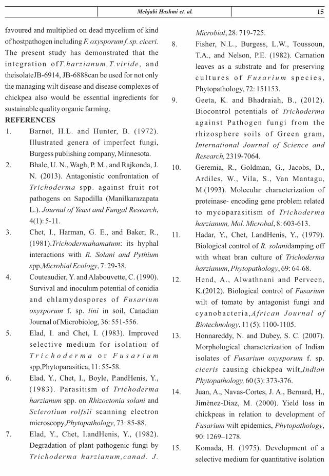
pathogen (Fusarium). Maximum growth inhibition
of pathogen observed with T.harzianumisolate. A
clear zone of inhibition was formed in all
Trichoderma pathogens interactions. Differential
action of the biocontrol agents was noticed on
mycelia growth of the Fusarium oxysporum f spp.
Ciceris(figure). Among the four isolates of
Trichoderma spp., maximum percentage of
i nh ib i t i on (50 .11%) was r eco rded wi th
T.harzianumfollowed by T.viride(44.97%) and JB-
6914 (38.75%) whereas the isolate JB-6888
(12.72%) was recorded with least effective in
parasitisation of mycelia growth of pathogen as
tabulated in Table 1.1 and figure1(a), 1(b) and 1(c).
It may be due to variable toxicity produced by all the
selected Trichoderma spp. attributed towards
combating the pathogen F. oxyxporum f. sp. ciceris.
In dual culture, particularly at the site of interaction
zone, Trichoderma spp. having multifarious action
against pathogen in which they would suppress to
the disease-causing microbe by coiling and
mycoparasite nature, releasing high toxin in
substrate where both are having sprace for growth.
In addition to above nature of Trichoderma spp., it is
also having antibiosis and lysis nature in presence of
antibiotics and enzymes (Chitinases, and
glucanases) respectively. In support of present
findings of dual culture test, a handsome amount of
work is available as review of literature, out of
which, Chet and Baker, (1981) studied very
extensively on Trichoderma spp. interacts with plant
pathogens in a variety of ways. The initial detectable
analysis by adopting appropriate method of analysis
of variance as described by Fisher (1958). Thedata
pertaining to weed population recorded at 20, 40, 60
DAS and harvest were subjected to Log (X+1) and
transformations as per requirement for
statistical analysis. Wherever, variance ratio
(calculated 'F' values) was found significant, critical
difference (C.D.) values were computed by
following formula for making comparisons between
the treatments:
where,
r : The number of replication,
V : mean sum of squares (MSE) and e
t : tabulated value of 't' at 5% level of
significance
The data have been presented in the form of
summary tables with mean values of the characters
and the C.D. at 5% level of probability. Suitable
graphical illustrations of the data have also been
given at appropriate places in the text. The analysis
of variance tables has been given in appendices.
The skeleton of analysis is given in Table 1.0
Table - 1.0 : Skeleton of ANOVA for the design of
the experiment
RESULTS AND DISCUSSIONS
In the present study, four isolates of
Trichoderma were evaluated against Fusarium
x + 0.5
�
t 2
r
V C.D.
e
xx=
S.N.
Source of Variation
D.F.
SS MSS FCal FTab
1.
Replication
2
2. Treatment 13
3. Errors 26 Ve
Total 41

14 Journal of Natural Resource and Development
Plate 1(a): Antagonistic potential of Trichoderma
spp. against F. oxysporum f. spp. ciceris (after 3
day).
Plate 1(b): Antagonistic potential of Trichoderma
spp. against Fusarium oxysporum f. spp. ciceris
(after 6 day).
Plate 1(c): Antagonistic potential of Trichoderma
spp. against Fusarium oxysporum f. spp. ciceris
(after 9 day).
Effect if antagonistic fungi on radial growth
inhibition of Fusarium oxysporumf. sp. cicerisin
dual culture test.
CONCLUSION
The nature of competition, Trichoderma is
interaction shows that the hyphae of the
mycoparasite grow directly towards the host by a
chemotrophic reaction. When the mycoparasite
reaches the host, its hyphae coils around it and
penetrates into the host mycelium by partial
degradation of its cell wall (Eladet al., 1983). It
appears that the main mechanism involved in the
antagonism to pathogenic fungi by Trichoderma
spp. is the release of lytic enzymes. The production
of extracellular β-1, 3 glucanases, chitinases (Eladet
al., 1982 & 1984) and protinase (Geremiaet al.,
1993) increased significantly when Trichoderma is
grown in the medium supplemented with either
autoclaved mycelium or fungal cell walls. These
enzymes play an important role in the destruction of
the pathogens (Hadaret al., 1979). Similarly,
Papavizas , (1985) , a p ioneer worker on
Trichoderma spp., also poses another characteristic
fiture as the lytic activity of several strains of
Trichoderma spp. on cell walls of phytopathogenic
fungi was correlated with the degree of biological
control of these pathogens in vitro.
Table - 1.1 : Effect of antagonistic fungi on radial
growth inhibition of Fusarium oxysporumin dual
culture test.
Treatment of fungal bio-control agent
Radial growth inhibition (%) of Fusarium oxysporum f. spciceris
Mean
3 Days 6 Days 9 Days
T-1 T.harzianum 31.98
(22.94)
37.28
(36.06)
50.11
(44.95)
39.79
(34.65)
T-2 T.viride 18.64
(23.28)
40.85
(39.73)
44.97
(42.06)
34.82
(35.02)
T-3
JB-6888
25.02
(29.94)
17.91
(17.91)
12.72
(16.74)
18.55
(21.53)
T-4
JB-6914
7.06
(14.64)
37.61
(37.61)
38.75
(38.42)
27.80
(30.22)
S.Em±
1.41
7.03 5.99
CD@5% 13.64 22.15 18.87
15 et. al.Mehjabi Hashmi
Microbial, 28: 719-725.
8. Fisher, N.L., Burgess, L.W., Toussoun,
T.A., and Nelson, P.E. (1982). Carnation
leaves as a substrate and for preserving
c u l t u r e s o f F u s a r i u m s p e c i e s ,
Phytopathology, 72: 151153.
9. Geeta, K. and Bhadraiah, B., (2012).
Biocontrol potentials of Trichoderma
aga in s t Pa thogen fung i f r om the
rh izosphere so i l s o f Green gram,
International Journal of Science and
Research, 2319-7064.
10. Geremia, R., Goldman, G., Jacobs, D.,
Ardiles, W., Vila, S., Van Mantagu,
M.(1993). Molecular characterization of
proteinase- encoding gene problem related
to mycoparasit ism of Trichoderma
harzianum, Mol. Microbal, 8: 603-613.
11. Hadar, Y., Chet, I.andHenis, Y., (1979).
Biological control of R. solanidamping off
with wheat bran culture of Trichoderma
harzianum, Phytopathology, 69: 64-68.
12. Hend, A., Alwathnani and Perveen,
K.(2012). Biological control of Fusarium
wilt of tomato by antagonist fungi and
cyanobac te r i a ,Afr ican Journa l o f
Biotechnology, 11 (5): 1100-1105.
13. Honnareddy, N. and Dubey, S. C. (2007).
Morphological characterization of Indian
isolates of Fusarium oxysporum f. sp.
ciceris causing chickpea wilt,Indian
Phytopathology, 60 (3): 373-376.
14. Juan, A., Navas-Cortes, J. A., Bernard, H.,
Jimènez-Diaz, M. (2000). Yield loss in
chickpeas in relation to development of
Fusarium wilt epidemics, Phytopathology,
90: 1269–1278.
15. Komada, H. (1975). Development of a
selective medium for quantitative isolation
favoured and multiplied on dead mycelium of kind
of hostpathogen including F. oxysporum f. sp. ciceri.
The present study has demonstrated that the
in teg ra t ion o fT.harz ianum,T.v i r ide , and
theisolateJB-6914, JB-6888can be used for not only
the managing wilt disease and disease complexes of
chickpea also would be essential ingredients for
sustainable quality organic farming.
REFERENCES
1. Barnet, H.L. and Hunter, B. (1972).
Illustrated genera of imperfect fungi,
Burgess publishing company, Minnesota.
2. Bhale, U. N., Wagh, P. M., and Rajkonda, J.
N. (2013). Antagonistic confrontation of
Trichoderma spp. against fruit rot
pathogens on Sapodilla (Manilkarazapata
L.). Journal of Yeast and Fungal Research,
4(1): 5-11.
3. Chet, I., Harman, G. E., and Baker, R.,
(1981).Trichodermahamatum: its hyphal
interactions with R. Solani and Pythium
spp,Microbial Ecology, 7: 29-38.
4. Couteaudier, Y. and Alabouvette, C. (1990).
Survival and inoculum potential of conidia
and ch lamydospores o f Fusar ium
oxysporum f. sp. lini in soil, Canadian
Journal of Microbiolog, 36: 551-556.
5. Elad, I. and Chet, I. (1983). Improved
select ive medium for isola t ion of
T r i c h o d e r m a o r F u s a r i u m
spp,Phytoparasitica, 11: 55-58.
6. Elad, Y., Chet, I., Boyle, P.andHenis, Y.,
(1983). Parasitism of Trichoderma
harzianum spp. on Rhizoctonia solani and
Sclerotium rolfsii scanning electron
microscopy,Phytopathology, 73: 85-88.
7. Elad, Y., Chet, I.andHenis, Y., (1982).
Degradation of plant pathogenic fungi by
Trichoderma harz ianum,canad. J .

16 Journal of Natural Resource and Development
22. Saxena, M.C. and Singh, K.B. (1987). The
chickpea published by C.A.B. Int.
ICARDA. 250-252.
23. Singh, R. S. and Alabouvette, C. (2007).
Antagonistic activity of selected isolates of
fluorescent Pseudomonas against Fusarium
oxysporum f. sp. Ciceris, Asian Journal of
Plant Sciences, 6 (3): 446-454.
24. Snyder, W.C. and Hansen, H.N. (1940). The
species concept in Fusarium. Journal of
Botany, 27: 64-67.
25. Sumitra P. K. (2006). Studies on Fusarium
oxysporumSchlecht Fr f. sp. gladioli
(Massey) Snyd. and Hans. causing wilt of
gladiolus. Ph.D Thesis submitted to the
University of Agricultural Sciences,
Dharwad.
26. VishwadharGurha, S. N. (1998). Integrated
Management of chickpea diseases.
Chamola and Dubey, O.P. (eds.) ABH
Publishing Co., New Delhi (India). p. 249.
of Fusarium oxysporumfrom natural
soil,Plant Protection Research, 8: 114-125.
16. Leslie, J.F. and Summerell, B.A. (2006).
The Fusarium Laboratory manual.
17. Muhammad Ansar Ahmad. (2010) .
Variability in Fusarium oxysporumf. sp.
ciceris for chickpea wilt resistance in
Pakistan. Ph.D Thesis submitted to the
Quaid-i-Azam University, Islamabad,
Pakistan.
18. Pandey, K. K., and Upadhyay, J. P. (2000).
Microbial population from rhizosphere and
non- rhizosphere soil of pigeonpea:
screening for resident antagonist and mode
of mycoparasitism,Journal of Mycology
and Plant Pathology, 30: 7-10.
19. Papavizas, G. C., (1985). Trichoderma and
Gliocladium: biology, ecology, and
potential for biocontrol,Annual Review of
Phytopathology. 23: 23-54.
20. Rangaswamy, G. and Mahadevan, A.
(1999). Diseases of crop plants in India (4th
edition) Prentice Hall of India Pvt. Ltd.,
New Delhi, pp. 607.
21. Rini, C.R. and Sulochana, K.K. (2007).
U s e f u l n e s s o f Tr i c h o d e r m a a n d
Pseudomonas against Rhizoctonia solani
and Fusarium oxysporuminfectingtomato,
Journal of Tropical Agriculture, 45 (1-2):
21– 28.

ABSTRACT
Black Gram (Vigna mungo) is a tropical, edible and leguminous plant belongs to the sub genus
Ceratotropis of the genus Vigna. Black gram is considered to have been domesticated in India from its
wild ancestral form V. mungo var silvestris. It is grown in various agro-ecological conditions and
cropping system with diverse agricultural practice. It is cultivated in a large groups compared to rice –
cultivation in India .It considered as a protein rich pulses.
A highly reproducible regeneration system through induction of somatic embryogenesis from
the 7 days old seedlings ( invitro germinated seeds) of black gram leaves were developed. The
regeneration of plants via somatic embryogenesis liquid shake culture of embryogenic calluses was
achieved in Vigna mungo (L.) Hepper (blackgram). The production of embryogenic callus was induced
by seeding primary leaf explants of V. mungo onto Murashige and Skoog (MS) medium supplemented
(optimally) with different concentration of /l 2,4-dichlorophenoxyacetic acid. The embryogenic callus
was then transferred to liquid MS medium supplemented (optimally) with different concentration of l
2, 4-dichloro-phenoxyacetic acid. Globular, heart-shaped, and torpedo-shaped embryos developed in
liquid culture.
Keywords : Blackgram, seed, somatic, esmbryogenesis.
HIGH FREQUENCY INDUCTION OF SOMATIC EMBRYOGENESIS
AND PLANT REGENERATION FROM SEEDLING EXPLANTS
OF BLACK GRAM (L) HEPPER
Priya Srivastava
Department of Biotechnology
Kulbhaskar Ashram PG College, Prayagraj, (U.P.), India
(Affilated to Prof Rajendra Singh (Rjju Bhaiya) University),
Received : 19.06.2020 Accepted : 25.07.2020
INTRODUCTION
The present study was undertaken to
establish an efficient and reproducible regeneration
system for black gram (Vigna mungo L.), an
important tropical grain legume rich in phosphoric
acid.
The seeds have 60% carbohydrate, 24%
protein and 1.3% fat on dry weight basis. Besides its
utility for human consumption, it also serves as a
nutritive fodder for milch cattle. The crop is used as
green manure and its deep root system binds soil
particles preventing erosion of the soil.
Journal of Natural Resource and Development 15 (2) 17-30, 2020 ISSN-0974-5033NAAS RATING : 3.46

18 Journal of Natural Resource and Development
Somatic embryogenesis is the direct way to
regenerate plant from single somatic cell and opens
up possibility to understand process of cell cycle
reprogramming from somatic to embryogenic type,
cloning and characterization of genes involved in
wounding, hormone activation, cell division,
differentiation and developmental processes. This
process also reproduced artificially by the
manipulation of tissue and cell in vitro.
According to the study of Feher (2006),
somatic embryogenesis may therefore occur if the
genes responsible for the embryogenic development
program are released from chromatin- mediated
gene silencing in vegetative cells. This may happen
in response to strong aspecific signal, such as high
auxin dose and/ sub lethal stress which evoke the
activation of large chromatin regions. Their
hypothesis had explained why less differentiated
cells (e. g. immature embryos) are more amenable
for somatic embryogenesis and why various
aspecific signals can evoke similar embryogenic
response. Regeneration via direct somatic
embryogenesis in liquid and solid media for M.
truncatula also has been established (Iantcheva A et
al., 1999; Iantcheva A et al., 2001; Iantcheva A et al.,
2005). Somatic embryogenesis in the genus
Selenium has been described for S. candallii
(Mathur, 1991). An efficient and reproducible
protocol for embryo formation and synthetic seed
formation in S. tenuifalium plant was developed by
using mature leaf tissue in presence of various conc.
of 2, 4-D and NAA (Meena Joshi et al., 2006). An
efficient and reproducible plant regeneration system
through somatic embryogenesis was established in
cassava by using somatic tissues, by which somatic
embryos were developed directly from shoot tips
and immature leaves on a medium containing 4-16
mg/ l 2, 4-D by Laszlo Szabados et al., 1987.
Somatic embryos from immature cotyledon
Black gram [Vigna mungo (L.) Hepper] is
an important leguminous source of protein for a
large segment of the vegetarian population in the
developing countries of Asia. The seeds of black
gram contain 78–80% nitrogen in the form of
albumin and globulin (Das et al ), and the dry . 1998
seeds are also a good source of phosphorus. Severe
yield losses in black gram crops, caused by a high
incidence of viral diseases and fungal pathogens
(Sahoo et al. 2002), have spurred research into the
development of disease-resistant cultivars by
genetic transformation. The first stage in transgenic
crop production is the definition of good in vitro
methods for shoot regeneration.
Because of its nutritional value, cooking
quality and easy digestibility, the demand for this
crop has been steadily increasing in the Indian
subcontinent, making breeders more and more
conscious about the urgent necessity to step up its
production. The foremost problem of Black gram is
its low yield. The factors contributing to its low yield
may be summed up as follows- narrow genetic base,
susceptibility to several diseases and pest, a year to
year fluctuation in their productivity. Cultivated
extent and production of black gram vary from year
to year with a decreasing trend consequently, the
production is not sufficient for the demand. Somatic
embryogenesis can be suitable option for
developing an asexual form of plant propagation
method in nature which inhibits many factors of
sexual reproduction. Somatic embryogenesis means
to produce embryo by somatic cells. Somatic
embryos are formed from plant cells that are not
normally involved in the development of embryos,
i.e. ordinary plant tissue. The establishment of
embryogenic suspension cultures for the
regeneration of plants is an ideal tool for the efficient
in vitro selection and production of transgenic plants
(Finer and McMullen, 1991; Christou, 1997).

19Priya Srivastava
sterile distilled water, seeds were germinated on MS
medium (Murashige and Skoog, 1962) containing
3.0% sucrose (w/v) and 0.8% agar (w/v) (Hi-media o o
Co., Mumbai, India) at 25 to 28 C in the dark for the
first 2 days and then transferred to a 16 hours
photoperiod of cool-white fluorescent light (120 -2 -1µmol m s ). The pH of all the media was adjusted to
o5.8 prior to autoclaving at 121 C temperature for 20
min.
Cal lus induct ion and maintenance o f
Embryogenic calli.
Primary leaves were excised from 7 days
old seedlings, cut into small segments and cultured
on 10 ml MS medium with 3% sucrose, 0.8% agar,
a n d d i f f e r e n t c o n c e n t r a t i o n s o f 2 , 4 -
dichlorophenoxyaceticacid acid in thrice set-up as
follows- 2, 4-D- (0.0, 0.3, 2.3, 4.3, 6.3, 8.3, 10.3,
12.3, 14.3, 16.3, 20.0 µM) for embryogenic callus
induction. The culture tubes were capped with
sterilized cotton plugs. The cultures were incubated o o
at 25 C to 28 C temperature under a dark condition
for 24 hour then kept in 16 hours light/ 8 hours dark
photoperiod (Haque et al., 2009) with a light -2 -1
intensity of 120 µmol m s . The callusing was
started after 12 days of inoculation and the pattern of
the growth of callus was observed by measuring the
diameter and % growth of the callus after every 15
days. This experiment was conducted in 3 replicates
in multiple of 3 of each 2, 4-D concentration
containing tubes. Callus growth and nature of calli
produced in each concentration is mentioned in
Table 1.1 and percentage of growth is indicated in
Table- 1.2 and graph 1.0 whereas the observations of
the effect of 2, 4-D concentration based upon the
diameter of calli producing is mentioned in Table-
1.3. The different form of regenerative calli,
proliferartive calli producing shoot buds,
regeneration of plantlet and regeneration of plant
have been mentioned in Figure- 1.0, 1.1 and 1.2.
explants of Vigna mungo (L). have been reported,
which however, failed to form well developed
plantlets (Eapen and George 1990). Gyorgyey et al. ,
(1991) had established a liquid culture system for
mass production of somatic embryos of alfalfa
(Medicago sativa) after initiating the embryos from
callus on 2, 4-di chlorophenoxyacetic acid- (2, 4-D)
containing semisolid medium. Similarly, Denchev
et al. (1991) have described conditions for
establishment of an embryogenic system based on
liquid medium in Medicago sativa, Medicago
falcata, and Medicago trautwetery. Repetitive
somatic embryogenesis of peanut in liquid medium
has been studied by Durham and Parrott (1992). It
was studied that the use of 2, 4- D alone or in combination with other hormones has become
almost routine and used successfully in inducing
somatic embryogenesisin seed cultures (Huang and Yeoman, 1984; Mordhors t e t a l . , 1998) .
Embryogenic suspension cultures have been
established in only a few grain legumes- Vigna
unguiculata (Kulothungan et al., 1995), Cajanus
cajan (Anbazhagan and Ganapathi, 1999).
Ontogeny of somatic embryo development has been
studied only in a few legumes, i.e. Vigna species
(Girija et al., 2000; Premanand et al., 2000), Glycine
(Phillips and Collins 1981; Samoylov et al., 1998a,
b), Arachis hypogaea (Ammirato, 1983; Eapen and
George 1993), and Phaseolus (Martins and Sondahl,
1984; Kumar et al., 1988).
MATERIALS AND METHODS
Collection of Plant material
Seeds of black gram (LBG- 645) were
obtained from the Indian Pulse Research Institute,
Kanpur, U. P., India. Seeds were washed under tap
water in presence of Tween – 20 and then disinfected
with serial immersion in 2% sodium hypochlorite
for 5 min, 70% ethanol (v/v) for 2 min, and 0.1%
HgCl (w/v) for 10 min. After three rinses with 2

20 Journal of Natural Resource and Development
were established in soil and grown to maturity in a
plant growth chamber under a 16 hours photoperiod o oat 25 to 28 C.
Statistical analysis
All the experiments were repeated three
t imes and data on growth percentage of
embryogenic calli and diameter of calli produced
(mm) were statistically analyzed by set up in (CRD)
completely randomized design (Appendix 1). The
effect of different concentration of 2, 4-D was
quantified and the level of significance was
determined by analysis of variance F- value at the
5%.
RESULTS AND DISCUSSION
Callus induction
Primary leaf explants from seven days old
seedlings produced greenish white friable calli on 2,
4-D containing medium within 10–12 days of
culture. The maximum proliferation and nature of
calli was obtained on 6.3 µM and 12.3 2, 4-D, while
minimal response was noted at 0.3 µM (Table 1.1).
Cell suspension culture and embryogenesis
Two-week-old leaf derived greenish white friable
calluses were sub cultured in liquid MS medium
containing different concentrations of 2, 4-D. After
10-15 days of culture on MS medium supplemented
with 6.0 µM 2, 4-D, cell division and proliferation
was observed. The cultures became thick,
mucilaginous, and brown in color after culture for 12
days in the same medium; therefore, it was
necessary to transfer the cells to fresh medium at
weekly intervals. Two weeks after initiation of
suspension culture, cells differentiated to form
somatic embryos. Microscopic observation of
suspension cultures showed that initial spherical
cells were embryogenic, containing visible dense
cytoplasm. These spherical cells were embryogenic
and divided transversely resulting into two, four,
and subsequently to a group of cells, that was
Maintenance of suspension culture and somatic
embryogenesis
Two-week-old, greenish white, friable
calluses (approximately 150 mg fresh mass) derived
from leaf segments were aseptically transferred to a
250 ml flask containing 30 ml of liquid MS medium
supplemented with 6.3 and 12.3 µM 2, 4-D. Cultures o
were agitated on a gyratory shaker at 130 rpm, 25 to o28 C, under a 16 hours light / 8 hours dark photo
-2 -1 period of 120 µmol m s light intensity. A 15 ml
aliquot of the cell suspension was replaced with
fresh medium at 7 days intervals. Cell suspension
culture allows rapid division of cells and increases
the rate of the growth. Homogeneous cell
suspension was formed after 1 month.
Differentiation of Embryogenic callus
Cell suspension cultures were observed
under a microscope during the culture period.
Embryos were sub cultured in liquid MS medium
containing different concentrations of 2, 4- D. After
20 days of culture, torpedo-shaped embryos were
transferred to full-strength MS liquid medium, MS
supplemented with 6.3 and 12.3 µM 2, 4-D for
maturation and germination. The germinated
embryos were transferred to agar-solidified MS
basal medium for further growth and development.
The frequency of embryo induction and different
stages of somatic embryos were observed.
Transplantation
The plantlets that developed from
germinated embryos on solid MS medium were
transferred to plastic pots containing vermiculite,
sand, and red soil mixture (1:1:1). Each pot was
covered with a polythene bag to ensure high
humidity for the initial 15 days, and then the
humidity was gradually reduced by making holes in
the polythene bags to harden the plants. The
hardened plantlets were nourished with half-
strength MS nutrient solution. The hardened plants

21Priya Srivastava
Table 1.1 and Graph 1.0. These results were
analyzed statistically by using CRD (Complete
randomized design) analysis. After calculating the
ANOVA table the F-value (Appendix I) was found
to be 6.273 which indicates the significance at the
tabulated value (5%) of F with a C.D. (critical
difference) (5%) of 25.828 (F >5%). The influence 6.27
of different concentration of 2, 4-D depending upon
the size of calli produced was also studied by using a
CRD test (Table-1.2 and Appendix- I) and analysis
of variance (5%). The highest mean of diameter of
embryogenic calli – 2.43 mm with a S. Er. (+) 0.07
was found at the concentration of 6.0 µM and the
minimal mean of size of calli was found 0.77 mm
with S. Er. (+) 0.5 at the 1.5 µM concentration of 2,
4- D. After calculating the ANOVA table, F- value
(Appendix II) was found to be significant- 6.4570,
which is greater than F- table value- 2.32 (5%) with a
C. D. of 0.77 at 5%.
The choice of initial explant is a critical
factor for embryogenic callus induction and
initiation. In the majority of legumes, immature
zygotic embryos, young cotyledons, or vegetative
shoot apices have been the most responsive explants
for the induction of somatic embryogenesis
(Hardwick et al., 1988). In the present study, leaf
segments were found to produce somatic embryos.
The acquisition of embryogenic potential under
auxin stimulus in such explants is manifested
through a callus phase. Among different auxin
tested, 2, 4-D at 6.0 µM was most effective for
inducing somatic embryogenesis in a liquid
medium. NAA failed to induce somatic
embryogenesis (Figure- 1.2), indicating that leaf
segments have different sensitivity to various auxin
and their concentration. In Vigna species, Full-
strength MS medium was found to be more effective
than the other media used for induction and growth
of somatic embryos. This may be due to the presence
considered to be the pro-embryo. The pro-embryo
further divided and formed globular (Figure. 1.2- a),
heart (Figure. 1.2- b), and torpedo- staged (Figure.
1.2- c) embryos. The torpedo shaped embryos
recallused on 2, 4-D -containing medium. Heart and
torpedo stages were transferred to fresh liquid
medium containing 3% sucrose, for complete
maturation. The differentiation of the embryogenic
callus into different stages was examined under
stereo microscope to identify the different stages of
it (Figure. 1.2- a, b, c).
Germination of the embryos and transplantation
After transfer of torpedo and stage embryos
from MS liquid to solid medium, the embryos
germinated into tiny plantlets [Figure. 1.1- (A1-A6)
and Figure. 1.2- (i, j)] within the same medium.
Media optimization
The effect of different concentrations of 2,
4-D (0.5-72.0 µM) in liquid MS medium was
assessed on induction of somatic embryogenesis. It
was observed that the frequency of somatic
embryogenesis increased with an increase in the
concentration of 2, 4-D from 0.5 to 6.0 µM (Table
1.2 and graph.1.0). Further increase in 2, 4-D
concentration resulted in a decrease in embryogenic
calli production and recallusing of embryos. Calli
were not obtained in MS medium containing NAA
(Figure- 1.2). The various concentrations of plant
growth regulators (NAA, and 2, 4 –D) were tested in
callus induction and plant regenerations.
Observations based on growth percentage and size
of calli forming embryo were collected. Mean of
growth percentage was found to be increased 86.67
with an S. Er. (+) of 6.67 at conc. of 6.0 µM 2, 4 D
whereas a decrease in mean growth % i. e 13.33
with an S. Er. (+) of 33.0 was noted at 0.5 µM of 2,
4-D. It is observed that the highest growth
percentage of somatic embryo was produced in MS
media supplemented with 6.0 µM 2, 4-D as shown in
22 Journal of Natural Resource and Development
2002). This work also showed that itwas possible to
obtain cell lines with continued embryogenic
potential if early-stage somatic embryos were
maintained on a solid medium with an increased concentration of the auxin; this observation is in
accordance to the protocol developedby Ikeda-Iwai
et al. (2002). In the present study, a protocol for
somat ic embryogenes i s was es tab l i shed
successfully and found to be reproducible and
developed by using different concentration of 2, 4-D
in increasing order from 0.5 – 72.0 µM
concentration in which the best proliferation and
embryo formation was observed at 6.0 µM. These
results are in accordance to finding of in vitro
regeneration of plant via somatic embryogenesis
through cell suspension culture achieved in horse
gram (S. Varisai Mohamed et al. 2004) by addition
of different concentration of 2, 4- D.
Induction of callus in plants is affected by many
factors, like explants, PGRs (Plant growth
regulators) and culture conditions. Among them,
PGRs play a very key role. Furthermore, different
concentrations and combinations of PGRs have
significant effects on callus induction, which has
been reported in many researches (Poeaim et al.,
2005; Sun et al., 2006). Friable callus, developed
from leaf and internode explants grown on
Murashige and Skoog (MS) medium supplemented
with 2, 4-dichlorophenoxyacetic acid (2, 4-D),
underwent somatic embryogenesis has been
reported in Ceropegia candelabrum L. (Beena and
Martin, 2003).
The results of this study revealed that 2, 4-D
was the most important PGR in callus induction,
followed by NAA. The frequency of embryogenic
callus formation had a dramatic drop with the
concentration of 2, 4-D rising and reached highest
when 2, 4-D was lowest. From this study, It was
observed that low concentration of 2, 4-D is helpful
of a high level of nitrogen, particularly the reduced
form (NH PO ), in MS medium. The use of the 4 4
synthetic auxin 2, 4-dichlorophenoxyacetic acid (2,
4-D) for the induction of somatic embryos (embryoids) oncultured explants can be traced to the
work of Halperin and Wetherell (1964) who showed
that a callus produced from any vegetative part of
carrot (Daucus carota) such as the root, petiole, or inflorescence stalk reared in a medium containinga
high concentration of 2, 4-D formed somatic
embryos upon transfer to a medium with a reduced level of the auxin. From this timeonwards, the use of
a defined medium and a single-step transfer of a
callus or a cell suspension growing in a medium supplementedwith a moderate quantity of 2, 4-D to
one containing a reduced amount of the auxin or
none at all, was adopted as the standard protocol to study the somatic embryogenesis in carrot and
became widely popular in inducing somatic
embryogenesis in a broad range of species (Thorpe
and Stasolla, 2001; Raghavan,2004 (a)). The role of
2, 4- D by continuous exposure for successful
induction of somatic embryogenesis was described
by Raghavan, 2004 (b). It was observed that using 2,
4-D as the sole hormone, heart-shaped embryos
were initially cultured in a liquid medium containing
6.0 µM 2, 4-D for 21 days to induce the formation of early-stage somatic embryos and shoot buds
followed by their transfer to an auxin- containing
medium for plantlet formation and regeneration of
plant as shown in Figure- 1.6 and 2.0. It was
observed that using 2, 4-D as the sole hormone, calli
were initially cultured in a liquid medium containing
6.0 µM 2, 4-D for 21 days used to induce the
formation of proliferating calli. In the present study
it was observed that some types of embryos
transferred for germination had also produced callus
(dedifferentiation). This result is found in live with
the report on Laptadenia reticulata (Hariharan et al.

23Priya Srivastava
showed poor differentiation similar to the present
finding. Harvey and Grasham (1969), while
working on 12 species of conifers also reported
species specificity for IAA, NAA and 2, 4-D in their
effectiveness for callus induction. Callus production
from cotyledon explants of Juglans nigra (Neuman
et al., 1993) and seedling explants of Albizzia
procera (Datta, 1987), all of which are woody trees
have also been reported. In this study, callus
production was successful in the presence of 2, 4-D
alone. This is in contrast to the observation by Xie
and Hong (2001) in Acacia mangium where calli
were reportedly induced from cotyledon explants of
mature zygotic embryos in MS basal medium
supplemented with both 2,4-D and Kinetin. The
choice of suspension culture was informed by the
work of Martin (2003), who reported the
development of higher number of somatic embryos
in suspension cultures than in solid medium
cultures. Callus production followed by somatic
embryogenesis has also been reported in cotyledon
explants of Juglans nigra (Neuman et al., 1993), leaf
explants of Holostemma ada-kodien (Martin, 2003)
as well as in stem petioles and leaflet explants of
Swainsona formosa (Sudhersan and Abo El-Nil,
2002).
A highly reproducible regeneration system
through somatic embryogenesis from the excised
mature embryos of dry seeds of a range of European
barley cultivars (Hordeum vulgare L.) was
developed by minimizing the induction of primary
callus and following influences like - the ratio of
carbon source and 2, 4-D in the induction medium,
soaking of seeds in water containing 2, 4-D solution
and direct culture of excised embryonic axes
(Sharma et al. 2005). The role of 2, 4-D was also
studied by Pitipong Thobunluepop, 2009 on the in
vitro evaluation and optimization of medium for
somatic embryogenesis, synthetic seed production
for embryogenic callus formation from leaf explant
whereas a successful somatic embryo was
developed from the roots of Panax ginseng by
Chang and Hsing, 1980 and Lycium barbarum by
Hu et al., 2008 as well as Wang found that high
concentration of 2, 4-D showed a promoting effect
in Areca catechu (Wang et al., 2006). In H.
brasiliensis, embryogenic callus from the pollen
(Chen et al., 1979) and inner integument of the seed
(Carron, 1981) was induced by high concentrations
of 2, 4-D. The results of present study was found to
be opposite as compared to those mentioned above.
It may be due to different explants used.
A study has been done by Amoo and Ayisire
2005 to produce plantlet by induction of somatic
embryogenesis from cotyledon explants of Parkia
biglobosa (Jacq.) Benth.They observed the effect of
naphthalene acet ic ac id (NAA) or 2 , 4-
d ich lorophenoxyacet ic ac id (2 , 4-D) on
embryogenic callus induction and noticed that with
2, 4-D, response in the form of callus production was
observed only at the cut edges of the explants and on
the abaxial surface, even when placed face down.
Present result shows the finding that callus
formation in a few cases is affected among other
factors by orientation of the explants on the culture
medium (Warren, 1991). This report agrees with the
findings of Morini et al. (2000) in which they
observed that callus formation occurred only on the
abaxial surface of Cydonia oblonga leaf, which had
been placed face up. Rita and Floh (1995) reported
similar observation with the leaf explants of Cuphea
ericoides. The fact that callus was induced by 2, 4-
D but not by NAA suggests that cotyledon
explants of Vigna mungo are auxin specific. Zafar
et al. (1995) also reported callus induction from
cotyledon, hypocotyl and root explants of Medicago
littoralis in the presence of 2, 4-D alone and when it
was replaced with NAA, the explants either died or
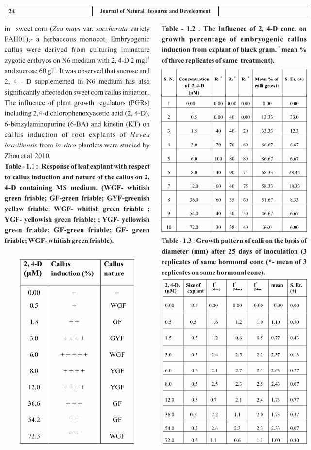
24 Journal of Natural Resource and Development
Table - 1.2 : The Influence of 2, 4-D conc. on
growth percentage of embryogenic callus (* induction from explant of black gram. mean %
of three replicates of same treatment).
Table - 1.3 : Growth pattern of calli on the basis of
diameter (mm) after 25 days of inoculation (3
replicates of same hormonal conc (*- mean of 3
replicates on same hormonal conc).
in sweet corn (Zea mays var. saccharata variety
FAH01),- a herbaceous monocot. Embryogenic
callus were derived from culturing immature -1
zygotic embryos on N6 medium with 2, 4-D 2 mgl -1and sucrose 60 gl . It was observed that sucrose and
2, 4 - D supplemented in N6 medium has also
significantly affected on sweet corn callus initiation.
The influence of plant growth regulators (PGRs)
including 2,4-dichlorophenoxyacetic acid (2, 4-D),
6-benzylaminopurine (6-BA) and kinetin (KT) on
callus induction of root explants of Hevea
brasiliensis from in vitro plantlets were studied by
Zhou et al. 2010.
Table - 1.1 : Response of leaf explant with respect
to callus induction and nature of the callus on 2,
4-D containing MS medium. (WGF- whitish
green friable; GF-green friable; GYF-greenish
yellow friable; WGF- whitish green friable ;
YGF- yellowish green friable; ; YGF- yellowish
green friable; GF-green friable; GF- green
friable; WGF- whitish green friable).
2, 4-D
(µM)Callus
induction (%)
Callus
nature
0.00
0.5
1.5
3.0
6.0
8.0
12.0
36.6
54.2
72.3
_
+
+ +
+ + + +
+ + + + +
+ + + +
+ + + +
+ + +
+ +
+ +
_
WGF
GF
GYF
WGF
YGF
YGF
GF
GF
WGF
S. N. Concentration
of 2, 4-D
(µM)
R1 * R2
* R3 * Mean % of
calli growth
S. Er. (+)
1 0.00 0.00 0.00 0.00 0.00 0.00
2 0.5 0.00 40 0.00 13.33 33.0
3 1.5 40 40 20 33.33 12.3
4 3.0 70
70
60
66.67
6.67
5 6.0 100
80
80
86.67
6.67
6 8.0 40
90
75
68.33
28.44
7 12.0 60
40
75
58.33
18.33
8 36.0 60
35
60
51.67
8.33
9 54.0 40
50
50
46.67
6.67
10 72.0 30
38
40
36.0
6.00
2, 4-D.(µM)
Size ofexplant
I*
(Mm.)I* (Mm.)
I*
(Mm.)
mean S. Er. (+)
0.00 0.5 0.00
0.00
0.00
0.00
0.00
0.5 0.5 1.6
1.2
1.0
1.10
0.50
1.5 0.5 1.2
0.6
0.5
0.77
0.43
3.0 0.5 2.4
2.5
2.2
2.37
0.13
6.0 0.5 2.1
2.7
2.5
2.43
0.27
8.0 0.5 2.5 2.3 2.5 2.43 0.07
12.0 0.5 0.7 2.1 2.4 1.73 0.77
36.0 0.5 2.2 1.1 2.0 1.73 0.37
54.0 0.5 2.4 2.3 2.3 2.33 0.07
72.0 0.5 1.1 0.6 1.3 1.00 0.30
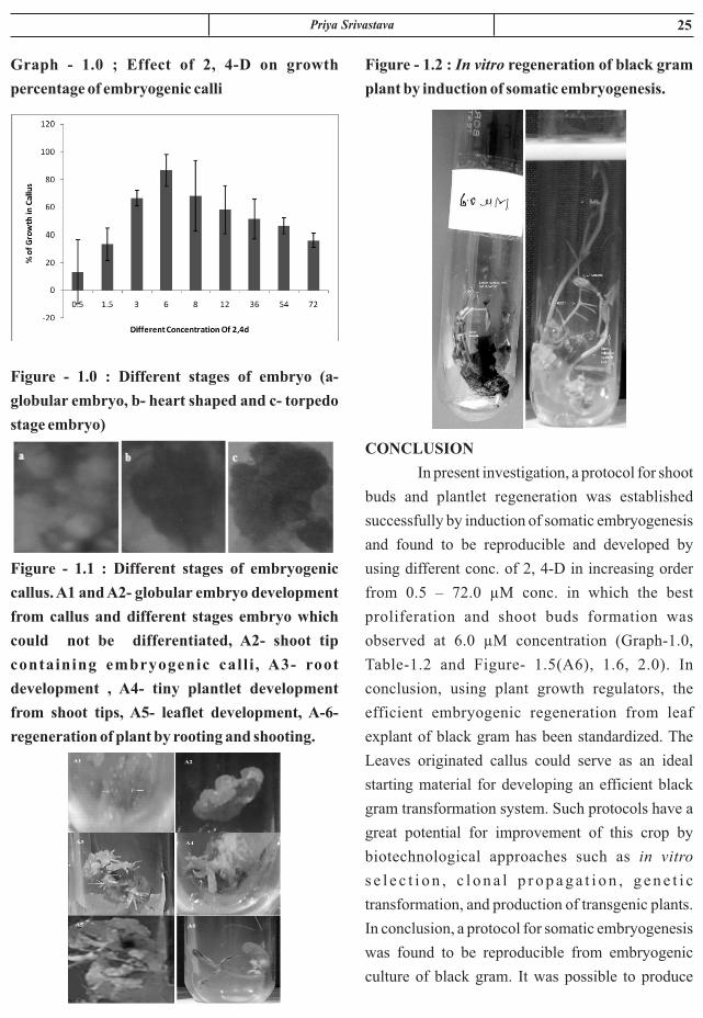
25Priya Srivastava
Figure - 1.2 : In vitro regeneration of black gram
plant by induction of somatic embryogenesis.
CONCLUSION
In present investigation, a protocol for shoot
buds and plantlet regeneration was established
successfully by induction of somatic embryogenesis
and found to be reproducible and developed by
using different conc. of 2, 4-D in increasing order
from 0.5 – 72.0 µM conc. in which the best
proliferation and shoot buds formation was
observed at 6.0 µM concentration (Graph-1.0,
Table-1.2 and Figure- 1.5(A6), 1.6, 2.0). In
conclusion, using plant growth regulators, the
efficient embryogenic regeneration from leaf
explant of black gram has been standardized. The
Leaves originated callus could serve as an ideal
starting material for developing an efficient black
gram transformation system. Such protocols have a
great potential for improvement of this crop by
biotechnological approaches such as in vitro
s e l e c t i o n , c l o n a l p r o p a g a t i o n , g e n e t i c
transformation, and production of transgenic plants.
In conclusion, a protocol for somatic embryogenesis
was found to be reproducible from embryogenic
culture of black gram. It was possible to produce
Graph - 1.0 ; Effect of 2, 4-D on growth
percentage of embryogenic calli
Figure - 1.0 : Different stages of embryo (a-
globular embryo, b- heart shaped and c- torpedo
stage embryo)
Figure - 1.1 : Different stages of embryogenic
callus. A1 and A2- globular embryo development
from callus and different stages embryo which
could not be differentiated, A2- shoot tip
containing embryogenic call i , A3- root
development , A4- tiny plantlet development
from shoot tips, A5- leaflet development, A-6-
regeneration of plant by rooting and shooting.

26 Journal of Natural Resource and Development
Zhang SJ, Xu XE, Ou XH, He YT, Lu Z M.,
1979. A process of obtaining pollen plants of
Hevea brasiliensis Muell. Argon. Sci. Sin.
22: 81-90.
8. Christou P., 1997. Biotechnology applied to
grain legumes. Field Crops Res. 53:83- 97.
9. Cooke T. J. Racusen R. H. and Cohen J. D.,
1993 . The ro l e o f aux in in p l an t
embryogenesis. The Plant Cell. 5: 1494-
1495.
10. Da t t a S K . , 1987 . Ti s sue cu l tu re
propagation of forest trees - limitations and
perspectives. In: PK Khosla and DK
Khurana (Eds.) Agroforesty for Rural
Needs, Vol. 1, Indian Soc. of Tree Scient. pp.
234-241.
11. Denchev P. D., Kuklin A. I., Velcheva M. R.,
Atanassov A., Kaimaktchiev A.C., 1991. A
new approach to large – scale micro
propagation in alfalfa. Biotechnol. Forum.
Eur. 8(3): 126-129.
12. De Vries S.C., Booij H., Meyerink P.,
Huiaman G., Wilde D. H., Thomas T. L.
and Van Kammen A., 1988. Acquisition of
embryogenic potential in carrot cell-
suspension cultures. Planta 176:196-204. a.
13. De Vries S.C., Booij H., Janssens R., Vogels
R., Saris L., Lo Schiavo F., Terzi M. &
Vankammen A., 1988. Carrotsomatic
e m b r y o g e n e s i s d e p e n d s o n t h e
phytohormone-controlled expression
ofcorrectly glycosylated extracellular
proteins. Genes & Development. 2: 462-
476. b.
14. Dudits D., Bögre L. and Györgyey J., 1991.
Molecular and cellular approaches to the
analysis of plant embryo development from
somatic cells in vitro. Journal of Cell
Science. 99: 475-484.
somatic embryos with in two months and to
regenerate plant from mature embryos in 2- 3
months in presence of 2, 4-D.
ACKNOWLEDGMENT
The author thankful to the DST-Integrated
Long Term Project, Government of India, New
Delhi for providing the funding opportunity and
IIPR, Kanpur, U.P., for providing the seeds of black
gram to accomplish this type of work.
REFERENCES
1. Ammirato P.V., 1983. The regulation of
somatic embryo development in plant cell
cultures: suspension culture techniques and
hormone requirements. Biotechnology 1:
68-74.
2. Amoo, Stephen Oluwaseun and Ayisire,
Benjamin Erhinmeyoma., 2005. Induction
of callus and somatic embryogenesis from
cotyledon explants of Parkia biglobosa
(Jacq.) Benth. Afr. Journal of Biotechnol. 4
(1): 68-71.
3. Anbazhagan V. R. and Ganapathi A., 1999.
Somatic embryogenesis in cell suspensions
of pigeonpea (Cajanus cajan L.). Plant Cell
Tiss. Organ Cult. 56: 179–184.
4. Beena M. R. and Martin K.P., 2003. In vitro
propagation of the rare medicinal plant
Ceropegia candelabrum L. through somatic
embryogenesis. In vitro Cell. Dev. Biol.
Plant. 39: 510-513.
5. Carron M P., 1981. Germination in vitro
d'embryons immatures d'hevea. Caoutch
Plast. 612: p. 93.
6. Chang WC, Hsing YI., 1980. Plant
r e g e n e r a t i o n t h r o u g h s o m a t i c
embryogenesis in root-derived callus of
Ginseng (Panax ginseng C. Meyer A).
Theor. Appl. Genet. 57: 133-135.
7. Chen Z H, Chen FT, Qian CF, Wang CH,

27Priya Srivastava
development in Citrus nucellar cell cultures.
Physiologia Plantarum. 82: 606- 616.
24. Gavish H., Vardi A. & Fluhr R., 1992.
Suppression of somatic embryogenesis in
Citrus cell cultures by extracellular
proteins. Planta, 186: 511-517.
25. Girija S., Ganapathi A., Ananthakrishnan
G., 2000. Somatic embryogenesis in Vigna
radiata L.Wilczek. Ind. J. Exp. Biol. 38:
1241–1244.
26. Grasham JL and Harvey A E., 1969.
Procedures and media for obtaining tissue
cultures of 12 conifer species. Can. J. Bot.
47: 547-549. 6: 80-82.
27. Gyorgyey J., Bogre L., Nemeth K., Bako L.,
Kapros T. Dudits D., 1991. Molecular
c h a n g e s d u r i n g s o m a t i c e m b r y o
development of alfalfa. Plant Biotechnol.
EC Hungary Workshop, 58.
28. Halperin W. D. F. Wetherell 1964.
Adventive embryony in tissue cultures of
the wild carrot, Daucus carota. American
Journal of Botany 51: 274-283.
29. Haque, R., S. Saha and T. Bera, 2009.
Micropropagat ion of an important
m e d i c i n a l p l a n t C h l o r o p h y t u m
borivilianum. Int. J. Ph. Sci., 1: 153-163.
30. Hariharan M., Sebastian D. P., Benjamin S.
a n d P r a s h y P. , 2 0 0 2 . S o m a t i c
embryogenesis in Leptadenia reticulata
Wieght and Arn. – A medicinal plant.
Phytomorphology. 52: 155-160.
31. Hartweck L. M., Lazzeri P. A., Cui D.,
Collins G. B., Williams E. G., 1988. Auxin-
o r i e n t a t i o n e f f e c t s o n s o m a t i c
embryogenesis from immature soybean
cotyledons. In Vitro Cell Dev. Biol. 24:
821–824.Huang B. B. M. M. Yeoman 1984.
Callus proliferation and morphogenesis in
15. Das D. K.; Siva Prakash N.; Bhalla-Sarin N.
An efficient regeneration system of black
g r a m ( Vi g n a m u n g o L . ) t h r o u g h
organogenesis. Plant Sci 134: 199–206;
1998. doi:10.1016/S0168-9452(98) 00044-
2.
16. Durham R. E. and Parrot W. A., 1992.
Repetitive somatic embryogenesis from pea
nut cultures in liquid medium. Plant Cell
Rep. 11: 122- 125.
17. Eapen S. and George L., 1990. Ontogeny of
somatic embryos of Vigna aconitifolia,
Vigna mungo and Vigna radiata. Ann. Bot.
66: 219–226.
18. Eapen S. and George L., 1993. Somatic
embryogenesis in peanut: influence of
growth regulators and sugars. Plant Cell
Tiss. Organ Cult. 35: 151–156.
19. Fehér A., 2006. Why Somatic Plant Cell
start to form Embryos? In Mujid A., Samaj J,
eds., Somatic Embryogenesis., Plant Cell
Monographs, vol. 2, Robinson DG, Series
ed., Springer- Verlog, Berlin Heidelberg,
Germany, pp. 85- 101.
20. Fehér A., Pasternak T., Dudits D., 2003.
Transition of somatic plant cell to an
embryogenic state. Plant Cell, Tissue,
Organ Cult. 74: 201-228.
21. Finer J. F. and McMullen D., 1991.
Transformation of soybean via particle
bombardment of embryogenic suspension
culture tissue. InVitro Cell. Dev. Biol. Plant.
27: 175–182.
22. Fischer C. and Neuhaus G., 1996.
Influence of auxin on the establishment of
bilateral symmetry in monocots. Plant
Journal. 9: 659- 669.
23. Gavish H.,Vardi A. & Fluhr R., 1991.
Extracellular proteins and early embryo

28 Journal of Natural Resource and Development
40. Kumar A. S., Gamborg O. L., Nabors M. W.,
1988. Regeneration from long- term cell
suspension cul tures of tepry bean
(Phaseolus acutifolius). Plant Cell Rep. 7:
322–325.
41. Laszlo Szabados., Rodrigo Hoyos and
Wiliam Roca., 1987. In vitro somatic
embryogenesis and plant regeneration of
cassava. Plant Cell Rep. 6(3): 248-251.
42. Liu C.-M., Xu Z. H. and Chua N.-H., 1993.
Auxin polar transport is essential for the
establishment of bilateral symmetry during
early plant embryogenesis. The Plant Cell.
5: 621-630.
43. Martins I. S., and Sondahl M. R., 1984.
E a r l y s t a g e s o f s o m a t i c e m b r y o
differentiation from callus cells of bean
(Phaseolus vulgaris L.) grown in liquid
medium. J. Plant Physiol. 117: 97–103.
44. Martin K P., 2003. Plant regeneration
through somatic embryogenesis on
Holostemma ada- kodien, a rare medicinal
plant. Plant Cell, Tissue and Organ cult. 72:
79-82.
45. Mathur J., 1991. Enhanced somatic
embryogenesis in Selinum candoli DC.
under a mineral oil overlay. Plant Cell,
Tissue and Organ Culture. 27: 23-26.
46. Meena Joshi., Uppeandra Dhar. and Sumit
Manjkhola., 2006. Somatic embryogenesis
and plantlet regeneration from encapsulated
embryos of Selinum tenuifolium. Journal of
Horticultural Science & Biotechnology.
81(4):570-574.
47. Michalczuk L., Ribnicky D. M., Cooke T. J.
and Cohen J. D., 1992. Regulation of
indole-3- acetic acid biosynthetic pathways
in carrot cell cultures. Plant Physiology.
100: 1346-1353.
tissue cultures of Arabidopsis thaliana L.
Plant Science Letters 33: 353-363.
32. Hu Z, Hu Y, Gao HH, Guan XQ, Zhuang
DH., 2008. Callus production, somatic
embryogenesis and plant regeneration of
Lycium barbarum root explants. Biol. Plant.
52: 93-96.
33. Ikeda-Iwai M. S. Satoh H. Kamada 2002
Establishment of a reproducible tissue
culture system for the induction of
Arabidopsis somatic embryos. Journal of
Experimental Botany 53: 1575-1580.
34. Ikeda- Iwai M., Umehara M., Satoh S.,
Kamada H., 2003. Stress induced somatic
embryogenesis in vegetative tissues of
Arabidopsis thaliana. Plant J. 34: 107- 114.
35. Iantcheva A., Bakalova E., Vlahova M.,
Kondorosi E., Elliott M., Atanassov A.
1999. Plant Cell Rep. 18: 904-910.
36. Iantcheva A., Vlahova M., Trinh T.H.,
Brown S., Slater A., Elliott M.C., Atanassov
A. 2001. Plant Sci., 160: 621-627.
37. Iantcheva A., Slavov S., Prinsen E.,
Vlahova M., van Onckelen H., Atanassov A.
2005. Plant Cell Tissue Organ Cult. 81: 37-
43.
38. Jiménez V., Thomas C., 2006. Participation
of Plant Hormones in Determination and
Progression of Somatic Embryogenesis in
Muj id A. , Samaj J , eds . , Somat ic
Embryogenesis., Plant Cell Monographs,
Vol. 2, Robinson DG, series ed., Springer-
Verlog, Berlin Heidelberg, Germany, pp.
103-118.
39. Kulothungan S., Ganapathi A., Shajahan A.,
K a t h i r a v a n K . , 1 9 9 5 . S o m a t i c
embryogenesis in cell suspension culture of
cowpea [Vigna unguiculata (L.) Walp.].
Israel J. Plant Sci. 43: 385–390.

29Priya Srivastava
123–129.
56. Pitipong Thobunluepop., 2009. The
somat ic embryogenes i s and p lan t
regeneration from immature embryo of
sweet corn inbred line. Journal of Plant
Breeding and Crop Science. 1(10): 330-335.
57. Poeaim A, Matsuda Y, Murata T., 2005.
Plant regenerat ion f rom immature
inflorescence of zoysiagrass (Zoysia spp.)
Plant Biotechnol. 22: 245-248.
58. Premanand R., Ganapathi, A., Anbazhagan
V. R., Vengadesen G., Selvaraj N., 2000.
High frequency plant regeneration via
somatic embryogenesis in cell suspension
cultures of cowpea [Vigna unguiculata (L.)
Walp]. In Vitro Cell. Dev. Biol. Plant 36:
475–480.
59. R a g h a v a n V . 2 0 0 4 . S o m a t i c
embryogenesis. In S. J. Murch and P. K.
Saxena [eds.], Journey of a single cell to
plant, 203–226. Oxford & IBH Publishing,
New Delhi, India. (a)
60. Raghavan V. 2004. Role of 2, 4- di
chlorophenoxyacetic acid (2, 4-D) in
Somatic Embryogenesis on cultured on
zygotic embryos of Arabidopsis: Cell
s u s p e n s i o n , C e l l c y c l i n g , a n d
morphogenesis during continuous exposure
of embryos to 2, 4-D. American J. of Botany.
91:1743-1756. (b)
61. Raemakers C. J. J. M., Jacobsen E. and
Visser R. G. F., 1995. Secondary somatic
embryogenesis and applications in plant
breeding. Euphytica. 81: 93-107
62. Rita I, Floh E I S., 1995. Tissue culture and
micro propagation of Cuphea ericoides, a
potential source of medium- chain fatty
acids. Plant Cell Tissue Organ Cult 40:187-
48. Mordhorst A. P. K. J. Voerman M. V. Hartog
E. A. Meijer J. van Went M. Koornneef S. C.
de Vries., 1998. Somatic embryogenesis in
Arabidopsis thaliana is facilitated by
mutations in genes repressing meristematic
cell divisions. Genetics 149: 549-563.
49. Morini S., D'Onofrio C., Bellocchi G.,
Fisichella M., 2000. Effect of 2, 4-D and
light quality on callus production and
differentiation from in vitro cultured quince
leaves. Plant Cell Tiss. and Org. Cult. 63:
45-55.
50. Murashige T. and Skoog F., 1962. A revised
medium for rapid growth and bioassays with
tobacco tissue cultures. Physiol. Plant. 15:
473–497.
51. Nawy T., Lukowitz W., Bayer M., 2008.
Talk global, act local- patterning the
Arabidopsis embryo. Curr. Opin. Plant Biol.
11: 28-33.
52. Neuman MC, Preece JE, Sambeek J W,
G a f f i n e y G R . , 1 9 9 3 . S o m a t i c
embryogenesis and callus production from
cotyledon explants of eastern black walnut
(Juglans nigra L.). Plant Cell Tissue Organ
Cult. 32: 9-18.
53. Nomura K. and Komamine A., 1985.
Identification and isolation of single cells
that produce somatic embryos at a high
frequency in a carrot suspension culture.
Plant Physiology. 79: 988- 991.
54. Fujimura T. and Komamine A., 1979. A
Synchronization of somatic embryogenesis
in a carrot suspension culture. Plant
Physiology. 64: 162- 164.
55. Phillips G. C., Collins G. B., 1981.
Induction and development of somatic
embryos from cell suspension cultures of
soybean. Plant Cell Tiss. Organ Cult. 1:

30 Journal of Natural Resource and Development
4. Thorpe T. A. C. Stasolla 2001. Somatic
embryogenesis. In S. S. Bhojwani and W. Y.
Soh [eds.], Current trends in the embryology
of angiosperms, 279–336. Kluwer
Academic Pub l i she r s , Dord rech t ,
Netherlands.
5. Varisai Mohamed S., Wang C. S.,
Thiruvengadam M. and Jayabalan N., 2004.
In Vitro Cell Dev. Biol.-Plant 40:284-289.
6. Vijendra K. Sharma, Robert Ha¨nsch, Ralf
R. Mendel and Jutta Schulze., 2005. Mature
embryo axis-based high frequency somatic
embryogenesis and plant regeneration from
multiple cultivars of barley (Hordeum
vulgare L.). Journal of Experimental
Botany. 56(417): 1913–1922.
7. Wang HC, Chen JT, Chang WC., 2006.
S o m a t i c e m b r y o g e n e s i s a n d p l a n t
regeneration from leaf, root and stem-
derived callus cultures of Areca catechu.
Biol. Planta. 50: 279-282.
8. Warren G., 1991. The regeneration of plants
from cultured cells and tissues. In: Plant Cell
and Tissue Culture A. Stafford and G.
Warren (Eds.) p. 85.
9. Xie D., Hong Y., 2001. In vitro regeneration
of Acacia mangium via organogenesis.
Plant Cell, Tissue Organ Cult. 66: 167-173.
10. Zafar Y., Nenz E., Damiani F., Pupilli F.,
Arcioni S., 1995. Plant regeneration from
explant and protoplast derived calluses of
Medicago littoralis. Plant Cell, Tissue and
Organ Culture. 41: 41-48.
11. Zhou L, Liu S, Song S., 2010. Optimization
of callus induction and plant regeneration
from germinating seeds of sweet sorghum
(Sorghum bicolor Moench). Afr. J .
Biotechnol. 9: 2367-2374
189.
63. Samoylov V. M., Tucker D. M., Parrott W.
A., 1998. A liquid medium based protocol
for rapid regeneration from embryogenic
soybean cultures. Plant Cell Rep. 18: 49–54.
(a)
64. Samoylov V. M., Tucker D. M., Parrott W.
A., 1998. Soybean [Glycine max (L. Merr.)]
embryogenic cultures: the role of sucrose
and total nitrogen content on proliferation.
In Vitro Cell Dev. Biol. Plant 34: 8–13. (b)
65. Schiavone F. M. and Cooke T. J., 1987.
Unusual patterns of somatic embryogenesis
in the domesticated carrot: developmental
effects of exogenous auxin and auxin
transport inhibitors. Cell Differentiation.
21: 53- 62.
66. Sahoo L.; Sugla T.; Jaiwal P. K. In vitro
regeneration and genetic transformation of
Vigna species. In: Jaiwal P. K.; Singh R. P.
(eds) Biotechnology for the improvement of
legumes. Kluwer, The Netherlands, pp
1–40; 2002.
1. Sudhersan C, Abo El – Nil M., 2002.
Somatic embryogenesis of Sturt's Desert
Pea. In: Program and Abstracts of the 7th
meeting of the Intl. Ass. Plant Tissue Culture
and Biotechnol. (Australian Branch) p. 63.
2. Sun Y, Zhang X, Huang C, Guo X, Nie Y.,
2006. Somatic embryogenesis and plant
regeneration from different wild diploid
cotton (Gossypium) species. Plant Cell Rep.
25: 289- 296.
3. Toonen M. A. J. and De Vries S. C., 1996.
Initiation of somatic embryos from single
cells. In: Wang, T. L. and Cuming, A. (Eds)
Embryogenesis: the generation of a plant.
Oxford, Bios Scientific Publishers, 1996. P.
173- 189.

ABSTRACT
In present study response of wheat to Different Species of arbuscular mycorrhizal inoculation was
studied. To identify suitable AM species for wheat, 6 AM fungi, belonging to Glomus fasciculatum,
Glomus arborence, Glomus intraredix, Glomus diaphanum, Glomus hoiandAcaulosporamelleawere
screened for good plant growth and yield of Wheat. All AMF inoculants increased plant height, fresh
weight per plant, yield per plant and dry weight per plant significantly as compared to un- inoculated
Plants.
Keywords : AM fungi, plantgrowth wheat.
SCREENING OF EFFICIENT AM FUNGI FOR VIGOROUS
PLANT GROWTH OF WHEAT
1 2 1 S. Hashmi , Mehjabi Hashmi and Delip Kumar1Institute of Agriculture Sciences, Bundelkhand University
2Sardar Vallabhbhai Patel University of A & T, Meerut, (U.P.), India
Received : 04.04.2020 Accepted : 10.05.2020
INTRODUCTION
Wheat (Triticum spp.) is a cereal grain,
originally from the Levant region of the Near East
but now cultivated worldwide and belonging to the
family Poaceae (graminae). In 2013, world
production of wheat was 713 million tons, making it
the third most-produced cereal after maize (1,016
million tons) and rice(745 million tons). Wheat was
the second most – produced cereal in 2009, world
production in year 2015 was 733.14 million tons,
after maize (817 million tons) and with rice as a
close third (679 million tons). This grain is grown on
more land area than any other commercial food.
World trade in wheat is greater than for all other
crops combined. Globally, wheat is the leading
source of vegetable protein in human food, having
higher protein content than other major cereal,
maize (corn) or rice. In term of total production
tonnages used for food, It is currently second to rice
as the main human food crop and a head maize, after
allowing for maize's more extensive use in animal
feeds. The archaeological record suggests that this
first occurred in the region known as the Fertile
Crescent.
In 100-gram, wheat provides 327 calories
and is an excellent source (more than 19% of the
Daily Value, DV) of multiple essential nutrition such
as protein, dietary fiber, manganese, phosphorus and
niacin. Several B vitamins and other dietary
minerals are in significant content. Wheat is 13%
water, 71% carbohydrates, fat 1.5% 13% protein.
Wheat is grown on more than 218,000,000
hectares (54,000,000 acres), larger than for any
other crop. World trade in wheat is greater than for
all other crops combined. With rice, wheat is the
world's most favored staple food. It is a major diet
Journal of Natural Resource and Development 15 (2) 31-35, 2020 ISSN-0974-5033NAAS RATING : 3.46

32 Journal of Natural Resource and Development
enhance water transport in plant (Safiret al 1971),
decrease injury (Mengeet al 1984), promote
establishment of plant in wasteland and reduce the
vulnerability to diseases caused by soil borne
pathogen (Schonbeck 1979). In Present study the
main object is testingof best AM species for
vigorous plant growth.
MATERIAL S AND METHODS
The study was conducted at institute of
Agricultural Sciences (IAS) Bundelkhand
University, Jhansi, Uttar Pradesh, India (24 “11“N
latitude, 78 “17 “E longitude and 271 m above msl).
In Uttar Pradesh, Bundelkhand region in the central
plains of India composed of 13 districts covering a
total area of 7.08 m ha, of which six districts with
4.12 m ha area are in Madhya Pradesh (MP) and
seven districts with 2.94 m ha area in Uttar Pradesh
(UP). On agro-ecological zone map of India, Jhansi
lies in 4agro-ecoregion Northern Plain and Central
Highlands, Hot Semi Arid Ecoregion with Alluvium
derived Soil.
Three distinct seasons are recognized in a
year. Summer (March-Mid June) is hot and dry,
rainy seasons (Mid-June – September) is warm and
wet, and winter (October – February) is cool and dry.
Means annual rainfall is 960 mm with an average of
52 rainy days per year. Most of the rainfall is
received during the monsoon season, which beings
in the las t week of June 26th S tandard
Meteorological Week (SMW) and remains active till ththe first week of September (36 SMW). Mean
maximum temperature range from 47.4C (June) to
23.5C (January) and mean minimum temperature
from 27.2C (June) to 4.1C (December). Diurnal
variation in temperature is quite high. May and June
are the hottest months. The maximum recorded
temperature on a particular day often touches 47-
48C during summer. Evapo-transpiration rate
ranges from a high of 13 mm per in May to a low of
component because of the wheat plant's agronomic
adaptability with ability to grow from near arctic
region to equator, from sea level to plains of Tibet,
approximately 4,000 m (13,000 ft) above sea level.
In addition to agronomic adaptability, wheat offers
ease of grain storage and ease of converting grain
into flour for making carbohydrate in majority of
countries.
Wheat protein is easily digested by nearly
99% of the human population (all but those with
gluten-related disorders), as is its starch. With a
small amount of animal or legume protein added,
wheat – based meal is highly nutritious.
In present study response of wheat to
arbuscular mycorrhizal inoculation was studied.
Mycorrhiza literally means fungus roots. Frank
(1985) coined the term for the symbiotic association
of fungi with vascular plants. Three general types of
mycorrhizal associations have been recognized: 1)
Arbuscular mycorrhiza, 2) Ectomycorrhizae, 3)
Ericoid or Ecto-endo mycorrhizae. Over a long
period of time, specific climate and edaphic factor
have been responsible for the selection of the
distinctive type of mycorrhizae being associated
with defined vegetation type. Species with ericoid
mycorrhizae are predominantly present in soil of
high altitudes and latitude, ectomycorrhizae species
predominant in forest ecosystem of intermediate
altitudes and latitudes and plant with AM dominant
herbaceous and woody plant communities on
mineral soils at low latitudes (Read 1991). Present
study consisted of arbuscular mycorrhizae (AM)
fungi, which are common in Bundelkhand region.
Mycorrhizae have received considerable attention
in recent years because mycorrhizal plants have
several advantages over non-mycorrhizal plants.
Mycorrhizal association enhances mineral nutrient
acquisition, especially phosphorus (P), which is
relatively immobile in the soil. Mycorrhizal fungi

33S. Hashmi et. al.
one-way analysis of variance. For each factor
analyzed, the means of the different treatments were
compared and ranked using Fischer F test (P<0.05).
The mean of the experiment was analyzed
statistically using a general linear model (GLM) for
analysis of variance in CRD. Least Significant
Difference (LSD) was used to compare treatment
differences. The statistical analysis was performed
by using statistical package SYSTAT version 11
(Wilkinson and Coward 2004).
RESULTS AND DISCUSSION
All AMF inoculants increased plant height,
fresh weight per plant, yield per plant and dry weight
per plant significantly. Maximum plant height was
recorded in G. intraradices (72.3 cm.) and
A.mellea(72.3 cm.), followed by G. arborense (71.7
cm.),G. diaphanum (71.0 cm.), G. hoi (70.1 cm.)
and G. fasciculatum (69.0 cm.) as compared to un-
inoculated pots with DAP (57.3 cm.).
Maximum fresh weight per plant was
recorded in G. hoi (14.9 gm.) followed by and
fo l lowed by A. me l l ea , (14 .0 gm. ) G.
diaphanum(13.2 gm.), G. fasciculatum (13.1 gm.)
and G. arborense (12.5 gm.) and G. intraradices
(11.8 gm.) as compared to un- inoculated pots with
DAP (10.0 gm.).
Maximum yield per plant was recorded in
G.fasciculatum (3.0 gm.) followed by A. mellea,
(2.7 gm.), G. intraradices (2.7 gm.), G. arborense
(2.7 gm.), G. diaphanum(2.5 gm.) and G. hoi (2.5
gm.) as compared to un- inoculated pots with DAP
(1.5 gm.).
Maximum Dry weight per plant was
recorded in G. hoi (3.6 gm.) followed by A. mellea,
(3.5 gm.), G. fasciculatum (3.1 gm.), G. arborense
(2.9 gm.), G. intraradices (2.5 gm.) and G.
diaphanum(2.2 gm.), as compared to un- inoculated
pots with DAP (1.0 gm.).
1.5 mm per day in December.
SCREENING FOR EFFICIENT AM SPECIES
To identify suitable AM species for wheat, 6
AM fungi, belonging to Glomus fasciculatum,
Glomus arborence, Glomus intraredix, Glomus
diaphanum, Glomus hoi and Acaulosporamellea
were screened for good plant growth and yield of
Wheat. Purified culture of Glomus fasciculatum,
Glomus arborence, Glomus intraredix, Glomus
diaphanum, Glomus hoiandAcaulosporamellea
were procured from national Research Centre for
Agroforestry, Jhansi. Black soil was used as potting
mixture, soil was passed through 2 mm, sieve, soil
spread out for the three days in open sunlight. Seed
of wheat were surface sterilized with 0.1% mercuric
chloride for 2 minutes, followed by 3 rinses in sterile
distilled water. Soil was potted in 7-8 kg capacity
pots(36 X 24 cm). At the time of sowing, 50 gm of
mycorrhizal inoculum was replicated three times.
The pots were kept under natural condition and
watered as and when required. After germination
one healthy plant were maintained in each pot. After
five month of sowing, the plant were harvested
carefully and analyzed for following parameters.
v Biomass production in term of plant
fresh/dry weight: Plants were washed in tap
water followed by 0.1% HCL and repeated
washing with de-ionized water. Plants were
gently blotted on to a blotting paper and
plant height and fresh weights were
recorded. Samples were dried in the oven at
68°C for 48 hours. Plant height theirdry
weights were subsequently recorded.
v Plant height
v Total yield / Plant
3.7 STATISTICAL ANALYSIS
Treatment effects were determined by
analysis of variance (ANOVA) using CRI. All the
data on plant growth parameter were subjected to

34 Journal of Natural Resource and Development
Fig. - 3 : Effect of inoculation of bio-fertilizers
(AMF) with chemical fertilizer (DAP) on Yield /
plant of Triticum aestivum
Fig. - 4 : Effect of inoculation of bio-fertilizers
(AMF) with chemical fertilizer (DAP) on Dry
weight / plant of Triticum aestivum
Fig. - 1 : Effect of inoculation of bio-fertilizers
(AMF) with chemical fertilizer (DAP) on plant
height / plant of Triticum aestivum
Fig. - 2 : Effect of inoculation of bio-fertilizers
(AMF) with chemical fertilizer (DAP) on fresh
weight / plant of Triticum aestivum
Treatments
Plant height (cm)
Fresh weight plant-1
(g)
Yield plant -1
(g)
Dry weight plant-1 (g)
DAP + G. intraradices
72.3
11.8
2.7
2.5
DAP + G. diaphanum 71.1
13.2
2.5
2.2
DAP + G. hoi
70.2
14.9
2.5
3.6
DAP + A. mellea
72.3
14.0
2.7
3.5
DAP + G. arborense
71.7
12.5
2.7
2.9
DAP + G. fasciculatum
69.1
13.1
3.0
3.1
DAP + Control
57.3
10.0
1.5
1.0
S. Em. ±
0.54
0.53
0.05
0.49
LSD0.05 1.6 1.62 0.16 1.51
Table - 1 : Effect of inoculation of bio-fertilizers (AMF) with chemical fertilizer (DAP)on
plant height, fresh weight, yield and dry weight per plant of Triticum aestivum:

35S. Hashmi et. al.
3. Lapopin, L., Gianinazzi-Pearson, V.,
Franken, P. (1999).Comparative differential
RNA display analysis of arbuscular
mycorrhiza in Pisum sativum wild type and
a m u t a n t d e f e c t i v e i n l a t e s t a g e
development,Plant Molecular Biol ,
41(5):699-677.
4. Menge, J. A. (1984). Inoculum production.
In: Powell CL, Bagyaraj DJ (eds) VA
Mycorrhiza, CRC Press, mc Boca Raton,
FL, 188-199.
5. Read, D. J. (1991). Mycorrhizas in
ecosystem In: Reinhard F Hutt, Klaus
Beilmaril (eds) Changes of atmospheric
chemistry and effect on forest ecosystem. A
experiment without roof. Kluwer academic
publishers, Experientia, 47:376-391.
6. Safir, G. R., Boyer, J. S., Gerdemann, J. W.
(1971). Nutrient status and mycorrhizal
enhancement in soybeans, Plant Physiol,
49:700-703.
7. Schonbeck, F. (1979).Endomycorrhiza in
relation to plant disease. In soil born plant
pathogens, eds schipper, B & Gams w pp.
271-280 new york: Academic press.
8. Sieverding, E. (1991). vesicular-arbuscular
mycorrhiza management in tropical
agrosystem. Schrifteneihe der GTZ, no.224.
Eschborn, Germany:,technical cooperation.
9. Wilkinson, L., Coward, M. (2004).Linear
models -general linear models. In: SYSTAT
(stat ist ics) . SYSTAT software Inc,
Richmond. pp 139.
I t s h o w s t h e i m p o r t a n c e o f A M
inoculations.This shows that Wheat depends
heavily on AMF for its Biomass and grain yield
production. The increased growth and seed yield in
Wheat could be attributed to increase in the soil
volume explored for nutrient and water uptake by
the mycorrhizal plant from soil solution as
compared to increased to non-mycorrhizal plants.
Better nutrients and water uptake lead to increase in
biomass (Sieverding 1991). The results are in
agreement with existing reports on beneficial effect
of AM inoculations on pea and other crops
(Laponinet al 1999).
Above results showed that Plant growth and
yield of Wheat can further be increased by inter
rating chemical fertilizer, farm yard manure and
other bio-fertilizers, like Rhizobium and phosphorus
solubilising bacteria (PSB). To realize full potential
of mycirrhiza technology, further work is required
on following aspects.
1. Screening of AMF in non-autoclaved
(natural) soils.
2. Identification of AM responsive varieties of
wheat.
3. Experiments on integrated nutrient
management, involving FYM, chemical
fertilizers and other bio-fertilizers.
REFERENCES
1. Chandra, S. , Kehri , H. K. (1994).
Mycorrhiza News 6:7-9.
2. Frank. (1985). Uber die auf Werzel
symbiose beruhende Ernahrungge wiser
Boumedur chunterirdische Pilze, Ber.
Dtsch. Bot,3:128-145.
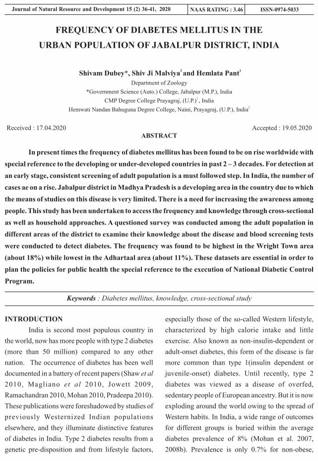
ABSTRACT
In present times the frequency of diabetes mellitus has been found to be on rise worldwide with
special reference to the developing or under-developed countries in past 2 – 3 decades. For detection at
an early stage, consistent screening of adult population is a must followed step. In India, the number of
cases ae on a rise. Jabalpur district in Madhya Pradesh is a developing area in the country due to which
the means of studies on this disease is very limited. There is a need for increasing the awareness among
people. This study has been undertaken to access the frequency and knowledge through cross-sectional
as well as household approaches. A questioned survey was conducted among the adult population in
different areas of the district to examine their knowledge about the disease and blood screening tests
were conducted to detect diabetes. The frequency was found to be highest in the Wright Town area
(about 18%) while lowest in the Adhartaal area (about 11%). These datasets are essential in order to
plan the policies for public health the special reference to the execution of National Diabetic Control
Program.
Keywords : Diabetes mellitus, knowledge, cross-sectional study
FREQUENCY OF DIABETES MELLITUS IN THE
URBAN POPULATION OF JABALPUR DISTRICT, INDIA
2 1 Shivam Dubey*, Shiv Ji Malviya and Hemlata Pant
Department of Zoology
*Government Science (Auto.) College, Jabalpur (M.P.), India1 CMP Degree College Prayagraj, (U.P.) , India
2Hemwati Nandan Bahuguna Degree College, Naini, Prayagraj, (U.P.), India
Received : 17.04.2020 Accepted : 19.05.2020
INTRODUCTION
India is second most populous country in
the world, now has more people with type 2 diabetes
(more than 50 million) compared to any other
nation. The occurrence of diabetes has been well
documented in a battery of recent papers (Shaw et al
2010, Magliano et al 2010, Jowett 2009,
Ramachandran 2010, Mohan 2010, Pradeepa 2010).
These publications were foreshadowed by studies of
previously Westernized Indian populations
elsewhere, and they illuminate distinctive features
of diabetes in India. Type 2 diabetes results from a
genetic pre-disposition and from lifestyle factors,
especially those of the so-called Western lifestyle,
characterized by high calorie intake and little
exercise. Also known as non-insulin-dependent or
adult-onset diabetes, this form of the disease is far
more common than type 1(insulin dependent or
juvenile-onset) diabetes. Until recently, type 2
diabetes was viewed as a disease of overfed,
sedentary people of European ancestry. But it is now
exploding around the world owing to the spread of
Western habits. In India, a wide range of outcomes
for different groups is buried within the average
diabetes prevalence of 8% (Mohan et al. 2007,
2008b). Prevalence is only 0.7% for non-obese,
Journal of Natural Resource and Development 15 (2) 36-41, 2020 ISSN-0974-5033NAAS RATING : 3.46

S. No. Characteristics Male in
No.
Female in No.
Total No.
1 Uneducated
17
21
38
2 Primary 58
42
100
3 Secondary 48 33 814 Higher secondary 84 16 1005 College 45 23 686 professional 98 25 123
37
Rudimentary data about knowledge, awareness,
treatment practices and traditional beliefs and other
parameters were included in the questionnaire. All
families in the localities were visited and people
were questioned and total 122 samples were
collected. The study was based upon the STEPS
approach of World Health Organization which
involved queries pertaining to smoking, diet,
alcohol consumption, physical activity as well as
history of treatment for hypertension and diabetes
mellitus. Physical parameters like weight, height,
waist circumference as well as blood pressure were
also recorded. The data was collected every
Saturday and Sunday over the period of 9 months.
The data was studied at RG Stone Hospital, New
Delhi with the help of literature available in
Research and Development Wing.
RESULTS AND DISCUSSION
The investigation discovered 289 males and
221 females with type 2 diabetes in the urban
population of the district. Out of 510 subjects
inspected, maximum persons 156, were in 30-39
years age group, trailed by 98 from 40-49 years age
group, however only 17 were belonging to 60 year
and above. The educational status of the population
was also taken into consideration. About 38 subjects
were uneducated. It was revealed that awareness
about diabetes was much better in College going and
Professionals as compared to illiterate and primary
passed persons. Myths about the cure of diabetes
was found to be highest in secondary and higher
secondary persons.
Table - 1: Educational status of the studied
population group:
physically active, rural Indians. It reaches 11% for
obese, sedentary, urban Indians; and it peaks at 20%
in the Ernakulam district of Kerala, one of India's
most urbanized states. Among lifestyle factors
predicting the incidence of diabetes in India, some
are familiar from the West; whereas others turn
expectations upside down (Mohan et al. 2008a). In
India, as in the West, diabetes is ultimately due to
chronically high levels of blood glucose, and some
of the clinical consequences are similar. The age of
onset in India has been shifting towards every
younger people even within the past decade among
Indians in their late teens, 'adult-onset' diabetes
already manifests itself more often than does
'juvenile onset' diabetes .Diabetes mellitus (DM)
refers to a group of common metabolic disorders
that share the phenotype of hyperglycemia.
Depending on the etiology of the DM, factors
contributing to hyperglycemia include reduced
insulin secretion, decreased glucose utilization, and
increased glucose production (Power et al. 2008).
There are an estimated 40 million persons with
diabetes in India in 2007 and this number is
predicted to rise to almost 70 million people by 2025
by which time every fifth diabetic subject in the
world would be an Indian (Sicree et al. 2006).
The objective of the study was to find out
the prevalence of diabetes in the urban population of
Jabalpur District, present study also finds out the
diabetes in different age groups, educational status.
MATERIALS AND METHODS
A cross-sectional survey was done among
adults aging 20 to 60 years in various locations of
Jabalpur district including Wright Town, Napier
Town, Vijay Nagar, Adhartaal, Hanumantaal,
Gorakhpur, Loabour Chowk and Sadar area. An
organized questionnaire was used to evaluate the
information about diabetes as well as capillary blood
screening test to enquire about the diabetes.
Journal of Natural Resource and Development
S. No. Locality Prevalence1 Wright Town
18.2
2 Napier Town
16.13 Vijay Nagar
14.34 Adhartaal
11.35 Hanumantal 13.36 Gorakhpur 15.17 Labour Chowk 14.88 Sadar 12.5
38 Shivam Dubey et. al.
The frequency of diabetes was evaluated in
numerous areas of Jabalpur district i.e. Wright
Town, Napier Town, Vijay Nagar, Adhartaal,
Hanumantaal, Gorakhpur, Loabour Chowk and
Sadar area. The occurrence was found to be highest
in Wright town area i.e. 18% and lowest in
Aadhartaal area i.e. 11%. Most of the population of
Wright Town and Napier Town area is of High-
Income Groups, following western life styles,
suffering hypertension, consuming high calorie
diets and living sedentary lifestyle. Due to these, this
group of people are very much susceptible to the
disease. On the other hand, the population of
Adhartaal, where occurrence of diabetes is low,
belonged to middle class income group.
Frequency of diabetes in urban people of
various cities is discussed in table 3, recent report of
WHO-ICMR showed that commonness of self-
reported diabetes was 7.3% in the urban population.
Frequency of diabetes in India study (PIOSD),
based on ADA criteria, the prevalence of diabetes in
urban population was 4.7%.
Table - 2 : Prevalence of Diabetes in various
studied places :
Table - 3 : Prevalence of diabetes in urban India
Year
Author (Reference)
Place
Prevalence (%) of diabetes mellitus in Urban Population in different studies Since 1971
1971
Tripathy et al
Cuttack
1972
Ahuja et al
Multicentre (ICMR)
1978
Gupta et al
Multicentre
1984
Murthy et al
Tenali
1986
Patel14
Bhadran
1988
Ramachandran et al
Kudremukh
1989
Kodali et al
Gangavathi
1989
Rao et al
Eluru
1991
Ahuja et al
New Delhi
1994
Wander et al
Punjab
2000
Ramankutty et al
Kerala
2000
Zargar et al
Kashmir
2001
Ramachandran et al
National Urban Diabetes Study (NUDS)
2001
Misra et al
New Delhi
2001
Sadikot et al
Prevalence of Diabetes in India Study (PODIS)
2003
Gupta et al
Jaipur
2004
Agarwal et al
Rajasthan
2004
Ramachandran et al
Chennai
2004
Mohan et al
Chennai (CURES)
2005 Basavanagowdappa et al Mysore
2005 Prabhakaran et al Delhi
2006 Reddy et al National
2006 Deo et al Maharastra
2006 Menon et al Ernakulam
2006 Chow et al Andhra
2007 Raghupathy et al Vellore
2008 Ramachandran Tamil Nadu
1.2
2.3
3.0
4.7
3.8
5.0
2.2
1.6
6.7
4.6
12.4
4.0
12.1
10.3
5.6
8.6
1.8
6.4
14.3
3.8
15.0
10.1
9.3
19.5
13.2
3.7
18.6

39
India in the context of social change.
B o m b a y , I n d i a : H e a l t h C a r e
Communications.
4. Basavanagowdappa H, Prabhakar AK,
Prasannaraj P, Gurudev KC, Virupaksha,
Suma. 2005. Study of prevalence of
diabetes mellitus and impaired fasting
glucose in a rural population. Int J Diab
Dev
5. Chennai Urban Population Study (CUPS
14). J Assoc Physicians India; 51:771.
6. Chow CK, Raju PK, Raju R, et al. 2006. The
prevalence and management of diabetes in
rural India. Diabetes Care;29:1717-18.
7. Deo SS, Zantye A, Mokal R, Mithbawkar S,
Rane S, Thakur K. 2006. To identify the risk
factors for high prevalence of diabetes and
impaired glucose tolerance in Indian rural
population. Int J Diab Dev Countries
;26:19-23.
8. Gupta A, Gupta R, Sarna M, Rastogi S,
Gupta VP, Kothari K. 2003. Prevalence of
diabetes, impaired fasting glucose and
insulin resistance syndrome in an urban
Indian population. Diabetes Res Clin
Pract; 61:69.
9. Gupta OP, Joshi MH, Dave SK.1978.
Prevalence of diabetes in India. Adv
Metabolic Disorders; 9:147-65.
10. Jowett, J. B. 2009. et al. Twin Res. Hum.
Genet. 12, 44–52.
11. Kodali VRR, Alberti KGMM. 1989.
Diabetes mellitus and hypertension among
rural-rural migrants in South India. Hum
Biol .
12. Magliano, D. J. et al. 2010. Diabetes Care
33, 1983–1989.
13. Menon VU, Kumar KV, Gilchrist A,
Sugathan TN, Sundaram KR, Nair V,
Kumar H. 2006. Prevalence of known and
CONCLUSION
Present study proposed to screen people
above 20 years for type 2 diabetes to assess the
occurrence and socio-demographic profile of
participants. Maximum occurrence of diabetes in
present study was 18% which proposes higher
commonness of diabetes in Jabalpur which is very
similar to any other developing cities. The
conclusion in present study may be due to greater
consciousness in target population. This study
shows that the occurrence of diabetes in Jabalpur is
similar to other municipal areas of northern and
southern India. So, policy makers and public health
sectors need to take the burden posed by diabetes
seriously and some sort of precautionary
programmes and screening strategies needs to be
executed to contain this epidemic. Inactive life style,
inappropriate food habits, late night working habits
are most affect ing reasons for diabetes .
Consumption of alcoholic drinks and smoking
customs leads to generation of free radical in body
which also develops leads to diabetes.
ACKNOWLEDGMENT
The authors are thankful to Dr. K.
Venkataraman Director Zoological Survey of India
and Director RG Stone Urology & Laparoscopy
Hospital, New Delhi for providing necessary
facilities and encouragement.
REFERENCES
1. Agrawal RP, Singh G, Nayak KC, Kochar
DK, Sharma RC, Beniwal R, et al. 2004.
Prevalence of diabetes in camel milk
consuming 'RAICA' Rural Community of
North West Rajasthan. Int J Diab Dev
Countries; 24:109–14.
2. Ahuja MMS, Sivaji L, Garg VK and Mitroo
P. 1991. Prevalence of diabetes in northern
India (Delhi area). Horn Metab Res; 4:321.
3. Ahuja MMS. 1972. Diabetes mellitus in
Journal of Natural Resource and Development

40
17th ed. New York: McGraw Hills
Medical;, pp 2475-2304.
23. Prabhakaran D, Shah P, Chaturvedi V,
Ramakrishnan L, Manhapra A, Reddy KS.
2005 . Card iovascu la r r i sk f ac to r
prevalence among men in a large industry
of northern India. Natl Med J India;18:59-
65.
24. Pradeepa, R. et al. 2010. Diabetes Technol.
Therapeutics 12, 755–761.
25. Raghupathy P, Antonisamy B, Fall CH,
Geethanjali FS, Leary SD, Saperia J, Priya
G, Rajaratnam A, Richard. 2007. High
prevalence of glucose intolerance even
among young adults in south India.
Diabetes Res Clin Pract ;77:269-79.
26. Ramachandran A, Jali MV, Mohan V,
Snehalatha C, Viswanathan M. 1988. High
prevalence of diabetes in an urban
population in south India. Br Med J;
297:587-90.
27. Ramachandran A, Mary S, Yamuna A,
Murugesan N, Snehalatha C. 2008. High
Prevalence of Diabetes and Cardiovascular
Risk Factors Associated with urbanization
in India. Diabetes Care; 31:893-8.
28. Ramachandran A, Snehalatha C, Baskar
AD, Mary S, Kumar CK, Selvam S,
Catherine S, Vijay V. 2004. Temporal
changes in prevalence of diabetes and
impaired glucose tolerance associated with
lifestyle transition occurring in the rural
population in India. Diabetologia ;47:860-
5.
29. Ramachandran A, Snehalatha C, Kapur A,
et al. 2001. Diabetes Epidemiology Study
Group in India (DESI). High prevalence of
diabetes and impaired glucose tolerance in
India: National Urban Diabetes Survey.
undetected diabetes and associated risk
factors in central Kerala - ADEPS.
Diabetes Res Clin Pract; 74:289.
14. Misra A, Pandey RM, Devi JR, Sharma R,
Vikram NK, Khanna N. 2001. High
prevalence of diabetes, obesity and
dyslipidaemia in urban slum population in
northern India. Int J Obes; 25:1722-9.
15. Mohan V, Deepa M, Deepa R, 2004. Secular
trends in the prevalence of diabetes and
glucose tolerance in urban South India-the
Chennai Urban Rural Epidemiology Study
(CURES-17). Diabetologia;49:1175.
16. Mohan V, Mathur P, Deepa R, Deepa M,
Shukla DK, Menon GR, Anand K, Desai
NG, Joshi PP, Mahanta J, Thankappan KR,
Shah B. 2008a. Urban rural differences in
prevalence of selfreported diabetes in
India-The WHO-ICMR Indian NCD risk
factor surveillance. Diabetes Res Clin
Pract ;80:159-68.
17. Mohan, V. et al. 2007 Indian J. Med. Res.
125, 217–230.
18. Mohan, V. et al. 2008b. Diabetes Res. Clin.
Practice 80, 159–168.
19. Mohan, V. et al. 2010. Indian J. Med. Res.
131, 369–372.
20. Murthy PD, Pullaiah B and Rao KV. 1984.
Survey for detection of hyperglycaemia
and diabetes mellitus in Tenali. In Diabetes
Mellitus in Developing Countries. Bajaj JS
Ed. Interprint, New Delhi, ,55.
21. Patel JC. 1986. Prevalence of hypertension
and diabetes mellitus in a rural village. J
Diabetic Assoc India; 26:68.
22. Power AC. Diabetes Mellitus. In: Fauci AS,
Braunwald E, Kasper DL, Hauser SL,
Longo DL, Jameson JL, 2008. et al. editors.
Harrison's Principles of Internal Medicine.
Shivam Dubey et. al.

41
35. Shaw, J. E., Sicree, R. A. & Zimmet, P. Z.
2010. Diabetes Res. Clin. Practice 87,
4–14.
36. Sicree R, Shaw J, Zimmet P. 2006.
Diabetes and impaired glucose tolerance in
India . Diabetes Atlas . Gan D Ed.
International Diabetes Federation,
Belgium. pp 15-103.
37. Tripathy BB, Panda NC, Tej SC, Sahoo GN
and Kar BK. 1971. Survey for detection of
glycosuria, hyperglycaemia and diabetes
mellitus in urban and rural areas of Cuttack
district. J Assoc Physicians
38. Wander GS, Khurana SB, Gulati R, Sachar
RK, Gupta RK, Khurana S, Anand IS.
1994. Epidemiology of coronary heart
disease and risk factors in a rural Punjab
population: prevalence and correlation
with various risk factors. Ind Heart J;
46:319-23.
39. Zargar AH, Khan AK, Masoodi SR, Laway
BA, Wani AI, Bashir MI, Dar FA. 2000.
Prevalence of type 2 diabetes mellitus and
impaired glucose tolerance in the Kashmir
Valley of the Indian subcontinent. Diabetes
Res Clin Pract;47:135.
Diabetologia; 44:1094.
30. Ramachandran, A., Ma, R. C. W. &
Snehalatha, C. 2010. Lancet 375, 408–418.
31. Ramankutty V, Joseph A, Soman CR. 2000.
High prevalence of type2 diabetes in an
urban settlement in Kerala, India. Ethn
Health Med;4:231-9.
32. Rao PV, Ushabala P, Seshiah V, Ahuja MM,
Mather HM. 1989 The Eluru survey:
prevalence of known diabetes in a rural
Indian population. Diabetes Res Clin Pract
; 7:29-31.
33. Reddy KS, Prabhakaran D, Chaturvedi V, et
al, 2006. on behalf of the Sentinel
Surveillance System for Indian Industrial
Populations Study Group. Methods for
establishing a surveillance system for
cardiovascular diseases in Indian industrial
populations. Bull WHO; 84:461-9.
34. Sadikot SM, Nigam A, Das S, et al. 2004.
The burden of diabetes and impaired
glucose tolerance in India using the WHO
1999 criteria: prevalence of diabetes in
India study (PODIS). Diabetes Res Clin
Pract ;66:301-307.
Journal of Natural Resource and Development

ABSTRACT
Present study 'the incidents of umbilical sepsis or Omphalitis' is made on Kailashi Poultry
Farm, Ambedkar Nagar during 2014 to 2017. All the chicks were hatched out in the same hatchery and
reared in deep litter (floor) brooding system. All of them were provided with compounded chick mash
with required additives and ad.lib. water. Resultantly, the incidence of omphalitis was 19.1% (AV) in
2014, 25.0% in 2015, 25.9% in 2016 and 26.9% (AV) in 2017.
Key words : Omphalitis, effect of season, incidence, gram positive and gram negative bacteria.
INCIDENCE OF UMBILICAL SEPSIS OMPHALITIS-A STUDY
S. P. Verma
Department of Animal Science
K.A.P.G. College, Prayagraj - 211 001, (U.P.), India
Received : 14.05.2020 Accepted : 22.06.2020
INTRODUCTION
Omphalitis is an infection of the umbilical
stump (Cushing, 1985). Aerobic bacteria are present
in approximately 85 percent of infections,
predominated by staphylococcus aureus, group A
streptococcus, E. coli Ktebstella pheaumonae and
protecus mirabilis (Airede, 1992, Brook, 1998,
Mashon et al. (1989) Savardekan, 2004). S. aureus is
also described with omphalitis infection (Sengupta
et al. 2016). More recent reports implicate with both
gram positive and gram negative bacteria in the
etiology of some cases, anaerobic bacteria have
been found ( Brook 2011). Omphalitis occasionally
mainfests from an immunologic disorder
(Leacocyte Adhesion Disorder (LAD) is most
prominent among the immunodef ic iency
syndromes (Hung et al. 1999). The overall incidence
of omphalitis varies from 0.2 percent to 0.7 percent
in industrialized countries (Mc Kenna and Jhonson,
1977). In full term infants, the mean age at onset is 5-
9 days. In preterm infants, the mean age at onset is 3-
5 days.
MATERIALS AND METHODS
O m p h a l i t i s o r n a v a l i n f e c t i o n i s
characterized by failure of the naval opening to close
properly, with resultant infection of the internal
organs (Biester and Schwarte, 1969). Bacteria gain
entry into the yolk sac either before or soon after
hatching, multiply in the yolk, results in generalised
septicaemic condition and ends in death of the chick.
A study was undertaken to find out the occurrence of
yolk sac infection in layer strain chicks for the
period from 2014 to 2017 (4 years). Necropsy
reports of all the layer strain chicks died were
collected, classified, tabulated and analysed. The
results were presented in the table. All the chicks
were hatched out in the same hatchery and reared on
floor brooding system. All of them were provided
with compounded chick mash with required
additives and ad libitum water. Brooder houses were
Journal of Natural Resource and Development 15 (2) 42-45, 2020 ISSN-0974-5033NAAS RATING : 3.46

43
were found to be responsible for yolk sac infection.
Sharma and Kousik (1986) observed that no
systematic efforts were made to identify the specific
causal agents responsible for the retention of yolk
sac recorded. However, faulty brooding was
mentioned as the important contributory factor.
The present study has given an indication
that the incidence was not specific for particular
season or month but existing almost in all the
months except in one or two months.
Regarding the source of infection,
Seneviratna (1969) describes that soiled eggs,
unhygienic condition of egg storage, high humidity
in incubator and transfer to hatchery are the
predisposing factor for transovarian infection.
Volkmar (1929) observed that after the chick hatch
the naval fails to close properly following drawing
of the yolk sac into the abdominal cavity and
infection thus gains entrance. He also stressed that
the condition may be due to the increased content of
bacteria of the air in the incubator at hatching time.
Brandly (1932) reported that the condition may be
related to the influence of high relative humidity in
preventing normal enclosure of the yolk sac within
the body cavity. Faecal contamination of eggs was
considered to be the most important source of
infection (Halfstead et al. 1972). Ardrey et al. (1968)
described that ovarian infection or salphingitis may
be responsible for the infection in chicks.
Coults (1981) described that the ability of
an organism to cause yolk sac infection depends on
its ability to degrade and break down protein.
Brandly (1932) noted that most of the losses
from omphalitis occured with in 72 hours after
hatching and the course of the disease was always
rapid with death taking place from 2-8 hrs. The
infection may carry to the internal organs
particularly intestines. Sometimes peritonitis may
be found when the yolk sac ruptures and mortality
maintained well away from adult birds with separate
attendants.
RESULTS AND DISCUSSION
The incidence of omphalitis was 19.1%
(Av) in 2014, 25.0% in 2015, 25.9% in 2016 and
26.9% (Av) in 2017. The incidence was heavy in the
months of February (21.1%), April (29.6%), July
(38.0%), in August (28.0%) and in November
(45.0%) in 2014. During 2015 it was more in the
months of January (64.7%), February (47.0%),
March (31.7%), August (24.0%), September
(35.7%), October (22.2%) and in December
(60.0%). In 2016 in the months of February (26.6%),
March (36.6%), April (47.0%), May (27.5%),
September (19.0%) and in December (22.6%) and in
the year 2017 it was more in the following months,
May (22.2%), June (40.0%), July (57.1%),
September (77.2%), October (36.9%).
On postmortem examination a general
odema of the abdominal muscles, an unabsorbed
yolk and peritonitis were the common lesions
observed. Often the contents of yolk were semisolid
or more liquid invariably the yolk sac was ruptured.
In some cases, the yolk contents were
caseaous in nature or yellow brown waterymaterial.
In some, along with infection of yolk, pericarditis
was noticed. Culture examination was taken up with
a few samples only and only E. coli was isolated
from them.
Sharma and Kousik (1986) recorded
25.12% of the mortality due to yolk sac in turkeys.
Out of which 20.13% were diagnosed during winter
and 4.99% in summer.
Sarma et al. (1985) isolated 14 strains of
bacteria in cases of omphalitis and attributed that
Gram negative bacteria was the causative factor for
76.50% of cases and Gram positive bacteria in
23.30% cases. They also observed that members of
Enterobacterioceae including Salmonella species
Journal of Natural Resource and Development

44
Hatchery rooms and equipment must be fumigated
with formaldehyde and potassium permanganate
mixture. It is necessary to fumigate the incubating
eggs also. The fumigation should be repeated every
second day till the infection is cleared (North, 1984).
may be as high as 10% (North, 1984).
In controlling this infection, cleanliness of
the incubators especially at hatching time should be
stressed as a possible means of preventing the
disease. Hatchery sanitation is utmost importance.
Avi. Dis. 12:505-11.
3. Brandly, C.A. (1932): Poul Sci. 11:269-
282.
4. Brook I Anaerobic infections in children
A d v. E x p . B i o l . 2 0 11 , 6 9 7 - 11 7 -
52(Medline).
5. Brook I: Microbiology of necrotizing
fascitis associated with omphalitis in new
born infants perinetol 1998 Jun-Feb. 18(1)
28-30.
6. Coults, GS. (1981): Poultry Diseases under
modern management, 2nd edition, Saiga
Pub. Co. Ltd. England.
The main causative organism, E. coli is
sensitive to streptomycin, chloramphenicol,
chlortetracycline, ni trofurans, neomycin,
oxytetracycline and sufa drugs. It is important to
determine the drug sensitivity before the drug being
administered and which ever the drug is chosen,
should be administered in an effective dose to the
entire flock.
REFERENCES
1. Ai rede Al , Pa thogens in neonata l
omphalitis, J. Trop, pediatr. 1992 June 38
(3) 29-31 (Medline).
2. Ardrey, W.B. and Peterson, G.F. (1968):
S. P. Verma
Table - 1 : Incidence of Omphalitis in layer strain chicks for the period 2014-2017
Months Years
2014 2015 2016 2017 No. % No. % No. % No. %
Jan. 21/92 22.8 11/17 64.7 4/137 2.91 24/124 19.3
Feb. 45/213 21.1 8/17 47.0 19/73 26.0 21/235 8.9 Mar.
1/206
0.48
13/41
31.7
11/30
36.6
6/193
3.1
Apr.
8/27
29.6
10/124
47.75
40/85
47.0
13/73
17.8
May
6/75
6.6
0/78
0.0
27/98
27.5
14/63
22.2
Jun
4/35
11.4
1/74
1.4
4/38
10.5
26/65
40.0
Jul.
8/21
38.0
3/79
3.8
0/8
00.0
32/56
57.1
Aug.
7/25
28.0
6/25
24.0
0/9
00.00
3/37
8.1
Sep.
6/33
18.1
5/14
35.7
1/1
100.0
17/22
77.2
Oct.
2/23
8.7
4/18
22.2
0/2
00.0
31/84
36.9
Nov.
18/40
45.0
1/40
2.5
5/10
50.0
9/48
18.7
Dec.
0/21
00.0
39/65
60.0
7/31
22.6
1/41
2.4
Average 19.1 25.0 25.9 26.9
Percentage has been worked out for the total mortality in a month

45
13. S a r m a , R . D . L a k s h m a n a c h a r, N .
Remakrishna Rao, M Inayathullah Khan,
D. and Narayana, G. (1985): Ind. J. Poul.
Sci. 1985: 20(4): 262-266.
14. Savardeker KP Changing spectrum of
neonatal omphalitis, Pediatr. Infect, dis J.
2004 Jan. 23(1) 22-6 (Medline).
15. Seneviratna, P. (1969): Diseases of Poultry
2nd edition, Bristol, John Wrigh and Sons
Ltd.
16. Sharma, N.K. and Koushik, R.K. (1986):
Ind. J. Poul Sci. 21(1) : 53-56.
17. Volkmar, F. (1929) Jour. Ame. Vet. Med.
Assn. 101:26-28.
18. Williams R.B. and Daines, L.L (1942) Jour
Am. Vet Med. Asn. 101:26-28.
7. Cushing A. H.: Omphalitis a review
pediatric infect dis, 1985 May-June, 4(3)
282-5 (Medline).
8. Haines, R.B. (1938): Vet. Rec. 69:1433.
9. Hatstad. H. S. Calnek, B.W, Helmboldt.
C.F. Reid, W.M. and Yoder, H.W. (1972):
Diseases of Poultry 6th edition, Iowa State
Uni. Press. p, 396-404.
10. Hung CH, Cheng SN, Hua YM, et al.
LADD, report of one case Acta, Pediatr.
Taiwan 1999 Mar-Apr. 40(2) - 128-31.
11. Mason W.H. Andrews, R. Ross LA, Wright
HT, Jr, Omphalitis in the newborn infant.
Pediatr. Infect. Dis. J. 1989, Aug. 8(8) 521-
5 (Medline).
12. Mc Kenna H, Jhonson D Bacteria in
neonatal omphalitis pathology 1977 Apr.
9(2) 111-3(Medline).
Journal of Natural Resource and Development

ABSTRACT
Diabetes mellitus or simply diabetes occurs throughout the world, but is more common (especially type
2) in the more developed countries. The disease affects more than 50 million Indians – 7.1% of the
nation's adults and kills about 1 million Indians a year. The high incidence is attributed to a
combination of genetic susceptibility plus adoption of a high-calorie, low-activity lifestyle by India's
growing middle class. All forms of diabetes have been treatable since insulin became available in 1921,
and type2 diabetes may be controlled with medications. Selenium is a universal essential trace element
for mammals which is important for many cellular processes. Selenium is relatively well absorbed from
diet better, so if it is an organic form it acts as an antioxidant in the form of selenoproteins. Selenate was
shown the process of regulatory effects on glycolysis, gluconeogenesis and fatty acid metabolism,
metabolic pathways which are disturbed in diabetic disorders. Selenium is a key component of a
number of selenoproteins involved in essential enzymatic functions, such as redox homeostasis, thyroid
hormone metabolism, immunity and reproduction. Because of antioxidant properties of
selenoproteins, and because selenate insulin activity in experimental models, selenium was expected to
prevent type 2 diabetes and cardiovascular disease (CVD).
Keywords : Diabetes, blood sugar, selenium.
HAEMATOLOGICAL STUDIES AND EFFECT
OF SELENIUM IN ALBINO RATS
1 2 3 4 5Himanshu Vatsal , Seema Rani , Kavita Verma , Swati Shekhawat , Seema Sharma 1,2,3,4,5
Department of Zoology, Meerut College Meerut. 250001(U.P.), INDIA
Received : 30.06.2020 Accepted : 30.07.2020
INTRODUCTION
Diabetes is a group of metabolic diseases in
which a person has high blood sugar, This high
blood sugar produces the classical symptoms of
polyuria, polydipsia (increased thirst) and
polyphagia (increased hunger). Two main types of
diabetes mellitus (DM) include Type 1 DM, or
“insulin dependent diabetes mellitus” (IDDM) or
“juvenile diabetes.” (results from the body's failure
to produce insulin), Type 2 DM (a condition in
which cells fail to use insulin properly), previously
referred to as non insulin – dependent diabetes
mellitus (NIDDM) or “about-onset diabetes”.
All forms of diabetes have been treatable
with medications. Insulin and some oral
medications can cause hypoglycemia (low blood
sugars). Several areas of uncertainty in the dietary
guidelines ,especially in the area of assessing
Journal of Natural Resource and Development 15 (2) 46-54, 2020 ISSN-0974-5033NAAS RATING : 3.46

47
libitum.
Induction of Diabetes
Diabetes mell i tus was induced by
intraperitoneally injecting alloxan monohydrate,
dissolved in normal saline (12.5mg/100g). After an
interval of 15 days, Diabetes mellitus was confirmed
by blood sugar analysis applying Folin-Wu method,
using a commercial kit.
Present investigation was conducted on 180
to 220 + 10 gm weight albino rats. The experimental
albino rats were categorized into two main groups
viz. control and experimental groups. Control group
contain five albino rats, experimental group contain
twenty alloxan induced diabetic rats. This group was
subdivided into two experimental sets A and B of
five diabetic rats in each. Set- A diabetic control,
Set-B diabetic rats treated with micronutrient
Chromium.
Control Group: The five rats of control
group were kept in separate from the micronutrient
treated group.
Experimental Group:
Experimental Set A: In this set five alloxan
induced diabetic rats were kept as diabetic control.
Experimental Set B: In this set five
diabetic rats were kept and fed upon Selenium (@
5.0mg/kg body wt.) mixed food for 30 days.
Collection of blood sample: After 30 days
of post treatment with micronutrients Se blood
samples were taken from both the groups I and II
directly from the ventricles of the dissected rats.
Blood samples were taken in vials for various
haematological and biochemical investigations and
transferred immediately into centrifuge tubes for the
separation of serum. The blood samples were
analyzed for pH using micro-blood pH assembly,
total number of RBCs, WBCs, heamoglobin
percentage and Packed Cell Volume (PCV)
individually to each animal.
micronutrient status and the role of micronutrients in
the pathogenesis of diabetes and its complications
exists. The role and importance of trace elements
such as Selenium, Chromium, Zinc, and Vanadium
are much less evident and subjected to chronic
debate. Some data indicate that these metals may
have a clinical interest in patients presenting
deficiencies in individual metal levels. The same
holds true for an association of some trace elements
such as Selenium or Chromium or Zinc with oral
anti diabetics. Believably, some of these trace
elements, such as Selenium, zinc, chromium and
manganese, play a major role in protecting the
insulin secreting pancreatic β-cells, which are
sensitive to free radical damage.
Selenium is an important component of
selenoproteins, which are implicated in modulating
oxidative stress and regulating thyroid hormone
activity. Two recent studies, examining the
relationship between serum selenium levels and the
prevalence of diabetes among U.S. adults found that
high serum selenium levels were positively
associated with the prevalence of diabetes.
Selenium has a narrow therapeutic range and large
inter individual variability in terms of metabolic
sensitivity. Selenium species such as selenite and
selenate may impair insulin responsiveness in Rats
and induce a catabolic response in muscle with
glycogen depletion and increased rates of
glycolysis.
MATERIALS AND METHODS
Experimental animal: The male albino rat,
Rattus norvegicus.
Maintenance and feeding of experimental animal
v The rats were acclimated for three weeks
prior to the experiment.
v The rats were fed on standard rat and mice
feed manufactured by Hindustan Lever
Ltd., India and water was provided ad
Journal of Natural Resource and Development

4
1020countedWBCof.Nomm/.C.B.Rof.No 3 ´´
=
4chamberWBC1ofArea
factorDepthDilutioncountedWBCmm/.C.B.Rof.No 3
´
´´=
48 Himanshu Vatsal et. al.
of 1 mm i.e. has an area = 1 × 1 = 1 square mm. The
depth of the counting chamber was 1/10 mm.
Therefore, the actual volume of the diluted blood in
each of the 4 WBC square was 1 cubic mm.
Calculation of WBC were done by following
formula after counting the number of WBC in four
WBC counting chambers.
c. Haemoglobin Concentration (Hb):
Sahli's method was used for the estimation
of hemoglobin percentage. This method required the
use of the Sahli 's haemoglobinometer or
haemometer and a hemoglobin tube and a standard
light brown glass rod. The hemoglobin pipette
contained uniform diameter and a 20 cubic
millimeter. Graduated hemoglobin tube was filled
with N/10 HCl upto mark 10. The blood was sucked
in the hemoglobin pipette up to mark 20 cubic mm.
From pipette the blood were transferred to
haemoglobin tube already containing N/10 HCl.
The tube was thoroughly shaken and kept for 10
minutes. After 10 minutes the haemoglobin of the
blood was converted into haematin. The addition of
N/10 HCI was continued drop by drop into the
haemoglobin tube till the colour matched with that
of the standard brown glass rods. Reading was
recorded on the haemoglobin tube at this point.
Dilution of blood was read on the haemoglobin tube
in terms of gram percent or grams per 100ml of
blood.
d. Packed cell volume (PCV): Wintrobe's Tube
Method
1.5 ml of blood was drawn into a dry and
clean pasteure pipette avoiding air bubble in the
Separation of serum: The centrifuge tubes
containing blood samples were allowed to stand in a
slanting position, for about one hour at room
temperature and were centrifuged at 3000 rpm for 15
minutes. The supernatant serum was taken carefully
transferred to sterilized plain glass vials with the
help of glass dropper for the biochemical
investigations.
Experimental investigations were made on
hypoglycemic effect of micronutrient Selenium in
albino rats on the basis of following haematological
studies:
HAEMATOLOGICAL STUDIES:
a. Total RBC Count: By haemocytometer (Henry et
al. 1989)
A drop of diluted blood was kept in the
Neubauer's Chamber. The counting chamber was
kept under light microscope and counting of RBC
was done. The counting was done in 850 small
squares of Neubauer's RBC counting chamber.
Calculations of RBC were done by following
formula after counting the number of red blood cells
per cubic millimeter.
The length of each small square was 1/20
mm and it had an area of 1/20 × 1/20 or 1/400 square
millimeters. The depth of the counting chamber was
1/10 mm, hence the actual volume of the diluted
blood in a small square was 1/400 × 1/10 or 1/4000
cubic mi l l imeter when the d i lu t ion was
1:200.4000squaresmall90incounted.C.B.RofN
umbermm/.C.B.Rof.No3´=
b. Total WBC count: By haemocytometer (Henry
et al. 1989)
A drop of diluting blood was kept in the
Neubauer's chamber. Counting chamber was kept
under the light microscope. Each of these 4 WBC
counting chamber having 16 small square had sides
4000countedsquaresmallof.No
DilutioncountedRBCof.Nomm/.C.B.Rof.No 3 ´
´=
50countedWBCmmcuperWBC ´=

49
(Serum Glutamic Oxaloacetate Transaminase) and
activity of SGPT (Serum Glutamic Pyruvate
Transaminase) in albino rats.
These findings may provide a clue to
evaluate the use of oral administration of
micronutrients in diabetic rats.
a. Total RBC Count
Control group (A):
In control group of albino rats, the total 3RBC count was 6.2 million/mm ; in diabetic control
group total no. of RBC count was observed to be 3
5.85 million/mm . Total erythrocyte count was
found to be significantly decreased due to the
diabetes in comparison to control group (Table-1,
Fig.-1).
Selenium Treated group (C):
The total number of RBC in this group 3treated with zinc found 6.35 million/mm . The
number of RBC found significantly (P<0.05)
increased in comparison to diabetic and healthy
control group (Table-1, Fig.-1).
b. Total WBC Count
Control group (A):
The number of total WBC in this group of 3albino rats was found 7400/mm ; in diabetic control
group total no. of WBC count was observed 3
7450/mm . Total leucocytes count was found to be
slightly increased due to the diabetes in comparison
to healthy control group (Table-1, Fig.-2).
Selenium Treated group (C):
The total number of WBC in this group 3treated with zinc found 7350/mm . The number of
WBC found slightly decreased in comparison to
diabetic and healthy control group (Table-1, Fig.-2).
c. Haemoglobin Concentration
Control group (A):
In control group of albino rats, haemoglobin
concentration was 11.5 gm/dl; in diabetic control
group haemoglobin was observed 11.2 gm/dl. Total
capillary stem. The Wintrobe's tube filled with blood
was then centrifuged at the rate of 3,000 rpm for 30
minutes. This was necessary to create the required
centrifugal force.
The upper level or red blood cell layer was
recorded in Wintrobe's tube which was centrifuged
again for fifteen minutes and reading was taken
again. Readings were confirmed after getting two
consecutive readings upper level of red cell layer in
Wintrobe's tube. Upper most pale yellow layer was
of plasma. Below plasma was a thin whitish layer of
platelets and leucocytes and below this was the
black line which marked the upper limit of red cell
layer. The line was due to the presence of reduced
hemoglobin of red cells lying adjacent to the whitish
layer. The column blood in the Wintrobe's
hematocrit tube was 100 ml. The volume of packed
cell was read directly as percentage (Henry, 1989).
RESULTS AND DISCUSSION
Appearance of a drug or chemical in the
blood depends upon the rate of absorption. When
these agents are administered orally they reach to
liver via hepatic portal system and reached to kidney
through nephritic system. On the contrary,
intraperitoneal administration the chemicals are
mostly absorbed directly through the blood
capillaries. The interactions of these agents with
blood components may induce their effects.
Furthermore the agents are carried to different
organs after binding through specific proteins.
Therefore the interaction with blood is the first
reaction which can be judged to access the action of
any chemical reagent used. Keeping in view these
facts, it became desirable to know the effect of
alloxan and micronutrients (Zinc, Selenium and
Chromium) on hematological parameters viz. Red
Blood Cells (RBC) count, White Blood Cells
(WBC) count, Haemoglobin (HB), Packed Cell
Volume (PCV), Blood Sugar, activity of SGOT
Journal of Natural Resource and Development

50
d. Packed Cell Volume
Control group (A):
In control group of albino rats, the packed
cell volume was 45%; in diabetic control group
42%. The packed cell volume was found to be
significantly decreased due to the diabetes in
comparison to control group (Table-1, Fig.-4).
Selenium Treated group (C):
In this group of albino rats, the packed cell
volume was 45%. The packed cell volume was
found to be increased in comparison to diabetic
control group (Table-1, Fig.-4).
haemoglobin concentration was found to be slightly
decreased due to the diabetes in comparison to
control group (Table-1, Fig.-3).
Selenium Treated group (C):
In this group of albino rats, haemoglobin
concentration was found 11.7 gm/dl. Total
haemoglobin concentration was found to be slightly
increased due to the selenium treatment in
comparison to healthy and diabetic control group
(Table-1, Fig.-3).
Table - 1 : Effect of micronutrients on biochemical parameters in experimental diabetic albino rats.
Parameters Healthy Control
Diabetic Control
Treatment Significance
value (P) Selenium
Blood glucose (mg/dl)
110 185 137* 1.6325
± S.E.
±0.1232
±1.2406
±0.2252
Glycogen (mg/dl)
11.0
21.4
13.2
1.2335
± S.E.
±1.1134
±1.3504
±0.6295
Total Urea (mg/dl)
8.6
12.55
9.5* 1.4532
± S.E.
±0.1252
±0.1625
±0.3820
Creatinine (mg/dl)
1.2
2.3
1.66
0.3425
± S.E.
±0.3422
±1.4550
±0.4552
Total Cholesterol
(mg/dl)
35.52
60.45
42.45
0.8592
± S.E. ±1.3112 ±1.6520 ±1.2558
Values are mean, ±S.E. (Standard Error) and n=5
*Statistical analysis: P versus respective control< 0.05
Himanshu Vatsal et. al.

51
Fig. - 4 : Showing packed cell volume in diabetic
experimental albino rats in comparison to healthy
control rats.
RESULTS AND DISCUSSION
Diabetes is characterized with the loss of
body weight as body protein or fats are being
u t i l i z e d f o r e n e rg y g e n e r a t i o n t h r o u g h
gluconeogenesis. The diabetic hyperglycemia
induces elevations of blood creatinine and urea
levels which are considered as significant markers
of renal dysfunction. A significant decrease in
plasma-urea-nitrogen and plasma creatinine.
Haematological Study:
In Selenium Treated Groups total number
of RBCs increased in comparison to diabetic and
healthy control group. Total number of WBCs found
s i g n i f i c a n t l y d e c r e a s e d . H e a m o g l o b i n
concentration was found to be significantly
increased. Packed cell volume was found to be
significantly increased.
HYPOGLYCEMIC EFFECT OF SELENIUM
Hamid R. Rasekh et al. (1919) studied the
effects of acute treatment (ip) of selenium (se) on
glycoregulation and on plasma levels of glucose,
insulin and corticosterone in both fed and 24 hour
fasted rats. The results showed that acute
intraperitoneal administration of Se (1.6 mg/kg or
more) causes hyperglycemia in rats.
Selenium was considered a toxin until 1957,
when this mineral was shown to be essenital in the
Fig. - 1 : Showing no. of RBCs in diabetic
experimental albino rats in comparison to healthy
control rats.
Fig. - 2 : Showing no. of WBCs in diabetic
experimental albino rats in comparison to healthy
control rats.
Fig. - 3 : Showing haemoglobin concentration in
diabetic experimental albino rats in comparison to
healthy control rats.
Journal of Natural Resource and Development

52
antioxidant protection. Studies have shown that
selenium can protect against oxidative damage
attributable to unregulated blood sugar. (Naziroglu
M. 2001 and Guney M. et al 2011).
REFERENCES
1. A k i n l o y e , O . ; O g u n l e y e , K . a n d
Oguntibeju, O.O, 2010: Cadmium lead,
arsenic and selenium levels in patients with
type 2 diabetes mellitus. African Journal of
Biotechnology, Vol. 9 (32), 5189-5195.
2. Al-Awadi, F.M., Anim J.T., Srikumar, T.S.,
and Al-Rustom, M.: Possible role of trace
elements in the hypoglycemic effect of
plant extract in diabetic rats. The journal of
trace elements in experimental medicine
17:31-44 (2004).
3. Aly H.F., Mantawy M.M. : Comparative
effects of Zinc, selenium and vitamin E or
their combination on carbohydrate
metabolizing enzymes and oxidative stress
in streptozotocin induced diabetic rats. Eur
Rev Med Pharmacol Sci 2012:16(1) : 66-78.
4. Bettger W.J : Zinc and selenium, site
specific versus general antioxidation. Can J
Physiol pharmacol 1993, 71 : 721-724.
5. Bleys J., Acien A.N., and Guallar E. : Serum
selenium and diabetes in U.S. adults. Indian
Journal of Experimental Biology. Vol. 47
(07), 2009.
6. Chehade J.M., Ali M.S., and Mooradian
A.D. : The role of micronutrients in
managing diabetes. Diabetes spectrum Vol
22, No 4, 2009.
7. Czech M.P., and Corvera S. : Signaling
mechanism that regulate glucose transport.
J. Biol Chem 274 : 1865-1868, 1999.
8. Diabetes Pandemic : International Diabetes thFederation. The IDF Diabetes Atlas. 5
Edi t ion , 2012 Update . Brusse l s :
prevention of necrotic liver damage in rats. The
hypothesis of selenium chemoprevention is
principally formulated by the observation that
cancer incidence is inversely associated with
selenium status. However, recent clinical and
epidemiological studies demonstrate a role for some
selenoproteins in exacerbating or promoting other
disease states, specifically type 2 diabetes, although
other data support a role of selenium in stimulating
insul in sensi t iv i ty. In vi t ro se inhibi ted
hyperglycemia or hyperinsulinaemia induced
expression of adhesion molecules via reduction in p
38 MAP kinase.
Eighty weanling beef calves were used to
determine the effects of Zn and se supplementation
on performance, immune response, and blood
characteristics during stress. Selenium improved
weight gains in calves with low initial selenium
status in the first 14 day of the study. (Judith K.
Reffett. et al 1986).
The low concentration of selenium in serum
could potentially expose the subject to oxidative
stress which is known to be associated with the
pathogenesis of disponeases such as diabetes
mellitus (Schwartz and Reis, 2000).
Selenium has also been shown to have
insulin-like properties. (Stapleton. S.R. 2000),
which qualifies it as a potential antidiabetic agent.
It has been reported that oxidative stress
reduces insulin secretion and increases insulin
resistance in some experimental models and may
thus play a causal role in the pathogenesis of
diabetes. (West, 2000; Stumvoll et al., 2005; Evans
et al., 2005).
Another study found that 41% of people
with pancreatitis and 12% of diabetics had a low
selenium concentration. (Quillio, D.et al. 2001).
Many diabetic complication are thought to
be caused by oxidative damage and decreased
Himanshu Vatsal et. al.

53
(4) : 223-41.
17. Lee H.K., Kwon J.H. , Park S.H., and Kim
C.W. : Enhanced hypoglycemic activity
following intratracheal administration of
insulin microcrystal suspension with
injection adjivant. Biosci, biotechnol.
Biochem, 70 (4), 1003-1005, 2006.
18. Li F., Wu Y., Zou Y., Zhao T., Xhang M.,
Feng W., and Yang L. : Food Chem. Toxical
50(5), 1623-1631 Epub Feb-18 (2012).
19. Lizuka Y., Sakurai F., Maeda K, and Hikichi
N : Effects of selenium on the glycolysis
and gluconeogenesis system in rat liver.
Journal of the pharmaceutical society of
Japan. 1993, 113 (7) : 525-531.
20. Mooradian A.D. and Morley J.E. :
Micronutrient status in diabetes mellitus.
American Journal of Clinical Nutrition.
May 1987, Vol. 45, No. 5, 877-895.
21. Mooradian A.D., Failla M., Hoogwerf B.,
Marynuik M., and Wylic-Rosett J. :
Selected vitamins and minerals in diabeties.
Diabetes care 1994; 17:464-479.
22. Mueller A.S., and Pallauf J. : Compendium
o f t h e a n t i d i a b e t i c e f f e c t s o f
supranutritional selenate doses. In vivo and
in vitro investigations with type II diabetic
db/db mice. The journal of nutritional
biochemistry vol. 17, Jesue 8, pg 548-560,
Aug 2006.
23. Pandey R.N., Singh R.K., Kalpna : Asian
Jornal of chem.. 23(6), 2739-2741 (2011).
24. Piero N.M., Joan N.M., Cromwell K.M.,
Moina D., Joseph N.J.N.,Eliud N.M.N.,
Wilson N.M., and Peter G.K. : Trace
element content of selected kenyan
antidiabetic medicinal plants. International
Journal of Current Pharmaceutical
Research Vol 4, Issue 3, 2012.
International Diabetes Federation; 2012.
9. Enas Ali kamel Mahamed : Antidiabetic,
Antihypercholestermic and antioxidative
effect of aloe vera gel extract in alloxan
induced diabetic rats. Australian journal of
Basic and Applied Sciences 5(11) : 1321-
1327, 2011.
10. Farvid M S, Homayouni F, Amiri Z, and
Adelmanesh F: Improving neuropathy
scores in type 2 diabetic patients using
micronutrients supplementation. Diabetes
Research and Clinical Practice 2011, 93:
86-94.
11. Hiromura M., and Sakurai H. : Action
mechanism of metallo-allixin complexes as
antidiabetic agents. Pure Appl. chem., Vol
80, No. 12, pp 2727-2733, 2008.
12. Holness M.J. : Impact of early retardation
on glucoregulatory control and insulin
action in mature rats. Am J. Physiol 270 : E
946-E 954, 1996.
13. J e y a b a l a n S . , a n d P a l a y a n M . :
Antihyperglycemic and antidiabetic
activity of leaves extracts of Sapindus
emarginatus vahl. Asian biomed. 2009, 3 :
313-18.
14. Joshi U, Raut P.D., Agarwal S.K., Patra
P.K., Masheshwari B.K., Apuro M, and
Dhirhe Tc: Evaluation of serum selenium
level in patients with uncomplicated
diabetes mellitus. Journal of clinical and
Diagnostic Research. 2011 Feb; (5):70-73.
15. Kimura K.:Role of essential trace elements
in the disturbance of carbohydrate
metabolism. Nippon Rinsho 1996;54:79-
84.
16. Krentz A.J., Ferner R.E., and Bailey C.J. :
Comparative tolerability profiles of oral
antidiabetic agents. Drug Saf. 1994 Oct, 11
Journal of Natural Resource and Development

54
mellitus. Biological Trace Element
Research. Summer 2004, Vol 99, issue-1-3,
pp 241-149.
30. Stranges S., Sieri S., Vineeti M., Grioni S.,
Guallar E., Laclaustra M., Muti P., Berrino
F., and Vittorio K. : A prospective study of
dietary selenium intake and risk of type 2
diabetes. BMC Public Health 2010, 10 :
564.
31. Tabar M.B. : Determination of serum
selenium in Patients with type II diabetes
mellitus. Middle-East Journal of Scientific
Research 12 (4) : 433-435, 2012.
32. Underwood E.J. : Trace elements in human thand animal nutrition, 4 edition. New york :
Academic press, pp 258-270, 1977.
33. Wiernsperger N., and Rapin J.R.: Trace
elements in glucometablic disorders : an
update. Diabetology and metabolic
syndrome 2010, 2:70.
34. Yang X., Palanichamy K., Onto A.C., Rao
M.N., Fang C.X., and Sreejayan N.: FEBS
lett; Feb,Vol. 28, 579(6), 1458-1464 (2012).
25. Reasner C.A.: Promising new approaches.
Diabetes Obes Metab. 1999 may, 1 Suppl
1:S 41-8.
26. Reffett J.K., Spears J.W., Hatch P.A., and
Brown T.T. : Influence of selenium and Zinc
on performance blood constituents, and
immune response in stressed calves.
Biological Trace element Research. Apr
1986, Vol 9, issue 3, pp 139-149.
27. Resekh H.R., Potmis R.A., Nanavineker
V.K., Early J.L., and Iszard M.B. : Effect of
selenium on plasma glucose of rats : role of
insulin and glucocorticoids. Toxicology
Letters. Vol 58, issue 2, Oct 1991, pp 199-
207.
28. Rocourt C.R.B., and Cheng W.S. : Selenium
supranutrition : Are the potential benefits of
chemoprevention outweighed by the
promotion of diabetes and insulin
resistance? Nutrients 2013, 5, 1349-1365.
29. Shen XiQ, Huang K.X., and Xu H.B.: New
e x p e r i m e n t a l o b s e r v a t i o n o n t h e
relationship of selenium and diabetes
Himanshu Vatsal et. al.

ABSTRACT
The present study was done at the Rani Durgawati Vishwavidyalaya famously known as RDVV,
situated at Pachpedhi in Jabalpur in the state of Madhya Pradesh. The university was established in
1956 under the Jabalpur University Act, 1956 (Act number 22 of 1956). The university was
reconstituted under MP VishwavidyalayaAdhiniyam, 1973 and given jurisdiction over Jabalpur,
Mandla, Seoni, Balaghat, Narsinghpur, Katni, Dindori and Chhindwara. The university campus is
spread over an area of about 100 acres accommodating administrative campus, various departmental
buildings, playgrounds and residential quarters. In the present study, a total of 81 avian species have
been recorded belonging to different families.Afterwards diversity indices were calculated based on
the data collected.
Keywords : Rani durgawati vishwavidyalaya, jabalpur, birds, faunal diversity.
STATISTICAL ANALYSIS OF AVIAN FAUNAL DIVERSITY AT
RANI DURGAWATI VISHWAVIDYALAYA (RDVV)
CAMPUS, JABALPUR, M.P.
1 2Hemlata Pant , Shiv Ji Malviya and Shivam Dubey*
Department of Zoology1 1 CMP Degree College Prayagraj, (U.P.) , India
2 2Hemwati Nandan Bahuguna Degree College, Naini, Prayagraj, (U.P.), India
*Government Science (Auto.) College, Jabalpur (M.P.), India
Received : 20.04.2020 Accepted : 29.05.2020
INTRODUCTION
Rani Durgawati Vishwavidyalaya or
RDVV is a government university in Jabalpur
district of Madhya Pradesh. It is also known as
Jabalpur University or University of Jabalpur. It was
named after the great Gond queen, Durgawati of
Mandla district. The university was established in
1956 under the Jabalpur University Act, 1956 (Act
number 22 of 1956). The university was
reconsti tuted under MP Vishwavidyalaya
Adhiniyam, 1973 and given jurisdiction over
Jabalpur, Mandla, Seoni, Balaghat, Narsinghpur,
Katni, Dindori and Chhindwara. The university
campus is spread over an area of about 100 acres
accommodating administrative campus, various
departmental buildings, playgrounds and residential
quarters. Major area of the campus is covered with
lush green vegetation which houses a large number
of floral and faunal diversity. Several species of
mammals, reptiles and birds etc are spotted here.
The natural forest and grassland has constituted a
good habitat for many residential as well as
migratory birds within the campus.
The faunal diversity of the study area
includes many species of mammals, reptiles,
insects, butterflies, dragonflies as well as spiders. A
broad account of avian diversity in the state of
Madhya Pradesh and Chhattisgarh was presented by
Journal of Natural Resource and Development 15 (2) 55-58, 2020 ISSN-0974-5033NAAS RATING : 3.46

56 Journal of Natural Resource and Development
Chandra and Singh (2004). They reported 517
species belonging to 69 families from the areas.
Similarly records of birds from Central Highlands of
Madhya Pradesh were reported by Jayapal et al.
(2005). In 2008, Ghosh et al. published a detailed
account of avian fauna from the states of Madhya
Pradesh (including Chhattisgarh), reporting
altogether 449 species. Talmale et al., in 2012
published an account of 173 bird species from
Singhori Wildlife Sanctuary (Raisen District),
Madhya Pradesh. Dubey et al. (2017) reported 56
avian species from Dumna Nature Reserve. Again
46 species of birds from Gun Carriage Factory
Estate were recorded by them in same year.
Similarly 118 species of birds belonging to 45
families were reported by Dubey et al. in 2018 from
College of Material Management (CMM), Jabalpur.
In similar context, 72 avian species belonging to 30
families were recorded by Bhandari et al. in 2018
from Ordnance Factory Khamaria (OFK) Estate. In
the present study, the data was collected during the
years from 2016 to 2018 which resulted in recording
a total of 81 avian species belonging to different
families.
Table - 1 : List of avian species recorded RDVV
Campus
S. N.
Name of Spceies
1 Common Teal Anas crecca Linnaeus, 1758
2 Indian Spot Billed Duck Anas poecilorhyncha, J.R. Forster, 1781
3 Knob Billed Duck Sarkidiornis melanotos (Pennant, 1769)
4 Ruddy Shelduck Tadorna ferruginea (Pallas, 1764)
5 Grey Francolin Francolinus pondicerianus (J.F. Gmelin, 1789)
6 Peafowl Pavo cristatus Linnaeus, 1758
7 Red JunglefowlGallus gallus (Linnaeus, 1758)
8 Emerald Dove Chalcophaps indica (Linnaeus, 1758)
9 Eurasian Collered Dove Streptopeliadecaocto (Frivaldszky, 1838)
10 Rock Pigeon Columba livia J.F. Gmelin, 1789
11 Spotted Dove Spilopelia chinensis (Scopoli, 1786)
12 Yellow Legged Green Pigeon Treron phoenicopterus
13 Indian Nightjar Caprimulgus asiaticus Latham, 1790
14 Asian Koel Eudynamys scolopaceus (Linnaeus, 1758)
15 Common Hawk Cuckoo Hierococcyx varius (Vahl, 1797)
16 Pied Cuckoo Clamatorjacobnus(Boddaert, 1783)
17 Common Moorhen Gallinula chloropus (Linnaeus, 1758)
18 Purple SwamphenPorphyrioporphyrio(Linnaeus, 1758)
19 White Breasted Waterhen Amaurornis phoenicurus (Pennant, 1769)
20 Sarus Crane Antigone antigone (Linnaeus, 1758)
21 Lesser Adjutant Leptoptilos javanicus (Horsfield, 1821)
22 Woolly Necked Stork Ciconia episcopus (Boddaert, 1783)
23 Cattle Egret Bubulcus ibis (Linnaeus, 1758)
24 Indian Pond Heron Ardeola grayii (Sykes, 1832)
25 Little Egret Egretta garzetta (Linnaeus, 1766)
26 Black Headed Ibis Threskiornis melanocephalus (Latham, 1790)
27 Little Cormorant Microcarbo niger (Vieillot,1817)
28 Eurasian Thick Knee Burhinus oedicnemus (Linnaeus, 1758)
29 Little Ringed Plover Charadrius dubius Scopoli, 1786
30 Red Wattled Lapwing Vanellus indicus (Boddaert, 1783)
31 Yellow Wattled Lapwing Vanellus malabaricus (Boddaert, 1783)
32 Bronze Winged Jacana Metopidius indicus (Latham, 1790)
33 Pheasant-tailed Jacana Hydrophasianus chirurgus (Scopoli, 1786)
34 Eurasian Curlew Numenius arquata (Linnaeus, 1758)
35 Spotted Redshank Tringa erythropus (Pallas, 1764)
36 Wood Sandpiper TringaglareolaLinnaeus, 1758
37 Barred Buttonquail Turnix suscitator (J.F. Gmelin, 1789)
38 Black Shouldered Kite Elanus caeruleus (Desfontaines, 1789)
39 Crested Serpent Eagle Spilornis cheela (Latham, 1790)
40 Egyptian Vulture Neophron percnopterus(Linnaeus, 1758)
41 Indian Vulture Gyps indicus(Scopoli, 1786)
42 Shikra Accipiter badius(J.F. Gmelin, 1788)
43 Common Barn owl Tyto alba(Scopoli, 1769)
44
Indian Scops Owl Otus bakkamoena Pennant, 1769
45
Spotted Owlet Athene brama (Temminck, 1821)
46
Indian Grey Hornbill Ocyceros birostris (Scopoli, 1786)
47
Common Hoopoe Upupa epops Linnaeus, 1758
48
Coppersmith Barbet Psilopogonhaemacephalus (Statius Muller, 1776)
49
Green Bee Eater Merops orientalis Latham, 1801
50
Indian Roller Coracias benghalensis (Linnaeus, 1758)
51
Common Kingfisher Alcedo atthis(Linnaeus, 1758)
52
Pied Kingfisher Cerylerudis(Linnaeus, 1758)
53
Stork Billed Kingfisher Pelargopsiscapensis(Linnaeus, 1766)
54
Common Kestrel Falco tinnunculusLinnaeus, 1758
55
Plum Headed Parakeet Psittacula cyanocephala (Linnaeus, 1766)
56
Rose Ringed Parakeet Psittaculakremeri(Scopoli, 1769)
57
Indian Pitta Pittabrachyura(Linnaeus, 1766)
58
Black Headed Cuckoo Shrike Lalage melanoptera(Rüppell, 1839)
59
Large Cuckoo Shrike Coracina javensis (Horsfield, 1821)
60
Eurasian Golden Oriole Oriolus oriolus(Linnaeus, 1758)
61
Black Drongo
Dicrurus macrocercus Vieillot, 1817
62
Greater Racket-Tailed Drongo Dicrurus paradiseus(Linnaeus, 1766)
63
Indian Jungle Crow CorvusmacrorhynchosWagler, 1827
64
House Crow Corvus splendens Vieillot, 1817
65
Rufous Treepie Dendrocitta vagabunda (Latham, 1790)
66
Indian Paradise Flycatcher Terpsiphone paradisi (Linnaeus, 1758)
67
Purple Sunbird Cinnyris asiaticus (Latham, 1790)
68
Scaly Breasted Munia Lonchura punctulata(Linnaeus, 1758)
69
Chestnut Shouldered Petronia Gymnoris xanthocollis (E. Burton, 1838)
70
House Sparrow Passer domesticus (Linnaeus, 1758)
71
Plain PriniaPriniainornataSykes, 1832
72
Red Vented Bulbul Pycnonotus cafer (Linnaeus, 1766)
73
Red Whiskered Bulbul Pycnonotus jocosus (Linnaeus, 1758)
74
Oriental White Eye Zosteorpspalpebrosus(Temminck, 1824)
75
Jungle Babbler Turdoides striata (Dumont, 1823)
76
Brahminy Starling Sturnia pagodarum(J.F. Gmelin, 1789)
77
Common Myna Acridotheres tristis (Linnaeus, 1766)
78
Bluethroat Luscinia svecica (Linnaeus, 1758)
79
Common Stonechat Saxicola maurus (Pallas, 1773)
80 Oriental Magpie Robin Copsychus saularis (Linnaeus, 1758)
81 Verediter Flycatcher Eumyiasthalassinus(Swainsin, 1838)

57 Hemlata Pant et. al.
ni= number of individuals of taxon i.
· Simpson index 1-D. Measures- 'evenness' of the
community from 0 to 1. Note the confusions in
the literature: Dominance and Simpson indices
are often interchanged.
· Shannon index (entropy). A diversity index is
taking into account the number of individuals as
well as a number of taxa. Varies from 0 for
communities with only a single taxon to high
values for communities with many taxa, each
with few individuals.
· Brillouin's index :
· Menhinick's richness index :
· Margalef's richness index: (S-1) / ln(n)
· Berger-Parker dominance- simply the number
of individuals in the dominant taxon relative to n.
Diversity Indices
A diversity index is a numerical measure of
species diversity in a given community which is
based on the species richness (the number of species
present) and species abundance (the number of the
individual per species), where the higher number of
species shows a higher diversity of the area.
However, here two kinds of indices, dominance
index and information statistic index were used for
data measurement. From the identified and recorded
avian numbers, these statistics apply to association
data, where the number of individuals was tabulated
in rows (taxa) and possibly several columns
(samples). The present statistics are as follows, for
all sample:
· No. of taxa is (S)
· Total no. of individuals is (n)
· Dominance = 1-Simpson index; Ranges from 0
(All the taxa are equally present) to1 (one taxon
dominates the community completely).
D = S nin
²))i
HB = ln (n!) ln (ni!)n
H = ninS
i- ni
nln
Sn
A
Lower
Upper
B
Lower
Upper
C Lower Upper
Taxa_S
81
81
81
81
80
81
81 80 81
Individuals
527
527
527
485
485
485
590 590 590
Dominance_D
0.03658
0.0312
0.0422
0.03551
0.02994
0.04104
0.03386 0.02952 0.03955
Simpson_1-D
0.9634
0.9578
0.9688
0.9645
0.9589
0.97
0.9661 0.9604 0.9705
Shannon_H
3.834
3.732
3.902
3.854
3.753
3.924
3.885 3.776 3.936
Evenness_e^H/S
0.5707
0.5158
0.6114
0.5825
0.527
0.6249
0.6008 0.5395 0.6331
Brillouin
3.589
3.495
3.656
3.593
3.501
3.662
3.656 3.556 3.707
Menhinick
3.528
3.528
3.528
3.678
3.633
3.678
3.335 3.294 3.335
Margalef
12.76
12.76
12.76
12.94
12.77
12.94
12.54 12.38 12.54
Equitability_J 0.8724 0.8494 0.8881 0.877 0.8542 0.893 0.8841 0.8595 0.896
Fisher_alpha 26.72 26.72 26.72 27.78 27.28 27.78 25.42 24.97 25.42
Berger-Parker 0.1101 0.08349 0.1328 0.1072 0.08041 0.1299 0.1051 0.07966 0.1271
Chao-1 81.68 82.56 96.83 81.58 82.89 98.5 81.38 81.56 94
Table - 2 : Tabular Compilation of Diversity Indices pertaining to
Avian Species recorded at RDVV Campus

58 Journal of Natural Resource and Development
Pachmarhi Biosphere Reserve, Conservation
Area Series, Published by the Director,
Zoological Survey of India, Kolkata. 2009;
39:43-116.
6. Dubey K. K., Dubey S. And Bhandari R.,
(2017) Diversity of Avian Fauna Of Dumna
Nature Park Jabalpur (M.P.). Life Science
Bulletin - June 2017 Vol. 14(1) : 00-00.
7. Dubey K. K., Bhandari R. and Dubey S.,
(2017).Avian fauna in gun carriage factory
estate area Jabalpur district, Madhya Pradesh,
India. International Journal of Fauna and
Biological Studies 2017; 4(5): 34-35
8. Dubey K. K., Dubey S., Bhandari R., and
Sharma J., (2018). Study of diversity in avian
fauna at College of Material Management
(CMM), Jabalpur, M.P. Journal of Natural
Resources and Development, 13(2) 11-17,
2018.
9. Ghosh S, Basu Roy S, Datta BK, Sett AK
(2008) Fauna of Madhya Pradesh (including
Chhattisgarh), State Fauna Series, 15(Part- 2),
Aves. 1-152 (Published by the Director,
Zoological Survey of India, Kolkata).
10. Javed, S., and Kaul, R. 2002. Field Methods
for Bird Surveys. Bombay Natural History
Society; Department of Wildlife Sciences,
Aligrah Muslim University, Aligarh and World
Pheasant Association, South Asia Regional
Office (SARO), New Delhi, India. 61 p.
11. Jayapal R, Qureshi Q and Chellam R (2005).
Some significant records of birds from the
central Indian highlands of Madhya Pradesh.
Indian Birds, 1(5): 98-102.
12. Talmale SS, Limje ME, and Sambath S (2012)
Avian diversity of Singhori Wildlife Sanctuary,
13. Raisen District, Madhya Pradesh. Biological
Forum – An International Journal, 4(2): 52-61.
RESULTS AND DISCUSSION
On the basis of data collected from 2016 to
2018 conclusions were drawn accordingly which
are compiled in the form of following table and
results were drawn. As per the data obtained, it can
be concluded that the study area is composed of a
number of species which are distributed evenly
throughout the study area. Also no single species is
showing dominance here. For tropical countries, the
value of Shannon diversity index can range from 1 to
5. It can be inferred that if the value is within this
range, then the area is said to have good diversity. In
present study, this value is within this range. Thus, it
can be concluded that the area under study has a
good diversity of avian fauna.
ACKNOWLEDGEMENTS
Authors are grateful to Principal, Govt.
Model Science College Jabalpur, Sandeep
Kushwaha, Zoological Survey of India Kolkata for
providing necessary facilities and encouragements.
REFERENCES
1. Bhandari R., Dubey S., Dube K.K. and Sharma
J. (2018). Checklist of avian faunal diversity at
Ordnance Factory Khamaria (OFK) Estate,
Jabalpur, M.P. Journal of Natural Resources
and Development, 13(2) 36-41, 2018.
2. Chandra. J. (1987). Bird ringing at Karera
Great lndian Bustard Sanctuary. Zoos' Print
Journal. 2(10): 5-6.
3. Chandra, K. and Singh, R.K. (2004). Avifauna
of Madhya Pradesh and Chhattisgarh.
Zoos'Print Journal. 19(7) : 1534 -1539.
4. Chandra K, Gupta RP. Aves, In: Fauna of
Bandhavgarh Tiger Reserve, Conservation
Area Series, Published by the Director,
Zoological Survey of India, Kolkata. 2009;
40:179-270.
5. Chandra K, Mahabal A. Aves, In: Fauna of

ABSTRACT
Organic matter might have provided balanced nutrition and congenial microclimate to grow and yield
with full potential..Hormonal influence of Vermi-compost (VC) might have augmented tuber
yield..Seed treatment with biofertilizer was at par with VC in respect to yield. Seed treatment might
have encouraged better stand establishment. Number of tuber per plant were significantly influenced
by the treatments. Lowest Number of tuber (8.11) were recorded in control. Highest number of tuber
were recorded (14.24)in T (1/2 FYM 1/2 vermicompost) treatment.4
Keywords : Biofertilizer, organic manure, potato.
EFFECT OF BIOFERTILIZER AND ORGANIC MANURE ON YIELD
OF POTATO (SOLANUM TUBEROSUM L.) CV KUFRI BADSHAH.
Surya Narayan
Department of Horticulture,2KulbhakSar Ashram Post Graduate College Prayagraj, (U.P.), India
Received : 25.03.2020 Accepted : 30.04.2020
INTRODUCTION
Potato crop is grown under short day
conditions in subtropical Indo-Gangetic plains.
Uttar Pradesh, West Bengal, Bihar and Gujarat are
the leading potato producing states in India . In year
2015 the area and production of potato was 33.7
thousand hectars and 0.23 million tones respectively
(Anonymous 2015) .Therefore, there is a need to
increase and sustain the productivity of potato,
which can be achieved by safeguarding the soil
health and improving soil fertility (Swaminathan,
2004) of potato fields. As no single source is capable
of supplying the required amount of plant nutrients,
integrated use of all sources of plant nutrients is best
to supply balanced nutrition to the crop .The
integrated nutrient management (INM) systems
envisage the use of organic manure along with
chemical fertilizers.These sources can reduce the
mining of soil nutrients and improve overall soil
productivity in terms improved physico-chemical
and biological conditions of soil. Higher food
production needs higher amount of plant nutrients.
Use of inorganic fertilizers has increased
considerably to meet the higher nutrient
requirements of the present day improved varieties.
This creates imbalance in nutrients supply, leading
to decline in soil fertility, crop productivity and
sustainability. Use of organic matter to meet the
nutrients requirement of crops would be an
inevitable practice in years to come. A number of
diverse organic sources are available for the use in
agriculture. Organic manures like farmyard manure,
poultry manure and vermin-compost can play
important role in potato productivity. The beneficial
Journal of Natural Resource and Development 15 (2) 59-64, 2020 ISSN-0974-5033NAAS RATING : 3.46

60 Journal of Natural Resource and Development
potato tuber (solanum tuberosum L.) cv Kufri
Badshah was conducted to study the effect of
organic manure and biofertilizer .
MATERIALS AND METHODS
Field experiment entitled ”Effect of
Biofertilizer and organic manure on growth and
yield of potato (Solanum tuberosum L.) “ was
conducted at the Horticulture Farm, Kulbhaskar
Ashram post graduate college, Prayagraj, Utter
Pradesh during winter season in 2018-19. The
details of the procedure adopted for crop raising and
criteria used for treatment evaluation during entire
course of investigation are described a under
EXPERIMENTAL MATERIAL:
The experiment consists of 8 treatment
combinations comprising of organic manures
with and without biofertilizer (viz. NPK liquid
consortia Bio). The details are as below.
effects of organic manure are manifested through
increase in soil organic matter, humus and over all
soil productivity over the period. Soil organic matter
and humus act in several ways, i.e., serves as slow
release source of plant nutrients to the crops and
increases water holding capacity to maintain the
water regime of the soil and act as a buffer against
change in soil PH. Biofertilizers like phosphorous
solubilizing bacteria (PSB) or Azotobacter may be
useful for improving phosphorous and nitrogen
nutrition in potato. Also, the application of PSB
would help in increasing the efficiency of available
phosphorous in the soil by converting unavailable
phosphorous into available form. Similarly,
nitrogen fixing biofertilizers like azotobacter has the
potential to meet a successful availability of
nitrogen requirement of potato.
Keeping above points in view a trial on”
Effect of biofertilizer and organic manure on yield of
Table - 1 : Details of treatments used in study.
S.N. Treatment symb.
Treatment details
1.
T0
Control unit (Recommonded Doze of Fertilizers=RDF)
2.
T1
FYM@15 t/ha
3.
T2
Vermicompost @5 t/ha
4.
T3
NPK Liquid consortium (Biofertilizer)@150ml/10kg seed treatment
5.
T4
7.5 tonnes FYM+2.5 tonnes vermicompost /ha.
6.
T5
7.5 tonnes FYM/ha +75ml NPK liquid consortium (Bio fertilizer) /10kg seed treatment.
7.
T6
2.5 tonnes vermicompost/ha+75ml NPK Liquid consortium (Bio fertilizer)/10kg seed treatment.
8.
T7
5 tonnes FYM/ha+1.66 tonnes vermicompost/ha+50ml NPK liquid consortium /10kg seed treatment.
EXPERIMENTAL DETAILS AND LAYOUT:
Design of experiments.
The experiment was laid out in Randomized
Block Design with three replications.
The treatments were randomly allotted to different
plots using random number table of Fisher and Yates
(1963).
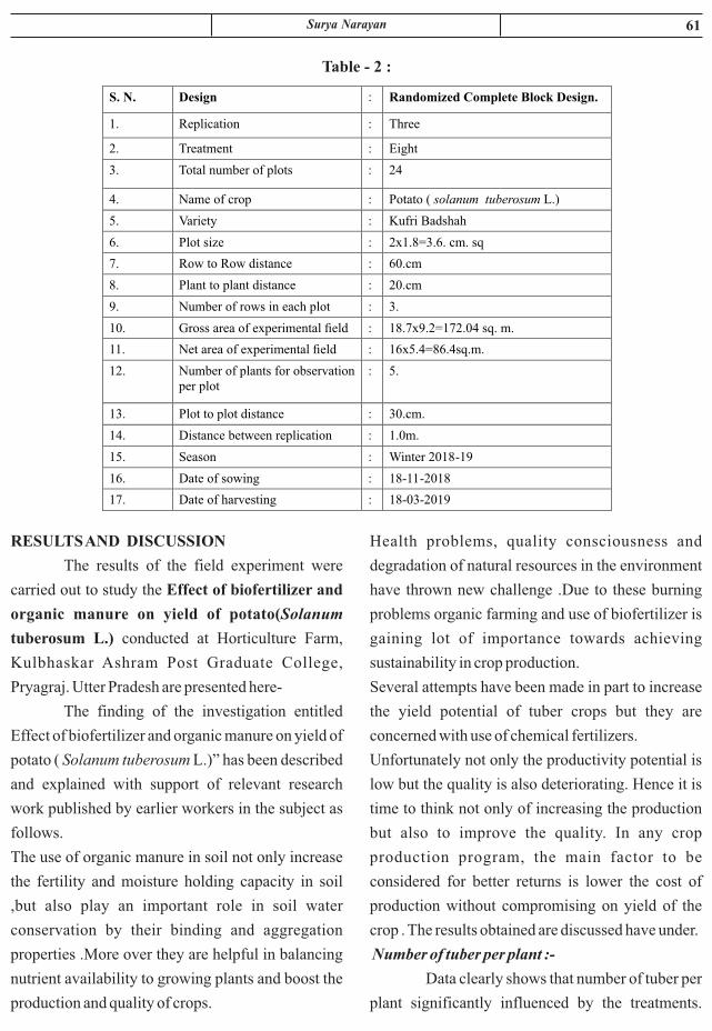
61Surya Narayan
Health problems, quality consciousness and
degradation of natural resources in the environment
have thrown new challenge .Due to these burning
problems organic farming and use of biofertilizer is
gaining lot of importance towards achieving
sustainability in crop production.
Several attempts have been made in part to increase
the yield potential of tuber crops but they are
concerned with use of chemical fertilizers.
Unfortunately not only the productivity potential is
low but the quality is also deteriorating. Hence it is
time to think not only of increasing the production
but also to improve the quality. In any crop
production program, the main factor to be
considered for better returns is lower the cost of
production without compromising on yield of the
crop . The results obtained are discussed have under.
Number of tuber per plant :-
Data clearly shows that number of tuber per
plant significantly influenced by the treatments.
RESULTS AND DISCUSSION
The results of the field experiment were
carried out to study the Effect of biofertilizer and
organic manure on yield of potato(Solanum
tuberosum L.) conducted at Horticulture Farm,
Kulbhaskar Ashram Post Graduate College,
Pryagraj. Utter Pradesh are presented here-
The finding of the investigation entitled
Effect of biofertilizer and organic manure on yield of
potato ( Solanum tuberosum L.)” has been described
and explained with support of relevant research
work published by earlier workers in the subject as
follows.
The use of organic manure in soil not only increase
the fertility and moisture holding capacity in soil
,but also play an important role in soil water
conservation by their binding and aggregation
properties .More over they are helpful in balancing
nutrient availability to growing plants and boost the
production and quality of crops.
S. N. Design : Randomized Complete Block Design.
1. Replication
:
Three
2. Treatment
:
Eight
3. Total number of plots
:
24
4. Name of crop
:
Potato ( solanum tuberosum L.)
5. Variety
:
Kufri Badshah
6. Plot size
:
2x1.8=3.6. cm. sq
7. Row to Row distance
:
60.cm
8. Plant to plant distance
:
20.cm
9. Number of rows in each plot
:
3.
10.
Gross area of experimental field
:
18.7x9.2=172.04 sq. m.
11.
Net area of experimental field
:
16x5.4=86.4sq.m.
12.
Number of plants for observation per plot
:
5.
13.
Plot to plot distance
:
30.cm.
14. Distance between replication : 1.0m.
15. Season : Winter 2018-19
16. Date of sowing : 18-11-2018
17. Date of harvesting : 18-03-2019
Table - 2 :

62 Journal of Natural Resource and Development
tuber (12.91/plant).Organic matter was beneficial
to increase the number of tuber per plant .Organic
matter was found to increase microflora level of the
soil which increase the mineralization of nutrients.
These nutrients become easily available to the plant.
Vigour of the plant was directly related to the
number of tuber per plant. Findings of Ghosh and
D a s ( 1 9 9 8 ) i n p o t a t o , S h a m b a v i a n d
Sharma(2011)in potato; Jaipul et al.(2011);
Chaudhary et al.(2010) ; Rex (1990)in potato ;
Aityeh et al.(2000)and Kumar et al. (2013).
Lowest number of tuber (8.11/plant) were recorded
in control ,while the highest number of tuber were
recorded (14.24/plant) in T (1/2FYM1/2 4
vermicompost) treatment .All the treatments were
better over control .Single applicat ion of
vermicompost was better over FYM.Second
t reatment was not as good as FYM and
vermicompost treatment. FYM and vermicompost
when applied togather reducing half dose the
number of tuber was increased .Reduction of FYM rd
&vermicompost to the 1/3 level reduced number of
Table - 3 : Effect of biofertilizer and organic manure on number of tuber per plant of potato:
Treatment
Symbol
Treatment Details
Number of tuber per
plant
T0
Control Unit( Recommended Dose of Fertilizer =RDF)
8.11
T1
FYM@ 15t/ha
9.24
T2
Vermicompost @5t/ha
10.14
T3
NPK liquid consortium (Biofertilizer) @150ml /10kg seed treatment
9.12
T4
7.5 tonnes FYM+2.5tonnes vermicompost /ha
14.24
T5
7.5tonnes FYM/ha+75ml NPK liquid consortium (Biofertilizer) /10kg seed treatment .
11.10
T6
2.5tonnes vermicompost /ha+75ml NPK liquid consortium (Biofertilizer)/10kg seed treatment
10.12
T7 5 tonnes FYM/ha+1.66 tonnes vermicompost/ha+50ml NPK liquid consortium/10kg seed treatment
12.91
SEm± 2.33
C. D. at 5% level 4.71

63
rd.Reduction of FYM &vermicompost to the 1/3
level reduced number of economic tuber
(8.49/plant). Organic matter was beneficial to
increase the number of economic tuber per plant
.Organic matter was found to increase microflora
level of the soil which increase the mineralization of
nutrients. These nutrients become easily available to
the plant.Vigour of the plant was directly related to
the number of tuber per plant. Findings of Naryan
et al.(2013)in potato ,Verma et al.(2010)in potato,
Verma et al.(2011) in potato; Hussain et al. (2007),
Kumar et al, (2005).
Number of economic tuber per plant :
Data clearly shows that number of
economic tuber per plant significantly influenced by
the treatments. Lowest number of economic tuber
(4.22/plant) were recorded in control ,whilethe
highest number of economic tuber were recorded
(11.35/plant)in T (1/2FYM1/2vermicompost) 4
treatment .All the treatments were better over
control.Single application of vermicompost was
better over FYM.Second treatment was not as good
as FYM and vermicompost treatment. FYM and
vermicompost when applied togather reducing half
dose, the number of economic tuber were increased
Table - 4 : Effect of biofertilizer and organic manure on number economic tuber per plant of potato
Treatment Symbol
Treatment Details Number of economic tuber per
plant
T0
Control Unit( Recommended Dose of Fertilizer =RDF)
4.22
T1
FYM@ 15t/ha
5.71
T2
Vermicompost @5t/ha
6.23
T3
NPK liquid consortium (Biofertilizer) @150ml/ 10kg seed treatment
5.25
T4
7.5 tonnes FYM+2.5tonnes vermicompost /ha
11.35
T5
7.5tonnes FYM/ha+75ml NPK liquid consortium (Biofertilizer) /10kg seed treatment .
8.49
T6
2.5tonnes vermicompost /ha+75ml NPK liquid consortium (Biofertilizer)/10kg seed treatment
9.71
T7 5 tonnes FYM/ha+1.66 tonnes vermicompost/ha+50ml NPK liquid consortium/10kg seed treatment
8.49
SEm± 1.23
C. D. at 5% level 2.14
Fig. - 2 : Number of economic tuber/plant affected by biofertilizer and organic manure on potato plants
Surya Narayan

64 Journal of Natural Resource and Development
5. Kumar M. Gupta, V.K., Gogoi M.B.
Kumar., S. Lal S.S. and Baishya, L . K .
(2005). Effect of Poultry manure J Potato
production under rainfed condition of
Meghalaya, Potato. 32 (3-4): 242.
6. Kumar, M Baishya, L.K. Ghosh, D.C.
Gupta, V.K. Dubey S.K. Das A and Patel,
D.P. (2013). Productivity and
7. Narayan S. Kant .R.H; Narayan .R. Khan
F.A. Singh .P.and Rehman (2013). Effect of
integrated Nutrient management practices
on yield of potato . potato journal , 40
(2):84-86.
8. Shambhavi, s. and Sharma. R.P. (2011).
Influence of vermicompost on t h e
performance of potato in an acid alfisole.
Potato Journal , 38(1):182-184
9. Singh.S.P. (2010).Effect of organic,
i n o r g a n i c a n d b i o f e r t i l i z e r
Azospirillum on yield and yield Attributing
characters of turmeric (Curcuma longia L.)
C. V. Rajendra Sonia .The AsianJournal, of
Horticulture. 6(1):16-18.
10. Verma,S.K..,Asati, B.S.,Tamrakar,S.K.,
Nanda, H.C.and Gupta C.R.,(2011). Effect
of organiccomponents on growth ,yields
and economic returns in potato . P o t a t o
Journal ,38:51- 55
CONCLUSION
The organic matter and Biofertilizer have
found to have synergistic effect on tuber number and
economic tuber per plant The farmer are advised to
use FYM and vermicompost to augment tuber yield
of potato.
REFERENCES
1. Atiyeh R, Sulber S, Edwards C, Bachman
G, Metzger J Shuster W) ( 2 0 0 0 ) .
Effect of vermicomposts and Composts on
plant growth in horticultural container
media and soil .Pedobiologia 44 (5) : 694-
699.
2. Choudhary, A.K. Rahi, S singh, A and
yadav, D.S.( 2010). Effect of v e r m i n -
compost and bio-fertilizers on
a. Productivity and profitability in potato in
northwestern Himalayas Current Advances
in Agricultural Science, 2(1):18-21.
3. Ghosh, D.C.and Das, A.K.(1998).Effect of
b i o - f e r t i l i z a t i o n a n d g r o w t h
regulatorson growth and productivity of
potato (Solanum tuberosum). Indian
Agriculturist: 42(2): 109-113.
4. Jai paul, Sharma.A.K.(2011).Effect of
organic fertilizers on growth yield
and quality of Potato under rainfed
Conditions of central Himalayan region of
Uttarakhand.Potato Journal. 38 (2):176-
181.

ABSTRACT
The study yielded the identification of 31 species belonging to 11 families of the order
Hemiptera and all these species are reported first time from Prayagraj and nearby district. The
distribution of the species in the locality is also provided.
Keywords : Prayagraj and nearby district, eleven, new records.
TRUE BUGS (INSECTA: HEMIPTERA) OF PRAYAGRAJ,
UTTAR PRADESH, INDIA
Hemlata Pant , Shiv Ji Malviya and Shivam Dubey*1 2
Department of Zoology 1CMP, Degree College Prayagraj, (U.P.) , India
2Hemwati Nandan Bahuguna Degree College, Naini, Prayagraj, (U.P.), India
*Government Science (Auto.) College, Jabalpur (M.P.), India
Received : 28.03.2020 Accepted : 12.04.2020
INTRODUCTION
Hemiptera is big diverse group of true bugs.
Hemiptera found worldwide, consisting about
184000-193000 species (Hodkinson & Casson,
1991). A detailed account of Hemiptera fauna of
north India had been done by Distant (1902, 1904 &
1906). Earlier no detailed study has been done in this
area. Some study carried out by various authors in
very scattered from, this study will enhance the bug
as well as ento fauna of Prayagraj District and its
surrounding area.
MATERIAL S AND METHODS
During the survey of the Prayagraj and nearby
district altogether 66 bugs were collected from
various localities of the Prayagraj and nearby district
viz. Ariculture University of Prayagraj, Gangetic
and Yamuna planes of Prayagraj, Jhusi, Kausambi
district, Naini, etc. by hand picking, net trap and
light tarp methods. The specimens were shorted out
and bugs were pinned and dried and identified with
the help of literature available.
RESULS AND DISCUSSION
List of Hemiptera studied from Prayagraj and
nearby district.
SYSTEMATIC ACCOUNT
ORDER : HEMIPTERA
SUBORDER : AUCHENORRHYNCHA
INFRAORDER : CICADOMORPHA
SUPERFAMILY : CERCOPOIDEA
FAMILY : CERCOPIDAE
1. Callitettix versicolor (Fabricius), 1794
1794. Callitettix versicolor Fabricius, Ent. Syst.
IV: 50.
1908. Callitettix versicolor, Distant, Fauna Br.
India, IV: 113.
2004. Callitettix versicolor: Biswas et al., State
Fauna Series10: Fauna of Manipur: 244
Distribution: India; Uttar Pradesh, Madhya
Pradesh, Bihar, Kashmir, Maharashtra, Sikkim,
Tamil Nadu and West Bengal. Elsewhere:
Burma, China and South Eastern Asia.
Journal of Natural Resource and Development 15 (2) 65-69, 2020 ISSN-0974-5033NAAS RATING : 3.46

66 Journal of Natural Resource and Development
1858. Prostemma carduelis Dohrn, Stett. Ent.
Zeit.,: 229.
1902. Prostemma carduelis Dohrn, Distant,
Fauna Br. India, Heteroptera, II: 223.
Distribution: Throughout India. Elsewhere: Sri
Lanka and Myanmar.
7. Ectomocoris cordiger Stal
1855. Ectomocoris cordiger Stal, Ofv. Vet.-Ak.
Forh.,256
1995. Ectomocoris cordiger Stal, Biswas et al.:
Fauna of West Bengal: State Fauna Series
3(V):400
Distribution: Uttar Pradesh, Madhya Pradesh,
Assam, Meghalaya, West Bengal and
Uttarakhand. Elsewhere: Sri Lanka and
Myanmar.
INFRAORDER : PENTATOMORPHA
SUPERFAMILY : LYGAEOIDEA
FAMIL Y : LYGAEIDAE
8. Spilostethus hospes (Fabricius, 1794)
1794. Lygaeus hospes Fabricius, Ent. Syst.,
IV:150
2009. Spilostethus hospes, Ghosh, Handbook
on Hemiptera pest in India: 371. Zoological
survey of India.
Distribution: India: Uttar Pradesh, Madhya
Pradesh, Maharashtra, Orissa, Tamil Nadu and
West Bengal. Elsewhere: Australia, China,
Malayan Archipelago, Pakistan, Sri Lanka and
New Caledonia,
9. Spilostethus pandurus militaris (Fabricius,
1775)
1775. Cimex militaris Fabricius Syst. Ent.: 717
1988. Spilostethus pandurus militaris:
Mukhopadhaya, Rec. Zool. Surv. India, Occ.
Paper No. 107:15
Distribution: India: Uttar Pradesh, Madhya
Pradesh, Karnataka, Maharashtra, Orissa,
Punjab, Uttar Pradesh, and West Bengal.
Elsewhere: Australia, Pakistan.
10. Metochus uniguttattus (Thunberg, 1822)
SUBORDER : HETEROPTERODEA
INFRAORDER : CIMICOMORPHA
SUPERFAMILY : CIMICOIDEA
FAMILY : REDUVIIDAE
2. Tribelocephala indica (Walker) 1873
1873. Tribelocephala indica Walker, Cat. Het. ,
VIII: 20.
1902. Tribelocephala indica Distant, Fauna Br.
India, Heteroptera, II: 220.
Distribution : Uttar Pradesh, Madhya Pradesh,
Maharashtra, Chhattisgarh, Assam and West
Bengal.
3. Ectrychotes dispar Reuter, 1881
1881. Ectrychotes dispar Reuter, Act. Soc. Sc.
Fenn., XII: 304.
1902. Ectrychotes dispar: Distant, Fauna Br.
India, II: 315.
Distribution: India; Uttar Pradesh, Madhya
Pradesh, Andhra Pradesh, Maharashtra and
West Bengal.
4. Polididus armatissimus Stal, 1859
1859. Polididus armatissimus Stal, Ofv. Vet.-
Ak. Forh,: 376.
1902. Polididus armatissimus, Distant, Fauna
Br. India, II: 386 - 387.
2007. Polididus armatissimus: Biswas and Bal,
Fauna of Andhra Pradesh: State Fauna Series
V:336
Distribution: India; Uttar Pradesh, Madhya
Pradesh, Chhattisgarh, Maharashtra and West
Bengal. Elsewhere: China, Japan, Myanmar,
Philippines and Sri Lanka.
5. Onchocephalus schioedtei Reuter 1883*
1830.Onchocephalus schioedtei Reuter Act.
Soc. Sc. Fenn. XII:702
1902. Onchocephalus schioedtei : Distant,
Fauna Br. India, Heteroptera, II : 232.
Distribution: India; Uttar Pradesh, Madhya
Pradesh, Chhattisgarh, Maharashtra and West
Bengal.
6. Prostemma carduelis, Dohrn 1858

67
SUPERFAMILY : COREOIDEA
FAMILY: COREIDAE
14. Anoplocnemis phasiana Fabricius, 1781
1781. Anoplocnemis phasiana Fabricius, Spec.
Ins.: 361.
1902. Anoplocnemis phasiana Distant, Fauna
Br. India, Rhynchota I: 346.
Distribution: India; Uttar Pradesh, Madhya
Pradesh, Sikkim, Orissa, Maharashtra,
Karnataka, Kerala. Elsewhere; Sri Lanka.
15. Elasmomia granulipes Westwood, 1842
1842. Elasmomia granulipes Westwood in
Hope Cat. II: 11
1904. Elasmomia granulipes, Distant, Fauna
Br. India, Heteroptera, I : 339
Distribution: India; Uttar Pradesh, Madhya
Pradesh Chhattisgarh and Sikkim.
16. Serinetha abdominalis Fabricius, 1803
1803. Serinetha abdominalis Fabricius Syst.
Rhyng.: 226
1904. Serinetha abdominalis Distant, Fauna Br.
India, Rhynchota I: 419.
Distribution: India: Uttar Pradesh, Madhya
Pradesh, Maharashtra, Assam and West Bengal.
Elsewhere: Myanmar and Sri Lanka.
FAMILY: ALYDIDAE
17. Riptortus fuscus (Fabricius), 1798
1798. Riptortus fuscus Fabricius, Ent. Syst.
suppl.: 539.
1902. Riptortus fuscus, Distant, Fauna Br.
India,I:414.
1994. Riptortus fuscus, Basu and Mitra, State
Fauna Series 3: Fauna of West Bengal V: 451.
Distribution: India: Uttar Pradesh, Madhya
Pradesh, Maharashtra, Karnataka and West
Bengal. Elsewhere: Myanmar, Sri Lanka.
18. Leptocorisa varicornis Fabricius, 1803
1803. Leptocorisa varicornis Fabricius, Syst.
Rhyng. : 260.
1902. Leptocorisa varicornis Distant, Fauna Br.
India I: 409.
1822. Dieuches uniguttattus Thunberg, Hem.
Rostr. cap., 4: 6.
1902. Dieuches uniguttattus Distant, Fauna
Br. India, Heteroptera, II: 82-83.
1988. Metochus uniguttatus: Mukhopadhaya,
Rec. Zool. Surv. India, Occ. Paper No. 107:56.
Distribution: India: Uttar Pradesh, Madhya
Pradesh, Chhattisgarh, Assam and Karnataka.
Elsewhere: Myanmar, Sri Lanka.
SUPERFAMILY: PYRRHOCOROIDEA
FAMILY: PHYRRHOCORIDAE
11. Physopelta gutta Burmeister, 1834
1902. Physopelta gutta: Distant Fauna Brit
India II: 97
1927. Physopelta gutta: Tacuber, Konowia VI:
174
Distribution: India: Uttar Pradesh, Madhya
Pradesh, Assam and West Bengal. Elsewhere:
Australia, Borneo, Sri Lanka, China, Japan and
Philippines.
12. Dysdercus koenigii (Fabricius, 1775)
1775. Dysdercus cingulatus Fabricius, Syst. Ent.
: 719.
1902. Dysdercus cingulatus, Distant, Fauna Br.
India, II: 118.
1914. Dysdercus koenigii, Bergroth, Ent.
Mitt.III : 335.
1995. Dysdercus koenigii, Sen et al. Fauna of
West Bengal: State Fauna Series 3(V):
Distribution: India: Uttar Pradesh, Assam,
Karnataka, Sikkim and West Bengal Elsewhere:
Pakistan and Sri Lanka.
13. Antilochus coqueberti, (Fabricius,1803)
1803. Antilochus coqueberti,(Fabricius), Syst.
Rhyng,: 222.
1902. Antilochus coqueberti, Fabricius, Fauna Br.
India, II: 101.
Distribution: India: Uttar Pradesh, Madhya
Pradesh, Kashmir, Assam, West Bengal and
Andhra Pradesh. Elsewhere: Sri Lanka and
Myanmar.
Hemlata Pant et. al.

68 Journal of Natural Resource and Development
1775. Halys dentatus Fabricius, Syst. Ent.:702.
1904. Halys dentatus Fabricius, Fauna Br.
India,I: 119.
2010. Halys dentatus, Biswas and Bal, Fauna of
Uttarakhand: State Fauna Series XVIII: 232.
D i s t r i b u t i o n : I n d i a ; U t t a r P r a d e s h ,
Chhattisgarh, Madhya Pradesh, Assam,
Maharashtra, Sikkim and West Bengal.
Elsewhere: Sri Lanka.
23. Nezara viridula (Linnaeus), 1758
1758. Nezara viridula Linnaeus, Syst. Nat. ed.
X: 444.
1904. Nezara viridula Distant, Fauna Br.
India,I: 220.
2010. Nezara viridula: Biswas and Bal, Fauna
of Uttarakhand: State Fauna Series 18: 236.
Distribution: Throughout India.
24. Plautia crossota (Fabrricius, 1787)
1787. Plautia fimbriata Fabricius, Mant. Ins.
295
1904. Plautia fimbriata, Distant, Fauna Br
.India, I:191
1989. Plautia fimbriata Ghosh, Biswas,
Chakaraborty and Sen, Fauna of Orissa: State
Fauna Series I: 205.
2002. Plautia crossota: Rider et al., Zoosyst
Rossica, 2: 144
Distribution: India: Uttar Pradesh, Madhya
Pradesh, Assam, Maharashtra, Nagaland,
Orissa, Sikkim, Tamil Nadu and West Bengal.
25. Eusarcocris ventralis (Westwood, 1837)
1837. Pentatoma ventralis Westwood, in Hope
cat., I:30
1904. Eusarcocris ventralis Distant, Fauna Br
.India, I:167
2010. Eusarcocris ventralis Biswas and Bal,
Fauna of Uttarakhand: State Fauna Series 18:
234.
Distribution: Uttar Pradesh, Madhya Pradesh,
Chhattisgarh, Karnataka, Maharashtra, Bihar
and Uttarakhand. Elsewhere: Myanmar and
Distribution: India: Uttar Pradesh, Madhya
Pradesh, Assam, Kerala, Meghalaya, Nagaland,
Sikkim and West Bengal. Elsewhere: China,
Myanmar.
SUPERFAMILY : PENTATOMOIDEA
FAMILY : PENTATOMIDAE
19. Antestia cruciata (Fabricius), 1775
1775. Antestia cruciata Fabricius, Syst. Ent. :
714
1902. Antestia cruciata, Distant, Fauna Br.
India,I: 185.
1995. Antestia cruciata, Ghosh et al., State
Fauna Series, 3: Fauna of West Bengal V: 497.
Distribution: India: Uttar Pradesh, Madhya
Pradesh, Maharashtra, Sikkim, Tamil Nadu and
West Bengal. Elsewhere: Myanmar and Sri
Lanka.
20. Canthecona furcellata Wolff, 1851
1851. Canthecona furcellata Wolf, List. Hem. I:
91.
1904. Canthecona furcellata Distant, Fauna.
Brit. India, I: 248.
2010.Canthecona furcellata: Biswas and Bal,
Fauna o f Ut tarakhand: S ta te Fauna
SeriesXVIII: 237.
Distribution: India: Uttar Pradesh, Madhya
Pradesh, Jharkhand, Maharashtra, Tamil Nadu
and West Bengal. Elsewhere: Sri Lanka,
Myanmar.
21. Erthesina fullo (Thunberg), 1783
1783. Cimex fullo Thunberg, Nov.Ins. Spec.II:42
1904. Erthesina fullo, Distant, Fauna Brit.India,
I:177
1998. Erthesina fullo, Chakroborty, S.P. &
Ghosh,L.K., State Fauna Series 4: Fauna of
Meghalaya, part-IV:403
Distribution: India: Uttar Pradesh, Madhya
Pradesh, Andaman Islands, Andhra Pradesh,
Assam, Kerala and West Bengal. Elsewhere:
Bangladesh, China, Japan and Sri Lanka.
22. Halys dentatus Fabricius, 1775
69
Distribution: India: Uttar Pradesh, Madhya
Pradesh, Maharashtra, Karnataka, Tamil Nadu
and West Bengal.
Elsewhere: Myanmar and Sri Lanka.
30. Cyclopelta siccifolia Westwood,1837
1837.Cyclopelta siccifolia Westwood in Hope
cat. I: 26
1854. Cyclopelta tartarea, Stal, vet.-Ak.Eorh,:
234
Distribution: Uttar Pradesh, Andhra Pradesh,
Assam, Madhya Pradesh, Maharashtra, Sikkim,
West Bengal, Elsewhere: Myanmar and Sri
Lanka.
FAMILY: SCUTELLERIDAE
31. Poecilocoris interruptus (Westwood,1837)
1837. Tectocoris interruptus Westwood., in
Hope Cat., I:14
1904. Poecilocoris interruptus, Distant Fauna
Brit.India,I:48
Distribution: Uttar Pradesh, Himachal Pradesh,
Madhya Pradesh, Nagaland, Sikkim and
Uttarakhand.
RESULS AND DISUCCION
Bugs mainly occur as a pest on various plants.
Present paper deals with study of 29 genera
belonging to 31 species of order Hemiptera. This
study will enhance the True bug fauna of Prayagraj
District.
REFERENCES
1. Distant W. L., 1902. The fauna of British India
including Ceylon and Burma I: 36-421.
2. Distant W. L., 1904. The fauna of British India
including Ceylon and Burma I: 26-416.
3. Distant W. L. 1906. The fauna of British India
including Ceylon and Burma, Rhynchota III: 1-
502.
4. Hodkinson I. D. and Casson. D, 1991. A lesser
predilections for bugs: Hemiptera (Insecta)
diversity in tropical rain forests. Biological
Journal of the Linnaean Society. 43: 101–109.
Malaya.
26. Eusarcocris montivagus, Distant, 1904
1904. Eusarcocris montivagus, Distant, Fauna
Br .India, I:166
Distribution: Uttar Pradesh, Madhya Pradesh,
Chhattisgarh, Nagaland, West Bengal and
Assam.
FAMILY : CYDNIDAE
27. Aethus indicus (Westwood, 1837)
1803. Cydnus indicus Westwood, Syst. Rhyn. :
184.
1994. Cydnus indicus Chakraborty et al., State
Fauna Series, Fauna of West Bengal,V: 489.
2007. Aethus indicus Lis & Lis. Peltoxys
sataranus, a new burrower bug species from
India and a new records of some other
heteropterans from Maharashtra State
(Hemiptera: Heteroptera) Genus. Wroclaw.18
(2):209-214.
Distribution: India: Uttar Pradesh, Madhya
Pradesh, Maharashtra and West Bengal.
Elsewhere: Australia, South Africa and
Southeast Asia.
FAMILY : ASOPINIDAE
28. Asopus malabaricus (Fabricius) 1775
1775. Asopus malabaricus Fabricius, Syst. Ent.
,: 718.
1904. Asopus malabaricus, Distant, Fauna Br.India,
Heteroptera, I: 255
Distribution: Uttar Pradesh, Assam, Madhya
Pradesh, Maharashtra, Karnataka and West
Bengal. Elsewhere: China, Myanmar and Sri
Lanka
FAMILY : DINIDORIDAE
29. Coridius ianus (Fabricius), 1775
1775. Aspongopus janus Fabricius, Syst. Ent.:
714.
1902. Aspongopus janus Distant, Fauna Br.
India, I: 281.
1992. Coridius ianus: Lis, Ann. Upper Silesian
Mus. Ent.,III: 38.
Hemlata Pant et. al.

ABSTRACT
Ber fruits are seasonal and highly perishable; it cannot be stored for longer period at ambient condition
and cannot be transported to distant places. Present investigation was conducted at Rainfed Research
Sub-Station for Sub-tropical Fruits, Raya, SKUAST-Jammu. The study was aimed at the formulation
of pickle product. The ber pickle was prepared by three different recipes i.e recipe I, recipe II and
recipe III. The pickle was stored for three months at room temperature. The developed pickle was
sensory evaluated by judges where using nine-point hedonic scale. No fungal growth was observed in
recipe III during three months of storage. On the basis of overall acceptability score, recipe III
recorded the highest score and most acceptable than other two recipes. The storage of the product in
room temperature showed that pickle prepared from recipe III remained unchanged even after three
months of storage.
Keywords : Ber, sensory evaluation, pickle, storage
STANDARDIZATION OF DIFFERENT RECIPES ON SENSORY
CHARACTERISTICS OF BER PICKLE DURING STORAGE
Neeraj Gupta
Division of Food Science and Technology, 2FOA, Chatha, SKUAST-Jammu, 180009, India
Received : 05.06.2020 Accepted : 12.07.2020
INTRODUCTION
The Ju jube o r t he Ber (Zizyphus
mauritianaLamk.) is a very old fruit of India and
China. Ber belongs to the genus Zizyphus of the
family Rhamnaceae. Zizyphusjujuba and
Zizyphusmaurtiana are the most important
cultivated species of ber. Z. jujuba is deciduous, has
glabrous leaves and is known as Chinese jujube or
Chinese date whereas, Z. mauritiana is evergreen,
has pubescent leaves and is commercially the most
important in India. This is called as Ber or Indian
jujube (Yamdagni, 1985). Ber can provide food
security, due to sustained production of the fruit,
irrespective of drought, as the tree is drought and
saline tolerant and can grow on poor degraded land
(Pareek, 2001) In India ranks first amongst the ber .
growing countries of the world.Ber popularly
known as “Apple of desert” and excels many
important fruits like apple and orange in vitamins
and mineral content. Its fruit is rich source of
vitamin C. The fruit ripens in March-April when
practically no other fruits are readily available in the
market. Moreover the shelf life of ber fruit is very
short as after harvesting, if not handled properly, it
becomes over mature within two days at ambient
t empera tu re . The re fo re deve lop ing and
standardization of processing techniques will help
to stabilize the price leveland utilize the surplus
produce (Gupta and Kaul, 2013). Ber fruits are very
nutritious and rich in vitamin C, A and B complex.
Journal of Natural Resource and Development 15 (2) 70-74, 2020 ISSN-0974-5033NAAS RATING : 3.46
71
attractive food items in dietary menu as well as
contr ibutes to generat ion of income and
employment. The objectives of this research work
are to standardize the bestrecipe of ber pickle
through sensory evaluation.
MATERIALS AND METHODS
The lab study was conducted at Rainfed
Research Sub-Station for Sub-Tropical Fruits
(RRSS), Raya, SKUAST-J, Samba, Jammu and
Kashmir UT. The study was conducted under adhoc
Research Project entitled, “Exploitation of under-
utilized fruits of kandi areas of Jammu region
through value addition for human resource
development” funded by SERB-DST, New Delhi,
GOI.For the standardization of ber pickle, the
matured fruits of green stage were selected. Ber fruit
was procured from RRSS, Raya which were used for
making pickle. Select fresh, greenber wash
thoroughly with tap water to remove dust and dirt.
Blanch it for 2 min and drain water properly. For
making ber pickle, mustard oil was heated and all the
spices mixed, fried for few seconds, blanched ber
was added and fry for 3-5 minin low flame till it
blended properly. Then salt and remaining oil were
added. Then, the fried ber pickle was cooled, filled
in to sterilized glass bottles and sealed airtight. The
three different recipes of ber pickle are given in
Table 1.The ber pickle was stored for a period of
three months. Stored pickle was drawn at monthly
intervals for visual observation of fungal growth and
sensory evaluation up to 3 months.
Ber fruits can be within the reach of the poor people
and hence known as poor man's fruit (Gupta et al.
2012). Pickling is one of the oldest known methods
of preserving foods, and a long-time favourite
among home canners (Kumar, 2015). It doubles the
taste of food, adds spicy flavour and arepalatable to
eat. Indian pickling process is different from other
countries mainly due to additional spice mixture
added to them (Horwitz, 2000).Pickle is a good
appetizer consumed by all age of people which
contain large amount of lactobacilli bacteria which
are important for the digestion of grains and
vegetables which have usual beneficial probiotics
properties used by the body (Shahzor et al., 2015).
For pickling proper concentration of salt is very
important for better shelf life and also to reduce the
infestation of mould, yeast and bacteria. If salt
concentration is less, the product gets slimy, soft and
holds lots of water. Therefore the average salt
concentration should not be less than 5.3%
(Rajablouet al., 2012). Sensory attribute is one of the
important factors which govern the consumers'
acceptance of food products and their purchase
intent. The overall quality of any food product is
related to several sensory attribute like appearance,
texture and flavour (Devi, 2019).Proper postharvest
technology for prolonging shelf life is, therefore,
necessary. Besides, alternate ways of using
jackfruits in no-seasons plays significant roles in
reducing post-harvest losses. Among them,
processing is important one. It adds diversified and
Table - 1 : Variation in addition of spices for pickle preparation of 1 kg of ber
Ingredients Recipe I Ber (Raw) Recipe II Ber (Ripe) Recipe III Ber (Raw)
Fennel powder 50 g 50 g 50 g
Mustard seeds
50 g
50 g
50 g
Coriander powder
20 g
20 g
20 g
Red chilli powder
20 g
20 g
20 g
Turmeric powder
20 g
20 g
20 g
Salt
50 g
50 g
50 g
Mustard oil 50 ml 250 ml 250 ml
Journal of Natural Resource and Development

72
that the bacterial counts were less in the fresh pickle
compared to stored sample. Similar findings have
been also reported by Devi, 2019.
Sensory performance of ber pickle
The change in colour, flavour, texture and
taste of the product was observed at a regular
interval of 1 month up to 3 months of storage. The
processed ber pickles were in good condition up to ndonemonths. In 2 month of storage change was
observed in colour, off flavour was observed in
flavour, soft in texture and good in taste was
observed in recipe I. This may be due to lack of
proper concentration of mustard oil. During rd
3 month of storage, maximum changes were
observed in recipe I and II, however no changes
were observed in recipe III due to right
concentration of mustard oilwhich might have
helped to extend the shelf life (Table 3). Devi (2019)
also reported similar results for brinjal pickle during
storage period.
RESULTS AND DISCUSSION
Visual observation of fungus growth developed
in ber pickle
The fungal growth developed in ber pickle
at different storage periods was examined through
visual observation. Details of the observation are
given in Table 2. Up to 1 months of storage, no nd
fungal growth was observed. During 2 monthof
storage, a slight fungal growth was observed in
recipe I due to low concentration of mustard oil.
Whitish fungal growth was observed on surface of
the pickle. They may come from spices, other
ingredients, from the air or from lid of the jar. At rd
3 month of storage,recipe I showed excessive
growth and recipe II showed slight growth, there
was no fungal appearance on the surface of the
pickle in recipe III duringthreemonths of storage.
The covering of oil as well as proper fruit stage for
preparing pickle helped to prevent microbial
contamination.Shoba and Bharti (2007) reported
Table - 2 : Visual observation of fungus growth developed
in ber pickle at different storage period
Storage period (months) Recipe Fungal growth
0 I No growth
II No growth
III No growth
1 I No growth
II No growth
III No growth
2 I Slight growth
II No growth
III No growth
3 I Excessive growth
II Slight growth
III No growth
Neeraj Gupta

73
moderately, 6 = like slightly, 5 = neither like or
dislike, 4 = Dislike slightly, 3 = Dislike moderately,
2 = Dislike very much, 1 = Dislike extremely. The
mean score of performance of ber pickle are
presented in Table 4. From the table 4, it was
The consumer's acceptability of processed
ber pickle was evaluated by a taste testing panel. The
hedonic rating test was used to determine the
acceptability of pickle. The scale was arranged such
that 9 = like extremely, 8 = like very much, 7 = like
Table - 3 : Effect of recipe and storage period on sensory performance of ber pickle
Table - 4 : Effect of recipe and storage period of mean overall acceptability score on ber pickle
Months Recipe Colour Flavour Texture Taste
0
I
No change
No off flavour
Firm
Very good
II
No Change
No off flavour
Firm
Very Good
III
No change
No off flavour
Firm
Very Good
1
I
No change
No off flavour
Firm
Very Good
II
No change
No off flavour
Firm
Very Good
III
No change
No off flavour
Firm
Very Good
2
I
Change
off flavour
Soft
Good
II
No change
No off flavour
Firm
Very Good
III
No change
No off flavour
Firm
Very Good
3
I
Excessive
change
Excessive
off flavour
Excessive
soft Fair
II Slight Change off flavour Soft Good
III No change No off flavour Firm Very Good
Months Recipe Overall acceptability
0 I 7.5
II 8.0
III 8.5
1 I 7.3
II 7.9
III 8.2 2
I
7.1
II
7.6
III
8.1
3
I
6.9
II
7.4
III
8.0
Journal of Natural Resource and Development

74
Indian journal of Horticulture, 70 (4):566-
570.
4. Horwitz, W. 2000. Official methods of
analys is of AOAC. In terna t ional
gaithersburg, Md.: Association of Official
Analytical Chemists.
5. Kumar, Y. 2015. Beetroot: A Super Food.
IJESTA, 1 (3): 20-26.
6. Pareek, O.P., 2001. International Center for
Underutilized Crop. Southampton, UK.,pp:
299.
7. Rajablou S, Aminafshar M, Jamalifar H and
M R F 2012. Make pickles probiotic with
using strain Lactobacillus plantarum native.
Journal of food technology & nutrition. 9
(2): 65-72.
8. Shahzor, G. K., Zheng, W., Sheikh, S. A.,
Khaskheli, A. A., Liu, Y., Wang, Y. F. and
Huang, W. 2015. Effects of processing
techniques on the quality and acceptability
of Auricularia auricula Mushroom pickle.
Journal of Food and NutritionResearch,3
(1): 46-51.
9. Shobha, D. and Bharati, P. 2007. Value
A d d i t i o n t o B e r ( Z y z i p h u s
mauritianaLamk.) through preparation of
pickle. Karnataka Journal of Agricultural
Sciences, 20(2): 353-355.
10. Yamadagni R. Ber. In-Fruits of India.
Tropical and subtropical. Ed. Bose, T.K.
NayaPrakash, Calcutta, India, 1985.
observed that recipe III secured the highest score
i.e8.5, 8.2, 8.1 and 8.0and the lowest score was 7.5,
7.3, 7.1 and 6.9 during initial to three months of
storage.
CONCLUSION
From the overall observations of the
experiment it was concluded that recipe III was rated
the best with highest sensory performance like
colour, flavour, texture taste. On the basis of overall
acceptability, it was found that recipe III secured
highest score during three months of storage.
ACKNOWLEDGEMENT
The corresponding author is thankful to
SERB, DST, GOI for providing funds to carry out
this research work.
REFERENCES
1. Devi, Y. P. 2019. Assessment of sensory and
storage life for determining the quality of
bitter brinjal pickle. International Journal
of Current Advanced Research, 8 (2):
17327-17330.
2. Gupta N, Wali, V. K, Singh V. B, Bakshi, P.,
Jamwal, M and Kumar, V. 2012. Variability
in physico-chemical characteristics of Ber
(Zizyphus mauritianaLamk) Varieties under
rainfed conditions of shivalik foothills of
himalayas. Madras Agricultural Journal,
99 (4-6): 218-220.
3. Gupta, N. and Kaul, R. K. 2013. Effect of
sugar concentration and time interval on
quality and storability of ber chuhara.
Neeraj Gupta
ABSTRACT
Coccidiosis is contagious disease caused by protozoan Eimeria spp., transmitted from animal to animal
by faecal-oral rout. Two female Goat died with the history of profuse diarrhoea. A loss of body weight
and symptoms like bloody diarrhea, loss of appetite and emaciated was observed in other five kids.
Fecal sample from the two dead Goat and live ten Goat revealed that the kids were severely affected
with coccidiosis. The faecal histopathological examination observed the presence of coccidian oocysts.
Histopathological examination of intestine exhibited the loss of villi and sloughing, and presence of
occidian oocysts along with inflammatory cells. The animals in the farm were dewormed by
(Oxyclosanid) and the kids were segregated. The animals were treated with Biotrim Inj (Sulphadiazine
400 mg + Trimethoprim 80 mg) intramuscularly along with vitamin and mineral supplements for seven
days. Strict hygienic measures like scrubbing and washing the floors by phenol, equipment and
disinfection of the premises by lime/potassium per magnet were followed. Uneventful recovery of kids
and significantly gain in weight after the treatment was noted. Regular cleaning and disinfection of the
sheds, hygienic practices in feed and water supply, reduction in the density of stock and isolation of the
carrier animals reduced the exposure of kids to coccidian oocysts.
Keywords : Goat, coccidiosis, prevention.
COCCIDIOSIS IN GOATS AND PREVENTION IN
AN ORGANIZED SHUATS GOAT FARM
1 2 3 1 Ngangkham James Singh , Ajit Singh , Aslam and Gaurav Jain
Department of Animal Husbandry and Dairying, SHUATS, Prayagraj-211007 , (U.P.), India1
2 Veterinary Medical Officer, Chilla block Prayagraj , (U.P.), India3National Dairy Research Institute Karnal , Haryana, India
Received : 02.06.2020 Accepted : 30.07.2020
INTRODUCTION
Coccidiosis is a gastrointestinal disease of
farm animals. It is caused by Eimeria spp, also
called Coccidia spp, and like E. arloingi, E.
christenseni, and E. ovinoidalis, is highly
pathogenic in kids. Eimeria are protozoa, a
unicellular microorganism naturally found in the
soil. Coccidia are host-specific, which means that
Coccidia of cattle and chicken are specific to these
species and do not cause disease in goats or vice
versa. However, Coccidia of goats can affect sheep.
There are numerous species of Coccidia that are
naturally found in the goat intestine. Goats are born
without Coccidia in the intestine. The infection
occurs by ingesting the pathogenic sporulated
oocysts (sporulated is a form of resistance of the
Coccidia). Oocysts can be found in the water or in
feed supplies contaminated with feces. Once
Journal of Natural Resource and Development 15 (2) 75-77, 2020 ISSN-0974-5033NAAS RATING : 3.46

76 Journal of Natural Resource and Development
without treatment, to severe cases, in which animals
quickly succumb to the infection and die.
DIAGNOSIS
Diagnosis depends on the herd history and signs of
infection The reports for viruses and bacteria came
negative, while the parasitological examination
revealed the presence of coccidian oocysts in faecal
sample under microscope. The parasitological
e x a m i n a t i o n w a s f u r t h e r s u p p o r t e d b y
histopathology. The histopathological examination
of intestine showed, loss of micro villi, areas of
s loughing, coccidian oocysts along with
inflammatory cells involving the entire intestine.
TREATMENT
· Injection Dextrose 10% @-20-50ml/ kg
body weight by I/V route would be given 2-
3 days.
· Injection Biotrim (Sulphadiazine 400 mg +
Trimethoprim 80 mg) I/M : 1 ml/20kg b.w
once daily for 3-5 days.
· Injection Flunimeg (Flunixin 50 mg) IM or
IV 1-2 ml/45kg b.wt. Once daily for 3-5
days.
· Injection Vitakey 1ml per 30 kg of body
weight S/C or I/M for 3 to 5 days.
These treatment was completely successful
without any complained.
ingested, oocysts penetrate the cells lining the
intestine where they go through several stages of
development and cause inflammation and
destruction of intestinal cells.
SIGNS AND SYMPTOM OF COCCIDIOSIS
Sheep and Goat are most susceptible to infection
between 1 and 6 months of age, but most clinical
disease is seen in lambs and kids between 4 and 8
weeks of age. There is subclinical and clinical forms
coccidiosis. Subclinical infection can cause
depressed appetite as well as decreased feed
efficiency from gut damage, which leads to poor
growth rates and weight gains.
The following clinical signs may be associated with
clinical coccidiosis:
· Diarrhea
· Anorexia
· Depression
· Weakness
· Abdominal pain
· Dehydration
· Pale mucous membranes
· Acute weight loss
Diarrhea is the most common clinical sign, and it
may be bloody or mucoid. The severity of disease
varies from self-limiting, in which animals recover
Fig no. 1 Treat the coccidiosis infected Goat in
SHUATS goat farm.
Fig no. 2. Successfully treat the coccidiosis
infected goat in SHUATS goat farm.

77 Ngangkham James Singh et. al.
2. Schoenian S. Coccidiosis: deadly scourge
of lambs and kids. Maryland small ruminant
p a g e . A v a i l a b l e a t :
http://www.sheepandgoat.com/coccidiosis.
2016. Accessed May 18, 2017.
3. Coccidiosis. Calfology. Available at:
http://calfology.com/library/wiki/coccidios
is. Accessed May 18, 2017.
4. C. Kotresh Prasad, John Abraham,
Deepandita Barman, R.S. Sagar, S.T.
Maruthi and M. Pavan (2017). Management
of coccidiosis in an organized goat farm.
In te rna t iona l Journa l o f Sc ience ,
Environment and Technology, Vol. 6, No 2,
1086 – 1089.
5. Rakesh Kumar, Rahul Katiyar, Surender
Kumar, Venketaramireddy Balena, Shailesh
Kumar Patel, Snehil Gupta, Vikas Kumar
and Sourabh Kant (2016). Coccidiosis in a
goat kid. International Journal of Science,
Environment and Technology, Vol. 5, No 2,
839 – 842.
6. Published by the Alabama Cooperative
Extension System (Alabama A&M and
Auburn Universities- 2009) in cooperation
with the U.S. Department of Agriculture.
An Equal Opportunity Educator and
Employer. (www.aces.edu/urban).
7. Sarah Tammy Nicole Keeton, and,
Christine B. Navarre (2018) Coccidiosis in
Large and Small Ruminants. Vet Clin Food
Anim 34, 201–208.
PREVENTIVE MEASURES
The control of coccidiosis relies on management
practices (www.aces.edu/urban):
· Improve hygiene of facilities, pastures,
pens, and feeding and) water sources. Avoid
moist areas without direct sunlight, such as
under feed bunks and near water troughs.
· Avoid crowded pens and pastures.
· Quarantine before introducing new animals
to existing herd.
· Minimize weaning stress. If needed, creep
feed to adjust the kids to a new diet prior to
weaning.
· Predict possible outbreaks during severe
weather and post weaning.
· Add coccidiostat to concentrate as a feed
additive. Coccidiostat suppresses the full
development of the life cycle of the
Coccidia and allows immunity to develop.
Monensin acts as a coccidiostat and can
enhance production performance.
CONCLUSION
Coccidiosis is an important parasitic
disease of ruminant livestock. Control of coccidiosis
in cattle, sheep, and goats is based on sound
management, the use of preventive medications, and
treatment of clinical cases as necessary.
REFERENCES
1. Coccidiosis in beef cattle - frequently asked
questions. Alberta agriculture and forestry.
A v a i l a b l e a t :
http://www1.agric.gov.ab.ca/$department/
deptdocs.nsf/all/faq8011. 2015. Accessed
May 18, 2017

ABSTRACT
The present study was done alongside river Narmada Basin in Jabalpur in the state of Madhya
Pradesh. The study was conducted at different locations at Jabalpur where this river flows which
included Khireinighat, Gwarighat, Tilwaraghat and Bhedaghat. In the present study, a total of 172
avian species have been recorded belonging to different families. Afterwards diversity indices were
calculated based on the data collected.
Keywords : Narmada river, Jabalpur, Birds.
AVIAN FAUNAL DIVERSITY OF RIVER NARMADA
BASIN AT JABALPUR DISTRICT OF MADHYA PRADESH
Hemlata Pant , Shiv Ji Malviya and Shivam Dubey*, 1 2
Department of Zoology 1CMP, Degree College, Prayagraj, (U.P.) , India
2Hemwati Nandan Bahuguna Degree College, Naini, Prayagraj, (U.P.), India
*Government Science (Auto.) College, Jabalpur, (M.P.), India
Received : 28.05.2020 Accepted : 02.07.2020
INTRODUCTION
Narmada river which is also known as
Nerbudda or Rewa is a central Indian river along
with Krishna and Godavari. Due to its major
influences, this river is also called as the lifeline of
the state of Madhya Pradesh and Gujrat. It rises from
Amarkantak near Anuppur district of Madhya
Pradesh and drains into the Gulf of Khambhat into
Arabian Sea at Bharuch city of Gujarat. Before that
it travels westwards, covering a distance of
approximately 1312 kms forming a sort of boundary
between Northern and Southern India. Mekulsuta,
Reva, Murla, Samodhbhava, Trikuta, Vanmala,
Shoukatmala, Purv-Ganga, DakshinGanga,
Mahajva, Nandana, Chandana, Gautami, Mahanand
and the Narmada or “The Giver ofPleasure‟. These
are just a few of the many names that the river is
called by and given.
There are many fables about the origin of the
Narmada. The river is also frequently mentioned
inthe Ramayana, the Mahabharata and the Puranas.
1. According to one of them, Lord Shiva, the
Destroyer of the Universe, meditated so
hardthat he started perspiring. Shiva‟s
sweat accumulated in a tank and started
flowing in theform of a river – the Narmada.
2. Another legend has it that two teardrops that
fell from the eyes of Lord Brahma,
theCreator of the Universe, yielded two
rivers – the Narmada and the Son
(pronouncedSoan).
3. Legends also say that for Lord Shiva, the
Hindu God, the river is especially sacred
onaccount of its origin, and it is often called
Shankari, i.e., daughter of Shankar
(LordShiva).
Journal of Natural Resource and Development 15 (2) 78-86, 2020 ISSN-0974-5033NAAS RATING : 3.46
79Journal of Natural Resource and Development
and Satpura ranges, spreads over an area of 98,796
km2 and lies between east longitudes 72 degrees 32'
to 81 degrees 45' and north latitudes 21 degrees 20'
to 23 degrees 45' lying on the northern boundary of
the Deccan Plateau. The basin covers large areas in
the states of Madhya Pradesh (82%), Gujarat (12%)
Maharashtra (4%) and in Chhattisgarh (2%). In the
river course of 1,312 km (815.2 mi) explained
above, there are 41 tributaries, out of which 22 are
from the Satpura range and the rest on the right bank
are from the Vindhya range. The highest point of the
Narmada basin is Dhupgarh (1,350 m), near
Pachmarhi.
The basin has five well defined physiographic
regions. They are:
1. The upper hilly areas covering the districts
of Shahdol, Mandla, Durg, Balaghat and
Seoni,
2. The upper plains covering the districts of
Jabalpur, Narsinghpur, Sagar, Damoh,
Chhindwara, Hosangabad, Betul, Raisen
and Sehore,
3. The middle plains covering the districts of
Khandwa, part of Khargone, Dewas, Indore
and Dhar,
4. The lower hilly areas covering part of the
west Nimar, Jhabua, Dhulia, Narmada and
parts of Vadodara, and
5. The lower plains covering mainly the
districts of Narmada, Bharuch, and parts of
Vadodara district.
The hill areas are well wooded. The upper,
middle and lower grasslands are broad and fertile
areas, well suited for agriculture. The Narmada
basin mainly consists of black soils. The seaside
plains in Gujarat are composed of alluvial clays with
a layer of black soils on the surface.The tropic of
Cancer crosses the Narmada Basin in the Upper
plains area. The climate of thebasin is humid and
4. All the pebbles in the river bed are said to
take the shape of Lord Shiva's emblem
withthe saying, "Narmada Ke Kanker utte
Shankar", which means that “pebble stones
ofNarmada, geta personified form of
Shiva‟. These lingam shaped stones called
“Banalinga” and are much sought after for
dai ly worship by the Hindus. Adi
S h a n k a r a m e t h i s g u r u G o v i n d a
Bhagavatpada on the banks of river
Narmada.
5. Narmada is also said to have been in love
with the Sonbhadra, another river flowing
onthe Chota Nagpur Plateau.
The first literary reference relating to the
name of the Narmada is in Raghuvamsa where it
iscalled – Revall (the flood). In the Matsya Purana it
is said that – all sins are purified by bathingseven
times in the Yamuna, once in the Ganges, but the
simple sight of the Narmada is sufficientto
exonerate one's sins once and for all.The Ganges is
regarded as sacred only in certain areas but the
Narmada is sacred everywhere itflows, as much in a
village as in a forest.It is also mentioned in the Rewa
Khand of the Skanda Purana, often called the
Narmada Purana, it says that the history of the
creation of the Narmada started with a devastating
flood whichoccurred to end the period of Satya
Yuga.
The Narmada was also renowned in the
ancient world. Ptolemy, a Greek astronomer
andgeographer, wrote regarding this river in the 2nd
century AD – “Even the Greeks and theEgyptians of
Alexandria had heard all about the sacred river and
the religious suicides ofAmarkantak: people who
fasted until death, who sacrificed themselves on the
banks of theNarmada, or who drowned in its water to
free themselves from the cycle of the reincarnation”.
The Narmada basin, edged between Vindya

80 Hemlata Pant et. al.
Similarly records of birds from Central Highlands of
Madhya Pradesh were reported by Jayapal et al.
(2005). In 2008, Ghosh et al. published a detailed
account of avian fauna from the states of Madhya
Pradesh (including Chhattisgarh), reporting
altogether 449 species. Talmale et al., in 2012
published an account of 173 bird species from
Singhori Wildlife Sanctuary (Raisen District),
Madhya Pradesh. Dubey et al. (2017) reported 56
avian species from Dumna Nature Reserve. Again
46 species of birds from Gun Carriage Factory
Estate were recorded by them in same year.
Similarly, 118 species of birds belonging to 45
families were reported by Dubey et al. in 2018 from
College of Material Management (CMM), Jabalpur.
In similar context, 72 avian species belonging to 30
families were recorded by Bhandari et al. in 2018
from Ordnance Factory Khamaria (OFK) Estate.
The present study was conducted on four ghats of
Narmada river in Jabalpur city namely Khireinighat,
Gwarighat, Tilwaraghat and Bhedaghat. The data
was collected and compiled on daily basis from
2016 to 2019 by the help of point count method. The
data collected in the present study, revealed a total of
172 avian species belonging to different families.
tropical, but at some places extremes of heat and
cold are often encountered.The four most important
seasons are winter, summer, the South west
monsoon and postmonsoon.Nearly 90% of this
rainfall is received during the five monsoon months
from June to Octoberabout 60% is received in the
two months of July & August.The rainfall is heavy in
the upper hilly area and upper plains of the basin. It
gradually decreasestowards the lower plains and the
lower hilly areas and again increases towards the
coast andsouth western portions of the basin.
Majority of study area is covered with lush
green vegetation which houses a large number of
floral and faunal diversity. Several species of
mammals, reptiles and birds etc are spotted here.
The natural forest and grassland have constituted a
good habitat for many residential as well as
migratory birds.
The faunal diversity of the study area
includes many species of mammals, reptiles,
insects, butterflies, dragonflies as well as spiders. A
broad account of avian diversity in the state of
Madhya Pradesh and Chhattisgarh was presented by
Chandra and Singh (2004). They reported 517
species belonging to 69 families from the areas.
S. No.
Family Common Name Scientific Name IUCN Status
Local Status
1 Anatidae Common Teal
Anas crecca Linnaeus, 1758
LC
WM
2 Indian Spot Billed Duck
Anas poecilorhyncha, J.R. Forster, 1781
LC
R
3 Knob Billed Duck
Sarkidiornis melanotos (Pennant, 1769)
LC
R
4 Lesser Whistling Duck
Dendrocygna javanica (Horsfield, 1821)
LC
R
5 Northern Pintail
Anas acuta Linnaeus, 1758
LC
WM
6 Ruddy Shelduck
Tadorna ferruginea (Pallas, 1764)
LC
WM
7 Bar-headed Goose
Anser indicus (Latham, 1790)
LC
WM
8 Phasianidae Grey Francolin
Francolinus pondicerianus (J.F. Gmelin, 1789)
LC
R
9 Peafowl
Pavo cristatus
Linnaeus, 1758
LC
R
10 Red Junglefowl
Gallus gallus
(Linnaeus, 1758)
LC
R
11 Podicipedidae Little Grebe
Tachybaptus ruficollis (Pallas, 1764)
LC
R
12 Columbidae Rock Pigeon Columba livia J.F. Gmelin, 1789 LC R
13Yellow Legged Green Pigeon
Treron phoenicopterus LC R
Table - 1 : List of Avian faunal Diversity

81Journal of Natural Resource and Development
S. No.
Family Common Name Scientific NameIUCN Status
Local Status
14
Emerald dove
Chalcophaps indica (Linnaeus, 1758)
LC R
15
Eurasian Collered Dove
Streptopelia decaocto (Frivaldszky, 1838)
LC R
16
Laughing Dove
Stigmatopelia senegalensis (Linnaeus, 1766) LC R
17
Spotted Dove
Spilopelia chinensis (Scopoli, 1786)
LC R
18
Pteroclidae
Painted Sandgrouse
Pterocles indicus (Gmelin, 1789)
LC R
19
Caprimulgidae
Common Indian Nightjar
Caprimulgus asiaticus
Latham, 1790
LC R
20
Apodidae
Asian Palm Swift
Cypsiurus balasiensis (J.E. Gray, 1829)
LC R
21
Crested Treeswift
Hemiprocne coronata (Tickell, 1833)
LC R
22
Cuculidae
Asian Koel
Eudynamys scolopaceus (Linnaeus, 1758)
LC R
23 Common Hawk Cuckoo Hierococcyx varius (Vahl, 1797) LC R
24 Sirkeer Malkoha Taccocua leschenaultii Lesson, 1830 LC R
25 Rallidae Common Moorhen Gallinula chloropus (Linnaeus, 1758) LC R
26
Purple Swamphen
Porphyrio porphyrio (Linnaeus, 1758)
LC R
27
White Breasted Waterhen
Amaurornis phoenicurus (Pennant, 1769)
LC R
28
Brown Crake
Amaurornis akool (Sykes, 1832)
LC R
29
Ciconiidae
Lesser Adjutant
Leptoptilos javanicus (Horsfield, 1821)
VU WM
30
Woolly Necked Stork
Ciconia episcopus (Boddaert, 1783)
VU R
31
Painted Stork
Mycteria leucocephala (Pennant, 1769)
NT R
32
Black-necked Stork
Ephippiorhynchus asiaticus (Latham, 1790)
NT R
33
Asian Openbill
Anastomus oscitans (Boddaert, 1783)
LC R
34
Ardeidae
Cattle Egret
Bubulcus ibis (Linnaeus, 1758)
LC R
35
Great Egret
Ardea
alba Linnaeus, 1758
LC R
36
Intermediate Egret
Mesophoyx intermedia Wagler, 1827
LC R
37
Little Egret
Egretta garzetta (Linnaeus, 1766)
LC R
38
Indian Pond Heron
Ardeola grayii (Sykes, 1832)
LC R
39
Grey Heron
Ardea cinerea Linnaeus, 1758
LC R
40
Purple Heron
Ardea purpurea (Linnaeus, 1766)
LC R
41
Threskiornithidae
Black Headed Ibis
Threskiornis melanocephalus (Latham, 1790) NT WM
42
Phalacrocoracidae
Little Cormorant
Microcarbo niger (Vieillot,1817)
LC R
43
Sulidae
Oriental Darter
Anhinga melanogaster Pennant, 1769
LC WM
44
Burhinidae
Eurasian Thick Knee
Burhinus oedicnemus (Linnaeus, 1758)
LC R
45
Recurvirostridae
Black-winged Stilt
Himantopus himantopus (Linnaeus, 1758)
LC WM
46
Pied Avocet
Recurvirostra avosetta Linnaeus, 1758
LC WM
47
Charadriidae
Little Ringed Plover
Charadrius dubius Scopoli, 1786
LC R
48
Red Wattled Lapwing
Vanellus indicus (Boddaert, 1783)
LC R
49
River Lapwing
Vanellus duvaucelii (Lesson, 1826)
NT R
50
Rostratulidae
Greater Painted Snipe
Rostratula benghalensis (Linnaeus, 1758)
LC R
51
Jacanidae
Bronze Winged Jacana
Metopidius indicus (Latham, 1790)
LC R
52
Pheasant-tailed Jacana
Hydrophasianus chirurgus (Scopoli, 1786)
LC R
53
Scolopacidae
Eurasian Curlew
Numenius arquata (Linnaeus, 1758)
NT WM
54 Spotted Redshank Tringa erythropus (Pallas, 1764) LC WM
55 Wood Sandpiper Tringa glareolaLinnaeus, 1758 LC WM
56 Common Greenshank Tringa nebularia (Gunnerus, 1767) LC WM
57 Common Snipe Gallinago gallinago (Linnaeus, 1758) LC WM
58 Glareolidae Small Pratincole Glareola lactea (Temminck, 1820) LC R
59 Indian Courser Cursorius coromandelicus (Gmelin, 1789) LC R
60 Laridae Brown-headed Gull Chroicocephalus brunnicephalus (Jerdon, 1840) LC WM

82 Hemlata Pant et. al.
S. No.
Family Common Name Scientific NameIUCN Status
Local Status
61 Black-headed Gull Chroicocephalus ridibundus (Linnaeus, 1766) LC WM
62 River Tern Sterna aurantia (J.E. Gray, 1831) NT R
63 Pandionidae Osprey Pandion haliaetus (Linnaeus, 1758) LC WM
64 Accipitridae Black Shouldered Kite Elanus caeruleus(Desfontaines, 1789) LC R
65 Black Kite
Milvus migrans (Boddaert, 1783)
LC R
66 Brahminy Kite
Haliastur Indus (Boddaert, 1783)
LC R
67 Crested Serpent Eagle
Spilornis cheela (Latham, 1790)
LC R
68 Short Toed Snake Eagle
Circaetus gallicus (Gmelin, 1788)
LC R
69 Lesser Fish Eagle
Haliaeetus humilis (Müller & Schlegel, 1841)
NT R
70 Egyptian Vulture
Neophron percnopterus(Linnaeus, 1758)
EN R
71 Indian Vulture
Gyps indicus(Scopoli, 1786)
CR R
72 White-rumped Vulture
Gyps bengalensis (Gmelin, 1788)
CR R
73 Shikra
Accipiter badius(J.F. Gmelin, 1788)
LC R
74 Tytonidae
Common Barn owl
Tyto alba(Scopoli, 1769)
LC R
75 Strigidae
Indian Scops Owl
Otus bakkamoena Pennant, 1769
LC R
76 Twany Fish Owl
Ketupa flavipes (Hodgson, 1836)
LC R
77 Brown Fish Owl
Ketupa zeylonensis (Gmelin, 1788)
LC R
78 Spotted Owlet
Athene brama (Temminck, 1821)
LC R
79 Bucerotidae
Indian Grey Hornbill
Ocyceros birostris (Scopoli, 1786)
LC R
80 Upupidae
Common Hoopoe
Upupa epops Linnaeus, 1758
LC R
81 Picidae
Lesser Goldern-backed Woodpecker
Dinopium benghalense (Linnaeus, 1758)
LC R
82Brown-capped Pygmy Woodpecker
Dendrocopos nanus (Vigors, 1832)
LC R
83 Eurasian Wryneck
Jynx torquilla (Linnaeus, 1758)
LC WM
84 Ramphastidae
Coppersmith Barbet Psilopogon haemacephalus
(Statius Muller,
1776) LC R
85 Brown-headed Barbet Megalaima zeylanica (Gmelin, 1788) LC R
86 Meropidae Green Bee Eater Merops orientalis Latham, 1801 LC R
87 Chestnut headed Bee-eater Merops leschenaulti Vieillot, 1817 LC R
88 Coraciidae
Indian Roller
Coracias benghalensis (Linnaeus, 1758)
LC R
89 Alcedinidae
Common Kingfisher
Alcedo atthis(Linnaeus, 1758)
LC R
90 Pied Kingfisher
Ceryle rudis(Linnaeus, 1758)
LC R
91 Stork Billed Kingfisher
Pelargopsis capensis (Linnaeus, 1766)
LC R
92 White Throated Kingfisher
Halcyon smyrnensis (Linnaeus, 1758)
LC R
93 Black-capped Kingfisher
Halcyon pileata (Boddaert, 1783)
LC R
94 Falconidae
Common Kestrel
Falco tinnunculusLinnaeus, 1758
LC WM
95 Psittaculidae
Plum Headed Parakeet
Psittacula cyanocephala (Linnaeus, 1766)
LC R
96 Rose Ringed Parakeet
Psittacula kremeri (Scopoli, 1769)
LC R
97 Pittidae
Indian Pitta
Pitta brachyura (Linnaeus, 1766)
LC SM
98 CampephagidaeBlack headed cuckoo shrike
Lalage melanoptera (Rüppell, 1839) LC SM
99 Large cuckoo shrike Coracina javensis (Horsfield, 1821) LC R
100 White-bellied Minivet Pericrocotus erythropygius (Jerdon, 1840) LC R
101 Oriolidae Eurasian Golden Oriole Oriolus oriolus(Linnaeus, 1758) LC R
102 Black-hooded Oriole
Oriolus xanthornus (Linnaeus, 1758)
LC
R
103 Dicruridae Black Drongo
Dicrurus macrocercus Vieillot, 1817
LC
R
104Greater Racket-Tailed Drongo
Dicrurus paradiseus(Linnaeus, 1766)
LC
R
105 Ashy Drongo Dicrurus leucophaeus Vieillot, 1817 LC WM

83Journal of Natural Resource and Development
S. No.
Family Common Name Scientific NameIUCN Status
Local Status
106
Rhipiduridae
White-browed Fantail
Rhipidura aureola Lesson, 1830
LC R
107
Laniidae
Long-tailed Shrike
Lanius schach Linnaeus, 1758
LC R
108
Brown Shrike
Lanius cristatus (Linnaeus, 1758)
LC WM
109
Isabelline Shrike
Lanius isabellinus Hemprich & Ehrenberg, 1833 LC WM
110
Corvidae
Indian Jungle Crow
Corvus macrorhynchosWagler, 1827
LC R
111
House Crow
Corvus splendens Vieillot, 1817
LC R
112
Rufous Treepie
Dendrocitta vagabunda (Latham, 1790)
LC R
113
Monarchidae
Indian Paradise Flycatcher
Terpsiphone paradisi (Linnaeus, 1758)
LC R
114
Dicaeidae
Pale Billed Flowerpecker
Dicaeum erythrorhynchos (Latham, 1790)
LC R
115 Thick-billed Flowerpecker Dicaeum agile (Tickell, 1833) LC R
116 Nectariniidae Purple Sunbird Cinnyris asiaticus (Latham, 1790) LC R
117 Purple - rumped Sunbird Leptocoma zeylonica (Linnaeus, 1766) LC R
118
Ploceidae
Baya Weaver
Ploceus philippinus (Linnaeus, 1766)
LC R
119
Estrildidae
Scaly breasted munia
Lonchura punctulata(Linnaeus, 1758)
LC R
120
Indian Silverbill
Euodice malabarica (Linnaeus, 1758)
LC R
121
Red Avadavat
Amandava amandava (Linnaeus, 1758)
LC R
122
Passeridae
Chestnut Shouldered Petronia
Gymnoris xanthocollis (E. Burton, 1838)
LC R
123
House Sparrow
Passer domesticus (Linnaeus, 1758)
LC R
124
Motacillidae
Yellow Wagtail
Motacilla flava Linnaeus, 1758
LC WM
125
Citrine Wagtail
Motacilla citreola (Pallas, 1776)
LC WM
126
Olive-backed Pipit
Anthus Hodgsoni (Richmond, 1907)
LC WM
127
Tree Pipit
Anthus trivialis (Linnaeus, 1758)
LC WM
128
Blyths Pipit
Anthus godlewskii (Taczanowski, 1876)
LC WM
129
Emberizidae
Crested Bunting
Melophus lathami (J.E. Gray, 1831)
LC R
130
Black-headed Bunting
Emberiza melanocephala Scopoli, 1769
LC WM
131
Red-headed Bunting
Emberiza bruniceps Brandt, 1841
LC WM
132
Alaudidae
Ashy-crowned Sparrow Lark
Eremopterix griseus (Scopoli, 1786)
LC R
133
Greater Short-toed Lark
Calandrella brachydactyla (Leisler, 1814)
LC WM
134
Rufous-tailed Lark
Ammomanes phoenicura (Franklin, 1831)
LC R
135
Indian Bushlark
Mirafra erythroptera Blyth, 1845
LC R
136
Cisticolidae
Plain Prinia
Prinia inornata Sykes, 1832
LC R
137
Jungle Prinia
Prinia sylvatica (Jerdon, 1840)
LC R
138 Zitting Cisticola Cisticola juncidis (Rafinesque, 1810) LC R
139 Acrocephalidae Paddyfield Warbler Acrocephalus agricola (Jerdon, 1845) LC WM
140 Hirundinidae Wire-tailed Swallow Hirundo smithii Leach, 1818 LC R
141 Barn Swallow Hirundo rustica Linnaeus, 1758 LC WM
142 Red-rumped Swallow Cecropis daurica (Laxmann, 1769) LC R
143 Dusky Crag Martin Ptyonoprogne concolor (Sykes, 1832) LC R
144
Pycnonotidae
Red Vented Bulbul
Pycnonotus cafer (Linnaeus, 1766)
LC R
145 Red Whiskered Bulbul Pycnonotus jocosus (Linnaeus, 1758) LC R
146 White-eared Bulbul Pycnonotus leucotis (Gould, 1836) LC R
147 Phylloscopidae Common Chiffchaff Phylloscopus collybita (Vieillot, 1817) LC WM
148 Sulphur-bellied Warbler Phylloscopus griseolus (Blyth, 1847) LC WM
149 Sylviidae Lesser Whitethroat Sylvia curruca (Linnaeus, 1758) LC WM
150 Yellow-eyed Babbler Chrysomma sinense (Gmelin, 1789) LC R

Eagle Haliaeetus humiliswhile on the other hand,
Lesser AdjutantLeptoptilos javanicusand Woolly
Necked Stork Ciconia episcopus are identified as
vulnerable species.
From migratory point of view, there are two
species which are summer migrants which included
Indian PittaPitta brachyuraand Black-headed
cuckoo shrike Lalage melanoptera. On the other
hand, number of winter migratory species is found to
be 41 which are listed as under -
Out of these 172 species, two species,
Indian VultureGyps indicusandWhite-rumped
Vulture Gyps bengalensisare critically endangered
while one species, Egyptian Vulture Neophron
percnopterus is an endangered species. Seven avian
species are near threatened which included Painted
StorkMycteria leucocephala, Black-necked Stork
Ephippiorhynchus asiaticus, Black Headed Ibis
Threskiornis melanocephalus, River Lapwing
Vanellus duvaucelii, Eurasian Curlew Numenius
arquata, River Tern Sterna aurantia, Lesser Fish
84 Hemlata Pant et. al.
S. No.
Family Common Name Scientific NameIUCN Status
Local Status
151 Zosteropidae Indian White Eye Zosteorps palpebrosus (Temminck, 1824) LC R
152 Timaliidae
Indian Scimitar Babbler
Pomatorhinus horsfieldii (Sykes, 1832)
LC R
153 Pellorneidae
Puff-throated Babbler
Pellorneum ruficeps Swainson, 1832
LC R
154 Leiothrichidae
Brown-cheeked Fulvetta
Alcippe poioicephala (Jerdon, 1844)
LC R
155 Common Babbler
Argya caudata (Dumont, 1823)
LC R
156 Jungle Babbler
Turdoides striata (Dumont, 1823)
LC R
157 Sturnidae
Brahminy Starling
Sturnia pagodarum(J.F. Gmelin, 1789)
LC R
158 Asian Pied Starling
Gracupica contra (Linnaeus, 1758)
LC R
159 Common Starling
Sturnus vulgaris Linnaeus, 1758
LC
160 Common Myna
Acridotheres tristis (Linnaeus, 1766)
LC R
161 Bank Myna
Acridotheres ginginianus (Latham, 1790)
LC R
162 Jungle Myna
Acridotheres fuscus (Wagler, 1827)
LC R
163 Muscicapidae Bluethroat Luscinia svecica (Linnaeus, 1758) LC WM
164 Common Stonechat Saxicola maurus (Pallas, 1773) LC WM
165 Oriental Magpie Robin
Copsychus saularis (Linnaeus, 1758) LC R
166 Red Breasted Flycatcher
Ficedula parva (Bechstein, 1792)
LC WM
167 Verediter Flycatcher
Eumyias thalassinus (Swainsin, 1838)
LC WM
168 Black Redstart
Phoenicurus ochruros (S. G. Gmelin, 1774)
LC WM
169 Indian Black Robin
Saxicoloides fulicatus (Linnaeus, 1766)
LC R
170 Brown Rock Chat
Cercomela fusca (Blyth, 1851)
LC R
171 Grey Bushchat Saxicola ferreus Gray & Gray, 1847 LC WM
172 Turdidae Orange Headed Thrush Geokichla citrine (Latham, 1790) LC R
Common Teal Anas crecca
Northern PintailAnas acuta
Ruddy Shelduck Tadorna ferruginea
Bar-headed Goose Anser indicus
Lesser Adjutant Leptoptilos javanicus
Black-winged Stilt Himantopus himantopus
B r o w n - h e a d e d G u l l C h r o i c o c e p h a l u s
brunnicephalus
Black-headed Gull Chroicocephalus ridibundus
Osprey Pandion haliaetus
Eurasian Wryneck Jynx torquilla
Common Kestrel Falco tinnunculus

85Journal of Natural Resource and Development
Zoological Survey of India, Kolkata. 2009;
40:179-270.
5. Chandra K, Mahabal A. Aves, In: Fauna of
P a c h m a r h i B i o s p h e r e R e s e r v e ,
Conservation Area Series, Published by the
Director, Zoological Survey of India,
Kolkata. 2009; 39:43-116.
6. Dubey K. K., Dubey S. And Bhandari R.,
(2017) Diversity of Avian Fauna Of Dumna
Nature Park Jabalpur (M.P.). Life Science
Bulletin - June 2017 Vol. 14(1) : 00-00.
7. Dubey K. K., Bhandari R. and Dubey S.,
(2017).Avian fauna in gun carriage factory
estate area Jabalpur district, Madhya
Pradesh, India. International Journal of
Fauna and Biological Studies 2017; 4(5):
34-35
8. Dubey K. K., Dubey S., Bhandari R., and
Sharma J., (2018). Study of diversity in
avian fauna at College of Material
Management (CMM), Jabalpur, M.P.
Journal of Natural Resources and
Development, 13(2) 11-17, 2018.
Pied Avocet Recurvirostra avosetta
Eurasian Curlew Numenius arquata
Spotted Redshank Tringa erythropus
Wood Sandpiper Tringa glareola
Common Greenshank Tringa nebularia
Common Snipe Gallinago gallinago
Tree Pipit Anthus trivialis
Blyth's PipitAnthus godlewskii
Black-headed Bunting Emberiza melanocephala
Red-headed Bunting Emberiza bruniceps
Greater Short-toed Lark Calandrella brachydactyla
Paddyfield Warbler Acrocephalus agricola
Barn Swallow Hirundo rustica
Common Chiffchaff Phylloscopus collybita
Sulphur-bellied Warbler Phylloscopus griseolus
ACKNOWLEDGEMENTS
Authors are grateful to Principal, Govt.
Model Science College Jabalpur, Sandeep
Kushwaha, Zoological Survey of India Kolkata for
providing necessary facilities and encouragements.
REFERENCES
1. Bhandari R., Dubey S., Dube K.K. and
Sharma J. (2018). Checklist of avian faunal
diversity at Ordnance Factory Khamaria
(OFK) Estate, Jabalpur, M.P. Journal of
Natural Resources and Development, 13(2)
36-41, 2018.
2. Chandra. J. (1987). Bird ringing at Karera
Great lndian Bustard Sanctuary. Zoos' Print
Journal. 2(10): 5-6.
3. Chandra, K. and Singh, R.K. (2004).
Avifauna of Madhya Pradesh and
Chhattisgarh. Zoos'Print Journal. 19(7) :
1534 -1539.
4. Chandra K, Gupta RP. Aves, In: Fauna of
Bandhavgarh Tiger Reserve, Conservation
Area Series, Published by the Director,
Ashy Drongo Dicrurus leucophaeus
Brown ShrikeLanius cristatus
Isabelline Shrike Lanius isabellinus
Yellow WagtailMotacilla flava
Citrine WagtailMotacilla citreola
Olive-backed Pipit Anthus Hodgsoni
Lesser Whitethroat Sylvia curruca
Bluethroat Luscinia svecica
Common Stonechat Saxicola maurus
Red Breasted Flycatcher Ficedula parva
Verediter Flycatcher Eumyias thalassinus
Black Redstart Phoenicurus ochruros
Grey Bushchat Saxicola ferreus
Black Headed Ibis Threskiornis melanocephalus
Oriental Darter Anhinga melanogaster

86
11. Jayapal R, Qureshi Q and Chellam R
(2005). Some significant records of birds
from the central Indian highlands of
Madhya Pradesh. Indian Birds, 1(5): 98-
102.
12. Talmale SS, Limje ME, and Sambath S
(2012) Avian diversity of Singhori
WildlifeSanctuary,Raisen District, Madhya
P r a d e s h . B i o l o g i c a l F o r u m A n
International Journal, 4(2): 52-61.
9. Ghosh S, Basu Roy S, Datta BK, Sett AK
(2008) Fauna of Madhya Pradesh
(including Chhattisgarh), State Fauna
Series, 15(Part- 2), Aves. 1-152 (Published
by the Director, Zoological Survey of India,
Kolkata).
10. Javed, S., and Kaul, R. 2002. Field Methods
for Bird Surveys. Bombay Natural History
Society; Department of Wildlife Sciences,
Aligrah Muslim University, Aligarh and
World Pheasant Association, South Asia
Regional Office (SARO), New Delhi, India.
61 p.
Hemlata Pant et. al.

SEX RATIO AND MATURITY STAGE OF THE WALLAGO ATTU
FROM BHADAR RESERVOIR OF GUJARAT, INDIA
Hari Prasad and A.Y. Desai
Department of Fisheries Resource Management College of Fisheries, Junagadh Agricultural University, Veraval - 362 265.
Received : 19.06.2020 Accepted : 31.07.2020
INTRODUCTION
The species of Wallago attu was first
described by Schneider as Silurus attu Srivastava.
Wallago attu is commonly called as Padhani or
Barari. It has good market demand as a food fish
having high nutritional value and high protein
content in its flesh. It is also popular as a good sport
fish. Recently it has also been documented to be
exported as indigenous ornamental fish from India
(Gupta, 2015).
The sex ratio provides basic
information to assess the reproductive potential and
to estimates stock size of fish populations. The
length-weight relationship is useful in determining
the weight when only the length measurements are
available, and it also indicates the condition of the
fish and permits comparisons of the parameters of
the relationship between species from different
regions ( Information on the Oliveira et al., 2012).
reproductive biology of fish is considered as
paramount importance for sustainable management
of exploited stock. It includes knowledge of
fecundity, diet composition and sex ratio which are
essential for evaluating the commercial potential of
stock, life history, practical culture and actual
management of the fishery ( Kareem et al., 2015).
MATERIALS AND METHODS
The present study is conducted at Bhadar
reservoir landed of Rajkot district (Saurashtra
region (22°30′N 70°78′33"E) in Gujarat, India.
Bhadar reservoir (site) is located at 21°76′28"N
70°42′37" E near Bhukhi village Dhoraji, Taluka of
Rajkot district during July 2018 to February 2019.
Data collected from the sites at every 1 month
interval. Wallago attu fishes were collected from
selected site of reservoir. The fishermen are mainly
using gill net for fishing. Fish samples were brought
to college of Fisheries, Veraval and used 5%
formalin solution in specimen jar according to the
size of species.
Biological Parameters ( Sex – ratio)
The month wise sex ratio has been
determined and Chi-square test will be performed to
test the homogeneity of male and female distribution
(Neethiselvan et al., 2001).
Maturity studies
Maturity has been observed based on the
stages given by (Acharya, 1990). It has been
described three maturity stages (I-immature,
II- maturing, III-mature) for males and five stages
(I-immature, II-immature, III-maturing IV-mature,
V-spent) for females.
Journal of Natural Resource and Development 15 (2) 87-89, 2020 ISSN-0974-5033NAAS RATING : 3.46
Short Communication
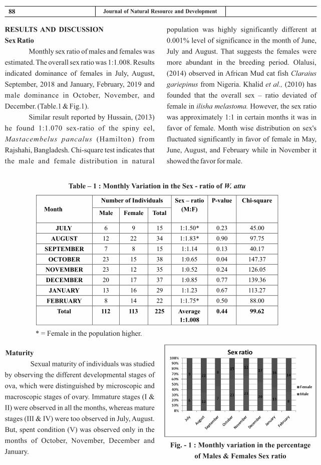
Fig. - 1 : Monthly variation in the percentage
of Males & Females Sex ratio
88 Journal of Natural Resource and Development
population was highly significantly different at
0.001% level of significance in the month of June,
July and August. That suggests the females were
more abundant in the breeding period. Olalusi,
(2014) observed in African Mud cat fish Claraius
gariepinus from Nigeria. Khalid et al., (2010) has
founded that the overall sex – ratio deviated of
female in ilisha melastoma. However, the sex ratio
was approximately 1:1 in certain months it was in
favor of female. Month wise distribution on sex's
fluctuated significantly in favor of female in May,
June, August, and February while in November it
showed the favor for male.
RESULTS AND DISCUSSION
Sex Ratio
Monthly sex ratio of males and females was
estimated. The overall sex ratio was 1:1.008. Results
indicated dominance of females in July, August,
September, 2018 and January, February, 2019 and
male dominance in October, November, and
December. (Table.1 & Fig.1).
Similar result reported by Hussain, (2013)
he found 1:1.070 sex-ratio of the spiny eel,
Mastacembelus pancalus (Hamilton) from
Rajshahi, Bangladesh. Chi-square test indicates that
the male and female distribution in natural
Table – 1 : Monthly Variation in the Sex - ratio of W. attu
* = Female in the population higher.
Month
Number of Individuals Sex – ratio
(M:F)
P-value Chi-square
Male Female Total
JULY 6 9 15 1:1.50* 0.23 45.00
AUGUST
12
22
34
1:1.83*
0.90 97.75
SEPTEMBER
7
8
15
1:1.14
0.13 40.17
OCTOBER
23
15
38
1:0.65
0.04 147.37
NOVEMBER
23
12
35
1:0.52
0.24 126.05
DECEMBER
20
17
37
1:0.85
0.77 139.36
JANUARY
13
16
29
1:1.23
0.67 113.27
FEBRUARY
8
14
22
1:1.75*
0.50 88.00
Total
112
113
225
Average
1:1.008
0.44 99.62
Maturity
Sexual maturity of individuals was studied
by observing the different developmental stages of
ova, which were distinguished by microscopic and
macroscopic stages of ovary. Immature stages (I &
II) were observed in all the months, whereas mature
stages (III & IV) were too observed in July, August.
But, spent condition (V) was observed only in the
months of October, November, December and
January.

89 Hari Prasad and A.Y. Desai
2. Gupta, S. 2015. Wallago attu (Bloch and
Schneider, 1801), a threatened catfish of Indian
waters, 5(4): 140 - 142.
3. Hussain, (2013) Length-weight relationship,
condition factor and sex-ratio of the spiny eel,
Mastacembelus pancalus (Hami l ton)
Department of Fisheries, Rajshahi University,
Rajshahi-6205, Bangladesh.
4. Kareem, O. K., Ajani, E. K., Orisasona, O. &
Olanrewaju, A. N., 2015. The Sex Ratio,
Gonadosomatic Index, Diet Composition and
Fecundity of African Pike, Hepsetus odoe
(Bloch, 1794) in Eleyele Lake, Nigeria. Journal
of Fisheries & Livestock Production, 3(3): 1- 4.
5. Khalid M.; Zarrien A. and Ghazala S., (2010).
Sex ratio maturation and spawning of the Indian
ilisha ilisha melastoma in coastal water of
pakishtan (northern Arabian sea). Central of
exacellence in marine biology, University of
Karachi, pakisthan.
6. Neethiselvan, N., Venkatramani, V. K. &
Srikrishnadas, B. 2001. Reproductive biology
of siboga squid Doryteuthis sibogae (Adam)
from Thoothukkudi (Tuticorin) coast ,
Southeast coast of India. Indian Journal of
Marine Sciences, 30(4): 257 -260.
7. Olalusi, ayo, C. I. 2014. Length weight
relationship, Condition factor and Sex ratio of
African Mud cat fish Claraius gariepinus
reared in flow through system tank. Department
of fish technology Nigeria. Journal of fisheries
and aquatic science, 9(5): 430- 434.
CONCLUSION
Present study was near Bhukhi village
Dhoraji Taluka of Rajkot district of Gujarat at
Bhadar reservoir were suitable environment
condition for W. attu fish. The overall sex ratio was
1:1.008 and the females dominated in the population
during July, August, and September. Male was
October, November and December. Stages of ovary.
Immature stages (I & II) were observed in all the
months, whereas mature stages (III & IV) were too
observed in July, August. But, spent condition (V)
was observed only in the months of October,
November, December and January. Sex ratio is play
the major role of maturity and spawanig activity of
fishes.
ACKNOWLEDGEMENTS
I am very grateful for getting an opportunity
to carry out my M.F.Sc. studies in the Department of
Fisheries Resource Management, College of
Fisheries Science, Junagadh Agricultural
University, Veraval. All my experiences in the past
years have made it an ever-good memory of my life.
It is a genuine pleasure to express my deep sense of
thanks and gratitude to my mentor and guide Dr. A.
Y. Desai, Dean and Principal College of Fisheries
J.A.U., Veraval. His dedication to work and support
had been solely responsible for completing my
work.
REFERENCES
1. Acharya, P. 1990. Studies on maturity,
spawning and fecundity of Nemipterus
japonicas (Bloch) off Bombay coast. Journal of
the Indian Fisheries Associatio, 20(1): 51 - 57.

HYPOTHETICAL DATA SOCIAL CATEGORIZATION
ON INTER GROUP CONTACT IN U.P. (INDIA)
Hargovind Bhargava* and S.M. Yadav
*Department of Agricultural Economics and Statistics
Kulbhasker Ashram P.G. College Prayagraj, U.P., India
Department of Agricultural Economics and Statistics
Ch.C.S.P.G. College, Heora, Etawah, U.P., India
Received : 05.06.2020 Accepted : 10.07.2020
INTRODUCTION
On the basic of general idea, Several
different model of Inter group contact have been
developed, each making some what different
predications of the optimal conditions for effective
contact experience.
1. Decategorization Model :-
The Hypothetical data social categorization
on Inter group in U.P. the first modal passed on the
idea that contact with be most effective in
interactions are person – based rather than
category– based (Brewer and miller, 1984) A
primary consequences of categorization is the
depersonalization of members of the out-group
Social behavior in Categorization based-
interactions is characterized by a tendency to treat
individual member of the out – group of
undifferentiated representatives of a unified social
category, impendent of individual differences that
may exist with in groups. This perspective on the
contact situations suggests that intergroup
interactions should be structured so as to reduce the
salience of category distinctions and to promote
opportunities to get to know out-group member as
individuals Attending to personals characteristics
of group member not only provide the opportunity to
disconfirm category stereo types, it also break down
the monolithic perception of the out- groups as a
homogeneous unit (wilder 1978) In this scheme ,
the complete situation encourages attention to
information at the individual lived that replaces
category identity as the most useful basic for
classifying participant , beware and miter(1984) a
spume that such contact experience effectively
breakdown stereo typing & Prejudice because
undermine the availability and usefulness of in
group – out group categorization in interactions
with group members.
2. Conditions of Intergroup Contact:-
In the year prior to All Port's framing of
intergroup contact theory, social scientists has
already begun discussing the conditions of
intergroup contact that would intergroup anxiety,
prejudice or other “ deter mental psychological
effects”. Wilner, walkly and coor, two year prior to
the nature of prejudice, studied segregation and
integration in housing projects and also suggested
four conditions. Under which intergroup attitudes
would changes for the better, under the assumption
that pre juice arise from racial segregation , they
suggested that it would diminish when members
occupy “ The same or equivalent rules in the
Journal of Natural Resource and Development 15 (2) 90-92, 2020 ISSN-0974-5033NAAS RATING : 3.46
Review Article

91
effects of intergroup contact were significantly
greater. When the contact situation was structured to
include Allport's facilitating conditions for optimal
contact.
Indirect Intergroup Contact:-
One of the most important advances in
research on intergroup contact is the growing
evidence for a number of indirect, non –face-to face
intergroup contact strategies as a means to improve
relations between social groups, while the benefits
of direct intergroup contact have been empirically
established, its implementation is offer not practical.
For example in many countries social and religious
groups are often residentially, educationally or
occupationally segregated, Which limits the
opportunity for direct contact however, even when
the opportunity for direct contact is high , Anxiety
and fear can produce a negative or positive contact
experience or head to the avoidance of the contact
situation altogether. Indirect forms of intergroup
contact include.
Extended Contact:-
The extended contac t hypothes is ,
established by Wright and colleagues in 1997, posits
that knowing that a member of one's own group has
a close relationship with a member of an out group
can lead to more positive attitudes towards that out
group correlation research had demonstrated that
individuals who report knowledge that an in group
member has an out group friend typically report
more posit ive out group att i tudes, while
experimented research as shown that providing in
group members with information creates the same
positive effect.
Imagined Contact:-
The imagined contact hypothesis was put
forward by Richard J. Crisy and Rhianan tunner
(2009) and propose that simply imagining a positive
encounter with a member or member's of on out
situation.” Share background characteristics like
education ,age, gender or socio economic status ,
perceive common interests or goods and when the “
Social climate is not Unfavorable to interracial
association.
3. Psychological Processes involved in
intergroup Contact:-
A number of psychological processes have
been hypothesized to explain how and why
intergroup contact is able to reduce prejudice and
improve intergroup relations.
1. Allport (1954) argued that intergroup
contact facilitates learning about the out-group and
this new out group knowledge leads to prejudice
reduction.
2. Intergroup contact is believed to reduces the
fear and anxiety people have when interacting
with the out-group which in turn reduces their
negative evaluation of the out group.
3. Intergroup contact hypothesized to increase
people activity to take the perspective of the out
group and empathies with their concerns.
Empirical research has only behind weak support for
role of out group knowledge in prejudice reduction
however the affective mechanisms of inter group
anxiety and out group empathy have accumulated
extensive empirical support.
4. The effects of intergroup Contact:-
Social Scientist have documented positive
effects of intergroup contact across field,
experimental and correlate at studies across a
variety of contact situations and between various
social groups, Pettigrew and Tropp's cronical 2006
data analysis of 512 separate studies found general
support for the contact hypothesis. Furthermore,
their analysis found that face- to –face contact
between group member significantly reduced
prejudice the more contact groups had the prejudice
group member reported .Moreover the beneficial
Journal of Natural Resource and Development

92
2. Poolini ,S, Harwood, J, Rubin, M (2010)
“Negative intergroup contact makes group
memberships salient Explaining why
intergroup contact endures.”
3. Groof,S poolini, SL Rubin, M (2014).
Negative intergroup, contact is more
inflyentiol but positive intergroup contact
is more comman assessing contact,
European Journal of social psychology.
4. Enos, Ryan (2017), The have between us
social Geography and politics Cambridge
University press.
5. Paluck, Elizabeth, Levy, Green, Sethoried,
Green Donald (2018)
group category can promote more positive
intergroup attitudes.
Electronic or E- Contact:-
Fiona white and her colleagues (2012-
2014) recently developed Electronic and E-
Contact, in values an in group member interacting
with an in group member interacting with an out
group member over the internet and includes text
based, video based on line interaction.
Criticisms:-
While large bodies of research have been
devoted to examine info group contact social
scientific reviews of the literature frequently
Skeptics about the likely hood of contacts optimal
condition occurring in concern and by expansion
about the generals ability of correlation research
and lab studies in contact.
REFERENCES
1. Wright, S.C. (2009) cross – group contact
effects N.S. Offer, T. Kesster and J, sqssen
org (Eds). Intergroup relations. The role of
emotion and motivation.
Hargovind Bhargava and S.M. Yadav

ABSTRACT
Atresia ani is a congenital embryological anomaly in which the hindgut fails to fully communicate with
the perineum. The anus may be either stenotic or imperforate; atresia ani may appear alone or in
combination with rectovaginal or recto vestibular fistula (RVF It is a congenital abnormality, ).
manifested clinically by an absence of faeces, dullness, and anorexia with abdominal distension,
discomfort and straining at an attempt to defecate. Rectal lumen usually bulges subcutaneously at
normal site of the anus when the abdomen is compressed. These congenital defects were corrected
surgically under caudal epidural analgesia using 2% Lignocaine hydrochloride. The present case
report describes successful surgical intervention of atresia ani in a new born ram lamb.
Keyword: Atresia ani, congenital abnormality, Rectum, Ram lamb. Lignocaine hydrochloride.
SURGICAL OPERATION OF ATRESIA ANI
(IMPERFORATE ANUS) IN A RAM LAMB
Ngangkham James Singh, Ashok Kumar yadav and Gaurav Jain
Department of Animal Husbandry and Dairying, SHUATS, Prayagraj (U.P) - 211007, India
Veterinary Medical officer, Chaka block, Prayagraj (U.P) - 211007, India
Received : 21.06.2020 Accepted : 30.07.2020
INTRODUCTION
The structural or functional defects that
occur during embryogenesis and can be identified
after birth are called inherited anomalies. The
genetic or environmental factors or sometimes
combination of both are responsible for these
defects Badawy A.M. (2011). Genetic factors like
defect in chromosome numbers, single gene defect
and recessive gene. Environmental factors such as
nutrition, maternal disease, plastic and its
components and use of pesticides in feed are
accountable for teratogenic anomalies during
embryogenesis. Sometimes these anomalies lead to
decline in the productivity of dam and economic
value of neonates. Atresia ani also known as
imperforate anus is an inherited embryological
anomaly mainly due to the failure of the anal
membrane to cessation or sometimes thin
membrane covering normal anal orifice. This defect
may progress when a dorsal part of the cloacal plate
fails to form and in female it is accompanied by
agenesis of genitalia Chaudhary G.R. et al.(2016).
Atresia ani is common genetic disorder in the male
pigs and calves which might be due to an Autosomal
recessive gene. Kilic N et al., (2004) Atresia ani is
the failure of the anal membranes to break down.
Affected animals may survive for up to ten days and
can be identified by their depression, anorexia,
colic, marked abdominal distension and lack of
faeces, faeces being replaced by thick white mucus,
Radostitis et al 2000. This surgical report
communicates a case of atresia ani (imperforate
anus) in ram lamb, which was successfully treated
by surgical intervention.
CASE HISTORY AND CLINICAL
OBSERVATIONS
A five day old male, non-descript ram lamb
was presented at Veterinary Hospital, chaka block,
Journal of Natural Resource and Development 15 (2) 93-94, 2020 ISSN-0974-5033NAAS RATING : 3.46

94 Journal of Natural Resource and Development
perineal muscles therein. This was done by putting
four stitches were dorsally, ventrally and laterally on
both sides. Post-operatively, Fortivir@ 2ml
(Enrofloxacin 10%W/V) for 5 days and Tolfine
(Tolfenamic acid) @1ml for 3 days were
administered intramuscularly, followed by routine
dressing and application of fly repellent Charmil
spray at the operative site to prevent cicatrisation.
The sutures were taken off on 8th day post-
operatively. Congenital anomalies (of digestive
system) frequently occur due to genetic or
environmental forces, or a combination of both,
during the process of embryogenesis (Oehme and
Prier, 1974; Mishra and Angelo, 1980). Animal
recovered uneventfully without any complications
on 10th day.
Prayagraj, Utter Pradesh (India) with the history of
non – passage of faeces since birth. After birth, ram
lamb was stand and suckle normally but weak. On
clinical observation, closely find with principal
clinical signs of dull, depression, anorexia, attempt
of defecation and mild abdominal distention. Also
the signs of tenesmus and abdominal pain were
observed but does not voided out the faeces. The
case was diagnosed as atresia and handover for
surgical intervention.
RESULTS AND DISCUSSION
The perineal reconstruction was undertaken
surgically under local anesthesia as described by
Frank (1964). Atresia Ani (imperforate anus) was
treated by excision of a circular piece of anal skin.
The rectum was exposed after due dissection of the
Fig. no. 1. Ram Lamb showing bulging in the perineal region due to Atresia ani
Fig 2: Photograph showing
the muconium immediately
after incision
Fig 3. Primal reconstruction (Atresia ani) & Ram
Lamb stand with minimum tenesmus immediate after
surgery.
CONCLUSION
It is concluded that, surgical intervention is
the only possible solution to treat such congenital
defects in animals to so as to make them survive.
REFERENCES
1. Badawy A.M. (2011) Benha Vet. Med. J.,
1,14 – 27.
2. Chaudhary G.R., Bhat A.R., Vikram R.,
Kharayat N.S., Suthar A.N., Kumar A.,
Katiyar R., Das G.K and Narayanan K and
Pawde A.M. (2016) Journal of Livestock
Science,7, 89-91
3. Kilic N and Sarierler M. (2004) Revue Méd.
Vét., 2004, 155, 7, 381-384.
4. Frank,E.R.(1964): Veterinary Surgery, 7th
edn, Minnapolis, Burgress Publishing
Co;pp.279-280.
5. Oehme,F.W. ,and Pr ie r, J .E . (1974) :
Textbook of large animal surgery. Williams
& Wilkins, Baltimore, U.S.A, pp.447-448.
6. Radostitis,O.M.,Gay,C.C.,Blood,D.C.,and
Hinchcliff, K.W. (2000): Veterinary
Medicine: A textbook of the diseases of
cattle, sheep, pigs, goats and horses, 9 thedn,
Saunders, Philadelphia, pp.1729.

Date:……………………..
MEMBERSHIP OF SBSRD, ALLAHABAD
Category
1. Annual
2. Life
3. Institutional
Category Indian
1. Annual Rs. 500/-
2. Life Rs. 4000/-
3. Institutional Rs. 10,000/
Declaration: I hereby declare that th e Information furnished above is true to the best of m y knowledge and belief and I am abiding by the rules of the Society of
Biological Sciences and Rural Development, Allahabad.
Signature: ……………………………………………………
Membership type (Please tick): *Life Annual
1. Name (in capital) ………………………………………
2. Designation ……………………………………………
3. Affiliation ………………………………………………
4. Address …………………………………………………
……………………………………………………………..
5. Date of Birth ……………………………………………
6. Mobile/Phone Nos ………………………………………………
7. Email ID …………………………………………………
8. Website (if any) …………………………………………
9. Academic Field …………………………………………………………………………
10. Research Field ………………………………………………………………………
11. Experience (in years) a) Research…………..……
12. Honours/Awards (Nos.) a)
13. Fellowships (Nos. only) a)
14. Publications (Nos. only)
(i)Research Papers/Rev. Articles
15. Fee Details ……………………………………………………………………………………………
…………………………………………………
……………………………………………………….
………………………………………………………….
……………………………………………………………..
……………………………………………………………...............
………………………………………………………
……………………………………………………………………
…………………………………………………………………………………
………………………………………………………………………
…………………………………………………………………………
………………………………………………………………………
Research…………..…………b) Teaching………
a) National…………………...b) International………
a) National….…………………b) International………
Research Papers/Rev. Articles….………………(ii) Books/Monographs
……………………………………………………………………………………………
…………………………………...
………………………………………………
…………………………………………….
……………………………………………………………………………………….
………………………………………………………………………………………
Teaching……………….……….
International……………….….
International……………….….
Books/Monographs…………….….
……………………………………………………………………………………………
Photo
Regd. Office: 10/96, Gola Bazar, New Jhusi, Prayagraj, (U.P.), India
(Registered under Soc. Reg. Act –1860)
APPLICATION FOR THE MEMBERSHIP OF SBSRD ALLAHABAD
The payment should be made through Demand Draft/E - Banking
favour of “Socity of Biological Sciences and Rural Development, (A/c No. 31105794798) Payable at
State Bank of India, Jhusi Branch (IFSC Code SBIN 0005440), Prayagraj, U.P., India.


![arXiv:1512.04738v1 [astro-ph.SR] 15 Dec 2015](https://static.fdokumen.com/doc/165x107/632e255700b9ef3cf20e06e3/arxiv151204738v1-astro-phsr-15-dec-2015.jpg)


![arXiv:1407.4095v1 [math.OC] 15 Jul 2014](https://static.fdokumen.com/doc/165x107/6325594f6d480576770c3f52/arxiv14074095v1-mathoc-15-jul-2014.jpg)

