JOURNAL OF SOCIAL SCIENCES, NURSING, PUBLIC ...
144
JOURNAL OF SOCIAL SCIENCES, NURSING, PUBLIC HEALTH AND EDUCATION SCIENTIFIC PEER-REVIEWED JOURNAL I ISSN 2644-6006 № 3, 2020
-
Upload
khangminh22 -
Category
Documents
-
view
4 -
download
0
Transcript of JOURNAL OF SOCIAL SCIENCES, NURSING, PUBLIC ...

JOURNAL OF SOCIAL SCIENCES, NURSING, PUBLIC HEALTH AND
EDUCATION
SCIENTIFIC PEER-REVIEWED JOURNAL
I
ISSN 2644-6006
№ 3, 2020

JOURNAL OF SOCIAL SCIENCES, NURSING, PUBLIC HEALTH AND EDUCATION
№ 3, 2020 Editorial Board
Editor in Chief:doc. PhDr. et Bc., Jarolav Stanchiak, PhD., MPH, UK, PdF Editorial board:prof. Andrzej Kryński, Ph.D.prof. MUDr. Jozef Novotný, PhD.prof. Piotr Lisowski, Ph.D.prof. RNDr. Edita Partová, PhD, PdF, UKprof. MUDr. Viktor Shatylo, PhD., Dr. h.c., UKprof. PaedDr. Alica Vanchová, CSc. PdF, UKprof. MUDr. Vitaliy Zabolotnov, PhDdoc. PhDr. Jana Boronova, PhD.doc. Svitlana Gordiichuk, Ph.D.doc. PhDr. et Bc., Jarolav Stanchiak, PhD., MPH, UK, PdFdoc. Natalia Shygonska, Ph.D.PhDr. Ján Holonich, PhD. MBA, LL.M., UK, PdFIng. Marek Nikel, MBA, (EBG)Mgr. Andrej Hutta, MBAprof. Yuriy Andrashko, DrSc.doc. Olena Yatsyna, CSc.PhDr. Oleksandr Rishko, CSc.prof. Evheny Kostenko, DrSc.prof. Oksana Klitinska DrSc.prof. Anatoly Potapchuk, DrSc.prof. Ivan Myronyuk, DrSc.prof. Hennadiy Slabkyi, DrSc.PhDr. Svetlana Steblyuk, CSc.Prof. Dr. Nick Palinchak, DrSc.Prof. JUDr. Dmytry Byelov, DrSc.JUDr. Myroslava Hromovchuk, CSc.Prof. Dr. Andriy Rusyn, DrSc. Quest editor:doc. RNDr. Edita Partová, CSc.prof. PaedDr. Bernhard Beckmann
ISSN 2644-6006

3
CONTENT
Doroshko V.A., Sokol V.V., Fedoriuk O.V., Fedoriak I.M.INFLUENCE DE HOMONES SEXUELLES SUR LA DISREGULATION POSTISCHEMIQUE DE L‘HOMOSTOSTASE ANTIOXIDANE- PROOXIDANTE DANS DES STRUCTURES CEREBRALES DE RATS DE DIFFÉRENTS ÂGES ...................................................................................5
Olena Dulo, Larisa Lyachovets, Oleksandr SuranKINESIOTHERAPY OF POST – STROKE PATIENTS DURING THE STATIONARY PERIOD OF REHABILITATION ..................................................................................................................11
Hlazunov О.А., Hruzdeva A.O., Stepanova S.V.ІNTRODUCTION OF INNOVATIONS TO ENSURE THE QUALITY OF POSTGRADUATE MEDICAL EDUCATION .................................................................................................................................17
Olesia HlukhanychPOLYCULTURE PHENOMENON OF THE TRANSCARPATHIAN COMPOSERS’ CREATIONS .......................................................................................................................................................23
Inna Horbatiuk? Iryna HorbatiukDISTANCE LEARNING THROUGH THE EYES OF MEDICAL STUDENTS OF THE 6TH COURSE OF BUKOVINIAN STATE MEDICAL UNIVERSITY ............................................................30
Myroslava HromovchukEUTHANASIA AND BIOETHICS: THEORETICAL ASPECT .............................................................34
Kovpak A.V.CHANGE INDICATORS OF FIBRINOLYSIS AND PROTEOLYSIS IN SPONTANEOUS HYPERTENSIVE RATS IN TREATMENT WITH RAMIPRIL ............................................................41
Kovpak A.V.«INFLUENCE OF CANDESARTAN ON THE ACTIVITY OF FREE RADICAL LIPID PEROXIDATION IN RATS WITH CONGENITAL ARTERIAL HYPERTENSION» ......................46
Lyakh O.I, Tovt- Korshynska M.I., Derbak M.A.THE DISEASES OF THE DIGESTIVE SYSTEM AMONG COMCOMINANT PATHOLOGY OF THE PATIENTS WITH CHRONIC OBSTRUCTIVE PULMONARY DISEASE ........................53
Lysenko V. A., Syvolap V. V., Potapenko M. S.THE LEVEL OF KIM-1 IN URINE AND CHANGES IN STRUCTURAL-GEOMETRIC AND FUNCTIONAL PARAMETERS OF THE HEART IN PATIENTS WITH CHF OF ISCHEMIC ORIGIN.................................................................................................................................................................59
Matviykiv Taras Igorovych, Rozhko Mykola Myhailovych, Gerelyuk Vitaliy IvanovychTHE PERIODONTAL STATUS AND ANALYSIS OF THE MEDICAL PROTOCOL TREATMENT OF THE COMPLICATED COURSE OF CORONAVIRUS DISEASE IN PERIODONTAL PATIENTS. ....................................................................................................................67

4 JOURNAL OF SOCIAL SCIENCES, NURSING, PUBLIC HEALTH AND EDUC ATION
Bohdan Pelekhan, Mykola Rozhko, Lyubomyr PelekhanIRRATIONAL PROSTHODONTIC TREATMENT AS AN ETIOLOGICAL FACTOR IN THE NEED FOR PRIMARY TREATMENT OF COMPLETE ABSENT DENTITION ON THE •LOWER JAW ..................................................................................................................................72
Piddubna A.A., Makoviichuk K.Y.PRINCIPLES OF RESPECT AND JUSTICE IN THE RELATIONSHIP BETWEEN MEDICAL STUDENTS AND THE PATIENT ...........................................................................................78
Volodymyr ( Ivanovych) Trishch, Andriy (Ivanovych) MysakEFFICIENCY OF TRANSURETHRAL RADIO FREQUENCY PROSTATE THERMOTHERAPY IN PATIENTS WITH CHRONIC NONBACTERIAL PROSTATITIS .........83
Trishyna Viktoriia, Gulyaev Vіtalіy MikhailovichEFFECT OF BIOLOGICALLY ACTIVE ADDITIVES AND CAROTENOIDS OF NATURAL ORIGIN IN THE DIET OF BROILLER CHIKENS ON BLOOD BIOCHEMICAL PARAMETERS ...................................................................................................................................................89
Zeleniuk O.DIFFERENTIAL DIAGNOSIS OF FUNCTIONAL AND ORGANIC DISORDERS IN PATIENTS WITH EXTRAHEPATIC CHOLESTASIS .............................................................................93
Byelov DmytroLEGAL EDUCATION IN UKRAINE: ISSUES OF GAINING PRACTICAL EXPERIENCE BY STUDENTS ............................................................................................................................................... 101
O.Ya. Bilynskyi, Ye.Ya. KostenkoCOMPARATIVE ANALYSIS OF THE DENTAL STATUS OF MONOZYGOTIC AND DIZYGOTIC TWINS ...................................................................................................................................... 104
Konoplitskyi V.S. Shavliuk R.V. Shavliuk V.M. Kyrychenko O.P.WIDERSPRUCH ZUM PROBLEM DER ANGEBORENEN UND ERWORBENEN ÄTIOLOGIE DER PILONIDALE KRANKHEIT BEI KINDERN ....................................................... 110
Konoplitskyi Viktor, Korobko YuriiIMPROVING THE EFFICIENCY OF DIAGNOSIS OF ACUTE APPENDICITIS IN FEMALE CHILDREN THROUGH THE USE OF ANAL MANOMETRY AND TOTAL INDEX OF ENDOGENOUS INTOXICATION ........................................................................................ 119
Konoplitskyi Viktor, Pasichnyk OlehSUTURING OF POSTOPERATIVE WOUNDS IN CHILDREN WITH DIFFERENT THICKNESS OF SUBCUTANEOUS FAT AS ONE OF THE MOMENTS OF IMPROVING THE QUALITY OF SURGERICAL TREATMENT ................................................................................ 125
Kostenko Yevhen, Kostenko Svitlana, Stetsyk Mariia Pirchak IlyaTHE EFFECT OF LONG-TERM IONIZING RADIATION ON ORGANS AND SYSTEMS OF THE HUMAN BODY .............................................................................................................................. 131
Myronyuk Ivan, Bilak-Lukianchuk Viktoria, Slabkyi GennadiyON THE ISSUE OF METHODOLOGICAL AND PEDAGOGICAL BASIS OF TEACHING PUBLIC HEALTH MASTERS THE SUBJECT “ORGANIZATION AND PRESENTATION OF SCIENTIFIC RESEARCH” .....................................................................................................................................................137

5
UDK 612,82: 612.013: 612.018
INFLUENCE DE HOMONES SEXUELLES SUR LA DISREGULATION POSTISCHEMIQUE DE L‘HOMOSTOSTASE ANTIOXIDANE- PROOXIDANTE DANS DES STRUCTURES CEREBRALES DE RATS DE DIFFÉRENTS ÂGES
Doroshko V.A., Sokol V.V., Fedoriuk O.V., Fedoriak I.M.Institut de physiologie pathologique Université d’État d’Ukraine “Université de médecine d’État de Bucovine” Pl. Theatre, 2, Chernivtsi, Ukraine, 58002
Résumé: L’influence de la castration sur les caractéristiques de stress oxydatif dans la structure du cortex et de l’hippocampe de rats d’un et trois mois a été étudiée. On a établi que la castration modifie le profondeur et la portée des modifications post-isémiques dans les structures cérébrales des animaux des deux groupes d’âge. L’influence de l’absence des hormones sexuelles sur les modifications post-traumatiques, liée à l’âge, se manifeste principalement dans les structures de la cortex cérébral.
Mots clés: ischémie carotidienne, testostérone, progestérone, cortex cérébral, hippocampe, hormones sexuelles, lésions posttishémiques, rats mâles.
Abstract. The authors have studied the effect of the castration on the parameters of oxidant stress in the structures of the cortex and hippocamp of one and three month old rats. It has been established that castration considerably widens and deepens the range of post-ischemic changes in the structures of the brain of animals of both age groups. The most marked age-related peculiarities of the effect of the deficiency of the sex hormones on the post-ischemic changes occur in the cortical structures.
Key words: carotid ischemia, testosterone, progesterone, cortex, hippocamp, sex hormones, postischemic damage, male rats, age peculiarities.
Introduction.Les vues modernes sur le rôle des stéroïdes sexuels dans le développement et les
progrès de la pathologie vasculaire cérébrale restent discursives. Selon une observation expérimentale, les hormones sexuelles et leurs analogues synthétiques sont capables de réduire l’effet vasoconstricteur de nombreux composés biologiquement actifs - prostaglandine F2α, thromboxane, noradrénaline, vasopressine, endothéline et d’autres, dont la grande majorité sont sécrétés pendant une ischémie en quantités élevées[2, 9]. L’effet vasodilatateur des stéroïdes sexuelles peut être lié avec leur effet sur le système rénine-angiotensine-aldostérone, et aussi avec les effets modulateurs sur la réactivité des récepteurs α-adrénergiques[4]. Par conséquent, on peut penser que la capacité des vaisseaux cérébraux à contrer les effets indésirables, dépend du contenu des hormones sexuelles.
Cependant, il existe un autre point de vue selon lequel les stéroïdes sexuels, modifiant la dynamique de la synthèse et de la libération de composés endogènes biologiquement actifs, peuvent également augmenter le risque de pathologie vasculaire.[2].

6 JOURNAL OF SOCIAL SCIENCES, NURSING, PUBLIC HEALTH AND EDUC ATION
Une telle ambiguïté prouve la nécessité de rechercher le rôle des hormones sexuelles dans l’évolution des changements post-traumatiques. Le stress oxydatif est l’un des mécanismes déclencheurs des lésions ischémiques de reperfusion dans le cerveau. L’étude de l’effet de la castration sur ses paramètres peut fournir de nouvelles informations sur les parties de la pathogenèse auxquelles ces hormones sont capables d’agir.
Matériels et méthodes.L’étude a été menée sur des mâles blancs rats, sans l`espèce, des âgés de 1 et 3 mois (96
animaux utilisés au total). Toutes les interventions et les abattages d’animaux ont été menés simultanément dans des groupes expérimentaux et des groupes témoins, conformément aux principes internationaux de la Convention européenne sur la protection des animaux vertébrés utilisés à des fins expérimentales ou à d’autres fins scientifiques (Strasbourg, 1985) et du Premier Congrès national de bioéthique (Kyiv, 2000). En regardant du temps de maturation du système neuroendocrinien chez les rats, ces tests ont été effectuées sur des animaux âgés d’un à trois mois. Les groupes témoins sont présentés chez des animaux des deux groupes d’âge, réalisés par la peau et la sécrétion des artères carotides sans être déplacés. Chez le rat du premier groupe expérimental, l’ischémie globale incomplète du cerveau a été modélisée par une coupure de 20 minutes des deux artères carotides communes.[8]. La période de reperfusion a durée 5 jours. Toutes les interventions chirurgicales ont été réalisées sous anesthésie au calypsole (75 mg / kg de poids corporel).
Le deuxième groupe expérimental était constitué d’animaux chez lesquels une intervention similaire avait été effectuée deux semaines après la castration. La période de reperfusion a duré 5 jours. Le sixième jour, l’euthanasie des animaux a été réalisée sous anesthésie légère dans l’éther. Selon l’atlas des coordonnées stéréotaxiques [9], on prenait l’écorce frontale, occipitale et les champs de l’hippocampe CA1, CA2, CA3. Les homogénats de ces structures ont déterminé la teneur en conjugués diène [3], aldéhyde malonique [6], activité de la superoxyde dismutase [7], de la glutathion peroxydase [1], de la catalase [5].
Le traitement statistique a été effectué par T-critère de Student.Les études expérimentales et l’euthanasie d’animaux ont été réalisées conformément
aux principes internationaux de la Convention européenne sur la protection des animaux vertébrés utilisés à des fins expérimentales ou à d’autres fins scientifiques (Strasbourg, 1985).
Résultats de la recherche et de leur discussion.L’analyse des études expérimentales a montré que chez les animaux d’un mois, les
effets retardés des lésions de reperfusion ischémique dans le cortex frontal consistaient à réduire l’activité de la catalase, ce qui réduisait la capacité du potentiel antioxydant (tableau 1). Les modifications post-traumatiques chez les animaux castrés étaient plus prononcées et se manifestaient par une augmentation de la teneur en aldéhyde malonique et une diminution significative de l’activité de la catalase et de la superoxyde dismutase, indiquant une augmentation prononcée des réactions des radicaux libres. Dans le cortex occipital, l’ischémie a entraîné une diminution du niveau fonctionnel de l’activité du système antioxydant-prooxydant en raison de la diminution simultanée de l’intensité de la lipoperoxydation et de l’activité de la glutathion peroxydase, tandis que la castration augmentait le contenu postchémique de conjugués de diène et d’aldéhyde malonique et d’activité antioxydante , c`est plus dangereux que réduxion simplement de niveau de fonctionnement du système.
Dans le domaine de l’hippocampe CA1, l’ischémie a réduit l’activité de la superoxyde dismutase et de la catalase et l’ischémie chez les animaux castrés a réduit le contenu des produits de lipoperoxydation et, de manière plus significative, l’activité d’enzymes à

DOROSHKO V.A., SOKOL V.V., FEDORIUK O.V., FEDORIAK I.M. 7
défense antioxydante (à l’exception du superoxyde dismutase) cela indique l’épuisement du système dans son ensemble, avec une défaite prédominante du composant antioxydant.
Changements post-traumatiques dans le champ de l’hippocampe CA2-diminution de la teneur en conjugués de diène et de l’activité de la superoxyde dismutase. La castration a intensifié les modifications postishémiques de la lipoperoxydation et affaibli l’effet de l’ischémie sur l’activité de la superoxyde dismutase, mais il existait une inhibition extrêmement prononcée de l’activité de la catalase.
Dans le domaine de la CA3, le déclin postmachémique de tous les paramètres étudiés (à l’exception de l’activité de la superoxyde dismutase) est très perceptiblement après la castration.
Chez les animaux plus âgées, dans le cortex frontal, l’ischémie a entraîné des modifications plus importantes qu’en un mois. Ils consistaient en une augmentation significative de l’intensité de la lipoperoxydation avec une augmentation moins significative de l’activité des enzymes antioxydantes (tableau 2). L’ischémie, réalisée après la castration, a eu des effets similaires, mais moins prononcés, sur la peroxydation lipidique et l’inverse (à l’exception de l’activité de la glutathion peroxydase) sur l’activité antioxydante.
Dans le cortex occipital, l’ischémie a entraîné une augmentation extrêmement prononcée des produits de la lipoperoxydation et une augmentation relativement faible de l’activité de la superoxyde dismutase, cela indique un changement significatif de l’équilibre prooxydant-antioxydant dans la direction du renforcement des processus des radicaux libres. Dans le cortex occipital, l’ischémie a entraîné une augmentation extrêmement significative des produits de la lipopéroxydation et une augmentation relativement faible de l’activité de la superoxyde dismutase, indiquant un fort déplacement de l’équilibre prooxydant-antioxydant vers le renforcement des processus des radicaux libres. Ainsi, la castration provoque également l’activation des processus prooxydants post-isémiques dans le cortex, mais contrairement à l’ischémie chez les animaux témoins, principalement en raison de l’affaiblissement de la défense anti-oxydante.
Il est important de noter que, dans les structures du cortex des deux groupes d’âge, la castration a augmenté l’activité de la glutathion peroxydase, malgré la diminution de l’activité des enzymes antioxydantes restantes.
Dans le domaine de l’hippocampe CA1, l’ischémie a entraîné une diminution de l’activité de la catalase et de la glutathion peroxydase et la castration a entraîné une diminution du contenu postisémique des conjugués de diène à un niveau de défense anti-oxydant deux fois plus forte.
L’amélioration post-ischémique des processus radicalaires dans le domaine de la CA2 est apparue en augmentant la teneur en conjugués de diène et en réduisant l’activité de toutes les enzymes antioxydantes. L’interférence ischémique après la castration a réduit l’intensité de la lipoperoxydation due aux deux produits, dont le niveau était même inférieur à celui des animaux témoins, mais entraînait une réduction encore plus prononcée de l’activité de la catalase qu’après une ischémie témoin. Il convient de noter que l’activité de la superoxyde dismutase est revenue dans le même temps à la normale.
Dans le champ CA3 de l’hippocampe de trois mois de rats, l’ischémie n’a provoqué aucun changement retardé; toutefois, après la castration, l’interférence ischémique-reperfusion a réduit l’intensité de l’oxydation des lipides peroxydes et de l’activité antioxydante, et le système est passé à un nouveau niveau de fonctionnement plus faible.
En conséquence, le manque d’hormones sexuelles modifie la nature de l’effet de l’ischémie sur les indicateurs de stress oxydatif dans les structures cérébrales étudiées chez les animaux des deux groupes d’âge.

8 JOURNAL OF SOCIAL SCIENCES, NURSING, PUBLIC HEALTH AND EDUC ATION
Conclusions:1. La gravité et, dans certains cas, la direction de l’effet de la castration sur les
paramètres du potentiel prooxydant-antioxydant ont une dépendance structurelle et liée à l’âge.
2. Dans la plupart des structures cérébrales étudiées chez les animaux des deux groupes d’âge, la castration réduit considérablement le potentiel antioxydant postisémique.
3. Les caractéristiques d’âge de l’influence du déficit en hormones sexuelles sur les indicateurs de stress oxydatif dans les lésions cérébrales de reperfusion ischémique sont particulièrement visibles dans les structures du cortex.
Perspectives de développement futur. Les résultats obtenus indiquent un effet modificateur expressif de la castration sur
les modifications postishémiques tardives des structures cérébrales, ce qui confirme la nécessité d’étudier le rôle des hormones sexuelles dans l’apparition de lésions de reperfusion ischémique du système nerveux central.
Littérature.1. Sébastien Déglise, Céline Dubuis, Pascal Mosimann, Stefan Engelberger, François
Saucy, Lorenz Hirt, Patrik Michel, Jean-Marc Corpataux, Jean-Marc Corpataux , “Prise en charge des sténoses carotidiennes” Rev Med Suisse 2013; volume 9. 1305-1311, 2012
2. Esra Birben, Umit Murat, Sahiner, Cansin Sackesen, Serpil Erzurum, Omer Kalayci, “Oxidative Stress et Protection Antioxidante” World Allergy Organization Journal 20125:270 World Allergy Organization; licensee BioMed Central Ltd., 2012
3. S. R. Wainwrigh, M. M. Roes, P. Duarte–Guterman, C. Chow, D. K. Hamson “Sexe, hormones et neurogenèse dans l’hippocampe: modulation hormonale de la neurogenèse et implications fonctionnelles potentielles”, 2012
4. Doris P. Molina, Olusegun J. Ariwodola, Jeff L. Weiner, Judy K., Brunso-Bechtold, Michelle M. Adams,”L’hormone de croissance et le facteur de croissance analogue à l’insuline I modifient la transmission synaptique excitatrice de l’hippocampe chez des rats jeunes et âgés”, 2012
5. NataliaPavóna, EduardoMartínez-Abundisa, Luz Hernándeza, Juan Carlos Gallardo-Péreza, Carolina Alvarez-Delgadoc, Marco Cerbónc, Israel Pérez-Torresc, Alberto Arandab, Edmundo Cháveza, “Hormones sexuelles: Effets sur l’activité cardiaque et mitochondriale après ischémie – reperfusion chez le rat adulte. Différence de genre”,2012
6. H. Cui, Y. Kong, and H. Zhang, “Oxidative stress, mitochondrial dysfunction, and aging,” Journal of Signal Transduction, vol. 2012, Article ID 646354, 13 pages, 2012.
7. W. Dröge, “Free radicals in the physiological control of cell function,”Physiological Reviews, vol. 82, no. 1, pp. 47–95, 2002
8. Benjamin A.Wagnera, Valerie C.Braddicka, Christopher G.Batsona, Brendan H.Cullena, L. Erin Millera, Mark D.Spritzerab, “Effects of testosterone dose on spatial memory among castrated adult male rats”,2018
9. R. Gredilla, “DNA damage and base excision repair in mitochondria and their role in aging,” Journal of Aging Research, vol. 2011, Article ID 257093, 9 pages, 2011.
10. Luis F. Jacome, Ketti Barateli, Dina Buitrago, Franklin Lema, Maya Frankfurt, Victoria N. Luine, “Gonadal Hormones Rapidly Enhance Spatial Memory and Increase Hippocampal Spine Density in Male Rats”, 2016
11. Thibault Cholvin “Rôle d’un circuit hippocampo-cortico-thalamique dans les processus de mémoire spatiale chez le rat”, 2016

DOROSHKO V.A., SOKOL V.V., FEDORIUK O.V., FEDORIAK I.M. 9
Tableau 1. Influence de l’ischémie sur la teneur en oxydes de peroxydes de lipides et sur l’activité
d’enzymes antioxydantes dans les structures cérébrales de rats castrés d’un mois
Groupe sur la montre
Contenu Activité des enzymes
conjugués de diène (nmol /
mg de protéine)
aldéhyde malonique
(nmol / mg de protéine)
superoxyde dismutase
(unité / min ∙ mg de
protéine)
catalase (μmol / min ∙ mg de
protéine)
glutathion peroxydase (nmol G-SH-min ∙ mg de
protéine)
Frontale cortexContrôle 7,27±0,93 3,05±0,37 6,70±0,69 18,24±2,35 4,79±0,50
Ischémie 6,02±0,51 3,07±0,30 6,41±0,49 11,86±1,84рк<0,05 4,16±0,32
Castration8,42±0,23
рі<0,005
4,68±0,20рк<0,01рі<0,005
3,02±0,17рк<0,005рі<0,005
7,17±1,41рк<0,01рі<0,05
5,09±0,17
рі<0,05Base cortex
Contrôle 7,18±1,26 4,75±0,31 9,70±0,43 15,71±1,28 5,95±0,26
Ischémie 4,44±0,78рк<0,005
3,30±0,33рк<0,05
7,06±1,16 15,44±1,24 4,69±0,37рк<0,05
Castration8,03±0,24
рі<0,005
4,42±0,20
рі<0,0125
4,01±0,53рк<0,005рі<0,005
9,40±1,05рк<0,005рі<0,005
5,48±0,13
рі<0,05
Champ de l’hippocampe CA1Contrôle 10,83±1,43 5,59±0,23 6,31±0,12 39,71±1,91 8,57±0,43
Ischémie 11,52±0,61 4,95±0,34 4,96±0,44рк<0,01
33,84±2,21рк<0,05
8,12±0,62
Castration7,95±0,74рк<0,05рі<0,05
3,90±0,41рк<0,05рі<0,05
4,94±0,37рк<0,01
8,51±0,73рк<0,05рі<0,05
5,12±0,27рк<0,005рі<0,005
Champ de l’hippocampe CA2Contrôle 19,89±1,23 5,83±0,37 5,49±0,56 25,12±2,00 5,00±0,28
Ischémie 16,11±1,17рк<0,05 5,5±0,40 2,67±0,23
рк<0,005 21,61±2,19 4,76±0,40
Castration9,86±0,84рк<0,005рі<0,005
4,40±0,44рк<0,05рі<0,05
4,13±0,32рк<0,05рі<0,05
7,94±1,03рк<0,005рі<0,005
5,17±0,49
Champ de l’hippocampe SA3Contrôle 31,38±2,25 8,66±0,78 4,20±0,33 30,22±2,34 7,22±0,31
Ischémie 24,72±1,81рк<0,05
6,49±0,52рк<0,05 3,82±0,35 20,12±1,31
рк<0,0055,35±0,44рк<0,005
Castration9,12±1,43рк<0,005рі<0,005
4,95±0,49рк<0,005рі<0,05
3,63±0,27 8,72±1,016рк<0,005рі<0,005
5,92±0,21рк<0,01
Notes: ici et dans le tableau suivant - la probabilité de changements par rapport à: pk - les indicateurs chez les animaux témoins; pi - indicateurs après ischémie.

10 JOURNAL OF SOCIAL SCIENCES, NURSING, PUBLIC HEALTH AND EDUC ATION
Tableau 2. Influence de l’ischémie sur la teneur en oxydes de peroxydes de lipides et sur l’activité
d’enzymes antioxydantes dans les structures cérébrales de rats castrés à trois mois
Groupe sur la montre
Contenu Activité des enzymes
conjugués de diène (nmol
/ mg de protéine)
aldéhyde malonique
(nmol / mg de protéine)
superoxyde dismutase
(unité / min ∙ mg de protéine)
catalase (μmol / min ∙ mg de
protéine)
glutathion peroxydase (nmol G-SH-min ∙ mg de
protéine)
Frontale cortexContrôle 5,52±0,59 2,50±0,33 6,035±0,77 13,26±1,00 3,25±0,22
Ischémie 15,40±0,23рк<0,005
11,19±0,17рк<0,005 6,44±6,57 21,69±4,31
рк<0,054,03±0,21рк<0,05
Castration7,85±0,33рк<0,01рі<0,005
4,35±0,22рк<0,005рі<0,005
2,95±0,51рк<0,01рі<0,005
7,79±0,75рк<0,05рі<0,005
5,36±0,19рк<0,005рі<0,005
Base cortexContrôle 5,40±0,82 3,68±0,36 5,62±1,59 20,75±5,01 4,81±0,29
Ischémie 40,08±3,54рк<0,005
29,17±2,11рк<0,005
9,66±1,18рк<0,05 19,87±4,33 4,42±0,39
Castration7,80±0,54рк<0,05рі<0,005
4,69±0,17рк<0,05рі<0,005
3,82±0,28
рі<0,005
7,89±0,97рк<0,05рі<0,05
5,56±0,24рк<0,05рі<0,05
Champ de l’hippocampe CA1
Contrôle 15,64±0,92 4,57±0,43 5,88 ± 0,49 29,23 ±1,98 9,42 ± 0,82
Ischémie 13,12±1,35 4,48 ±0,31 4,95 ±0,41 14,74±1,28 рк<0,005 7,31±0,47 Рк<0,05
Castration7,47±1,05рк<0,005рі<0,01
4,10±0,27 4,10±0,26рк<0,01
10,01±1,23рк<0,005рі<0,05
5,24±0,47рк<0,005рі<0,01
Champ de l’hippocampe CA2
Contrôle 11,69±1,20 6,53±0,52 5,08±0,45 42,04 ±3,17 8,83±0,72
Ischémie 15,96 ±0,95 рк<0,0125 5,24±0,50 3,12 ±0,32
Рк<0,00521,56 ±1,22 рк<0,005
4,33±0,32 рк<0,005
Castration8,32±0,75рк<0,025рі<0,005
3,34±0,24рк<0,005рі<0,01
5,32±0,17
рі<0,005
6,39±0,84рк<0,005рі<0,005
4,65±0,21рк<0,005
Champ de l’hippocampe SA3
Contrôle 23,25±2,41 5,38±0,41 4,31±0,40 13,55±1,09 5,10±0,46
Ischémie 22,39 ±1,09 5,59 ± 0,34 4,74 ± 0,35 13,95±1,12 4,11±0,34
Castration7,69±0,78рк<0,005рі<0,005
3,86±0,35рк<0,0125рі<0,0125
3,38±0,66рк<0,05рі<0,05
6,29±1,28рк<0,005рі<0,005
4,87±0,26

11
KINESIOTHERAPY OF POST – STROKE PATIENTS DURING THE STATIONARY PERIOD OF REHABILITATION
Olena Dulo,Docent of the Department of Clinical Disciplines of the Uzhhorod National University1, Candidate of Medical Science, Full docent orcid.org/0000-0003-0473-5605, Researcher ID is: F-8276-2019 e-mail: [email protected]
Larisa Lyachovets,Lecturer of the Department of Physical Rehabilitation
Oleksandr Suran,Head doctor of the Svalyava Central District Hospital
Summary. Improving the effectiveness of rehabilitation of patients with acute cerebrovascular accident (ACVA), based on the correction of motor disorders, normalization of muscle tone, increasing muscle strength by kinesiotherapy in the stationary period of rehabilitation.
The study included 24 patients with a diagnosis of ACVA, aged 50 to 70 years, who were in the stationary period of rehabilitation in the neurological department of Svalyava Central District Hospital, Transcarpathia. The diagnosis was established on the basis of the clinical picture, data of computer tomography, magnetic-resonance tomography, laboratory diagnostics, and anamnesis data. Patients were divided voluntarily into the main group (MG) – 12 people and the control group (CG) – 12 patients. CG patients were treated according to standard methods, as compiled on the basis of “Guidelines for rehabilitation of persons with ACVA “. Patients MG received kinesiotherapy. It includes features of the technique of kinesiotherapy for spastic hemiparesis, positional treatment, kinesiotherapy exercises for fingers and hands, kinesiotherapy exercises for the lower extremities, kinesiotherapy paralysis and the use of kinesiotherapy for vestibular syndrome. Classes in groups were conducted daily, duration 45 minutes, throughout the treatment period (3 weeks). Comparison of the effectiveness of the kinesiotherapy program in patients from MG and CG was performed using functional and neurological tests and scales, namely: testing of muscle spasticity on the Ashford scale, manual muscle test, modified Rankin scale, Scandinavian scale, Bartell index of activity of daily life, Orgogozo test, goniometry. Statistica 7.0 applications were used for mathematical processing of numerical data and IBM SPSS Statistics 21. T – Student’s criterion was used to assess the significance of the difference in the presence of the normal distribution results of study.
During studying the condition of the studied patients in the process of rehabilitation, it can be argued that in all groups there is a positive dynamics because of improves neurological and functional status of patients, because of increasing the amplitude of movements in the joints of the upper and lower extremities, reducing muscle spasticity, improving the ability to self-care, increasing mobility and independence in everyday

12 JOURNAL OF SOCIAL SCIENCES, NURSING, PUBLIC HEALTH AND EDUC ATION
life. These results confirm statistically better efficiency program of physical therapy, received by the patients of the main group.
Key words: stroke, physical therapy, rehabilitation, goniometry, scales
Introduction. There is a tendency of increasing of neurological diseases in Ukraine and in other countries nowadays. In the structure of neurological pathology, the most relevant and socially significant are vascular diseases of the brain, among which the leading place is occupied by acute cerebrovascular disorders [1, 2]. More than 5 million people die from strokes in the world every year [3].
Every year from 100 to 120 thousand residents of Ukraine suffer from a stroke at first time [4, 5], (more than a third of them are people of working age). 30-40% of stroke patients die within the first 30 days and up to 50% - within 1 year from the onset of the disease, 20–40% of surviving patients become dependent on outside help, and only about 10% return to full life [6].
The affections of the complex motor systems, which occur as a result of ischemic stroke, are not manifested by stereotyped motor deficit and almost always represented by a difficult and undetermined clinical structure [1, 7].
Motor disorders develop in 75% of patients in the acute period of the disease and the resistant motor defect still exist in 53% of patients, who have suffered a stroke even after six months [6].
High level of disability, which is characterized for this pathology, in most cases is associated with impairment of the motor function [1, 2, 4]. Stroke can disrupt some part of the statolocomotor system, also the significant amount of suffered from hemispheric stroke patients have a complex statolocomotor defect, which is different in nature and severity. It can not be explained by only one of the factors. It can be only the complex of factors such as: degree of hemiparesis, spasticity, sensory disorders and other disorders.
Most patients who have suffered a stroke and survive become disabled (70-80%), and 20-25% of them for the end of their lives need outside help in everyday life [1, 3, 5]. First of all, restoring the functions of movement and support, it is impossible not to use in the process of treatment the natural function of movement, inherent in the affected system. Therefore, kinesiotherapy occupies a special place in the system of treatment of motor disorders. Kinesiotherapy, as one of the leading means of physical rehabilitation, has its effect on the patient’s organism using the therapeutic effect of exercise [1, 6, 7].
Therefore, the problem of physical therapy of post-stroke patients is extremely important.Presentation of the main material of the articleThe aim of the study. Improving the effectiveness of rehabilitation of patients with
acute cerebrovascular accident (ACVA), based on the correction of motor disorders, normalization of muscle tone, increasing muscle strength by kinesiotherapy in the stationary stage of rehabilitation.
Materials and methods. The study included 24 patients with a diagnosis of ACVA, aged 50 to 70 years, who were in the stationary stage of rehabilitation in the neurological department of Svalyava Central District Hospital, Transcarpathia.
The diagnosis was established on the basis of the clinical picture, data of computer tomography, magnetic-resonance tomography, laboratory diagnostics, and anamnesis data. All patients were conscious and were available for verbal contact at the time of examination. The study was conducted with the consent of patients and did not contradict generally accepted ethical standards. Patients were divided voluntarily into the main group (MG) - 12 people and the control group (CG) - 12 patients. CG patients were treated

OLEKSANDR SURAN, 13
according to standard methods, as compiled on the basis of «Guidelines for rehabilitation of persons with ACVA «. Patients MG received kinesiotherapy. There was developed the intervention for the MG patients according to the individual capabilities and needs of each patient. It includes features of the technique of kinesiotherapy for spastic hemiparesis, positional treatment, kinesiotherapy exercises for fingers and hands, kinesiotherapy exercises for the lower extremities, kinesiotherapy paralysis and the use of kinesiotherapy for vestibular syndrome. Classes in groups were conducted daily, duration 45 minutes, throughout the treatment period (3 weeks). At the end rehabilitatoin, a repeated, final examination was performed for all patients in the relevant domains.
Comparison of the effectiveness of the kinesiotherapy program in patients from MG and CG was performed using functional and neurological tests and scales, namely: testing of muscle spasticity on the Ashford scale, manual muscle test, modified Rankin scale, Scandinavian scale, Bartell index of activity of daily life, Orgogozo test, goniometry. Statistica 7.0 applications were used for mathematical processing of numerical data and IBM SPSS Statistics 21. T – Student’s criterion was used to assess the significance of the difference in the presence of the normal distribution results of study.
Results and discussion. At baseline functional status of persons who have had ACVA, statistical analysis
found no significant differences between patients of MG and CG, p>0,05 (Table 1).
Table 1. Indicators of functional and neurological state of patients in the studied groups who have had
ACVA before the rehabilitation, n=24 (М±m)
Main group (MG) Control group (CG) d t p
Parameter indicator of Ashfort scale, pointvalue 3.00±0.14 3.08±0.13 0.08 0.42 р>0.05
Parameter indicator of manual-muscle test, %value 27.50±1.01 28.58±1.48 1.08 0.73 р>0.05
Parameter indicator of Rankin scale, pointvalue 3.75±0.14 4.00±0.19 0.25 1.04 р>0.05
Parameter indicator of Scandinavian scale, pointvalue 12.92±0.79 13.08±0.64 0.16 0.16 р>0.05
Parameter index of test Bartella, pointvalue 28.33±0.79 30.42±1.09 2.09 1.56 р>0.05
Parameter index of test Orgogozo, pointvalue 58.33±0.85 58.35±0.89 0.02 0.02 р>0.05
Parameter indicator of goniometry (shoulder flexion), degvalue 148.42±1.61 145.42±1.98 3.00 1.18 р>0.05
Parameter indicator of goniometry (shoulder extension), degvalue 40.33±1.43 40.25±1.61 0.08 0.04 р>0.05
Parameter indicator of goniometry (adduction the shoulder joint), degvalue 29.25±0.98 29.58±1.12 0.33 0.22 р>0.05
Parameter indicator of goniometry (abduction the shoulder joint), degvalue 140.42±1.40 136.92±1.36 3.5 1.79 р>0.05

14 JOURNAL OF SOCIAL SCIENCES, NURSING, PUBLIC HEALTH AND EDUC ATION
Main group (MG) Control group (CG) d t p
Parameter indicator of goniometry (flexion in the elbow joint), degvalue 134.75±1.39 136.92±1.08 2.17 1.23 р>0.05
Parameter indicator of goniometry (flexion in the hip joint during extension in the knee joint), deg
value 77.58±1.44 75.92±1.64 1.66 0.76 р>0.05
Parameter indicator of goniometry (flexion in the hip joint during flexion in the knee joint), deg
value 104.75±1.75 103.08±1.07 1.67 0.81 р>0.05Parameter indicator of goniometry (flexion in the knee joint), deg
value 117.33±0,83 118.76±1,41 1,43 0.87 р>0.05
Note: d – the difference between the average values; t – the value of the Student’s criterion; М – the average value, р – the significance of the difference between patients with MG and CG.
A comparison of the dynamics of changes in the study between patients in the main and control groups is presented in Table 2. During studying the condition of the studied patients in the process of rehabilitation, it can be argued that in all groups there is a positive dynamics because of improves neurological and functional status of patients. Thus, we found that the indicators of the Ashfort muscle spasticity test were reduced significantly in patients of the MG of 2.08±0.26 points compared with patients in the CG of 2.66±0.13 points (p<0.05), which indicates about reducing the difficulty of movements in the extremities; the indicator of manual-muscle test increases significantly in patients of the MG 33.99±1.98% (p<0.01) in contrast to patients in the CG 29.47±0.82%, which indicates an improvement in motor function in limbs; the value of the Rankin scale is reduced significantly in patients of the MG 2.70±0.36 points (p <0.05), which indicates an increase in the level of functional independence of the examined patients, while in patients of the CG the indicator decreases, but the changes are insignificant 3.65±0.14 points (p>0.05); the indicator of Scandinavian scale increases significantly in patients of the MG to 14.98±0.37 points, in contrast to patients in the CG 13.70±0.49, who have a tendency to increase value the Scandinavian scale, which confirms the reduction of neurological deficit; the indicator of the Bartell index of activity of daily life increases significantly in patients of the MG 33.25±0.43 points (p<0.05), which leads to greater independence of the patient in household activities, while in patients of the CG changes in the Bartell’ index unreliable 31.33±0.85 points (p>0.05); the indicator of Orgogozo test increased significantly in patients of the MG 63.54±0.95 points (p<0.05), which indicates an improvement in communication and motor functions of patients, while in patients of the CG positive changes in Orgogozo test are insignificant 60.95±0.95 points (p>0.05).
Table 2. Indicators of functional and neurological state of patients in the studied groups who have had
ACVA after the rehabilitation, n=24 (М±m)
Main group (MG) Control group (CG) d t PParameter indicator of Ashfort scale, point
value 2.08±0.26 2.66±0.13 0.58 2.15 р<0.05Parameter indicator of manual-muscle test, %

OLEKSANDR SURAN, 15
value 33.99±1.98 29.47±0.82 4.52 2.79 р<0.01
Main group (MG) Control group (CG) d t PParameter indicator of Rankin scale, point
value 2.70±0.36 3.65±0.14 0.95 2.44 р<0.05Parameter indicator of Scandinavian scale, point
value 14.98±0.37 13.70±0.49 1.28 2.06 р<0.05Parameter index of test Bartella, point
value 33.25±0.43 31.33±0.85 1.92 2.13 р<0.05Parameter index of test Orgogozo, point
value 63.54±0.95 60.95±0.95 2.59 2.38 р<0.05Parameter indicator of goniometry (shoulder flexion), deg
value 156.73±1.34 152.12±1.78 4.61 2.07 р<0.05Parameter indicator of goniometry (shoulder extension), deg
value 48.95±1.51 44.85±1.12 4.10 2.18 р<0.05Parameter indicator of goniometry (adduction the shoulder joint), deg
value 35.45±1.12 32.51±0.81 2.94 2.13 р<0.05Parameter indicator of goniometry (abduction the shoulder joint), deg
value 145.22±1.05 140.92±1.39 4.30 2.47 р<0.05Parameter indicator of goniometry (flexion in the elbow joint), deg
value 142.92±0.98 139.85±1,05 3.07 2.13 р<0.05
Parameter indicator of goniometry (flexion in the hip joint during extension in the knee joint), deg
value 85.92±1.85 81.22±0.99 4.70 2.24 р<0.05
Parameter indicator of goniometry (flexion in the hip joint during flexion in the knee joint), deg
value 113.55±1.37 108.28±1.75 5.27 2.37 р<0.05Parameter indicator of goniometry (flexion in the knee joint), deg
value 126.33±1.41 122.15±0.84 4.18 2.55 р<0.05
Note: d – the difference between the average values; t – the value of the Student’s criterion; М – the average value, р – the significance of the difference between patients with MG and CG.
According to data of goniometry we observed a significant increase in the amplitude of movements in the joints of patients of the main group, while in patients of the control group the amplitude of movements increased, but the changes were insignificant, Table 2. The volume of movements increased significantly in the shoulder joint of patients of the MG with flexion from 148.42±1.61 deg to 156.73±1.34 deg, p<0.05, with extension with 40.33±1.43 deg up to 48.95±1.51 deg, p <0.05; in patients of the CG increased with flexion from 145.42±1.98 deg to 152.12±1.78 deg (p>0.05), with extension from 40.25±1.61 deg to 44.85±1.12 deg, (p> 0.05), respectively. Also, the volume of movements increased significantly in the shoulder joint both when adduction from 29.25±0.98 deg to 35.45±1.12 deg (p<0.05) and when abduction from 140.42±1.40 deg to 145.22±1.05 deg (p<0.05) in patients of the MG; in patients of the CG changes in the volume of movements in the joint during adduction (from 29.58±1.12 deg to 32.51±0.81 deg) and abduction (from 136.92±1.36 deg to 140.92±1.39 deg) were insignificant (p>0.05). The volume of

16 JOURNAL OF SOCIAL SCIENCES, NURSING, PUBLIC HEALTH AND EDUC ATION
movements increased significantly in the elbow joint during flexion in patients of the MG from 134.75±1.39 deg to 142.92±0.98 deg, p<0.05, in patients of the CG the amplitude of movements increased according to from 136.92±1.08 deg to 139.85±1.05 deg, p>0.05. The volume of movements increased significantly in the hip joint of patients of the MG when flexing the hip joint with extension knee joint from 77.58±1.44 deg to 85.92±1.85 deg, p<0.05, when flexing in the hip joint with flexion knee joint from 104.75±1.75 deg to 113.55±1.37 deg, p<0.05; in patients of the CG increased the volume of movements with flexion in the hip joint with extension knee joint from 75.92±1.64 deg to 81.22±0.99 deg, (p>0.05), with flexion in the hip joint with flexion knee joint from 103.08±1.07 deg to 108.28±1.75 deg, (p>0.05), respectively. The volume of movements in the knee joint during flexion increased significantly in patients of the MG from 117.33±0.83 deg to 126.33±1.41 deg, p<0.05, in patients of the CG the amplitude of movements increased according to 118.76±1.41 deg to 122.15±0.84 deg, difference of changes is insignificant (p>0.05).
Conclusion. Thus, the use of kinesiotherapy in the rehabilitation of patients who have suffered an acute cerebrovascular accident contributes to a significant improvement in the neurological status and functional state of the joints of investigated persons, because of increasing the amplitude of movements in the joints of the upper and lower extremities, reducing muscle spasticity, improving the ability to self-care, increasing mobility and independence in everyday life. These results confirm statistically better efficiency program of physical therapy, received by the patients of the main group.
References:1. Dido, Y. M., Dulo, O.А. Modern approaches to the motor function restoration of
patients after the stroke by the physical therapy and erghotherapy (literature review) //Science and Education a New Dimension, Natural and Technical science, VІ (21), 2018. Issue 179. Р. 39 – 42. doi.org/10.31174/SEND-NT2018-179VI21-09
2. Dulo, O.А., Dido, Y. M. Determination of the severity of the neglect as a prerequisite for designing a physical therapist intervention //Sports medicine, physical therapy and erghotherapy, 2019. Вип. ІІ. Р. 72-76. https://doi.org/10.32652/spmed.2019.2.72-76
3. Zozulya, I.S., Golovchenko, U. I., Zozulya, A.I., Onoprienko, O.P., Volosovets, A.O. Basic principles of diagnostics, formation of diagnosis, treatment and prevention of the brain stroke //Ukrainskyi medychniy chasopys, 2015. № 5 (109). IX/X. P.34-38.
4. Magas, V.O., Romanyshyn, M.Y. Features of the physical rehabilitation specialist examination of the patient after stroke //XV Mizhnarodna naukovo-praktichna conferentsia “Phisychna cultura, sport ta zdorovia”, 2015. Р. 215 – 217.
5. Suzanne, J., Winston, D., Byblow, P., Alan Barber, Hayley MacDonald, Andrew McIntyre-Robinson, Cathy, M. Primed Physical Therapy Enhances Recovery of Upper Limb Function in Chronic Stroke Patients //Brain Stimulation, 2015. 8(2):362. Р. 12-17.
6. Veerbeek, J.M., van Wegen, R., van Peppen, E., van der Wees, P.J., Hendriks, E., Rietberg, M., Kwakkel G. What is the evidence for physical therapy poststroke? A systematic review and meta-analysis //PLoS One, 2014. Feb 4. 9(2). Р. 23-28.
7. Taub, E., Uswatte, G., Pidikiti, R. Constraint-Induced Movement Therapy: a new family of techniques with broad application to physical rehabilitation a clinical review //J. Rehabil. Res. Dev.,1999. Jul;36(3):237-51. PMID: 10659807

17
UDC: 378.046.4.001.895:61
ІNTRODUCTION OF INNOVATIONS TO ENSURE THE QUALITY OF POSTGRADUATE MEDICAL EDUCATION
Hlazunov О.А., Hruzdeva A.O., Stepanova S.V.SE “Dnipropetrovsk Medical Academy of the Ministry of Health of Ukraine” V.Vernadsky str., 9, Dnipro, 49044, Ukraine e-mail: [email protected] ORCID ID Hlazunov О.А. 0000-0002-9164-6151 ORCID ID Hruzdeva A.O. 0000-0001-6646-7955 ORCID ID Stepanova S.V. 0000-0002-3088-712Х
Abstract. Modernity requires a set of completely new qualities, skills, abilities, and personal features from preparation of doctors at the postgraduate stage of their education. An important role in solving these tasks is played by methods and forms of organization of the educational process, aimed at optimizing the development of clinical thinking. Reforming approaches, methods and forms of training in accordance with the requirements of modern education is carried out as part of the educational process. To improve the quality of postgraduate education, it is necessary to widely use innovative methods of the educational process. The purpose of this work was to: analyze the effectiveness and appropriateness of the use of interactive methods and forms of training within the educational process at the stage of postgraduate education of interns and dentists. Scientific-pedagogical sources and the results of own observations have been studied, summarized and processed by using methods of literary synthesis, structural and logical analysis, based on the principles of systemic approach and systemic analysis. The article discusses some methods of interactive teaching (interactive lecture, algorithmization of the treatment process, educational discussions, the method of project work, the use of the case method) and substantiates their need for application and high efficiency in the formation of clinical thinking, motivation for in-depth study of the subject, as well as accumulation and analysis of clinical experience.
Key words: innovation, postgraduate medical education
Over the last decade, a significant modernization of medical education has taken place, and new approaches in the preparation of medical students and their further improvement during the internship period have been formed. Medical education is constantly evolving, gradually but substantially moving from traditional methods (such as textbooks, lectures) to more complex approaches that use modern information and communication technology tools (e.g., e-learning, interactive algorithms, computer modeling, virtual patients). Such approaches have shown to enhance and improve the leaning skills of medical students, interns, medical cadets during the postgraduate stages, compared to traditional methods.
The main focus of postgraduate education, especially at the stage of specialization (internship), is to find innovative forms and methods of training aimed at improving the
18 JOURNAL OF SOCIAL SCIENCES, NURSING, PUBLIC HEALTH AND EDUC ATION
quality of specialist training and their self-improvement. These are, first and foremost, new forms, methods and means of learning that encourage active mental and practical activity in the process of mastering the learning material. The use of such a system of methods is primarily aimed not at learning the already known from a teacher, its memorization and reproduction, but on independent mastering of knowledge and skills in the process of active mental and practical activity.
Modernity requires a set of completely new qualities, skills, abilities and personal features from the training of doctors. In addition to having a deep theoretical knowledge, doctors should be able to apply specific methods of diagnosis, treatment and prevention of dental diseases in unusual situations, be able to generalize and analyze the discovered facts, optimize the solutions offered by the techniques and methods of treatment of diseases of the oral cavity tissues and organs, learn to conduct scientific research, master the methods of statistical analysis, understand the need for continuous professional development, independent learning of new knowledge, and also have a creative thinking and activity. It is important to master social communication skills, to be able to defend one’s point of view, to take responsibility, to be tolerant, to have communicative competencies, to speak one or two foreign languages [1].
The purpose of this work was to: analyze the effectiveness and appropriateness of the use of interactive methods and forms of training within the educational process at the stage of postgraduate education of interns and dentists.
Materials and methods of research. Scientific-pedagogical sources and the results of own observations have been studied, summarized and processed by using methods of literary synthesis, structural and logical analysis, based on the principles of systemic approach and systemic analysis.
Results and their Discussion. Specificity of education of the third millennium involves the use of various modern technologies. Along with the vast introduction of technology into the process of education, the process of its humanization becomes inevitable. The purpose of innovative approach to postgraduate education is to reach a qualitative change in the personality of a dentist or intern physician compared to the traditional system. This is made possible by the introduction into the educational process of didactic programs, forms and approaches aimed at developing the ability to motivate actions, to navigate independently in the information space, and to form creative and non-template thinking [2].
The reform of approaches, methods and forms of education according to the requirements of modern education is carried out within the educational process at the stages of postgraduate training.
In the traditional organization of the educational process, a one-way form of communication is used as a mean of transferring and generating knowledge. The main source of information, in this case, is the teacher with his level of knowledge, experience and intelligence. One-way communication is characteristic of lectures, and it also can take place during seminars. This may include answers of interns to questions posed by their teachers, a reproduction of lecture material. Such form of communication which is traditional in our school has several disadvantages and needs improvements. First of all, this form of passive training is not effective enough. The second reason for the imperfection of this form is related to access to sources of information and is justified only in the case where it is impossible to obtain knowledge in any other way, except from the lecturer. The teacher does not always use a material that is original and not accessible in the information space. Only original methods of presentation, logic and

HLAZUNOV О.А., HRUZDEVA A.O., STEPANOVA S.V. 19
teaching style are a common thing. It certainly testifies to the high skill of the teacher, but somebody else’s structure of knowledge, even if beautifully presented, will never become your own.
Traditional presentation of the lecture material should be combined with the involvement of interns and cadets into active discussion. Listeners are given the opportunity to express their opinions, or information they have used from other sources, and to ask questions. The lecture becomes an active element of educational process that includes feedback from the listeners. All lectures are presented in a multimedia version, which offers the opportunity fro them to be sufficiently illustrated. And some lectures are made in the form of video films. In this case, the teacher reserves some time to discuss questions or comments and to summarize the material [12].
One of the progressive approaches, the one that combines traditional methods of education and technological progress, is the theory of algorithms, without which the theory of programming, mathematical logic, and cybernetics cannot do. To date, the concept of “algorithm” has gone beyond mathematics and has become applicable in various fields: economics, medicine, pedagogy [6].
The algorithmization of the healing process has become very popular as a result of the fact that the quality of medical care is constantly evaluated; quality management systems are being developed and implemented in order to improve the level of services provided in the field of medical care. It is obvious that a physician who thinks clearly and in a structured manner is able to respond more effectively and immediately, in both planned and emergency situations, which occur so frequently in medicine [5, 8].
Visualization of algorithmic schemes of sequences of operations by means of various multimedia is an integral part of modern training and is perceived by the students as a very effective method of teaching. Combining algorithms and multimedia materials creates unique learning objects that make it possible for students to understand complex issues more carefully and deeply.
Effective training in medical science requires flexibility, energy and dedication of the teacher. The main task of a teacher is to teach a future doctor the correct clinical thinking. At the same time, medical science also requires from teachers to be able to assess the needs of their students and understand changes in teaching styles and approaches. Teaching cannot be done based only on existing algorithms, or, in other words, protocols. Medical protocol for a particular disease regulates a method of treatment, but rapidly advancing technologies, especially in dentistry, must be adopted first and foremost by an educator himself, in order to teach and enable a doctor to develop professionally in order to keep up with the times.
In addition, it is also necessary to have a notion and vision of the flaws inherent to the standards. Each patient and his illness are individual, personified. Therefore, the standards do not take into account the peculiarities of patients, diseases and their treatment. In the standards, the main part should be their algorithmization, i.e. medical and technological orientation of treatment. But blind adherence to standards can deprive a doctor of the creative principle and individual approach, which can ultimately hurt both the patient and the doctor.
Therefore, the main role of a teacher should emphasize that the standards should be implemented with implication of the etiopathogenetic orientation of treatment, and the choice of technology should always remain with the doctor, within his knowledge and capabilities, the patient’s features, the nosological form and course of the disease, as well as the patient’s consent to treatment.

20 JOURNAL OF SOCIAL SCIENCES, NURSING, PUBLIC HEALTH AND EDUC ATION
Practical work shows that in the organization of classes with medical interns and cadets, interactive teaching methods should dominate. They help to enhance the activation in the mastering of theoretical material, form a reasoned opinion, relationships and behavioral skills, stimulate self-education, and excite interest [10, 11].
The essence of the interactive teaching method is reflected in one Chinese fairy tale, which says: “Tell me – and I’ll forget; show me – and I will remember; let me do it – and I’ll understand.” When using interactive methods, a teacher does not provide the already known answers and knowledge, but encourages interns to search independently [3].
Educational discussions are a form of interactive learning, during which interns exchange their thoughts and ideas on issues under discussion. Discussions get more and more increasingly used in the preparation of interns and cadets. Forming the ability to critically analyze and synthesize information based on fundamental medical knowledge, as well as the ability to justify and defend one’s knowledge – these skills are formed by educational discussion.
In order to encourage medical cadets and interns to work independently (because their independent work is the most valuable and important thing) and preserve motivation to study (because it is simply impossible to teach a doctors something, if he does not want to know it!), scientific and practical conferences on different topics of the curriculum are organized. For this purpose, cadets or interns prepare essays and reports on the topic of study; each speaker gets his reviewers and opponents appointed. When covering the issues of discussion, they work independently on literature sources, use Internet data, and summarize their experience in receiving patients with relevant diseases. In this way, cadets and interns are trained on their own, while the department educator is an assistant and controller of this process, which, in turn, encourages him to study the issue more extensively and deepen his knowledge.
The method of project work of cadets and interns can also be actively used. Motivated study of the subject prompts the cadets to the most complete and sophisticated coverage of the material, which, in a good sense, even takes a form of competition.
An in-depth study of material from bibliographical sources allows the teacher to focus during lectures or workshops or seminars only on unexplained issues or problems, or on information related to new methods of diagnosis or treatment, which will facilitate better assimilation of new information by medical cadets and interns. Thus, this model of educational process is positively used by both interns and medical cadets.
Activation of the cognitive activity of a medical intern, the development and formation of clinical thinking is developed by the case method which is applied during practical classes. Case study, or case-specific method, is a teaching technique, which is based on the use of descriptions of real clinical situations [4, 7, 9]. This is a non-play-based simulation method of active learning, which is considered as a tool that allows you to use the existing theoretical knowledge in order to solve practical problems. Essentially, cases are complex situational tasks. It is advisable to use them in the absence of thematic patients related to the topic of a corresponding class, as well as in the organization of independent work of interns. Case is both a task and a source of information for a particular problem. To replenish the list of cases, both typical and non-typical interesting clinical cases of particular patients and the results of their examination are used. The tasks of the cases may include issues of diagnostics of dental diseases, differential diagnostics and drawing up a rational treatment plan for the investigated pathology.
When working on a case, doctors conduct search and analysis of additional information on related subjects. They form clinical thinking, as well as the ability to solve problems,

HLAZUNOV О.А., HRUZDEVA A.O., STEPANOVA S.V. 21
communicate, apply subject knowledge in practice, tolerate and take responsibility. It is also important that the analysis of real clinical situations positively influences the professionalization of interns, generating interest and motivation for the study of the subject and practical activity.
At the department of dentistry of the faculty of postgraduate education, interns are actively involved in the development of skills required in scientific work. In the course of scientific research, they develop the skills of collecting material, analyzing bibliographical source data on the problem of scientific research, learning to conduct critical reviews of published works. In the course of research work, interns have master the skills of processing and analysis of material, the skills of generalizing scientific research, the skills of participating in discussion and mastering new knowledge. They defend their work at inter-departmental scientific conferences.
One of the priorities of working with medical interns and cadets is to improve their practical skills. Training a modern qualified dentist is not possible without learning the latest technologies.
Dentistry is experiencing a real boom, since many services that were previously unavailable to patients are now available. Currently, new technologies are everywhere, for instance, bone repair, laser root canal treatment, and more. All these technologies are already used in the educational and medical processes at the department.
Teachers pass the knowledge of modern approaches and methods of diagnostics and treatment “from hand to hand” during joint admissions of patients in the course of practical classes.
Conclusions. The use of innovative forms and methods of teaching in the framework of postgraduate training of interns (in the specialty: “Dentistry”) and dentists contributes to the acquisition of skills of self-education, the formation of clinical thinking, as well as the activation of the learned material. Modern training of doctors in combination with traditional education is unthinkable without the use of innovative technologies that allow them to form their high competence, and ensure the quality of their future professional activity.
Modern dental equipment and the latest technologies combined with a variety of innovative techniques in postgraduate training allow preparing a dentist for skilled work in accordance with the requirements of today. And the introduction of innovative methods of teaching into the educational process by the department staff allows to train medical interns according to European and world standards.
References:1. Gruzdeva A.A. [Modern technologies of higher education in the training of interns
and dentists]. Sovremennaya stomatologiya. 2018;2(91):100-02. Russian.2. Deberdeeva T.H. [New values of education in the information society]. Innovatsii v
obrazovanii.2005;3:79-82. Russian.3. Ioffe A.N. [An active technique is the key to success. Civic education. international
project materials] St. Petersburg: Izd-vo RGPU im.Gertsena. 2000; 382. Russian4. Maksimenko S.D. [Pedagogy of medical education]. Textbook. Kyiv: TOV
“Vydavnytstvo “Tsentr navchalnoi literatury”. 2014; 286. Ukrainian5. Moreva N.A. [Modern technology of the lesson]. Moskow: Prosveschenie. 2007;158 с.
Russian.6. Nazarenko G.I., Andropova O.V. [Algorithmic model for laboratory diagnostics
optimization.]. Ratsionalnaya pharmakoterapiya v kardiologii. 2007;3(4):46-50. Russian.

22 JOURNAL OF SOCIAL SCIENCES, NURSING, PUBLIC HEALTH AND EDUC ATION
7. Paveleva N. [Case method in vocational education]. Menedzhment znaniy. 2008;8:33-42.Russian.
8. Shamov I.A., Gadzhiev I.A., Gadzhiev G.E. [Business game in a medical university: a manual for teachers]. Makhachkala: IPTS DGMA. 2008;56 Russian.
9. Bowe Constance M. Case method teaching: An effective approach to integrate the basic and clinical sciences in the preclinical medical curriculum. Medical teacher. 2009; 31(9):834-41.
10. Skeff Kelley M., Stratos Georgette A.. Methods for Teaching Medicine. Amer College of Physicians; 2010 April 30; 1 edition: 141 p.
11. Vaughn Lisa, Baker Raymond. Teaching in the medical setting: balancing teaching styles, learning styles and teaching methods. Medical Teaher. 2001; 23 (6): 610-12.
12. Babik JM, Luther VP.Creating and Presenting an Effective Lecture. J Contin Educ Health Prof. 2020 Winter; 40(1):36-41.
13. Zaviš Monika. Paradigma osobnosti kresťanského učiteľa v minulosti a dnes. Bratislava: Univerzita Komenského v Bratislave, 2013. ISBN 978-80-223-3218-7.
14. Zaviš Monika. Úvod do systematickej etiky. Žilina: Žilinská univerzita v Žiline, EDIS, 2017. ISBN 978-80-554-1410-2.
15. Mačkinová Monika, Kopinec Pavol, Holonič Ján, Stančiak Jaroslav. Language communication skills in health and social care workers. Iranian Journal of Public Health. 2019; 48 (4): 773-4.
16. Chymynec Vasyľ, Holonič Ján, Vasyľovic Volodymyr. Innovative educational activities. Fairmont: Fairmont State University, 2020. ISBN 978-1-953260-03-1.

23
POLYCULTURE PHENOMENON OF THE TRANSCARPATHIAN COMPOSERS’ CREATIONS
Olesia Hlukhanych(Uzhhorod)
Summary. The purpose of the article is to reproduce a complete picture of the development of art in Transcarpathia through the prism of using folk-written intonations. The methodological base consists of studies on the history of world culture of the twentieth century, studies of creativity of Transcarpathian composers, theoretical works, articles and materials. Research methodology involves the use of methods of historical and theoretical musicology, systematic and analytical methods, as well as methods of comparison, analysis and synthesis, which helped to formulate conclusions. The scientific novelty of the article is to study the multicultural phenomenon of choral creativity of Transcarpathian composers. For the first time a comparative analysis of the peculiarities of the use of folk-written intonations in the choral works of Transcarpathian composers has been carried out. The folk song was crucial for forming the creative style of the composers, the approach to the use and processing of folklore material in the creativity of the composers of Transcarpathia was significantly different.
Keywords: choral creativity of Transcarpathian composers, folklore intonation, multicultural interplay, choral arrangements, folklore material.
At the stage of its formation in the 19th century, professional musical culture of Transcarpathia underwent significant cultural influence from different ethnic groups living in the region, adapting both Western and Eastern European professional music traditions. The musical culture of Transcarpathia has a strong influence of Hungarian, Slovak, Romanian, Polish, Czech, German, Gypsy ethnic groups. In order to understand the cultural and artistic features of the region, it is worth noting the multinational character of the cultural thesaurus of the region as a whole, and at the same time the individual characteristics of each ethnic group in particular and the intervolving and interconnection between them. At the same time, the interesting geographical position of the region (the presence of both mountainous, more isolated, and lowland areas) contributed to the specific cultural layers’ formation, which sometimes differed significantly from each other. That is why «folk songs have more or less noticeable specific features in different localities, which gives grounds to isolate individual musical and dialectical regions within the region»1.
Late 19th and early 20th centuries gave then Pidkaspatska Rus a number of talented composers, musicians, authors of choral and instrumental works. Among them Ioan Bokshay, Petro Miloslavskyi, Olexander Kizym, Mykhailo Roshchakhivsky, Desideriy Zador, Mykhailo Goyer, Sion Silvay and others. Their creativity contributed to the music
1 P.Rak, Cechy pieśni folklorystycznej Zakarpacia, Materiały z ogólnoukraińskiej konferencji naukowo-praktycznej 4-5 czerwca 2009, Kijów, s.248.

24 JOURNAL OF SOCIAL SCIENCES, NURSING, PUBLIC HEALTH AND EDUC ATION
development in Transcarpathia. The diverse influence of folk-song intonations is traced in their works. It concerns both the melody construction peculiarities of musical works and the general musical and thematic material development direction, as well as the use of special development methods, specific musical forms inherent in folk creativity. All this gives ground to speak about their use of folklore and mode and intonational thinking. The use of folklore elements gives the melody of the art work a pronounced folk colouring, even if the author does not directly quote folk songs. The individual interpretation of folk models by each individual composer creates their unique identity in their works. Polyethnic influences had a decisive effect on the musical traditions’ formation of Transcarpathia. It is interesting to note that as a result of the synthesis of poly-ethnic influences and cultures of different ethnic groups, «a single Transcarpathian culture was formed, not just a quantitative summation of individual elements of different ethnic cultures, but a musical culture was formed, having ethno-cultural character as its core»2.
At the beginning of the Transcarpathian professional music formation, in the late nineteenth century, spiritual music, composed by such composer-priests as Emil Talapkovich, Sion Silvay, Emilian Zheltwei and many others, dominated. Among them, the figure of Father S. Sylvay should be highlighted. He was a conductor, musical instrument maker, poet, playwright, painter, and woodcarver. Along with spiritual music, he was one of the first to create secular choral works. These were mostly small single or double versed choir miniatures created on author’s texts, often of a joking nature. The influence of folk intonations was clearly felt in these works’ melody, although there is no direct quotation of folk tunes. Ethnographic folklore intonations are noticeable primarily in the diatonic, small range melodies with frequent use of the dotted and syncopated rhythm characteristic of Transcarpathian folk melodies.
A somewhat different approach to the folk intonations’ use is observant in the choral work by I. Bokshay, a priest, composer and conductor, leader of the Uzhgorod Greek Catholic Cathedral choir. He was one of the first Transcarpathian composers to work in the genre of choral arrangement of folk songs. It is difficult to find at least two similar choral arrangements in the creative work of the composer, they all are original, varying not only in music and thematic material, but also in form, choral texture, musical and harmonious language, means of musical expression.
Two groups can be differentiated in the I. Bokshay’s choral heritage. The first is his author’s opuses, in which he individually interprets the folk songs’ rhythm and intonation models, although he does not cite them.
Among them the following choral miniatures should be noted: «And so, my brother» (by O. Dukhnovich), «Our bells» (by D.Popovich), the choral play «Come to us» (by O. Dukhnovich).What is typical for the above mentioned compositions is that all of them are written for a mixed choir a capella, have a secular character and contain folk-song intonations. So, in the choir miniature «Our Bells», the author uses his own theme, which in its intonation structure is similar to a folk wedding march song. As T. Rosul notes, «the composer has attempted to combine graphic imagery, illustrative elements with simple folk-song melody» in this work”3. Such illustrative musical language makes it possible to fully and accurately reflect all the features of the literary text content. The work’s harmonious language is simple, the author uses predominantly the T-D combination of
2 L.Mykulanynecj, Etnokulturowa formacja profesjonalnej kultury muzycznej na Zakarpaciu w drugiej połowie XX wieku, Millennium, Kijów, 2009, s.186.3 T.Rosul, Życie muzyczne Zakarpacia w latach 20-30 XX wieku, PolyPrint, Użhorod, 2002, s. 108.

OLESIA HLUKHANyCH 25
harmonies, which creates a special solemn character of the wedding campaign. A similar principle is found in the comic play «Come To Us», which is of humorous nature. It is written for a soloist and a choir. The main theme of the work has a strong connection with the folk song, which can be traced in its intonation structure, reliance on the diatonicism, use of mode variability, which is characteristic of Transcarpathian songs. In this work, the author widely uses the variational principle of the thematic material development, which allows to convey the work’s figurative content in the most complete and accurate way. The solo and choir parts interact closely, and the choir not only makes the solo part more prominent in a harmonious way, but it is an active participant in the development. If at the beginning of the work the soloist leads a simple lyrical story, the melody with its contours resembles a lyric folk song and the choir harmoniously makes it more prominent, then in the development process it transforms, acquires bright emotionally coloured romantic features. The author uses the sequential principle of development, consolidation of the choral texture, the use of supporting voices, and alternations of subdominant group chords.
The second more numerous group includes choral arrangements of Transcarpathian folk songs. In total, there are four so called «vyazankas» created by the composer left to date. These are not separate choral works, but rather small cycles consisting of several songs, connected by a common content orientation and grouped by the musical contrast principle. The composer chose popular folk songs to create «Vyazankas». Using an authentic folk melody, the author tried to bring the choral form closer to the content of the song.
The peculiarity of the composer’s creative style is that he did not limit his work on writing the folk song arrangement to merely harmonizing the melody. On the contrary, he very creatively interpreted the folklore material and created whole choral paintings based on it with vividly and perfectly elaborated details.
By tracing the stylistic changes from the first to the fourth «Vyazanka», the change of the composer’s approach to the use of folk tunes can be clearly noticed. If in the first «Vyazanka» the author shows the folk songs in virtually unchanged presentation of the melody, arranging the choral texture, and the division into parts coincides with the division into songs (three parts – three songs «Over the High Mountain», «Two Doves Were Drinking Water», «Maramorosh Good Town»), then in the second «Vyazanka» the first and the second parts are represented by two similar melodies, the third part is the most interesting, it is built not even on the whole melody, but on the second intonation, which is varied by the texture of the choral presentation, tonality variability (D minor - G-minor), in fact , it plays the role of tone reprise of the work. That is, the author freely approaches the use of the folk theme, allowing for its modification, the theme’s arranging concerns not only the harmonization of the unchanging melody, but a freer variational presentation, changes in interval structure, rhythm, textural presentation, etc.
The author went even further in the fourth «Vyazanka», where his author’s interpretation of the song origins reached its maximum. Here 6 folk songs were used: «Oh, am I in the meadow», «Oh, fly the coocoo», «Without you, Olenko», «Through the Meadow I Go», «We were taken to recruits», «Oh, jigun, jigun». All of them are presented in the form of an expositional stanza, the content unfolding is complete, continuous, with no pauses in the sounding, one song logically flowing into another. Their melodic-harmonic and intonational affinity contributes to it. The author mainly uses the presentation of the melody by parallel thirds, which is characteristic of folk songs. The use of a high IV degree, each song’s unison cadences enhance the folk colouring. The

26 JOURNAL OF SOCIAL SCIENCES, NURSING, PUBLIC HEALTH AND EDUC ATION
plot development continuity (call for troops, farewell to the girl, recruiting) is reinforced by the complexity of musical development. The lyric and somewhat sad beginning of the «Vyazanka» (a four-part presentation) is transformed into an energetic and fun theme («Through the Meadow I Go»), presented in a soprano part with a harmonious complement of other choral parts. It is replaced by bright and incendiary recruiting songs, the use of a clear marching rhythm, enhanced by a chordal texture presentation, contrasting dynamic hues and accentuating beats, create a vivid image of youthful passion and unrestrained energy.
So, as we can see, the author has created a coherent composition with a continuous development of the content and artistic background on the basis of six folk themes.
A slightly different approach to the use of folk songs was demonstrated in the work by another prominent Transcarpathian musician, choirmaster, composer Petro Miloslavskyi. His creative heritage is limited only to his choral works, mainly the Transcarpathian folk songs arrangements. While working with folk songs, P. Miloslavskyi used a more conservative approach than I. Bokshai. He kept the melodic, rhythmic and mode features of each song, varying the choral presentation by opposing the soloist and the chorus, using different texture types - homophonic and harmonic, polyphonic supporting voice, polyphonic imitation, chord (with the dominant theme of one of the parts), register and timbre possibilities of the choral part. In many of the arrangements, the author does not limit himself to the verse structure of the song’s authentic version, but significantly extends it by referring to the verse-variational form.
Among the arrangements created by P.P. Miloslavskyi, the famous Transcarpathian folk song «Over the High Mountain» is one of the best examples of his work. The lyric and monologic nature of the song, in which the girl mourns her unhappy fate, is subtly transmitted in a continuous flow of melodic movement, whimsical syncopated rhythm, sometimes imitating «sobbing». The metro-rhythm of this work in particular is complex, two-part - 4/4, note values with dots are encountered quite often, there is also syncope, which is characteristic of folk songs of Transcarpathia and it reflects the Hungarian folklore influence with its extremely elastic syncopated rhythm. There are no pauses in the melodic line throughout the song. It conveys a monotonous, continuous movement of the melody, which is connected with the psychological state of the unfortunate girl, who no longer has the strength to fight for her happiness, but simply states the facts of her life.
The Transcarpathian folk song «Over the Mountain High» arrangement is written for a mixed choir in a simple two-part couplet reprise form, which in turn consists of two periods and reprise. The key of the work is unchanged - E-flat major with the «Andante» tempo. Instead of the traditional chorus inherent in folk songs, the third and fourth stanzas of the couplet are repeated twice here — it is the part where the girl remembers her troubles.
Accordingly, the very presentation of musical thought (texture) changes. If the first two stanzas are performed by all choral parts at the same time (S A T B), then in the third stanza - women parts (S A) begin, and they are followed by men ones (T B) on the third beat. Moreover, T and B repeat the text of the S and A, and in the tenor part this dubbing takes place on one sound with equal eights values (quaver) – as «Schicksals-Motiv».
As we can see, the author has a careful approach to the use of the folk song, making only minimal changes to its presentation. At the same time, this work traces the poly-ethnic influences of other cultures, which is reflected in the metrorhythmic organization of the melody.
OLESIA HLUKHANyCH 27
Other representatives of the Transcarpathian Composing School, such as Mykhailo Roshchakhivskyi and Alexander Kizyma, have followed similar traditional approaches to choral arrangements of Transcarpathian folk songs. They used unchanged folk tunes as a basis for their choral arrangements, basically harmonizing them. This is especially evident in the work of A. Kizyma. He wrote small arrangements of popular folk songs and romance songs of that time. The vast majority of his works were compiled into a collection of «Podkarpackie Songs for Choruses», which was published in 1921. As the researchers note, this way the composer «tried to fill the repertoire vacuum with the arrangements of local folk songs and romance songs popular at that time in the region»4. With simple and accessible means of expression, simple harmonies, reliance on diatonicism, the use of a simple four-tone homophonic harmonic texture with elements of polyphonic supporting voices, A. Kizyma tried to disclose the artistic and figurative content of folk songs and make them accessible to as many people as possible. This approach is followed in the works of M.Roshchakhivskyi.
We find a completely different approach to folk song in the works of other representatives of the composing school of Transcarpathia D. Zador and M. Goyer. First of all, it should be noted that there is practically no quotation of folk songs in the works of either of D.Zador or M.Goyer. They have come to a different level of folk song intonations’ usage – the creation of their own tunes based on the folk songs’ characteristic features. The tunes of their choral works are completely unique. However, they remind Transcarpathian folk songs in their structure, character, rhythmic and interval composition. The author creatively interpreted folk song intonations, singled out the most striking, characteristic motions and used them to write his own melodies.
We also find beautiful folk songs samples in the works of D.Zador. A striking example of the instrumental arrangement of the folk song “Fly Birds Above Water” is “Gadanochka”, written for clarinet accompanied by piano. It has a lyrical, extremely bright, gentle, and very moving character. The song is written in the form of a dialogue between a girl and a boy, which the composer very skilfully conveyed with the musical expressive means: the melody seems to flow from the clarinet part to the piano part and vice versa. The work is written in a verse-variant form with a clarinet introduction and cadence. The work has three statements. The first statement is reflected in the clarinet part, and has a gentle, lyrical character, the piano part organically complements the melodic line, enriching it with the supporting voices.
The second statement is also reflected in the clarinet part, but the accompaniment part changes here, filled with melodious supporting voices, chords in high register. The tune goes from piano to clarinet every two bars, reflecting the dialogue between the boy and the girl.
The 3rd statement is the climax, it is the most thrilling and dramatic. The piano part leads the melody in low register, and the clarinet part leads the supporting voices, which are presented in the form of short remarks and have a worried character. The tempo accelerates, the dynamic tint reaches its peak (f). The overall melody development belongs to the clarinet part and is poured into a virtuoso and improvisational cadence, based both on the intonation themes and the short replicas of the sixteenth notes (semiquaver) of the third statement’s supporting voices.
The “Gadanochka” ’s texture is extremely diverse; the author uses a mixed texture. There are also fragments written in a homophonic and harmonic expression and
4 T.Rosul, Życie muzyczne Zakarpacia w latach 20-30 XX wieku, PolyPrint, Użhorod, 2002, s. 121.

28 JOURNAL OF SOCIAL SCIENCES, NURSING, PUBLIC HEALTH AND EDUC ATION
fragments of a chordal texture. But the dominant texture type is polyphonic, supporting voice polyphony in particular. From the beginning to the end, the work is adorned with supporting voices that, like the melody itself, flow smoothly from one part to another.
As we can see, D. Zador has a very free approach to the folklore material’s interpretation in this work. He created a completely original piece of music work based on it, enriching its sounding with colourful harmonies, refined texture of musical material presentation. Here the mutual influences of the Hungarian Composer School are clearly traced, the influence of B. Bartok and Z. Kodai in particular, reflected in the interesting harmonious language, the free work’s construction with the author’s interpretation of the folk material being the basis for this creation.
Istvan Marton was one of the first Transcarpathian composers to interpret folklore in a new way, and in every folk pearl sought to “convey its unique figurative content and original artistic beauty, using the musical heritage of European music and choir culture”5. At the same time, I.Marton’s style is distinguished, among other things, by the flexibility and naturalness of the movement of voices, the careful work with the details. He was one of the composers who skilfully used sound depicting techniques and subordinated his high sense of the song’s style nature to it. Not only did I.Marton record and harmonize the folk song, he took the folk melody only as the source material to further write a fully completed original and distinctive choral work.
We find similar creative attitude to the use of folk-song intonations in the works of other Transcarpathian composers such as Mykola Popenko, Mykhailo Krechko, Igor Polyanskiy.
We notice both approaches to the use of folk-song intonations in the M.Popenko’s creative work. On the one hand, the composer wrote a large number of choral arrangements of folk songs, in which he uses unchanged folk melodies, arranging their textures. On the other hand, he is the author of a number of his own choral works, in which the folk song intonations are integrated into the work’s musical canvas, as a result of which the work acquires a characteristic colour typical for the folk songs of Transcarpathia.
Choral arrangements of Ukrainian and Transcarpathian folk songs represent the most significant part of Mykhailo Krechko’s creative heritage. He especially carefully approached the arrangement writing, trying not to disturb the folk song’s originality and distinctiveness. The most significant part of Mykhailo Krechko’s creative heritage is the choral arrangements of Ukrainian and Transcarpathian folk songs.
The influence of Hungarian folk song is noticeable in the works of the young composer and choirmaster I. Polyanskiy. He often refers to the arrangements of Hungarian folk songs.
A detailed analysis of the choral creative heritage of the Transcarpathian composers showed that the folk song was decisive in shaping their creative style. Almost all Transcarpathian composers worked in the genre of folk song arrangement, and their approach to the folk material’s use and arrangement differed significantly. Some authors (P.Myoslavskyi, O.Kizyma) took an unchanged folk song and wrote choral arrangements based on it, suitable for both professional and amateur choir performance. Other authors (I.Bokshay, M.Roshchakhivsky, M.Popenko, M.Krechko, I.Polyansky) demonstrated a more creative approach to the Transcarpathian folk songs arrangement, substantially
5 N. Picur, Istvan Marton. Portret kreatywny: szkic monograficzny, samonośny dział redakcyjny i wydawniczy Komitetu Informacyjnego, Użhorod, 1998, s. 17
OLESIA HLUKHANyCH 29
elaborating the folklore material, bringing in the characteristic features of their creative style. Some authors (S.Silvay, D.Zador) creatively interpreted folk intonations, rejecting their direct quotation and creating their own original melodies on their basis resembling the folk song sounding as close as possible.
References:1. Гошовский В. Украинские песни Закарпатья. Москва. 1968. 480 с. – Ghoshovskyj
V. Ukraynskye pesny Zakarpatjja. Moskva. 1968. 480 s.2. Грін О.О. Професійне музичне мистецтво Закарпаття ХІХ – першої половини
ХХ століття: історичний аспект. Ужгород: Госпрозрахунковий редакційно-видавничий відділ управління у справах преси та інформації. 2004. 160 с. – Ghrin O.O. Profesijne muzychne mystectvo Zakarpattja KhIKh – pershoji polovyny KhKh stolittja: istorychnyj aspekt. Uzhghorod: Ghosprozrakhunkovyj redakcijno-vydavnychyj viddil upravlinnja u spravakh presy ta informaciji. 2004. 160 s.
3. Микуланинець Л. М. Етнокультурологічне становлення професійної музичної культури Закарпаття у другій половині ХХ століття. // Мистецтвознавчі записки : зб. наук. праць. Київ: Міленіум. 2009. Вип. 15. С. 184–188. – Mykulanynecj L. M. Etnokuljturologhichne stanovlennja profesijnoji muzychnoji kuljtury Zakarpattja u drughij polovyni KhKh stolittja. // Mystectvoznavchi zapysky : zb. nauk. pracj. Kyjiv: Milenium. 2009. Vyp. 15. S. 184–188.
4. Пасічник В. Питання музичних діалектів у науковій спадщині Володимира Гошовського. Вісник Львівського університету. Серія мист-во. 2013. Вип.12. С.103-113. – Pasichnyk V. Pytannja muzychnykh dialektiv u naukovij spadshhyni Volodymyra Ghoshovsjkogho. Visnyk Ljvivsjkogho universytetu. Serija myst-vo. 2013. Vyp.12. S.103-113.
5. Піцур Н.Т. Іштван Мартон. Творчий портрет: Монографічний нарис. Ужгород: госпрозрахунковий редакційно-видавничий відділ комітету інформації. 1998. 64 с. – Picur N.T. Ishtvan Marton. Tvorchyj portret: Monoghrafichnyj narys. Uzhghorod: ghosprozrakhunkovyj redakcijno-vydavnychyj viddil komitetu informaciji. 1998. 64 s.
6. Професійна музична культура Закарпаття: етапи становлення // Збірка статей, есе про музичну культуру Закарпаття. Ужгород: Карпати. 2005. 420 с. – Profesijna muzychna kuljtura Zakarpattja: etapy stanovlennja // Zbirka statej, ese pro muzychnu kuljturu Zakarpattja. Uzhghorod: Karpaty. 2005. 420 s.
7. Рак П.М. Особливості пісенного фольклору Закарпаття. // Матеріали Всеукраїнської науково-практичної конференції 4-5 червня 2009р. Київ. 2009. С.247-253. – Rak P.M. Osoblyvosti pisennogho foljkloru Zakarpattja. // Materialy Vseukrajinsjkoji naukovo-praktychnoji konferenciji 4-5 chervnja 2009r. Kyjiv. 2009. S.247-253.
8. Росул Т. Музичне життя Закарпаття 20-30-х років ХХ століття. Ужгород: ПоліПрінт. 2002. 207 с. – Rosul T. Muzychne zhyttja Zakarpattja 20-30-kh rokiv KhKh stolittja. Uzhghorod: PoliPrint. 2002. 207 s.
9. Zaviš M. Repetitórium z religionistiky 1. Bratislava: Univerzita Komenského v Bratislave, 2008. ISBN 978-80-223-2554-7.
10. Palinchak M., Holonič J. Vzťahy štátu a cirkvi v krajinách strednej a východnej Európy. Trebišov: Merci, n. o., 2019. ISBN 978-80-972564-3-2.

30
DISTANCE LEARNING THROUGH THE EYES OF MEDICAL STUDENTS OF THE 6TH COURSE OF BUKOVINIAN STATE MEDICAL UNIVERSITY
Inna HorbatiukPhD, Assistant of Department of Pediatrics and Children Infectious Diseases of Bukovinian State Medical University, Teatralna Sq., 2, Chernivtsi, Ukraine, 58002 email [email protected]
Iryna HorbatiukPhD, Assistant of the Department of Internal Medicine, Clinical Pharmacology and Occupational Diseases, Bukovinian State Medical University, Teatralna Sq., 2, Chernivtsi, Ukraine, 58002 email [email protected]
Summary. Time dictates the use of information technology in medical education. Recent realities have shown that higher education institutions need to make the transition to distance education in a short period of time. The article includes the importance of online learning, describes the possibilities of integrating distance learning for practical classes in higher medical education, the ways to provide educational materials and options for monitoring the implementation of the curriculum.
The article emphasizes that self-education plays a significant role in distance learning, and the role of a teacher becomes a mentor who organizes independent learning of students, develops their professional level and creative potential.
As a demonstration of their own experience of online learning, the results of a survey of Ukrainian-speaking medical students of the 6th year of the specialty «Medical Affairs» on distance learning in the discipline «Children’s Infectious Diseases» are presented. The survey involved 72 respondents who studied remotely during March-May 2020.
94.4% of respondents were satisfied with the online format of classes on childhood infectious diseases in the 6th year. A significant proportion of respondents (63.4%) noted an increase in time for independent preparation for classes and information retrieval. Among the factors that make it difficult to study online, almost 34% of respondents noted personal disorganization, almost 27% - indicated the need to combine study and work.
Distance learning can be considered as an alternative way of learning in higher medical school to master the theoretical material, however, as for mastering the practical part of learning to implement the acquisition of all competencies provided by the curriculum, of course, priority remains offline learning.
Key words: medical education, distance learning, medical students, academic discipline
Introduction. The COVID-19 pandemic caused by the SARS-CoV-2 virus began a year ago, in December 2019, in Wuhan, People’s Republic of China. On January 30, 2020,
IRyNA HORBATIUK 31
the Emergency Committee of the World Health Organization (WHO) declared a global health emergency [1]. Since then, the overall incidence of Covid-19 has risen sharply. In early March 2020, the first confirmed case was registered in Ukraine, Chernivtsi city. On March 6, quarantine was declared, then a state of emergency and a “red zone” in Chernivtsi, in connection with which, all schools were forced to close for quarantine. The educational process was suspended, but in order to maintain the continuity of medical education at the Higher State Educational Institution of Ukraine “Bukovinian State Medical University”, it was decided to switch to distance e-learning. Starting from March 16, 2020, medical students continued the educational process in a new format for them and for university teachers.
In the developed world, distance learning is a fairly common method of obtaining education. Distance e-learning is defined as the use of computer technology for training, including programs that provide learning both online and offline, or both [2] and aims to effectively build knowledge about individual experience and practice students [3]. There are different types of e-learning, including e-learning, computer learning, virtual and digital classes. There are also modes of e-learning: distance learning and computer interaction. Distance learning provides access to learning for students geographically distant from the teacher, while computer interaction is an interactive technique that presents learning material, observes and evaluates the progress of students [3]. The effectiveness of distance e-learning has been proven in various educational and government studies [4-6]. In particular, data from the Institute for Educational Research in Canada showed that students were more committed to learning using different methods, and e-books and online articles were introduced into the learning process [5, 7].
The COVID-19 pandemic poses a threat to traditional medical education, however, it has demonstrated the potential for recognizing online learning as an effective pedagogical method. Distance learning not only expands opportunities for quality education, but also allows it to diversify the learning process, which arouses students’ interest in discipline and motivation. Many new software tools, such as Zoom, Skype, Hangouts Meet, allow for real-time discussions, debates, and student surveys. Demonstration of the teacher’s screen allows you to perform a number of functions for the reproduction of information, helps to conduct an interactive discussion of the results of laboratory tests, electrocardiograms, X-rays and other additional materials. These features are also used for streaming lectures, where in addition to demonstrations, you can provide comments on the material presented. This allows you to objectively test the assessment of knowledge, skills, abilities and avoids the possible subjectivity of the teacher. Another advantage of distance learning is that they have many services that provide this capability. The simplest version of testing is Google Forms, which allows the teacher for a very limited time to simultaneously test the quality of knowledge in a large number of students, evaluate and discuss the problem [8-9].
The aim of the work was to analyze the attitude to distance learning of medical students.
Materials and methods. At the Department of Pediatrics and Pediatric Infectious Diseases of Bukovinian State Medical University, an online survey was conducted on distance learning of medical students who during March-May 2020 studied the discipline «Pediatric Infectious Diseases». Ukrainian-speaking students of medical faculties №1 and №2, specialty «Medical Affairs» took part in the survey. A total of 72 questionnaires were processed using the Google form. The share of boys was 33.8% (24 students), the share of girls - 67.6% (48 students).

32 JOURNAL OF SOCIAL SCIENCES, NURSING, PUBLIC HEALTH AND EDUC ATION
Results and discussion. Almost 95% (94.4%) of respondents were satisfied with the online format of classes on childhood infectious diseases and only 6 students (8.5%) did not satisfy this format of study. In the future, medical students would like to continue distance learning to study only theoretical material, which in their imagination would average 60% (25% to 80%) of the duration of the lesson, but the practical part of the study, clearly only offline. According to students: «Online - the format will not replace inpatient skills (curation, communication with patients, the practical part of training)!». Due to quarantine, 63.4% of respondents noted an increase in time for independent preparation for classes and search for information, but for 36.6% of students forced learning conditions did not affect the preparation for classes in any way. Staying at home contributed to the intensification of self-study for more than half of students (60.6%), and a third of students (35.2%) did not change their attitude to learning in quarantine. The results of this survey correlated with the results obtained in terms of increasing the time to self-preparation during distance learning. Among the factors that currently complicate online learning, almost 34% of respondents noted personal disorganization, almost 27% - indicated the need to combine study and work, 7% - excessive workload, 2.8% - were disappointed in online learning. When students were asked about the deterioration of knowledge due to online learning, 33.8% of respondents said that 59.2% of respondents said that distance learning will not have a negative impact on the quality of knowledge, 1.5% pointed to the deterioration of knowledge in the practical part and another 1.5% were undecided.
Conclusions. Distance learning in the health care system in Ukraine has just begun, and this method of learning has great potential. This form of education at the undergraduate stage of medical education was relevant and indispensable in a pandemic. Teachers and students quickly mobilized, adapted and continued the learning process. Online learning can be considered as an alternative way of learning in higher medical school only to master the theoretical material, however, as for mastering the practical part of learning to implement the acquisition of all competencies provided by the curriculum, certainly, priority remains offline learning.
References:1. Chronology of WHO actions to combat COVID-19 (2020) https://www.who.int/en/
news/item/29-06-2020-covidtimeline2. Samar A. Ahmed, Nagwa N. Hegazy, Hany W. Abdel Malak et al. Model for utilizing
distance learning post COVID-19 using (PACT) cross a cross sectional qualitative study BMC Medical Education (2020) 20: 400 https://doi.org/10.1186/s12909-020-02311-1
3. Mahmoud Al-Balas, Hasan Ibrahim Al-Balas, Hatim M. Jaber, Khaled Obeidat et al. Distance learning in clinical medical education amid COVID-19 pandemic in Jordan: current situation, challenges, and perspectives BMC Medical Education (2020) 20: 341 https://doi.org/10.1186/s12909-020-02257-4/
4. Ghanizadeh A, et al. Use of E-learning in education: attitude of medical students of shiraz, Iran. Int Med Medical Investigation J. 2018; 3 (3): 108–11. https://doi.org/10.24200/imminv.xxxxxx.
5. Long P, Kiplinger N Making it happen: using distance learning to improve reproductive health provider performance. Available from: https://www.intrahealth.org/sites/ihweb/files/attachment-files/DL_ high_res.pdf. Accessed July 13, 2019.
6. Rotimi O, Walnut N, Shaaban A, Daramola AO, Abdulkareem FB. Remote teaching of histopathology using scanned slides via skype between the United Kingdom and Nigeria. Arch Pathol Laboratory Med. 2017; 141 (2): 298–300.

IRyNA HORBATIUK 33
7. Bernard RM, Abrami PC, Lou Y, Borokhovski E, Wade A, Wozney L, et al. How does distance education compare with classroom instruction? A meta-analysis of the empirical literature. Rev Educ Res. 2004; 74 (3): 379–439.
8. Horbatiuk Ir .B., Horbatiuk I.B. The distance learning solutions in high medical school in covid-19 pandemic period. Colloquium journal. 2020; 28 (80) Сzesc 1. P. 19-20.
9. Skrypnyk MI, Maslova GS, Prikhodko NP, Hopko OF. The use of distance learning methods in medical education. Medical education. 2020; 3 (39): 29-32.

34
EUTHANASIA AND BIOETHICS: THEORETICAL ASPECT
Myroslava Hromovchuk,associate professor of the Department of Constitutional Law and Comparative Law SHEI Uzhhorod National University, Candidate of Law Science
Summary. Some aspects of the possibility of using euthanasia are covered. The author draws attention to the relation between the categories “euthanasia” and “bioethics”. The emphasis has been placed on the legal and medical aspects of the applying of euthanasia, based on the practice of the Netherlands.
Key words: euthanasia, bioethics, medical procedures, termination of human life, murder.
1. Euthanasia as category of law science.For many years, the issue of euthanasia has got a mixed reaction in society. The
term “euthanasia” was introduced in XVI century by an English philosopher F. Bacon who discussing the purpose and tasks of medicine in his paper “On the Dignity and Advancement of Learning” focused on the issue of incurable diseases1. Furthermore, M.Koval, referring to H. Tereshkevych, marks that originally, in medicine, the term “euthanasia” meant loving help to a person who is dying, a desire to reduce his/her patience and fear. Subsequently, the term got a radically different meaning than F. Bacon’s interpretation – the care of the terminally ill persons or people who are knocking on heaven’s door2.
Nowadays, “euthanasia” means completely negative and opposite concept than F. Bacon proposed. For this very reason, one can observe numerous disputes between medical workers, lawyers, psychologists, as well as religious leaders. Thus, according to some modern scholars, an attitude to death serves as a standard, indicator and characteristic of civilization, but when one looks at modern society, one observes that it represses death from the collective consciousness; the society acts as if nobody dies, and the death of the individual leaves no marks in the social structure. Moreover, in the most developed and democratic countries of the world, the death of a person is perceived as a matter of doctors and business people who deal with funeral service3.
Euthanasia, as a medical procedure, is applied to patients whose biological death is inevitable and who feel severe physical sufferings while dying. There is another category of patients – persons who are in a persistent vegetative state. At the same time, the problem concerned has the other side. Many scholars are a bit apprehensive that a formal solution to this problem may become a kind of brake for the search for more
1 Коваль М.І. Контрміра евтаназії – паліативне лікування. Вісник соціальної гігієни та організа-ції охорони здоров’я України. 2015. № 3 (65). С. 55.2 Терешкевич Г. Т. Основи біоетики та біобезпеки : підручник. Тернопіль : ТДМУ, 2014. 400 с.3 Коротких К. С. Эвтаназия как философско-правовая проблема. Вісник Національного універ-ситету «Юридична академія України імені Ярослава Мудрого». 2012. № 4 (14). С. 141–149.

MyROSLAVA HROMOVCHUK, 35
effective means of diagnosis and treatment of acute patients. It is beyond the argument that a physician shouldn’t bow to a patient wishing to use this procedure. It is permissible only in exceptional cases, that is, when there are no chances for a cure and protracting a person’s life, one foredooms him/her to sufferings.
In the context of the above, we fully share M. Koval’s statement that “at the same time, there cannot be two true or objective laws in the world. The truth does not need confirmation of another truth as the truth is absolute. The voice of nature originating from the Law of the Lord says “You shall not murder” (Exodus 20:13). However, the scholar says that along with the law, there is anti-law which always seeks to falsify its truth and denies the truth of the law. There is the same situation with euthanasia. The modern stage of reforming healthcare in Ukraine involves extending the bioethical knowledge of a young physician or pharmacist to form his/her moral, ethical and deontological mentality to evaluate events and phenomena from the standpoint of absolute, eternal and unchanging universal humanistic values”4.
2. Euthanasia and bioethics: correlation issues.A terminally ill patient should be treated differently than other patients. However,
there are no any legal documents which regulate a physician’s actions towards a dying patient, and they can’t be. Most scholars tend to think that above all, one should follow the ethical principles enshrined in the Hippocratic Oath as well as the recommendations of the World Medical Association Declaration of Helsinki5.
However, when analyzing the issues of medical and legal aspects of euthanasia, it is also essential to pay attention to the category “bioethics”. Modern international documents on medical ethics (bioethics) developed by the World Medical Association, the Council of Europe, the World Health Organization, UNESCO, World Psychiatric Association etc. include more than one hundreds of pages. Thus, let’s consider extracts from the documents of the World Psychiatric Association:
- “Joining medical community: I solemnly pledge myself to consecrate my life to the service of humanity… I will maintain the utmost respect for human life from the time of conception… I will respect the secrets that are confided in me, even after the patient has died…” (WMA Declaration of Geneva, 1948, 1968, 1983, 1994);
- “A physician shall be dedicated to providing competent medical service in full professional and moral independence, with compassion and respect for human dignity. A physician shall not allow his/her judgment to be influenced by personal profit or unfair discrimination” (International Code of Medical Ethics, 1949, 1968, 1983);
- “The patient has the right to accept or refuse treatment after receiving adequate information. The patient is entitled to humane terminal care and to be provided with all available assistance in making dying as dignified and comfortable as possible” (WMA Declaration of Lisbon on the Rights of the Patient, 1981, 1955);
- “The physician may relieve suffering of a terminally ill patient by withholding treatment with the consent of the patient or his immediate family if unable to express his will” (Declaration of Venice on Terminal Illness, 1983);
- “Euthanasia, that is the act of deliberately ending the life of a patient, even at the patient’s own request or at the request of close relatives, is unethical. This does not
4 Коваль М.І. Контрміра евтаназії – паліативне лікування. Вісник соціальної гігієни та організа-ції охорони здоров’я України. 2015. № 3 (65). С. 54.5 URL: https://zakon.rada.gov.ua/laws/show/990_005

36 JOURNAL OF SOCIAL SCIENCES, NURSING, PUBLIC HEALTH AND EDUC ATION
prevent the physician from respecting the desire of a patient to allow the natural process of death to follow its course in the terminal phase of sickness” (WMA Resolution on Euthanasia, 1987);
- “The care of terminally ill patients with severe chronic pain should provide treatment that permits these patients to close their lives with dignity and purpose. It is incumbent on the physician and on all others who care for the dying patient with severe chronic pain to understand… the needs of the patient, family and friends” (WMA Statement on the Care of Patients with Severe Chronic Pain in Terminal Illness, 1990);
- “Physicians played a prominent role in the elderly abuse movement by defining and publicizing the problem…Once high-risk individuals and families have been identified, physicians can participate in the primary prevention of maltreatment by making referrals to appropriate community and social service centres” (WMA Declaration of Hong Kong on the Abuse of the Elderly, 1989, 1990).
- “Patients with AIDS and those who test positively for the antibody to the AIDS virus must be provided with appropriate medical care... Physicians have a long and honored tradition of tending to patients afflicted with infectious diseases with compassion and courage. That tradition must be continued throughout the AIDS epidemic (WMA Statement on the Professional Responsibility of Physicians in Treating Aids Patients, 1988; WMA Interim Statement on AIDS);
- “Physicians treating hunger strikers are faced with the following conflicting values: …moral obligation on every human being to respect the sanctity of life… physicians should respect individuals’ autonomy… Ethical conduct: … any treating provided to the patient should be approved by him…Artificial feeding: when the hunger striker has become confused and is therefore unable to make an unimpaired decision or has lapsed into a coma, the physician shall be free to make the decision for his patient as to further treatment which he considers to be in the best interest of that patient, always taking into account the decision he has arrived at during his preceding care of the patient during his hunger strike” (WMA Declaration on Hunger Strikers, 1992);
- “…To be sure, the individuals involved were seriously ill, perhaps even terminally ill, and were wracked with pain… Furthermore, the individuals were apparently competent and made their own decision to commit suicide… In other instances the physician has provided medication to the individual with information as to the amount of dosage that would be lethal… Physician-assisted suicide, like euthanasia, is unethical and must be condemned by the medical profession” (WMA Statement on Physician-Assisted Suicide, 1992);
- “It is unethical for physicians to participate in capital punishment that is not a problem for physicians to pronounce death” (WMA Resolution on Physician Participation in Capital Punishment, 1981).
However, despite a significant number of regulations related to euthanasia, studies conducted in the US and the Netherlands indicate that only a third of requests on life termination using euthanasia are caused by insufferable pain of a patient6.
A scholar A. Panishchov7 provides several examples where euthanasia supporters under the slogan of assistance in its implementation killed healthy people. Thus, in the
6 URL: Kebuladze B. Termination of Life on Request and Assisted Suicide European Scientific Jour-nal December 2016 /SPECIAL/ edition ISSN: 1857 – 7881 (Print) e - ISSN 1857- 7431 S.424 (421-425)7 Панищев А.Л. Эвтаназия (дидактические материалы по биомедицинской этике) URL: http://econf.rae.ru/pdf/2014/11/3806.pdf

MyROSLAVA HROMOVCHUK, 37
USA, in 1956 Jack Kevorkian, who is called “Doctor Death”, substantiated the expediency of euthanasia introduction. In 1989, he constructed a so-called “suicide machine” which assisted the death of more than 120 persons. In December 2000, a group of physicians stated that J. Kevorkian used it in the cases not related to terminal illnesses. According to this conclusion, 75% of patients treated by Death Doctor with mild death were patients who were not incurable, and 5% of them were healthy.
Another example is H. Shipman, who was a life-sentence prisoner for the murder of 15 patients. During the investigation, it was proved that the physician committed the first murder in 1984. When he visited an older woman suffering from joint pain, G. Shipman offered to give her an injection of an analgetic, the woman agreed, and the doctor administered her 30 milligrams of diamorphine (the medical term for heroin). Then he was observing as the victim was dying.
In January 2001, the UK Department of Health released a report suggesting that Mr Shipman committed about 300 murders of patients during his many years of practice in Hyde, Manchester. Before leaving the home of a murdered patient, he usually took a little knickknack as a keepsake and always sent a sympathy card to relatives. H. Shipman was suspected when he had given Hyde’s former mayor an injection and then fabricated a will according to which a family physician inherited the wealth amounting £ 350,000.
It should be emphasized that in Europe, active euthanasia is permitted in three countries: the Netherlands since 2002, Belgium since 2002, Luxembourg since 2009 and the Swiss canton of Zürich since 20118.
Thus, among many other judgments of the Netherlands Supreme Court, the attention is paid to the decision of 1984, which led to the recognition of voluntary euthanasia at the legislative level. The so-called “Alkmaar case” was about a 95-year-old woman who was terminally ill. A few days before her death, her health deteriorated significantly: she could neither drink nor eat and subsequently, she went faint. Regained consciousness, the woman begged her physician to end her life because she did not want to experience it again. The physician was convinced that day after day, the patient’s condition would worsen and decided to act according to the patient’s will. The Netherlands Supreme Court pointed out that although the actions of the physician were triggered by “force majeure”, which caused a conflict of duties: on the one hand, the physician shall alleviate the patient’s hopeless sufferings and, on the other, he has the duty to the law – to save a life. However, the physician had to provide a medical report that made it clear that the person had carefully considered the decision and allowed the patient to die with dignity. The case was taken to court in The Hague where the physician was declared not guilty.
At the same year, the Royal Dutch Medical Association marked that euthanasia might be allowable under certain circumstances. In the statement, it relied on the criteria on which courts had focused when deciding on euthanasia cases. In particular: 1) a patient should request for euthanasia, and the decision must be carefully considered and persistent; 2) a patient feels unbearable suffering (physical or mental), and if recovery is impossible; 3) a physician shall carry out euthanasia after counseling of independent expert, who is experienced in the area concerned.
Within a year (in 1985), the Netherlands State Commission on Euthanasia, which appealed to the Ministry of Welfare, Health and Culture, the Ministry of Justice to amend the Criminal Code on the part of euthanasia and assisted suicide, was established.
8 Громовчук М.В. Право людини на життя: теоретичні та практичні засади. Порівняльно-ана-літичне право. 2017. № 2. С. 38.

38 JOURNAL OF SOCIAL SCIENCES, NURSING, PUBLIC HEALTH AND EDUC ATION
The Commission proposed to amend the Criminal Code in such a way that deliberate termination of another person’s life at his/her request would not be a crime if it is performed by a physician towards a patient who is “in an unfavourable situation without prospects for recuperation”. The physician shall provide recommendations on minors, mentally disabled people, persons with disabilities and prisoners as well as on funeral procedures and death certificate, the non-involvement of others, except patients and physicians, in decision making and the preparation and dispensing drugs prescribed to end up. However, the proposal was not included in the Criminal Code.
Another factor that influenced the introduction of euthanasia at the legislative level in the Netherlands was the medical practice of physicians. After the adoption of court decisions in 1991, the Netherlands State Commission on Euthanasia headed by Prof. J. Remmelink published the international report “End-of-life decisions”, which included data concerning not only euthanasia but also other medical decisions that had caused a patient’s death9. The researches were conducted in 1990 by Dr P. van der Maas from Erasmus University Rotterdam. The researches provide the data that euthanasia was applied to 2300 persons that are 1.8% of the total death rate – 129000 persons. It also involves 400 cases – suicides assisted by a physician (0.3 % of all deaths). In 22500 cases, patients died due to the discontinuation or refused treatment that caused the end of life. In 40 %, the decision on the increase of the drugs doze to hasten the death was previously discussed with a patient, and in 73% of cases, patients were not able to make that sort of decision.
Therefore, the data provided in the report made it possible to conclude that in most cases of euthanasia, the patient showed the initiative to use it. The rest of the patients were terminally ill but were incapable of making that decision. Therefore, the consent for the euthanasia was provided by close relatives or family members. In most cases, according to the physician, the time hastening the death ranged from several hours to several days.
Another research conducted by G. van der Wald from the Medical Inspectorate of Health was based on private messages from physicians received confidentially. The research was published a year later and confirmed the findings of euthanasia report of the committee. Besides, statistics indicating that in 0.8% of all deaths, physicians prescribed or administered pharmaceutical drugs to terminate patients’ lives without their explicit request drawn attention. In most of these cases, death was inevitable as patients had an end-stage malignant tumour.
In 1990, the Ministry of Justice of the Netherlands and the Royal Dutch Medical Association developed a list of obligatory procedures while exercising euthanasia, which would guarantee immunity from prosecution according to Arts. 293 and 294 of the Criminal Code of the Kingdom of Netherlands. The rules are based on the abovementioned proposals which were developed by the Royal Dutch Medical Association in 1984. Therewith, procedural issues concern the following points: a physician shall conduct euthanasia; before euthanasia, the physician shall consult with an independent expert (physician) who has experience in the area under consideration; the physician shall carry out the full written history of the case; it is necessary to notify the prosecutor’s office on death as about euthanasia or physician-assisted suicide but not as about natural cause death.
9 Громовчук М.В. Право людини на життя: вибрані аспекти. Visegrad Journal on Human Rights. 2017. № 2/1. S. 59.

MyROSLAVA HROMOVCHUK, 39
In the case of notice about death as about a case of euthanasia or physician-assisted suicide, the physician shall complete a form including some questions about the death. Based on the form, it is analyzed the procedure of compliance with all requirements. Subsequently, the procedure of notification was introduced into the Dutch Law “On Burial and Cremation Act”.
Thus, following research and discussion, on 1 June 1994, the amendments to Article 293 of the Criminal Code of the Kingdom of Netherlands came in force, which recognized euthanasia as a crime (but not a homicide) and are contained in Section XIX of the Criminal Code of the Kingdom of Netherlands “Violent crimes against life”. Under Article 293 of the Criminal Code, a person who deprived another person of life at his/her express and sincere request shall be imprisoned for a term not exceeding twelve years or set a fine of the fifth category. Then, it is said that a person should not be punished if he/she is a physician and has committed a crime on the grounds of due care following Part 2 of “Termination of Life on Request and Assisted Suicide (Review Procedures) Act” and who has informed the municipal forensic pathologist according to section 7 (2) of “Burial and Cremation Act”.
At the same time, the natural evolution of the issue of euthanasia legalization, which took place in several European countries, came to an end on April, 2, 2002, when “Termination of Life on Request and Assisted Suicide (Review Procedures) Act” of the Kingdom of Netherlands consolidated the right to assisted suicide and euthanasia. Under the conditions of the act, persons who have reached the age of 16 have the right to manage end-of-life independently. Individuals aged between 12 and 16 need the consent of parents or other legal to carry out this act. The physician conducting euthanasia must be sure that the patient’s request is independent, repeated and conscious, and the suffering of the person is long-lasting and unbearable. Moreover, it is obligatory to inform the patient about his condition and prospects for restoration. The decision on euthanasia is taken collectively by consensus, taking into account individual opinions. However, it should be noted that patients from other countries cannot come to the Netherlands for euthanasia – it is prohibited by law. The prohibition is substantiated by the fact that there must be a trusting relationship between the physician and the patient10.
Therefore, nowadays, euthanasia can only be used in the Netherlands if the following conditions are simultaneously met: 1) the patient’s suffering is unbearable, and there is no chance for recuperation; 2) the patient’s request for euthanasia must be voluntary and cannot be fulfilled within a certain time if the person is under the influence of drugs or other people, has a mental disorder; 3) the patient should be fully aware of his/her condition, prognosis and his/her rights; he/she should be acquainted with at least one independent physician who must confirm the patient’s health condition (in practice, two physicians are involved); 4) euthanasia should be medically performed by a physician or a patient in the presence of the physician11.
However, the issue of euthanasia runs to the absurd today. Thus, as Minister of Health Edith Schippers and Minister of Security and Justice Art van der Stehr reported
10 Громовчук М., Евтаназія в зарубіжних країнах: питання конституційно-правового закрі-плення. Реформування законодавства України та розвиток суспільних відносин взаємодії: Матеріали міжнародної науково-практичної конференції (21-22 квітня 2017 р.). Ужгород. С. 17.11 Громовчук М., Евтаназія в зарубіжних країнах: питання конституційно-правового закрі-плення. Реформування законодавства України та розвиток суспільних відносин взаємодії: Матеріали міжнародної науково-практичної конференції (21-22 квітня 2017 р.). Ужгород. С. 18.

40 JOURNAL OF SOCIAL SCIENCES, NURSING, PUBLIC HEALTH AND EDUC ATION
in the Dutch media on October 13, 2016, in the Netherlands, the legislators are going to release a draft law according to which not only terminally ill persons but also all who consider “their life is terminated” can obtain permission for euthanasia.
3. Conclusions.Taking into account the provisions specified in the declarations, codes, statements
and resolutions that directly relate to and regulate the activities of health workers while exercising their powers (medical practice), the authors can conclude that none of these documents provides provisions for the use of euthanasia as a primary duty of the physician. On the contrary, the medical professionals carry out their activities following the principles “do not to harm”, “to reduce suffering”, “to help”. However, based on the analysis of the medical practice considered in the article, it is clear that the use of euthanasia didn’t follow the principles of help. Moreover, all relevant procedures for euthanasia use were not observed, and physicians’ decisions were untimely and unjustified. For this very reason, this practice has led to the fact that the number of euthanasia applicants is increasing today, and the medical indicators for its use are leveled off. Not only people who are terminally ill and suffering but also mentally ill people or those who have depression request for euthanasia. Taking into account the above, the authors believe, the countries which are going to introduce euthanasia at the legislative level, first of all, should pay attention to those negative factors that have arisen during its long-term application, in particular, the experience of the Netherlands.
References:1. Коваль М.І. Контрміра евтаназії – паліативне лікування. Вісник соціальної гігієни
та організації охорони здоров’я України. 2015. № 3 (65). С. 54-58.2. Терешкевич Г. Т. Основи біоетики та біобезпеки : підручник. Тернопіль : ТДМУ,
2014. 400 с.3. Коротких К. С. Эвтаназия как философско-правовая проблема. Вісник
Національного університету «Юридична академія України імені Ярослава Мудрого». 2012. № 4 (14). С. 141–149.
4. URL: https://zakon.rada.gov.ua/laws/show/990_0055. URL: Kebuladze B. Termination of Life on Request and Assisted Suicide European
Scientific Journal December 2016 /SPECIAL/ edition ISSN: 1857 – 7881 (Print) e - ISSN 1857- 7431 S.424 (421-425)
6. Панищев А.Л. Эвтаназия (дидактические материалы по биомедицинской этике) URL: http://econf.rae.ru/pdf/2014/11/3806.pdf
7. Громовчук М.В. Право людини на життя: теоретичні та практичні засади. Порівняльно-аналітичне право. 2017. № 2. С. 37-41.
8. Громовчук М.В. Право людини на життя: вибрані аспекти. Visegrad Journal on Human Rights. 2017. № 2/1. S. 58-63.
9. Громовчук М., Евтаназія в зарубіжних країнах: питання конституційно-правового закріплення. Реформування законодавства України та розвиток суспільних відносин взаємодії: Матеріали міжнародної науково-практичної конференції (21-22 квітня 2017 р.). Ужгород. С. 15-21.
10. Baková D., Holonič J. Humanistický prístup v dejinách kresťanstva. Užhorod: TOV “RiK - U”, 2018. 441 s.
11. Zaviš M. Úvod do systematickej etiky. Žilina: Žilinská univerzita v Žiline, EDIS, 2017. 133 s.

41
UDC: 612.115+612.398]:616.12-008.331.1-092.9
CHANGE INDICATORS OF FIBRINOLYSIS AND PROTEOLYSIS IN SPONTANEOUS HYPERTENSIVE RATS IN TREATMENT WITH RAMIPRIL
Kovpak A.V.Assistant, Department of pathological physiology VDNZ Ukraine “Bukovinian State Medical University” Teatralna sq., 2, Chernivtsi, Ukraine, 58002 [email protected]
Abstract. The article concerns investigation of the state of proteolytic and fibrinolytic activity in rats SHR series with ramipril treatment. The study found that SHR rats a reduction process fibrinolytic activity, and after treatment with this drug product level increases fibrinolysis and proteolysis.
Key words: proteolysis, fibrinolysis, spontaneously hypertensive rats-line SHR, lysis low-molecular proteins azoalbuminu, lysis collagen azokolu.
Introduction. Every year in the world, recorded 3 million deaths due to hypertension (AH) associated with the disease and its complications such as stroke, coronary heart disease (CHD), heart failure. In the study of pathophysiological mechanisms of these diseases, particular attention should rheological and coagulation destabilization in people with high blood pressure in terms of its participation in the development of stroke, myocardial infarction. That is why one of the urgent problems of modern cardiology and neurology - early detection of changes in haemostatic balance and use adequate individual therapy. Coronary heart disease and stroke, of course, affects the growth processes of peroxidation and reduced antioxidant enzyme activity. Oxidative stress, underlying tissue degeneration can initiate many other pathogenic mechanisms. One of these mechanisms is proteo - tissue modification and fibrinolytic activity, the result of which may vary homeostasis at the cellular level, tissue, and whole organism level, hence the feasibility study proteo - reactions and fibrinolytic systems. Interaction of proteolysis and fibrinolysis largely determines the pathogenesis of various changes, the degree of cell damage, and adaptation and survival of the affected tissue.
In the treatment of hypertension is preferable to use those antihypertensive drugs, which can not only effectively reduce blood pressure, but also positively influence the process of fibrinolysis and proteolysis. However, the impact of certain antihypertensive drugs on the activity of certain products of proteolysis and fibrinolysis in patients with hypertension to date been insufficiently studied.
The aim of the study. Investigation of a comparative analysis of fibrinolysis and proteolysis in spontaneously hypertensive rats SHR series under the influence of drugs treatment of angiotensin converting enzyme.
Material and methods. Experiments conducted on laboratory rats 20 SHR series, weighing 0,248-0,431 kg (nursery of laboratory animals «Biomodelservis» m. Kyiv). In recent years, researchers raised specific transgenic lines with spontaneous hypertension

42 JOURNAL OF SOCIAL SCIENCES, NURSING, PUBLIC HEALTH AND EDUC ATION
rats (spontaneously hypertensive rats, SHR), which is an adequate model of the disease in humans. With this disease the animals are born, thereby increasing the possibility of a pilot study pathogenic mechanisms of hypertension and its possible farmakokorektsiyi [12]. Line rats with spontaneous hypertension SHR was launched in 1963 by Japanese scientists L. Okamoto and Aold of Wistar rats that have high blood pressure. Spontaneously hypertensive rats, SHR line in the first weeks of life have normal blood pressure. In this rat lines increased blood pressure observed in the age of 4-12 weeks. Hypertension occurs for no apparent reason in 100% of cases and is transmitted by heredity. In the process of aging animals raises blood pressure, myocardial hypertrophy develops. AG also accompanied by significant metabolic disorders of water-electrolyte metabolism [7].
During the acclimatization after transport from the nursery (14 days) and during the experiment, animals were kept in vivarium conditions at a constant temperature and humidity on the standard diet. [3] Determination of the total, and non-enzymatic fibrinolysis fer¬mentatyvnoho in plasma was performed by lysis azofibrynu ( «Simko Ltd», Ukraine) [5]. Proteolytic activity of the plasma was determined by lysis azoalbuminu, azokazeyinu and azokolu [1]. The experimental group of animals was administered SHR series of angiotensin-converting enzyme (ACE). Ramipril dose was 5 mg / kg rat (Hoechst AG, Germany), which meet a range of daily doses for humans with the expectation conversion factor for rats. The drug was administered 1 time per day for 7 days with food (for short-term therapy) and for 21 days (for long-term treatment). 33nelikovanyh control group of rats SHR series of daily intragastric injected 0.9% sodium chloride in equivalent volume (0.2 mL / 200 g). All stages of the experiment conducted in compliance with the basic requirements of the European Convention for the humane treatment of animals.
Experimental data processed on personal computers software package EXCE-2003 (Microsoft Corp., USA). Statistical analysis was performed using applications «Statistica 8.0». For all indices calculated value of the arithmetic mean of the sample (s), its variance and average error (Sx). To identify differences probability results in the experimental and control groups of animals determined by Student coefficient (t), and then determine the probability of difference samples (p) and average confidence interval for the Student distribution tables. Considered likely value for which p <0.05.
Results and discussion. Activity of proteolysis and fibrinolysis depends on the consistency of activators and inhibitors. As excessive activation and inhibition of proteolysis and excessive fibrinolysis may result in the development of some pathological processes [1]. Disbalance protease-antiproteases is part of the pathogenesis of many tissue damage [2]. Tkanynna fibrinolytic activity plays a crucial role in the destruction of cells in ischemic angiogenesis and vascular remodulation [10]. Proteolytic system of the body is actively involved in the regulation of blood circulation and blood flow to various organs [8]. Fibrin is the structural basis of blood clots. Just the co platelets in endothelial damage. Increased levels of fibrinogen is of some importance in the development of thrombotic complications. It was found that increased levels of fibrinogen increase in the number of times the risk of heart attacks the brain and heart. Increased levels of fibrinogen are unfavorable prognostic factor associated with increased risk of death in patients with atherosclerotic vascular lesions of the brain and heart.
The use of ramipril caused significant changes fibrinolytic activity and processes of proteolysis in blood plasma (at system level) rats series SHR.
The results of the comparative analysis of fibrinolysis and proteolysis in spontaneously hypertensive rats SHR series under the influence of drugs treating angiotensin converting enzyme ramipril presented in Tables 1-2.

KOVPAK A.V. 43
Lysis low molecular weight proteins in the blood suffered tendency to increase in comparison with the control of high-protein lysis experienced somewhat more pronounced activation - 17.5% compared with the control, while the lysis of collagen decreased by 16.8% compared with the control group.
Use of the drug ramipril led to the likely growth rates lysis of high protein and collagen - in 51.7% and 22.06% respectively compared with the control, and low molecular weight compounds lysis conversely experienced downward trends. Presumably such changes may be associated with a strong activation of oxidative modification of proteins, which primarily causes damage to the high-protein, followed by lysis [6].
Use of the drug ramipril has had the most pronounced effect on proteolytic processes for collagen, hardly touching the other indicators. Thus, compared with ramipril in normal schiruv lysis of collagen in blood plasma increased by 12.1%, but the overall effect on the lysis of collagen preparatuyi is common in both species and manifests its decline compared to the control group. Changes lysis of low molecular weight proteins exhibit opposite direction, while the high-performance lysis proteins are consistently higher than the control for all applied research impacts. Changes proteolytic processes in terms of the drug ramipril may indicate a more significant its damaging effect.
The total fibrinolytic activity (SFA) for the actions of plasma ramipril varied depending on a series of rats (Table 2). The observed increase in plasma SFA by 22.3% in terms of the drug was likely caused by increased enzymatic fibrinolysis (32.8%) with a simultaneous increase in the intensity of NFA.
Table 1. Changes proteolytic processes in the plasma of rats SHR series combined with the usual line of
rats in the treatment of ramipril (M ± m).
Number of Terms experimentLysis of low-molecular
proteins azoalbuminu mg / ml per hour
Lysis collagen azokolu mg / ml
per hour
Lysis of high-protein, mkg azokazeyinu
1control
Rats SHR series n = 6 2,83±0,0282 0,274±0,0506 2,21 ±0,102
2 Normal rats, n = 10 3,04±0,0831* 0,228±0,0224* 2,60±0,1209
3Rats series SHR,
who received ramipril n = 8
2,86±0,0685 0,255±0,0240* 2,64±0,0988*
Note. Here in after: * - probably for a group 1 (control), (p <0.05).
Table 2. Changes of fibrinolytic processes in the plasma of rats SHR series combined with the usual line
of rats in the treatment of ramipril (M ± m).
Number of Terms experimentLysis of low-molecular
proteins azoalbuminu mg / ml per hour
Lysis collagen azokolu mg / ml
per hour
Lysis of high-protein, mkg azokazeyinu
1control
Rats SHR series n = 6 0,599±0,0223 0,299±0,0103 0,299±0,0135
2 Normal rats, n = 10 0,736±0,0674* 0,338±0,0204* 0,398±±0,0557*
3Rats series SHR,
who received ramipril n = 8
0,686±0,0317* 0,329±0,0118* 0,357±0,0246*

44 JOURNAL OF SOCIAL SCIENCES, NURSING, PUBLIC HEALTH AND EDUC ATION
Conclusion. Reduced blood anticoagulant activity and inhibition of fibrinolysis inhibitors due to higher content of the activation of plasminogen and antiplasmin, including rapid action is observed in most patients with chronic coronary insufficiency due to atherosclerotic lesions express coronary arteries irrespective of hypertension syndrome. Changing the fibrinolytic system shows the reaction of all the endothelium and does not carry information about the immediate prognosis of the disease. System increase plasminogen activator inhibitor is not equivalent to the local activation processes of blood coagulation, as well as increase the production of plasminogen activator is not indicative of local fibrinolysis enhancing.
In SHR rats observed changes in systemic and local fibrinolysis and proteolysis, which may be due to the activation of the adaptive response.
The use of ramipril led to activation of proteolytic activity especially high-protein while azokolu and lysis of collagen in the use of the drug increased, but the figures were lower than the control group.
In SHR rats a reduction process fibrinolytic activity, and after treatment with this drug levels of fibrinolytic activity recovers, but remains lower than in the control group animals.
The use of ramipril showed a mixed impact on fibrinolysis and proteolysis.Prospects for further research. Clarify the characteristics of changes in fibrinolysis
and proteolysis in spontaneously hypertensive rats, provided rampirylom treatment is important for the knowledge of the mechanisms of occurrence and development of pathological conditions that will enable to improve early diagnosis, improved treatment of cerebrovascular disease and timely preventive measures.
References:1. Veremeyenko KN Protein inhibitors of blood plasma - controls the activity of
proteolytic enzymes / KN Veremeyenko // Systemic enzyme therapy. Theoretical basics of clinical experience. - K .: MORION, 2000. - P. 21-53.
2. Vishnevsky AA Endok¬rinnye and messendzhernye system during adaptation to high altitude uslo¬viyam / A.A.Vishnevsky, D.Z.Zakirov, V.M.YA- kovlev [et al.] // Problems. Endocrinol 2003-T.49, number 1. S.53--56.
3. Zamorsky І.І. Vpliv melatonіnu on rіven corticosterone i prolactin plazmі krovі schurіv for rіznoї dovzhi¬ni fotoperіodu that gostroї gіpoksії / І.І.Zamorsky, V.P.Pіshak, G.І.Hodorovsky // Endokrinologіya.-2000 V.5, number 1.-S. 22-28.
4. Іvanov SV Stresa is the oxide gіpoksichnі Stani: Look at the problem / S.V.Іvanov, S.A.Olіynik, YA.D- .Repetuha, SM Futornyi // Vіysk. Medicine Ukraїni.- 2005.-V.5, №1.-S.78-86.
5. OL Kukharchuk Pathogenetic role that methodological korektsії іntegrativnih torn down hormonally mesendzhernih systems regulyatsії homeostasis natrіyu at patologії nirok: Author. Dis. Dr. med. Sciences: 14.03.05 / OL Kukharchuk. - Odessa, 1996. - 37 p.
6. LD Lukyanova The role of bioenergy disorders in the pathogenesis of hypoxia // Patol.fiziol. and eksperim.terapiya. - 2004. - № 2. - S. 2-11.
7. Moybenko AA Dosenko VE, VS Nagibin Enzymatic apoptosis mechanisms // pathologist. fiziolo¬giya and eksperim. terapiya.-2005.-№3.-S.17-26.
8. Sazontova TE The role of free radical processes or redox signalizaii in adaptation to changes in the level of oxygen / T.E.Sazontova, Yu.V.Arhipenko // Ross. Fiziol. their magazine. THEM. Sechenova.-2005-T.91, №6.-S.636-b56.

KOVPAK A.V. 45
9. Shevchenko YL Eipoksiya. Adaptation, pathogenesis, clinical features. / YL Shevchenko - SPb, Ltd. «ELBI», 2000. - 384 p.
10. Yasіnska OV Osoblivostі fotoperіodichnih Change log PROOXIDANT protsesіv, antioksidantnoї system is the brain behind nadnirkovih zaloz ekzogennoї gіpoksії: Abstract. Dis. on zdobuttya Sciences. Candidate stage. honey. Sciences: spets.14.03.03 «normal fіzіo¬logіya» / O.V.Yasіnska.-Vshnitsya, 2007.-24.
11. Groenendaal F. Selection of babies for intervention after birth asphyxia / F. Groenendaal, L.S. de Vries // Semin. Neonatale.-2000.-Vol.5, N1.- P.17-32.
12. Zayour Dany Endocrine changes in a rat model of chronic hypoxia mimicking cyanotic heart disease / Zayour Dany, Azar Sami T., Bitar Fadi F. // Endocr.Res.-2003.- 29, № 2. - S.191- 200.
13. The gene polymorphism of fibrinogen in patients with ischemic stroke / EI Gusev, OO Favorova, MA Sudomoina [et al.] // Journal of Neurology and Psychiatry. - 2008. - №4. - S. 91-98.

46
UDC: 616.12-008.331.1-0531-085.225.2:577.115
«INFLUENCE OF CANDESARTAN ON THE ACTIVITY OF FREE RADICAL LIPID PEROXIDATION IN RATS WITH CONGENITAL ARTERIAL HYPERTENSION»
Kovpak A.V.Assistant of the pathological physiology department Bukovinian State Medical University Ковпак А.В. Ассистент кафедры патологической физиологии Буковинский государственный медицинский университет [email protected]
Abstract. The article is devoted to the study of the peculiarities of the state of free radical peroxidation of lipids in rats with congenital arterial hypertension in the treatment of candesartan. In the test, it has been established that candesartan in SHR series rats normalizes the content of peroxide products of free radical reactions in myocardial homogenates and promotes the resistance of cardiomyocyte membranes lipids to reoxidation.
Key words: candesartan, arterial hypertension, free radical lipid peroxidation oxidation, spontaneously hypertensive rats of the SHR line.
Introduction. Candesartan is one of the current high-specific, non-competitive angiotensin II receptor antagonists, which has been successfully prescribed for over 10 years to treat hypertension [7]. Candesartan is rapidly adsorbed from the digestive canal, its bioavailability is 60-80%, and for effecting the effect does not require metabolic activation. About 96% of the drug is bound to plasma proteins in the blood. Metabolised in the liver by conjugation to form glucuronide and by oxidation. Candesartan and metabolites are excreted from the body with bile and urine [5].
At the Department of pathological physiology of the VDNZ Ukraine “Bukovinian State Medical University”, studies have been conducted on the effects of drugs with various mechanisms of antihypertensive pharmacological action in experiments on rats with arterial hypertension (AG). Models of spontaneous and congenital arterial hypertension by the pathogenetic mechanism are most consistent with the development of hypertonic disease in humans. Therefore, it studies the effectiveness of the use of drugs with antihypertensive type of pharmacological action [6, 8, 9, 14]. Morphofunctional, biochemical features were revealed in rats with hypertension, including the activity of non-fermentative peroxide free radical processes in the myocardium and in general in the body [1-4, 11, 12].
In particular, a significant difference in the activity of the system of free radical lipid peroxidation oxidation (VRPOL) in blood plasma, myocardial and liver tissues in control normalized rats and rats with hypertension was shown. The latter leads to the need to study the effectiveness of irbesartan in the processes of LPA in blood plasma,

KOVPAK A.V. 47
myocardium, liver of rats with hypertension. Such studies on the effects of candesartan on lipid peroxidation processes in rats with hypertension are conducted for the first time.
The purpose of the study was to determine the peculiarities of the effect of candesartan on the activity of the VRPOL system in the blood, myocardium and liver of rats with hypertension.
The aim of the study. to find out the peculiarities of the influence of candesartan on the activity of the VRPOL system in the blood, myocardium and liver of rats with hypertension.
Material and methods. Experiments were carried out on 12 laboratory rats of the SHR series and 6 control normalizing rats of the WKY lineage with an initial weight of 190-210 g, weighing 190-210 g (Biomodelservice kennel of the city of Kyiv). These rats were divided into two groups of 6 animals in each:
1) control rat SHR; 2) Experimental SHR rats receiving Irbesartan 30 mg / kg body weight for 60 days.
In recent years, researchers have put forward specific transgenic lines with spontaneous hypertonic rats (spontaneous hypertonic rats, SHR) that are adequate for human disease. With this disease animals are born, thus increasing the possibility of experimental study of pathogenic mechanisms of hypertension and its possible pharmacokorectification [12]. The line of rats with spontaneous hypertension, SHR, was initiated in 1963 by Japanese scientists L. Okamoto and Aold from Wistar rats, who have high blood pressure. Spontaneous hypertensive rats, the SHR line in the first weeks of life have normal arterial pressure. In these lines of rats, elevated blood pressure is observed at the age of 4-12 weeks. Hypertension occurs without obvious causes in 100% of cases and is transmitted inherited. In the process of aging of animals increased arterial pressure, develops hypertrophy of the myocardium. AG was also accompanied by significant metabolic violations of water-electrolyte exchange [7].
The animals were kept in a clinic for experimental animals on a standard diet with free access to food and water. Rats of the experimental and control groups were withdrawn from the experiment by decapitation under a light etheric inhalation anesthesia, adhering to the rules of humane treatment of laboratory animals. To study, mixed arterial-venous blood was collected with anticoagulant and liver and myocardial tissue on an ice bath.
The activity of the VRPOL system was investigated in a mixed arteriovenous plasma of blood and liver and myocardium tissue homogenates. To this end, the registration of spontaneous (CFL) and Fe2 + -induced superconducting luminescence (chemiluminescence) was used with the help of a chemiluminescenter XLM1C-01 [10].
Blood plasma samples were obtained by mixing 0.2 ml of arteriovenous blood from 9.0 ml of potassium phosphate buffered chemiluminescence solution (dilution 1:46) in glass tubes to prevent its coagulation. Buffer solution composition: 100 mmol KCl, 20 mmol KH2PO4 7H2O. The pH 7.4 was corrected by 0.1 N solution of KOH or by 0.1 N HCl solution, respectively. The tubes were centrifuged for 15 minutes at 3000 rpm to separate the formed blood elements from the plasma, after which the blood plasma in full volume was transferred to a plastic cuvette of the chemiluminescope.
Swabs of myocardium and liver tissue were homogenized in a glass homogenizer in an ice bath in a potassium phosphate buffer for chemiluminescence, filtered through four layers of gauze, and then diluted with a buffer solution to a final concentration of 3.7 mg / ml and 5.6 mg / ml, respectively. Before chemiluminescence registration

48 JOURNAL OF SOCIAL SCIENCES, NURSING, PUBLIC HEALTH AND EDUC ATION
samples of blood plasma and tissue homogenates were stored in an ice bath in a shaded room no longer than 3 hours.
Before recording chemiluminogram, plasma samples of blood and liver and myocardial tissue homogenates (biological substrates) were kept in full darkness in the Biostatic unit of the chemiluminescope for 10 minutes at +37,0 ± 0,1 ° С. After that, the level of CFL of the biological substrate was determined on the basis of chemiluminescence readings for 1 min (imp / min). Then 1.0 ml of FeSO4 · 7H2O solution (1.7 mg / ml of bistilled water) was added to it and the Fe2 + -initiated chemiluminogram (ICHL) was recorded for 6 minutes. The following indicators were determined on it: 1) the amplitude of a fast flash of light (h, imp / s), which reflects the content in the biological substrate of lipids hydroperoxides; 2) the maximum amplitude of the slow flash of ultra-weak glow (N, imp / s) and its amplitude for 6 minutes of registration of ICHL (I6, imp / s), which characterize the intensity of the flow in the biological substrate of the VRPOL process; 3) the magnitude ∠α of the slope of the slow flare of the XLL of the biological substrate, which indicates the rate of lipid peroxidation in it; 4) latency of the reaction after the initiation of CL - time from the moment of introduction of the standard concentration of Fe2 + to the biological substrate prior to the development of a slow flash of IHL (t1, c) and the time of release of the ICHL curve on the plateau (t2, c), characterizing the ratio in the biological substrate of the prooxidants and antioxidants. On the testimony of the chemiluminometer, ICHL was obtained for 6 minutes of registration (S1, IMP / 6 min), which reflects the content of peroxide products of free radical reactions in the biological substrate accumulated in it as a result of the initiation of VRPOL by Fe2 + ions. The index of lipid resistance of the biological substrate to reoxidation (S2, IMP / 6 min) was calculated as the difference between S1 and the sum of the level of CFL for 6 minutes of ICHL registration. The evaluation of the functional state of the VRPOL system in the investigated biological substrates was carried out in accordance with [10]. Blood pressure (AT) in rats was measured on the caudal artery using a plethysmograph.
The results of the surveys are statistically calculated using Student’s t-criterion.
Results and discussion. It has been established that candesartan lowers blood pressure in rats with hypertension by 17% (rats with AG of 156.0 ± 2.0 mm Hg, under the influence of candesartan 139.0 ± 5.0 mm Hg). When comparing the activity of the VRPOL system in the biological substrates of the control WKY and SHR rats examined (Table 1-3), it was found that in the group of rats with hypertension:
– in the integrative environment of the organism, blood plasma was almost twice as prolonged by the latent period of the development of a slow IHL outbreak (110.0 ± 14.0 s versus 57.5 ± 6.2 s, p <0.01) with unchanged other indicators of ultra-weak luminosity, which indicated an increase in the antioxidant defense of the organism;
– in liver tissue homogenates there was a tendency to increase the level of CFL (868 ± 38 imp / min vs. 655 ± 90 imp / min, p <0,1), which indirectly confirmed the activation of VRPOL;
– in the homogenates of the myocardium tissue, more significant violations of the activity of the VRPOL system were revealed, indicating a decrease in the content of the primary products of the lipid peroxidation process (44.0 ± 5.2 imp / s versus 52.7 ± 2.1 imp / s, p < 0,2) and peroxide products of free radical reactions (6325 ± 2447 imp / 6 min vs. 11756 ± 1612 imp / 6 min, p <0,1), as well as increased lipid resistance of membranes of cardiomyocytes to the process of reoxidation (2635 ± 1654 imp / 6 min

KOVPAK A.V. 49
versus 8314 ± 2305 imp / 6 min, p <0,1), which was also combined with a significant extension of the latent period of slow spa development Laha Ihl (115.0 ± 20.5 s versus 49.2 ± 3.5 s, p <0.02).
Thus, it was found that in rats of the control group SHR with arterial hypertension, in comparison with normalizing WKY rats, violations of the activity of the VRPOL system in blood plasma, liver tissue and, especially, myocardial tissue, were characteristic of prolonged activation of the lipoperoxidation process. In blood plasma of hypertensive rats receiving candesartan, accumulation of the primary products of the lipid peroxidation process (60.6 ± 3.0 imp / s against 44.0 ± 3.6 imp / s, p <0) was shown in comparison with the standardized WKY rats , 01), acceleration of the rate of oxidation of lipids (12,8 ± 1,8 ° against 8,0 ± 0,7 °, p <0,05), the tendency to accumulation of peroxide products of free radical reactions (13690 ± 2509 imp / 6 min against 9004 ± 1025 imp / 6 min, p <0,2) and to decrease the resistance of its lipids to the reoxidation process (10888 ± 2204 imp / 6 minutes versus 6631 ± 1352 imp / 6 min, p <0,2). At the same time, the latent period of the slow flash development and the time of the output of the ICH curve on the plateau were prolonged (Table 1). Then, as compared to the control group of SHR rats after candesartan administration, only the increase in the content of plasma lipid hydroperoxide blood (p <0.1) was observed and the lipid oxidation rate significantly increased (p <0.05) (Table 1).
Table 1. The activity of the system of free radical peroxidation of lipids in blood plasma of rats with
arterial hypertension under the influence of candesartan, M ± m
Group of animals
CFL, IMP / min
Fe2+- induced chemiluminescence
h, imp / m
H, imp / m
I6 min, imp / m ∠α, ° t1, с t2, с S1, іmp/
6 mS2, іmp/
6 m
1.Control WKy
479± 129
44,0± 3,6
29,3± 4,2
28,0± 4,2
8,0± 0,7
57,5± 6,2
348,6± 8,3
9004± 1025
6631± 1352
2. Control SHR
654± 133
47,0± 6,7
37,0± 15,7
38,0± 14,6
6,5± 1,7
110,0± 14,0
352,5± 8,4
13199± 3906
9269± 4467
3.SHR + candesartan
464± 103
60,6± 3,0
42,0± 10,1
32,0± 10,1
12,8± 1,8
109,0± 12,9
360,0± 0,0
13690± 2509
10888± 2204
Р1-2 < 0,5 > 0,5 > 0,5 > 0,5 < 0,5 < 0,01 > 0,5 < 0,5 > 0,5
Р1-3 > 0,5 < 0,01 < 0,05 > 0,5 < 0,05 < 0,01 < 0,2 < 0,2 < 0,2
Р2-3 > 0,5 < 0,1 > 0,5 > 0,5 < 0,05 >0,5 > 0,5 > 0,5 > 0,5
The use of candesartan in rats led to the activation of the system of VRPOL in plasma, which may be due to its ability to bind to its proteins [6].
In contrast, in the liver tissue homogenates of the rats group used candesartan, no significant violations of the activity of the VRPOL system were detected in comparison with control normotensive and control hypertensive rats (Table 2). The latter, probably, is due to the fact that for the realization of its pharmacological effect, candesartan does not require metabolic activation with the participation of hepatocytes [7].

50 JOURNAL OF SOCIAL SCIENCES, NURSING, PUBLIC HEALTH AND EDUC ATION
Table 2. The activity of the system of free radical peroxidation of lipids in the liver tissue of rats with
arterial hypertension for the influence of candesartan, M ± m
Group of animals
CFL, IMP / min
Fe2+- induced chemiluminescenceh, imp
/ mH, imp
/ mI6 min, imp / m ∠α, ° t1, с t2, с S1, іmp/
6 mS2, іmp/
6 m
1.Control WKy
65± 90
76,6± 4,8
252,6± 15,7
93,1± 7,9
76,1± 2,4
25,0± 2,3
160,7± 9,8
65758± 3073
60546± 3222
2. Control SHR
868± 38
71,2± 5,2
236,0± 4,5
111,2± 11,2
78,6± 1,7
25,0± 3,2
163,0± 17,2
70006± 6904
65749± 5471
3.SHR + candesartan
830± 170
73,1± 4,8
244,0± 13,9
95,4±10,3
79,1± 1,7
22,1± 1,5
161,4± 11,3
65960± 1924
60079± 2751
Р1-2 < 0,01 < 0,5 < 0,5 < 0,5 < 0,5 > 0,5 > 0,5 > 0,5 < 0,5Р1-3 < 0,5 > 0,5 > 0,5 > 0,5 < 0,5 < 0,5 > 0,5 >0,5 > 0,5Р2-3 > 0,5 > 0,5 > 0,5 > 0,5 > 0,5 > 0,5 > 0,5 > 0,5 > 0,5
In the homozygous tissue of the HSIAH group of myocardial tissues, which was used as candesartan, the intensity of the flow of the LPO process (21.3 ± 6.4 imp / s versus 38.0 ± 9.0 IU / s, p < 0.2) and the content of peroxide products of free radical reactions (9206 ± 2100 mg / min 6 min versus 11756 ± 1612 mg / min, p <0.5) and the lipid resistance of the cardiomyocyte membranes to reoxidation (4575 ± 2103 imp / 6 min against 8314 ± 2305 imp / 6 min, p <0.5). At the same time there was an extended latent period of development of a slow flash of IHL. At the same time, in the homogenates of the myocardial tissue of the group of rats with AG, which were used candesartan, compared with the rats of the control group, the activity of the system of VRPOL remained unchanged (Table 3).
Table 3. The activity of the system of free radical lipid peroxidation oxidation in myocardial tissue of rats
with arterial hypertension due to candesartan, M ± m
Group of animals
CFL, IMP / min
Fe2+- induced chemiluminescenceh, imp
/ mH, imp
/ mI6 min, imp / m ∠α, ° t1, с t2, с S1, іmp/
6 mS2, іmp/
6 m
1.Control WKy 559± 106
52,7± 2,1
38,0± 9,0
38,0± 9,0
9,0± 1,7
49,2± 3,5
360,0± 0,0
11756± 1612
8314± 2305
2. Control SHR 574± 111
44,0± 5,2
24,0± 9,0
24,0± 9,0
8,6± 1,7
115,0± 20,5
360,0± 0,0
6325± 2447
2635± 1654
3.SHR + candesartan
712± 128
50,9± 1,2
21,3± 6,4
21,3± 6,4
6,7± 1,6
85,0± 9,8
360,0± 0,0
9206± 2100
4575± 2103
Р1-2 < 0,5 < 0,2 < 0,5 < 0,5 > 0,5 < 0,02 > 0,5 < 0,01 < 0,01
Р1-3 < 0,5 < 0,5 < 0,2 < 0,2 < 0,5 < 0,01 > 0,5 < 0,5 < 0,1
Р2-3 < 0,5 < 0,5 < 0,5 < 0,5 < 0,5 < 0,5 < 0,5 < 0,5 < 0,5

KOVPAK A.V. 51
Irbesartan in hypertensive rats contributes to the normalization of such important indicators of chemiluminescence of myocardial homogenates as the content of peroxide products of free radical reactions and lipid resistance of membranes of cardiomyocytes prior to reoxidation, which testified to the positive changes in the activity of the VRPOL system in the cardiac muscle.
One of the mechanisms of the normalizing effect of candesartan on VRPOL processes is a decrease in blood pressure in rats with hypertension. Possible mechanisms are also inhibition of the biological effects of angiotensin II and its vasoconstrictive effect. Contributes to the regulation of VRPOL processes under the influence of candesartan, reduction of pre- and post-loading on the myocardium, normalization of carbohydrate and lipid metabolism [5, 13, 15-17], as well as renal function [18]. Further research is needed to establish the molecular mechanisms of the action of irbesartan on the processes of VRPOL in rats with hypertension.
Conclusions: 1. In rats of the HSIAH control group with congenital stress-induced arterial hypertension, in comparison with control WKY normotensive rats, violations of the activity of the VRPOL system in blood plasma, liver tissue, and especially myocardial tissue, are characteristic of prolonged activation of the lipoperoxidation process.
2. Candesartan, which was injected intragastrically in SHR series 60 days at a dose of 30 mg / kg body weight, caused a slight functional activation of the system of VRPOL in plasma, which probably is due to its ability to bind to its proteins. At the same time, its application did not lead to a violation of the activity of the VRPOL system in the liver tissue, probably due to the fact that for the realization of the pharmacological effect candesartan does not require metabolic activation with the participation of hepatocytes.
3. Candesartan in the SHR series of rats normalizes the content of peroxide products of free radical reactions in myocardial homogenates and promotes lipid resistance of membranes of cardiomyocytes to reoxidation, which is typical for control normotensive rats of the WKY line, and indicates the presence of a positive effect of the drug on the activity of the VRPOL system in the cardiac muscle.
References:1. Effect of biprolol and metabolic preparations on ultrastructure of myocardium in
rats with spontaneous arterial hypertension / I. S. Chekman, R. S. Dovgan, L. O. Stechenko, T. P. Kuftireva // Scientific. Visn National honey. Univer. - 2008. - No. 2. - P. 40-49.
2. Dovgan R. S. Influence of biprolol on the permeability of erythrocyte membranes in rats with spontaneous arterial hypertension / R. S. Dovgan // UMMZh. - 2008. - No. 1-2. - P. 4-6.
3. Zagorodniy M.I. Influence of corvacan on free radical lipid peroxidation oxidation in rats with spontaneous hypertension / M. I. Zagorodniy, VA Tracks // Ukr. youth jury - 2009. - No. 4. - P. 30-35.
4. Zagorodniy M.I. Influence of thiotriazolin on the system of free radical lipid peroxidation oxidation in rats with spontaneous arterial hypertension / M. I. Zagorodny, V. A. Stezhko, V. V. Moskalenko // Scientific Bulletin of the National Medical University named after I. Zagorodny. O. O. Bohomolets. - 2010. - No. 2. - P. 21-28.
5. Kislyak O. A. Blockers of angiotensin receptors. Modern Approaches to the Treatment of Arterial Hypertension / OA Kislyak // Breast Cancer. - 2004. - No. 15. - C. 935-940.
6. Complex effect of activators and blockers of membrane channels on indicators of geodynamics of rats with normal arterial pressure and spontaneous arterial

52 JOURNAL OF SOCIAL SCIENCES, NURSING, PUBLIC HEALTH AND EDUC ATION
hypertension / Tarasova KV, Karvatsky I. M., Shevchuk V.G. [and others] // Physiological journal. - 2004. - Vol. 50, No. 4. - P. 117-122.
7. Malyshevsky MV Irbesartan in clinical practice / MV Malyshevsky // Cardiology. - 2012. - Vol. 52, No. 11. - P. 66-74.
8. Ryzhikova O. P. Neurogenic vasoconstriction of the pioral arterial vessels of various branches of branching in normotensive and spontaneously hypertensive rats / O. P. Ryzhikov, V. N. Shuvaev, D. P. Dvoretsky // Bulletin of Experimental Biology and Medicine. - 2006. - T. 141, No. 1. - P. 12-15.
9. Soreder R. Effects of lisinopril on tissue levels of neuropeptide Y in normotensive and spontaneously hypertensive rats / R. Sourder // J. Hum. Hypertens -2 000. - V. 14, No. 6. - P. 381-384.
10. STAGE VA Declarative Patent of Ukraine for Utility Model No. 14624, G01N21 / 76, G01N33 / 52. Method of determining the activity of free radical lipid peroxidation in biological substrates. Pubwished May 15, 2006, Bul. No. 5, 2006.
11. Chekman I. S. Spontaneous arterial hypertension in rats, pathogenetic mechanisms of development / I. S. Chekman, Ya. M. Korniikova, R. S. Dovgan // Laki. - 2007. - No. 1-2. - P. 10-14.
12. Effects of lisinopril and amlodipine on antioxidant status in experimental hypertension / D. Mantle, V. Patel, H. J. Why [et al.] // J. Clin. Chim Acta - 2000. - Vol. 299, No. 1-2. - C. 1-10.
13. Renin-Angiotensin System Inhibitors: A Meta-Analysis of Randomized Controlled Trials / M. Han, Y. Zhang, S. Sun [et al.] // J. Cardiovasc. Pharmacol - 2013. - V. 62, No. 4. - P. 405-415.
14. Huang K. The effect and mechanism of forsinopril is ventricular hypertrophy of SHR and left ventricular pressure overloading rat / K. Huang, G. Dai // J. Huazhong Univ. Sci Technologist Med. Sci - 2002. - V. 22, No. 1. - R. 17-20.
15. Potential of the Angiotensin Receptor Blockers (ARBs) Telmisartan, Irbesartan, and Candesartan for Inhibiting the HMGB1 / RAGE Axis in Prevention and Acute Treatment of Stroke / K. Kikuchi, S. Tancharoen, T. Ito [et al.] // Int. J. Mol. Sci - 2013. - V. 14, No. 9. - P. 18899-18924.
16. Cardiac and renal protective effects of irbesartan via peroxisome proliferator-activated receptor-hepatocyte growth factor pathway independent of angiotensin II Type 1a receptor blockade in mouse model of salt-sensitive hypertension / H. Kusunoki, Y. Taniyama, H. Rakuki, R Morishita // J. Am. Heart Assoc. - 2013. - V. 2, No. 2. - e000103.
17. The effect of irbesartan on blood pressure and glucose / lipid metabolism in hemodialysis patients withhypertension / A. Onishi, Y. Morishita, M. Watanabe [et al.] // Int. J. Gen. Med. - 2013. - V. 6. - P. 405-411.
18. Renal and cardiovascular effects of irbesartan in dialysis patients - a randomized controlled trial protocol (SAFIR study) C. D. Peters, K. D. Kjorgaard, B. Jespersen [et al.] // Dan. Med. J. - 2013. - V. 60, No. 4. - A4602.

53
THE DISEASES OF THE DIGESTIVE SYSTEM AMONG COMCOMINANT PATHOLOGY OF THE PATIENTS WITH CHRONIC OBSTRUCTIVE PULMONARY DISEASE
Lyakh O.I,Assistant of professor of the department of internal medicine, Medical Faculty 2, Uzhhorod National University, Uzhhorod, 88000, Ukraine
Tovt- Korshynska M.I.,Head of the department of internal medicine, professor, Doctor of Medical Sciences, Medical Faculty 2, Uzhhorod National University, Uzhhorod, 88000, Ukraine
Derbak M.A.,Professor of the department of propaedeutics of internal medicine, Doctor of Medical Sciences, Medical Faculty, Uzhhorod National University, Uzhhorod, 88000, Ukraine
Abbreviations: COPD - chronic obstructive pulmonary disease, WHO - World Health Organization, CHD - coronary heart disease, FD - functional dyspepsia, CНG- chronic gastritis, GERD - gastro-esophageal reflux disease, GOLD - global initiative for chronic obstructive lung disease.
Summary. The article presents the results of examination of 121 patients with chronic obstructive pulmonary disease (COPD) in the period of exacerbation for the period 2018 - 2020. Among those surveyed, men were dominated by 74.4% (90) versus 25.6% (31) women. The study group included patients with COPD of all severity: mild (stage I) - 6% (7/121), moderate (stage II) - 56% (68/121), severe (stage III) - 26% (32/121) and very severe (stage IV) - 12% (14/121) of persons. The average duration of the disease was 8.00 ± 1.1 years. Analyzing the obtained data, it was found that the number of comorbidities in patients with COPD ranged from 0 to 6. 92.6% (112/121) had comorbidities, of which: 30.6% (37/121) of patients had one comorbidity, two or more comorbid conditions were found in 22.3% (27/121), in 23.1% (28/121) three pathologies were observed and in 16.6% (20/121) of the examined four or more concomitant diseases. The data on the gender distribution of concomitant pathology in patients with COPD are interesting. It should be noted that a significantly higher frequency of background pathology occurred in men - 97.8% (88/90) against 77.4% (24/31) of women (p <0.05). In the study of concomitant pathology in patients with COPD, it was found that the most common were diseases of the cardiovascular system. The vast majority of patients suffered from angina pectoris, hypertension, and heart failure I-II FC. The second most common comorbidities were diseases of the gastrointestinal tract, namely: dyspepsia, chronic gastritis, GERD, peptic ulcer of the stomach and duodenum, dyskinesia of the biliary tract, chronic cholecystitis, hepatic steatosis, chronic hepatitis B and C, liver cirrhosis. Diabetes mellitus is registered
54 JOURNAL OF SOCIAL SCIENCES, NURSING, PUBLIC HEALTH AND EDUC ATION
in the third place. Focusing on the pathology of the digestive system found that 42% (47/112) of patients had concomitant gastrointestinal pathology. Among them, 34.8% (23/112) of patients had stage II COPD, stage III and IV had 16.7% (18/112) and 5.3% (6/112) of the subjects, respectively. Among the lesions of the digestive system, functional dyspepsia (FD) and chronic gastritis (CHG) were the most common.
Therefore, concomitant gastropathies are a common comorbid pathology in patients with COPD, as they are registered more often than in every fourth patient.
Key words: concomitant pathology, diseases of the gastrointestinal tract, exacerbation of the disease.
Introduction. Chronic obstructive pulmonary disease (COPD) is one of the most important and urgent medical and social problems. Approximately 251 million people suffer from COPD [15]. According to the World Health Organization (WHO), COPD is one of the most common diseases. Therefore, it is especially important to improve methods and approaches to the treatment of COPD, treatment that can alleviate symptoms, improve the quality of life of the patient and reduce the risk of death [16].
In addition, COPD is a heterogeneous disease characterized by severe systemic manifestations and is often associated with comorbidities. It has been proven that the coexistence of several diseases significantly increases the cost of medical care [14]. On the other hand, exacerbation is recognized as the most important factor in significant disability and high mortality in COPD [3,9].
The presence of concomitant pathology can lead to late diagnosis of COPD. For example, in the presence of coronary heart disease (CHD), shortness of breath can be assessed for a long time only as a symptom of heart failure [10].
The issue of the relevance of taking into account systemic manifestations and comorbidities in COPD was covered in the Global Initiative for Chronic Obstructive Lung Disease (GOLD,2011). In this document, the definition emphasizes that comorbid states affect the overall severity of the disease in each individual patient [5].
Opinions of scientists are divided into two points of view on the relationship between COPD and comorbidities. On the one hand, comorbid pathology occurs due to “side” systemic inflammation, which is a consequence of a number of inflammatory and reparative events occurring in the lungs in COPD. On the other hand, some authors believe that COPD is only one of the manifestations of a systemic inflammatory condition with multi-organ lesions [2,4,5,6,11,12]. However, whichever of these two points of view is followed, there is no doubt that targeted therapy for COPD should focus not only on pulmonary symptoms but also on systemic manifestations of the disease [11], and treatment and prevention programs for COPD should be designed with taking into account the background pathological conditions [4].
According to foreign researchers, the most common comorbid conditions in COPD are cardiovascular disease (coronary heart disease, heart failure), skeletal muscle dysfunction, metabolic syndrome, diabetes, obstructive sleep apnea, osteoporosis, depression and lung cancer [1,7]. Similar results were obtained in a number of studies conducted by Ukrainian scientists. According to their data, among the patients with COPD who were hospitalized, the most common were diseases of the cardiovascular system (including hypertension, coronary heart disease, heart failure) [12,13]. Musculoskeletal disorders, endocrine pathology, depression and lung cancer were also quite common [13].
Musculoskeletal disorders, endocrine pathology, depression and lung cancer were also quite common [15]. As systemic manifestations of COPD and comorbid diseases can

DERBAK M.A., 55
occur at any stage of bronchial obstruction, and, regardless of the severity or phase of COPD, they will significantly affect mortality and hospitalization rates [14], and should be actively detected and treated. Also, timely detection of systemic signs of the disease and concomitant pathology will allow to apply an individual approach in the treatment of COPD. All of the above emphasizes the relevance of our research.
The aim of the research was to identify the frequency of diseases of the digestive system in the structure of concomitant pathology in patients with chronic obstructive pulmonary disease.
Materials and methods of the research: a retrospective analysis of 121 case histories of patients with COPD who were treated in the pulmonology department of the Transcarpathian Regional Clinical Hospital of A. Novak in the period 2018 - 2020. The average age of patients was (57±1.2) years. Among the surveyed men, 74.4% (90) and 25.6% (31) were women.
The studies were conducted with the consent of patients, and the method of conducting them in accordance with the Helsinki Declaration of 1975 and its revision in 1983. The study was approved by the local ethics commission (protocol 2/4 from 27.09.2019), and its participants were acquainted and signed a letter of approval in a hospital, the structure of which corresponded to the officially accepted.
Inclusion criteria: patients with exacerbation of COPD, age>40 years, FEV1 was <60% of normal and FEV1 / FVC <70%, increase in FEV1 after inhalation of short-acting β2-agonist less than 12% compared to baseline.
The diagnosis of COPD was confirmed in accordance with the order of the Ministry of Health of Ukraine №555 dated 27.06.2013 “On approval of clinical protocols for medical care in the specialty” Pulmonology “and the provisions set out in the document GOLD [2018].
The diagnosis of concomitant pathology was verified at the previous stages of research and treatment. To clarify the diagnosis, all patients underwent general clinical and laboratory-instrumental methods of examination: chest radiography, spirographic examination with evaluation of indicators after testing with bronchodilators (level FEV1, FVC and FEV1/FVC ratio), general blood test and electrocardiography. Data analysis was performed using SPSS Statistics 13.0 and Microsoft Excel 2007.
Results and discussion: The study group included patients with COPD of all severity: mild (stage I) - 6% (7/121), moderate (stage II) - 56% (68/121), severe (stage III) - 26% (32 /121) and very severe (stage IV) - 12% (14/121) of persons. Thus, most of the patients who sought inpatient care were with moderate or severe disease. The average duration of the disease was 8.00 ± 1.1 years. Analyzing the obtained data, it was found that the number of comorbidities in patients with COPD ranged from 0 to 6. In 92.6% (112/121) had comorbidities, of which: 30.6% (37/121) of patients had one comorbidity, two or more comorbid conditions were found in 22.3% (27/121), in 23.1% (28/121) three pathologies were observed and in 16.6% (20/121) of the examined four or more concomitant diseases. It was found that 7.4% (9/121) of patients did not have concomitant pathology.
The data obtained indicate that with the progression of COPD, the frequency of comorbidities increases (table 1).
Table 1. The frequency of comorbidities depending on the stage of COPD
Comorbid states COPD ІІ(n=66)
COPD ІІІ(n= 32)
COPD ІV(n=14)
COPD + 1 (abs/%) 33/50,0 4/12,5 0/0

56 JOURNAL OF SOCIAL SCIENCES, NURSING, PUBLIC HEALTH AND EDUC ATION
Comorbid states COPD ІІ(n=66)
COPD ІІІ(n= 32)
COPD ІV(n=14)
COPD + 2 (abs/%) 21/31,8 5/15,6 1/7,1COPD + 3 (abs/%) 11/16,7 13/40,6 4/28,6
COPD + 4 or more (abs/%) 1/1,5 10/31,3 9/64,3
The data on the gender distribution of concomitant pathology in patients with COPD are interesting. It should be noted that a significantly higher frequency of background pathology occurred in men - 97.8% (88/90) against 77.4% (24/31) of women (p<0.05) (Table 2).
Table 2. Distribution of concomitant pathology by sex in patients with COPD
Comorbid states All patients (n=121)
Men(n=90)
Women(n=31)
COPD only (abs /%) 9/7,4 2/2,2 7/22,5COPD + 1 37/30,6 32/35,6* 5/16,2COPD + 2 27/22,3 17/18,9 10/32,2COPD + 3 28/23,1 23/25,6* 5/16,2
COPD + 4 or more 20/16,6 16/17,7* 4/12,9
Note. Significance of the difference: * - in comparison with women at (p<0,05).
In the study of concomitant pathology in patients with COPD, it was found that the most common were diseases of the cardiovascular system. The vast majority of patients suffered from angina pectoris, hypertension, and heart failure I-II FC. The second most common comorbidities were diseases of the gastrointestinal tract, namely: dyspepsia, chronic gastritis, GERD, peptic ulcer of the stomach and duodenum, dyskinesia of the biliary tract, chronic cholecystitis, hepatic steatosis, chronic hepatitis B and C, liver cirrhosis. Diabetes mellitus is registered in the third place (Table 3).
Table 3. Concomitant pathology in patients with COPD
Concomitant diseases (abs/%) COPD ІІ(n= 66)
COPD ІІІ(n= 32)
COPD ІV(n= 14)
Hypertension 29/43,9 26/81,2 11/78,5Angina pectoris 7/10,6 11/34,3 5/35,7
Heart failure I-II FC 8/12,1 17/53,1 6/42,8Diabetes 4/6,0 14/43,7 9/64,2Anemia 7/10,6 3/9,3 2/14,2
Diseases of the gastrointestinal tract 23/34,8 18/56,2 6/42,8
Adiposity 5/7,5 6/18,7 3/21,4Osteopenia and osteoporosis 5/7,5 1/3,1 1/7,1

DERBAK M.A., 57
The obtained data only confirm long-term studies of scientists around the world [1,11,12], according to which the most common concomitant conditions in COPD are cardiovascular diseases, so they are the most studied. At the same time, it is worth paying attention to the frequency of combined pathology of the digestive organs and COPD, which are not sufficiently studied. It was found that 42% (47/112) of patients had concomitant gastrointestinal pathology. Among them, 34.8% (23/112) of patients had stage II COPD, stage III and IV had 16.7% (18/112) and 5.3% (6/112) of the subjects, respectively. Among the lesions of the digestive system, functional dyspepsia (FD) and chronic gastritis (CHG) were the most common (Table 4).
Table 4. The structure of diseases of the gastrointestinal tract in patients with COPD
Gastrointestinal diseases (abs /%) COPD ІІ(n= 66)
COPD ІІІ(n= 32)
COPD ІV(n= 14)
Functional dyspepsia 20/30,3 15/46,8 3/21,8Chronic gastritis 17/25,8 11/34,3 5/35,8
GERD 9/13,6 5/15,6 1/7,1Gastric ulcer 4/6,0 3/9,3 1/7,1
Duodenal ulcer 0/0 1/3,1 4/28,5Biliary dyskinesia 6/9,0 8/25,0 3/21,8
Chronic cholecystitis 5/7,5 7/21,8 4/28,5Hepatic steatosis 9/13,6 5/15,6 2/14,3Steatohepatitis 4/6,0 9/28,1 5/35,8
Chronic hepatitis B 0/0 0/0 3/21,8Chronic hepatitis С 0/0 1/3,1 2/14,3
Cirrhosis 2/3,0 1/3,1 4/28,5
In study of the gender characteristics of the most common concomitant gastrointestinal pathology among patients with stage II and III COPD, it is necessary to note the predominance of males (29.2% vs. 3.0% and 28.1% vs. 18.7%, respectively; at p<0.05).
Conclusions.1. In 92.6% of patients with COPD comorbidities were detected, among which in the
first place are cardiovascular diseases (82%).2. Diseases of the gastrointestinal tract are registered in 42% of patients with COPD,
with the highest incidence of functional dyspepsia (31.4%) and chronic gastritis (24.3%).The results of the study indicate a significant prevalence of pathology of the
gastrointestinal tract among comorbidities in patients with chronic obstructive pulmonary disease, which is the goal for further research.
References:1. Barnes, P. J. (2017). Senescence in COPD and its comorbidities. Annual review of
physiology, 79, 517-539.2. Gashinova, K.Yu. (2017) Hronichne obstruktivne zahvoryuvannya legen:
prognozuvannya perebigu ta optimizatsiya terapiyi z urahuvannyam lokalnogo

58 JOURNAL OF SOCIAL SCIENCES, NURSING, PUBLIC HEALTH AND EDUC ATION
urazhennya ta sistemnih proyavIv. Dnipropetrovska medichna akademіya, Dnipro, Ukrayina.
3. Gashinova, K. Yu. (2014). Vpliv komorbidnoyi patologiyi na chastotu gospitalizatsiy pri zagostrennі na hronichne obstruktivne zahvoryuvannya legen. Galitskiy likarskiy visnik, (21, chislo 3), 15-18.
4. Gerich, P. R. (2020). Hronichne obstruktivne zahvoryuvannya legeni ishemіchna hvoroba sertsya: patogenez poednanoyi kardio-respiratornoyi patologiyi ta diferentsiyovani pidhodi do viboru terapiyi.
5. Getman, O. A. (2018). Harakterni oznaki re modelyuvannya sertsya u hvorih na HOZL iz suputnoyu IHS pri rozvitku legenevoi gipertenzii/ O.A. Getman, O. O. Krahmalova. - BIOLOGICAL SCIENCES, 1(25), 30.
6. Jones, P. W. (2013). Comparisons of health status scores with MRC grades in COPD: implications for the GOLD 2011 classification. /P. W. Jones, L. Adamek, G. Nadeau, N. Banik - European Respiratory Journal, 42(3), 647-654
7. Lenferink, A. (2019). Exacerbation action plans for patients with COPD and comorbidities: a randomised controlled trial. A. Lenferink, J. van der Palenvan , P. D. der Valk, P.Cafarella, A.van Veen, S.Quinn, T. W. Effing, - European respiratory journal, 54(5).
8. Maksiv, K.Y. (2019) Komorbidnist hronichnogo obstruktivnogo zahvoryuvannya legen z arterialnoyu gipertenzieyu: stan sistemi antioksidantnogo zahistu./ K. Y. Maksiv, M. I. Marushchak– Visnik medichnih i bіologichnih doslidzhen, (1), 35-40
9. Ostrovskiy, M. M. (2011). Do pitannya polimorbidnosti ta komorbidnosti u hvorih na HOZL/ M.M. Ostrovskiy, P.R. Gerich–Ukrayinskiy pulmonologichniy zhurnal, 4, 14-16.
10. Privalova, A. O. (2017). Osoblivosti arterialnoyi rigidnosti ta endotelialnoyi disfunktsiyi u hvorih na hronichne obstruktivne zahvoryuvannya legen v poednannі z hronichnoyu sertsevoyu nedostatnistyu (Master’s thesis, Sumskiy derzhavniy universitet)
11. Rasputina, L. V. (2011). Komorbidnist nespetsifichnih zahvoryuvan organiv dihannya ta sertsevo-sudinnoyi sistemi v praktitsi likarya. Ukrayinskiy pulmonologichniy zhurnal, 4, 25-27.
12. Todoriko, L. D. (2019). Formuvannya sistemnih proyaviv ta viznachennya prediktoriv progresuvannya pri hronichnomu obstruktivnomu zahvoryuvannі legen za rezultatami faktornogo analizu. Ukrayinskiy pulmonologichniy zhurnal, (1), 49-54
13. Trinkmann, F. (2019). Cardiovascular Comorbidities in Chronic Obstructive Pulmonary Disease (COPD)—current considerations for clinical practice./F. Trinkmann, J. Saur, M.Borggrefe, I. Akin - Journal of clinical medicine, 8(1), 69.
14. Wacker, M. E. (2016). Direct and indirect costs of COPD and its comorbidities: results from the German COSYCONET study. / M. E. Wacker, R. A.Jörres, H.Schulz, J.Heinrich, S.Karrasch,A.Karch, COSYCONET-Consortium - Respiratory medicine, 111, 39-46.
15. World Health Organisaion (2019) Chronic obstructive pulmonary disease (COPD). Available at: https://www.who.int/news-room/fact-sheets/detail/chronic obstructive-pulmonary-disease-(copd). Accessed November 2019. 19.
16. World Health Organization (2019) Chronic respiratory diseases. Burden of COPD. Available from: http://www.who.int/respiratory/copd/burden/en/index.html. Accessed November 2019
59
THE LEVEL OF KIM-1 IN URINE AND CHANGES IN STRUCTURAL-GEOMETRIC AND FUNCTIONAL PARAMETERS OF THE HEART IN PATIENTS WITH CHF OF ISCHEMIC ORIGIN.
Lysenko V. A.,Postgraduate student of the Department of Propedeutic Internal Medicine, Radiation Diagnostic and Radiation Therapy, Zaporizhzhia State Medical University, Ukraine. Email: [email protected]
Syvolap V. V.,MD, PhD, DSc, Professor, Head of the Department of Propedeutic Internal Medicine, Radiation Diagnostic and Radiation Therapy, Zaporizhzhia State Medical University, Ukraine. ORCID ID: 0000-0001-9865-4325
Potapenko M. S.,MD, PhD, Associate professor of the Department of Anesthesiology and Intensive Care, Zaporizhzhia State Medical University, Ukraine.
Summary: The effect of KIM-1 in urine on cardiac remodeling in patients with CHF remains insufficiently studied.
Aim: To investigate the relationship between the biomarker of tubulo-interstitium KIM-1 in urine with structural-geometric and functional remodeling of the heart in patients with CHF of ischemic origin.
Material and methods. The study involved 50 patients with CHF of ischemic genesis II-IV FC, divided into 2 groups depending on the content of KIM-1. ROC analysis revealed a critical value for KIM-1 in urine> 2316 pg/ml. Patients with CHF of ischemic origin with elevated and normal levels of KIM-1 in urine didn’t differ in age (p=0.560), height (p=0.466), weight (p=0.727), body surface area (p=0.455). All patients underwent Doppler echocardiography according to standard methods to determine the baseline. The level of KIM-1 in urine (pg/ml) was analyzed using the ELISA kit (SEA 785 Hu, Cloud-Clone Corp., USA), sensitivity <28 pg/ml, measurement range 78-5000 pg/ml with a variation in the coefficient of internal analysis <10%.
Results. There was no significant difference between the linear and volumetric parameters of the left and right ventricles, left atrium, wall thickness and myocardial mass index in groups of patients with CHF with elevated and normal levels of KIM-1 in urine. The vast majority of patients in both groups had eccentric hypertrophy (67% vs. 65%, (p=0.8821)). Parameters of systolic function (LVEF 51.85±19.65% vs. 55.33±14.92%, (p= 0.719); S 8.00±2.93 cm/s vs. 6.50±2.72 cm/s, (p=0.248), TEI LV 0.51±0.17 ppm vs. 0.56±0.28 ppm, (p=0.955)) and diastolic function (E/e´ average 8.99±2.56 vs. 10.07±6.52, (p=0.786)) of the left ventricle didn’t differ in groups. The vast majority of patients with CHF, regardless of the level of KIM-1 in the urine had diastolic dysfunction by type of relaxation disorder.
Conclusions. KIM-1 in urine - a recognized marker of acute renal tubulo-interstitial injury – hasn’t proven its clinical significance in patients with CHF in terms of effects on

60 JOURNAL OF SOCIAL SCIENCES, NURSING, PUBLIC HEALTH AND EDUC ATION
the structure and function of the heart. There are no significant changes in the structure and function of the heart in patients with CHF of ischemic origin associated with changes in the concentration of KIM-1 in the urine.
Key words: urine KIM-1, chronic heart failure, cardiac remodeling, left ventricular systolic function, renal dysfunction, biomarker of tubulo-interstitial injury.
Chronic kidney disease (CKD) is a well-established risk factor for cardiovascular disease (CVD), but the mechanisms connecting the most common measure of kidney function, estimated glomerular filtration rate (eGFR), to atherosclerosis are unclear [15].
Cardiorenal syndrome (CRD) is defined as acute or chronic heart and kidney dysfunction with common important interrelated chains of pathogenesis. Hemodynamic dependence between heart and kidneys is considered as the main driving force of cardio-renal interaction leading to adverse outcomes [6].
Typically, cardiac diseases are associated with the progression of kidney disease and decrease in renal function [1].
It is known that the presence of cardiorenal syndrome in patients with CHF and coronary heart disease (CHD) is characterized not only by widespread, but also a negative prognosis for the risk of both renal and cardiovascular events[11]. Renal dysfunction is the result of several mechanisms associated with venous congestion and hemodynamic changes that lead to tubular damage (and secondary glomerular injury), salt and water retention (followed by fluid overload), renal congestion, sclerosis, and fibrosis. Renal impairment is more evident in severe patients or in elderly patients with acute decompensated HF who have lost intrarenal vascular autoregulation. [20].
Biomarkers of kidney tubular injury, such as neutrophilic gelatinase-associated lipocalin (NGAL), kidney injury molecule-1 (KIM-1), N-acetyl-b-D-glucosaminidase (NAG), were initially identified as markers of acute kidney injury, later became associated with the progression of CKD. Nowadays, CKD has a proven connection to cardiovascular disease, and acute kidney injury (AKI) also affects on cardiovascular events [19].
Renal function may act as a “barometer” of the severity of CHF [16]. Hypoperfusion of renal tissue and damaged glomerular filtration barrier are
considered to be the cause of renal tubular damage in patients with CHF [22]. Recent studies have shown that higher levels of markers of tubulointerstitial injury,
such as N-acetyl-β-D-glucosaminidase and kidney injury molecule (KIM)-1, are associated with a more poor prognosis in patients with CHF regardless of eGFR [4].
Kidney Injury Molecule-1 (KIM-1) in urine is a soluble form of the external immunoglobulin domain of transmembrane glycoprotein type 1, which is localized mainly on the lumenal membranes of proximal tubular cells [14].
In tubular cells, KIM-1 also functions as a receptor that is participated in the oxidation of various lipoproteins and is involved in the processes of neutralization of substances with immunomodulatory and toxic properties [3].
KIM-1 (Kidney injury molecule-1) is not found in the kidneys of healthy people and animals. KIM-1 is a highly specific marker of ischemic AKI, and its concentration in the urine is a predictor of the efficiency of treatment of patients with acute renal damage[25].
Acute kidney injury of ischemic or other origin causes rapid expression of the KIM-1 gene in undifferentiated proximal tubule cells. The concentration of KIM-1 in the urine increases 6 hours after AKI and increases rapidly over the next 24 hours, being involved in the mechanism that protects the kidneys from acute damage. Clinical studies to determine the diagnostic and prognostic value of urinary KIM-1 in patients with CKD are limited to small clinical observations [28].

POTAPENKO M. S., 61
Park M. et al. (2017) found that KIM-1 is independently associated with atherosclerotic cardiovascular disease [19].
However, the effect of glomerular-tubular relationships of the kidneys on the structural and functional remodeling of the heart in patients with CHF remains insufficiently studied. Peculiarities of arterial wall remodeling in the conditions of CHF and CKD have been insufficiently studied and are contradictory [13].
In this context, the question of the relationship between the marker of tubulo-interstitial injury KIM-1 in urine and pathological remodeling of the heart in patients with CHF of ischemic origin is relevant.
The aim: To investigate the relationship of the biomarker of tubulo-interstitial injury KIM-1 in urine with structural-geometric and functional remodeling of the heart in patients with CHF of ischemic origin.
Materials and methods: The study was performed on the clinical basis of the Department of Propaedeutics of Internal Medicine, Radiation Diagnostics and Radiation Therapy ZSMU in the cardiology department of the City Hospital №6, Zaporizhzhya, in accordance with the standards of good clinical practice and principles of good clinical practice. The study protocol was approved by the Ethics Committee of Zaporizhia State Medical University. After signing the informed consent, 50 patients with CHF of ischemic genesis II-IV FC were included in the study. The diagnosis of CHF of ischemic origin was established in accordance with the Recommendations for the diagnosis and treatment of chronic heart failure (2017) of the Association of Cardiologists of Ukraine and the Ukrainian Association of Heart Failure Specialists [24]. Doppler echocardiographic examination was performed on the device “Esaote MyLab Eight” (Italy) according to the standard method with the definition of baseline parameters [26]. Patients were divided into 2 groups depending on the concentration of KIM-1. The critical value of KIM-1 was established by ROC analysis depending on the cumulative endpoint (death, ACS, stroke, gradient HF). For the level of KIM-1 in urine, the cut-off point was> 2316 pg/ml (area under the ROC curve 0.545; 95% CI 0.399-0.687; p = 0.652), sensitivity 63.6%, specificity 53.9%. In the first group (n = 26) the concentration of KIM-1 was greater than 2316 pg / ml, in the second (n = 24) - less than 2316 pg / ml. The average concentration of KIM-1 in urine in the first group was 3767 (2679; 4654) pg/ml, in the second group - 1345 (658; 1868) pg/ml. Patients with CHF of ischemic origin with tubulo-interstitial injury (according to the concentration of KIM-1 in the urine) probably didn’t differ from patients with CHF of ischemic origin without tubulo-interstitial injury by age (p = 0.560), height (p = 0.466), weight (p = 0.727), body surface area (p = 0.455). The level of KIM-1 in urine (pg/ml) was analyzed using an ELISA kit (enzyme-linked immunosorbent assay) (SEA 785 Hu, Cloud-Clone Corp., USA) following the manufacturer’s instructions based on Diagnostic Center “Medlife-Bio” (Director - Ostashinskaya O.S.). Sensitivity <28 pg/ml. The measurement range of the kit is 78 - 5000 pg/ml with a variation of the internal analysis ratio <10%. Statistical processing of the material was performed using the software package Statistica 13.0 (StatSoft, USA), license number JPZ8041382130ARCN10-J. The normality of the distribution of quantitative traits was analyzed using the Shapiro – Wilk test. The parameters that had a normal distribution are given as the arithmetic mean and standard deviation (M ± SD). For indicators that had a distribution that differed from normal, descriptive statistics are given as the median and lower and upper quartiles - Me (Q25; Q75). Quantitative indicators in the groups were compared using Student’s criteria (for the normal distribution of traits), Mann-Whitney (for the distribution of traits other than normal). The difference at p <0.05 was considered statistically significant. All tests were bilateral.

62 JOURNAL OF SOCIAL SCIENCES, NURSING, PUBLIC HEALTH AND EDUC ATION
Results: We didn’t find a significant difference between linear and volumetric parameters of the left and right ventricles, left atrium in groups of patients with CHF with and without injury of the tubulo-interstitium depending on the concentration of KIM-1. The groups of patients with CHF with elevated and normal levels of KIM-1 in urine didn’t differ statistically in terms of wall thickness and left ventricular myocardial mass index. There were also no differences in the proportion of types of LV geometry (table 1). The vast majority of patients in both groups had eccentric hypertrophy (67% vs. 65%, (p = 0.8821)).
Table. 1. Types of LV geometry in patients with CHF with normal and elevated levels of KIM-1 in urine
Types of LV geometry
Group of patients with CHF with a normal level
of KIM-1 in the urine, n=24
Group of patients with CHF with an elevated level of KIM-1 in the
urine, n=26
р
Normal geometry 8 % (2) 12 % (3) 0,6409eccentric hypertrophy 67 % (16) 65 % (17) 0,8821
concentric hypertrophy 17 % (4) 19 % (5) 0,8550eccentric remodeling 8 % (2) 4 % (1) 0,5521
concentric remodeling 0 % (0) 0 % (0) 1,0
Parameters of systolic function of the left ventricle (LV EF 51.85 ± 19.65% vs. 55.33 ± 14.92%, (p = 0.719); dP/dt 934.62 ± 591.79 mm Hg vs. 764, 13 ± 234.89 mm Hg (p = 0.747); S 8.00 ± 2.93 cm/s vs. 6.50 ± 2.72 cm/s, (p = 0.248); S lat 9, 33 ± 1.53 cm/s vs. 6.00 ± 1.41 cm/s, (p = 0.083); TEI LV 0.51 ± 0.17 ppm vs. 0.56 ± 0.28 ppm, (p = 0.955) in patients with CHF of ischemic origin with tubulo-interstitial injury (according to KIM-1 in urine) did not differ significantly from similar parameters in patients with CHF of ischemic origin without tubulo-interstitial injury. Groups of patients with CHF with elevated and normal levels of KIM-1 in urine didn’t have a statistically significant difference in LV diastolic function (E/e´ medial 11.61 ± 6.30 versus 14.20 ± 6.85 (p = 0.778); E/e´ lat 7.64 ± 3.43 vs. 8.27 ± 4.95, (p= 0.865); E/e ´average 8.99 ± 2.56 vs. 10.07 ± 6.52, (p = 0.786); e´ medial 6.12 ± 1.90 cm/s vs. 6.50 ± 2.84 cm/s, (p = 0.485); e´ lateral 8.81 ± 2.56 cm/s vs. 9.26 ± 3.52 cm/s (p = 0.818)). The vast majority of patients with CHF with regardless of the level of KIM-1 in the urine had diastolic dysfunction by type of relaxation disorder (Table 2)
Table. 2. Types of disorders of diastolic filling of the left ventricle in patients with CHF with normal and
elevated levels of KIM-1 in the urine.
Types of diastolic dysfunction of LV
Group of patients with CHF with a normal level
of KIM-1 in the urine, n=24
Group of patients with CHF with an elevated level of KIM-1 in the
urine, n=26
р
none 0 % (0) 0 % (0) 1,0Type of relaxation disorder 46 % (11) 54 % (14) 0,5745
“Pseudonormal” type 38 % (9) 23 % (6) 0,2541
Restrictive type 17 % (4) 23 % (6) 0,5994

POTAPENKO M. S., 63
Groups of patients with CHF with elevated and normal levels of KIM-1 in urine also did not differ statistically in creatinine concentration (0.107 ± 0.023 mmol/l vs. 0.115 ± 0.018 mmol/l, (p = 0.200)); glomerular filtration rate by CKD-EPI (p = 0.648), MDRD (p = 0.600), and Cockcroft-Gault (p = 0.831).
Therefore, the study didn’t identify significant changes in the structure and function of the heart in patients with CHF of ischemic origin associated with changes in the concentration of KIM-1 in the urine. Although KIM-1 in urine is a declared marker of acute renal tubulo-interstitial injury, it hasn’t proven its diagnostic power to affect cardiac remodeling in patients with CHF of ischemic origin.
Discussion: The Bio-SHiFT study demonstrated the role of NAG and KIM-1 markers as markers for determining the degree of renal tubular damage [4]. They are qualified by the FDA as biomarkers of toxic kidney damage in preclinical settings. These markers have been used to detect and quantify the degree of tubular damage, and they are able to distinguish the histopathological severity of tubular damage caused by both ischemic injury and nephrotoxins [17].
Patients with CKD have a significantly increased risk of developing atherosclerotic cardiovascular disease and heart failure, which are largely responsible for reducing of their life expectancy [9].
The GISSI-HF study demonstrated that the determination of KIM-1 in urine can be used as a diagnostic test for early detection in patients with CHF with mild renal dysfunction, tubulo-interstitial injury, which in this category of patients has an unfavorable prognostic value [14].
According to B. Beker (2018), the increase in KIM-1 occurs within the first hours after kidney damage, and among the potential benefits of this biomarker is its obvious specificity for ischemic kidney damage. The authors emphasize that the levels of KIM-1 in the urine were significantly increased in acute renal failure compared with patients in the control group [2].
According to Yang C.H. increased urinary KIM-1 levels effectively predicted acute renal failure and subclinical renal dysfunction in more than 100 patients with acute decompensated HF [27].
In research Damman K. et al. (2013) [7] in 90 patients with heart failure, urinary KIM-1 and N-acetyl-b-D-glucosaminidase (NAG) levels were also associated with an increase in hospitalizations due to progression of decompensation, or death of patients.
In investigation of Jungbauer C. and co-authors (2016) analyzed the diagnostic and prognostic potential of KIM-1 in urine, both in chronic and acute heart failure, in relation to cardiac events. KIM-1 has established itself as an independent predictor of the combined endpoint of CKD progression and all-cause mortality [12].
The Health ABC study (median follow-up was 12.4 years) showed that an increased risk of heart failure and mortality in the elderly was associated with the urinary marker tubulointerstitium KIM-1 [8].
The CRIC study, a large, well-randomized cohort trial of patients with CKD, found that urinary KIM-1 was independently associated with HF events, atherosclerotic CVD, and all-cause death. This distinguishes this trial from the results of the Health ABC study, which included only the elderly, and analyzed the dependence of KIM-1 levels only on deaths, excluding cardiovascular events [19].
However, in the ASCEND-HF study, the concentration of KIM-1 at the beginning of hospitalization was not associated with adverse clinical implications in acute decompensated HF after standardization of renal function [10].

64 JOURNAL OF SOCIAL SCIENCES, NURSING, PUBLIC HEALTH AND EDUC ATION
In addition to the important role of the biomarker in acute renal damage, it is suggested that urinary KIM-1 levels may also be considered a sensitive biomarker of tubular damage in both acute and chronic heart failure, in which renal dysfunction is common [18].
In this regard, the investigation of Savic-Radojevic A. and co-authors (2017) is very important, which proved that elevated levels of KIM-1 in the urine of patients with HF correlate with the severity of the disease. The authors concluded that urinary KIM-1 levels can be used to predict cardiorenal syndrome and may be further associated with long-term clinical outcomes in patients with chronic HF [21].
We didn’t confirm the association of KIM-1 in urine with cardiac remodeling, and although it is a recognized marker of acute renal tubulo-interstitial injury, it hasn’t been shown to be clinically relevant in patients with CHF of ischemic origin in terms of structure and heart function.
In our exploration, no probable structural and geometric changes in the heart were found in patients with CHF of ischemic origin associated with changes in the concentration of KIM-1 in the urine. The vast majority of patients in both groups had eccentric LV hypertrophy (67% vs. 65%, (p = 0.8821)). Groups of patients with CHF with elevated and normal levels of KIM-1 in urine didn’t have a statistically significant difference in diastolic function, and the vast majority of patients with CHF with regardless of the level of KIM-1 in urine had diastolic dysfunction by type of relaxation disorder.
A similar result was demonstrated in the research Kolegova I.I. The authors didn’t find significant differences between the groups with CHF and CKD compared with the group of patients without renal dysfunction in KIM-1, which reflects tubular disorders (p = 0.078) [13].
However, in the investigation Jungbauer C.G. et al. (2011) [12] KIM-1 levels have been shown to be associated with decreased LV systolic function.
In our exploration, the parameters of left ventricular systolic function in patients with CHF of ischemic origin with tubulo-interstitial injury (according to KIM-1 in urine) didn’t differ significantly from similar parameters in patients with CHF of ischemic genesis without tubulo-interstitial injury.
Our findings coincided with the information from research Atici A. et al. [1]. Its results didn’t confirm the superiority of KIM-1 in predicting cardio-renal syndrome in decompensated CHF. No association was found between KIM-1 and LV ejection fraction in patients (p = 0.412) or NYHA CHF classes (p = 0.950). Although baseline KIM-1 levels were higher in patients with cardiorenal syndrome than in control patients, the difference was not statistically significant (p = 0.117).
Small studies demonstrating predictive value for KIM-1 have not been confirmed by large RCTs. Verbrugge et al. couldn’t confirm the role of the marker KIM-1 in urine in predicting persistent renal dysfunction or all-cause mortality in patients with acute decompensated HF [23]. No prognostic benefit of KIM-1 has been proven in research of patients with CHF [5].
Conclusion: KIM-1 in urine, an accepted marker of acute renal tubulo-interstitial injury, has not been shown to be clinically relevant in patients with CHF in terms of its effect on heart structure and function. There are no significant changes in the structure and function of the heart in patients with CHF of ischemic origin associated with changes in the concentration of KIM-1 in the urine.

POTAPENKO M. S., 65
References:1. Atici A. et al. (2019). The role of kidney injury molecule-1 in predicting cardiorenal
syndrome type 1 after diuretic treatment. Archives of medical sciences. Atherosclerotic diseases. Vol 4. p 208–214. https://doi.org/10.5114/amsad.2019.87305
2. eker B. M. et al. (2018). Novel acute kidney injury biomarkers: their characteristics, utility and concerns. International urology and nephrology. 50(4). p.705–713. https://doi.org/10.1007/s11255-017-1781-x
3. Bonventre J.V. (2014) Kidney injury molecule-1: a translational journey. Trans Am Clin Climatol Assoc. vol 125. P. 293-299
4. Brankovic M. et al. (2020). Renal tubular damage and worsening renal function in chronic heart failure: Clinical determinants and relation to prognosis (Bio-SHiFT study). Clinical cardiology. vol 43(6), p. 630–638. https://doi.org/10.1002/clc.23359
5. Cediel G. et al. (2018). Biomarkers and heart–kidney interaction. European Heart Journal Supplements, 20 (suppl_G), G28–G36. doi:10.1093/eurheartj/suy021
6. Damman K. (2015). The kidney in heart failure: an update. Eur Heart J. vol. 36(23). p.1437-1444.
7. Damman K. et al. (2013). Tubular damage and worsening renal function in chronic heart failure. JACC Heart Fail. Vol.1 (5). p. 417-424. doi:10.1016/j.jchf.2013.05.007
8. Driver T. H. et al. (2014). Urinary kidney injury molecule 1 (KIM-1) and interleukin 18 (IL-18) as risk markers for heart failure in older adults: the Health, Aging, and Body Composition (Health ABC) Study. American journal of kidney diseases: the official journal of the National Kidney Foundation, Vol. 64(1). p. 49–56. https://doi.org/10.1053/j.ajkd.2014.01.432
9. Garimella P. S. (2019). Markers of kidney tubule function and risk of cardiovascular disease events and mortality in the SPRINT trial. European heart journal, 40 (42), 3486–3493. https://doi.org/10.1093/eurheartj/ehz392
10. Grodin J. L. et al. (2015). Circulating Kidney Injury Molecule-1 Levels in Acute Heart Failure: Insights From the ASCEND-HF Trial (Acute Study of Clinical Effectiveness of Nesiritide in Decompensated Heart Failure). JACC. Heart failure, vol. 3 (10), 777–785. https://doi.org/10.1016/j.jchf.2015.06.006
11. Ishigami J. et al. (2018). Hemoglobin, Albuminuria, and Kidney Function in Cardiovascular Risk: The ARIC (Atherosclerosis Risk in Communities) Study. J Am Heart Assoc. Vol. 7. (2). pii: e007209. DOI: 10.1161/JAHA.117.007209.
12. Jungbauer, C. G. et al. (2016). N-acteyl-ß-D-glucosaminidase and kidney injury molecule-1: New predictors for long-term progression of chronic kidney disease in patients with heart failure. Nephrology (Carlton, Vic.), 21(6), p. 490–498. https://doi.org/10.1111/nep.12632
13. Kolegova I.I. et al. (2018). Сharacteristics of the chronic heart failure course and target organ condition in cardiorenal syndrome. Russian Journal of Cardiology. Vol 1.p.21-26 [In Russian] https://doi.org/10.15829/1560-4071-2018-1-21-26
14. Kuz’min O. B., et al. (2017). Diagnosticheskaya i prognosticheskaya tsennost’ biomarkerov povrezhdeniya pochechnykh kanal’tsev NGAL, KIM-1, L-FABP u patsiyentov s khronicheskoy bolezn’yu pochek [Diagnostic and prognostic value of biomarkers of renal tubular damage NGAL, KIM- 1, L-FABP in patients with chronic kidney disease]. Nefrologiya, vol. 21. 2. p.24-32.[in Russian]
15. Lee, A. K. et al. (2019). Distinct Dimensions of Kidney Health and Risk of Cardiovascular Disease, Heart Failure, and Mortality. Hypertension (Dallas, Tex.: 1979). Vol 74 (4). p. 872–879. https://doi.org/10.1161/HYPERTENSIONAHA.119.13339

66 JOURNAL OF SOCIAL SCIENCES, NURSING, PUBLIC HEALTH AND EDUC ATION
16. Mascarenhas J. et al. (2018). Prognostic effect of renal function in ambulatory patients with heart failure and reduced ejection fraction: the kidney is a marker of cardiac function. Can J Cardiol. Vol 34(10). p.1325-1332.
17. Parikh C.R., et al. (2016). Application of new acute kidney injury biomarkers in human randomized controlled trials. Kidney Int. vol 89(6). p. 1372-1379.
18. Park. M. et al. (2013). Urine biomarkers neutrophil gelatinase-associated lipocalin (NGAL) and kidney injury molecule-1 (KIM-1) have different patterns in heart failure exacerbation, Biomark. Insights 8. p.15–18, http://dx.doi.org/10.4137/BMI.S11479.
19. Park M. et al. (2017). Urine Kidney Injury Biomarkers and Risks of Cardiovascular Disease Events and All-Cause Death: The CRIC Study. Clinical journal of the American Society of Nephrology: CJASN. Vol 12(5), p. 761–771. https://doi.org/10.2215/CJN.08560816
20. Ronco, C., et al. (2019). Implication of Acute Kidney Injury in Heart Failure. Heart failure clinics. Vol 15 (4), p. 463–476. https://doi.org/10.1016/j.hfc.2019.05.002
21. Savic-Radojevic, A., Pljesa-Ercegovac, M., Matic, M., Simic, D., Radovanovic, S., & Simic, T. (2017). Novel Biomarkers of Heart Failure. Advances in clinical chemistry, vol. 79, 93–152. https://doi.org/10.1016/bs.acc.2016.09.002
22. Tsuruya K. (2015) Cardiorenal syndrome in chronic kidney disease. Curr Opin Nephrol Hypertens. Vol 24(2). P.154-162
23. Verbrugge F.H. et al. (2013). Novel urinary biomarkers in detecting acute kidney injury, persistent renal impairment, and all-cause mortality following decongestive therapy in acute decompensated heart failure. J Card Fail. Vol.19. p.621–628.
24. Voronkov, L. H., Amosova, K. M., Bahriy, A. E. et al. (2017). Rekomendatsiyi z diahnostyky ta likuvannya khronichnoyi sertsevoyi nedostatnosti [Recommendations for the diagnosis and treatment of chronic heart failure]. Sertseva nedostatnistʹ., dodatok 1, p.1–66. [in Ukranian].
25. Yakubtsevich R.E. et al. (2016). Sovremennyye podkhody k otsenke ostrogo povrezhdeniya pochek (novyye kriterii otsenki pochechnogo povrezhdeniya)[Modern approaches to the assessment of acute kidney injury (new criteria for the assessment of renal injury)]. Chast’ 2. Zhurnal Grodnenskogo gosudarstvennogo meditsinskogo universiteta, vol 3 (55), p.6-11
26. Yancy C., Jessup M., Bozkurt B. et al. (2016) ACC/AHA/HFSA Focused Update on New Pharmacological Therapy for Heart Failure: An Update of the 2013 ACCF/AHA Guideline for the Management of Heart Failure: A Report of the American College of Cardiology/American Heart Association Task Force on Clinical Practice Guidelines and the Heart Failure Society of America. Circulation. vol. 134 (13), p.282–293.
27. Yang C.H., et al. (2016). Combination of urinary biomarkers improves early detection of acute kidney injury in patients with heart failure. Circ J vol. 80(4). p.1017–23.
28. Zhang Z. (2016). Kidney injury molecule-1 (KIM-1) mediates renal epithelial cell repair via ERK MAPK signaling pathway. Mol Cell Biophem. Vol 416 (1-2). p.109-116 doi: 10.1007/s11010- 016-2700-7

67
THE PERIODONTAL STATUS AND ANALYSIS OF THE MEDICAL PROTOCOL TREATMENT OF THE COMPLICATED COURSE OF CORONAVIRUS DISEASE IN PERIODONTAL PATIENTS.
Matviykiv Taras Igorovych,Candidate of medical sciences, docent at Department of Therapeutic Dentistry, Ivano-Frankivsk National Medical University [email protected] ORCID ID: I-3566-2018
Rozhko Mykola Myhailovych,Doctor of medical sciences, Professor at Department of Dental Postgraduate Education, Ivano-Frankivsk National Medical University ORCID ID: https://orcid.org/0000-0002-6876-2533 [email protected]
Gerelyuk Vitaliy Ivanovych,Doctor of medical sciences, Professor at Department of Therapeutic dentistry, Ivano-Frankivsk National Medical University
Abstract. The pathogenetic mechanisms of coronaviral infection at a present time are insufficiently studied. The primary gateway of the infection is the mucous membrane of the oral cavity. The aggravated course of coronavirus disease and massive pharmacological load may be associated with an increased risk of development, progression or acute course of generalized periodontitis. The oral cavity of a periodontal patient diagnosed with complications of COVID-19, and the body as a whole suffers “terrible” testings on the background of reduced immunity and under the influence of enormous pharmacological and psychological stress. In such patients, cases of exacerbation of dystrophic-inflammatory, viral, aphthous mucosal lesions in all their possible manifestations were recorded, and the impossibility of adequate oral care in the hospital has an extremely aggressive and severe clinical manifestation in periodontal tissues according to hygiene index, gingival bleeding index and inflammation index. There is an urgent need to develop a scientifically proved, clear set of complex treatment and prevention measures that will improve the treatment of patients with generalized periodontitis who were treated for diagnosed complications of coronavirus disease.
Keywords: coronavirus disease, complications of coronavirus disease, generalized periodontitis, scheme of treatment
68 JOURNAL OF SOCIAL SCIENCES, NURSING, PUBLIC HEALTH AND EDUC ATION
Introduction.Coronavirus disease (COVID-19) is an infectious disease caused by a new strain
of coronavirus, characterized by a predominant lesion of the respiratory system and gastrointestinal tract. The virus is mainly transmitted by air-borne, air-dusting or fecal-oral route. Transmission factors might also include nasopharyngeal discharge, oral and gingival fluid, purulent-serous exudate from periodontal pockets, vomit of a infected person. This disease threatens all age groups and antigenic heterogeneity of viruses causes a significant frequency of re-infection with pathogens of other serological types. The pathogenetic mechanisms of coronaviral infection at a present time are insufficiently studied. The primary gateway of the infection is the mucous membrane of the oral cavity, retina of the eyes and upper respiratory tract, from where the entire virus particles spread rapidly to the bronchi and lungs.
Most of the patients with COVID-19 have only mild or moderate symptoms and recover without special treatment, [1,2]. However, there is a large percentage of patients with coronavirus disease, that is extremely severe and requires hospitalization and treatment under the control of medical specialists. The medicinal treatment of this clinical form oftenly requires and includes systemic, concomitant usage of different by types of administration groups of antibacterial, antifungal, hormonal, antithrombotic and other drugs, which have an ambiguous effect on the course of generalized periodontitis and the condition of the oral cavity of this category of patients. The aggravated course of coronavirus disease and massive pharmacological load may be associated with an increased risk of development, progression or acute course of generalized periodontitis, because, as recent foreign scientific studies show, there are correlations and associated links of complicated periodontal disease in patients positive for COVID-19 and vice versa, [3,4].
Purpose. The aim of this scientific research is to study the clinical indicators of the oral cavity and condition of periodontal tissues and provide a brief analysis of medicamentous treatment schemes of the complicated coronavirus disease in periodontal patients.
Materials and methods. In the archives of Ivano-Frankivsk Municipal Clinical Hospital №1, Ivano-Frankivsk
Regional Clinical Hospital and Ivano-Frankivsk Ftyzio-Pulmonological Dispensary we collected and analyzed 200 medical histories of the patients, diagnosed and treated due to diagnosed pulmonary complications of coronavirus disease. During the rehabilitation period (approximately 14-17 days after discharge from the hospital) all patients were examined by the dental specialist and were included into the examination groups as part of the scientific study for the degree. The stomatological examination of the patients was performed according to the classical scheme of subjective and objective dental examination. We started from the collection of anamnesis, patients complaints and palpation of regional lymphatic nodes. Then we performed an external examination of the skin of the face, nasolabial triangle and moved to the non-moistened mucous membrane of the lips, corners of the mouth. At intraoral examination included observation of the oral and labial mucous membrane, transitional folds, cheeks, gums, tongue, paying attention to various elements of the oral mucosa lesion. The periodontal tissues status was examined using the inflammatory index, (PMA) the presence of bleeding, using the gingival sulcus bleeding index, (SBI), introduced by H.R. Muhlemann and oral hygiene index, (HI), introduced by Green-Vermillion. The age of patients was from 44 to 61 years. There were 93 men and 107 women among them. The periodontal diagnosises were established accordingly to the classification of M.F. Danilevsky (1994).

GERELyUK VITALIy IVANOVyCH, 69
The results and their discussion:The objective examination of the patients, revealed that the HI average in the group
was 2.3 ± 0.03 and describes an extremely unsatisfactory status of oral hygiene. It is important to note that only 1.5% of the examined patients were diagnosed with good oral hygiene (HI = 0.16). In the process of anamnesis collection and determining the timing of the generalized periodontitis development, periods of its exacerbations, patients were complaining on periodic bleeding from the gums during meals and brushing, bad breath. At the assessement of gingival bleeding index (SBI) we figured out the average score of 2.8 ± 0.12 points, where bleeding from the gingival sulcus appeared almost immediately after the probing. According to the periodontal inflammatory index PMA, which was determined at first in the period before the appointment of general drug therapy of the underlying disease, the condition of periodontal tissues for the patients with concomitant complications of COVID-19 was assessed as “severe” gingivitis and the average over the clinical groups was 65,5 ± 0,4%.
Table №1. Indicators of inflammation, bleeding and oral hygiene indices
The observation period after the discharge from the hospital, approximately 14-17 days.
Amount of patients Indeces Results in points and %200 persons Green-Vermillion 2,3±0,03200 persons SBI 2,8±0,12200 persons РМА, % 65,5±0,4
It was found that 11 patients from the study protocol, after the complications of coronavirus infection, had complaints on halitosis, intense pain in the gums and their severe bleeding, general weakness. Due to objective examination we revealed bilateral mandibular lymphadenopathy, confirmed fact of severe halitosis, moderate or severe swelling and redness of the gums, some areas of necrotized interdental papillas in both maxillary and mandibular jaws. The preliminary clinical diagnosis-ulcerative-necrotizing gingivitis of mild or moderate severity. Those patients were treated according to the treatment protocol for ulcerative necrotizing gingivitis after receiving the results of the laboratory study. Among the examined patients, 15 people complained on herpetic lesions of the mucous membranes of the lips and corners of the mouth, which were associated with fever due to complications of coronavirus infection. They were treated independently, mainly by the application of the ointment forms of antiviral drugs. The aphthous lesions of the oral mucosa were diagnosed for 7 patients, mostly young people aged 25-30. In particular, the objective examination of these patients informed about existing oval ulcers of various sizes surrounded by a hyperemic border, the surface of which was covered with fibrinous plaque and they were extremely painful on palpation. The edges of the ulcers protruded above the level of the mucous membrane. Treatment was carried out according to the scheme proposed by doctors O.V. Savychuk, E.M. Zaitseva, Y.P. Nemyrovich, G.P. Beketova, 2015.
To achieve the goal of the study, we also conducted a detailed analysis of more than 200 case histories of periodontal patients treated in hospital for complications of coronavirus disease, most of which are represented by community-acquired pneumonias of viral and bacterial origin with different localization in the lungs. The numerous randomized

70 JOURNAL OF SOCIAL SCIENCES, NURSING, PUBLIC HEALTH AND EDUC ATION
controlled trials by various groups of clinicians around the world in the management of patients with complicated course of COVID-19 are ongoing, but we did not find any prescribed antiviral drug in treatment regimens, possibly due to inaccessibility and high cost in the Ukrainian pharmaceutical market. Regarding the appointment of antibiotic therapy, COVID-19 itself is not an indication, but if severe bacterial pneumonia is suspected as a complication, patients underwent a general blood test, bacteriological tests (cultures) and determined the level of Procalcitonine, C-reactive protein in serum blood before treatment using antibiotics. The patients were treated with various types of antibacterial drugs, which were mainly used in the form of intravenous infusions, intramuscular or parenteral administration. The most commonly used synthetic antibiotics from the group of carbapenems was “Meropenem”, from the group of fluoroquinolones of IV generation “Avelox”, “Moxifloxacin”, from the group of macrolides - “Sumamed”, from the group of macrolides - azalides -”Azithromycine”, from the semi-synthetic group –”Ceftriaxone”. In addition to systemic antibacterial therapy, the patients received appropriate and short-term usage of glucocorticosteroids to inhibit the cytokine cascade and to prevent disease progression. They were mainly prescribed to those with: severe pneumonia caused by COVID-19; whose computerized tomography (CT) showed spotted eclipses on the type of “frosted glass” or more than 30% of the lung area were involved in the process and there was a tendency to rapid progression, as well as those patients with high levels indicators of proinflammatory cytokine IL-6 ≥ 5 (higher limit of normal) used mainly injectable solutions of “Dexamethasone”, “Prednisolone” and “Methylprednisolone”. studied interleukin-6, the rates of which will increase in periodontitis, as well as in parallel with complications of coronavirus disease, at high rates of which in the serum observed a higher mortality rate, [5]. The American researchers at Mount Sinai Hospital in New York have found that blood-thinning drugs can help treat patients with severe Covid-19, so in the treatment of our patients, in almost 98% of cases, we figured out a wide usage of antithrombotic drugs, such as as “Kleksan”, “Aspirin-Cardio”, “Plaviks”, “Lospirin”. The nonsteroidal anti-inflammatory drugs have also been widely used in the treatment of complications of coronavirus disease for periodontal patients, such as “Infulgan”, “Arkoxia”, “Nimesulide”
The big amount of patients developed hypoxemia due to respiratory distress caused by COVID-19. The oxygenating therapy through a mask or artificial ventilation in many corrected this problem and reduced secondary damage to organs and tissues, including periodontal disease, caused by respiratory distress syndrome. Virtually all of our patients with COVID-19 have gastrointestinal symptoms, such as abdominal pain and diarrhea, due to a direct effect on the intestinal mucosa of a viral infection or antiviral and antimicrobial drugs. There are scientific reports that the balance of intestinal and oral microflora in periodontal patients with COVID-19 is disturbed, a significant decrease in intestinal probiotics, especially lactobacilli and bifidobacteria, leads to additional translocation and secondary infection of the microbialbalance, so it was important to and nutritional support, through the usagee of such drugs “Enterogermina”, “Subalin”, “Linex”.
The COVID periodontal patients oftenely had comorbidities, such as (hypertension, diabetes, etc.), for which they receive several types of drugs for years, so in the treatment regimens much attention was paid to side effects of drugs and their interaction to avoid drug-induced organ damage (stomach, liver) and improve the success rate of treatment. They were prescribed tablets of “Kontrolok” and “Nolpaza”, which contain pantoprazole, a benzimidazole derivative that belongs to the group of proton pump

GERELyUK VITALIy IVANOVyCH, 71
inhibitors, and “Glutargin” tablets, a mixture of glutamic acid and arginine salts used in the treatment of the liver. The antifungal drugs were also prescribed for the prevention of mycotic lesions of the oral mucosa and urogenital tract, mainly tablets of “Difluzol”, “Fluconazole” and “Pimafucin”.
As a result, almost 60% of our patients after discharge from the hospital had psychological disorders symptoms such as: loneliness and helplessness, depression, anxiety and phobia, irritability and lack of sleep, there were even cases of panic attacks.
ConclusionsAmong the pulmonary complications of coronavirus infection, community-acquired
viral-bacterial pneumonia occupies a central place. The oral cavity of a patient with COVID-19, and the body as a whole suffers “terrible” testings on the background of reduced immunity and under the influence of enormous pharmacological and psychological stress. In such patients, cases of exacerbation of dystrophic-inflammatory, viral, aphthous mucosal lesions in all their possible manifestations were recorded, and the impossibility of adequate oral care in the hospital has an extremely aggressive and severe clinical manifestation in periodontal tissues according to hygiene index, gingival bleeding index and inflammation index in periodontal tissues. The periodontal pathology in such patients is mainly represented by generalized periodontitis of the I degree of development in 63.7% of examined patients and generalized periodontitis of the II degree of development in 36.3% of cases. There is an urgent need to develop a scientifically proved, clear set of complex treatment and prevention measures that will improve the treatment of patients with generalized periodontitis who were treated for diagnosed complications of coronavirus disease.
References:1. Badran Z, Gaudin A, Struillou X, Amador G, Soueidan A. Periodontal pockets: a
potential reservoir for SARS-CoV-2? Med Hypotheses 2020; 143:109907. 2 Madapusi Balaji T, Varadarajan S, Rao USV, Raj AT, Patil S, Arakeri G, et al. Oral
cancer and periodontal disease increase the risk of COVID 19? A mechanism mediated through furin and cathepsin overexpression. Med Hypotheses 2020;
3. Wu D, Yang XO. TH17 responses in cytokine storm of COVID-19: An emerging target of JAK2 inhibitor Fedratinib. J Microbiol Immunol Infect 2020.
4. Dental Products Report, https://www.dentalproductsreport.com/view/new-research-suggests-a-deadly-link-between-gum-disease-and-covid-19
5. The Mouth-COVID Connection, IL-6 Levels in Periodontal Disease, Potential Role in COVID-19-Related Respiratory Complications, Dr. Shervin Molayem DDS, Dr. Carla Cruvinel Pontes DDS, http://www.mouthcovidconnection.com
72
IRRATIONAL PROSTHODONTIC TREATMENT AS AN ETIOLOGICAL FACTOR IN THE NEED FOR PRIMARY TREATMENT OF COMPLETE ABSENT DENTITION ON THE •LOWER JAW
Bohdan Pelekhan,PhD Candidate at the Department of Dentistry of Postgraduate Study Faculty Ivano-Frankivsk National Medical University Ivano-Frankivsk, Ukraine ORCID ID: 0000-0002-1201-0383 [email protected]
Mykola Rozhko,Doctor of Medicine, Professor at the Department of Dentistry of Postgraduate Study Faculty Ivano-Frankivsk National Medical University Ivano-Frankivsk, Ukraine ORCID ID: 0000-0002-6876-2533
Lyubomyr Pelekhan,PhD., Docent at the Department of Dentistry of Postgraduate Study Faculty Ivano-Frankivsk National Medical University Ivano-Frankivsk, Ukraine ORCID ID: 0000-0003-3038-137X
SummaryThe objective of the research was to define the need for primary prosthodontic
treatment of completely absent dentition in patients who underwent prosthodontic treatment of partially absent dentition on the lower jaw in the period up to 4 years after the fixation of the dental prosthesis; to analyze the factors affecting the irrational functioning of dental prostheses in case of the treatment of partially absent dentition on the lower jaw.
A control clinical examination was conducted to 42 patients who had undergone the treatment of partially absent dentition on the lower jaw. The patients were divided into 3 examination groups. In order to achieve the objective of the research, we conducted the following:
І. clinical examination of the dental prosthesis and abutment teeth;ІІ. occlusiography to assess supracontacts in static and dynamic occlusion. 0.6 mm
thick wax plates “Clasp wax-02” produced by “Stoma”, Ukraine, were used for occlusiography.
ІІІ. cone-beam computerized tomography (CBCT) to obtain the results on the state of periodontal support of abutment teeth and the state of the cellular part of the lower jaw.

LyUBOMyR PELEKHAN, 73
The results of the clinical examination of the dental prosthesis and abutment teeth, occlusiography and CBCT of 42 patients, who were divided into 3 groups according to the type of the fixed dental prosthesis, showed the need for primary treatment of completely absent dentition on the lower jaw in 41.7% of the examined patients. It should be noted that the examination groups included patients who had had the dental prostheses for up to 4 years.
According to the conducted analysis, the high level of complications was associated with:
а) partial removable laminar dentures providing irrational load on the tissues of the abutment teeth, creating horizontal forces acting as an extractor;
b) occlusal relations where occlusal schemes, the prevalence of supracontacts in static and dynamic occlusion led to traumatic occlusion.
Keywords: partially absent dentition, lower jaw, partial removable laminar denture.
IntroductionAccording to the World Health Organization, partially absent dentition occurs in
75% of people on Earth over the age of 40 [1]. According to the results of the statistical research conducted by Z.R. Ozhohan [2], D.M. Korol [3], the prevalence of medium and large dentition defects in some regions of Ukraine reaches more than 70%.
Dentition defects require rational prosthodontic treatment to restore the optimal functional and aesthetic status of the maxillofacial area [4].
Conventional methods of partially absent dentition treatment include the manufacture of partial or complete removable dentures. V.M. Sturmak [5] proved in his research that planning of dentition restoration with partial removable laminar dentures (PRLD) required the use of methods to determine the level of damage to periodontal tissues and their ability to withstand the dosed functional load caused by the action of these dentures. PRLD manufactured according to the common methods for the purpose of treatment of Kennedy Class I partially absent dentition on the lower jaw are capable to cause rapidly progressing horizontal and vertical atrophy of a cellular part [6].
Irrational prosthodontic treatment of partially absent dentition leads to functional overload of abutment teeth [7]. Compensatory features of the body are limited; therefore, irrational prosthodontic treatment of partially absent dentition leads to the need for primary prosthodontic treatment of completely absent dentition.
PurposeThe objective of the research was to define the need for primary prosthodontic treatment
of completely absent dentition in patients who underwent prosthodontic treatment of partially absent dentition on the lower jaw in the period up to 4 years after the fixation of the dental prosthesis; to analyze the factors affecting the irrational functioning of dental prostheses in case of the treatment of partially absent dentition on the lower jaw.
Materials and methods A control clinical examination was conducted to 42 patients who had undergone the
treatment of partially absent dentition on the lower jaw at the premises of the Department of Dentistry of Postgraduate Study Faculty at the Ivano-Frankivsk National Medical University. The patients had used dental prostheses for 2-4 years.
The patients were divided into 3 examination groups. The examination group I included 18 patients diagnosed with Kennedy Class I
partially absent dentition on the lower jaw and who used partial removable laminar

74 JOURNAL OF SOCIAL SCIENCES, NURSING, PUBLIC HEALTH AND EDUC ATION
denture with clasp fixation where clasps for PRLD fixation were placed on teeth with splinted fixed porcelain-fused-to-metal bridge prostheses in the frontal area of the lower jaw.
The examination group II included 10 patients diagnosed with Kennedy Class I partially absent dentition and who used partial removable laminar denture with clasp fixation of PRLD on healthy teeth in the frontal area of the lower jaw and/or on the teeth restored by tooth root inlays not splinted among themselves.
The examination group III included 14 patients who underwent the fixation of a fixed dental prosthesis on the lower jaw in order to restore the integrity of the dentition. The period of use constituted 2-4 years.
In order to achieve the objective of the research, we conducted the following:І. clinical examination of the dental prosthesis and abutment teeth;ІІ. occlusiography to assess supracontacts in static and dynamic occlusion. 0.6 mm
thick wax plates “Clasp wax-02” produced by “Stoma”, Ukraine, were used for occlusiography.
ІІ. cone-beam computerized tomography (CBCT) to obtain the results on the state of periodontal support of abutment teeth and the state of the cellular part of the lower jaw.
Results of the research and their discussionІ. The results of the main and additional methods of examination of the patients
in the examination group I.The results of clinical examination of the patients in the examination group I indicated
decementation of the fixed bridge prosthesis in the frontal area in 27.8% of clinical cases (in 5 patients). Inflammations were observed in the area of abutment teeth for the bridge prosthesis (congestive hyperemia; the depth of gingival pockets probing was 3-5 mm on average) in 14 patients (77.8%) who used a partial removable laminar denture with clasp fixation where clasps for PRLD fixation were placed on teeth with splinted fixed porcelain-fused-to-metal bridge prostheses in the frontal area of the lower jaw. The mobility of the fixed dental prosthesis was determined in 38.9% of patients.
Subjectively, 44.4% (8 patients) of patients in group I were satisfied with the quality of the partial removable laminar denture functioning. The lack of satisfactory fixation and PRLD stabilization (in 60.0% of people), difficulty in chewing food (in 40.0% of respondents) was among the dominant complaints in 65.6% of dissatisfied patients.
Understanding the proven fact of the occlusal scheme influence on the prosthesis functioning, the attention was paid to the abrasion of the surfaces of plastic artificial teeth when examining the partial removable laminar dentures. Since masticatory efficiency, stabilization of the prosthesis, occlusal vertical dimension depended on the anatomical structure of artificial teeth of laminar dentures, the uniform abrasion was determined to constitute 38.9% (in 7 subjects) and was more often observed in individuals where the antagonists of PRLD on the lower jaw were PRLD or complete removable laminar dentures on the upper jaw. Local abrasion of artificial teeth was observed in 11.1% (in 2 people). Unilateral abrasion was observed in 33.3% of cases (in 6 people) which indicated chewing on one side as a result of asynchrony of the masticatory muscles. The absence of the abrasion of anatomical formations was observed in 16.6% of the examined patients (3 individuals) testifying to the adequacy of the construction of the occlusal jaw relation.
Supracontacts in static occlusion and static eccentric occlusion (anterior, lateral) were evaluated. The points of supracontacts in static central occlusion did not correspond to

LyUBOMyR PELEKHAN, 75
the areas of increased local abrasion of artificial teeth in 83.3% but they were determined on the surfaces of fixed dental prostheses in the frontal area. The result of static eccentric occlusion analysis was the presence of balancing supracontacts (from the balancing (non-working) side) in 55.5% of respondents; supracontacts from the working side in 33.3% of cases. Protrusive supracontacts were observed in 14 patients constituting 77.8%.
CBCT purpose was X-ray assessment of the condition of the abutment teeth and the cellular part of the lower jaw. The events of bone tissue destruction in the areas of abutment teeth on ½ - ⅔ of the root were observed in 88.9% of clinical cases (16 patients). However, the progression of chronic generalized periodontitis was evidenced in comparison with the data of X-ray examination performed before the fixation of dental prosthesis. Progressive osseous resorption in the area of abutment teeth was recorded in 44.4% of cases (in 8 patients).
The condition of the bone tissue in the denture foundation area indicated progressive atrophy of the cellular part of the lower jaw, probably, as a consequence of PRLD long-term use. It is noteworthy that the average bone supply in the distal parts of the lower jaw was 2-6 mm to the n.mandibularis canal creating difficulties in the possibility of implant treatment.
The results of clinical examination of the dental prosthesis and abutment teeth, occlusiography and CBCT in the patients of group I determined:
– the need to remove the fixed bridge prosthesis as well as abutment teeth and the need for primary prosthodontic treatment of completely absent dentition on the lower jaw in 61.1% of patients;
– the need for repeated prosthodontic treatment of partially absent dentition by making a new PRLD on the lower jaw taking into account the satisfactory condition of the fixed dental prosthesis in the frontal area in 27.8% of patients;
– satisfactory functioning of dental prostheses and no need for repeated prosthodontic treatment was recorded in 11.1% of patients.
Analyzing the results of the examination of the patients in group I, we concluded that the irrational manufacture of partial removable laminar denture was the predominant etiological factor in complications leading to the need for primary prosthodontic treatment of completely absent dentition on the lower jaw. Removable dental prosthesis created planar movements transferring the masticatory load to the supporting structure.
ІІ. The results of the main and additional methods of examination of the patients in the examination group II.
The results of clinical examination of the patients in the examination group II who used partial removable laminar dentures with clasp fixation of PRLD on healthy teeth in the frontal area of the lower jaw and/or on the teeth restored by tooth root inlays not splinted among themselves indicated decementation of tooth root inlays together with single crowns in 6 patients (60.0% of the examined). Predominantly, the distal abutment teeth bordering on the partial removable laminar denture were decemented. Fan-shaped teeth divergence was observed in 20.0% of patients in case of manufactured PRLD with clasp fixation on abutment endodontically treated teeth in the frontal area. Horizontal fracture of endodontically treated teeth occurred in 10.0% of cases.
The attention was paid to the abrasion of the surfaces of plastic artificial teeth when examining the partial removable laminar dentures. The uniform abrasion constituted 70.0% (7 patients). Local abrasion of artificial teeth was observed in 10.0% (1 person). Unilateral abrasion was observed in 20.0% of cases (2 people).

76 JOURNAL OF SOCIAL SCIENCES, NURSING, PUBLIC HEALTH AND EDUC ATION
A higher percentage of patients with uniform abrasion of plastic artificial teeth in the examination group II compared to the patients in the examination group I indicated the uniformity of the occlusal load. The absence of a splinting effect of a fixed dental prosthesis in the frontal area had a positive effect on the distribution of the masticatory load.
The results of cone-beam computed tomography of the patients in the examination group II indicated the need to re-manufacture the tooth root inlays in 40.0% of the patients. The results of CBCT indicated roots dehiscence in 4 examined patients (40.0%) which was probably caused by horizontal forces on the abutment teeth of partial removable laminar dentures, acting similarly to orthodontic appliance.
The results of clinical examination of the dental prosthesis and abutment teeth, occlusiography and CBCT in the patients of group II determined:
the need for tooth extraction and treatment of primary tooth loss on the lower jaw in 60.0% of patients;
the need for repeated prosthodontic treatment: extraction of teeth with periodontal support less than 75% (a hopeless prognosis) and splinting of abutment teeth in the frontal area (a doubtful and satisfactory prognosis) with fixed dental prosthesis and re-treatment of Kennedy Class I partially absent dentition in 40.0% of patients.
ІІІ. The results of the main and additional methods of examination of the patients in the examination group III.
The examination group III included 14 patients who had a fixed dental prosthesis on the lower jaw based on endodontically treated teeth to restore the integrity of the dentition. The integrity of the dentition was restored with a whole piece ceramic-coated prosthesis in 8 patients (57.1%) out of the 14 patients in this examination group, and the integrity of the dentition was restored with a whole piece plastic-coated prosthesis in 6 patients (42.9%). The examination group III included the patients who had the fixed dental prosthesis supported by 5-7 teeth and did not contain distal cantilever units.
In the course of physical examination, the clinical picture changed depending on the location of coronal edge in relation to the gums, i.e. the level of the created shoulder. The gingival margin and interdental papillae were swollen, enlarged, hyperemic with a cyanotic discoloration in case of the subgingival location and at the gingival level. Violation of the dento-gingival attachment integrity was detected in most patients (64.2%) regardless of the type of covering material, the depth of the real periodontal pockets constituted 4-5 mm.
Recession of the gingival margin of up to 3 mm was noted in 62.5% of patients with a ceramic-coated prosthesis.
Dental prosthesis mobility not related to decementation was observed in 14.2% of patients.
Analysis of the state of static central occlusion showed the presence of supracontacts in 12 patients (85.7%), mainly in the area of the buccal (supporting) cusps of the first molars (88%). The result of the static eccentric occlusion analysis was the presence of balancing supracontacts (from the balancing (non-working) side) in 50.0% of cases; supracontacts from the working side were observed in 35.7% of the respondents. Protrusion supracontacts were noted in 5 patients constituting 35.7%.
Analysis of CBCT results showed the presence of stage II-III generalized periodontitis in 57.1% of patients. Non-apical changes of abutment teeth were observed in 42.8% of patients; however, the absence of complaints indicated a chronic process.

LyUBOMyR PELEKHAN, 77
The results of the clinical examination of the dental prosthesis and abutment teeth, occlusiography and CBCT in the patients of group III determined:
– the need for dental prosthesis removal, abutment teeth extraction and primary prosthodontic treatment of completely absent dentition on the lower jaw in 21.4% of patients;
– the need for re-manufacture of a fixed dental prosthesis in combination with PRLD or the installation of dental implants for the treatment of free-end edentulous spaces on the lower jaw according to Kennedy class I-II in 50.0% of patients;
– satisfactory dental condition of the oral cavity in 28.6% of patients. Conclusions The results of the clinical examination of the dental prosthesis and abutment teeth,
occlusiography and CBCT of 42 patients, who were divided into 3 groups according to the type of the fixed dental prosthesis, showed the need for primary treatment of completely absent dentition on the lower jaw in 41.7% of the examined patients. It should be noted that the examination groups included patients who had had the dental prostheses for up to 4 years.
According to the conducted analysis, the high level of complications was associated with:
а) partial removable laminar dentures providing irrational load on the tissues of the abutment teeth, creating horizontal forces acting as an extractor;
b) occlusal relations where occlusal schemes, the prevalence of supracontacts in static and dynamic occlusion led to traumatic occlusion.
References:1. Carossa S (1991). L`occlusion nell`etiologia dei disordini cranio-mandibolari.
Orientamenti attuali. 40 (1-2):23-282. Ozhohan ZR, Dmytrenko IA (2014). Characteristics of dentofacial system in patients
with moderate and severe dentition defects. Ukrainian dental almanac 4:66-713. Korol DM, Nidzelskyi Mya, Korol MD (2017). Functional activity of masticatory
muscles in case of intact dentition, defects and deformities of dentitions. Visnyk problem biologiji I medycyny. 2(2):189-192
4. Prasad KD, Prasad AD (2020). Applied aspects of behavioural sciences in prosthodontics. J. Evolution Med. Dent. Sci.;9(15): 1305-1309, DOI: 10.14260/jemds/2020/283
5. Shturmak VM (2014). Influence of the partial removable dentures made using various methods for the periodontal tissue in patients with partial dentition defects complicated by chronic periodontitis of the S-SS severity degrees. Archive of clinical medicine.1(20):75-77
6. Seo YH, Bae EB, Kim JW, et al. Clinical evaluation of mandibular implant overdentures via Locator implant attachment and Locator bar attachment. J Adv Prosthod. 2016;8(4):313-330.
7. Kerimov RA (2013). Rezultaty klinicheskich issledovanij pri stomatologicheskoy reabilitacyi u bolnych saharnym diabetom 2-go typa. Svit medycyny ta biologiyi.; 4(42):27-30

78
UGC 614.253
PRINCIPLES OF RESPECT AND JUSTICE IN THE RELATIONSHIP BETWEEN MEDICAL STUDENTS AND THE PATIENT
Piddubna A.A.,ORCHID: 0000-0002-9143-9574 Candidate of medical sciences, Docent of Endocrinology, Allergology and Immunology Department, Higher State Medical Establishment «Bukovinian State Medical University» Chernivtsi, Ukraine [email protected]
Makoviichuk K.Y.,2th year student, Higher State Medical Establishment «Bukovinian State Medical University» Chernivtsi, Ukraine [email protected]
Abstract: In addition to moral and ethical personal qualities, a doctor must have appropriate professional training and have the skills and abilities necessary for professional activity, be able to adequately assess the patient’s condition and their own professional opportunities and risks, be patient and confident, independent in decision-making and ready be responsible for them, proactive, persistent, purposeful, as well as have such communicative qualities as personal attractiveness, politeness, respect for others, tact, attentiveness, observation, sociability, accessibility, trust in others and the ability to gain trust from them.
Keywords: ethical personal qualities, respect, justice, medical students, patients, professional training
As you know, without a doubt, medical activity is one of the most difficult. Usually, people go to work as doctors, who since childhood feel the need to help other people, ie show humanism. Apparently, even those primitive people who helped their neighbors did so not only for mercantile reasons, but also in an attempt to alleviate someone else’s pain or save lives, that is, out of a sense of humanity. All patients expect from the doctor a sincere desire to help them and are convinced that the doctor can not be otherwise. That is why it is customary to endow a doctor with the best human qualities, the main of which is humanism.
The basic moral and ethical norms of the medical profession were formulated by the philosopher and physician Hippocrates in his «Oath», which had the greatest influence on the formation of the moral face of the physician and has not lost its relevance to this day its main provisions: prohibition of harm to the patient; respect for life; respect for the patient’s personality; medical secrecy; respect for teachers and students; respect for the profession. In addition to moral and ethical personal qualities, a doctor must have appropriate professional training and have the skills and abilities necessary for

MAKOVIICHUK K.y., 79
professional activity. It is clear that, describing these personal characteristics, we realize that they can be inherent only in exceptional individuals, the so-called. ideal doctors who usually leave a noticeable mark in the history of medicine and humanity. Because most physicians are ordinary people, they rarely have the full set of properties necessary for effective medical practice. In addition, the low social status of doctors in our society, humiliatingly low wages and lack of decent living standards, material, social and legal security, as well as prospects for the future, their vulnerability in trying to improve or change the treatment or hospital conditions, complete dependence (especially in small towns) on the commitment of local authorities, dependence on the material «gratitude» of patients and many other factors can not but affect the true image of the modern doctor. And given the fact that professional activities are also influenced by personal problems (often caused by the peculiarity of professional activities), as well as purely professional hazards of providing medical care at the appropriate level and does not seem possible. However, despite the large number of physicians that patients avoid or seek only when absolutely necessary, ie inability to choose, many physicians not only work effectively, but also approach the portrayed image of the «ideal physician.» In our opinion, psychological training and psychological readiness of doctors for professional activity and its consequences are crucial here. The common belief that doctors should love patients, in our opinion, is inherently wrong. Doctors must love the healthy and try to help sick people become healthy. And for patients, doctors should rather feel respect and understanding. First, every emotion is detrimental to rational thinking, and therefore worsens the results of medical intervention. Secondly, it is difficult for us to cause pain to loved ones, and therefore, love limits the possibilities of both diagnosis and treatment of the patient. Qualities that are important for the doctor’s communication with the patient:
– Empathy - the ability to see the world through the eyes of another person, the patient, to understand and perceive actions from their positions.
– Kindness, the ability not only to feel, but also to show their friendly attitude, sympathy for the patient, the ability to accept him even when you do not approve of his actions, willingness to support him.
– Authenticity - the doctor’s ability to be natural in relationships, not to hide behind masks or roles, the ability to be yourself in contact with the patient, his relatives and colleagues.
– Concreteness - rejection of general, as well as ambiguous and incomprehensible considerations and comments, the ability to talk about specific experiences, thoughts, actions, willingness to unambiguously answer the patient’s questions.
– Initiative - the tendency to take an active position in relations with patients to move forward, the ability to establish contacts with different people; willingness to do anything in a situation that requires active intervention, rather than waiting for someone else to do so, regardless of social roles and status.
– Immediacy - the ability to speak and act directly, open demonstration of their attitude to the problem, to the patient.
– Openness and sincerity, which is not equivalent to the willingness to disclose all their intimate secrets, but to satisfy the patient’s interest, because the doctor himself as a person, not his secrets are of interest to patients.
– Perception of feelings - no fear of direct contact with the feelings of patients and their loved ones, willingness to accept emotional expression from them.
Determining the list of personal qualities of a doctor, the most important for effective

80 JOURNAL OF SOCIAL SCIENCES, NURSING, PUBLIC HEALTH AND EDUC ATION
professional activity should be considered: • high level of personal responsibility; • respect, tolerance, invaluable attitude to all patients; • genuine interest in the patient’s personality and the causes that led to mental disorders; • intuition based on knowledge and life and professional experience, the ability to predict events; • desire for self-knowledge, self-development; • tact, politeness; • ability to maintain medical secrecy; • curiosity; • initiative, purposefulness, persistence; • creativity; • erudition.
Of course, the list provided is not absolute in its content, it includes only those abilities that are extremely important in the work of a doctor. However, experience shows that in the absence of these abilities not only suffers the level of care to the patient, but also the specialist himself begins to develop psychological problems associated with the realization of incomplete compliance or inability to work. Many young professionals, not fully aware of the danger of such undesirable manifestations, neglect the need for self-development, self-improvement, additional training. This causes a significant amount of complaints from customers who have not received proper assistance, and forms a negative public opinion. Absolute contraindications to working with a doctor are: • mental and emotional imbalance; • aggression; • isolation; • indecision; • low intellectual level; • unwillingness to work with people, lack of respect for them, indifference; • unwillingness and inability to understand and accept another person as he is; • desire to use traditional treatment regimens, inability to find individual methods; • insufficient professional level. Reluctance to work with people, lack of respect for them, indifference, as well as unwillingness and inability to understand and accept another person as he is, make any doctor’s job impossible. If we talk about doctors, there are many of them who do not try to understand their patients or remain indifferent to them. Of course, this makes it difficult to communicate with patients and probably impairs efficiency, but this may not be an absolute contraindication to medical practice. Some medical specialties do not require or anticipate the need for an excessive level of sociability and empathy of the doctor. First of all, we are talking about laboratory physicians, radiologists, physicians engaged in functional diagnostics, pathologists, each of whom has only short-term sporadic contacts with patients and can not significantly affect the patient’s condition with their personal characteristics. As for many other specialists - surgeons, anesthesiologists, resuscitators, diagnosticians, given the high level of their professionalism, the vast majority of patients do not make other demands on them. However, in the work of physicians, pediatricians, oncologists, traumatologists, dermatologists, phthisiologists, dentists - each of whom not only treats the patient, but also must communicate with him in the process of treatment - these qualities are necessary, and their absence significantly worsens, if at all does not make it impossible for a doctor to perform his professional functions. The desire to use traditional treatment regimens, the inability to find individual methods - qualities are equally unacceptable for a doctor. However, in medical practice there are a very large number of prescriptions and rules that require compliance with a certain treatment regimen or procedure of surgical intervention, without which the doctor actually violates the law. However, this is about the need to apply an individual approach to each person, to consider each patient as a unique person, and his illness - as a unique disorder of the functioning of this person. After all, it is known that patients with the same diagnosis, receiving identical treatment, react to it quite differently. Therefore, the doctor should select for each patient an individual style of treatment, no matter how attractive and familiar to the use of the traditional scheme. The last item on the list - insufficient professional level - is actually the main one.

MAKOVIICHUK K.y., 81
Unfortunately, in our country, everyone has repeatedly found that she turned for help to a specialist who does not live up to her expectations. And although our expectations are not met by various experts: teachers - do not know their subject and do not know how to teach, builders - design buildings that are collapsing; journalists - in search of “hot” news are not interested in their truthfulness and scientificity, farmers - do not know how to manage the land, etc. - dissatisfaction with their activities is limited to ridicule and public condemnation. Insufficient professional level of doctors, in whose hands the life and health of each person - is perceived by society as a crime. The inability of a doctor to provide care is much more painfully perceived by people than the loss of savings due to ignorance of the basics of monetary policy by a banker, because it is an irreversible and irreplaceable loss - life and health. For a successful self-presentation of a doctor, he must clearly present his own image, work on it, improving his own knowledge of himself and self-regulation skills. Working on one’s own image is forming an impression of oneself that has specific goals and objectives - to convince the patient of one’s own competence. The central part of the image is occupied by a well-thought-out appearance, which should signal the professional and value qualities of the doctor’s personality and contribute to the formation of his reputation. The requirements for the professionalism of doctors have been and remain very high, and that is why everyone who wants to dedicate their lives to such activities, can and should try to achieve the highest possible level of knowledge and skills and, working, constantly improve and enhance it.
Conclusions. For a successful professional activity, a doctor must reach the pinnacle of communication skills, make it automatic, turn into an internal need a perfect technique of professional communication. Each social role always leaves a certain personal space for its performer, which is the basis for the emergence of interpersonal (psychological) relationships, which are formed on an emotional basis (based on certain feelings that arise between people).
References: 1. Karpenchuk S.G. Theory and methods of education / S.G . Karpenchuk. - K .: “Higher
school”,1997. – 305 p. 2. C. McManus , A. Furnham Aesthetic activities and aesthetic attitudes: Influences
of education, background and personality on interest and involvement in the arts. British J. of Psychology Volume 97, Issue 4 (2006) p.555-587. Available at: https://doi.org/10.1348/000712606X101088
3. Kong Jin Some Ideas about Aesthetic Education in Higher Vocational General Education. Journal of Yangzhou College of Education 2011-02. Available at: http://en.cnki.com.cn/Article_en/CJFDTotal-YZJY201102014.htm
4. Jonathan Lake, Louise Jackson, Claire Hardman A fresh perspective on medical education: the lens of the arts. Medical educational 49- 8 (2015) P.759-772 . Available at: https://doi.org/10.1111/medu.12768
5. Jing Lu Strengthening Aesthetic Education to Improve the Undergraduates’ Ouality. Journal of Huizhou University 2007-01. Available at: http://en.cnki.com.cn/Article_en/CJFDTotal-HZDB200701017.htm
6. Yuan Cheng, Chao Du Research on the Application of Virtual Reality System in Higher Education to Aesthetic Education. Proceedings of the 2017 2nd International Conference on Humanities and Social Science (HSS 2017). Available at: https://doi.org/10.2991/hss-17.2017.14

82 JOURNAL OF SOCIAL SCIENCES, NURSING, PUBLIC HEALTH AND EDUC ATION
7. Wang Xin An Analysis of the Effectiveness of the Teaching of Aesthetics in Enhancing the Aesthetic Capacity of College Students. Journal of Aesthetic Education 2012-02 . Available at: http://en.cnki.com.cn/Article_en/CJFDTotal-MYXK201202010.htm
8. Wang Weiping On Medical Aesthetic Creation of Medical and Nursing Staff . Journal of Jinzhon Medical College 2004-03. Available at: http://en.cnki.com.cn/Article_en/CJFDTotal-JZyK200403005.htm
9. Baková D., Holonič J. Humanistický prístup v dejinách kresťanstva. Užhorod: TOV “RiK - U”, 2018. 441 s.
10. Zaviš, M. Staroveký svet zdravia a choroby. Bratislava: Univerzita Komenského v Bratislave, 2012. 210 s.
11. Zaviš M. Úvod do systematickej etiky. Žilina: Žilinská univerzita v Žiline, EDIS, 2017. 133 s.
12. Mačkinová M., Kopinec P., Holonič J., Stančiak J. Language communication skills in health and social care workers. Iranian Journal of Public Health. 2019; 48 (4): 773-4

83
EFFICIENCY OF TRANSURETHRAL RADIO FREQUENCY PROSTATE THERMOTHERAPY IN PATIENTS WITH CHRONIC NONBACTERIAL PROSTATITIS
Volodymyr ( Ivanovych) Trishch,Department of Urology Ivano-Frankivsk National Medical University, Ukraine Address: Halytska Street, 2, 76000, Ivano-Frankivsk Ukraine Phone: +38(067)402-53-23 Candidate of Medical Sciences (Ph. D.) assistant of the department of Urology https://orcid.org/0000-0001-6569-0258 email: [email protected]
Andriy (Ivanovych) Mysak,Department of Urology I. Horbachevsky Ternopil National Medical University. Ukraine Address: 1 Maidan Voli, Ternopil, Ukraine 46001 Phone: +38(067)255-88-99 Candidate of Medical Sciences (Ph. D.) Associate Professor L.Ya. Kovalchuk Department of Surgery No.1, Urology, Minimally Invasive Surgery and Neurosurgery https://orcid.org/0000-0002-0151-7066 email: [email protected]
The aim: Efficiency evaluation of transurethral radio frequency bipolar thermotherapy (TURF) of the prostate in patients with chronic nonbacterial prostatitis with inflammatory chronic pelvic pain syndrome (CP / CPPS (NIH III A)).
Materials and methods. Under observation there were 64 patients with chronic nonbacterial prostatitis with inflammatory chronic pelvic pain syndrome (NIH IIIA), with a disease duration of more than 4 years, and low treatment efficiency on the background of standard therapy. All patients received standard therapy according to the clinical protocol. Part of the patients (Group 44-ІІ) received transurethral bipolar prostate radiotherapy using the “Tempro” apparatus with further comparative evaluation of clinical results relatively to the control group (20-I group) which received only standard therapy after 3,6 and 12 months after treatment.
Results. It was found that the use of transurethral radio frequency bipolar thermotherapy with the “Tempro” system on the background of standard therapy in patients with chronic nonbacterial prostatitis with inflammatory chronic pelvic pain syndrome (CP / CPPS - NIH III A), facilitated more extended and stable result in contrast to the results in the group of patients who received only standard therapy. When, according to the assessment of general condition (S + QoL) in group I of patients,

84 JOURNAL OF SOCIAL SCIENCES, NURSING, PUBLIC HEALTH AND EDUC ATION
after 3 and 6 months after the standard course of treatment, the indicator improved by 38.9% and 41.4% respectively (p <0.05), after 12 months it changed only by 10.3% without significant statistical difference with an indicator before treatment (p> 0.05). Whereas in group II of patients who additionally received transurethral radio frequency bipolar thermotherapy, the score (S + QoL) after 3 months and 6 months was 64.0% and 68.2%, respectively which is lower than before treatment ( p <0.05). After 12 months after treatment this result remained without negative dynamics which is 68.4% lower than the pre-treatment index (p <0.05). According to the TRUS of the prostate, the median volume of the prostate 12 months after treatment in group II was 28.1% lower than the indicator before treatment (p <0.05). When after 3 and 6 months after therapy the average prostate volume in group I of patients decreased significantly by 14.1% and 11.9% respectively, there was no statistically significant difference after 12 months (p> 0.05). Conclusion: The use of transurethral bipolar radiotherapy on the background of standard therapy in patients with inflammatory CP / CPPS who are hardly affected by traditional therapy, is safe with long-lasting and stable clinical effects on the background of standard therapy.
Кey words: chronic nonbacterial prostatitis, inflammatory chronic pelvic pain syndrome, transurethral radio frequency bipolar thermotherapy.
Introduction. According to various authors, the prevalence of chronic prostatitis (CP) in the male population is from 3 to 35% [1, 2, 6]. Among men of working age (up to 50 years) CP is the most common urological disease, and its inherent symptoms are a common (up to 8% of cases) reason for seeking urological care [4]. In 80% of cases, the cultural analysis of the secretion of the prostate gland does not reveal the infectious factor of CP. This form of CP, according to the classification of the National Institutes of Health (NIH) in the USA, is defined as chronic nonbarterial prostatitis or chronic pelvic pain syndrome (category III) (CPPS) [7,8,9]. In the absence of clear ideas about the etiology and pathogenesis of CPPS, the treatment of patients with this pathology is empirical and mostly symptomatic. Therapeutic measures are traditionally aimed at eliminating or reducing the intensity of pain, correction of urination and psycho-emotional state. But traditional therapies are not always effective enough and are usually short-lasting. Among the various methods of CP / CPPS treatment, heat therapy may have a pathogenetic justification [3]. Perachino et al. suggested that transurethral thermotherapy causes prolonged blockade of alpha-adrenoceptors [8]. The decrease in the symptoms of the lower urinary tract in patients after transurethral thermotherapy is due to sensory denervation of the posterior part of the urethra [3]. Nickel JC, Sorenson R. indicated a significant reduction in symptoms 3 months after transurethral microwave thermotherapy in 20 patients with chronic nonbarterial prostatitis comparing to placebo group [7]. Kastner et al. reported the results of a pilot study in which 35 patients with chronic prostatitis showed an improvement in the average indicator NIH-CPSI by 51% with chilled transurethral microwave thermotherapy after 12 months of observation [5]. Thermotherapy as a term was invented to describe treatment with temperatures above 45 ℃, and the term hyperthermia is lower than this indicator. At transrectal approaches they use a temperature range from 41 to 45 ℃. The advantage of radio waves over microwaves is due to deeper and more uniform heat transfer. The term “radio frequency” does not refer to the emitted wave, but to an alternating electric current that fluctuates in the high frequency range (from 200 to 1200 kHz). Radio frequency energy passes through the electrodes and causes excitation of ions. Mixing and friction of ions creates

ANDRIy (IVANOVyCH) MySAK, 85
heat and sufficient temperatures are reached. The Tempro system is a transurethral heating procedure with a temperature of 45 to 60 ℃ for thermotherapy. Transurethral access is more effective than transrectal due to the direct effect on the prostate. Bipolar technology provides a concentrated radio frequency transmission to the prostate with minimal energy exposure to surrounding tissues. In addition, there is no need for a cooling system in contrast to transurethral or transrectal microwave thermotherapy [9].
The aim. Efficiency evaluation of transurethral radio frequency bipolar thermotherapy (TURF) of the prostate in patients with chronic nonbacterial prostatitis with inflammatory chronic pelvic pain syndrome (CP / CPPS (NIH III A)).
Materials and methods: Under observation there were 64 patients with chronic nonbacterial prostatitis with inflammatory chronic pelvic pain syndrome CP / CPPS (NIH III A), with a long recurrent course of the disease which was difficult to treat with traditional therapy. The average age of patients was 36.4 ± 8.2 years and the duration of the disease ranged from 4 to 7 years (mean 5.6 ± 1.3 years).
Patients were surveyed according to the International Symptom Assessment System using the Chronic Prostatitis Symptom Index (NIH-CPSI, 1999) with Patient Quality of Life (QoL).
Patients were divided into two groups. The first was control group of 20 patients, it received standard therapy for a month: α-blockers, nonsteroidal anti-inflammatory drugs, antibacterial drugs of the fluoroquinolone series. The second group of 44 patients, underwent a single session of transurethral bipolar radiotherapy on the background of standard therapy. The Tempro system from Direx-Initia was used, an average intraprostatic temperature of 53.5 ° C was set with an average heating rate for one hour. The Tempro system includes a 6-ring electrode mounted on a silicone-coated Foley 16 Fr latex catheter and includes a computer-controlled radio frequency generator that provides bipolar processing. The mode (area of treatment) is chosen depending on the length of the prostatic part of the urethra and the areas of the most apparent structural changes of the prostate, with a preliminary determination using TRUS prostate. The length of the prostatic part of the urethra should be longer than 20 mm and the volume of the prostate more than 22 cm3.
The average level of prostate-specific antigen was 0.8 ± 0.3 ng / ml. The mean prostate volume before treatment in group I was 29.1 ± 4.3 cm3, in group II 28.7 ± 4.1 cm3, with no significant statistical difference.
Assessment of symptom dynamics and prostate volume was performed in both groups of patients after 3, 6, and 12 months after treatment.
Statistical processing of the obtained data was performed using Microsoft Excel spreadsheet programs and statistical processing programs Statistika and “MedCalc”. The U-Mann-Whitney Test was used to establish the statistical significance of the difference in average indicators.
Results. Thus, before treatment, when questioning patients in the control group (group I), the total score was (S + QoL) – 22.66 ± 0.15 and when questioning patients before treatment, who underwent a session of Tempro on the background of standard therapy group, the total score was (S + QоL) –22.89 ± 0.24.
The results of the symptoms severity dymamics after treatment are presented in table 1.Therefore, when surveying patients of group I who received basic therapy, the sum
of scores on the assessment of the general condition (S + QoL) 3 months after treatment was 13.83 ± 0.52, which is 38.9% lower than before treatment (p <0.05), after 6 months – 13.28 ± 0.23, which is 41.4% lower than before treatment (p <0.05), while after 12 months

86 JOURNAL OF SOCIAL SCIENCES, NURSING, PUBLIC HEALTH AND EDUC ATION
– 20.33 ± 0, 38 (10.3%), without a significant statistical difference with the indicator before treatment (p> 0.05). Thus, the data obtained indicate the return of symptoms 12 months after the standard course of treatment in patients with chronic nonbacterial prostatitis.
Таблиця 1. Assessment of severity dynamics of subjective manifestations of the disease in patients with
chronic nonbacterial prostatitis before and after treatment (M ± m)
IndicatorІ group (n=20)
before treatment after 3 months after 6 months after 12 months
NIH-CPSI 18,68±0,21 11,41±0,53p<0,05
10,97±0,27p<0,05; p¹>0,05
16,77±0,19p>0,05; p¹<0,05;
p²<0,05
QoL 3,98±0,10 2,42±0,07p<0,05
2,31±0,10p<0,05; p¹>0,05
3,56±0,17p>0,05; p¹<0,05;
p²<0,05
S+ QoL 22,66±0,15 13,83±0,52p<0,05
13,28±0,23p<0,05; p¹>0,05
20,33±0,38p>0,05; p¹<0,05;
p²<0,05
ІІ group (n=44)Indicator before treatment after 3 months after 6 months after 12 months
NIH-CPSI 18,82±0,25 6,73±0,26p<0,05
5,98±0,12p<0,05; p¹>0,05
6,04±0,15p<0,05; p¹>0,05;
p²>0,05
QoL 4,07±0,09 1,5±0,15p<0,05
1,3±0,09p<0,05; p¹<0,05
1,28±0,11p<0,05; p¹>0,05;
p²>0,05
S+QoL 22,89±0,16 8,23±0,21p<0,05
7,28±0,11p<0,05; p¹>0,05
7,32±0,13p<0,01; p¹>0,05;
p²>0,05
Notes:1. p- reliability of differences before and after treatment;2. p¹- reliability of differences in indicators after treatment after 3 months, compared with indicators after 6 months and 12 months.3. p² - reliability of differences in indicators after treatment after 6 months, compared with indicators after 12 months.
According to results of the questionnaire of patients group II who underwent transurethral bipolar radiotherapy of the prostate on the background of standard therapy, the sum of points on the assessment of the general condition (S + QoL) 3 months after treatment was 8.23 ± 0.21, which is 64.0% lower than before treatment (p <0.05), after 6 months – 7.28 ± 0.11, which is 68.2% lower than before treatment (p <0.05), and after 12 months – 7 , 32 ± 0.13, which is 68.4% lower than before treatment (p <0.05).

ANDRIy (IVANOVyCH) MySAK, 87
According to prostate TRUS in patients of both groups with ICPPS, the volume of the prostate was increased due to edema against the background of inflammatory process. The dynamics of prostate volume after treatment in both groups was analyzed (Table 2).More effective results, especially long-term ones, were found in group II who received TURF against the background of standard therapy.
After three months after treatment a decrease in prostate volume was noticed, due to a decrease in its edema without much difference between groups, by 14.8% in the first group and 14.1% in the second group, compared with data before treatment (p <0,05). After treatment in I group of patients after 6 months, the average prostate volume was reduced by 11.9% from pre-treatment, while in group II by 26.3% (p <0.05; p1<0,05). The results of the average prostate volume after 12 months after treatment in the first group are almost not different from the indicator before treatment (29.1 ± 4.3 vs. 28.47 ± 1.19), while in the second group of people the average prostate volume was 28.1% less than before treatment (p <0,05).
Table 2. The results of the prostate volume dynamics according to TRUS after treatment in patients
with chronic nonbacterial prostatitis (M ± m)
Indicatorgroup І (n=20)
before treatment after 3 months after 6 months after 12 months
prostate V cm³ 29,1±4,3
24,8±2,47p<0,05
25,62±1,23p<0,05; p¹>0,05
28,47±1,19p>0,05; p¹<0,05;
p²<0,05
group ІІ (n=44)Indicator before treatment after 3 months after 6 months after 12 months
prostate V cm³ 28,7±4,1
24,67±1,26p<0,05
21,14±0,92p<0,05; p¹<0,05
20,62±0,95p<0,05; p¹<0,05;
p²>0,05
Notes:1. p- reliability of differences before and after treatment;2. p¹- reliability of differences in indicators after treatment after 3 months, compared with indicators after 6 months and 12 months.3. p² - reliability of differences in indicators after treatment after 6 months, compared with indicators after 12 months.
No significant side effects were observed in any of the patients undergoing transurethral radio frequency bipolar thermotherapy of the prostate. After TURF 54% of patients reported slight burning sensation when urinating during the first day which did not require additional pharmacological correction.
Thus, the literature and the results of the study indicate that the use of transurethral radio frequency bipolar thermotherapy of the prostate is safe with a long and lasting clinical effect in the treatment of patients with chronic nonbacterial prostatitis who are difficult to treat with traditional therapy.
88 JOURNAL OF SOCIAL SCIENCES, NURSING, PUBLIC HEALTH AND EDUC ATION
Conclusions: Nowadays, the importance of chronic nonbacterial prostatitis / CPPS is receiving increasing attention in both clinical and scientific fundamental research. There is still no single universal effective treatment. Among the multimodal strategies in treating of patients with CPPS who hardly undergo traditional therapy, transurethral radio frequency bipolar thermotherapy of the prostate with the Tempro system provides a long-lasting and stable clinical effect.
References:1. Gorpinchenko I.I. Chronic pelvic pain syndrome. New problem – new solution?/ I.I.
Gorpinchenko, V.G. Migov, V.V Bilogolovskaya , [ et al.] // Man’s health.-2013.-№4.– P.35-42.
2. Lytvynets Ye.A. Efficacy of Distreptase suppositories in the treatment of patients with chronic nonbacterial prostatitis (chronic pelvic pain syndrome, cat. IIIA) /Ye.A. Lytvynets // Man’s health.- 2011.- №2.– C.86-88.
3. Brehmer M, Baba S. Transurethral microwave thermotherapy: how does it work? J Endourol. 2000;14:611–615.
4. Collins MM, Stafford RS, O’Leary MP, Barry MJ. How common is prostatitis? A national survey of physician visits. J Urol. 1998;159:1224–1228.
5. Kastner C, Hochreiter W, Huidobro C, Cabezas J, Miller P. Cooled transurethral microwave thermotherapy for intractable chronic prostatitis: results of a pilot study after 1 year. Urology. 2004;64:1149–1154.
6. Nickel J.C. Chronic prostatitis/chronic pelvic pain syndrome: finding a way forward in the United Kingdom /J.C. Nickel, M. Patel, M. Cameron // Rev. Urol. 2008. - Vol.10, №2. P. 160-163.
7. Nickel JC, Sorenson R. Transurethral microwave thermotherapy of nonbacterial prostatitis and prostatodynia: initial experience // Urology. - 1994; Vol . 44 №3 :458-460.
8. Perachino M, Bozzo W, Puppo P, Vitali A, Ardoino S, Ferro MA. Does transurethral thermotherapy induce a long-term alpha blockade? An immunohistochemical study. Eur Urol. 1993;23:299–301
https://www.direxgroup.com/ru/thermal-therapy/tempro-clinical-data?start=0

89
EFFECT OF BIOLOGICALLY ACTIVE ADDITIVES AND CAROTENOIDS OF NATURAL ORIGIN IN THE DIET OF BROILLER CHIKENS ON BLOOD BIOCHEMICAL PARAMETERS
Trishyna Viktoriia,Postgraduate student, Dniprovsk State Technical University, Ukraine orcid.org/0000-0001-7275-0503 [email protected]
Gulyaev Vіtalіy Mikhailovich,Doctor of Technical Sciences, Professor, Vice Rector, Dniprovsk State Technical University, Ukraine [email protected]
Summary. As a result of the study, it was found that the feed mixture of our own production with the addition of biologically active substances, namely the full-enzyme biocatalyst complex “PKB PLUS” with the addition of paprika extract as a natural carotene-containing component in broiler feeds increases poultry productivity and improves product quality, as well as provides environmentally safe products due to the rejection of feed antibiotics and growth hormones.
The main problem of poultry farming and industrial poultry keeping is to increase the efficiency of using hard-to-digest compound feeds, which is due to the improvement of existing and development of new feed production technologies. [1] Development of modern feed mixtures using high-protein components, enzymes to increase the digestibility of nutrients, increase the average daily increments and high product quality at low costs of energy carriers and material and technical means. In the industrial production of poultry products, one of the important aspects is the normal functioning of the intestines of poultry. Currently, much attention is paid everywhere to the development of feed mixtures with a high content of biologically active substances for broiler chickens, in order to increase feed conversion, as well as improve the quality of meat products. [2]
Purpose. To study the effect of naturally occurring carotenoids and phosphatase enzymes on the productivity and changes in the biochemical processes of broiler chickens.
Methods. Observation, biochemical, physic-chemical and mathematical method of analysis
Results and conclusions. According to the substantiated conclusion, the addition of additives to the feed had a positive effect on the hematological parameters of the blood of broiler chickens. Increased the safety of livestock by 5%, as well as the addition of an enzyme complex and carotenoids to the diet of poultry showed a positive effect on the general condition of broiler chickens. This makes it possible to use the improved formula of mixed feed for the production of feed products, its use in agricultural enterprises for raising poultry.

90
Keywords: biologically active substances, enzymes, broiler carcass, carotenoids, conversion, feed
Broiler chickens are a meat breed of poultry, the peculiarity of which is rapid weight gain with low feed costs. Broilers require high-quality feed and proper organization of the diet. Feed for this category of birds is more high in calories. Therefore, broiler food should consist of proteins, vitamins, and minerals. Mixed feed for broilers should be enriched with protein (grass flour), corn (a source of carbohydrates), wheat grain. It is best to use granulated feed for broilers. The taste preferences of the feed, the color, smell, and formula of the pellets should be attractive to chickens. The feed formulation used in the experiment is shown in the table below (table 1)
Table 1. The feed formulation
Feed components Вік птиці, дні
1 - 14 day 15 - 35 day 35 - 45 day
kg kg kgCorn 5 2,5 2,5
Wheat 0,6 0,6 0,6Triticale 0 2,5 2,5
Sunflower meal 1 1 1 Blood flour 0,4 0,4 0,4
Bone-feather flour 0,8 0,8 0,8PKB-PLUS 0,005 0,005 0,005
Whole milk substitute 0,4 0,4 0,4Mineral and vitamin
supplement 1,595 1,59 1,595
Sunflower oil 0,2 0,2 0,2Paprika extract 0 0,005 0
This food will be granulated. Chickens from the 1st to the 14th day of life will be given food in the form of micro granules. From the 15th to the 35th day of life, we will give granules of 2-3.5 mm in size and add a coarse scattering. From the 36th day of life until slaughter, we give pellets with a size of 3.5-4 mm and a coarse scattering. The developed feed recipe will be used when feeding broiler chickens until the moment of slaughter. [3]
For the study of feed for day-old chickens, two groups of 50 heads each were created. Maintenance, feeding, planting density, and microclimate parameters were the same. The rearing time of broiler chickens before slaughter is 45 days. The process of raising broiler chickens. To conduct the study, the chickens were placed on the floor material in sections. There are 12 chickens in each section. The bedding material must be dry, clean, mildew-free, have insulating properties and do not collect dust.
In the process of raising chickens, it is important to observe the temperature regime and humidity of the surrounding air. These two indicators will be monitored at 7 am

GULYAEV VІTALІY MIKHAILOVICH, 91
and 3 pm. At the age of 1 to 7 days of broiler chickens, the temperature was +33 degrees Celsius, humidity was 56%, from 8 to 20 days the temperature was +25 degrees Celsius and at the age of 21 to 40 days the temperature regime was maintained at 22 degrees Celsius, humidity 65%. Chickens should have clean drinking water. The water must be purified. It is important to observe the temperature regime. It depends on the age of the bird. We will water the chickens from nipple drinkers, which we will raise as the chickens grow.
The feed was developed in accordance with the standards of poultry feeding (DSTU 4120-2002).Studies have shown that chickens grew and developed well, willingly ate food, and were active. The following values were studied during the research: increase the live weight of chickens by weighing on electronic scales, feed consumption per 1 head, conservation of livestock, analysis of morphological and biochemical studies of blood, liver and chicken droppings. [1]
Indicators of growth and development of broilers are live weight and growth. In experimental studies, the live weight of chickens was studied by age periods - 1,7,14,21,28,35,40 days. In the control and experimental groups, chickens were weighed. The data is shown in the table . (Table 2)
Table 2. Broiler chicken performance data
DayGroup, grams
Control Experimental
1 42,40 42,50
7 123,30 162,10
14 279,70 385,60
21 678,20 793,80
28 1145,20 1379,10
35 1670,30 1977,10
40 2150 2550,17
At Daily age, the live weight of chickens in both groups was the same and amounted to 42.40 and 42.50 grams, respectively. Further, the chickens of the experimental group began to outstrip the chickens of the control group, as they received biologically active additives together with the main feed, which had a positive effect on their growth and development.
In both groups, the same amount of feed was given 4024.0 grams. in the experimental group, more feed was eaten and feed consumption was less than in the control group. This is due to the inclusion of a biologically active additive in the feed of chickens of the experimental group, which led to an increase in their weight. The table below shows the indicator of livestock conservation, which determines the viability of the bird. At the beginning of rearing, the number of broiler chickens in both groups was the same and amounted to 50 heads. At the end of rearing, the number of broiler chicken heads decreased in both groups and amounted to 47 heads in the control group and 49 heads in the experimental group.
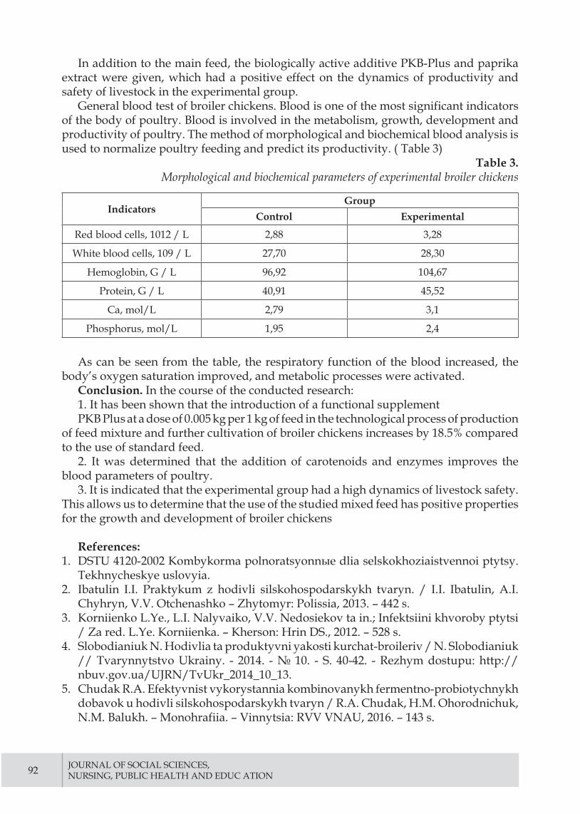
92 JOURNAL OF SOCIAL SCIENCES, NURSING, PUBLIC HEALTH AND EDUC ATION
In addition to the main feed, the biologically active additive PKB-Plus and paprika extract were given, which had a positive effect on the dynamics of productivity and safety of livestock in the experimental group.
General blood test of broiler chickens. Blood is one of the most significant indicators of the body of poultry. Blood is involved in the metabolism, growth, development and productivity of poultry. The method of morphological and biochemical blood analysis is used to normalize poultry feeding and predict its productivity. ( Table 3)
Table 3. Morphological and biochemical parameters of experimental broiler chickens
IndicatorsGroup
Control Experimental
Red blood cells, 1012 / L 2,88 3,28
White blood cells, 109 / L 27,70 28,30
Hemoglobin, G / L 96,92 104,67
Protein, G / L 40,91 45,52
Ca, mol/L 2,79 3,1
Phosphorus, mol/L 1,95 2,4
As can be seen from the table, the respiratory function of the blood increased, the body’s oxygen saturation improved, and metabolic processes were activated.
Conclusion. In the course of the conducted research:1. It has been shown that the introduction of a functional supplementPKB Plus at a dose of 0.005 kg per 1 kg of feed in the technological process of production
of feed mixture and further cultivation of broiler chickens increases by 18.5% compared to the use of standard feed.
2. It was determined that the addition of carotenoids and enzymes improves the blood parameters of poultry.
3. It is indicated that the experimental group had a high dynamics of livestock safety. This allows us to determine that the use of the studied mixed feed has positive properties for the growth and development of broiler chickens
References:1. DSTU 4120-2002 Kombykorma polnoratsyonnыe dlia selskokhoziaistvennoi ptytsy.
Tekhnycheskye uslovyia.2. Ibatulin I.I. Praktykum z hodivli silskohospodarskykh tvaryn. / I.I. Ibatulin, A.I.
Chyhryn, V.V. Otchenashko – Zhytomyr: Polissia, 2013. – 442 s.3. Korniienko L.Ye., L.I. Nalyvaiko, V.V. Nedosiekov ta in.; Infektsiini khvoroby ptytsi
/ Za red. L.Ye. Korniienka. – Kherson: Hrin DS., 2012. – 528 s.4. Slobodianiuk N. Hodivlia ta produktyvni yakosti kurchat-broileriv / N. Slobodianiuk
// Tvarynnytstvo Ukrainy. - 2014. - № 10. - S. 40-42. - Rezhym dostupu: http://nbuv.gov.ua/UJRN/TvUkr_2014_10_13.
5. Chudak R.A. Efektyvnist vykorystannia kombinovanykh fermentno-probiotychnykh dobavok u hodivli silskohospodarskykh tvaryn / R.A. Chudak, H.M. Ohorodnichuk, N.M. Balukh. – Monohrafiia. – Vinnytsia: RVV VNAU, 2016. – 143 s.

93
UDC 616.329-002.2 + 617-089 / 616-072.7 + 612.857.75
DIFFERENTIAL DIAGNOSIS OF FUNCTIONAL AND ORGANIC DISORDERS IN PATIENTS WITH EXTRAHEPATIC CHOLESTASIS
Zeleniuk O.,Head of the Department of Digestive Surgery of the Institute of Gastroenterology of the National Academy of Medical Sciences of Ukraine Tel.mob. +380667040674 Email: [email protected] https://orcid.org/0000-0002-3703-7064
Abstract.The aim of the study: to assess the autonomic status, adaptive potential and level
of stress resistance in terms of heart rate variability in extrahepatic cholestasis (EHC) in patients with complications of cholelithiasis (CL).
Materials and methods of research. Were examined 46 patients with EHC who were treated in the Department of Surgery of the Digestive System of the Institute of Gastroenterology of the National Academy of Medical Sciences of Ukraine for the period from 2018 to 2019. Group I consisted of 16 patients with EHC on the background of functional biliary disorders (FBD) with sphincter of Oddi dysfunction; group II included 30 patients with EHC on the background of stenotic papillitis from 1 to 3 stage with choledocholithiasis in 12 patients.
EHC was evaluated based on the results of PRECISE diagnostics (cloud automated interpretation technology).
Research results and their discussion. Studies have shown that most patients with EHC and FBD (dysfunction) activation
of the sympathetic ANS, in contrast to patients with EHC with stenotic papillitis, who in the spectrum of identified ANS disorders was observed as vagotonia (50.0%) with a decrease in sympatho-vagal balance to 0.48 ± 0.14, and moderate sympathicotonia in 43.3% of patients with an increase in the ratio of LF / HF to 2.1 ± 0.7 (p <0.05).
Conclusions.Assessment of HRV, in particular, using PRECISE diagnostics, is a highly informative,
convenient method, allows to assess the state of the ANS, adaptive capacity of the body in surgical patients, including the severity of stress, and can be recommended for use in screening differential diagnosis of functional and organic disorders in surgical patients with EHC on the background of complications of CL.
Key words: extrahepatic cholestasis; heart rate variability.
Introduction.Extrahepatic cholestasis (EHC) in complications of cholelithiasis (CL) is associated
with impaired bile flow caused by mechanical factors (choledocholithiasis, stricture in the distal part of the common bile duct), as well as functional disorders of the biliary tract (FDBT).
Outlining the definition of FDBT, it is necessary to mention the domestic formulation of this problem - biliary dyskinesia (BD). In the classical sense, BD is a disorder of coordinated

94 JOURNAL OF SOCIAL SCIENCES, NURSING, PUBLIC HEALTH AND EDUC ATION
motor activity of the gallbladder (GB) and / or sphincters of the biliary system, which is manifested by impaired excretion of bile from the liver and GB to the duodenum [1-8]. In the long-term course of FDBT, when long-standing biliary hypertension leads to a number of pathological changes in the biliary tract and liver, causing the transition of dyskinesia of the sphincter of Oddi (SO) in stenosis, the development of diseases such as CL and its complications - cholecystitis, cholecystocholangitis . One of the options for the development of FDBT in EHC is vegetative discoordination. After all, the formation of the pathological process reflects the general biological patterns of development, which are characterized first by processes of adaptation and compensation, then - disorganization of regulation, relative stabilization at a lower quantitative level with limited functional reserves and, finally, gross morphological changes and sharp suppression of organ function.
Analysis of the literature in recent years shows that vegetative dysfunction is becoming increasingly important in the emergence and progression of FDBT, which justifies the analysis of the functional state of the autonomic nervous system (ANS) [1-8]. It was shown that moderate irritation of the vagus nerve causes coordinated activity of the gastrointestinal tract and sphincters, and severe irritation - spastic contraction with delayed bile evacuation. Irritation of the sympathetic nerve contributes to the relaxation of the gastrointestinal tract [1-8]. Research by Loranska I. [5] proved that autonomic dysfunction was observed in most patients with disorders of GB reduction. In 69% of patients with hypokinetic dysfunctions the prevalence of parasympathetic ANS tone was noted, while sympathetic tone was observed in 31%. Patients with hypokinetic dysfunction had parasympathicotonia in 66% of cases, in 34% of cases - sympathicotonia. At the same time, there are studies [6] that show no correlation between the ejection fraction of the gallbladder and autonomic neuropathy (p> 0.05), so the question of the influence of the autonomic nervous system on the motility of the biliary system is still debatable.
The polyetiology of primary and secondary FPBT, the diversity of the clinical picture, multiple disorders of interorganic relationships in this pathology, the large role of psychoemotional and autonomic disorders determine the diverse, often nonspecific, clinical picture of the pathology. This situation determines the high frequency of a kind of clinical “masks”, such as cardiac, migraine mask, irritative autonomic syndrome, which are associated with autonomic dysfunction in FPBT, which determines the risk of inadequate diagnosis and, accordingly, treatment tactics, which can not be reflected on the prognosis, treatment effectiveness, quality of life of patients on pharmacoeconomic indicators [3].
To date, the differential diagnosis of the mechanical, functional or combined nature of EHC in complications of CL presents significant difficulties [4-8].
Therefore, the study of vegetative status for the differential diagnosis of EHC in patients with complicated CL is particularly relevant. One of the first to respond to fluctuations in the balance of the ANS is the cardiovascular system and it is the indicators of its activity used to assess the strength of individual stress reactions and autonomic balance [9-12]. So one of the most promising methods, which is increasingly used as a biomarker of stress, is to study heart rate variability (HRV) [11-13], as the severity of fluctuations in heart rate (R-R-intervals) relative to its average level (fluctuations in the size of the intervals between successive heart contractions - R-R-intervals), which is a fundamental physiological property of the human body. The method is based on the recognition and measurement of time intervals between R-R-intervals of the electrocardiogram (ECG) taken with four or more electrodes, construction of time series of cardiointervals

ZELENIUK O., 95
(cardiointervalogram) and subsequent analysis of the obtained numerical series by various mathematical methodsThe HRV study provides information on the functional reserve and adaptive potential of the human body as a whole, the limit of stress resistance and neurohumoral regulation of the heart, the state of the ANS and the relationship between its sympathetic and parasympathetic divisions, and has important prognostic and diagnostic value in a variety of pathologies [12].
Recently, in 2018, PRECISE diagnostics was introduced in Ukraine, which is used to analyze HRV using cloud technology of automated ECG interpretation with the analysis of more than 700 parameters based on the Minnesota code [11-13]. Unlike other methods of ECG interpretation, the PRECISE algorithm, instead of finding and analyzing individual patterns, analyzes every millimeter of the signal. It does not miss any deviations and has a perfect interference filter. PRECISE diagnostics is used to analyze the state of the ANS, the level of stress, adaptation and risk of cardiovascular disease.
The aim of the study: to assess the autonomic status, adaptive potential and level of stress resistance in terms of heart rate variability in extrahepatic cholestasis in patients with complications of cholelithiasis.
Materials and methods of research. We examined 46 patients with EHC who were treated for complications of CL in the Department of Digestive Surgery of the Institute of Gastroenterology of the National Academy of Medical Sciences of Ukraine for the period from 2018 to 2019. Among the complications of CL were chronic calculous cholecystitis in 4 patients, chronic calculous cholecystitis with choledocholithiasis in 12 patients, chronic calculous cholecystitis with stenotic papillitis from 1 to 3 stages - in 30 patients. Manifestations of EHC were established on the basis of biochemical studies and ultrasound data. Depending on the nature of EHC, patients were divided into groups. Group I consisted of 16 patients with EHC on the background of FDBT with sphincter of Oddi dysfunction; Group II included 30 patients with EHC on the background of stenotic papillitis from 1 to 3 st., including concomitant choledocholithiasis in 12 patients.
For the preoperative differential diagnosis and assessment of the motoric and evacuation function of the GB and SO before the operation in all 46 patients used following method: the initial volume (V1) of GB was determined on an empty stomach, then after a trial cholekinetic breakfast (20 g of sorbitol dissolved in 50 ml of warm water) the volume of GB was determined per minute for the first 10 min and then every 10 min until the GB was relaxed. The volume of GB after its maximum contraction (V2) was used to determine the efficiency of bile secretion (EBS):
The EBS of GB was considered normal if in 20-40 minutes after a cholekinetic breakfast, the maximum EBS was 40 - 70% of the initial volume of GB. Assessment of the functional state of the GB was performed taking into account the primary reaction, latency period, and a time of maximum contraction of the GB [14].
HRV was assessed using PRECISE diagnostics (cloud automated interpretation technology) using a CONTECT 8000GW electrocardiograph (China) with bluetooth connection and software. To interpret the ECG results, a connection to the AMAZON web service was made.
The following indicators were analyzed: sympathetic-vagal balance HF / LF to determine autonomic tone, stress index, RMSSD - the square root of the mean square of

96 JOURNAL OF SOCIAL SCIENCES, NURSING, PUBLIC HEALTH AND EDUC ATION
the difference between the values of successive pairs of intervals NN, which estimates the level of adaptation (quantification of depletion of adaptation reserves); stress index (Stress index, SI), (Table 1) [11-12].
Table 1. The main indicators
of heart rate variability by the method of PRECISE diagnosis
Indicator Description Value Interpretation
RMSSD
The square root of the mean sum of the squares of the
differences between adjacent NN intervals
up to 20 m/s Disruption / violation of adaptation
20-100 m/s Normal adaptation
more than 100 m/s Good physical training
LF/HF Sympathetic-vagal balance
up to 0,9 Vagotonia0,9-1,6 Normotonia
more than 1,6 Sympathicotonia
SI (stress index) stress index
up to 100 m/s Norm
100-300 m/s Anxiety
300-500 m/s Moderate painmore than 500
m/s Acute pain syndrome
During the operation, all patients underwent cholangiomanodibetometry, the indicators of which were compared with the data of X-ray cholangioscopy to confirm the diagnosis and determination of tactics of further surgical treatment.
The research was performed in compliance with the “Rules of ethical principles of scientific medical research with human participation”, approved by the Declaration of Helsinki (1964-2013), ICH GCP (1996), EEU Directive № 609 (dated 24.11.1986), orders Ministry of Health of Ukraine № 690 dated 23.09.2009, № 944 dated 14.12.2009, № 616 dated 03.08.2012 Each patient signed an informed agreement to participate in the study, taking all measures to ensure the anonymity of patients.
Statistical data processing was performed using computer programs Statistica 6.0 and Microsoft Excel 7.0. The statistical significance of the differences between the distribution of responses in the comparison groups was assessed using the Student’s criterion and the “chi-square” criterion;
Research results and their discussion. In the studied patients, the average values of RMSSD, which characterizes the adaptive capacity of the organism, were within normal values, but with a probable decrease in this indicator in patients of group II to (35.1 ± 4.6) m/s compared with patients of group I ( 51.5 ± 5.3) m/s (p <0.05). Analysis of the obtained data revealed that in group I patients with normal adaptation predominated (62.5%). In the majority of patients of group II (83.3%) signs of disturbance (failure) of adaptation with decrease in RMSSD indicator to (7,8 ± 2,9) m/s (p <0,05) are revealed that testifies to weakening of parasympathetic influences on heart rhythm with possible further complications of the disease in these patients (table 2).
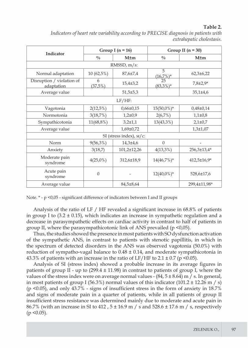
ZELENIUK O., 97
Table 2. Indicators of heart rate variability according to PRECISE diagnosis in patients with
extrahepatic cholestasis.
IndicatorGroup I (n = 16) Group II (n = 30)
% M±m % M±mRMSSD, m/s:
Normal adaptation 10 (62,5%) 87,6±7,4 5(16,7%)* 62,3±6,22
Disruption / violation of adaptation
6(37,5%) 15,4±3,2 25
(83,3%)* 7,8±2,9*
Average value 51,5±5,3 35,1±4,6
LF/HF:
Vagotonia 2(12,5%) 0,66±0,15 15(50,0%)* 0,48±0,14Normotonia 3(18,7%) 1,2±0,9 2(6,7%) 1,1±0,8
Sympathicotonia 11(68,8%) 3,2±1,1 13(43,3%) 2,1±0,7Average value 1,69±0,72 1,3±1,07
SI (stress index), м/с:Norm 9(56,3%) 14,3±4,6 0 -
Anxiety 3(18,7) 101,2±12,26 4(13,3%) 256,3±13,4*
Moderate painsyndrome 4(25,0%) 312,6±18,9 14(46,7%)* 412,5±16,9*
Acute painsyndrome 0 - 12(40,0%)* 528,6±17,6
Average value 84,5±8,64 299,4±11,98*
Note. * - p <0,05 - significant difference of indicators between I and II groups
Analysis of the ratio of LF / HF revealed a significant increase in 68.8% of patients in group I to (3.2 ± 0.15), which indicates an increase in sympathetic regulation and a decrease in parasympathetic effects on cardiac activity in contrast to half of patients in group II, where the parasympathicotonic link of ANS prevailed (p <0,05).
Thus, the studies showed the presence in most patients with SO dysfunction activation of the sympathetic ANS, in contrast to patients with stenotic papillitis, in which in the spectrum of detected disorders in the ANS was observed vagotonia (50.0%) with reduction of sympatho-vagal balance to 0.48 ± 0.14, and moderate sympathicotonia in 43.3% of patients with an increase in the ratio of LF/HF to 2.1 ± 0.7 (p <0.05).
Analysis of SI (stress index) showed a probable increase in its average figures in patients of group II - up to (299.4 ± 11.98) in contrast to patients of group I, where the values of the stress index were on average normal values - (84, 5 ± 8.64) m / s. In general, in most patients of group I (56.3%) normal values of this indicator (101.2 ± 12.26 m / s) (p <0.05), and only 43.7% - signs of insufficient stress in the form of anxiety in 18.7% and signs of moderate pain in a quarter of patients, while in all patients of group II insufficient stress resistance was determined mainly due to moderate and acute pain in 86.7% (with an increase in SI to 412 , 5 ± 16.9 m / s and 528.6 ± 17.6 m / s, respectively (p <0.05).

98 JOURNAL OF SOCIAL SCIENCES, NURSING, PUBLIC HEALTH AND EDUC ATION
When analyzing the level of stress resistance (SR) depending on the tone of the ANS (Fig. 1), the highest frequency of high SR was found in patients of group I: in 100.0% - eytonia, and in 80.0% - vagotonia (p <0, 05), while reduced and sharply reduced SR was detected only in sympathicotonia (66.7% and 33.3%, respectively). Among patients of group II with vagotonia the distribution of different levels of stress resistance did not differ significantly, in patients with eytonia the frequency of high SR also prevailed, and in patients with sympathicotonia the frequency of sharply reduced SR prevailed (80.0%).
Group I Group II
Fig. 1 - Analysis of the level of stress resistance depending on the tone of the autonomic nervous system in patients with extrahepatic cholestasis.
In general, our data confirm previously conducted single studies of autonomic status in patients with cholelithiasis [2-5], specifying them in the differential diagnosis of functional and organic disorders in surgical patients with EHC on the background of complications of CL.
Conclusions.1. Assessment of HRV, in particular, using PRECISE diagnostics, is a highly
informative, convenient method, allows to assess the state of the ANS, the adaptive capacity of surgical patients, including the severity of stress, and can be recommended for use in screening differential diagnosis of functional and organic disorders in surgical patients with EHC on the background of complications of CL.
2. ANS in patients with EHC on the background of complications of CL functioned in the mode of imbalance of its sympathetic and parasympathetic links with the prevalence of hypersympathicotonia in 68.8% of patients with EHC of functional origin, while among patients with stenotic papilitis, regardless of the stage of the process, the frequency of vagotonia and moderate sympathictonia was observed almost equally (in 50.0% and 43 3%, respectively) (p <0.05).
3. Analysis of the adaptive capacity of the examined patients according to RMSSD showed that in 82.3% of patients with EHC on the background of sphincter of Oddi dysfunction adaptation was not detected, while disease progression among patients with EHC on the background of stenotic papillitis led to a decrease in the frequency normal RMSSD - in 83.3% of patients up to (7.8 ± 2.90) m / s, which indicated insufficient adaptive potential of the body in these patients (p <0.05).

ZELENIUK O., 99
4. The frequency of normal stress index was maximum in patients with complications of CL and EHC on the background of sphincter of Oddi dysfunction (56.3%) with a significant increase in stress index to the level of “anxiety” and “moderate pain” in 43.7% of patients ( p <0,05) which determined reduced stress resistance, while in 86.7% of patients with EHC on the background of stenotic papillitis found a sharply reduced level of stress resistance mainly due to “moderate” and “acute pain” with an increase in SI to 412.5 ± 16.9 m / s and 528.6 ± 17.6 m / s, respectively (p <0.05).
Prospects for further research. Diagnosis of the vegetative status of patients with cholelithiasis can be used as an additional method in early screening for possible complications of gallstone disease. Further determination of the possible interdependence of vegetative status with choledochomanodebitometry data in patients with cholelithiasis and its other possible complications, as well as the relationship of vegetative status in patients with other pathologies of the gastrointestinal tract is promising.
References:1. S. V. Gridneva Disfunktsiia sfinktera Oddi posle kholetsistektomii: mekhanizmy
razvitiia i taktika lecheniia [Dysfunction of the sphincter of Oddi after cholecystectomy: mechanisms of development and treatment tactics]. Suchasna gastroenterologіia.2015. № 3 (83):122-127 [in Russian]
2. Sheptulin A.A., Kardasheva S.S. Spornye voprosy funktsional’nykh rasstroistv zhelchnogo puzyria i sfinktera Oddi: analiz rimskikh kriteriev IV peresmotra (2016)[Controversial issues of functional disorders of the gallbladder and the sphincter of Oddi: analysis of the Roman criteria of the IV revision (2016)] Klinicheskaia meditsina. 2018; 96(1): 84-87. [in Russian] DOI http://dx.doi.org/10.18821/0023-2149-2018-96-1-84-87
3. Peter B. Cotton, Grace H. Elta, C. Ross Carter, Pankaj Jay Pasricha, Enrico S. Corazziari. Gallbladder and Sphincter of Oddi Disorders. 2016. Gastroenterology 2016;150:1420–1429. . [English] http://dx.doi.org/10.1053/j.gastro.2016.02.033
4. Hiroyuki Miyatani, Hirosato Mashima , Masanari Sekine , Satohiro Matsumoto. Clinical features and management of painless biliary type sphincter of Oddi dysfunction. 2019.J Int Med Res. 2019 Jul;47(7):2940-2950. . [English] doi: 10.1177/0300060519848628
5 Loranskaya YD, Kukushkyn ML, Panyna NA. Bylyarnye dysfunktsyy y ykh profylaktyka. Eksperimental’naya i Klinicheskaya Gastroenterologiya [Biliary dysfunctions and their prevention]. Eksperimental’naia i klinicheskaia gastroenterologiia. 2011; 05: 48-52. [in Russian]
6. Kemal Kul 1, Ender Serin, Tolga Yakar, Arif Mansur Coşar, Birol Özer. Autonomic neuropathy and gallbladder motility in patients with liver cirrhosis.Turk J Gastroenterol. 2015 May;26(3):254-8. [English] doi:10.5152/tjg.2015.4469
7. Mariana Ungur , Petru Aurel Babes , Adrian Maghiar , Gheorghe Mircea Pop, Bogdan Feder. Diagnosis and Therapeutic Management in Postcholecistectomy Alithiasic Cholangitis.2019.Maedica(Bucur).2019,Sep;14(3):247-253. [English] doi:10.26574/maedica.2019.14.3.247.
8. M. V. Repin, V. Iu. Mikriukov, T. E. Vagner, N. M. Pleshkova. . Diagnostika nedostatochnosti sfinktera Oddi u bol’nykh s postkholetsistektomicheskim sindromom po dannym gepatobilistsintigrafii [Diagnosis of sphincter of Oddi dysfunction in patients with postcholecystectomy syndrome from hepatobiliary scintigraphic findings]. Vestnik rentgenologii i radiologii № 6, 2015:5-11 [in Russian]

100 JOURNAL OF SOCIAL SCIENCES, NURSING, PUBLIC HEALTH AND EDUC ATION
doi:10.20862/0042-4676-2015-0-6-107-1139. V.T. Ivashkin, I.V. Maev, Iu.O. Shul’pekova, E.K. Baranskaia,A.V. Okhlobystin,
A.S. Trukhmanov, T.L. Lapina, A.A. Sheptulin.Klinicheskie rekomendatsii Rossiiskoi gastroenterologicheskoi assotsiatsii po diagnostike i lecheniiu diskinezii zhelchevyvodiashchikh putei [Clinical guidelines of the Russian Gastroenterological Association for the diagnosis and treatment of biliary dyskinesia] Rus J Gastroenterol Hepatol Coloproctol.2018; 28(3):63-80. [in Russian] doi:10.22416/1382-4376-2016-26-3-64-80
10. Pokhabova E. Iu., Belova G. V. Sovremennyi vzgliad na neopukholevye stenoziruiushchie porazheniia BDS. Etiologiia i patogenez, vozmozhnosti diagnostiki.[Modern view of non-neoplastic stenosing lesions of CBD. Etiology and pathogenesis, diagnostic possibilities].Eksperimental’naia i klinicheskaia gastroenterologiia. 2012. №6:69-75 [in Russian]
11. Sen J, McGill D. [Fractal analysis of heart rate variability as a predictor of mortality: A systematic review and meta-analysis]. Chaos: An Interdisciplinary Journal of Nonlinear Science. 2018; 28(7): 72-101. [English] https://doi.org/10.1063/1.5038818.
12. Sessa F, Anna V, Messina G. [Heart rate variability as predictive factor for sudden cardiac death]. Aging (Albany NY). 2018; 10(2): 166-177. [English] doi: 10.18632/aging.101386.
13. Prineas RJ, Crow RS, Zhang Z. [The Minnesota Code Manual of Electrocardiographic Findings including measurement and comparison with the Novacode]. Standards and Procedures for ECG Measurement in Epidemiologic and Clinical Trials. Second Edition New and Enlarged. Springer-Verlag London Limited. 2010; 13: 328. [English] doi:10.1007/978-1-84882-778-3
14. Maikova T.V., Mosіichuk L.M., Petіshko O.P.. Osoblivostі funktsіonal’nogo stanu zhov-chnogo mіkhura, sfіnktera Oddі ta motorno-evakuatornoї funktsії shlunka u khvorikh na gastro-ezofageal’nu refliuksnu khvorobu [Peculiarities of functional status of gallbladder, sphincter of Oddi and motor-evacuation function of the stomach in patients with gastroesophageal reflux disease]. Gastroenterologіia. 2014. № 1 (51): 31-36 [in Russian]

101
LEGAL EDUCATION IN UKRAINE: ISSUES OF GAINING PRACTICAL EXPERIENCE BY STUDENTS
Byelov Dmytro,Department of Constitutional Law and Comparative Law SHEI Uzhhorod National University
Summary. Activation and stimulation of development of legal clinics in Ukraine is one of the directions of formation of high legal culture and legal consciousness, affirmation of freedom and democracy through which it is possible to approach achievement of the constitutional purpose - formation of civil society and the rule of law.
Key words: legal experts, jurisprudence standards, education institutions, legal clinic, clinical legal education.
Today, the issue of practical professional training of domestic legal experts is one of the problems taking place in the sphere of legal education. It should be professionals of such kind who will be able and will have a desire to provide citizens, who really need it, with legal assistance and protect their rights and freedoms to a good quality. It is very unfortunate that training is imbued with theoretical nature, which forms more abstract student vision of future work. Thus, typically graduates of education institutions, who continue with practical activity, literally have to be re-trained adjusting to real labour conditions [2, p. 1].
The introduction of state jurisprudence standards is aimed at improving training quality of law students through extending the cycle of general education, the introduction of natural science subjects, the increase of specialized disciplines. Moreover, unfortunately the state standards are still characterised by a traditional disadvantage peculiar to legal education: exaggeration of the role of theoretical disciplines and a lack of subjects which allow gaining practical skills.
One of the ways to solve the situation is to create a “legal clinic”. It is a quite new concept for Ukrainian legal education. Traditionally, this form is mainly used at medical universities. Everyone knows that the clinic is called a place where a practitioner can apply his/her knowledge in practice, either by treating people or by conducting experiments or teaching his/her students. Thus, if a medical professional or a chemist has this kind of place, why can’t a legal expert have it? In the process of training, law students get a quite narrow vision of legal practice. Under the best of circumstances, the familiarization occurs during on-the-job training or pre-degree apprenticeship, but any lawyer knows that a student is not able to gain the skills and secrets of the future profession in a few weeks [4, p. 10].
For the first time, the term “legal clinic” appeared in the United States of America. American lawyers began to concern about adjusting their academic education to practical issues 15-20 years ago when the principle “forget everything you have been learned” ceased to satisfy them. The ineffectiveness of the way of combining theory with practice became evident then. It is clear that a student who is appointed to a law firm may not bother lawyers working there, but nevertheless, he will be treated as a source of the free

102 JOURNAL OF SOCIAL SCIENCES, NURSING, PUBLIC HEALTH AND EDUC ATION
and semi-skilled work force. Duties of a legal practitioner don’t include the obligation to teach someone.
It is another matter when it is referred to an organisation which deals with practical activities and at the same time is aimed at training. Moreover, hands-on training is a great task. Thus, does a law school train such kind of skills as legal responsibility and professional ethics, or does it teach you to express your opinion in a written form correctly or to make a public speech, etc.? Practically, it doesn’t. However, these skills play a significant role in a legal career. Consequently, the legal clinic is one of the solutions for this situation.
If we try to find out Ukrainian equivalent for “clinical legal education” it looks like “experiential learning of lawyers through profession immersion”. The Ukrainian interpretation doesn’t reflect completely a content which the concept “legal clinic” means in the USA. There, it includes free legal consulting, in the first place for financially disadvantaged citizens, and the system of so-called “coaching” of students in order to master practical skills majoring in their future legal activity as well as mock trials and working on the real issues.
The professions of a lawyer and a medical professional are very similar in the sense that people address both lawyer and medical professional mainly in critical situations when their fate is solved. It is generally recognized that a health worker cannot be taught separately from a patient and burden exclusively with theory. Unfortunately, in Ukraine, a lawyer can be taught in such a way. We believe that a legal expert needs “real clients” with their legal problems no less than a medical worker. One can give hundreds of wonderful lectures, thousands of seminars devoted to human rights issues, but all this stuff can be kept at the level of empty talks. The student will more likely wrap his mind around the idea of human rights if he/she feels it through the fate of particular people while trying to counsel, draw up legal documents, represent before the court.
At the same time, the “legal clinic” should not be considered exclusively foreign achievement. Each law student more or less has applied methods and practices of clinical education depending on own abilities, qualifications, etc. Almost every law student participated in the “trial” during the seminars, compiled samples of service documents – all of these things are methods and practices of clinical education.
Unfortunately, educational process has a lack of hours dedicated to practical classes in individual disciplines, and generally, representatives of judicial and law enforcement agencies don’t have either time or desire to perform on-the-job training for law students. At the same time, there is a fairly simple way to ensure students training through “real affairs” – to open the doors of the university for people who really need legal assistance.
Law students are interested in gaining practical skills that could be a guarantee for further employment and career but not only for obtaining “pro forma diplomas” that give certain rights. Unfortunately, today university diploma is often associated with the possibility to belong to government agencies or to earn a lot of money in a bar or court from the very beginning of practice activities regardless of the actual qualification [6, p. 233].
History of the establishment of “legal clinics” in Ukraine actually dated back to its independence. During this period, public organisations of various focus area began to emerge. At the same time, the Ukrainian jurisprudence originated. Then, a special symbiosis of legal education and practice, which found its manifestation in the establishment of public organisations of “legal clinics”, was created.
At the same time, today there is a paradoxical situation in Ukraine: some higher education institutions do not want to create that sort of course, because it allegedly causes

ByELOV DMyTRO, 103
considerable financial, organisational costs. And another important argument is that the course will be a duplication of other available courses where students are provided with practical skills for work, or there are personal convictions of deans in relation to the absurdity of teaching and clinics availability. Therefore, sometimes student organisations independently establish a clinic and solve all problems related to its arrangement. There are also situations when the administration is against the creation of the course because it does not like the very idea and philosophy of the legal clinic [2, p. 113].
In order to ensure the relevant place of legal clinics in public life, their identification as a really effective institute of civil society, it is necessary to increase the professionalism and effectiveness of their activities to a great extent. It is also important to inform the society, first of all, the legal community, about the importance of legal clinics for the qualitative improvement of higher legal education, the elimination of legal nihilism among the population.
Thus, activation and stimulation of the development of legal clinics in Ukraine are one of the areas for the generation of high legal culture and legal consciousness, the consolidation of freedom and democracy due to which it is possible to make the achievement of the constitutional goal closer – the formation of civil society and state of law.
References:1. Волочай О. Активні методи навчання [Електронний ресурс] / Олена Во- лочай.
– Режим доступу : http://www. u21.org. ua/ukr/kliniky_stat4.htm. 2. Єлов В. А. Юридична клініка : навч. посіб. / В. А. Єлов, С. І. Молібог, Д. Г.
Павленко. – К. : Школяр, 2004. – 315 с 3. Положення про порядок легалізації об’єднань громадян : затв. постано- вою
Кабінету Міністрів України від 26 лют. 1993 p., No 140 // Конституційно- правовий статус об’єднань громадян в Україні: навч. посіб. – X. : Арсіс, 1998. – С 77-81.
4. Розвиток громадянського суспільства в Україні / [уклад.: Л. Шара, І. Підлуська та ін.]. – К., 2002. – 59 с
5. Теорія держави і права (опорні конспекти) : навч. посіб. [для студент. вищ. навч. закл.] / [авт.-упоряд. М. В. Кравчук]. – К. : Атіка, 2005. – 288 с
6. Хуснутдінов О. Процес формування громадянського суспільства в Україні як передумова становлення правової держави // Вісник Української Академії державного управління при Президентові України. – 2001. – No 2, ч. II. – С 233-241.
7. Штюдеманн Д. Громадянське суспільство в Німеччині [Електронний ре- сурс] / Дітмар Штюдеманн // Дзеркало тижня. – 2002. – 9-15 лют. – Режим дос- тупу : www. zn. kiev. ua.
8. Щукотске Д. Клінічна освіта: міст між теорією та практикою [Електрон- ний ресурс] / Джейн Щукотске. – Режим доступу : www. u21.org. ua/ukr/kliniky_stat5.htm.
9. Zaviš M. Úvod do systematickej etiky. Žilina : Žilinská univerzita v Žiline, EDIS, 2017. 133 s.
10. Mačkinová M., Kopinec P., Holonič J., Stančiak J. Language communication skills in health and social care workers. Iranian Journal of Public Health. 2019; 48 (4): 773-4.

104
UDC: 616.314-055.76(477.87)
COMPARATIVE ANALYSIS OF THE DENTAL STATUS OF MONOZYGOTIC AND DIZYGOTIC TWINS
O.Ya. Bilynskyi, Ye.Ya. KostenkoUzhhorod National University, Uzhhorod, Ukraine
Summary: This article rewies a comparative analysis of dental status, including the intensity and prevalence of caries among pairs of monozygotic and dizygotic twins living in the Transcarpathian region. It is established that the use of the studied model of twins contributes to the possibility of verifying the significance of two components in the system of predicting changes in dental status: specific that is to study the impact of a particular component of the genetic variation in phenotype; and general, which is to study the effect of the entire genotype pattern on a set of all registered variations.
Key words: caries, caries intensity, caries prevalence, concordance, genetics, monozygotic twins, dizygotic twins.
Introduction. Modern views on the causes of carious pathology determine its polyetiological nature, and the risk of formation of enamel and dentin defects with the simultaneous progression of demineralization processes is associated with the influence of environmental factors and the influence of genetically mediated factors [1, 2, 3, 5, 6, 7 , 10, 11, 13, 14].
In order to improve the quality of dental care to the population at the present stage of development of the dental industry, it is advisable to provide a more in-depth study of genetic and environmental factors associated with caries risk, which will improve existing programs for prevention and treatment of this pathology [4, 12, 16, 17, 18 , 19].
One of the most adapted methodological models for studying changes in dental status and their associations with the genetic component is the method of their study between pairs of twins at different ages under the same and different environmental conditions. [3, 5, 8, 9, 15].
In order to solve the problem of studying the role of the genetic component in the formation of features and development of changes in the dental system, an analysis of the dental status among monozygotic and dizygotic twins have done. The formation of samples were carried out according to appropriate criteria, on the basis of registration of available facts of dental history of twins patients and their subsequent clinical examination.
Research results. Among all surveyed, a study population of 57 pairs of twins (23 monozygotic) (40.35%): 14 males (24.56%) and 9 females (15.79%), and 34 dizygotic (59.65%) were formed: 16 males (28.07%) and 18 females (31.58%). To study the influence of age and sex on the severity of variations in dental status among patients, a stratification analysis of different ages was performed (categories 18-25, 26-30 and 31- 35 years) and gender subgroups in the structure of comparable samples of monozygotic and dizygotic twins.
The average caries prevalence among the sample of monozygotic twins was 92.56 ± 1.22%, and among the sample of dizygotic twins 94.39 ± 1.08%. During the analysis

O.yA. BILyNSKyI, yE.yA. KOSTENKO 105
it was found that the average DMFT index (D – decayed, M – missing, F-filled teeth) among monozygotic men aged 18-25 years was 7.58 ± 0.45, with the component D reached 3.19 ± 0.14, M - 1.54 ± 0.22 and F - 2.85 ± 0.49. The level of this indicator among monozygotic men aged 26-30 years reached 8.77 ± 0.43, with the following distribution of components: D - 3.32 ± 0.18, M - 1.67 ± 0.23, F - 3.78 ± 0.79,. Among monozygotic men aged 31-35 years, the level of caries intensity reached 9.52 ± 0.56, in the structure of which D - 3.21 ± 0.59, M - 2.19 ± 0.17, F - 4.12 ± 0.44. The pattern of DMFT index distribution among monozygotic women of different age groups was as follows: 18-25 years - 7.19 ± 0.57 (D - 3.04 ± 0.19, M - 1,23 ± 0.61, F - 2.92 ± 0.25); 26-30 years - 8.75 ± 0.41 (D - 3.15 ± 0.19, M - 1.75 ± 0.07, F - 3.85 ± 0.25); 31-35 years - 8.93 ± 0.78 (D - 3.18 ± 0.47, M - 2.01 ± 0.12, F - 3.74 ± 0.48). Thus, in different gender samples of monozygotic twins there was a similar tendency to increase the level of DMFT with age, but the difference in the index was statistically significant only when comparing the age groups 18-25 years and 31-35 years (p <0.05); when comparing the intensity of caries between the age groups of 18-25 years and 26-30 years, as well as between the age groups of 26-30 years and 31-35 years, the statistical difference between the studied indicators could not be registered (p> 0.05).
The study of the intensity of the carious process among dizygotic male twins was characterized by the following distribution of indicators in different age groups: 18-25 years - 7.42 ± 0.64 (D - 3.21 ± 0.56, M - 1.62 ± 0.64, F - 2.59 ± 0.18); 26-30 years - 8.67 ± 0.41 (D - 3.28 ± 0.25, M - 1.61 ± 0.19, F - 3.78 ± 0.31); 31-35 years - 9.48 ± 0.49 (D - 3.19 ± 0.32, M - 2.14 ± 0.59, F - 4.15 ± 0.08). Among dizygotic female twins aged 18-25 years, the level of DMFT index was 7.35 ± 0.56, with D component reaching 2.89 ± 0.19, M - 1.45 ± 0.59 and F - 3.01 ± 0.43. The level of this indicator among dizygotic female twins aged 26-30 years reached 8.65 ± 0.52, with the following distribution of components: D - 3.23 ± 0.54, M - 1,78 ± 0.39, F - 3.64 ± 0.51. Dizygotic female twins aged 31-35 years were characterized by an indicator of caries intensity at the level of 9.01 ± 0.33, in the structure of which D reached 3.35 ± 0.74, M - 2,19 ± 0.45, F - 3.47 ± 0.15. In general, among the studied samples of dizygotic twins of different age groups, the same trend was observed as among monozygotic twins: when comparing the indicators of caries intensity between the age groups of the studied 18-25 years and 26-30 years, and 26-30 years and 31-35 years of statistical the difference between DMFT levels could not be registered (p> 0.05). However, DMFT levels among dizygotic twins of both sexes were characterized by a statistically significant difference when comparing the age groups 18-25 years and 31-35 years (p <0.05).
In the process of mathematical processing of the obtained numerical data, it was possible to systematize the average values of the DMFT index among samples of monozygotic and dizygotic twins, regardless of sex. Thus, it was found that the average DMFT among monozygotic twins aged 18-25 years was 7.39 ± 0.44, with D component reaching 3.12 ± 0.18, M - 1.39 ± 0.45 and F - 2.89 ± 0.35. The average of this index among monozygotic twins aged 26-30 years reached 8.76 ± 0.38, with the following distribution of components: D - 3.24 ± 0.27, M – 1.71 ± 0.34, F - 3.82 ± 0.51. Among monozygotic twins aged 31-35 years, the level of caries intensity reached the values of DMFT 9.23 ± 0.24, in the structure of which D - 3.20 ± 0.27, M - 2.10 ± 0.25 and F - 3.93 ± 0.16.
The average rate of caries intensity among dizygotic twins aged 18-25 years, regardless of gender was 7.39 ± 0.51, with D component reaching 3.05 ± 0.24, M - 1,54 ± 0.42 and F - 2.80 ± 0.79. The average level of this index among dizygotic twins aged 26-30 years is 8.66 ± 0.59, with the following distribution of components: D - 3.26 ± 0.55, M – 1.70 ±

106 JOURNAL OF SOCIAL SCIENCES, NURSING, PUBLIC HEALTH AND EDUC ATION
0.52, F - 3.71 ± 0.48. The group of dizygotic twins aged 31-35 years was characterized by the highest average level of caries intensity among all studied age groups of dizygotic twins, in which the DMFT values reached 9.25 ± 0.24, in the structure of which D - 3.27 ± 0.45, M - 2.17 ± 0.37, F - 3.81 ± 0.59.
Fig.1. The average values of the components of the DMFT index among monozygotic twins of different age categories
Fig.2. The average values of the components of the DMFT index among dizygotic twins of different age categories

O.yA. BILyNSKyI, yE.yA. KOSTENKO 107
In general, it was not possible to establish a statistical difference between the indicators of caries intensity between the studied groups of monozygotic and dizygotic twins (p <0.05), while among both samples there was a similar tendency to increase the DMFT with age.
The recorded average difference in the intensity of caries between pairs of studied monozygotic twins aged 18-25 years was 0.46 ± 0.32, between pairs of monozygotic twins aged 26-30 years - 0.50 ± 0.18, and between pairs of monozygotic twins aged 31 -35 years - 0.39 ± 0.27. These indicators of the difference in caries intensity between pairs of monozygotic twins of different ages are not statistically significant (p> 0.05), which can be interpreted as the homogeneity of the pattern of carious pathology among this study sample, in which the average intensity difference was 0.45 ± 0 , 23 (p> 0.05). In the group of dizygotic twins, the average difference between pairs of subjects aged 18-25 years was 0.94 ± 0.47, between pairs of dizygotic twins aged 26-30 years - 1.04 ± 0.31, while between pairs dizygotic twins aged 31-35 years - 1.01 ± 0.27. In the two age categories (26-30 years and 31-35 years) the calculated difference in caries intensity between pairs of dizygotic twins was statistically significant (p <0.05), while in the age category 18-25 years - statistically insignificant (p> 0.05), and the average difference of the studied indicator in this sample was 0.99 ± 0.32 (p <0.05) (table 1).
Table 1. Comparison of the average indicators of the difference in the criterion of caries intensity
between pairs of twins
Aged group Monozygotic twins Dizygotic twins
18-25 years 0,46±0,32 р>0,05 0,74±0,47 р>0,05
26-30 years 0,50±0,18 р>0,05 1,04±0,31 р<0,05
31-35 years 0,39±0,27 р>0,05 1,01±0,27 р<0,05
Average values 0,45±0,23 р>0,05 0,93±0,32 р<0,05
In the course of statistical processing of all obtained results, the concordance of the corresponding changes and features of dental status among monozygotic and dizygotic twins of different sexes were established, which were presented as follows: the level of concordance of carious pathology among monozygotic male twins was - 79.46 ± 3.25 %, among monozygotic female twins - 75.59 ± 3.19%, among dizygotic male twins - 34.18 ± 4.07%, among dizygotic female twins - 30.92 ± 3.27% (table 2 ).
Table 2. The level of concordance of changes in dental status among pairs
of monozygotic and dizygotic twins of different sexes
Parameters of dental status
Level of concordance
Monozygotic male twins
Monozygotic female twins
Dizygotic male twins
Dizygotic female twins
Caries 79,46±3,25% 75,59±3,19% 34,18±4,07% 30,92±3,27%

108 JOURNAL OF SOCIAL SCIENCES, NURSING, PUBLIC HEALTH AND EDUC ATION
In the process of systematization of the results, the average generalized indicators of concordance of features of dental status for pairs of monozygotic and dizygotic twins for carious pathology 77.53 ± 3.08% and 32.55 ± 2.72%, respectively, were calculated (Table 3).
Table 3. Averages levels of dental status changes concordance among
monozygotic and dizygotic pairs of twins
Parameters of dental status
Level of concordance
Monozygotic twins Dizygotic twins
Caries 77,53±3,08% 32,55±2,72%
Conclusions. It should be noted that among the sample of monozygotic twins, regardless of sex and age, there were statistically significant associations between the actual registered level of cariogenic lesions and indicators such as the number of already restored teeth, the number of affected tooth surfaces, etc. In some subgroups of dizygotic twins, similar trends were also statistically confirmed, but the level of severity of these varied between different members of the same subgroups, formed by age and sex. Previous studies also indicate that the pattern of distribution of caries is characterized by a specific level of variation depending on the influence of a component of the genetic factor. The use of the studied model of twins contributes to the possibility of verifying the significance of two components in the system of predicting changes in dental status: specific, which consists in studying the influence of a particular genetic component on phenotype variation; and general, which is to study the effect of the entire genotype pattern on a set of all registered variations. Prospects for further research are to find appropriate dental criteria that would help identify levels of identity and discordance of pairs of monozygotic and dizygotic twins. The use of dental research methods for this purpose would help optimize the process of calculating the index of significance of the influence of heredity between pairs of twins on the development of different dental disorders, as an indicator of the difference of correlation coefficients between the studied samples of monozygotic and dizygotic twins.
References:1. Carter, F., & Irish, J. (2019). A sub-continent of caries. Dental Anthropology Journal,
32(2), 22-29.2. Gomez, A., Espinoza, J. L., Harkins, D. M., Leong, P., Saffery, R., Bockmann, M., ... &
Hughes, T. (2017). Host genetic control of the oral microbiome in health and disease. Cell host & microbe, 22(3), 269-278.
3. Hajishengallis, E., Parsaei, Y., Klein, M. I., & Koo, H. (2017). Advances in the microbial etiology and pathogenesis of early childhood caries. Molecular oral microbiology, 32(1), 24-34.
4. Huang, Y., Wang, J., Tan, Y., Wang, L., Lin, H., Lan, L., ... & Shu, W. (2018). Low-mineral direct drinking water in school may retard height growth and increase dental caries in schoolchildren in China. Environment international, 115, 104-109.
5. Kutsch, V. K., & Young, D. A. (2011). New directions in the etiology of dental caries disease. Journal of the California Dental Association, 39(10), 716-721.

O.yA. BILyNSKyI, yE.yA. KOSTENKO 109
6. Piekoszewska-Ziętek, P., Turska-Szybka, A., & Olczak-Kowalczyk, D. (2017). Single nucleotide polymorphism in the aetiology of caries: systematic literature review. Caries research, 51(4), 425-435.
7. Rechmann, P., Chaffee, B. W., Rechmann, B. M., & Featherstone, J. D. (2019). Caries management by risk assessment: results from a Practice-Based Research Network study. Journal of the California Dental Association, 47(1), 15.
8. Ritter, A. V., Eidson, R. S., & Donovan, T. E. (2014). Dental caries: etiology, clinical characteristics, risk assessment, and management. Sturdevant’s Art & Science of Operative Dentistry-E-Book, 41.
9. Yildiz, G., Ermis, R. B., Calapoglu, N. S., Celik, E. U., & Türel, G. Y. (2016). Gene-environment interactions in the etiology of dental caries. Journal of dental research, 95(1), 74-79.
10. Zafar, S., Harnekar, S. Y., & Siddiqi, A. (2009). Early childhood caries: etiology, clinical considerations, consequences and management. Int Dent SA, 11(4), 24-36.
11. Антоненко, М. Ю., Зелинська, Н. А., & Мельничук, Т. А. (2015). Роль міждисцплінарного навчання в розумінні етіології та патогенезу каріозної хвороби. Современная стоматология, (3), 116-117.
12. Безвушко, Е. В. (2014). Профілактика карієсу зубів у дітей, які проживають у несприятливих умовах довкілля. Вісник проблем біології і медицини, (3 (2)), 338-342.
13. Дорошенко, С. И., & Кульгинский, Е. А. (2015). Клиническая генетика в ортодонтической практике. Современная ортодонтия, (2), 25-27.
14. Емелина, Е. С., Никонова, А. В., & Корецкая, Е. А. (2016). Этиология развития кариеса. Новая наука: Проблемы и перспективы, (2-3), 56-58.
15. Лобач, О. И., & Лапина, Н. В. (2017). Современные аспекты этиологии и патогенеза кариеса зубов. Обзор литературы, ч. I. Клиническая стоматология, (3), 26-30.
16. Лукашевич, И. К., Вардугина, О. К., & Горбунова, И. Л. (2018). Обоснование выбора средств профилактики кариеса у беременных с учетом частоты полиморфизмагена каллекреина-4. Современная стоматология, (2 (71)).
17. Октисюк, Ю. В., & Рожко, М. М. (2014). Комплексний метод профілактики карієсу зубів у дітей, що проживають у різних кліматогеографічних зонах Івано-франківської області. Вісник проблем біології і медицини, (3 (3)), 310-315.
18. Размахнина, Е. М. (2018). Клинические и молекулярно-генетические особенности прогнозирования и оценки эффективности профилактики кариеса (Doctoral dissertation, Кемеровский государственный медицинский университет Министерства здравоохранения).
19. Савранский, Ф. З., Чигарина, С. Е., & Гришин, П. О. Наследственные и средовые факторы в развитии кариеса зубов.

110
WIDERSPRUCH ZUM PROBLEM DER ANGEBORENEN UND ERWORBENEN ÄTIOLOGIE DER PILONIDALE KRANKHEIT BEI KINDERN
Konoplitskyi V.S.Professor, Doktor der Medizin, Leiter der Abteilung für Kinderchirurgie von Nationale Pirogov Memorial Medical University, Winnyzja, Ukraine https://orcid.org/0000-0001-9525-1547
Shavliuk R.V.Doktorand der Abteilung für Kinderchirurgie von Nationale Pirogov Memorial Medical University, Winnyzja, Ukraine, https://orcid.org/0000-0002-3542-1599
Shavliuk V.M.Leiter der chirurgischen Abteilung „ Kozijatin Centralregionalkrankenhaus Kozyatin Central District Hospital Municipal Enterprise, Kozyatin, Ukraine
Kyrychenko O.P.Student, Touro College für Osteopathische Medizin, New York, USA, https://orcid.org/0000-0002-8555-2300
ZusammenfassungPilonidale Krankheit ist eine Pathologie, die vor mehr als 100 Jahren erstmals
beschrieben wurde. Es bleibt jedoch weiterhin umstritten, da es noch keine eindeutige Definition dieser Pathologie oder ihrer bekannten Ätiologie gibt.
Das Ziel: Klärung von Daten zur Ätiologie von Piloniderkrankungen bei Kindern.
Materialen und Methoden. Die Studie basiert auf den Ergebnissen einer Analyse der Behandlung von 37 Kindern, bei denen eine „Pilonidale Krankheit“ diagnostiziert wurde, von denen 26, zweieinhalb Jungen sind. Das Durchschnittsalter betrug 16,4 ± 0,4 g. Schnitte von Gewebeproben wurden mit Hämatoxylin und Eosin gefärbt. Die Mikroskopie und das fotografische Archiv von histologischen Proben wurden unter Verwendung eines OLIMPUS BX 41-Lichtmikroskops bei 100- und 200-facher Vergrößerung in der Softwareumgebung Quick PHOTO MICRO 2.3 durchgeführt.
Ergebnisse: Die Studie ergab signifikante Unterschiede in der histologischen Struktur von Pilonidzysten bei Kindern und Erwachsenen. Bei Patienten im pädiatrischen Alter wurde mesenchymales Gewebe gefunden, das für dieses Patientenkontingent nicht charakteristisch ist. Es wurde auch festgestellt, dass die Pilonidzystenhöhle bei Kindern mit einem mehrschichtigen flachen, nicht korrosiven Epithel bedeckt ist und es fast keine Hinweise auf Granulationsgewebe gibt.
Schlussfolgerung: Nach unserer Meinung ist die Pilonidale Krankheit eine polyetiologische Krankheit, deren Hauptursache die angeborenen Merkmale des

KyRyCHENKO O.P. 111
Organismus sind, die aufgrund sozioökonomischer und umweltbedingter Faktoren erkannt werden
Schlüsselwörter. Pilonidale Krankheit, epithelialer Steißbeingang, morphologische Struktur.
Einführung. Derzeit besteht in der wissenschaftlichen Gemeinschaft kein Konsens über die Ätiologie der Pilonidale Krankheit (PK). Inländische (ukrainische) Forschungen stimmen größtenteils darin überein, dass die Pilonidale Krankheit eine angeborene Krankheit ist. Ihre ausländischen Kollegen neigen dazu zu denken, dass PK eine erworbene Krankheit ist.
Basierend auf der Analyse die Pilonidale Krankheit während ihrer Dauer sowie auf der Behandlung von 6000 Patienten mit PK, G.E. Karydakis (1992) schlug eine Formel vor, die die Pathogenese von PK zusammenfasst: PK = H x F x V2. Diese Formel basiert auf den drei wichtigsten Einflussfaktoren: H (Vorhandensein und Aussehen von Haaren in der Sakro-Steißbeinregion), F (Kraft der auslösenden Faktoren), V (Zustand und Aussehen der Haut über der Sakro-Steißbeinregion) ) [1] (Tabelle 1).
Tabelle 1. Faktoren, die die Pathogenese der Parkinson-Krankheit beeinflussen,
nach G. E. Karydakis (1992) [2].
Hare (H) Kraft der auslösenden Faktoren (F)
Zustand und Aussehen der Haut (V)
Menge freies Haar Tiefe der Gesäßfalte HautelastizitätAkutes / abruptes Auftreten
des Haarfollikels Breite der Gesäßfalte Hautmazeration
Haartyp (grob oder fein) Reibung in der Gesäßfalte HauterosionenForm des Haarschafts (gerade Hinweis auf eine schwerere
Erkrankung)Wunden / Narben
Schuppigkeit des Haarschafts Fettleibigkeit
Anschließend wurde die Theorie von G.E. Karydakis et. al wurde von J. Bascom (1980) unterstützt und weiterentwickelt, der die Follikelretentionstheorie der PK formulierte, indem er einen Trichogenpumpenmechanismus beschrieb, der zur Entwicklung von Sekundärfisteln im Kontext der PK führt. Diese sekundären Fisteln entwickeln sich entlang der primären anatomischen Struktur eines Individuums und ergeben eine große Vielfalt in ihrem Aussehen, wie ursprünglich von D. H. Patey und R. W. Scaeff herausgestellt wurde. Patey und Scaeff stellten außerdem fest, dass das Vorhandensein von Haarschäften im Epithel über dem Steißbein auf gelegentlichen Unterdruck in der Epithelschicht zurückzuführen ist, der durch die Spannung der Gesäßmuskulatur erzeugt wird. Diese Vorstellung stand in krassem Gegensatz zu R.M. Hodges ‚(1880) Verständnis des Phänomens, der das Vorhandensein von Haarschäften als zufälligen Befund postulierte [8,9,10,11].
Befürworter angeborener und erworbener Theorien der Parkinson-Krankheit legen großen Wert auf verschiedene anatomische Determinanten, nämlich: Beckenbreite, Tiefe der Gesäßfalte, Aussehen der Haut über der Sakro-Steißbein-Region, Vorhandensein / Aussehen von Haaren in der Sakro-Steißbein-Region und die übermäßige Fettablagerung im Gesäßbereich. Es ist daher besonders wichtig, anatomische Unterschiede innerhalb einer Population zu berücksichtigen, wenn die individuellen und kombinierten Beiträge verschiedener Faktoren zur Ätiologie und zum Fortschreiten der Parkinson-Krankheit
112 JOURNAL OF SOCIAL SCIENCES, NURSING, PUBLIC HEALTH AND EDUC ATION
bewertet werden. Die Konstitution eines Individuums ist eine kombinierte Summe seines genetischen Potenzials und seines Umwelteinflusses (auf das genetische Potenzial) - seine Natur und Ernährung, ein einzigartiges Merkmal, das während des gesamten Lebens eines Individuums konstant bleibt. Unter den externen Faktoren, die die individuellen anatomischen und genetischen Unterschiede weiter beeinflussen, ist es wichtig, das soziale und wirtschaftliche Umfeld zu berücksichtigen, in dem ein Individuum seine Kindheit und Jugend verbringt, seine Ernährung, seinen Zugang zur Gesundheitsversorgung, seinen Impfstatus und die Menge an täglichen Aktivitäten. Wir schlagen vor, die individuelle Konstitution als Summe der morphologischen und funktionellen Merkmale einer Person zu definieren. Daher umfasst es nicht nur die physische Struktur, sondern auch die kognitive und psychologische Funktion, Unterschiede im Stoffwechsel, die adaptiven, kompensatorischen und pathologischen Mechanismen, die ein Individuum aufweist.
Forscher haben bestimmten PK-Einflussfaktoren große Bedeutung beigemessen. Faktoren wie ein schmales Becken und eine tiefe Gesäßfalte, die beide zu einer Mazeration der Haut in der Region führen können. Diese geschädigte Haut kann später anfällig für Haarschäfte werden, die in die Hautschicht eindringen. Weitere wichtige Faktoren sind übermäßige Fettablagerung in der Region, Hirsutismus (insbesondere primärer Hirsutismus) und Hypertrichose, abnormales Wachstum des Haarschafts und Veränderungen seiner Konsistenz sowie abnormale Epithelisierung in der Region [12, 13, 14].
Eine andere Idee, die die angeborene Theorie der Parkinson-Krankheit unterstützt, ist, dass einige Forscher in 10,4 - 38,0% der Fälle eine familiäre Veranlagung gefunden haben [3,4,5]. Darüber hinaus hat D. Dool (2009) festgestellt, dass PK in 12% der Fälle im ersten Grad beobachtet wird [6]. Pilonidale Krankheit betreffen tendenziell häufiger junge und junge erwachsene Patienten (15 bis 30 Jahre), was auch darauf hindeutet, dass die Parkinson-Krankheit eine angeborene Erkrankung ist [7].
Sogar die allgemein akzeptierte erworbene Theorie der Parkinson-Krankheit nach J. Bascom in den frühen Stadien des Fortschreitens der Krankheit (gekennzeichnet durch einen verbreiterten Haarfollikel) wird durch eine übermäßige Keratinproduktion infolge hormoneller Veränderungen während der Pubertät ausgefällt [8].
Dennoch beschreiben Befürworter von Theorien für verschiedene Ursachen der Parkinson-Krankheit Haare, die in der Pilonidzyste enthalten sind, sowohl als primären als auch als sekundären Faktor bei der Entwicklung pilonidaler Erkrankungen. Ein solches Beispiel ist die von O. N. Davage (1954) vorgeschlagene Entzündungstheorie der Parkinson-Krankheit. Davage betrachtete eine Entzündungsreaktion in der intraglutealen Falte als einen Hauptfaktor, aufgrund dessen sich eine intradermale Epithelhöhle bilden würde [13].
Im Jahr 1931 wurde Stone H.B. erarbeitete die angeborene Theorie mit seiner eigenen Urogenitaltheorie. Stone zog Parallelen zwischen der Entwicklung von Pilonidzysten aus den aromatischen (urogenitalen) Drüsen - die bei Vögeln und Säugetieren ein sekundäres Geschlechtsmerkmal darstellen - und der ektodermalen Entwicklung von Ohr und Brustdrüse beim Menschen [15].
Zur Unterstützung der Urogenitaltheorie hat Kallet H.I. (1936) schlugen seine eigenen Ergebnisse vor, wonach sich die Parkinson-Krankheit hauptsächlich in der späten Pubertät manifestiert, nämlich in der Zeit der Entwicklung der Geschlechtsdrüsen unter dem Einfluss der Hypophyse [16].
Die ektodermale Theorie der Parkinson-Krankheit besagt, dass das in einer Pilonidzyste gefundene Epithelgewebe im Endstadium aus der ektodermalen Proliferation stammt. Basierend auf der histologischen Analyse von chirurgisch resezierten pathologischen

KyRyCHENKO O.P. 113
Herden haben wir vorgeschlagen, dass die PK das Ergebnis einer abnormalen zellulären Bestimmung sein kann, insbesondere in den Stadien der Gewebeentwicklung aus embryonalen Stammzellen in der Sacrococcygealregion.
Das Ziel: Beitrag zu Daten, die die Ätiologie und Pathogenese der Pilonidal Disease bei Kindern beschreiben.
Materialen und Methoden. Diese Studie basiert auf 37 Kindern mit PK-Diagnose, die von 2010 bis 2017 an der Pediatric Surgical Clinic der Pyrohoff Vinytisa National Medical University behandelt wurden. Das Durchschnittsalter der Patienten betrug 16,4 Jahre ± 0,4 Jahre. Es gab 26 Männer und 11 Frauen. Um die Morphologie der Proben von 37 pädiatrischen Patienten mit denen von Erwachsenen zu vergleichen, erhielten wir 17 Proben von Erwachsenen aus den Archiven des Vinytsia County Bureau of Pathology and Anatomy. Schnitte von chirurgisch resezierten Gewebeproben wurden mit Hämatoxylin und Eosin gefärbt. Wir haben das OLIMPUS BX 41-Mikroskop verwendet, um die vorbereiteten Objektträger unter 100- und 200-facher Vergrößerung zu beobachten. Die Bilder wurden mit „Quick PHOTO MICRO 2.3“ aufgenommen.
Resultate und Diskussion.Nach der Analyse von Proben aus epithelialen Steißbeinhöhlen von 15 Kindern
(41,66%), die im Zusammenhang mit unspezifischer chronischer Entzündung beobachtet wurden, ermittelten wir minimale Mengen an unreifem mesenchymähnlichem Gewebe mit Einschlüssen von Hautgewebe (apokrine Schweißdrüsen, missgebildete Haarfollikel mit mehreren Haaren) Schäfte), die von Granulationsproblemen umgeben sind und in keinem Fall mit der Hautoberfläche verbunden sind (Bild 1,2).
Bild 1. Ungewöhnlich entwickelte Hautcluster in epithelialen Steißbeinhöhlen, gefunden bei einem 14-jährigen Mann. Mit Hämatoxylin und Eosin gefärbt, beobachtet unter 100-facher Vergrößerung. 1-eine große Menge dermaler Gewebecluster (apokrine Schweißdrüsen), die sich in unreifem mesenchymenähnlichem Gewebe befinden und eine minimale unspezifische chronische Entzündung aufweisen, umgeben von Granulationsgewebe, ohne Verbindung zur Hautoberfläche.

114 JOURNAL OF SOCIAL SCIENCES, NURSING, PUBLIC HEALTH AND EDUC ATION
Bild 2. Ungewöhnlich entwickelte Hautcluster in epithelialen Steißbeinhöhlen, die bei einem 16-jährigen Mann gefunden wurden. Mit Hämatoxylin und Eosin gefärbt, beobachtet unter 100-facher Vergrößerung. 1-eine große Menge dermaler Gewebecluster (missgebildete Haarfollikel mit mehreren Haarschäften), die sich in unreifem mesenchymenähnlichem Gewebe befinden und eine minimale unspezifische chronische Entzündung aufweisen, umgeben von Granulationsgewebe, ohne Verbindung zur Hautoberfläche.
Das mesodermale Parenchym oder Mesenchym (aus dem Griechischen μέσο - Mitte und έγχυμος - Gravid) ist ein Konglomerat von Zellen, die hauptsächlich mesodermal und in geringerem Maße ektodermal sind und in einem bestimmten Stadium der Embryogenese durch gemeinsame Merkmale (Vorhandensein von Stielen) gekennzeichnet sind Basophilie). Zellen aus dieser embryologischen Schicht gelangen jedoch nach weiterer Differenzierung in verschiedene Gewebe, wie z. B. mehrere Blutzelllinien, Muskelgewebe und Bindegewebe. Dem Mesenchym fehlt die Zell-zu-Zell-Architektur, die in reifen Geweben vorhanden ist. Die Zellen selbst überspannen die Schicht chaotisch mit sehr wenigen (wenn überhaupt) interzellulären Bindeproteinen. Dies widerspricht früheren Vorstellungen, in denen Mesenchym als „Bindegewebe“ des Embryos beschrieben wurde. Die weitere Entwicklung von Mesenchym erfolgt entlang eines vorgegebenen Differenzierungsweges. Zum Zeitpunkt der Geburt fehlt das eigentliche Mesenchym beim normalen Fötus praktisch. Von diesem Punkt an werden wir über „mesenchymales Gewebe“ als dessen Endprodukte der Differenzierung sprechen.
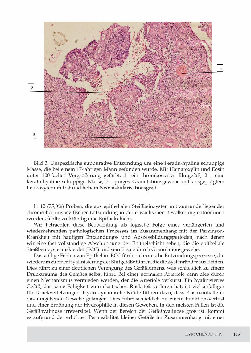
KyRyCHENKO O.P. 115
Bild 3. Unspezifische suppurative Entzündung um eine keratin-hyaline schuppige Masse, die bei einem 17-jährigen Mann gefunden wurde. Mit Hämatoxylin und Eosin unter 100-facher Vergrößerung gefärbt. 1- ein thrombosiertes Blutgefäß; 2 - eine kerato-hyaline schuppige Masse; 3 - junges Granulationsgewebe mit ausgeprägtem Leukozyteninfiltrat und hohem Neovaskularisationsgrad.
In 12 (75,0%) Proben, die aus epithelialen Steißbeinzysten mit zugrunde liegender chronischer unspezifischer Entzündung in der erwachsenen Bevölkerung entnommen wurden, fehlte vollständig eine Epithelschicht.
Wir betrachten diese Beobachtung als logische Folge eines verlängerten und wiederkehrenden pathologischen Prozesses im Zusammenhang mit der Parkinson-Krankheit mit häufigen Entzündungs- und Abszessbildungsperioden, nach denen wir eine fast vollständige Abschuppung der Epithelschicht sehen, die die epitheliale Steißbeinzyste auskleidet (ECC) und sein Ersatz durch Granulationsgewebe.
Das völlige Fehlen von Epithel im ECC fördert chronische Entzündungsprozesse, die wiederum zu einer Hyalinisierung der Blutgefäße führen, die die Zystenränder auskleiden. Dies führt zu einer deutlichen Verengung des Gefäßlumens, was schließlich zu einem Drucktrauma des Gefäßes selbst führt. Bei einer normalen Arteriole kann dies durch einen Mechanismus vermieden werden, der die Arteriole verkürzt. Ein hyalinisiertes Gefäß, das seine Fähigkeit zum elastischen Rückstoß verloren hat, ist viel anfälliger für Druckverletzungen. Hydrodynamische Kräfte führen dazu, dass Plasmainhalte in das umgebende Gewebe gelangen. Dies führt schließlich zu einem Funktionsverlust und einer Erhöhung der Hydrophilie in diesen Geweben. In den meisten Fällen ist die Gefäßhyalinose irreversibel. Wenn der Bereich der Gefäßhyalinose groß ist, kommt es aufgrund der erhöhten Permeabilität kleiner Gefäße im Zusammenhang mit einer

116 JOURNAL OF SOCIAL SCIENCES, NURSING, PUBLIC HEALTH AND EDUC ATION
Lumenverengung und einem erhöhten arteriellen Druck zu einem signifikanten Verlust der Gewebefunktion. Dies schafft wiederum günstige Bedingungen für entzündliche und infektiöse Prozesse in den umgebenden Geweben.
Bei der Beobachtung von epithelialen Steißbeinzysten bei erwachsenen Patienten stellten wir fest, dass in 15 (93,75%) Fällen die das ECC umgebenden Strukturen aus rauem Bindegewebe mit ausgeprägter Lympho- und Histiozytose bestanden. Die Lumen der Zysten enthielten keratinisierte Massen, desquamierte Epithelzellen, Haarsträhnen und ganze Haarschäfte, während die Wände der Zysten mit Granulationsgewebe ausgekleidet waren. Das oberflächengeschichtete Plattenepithel, das die Fisteln umgab, war stark verdickt und bestand aus 20 bis 22 Schichten, häufig mit Anzeichen von Angiomatose (Bild 4).
Bild 4. Fehlgebildete Hautcluster (Talgdrüsen, Haarschäfte) innerhalb eines ECC mit umgebender chronischer Entzündung und fibrosiertem Gewebe. Mit Hämatoxylin und Eosin gefärbt, unter 100-facher Vergrößerung betrachtet. 1 - ein verdickter gespaltener Haarschaft; 2 - eine Talgdrüse.
Ähnliche morphologische Befunde bei Kindern mit Parkinson wurden nur bei 11 (30,55%) Patienten im Alter von 17 bis 18 Jahren beobachtet, insbesondere bei Patienten mit einer wiederkehrenden Form der Krankheit. Anders als in erwachsenen Fällen gab es keine vollständig gebrochenen / lysierten Haarschäfte, sondern nur teilweise beschädigte, die an nicht atrophierten Haarfollikeln hafteten, was auf ihren nicht exogenen, nicht transdermalen Ursprung hinweist.

KyRyCHENKO O.P. 117
In der interglutealen Falte ist die Haut ziemlich eng mit der darunter liegenden Faszie und dem Periost verwachsen. Während des Wechsels
ng der Position (insbesondere zur vertikalen Position des Rumpfes), des normalen Gangs und in sitzender Position erweitern und vertiefen sich die Hautporen im Bereich des Kreuzbeins. Dies kann zu einer übermäßigen Ablagerung von Epithelresten, Schweiß, Talgsekreten und Haarfragmenten in diesen Poren führen. Alle diese Materialien können die bereits gedehnte Pore noch weiter verbreitern und möglicherweise zu Entzündungen führen. Schließlich bildet sich eine pilonidale Höhle, die sehr günstige Bedingungen für mehrere pathogene Mikroben schafft. Aus diesem Grund sind freie Haare und primäre Fisteln nicht immer in der pilonidalen Zyste / Abszess vorhanden.
Basierend auf der vorherigen histologischen Analyse von biopsierten Gewebeproben haben wir festgestellt, dass die Tiefe der Epithelaussparung, aus der sich Primärfisteln bilden können, 3-6 mm oder mehr beträgt.
Im ECC gefundene Haarfollikel waren selten morphologisch normal. Viel häufiger waren sie deformiert, zeigten heterogene Formen und Größen und wuchsen mehrere Haarschäfte aus ihnen heraus. Bindegewebsadhäsionen waren dick fibrinös. Fibrose im Zusammenhang mit PK ist eine Folge einer chronischen unspezifischen Entzündung in Bereichen, die die Pilonidzyste umgeben. Darüber hinaus ist die Fibrose bei häufiger akuter Entzündung, die der chronischen überlagert ist, stärker ausgeprägt. Bei Kindern aller Altersgruppen mit Parkinson fanden wir mehrere Fibroseherde im subkutanen Fett, das der Sacrococcygealregion zugrunde liegt.
Bei Kindern kann das ECC Schweiß- und Talgdrüsen sowie missgebildete Haarschäfte in erheblicher Anzahl enthalten.
Das beobachtete Granulationsgewebe enthielt häufig kleine neu gebildete kapillarähnliche Gefäße. Gefäße mit ausgeprägter Wandhyalinose wurden in Bereichen mit ausgeprägter Gewebefibrose und Skarifizierung gefunden.
In unseren Vorschlägen zur angeborenen PK-Hypothese haben wir die Vorstellung berücksichtigt, dass sich mesenchymales Gewebe in provisorischen Organen schneller differenziert als in embryonalen Organen, was bedeutet, dass letztere länger „unreif“ bleiben. Auch das Vorhandensein von „übermäßig komplexem“ mesenchymalem Gewebe während Perioden der postnatalen Kindheitsentwicklung kann zu Fällen von mesenchymaler Dysproteinose führen, die schließlich zu fibrinösen Veränderungen im umgebenden Gewebe sowie zur Hyalinisierung der Blutgefäßwand führen können Letzteres ist die Grundlage für akute oder chronische Entzündungen.
AbschlussPilonidale Krankheit hat viele mögliche Ursachen und kann durch eine Reihe von
provozierenden Faktoren ausgelöst werden. Die Parkinson-Krankheit weist mehrere Stadien auf: Bildung eines akuten Abszesses in der Sakrococcygealregion, anhaltende chronische Entzündung und wiederkehrende akute chronische Entzündung.
Das Vorhandensein von unreifem Mesenchym-ähnlichem Gewebe, das zu abnormalem oder missgebildetem Bindegewebe der Dermis und ihrer verschiedenen Stützstrukturen führt, dem die Epithelauskleidung fehlt und das eine Umgebung für pathologische Prozesse bei Kindern verschiedener Altersgruppen schafft, unterstützt die angeborene Theorie von PK.
Verweise:
1. Ramzan M., Mirza M.K., Ali M.A. Experience with modified karydakis procedure using subcuticular skin closure. Professional Med. J. 2016;26(12):1499-1504.

118 JOURNAL OF SOCIAL SCIENCES, NURSING, PUBLIC HEALTH AND EDUC ATION
2. Karydakis G.E. Easy and successful treatment of pilonidal sinus after explanation of its causative. Aust. N. Z. J. Surg. 1992;62(5):385-389.
3. Akinci O. F., Bozer M., Uzunkoy A., et al. Incidence and etiological factors in pilonidal sinus among Turkish. Eur. J. Surg. 1999;165:339-342.
4. Sondena K., Andersen E., Nesvik I., et al. Patient characteristics and symptoms in chronic pilonidal sinus disease. Int. J. Colorectal. Dis. 1995;10(3):39-42.
5. Tabidze D.L., Saenko V.V. Radical treatment of pilonidal cysts (epithelial coccygeal course) by bascom II (cleft lift) procedure. Surgery of Ukraine. 2016;4:63-66.
6. Doll D., Matevossian E., Wietelmann K., et al. Family history of pilonidal sinus predisposes to earlier onset of disease and a 50 % longterm recurrence rate. Dis. Colon. Rectum. 2009;52(9):5-11.
7. Nechai I.А., Maltcev N.P. Minimally invasive methods in treatment of pilonidal disease (review of the literature). Grekov’s Bulletin of Surgery. 2019;178(3):69-73.
8. Bascom J. Pilonidal disease: origin from follicles of hairs and results of follicle removal as treatment. Surgery. 1980;87(5):567-572.
9. Lurin I.A., Tsema E.V. Aetiology and pathogenesis of pilonidal disease (review article). Koloproktologiya. 2013:3:35-50.
10. Isik A., Idiz O., Firat D. Novel Approaches in Pilonidal Sinus Treatment. Prague Med. Rep. 2016;117(4):145-152.
11 Milone M., Di Minno M.N., Musella M., et al. The role of drainage after excision and primary closure of pilonidal sinus: a meta-analysis. Tech. Coloproctol. 2013;17(6):625-630.
12. Kleitsch W.P., Cherry L.D. Pilonidal sinus: a rational scheme of treatment. Mil. Surg. 1952;111(2):96-106.
13. Davage O.N. The origin of sacrococcygeal pilonidal sinuses. American Jour. Pathol. 1954;30(6):1191-1205.
14. Raffman R.A. A reevaluation of the pathogenesis of pilonidal sinus. Ann. Surg. 1959;150(5):895-903
15. Stone H.B. The origin of pilonidal sinus (coccygeal fistula). Ann Surg. 1931;94:317-320.
16. Kallet H.I. Pilonidal sinus the factor of adolescence. Tr. Am. Proct. Soc. 1936:163.

119
IMPROVING THE EFFICIENCY OF DIAGNOSIS OF ACUTE APPENDICITIS IN FEMALE CHILDREN THROUGH THE USE OF ANAL MANOMETRY AND TOTAL INDEX OF ENDOGENOUS INTOXICATION
Konoplitskyi Viktor,head of the Department of children surgery National Pirogov Memorial Medical University, Vinnytsia, Ukraine; doctor of Medical Science, Full professor; orcid.org/0000-0001-9525-1547; [email protected].
Korobko Yurii,assistant lecturer of the of the department of children surgery National Pirogov Memorial Medical University, Vinnytsia, Ukraine; orcid.org/0000-0002-3299-878X; [email protected].
Summary. The aim of the study was to increase the efficiency of the diagnostic process in children with abdominal pain by using anal manometry and the total index of endogenous intoxication.
Research methods. 400 female patients with abdominal pain aged 3 to 17 years, on the basis of Vinnytsia Children’s Regional Clinical Hospital (Ukraine), were involved in the analysis of endogenous intoxication and anal pressure.
Results. The study showed a clear proportional increase in the values of anal pressure and total index of endogenous intoxication in accordance with the extent of the inflammatory process in the abdominal cavity due to acute appendicitis.
Conclusions. Having conducted a clinical study, which involved measuring the anal pressure of the sphincters in various pathologies of the abdominal cavity and pelvic organs, we can say about the feasibility of using this technique in the clinical practice of surgeons. The above method demonstrates a fairly high informativeness. Therefore, it can be used as an additional element of diagnosis in complex clinical cases, especially in girls with overweight, cases with atypical location of the appendix, and atypical course of the disease. This method clearly demonstrates the dependence of the tone of the rectal sphincters with the degree of spread of the inflammatory process in the abdominal cavity and pelvic cavity. An new approach to the interpretation of endogenous intoxication according to general blood analysis based on the value of the total index of endogenous intoxication allows to assess the severity of endotoxicosis, based on which it is possible to predict the form of pathology and timely choose the necessary treatment tactics. Exceeding the value of the proposed total index of endogenous intoxication of the body more than twice indicates the child has the level of endotoxicosis characteristic of destructive forms of acute appendicitis, exceeding the index more than 2.5 times indicates a possible complicated pathology in the form of peritonitis. The introduction of the

120 JOURNAL OF SOCIAL SCIENCES, NURSING, PUBLIC HEALTH AND EDUC ATION
developed total index of endogenous intoxication in the algorithm of acute appendicitis will improve the results of early diagnosis and predict the form of pathology and its nature in children.
Key words: children, acute appendicitis, diagnosis.
Topicality. Acute appendicitis is one of the most common acute surgical diseases of the abdominal cavity in children. About 65–75% of surgical interventions in children account for acute appendicitis, which is due to the peculiarities of functional and morphological processes of the child’s body, the rapid development of destructive processes [2,3,5,6].
The problem of early diagnosis of acute appendicitis in children of different ages remains one of the most pressing in the provision of emergency surgery. At the diagnostic stage in determining the main clinical symptoms of acute appendicitis, differential diagnostic signs of concomitant pathology, features of the clinical course, similarity of symptoms, there are usually significant difficulties in diagnosing the inflammatory process of the appendix, which requires auxiliary laboratory and instrumental tests – ultrasound method, computed tomography of the abdominal organs, indicators of which are necessary for pediatric surgeons [1,5,7]. However, even the use of high-tech tools and clinical experience does not always help in the timely recognition of an acute surgical problem.
In patients with acute appendicitis, especially in complicated cases, the presence of endogenous intoxication in the body is of great importance. Determination of integrated indicators of endotoxicosis allows to assess the patient’s condition without using special research methods, according to the general clinical blood test [4]. The development of a reliable and accessible algorithm for the diagnosis of acute appendicitis, including taking into account the integrated indicators of the hemogram, which would allow in a short time to decide on the feasibility of appendectomy, remains an unsolved problem of pediatric surgery.
The aim of the study was to increase the efficiency of the diagnostic process in children with abdominal pain by using anal manometry and the total index of endogenous intoxication.
Materials and discussion of the study. The study on the measurement of anal pressure included 60 female children who were hospitalized in the surgical departments of the Vinnytsia Regional Children’s Clinical Hospital (Ukraine). We examined children of the older age group. The age range of the examined girls was in the range of 3 – 17 years. The main reason for refering medical help were abdominal pain, mainly in the lower abdomen. For anal manometry, patients were divided into three equal groups – 20 children each. The first group consisted of girls who were hospitalized with abdominal pain with suspected acute appendicitis, but during the dynamic observation of acute surgical pathology was excluded. The second group included girls who underwent surgery for acute appendicitis. Girls in this group had no complications in the form of peritonitis. The third group consisted of girls who underwent surgery for acute appendicitis and had peritonitis of varying severity. All patients and their parents had prior informed consent prior to anal manometry after being introduced to the method of anal manometry. Anal manometry was performed immediately after rectal examination, which is mandatory in children with suspected acute appendicitis according to clinical protocols in Ukraine. Anal manometry was performed in the treatment room in the position of the child on his back. The device we developed for anal manometry consisted

KOROBKO yURII, 121
of a standardized manometer, to which was attached a cuff made of an intubation tube (tube diameter without cuff 4.5 mm) and a manual supercharger with a clamp to create a constant pressure in the sensitive cuff. All modules of the device were connected by rubber tubes (Fig. 1).
Fig. 1. Device for measuring the tone of the rectal sphincters. The components of the device are: 1– manometer (mm Hg); 2 – sensitive cuff, which is inserted into the rectum; 3 – clamp; 4 – pressure blower.
At the beginning of the procedure, up to 20 mm Hg air was pumped into the circuit of the device by means of a manual supercharger and the lumen of the tube was closed at the level of the clamp to maintain the appropriate constant pressure in the circuit and give the cuff sensitivity by fully straightening. The cuff and anus were then lubricated with a solution of Vaseline to reduce soft tissue resistance and reduce discomfort during the procedure. The procedure was performed without general and local anesthesia, because in the straightened state the diameter of the cuff does not exceed the diameter of an adult’s finger and does not cause significant pain. The sensitive cuff was inserted into the rectum to a depth of 3 cm in order to establish it in the lumen of the anal sphincter. Immediately after the introduction of the cuff, the value of the manometer in

122 JOURNAL OF SOCIAL SCIENCES, NURSING, PUBLIC HEALTH AND EDUC ATION
millimeters of mercury was recorded. This indicator, the so-called reactive pressure, is an indicator that includes passive sphincter tone, as well as active conscious contraction of the sphincters in response to the cuff. To obtain the second, more important indicator (basal pressure), which takes into account only the passive tension of the sphincter muscles, it is necessary to hold the cuff in the lumen of the sphincter for 60 seconds. It is during this time that the sphincter pressure gets used to the cuff and significantly reduces the element of active tension of the striated muscles of the closing apparatus.
The diagnostic procedure was completed by determining the basal pressure. Measurement of basal pressure is a more important indicator than reactive pressure, because it more objectively reflects the constant tension of the sphincter muscles at rest. It is the basal pressure that reflects the nonspecific reaction of the sphincter apparatus to pain, and hence the inflammatory reaction, in the abdominal cavity and pelvic cavity by the mechanism of the visceromotor reflex arc. After the procedure, the device was cleaned and sanitized by disinfection in an antiseptic solution. After measuring the anal pressure, it was necessary to subtract from each value the value of the pressure that was created to straighten the cuff. That is, from the value that was set during the pressure measurement it was necessary to subtract 20 mm Hg. Statistical processing was performed using the computer program MS Statistica 5.0.
Research results. In the course of our research, the following indicators were identified. In children of the first group, the average value of reactive anal pressure was 59.65 ± 2.11 mm Hg, the average basal pressure was observed at 50.35 ± 2.53 mm Hg. In patients of the second group, the data of sphincterometry were as follows. The average value of the reactive pressure was at the level of 89.1 ± 3.27 mm Hg, after holding the cuff in the lumen of the sphincter, the pressure decreased to its basal level, the average value of which was 70.7 ± 1.94 mm Hg. The highest value of mean anal pressure, compared to the previous two groups, was observed in the third group. The average value of anal pressure had the following indicators: reactive pressure – 106.4 ± 4.3 mm Hg , basal pressure – 77.85 ± 2.81 mm Hg. The overall mean value for patients with acute appendicitis was 97.75 ± 3 mm Hg – reactive pressure and 74.28 ± 1.78 mm Hg – basal pressure.
Based on the results of the study, the dynamics was characterized by an increase in both reactive and basal pressures, which is directly proportional to the degree of inflammation in the abdominal cavity (Table 1).
Table 1. The average value of anal pressure in children hospitalized with abdominal pain.
Type of anal pressure
І group(mm Hg)
ІІ group (mm Hg)
ІІІ group(mm Hg) p
Reactive 59,65 ± 2,11 89,1 ± 3,27 106,4 ± 4,3 р1<0.01р2<0.01
Basal 50,35 ± 2,53 70,7 ± 1,94 77,85 ± 2,81 р1<0.05р2<0.01
* Footnote: р1 – reliability of comparison of results between I and II groups;р2 – reliability of comparison of results between I and IІI groups.
However, given the above indicators, it should be noted a significant difference

KOROBKO yURII, 123
between the indicators in patients with acute appendicitis and patients in whom acute surgical pathology was excluded. It is this difference in pressure that helps in resolving the issue of surgical tactics for the patient. Indicators between groups II and III had less significant differences. The difference between mean basal pressures in patients of the last two groups was less than 10 mm. rt. Art. The difference between the indicators of groups II and III is not so clinically important, because patients from these groups were diagnosed with acute appendicitis, and therefore they underwent emergency surgery in any case.
Also during the study was an analysis of endogenous intoxication. 400 female patients aged 3 to 17 years were involved on the basis of Vinnytsia Children’s Regional Clinical Hospital (Ukraine), which was divided into 4 groups (100 children per group): group I – actually healthy children; group II – patients with abdominal pain in whom after further research surgical pathology was excluded; group III – patients operated with destructive forms of acute appendicitis, without peritonitis; group IV – patients operated with destructive forms of acute appendicitis with peritonitis. Children with catarrhal forms of acute appendicitis were not included in the study. The mean age of patients was 12.6 ± 1.2 years.
Taking into account the data and information reports of other researchers, we proposed a total index of endogenous intoxication (TIEI), which took into account all components of the general blood analysis, and which was calculated by the formula
2/))(
)(100
)((TIEIlymmon
myeljsbanbaseESRL+
++++++
×= ,
L – the number of leukocytes; ESR – erythrocyte sedimentation rate; e – eosinophils; bas – basophils; ban – band neutrophils; s – segmented neutrophils; j – juvenile neutrophils; myel – myelocytes; mon – monocytes; lym – lymphocytes. TIEI normally averages is 1.42 ± 0.06 c. u.
The study of the value of TIEI in various forms of pathology has determined the presence of positive dynamics. The total index of endogenous intoxication was 1.42 ± 0.06 (p <0.05) in patients of the first group. In group II, the index level was 2.31 ± 0.32 (p <0.05). The value of the total index of endogenous intoxication was 3.52 ± 0.45 (p <0.05) in patients of group III. In children of group IV, the total index of endogenous intoxication was 3.80 ± 0.31 (p <0.05).
The presence of such a positive dynamics of the total index of endogenous intoxication indicates the presence and increase in the value of endogenous intoxication with the growth of tissue destruction, which is regarded as a direct sign of intoxication.
Conclusions. Therefore, after conducting a clinical study, which involved measuring the anal pressure of the sphincters in various pathologies of the abdominal cavity and pelvic organs, we can say about the feasibility of using this technique in the clinical practice of surgeons. The above method demonstrates a fairly high informativeness. Therefore, it can be used as an additional element of diagnosis in complex clinical cases, especially in girls with overweight, cases with atypical location of the appendix, and atypical course of the disease. This method clearly demonstrates the dependence of the tone of the rectal sphincters with the degree of spread of the inflammatory process in the abdominal cavity and pelvic cavity.
An new approach to the interpretation of endogenous intoxication according to general blood analysis based on the value of the total index of endogenous intoxication

124 JOURNAL OF SOCIAL SCIENCES, NURSING, PUBLIC HEALTH AND EDUC ATION
allows to assess the severity of endotoxicosis, based on which it is possible to predict the form of pathology and timely choose the necessary treatment tactics. Exceeding the value of the proposed total index of endogenous intoxication of the body more than twice indicates the child has the level of endotoxicosis characteristic of destructive forms of acute appendicitis, exceeding the index more than 2.5 times indicates a possible complicated pathology in the form of peritonitis. The introduction of the developed total index of endogenous intoxication in the algorithm of acute appendicitis will improve the results of early diagnosis and predict the form of pathology and its nature in children.
References:1. Barsukova, I. M., & Krivov, A. P. (2018). Ostryj appendicit: istoriya i sovremennaya
organizaciya medicinskoj pomoshchi. Uchenye zapiski SPBGMU im. IP Pavlova, 25(3).
2. Jaschinski, T., Mosch, C., Eikermann, M., & Neugebauer, E. A. (2015). Laparoscopic versus open appendectomy in patients with suspected appendicitis: a systematic review of meta-analyses of randomised controlled trials. BMC gastroenterology, 15(1), 48.
3. Khanafer, I., Martin, D. A., Mitra, T. P., Eccles, R., Brindle, M. E., Nettel-Aguirre, A., & Thompson, G. C. (2016). Test characteristics of common appendicitis scores with and without laboratory investigations: a prospective observational study. BMC pediatrics, 16(1), 147.
4. Konoplitsky, V. S., Motygin, V. V., yakimenko, A. G., Dmytriiev, D. V., & Mikhalchuk, T. I. (2018). Mathematical modeling for prediction of complicated acute appendicitis in children. Perioperaciina Medicina, 1(1), 70-80.
5. Pogorelic, Z., Rak, S., Mrklic, I., & Juric, I. (2015). Prospective validation of Alvarado score and Pediatric Appendicitis Score for the diagnosis of acute appendicitis in children. Pediatric emergency care, 31(3), 164-168.
6. Salminen, P., Paajanen, H., Rautio, T., Nordström, P., Aarnio, M., Rantanen, T., ... & Grönroos, J. M. (2015). Antibiotic therapy vs appendectomy for treatment of uncomplicated acute appendicitis: the APPAC randomized clinical trial. Jama, 313(23), 2340-2348.
7. Zingone, F., Sultan, A. A., Humes, D. J., & West, J. (2015). Risk of acute appendicitis in and around pregnancy: a population-based cohort study from England. Annals of surgery, 261(2), 332-337.

125
SUTURING OF POSTOPERATIVE WOUNDS IN CHILDREN WITH DIFFERENT THICKNESS OF SUBCUTANEOUS FAT AS ONE OF THE MOMENTS OF IMPROVING THE QUALITY OF SURGERICAL TREATMENT
Konoplitskyi Viktor,head of the Department of children surgery National Pirogov Memorial Medical University, Vinnytsia, Ukraine; doctor of Medical Science, Full professor; orcid.org/0000-0001-9525-1547; [email protected].
Pasichnyk Oleh,assistant lecturer of the of the department of children surgery National Pirogov Memorial Medical University, Vinnytsia, Ukraine; orcid.org/0000-0001-8302-3520; [email protected].
Summary. The aim of the work is to improve the results of treatment of children with neoplasms of the dermis and hypodermis at different thicknesses of subcutaneous fat by developing optimal tactics for suturing the skin.
Research methods. The study analyzed the results of wound closure in 36 patients after removal of skin tumors. The study was performed among patients who were hospitalized in the pediatric surgery clinic of Vinnytsia National Medical University (Ukraine) on the basis of the oncohematology department of the Vinnytsia Regional Children’s Clinical Hospital in the period from 2017 to 2020. There were 17 boys (47.22%) and 19 girls (52.78%). The average age of patients was 8.6 +1.2 years old. In all patients after removal of tumor-like formations of the dermis and hypodermis, postoperative wound closure was performed according to the proposed method. In all children, the results of histological examinations of biopsies revealed benign tumors.
Results. During any surgery, there are areas affected by the surgeon with varying degrees of tissue destruction. Given the zonation of the postoperative wound, the methodology of the proposed method was as follows. After the main stage of surgery and thorough hemostasis, the edges of the wound are widely spread with surgical hooks. Starting from the side of the postoperative wound with a thicker subcutaneous fat layer, apply an adaptive suture, starting with the injection of an atraumatic needle with biodegradable thread from the inside out and from top to bottom, through the subcutaneous fat and superficial fascia with the capture of its own fascia to a depth of 2.0 – 2.5 cm with a puncture outward on the same side in the reverse order and the subsequent conduction of the ligature from the inside out through the superficial fascia of the subcutaneous fat layer of the opposite wall without separation of the skin. When tightening the ends of the ligature of the thread presses into the wound depth of the upper edge of the thicker fat layer, and the opposite horizontal part of the ligature

126 JOURNAL OF SOCIAL SCIENCES, NURSING, PUBLIC HEALTH AND EDUC ATION
«C–D» raises the upper edge of the fat layer thus simultaneously singing the upper edges of the subcutaneous fat layer. on both sides of the wound, due to which there is an adaptation of tissues with different thicknesses
Conclusions. The main task of the surgeon when suturing a postoperative wound is to compare the edges of the divided skin and keep it in the position of opposition until the healing process leads to complete fusion of its edges. Mechanical support of the edges of the postoperative wound by suturing should take into account the areas of surgical damage to the surgical wound, namely not less than the depth of the area of indirect damage. The beginning of suturing the wound on the side with a greater thickness of subcutaneous fat in the direction from the inside out and from top to bottom with the capture of its own fascia conducting ligature strictly in the opposite direction without additional separation of the skin, allows you to adapt the wound edges without excessive tension. lack of pronounced ischemia along the entire depth of the opposite walls.
Key words: children, postoperative wound, surgical sutures.
Topicality. The need for suturing postoperative wounds is relevant for representatives of various areas of surgery. Adequate closure of skin wounds ensures the course of postoperative wounds without complications, and sutures on exposed areas of the body improve a good cosmetic result. All sutures, regardless of their purpose, have the same requirements: precision (accurate adaptation of the wound edges); elimination of cavities and “pockets”; minimal tissue trauma (exclusion of excessive skin tension and tissue hypoxia, without interfering with the natural drainage of the wound); providing hemostatic effect; achieving a good cosmetic result; the possibility of complete removal or biodegradation of suture material; quick application and removal of suture; getting the minimum amount of suture material into the wound cavity [3]. But there is no perfect suture that meets all these requirements at the same time, as some of them contradict each other. Therefore, the choice of a particular seam should be individual and depend on the specific situation [1].
The question of methods and ways to close surgical wounds still remains one of the urgent problems of surgery, especially in the process of comparing its size and the size of the surgical object that was removed. In connection with the fundamental study of angioarchitectonics of soft tissues, a large number of methods of suturing wounds have been proposed, but the problem remains far from its final solution [5].
There are ways to suture the opposite edges of the subcutaneous fat layer of the postoperative wound by applying traditional nodal, “P” – similar, “S” – similar, “8” – similar sutures [1]. The main disadvantages of the proposed techniques are excessive trauma to infiltrated, inflamed and rigid tissues during their contraction and juxtaposition, deterioration of blood / lymph circulation, which prevents uniform capture of subcutaneous fat and completely eliminate closed cavities and «pockets» in the wound. Based on the fact of the lack of a universal method of postoperative wound closure, the great dissatisfaction with the cosmetic and precise results becomes obvious relevance of this study.
The aim of the work is to improve the results of treatment of children with neoplasms of the dermis and hypodermis at different thicknesses of subcutaneous fat by developing optimal tactics for suturing the skin.
Materials and methods. The study analyzed the results of wound closure in 36 patients after removal of skin tumors. The study was performed among patients who

PASICHNyK OLEH, 127
were hospitalized in the pediatric surgery clinic of Vinnytsia National Medical University (Ukraine) on the basis of the oncohematology department of the Vinnytsia Regional Children’s Clinical Hospital in the period from 2017 to 2020.
There were 17 boys (47.22%) and 19 girls (52.78%). The average age of patients was 8.6 +1.2 years old. In all patients after removal of tumor-like formations of the dermis and hypodermis, postoperative wound closure was performed according to the proposed method. In all children, the results of histological examinations of biopsies revealed benign tumors.
Results and discussion. When developing the technique of suturing postoperative wounds in children, we took into account the fact that a characteristic feature of any surgery is prolonged tissue trauma in the process. There are specific consequences of damage to each type of tissue in the form of their dysfunctions and systemic disorders, which in the postoperative period reduces their functional capacity.
In addition, non-specific local tissue trauma due to static pressure of instruments and power of surgical equipment and physical efforts of surgeons aimed at local tissue damage are of great importance in assessing the extent of surgical damage.
Such trauma occurs continuously throughout the operation, after which the entire area of injury is subject to inflammatory-regenerative changes, the severity of which, both local and systemic consequences are largely determined in advance by nonspecific surgical aggression. By analogy with other types of injuries, according to the degree of change, M. Prudkov (2007), divided the surgical wound into separate zones (Figure 1) [4].
Figure. 1. The scheme of zones of surgical damage of an operative wound: I – the view before suturing; II – view after sewing. A – necrosis zone; B – area of contact damage; C – area of indirect damage.
Zone “A” is represented by a site of tissue destruction, in which there are no viable tissues that are deprived of blood supply, crushed or dissected by instruments (under the influence of physical factors of destruction). In the subsequent there is a rejection of necrotic tissues, their lysis and resorption of dead structures with subsequent replacement by connective tissue elements. Dead areas of tissue, possible foreign bodies in the subsequent provide a favorable environment for the emergence and development of wound infection.
Zone “B” is an area of severe damage that develops due to direct contact with surgical instruments and the influence of surgeons. Part of the cellular structures in zone “B”

128 JOURNAL OF SOCIAL SCIENCES, NURSING, PUBLIC HEALTH AND EDUC ATION
receives irreversible changes, which subsequently lead to their death, including due to local circulatory disorders or infectious complications.
Important is the fact that the total mass of tissues that fell into zones “A” and “B”, as well as the degree of their damage, largely determine the features of the local wound process and can be regarded as a certain prognostic factor for wound healing. Tissue structures that fall into the “C” zone do not come into direct contact with the surgeon’s instruments and hands. Their damage occurs indirectly, due to pressure through the tissues. According to this mechanism of damage, the degree of tissue destruction in zone “C” is much smaller, but can be determined in a fairly wide range. Therefore, an important factor for reducing the zone “C” are sufficient linear parameters (length and width) of the surgical wound.
Therefore, in the presence of appropriate areas of surgical wound damage, the main conditions for adequate suturing of the skin should be: the absence of excessive tension when comparing the edges of the wound along its entire depth; satisfactory blood supply to all layers of the wound; no signs of local infection and tissue necrosis. The importance of compliance with these conditions is especially great if the patient has different thickness of the opposite layers of the hypodermis.
Given the zonation of the postoperative wound, the methodology of the proposed method was as follows. After the main stage of surgery and thorough hemostasis, the edges of the wound are widely spread with surgical hooks. Starting from the side of the postoperative wound with a thicker subcutaneous fat layer, apply an adaptive suture, starting with the injection of an atraumatic needle with biodegradable thread from the inside out and from top to bottom, through the subcutaneous fat and superficial fascia with the capture of its own fascia to a depth of 2.0 – 2.5 cm with a puncture outward on the same side in the reverse order and the subsequent conduction of the ligature from the inside out through the superficial fascia of the subcutaneous fat layer of the opposite wall without separation of the skin. When tightening the ends of the ligature І↔ІІ part of the thread «A–B» presses into the wound depth of the upper edge of the thicker fat layer, and the opposite horizontal part of the ligature «C–D» raises the upper edge of the fat layer thus simultaneously singing the upper edges of the subcutaneous fat layer. on both sides of the wound, due to which there is an adaptation of tissues with different thicknesses (Figure 2).
Figure 2. The scheme of suturing according to the proposed method of closing the postoperative wound (explanation of the text).

PASICHNyK OLEH, 129
All needle injections and ligatures during suturing of the wound were performed at the level of zone “C” and deeper.
This technique is protected by the patent of Ukraine №121938 “Method of suturing postoperative wounds with different thickness of subcutaneous fat”.
Clinical example. Patient P., 8 years old, was hospitalized in the oncohematology depar inserting tment with a diagnosis of synovioma in the lower third of the right shoulder. After preoperative preparation, the child underwent surgery: removal of the tumor-like formation of the right shoulder. The operating field was treated with an antiseptic solution three times. An incision up to 8.0 cm long above the tumor-like formation dissected the skin and removed the tumor. Hemostasis. The edges of the postoperative wound are diluted with toothed surgical hooks. Starting from the lateral side of the postoperative wound, where the thickness of the subcutaneous fat layer was greater, three adaptive sutures were applied, starting the formation of each of them by puncturing an atraumatic needle thread “Vicryl 4/0” from the inside out and from top to bottom, through the subcutaneous fat and surface fascia with the capture of its own fascia length of 2.0 cm in the depth of the wound along its midline with a puncture on the same side in the reverse order and with subsequent conduction of the ligature from the inside out with the capture of the superficial fascia of the subcutaneous fat layer of the opposite wall without separation of the skin. The distance between the adaptive sutures is up to 1.0 cm. An intradermal cosmetic suture is applied to the skin (Figure 3).
Figure 3. Patient P., 8 years old. Diagnosis: synovioma in the lower third of the right shoulder. On the left – a general view of the wound 1 day after surgery, On the right – a general view of the postoperative wound on the 10th day after surgery.
The postoperative period was satisfactory. On the 7th day, the skin suture was removed, the child was discharged in satisfactory condition for outpatient treatment.
Histological examination of the removed tumor fully confirmed the clinical diagnosis (Figure 4).
Among all patients in whom postoperative wounds were sutured by the proposed method, their normal healing was determined, local infectious complications were absent in all clinical cases. At follow-up examinations of patients at 3, 6 and 9 months after suturing postoperative wounds, gross keloid scar deformities were not observed in any case.

130 JOURNAL OF SOCIAL SCIENCES, NURSING, PUBLIC HEALTH AND EDUC ATION
Fig. 4. Patient P., 8 years old. Diagnosis: synovioma in the lower third of the right shoulder. Histological conclusion: synovioma. Hematoxylin and eosin staining. Magnification × 200.
Conclusions.The main task of the surgeon when suturing a postoperative wound is to compare
the edges of the divided skin and keep it in the position of opposition until the healing process leads to complete fusion of its edges. Mechanical support of the edges of the postoperative wound by suturing should take into account the areas of surgical damage to the surgical wound, namely not less than the depth of the area of indirect damage. The beginning of suturing the wound on the side with a greater thickness of subcutaneous fat in the direction from the inside out and from top to bottom with the capture of its own fascia conducting ligature strictly in the opposite direction without additional separation of the skin, allows you to adapt the wound edges without excessive tension. lack of pronounced ischemia along the entire depth of the opposite walls.
References:1. Kozlov, V. G. Kozhnyj shov : ucheb.-metod. posobie / V. G. Kozlov, A. V. Bol’shov.
– Minsk : BGMU, 2016. – 15 p.2. Loginov, V. I., Parshikov, V. V., Kasimov, R. R., & Baburin, A. B. (2015).
Sovremennye mekhanicheskie sposoby intraoperacionnoj profilaktiki infekcij oblasti khirurgicheskogo vmeshatel’stva. Novosti khirurgii, 23(5).
3. Prudkov, M. I., Shulutko, A. M., Galmzyanov, F. V., Levit, A. L., Kovalevskij, A. D., & Alferov, S. YU. (2007). Osnovy minimal’no invazivnoj khirurgii.
4. Bordakov, V. N. Rana. Ranevoj process. Principy lecheniya ran : ucheb.-metod. posobie / V. N. Bordakov, Belorus. gos. med. un-t, Kaf. obshch. khirurgii. - Minsk : BGMU, 2014. - 30 p.
5. Tihvinskaya, O. A., Volkova, N. A., Rogul’skaya, E. yU., Revenko, E. B., & Mazur, S. P. (2018). Zazhivlenie ehkscizionnykh kozhnykh ran u myshej v prisutstvii matric iz plazmy krovi. Visnyk Problem Biolohii I Medytsyny, 2(4 (147)).

131
THE EFFECT OF LONG-TERM IONIZING RADIATION ON ORGANS AND SYSTEMS OF THE HUMAN BODY
Kostenko Yevhen,Kostenko Svitlana,Stetsyk MariiaPirchak IlyaSHEI «Uzhhorod national university»
Summary. The biological effects of ionizing radiation on the human body can be both genetic and somatic. One of the characteristic features of an organism that has withstood radiation damage is a state of long-term compensatory voltage, which requires constant mobilization and additional costs of the body’s reserves and can lead to disruption of compensatory reactions. The aim of the study. To establish the clinical and morphological features of the development of pathological processes in the human body that occur over a long period of time after exposure to high doses of radiation and to establish their relationship with the development of periodontal disease. People who permanently live in radiation-contaminated areas develop enzymopathy, metabolic processes are disrupted, toxic metabolic products, radionuclides accumulate, slag excretion is complicated, lipid peroxidation and cell membranes are significantly increased, and antioxidant deficiency develops due to antioxidant deficiency. which causes an imbalance in the nervous, cardiovascular, digestive, immune systems, which in combination leads to changes in periodontal tissues. The action of ionizing radiation can have an initiating, accelerating and modifying effect on the manifestations and course of general somatic diseases. That is why generalized periodontal diseases are polyetiological (comorbid) diseases with an autoimmune component. Conclusions: Based on previous clinical and laboratory and experimental morphological studies, as well as literature data, we can clearly state that the variability of kinetic characteristics of bone tissue can be due to both hereditary (family) and non-hereditary (modification) components. Under the action of ionizing radiation, patients develop not only general somatic pathology, but also dental, in particular, from the periodontal tissues. That is why generalized periodontal disease (periodontitis) is a polyetiological (comorbid) disease with an autoimmune component, in which distinct individual correlations in the kinetics of radionuclides in individuals of the same age and sex are clearly traced.
Key words: ionizing radiation, radionuclides, bone tissue, periodontitis.
The study of remote radiation pathology is an urgent task of clinical practice and the need for deep explanation of adaptive-compensatory mechanisms, as a manifestation of the body’s response to ionizing radiation.
The action of ionizing radiation can have an initiating, accelerating and modifying effect on the manifestations and course of general somatic diseases.
One of the characteristic features of an organism that has withstood radiation damage is a state of prolonged compensatory tension, which requires constant mobilization and additional costs of the body’s reserves and can lead to disruption of compensatory
132 JOURNAL OF SOCIAL SCIENCES, NURSING, PUBLIC HEALTH AND EDUC ATION
reactions, which is why the remote effects of radiation damage take different forms and require constant attention from both science and practical medicine.
The relevance of this issue is determined by two aspects – general biological and medical.
The theoretical significance of the problem of individual variability of metabolic processes follows from the fundamental position, which states that the discreteness of life is represented by individuals with morpho-physiological features present to them, which play an important role in life activity.
In medicine, the importance of individual characteristics of the human body has long been recognized as the principle of an individual approach to each patient, which requires a comprehensive examination.
One of the characteristic features of an organism that has withstood radiation damage is a state of prolonged compensatory tension, which requires constant mobilization and additional costs of the body’s reserves and can lead to disruption of compensatory reactions, which is why the remote consequences of radiation damage are diverse, and their study remains a topical issue of our time.
That is why special attention in the problem of this issue is paid to individual features of the kinetics of osteotropic substances.
The aim of the study. To establish clinical and morphological features of the development of pathological processes in the human body that occur after a long period of time after exposure to high doses of radiation and to establish their relationship with the development of periodontal tissue diseases.
Main content. The biological effect of ionizing radiation on the human body can be both genetic and somatic.
The genetic consequences of radiation exposure are manifested in the descendant of a person who has been exposed to radiation. These include birth defects and deformities that occur as a result of mutations in germ cells.
Somatic consequences of radiation exposure are manifested in a person who was directly exposed to radiation.
The early somatic effect of ionizing radiation occurs in the period from a few minutes to 60 days. It is characterized by such changes as redness and peeling of the skin, clouding of the lens, damage to hematopoietic organs, radiation sickness, and deaths.
The remote effect of somatic impact manifests itself several months or years after exposure. It consists in persistent skin changes, the appearance of malignant neoplasms, a decrease in immunity, and a reduction in life expectancy.
If we take morphological changes as a criterion of sensitivity to ionizing radiation, then the cells and tissues of the human body can be distributed according to the degree of sensitivity decrease as follows:
1) lymphoid tissue and bone marrow; 2) sex glands; 3) mucous membranes; 4) cutaneous integument; 5) lungs; 6) thyroid; 7) GI internal secretion glands; 8) connective tissue; 9) muscle tissue; 10) cartilage and bone tissue; 11) nerve tissue.
Target organs of radioactive substances in the body: 40Ca, 90Sr, 226Ra are accumulated in the skeleton; 244Pu, 139La are concentrated in the liver; 137Cs are concentrated in muscle tissue; 222Rn - in the lungs; 209Po, 3H are distributed evenly throughout the body; radioactive 127L is accumulated in the thyroid gland.
Osteotropic chemical elements, including radionuclides, occupy an important place among man-made pollutants. These include stable elements such as lead, fluorine, zinc, beryllium, alkaline earth metals, radionuclides such as 90Sr, 224, 226, 228Ra, 238, 239Pu,

PIRCHAK ILyA 133
24Am and others. Most of them are toxic. For example, man-made fluoride and lead cause fluorosis and lead intoxication, which in the oral cavity is manifested by «lead stomatitis», beryllium leads to «beryllium rickets», stable strontium causes a violation of mineral metabolism (Knizhnikov V. A., Tseitlin O. Y.) [17, p. 451, 19, p.328].
A number of publications of domestic and foreign scientists have described that radionuclides are sources of internal radiation of the body, with a detailed effect and patterns of accumulation and elimination from the body (Akleev A.V., Balabukha V. S., Buldakov L. O., Ilyin B. M., Lyubashevsky M. M., Moskalev Y.I., Newman M., Newman U., Sokolov y.O., Fradkin G. E., Shvedov V. L., Comar C.L., Degteva M.O., Kozheurov V.P., Lloyd R.D., Stover B.J., Tolstykh E.I. Wasserman R.H.) [11, p. 84, 12, p. 79, 13, p. 10, 14, p. 6].
Gradually «integrating» into bone tissue, osteotropic substances are slowly removed from the human body (Zakutinsky D. I., Durbin P.W., Vaughan J.M.). In addition, a number of radionuclides have a long half-life (for example, 90Sr – 30 years, 226Ra – 1622 years, 239Pu – 24400 years, 235U-7.1 x 10⁸ years). The effect of their action depends on the sensitivity (for example, radiosensitivity) of the body and the kinetics of substances (the level of deposition, localization, time spent in the body) [17, p. 451, 19, p. 328].
There are significant correlations in the differences in the kinetics of radionuclides in the body of people of the same age and gender.
Variability in the kinetic characteristics of bone tissue can be caused by both hereditary (familial) and non-hereditary (modification) components.
Forecasting individual tolerance to osteotropic toxins, based on the mechanisms of their metabolism, is especially relevant in the conditions of a man-made accident, since then a large number of organisms and animals receive osteotropic toxic substances from the external environment.
To interpret the behavior of osteotropic substances in the human body, the concept of limiting morpho-physiological metabolic factors was used previously, which interpreted the mechanisms of metabolism of stable elements and radionuclides in bone tissue from the same positions. It involved the separation of 10 endogenous factors, which are physiological processes, physicochemical reactions and structural components, which together determine the final results of deposition in bone tissue, tissue redistribution and excretion of osteotropic substances from the body. This concept mediated the influence of other processes of vital activity and the external environment (type, gender, age, physiological state, exogenous influence) and allowed us to formulate the significance and direction of shifts in radionuclide exchange in bone tissue [18, p. 168].
In unfavorable environmental conditions after the Chernobyl accident, along with the occurrence of an increased radiation background, the incorporation of radionuclides that enter the body by biological food chain and inhalation is also essential.
The effect of 90Sr on the body is reflected in bone remodeling. The constant inclusion of radioactive strontium in the mineral bone matrix leads to the development of a number of pathological and compensatory-adaptive reactions in bone tissue at the cellular and tissue levels.
Radiobiological effects of osteotropic radionuclides in bone tissue:- preosteoblastic failure;- destruction;- dysplastic fibrosis;- accumulation in hydroxyapatite crystals;- preosteoblastic failure;- changes in energy metabolism.

134 JOURNAL OF SOCIAL SCIENCES, NURSING, PUBLIC HEALTH AND EDUC ATION
The incorporation of 90Sr depends on the intensity of growth and metabolic processes. Incorporated radionuclides interact with the electrons of neighboring atoms, osteotropic ions, and cause ionization. Ionized atoms are actively involved in complex chain reactions, resulting in the formation of active “free radicals”. The latter react with each other, causing chemical and biological changes in cellular elements.
The greatest risk of radiation damage in young bone cells: preosteoblasts and osteoblasts (preodontoblastic failure is the result of a specific action of 90Sr). Osteoblasts secrete polymers of collagen fibrils into the intercellular space, on the surface of which mineralization processes occur. Impaired osteoblast function leads to delayed formation of hydroxyapatite crystals.
Incorporated radioactive 90Sr causes an imbalance in the osteoblast – osteoblast ratio, which disrupts bone remodeling and serves as a trigger for bone destruction.
Immune mediators – cytokines and growth factors-play a significant role in the pathogenesis of osteoporosis.
Lesions of the immune system and gastrointestinal tract should be considered dominant in the progressive development of an autoimmune condition in generalized periodontal diseases.
People who permanently live in radiation-contaminated areas develop fermentopathies, metabolic processes are disrupted, toxic metabolic products and radionuclides are accumulated, the removal of toxins is complicated, lipid peroxidation and cell membranes significantly increases, antioxidant insufficiency develops, the work of the neuro-hormonal system is disorganized, followed by an imbalance of the nervous, cardiovascular, digestive, immune and other systems [1, p. 87, 2, p. 24, 3, p. 304, 7, p. 5].
For most patients in modern conditions the “enteral syndrome” is characterized. The enzymatic activity of the small intestine is disrupted, which leads to morphological changes in the structure of the epithelium and weak adsorption of enzymes on the surface of the mucous membranes. A decrease in the hydrolysis of carbohydrates in the small intestine leads to their ingestion in large quantities in undigested form into the large intestine. Dysbacteriosis of a large intestine, a syndrome of insufficiency of absorption develops (fermentation processes).
There is a correlation between functional disorders in the insular apparatus of the pancreas and periodontal disease.
Most individuals have changes in the neuro-endocrine system, impaired metabolism of endocrine hormones, and hypothalamic dysfunction. At the same time, the intensity of lipid peroxidation increases, immune insufficiency develops, the protection of the antioxidant system decreases against the background of a significant deficiency of vitamins of group A, B, C, K, E, macro - and microelements, etc.
The causes of impaired neuro-endocrine regulation are numerous: hereditary predisposition, mental factors, insufficient endometrial receptor response to the effects of ovarian hormones, ovarian damage, severe endocrinopathies, toxic-infectious damage to diencephalic areas, etc. [6, p. 633, 15, p. 24].
Individuals with thyroid and parathyroid diseases have a high frequency and severe course of generalized periodontal diseases.
In Itsenko-Cushing’s disease, characterized by an excessive content of corticosteroids in the blood, along with damage to the long tubular bones and spine, the phenomena of resorption of the alveolar bone of the jaws are noted.
Individuals with hypercorticism have developed a dystrophic process in the periodontium.
PIRCHAK ILyA 135
The biosynthesis of sex hormones largely depends on a sufficient intake of vitamins, especially vitamin E.
The effect of vitamin E may be mediated through the anterior pituitary gland on the sex glands. At the same time, the effect of vitamin E is not limited to changes only in the gonadotropic function of the pituitary gland, but also in its adrenotropic and thyroid-stimulating functions. Which in turn affects the functional state of the adrenal glands and thyroid gland, as well as on those metabolic processes in the body for which they are responsible. Vitamin E can directly affect redox reactions and metabolic processes [4, p. 30, 16, p. 120].
Due to a violation of the metabolism of sex hormones, shifts in the hypothalamus-pituitary-gonad system, and diencephalic changes, most patients develop generalized periodontal diseases [5, p. 60, 6, p. 633, 8, p. 124].
With reduced estrogen secretion, spastic-atonic state of gum capillaries, anemia and their atrophy, pronounced phenomena of osteoporosis, destructive and resorption processes are detected [9, p. 13, 10, p. 23, 15, p. 24].
An increased content of estrogen in the body causes desquamative phenomena in the gum epithelium, connective tissue proliferation, and increased vascular permeability.
Conclusion. Based on previous clinical, laboratory and experimental morphological studies, as well as literature data, we can clearly state that the variability of the kinetic characteristics of bone tissue can be caused by both hereditary (familial) and non-hereditary (modification) components. Under the influence of ionizing radiation, patients develop not only general somatic pathology, but also dental, in particular, from periodontal tissues. That is why generalized periodontal diseases (periodontitis) are polyethological (comorbid) diseases with an autoimmune component, in which excellent individual correlations in the kinetics of radionuclides in the organisms of individuals of the same age and gender are clearly traced.
References:1. Барер Г.М., Лемецкая Т.И. Болезни пародонта. Клиника, диагностика и лечение.
М.: ВУНМЦ, 1996. 84 с. 2. Борисенко А.В. Нарушение белкового обмена в тканях пародонта при патологии
и их коррекция в комплексном лечении: автореф. дис. д-ра мед. наук. К., 1992. С. 24-29.
3. Боровский Е.В., Леонтьев В.К. Биология полости рта. М.: Медицина, - 1991. 304 с. 4. Вишняк Г.Н. Патогенез и клиника пародонтоза при патологии полового
созревания (клинико-экспериментальное исследование): автореф. дис. д-ра мед. наук. К., 1974. 30 с.
5. Вишняк Г.Н. Роль диэнцефальных нарушений в патогенезе пародонтоза у подростков. Стоматология, 1980. №5. С. 60-61.
6. Вишняк Г.М., Гордієнко В.М. Розвиток генералізованих захворювань пародонту при порушеннях функціонального стану системи гіпоталамус - гіпофіз – гонади: зб. наук, праць співробітників КМАПО. Київ, 1998. В.1, книга 1. С. 633-638.
7. Воскресенський О.Н., Ткаченко Е.К. Роль перекисного окисления липидов в патогенезе пародонтита. Стоматология, 1991. №4. С. 5-10.
8. Гордиенко В.М., Вишняк Г.Н. Влияние функционального состояния системы гипоталамус- гипофиз-гонады на развитие пародонтоза в эксперименте. «Проблемы физиологии гипоталамуса», 1984. В.18. С. 124-128.
136 JOURNAL OF SOCIAL SCIENCES, NURSING, PUBLIC HEALTH AND EDUC ATION
9. Копейкин В.H., Кушлинский Н.Е., Семенов И.Ю. Рецепторы эстрогенов в тканях маргинального пародонта у больных хроническим генерализованным пародонтитом. Стоматология, 1995. №4 - С. 13-14.
10. Кузьмина Э.М., Торчинов А.М., Доронин Г.Д. Состояние тканей пародонта у женщин с гипоэстрогенемией. Новое в стоматологии, 1998. №7. С. 23-28.
11. Любашевский Н.М., Попов Б.В., Мокроносов А.А., Любашевская З.А., Стариченко В.И. Биологические основы межвидовых экстраполяций параметров скелетного метаболизма Пограничные проблемы экологии: сб. науч. тр.. Свердловск: УНЦ АН СССР, 1986. С. 84-102.
12. Любашевский Н.М., Григоркина Е.Б., Стариченко В.И., Расина Л.Н., Чибиряк М.В. Экстраполяционная оценка риска для будущих поколений аварийных радиационных и техногенных загрязнений и их реабилитация. Совершенствование защиты населения и территорий от чрезвычайных ситуаций природного и техногенного характера в условиях Уральского региона: Особенности, проблемы, пути решения: материалов региональной научно-практической конференции (28-29 апреля 1998 г., г. Екатеринбург). Екатеринбург, 1998. С. 79-81.
13. Любашевский Н.М., Попов Б.В., Стариченко В.И., Мокроносов А.А. Лимитирующие морфофизиологические факторы в межвидовой и межвозрастной экстраполяции и в индивидуальном прогнозировании обмена остеотропных радионуклидов Радиобиологический эксперимент и человек: сб. науч. тр. – М.: МЗ СССР, 1986. С. 10-19.
14. Любашевский Н.М., Стариченко В.И. Медико-биологические аспекты индивидуальной изменчивости метаболизма и патологического действия остеотропных токсических веществ Ускорение социально-экономического развития Урала: Блок 4: Социально-экономическое развитие Урала (Здравоохранение): тез. докл. Всесоюз. науч.-практ. конф., (6-19 ноябрь 1989 г., г. Свердловск). Свердловск, 1989. Ч. 1. С. 6-8.
15. Максимовський Ю.М. Состояние пародонта при генерализованном остеопорозе у женщин с недостаточной функцией яичников. / Ю.М. Максимовський, А.И. Мощиль, А.И. Воложин // Стоматология. - 1991. - №1. - С. 24-26.
16. Мазур I.П., Поворознюк В.В., Вишняк Г.М., Богдан А.С. Структурно-функціональний стан тканин пародонту та кісткової системи у жінок різного віку: вплив менопаузи. Педіатрія, акушерство, гінекологія, 1998. №1. С. 120-123.
17. Стариченко В.И. Индивидуальная изменчивость депонирования 90Sr и ее вариабельность в зависимости от генотипической однородности выборки. Радиационная биология. Радиоэкология, 2000. Т. 40, № 4. С. 451-455.
18. Стариченко В.И., Любашевский Н.М., Попов Б.В.. Индивидуальная изменчивость метаболизма остеотропных токсических веществ. Екатеринбург: Наука, 1993. 168 с.
19. Стариченко В.И. Кинетика 90Sr: генотипическая детерминация. Радиационная биология. Радиоэкология, 2005. Т. 45, № 3. С. 328-332.

137
ON THE ISSUE OF METHODOLOGICAL AND PEDAGOGICAL BASIS OF TEACHING PUBLIC HEALTH MASTERS THE SUBJECT “ORGANIZATION AND PRESENTATION OF SCIENTIFIC RESEARCH”
Myronyuk Ivan Doctor of Medical Sciences, professor, dean of the Faculty of Health and Physical Education of Uzhhorod National University; [email protected].
Bilak-Lukianchuk ViktoriaCandidate of Medical Sciences, Associate Professor of the Department of Health Sciences, Uzhhorod National University; [email protected].
Slabkyi GennadiyDoctor of Medical Sciences, Professor, Head of the Department of Health Sciences, Uzhhorod National University; [email protected]
Summary. The aim is to determine the methodological and pedagogical basis for teaching masters of public health the discipline “Organization and presentation of scientific research.”
The materials of the research are the current legislative acts and regulations of Ukraine regarding the functioning of health care system and training of masters of public health, curricula for the training of public health masters. Methods of content analysis, structural-logical analysis and system approach were used in the work.
article presents the structure of educational and scientific program of the discipline “Organization and presentation of scientific research”, methods, forms and results of teaching “Organization and presentation of scientific research”.
Conclusions. The methodological and pedagogical basis for teaching masters of public health the subject “Organization and presentation of scientific research” is to use active and interactive forms of classroom teaching using the method of controlled individual learning and research tasks for independent work of students.
Keywords: public health, masters, training, the subject “Organization and presentation of scientific research”, methodological and pedagogical basis.
In Ukraine, the implementation and development of the public health system is defined at state level [1, 2]. The Cabinet of Ministers of Ukraine approved the specialty “Public Health” [3], which made it possible to introduce training for the system and approve the State Standard of Higher Education in the specialty “Public Health” for first and second level of higher education [4].
The approved standards are based on the competency approach, which is enshrined in the European Commission’s international project “Harmonization of Educational Structures in Europe” and its methodology lies in formation of competencies at all
138 JOURNAL OF SOCIAL SCIENCES, NURSING, PUBLIC HEALTH AND EDUC ATION
stages of learning. At the same time, competencies represent a dynamic and complex combination of knowledge, understanding, skills, abilities and capabilities. Acquisition of competencies by students is the goal of educational programs. Competences are formed during studying different disciplines, and the level of their acquisition is assessed at all stages of training [5].
The aim is to determine the methodological and pedagogical basis for teaching masters of public health to the discipline “Organization and presentation of scientific research.”
Materials and methods. The materials of the research are the current legislative acts and regulations of Ukraine regarding the functioning of health care system and training of masters of public health, curricula for the training of public health masters. Methods of content analysis, structural-logical analysis and system approach were used in the work.
ResultsThe purpose of teaching the subject “Organization and presentation of scientific
research” to masters of public health according to educational-scientific program is the formation of theoretical knowledge about the principles and methods of research and practical skills and abilities to use them in organizing and conducting various types of research and scientific-research work in the field of public health, using the principle of academic integrity, practical preparation of future specialists for implementation of research and analytical professional functions.
As a result of studying the discipline “Organization and presentation of scientific research”, a master must be able to use modern methodology for analysing problems and phenomena in the field of public health; to be aware of features in carrying out various types of applied researches; to develop a research concept; to make the program and design of applied scientific research, to choose optimum methods of its performance; to form a sample; to analyse scientific sources, legislative, regulatory and other documents, data of sectoral statistical reporting and monitoring of the situation in the field of public health; to conduct organizational experiment, observation, questionnaire, etc.; statistically, using the methods of biostatistics, process, analyse and design the results of the study, using the methods of clarity and forms of generalization of the research results; independently improve their professional scientific culture.
According to academic program of this discipline, 120 academic hours have been approved for mastering it, including 20 academic hours for full-time lecture material, 28 academic hours for practical classes and 72 academic hours for independent work of students. In the case of distance (extramural) studying, 14 academic hours are approved for teaching students and 106 academic hours for independent work.
The structure of the discipline involves mastering the material in 10 learning topics, which are divided into two content modules.
Content module 1Methodology of preparation and carrying out of scientific research by studentsTopic 1. Preparatory stage of scientific research. Analytical review of the literature.
Transliteration. Choice of topic and objective, formation of research tasks.Topic 2. Program development, selection of object and subject, research base.Topic 3. The choice of the unit, the calculation of the sample size of the study. Research
methods. Sources of primary scientific information.Topic 4. The procedure for compiling questionnaires of sociological research.

SLABKyI GENNADIy 139
Questionnaire structure. Types of questions. Organization of sociological research. Collection of information. Verification of the data obtained.
Topic 5. The concept of academic integrity and plagiarism. Plagiarism in the scientific work of students and ways to prevent it.
Content module 2Technology of statistical processing of research resultsand basic requirements for content, design, presentationand protection of students’ research worksTopic 6. Methodical approaches to statistical processing of the data obtained.
Statistical grouping of the data. Statistical tables and rules for filling them. Statistical processing of the data obtained with the use of modern mathematical and statistical methods and information technologies.
Topic 7. Analysis of research results. Literary and graphic design of the results of statistical and sociological research.
Topic 8. Implementation of research results in the practice of the public health system and evaluation of their effectiveness.
Topic 9. Rules for writing scientific articles, reports, abstracts, messages, annotations and methodology of presentations.
Topic 10. Methodology of preparation, design and presentation of the results of scientific master’s theses.
Mastering this subject provides students with the acquisition of competencies defined by the State Standard of Higher Education in the specialty “Public Health” of second-level of higher education (Master’s degree): the ability to abstract thinking, analysis and synthesis, skills of information and communication technologies, the ability to research at the appropriate level, the ability to learn and master modern knowledge, the ability to search, process and analyse information from various sources, the ability to be critical and self-critical, the ability to make reasonable decisions, the ability to develop and manage projects, the ability to define the priorities of public health, to provide health care needs assessment, propose science-based measures and develop appropriate action plans and strategies, the ability to draw conclusions, develop forecasts and analyse the impact of different determinants on public health, identify the needs of different population groups in relation to health, based on information obtained from epidemiological surveillance systems, the ability to characterize and study the impact of key individual determinants of health on public health in order to maintain and enhance public health, the ability to develop and implement strategies, policies and measures in health promotion, to conduct effective public health communication using a variety of communication channels and techniques, the ability to develop and implement evidence-based public health strategies, policies and interventions based on public health methods and approaches with stakeholder involvement based on a cross-sectoral approach, the ability to apply ethical and legal principles in relation to research design planning, data collection, dissemination and exploitation of research results.
Teaching of this subject is carried out in the form of lectures, practical classes and independent supervised work of master students. Only active and interactive teaching methods are used.

140 JOURNAL OF SOCIAL SCIENCES, NURSING, PUBLIC HEALTH AND EDUC ATION
The practice of lecturing includes such forms of teaching as binary lecture, lecture-conversation and lecture-discussion, which require appropriate preparation of students during self-study on predetermined topics.
The methods of conducting practical classes are mainly business games, brainstorming, educational discussion and case method. These methods develop students’ ability to analyse and synthesize, to communicate, to act and evaluate their actions, to apply the acquired practical skills.
The method of individual educational and research tasks is used for independent work of students. Performance of the tasks provides not only active mastering by students of the course program, but also preparation for carrying out research for master’s thesis.
During the classes, students actively exploit the knowledge gained in studying of the following disciplines: “Biostatistics”, “Fundamentals of Epidemiology”, “Scientific Communications in Public Health”, “Informatization and Communication in Health Care”, “Monitoring and Evaluation in the Public Health System”. This comprehensive approach to the application of knowledge acquired by students during the study of various disciplines, allows to stimulate students’ learning and cognitive activity, to carry out current comprehensive control of student achievements for the entire period of study at the department, to develop students’ ability to apply knowledge, skills and abilities in total and develop such important qualities as the ability to analyse and synthesize and systemic actions through the application of structural-logical thinking.
Upon completion of training in this subject, masters must be fully prepared to perform a scientific master’s thesis. Thus, during the study of the subject masters directly determine the direction and topic of scientific master’s work and its purpose and objectives of the study, object, subject, base of research and sample size, as well as define research methods, determine the structure of analytical review of scientific literature on the topic of master’s thesis.
Masters of Public Health of Uzhhorod National University (11 full-time and 9 part-time students) in addition to the above during studying of this subject prepared 11 review articles and 4 analytical articles on master’s theses, which were discussed in practical classes, reviewed and sent for publishing in professional journals of Ukraine. It is important that out of the total number of articles, 12 (80.0%) are independent. Masters, during practical classes and independent work, conducted statistical processing of data from sectoral statistical reporting and prepared 24 abstracts of reports. The reports were presented at the conference of masters of the university and published in the conference materials, 4 abstracts were presented at the II International Student Scientific Conference in Michalovce (Slovakia), 5 abstracts at the 74th International Scientific-Practical Conference of medical students and young scientists “Actual Problems of Modern Medicine” of Samarkand State Medical Institute (Uzbekistan) and 2 abstracts at the International Scientific and Practical Conference of Young Scientists to the 100th anniversary of the Tashkent Medical Academy “Medical Science of the XXI century. A look into the future”.
Conclusions. The use of active and interactive forms of classroom training and controlled forms of self-preparation of master students majoring in “Public Health” in studying the subject “Organization and presentation of scientific research” allows them to master basic competencies and develop students’ ability to analyse and synthesize, communicate, employ systemic actions and to evaluate them, apply the acquired practical skills, as well as prepare for a scientific master’s study. Thus, the methodological and pedagogical basis for teaching masters of public health the subject “Organization and
SLABKyI GENNADIy 141
presentation of scientific research” is to use active and interactive forms of classroom teaching using the method of controlled individual learning and research tasks for independent work of students.
References:1. Pro skhvalennia Kontseptsii rozvytku systemy hromadskoho zdorovia.
Rozporiadzhennia Kabinetu Ministriv Ukrainy vid 30 lystopada 2016 r. № 1002-r. [online]. Available at: https://zakon.rada.gov.ua/laws/show/1002-2016-%D1%80
2. Pro zatverdzhennia planu zakhodiv shchodo realizatsii Kontseptsii rozvytku systemy hromadskoho zdorovia. Rozporiadzhennia Kabinetu Ministriv Ukrainy vid 18 serpnia 2017 r. № 560-r. [online]. Available at: https://zakon.rada.gov.ua/laws/show/560-2017-%D1%80
3. Pytannia Ministerstva okhorony zdorovia. Rozporiadzhennia Kabinetu Ministriv Ukrainy vid 2 veresnia 2015 r. № 909-r. [online]. Available at: https://zakon.rada.gov.ua/laws/show/909-2015-%D1%80
4. Nakaz MON Ukrainy vid 12.12. 2018 № 13 83 « Pro zatverdzhennia standartu vyshchoi osvity za spetsialnistiu 229 «Hromadske zdorovia» dlia druhoho (mahisterskoho) rivnia vyshchoi osvity» . [online]. Available at: https://mon.gov.ua/storage/app/media/vishcha-osvita/zatverdzeni%20standarty/12/21/229-gromadske-zdorovya-magistr.pdf.
5. Rozroblennia osvitnikh prohram. Metodychni rekomendatsii / Avt.: V.M. Zakharchenko, V.I. Luhovyi, Yu.M. Rashkevych, Zh.V. Talanova / Za red. V.H. Kremenia. – K. : DP «NVTs «Priorytety», 2014. – 120 s. ISBN 978-966-2432-08-4


JOURNAL OF SOCIAL SCIENCES, NURSING, PUBLIC HEALTH AND EDUCATION
Publisher
ISSN 2644-6006
Layout : Ivanna Polianska