JCS 2016 Guideline on Diagnosis and Treatment of Cardiac ...
60
Circulation Journal Vol.83, November 2019 Circulation Journal Circ J 2019; 83: 2329 – 2388 doi: 10.1253/circj.CJ-19-0508 Abbreviations J-STAGE Advance Publication released online October 9, 2019 Mailing address: Scientific Committee of the Japanese Circulation Society, 18F Imperial Hotel Tower, 1-1-1 Uchisaiwai-cho, Chiyoda-ku, Tokyo 100-0011, Japan. E-mail: [email protected] This English document is a digest version of JCS 2016 Guideline on Diagnosis and Treatment of Cardiac Sarcoidosis reported at the Japanese Circulation Society Joint Working Groups perfomed in 2016. (Website: http://www.j-circ.or.jp/guideline/pdf/ JCS2016_terasaki_d.pdf). Refer to Appendix 1 for the detailes of members. Joint Working Groups: The Japanese Circulation Society, Japanese College of Cardiology, The Japanese Heart Failure Society, Japan Society of Sarcoidosis and Other Granulomatous Disorders, The Japanese Society of Nuclear Cardiology, Japanese Heart Rhythm Society, The Research Group of Idiopathic Cardiomyopathy, Health, Labour and Welfare Sciences Research Grants, Research on Rare and Intractable Diseases. ISSN-1346-9843 All rights are reserved to the Japanese Circulation Society. For permissions, please e-mail: [email protected] JCS 2016 Guideline on Diagnosis and Treatment of Cardiac Sarcoidosis ― Digest Version ― Fumio Terasaki; Arata Azuma; Toshihisa Anzai; Nobukazu Ishizaka; Yoshio Ishida; Mitsuaki Isobe; Takayuki Inomata; Hatsue Ishibashi-Ueda; Yoshinobu Eishi; Masafumi Kitakaze; Kengo Kusano; Yasushi Sakata; Noriharu Shijubo; Akihito Tsuchida; Hiroyuki Tsutsui; Takatomo Nakajima; Satoshi Nakatani; Taiko Horii; Yoshikazu Yazaki; Etsuro Yamaguchi; Tetsuo Yamaguchi; Tomomi Ide; Hideo Okamura; Yasuchika Kato; Masahiko Goya; Mamoru Sakakibara; Kyoko Soejima; Toshiyuki Nagai; Hiroshi Nakamura; Takashi Noda; Takuya Hasegawa; Hideaki Morita; Tohru Ohe; Yasuki Kihara; Yoshihiko Saito; Yukihiko Sugiyama; Shin-ichiro Morimoto; Akira Yamashina on behalf of the Japanese Circulation Society Joint Working Group Table of Contents I. Introduction∙∙∙∙∙∙∙∙∙∙∙∙∙∙∙∙∙∙∙∙∙∙∙∙∙∙∙∙∙∙∙∙∙∙∙∙∙∙∙∙∙∙∙∙∙∙∙∙∙∙∙∙∙∙∙∙∙∙∙∙∙∙∙∙∙ 2330 1. Background of the Guidelines ∙∙∙∙∙∙∙∙∙∙∙∙∙∙∙∙∙∙∙∙∙∙∙∙∙∙∙∙∙∙∙∙∙∙∙∙∙ 2330 2. Basic Principles for the Preparation of the Guidelines ∙∙ 2330 II. Outline of Sarcoidosis ∙∙∙∙∙∙∙∙∙∙∙∙∙∙∙∙∙∙∙∙∙∙∙∙∙∙∙∙∙∙∙∙∙∙∙∙∙∙∙∙∙∙∙∙ 2331 1. Epidemiology of Sarcoidosis ∙∙∙∙∙∙∙∙∙∙∙∙∙∙∙∙∙∙∙∙∙∙∙∙∙∙∙∙∙∙∙∙∙∙∙∙∙∙∙ 2331 2. Etiology and Pathophysiology of Sarcoidosis ∙∙∙∙∙∙∙∙∙∙∙∙∙ 2331 3. Diagnosis of Sarcoidosis ∙∙∙∙∙∙∙∙∙∙∙∙∙∙∙∙∙∙∙∙∙∙∙∙∙∙∙∙∙∙∙∙∙∙∙∙∙∙∙∙∙∙∙∙∙ 2333 4. Pulmonary Lesions of Sarcoidosis ∙∙∙∙∙∙∙∙∙∙∙∙∙∙∙∙∙∙∙∙∙∙∙∙∙∙∙∙∙∙ 2336 5. Treatment of Sarcoidosis∙∙∙∙∙∙∙∙∙∙∙∙∙∙∙∙∙∙∙∙∙∙∙∙∙∙∙∙∙∙∙∙∙∙∙∙∙∙∙∙∙∙∙∙∙ 2337 III. Diagnosis of Cardiac Sarcoisosis∙∙∙∙∙∙∙∙∙∙∙∙∙∙∙∙∙∙∙∙∙∙ 2339 1. Pathology∙∙∙∙∙∙∙∙∙∙∙∙∙∙∙∙∙∙∙∙∙∙∙∙∙∙∙∙∙∙∙∙∙∙∙∙∙∙∙∙∙∙∙∙∙∙∙∙∙∙∙∙∙∙∙∙∙∙∙∙∙∙∙∙∙∙∙∙∙ 2339 2. Clinical Findings and Examinations∙∙∙∙∙∙∙∙∙∙∙∙∙∙∙∙∙∙∙∙∙∙∙∙∙∙∙∙∙ 2344 3. Diagnostic Guidelines∙∙∙∙∙∙∙∙∙∙∙∙∙∙∙∙∙∙∙∙∙∙∙∙∙∙∙∙∙∙∙∙∙∙∙∙∙∙∙∙∙∙∙∙∙∙∙∙∙∙∙∙ 2359 IV. Treatment of Cardiac Sarcoidosis ∙∙∙∙∙∙∙∙∙∙∙∙∙∙∙∙∙∙∙∙∙ 2364 1. Pharmacotherapy ∙∙∙∙∙∙∙∙∙∙∙∙∙∙∙∙∙∙∙∙∙∙∙∙∙∙∙∙∙∙∙∙∙∙∙∙∙∙∙∙∙∙∙∙∙∙∙∙∙∙∙∙∙∙∙∙ 2364 2. Non-Drug Therapy ∙∙∙∙∙∙∙∙∙∙∙∙∙∙∙∙∙∙∙∙∙∙∙∙∙∙∙∙∙∙∙∙∙∙∙∙∙∙∙∙∙∙∙∙∙∙∙∙∙∙∙∙∙∙∙ 2366 3. Surgical Treatment ∙∙∙∙∙∙∙∙∙∙∙∙∙∙∙∙∙∙∙∙∙∙∙∙∙∙∙∙∙∙∙∙∙∙∙∙∙∙∙∙∙∙∙∙∙∙∙∙∙∙∙∙∙∙∙∙ 2370 4. Treatment Algorithm ∙∙∙∙∙∙∙∙∙∙∙∙∙∙∙∙∙∙∙∙∙∙∙∙∙∙∙∙∙∙∙∙∙∙∙∙∙∙∙∙∙∙∙∙∙∙∙∙∙∙∙∙ 2373 V. Conclusion∙∙∙∙∙∙∙∙∙∙∙∙∙∙∙∙∙∙∙∙∙∙∙∙∙∙∙∙∙∙∙∙∙∙∙∙∙∙∙∙∙∙∙∙∙∙∙∙∙∙∙∙∙∙∙∙∙∙∙∙∙∙∙∙∙ 2373 1. Future Challenges ∙∙∙∙∙∙∙∙∙∙∙∙∙∙∙∙∙∙∙∙∙∙∙∙∙∙∙∙∙∙∙∙∙∙∙∙∙∙∙∙∙∙∙∙∙∙∙∙∙∙∙∙∙∙∙ 2373 2. Summary ∙∙∙∙∙∙∙∙∙∙∙∙∙∙∙∙∙∙∙∙∙∙∙∙∙∙∙∙∙∙∙∙∙∙∙∙∙∙∙∙∙∙∙∙∙∙∙∙∙∙∙∙∙∙∙∙∙∙∙∙∙∙∙∙∙∙∙∙∙ 2374 VI. Q&A∙∙∙∙∙∙∙∙∙∙∙∙∙∙∙∙∙∙∙∙∙∙∙∙∙∙∙∙∙∙∙∙∙∙∙∙∙∙∙∙∙∙∙∙∙∙∙∙∙∙∙∙∙∙∙∙∙∙∙∙∙∙∙∙∙∙∙∙∙∙∙∙∙∙∙∙∙ 2375 References∙∙∙∙∙∙∙∙∙∙∙∙∙∙∙∙∙∙∙∙∙∙∙∙∙∙∙∙∙∙∙∙∙∙∙∙∙∙∙∙∙∙∙∙∙∙∙∙∙∙∙∙∙∙∙∙∙∙∙∙∙∙∙∙∙∙∙∙∙∙∙ 2378 Appendix 1∙∙∙∙∙∙∙∙∙∙∙∙∙∙∙∙∙∙∙∙∙∙∙∙∙∙∙∙∙∙∙∙∙∙∙∙∙∙∙∙∙∙∙∙∙∙∙∙∙∙∙∙∙∙∙∙∙∙∙∙∙∙∙∙∙∙∙∙∙∙∙ 2386 Appendix 2∙∙∙∙∙∙∙∙∙∙∙∙∙∙∙∙∙∙∙∙∙∙∙∙∙∙∙∙∙∙∙∙∙∙∙∙∙∙∙∙∙∙∙∙∙∙∙∙∙∙∙∙∙∙∙∙∙∙∙∙∙∙∙∙∙∙∙∙∙∙∙ 2387 ACE angiotensin converting enzyme BAL bronchoalveolar lavage CRT cardiac resynchronization therapy CT computed tomography 18 F-FDG fluorine-18 fluorodeoxyglucose 67 Ga gallium-67 HRCT high resolution computed tomography ICD implantable cardioverter defibrillator MRI magnetic resonance imaging PET positron emission tomography sIL-2R soluble interleukin 2 recepter SPECT single photon emission computed tomography JCS GUIDELINES
-
Upload
khangminh22 -
Category
Documents
-
view
2 -
download
0
Transcript of JCS 2016 Guideline on Diagnosis and Treatment of Cardiac ...

Circulation Journal Vol.83, November 2019
Circulation JournalCirc J 2019; 83: 2329 – 2388doi: 10.1253/circj.CJ-19-0508
Abbreviations
J-STAGE Advance Publication released online October 9, 2019Mailing address: Scientific Committee of the Japanese Circulation Society, 18F Imperial Hotel Tower, 1-1-1 Uchisaiwai-cho,
Chiyoda-ku, Tokyo 100-0011, Japan. E-mail: [email protected] English document is a digest version of JCS 2016 Guideline on Diagnosis and Treatment of Cardiac Sarcoidosis reported at
the Japanese Circulation Society Joint Working Groups perfomed in 2016. (Website: http://www.j-circ.or.jp/guideline/pdf/JCS2016_terasaki_d.pdf).
Refer to Appendix 1 for the detailes of members.Joint Working Groups: The Japanese Circulation Society, Japanese College of Cardiology, The Japanese Heart Failure Society,
Japan Society of Sarcoidosis and Other Granulomatous Disorders, The Japanese Society of Nuclear Cardiology, Japanese Heart Rhythm Society, The Research Group of Idiopathic Cardiomyopathy, Health, Labour and Welfare Sciences Research Grants, Research on Rare and Intractable Diseases.
ISSN-1346-9843 All rights are reserved to the Japanese Circulation Society. For permissions, please e-mail: [email protected]
JCS 2016 Guideline on Diagnosis and Treatment of Cardiac Sarcoidosis
― Digest Version ―
Fumio Terasaki; Arata Azuma; Toshihisa Anzai; Nobukazu Ishizaka; Yoshio Ishida; Mitsuaki Isobe; Takayuki Inomata; Hatsue Ishibashi-Ueda; Yoshinobu Eishi;
Masafumi Kitakaze; Kengo Kusano; Yasushi Sakata; Noriharu Shijubo; Akihito Tsuchida; Hiroyuki Tsutsui; Takatomo Nakajima; Satoshi Nakatani; Taiko Horii; Yoshikazu Yazaki;
Etsuro Yamaguchi; Tetsuo Yamaguchi; Tomomi Ide; Hideo Okamura; Yasuchika Kato; Masahiko Goya; Mamoru Sakakibara; Kyoko Soejima; Toshiyuki Nagai; Hiroshi Nakamura;
Takashi Noda; Takuya Hasegawa; Hideaki Morita; Tohru Ohe; Yasuki Kihara; Yoshihiko Saito; Yukihiko Sugiyama; Shin-ichiro Morimoto; Akira Yamashina
on behalf of the Japanese Circulation Society Joint Working Group
Table of ContentsI. Introduction ∙∙∙∙∙∙∙∙∙∙∙∙∙∙∙∙∙∙∙∙∙∙∙∙∙∙∙∙∙∙∙∙∙∙∙∙∙∙∙∙∙∙∙∙∙∙∙∙∙∙∙∙∙∙∙∙∙∙∙∙∙∙∙∙∙ 2330
1. Background of the Guidelines ∙∙∙∙∙∙∙∙∙∙∙∙∙∙∙∙∙∙∙∙∙∙∙∙∙∙∙∙∙∙∙∙∙∙∙∙∙ 2330 2. Basic Principles for the Preparation of the Guidelines ∙∙ 2330
II. Outline of Sarcoidosis ∙∙∙∙∙∙∙∙∙∙∙∙∙∙∙∙∙∙∙∙∙∙∙∙∙∙∙∙∙∙∙∙∙∙∙∙∙∙∙∙∙∙∙∙ 2331
1. Epidemiology of Sarcoidosis ∙∙∙∙∙∙∙∙∙∙∙∙∙∙∙∙∙∙∙∙∙∙∙∙∙∙∙∙∙∙∙∙∙∙∙∙∙∙∙ 2331 2. Etiology and Pathophysiology of Sarcoidosis ∙∙∙∙∙∙∙∙∙∙∙∙∙ 2331 3. Diagnosis of Sarcoidosis ∙∙∙∙∙∙∙∙∙∙∙∙∙∙∙∙∙∙∙∙∙∙∙∙∙∙∙∙∙∙∙∙∙∙∙∙∙∙∙∙∙∙∙∙∙ 2333 4. Pulmonary Lesions of Sarcoidosis ∙∙∙∙∙∙∙∙∙∙∙∙∙∙∙∙∙∙∙∙∙∙∙∙∙∙∙∙∙∙ 2336 5. Treatment of Sarcoidosis∙∙∙∙∙∙∙∙∙∙∙∙∙∙∙∙∙∙∙∙∙∙∙∙∙∙∙∙∙∙∙∙∙∙∙∙∙∙∙∙∙∙∙∙∙ 2337
III. Diagnosis of Cardiac Sarcoisosis ∙∙∙∙∙∙∙∙∙∙∙∙∙∙∙∙∙∙∙∙∙∙ 2339
1. Pathology ∙∙∙∙∙∙∙∙∙∙∙∙∙∙∙∙∙∙∙∙∙∙∙∙∙∙∙∙∙∙∙∙∙∙∙∙∙∙∙∙∙∙∙∙∙∙∙∙∙∙∙∙∙∙∙∙∙∙∙∙∙∙∙∙∙∙∙∙∙ 2339 2. Clinical Findings and Examinations ∙∙∙∙∙∙∙∙∙∙∙∙∙∙∙∙∙∙∙∙∙∙∙∙∙∙∙∙∙ 2344 3. Diagnostic Guidelines ∙∙∙∙∙∙∙∙∙∙∙∙∙∙∙∙∙∙∙∙∙∙∙∙∙∙∙∙∙∙∙∙∙∙∙∙∙∙∙∙∙∙∙∙∙∙∙∙∙∙∙∙ 2359
IV. Treatment of Cardiac Sarcoidosis ∙∙∙∙∙∙∙∙∙∙∙∙∙∙∙∙∙∙∙∙∙ 2364
1. Pharmacotherapy ∙∙∙∙∙∙∙∙∙∙∙∙∙∙∙∙∙∙∙∙∙∙∙∙∙∙∙∙∙∙∙∙∙∙∙∙∙∙∙∙∙∙∙∙∙∙∙∙∙∙∙∙∙∙∙∙ 2364 2. Non-Drug Therapy ∙∙∙∙∙∙∙∙∙∙∙∙∙∙∙∙∙∙∙∙∙∙∙∙∙∙∙∙∙∙∙∙∙∙∙∙∙∙∙∙∙∙∙∙∙∙∙∙∙∙∙∙∙∙∙ 2366 3. Surgical Treatment ∙∙∙∙∙∙∙∙∙∙∙∙∙∙∙∙∙∙∙∙∙∙∙∙∙∙∙∙∙∙∙∙∙∙∙∙∙∙∙∙∙∙∙∙∙∙∙∙∙∙∙∙∙∙∙∙ 2370 4. Treatment Algorithm ∙∙∙∙∙∙∙∙∙∙∙∙∙∙∙∙∙∙∙∙∙∙∙∙∙∙∙∙∙∙∙∙∙∙∙∙∙∙∙∙∙∙∙∙∙∙∙∙∙∙∙∙ 2373
V. Conclusion ∙∙∙∙∙∙∙∙∙∙∙∙∙∙∙∙∙∙∙∙∙∙∙∙∙∙∙∙∙∙∙∙∙∙∙∙∙∙∙∙∙∙∙∙∙∙∙∙∙∙∙∙∙∙∙∙∙∙∙∙∙∙∙∙∙ 2373
1. Future Challenges ∙∙∙∙∙∙∙∙∙∙∙∙∙∙∙∙∙∙∙∙∙∙∙∙∙∙∙∙∙∙∙∙∙∙∙∙∙∙∙∙∙∙∙∙∙∙∙∙∙∙∙∙∙∙∙ 2373 2. Summary ∙∙∙∙∙∙∙∙∙∙∙∙∙∙∙∙∙∙∙∙∙∙∙∙∙∙∙∙∙∙∙∙∙∙∙∙∙∙∙∙∙∙∙∙∙∙∙∙∙∙∙∙∙∙∙∙∙∙∙∙∙∙∙∙∙∙∙∙∙ 2374
VI. Q&A∙∙∙∙∙∙∙∙∙∙∙∙∙∙∙∙∙∙∙∙∙∙∙∙∙∙∙∙∙∙∙∙∙∙∙∙∙∙∙∙∙∙∙∙∙∙∙∙∙∙∙∙∙∙∙∙∙∙∙∙∙∙∙∙∙∙∙∙∙∙∙∙∙∙∙∙∙ 2375
References ∙∙∙∙∙∙∙∙∙∙∙∙∙∙∙∙∙∙∙∙∙∙∙∙∙∙∙∙∙∙∙∙∙∙∙∙∙∙∙∙∙∙∙∙∙∙∙∙∙∙∙∙∙∙∙∙∙∙∙∙∙∙∙∙∙∙∙∙∙∙∙ 2378
Appendix 1 ∙∙∙∙∙∙∙∙∙∙∙∙∙∙∙∙∙∙∙∙∙∙∙∙∙∙∙∙∙∙∙∙∙∙∙∙∙∙∙∙∙∙∙∙∙∙∙∙∙∙∙∙∙∙∙∙∙∙∙∙∙∙∙∙∙∙∙∙∙∙∙ 2386
Appendix 2 ∙∙∙∙∙∙∙∙∙∙∙∙∙∙∙∙∙∙∙∙∙∙∙∙∙∙∙∙∙∙∙∙∙∙∙∙∙∙∙∙∙∙∙∙∙∙∙∙∙∙∙∙∙∙∙∙∙∙∙∙∙∙∙∙∙∙∙∙∙∙∙ 2387
ACE angiotensin converting enzyme
BAL bronchoalveolar lavage
CRT cardiac resynchronization therapy
CT computed tomography18F-FDG fluorine-18 fluorodeoxyglucose67Ga gallium-67
HRCT high resolution computed tomography
ICD implantable cardioverter defibrillator
MRI magnetic resonance imaging
PET positron emission tomography
sIL-2R soluble interleukin 2 recepter
SPECT single photon emission computed tomography
JCS GUIDELINES

Circulation Journal Vol.83, November 2019
2330 TERASAKI F et al.
I. Introduction
1. Background of the Guidelines
Sarcoidosis is a systemic granulomatous disease of unknown cause. Sarcoidosis involving the heart, i.e., cardiac sarcoid-osis, affects the prognosis of patients significantly as it may lead to life-threatening arrhythmias or severe heart failure, and may cause sudden death. The prevalence of cardiac sarcoidosis is higher in Japan than Western countries. Since immunosuppressive therapy using corticosteroids is expected to delay the progression of cardiac involvement, an accurate and early diagnosis is essential.
In the clinical setting, a diagnosis of cardiac sarcoidosis is typically made when cardiac symptoms develop in patients who have been diagnosed with sarcoidosis involving organs other than the heart, or when patients with myocar-dial disease or arrhythmias of unknown cause are examined in detail. Making a diagnosis of cardiac sarcoidosis is not easy in many cases. In fact, it is difficult to differentiate it from dilated cardiomyopathy,1–3,224 chronic myocarditis and giant cell myocarditis.4 Some patients are not diagnosed with cardiac sarcoidosis before histological examination of myocardium obtained at autopsy, heart transplantation or left ventricular restoration reveals it.
In Japan, according to the “Guidelines for the diagnosis of cardiac sarcoidosis” published in 1992,5 a diagnosis could be made only when epithelioid granulomas are found in at least one organ. Some patients were not diagnosed even when clinical findings were strongly suggestive of cardiac sarcoidosis. Since the guidelines listed non-specific ECG findings such as ST-T changes and left ventricular hyper-trophy as ECG findings suggestive of sarcoidosis, patients with hypertensive heart disease might be misdiagnosed with cardiac sarcoidosis. In 2006, the Japan Society of Sarcoidosis and Other Granulomatous Disorders and the Japanese College of Cardiology collaborated with other academic societies to revise the guidelines, and published the “Diagnostic Standard and Guideline for Sarcoidosis - 2006.”6 In the “Guidelines for the Diagnosis of Cardiac Involvement in Patients with Sarcoidosis” in the 2006 version and “Diagnostic Guidelines for Cardiac Manifes-tations of Cardiac Sarcoidosis” in the “Guidelines for Diagnosis and Treatment of Myocarditis (JCS 2009)” proposed by the Japanese Circulation Society7,8 (1) patho-histological criteria were updated, and (2) clinical findings that are typical or frequently observed in patients with cardiac sarcoidosis are weighed and included in the major criteria for diagnosis. These revisions have raised awareness of cardiac sarcoidosis among physicians and helped them more accurately diagnose patients who were previously overlooked. In Japan, patients with cardiac sarcoidosis have been treated according to the section “Treatment of Cardiac Sarcoidosis” in “Views on the Treatment of Sarcoidosis - 2003”9 prepared by the Japan Society of Sarcoidosis and Other Granulomatous Disorders, the Japanese College of Cardiology and other groups.
Recent advancements in imaging techniques such as 18F-FDG PET, cardiac MRI, cardiac CT and echocardiog-raphy as well as accumulated clinical experience have enabled physicians to suspect cardiac sarcoidosis earlier than before. Appropriate criteria for early diagnosis are becoming increasingly more important. Also, it has been
revealed that some patients have isolated cardiac sarcoidosis with no involvement of other organs. New, non-steroidal pharmacological treatments using immunosuppressants and novel approaches have been developed. Non-pharma-cological approaches such as catheter ablation for severe ventricular arrhythmias and CRT for severe heart failure have also been advanced. We therefore decided to revise the guidelines for the diagnosis and treatment of cardiac sarcoidosis.
The Ministry of Health, Labour and Welfare (MHLW) has included sarcoidosis in its list of intractable diseases for which the MHLW conducts research projects. As one of the focused intractable diseases, the government has implemented measures to (1) promote research and studies, (2) build systems for diagnosis and treatment in medical institutions, (3) encourage local healthcare/welfare systems to collaborate each other, (4) improve welfare policies for patients to improve their quality of life (QOL), and (5) reduce financial burden of patients with sarcoidosis. This indicates that the Japanese government places a special emphasis on the diagnosis and treatment of sarcoidosis (See the Japanese Intractable Diseases Information Center http://www.nanbyou.or.jp/).
The present guidelines are based on a comprehensive review of evidence on the diagnosis and treatment of sarcoidosis, especially cardiac sarcoidosis.
2. Basic Principles for the Preparation of the Guidelines
The present guidelines are structured in a way similar to other JCS guidelines. In the body text and tables, evidence is categorized according to the following classifications.
Level of evidence and grade of recommendation
(1) Level of EvidenceLevel 1: Data derived from systematic reviews or meta-
analyses of multiple randomized clinical studiesLevel 2: Data derived from one or more randomized
clinical studiesLevel 3: Data derived from non-randomized studiesLevel 4a: Data derived from analytical epidemiological
studies (cohort studies)Level 4b: Data derived from analytical epidemiological
studies (case-control studies or cross-sectional studies)
Level 5: Data derived from descriptive studies (case reports or case series)
Level 6: Reports of expert committees and opinions of experts.
The level of evidence is classified according to the type of study design.
When more than one literature with different levels of evidence is available, the highest level of evidence is indi-cated.
(2) Grade of RecommendationGrade A: Strongly recommended and supported by strong
evidenceGrade B: Recommended with moderately strong sup-

Circulation Journal Vol.83, November 2019
2331JCS 2016 Guideline on Diagnosis and Treatment of Cardiac Sarcoidosis
porting evidenceGrade C1: Recommended despite no strong supporting
evidenceGrade C2: Not recommended because of the absence of
strong supporting evidence.Grade D: Not recommended as evidence indicates that
the treatment is ineffective or even harmful.The grade of recommendation is determined through a
comprehensive assessment of (1) the level of evidence, (2) the number of evidence sources and the distribution of numbers of evidence sources by evidence level, (3) the magnitude of clinical efficacy, (4) clinical applicability (e.g., physicians’ capabilities, local characteristics, medical resources, and health insurance systems), and (5) evidence on harmful effects and costs.
Some recommendations are classified as follows in order to ensure consistency with guidelines published by other relevant academic groups.
(3) Classification of RecommendationsClass I: There is evidence and/or general agreement that
a given procedure or treatment is useful and effective
Class II: There is conflicting evidence and/or a divergence of opinion about the usefulness/efficacy of a given procedure or treatment
Class IIa: Weight of evidence and data and opinion is in favor of usefulness and/or effectiveness.
Class IIb: Usefulness/efficacy is less well established by evidence/opinion.
Class III: There is evidence and/or general agreement that the procedure/treatment is not useful/effective, and in some cases may be harmful.
The present guidelines basically reflect treatments and procedures that are currently available and covered by the National Health Insurance (NHI) in Japan. Those not covered by the NHI are described as such. Diagnostics and treatments that are currently under research and are expected to become clinically available in the near future are described as topics or future prospects. This guideline document outlines of cardiac sarcoidosis and describes how to diagnose and treat cardiac sarcoidosis. In the questions and answers (Q&A) section at the end of the guideline document, the relevant chapter is referenced for each Q&A.
II. Outline of Sarcoidosis
1. Epidemiology of Sarcoidosis
Sarcoidosis is slightly more prevalent in women, and develops mainly in adults under 40 years of age with a peak at 20 s.10 However, in Japan and Scandinavian countries the inci-dence also peaks at 50 s, showing a biphasic pattern.11,12 According to a recent report from Japan,13 the incidence of sarcoidosis is increasing among older people. The incidence in women is changing from a biphasic to a monophasic pattern where the incidence peaks among middle-aged and older people, while the incidence in men is changing from a monophasic pattern with a peak at young adults to a biphasic pattern. A population-based study in the United States has reported that the annual prevalence of sarcoidosis is 5.9 in 100,000 men and 6.3 in 100,000 women.14 A report from the United States14 has described that the lifetime risk of sarcoidosis (accumulated incidence of sarcoidosis) is 0.85% for Caucasian Americans and 2.4% for African Americans. Another report from the United States15 has described that the prevalence of sarcoidosis is 10.9/100,000 for Caucasian Americans and 35.5/100,000 for African Americans. Both reports indicate that sarcoidosis is more common among African Americans. Sarcoidosis is more prevalent in Scandinavian countries such as Sweden and Denmark than many other countries.11 The prevalence of sarcoidosis differs between races and geographical areas, and is generally higher in Northern countries than in Southern countries. In Japan, the prevalence of sarcoidosis is estimated to range from 7.5 to 9.3 patients in 100,000 people, and the annual incidence is about 1 in 100,000 people.16 The severity and affected organs also differ between races. It has been reported that sarcoidosis is generally more severe among black patients than in white patients, and that cardiac and eye involvement of sarcoid-osis is more common in Japanese17 (Evidence level 4a).
It is not fully understood how often sarcoidosis involves
the heart. It has been reported that about 5% of patients with sarcoidosis have cardiac signs/symptoms, but cardiac findings are more often found in autopsy.18,19 In Western countries, no gender difference has been reported in the prevalence of cardiac involvement, and cardiac sarcoidosis is listed as a risk factor of sudden death among young individuals.20 In Japan, cardiac sarcoidosis is more common in middle-aged or older women,21 which indicates a clear racial difference. However, no peak is found in the incidence of cardiac sarcoidosis in Japanese men. Physicians should be aware that cardiac sarcoidosis is not rare among young men in Japan21 (Evidence level 4a).
2. Etiology and Pathophysiology of Sarcoidosis
▋2.1 Etiology and PathophysiologyThe following sections describe how sarcoidosis develops from the viewpoints of causal factors, predispositions and pathogenesis of granuloma.
▋ 2.1.1 Causal FactorsFrom 1970 to 1990, researchers in Japan tried to isolate causative organisms from lymph nodes obtained from patients with sarcoidosis, and reported that the concentra-tion of Propionibacterium acnes, a commensal bacterium in the human skin microbiome, in lymph nodes was higher in patients than controls,22 and the concentrations of DNA of P. acnes and Propionibacterium granulosum were higher in patients than controls23 (Evidence level 4b). It has also been reported that P.acnes can induce pulmonary granu-loma in mice.24 However, it is still unclear whether these Propionibacterium species are the true cause of sarcoidosis because antimicrobial agents that were evaluated in the early years were likely to be ineffective,25 and epithelioid granuloma may have a specific feature that allows P. acnes to survive and proliferate.26

Circulation Journal Vol.83, November 2019
2332 TERASAKI F et al.
Moller et al at Johns Hopkins University have detected antibodies against Mycobacterium tuberculosis catalase-peroxidase (mKatG) in sera obtained from patients with sarcoidosis, and mkatG DNA in biopsy tissues in 39% of patients.27 Many of these sarcoidosis tissues contained DNA that encodes ribosomal RNA (rRNA) of M. tuberculosis. Based on these findings, Moller et al suggested that mKatG is a target of the host immune response in some patients with sarcoidosis (Evidence level 5). They have also suggested that sarcoidosis is triggered by the host’s T helper type 1 (Th1) immune response to promote the production of serum amyloid A protein that binds to Toll-like receptor 2 (TLR-2) and thereby induce the production of tumor necrosis factor α (TNF-α), and interleukin 18 (IL-18), which induces granuloma formation.28
However, DNA of M. tuberculosis has been detected only at low copy numbers in some lymph node samples from patients with sarcoidosis,23 and only 3.3% of patients with sarcoidosis had increased interferon-gamma (IFN-γ) production resulting from immune response to early secretory antigen target 6 (ESAT-6) and culture filtrate protein 10 (CFP-10), which are specific to M. tuberculosis.29 It is not convincing enough to conclude that mKatG or mycobacteria induces sarcoidosis.
▋ 2.1.2 PredispositionsFamilial clustering of sarcoidosis has been reported in a limited number of families, and the relative risk (odds ratio) of sarcoidosis in the family members of a patient with sarcoidosis has been reported to be 8.1 in Japanese people and 18.0 in American Caucasian30,31 (Evidence level 4b).
As type IV hypersensitivity reactions to exogenous antigens are considered to play an important role in the pathogenesis of sarcoidosis, researchers have investigated possible relationships between the disease and human leukocyte antigen (HLA) class II antigens. A study in Japan32 has reported that the frequency of HLA alleles such as DRB1*1101, DRB1*1201, DRB1*1401, and DRB1*0802 is significantly higher in patients with sarcoidosis than people without it, and these HLA-DR molecules have a common sequence of amino acid (Tyr-Ser-Thr) in the 10th through 12th amino acid residues of the β-chain. The relationships between sarcoidosis and the DRB1*1201 and DRB1*1401 alleles have also been reported in Caucasian populations,33,34 indicating that there truly is a relationship between sarcoidosis and HLA-DR alleles (Evidence level 4b). It has been reported that the HLA-DRB1*0301 allele is frequently found in Scandinavian patients with sarcoidosis, and those with this allele tend to have a favorable clinical course35 (Evidence level 4b).
There has been reported that susceptibility to sarcoidosis may be associated with the V64I mutation in the C-C motif chemokine receptor 2 (CCR2),36 and polymorphisms of the gene encoding nucleotide-binding oligomerization domain-containing protein 1 (NOD1) which recognizes intracellular pathogens and activates cells through nuclear factor kappa B (NF-κB).37
A genome-wide linkage analysis has revealed that a single nucleotide polymorphism (rs2076530) in the butyrophilin-like 2 (BTNL2) gene is linked to susceptibility to sarcoid-osis.38 The researchers assumed that a splice variant of BTNL2 does not fully transmit an inhibitory signal to T cells, and thereby promote the onset of sarcoidosis. This finding was not replicated in other populations,34,39 sug-gesting a possibility that the association between sarcoidosis
and BTNL2 may be a result of linkage disequilibrium with its nearby locus HLA-DRB1.
Genome-wide association studies that compared the frequency of SNPs between patients with sarcoidosis and controls have revealed the annexin A11 (ANXA11) gene,40 RAB23,41 CCDC88B,42 and OS943 in Caucasian popula-tions, and XAF-144 in African Americans may confer susceptibility to sarcoidosis.
▋ 2.1.3 PathophysiologyThis section describes the pathogenesis of granulomas in sarcoidosis through the recruitment and activation of macrophages or lymphocytes, granuloma development and fibrosis.
Patients with sarcoidosis have been reported to have an increased expression of mRNA of C-C motif chemokine ligand 5 (CCL5, also called as RANTES) by bronchoal-veolar lavage (BAL) cells,45 as well as the increased antigen-presentation capacity, accessory cell function to T cells and production of cytokines such as IL-1β, IL-15, TNF-α, granulocyte macrophage colony stimulating factor (GM-CSF) of alveolar macrophages.46–48 IFN-γ plays an important role in activating macrophages and other cells of the immune system49,50 (Evidence level 4b), and IL-12 and IL-18 are involved in its production51,52 (Evidence level 4b). It has been reported that patients with sarcoidosis show an increase in C-X-C motif chemokine ligand 10 (CXCL10, also called as IP-10) level in BAL fluid, and this level correlates with the number of memory CD4+ cells,53 which indicates the accumulation of CD4+ T cells in sarcoidosis lesions.
Recently, Th17 was identified as a new T cell subset. A study has described patients with sarcoidosis have an increased number of cells that produce both INF-γ and IL-17 in peripheral blood,54 while another study has reported that IL-17 production in response to the putative antigen is lower in patients with sarcoidosis than in con-trols.55 The significance of IL-17 in sarcoidosis remains unclear. Hypergammaglobulinemia, which reflects B cell activation often observed in patients with sarcoidosis, has been suggested to be caused by increased blood levels of B cell-activating factor from the TNF family (BAFF).56
Chronic granulomas in sarcoidosis may become fibrous. It has been suggested that platelet derived growth factor-B (PDGF-B), insulin-like growth factor 1 (IGF1) and insulin-like growth factor binding protein-related protein 2 (IGFBP-rP2) play roles in this process.57–59
▋2.2 Topics on P. acnes as a Cause of SarcoidosisP. acnes is drawing attention as a cause of sarcoidosis. P. acnes is the only microorganism that has been isolated from sarcoidosis lesions.22,60 P. acnes DNA has been detected at high levels in sarcoidosis lesions,23,61 and the accumulation of P. acnes genomes in and around sarcoid granulomas has been reported.62 Immunohistochemistry using an anti-Propionibacterium acnes monoclonal antibody (PAB antibody) can detect P. acnes in sarcoidosis granulomas readily and specifically,63 which is found as different-sized small round bodies in epithelioid cells and giant cells. Immunohistochemistry with PAB antibody is increasingly used in the diagnosis of sarcoidosis.
In patients with cardiac sarcoidosis, PAB antibody-positive cells are found in granulomas and their surrounding tissues infiltrated with inflammatory cells (Figure 1). The

Circulation Journal Vol.83, November 2019
2333JCS 2016 Guideline on Diagnosis and Treatment of Cardiac Sarcoidosis
sensitivity and specificity of the presence of P. acnes in the diagnosis of cardiac sarcoidosis are 83% and 100% in surgically operated myocardial biopsy and 77% and 100% in endomyocardial biopsy, which indicates a possibility that a diagnosis of sarcoidosis may be made by immunos-taining tissue sections containing inflammatory foci even if granuloma is not confirmed with endomyocardial biopsy in patients suspected to have cardiac sarcoidosis.
According to a hypothesis that P. acnes is involved in the etiology of sarcoidosis, intracellular proliferation of cell-wall-deficient (L form) P.acnes during latent infection may trigger the onset of granulomas.64 Granulomas are considered to develop in people with an allergic disposition to P. acnes. In patients with cardiac sarcoidosis, intracellular proliferation of P.acnes may not only lead to granuloma formation but also trigger new latent infection in neighboring tissues. Inflammation may continue to recur until this latent
infection is eliminated completely. Recurrent infections cause granulomatous inflammation that damages the myocardium. Postinflammatory fibrosis and recurrent inflammation promote the expansion of sarcoidosis lesions (Figure 2). Accordingly, immunosuppressive treatment including corticosteroids and prophylactic antimicrobial therapy may prevent recurrent inflammation and the expansion of sarcoidosis lesions.
3. Diagnosis of Sarcoidosis
According to the diagnostic standard called “histological diagnosis group,” sarcoidosis is diagnosed histologically when histological examination demonstrates the presence of epithelioid granulomas and granulomas due to other causes can be ruled out. As sarcoidosis is a specified intrac-table disease where the national and local governments in Japan cover its healthcare expenses, the government needed another diagnostic standard based on clinical findings for people who cannot undergo histological examination. In 1976, the MHLW created the criteria called “clinical diagnosis group” for people suspected to have sarcoidosis but cannot undergo biopsy.65 The Japan Society of Sarcoid-osis and other Granulomatous Disorders revised its diag-nostic criteria in 2006,6 while the MHLW’s criteria had not been revised. In 2013, the Japan Society of Sarcoidosis and Other Granulomatous Disorders and the MHLW’s diffuse pulmonary disease study group decided to revise these criteria for the diagnosis of sarcoidosis, and published the revised criteria and severity classification in January 201566,67 (Evidence level 6, Recommendation grade C1). In January 2015, the Act on Medical Care for Patients with Rare/Intractable Diseases was newly implemented, and the diagnostic standard of sarcoidosis as a designated intrac-table disease was revised as described in the following sections. The definition of the histological diagnosis group was revised to require that the presence of epithelioid gran-ulomas should be demonstrated and granulomas due to
Figure 1. PAB antibody-positive cells in lesions with cardiac sarcoidosis. Left: HE staining; Right: PAB-antibody staining.
Figure 2. Mechanism of disease progression in cardiac sarcoidosis.
Circulation Journal Vol.83, November 2019
2334 TERASAKI F et al.
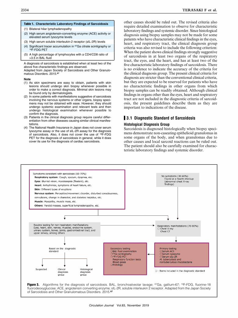
other causes should be ruled out. The revised criteria also require detailed examination to observe for characteristic laboratory findings and systemic disorder. Since histological diagnosis using biopsy samples may not be made for some patients who have characteristic clinical findings in the eyes, heart, and respiratory tract, the clinical diagnosis group criteria was also revised to include the following criterion: When the patient shows clinical findings strongly suggestive of sarcoidosis in at least two organs of the respiratory tract, the eyes, and the heart, and has at least two of the five characteristic laboratory findings of sarcoidosis. There is no evidence to indicate the accuracy of the criteria for the clinical diagnosis group. The present clinical criteria for diagnosis are stricter than the conventional clinical criteria, but they are expected to be reserved for patients who have no characteristic findings in other organs from which biopsy samples can be readily obtained. Although clinical findings in organs other than the eyes, heart and respiratory tract are not included in the diagnostic criteria of sarcoid-osis, the present guidelines describe them as they are important to indications of the disease.
▋3.1 Diagnostic Standard of SarcoidosisHistological Diagnosis GroupSarcoidosis is diagnosed histologically when biopsy speci-mens demonstrate non-caseating epithelioid granulomas in some organs of the body, and when granulomas due to other causes and local sarcoid reactions can be ruled out. The patient should also be carefully examined for charac-teristic laboratory findings and systemic disorder.
Table 1. Characteristic Laboratory Findings of Sarcoidosis
(1) Bilateral hilar lymphadenopathy
(2) High serum angiotensin-converting enzyme (ACE) activity or elevated serum lysozyme levels
(3) High serum soluble interleukin-2 receptor (sIL-2R) levels
(4) Significant tracer accumulation in 67Ga citrate scintigraphy or 18F-FDG PET
(5) A high percentage of lymphocytes with a CD4/CD8 ratio of >3.5 in BAL fluid
A diagnosis of sarcoidosis is established when at least two of the above five characteristic findings are observed.Adapted from Japan Society of Sarcoidosis and Other Granulo-matous Disorders. 2015.67
Note(1) As skin specimens are easy to obtain, patients with skin
lesions should undergo skin biopsy whenever possible in order to make a correct diagnosis. Minimal skin lesions may be found only by dermatologists.
(2) In some patients with manifestations suggestive of sarcoidosis involving the nervous systems or other organs, biopsy speci-mens may not be obtained with ease. However, they should undergo systemic examination and relevant tests and then undergo histological examination whenever possible to confirm the diagnosis.
(3) Patients in the clinical diagnosis group require careful differ-entiation from other diseases causing similar clinical manifes-tations.
(4) The National Health Insurance in Japan does not cover serum lysozyme assay or the use of sIL-2R assay for the diagnosis of sarcoidosis. Also, it does not cover the use of 18F-FDG PET for the diagnosis of sarcoidosis in general, while it does cover its use for the diagnosis of cardiac sarcoidosis.
Figure 3. Algorithms for the diagnosis of sarcoidosis. BAL, bronchoalveolar lavage; 67Ga, gallium-67; 18F-FDG, fluorine-18 fluorodeoxyglucose; ACE, angiotensin converting enzyme; sIL-2R, soluble interleukin 2 receptor. Adapted from the Japan Society of Sarcoidosis and Other Granulomatous Disorders. 2015.67

Circulation Journal Vol.83, November 2019
2335JCS 2016 Guideline on Diagnosis and Treatment of Cardiac Sarcoidosis
Clinical Diagnosis GroupSarcoidosis is diagnosed clinically when clinical findings are strongly suggestive of sarcoidosis in at least two organs of the respiratory tract, the eyes, and the heart, and when at least two of the five characteristic laboratory findings of sarcoidosis (Table 1) are present, although epithelioid granulomas are not found.
How to Diagnose SarcoidosisSarcoidosis is expected to be diagnosed as outlined in Figure 3.67 Sarcoidosis is known to cause diverse manifes-tations. Some patients are asymptomatic when it was found during health checkups, while other patients have manifestations involving many organs and generalized symptoms. In Japan where respiratory, eye and cardiovas-cular complaints are particularly common among patients with sarcoidosis, physicians typically order examinations specific to sarcoidosis when patients present with signs and symptoms strongly suggestive of sarcoidosis involving the respiratory tract, eyes or heart. Patients presenting with abnormal findings in other organs may be diagnosed as sarcoidosis when biopsies reveal non-caseating epithelioid granulomas, and detailed systemic examinations are performed to confirm the diagnosis. In both cases, histo-pathological examinations should be ordered whenever possible, and detailed examinations should be performed to rule out other possible conditions.
▋3.2 Clinical Findings Suggestive of Sarcoidosis Involving Different Organs
This section describes clinical findings characteristic for sarcoidosis involving the respiratory tract, eyes and skin and other organ dysfunctions associated with sarcoidosis-related diseases according to the “Diagnostic Standard and Guideline for Sarcoidosis - 2015”.67 The diagnosis of sarcoidosis should be based on histological examination, but patients with clinical findings strongly suggestive of sarcoidosis involving the respiratory tract, eyes or heart may be regarded to have lesions involving the relevant organ even when they are not confirmed histologically.
a. Clinical Findings Defining Respiratory InvolvementSarcoidosis of the respiratory system may cause lesions in the alveoli (alveolitis), bronchial vessels, hilar lymph nodes, mediastinal lymph nodes, trachea and bronchi, and pleura.
Clinical findings strongly suggestive of respiratory tract involvement of sarcoidosis are defined as the presence of (1) or (2) of Table 2 (See section “4. Pulmonary Lesions of Sarcoidosis” for details).
b. Clinical Findings Defining Eye InvolvementClinical findings strongly suggestive of eye involvement of sarcoidosis are defined as the presence of at least two of the six clinical findings listed in Table 3.
c. Clinical Findings Defining Cardiac InvolvementSee Section “3. Diagnostic Guidelines” in Chapter III, for clinical findings defining cardiac involvement.
d. Clinical Findings Defining Skin InvolvementTable 4 lists clinical findings that define skin involvement of sarcoidosis.
e. Clinical Findings Strongly Suggestive of Sarcoidosis of Organs Other Than the Respiratory Tract, Eyes, Heart and Skin.
Clinical findings strongly suggestive of sarcoidosis in organs other than the respiratory tract, eyes, heart and skin include imaging findings on CT, MRT, ultrasonography, endoscopy, 67Ga scintigraphy, and 18F-FDG PET, among others. In patients with clinical findings strongly suggestive of sarcoidosis in organs other than the respiratory tract, eyes, heart and skin, the presence of epithelioid granulomas in at least one organ must be demonstrated to confirm the diagnosis of sarcoidosis (Table 5).
Table 2. Respiratory Manifestations of Sarcoidosis
(1) Bilateral hilar lymphadenopathy is observed.
(2) CT/HRCT images show thickened interstitium surrounding the bronchial vascular bundles and multiple nodular shadows along lymphatic vessels. Multiple nodular shadows along lymphatic vessels may be found in the central and/or peripheral parts of lobules along the pleura, interlobular septa, and bronchopulmonary arteries.
Adapted from the Japan Society of Sarcoidosis and Other Granu-lomatous Disorders. 2015.67
Table 3. Ocular Manifestations of Sarcoidosis
(1) Granulomatous anterior uveitis (mutton-fat keratic precipitate/iris nodules)
(2) Trabecular meshwork nodules and/or tent-shaped peripheral anterior synechia
(3) A mass of vitreous opacities (snowball or string-of-pearls type opacities)
(4) Retinal perivasculitis (mainly periphlebitis) with perivascular nodules
(5) Multiple candle-wax type chorioretinal exudates and nodules and/or laser photocoagulation spots-like chorioretinal atrophy
(6) Optic disc nodule(s)/granuloma(s) and/or solitary choroidal nodule
Other possible ocular manifestations: Corneal xerosis, episcleritis/scleritis, lacrimal swelling, swelling of eyelids, and facial palsy. Adapted from the Japan Society of Sarcoidosis and Other Granu-lomatous Disorders. 2015.67
Table 4. Dermal Manifestations of Sarcoidosis
(1) Skin sarcoids (specific lesions)
i. Nodular form; ii. Plaque form; iii. Diffuse infiltrative form; iv. Subcutaneous form; v. Other forms (Lichenoid type, nodular erythema-like, Ichthyosis type, or other rare forms.)
(2) Infiltrated scars
(The patient should show clinical findings strongly suggestive of skin lesions, and the presence of granuloma should be confirmed histologically.)
Adapted from the Japan Society of Sarcoidosis and Other Granu-lomatous Disorders. 2015.67
NoteErythema nodosum, which is not specific to sarcoidosis, may develop without nodules in some patients, but the prevalence of this finding is low in Japan.

Circulation Journal Vol.83, November 2019
2336 TERASAKI F et al.
▋3.3 Exclusion CriteriaPossible causes other than sarcoidosis should be ruled out according to the following exclusion criteria.(1) Rule out systemic diseases of known causes or other
types of systemic diseases: e.g., malignant lymphoma, other lymphoproliferative disorders, cancer (lymphan-giosis carcinomatosa), tuberculosis, non-tuberculous granulomatous infections (e.g., non-tuberculous mycobacterial infection and mycosis), BehÇet’s disease, amyloidosis, granulomatosis with polyangiitis, Wegener’s granulomatosis, and IgG4-related disease
(2) Rule out sarcoid reaction to foreign substances or cancer
(3) Rule out other types of pulmonary granuloma: e.g., beryllium lung disease, pneumoconiosis, and hypersen-sitivity pneumonitis
(4) Rule out giant cell myocarditis(5) Rule out uveitis of known cause: e.g., herpes uveitis,
HTLV-1 uveitis, and Posner-Schlossman syndrome(6) Rule out other skin granuloma: e.g., granuloma
annulare, annular elastolytic giant cell granuloma, necrobiosis lipoidica, Melkersson-Rosenthal syndrome, lupus miliaris disseminatus faciei, and rosacea.
(7) Rule out other types of hepatic granulomas: e.g., primary biliary cirrhosis
▋3.4 Important Points in the Diagnosis and Follow-up Evaluation of Sarcoidosis
Since sarcoidosis is a systemic disease that can affect multiple organs simultaneously or consecutively, physicians should carefully review the patient’s clinical history, and observe the patient periodically for organ involvements of sarcoidosis. In Japan, patients with typical clinical findings that consist with the diagnostic standard or sarcoidosis in the relevant organ may apply to the national patient support program as those who meet the definition of the clinical diagnosis group even when histopathological examination is difficult to conduct. However, these patients need detailed examination to rule out other possible diseases. Patients who are suspected to have sarcoidosis but do not meet the above-mentioned criteria and who do not have to start treatment immediately should be classified as suspected cases and be observed periodically. For patients who are strongly suspected to have cardiac sarcoidosis or central nervous system (CNS) sarcoidosis and are at risk of life-threatening conditions, a therapeutic diagnosis may be made to start treatment.
4. Pulmonary Lesions of Sarcoidosis
Since it has been reported that pulmonary involvement is present in more than 90% of patients with sarcoidosis,68,69 chest imaging findings are important information in the diagnosis of sarcoidosis (Evidence level 4b, Recommendation grade A). A typical finding of sarcoidosis on plain chest X-ray is bilateral hilar lymphadenopathy (BHL) (Figures 4,5). Intrathoracic lymphadenopathy is the most common X-ray finding among patients with pulmonary sarcoidosis. In addition to BHL, lymphadenopathy may also be found in upper mediastinal lymph nodes. Lymphadenopathy usually occurs bilaterally, and unilateral lymphadenopathy accounts for less than 5% in patients with pulmonary
Table 5. Findings of Other Organ Involvement
(1) Nerve lesions
i. CNS lesions
a. Granulomatous lesions in the parenchyma
a-1. Localized mass lesions; a-2. Diffuse sporadic granu-lomatous lesions; a-3. Spinal lesions
b. Meningeal lesions
b-1. Meningitis/meningoencephalitis; b-2. Hypertrophic granulomatous pachymeningitis
c. Hydrocephalus
d. Vascular lesions
d-1. Vasculitis; d-2. Periventricular white matter lesions; d-3. Sinus thrombosis
e. Encephalitis
ii. Peripheral nerves
a. Cranial nerve paralysis
a-1. Facial palsy; a-2. Glossopharyngeal/vagus nerve disorder; a-3. Auditory nerve disorder; a-4. Optic nerve disorder; a-5. Trigeminal nerve disorder; a-6. Olfactory nerve disorder; a-7. Other cranial nerve disorders
b. Spinal nerves paralysis
b-1. Mononeuritis multiplex; b-2. Polyneuritis (including small fiber neuropathy); b-3. Single nerve paralysis; b-4. Other disorders: e.g., Radiculopathy and cauda equina syndrome
(2) Hepatic lesions: Hepatomegaly, multifocal nodules
(3) Splenic lesions: Splenomegaly, hypersplenism, multifocal nodules
(4) Gastrointestinal lesions: Ulcer, mucosal hypertrophy, raised lesions
(5) Renal lesions: Renal mass, renal lesions associated with calcium metabolism disorder, tubulointerstitial nephritis, granulomatous nephritis, glomerulonephritis, renal vasculitis
(6) Extrathoracic lymph node lesions: Superficial lymphade-nopathy, intraabdominal lymphadenopathy, etc.
(7) Exocrine lesions: Parotid gland enlargement, submaxillary gland enlargement, lacrimal gland enlargement
(8) Upper respiratory tract lesions: Nasal cavity lesions, mass in the upper respiratory tract
(9) Bone lesions: Lacy trabecular patterns, osteolytic lesions, round cystic radiolucent area in the bone
(10) Muscle lesions
i. Acute to subacute myositis-like lesions
ii. Chronic myopathy
iii. Nodular myopathy
(11) Joint lesions: Joint swelling and deformity
(12) Genital organ lesions: Mass in the uterus, testicles, epididymis or spermatic cord.
(13) Other lesions: Bone marrow lesions, pancreatic lesions, biliary/gallbladder lesions, peritoneal lesions, mammary lesions, thyroid lesions, etc.
Adapted from the Japan Society of Sarcoidosis and Other Granu-lomatous Disorders. 2015.67
Note Sarcoidosis may be associated with the following related condi-tions as well as organ disorders that result from these related conditions. These conditions are not “clinical findings strongly suggestive of organ involvement of sarcoidosis” but are important conditions associated with sarcoidosis.(1) Abnormal calcium metabolism (hypercalcemia, hypercalciuria,
nephrolithiasis and urinary calculus)(2) Neurological disorders underlined in Table 5(3) Kidney disorders underlined in Table 5

Circulation Journal Vol.83, November 2019
2337JCS 2016 Guideline on Diagnosis and Treatment of Cardiac Sarcoidosis
sarcoidosis. Typical pulmonary imaging findings are fine nodular and patchy ground-glass opacities, pale infiltrates, and cyst formation that distribute along the lymphatic vessels. Lesions are found more commonly in the upper lung fields. Patients with progressive pulmonary sarcoidosis show diverse imaging findings including reticular shadows due to fibrosis, and reduced total lung capacity. Pulmonary sarcoidosis is classified into five stages according to plain chest X-ray findings. This staging can predict prognosis, but does not always reflect disease activity or the severity of pulmonary dysfunction10,70 (Table 6).71 Chest X-ray findings of pulmonary sarcoidosis are classified into the following five stages: Stage 0: No intrathoracic involvement is found. Stage I: Lymphadenopathy including BHL is found with no lung involvement. However, lung biopsy often reveals granulomas. Stage II: BHL and lung involve-ment are found. Stage III: Lung involvement without BHL is found. Stage IV: Lung fibrosis and tissue damage are found.
Since pulmonary involvement in sarcoidosis is mainly characterized with fine nodular shadows (about 0.2 mm in diameter) that distribute along the lymphatic vessels, HRCT should be performed to examine the lesions in detail72–75 (Evidence level 5, Recommendation grade A). In addition to BHL, HRCT typically reveals very fine to fine nodular shadows along the peribronchovascular sheaths,
interlobular septa and pleural surface, tracheal wall thickening, ground-glass opacities, and mass-like lesions associated with cavities and cysts. In advanced cases, HRCT may reveal fibrosis and traction bronchodilation. In rare cases, nodular shadows or mass-like shadows with large coalescent parenchymal nodules described as the “sarcoid galaxy sign” may be found. In some patients, HRCT reveals mosaic attenuation caused by air trapping due to airway stenosis by granulomas and cystic shadows. HRCT findings are mainly observed in the upper and middle lobes. In the “Diagnostic Standard and Guideline for Sarcoidosis - 2006”, the presence of (1) BHL or (2) one or more findings in Table 76 are listed as imaging findings strongly suggestive of pulmonary involvement of sarcoidosis.
5. Treatment of Sarcoidosis
▋5.1 IntroductionSarcoidosis, a systemic granulomatous inflammatory disease, generally does not cause significant symptoms and may resolve spontaneously, but may lead to serious life-threatening conditions when it involves the heart and lungs. Prolonged involvement of the eyes, nerves, skin and kidneys among other organs may not affect the life expec-tancy of patients, but does reduce their QOL significantly. Appropriate intervention should also be made.
It is well known that corticosteroids are effective at least in the short-term control of granulomatous inflammation, and there is no doubt that corticosteroid therapy should be initiated without delay for patients with acute exacerbation
Figure 4. Chest X-ray finding of a patient with sarcoidosis. Showing typical findings of bilateral hilar lymphadenopathy (BHL). No fine nodular shadows are found in the lung fields.
Figure 5. Contrast CT finding of a patient with sarcoidosis. Showing clear bilateral hilar lymphadenopathy (BHL).
Table 6. Staging Classification of Pulmonary Sarcoidosis
Stage Chest X-ray Prevalence (%)
Spontaneous cure rate (%)
0 Normal chest X-ray findings 5 to 15 –
I Bilateral hilar lymphadenopathy 45 to 65 50 to 90
II Bilateral hilar lymphadenopathy+lung opacity 30 to 40 30 to 70
III Lung opacity only (without bilateral hilar lymphadenopathy) 10 to 15 10 to 20
IV Lung fibrosis 5 0
Adapted from Fujimoto K. 201371 with modification.

Circulation Journal Vol.83, November 2019
2338 TERASAKI F et al.
of sarcoidosis. The most important point in the treatment of sarcoidosis is to consider the indications of treatment, types and doses of drugs used, and length of treatment for diverse types of conditions that are often asymptomatic and may improve spontaneously. Treatment benefits must overweigh the burden imposed by the treatment. However, its clinical course varies and lasts long, it is difficult to assess the efficacy of treatments appropriately. A limited number of large-scale prospective clinical studies in sarcoidosis have been conducted, but these studies focused on patients with pulmonary sarcoidosis.
Corticosteroids work as a double-edged sword as they may quickly relieve granulomatous inflammation but also suppress lymphocytes that remove the pathogen. Adverse drug reactions (ADRs) to corticosteroid therapy may sometimes lower the QOL of patients significantly, which may cause corticosteroid phobia. The diverse clinical course of sarcoidosis, the dual nature of corticosteroids, and ADRs of corticosteroids and patients’ fear of corticosteroids make it difficult to standardize corticosteroid therapy for patients with sarcoidosis.
▋5.2 Indications of Corticosteroids for Patients With Pulmonary Involvement of Sarcoidosis
Large-scale prospective randomised clinical trials to investigate the efficacy of systemic corticosteroid therapy in sarcoidosis have only been conducted in patients with pulmonary sarcoidosis. The Cochrane review on cortico-steroids for pulmonary sarcoidosis have summarized that the results fits with the currently held view that patients with stage I disease (bilateral hilar lymphadenopathy alone) do not need treatment with oral steroids, but that those with interstitial lung disease (stage II and III) may show an improvement in chest X-ray and in global scores when treated with oral steroids.
Gibson et al77 (Evidence level 3) and Pietinalho et al78 (Evidence level 2) have reported that patients receiving corticosteroid therapy, including those with relatively mild symptoms, showed better pulmonary function and chest X-ray findings over the subsequent 5 years than patients not receiving corticosteroids. The editorial article79 on the report by Pietinalho et al discussed these two articles and described that corticosteroid therapy should be initiated
early when no significant symptoms present. Gibson described the results of corticosteroid therapy as “a small but definite long-term advantage.”
However, a slightly better outcome at 5 years in terms of the percentages of patients in whom abnormal chest X-ray findings disappeared and pulmonary function improved is not a strong basis supporting corticosteroid therapy for relatively asymptomatic patients with pulmonary sarcoidosis. Corticosteroid therapy should be advised only when expected benefits are greater than potential harms. The above-mentioned two reports indicated a slight supe-riority of the corticosteroid group over the control group in terms of chest X-ray findings and pulmonary functions at 5 years, which does not guarantee better QOL and life expectancy at 5 years or thereafter. Several reports have pointed out that intensive corticosteroid therapy in rela-tively asymptomatic patients may deteriorate prognoses.80–82
Recent reports from Western countries have described that drug treatment is not indicated for relatively asymp-tomatic patients without pulmonary dysfunction83,84 (Evidence level 6). Judson has described that corticosteroids are not indicated for asymptomatic patients with forced vital capacity (FVC) 70% or more.85
In response to a question whether asymptomatic patients with intact respiratory function should be treated corticosteroids, an increasing number of researchers and physicians answer that corticosteroid therapy is not a positive option even though clinical evidence on the risk and benefits of corticosteroids in this patient population has not yet been accumulated sufficiently. This represents how difficult it is to conduct prospective clinical research in patients with sarcoidosis, a disease that progresses over decades.
▋5.3 Treatment Indications Considering Fibrosis of Granulomas
The section “treatment procedures for patients with pulmo-nary sarcoidosis” in “Views on the Treatment of Sarcoidosis - 2003”9 prepared by the Japan Society of Sarcoidosis and Other Granulomatous Disorders in 2003 describes that asymptomatic or mildly symptomatic patients with stage II or III pulmonary sarcoidosis should be left untreated and monitored carefully, and corticosteroid therapy should be
Table 7. Chest X-ray, CT/HRCT and Bronchoscopic Findings of Pulmonary Sarcoidosis
1. Chest X-ray findings
(1) Diffuse lung opacities, mainly fine nodular or patchy opacities, are observed mainly in the upper lung fields.
(2) Irregular opacities and thickening of the area surrounding the bronchial vascular bundles.
(3) When advanced, fibrosis and shrinking of the lung develop mainly in the upper lungs.
2. CT/HRCT findings
(1) Fine nodular shadows in lung fields and thickened interstitium surrounding the bronchial vascular bundles are often found. Fine nodular shadows with focal shrinking reflect the distribution of lymph components and are found both in the central and peripheral parts of lobules along the pleura, interlobular septa, and bronchopulmonary arteries.
(2) Nodular shadows, mass shadows, or evenly-distributed shadows may be observed in rare cases. Pleural fluid is rare. Advanced fibrous lesions are observed as evenly-distributed shadows indicating tissue shrinking associated with traction bronchodilation rather than as typical honeycomb lungs.
3. Bronchoscopy findings
(1) Network formation
(2) Nodules
(3) Bronchostenosis
Adapted from Committee for revision of the diagnostic standard for sarcoidosis. 2006.6

Circulation Journal Vol.83, November 2019
2339JCS 2016 Guideline on Diagnosis and Treatment of Cardiac Sarcoidosis
considered according to their individual clinical conditions for those with progression of chest X ray findings.
“Patients with chronic granulomatous disease with no or mild fibrosis (granuloma type lesions)” tend to maintain intact pulmonary function, are expected to at least partially improve without particular treatment, respond well to corticosteroid therapy, and often lead to clinical remission even though the disease may sometimes flare up. Accord-ingly, patients diagnosed as having granuloma type lesions may be treated with short-term, low-dose corticosteroid therapy to wait for spontaneous improvement.
The main factor that reduces life expectancy in patients with pulmonary involvement of sarcoidosis is fibrosis secondary to granulomatous disease. Patients who have a poor outcome due to respiratory failure, pulmonary hyper-tension and lung infection among other conditions are almost as limited as those with advanced lung fibrosis. Accordingly, it is important to identify patients with “diffuse fibrosis of granulomatous lesions (diffuse fibrosis type lesions)” based on slight changes in chest X-ray findings at an early stage and intervene appropriately. However, no data have been reported which types of chest X-ray findings indicate the risk of diffuse fibrosis. Further reports are awaited.
▋5.4 Dose and Duration of Corticosteroid TherapyThere has been a consensus where corticosteroid therapy for patients with pulmonary sarcoidosis should be initiated at 0.5 mg/kg/day (20 to 40 mg/day) of prednisolone and then reduced thereafter9,10 (Evidence level 6, Recommendation grade C1). However, it is unknown whether the dose is optimal or not. Considering a report describing that low-dose prednisolone therapy at doses of 5 to 10 mg/day is effective for patients complicated with diabetes mellitus or infectious diseases,86 corticosteroid therapy at lower doses for a shorter period of time than those previous reported would be effective for patients with granuloma-type lesions.
As “if the regimen is discontinued while the antigen is still present, the patient will relapse”85 corticosteroid therapy for pulmonary sarcoidosis may last for decades. In the United States where severe pulmonary sarcoidosis is more prevalent than in Japan, those who are maintained on ≤15 mg/day of prednisolone are considered successful cases, while in Japan the target maintenance dose is ≤5 mg/day.
▋5.5 Treatment Indications for Extrapulmonary Manifestations of Sarcoidosis
Only a limited number or reports have described indications of corticosteroid therapy for extrapulmonary manifesta-tions of sarcoidosis. In many cases extrapulmonary mani-festations are treated to improve QOL rather than prolong the life of patients. Treatment is often initiated after symptoms develop, and corticosteroids are administered
when required.However, cardiac involvement is a life-threatening
condition where corticosteroid therapy is recommended regardless of whether symptoms are present or not21,87 (Evidence level 4b, Recommendation grade C1). Nerve involvement and ocular involvement, which may signifi-cantly reduce QOL if they remain untreated, are indicated for corticosteroid therapy even if patients are asymptomatic. However, there is no evidence on the optimal dose and duration of corticosteroid therapy for these conditions. Corticosteroids are used at the discretion of physicians.
▋5.6 Drug Treatment Other Than CorticosteroidsEvidence on the efficacy of methotrexate and azathioprine, which are used as immunosuppressants, has been accu-mulated in many countries88–91 (Evidence level 5, Recom-mendation grade C1). Patients who cannot discontinue corticosteroid therapy for a long period of time should also receive these drugs to achieve the steroid-sparing effect. Low-dose methotrexate therapy is commonly used for patients with rheumatoid arthritis, and regimens combining low-dose corticosteroids and low-dose methotrexate are expected to be beneficial in the treatment of sarcoidosis90 (Evidence level 3). Methotrexate monotherapy is known to be somewhat effective,91 and may be considered for patients who want to avoid or are contraindicated for corticoste-roids. (Note: Currently, methotrexate therapy in sarcoidosis is not covered by the National Health Insurance in Japan).
Antimicrobial drugs have been used for the treatment of sarcoidosis. Chloroquine hydrochloride, an antimalarial drug, has been used widely for the treatment of sarcoidosis in the United States even though its efficacy has not been proven yet. As some patients, especially those with skin lesions, respond well to tetracyclines,92,93 these drugs may be prescribed to patients who accept the treatment after detailed assessment and explanation by the physician (Evidence level 5).
▋5.7 Treatment for General SymptomsPatients with sarcoidosis often complain of non-organ-specific general symptoms such as pain, fatigue and short-ness of breath, which often represent their most annoying symptoms.94,95 No sufficient treatment has been established to manage these symptoms, but Chinese herbal medicine96 and high-dose corticosteroids97 are effective in some patients.
▋5.8 ConclusionAs described in this section, no sufficient evidence has been accumulated on the indications, types, duration, doses and endpoints of treatments that can be used to develop guidelines for the treatment of sarcoidosis. Further findings are needed.
III. Diagnosis of Cardiac Sarcoidosis
1. Pathology
As in sarcoidosis affecting other organs, cardiac sarcoidosis
is characterized histologically by the presence of multinu-cleated giant cells in non-caseating epithelioid granulomas. The presence of giant cell is an important finding to differ-entiate cardiac sarcoidosis from other cardiac diseases
Circulation Journal Vol.83, November 2019
2340 TERASAKI F et al.
(Evidence level 4a, Recommendation grade A).It is often difficult to make a definitive diagnosis of
cardiac sarcoidosis because it manifests diverse pathological conditions and no diagnostic measures other than endo-myocardial biopsy can identify the presence of cardiac sarcoidosis. Although pathological findings of cardiac sarcoidosis are highly specific, endomyocardial biopsy
samples often show fibrosis and lymphocytic infiltration only without epithelioid granulomas or giant cells. Only about 20% of biopsy samples obtained from patients with cardiac sarcoidosis show positive findings.
▋1.1 Endomyocardial BiopsyThe diagnostic rate achieved with endomyocardial biopsy in cardiac sarcoidosis is as low as about 20% due to frequent errors in obtaining positive biopsy samples.98,99 Accordingly, cardiac sarcoidosis cannot be ruled out even if endomyo-cardial biopsy shows a negative result.100
▋1.2 Macro-Pathological Findings of Cardiac Sarcoidosis
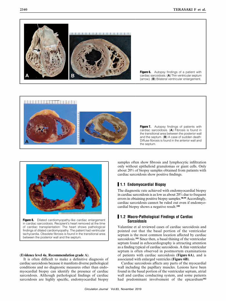
Valantine et al reviewed cases of cardiac sarcoidosis and pointed out that the basal portion of the ventricular septum is the most common location affected by cardiac sarcoidosis.101 Since then, a basal thining of the ventricular septum found in echocardiography is attracting attention as a finding typical of cardiac sarcoidosis. A thin ventricular septum is often observed in postmortem examinations of patients with cardiac sarcoidosis (Figure 6A), and is associated with enlarged ventricles (Figure 6B).
Cardiac sarcoidosis affects any parts of the myocardial wall including the papillary muscles. Lesions have been found in the basal portion of the ventricular septum, atrial wall and cardiac conducting system, and some patients had predominant involvement of the epicardium102
Figure 6. Autopsy findings of a patient with cardiac sarcoidosis. (A) Thin ventricular septum (arrow). (B) Bilateral ventricular enlargement.
Figure 7. Autopsy findings of patients with cardiac sarcoidosis. (A) Fibrosis is found in the transitional area between the posterior wall and the septum. (B) A case of sudden death: Diffuse fibrosis is found in the anterior wall and the septum.
Figure 8. Dilated cardiomyopathy-like cardiac enlargement in cardiac sarcoidosis. Recipient’s heart removed at the time of cardiac transplantation: The heart shows pathological findings of dilated cardiomyopathy. The patient had ventricular tachycardia. Obsolete fibrosis is found in the transitional area between the posterior wall and the septum.

Circulation Journal Vol.83, November 2019
2341JCS 2016 Guideline on Diagnosis and Treatment of Cardiac Sarcoidosis
(Figures 6B,7). When cardiac involvement manifests clini-cally, granulomas have already started to become fibrous in many patients. The borders between lesions and non-lesions are clear, and fibrous granulomas can be seen as patchy or linear lesions (Figures 7,8). When fibrosis pro-gresses, ventricular aneurysm or dilated cardiomyopathy-like ventricular enlargement develop103 (Figures 6,8).
▋1.3 Histological Findings in Cardiac SarcoidosisAs in sarcoidosis in other organs, cardiac sarcoidosis is characterized histologically by the presence of multinu-cleated giant cells in non-caseating epithelioid granulomas (Figure 9).17 Granulomas consist of epithelioid cells, multinucleated giant cells, macrophages, lymphocytes and plasma cells among others. As described earlier, granuloma typical of sarcoidosis can be detected only in about 20% of patients undergoing myocardial biopsy,98,99 and many biopsy samples contain giant cells and epithelioid cells only. When granuloma is not detected, serial sections should be examined for the presence of giant cells. Caseating necrosis suggestive of tuberculosis is not present in patients with cardiac sarcoidosis. Giant cells often appear as Langhans-type multinucleated giant cells with nuclei arranged peripherally (Figure 10), but may appear as foreign-body-type or Touton-type multinucleated giant cells (Figures 11,12). Giant cells may also contain cytoplasmic
inclusions such as asteroid bodies (Figure 13A) and Schaumann bodies (Figure 13B).104 These cytoplasmic inclusions are useful in the diagnosis of sarcoidosis. The degree of lymphocytic infiltration in tissues adjacent to granulomas relates to the stage (activity) of sarcoidosis. Generally, CD4 lymphocytes accumulate predominantly in granulomas, while CD8 lymphocytes are found sporadi-
Figure 9. Histopathological findings of cardiac sarcoidosis: Non-caseating epithelioid granulomas (HE staining). Granu-lomas consist of multinucleated giant cells surrounded by lymphocytes and epithelioid cells.
Figure 10. Histopathological findings of cardiac sarcoidosis: Langhans-type multinucleated giant cells. Multiple nuclei are observed in the periphery of giant cells.
Figure 11. Histopathological findings of car-diac sarcoidosis: Various patterns of multinu-cleated giant cells. (A) Foreign body type giant cells surrounded by fibrosis. (B) Touton-type giant cells with a ring of nuclei. Cardiac myocytes adjacent to granulomas appear nearly normal.
Figure 12. Histopathological findings of cardiac sarcoidosis: Foreign body type giant cells. Multinucleated giant cells have foreign body-like structures and vacuoles in the cytoplasm.

Circulation Journal Vol.83, November 2019
2342 TERASAKI F et al.
Figure 13. Histopathological findings of car-diac sarcoidosis: Inclusion bodies in giant cells. (A) Asteroid body (arrow). (B) Schaumann body. Vacuoles are found in the cytoplasm.
Figure 14. Immunostained myocardial biopsy specimen in a patient with cardiac sarcoidosis: Lymphocyte subtyping in granulomas. (A) CD4-positive cells. (B) CD8-positive cells.
Figure 15. Granuloma detected by myocardial biopsy in a patient with cardiac sarcoidosis. Giant cells and lymphocytes remain in fibrosis.
Figure 16. Histopathological findings of a myocardial biopsy specimen in a patient with cardiac sarcoidosis. (A) Multinucleated giant cells surround the fibrous part of the endocar-dium. The boundary between the lesion and intact tissues is clear. (B) Magnified image of the square area in (A).

Circulation Journal Vol.83, November 2019
2343JCS 2016 Guideline on Diagnosis and Treatment of Cardiac Sarcoidosis
Figure 17. Histopathological findings of giant cell myocarditis. Langhans-type giant cells are observed. Giant cell myocarditis may be differentiated from cardiac sarcoidosis as the former shows myocardial cells invaded by lymphocytes, and does not show epithelioid cells or granulomas.
Figure 18. Histopathological findings of a myocardial biopsy specimen in cardiac sar-coidosis. (A) HE stained image. (B) CD68 immunostained image. Multinucleated giant cells and epithelioid cells are positive for CD68.
Figure 19. Histopathological findings of a myocardial biopsy specimen in a patient with cardiac sarcoidosis. Scar stage of granuloma, replacement fibrosis with a small number of lymphocytes is observed. Sarcoidosis should be suspected when scarred granulomas are found in myocardial biopsy.
Figure 20. Localized focal lymphocytic infil-tration in a myocardial biopsy specimen in a patient with cardiac sarcoidosis. (A) Lympho-cytic infiltration is found in fibrous myocardial stroma. (B) Giant cells are found in serial sections.

Circulation Journal Vol.83, November 2019
2344 TERASAKI F et al.
cally at the periphery of granulomas (Figure 14). At the scarring stage, fibrosis becomes prominent with little lymphocyte infiltration, and granulomas tend to disappear over time. In some lesions only giant cells are found (Figure 15). The presence of giant cells in myocardial biopsy samples is strongly suggestive of cardiac sarcoidosis. The borders between lesions and non-lesions are often clear, and myocardial cells in non-lesions are intact in some cases (Figure 16).
Giant cell myocarditis (Figure 17), an important condi-tion that should be differentiated from cardiac sarcoidosis, is characterized by necrosis of myocardial cells, which is absent in cardiac sarcoidosis.105 Eosinophilic infiltration is also rare in cardiac sarcoidosis. Giant cells found in cases of giant cell myocarditis are derived from macrophages and cardiac myocytes, while both epithelioid cells and giant cells found in cases of cardiac sarcoidosis are derived from macrophages (Figure 18). In cardiac sarcoidosis, activated T lymphocytes differentiate into Th1 cells, which produce Th1 cytokines such as IL-2 and IFN-γ that mediate migration, activation and differentiation of monocytes and macrophages, and eventually lead to granuloma formation. In order to rule out other conditions causing epithelioid granulomas, mycosis and tuberculosis, Grocott staining for fungal infection and Ziehl-Neelsen staining for acid-fast microorganisms should be conducted whenever neces-sary. Since fibrosis becomes prominent (Figure 19) and only slight lymphocyte infiltration is present at the scarring stage, serial sections of biopsy specimens should be prepared to increase the accuracy of diagnosis (Figure 20). Lymph node biopsy may be needed in some cases.
2. Clinical Findings and Examinations
▋2.1 Clinical ManifestationsSarcoidosis is a systemic inflammatory disease that causes clinical symptoms in 60 to 70% of patients. However, symptoms are not specific. Sarcoidosis is often diagnosed after abnormal findings are detected at chest X-ray or ECG during routine health checkups or screening tests to evaluate non-specific symptoms.6
a. Clinical Manifestation of Systemic SarcoidosisAbout a third of patients with sarcoidosis complain of non-specific symptoms of systemic inflammation such as fever (mild fever in many cases), general malaise, night sweats and decreased weight.10 Sarcoidosis is an important possible cause of fever of unknown origin, but these non-specific symptoms are not common among Japanese patients with sarcoidosis.106 Sarcoidosis may affect the lungs and hilar lymph nodes as well as the skin, eyes, muscles, bones, heart and central nervous system, and may cause organ-specific signs and symptoms. It has been reported that the incidence of extrapulmonary lesions differs among races, and that the heart and eyes are more often affected in Japanese patients than in other races.106 However, many Japanese patients with cardiac or eye involvement of sarcoidosis are asymptomatic, and are often diagnosed at routine health checkups or screening tests to evaluate non-specific symptoms.
b. Clinical Manifestations of Cardiac SarcoidosisCardiac lesions are found at autopsy in about 25% of patients with sarcoidosis, while imaging studies reveal
asymptomatic cardiac involvement in 3.7 to 54.9% of the patients with extracardiac sarcoidosis, depending on the type of imaging study used, of patients with cardiac sarcoidosis.107 Patients with mild cardiac involvement are often asymptomatic, but symptoms occur when lesions affect the conduction system or the cardiac function of the heart. When sarcoidosis affects the cardiac conduction system, bundle branch block and atrioventricular block may develop. Atrioventricular block may cause mild bradycardia causing palpitations and dizziness or may even cause severe bradycardia leading to syncopes or sudden death. Myocardial inflammation and fibrosis due to sarcoid-osis may cause ventricular arrhythmias such as premature ventricular contractions, ventricular tachycardia and ventricular fibrillation. Premature ventricular contractions may cause palpitations and shortness of breath. Sustained ventricular tachycardia and ventricular fibrillation may lead to sudden death (See Section “2.4 Electrocardiogram”).
As cardiac involvement progresses, myocardial function deteriorates further to cause systolic and/or diastolic dysfunction, causing heart failure. Symptoms of heart failure include those associated with fluid retention and those associated with lower cardiac output. Fluid retention causes edema, cough and dyspnea. As dyspnea and cough are common symptoms of respiratory diseases, it is impor-tant to differentiate whether they are caused by pulmonary or cardiac lesions when such symptoms develop or worsen in patients with sarcoidosis mainly affecting the respiratory tract. Decreased cardiac output may cause oliguria, general malaise, insomnia, depression and dizziness, and may result in syncope, confusion, and decreased level of consciousness in severe cases. Clinical manifestations of cardiac sarcoid-osis may include those associated with cardiac conduction disturbance and those associated with myocardial impair-ment, depending on the location and severity of granulo-matous inflammation.
These symptoms are not specific to cardiac sarcoidosis, but physicians should always consider this as a possible cause of cardiac symptoms. Mehta et al have reported the prevalence of symptoms and the results of cardiac MRI and 18F-FDG PET scanning in 62 patients diagnosed as sarcoidosis (mainly pulmonary sarcoidosis) based on pathological examination of non-cardiac organs.108 They described that the prevalence of cardiac symptoms (e.g., palpitations and presyncope) was significantly higher in patients with cardiac involvement than those without it (46% vs. 5%), and concluded that cardiac symptoms are an important indicator of cardiac involvement in patients diagnosed with non-cardiac sarcoidosis (Evidence level 4a, Recommendation grade B).
▋2.2 Blood and Urine Tests (Biomarkers)No biological materials currently being investigated as potential biomarkers of sarcoidosis are highly specific in determining the progression of cardiac sarcoidosis or detecting isolated cardiac involvement of sarcoidosis. However, these markers are useful as screening tests for myocardial injuries to detect abnormalities without using imaging modalities such as MRI, echocardiography and 18F-FDG PET. Further studies are expected to reveal which types of biomarkers would be more useful in assessing treatment efficacy and prognosis in patients with cardiac sarcoidosis or isolated cardiac sarcoidosis.

Circulation Journal Vol.83, November 2019
2345JCS 2016 Guideline on Diagnosis and Treatment of Cardiac Sarcoidosis
▋ 2.2.1 Biomarkers in Screening for Cardiac Involvement in Patients Diagnosed With Systemic Sarcoidosis
a. Angiotensin Converting Enzyme (ACE) ActivityIn a study in Japanese patients diagnosed with cardiac sarcoidosis, serum ACE activity was high in 21.8% of patients.109 In another study in 516 patients with sarcoidosis with histologically-proven epithelioid granulomas, serum ACE activity and serum lysozyme activity were high in 49.8% and 51.7% of the patients, respectively.110 It has been also reported that serum ACE activity was high in 62.3% of 106 patients with sarcoidosis-associated uveitis and 18% of 100 patients suspected to have the same condition.111 Conversely, a study has described that serum ACE activity was significantly lower in patients diagnosed with cardiac sarcoidosis than those with sarcoidosis but without cardiac involvement.112 Further studies are needed to conclude whether serum ACE activity relate to the occurrence or severity of cardiac involvement in sarcoidosis (Evidence level 4b, Recommendation grade C1).
b. Atrial Natriuretic Peptide (ANP) and Brain Natriuretic Peptide (BNP)
In a comparison between patients with sarcoidosis with and without cardiac involvement, plasma levels of atrial natriuretic peptide (ANP) and brain natriuretic peptide (BNP) were higher in those with cardiac involvement than those without it, and all patients with a plasma BNP level of >100 pg/mL had cardiac involvement.113 However, BNP may not be an accurate marker to differentiate cardiac sarcoidosis from systemic sarcoidosis complicated with cardiac hypertrophy, cardiac dysfunction, atrial fibrillation, renal dysfunction, or conditions associated with aging. A report described that plasma levels of N-terminal pro brain natriuretic peptide (NT-proBNP) were higher in patients with sarcoidosis with cardiac involvement than those without it112 (Evidence level 4b, Recommendation grade C1).
c. High-Sensitivity Cardiac Troponin T (hs-cTnT) and High-Sensitivity Cardiac Troponin I (hs-cTnI)
In a study in patients with cardiac sarcoidosis to assess the usefulness of high-sensitivity cardiac troponin T (hs-cTnT) as a marker of the activity of cardiac involvement as compared with BNP, ACE, lysozyme, cardiac MRI, 67Ga scintigraphy and 18F-FDG PET, hs-cTnT was comparable with and superior to BNP in terms of sensitivity (87.5% vs 87.5%) and specificity (75% vs 50%).114 This study also reported hs-cTnT levels decreased after corticosteroid therapy in some patients, and concluded that hs-cTnT is a good marker of the activity of cardiac sarcoidosis (Evidence level 4b, Recommendation grade C1). In another study in patients with cardiac sarcoidosis where serum levels of high-sensitivity cardiac troponin I (hs-cTnI) were determined in serial samples before and after corticosteroid therapy, hs-cTnI levels decreased as cardiac function improved115 (Evidence level 5).
Also, in a study where hs-cTnT and hs-cTnI levels in patients with cardiac sarcoidosis were determined before and after corticosteroid therapy, patients with elevated hs-cTnT/I levels had significantly lower left ventricular ejection fraction and tended to have more cardiac accidents than those with normal hs-cTnT/I levels.116 These findings indicated that hs-cTnT or hs-cTnI may be used as an index of disease activity and treatment response in patients with cardiac sarcoidosis (Evidence level 4b, Recommendation grade C1). (Note: cardiac troponin test in sarcoidosis is not
covered by the National Health Insurance in Japan.)
d. Urinary 8-Hydroxy-2’-Deoxyguanosine (8-OHdG)In a study to assess the relationship between urinary 8-hydroxy-2’-deoxyguanosine (8-OHdG) levels and 18F-FDG PET findings as makers of disease activity in patients with cardiac sarcoidosis, urinary-8-OHdG was a powerful predictor of disease activity with a sensitivity of 88.2% and a specificity of 92.9%.117 In patients who responded well to corticosteroid therapy, urinary 8-OHdG levels decreased as 18F-FDG PET findings improved (Evidence level 4b).
e. Soluble Interleukin-2 Receptors (sIL-2R)Studies have reported that serum levels of soluble inter-leukin-2 receptors (sIL-2R) are a useful marker of T cell activity in patients with sarcoidosis.118,119 A large-scale study in the Netherlands where serum levels of CRP, ACE, amyloid and sIL-2R were compared between patients with sarcoidosis who are receiving and not receiving treatment has reported that sIL-2R appears to be a useful marker of the severity of respiratory involvement in sarcoidosis, especially in untreated patients.120 No evidence is available as to whether sIL-2R levels reflect the severity of cardiac involvement. (Note: Currently, sIL-2R tests in sarcoidosis is not covered by the National Health Insurance in Japan).
▋ 2.2.2 Biomarkers in Diagnosing Isolated Cardiac Sarcoidosis
a. ACE and LysozymeIn a study in 89 patients with an implantable pacemaker who have not been diagnosed as sarcoidosis affecting non-cardiac organs, screening using biomarkers including ACE and lysozyme detected cardiac sarcoidosis in 10 patients among those who could be followed up.121 In this study, serum levels of ACE and lysozyme were high in all patients diagnosed as cardiac sarcoidosis, suggesting their usefulness in the diagnosis of cardiac sarcoidosis. Of note, two of the 10 patients had positive findings of myocardial biopsy. This means that 2 (2.2%) of the 89 patients with atrioventricular block had histologically-proven isolated cardiac sarcoidosis (Evidence level 4b, Recommendation grade C1).
b. Th1-Related CytokinesA study investigated the expression of cytokines in myocar-dial tissues obtained through left ventricular restoration or other cardiac surgeries from patients with histologically proven cardiac sarcoidosis as compared with control patients with idiopathic dilated cardiomyopathy.122 Patients with cardiac sarcoidosis had atrioventricular block more frequently and had smaller end-diastolic left ventricular diameters than patients with dilated cardiomyopathy. A significantly higher mRNA expression of Th1-related cytokines such as IL-1α, IL-2, IL-12 p40 and INF-γ was observed in patients with cardiac sarcoidosis. However, this study did not assess for the presence of sarcoidosis in other organs. Further studies should be conducted to assess whether Th1-related cytokines can be used as markers of isolated cardiac sarcoidosis (Evidence level 4b).
c. Myeloid-Related Protein 8/14 Complex (MRP8/14, Also Known as S100A8/A9)
In a study to measure serum levels of myeloid-related protein 8/14 complex (MRP8/14) in healthy volunteers, patients with idiopathic dilated cardiomyopathy, patients

Circulation Journal Vol.83, November 2019
2346 TERASAKI F et al.
with histologically proven sarcoidosis without cardiac involvement, and patients with histologically proven cardiac sarcoidosis, serum MRP8/14 levels were significantly higher in patients with cardiac sarcoidosis than in other groups123 (Evidence level 4b).
▋2.3 Chest X-ray and Chest CTPatients who are found to have pulmonary lesions of sarcoidosis (See Section “4. Pulmonary Lesions of Sarcoidosis” in Chapter II) should be examined for cardiac involvement.
Patients with cardiac sarcoidosis do not show any specific cardiac shadows or may show even only normal findings when they have no pulmonary lesions of sarcoidosis. If they have cardiac dysfunction causing ventricular enlargement or heart failure, enlarged cardiac shadow and pulmonary congestion are found on chest X-ray. Enlarged cardiac shadow is also found in patients with pericardial effusion or ventricular aneurysm.
Autopsy and detailed examination of patients with cardiac sarcoidosis often reveal epithelioid granulomas in non-cardiac organs and hilar or mediastinal lymph nodes.124 Hilar or mediastinal lymphadenopathy is an important finding of chest X-ray or chest CT, and is commonly observed in patients with cardiac sarcoidosis. A study has reported that these findings were found in about 50 to 60% of patients.109 Otsuka et al retrospectively analyzed CT images obtained from 20 patients with idiopathic dilated cardiomyopathy and 8 patients with cardiac sarcoidosis who had findings of dilated cardiomyopathy but did not have hilar lymphadenopathy on chest X-ray, and reported that mediastinal lymphadenopathy was found on chest CT specifically in patients with cardiac sarcoidosis.2 This finding indicates that it is important to examine for mediastinal lymphadenopathy in addition to hilar lymphadenopathy in the diagnosis of cardiac sarcoidosis (Evidence level 4b,
Recommendation grade B).
▋2.4 Electrocardiogram (ECG)Common ECG findings related to cardiac sarcoidosis include right bundle branch block and atrioventricular block due to dysfunction of specialized cardiac muscles in the cardiac conduction system, axis deviation, abnormal Q waves, ST changes, and ventricular arrhythmias reflecting damaged ordinary cardiac muscles of the left ventricle5,125–127 (Figure 21). These abnormal ECG findings are observed in more than 90% of patients diagnosed with cardiac sarcoid-osis, but a few percentage of patients present with no abnormal ECG findings.128,129 ECG is essential for patients with sarcoidosis who need long-term follow-up, since a considerable number of patients are diagnosed with cardiac sarcoidosis based on ECG changes several years after the onset of non-cardiac sarcoisosis128,130 (Evidence level 4b, Recommendation grade B).
a. Right Bundle Branch BlockRight bundle branch block is a typical finding of cardiac sarcoidosis that is in about 12 to 66% of patients.126,128,131,132 Although this is not a finding specific to this condition, cardiac sarcoidosis should be suspected when this is observed in patients with non-cardiac sarcoidosis during follow-up or patients with dilated cardiomyopathy.5
b. Complete Atrioventricular Block and Advanced Atrioventricular Block
Complete or advanced atrioventricular block is found as the initial manifestation of cardiac sarcoidosis in 23% to 77.4% of patients109,125,126,128,131–134 (Evidence level 4b). About 50% of patients present with left ventricular dysfunc-tion, but the presence of complete or advanced atrioven-tricular block does not correlate with the progression of
Figure 21. Pathophysiology and ECG findings in cardiac sarcoidosis. 18F-FDG, fluorine-18 fluorodeoxyglucose; 67Ga, gallium-67; ECG, electrocardiogram.
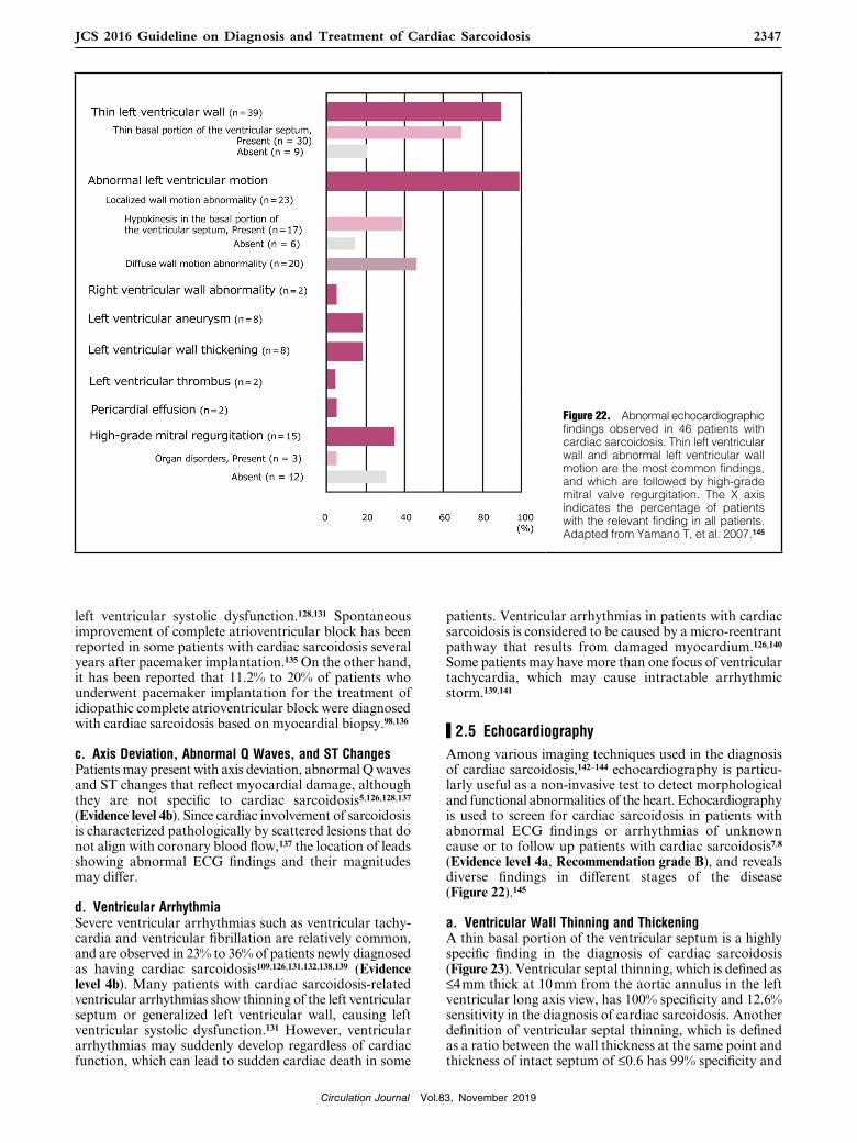
Circulation Journal Vol.83, November 2019
2347JCS 2016 Guideline on Diagnosis and Treatment of Cardiac Sarcoidosis
left ventricular systolic dysfunction.128,131 Spontaneous improvement of complete atrioventricular block has been reported in some patients with cardiac sarcoidosis several years after pacemaker implantation.135 On the other hand, it has been reported that 11.2% to 20% of patients who underwent pacemaker implantation for the treatment of idiopathic complete atrioventricular block were diagnosed with cardiac sarcoidosis based on myocardial biopsy.98,136
c. Axis Deviation, Abnormal Q Waves, and ST ChangesPatients may present with axis deviation, abnormal Q waves and ST changes that reflect myocardial damage, although they are not specific to cardiac sarcoidosis5,126,128,137 (Evidence level 4b). Since cardiac involvement of sarcoidosis is characterized pathologically by scattered lesions that do not align with coronary blood flow,137 the location of leads showing abnormal ECG findings and their magnitudes may differ.
d. Ventricular ArrhythmiaSevere ventricular arrhythmias such as ventricular tachy-cardia and ventricular fibrillation are relatively common, and are observed in 23% to 36% of patients newly diagnosed as having cardiac sarcoidosis109,126,131,132,138,139 (Evidence level 4b). Many patients with cardiac sarcoidosis-related ventricular arrhythmias show thinning of the left ventricular septum or generalized left ventricular wall, causing left ventricular systolic dysfunction.131 However, ventricular arrhythmias may suddenly develop regardless of cardiac function, which can lead to sudden cardiac death in some
patients. Ventricular arrhythmias in patients with cardiac sarcoidosis is considered to be caused by a micro-reentrant pathway that results from damaged myocardium.126,140 Some patients may have more than one focus of ventricular tachycardia, which may cause intractable arrhythmic storm.139,141
▋2.5 EchocardiographyAmong various imaging techniques used in the diagnosis of cardiac sarcoidosis,142–144 echocardiography is particu-larly useful as a non-invasive test to detect morphological and functional abnormalities of the heart. Echocardiography is used to screen for cardiac sarcoidosis in patients with abnormal ECG findings or arrhythmias of unknown cause or to follow up patients with cardiac sarcoidosis7,8 (Evidence level 4a, Recommendation grade B), and reveals diverse findings in different stages of the disease (Figure 22).145
a. Ventricular Wall Thinning and ThickeningA thin basal portion of the ventricular septum is a highly specific finding in the diagnosis of cardiac sarcoidosis (Figure 23). Ventricular septal thinning, which is defined as ≤4 mm thick at 10 mm from the aortic annulus in the left ventricular long axis view, has 100% specificity and 12.6% sensitivity in the diagnosis of cardiac sarcoidosis. Another definition of ventricular septal thinning, which is defined as a ratio between the wall thickness at the same point and thickness of intact septum of ≤0.6 has 99% specificity and
Figure 22. Abnormal echocardiographic findings observed in 46 patients with cardiac sarcoidosis. Thin left ventricular wall and abnormal left ventricular wall motion are the most common findings, and which are followed by high-grade mitral valve regurgitation. The X axis indicates the percentage of patients with the relevant finding in all patients. Adapted from Yamano T, et al. 2007.145

Circulation Journal Vol.83, November 2019
2348 TERASAKI F et al.
35.4% sensitivity146 (Evidence level 4b). Lesions invading the basal portion of the ventricular septum may lead to atrio-ventricular block. Some patients with cardiac sarcoidosis showed findings mimicking hypertrophic cardiomyopathy due to substantial ventricular wall thickening147,148 (Evidence level 5), and others showed transient thickening of right ventricular free wall.149
b. Localized Wall Motion Abnormalities and Diffuse Wall Hypokinesis
Cardiac sarcoidosis should be suspected in patients with localized wall motion abnormalities that cannot be explained by coronary status. In rare cases, coronary involvement may cause coronary spasm and vasculitis, leading to abnormal coronary perfusion and asynergy.150 As abnormalities may occur only in the right ventricle in some cases, right ventricular wall motion should also be observed.151 Cardiac involvement may commonly occur in the ventricular septum (especially upper septum), left ventricular posterolateral wall, left ventricular free wall including papillary muscles and right ventricular free wall, and rarely affects the atrial wall102 (Evidence level 4b).
Some patients present with findings mimicking dilated cardiomyopathy due to diffuse left ventricular myocardial infiltration. Cardiomegaly is an important prognostic factor21 (Evidence level 4b). The presence of abnormal wall thickness and uneven wall motion abnormalities suggests the presence of cardiac sarcoidosis.1 An uneven distribution of wall motion abnormalities in cardiac sarcoidosis is considered to reflect scattered epithelioid granulomas and fibrous scars. Some patients have right ventricular enlargement (Figure 24).
Patients with diffuse wall hypokinesia have enlarged cardiac chamber and decreased left ventricular ejection fraction, and may also have intracardiac thrombus. Left ventricular inflow velocity pattern shows abnormal relax-ation, pseudo-normalization or restriction.
c. Ventricular AneurysmVentricular aneurysm is found in 8% to 10% of patients with cardiac sarcoidosis, and may cause intractable ventricular arrhythmias (Figure 25). The aneurysm wall mainly consists of fibrous tissue, and contains no or few epithelioid granu-lomas.102 Corticosteroids may promote myocardial fibrosis and modify the course of aneurysm formation.
d. Pericardial EffusionSubstantial pericardial effusion due to pericardial involve-ment is found in 2% to 8% of patients with cardiac sarcoid-osis.102 Some patients may have epithelioid granulomas in the epicardial myocardium and may also have serous effu-sion. Pericardial effusion may cause cardiac tamponade.152
e. Valvular RegurgitationSystolic dysfunction and functional mitral regurgitation due to cardiomegaly may be observed (Figure 26). Infiltration of the papillary muscles or valves may play a role, but is not the main cause of valvular regurgitation.102 Papillary muscle dysfunction may cause mitral valve prolapse. In patients with valvular regurgitation complicated with atrial fibrillation develops, annulus dilation occurs and mitral or tricuspid regurgitation worsens. Severely calcified mitral annulus associated with hypercalcemia may present in some patients.102
f. OthersIn patients with severe pulmonary sarcoidosis, prolonged pulmonary hypertension may show findings of cor pulmo-nale such as right ventricular enlargement, hypertrophy or dysfunction. It has been reported that some patients required differential diagnosis between sarcoid nodules and cardiac tumors.
Figure 23. Thin basal portion of the ventricular septum in cardiac sarcoidosis (arrows).
Figure 24. Right ventricular enlargement and right ventricular wall dyskinesia in cardiac sarcoidosis. This patient was first suspected to have arrhythmogenic right ventricular cardio-myopathy, but examinations revealed the presence of cardiac sarcoidosis.

Circulation Journal Vol.83, November 2019
2349JCS 2016 Guideline on Diagnosis and Treatment of Cardiac Sarcoidosis
g. New Technologies in the Evaluation of Cardiac Sarcoidosis
In a study, two-dimensional echocardiography revealed typical findings of cardiac sarcoidosis in 14% of patients with systemic sarcoidosis, and echocardiography was not sensitive for diagnosis of cardiac sarcoidosis.153 It has been reported that left ventricular global longitudinal strain reduced before the reduction of conventional measures such as left ventricular ejection fraction, and is useful as an independent prognostic factor for cardiac events or progression of cardiac sarcoidosis.154
▋2.6 Cardiac Magnetic Resonance Imaging (MRI)Cardiac MRI, which provides images with high spatial, temporal and tissue resolution with no exposure to radia-tion, is considered useful in the diagnosis of cardiac sarcoidosis.155 Cardiac MRI may detect histopathological changes in patients with cardiac sarcoidosis, and may be useful in screening patients with non-cardiac sarcoidosis for cardiac involvement.
Cardiac MRI can (1) assess the function and anatomy of the heart such as cardiac function and the presence/absence of localized ventricular aneurysm and thin or thick ventricular septum156 (2) obtain histopathological informa-tion to assess for myocardial edema or fibrosis through T2-weighted black blood imaging with short TI inversion recovery (STIR) or early and late gadolinium enhancement images.
Table 8 outlines imaging protocols recommended by working groups of the Society for Cardiovascular Magnetic Resonance and the European Society of Cardiology.157,158 Typically, patients undergo cine steady-state free precession (SSFP) image, T2-weighted STIR imaging for the evalua-tion of myocardial edema, early gadolinium enhancement (EGE) (1 to 3 minutes after gadolinium contrast injection), and late gadolinium enhancement (LGE) (from 10 minutes after gadolinium contrast injection). Usually, about 1,000 images are recorded during a 60-minute cardiac MRI scan.
Table 9 outlines what should be assessed in cardiac MRI.157,158 Images obtained through multiple imaging protocols should be assessed comprehensively to review information on cardiac function and anatomy, histopatho-
logical findings and extracardiac lesions.
a. Cine MRI (Cine SSFP): Evaluation of Cardiac Anatomy and Function (Evidence level 4b, Recommendation grade B)
Cine MRI can image any imaging cross section. In the evaluation of cardiac sarcoidosis, cine MRI is usually performed with a temporal resolution of ≤45 msec to record images from the left-ventricular long axis two-chamber view, left-ventricular short axis view and left-ventricular four-chamber view. When necessary, images from the left-ventricular long-axis three chamber view and right-ventricular long-axis view should also be recorded.
Cine MRI can be used to detect typical morphological abnormalities in cardiac sarcoidosis such as localized myocardial thickening (Figure 27①a,②a, Figure 28①a, Figure 29①a,②a), ventricular aneurysm, diffuse myocardial thinning and left ventricular dilatation (dilated cardiomy-opathy-like pattern).
Figure 25. Apical ventricular aneurysm in cardiac sarcoidosis (arrows). ED, end-diastolic; ES, endo-systolic.
Figure 26. Functional mitral regurgitation in cardiac sarcoidosis.

Circulation Journal Vol.83, November 2019
2350 TERASAKI F et al.
b. T2-Weighted STIR in the Evaluation for Myocardial Edema (Evidence level 4b, Recommendation grade B)
T2-weighted short-tau inversion recovery (STIR), also called as triple IR, is a technique to delineate edema (water retention) as hyper-intense signal area by combining T2-weighted images, black blood pulse sequences that suppress blood flow signals, and STIR sequences that non-selectively suppress fat signals. The left-ventricular short axis view is commonly used, but the left-ventricular two-chamber view or left-ventricular four-chamber view should also be used to assess for apical myocardial edema. Currently, T2-weighted STIR images are assessed by visual inspection or comparison with late-enhanced images described below. Further studies should be conducted to investigate a possible relationship between T2-weighted STIR images and the activity of cardiac sarcoidosis (Figure 27①b,②b,③,④, Figure 28①b,②,③).
c. Gadolinium-Enhanced MRI in Histopathological Evaluation of Myocardium (Evidence level 4a, Recommendation grade A)
Currently, early gadolinium enhanced MRI (1 to 3 minutes after gadolinium contrast injection; Figure 28①c, Figure 29①c,②c) and late gadolinium enhancement MRI (from 10 minutes after gadolinium contrast injection; Figure 27①c,②c, Figure 28①d, Figure 29①d,②d). It has been reported that in patients with cardiac sarcoidosis, late enhancement was observed (1) relatively frequently in the septum near the base of the heart and in the lateral wall but in other area at lower frequencies and (2) in epicardial myocardium and in all layers of the heart,159–162 consisting with the distribution of lesions of cardiac sarcoidosis.18 (3) Cardiac sarcoidosis is also characterized by clear late enhancement, but some cases showed only modest late enhancement in the middle layer of the myocardium,
Table 8. Preferable Imaging Module in Cardiac Sarcoidosis
Module Imaging sequences
Imaging cross sections
Slice thickness/gap Remarks
Anatomical/morphological information
T1W BB (TSE / FSE)
Axial 8 to 10 mm Examine from the aortic arch to the infradiaphragmatic region.
Information on left ventricular function and wall motion
Cine SSFP SA, VLA, HLA, 3-ch. view
6 to 10 mm/4 to 0 mm (slice interval=10 mm)
Time resolution ≥45 msec
Right ventricular function and wall motion
Cine SSFP SA, VLA, HLA, 3-ch. view
6 to 8 mm/0 mm (gapless)
Time resolution ≥45 msec
Myocardial edema T2W BB (TSE / FSE with STIR)
SA, VLA, HLA ≥10 mm Obtain images before contrast injection.
Pathological/histopathological information
EGE3D-segmental IR GRE
3D: SA Same as Cine 1 to 3 minutes after contrast injection at 0.1 mmol/kg (TI >400 msec)
LGE2D-/3D-segmental IR GRE
2D: SA3D: SA, VLA, HLA
Same as Cine 10 minutes after contrast injection at 0.1 mmol/kg (Determine TI according to TI Scout or LLS)
Axial, transverse axial image; BB, black blood; EGE, early gadolinium enhanced; GRE, gradient echo; HLA, horizontal long axis (left-ventricular four-chamber view); IR, inversion recovery; LGE, late gadolinium enhancement; LLS, look-locker sequence; SSFP, steady-state free precession; SA, left ventricular short axis; STIR, short TI inversion recovery; TI, inversion time; TSE/FSE, turbo spin echo/fast spin echo; T1W, T1-weighted; T2W, T2-weighted; VLA, vertical long axis (left ventricular two-chamber view); 3-ch. view, left ventricular three-chamber view.Source: Prepared based on Kramer CM, et al. 2008.157 and Herzog B, et al. 2013.158
Table 9. Points to Examine in MRI for Cardiac Sarcoidosis
1. Information on cardiac function and morphology: Evaluate mainly with cine SSFP images
• Use cine images to evaluate left ventricular function (EDV/ESV/EF) and, if necessary, right ventricular function.
• Evaluate left ventricular regional wall motion and, if necessary, right ventricular wall motion.
• Evaluate for basal thinning of the ventricular septum, regional ventricular aneurysm, and myocardial thinning inconsistent with coronary blood flow.
• Presence/absence of myocardial hypertrophy.
2. Pathological information: Use T2-weighted STIR images and early and late enhancement images
• Changes due to myocardial edema: Use T2-weighted STIR images and early enhancement images.
• Late enhancement of left ventricular myocardium: Edema (pale) and fibrosis (clear).
• Contrast patterns: e.g., mainly in the epicardium, all layers, intermediate layer, or diffuse.
• Late enhancement of right ventricular myocardium: Preferably, a 3.0T instrument should be used.
3. Extracardiac findings
• Hilar lymphadenopathy.
• Lung lesions.
• Hepatic lesions, etc.
EDV, left ventricular end-diastolic volume; EF, left ventricular ejection fraction; ESV, left ventricular end-systolic volume.Source: Prepared based on Kramer CM, et al. 2008.157 and Herzog B, et al. 2013.158

Circulation Journal Vol.83, November 2019
2351JCS 2016 Guideline on Diagnosis and Treatment of Cardiac Sarcoidosis
which indicates that late enhancement associated with early-phase cardiac sarcoidosis may differ depending on the site of inflammation. Reports have described that (4) the area of late enhancement relates to the severity of left ventricular enlargement and left ventricular dysfunction (dilated cardiomyopathy-like pattern).163,164 It has been pointed out that late enhancement may predict the risk of cardiac accident in patients with cardiac sarcoid-osis,143,165–168 multicenter studies should be conducted to confirm the hypothesis. Early enhancement (Figure 28①c, Figure 29①c,②c) is assessed for images obtained within 1
to 3 minutes after contrast injection with an inversion time (TI) of around 400 msec. However, no findings possibly related to cardiac sarcoidosis have been obtained. Further studies should be conducted to examine this matter.
d. Cardiac MRI in the Diagnosis of Isolated Cardiac Sarcoidosis (Evidence level 4b, Recommendation grade B)
The present document includes guidelines for the diagnosis of isolated cardiac sarcoidosis according to the histological diagnosis group criteria and the clinical diagnosis group
Figure 27. MRI, 18F-FDG PET and 67Ga SPECT images of a male patient in his 30 s with systemic sarcoidosis (clinical diagnosis group with mediastinal, eye, and cardiac findings).

Circulation Journal Vol.83, November 2019
2352 TERASAKI F et al.
criteria in Section “3.2 Isolated Cardiac Sarcoidosis”. One condition in the clinical diagnosis group criteria is late enhancement in cardiac MRI. Typical findings of late enhancement in cardiac sarcoidosis are described in the previous section. It is important to differentiate cardiac sarcoidosis from coronary artery diseases and myocardial diseases such as dilated cardiomyopathy. Findings of cardiac MRI should be evaluated together with 18F-FDG PET169,170 to assess disease activity171–174 (Figure 27③d,③e,
Figure 28②a,②b).
e. Cardiac MRI in the Evaluation of Treatment Efficacy (Evidence level 4a, Recommendation grade C1)
It has been reported that change in cardiac MRI findings (e.g., normalization of myocardial hypertrophy, a decrease in late enhancement area, changes in signal intensity and disappearance of high-intensity signal in T2-weighted STIR images) were observed after starting treatment with
Figure 28. MRI, 18F -FDG PET and 67Ga SPECT images of a female patient in her 80 s with systemic sarcoidosis (clinical diagnosis group with mediastinal and cardiac findings).

Circulation Journal Vol.83, November 2019
2353JCS 2016 Guideline on Diagnosis and Treatment of Cardiac Sarcoidosis
corticosteroids and immunosuppressive drugs,175,176 but no consensus achieved regarding the usefulness of cardiac MRI as a measure to assess the appropriateness or efficacy of treatment. Further case series assessment and multicenter studies should be conducted to compare cardiac MRI with conventional measures of disease activities (biomarkers in blood and 67Ga SPECT) and 18F-FDG PET.
f. New Promising TechnologiesAs of Spring 2015, some MRI devices can perform non-contrast and contrast T1 mapping to determine absolute T1 values of myocardium or myocardial extracellular volume fraction using the modified look-locker inversion recovery (MOLLI) sequence,177 or measure myocardial strain that reflects the deformation of the myocardium,178 and these techniques are being increasingly used in the clinical setting. Further reports and studies on the usefulness of these techniques are awaited. Disease activity assessment using T2 mapping techniques179 is also expected to be useful, and future studies are needed.
▋2.7 Nuclear Imaginga. 18F-FDG PET (Evidence level 4a, Recommendation
grade A)18F-fluorodeoxyglucose (18F-FDG) is a glucose analogue that is taken up by cells that have increased glucose use such as brain cells, myocytes, cancer cells, and inflammatory cells, and positron emission tomography (PET) delineates
areas of high 18F-FDG uptake. The high 18F-FDG uptake in sarcoidosis lesions reflects infiltration of inflammatory cells such as macrophages.180,181 In order to detect lesion of cardiac sarcoidosis accurately, it is important to delineate lesions only by avoiding physiological 18F-FDG uptake by myocardium. Accordingly, detailed preparation conditions have been developed to decrease blood glucose level and increase free fatty acid levels to better delineate cardiac lesions from intact myocardium by modifying myocardial glucose uptake via the glucose fatty acid cycle.182 Specifi-cally, it is recommended that patients should (1) fast for 12 hours before examination, (2) eat low-carbohydrate meal containing less than 5 g carbohydrate,183 and (3) receive heparin injection immediately before examination (to elevate free fatty acid levels in the blood).184 A recent report has described that longer fasting (18 hours) is more effective.185
The standardized protocol including the above-mentioned preparation conditions is summarized in the “Recommen-dations for 18F-FDG PET imaging for cardiac sarcoidosis” proposed by the Japanese Society of Nuclear Cardiology169,170 (Figure 30)169 (Evidence level 4a, Recommendation grade A). 18F-FDG PET images should be reviewed as follows: First, whole body scan image, such as maximum intensity projec-tion (MIP) and transaxial view, should be examined for abnormal tracer accumulations in lymph nodes or other organs to search for systemic sarcoidosis. Next, chest images should be examined for abnormal tracer accumula-tion in the heart to search for cardiac sarcoidosis. Among
Figure 29. MRI images of a male patient in his 50 s with systemic sarcoidosis (clinical diagnosis group with lung and cardiac findings).

Circulation Journal Vol.83, November 2019
2354 TERASAKI F et al.
typical cardiac 18F-FDG PET images listed in Figure 31, photos C and D represent abnormal tracer accumulations indicative of cardiac involvement in the left ventricular myocardium. The ventricular septum and other frequently affected parts should also be examined carefully. Quantita-tive analysis using the standardized uptake value (SUV) provides useful information. A comparison of 18F-FDG PET images with myocardial blood flow SPECT (or PET) images that delineate myocardial injuries (perfusion defect) is very useful as well186–189 (Evidence level 4b, Recommenda-tion grade A). 18F-FDG accumulation in or around perfu-sion defects strongly indicate the presence of cardiac lesions.
The following precautions have been suggested to ensure accurate diagnosis. (1) Some patients with isolated cardiac sarcoidosis may show no abnormal tracer accumulations
in other organs.136,190 (2) Other conditions that cause abnormal tracer accumulation in myocardium such as ischemic heart disease,191 hypertrophic cardiomyopathy, and inflammatory myocarditis192 other than sarcoidosis should be ruled out. (3) A small number of healthy people may show localized tracer accumulations in the lateral heart wall or other parts of the heart.193–195 (4) Tracer accumulation in intact myocardium may not be inhibited in patients with severe heart failure, which may make diagnosis of cardiac sarcoidosis difficult.196,197
In a meta-analysis202 of seven recent studies,108,184,189,198–201 18F-FDG PET for detecting cardiac involvement had a sensitivity of 89% (range: 79% to 100% in the seven studies) and a specificity of 78% (38% to 100%) in 164 patients with sarcoidosis, of whom 50% had cardiac sarcoidosis. This
Figure 30. Protocol for 18F-FDG PET in cardiac sarcoidosis. Adapted from the Japanese Society of Nuclear Cardiology, 2013.169
Figure 31. 18F-FDG accumulation in the heart under fasting conditions. (A) ‘None’ pattern. (B) ‘Diffuse’ pattern. (C) ‘Focal’ pattern. (D) ‘Focal on diffuse’ pattern. MIP, maximum intensity projection. Adapted from Ishimaru S, et al. 2005.201 ©European Society of Cardiology.

Circulation Journal Vol.83, November 2019
2355JCS 2016 Guideline on Diagnosis and Treatment of Cardiac Sarcoidosis
finding indicates that 18F-FDG PET is highly sensitive for the detection of cardiac sarcoidosis, and may be useful to rule out the condition (Evidence level 4a, Recommendation grade A). However, the specificity of 18F-FDG PET differs substantially among institutions. Further studies should be conducted to address this issue. In the diagnosis and treatment of cardiac sarcoidosis, 18F-FDG PET is expected to be used in (1) assessment of disease severity and prog-nosis,203,204 (2) disease staging by comparing with imaging findings of myocardial injury (perfusion defects), (3) assess-ment of indications for and efficacy of immunosuppressive treatment using corticosteroids or other drugs,205,206 and (4) detecting flare-ups.205
b. Gallium-67 (67Ga) Citrate Scintigraphy (Evidence level 4a, Recommendation grade B)
Gallium scintigraphy using gallium-67 (67Ga) citrate has been reported to be effective in delineating not only malignant tumors but also inflammatory lesions, and has been used to detect active lesions and evaluating the efficacy of corticosteroid therapy in sarcoidosis since 1970 s.207 Typically, 48 to 74 hours after an intravenous injection of 67Ga citrate at a dose of 74 to 111 MBq, whole-body planar images are recorded. As the 67Ga tracer does not physiologically accumulate in the heart, accumulation of the tracer in the heart indicates the presence of cardiac lesions (i.e., cardiac sarcoidosis). However, the possibility of myocarditis, infective pericarditis and heart tumors should be excluded before the diagnosis of cardiac sarcoid-osis. Because of its high specificity in cardiac sarcoidosis, 67Ga citrate scintigraphy has been used as a major criterion in the criteria for diagnosis of sarcoidosis. However, this technique is not sensitive and may overlook some patients.208 In order to overcome this situation, SPECT images are obtained in addition to conventional planar images.209–211 SPECT imaging, which provides superior spatial resolution in comparison of planer images, can delineate small lesions, differentiate lesions from tracer accumulation in bones, and provide data for comparison with myocardial blood flow SPECT. More recently, new techniques such as 67Ga SPECT scanning integrated with CT scanning using technetium-99 m (99 mTc)-labeled agents that can trace myocardial perfusion and hybrid SPECT/CT system are being investigated.212,213
67Ga-planar images have a specificity of 80% to 100% and a sensitivity of as low as 0% to 36%.208 In a recent multicenter study in 66 patients, the sensitivity was 48.5%.109 On the other hand, recent studies have reported that 67Ga SPECT images have a specificity of 54% to 100% and a sensitivity of 64% to 77%,186,201,212,213 showing an improvement in sensitivity (Evidence level 4b). These find-ings indicate that 67Ga SPECT images may help increase sensitivity of diagnosing cardiac sarcoidosis, but many studies have pointed out its lower sensitivity as compared with 18F-FDG PET.109,186,189,201 Gallium scintigraphy, a fea-sible and available technique for many medical institu-tions, has been used widely, but physicians must be aware of its limitations in the diagnosis of cardiac sarcoidosis (Evidence level 4a, Recommendation grade B).
c. Myocardial Perfusion ScintigraphyMyocardial perfusion scintigraphy is a technique that can detect (1) localized myocardial injury at rest imaging, and (2) reduction of regional myocardial perfusion reserve at stress and rest imaging. Tracers used in this technique
include thallium-201 chloride (Tl), technetium-99 m (99 mTc) methoxy isobutyl isonitrile (MIBI), and 99 mTc-tetrofosmin (99 mTcTF). Many studies of myocardial perfusion scintig-raphy in cardiac sarcoidosis published in 1970 s to 1990 s used Tl.209–211,214–216 Recently, studies using 99 mTc-MIBI and 99 mTcTF are conducted to obtain evidence in current practice.217–219
Resting myocardial perfusion scintigraphy can detect localized perfusion defects that reflect regional myocardial injuries in cardiac sarcoidosis.214 Perfusion defects that are inconsistent with coronary artery territories, or occur in the ventricular septum which is often affected by sarcoidosis, are important findings to suspect cardiac sarcoidosis. Myocardial perfusion scintigraphy is generally used in combination with the above-mentioned inflammatory imaging techniques such as 67Ga scintigraphy and 18F-FDG PET to contribute to better diagnosis of cardiac sarcoid-osis186,188,209,211,217 (Evidence level 4a, Recommendation grade B). In a study that compared Tl with a 99 mTc-labeled tracer (99 mTc-sestamibi), the latter was better in sensitivity for the diagnosis of myocardial sarcoidosis (46% vs. 65%).218
Studies have reported that reverse redistribution, a perfusion detect that develops or becomes more evident at rest imaging as compared with exercise or dipyridamole imaging, develops frequently in patients with cardiac sarcoidosis.209,216,218–221 A report has pointed out that reversible microvascular constriction in coronary arteriole surrounding granulomas is the most likely explanation of this phenomenon.216 However, further studies should be carried out to assess its diagnostic value.
▋2.8 Cardiac Catheterization ▋ 2.8.1 Contrast Left Ventriculography and Coronary
AngiographySarcoidosis involving the coronary arteries are very rare,222,223 but coronary lesions as well as other non-ischemic myocar-dial diseases should be ruled out before the diagnosis of cardiac sarcoidosis. Like in cases of dilated cardiomyopathy and secondary cardiomyopathies, coronary angiography is indicated for patients with angina in whom treatment for myocardial ischemia may improve the condition (Evidence level 2, Recommendation grade A), and patients who have findings suggestive of myocardial ischemia do not have angina (Evidence level 3, Recommendation grade B). Accordingly, patients without symptoms or ECG findings suggestive of myocardial ischemia should first undergo non-invasive examinations such as exercise ECG, stress scintigraphy and coronary CT.224–228
As cardiac resynchronization therapy should be consid-ered for patients with advanced heart failure not responding to drug treatment, it is important during coronary angiog-raphy to evaluate the venous phase extensively in addition to the arterial phase to specify the optimal location to place the left ventricular pacing lead.224 During contrast left ventriculography, left ventricular volume and regional wall motion should be evaluated in a manner similar to that for patients with dilated cardiomyopathy and secondary cardiomyopathies.224 It has been reported that patients with cardiac sarcoidosis may show regional wall motion abnormalities that cannot be explained by coronary status20 (Evidence level 4b), and left ventricular aneurysm develops in 8% to 10% of patients.229 Accordingly, echocardiography, cardiac CT, cardiac MRI and contrast left ventriculography may be used in the diagnosis of cardiac sarcoidosis

Circulation Journal Vol.83, November 2019
2356 TERASAKI F et al.
(Figure 32).190,230–234 Left ventricular aneurysm without coronary artery disease should be examined to rule out hypertrophic cardiomyopathy, Chagas disease, arrhyth-mogenic right ventricular cardiomyopathy, chronic myocarditis, glycogen storage disease and idiopathic left ventricular aneurysm.235 Left ventricular aneurysm in cardiac sarcoidosis is an important finding as it has been reported that the presence of ventricular aneurysm predicts poor clinical outcome in cardiac sarcoidosis.236
▋ 2.8.2 Myocardial BiopsyPercutaneous transvenous endomyocardial biopsy was developed by Konno and Sakakibara in 1962,237 and has been used worldwide. As epithelioid granulomas in cardiac sarcoidosis develop sporadically in the myocardium, it has been reported that only 20% of patients with epithelioid granulomas can be correctly diagnosed with an endo-myocardial biopsy because biopsy samples do not contain granulomatous tissues.99 The positive detection rate in endomyocardial/myocardial biopsy is much lower in patients with normal heart function. A correct histological diagnosis is difficult to make in patients at an early stage of cardiac sarcoidosis.238,239 The “Guidelines for Management of Dilated Cardiomyopathy and Secondary Cardiomy-opathy” proposed by the Japanese Circulation Society and other guidelines recommend that myocardial biopsy should be performed within 2 weeks after the onset of heart failure in patients with normal systolic function and no cardio-megaly (Evidence level 2, Recommendation grade A).224,240
Myocardial biopsy should also be considered for patients findings mimicking dilated cardiomyopathy associated with ventricular aneurysm of unknown cause (Evidence level 2, Recommendation grade B).241
As in any other invasive percutaneous techniques,
endomyocardial biopsy has a risk of complications such as ventricular perforation, bundle branch block, cerebral embolism, vascular injury, and chordal rupture causing worsening of valvular regurgitation. The most serious complication associated with endomyocardial biopsy is cardiac tamponade due to ventricular wall perforation. In 1998, a national survey report has described that perfora-tion occurred in 0.7% and death occurred in 0.05% in a total of 19,964 cases of percutaneous intravenous cardiac biopsy.242 When echocardiographically proven pericardial effusion, hypotension and tachycardia develop in associa-tion with a catheter-based procedure, echocardiographi-cally-guided or open pericardiocentesis should be performed.243 Mortality of left ventricular wall perforation is 12.9% and that of right ventricular wall perforation is 5.2%.
However, histological diagnosis is important in cardiac sarcoidosis. A recent study has reported that electroana-tomic voltage mapping improves the diagnostic yield of myocardial biopsy.244 Histological assessment of myocardial biopsy samples should be conducted whenever possible to understand the concept of cardiac sarcoidosis and create more effective treatment strategies (Evidence level 4b, Recommendation grade B).
a. Gross Pathological Findings of Cardiac SarcoidosisIt is often difficult to differentiate cardiac sarcoidosis from other types of dilated cardiomyopathy based only on gross pathological findings. However, some cases of cardiac sarcoidosis may have yellowish or grayish white patchy granuloma lesions in the myocardium, or substantial thinning of the anterior half of the ventricular septum due to scarring (See Section “1. Pathology”).245
Figure 32. Left ventricular aneurysm in cardiac sarcoidosis. Left ventricular aneurysm formation is found in Areas 4 and 7 (arrowheads).

Circulation Journal Vol.83, November 2019
2357JCS 2016 Guideline on Diagnosis and Treatment of Cardiac Sarcoidosis
b. Histopathological Findings of Cardiac SarcoidosisIn cardiac sarcoidosis, different levels of lymphocyte infil-trations may be found in myocardial stroma. Cases of significant cell infiltration should be examined to rule out chronic myocarditis.7,8 Samples containing multinucleated giant cells should be examined to differentiate between granulomatous inflammatory diseases including sarcoidosis, giant cell myocarditis, and non-specific foreign body reactions. It should be noted that some patients with cardiac sarcoidosis may not have typical granulomatous lesions but may only have scar-like fibrosis resulting from inflammation.245 Since sarcoidosis may show different types of histological feature, it is relatively easy to make a pathological diagnosis in patients with typical findings but may be difficult in some cases. Physicians should be aware of the importance of comprehensive clinical-radiological-pathological (CRP) diagnosis246 (Evidence level 5, Recom-mendation grade C1).
c. Diagnosis of Cardiac Sarcoidosis Using ImmunostainingIn addition to commonly used staining procedures such as hematoxylin-eosin (HE) staining, immunostaining procedures using antibodies to macrophages and other inflammatory cells, P. acnes, and tenascin C (an extracel-lular matrix protein) are used in diagnosing pulmonary sarcoidosis or differentiating it from other diseases63,247 (Evidence level 4b, Recommendation grade C1).
▋ 2.8.3 Electrophysiological TestingVentricular tachycardia is the second common ECG sign of cardiac sarcoidosis next to atrioventricular block, and is found in about 23% of patients.248 It is important to assess for ventricular tachycardia, which affects the prognosis of patients significantly, in order to determine optimal treatment strategies. Atrial arrhythmia is found in about 19% of patients with cardiac sarcoidosis.
a. Purpose of Electrophysiological TestingElectrophysiological testing is performed to assess the prognosis of patients with cardiac sarcoidosis by evaluating the inducibility of ventricular arrhythmia. This test is performed to assess the risk of sudden death in asymptom-
atic patients or patients presenting with atrioventricular block. In patients presenting with ventricular tachycardia, this test is usually performed during ablation for the treatment of ventricular tachycardia to investigate its mechanisms (Evidence level 4a, Recommendation grade B). Ventricular tachycardia associated with cardiac sarcoidosis is reentry ventricular tachycardia in most patients,249 but some patients with cardiac conduction disorder have been found to have non-reentry ventricular tachycardia origi-nating from Purkinje fiber.250
It has been reported that the inducibility of ventricular tachycardia does not always relate to the activity of sarcoidosis assessed with 67Ga scintigraphy, and that patients complicated with ventricular tachycardia tend to have poorer left ventricular function than those complicated with atrioventricular block.248,251 It also has been indicated that patients with late gadolinium enhancement in cardiac MRI have higher risk of ventricular tachycardia and sudden death,165 and that implantable cardioverter-defibrillators (ICDs) may reduce the risk of sudden death in these patients.252
Electrophysiological testing may also be used to assess the efficacy of drug treatment. A report described that patients with cardiac sarcoidosis with ventricular tachycardia underwent electrophysiological testing to assess the induc-ibility of ventricular tachycardia before and after drug treatment including corticosteroids to determine the timing when drug treatment may be terminated.253 However, it is difficult to assess the efficacy of treatment as the condition may recur even when no abnormal findings were found in pharmaceutical stress test.141
b. Prognosis Assessment Based on Electrophysiological Findings
It has been reported that patients suspected to have cardiac sarcoidosis in whom sustained ventricular arrhythmias was induced in electrophysiological testing have higher risk of sudden death and ICD therapies (relative hazard 4.47).254 In patients presenting without spontaneous sustained ventricular arrhythmias, those in whom ventricular arrhythmias were induced during electrophysiological testing have a 6.97-fold higher risk of sudden death and
Figure 33. Relationship between electrophysiological test results and the prognosis of patients with cardiac sarcoidosis. Adapted from Mehta D, et al. 2011.255 © American Heart Association.

Circulation Journal Vol.83, November 2019
2358 TERASAKI F et al.
ICD therapies as compared those in whom no ventricular arrythmias were induced. It is also reported that more than 50% of patients with spontaneous and inducible ventricular tachycardia needed ICD therapies within 1 year after ICD implantation.
A considerable percentage of patients with sarcoidosis who have no cardiac symptoms or abnormal ECG findings are diagnosed as have cardiac sarcoidosis based on the findings of imaging studies such as cardiac MRI and 18F-FDG PET. A report has described that prognosis assessment is quite important in this patient population.255 In this study, about 10% of patients assessed were inducible for sustained ventricular arrhythmias during electrophysi-ological testing and received an ICD. During the follow-up period for about 5.5 years, ventricular tachycardia or death developed in 75% of patients with inducible ventricular arrhythmia and in 1.5% of patients without it (Figure 33).255 Average left ventricular ejection fraction was 36.4% in patients with inducible ventricular arrhythmia, and 55.8% in patients without it. Accordingly, electrophysiological testing may be performed in patients with a left ventricular ejection fraction of >35% to assess the risk of sudden cardiac death (Figure 34) (Evidence level 4a, Recommenda-tion grade C1).
▋2.9 Future Prospects: Genetic Assessment(See also Section 2 “Etiology and Pathophysiology of Sarcoidosis” in Chapter II)Cardiac sarcoidosis is considered a multifactorial disorder involving immune dysfunction driven by complex networks of cytokines and chemokines. Genetic studies have been conducted to identify genetic factors possibly associated with sarcoidosis. Further studies are expected to reveal key factors for more accurate diagnosis and successful
treatment.
a. HLA Genes and Susceptibility to Sarcoidosis Manifestation
The human leukocyte antigen (HLA) gene is one of the most polymorphic genes in the human genome. Among the HLA genes which are essential for recognizing non-self, HLA-DRB1 alleles in the HLA class II region are known to be strongly associated with sarcoidosis.256–258 However, HLA-DQB1*0501 is prevalent in Japanese patients with cardiac sarcoidosis but is not prevalent in Western patients. HLA alleles differ substantially among races. For example, the DRB1*401–DQB1*301 haplotype was found to be significantly protective in UK cohort (OR 0.54, P=0.008) but a risk factor for sarcoidosis manifestation in Japanese patients (OR 7.49, P=0.03).256 This finding indicates unique HLA associations with sarcoidosis in Japanese, which suggests that different HLA alleles and haplotypes associate with sarcoidosis in different races, organs, and disease types (Evidence level 4b).
b. Other Susceptibility FactorsA genome-wide association study (GWAS) in Germany has reported that a splice variant of the BTNL2 gene encoding butyrophilin-like protein 2, which was identified as a T-cell activation inhibitor, is associated with sarcoidosis manifestation.38 A GWAS project in Japanese has revealed a strong association between HLA class II region on chromosome 6 with sarcoidosis susceptibility at P=1.1×10⁻¹⁶ and also an association between BTNL2 and sarcoidosis.259 Since, association between BTNL2 gene polymorphisms and sarcoidosis susceptibility has been found in African American and Greek patients, it is highly likely that HLA class II region is associated with susceptibility to sarcoidosis, and that HLA-DRB1 and BTNL2 are sarcoidosis suscep-
Figure 34. Algorithms for the use of ICD therapy in patients with cardiac sarcoidosis. Note: In Japan, the National Health Insurance covers the use of ICD therapy based on the confirmation of spontaneous or induced hemodynamically compromising ventricular tachycardia or fibrillation (this rule was issued on April 1, 1996). It should be noted that this rule is not consistent with the classification of recommendations in this guideline (Figure 38). 18F-FDG, fluorine-18 fluorodeoxyglucose; 67Ga, gallium-67; ICD, implantable cardioverter defibrillator.

Circulation Journal Vol.83, November 2019
2359JCS 2016 Guideline on Diagnosis and Treatment of Cardiac Sarcoidosis
tibility genes in all races.38,260 Studies also indicate that annexin A11 (ANXA11) gene and XAF1 may also play a role as sarcoidosis susceptibility genes to contribute to the modification of immune cell activity.40,261
TNF-α is known as a cytokine involved in the activation of macrophages, and anti-TNF-α antibodies have been suggested to be effective in the treatment of uveitis and other conditions associated with sarcoidosis. The allele A of TNF-α-308G/A polymorphism has been suggested to increase the risk of sarcoidosis in Asian people and Caucasian people, and be associated particularly with the susceptibility to cardiac sarcoidosis. It has been reported that responsiveness to TNF inhibitors differs substantially among the three genotypes (GG, GA and AA) of this polymorphism.262 This difference should be considered when anti-TNF-α antibody therapy is tried for the treat-ment of cardiac sarcoidosis (Evidence level 5).
c. Assessment of the Effects of Th17-IL-23 on Immune Cell Dysfunction
Th17 cells were identified as an independent T cell subset different from the conventional T cell subsets Th1 and Th2, and were found to play an important role in T-cell dependent immune responses.263 Studies have suggested that Th17 cells in tissues or peripheral blood may be used as a marker of active sarcoidosis, and is attracting attention as a new marker or treatment target.264,265
Detailed genetic analyses such as genome-wide associa-tion studies and protein complex studies have revealed complex associations between genetic variants and sarcoid-osis which may explain different prevalence of organ involvement among races. In the future, the patient’s genotype will be considered in the diagnosis to make better treatment strategies for individual patients.
3. Diagnostic Guidelines
▋3.1 Cardiac SarcoidosisUp until now, three guidelines for the treatment of cardiac sarcoidosis have been reported internationally. The first one is the guidelines created in Japan in 1992 and revised in 2006 (Evidence level 6).6–8 The second one is the guideline document published by the World Association of Sarcoid-osis and Other Granulomatous Disorders (WASOG) in 1999 as the sarcoidosis assessment instrument developed by the steering committee of A Case Control Etiologic Study of Sarcoidosis (ACCESS)266 (Evidence level 4b) and revised in 2014.267 In this document, a diagnosis of cardiac sarcoidosis is confirmed by the presence of histologically-proven granulomatous inflammation of the heart. The third one is the guideline document proposed by the Heart Rhythm Society (HRS) in the United States in 2014107 (Evidence level 6) and is basically consistent with the concept of the ACCESS assessment instrument. The clinical diagnosis group criteria in this guideline is expected to be applied to patients with histologically-proven sarcoidosis in organs other than the heart.
Making a diagnosis of cardiac sarcoidosis is not easy in many cases. In fact, it is difficult to differentiate it from dilated cardiomyopathy,1–3,224 chronic myocarditis and giant cell myocarditis,4 and myocarditis associated with systemic diseases. Some patients are not diagnosed with cardiac sarcoidosis before histological examination of myocardium obtained at autopsy, heart transplant, or left
ventricular restoration reveals it. The “Guidelines for the diagnosis of cardiac sarcoidosis” proposed by Hiraga et al in 1992 were prepared based on cases of cardiac granulomas found in myocardial biopsy or autopsy.5 However, as the guidelines requests physicians to histologically prove the presence of epithelioid granuloma in some organs, some patients with clinical manifestations strongly suggestive of sarcoidosis may not be diagnosed as having the disease. To address this issue, the Japan Society of Sarcoidosis and Other Granulomatous Disorders and the Japanese College of Cardiology collaborated with other academic societies to revise the guidelines in 2006, and published the “Diag-nostic Standard and Guideline for Sarcoidosis- 2006”.6 In the 2006 version of the “Guidelines for the Diagnosis of Cardiac Involvement in Patients with Sarcoidosis” and the “Diagnostic Guidelines for Cardiac Manifestations of Cardiac Sarcoidosis” in the “Guidelines for Diagnosis and Treatment of Myocarditis (JCS 2009)”,7,8 clinical findings that are typical or frequently observed in patients with cardiac sarcoidosis are weighed and included in the major criteria for diagnosis.
As described in Section “3. Diagnosis of Sarcoidosis” in Chapter II, sarcoidosis is a condition of unknown cause that leads non-caseating epithelioid granulomas with high Th1 responses in different organs. In the diagnosis of sarcoidosis, it is important to confirm the presence of (1) non-caseating epithelioid granulomas, (2) organ-specific clinical findings, and (3) laboratory findings specific to sarcoidosis. Accordingly, in the diagnosis of cardiac sarcoidosis, it is important to diagnose the presence of cardiac involvement (cardiac sarcoidosis) in patients diagnosed as having sarcoidosis in other organs according to the clinical diagnosis group or clinical diagnosis group criteria. On the contrary, patients with findings strongly suggestive of cardiac sarcoidosis should be examined carefully for involvement in other organs.267 In 2014, the “Diagnostic criteria for sarcoidosis” was revised by the Japan Society of Sarcoidosis and Other Granulomatous Disorders and the MHLW’s diffuse pulmonary disease study group to allow patients in whom non-caseating epithelioid granulomas in the myocardium were found through endomyocardial biopsy or cardiac surgery and other possible diagnoses were clearly ruled out to be diag-nosed as having cardiac sarcoidosis on the basis of the histological diagnosis group criteria.67,268 The clinical diagnosis group criteria for sarcoidosis were also revised to emphasize the presence of lesions in the respiratory tract, eyes and heart in the clinical diagnosis of sarcoidosis (see Section “3.1 Diagnostic Standard of Sarcoidosis” in Chapter II).
Recent advancements of imaging techniques such as 18F-FDG PET, cardiac MRI, and echocardiography and accumulated clinical experience have helped physicians diagnose cardiac sarcoidosis more correctly. The WASOG and the HRS have proposed international guidelines for the diagnosis of sarcoidosis. Cases of isolated cardiac sarcoidosis with no involvement in organs other than the heart have been reported, and physicians have shared the clinical importance of this condition. Considering these situations, in 2014 and 2015, the Guideline Writing Group reviewed the “Guidelines for the Diagnosis of Cardiac Involvement in Patients with Sarcoidosis” in the “Diag-nostic Standard and Guideline for Sarcoidosis - 2006” and proposed here the “Diagnostic guidelines for cardiac sarcoidosis” (Table 10, Evidence level 6).
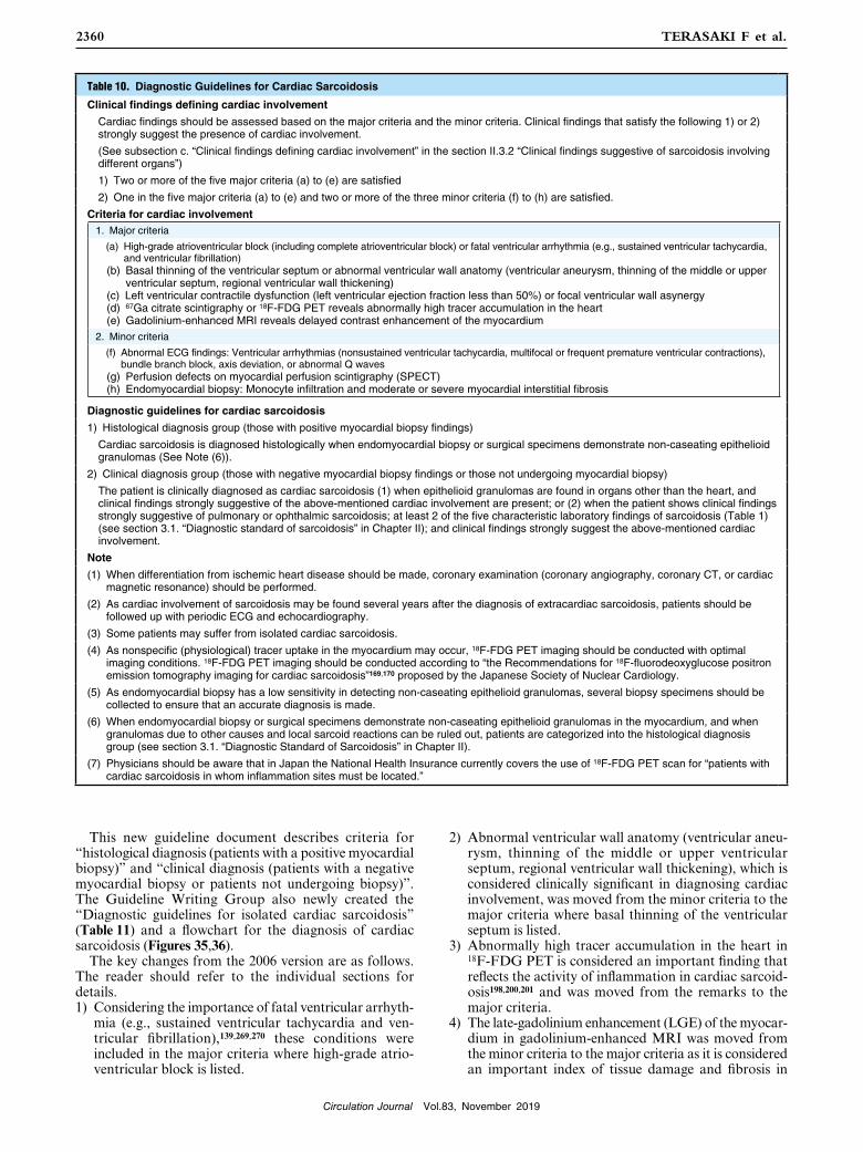
Circulation Journal Vol.83, November 2019
2360 TERASAKI F et al.
This new guideline document describes criteria for “histological diagnosis (patients with a positive myocardial biopsy)” and “clinical diagnosis (patients with a negative myocardial biopsy or patients not undergoing biopsy)”. The Guideline Writing Group also newly created the “Diagnostic guidelines for isolated cardiac sarcoidosis” (Table 11) and a flowchart for the diagnosis of cardiac sarcoidosis (Figures 35,36).
The key changes from the 2006 version are as follows. The reader should refer to the individual sections for details.1) Considering the importance of fatal ventricular arrhyth-
mia (e.g., sustained ventricular tachycardia and ven-tricular fibrillation),139,269,270 these conditions were included in the major criteria where high-grade atrio-ventricular block is listed.
2) Abnormal ventricular wall anatomy (ventricular aneu-rysm, thinning of the middle or upper ventricular septum, regional ventricular wall thickening), which is considered clinically significant in diagnosing cardiac involvement, was moved from the minor criteria to the major criteria where basal thinning of the ventricular septum is listed.
3) Abnormally high tracer accumulation in the heart in 18F-FDG PET is considered an important finding that reflects the activity of inflammation in cardiac sarcoid-osis198,200,201 and was moved from the remarks to the major criteria.
4) The late-gadolinium enhancement (LGE) of the myocar-dium in gadolinium-enhanced MRI was moved from the minor criteria to the major criteria as it is considered an important index of tissue damage and fibrosis in
Table 10. Diagnostic Guidelines for Cardiac Sarcoidosis
Clinical findings defining cardiac involvement
Cardiac findings should be assessed based on the major criteria and the minor criteria. Clinical findings that satisfy the following 1) or 2) strongly suggest the presence of cardiac involvement.
(See subsection c. “Clinical findings defining cardiac involvement” in the section II.3.2 “Clinical findings suggestive of sarcoidosis involving different organs”)
1) Two or more of the five major criteria (a) to (e) are satisfied
2) One in the five major criteria (a) to (e) and two or more of the three minor criteria (f) to (h) are satisfied.
Criteria for cardiac involvement1. Major criteria
(a) High-grade atrioventricular block (including complete atrioventricular block) or fatal ventricular arrhythmia (e.g., sustained ventricular tachycardia, and ventricular fibrillation)
(b) Basal thinning of the ventricular septum or abnormal ventricular wall anatomy (ventricular aneurysm, thinning of the middle or upper ventricular septum, regional ventricular wall thickening)
(c) Left ventricular contractile dysfunction (left ventricular ejection fraction less than 50%) or focal ventricular wall asynergy (d) 67Ga citrate scintigraphy or 18F-FDG PET reveals abnormally high tracer accumulation in the heart (e) Gadolinium-enhanced MRI reveals delayed contrast enhancement of the myocardium2. Minor criteria
(f) Abnormal ECG findings: Ventricular arrhythmias (nonsustained ventricular tachycardia, multifocal or frequent premature ventricular contractions), bundle branch block, axis deviation, or abnormal Q waves
(g) Perfusion defects on myocardial perfusion scintigraphy (SPECT) (h) Endomyocardial biopsy: Monocyte infiltration and moderate or severe myocardial interstitial fibrosis
Diagnostic guidelines for cardiac sarcoidosis
1) Histological diagnosis group (those with positive myocardial biopsy findings)
Cardiac sarcoidosis is diagnosed histologically when endomyocardial biopsy or surgical specimens demonstrate non-caseating epithelioid granulomas (See Note (6)).
2) Clinical diagnosis group (those with negative myocardial biopsy findings or those not undergoing myocardial biopsy)
The patient is clinically diagnosed as cardiac sarcoidosis (1) when epithelioid granulomas are found in organs other than the heart, and clinical findings strongly suggestive of the above-mentioned cardiac involvement are present; or (2) when the patient shows clinical findings strongly suggestive of pulmonary or ophthalmic sarcoidosis; at least 2 of the five characteristic laboratory findings of sarcoidosis (Table 1) (see section 3.1. “Diagnostic standard of sarcoidosis” in Chapter II); and clinical findings strongly suggest the above-mentioned cardiac involvement.
Note
(1) When differentiation from ischemic heart disease should be made, coronary examination (coronary angiography, coronary CT, or cardiac magnetic resonance) should be performed.
(2) As cardiac involvement of sarcoidosis may be found several years after the diagnosis of extracardiac sarcoidosis, patients should be followed up with periodic ECG and echocardiography.
(3) Some patients may suffer from isolated cardiac sarcoidosis.
(4) As nonspecific (physiological) tracer uptake in the myocardium may occur, 18F-FDG PET imaging should be conducted with optimal imaging conditions. 18F-FDG PET imaging should be conducted according to “the Recommendations for 18F-fluorodeoxyglucose positron emission tomography imaging for cardiac sarcoidosis”169,170 proposed by the Japanese Society of Nuclear Cardiology.
(5) As endomyocardial biopsy has a low sensitivity in detecting non-caseating epithelioid granulomas, several biopsy specimens should be collected to ensure that an accurate diagnosis is made.
(6) When endomyocardial biopsy or surgical specimens demonstrate non-caseating epithelioid granulomas in the myocardium, and when granulomas due to other causes and local sarcoid reactions can be ruled out, patients are categorized into the histological diagnosis group (see section 3.1. “Diagnostic Standard of Sarcoidosis” in Chapter II).
(7) Physicians should be aware that in Japan the National Health Insurance currently covers the use of 18F-FDG PET scan for “patients with cardiac sarcoidosis in whom inflammation sites must be located.”

Circulation Journal Vol.83, November 2019
2361JCS 2016 Guideline on Diagnosis and Treatment of Cardiac Sarcoidosis
cardiac sarcoidosis.143,163,271,272
5) Remarks are added to describe important points and a supplemental explanation.
▋3.2 Isolated Cardiac SarcoidosisAs a systemic disorder, physicians had to confirm lesions in at least two different organs (including lymph nodes) to make a diagnosis of the disease according to the criteria published in Japan in 2006.6 However, there have been reported cases of non-caseating epithelioid cell granuloma and/or monocyte infiltration found in myocardial biopsy, and cases of clinical findings consistent with cardiac sarcoidosis in patients with no involvement other than the heart.134,136,171,190,273–283 These cases are referred to as isolated cardiac sarcoidosis. Isobe et al described that this condition may develop as (1) the initial lesion of sarcoidosis with no involvement in other organs (2) a possible type of sarcoidosis affecting the heart only, or (3) the detection of findings consistent with sarcoidosis only in the heart without findings in other organs because inflammation is too modest to detect or typical findings cannot be detected for other reasons.171
It has been estimated that cases of isolated cardiac sarcoidosis account for 5 to 15% of patients who are confirmed or suspected to have cardiac sarcoidosis.134,283 However, the following three points should be considered to interpret these estimations: (1) As isolated cardiac sarcoidosis does not satisfy the criteria for the diagnosis of sarcoidosis in 2006, physicians might not report these patients as cases of sarcoidosis. (2) Non-caseating epithe-lioid granuloma may not be proven in many myocardial
biopsy samples obtained from patients with cardiac sarcoid-osis. In a study of patients with relatively severe cardiac sarcoidosis who meet the conventional diagnostic standard, cardiac sarcoidosis is histologically diagnosed only in 10 to 17%.99 In other studies in patients suspected to have isolated cardiac sarcoidosis, the corresponding percentage was as low as 10%.273,274 (3) This patient population may include those presented with cardiac sarcoidosis who have not been evaluated for sarcoidosis in other organs. The above items (1) and (2) may cause underestimation and (3) may cause overestimation of the prevalence of isolated cardiac sarcoidosis.
Reports have described that cardiac involvement leads to about two-thirds of deaths in patients who died from sarcoidosis in Japan.17,284 In a multicenter retrospective survey in patients with cardiac sarcoidosis in Japan, corti-costeroid therapy is considered to improve long-term prognosis more in patients with better left ventricular systolic function than those with poorer function.21 These findings suggest earlier treatment intervention after the detection of cardiac involvement may contribute to a better prognosis of patients with sarcoidosis. As isolated cardiac sarcoidosis is considered not to differ from cardiac sarcoid-osis affecting other organs as well in terms of pathophysi-ology and prognosis,171 it is therefore important to start appropriate treatment without delay. In this guideline, we propose the “Diagnostic guidelines for isolated cardiac sarcoidosis” (Table 11). As a prerequisite, patients should be confirmed not to have clinical findings of sarcoidosis affecting other organs. Myocardial biopsy is important in considering treatment strategies. The diagnosis of isolated cardiac sarcoidosis may be confirmed when non-caseating
Table 11. Diagnostic Guidelines for Isolated Cardiac Sarcoidosis
Prerequisite
1. No clinical findings characteristics of sarcoidosis are observed in any organs other than the heart (The patient should be examined in detail for respiratory, ophthalmic, and skin involvements of sarcoidosis. When the patient is symptomatic, other etiologies that can affect the corresponding organs must be ruled out.).
2. 67Ga scintigraphy or 18F-FDG PET reveals no abnormal tracer accumulation in any organs other than the heart.
3. A chest CT scan reveals no shadow along the lymphatic tracts in the lungs or no hilar and mediastinal lymphadenopathy (minor axis >10 mm).
1) Histological diagnosis group
Isolated cardiac sarcoidosis is diagnosed histologically when endomyocardial biopsy or surgical specimens demonstrate non-caseating epithelioid granulomas.
2) Clinical diagnosis group
Isolated cardiac sarcoidosis is diagnosed clinically when the criterion (d) and at least three other criteria of the major criteria (a) to e) are satisfied (Table 10).
Note
(1) When the patient meets at least four criteria for cardiac involvement other than the criterion (d), or when the patient meets the criteria (b) and (d) plus one of the remaining criteria, the patient should be suspected to have isolated cardiac sarcoidosis.
(2) When the patient is strongly suspected to have isolated cardiac sarcoidosis that may pose a risk of death according to clinical diagnosis, he/she may be treated empirically with corticosteroids or immunosuppressive drugs before a confirmatory diagnosis is made.
(3) Coronary artery disease and other inflammatory myocardial diseases (e.g., chronic myocarditis, giant cell myocarditis, and myocarditis associated with systemic disorders) should be ruled out.
(4) As nonspecific (physiological) 18F-FDG uptake in the myocardium may occur, 18F-FDG PET imaging should be conducted with optimal imaging conditions. 18F-FDG PET imaging should be conducted according to “the Recommendations for 18F-fluorodeoxyglucose positron emission tomography imaging for cardiac sarcoidosis”169,170 proposed by the Japanese Society of Nuclear Cardiology.
(5) As endomyocardial biopsy has a low sensitivity in detecting non-caseating epithelioid granulomas, several biopsy specimens should be collected whenever possible to make histological diagnosis, which helps the physician plan the optimal treatment strategy reliably.
(6) Physicians should be aware that in Japan the National Health Insurance currently covers the use of 18F-FDG PET scan for “patients with cardiac sarcoidosis in whom inflammation sites must be located.”
(7) Cases of isolated cardiac sarcoidosis that are categorized into the clinical diagnosis group are consistent with those “suspected to have sarcoidosis” in the diagnostic standard for sarcoidosis (see section 3.1. “Diagnostic Standard of Sarcoidosis” in Chapter II).

Circulation Journal Vol.83, November 2019
2362 TERASAKI F et al.
epithelioid granulomas are found in myocardial tissues (Evidence level 5, Recommendation grade B). Patients with clinical findings suggestive of isolated cardiac sarcoidosis in whom myocardial biopsy is impossible or did not reveal the presence of granulomas in myocardial tissues should be evaluated according to the “Diagnostic guidelines for isolated cardiac sarcoidosis” (Evidence level 6, Recommen-dation grade C1). Patients who meet at least 4 items including (d) in the five major criteria (a) to (e) in the cardiac findings in the “Diagnostic guidelines for cardiac sarcoidosis” (Table 10) should be diagnosed as isolated cardiac sarcoid-osis according to the clinical diagnosis group criteria.
Case reports on isolated cardiac sarcoidosis are still limited in number, and only a little evidence is available. Some cases may not be differentiated clearly from other types of inflammatory myocardial diseases or cardiomy-opathies according to this guideline document. Physicians should carefully monitor over time and observe for other clinical findings to make a diagnosis.
▋3.3 Diagnostic Procedures (Flowcharts)a. Follow-up Evaluation of Patients Diagnosed With
Sarcoidosis Affecting Non-Cardiac Organs (e.g., Lungs, Skin, or Eyes) (Figure 35)107,285,286
Basically, patients should be followed up periodically through standard 12-lead ECG, which is an economical, convenient, and repeatable procedure. Echocardiography and Holter ECG may also be used in combination with
12-lead ECG as these procedures may detect abnormalities in patients with normal 12-lead ECG findings. When the 12-lead ECG detects arrhythmias such as atrioventricular block, bundle branch block, axis deviation, and premature ventricular contractions during follow-up, the patient should be consulted by a cardiologist and should undergo detailed examinations such as 18F-FDG PET, 67Ga scintig-raphy and cardiac MRI in addition to echocardiography and Holter ECG. Findings of 18F-FDG PET and 67Ga scintigraphy should be assessed in comparison with findings of myocardial blood flow SPECT.169,170 Patients with abnormal findings on these imaging studies should be considered for cardiac catheterization to rule out coronary artery diseases, evaluate the disease condition, and obtain endomyocardial biopsy samples. Patients who have minor abnormal ECG findings without apparent abnormalities on imaging or invasive studies should be observed carefully and periodically through echocardiography, Holter ECG, or other appropriate methods.
Physicians should follow-up patients with cardiac sarcoidosis carefully as cardiac function may deteriorate rapidly within months or a year in some patients.287,288
b. How to Examine for Cardiac Sarcoidosis During Evaluation of Patients Presenting With Myocardial Diseases or Arrhythmias of Unknown Cause107,286 (Figure 36)
Cardiac sarcoidosis should be suspected for middle-aged or elderly women who have complete atrioventricular
Figure 35. Diagnostic flowchart for cardiac sarcoidosis in patients diagnosed with sarcoidosis based on extracardiac lesions. ECG, electrocardiogram; SPECT, single photon emission computed tomography, 18F-FDG, fluorine-18 fluorodeoxyglucose; 67Ga, gallium-67.

Circulation Journal Vol.83, November 2019
2363JCS 2016 Guideline on Diagnosis and Treatment of Cardiac Sarcoidosis
block or bifascicular block (e.g., right bundle branch block plus left axis deviation), basal thinning of the ventricular septum, co-presence of ventricular wall thinning and thickening and/or uneven wall motion (e.g., localized ventricular aneurysm).1,98 Imaging studies such as 18F-FDG PET, 67Ga scintigraphy, cardiac MRI, and myocardial blood flow SPECT or endomyocardial biopsy should be considered for such patients, but they should first undergo systemic evaluation for the presence of sarcoidosis in other organs.
Attending physicians (1) should consult with respiratory physicians, ophthalmologists, and dermatologists to evaluate for extracardiac lesions and (2) should evaluate whether the patient has at least 2 of the five laboratory findings typical for sarcoidosis (Table 1, Figure 36) (see Section “3.1 Diagnostic Standard of Sarcoidosis” in Chapter II). Specifi-cally, chest CT should be conducted to evaluate for hilar or mediastinal lymphadenopathy, and serum levels of ACE, lysozyme, and sIL-2R should be determined. In 18F-FDG PET or 67Ga scintigraphy, whole-body images but not limited to chest images should be obtained as tracer accumulations may be found in the hilar or mediastinal lymph nodes, skin, subcutaneous nodules, skeletal muscle or lymph nodes. Tracer accumulations at sites where biopsy samples can be obtained will help physicians to make a histological diagnosis of sarcoidosis. Respiratory physicians should also be consulted to consider bronchoal-veolar lavage fluid examination and transbronchial lung
biopsy. Patients diagnosed with sarcoidosis should then be evaluated according to the flowchart for patients with sarcoidosis based on extracardiac lesions (Figure 35). When they meet the “Diagnostic guidelines for cardiac sarcoidosis,” the diagnosis of cardiac sarcoidosis is con-firmed. Patients who were not diagnosed with sarcoidosis in other organs should be evaluated according to the “Diagnostic guidelines for isolated cardiac sarcoidosis”. It should be noted that some patients may not meet the prerequisite in the “Diagnostic guidelines for isolated cardiac sarcoidosis” but may be suspected to have cardiac sarcoidosis as they meet some of the criteria in the “Diag-nostic guidelines for cardiac sarcoidosis”. Examples of such patients include those who have eye lesions but do not show typical laboratory findings and those who have no extracardiac lesions but show typical laboratory findings such as accumulations of 67Ga or 18F-FDG in the medias-tinum. Since these patients may have cardiac sarcoidosis, appropriate treatment according to their disease conditions and careful follow-up are necessary.
Physicians should also note that the diagnostic criteria for cardiac sarcoidosis and the definition of isolated cardiac sarcoidosis in Japan differ from those in Western countries. In Western countries, diagnosis should be made histologi-cally, while in Japan, diagnosis based only on clinical findings is allowed.136,171 Kandolin et al do not consider the presence/absence of mediastinal lymph node lesions as a prerequisite for isolated cardiac sarcoidosis and handle
Figure 36. Diagnostic flowchart for cardiac sarcoidosis in patients who present with cardiac manifestations and are strongly suspected to have cardiac sarcoidosis. AV, atrioventricular; ECG, electrocardiogram; 18F-FDG, fluorine-18 fluorodeoxyglucose; 67Ga, gallium-67; SPECT, single photon emission computed tomography; ACE, angiotensin converting enzyme; sIL-2R, soluble interleukin 2 receptor.

Circulation Journal Vol.83, November 2019
2364 TERASAKI F et al.
patients with positive mediastinal lymph node biopsy as cases of isolated cardiac sarcoidosis. Western reports have described that endomyocardial biopsy guided with imaging
studies such as 18F-FDG PET and cardiac MRI136 or intra-cardiac potentials107 may improve the detection rate of biopsy.
IV. Treatment of Cardiac Sarcoidosis
1. Pharmacotherapy
▋1.1 Immunosuppressive TherapyPharmacotherapy for cardiac sarcoidosis mainly consists of immunosuppressants that are used for controlling inflammation and thereby improve clinical symptoms.289 Corticosteroids are widely used as first-line immunosup-pressants for patients with cardiac sarcoidosis.290 The “Views on the treatment of sarcoidosis - 2003” published in 20039 describe that corticosteroid therapy should be considered for patients with cardiac sarcoidosis who have high-grade atrioventricular block, ventricular arrhythmias, or cardiac dysfunction (Evidence level 6, Recommendation
grade C1). Although no placebo-controlled, prospective studies have demonstrated that corticosteroids may improve prognosis of cardiac sarcoidosis, clinical experience in patients showing improvement in clinical findings after starting corticosteroids has suggested their benefits.87,290 Reports have described that prognosis was better when corticosteroid therapy for cardiac sarcoidosis was initiated before rather than after the occurrence of cardiac dysfunc-tion21,291 (Evidence level 4b, Recommendation grade C1). However, further evaluation should be undertaken to determine whether corticosteroid therapy should be intro-duced for patients with only mild manifestations of cardiac sarcoidosis such as abnormal ECG findings or localized late enhancement in cardiac MRI.166 On the other hand, corticosteroid therapy is considered less effective for patients
Figure 37. Systemic corticosteroid therapy commonly prescribed for patients with cardiac sarcoidosis. Source: Prepared based on Sarcoidosis Treatment Guideline Preparation Committee, 2003.9

Circulation Journal Vol.83, November 2019
2365JCS 2016 Guideline on Diagnosis and Treatment of Cardiac Sarcoidosis
who have severe cardiac dysfunction but do not show abnormal accumulation of 67Ga or 18F-FDG in the myocar-dium, but a report has suggested that corticosteroids therapy should be initiated regardless of whether myocardial tracer accumulation is observed or not.292
There are no established protocols regarding how to initiate, taper, or maintain prednisolone therapy in patients with cardiac sarcoidosis.290 According to the “Views on the Treatment of Sarcoidosis - 2003,” oral corticosteroid therapy for sarcoidosis should be started with the initial dose of 30 mg daily (0.5 mg/kg daily) as or 60 mg every other day (1.0 mg/kg every other day) prednisolone equivalent for the first 4 weeks. Then the dose should be reduced by 5 mg daily or 10 mg every other day at intervals of 2 to 4 weeks to maintain at 5 to 10 mg daily or 10 to 20 mg every other day (Evidence level 6, Recommendation grade C1) (Figure 37).9 Although a retrospective study in patients with cardiac sarcoidosis has reported that the prognosis did not differ between patients receiving prednisolone at an initial dose of 30 mg/day and ≥40 mg/day,21 some patients with active and rapidly progressing cardiac sarcoidosis have started corticosteroid therapy at high initial doses or underwent corticosteroid pulse therapy.293,294 Since there are no reliable markers or imaging modalities that reflect the activity of cardiac sarcoidosis,295 corticosteroid doses are reduced automatically. It is not easy to assess the progression and flare of cardiac involvement of sarcoidosis, but physicians determine the doses of corticosteroids for individual patients based on imaging studies such as 67Ga scintigraphy and 18F-FDG PET and clinical findings. Some patients may discontinue corticosteroid therapy, but many patients continue corticosteroid therapy for a long period of time (Evidence level 6, Recommendation grade C1).
Second-line immunosuppressive therapy for patients with extracardiac involvement of sarcoidosis such as those with intractable pulmonary sarcoidosis has been tried using cyclophosphamide,296 cyclosporine,297 azathioprine,298 methotrexate,299 thalidomide,300 hydroxychloroquine,301 pentoxifylline,302 and mycophenolic acid,303 among others (Evidence level 5). These drugs may be used as monotherapy or in combination with corticosteroids in patients who do not respond well to corticosteroids or cannot use them or increase their dose due to adverse drug reactions,290,304 but the use of these second-line drugs in cardiac sarcoidosis has only been described in case reports. Low-dose methotrexate is most commonly used for this purpose in Japan (Evidence level 5, Recommendation grade C1) and is expected to have a steroid-sparing effect or alleviate the severity of adverse drug reactions to corticosteroids.91 However, physicians should carefully observe for adverse drug reactions such as decreased white blood cell count, hepatic dysfunction and interstitial pneumonia. The recommended dose of metho-trexate in Western countries is 10 to 20 mg/week,290 but methotrexate is often administered at doses of 5 to 8 mg/week in Japan.91,305 A report has described that cardiac function in patients with cardiac sarcoidosis was better maintained in patients who received methotrexate in combination with prednisolone than patients who started treatment with prednisolone monotherapy.305 Further studies are needed.
Recently, infliximab, an anti-TNF-α antibody that relieves inflammation by blocking TNF-α in the blood, inhibiting TNF-α to bind to receptors, and destroying TNF-α-producing cells has been tried as the third treatment option for pulmonary sarcoidosis in Western countries306 (Evidence
level 5). Infliximab may be used for patients who do not respond to corticosteroids and at least one second-line therapy.290 Case reports have described the effectiveness of infliximab in patients with cardiac sarcoidosis.307,308
▋1.2 Treatment of Heart Failurea. Basic Policies for Heart Failure ManagementNo clinical research has focused on the management of cardiac failure in patients with cardiac sarcoidosis. Accordingly, treatment should consist of immunosuppres-sive therapy for the treatment of cardiac sarcoidosis and basic heart failure management regardless of the stage of heart failure (Evidence level 6, Recommendation grade C1). Patients with heart failure associated with cardiac sarcoid-osis should receive pharmacological treatment generally recommended based on evidence for patients with left ventricular systolic dysfunction, such as ACE inhibitors, angiotensin II receptor blockers, beta-blockers, and aldosterone antagonists, as well as immunosuppressants, for the treatment of sarcoidosis.309 Additional drugs should be given to patients with signs of congestions and lower cardiac output. Vasodilators and diuretics are used for the treatment of congestion, and inotropic drugs such as catecholamines are used for the treatment of low cardiac output. Patients who do not respond well to these drugs may need mechanical circulatory support. Antiarrhythmic drugs and pacemakers should be used for patients with arrhythmias that cause unstable cardiac rhythm or heart rate and may affect hemodynamics. Patients with hypox-emia due to pulmonary congestion should be treated with oxygenation and artificial respiration.310
b. Heart Failure Management Based on Disease Stages and Types
As indicated in the ACCF/AHA heart failure classification system,311 appropriate treatment to prevent disease progres-sion is important for the treatment of heart failure in patients with cardiac sarcoidosis. Corticosteroids are less effective in patients with advanced heart dysfunction.21 Also, once myocardial damage in sarcoidosis occurs, left ventricular remodeling continues to progress even when the inflammation caused by sarcoidosis is alleviated. Heart failure should be treated according to the guidelines309–311 for the treatment of heart failure in general.
Left ventricular systolic dysfunction is the most common type of abnormal heart pumping function in patients with cardiac sarcoidosis.224 Patients with activated myocardial inflammation may show edematous wall thickening, which is similar to those observed in hypertrophic cardiomy-opathy.156 However, no manifestations of heart failure due to diastolic dysfunction are found in many cases, and treatment of the underlying cardiac sarcoidosis is the main component of clinical management for patients with heart failure associated with cardiac sarcoidosis. Cases of cardiac sarcoidosis affecting the right ventricle with or without left ventricular involvement have been reported. In some such cases, differentiation from arrhythmogenic right ventricular cardiomyopathy is important.312 Patients with right and left ventricular involvement need more complex manage-ment such as careful fluid management and biventricular circulatory support.310

Circulation Journal Vol.83, November 2019
2366 TERASAKI F et al.
▋1.3 Treatment of Arrhythmias<Indications for corticosteroid therapy for the treatment of tachyarrhythmia and bradyarrhythmia associated with cardiac sarcoidosis>
Class IPatients with cardiac involvement demonstrated by positive 67Ga scintigraphy or 18F-FDG PET findings (Evidence level 4a, Recommendation grade A)Class IIaPatients without cardiac involvement demonstrated by positive 67Ga scintigraphy or 18F-FDG PET findings (Evidence level 4b, Recommendation grade C1)
<Indications for antiarrhythmic drugs for the treatment of tachyarrhythmia associated with cardiac sarcoidosis>
Class INoneClass IIa(1) Beta blockers (Evidence level 6, Recommendation
grade C1)(2) Amiodarone, sotalol, and class Ib* antiarrhythmic
drugs for the treatment of uncontrollable ventricular tachycardia (Evidence level 6, Recommendation grade C1)
Class IIb(1) Amiodarone, sotalol, and verapamil for the treat-
ment of controllable ventricular tachycardia (Evidence level 6, Recommendation grade C2)
(2) Class Ia* and Ic* antiarrhythmic drugs for the treat-ment of uncontrollable ventricular tachycardia (Evidence level 6, Recommendation grade C2)
Class IIIClass Ia* and Ic* antiarrhythmic drugs for the treat-ment of controllable ventricular tachycardia (Evidence level 6, Recommendation grade D)
*Vaughan Williams Classification
Atrioventricular block is the most frequent initial manifes-tation of arrhythmia associated with cardiac sarcoidosis,6 and fatal ventricular arrhythmia may often develop. Patients with advanced cardiac sarcoidosis may have cardiac dysfunction resulting from loss of cardiac myocytes, which may cause secondary tachyarrhythmia. Accordingly, antiarrhythmic drugs should be selected based on the remaining cardiac function and activity of cardiac sarcoid-osis as well as the indications for non-pharmacological treatments such as pacemakers and ICD (Evidence level 6, Recommendation grade C1).
As it has been reported that corticosteroids may improve atrioventricular conduction in patients with atrioventricular block associated with cardiac sarcoidosis,87,248,313 the activity of inflammation should be evaluated whenever possible. Since tachyarrhythmia often develops in patients with poor cardiac function and corticosteroid therapy is important in maintaining cardiac function,292 corticosteroids should also be considered for patients without findings of inflammation. Ventricular arrhythmia may often recur even after successful ablation,270,314 patients should continue antiarrhythmic treatment.249,250,315 As arrhythmias associated with cardiac sarcoidosis are often originated from scar tissues or result
from reentry,138,316 physicians may consider beta blockers (Evidence level 6, Recommendation grade C1) and drugs that prolong repolarization (e.g., amiodarone and sotalol) (Evidence level 6, Recommendation grade C1) for the treat-ment of arrhythmia associated with cardiac sarcoidosis but should try beta blockers to control heart failure as well (Evidence level 6, Recommendation grade C1). Calcium-channel blockers (verapamil) are not recommended (Evidence level 6, Recommendation grade C2). Sodium-channel blockers, especially Vaughan-Williams class Ia* and Ic* antiarrhythmic drugs, are not recommended for patients with poor cardiac function as they have proar-rhythmic properties.317 The antiarrhythmic effects of corti-costeroids are very limited in patients with severe cardiac dysfunction.318
2. Non-Drug Therapy
▋2.1 Permanent Pacing<Indications for permanent pacing for the treatment of atrioventricular block>
Class I(1) Patients with second-degree, advanced or third-
degree atrioventricular block with symptoms of bradycardia (Evidence level 6, Recommendation grade B).
(2) Patients with advanced or third-degree atrioven-tricular block who experience profound bradycardia or long-lasting ventricular arrest during wakeful-ness (Evidence level 6, Recommendation grade B).
Class IIa(1) Asymptomatic, persistent third-degree atrioven-
tricular block (Evidence level 6, Recommendation grade C1)
(2) Asymptomatic, second-degree or advanced atrio-ventricular block with at least one of the following conditions (Evidence level 6, Recommendation grade B):
① When the block occurs at or below the bundle of His;
② when the block is associated with progressive cardiomegaly due to bradycardia;
③ when exercise or atropine sulfate does not change or impair atrioventricular conduction.
(3) Patients who have symptoms that are likely due to bradycardia, who have first-degree atrioventricular block without other causes, and in whom the block occurs at or below the bundle of His (Evidence level 6, Recommendation grade B).
Class IIbPatients with first-degree atrioventricular block and heart failure in whom hemodynamics may be improved by setting the optimal atrioventricular interval (Evidence level 6, Recommendation grade C2).
<Treatment guidelines for permanent pacemaker implanta-tion in patients with cardiac sarcoidosis>
(1) It is advisable that physicians refer to the “Guide-lines for Non-Pharmacotherapy of Cardiac Arrhythmia (JCS 2011)” published by the Japanese Circulation Society319,320 to determine whether

Circulation Journal Vol.83, November 2019
2367JCS 2016 Guideline on Diagnosis and Treatment of Cardiac Sarcoidosis
permanent pacemaker implantation is indicated for their patients with cardiac sarcoidosis (Evidence level 6, Recommendation grade A).
(2) Biventricular cardiac pacing rather than right ventricular pacing should be selected for patients with poor cardiac function with a LVEF ejection fraction of ≤35% (Evidence level 6, Recommendation grade B).
(3) Immunosuppressive drugs are expected to be effective in the treatment of conduction disturbance in some patients with cardiac sarcoidosis, but it is difficult to predict whether individual patients will respond to the treatment or not. It is desirable that patients who are indicated for permanent pace-maker implantation undergo pacemaker implanta-tion before starting immunosuppressive therapy (Evidence level 6, Recommendation grade B).
(4) Patients who are indicated for pacing should be considered for ICD implantation to prevent sudden death (Evidence level 6, Recommendation grade C2).
Evidence level and recommendation grade are based on the classification scales in the reference articles 319 and 320.
Atrioventricular block is a common complication found in 26 to 67% of patients with cardiac sarcoidosis,285 and is caused by basal thinning or granuloma formation of the ventricular septum, or ischemia due to the involvement of the atrioventricular nodal artery.102 Indications for perma-nent pacemaker implantation should basically be considered according to the “Guidelines for Non-Pharmacotherapy of Cardiac Arrhythmias (JCS 2011)” proposed by the Japanese Circulation Society319,320 based on the cardiac function of individual patients. Also, CRT should be considered for patients who have a LVEF of ≤35%, are planned to undergo pacemaker implantation for the management of bradycardia, and are expected to frequently depend on ventricular pacing. In the BLOCK HF study, CRT signifi-cantly reduced the mortality and incidence of heart failure events.321 CRT should be strongly considered for patients with atrioventricular block and poor cardiac function.
Although many studies have reported that corticosteroid therapy is effective in the treatment of atrioventricular block in patients with cardiac sarcoidosis,149,313,322 no randomized clinical studies of immunosuppressive drugs were conducted in this patient population. Considering the difficulty in predicting the efficacy of treatment, the risk of recurrence, and the risk of device infection during treatment with oral corticosteroids, corticosteroid therapy should be initiated after device implantation.
ICD is indicated for patients with ventricular arrhythmia. Patients with atrioventricular block should be observed carefully since fatal ventricular arrhythmias have developed in patients with cardiac sarcoidosis before ICD implanta-tion.323 ICD as the primary prevention strategy for atrio-ventricular block in patients with cardiac sarcoidosis has not been evaluated in randomized clinical studies. Further studies are awaited.
▋2.2 Implantable Cardioverter Defibrillator (ICD)a. Risk Assessment for Sudden DeathPatients with cardiac sarcoidosis are at risk of sudden death due to ventricular arrhythmia,140,324 and ICD is indicated for those with a high risk of sudden death. It has
been reported that patients with a history of life-threatening arrhythmias are at risk of recurrent arrhythmias and that the incidence of life-threatening arrhythmias is high among patients with poor cardiac function with no history of serious arrhythmias.325–327 ICD implantation is recommended for patients with a LVEF of ≤35% even under appropriate pharmacotherapy.328,329 Also, care should be taken for patients with modestly mild ventricular dysfunction with a LVEF of ≤36 to ≤49% when they have right ventricular dysfunction (right ventricular ejection fraction (RVEF): <40%). Reports have described the benefits of electro-physiological testing in risk assessment for sudden death in patients with cardiac sarcoidosis.254,255 It has been reported that the risk of death, sudden death, or appropriate ICD discharge is high in patients with late enhancement in cardiac MRI,143,330 but some reports have pointed out that imaging findings are not useful in risk assessment.108 An association between positive findings on 18F-FDG PET and the risk of death or sustained ventricular tachycardia has been reported,204 but this has not been investigated in larger populations.
b. Indications of ICD<Indications of ICD for patients with cardiac sarcoidosis>
Class I(1) Patients with a history of cardiac arrest or sustained
ventricular tachycardia (Evidence level 4a, Recom-mendation grade B).
(2) Patients with a LVEF of ≤35%, and either (a) who have non-sustained ventricular tachycardia (ICD is not covered by the NHI in Japan) or (b) in whom sustained ventricular tachycardia or ventricular fibrillation was induced during electrophysiological testing** (Evidence level 4a, Recommendation grade C1).
Class IIa(1) Patients with a LVEF of ≤35% even with optimal
pharmacotherapy, including immunosuppressive therapy, (ICD is not covered by the NHI in Japan) (Evidence level 4a, Recommendation grade C1).
(2) Patients with syncope of unknown cause, and either (a) who have non-sustained ventricular tachycardia (ICD is not covered by the NHI in Japan) or (b) in whom sustained ventricular tachy-cardia or ventricular fibrillation was induced during electrophysiological testing** (Evidence level 4a, Recommendation grade C1).
(3) Patients with a LVEF of <50%, who are indicated for pacemaker implantation, and either (a) who have non-sustained ventricular tachycardia (ICD is not covered by the NHI in Japan) or (b) in whom sustained ventricular tachycardia or ventricular fibrillation was induced during electrophysiological testing** (Evidence level 4a, Recommendation grade C1).
(4) Patients with a LVEF of <50%, and either (a) late enhancement in cardiac MRI, (b) positive findings of 18F-FDG PET or (c) 67Ga-scintigraphy, and in whom sustained ventricular tachycardia or ventricular fibrillation was induced during electro-physiological testing** (Evidence level 4a, Recom-mendation grade C1).
**Sustained ventricular tachycardia is defined as having a

Circulation Journal Vol.83, November 2019
2368 TERASAKI F et al.
duration of ≥30 seconds. Induction of VT/VF by 3 consecu-tive extrastimuli at a coupling interval of <220 msec is handled as a non-specific reaction.NHI coverage: According to the rule issued on April 1, 1996, in Japan, the National Health Insurance covers the use of implantable cardioverter defibrillator (ICD) therapy based on the confirmation of spontaneous or induced hemodynamically compromising ventricular tachycardia or fibrillation. It should be noted that this rule is not consistent with the classification of recommendations in the present guideline document.
Indications of ICD should be determined based on whether the patient has had life-threatening arrhythmias, and, if absent, (1) left ventricular dysfunction, (2) nonsustained ventricular tachycardia, and (3) inducibility of arrhythmias during electrophysiological testing (Figure 38). The above descriptions are similar to those described in the HRS expert consensus statement on the diagnosis and manage-ment of arrhythmias associated with cardiac sarcoidosis published by the Heart Rhythm Society,107 but there are differences in terms of severity classification of cardiac dysfunction and indications of ICD in patients indicated for pacemaker implantation. However, physicians should be aware that the risk of life-threatening arrhythmias is high among patients with atrioventricular block who were waiting for pacemaker implantation.323,325 Such patients should be monitored periodically.
▋2.3 Cardiac Resynchronization Therapy (Defibrillator) [CRT(D)]
<Indications of CRT for patients with cardiac sarcoidosis>
Class I
Patients with NYHA class III or ambulatory class IV chronic heart failure despite optimal drug therapy, with a LVEF of ≤35%, a QRS interval of ≥120 msec, and sinus rhythm (Evidence level 6, Recommendation grade B).Class IIa(1) Patients with NYHA class III or ambulatory class
IV chronic heart failure despite optimal drug therapy, a LVEF of ≤35%, a QRS interval of ≥120 msec, and atrial fibrillation (Evidence level 6, Recommen-dation grade B).
(2) Patients with NYHA class III or ambulatory class IV chronic heart failure despite optimal drug therapy, a LVEF of ≤35%, and who are using or plan to use a permanent pacemaker for the treatment of bradycardia and require or are expected to require ventricular pacing frequently (Evidence level 6, Recommendation grade B).
Class IIbPatients with NYHA class II chronic heart failure despite optimal drug therapy, a LVEF of ≤35%, and who plan to use a permanent pacemaker for the treat-ment of bradycardia and are expected to require ventricular pacing frequently (Evidence level 6, Recom-mendation grade C2).Class III(1) Asymptomatic patients with low LVEF who are
not indicated for pacemaker implantation for the treatment of bradycardia (Evidence level 6, Recom-mendation grade C2).
(2) Patients with restricted physical activity due to chronic diseases other than heart failure or those with a life expectancy of 12 months or less (Evi-dence level 6, Recommendation grade C2).
Figure 38. Indications for ICD therapy in patients with cardiac sarcoidosis. EPS, electrophysiological study; LVEF, left ventricular ejection fraction; NSVT, nonsustained ventricular tachycardia; VT, ventricular tachycardia; VF, ventricular fibrillation; ICD, implantable cardioverter defibrillator.

Circulation Journal Vol.83, November 2019
2369JCS 2016 Guideline on Diagnosis and Treatment of Cardiac Sarcoidosis
<Indications of CRT-D in patients with cardiac sarcoidosis>
Class IPatients with NYHA class III or ambulatory class IV chronic heart failure despite optimal drug therapy, a LVEF of ≤35%, a QRS interval of ≥120 msec, and sinus rhythm, and who are indicated for ICD (Evidence level 6, Recommendation grade A).Class IIa(1) Patients with NYHA class III or ambulatory class
IV chronic heart failure despite optimal drug therapy, a LVEF of ≤35%, a QRS interval of ≥120 msec, and atrial fibrillation, and who are indicated for ICD (Evidence level 6, Recommendation grade B).
(2) Patients with NYHA class II chronic heart failure despite optimal drug therapy, a LVEF of ≤30%, a QRS interval of ≥150 msec, and sinus rhythm, and who are indicated for ICD (Evidence level 6, Recommendation grade B).
(3) Patients with NYHA class III or ambulatory class IV chronic heart failure despite optimal drug therapy, a LVEF of ≤35%, and who are using or plan to use ICD and require or are expected to require ventricular pacing frequently (Evidence level 6, Recommendation grade B).
Class IIbPatients with NYHA class II chronic heart failure despite optimal drug therapy, a LVEF of ≤35%, and who plan to use ICD and are expected to require ventricular pacing frequently (Evidence level 6, Recom-mendation grade C2).Class III(1) Asymptomatic patients with low LVEF who are
not indicated for ICD (Evidence level 6, Recom-mendation grade C2).
(2) Patients with restricted physical activity due to chronic diseases other than heart failure or those with a life expectancy of 12 months or less (Evidence level 6, Recommendation grade C2).
In patients with heart failure due to cardiac sarcoidosis, dyssynchrony often occurs between an atrium and a ven-tricle, between the left and right ventricles, or in a ventricle in addition to intraventricular conduction disturbance. CRT is used to treat these conditions. Large-scale studies331–333 have revealed that CRT may prevent the progression of heart failure and improve the prognosis of patients. LVEF and the QRS interval of body surface ECG are important measures to predict the efficacy of CRT. In Japan, a wide QRS interval is defined as a QRS interval of ≥120 msec.319,320
Reports on the benefits of CRT in patients with cardiac
sarcoidosis are limited. Accordingly, indications of CRT for patients with cardiac sarcoidosis are described based on the “Guidelines for Non-Pharmacotherapy of Cardiac Arrhythmias” proposed by the Japanese Circulation Society.319,320 The “HRS expert consensus statement on the diagnosis and management of arrhythmias associated with cardiac sarcoidosis” proposed by the Heart Rhythm Society in 2014107 also recommends physicians to apply the guidance on device implantation to patients with cardiac sarcoidosis.
▋2.4 Catheter Ablationa. Mechanism of Ventricular TachycardiaVentricular tachycardia develops as the initial sign of cardiac sarcoidosis in about 23% of patients248 and may lead to sudden death in some patients. Reentry is the most common mechanism of ventricular tachycardia in patients with cardiac sarcoidosis, but triggered activity and abnormal automaticity have also been reported.138,318,334 Ventricular tachycardia arising from the Purkinje fiber develops through triggered activity and is common among patients with conduction disorder.250 It is considered that there is no relationship between arrhythmogenesis and disease activity evaluated by 67Ga scintigraphy in cardiac sarcoidosis.248
b. Characteristics of Arrhythmogenic SubstratesIt is important to identify arrhythmogenic substrates through late enhancement cardiac MRI before catheter ablation and 3D-mapping system during the procedure. In many patients, right ventricular scarring is confluent while left ventricular scarring is patchy.249 Epicardial scarring that requires epicardial ablation is found in about 30% of patients.249 Patients without endocardial or epicardial scarring are considered to have scarring deep inside the myocardium.
c. Indications of Ablation<Indications of catheter ablation for patients with cardiac sarcoidosis> (Evidence level 5, Recommendation grade B)
Class I(1) Patients with ventricular tachycardia uncontrollable
with corticosteroids or antiarrhythmic drugs(2) Patients with paroxysmal ventricular tachycardia
who cannot take antiarrhythmic drugs(3) Patients with or without ICD who experience
storms of ventricular tachycardia.
Catheter ablation is indicated for patients in whom ven-tricular tachycardia cannot be controlled with corticoste-roids or antiarrhythmic drugs, patients who cannot tolerate
Table 12. Results of Ablation Therapy for the Treatment of Ventricular Tachycardia Associated With Cardiac Sarcoidosis
Reports No. of patients
Mean LVEF (%)
No.of VT events
Complete elimination (cases)
Decreased (cases)
No.of recurrent cases
Follow-up period
Koplan BA, et al.270 8 34 32 2/8 4/8 6/8 6 to 84 months
Jefic D, et al.335 9 42 44 5/9 3/9 4/9 19.8 months
Dechering DG, et al.336 8 36 5/8 6 months
Kumar S, et al.249 21 36 99 9/21 10/21 18/21 1 to 9.5 years
Naruse Y, et al.250 14 40 37 8/14 6/14 24 to 46 months
LVEF, left ventricular ejection fraction; VT, ventricular tachycardia.

Circulation Journal Vol.83, November 2019
2370 TERASAKI F et al.
drug treatment, and patients who experience storms of ventricular tachycardia. Ventricular tachycardia due to triggered activity or abnormal automaticity may be con-trolled with corticosteroid therapy.318,334
d. Acute-Phase Results (Table 12)249,250,270,335,336
During catheter ablation, abnormalities other than symp-tomatic ventricular tachycardia are often found, and about 50% of patients require more than one ablation procedure. The most common reasons for unsuccessful ablation include ventricular tachycardia arising from the ventricular septum, diffuse right ventricular scarring with more than one reentry circuit, and tachycardia arising adjacent to a coronary artery or the His bundle. Kumar et al have reported that a single ablation procedure was enough to ablate all arrhythmogenic substrates of ventricular tachy-cardia in about 40% of patients, and more than one ablation procedure was needed in about 30% of patients to control ventricular tachycardia. Ablation terminated ventricular tachycardia storms not responding to pharmacotherapy in 71% of patients.249 Patients with ventricular tachycardia not responding to ablation, corticosteroids, and antiarrhythmic drugs are at a high risk of death and often require heart transplantation.
e. Long-Term ResultsMany patients have more than one arrhythmogenic sub-strate causing ventricular tachycardia, and the incidence of recurrent arrhythmia after catheter ablation relates to the number of arrhythmogenic substrates where ventricular arrhythmia was induced.
Since ventricular arrhythmia may recur even in patients with successful ablation, the progression of cardiac sarcoid-osis cannot be predicted, and the fibrosis of myocardium may continue, patients should undergo ICD implantation even after successful ablation (Evidence level 5, Recommen-dation grade B).
3. Surgical Treatment
▋3.1 Left Ventricular Restoration and Mitral Valvuloplasty
a. Feasibility of Surgical TreatmentIn patients with cardiac sarcoidosis, granulomas lead to myocardial degeneration and then fibrosis. Myocardial injury may develop in any part of the heart. Some patients may have focal myocardial injuries, and many patients often have multifocal myocardial injuries.102 Focal injuries may progress in different patterns in different patients. Some patients may have left ventricular aneurysm, and other patients may show dilated cardiomyopathy-like findings associated with diffuse myocardial injuries. Patients with focal injuries may undergo resection and plasty to improve the shape and function of the left ventricle. As left ven-tricular dilation may lead to tethering the mitral valve and cause functional mitral valve insufficiency, mitral valvuloplasty should also be considered. Late gadolinium enhancement MRI and 18F-FDG PET have helped physi-cians identify whether lesions are focal or diffuse. Ven-tricular function may improve when appropriate procedures of left ventricular restoration are selected according to the location of myocardial lesions and conducted in combination with mitral or tricuspid valvuloplasty.
b. Results of Surgical TreatmentCase reports have described the results of surgical treatment in patients with left ventricular aneurysm associated with cardiac sarcoidosis232 and patients suspected to have sarcoidosis because of their history of atrioventricular block.337,338 In a report on 384 patients undergoing surgery for the treatment of non-ischemic cardiomyopathy,339 14 patients (3.6%), including 4 patients diagnosed as cardiac sarcoidosis before surgery, who underwent left ventricular restoration for the treatment of cardiac sarcoidosis had NYHA class IV severe heart failure not responding to medical treatment. Procedures of left ventricular restoration were selected according to the location of myocardial injuries and used in combination with mitral and/or tricuspid valvuloplasty. Among 12 patients who underwent planned surgery, 2 patients (17%) died. All 10 survived patients started corticosteroid therapy after surgery. Six of the 10 patients showed an improvement of heart failure to NYHA class II.339 Three-year and five-year survival rates were 65% and 52%, which were higher than those (50% and 37%)1 in patients receiving conventional medical therapy.
c. Limitations and Indications of Surgical TreatmentIn patients with diffuse myocardial injuries in whom viable myocardium is limited, conventional surgical treatment cannot improve left ventricular function, and heart trans-plantation is an option. It has been reported that patients with cardiac sarcoidosis undergoing heart transplantation have acceptable long-term outcomes340 or better than the majority of heart transplant recipients.341 On the other hand, Akashi et al at Columbia University Medical Center have reported that among 825 patients undergoing heart transplantation, 14 patients (1.7%) had cardiac sarcoidosis and 2 of them (14%) died in the hospital. The 1 and 5-year survival rates were 78.5% and 52.4%, showing a post-transplant outcome.342 Akashi et al discussed that patients who received a diagnosis of cardiac sarcoidosis prior to heart transplant were more prone to be in a systemic pro-inflammatory condition, which might be related to the poor outcome in this patient population. Accordingly, patients with cardiac sarcoidosis should be assessed for whether they are indicated for left ventricular restoration or mitral valvuloplasty before considering heart transplantation.
d. Precautions for the Management of Patients After Surgery
Patients with cardiac sarcoidosis are always at a risk of the exacerbation of remaining lesions or the development of new lesions. Although many patients with cardiac sarcoid-osis respond well to corticosteroid therapy, patients may be complicated with infectious diseases or have flares of cardiac sarcoidosis after reducing corticosteroid doses. Patients after surgery must be closely monitored for a long time and continue treatment. The recurrence of cardiac sarcoidosis after heart transplantation has been reported.343 Patients after heart surgery should be carefully observed for exacerbation or recurrence of cardiac sarcoidosis.
▋3.2 Artificial Heart and Heart Transplantationa. Cardiac Sarcoidosis in Heart TransplantationHeart transplantation is a possible treatment option for patients with cardiac sarcoidosis in Japan as well as in other countries341,344 (Evidence Level 5, Recommendation grade C1). In a report in the United States in 2013, among

Circulation Journal Vol.83, November 2019
2371JCS 2016 Guideline on Diagnosis and Treatment of Cardiac Sarcoidosis
1,069 patients undergoing heart transplantation, 19 patients had cardiac sarcoidosis.340 In Japan, a total of 222 patients underwent heart transplantation as of December 2014, and 3 of them had cardiac sarcoidosis (Figure 39).345 The percentage of patients with cardiac sarcoidosis is similar in the above report from the United States. The indication of heart transplantation and the use of a ven-tricular assist device (VAD) during the waiting period should be considered for patients with cardiac sarcoidosis.
b. Indications of Heart Transplantation and the Use of Ventricular Assist Devices (VADs)
Indications of heart transplantation and the use of VADs for patients with cardiac sarcoidosis are similar to those for other types of patients (Table 13),346 but there are some precautions specific to cardiac sarcoidosis. In general, the indication of heart transplantation is considered based on:1) Whether the patient has irreversible heart dysfunction
with decreased exercise tolerance;2) Whether the patient can physically tolerate heart
transplantation; and3) Whether the patient has a social environment encouraging
heart transplantation.VADs are indicated for patients who need prolonged circulatory support for the treatment of cardiogenic circu-
latory failure and patients in whom circulation cannot be maintained with aortic balloon pumping or percutaneous cardiopulmonary support, and who do not meet any of the exclusion criteria.
In Japan, patients who are classified into Level 1 to 3 according to the criteria used in the Japanese Registry for Mechanically Assisted Circulatory Support (J-MACS) that was developed based on the Interagency Registry for Mechanically Assisted Circulatory Support (INTERMACS) are indicated (Table 14).347,348 The detailed criteria for the indication of VADs have been proposed by the Japan VAD Council, a group of academic associations related to VAD use, and are described in the “Guidelines for Device Therapy: Implantable Left Ventricular Assist Device for Patients with Severe Heart Failure”349 proposed by the Japanese Circulation Society and the Japanese Society for Cardiovascular Surgery (Table 15).350
c. Precautions for the Application of Heart Implantation for Patients With Cardiac Sarcoidosis
Corticosteroids are used as the main component of the drug treatment of cardiac sarcoidosis. It has been reported that corticosteroid therapy may improve exercise tolerance even in patines with NYHA Class III severe heart failure not responding to other types of drugs.171 Accordingly, the
Figure 39. Indications of heart trans-plantation (as of December 2014). Adapted from Document for the Heart Transplantation Committee of the Japanese Circulation Society.345
Table 13. Heart Transplant Recipient Selection Criteria
I. Are there any other options to save the life of the patient?
II. How long can the patient live if he/she does not undergo heart transplantation?
III.
Can the patient mentally and physically endure periodic (and sometimes emergent) examinations after transplantation and follow immunosuppressive regimens prescribed based on the results of examinations?
IV.Does the patient understand the necessity of transplantation and desire it? Do his/her family members support the patient?
Adapted from the Heart Transplantation Committee of the Japanese Circulation Society. 2013 346
Table 14. INTERMACS Profile and J-MACS Levels
INTERMACS J-MACS
1 Critical cardiogenic shock Critical cardiogenic shock
2 Progressive decline Progressive decline
3 Stable but inotrope dependent
Stable but inotrope dependent
4 Resting symptoms Resting symptoms
5 Exertion intolerant Exertion intolerant
6 Exertion limited Exertion limited
7 Advanced NYHA III Stable condition
Source: Prepared based on INTERMACS.347 and the Pharmaceu-ticals and Medical Devices Agency.348

Circulation Journal Vol.83, November 2019
2372 TERASAKI F et al.
indication of heart transplantation should be considered after corticosteroid therapy is tried. For patients who are considered to have limited viable myocardium that may respond to corticosteroids, physicians should describe the reasons for such consideration and provide findings of imaging in the application form for heart transplantation. When the patient has systemic sarcoidosis at the time of application for heart transplantation, experts should reach a consensus that non-cardiac sarcoidosis will not become a factor that decides the prognosis of the patient for at least 5 years.
As cardiac sarcoidosis is difficult to diagnose, this condi-tion may be first diagnosed based on the examination of myocardial samples obtained at the time of implantation of a left ventricular support device.351 Corticosteroids are administered to patients who are using external VAD to examine whether they should be included on the waiting list for heart transplantation but are often not administered to patients with VAD implantation because the risk of
infection outweighs the beneficial effects of corticosteroids on cardiac function. It is thus advisable that a diagnosis of cardiac sarcoidosis should be made before VAD implanta-tion whenever possible. However, patients in Japan wait for donor hearts for a long period of time after VAD implantation. During the waiting period, cardiac sarcoid-osis, especially right ventricular involvement of the disease, may progress, but it is unknown whether corticosteroid therapy should be conducted or not.
d. Outcome of Heart TransplantationAccording to Western reports, the outcome of heart transplantation in patients with cardiac sarcoidosis is comparable to other patient populations.352 A report from the United States in 2013 has described that there were no significant differences between patients with cardiac sarcoidosis and other patients in first-year freedom from any treated rejection, 5-year survival, 5-year freedom from chronic coronary artery disease, and 5-year freedom from
Table 15. Practice Standard for the Use of Ventricular Assist Devices (Draft Published on November 16, 2010)
Eligible patients
Disease conditions Patients with end-stage severe heart failure that satisfies the criteria for heart transplantation who have underlying conditions such as dilated or hypertrophic cardiomyopathy, dilated phase of hypertrophic cardiomyopathy, ischemic myocardial disease, valvular disorder, congenital heart disease, and post-myocarditis cardiomyopathy.
Eligibility criteria
Cardiac function NYHA Class III to IV (with a history of NYHA Class IV condition).
Stage D (Patients with severe structural heart disease who have apparent symptoms of heart failure despite maximal medical therapy).
Drug treatment Maximal drug treatment using digitalis, diuretics, ACE inhibitors, angiotensin II receptor blockers, nitrates, and beta-blockers has been given.
Inotropic drugs and assisted circulation
Drug treatment with dobutamine, dopamine, epinephrine, norepinephrine and/or phosphodiesterase III inhibitors, or intra-aortic balloon pumping or external ventricular assist devices are necessary.
Age Preferably patients ≤65 years of age (those over 65 years with relatively good physical conditions may also be included).
Body surface area See the specification of the system to be used.
Hemodynamics Stage D, a history of NYHA Class IV condition.
Conditions Patients in whom other treatments cannot prolong life or whose QOL is severely affected, and in whom the treatment is expected to improve QOL, help them receive home-based treatment for a long period of time and return to daily activities.
Understanding of treatment
Patients who understand the limitations of ventricular assist devices and the risk of complications, and whose family members understand these situations and can support them.
Exclusion criteria
Infections Patients with severe infections.
Respiratory diseases
Patients with severe chronic obstructive pulmonary disease.
Patients with severe pulmonary hypertension.
Patients within 30 days after the onset of pulmonary arterial embolism developed.
Cardiovascular diseases
Patients immediately after open-heart surgery (about 2 weeks).
Patients with untreatable abdominal aortic aneurysm or severe peripheral vascular diseases.
Patients with thoracic aortic aneurysm, ventricular aneurysm, or interventricular septum rupture.
Patients with moderate or severe aortic valve insufficiency.
Serious calcification of the thoracic aorta.
Nervous system disorders
Severe CNS disorder.
History of drug or alcohol addiction.
Patients with psychiatric/nervous system disorder who are considered not to be able to follow or understand the protocol.
Other organ disorders
Patients with severe hepatic disorder.
Patients with severe bleeding tendency, those with advanced chronic renal failure, those on hemodialysis for the treatment of chronic renal failure, those with malignant diseases such as cancer with poor life expectancy, those with systemic diseases such as collagen disorders, or those with insulin-dependent severe diabetes mellitus.
Pregnancy During pregnancy.
Others Other patients for whom the institutional review committee considered inappropriate for the treatment for reasons such as severe obesity or refusal of blood transfusion.
Adapted from the Japanese Association for Clinical Ventricular Assist Systems. 2011.350

Circulation Journal Vol.83, November 2019
2373JCS 2016 Guideline on Diagnosis and Treatment of Cardiac Sarcoidosis
nonfatal major adverse cardiac events.340 Further evaluation should be made to determine whether similar outcomes can be observed in patients in Japan who wait for donors for a long period of time.
Patients should be monitored carefully because the recurrence of cardiac sarcoidosis in a heart transplant recipient has been reported.343 However, this case is a recurrence of cardiac involvement of systemic sarcoidosis. Another report has described that no recurrence of sarcoid-osis has been observed in patients with isolated cardiac sarcoidosis.353 Further findings are awaited.
4. Treatment Algorithm
It has been estimated that two out of three patients with sarcoidosis die from cardiac involvement (cardiac sarcoid-
osis). The presence or absence of cardiac involvement may determine the prognosis of patients with sarcoidosis. In general, immunosuppressive therapy (corticosteroid therapy) is effective in the treatment of cardiac involvement. Patients diagnosed with cardiac sarcoidosis should basically be treated with corticosteroids. Patients should also receive appropriate treatment for high-grade atrioventricular block, severe ventricular arrhythmia, heart failure, or other conditions. Figure 40 describes the treatment algorithm for patients with cardiac sarcoidosis (Evidence level 6, Recom-mendation grade B). This algorithm includes surgical options such as mitral valvuloplasty or the treatment of mitral regurgitation due to cardiac remodeling and VADs. Some treatments should typically be given to all patients regard-less of the severity of cardiac pump dysfunction, and other treatment should be modified according to the severity. The reader should refer to the relevant section for details.
V. Conclusion
1. Future Challenges
The etiology and pathogenesis of cardiac sarcoidosis have not been fully clarified. However, cardiac sarcoidosis develops more frequently in Japan than in Western coun-tries and is an important complication that defines the prognosis of patients with sarcoidosis. Early diagnosis and treatment are important, but it is difficult to confirm the diagnosis of cardiac sarcoidosis in patients with no histo-logical diagnosis of sarcoidosis affecting other organs because of the absence of disease-specific serological markers and the low sensitivity of endomyocardial biopsy.
The recent advancements of imaging techniques such as cardiac MRI and 18F-FDG PET have helped physicians
identify patients suspected to have cardiac sarcoidosis earlier than before. However, even though patients have become suspected in an early phase of cardiac sarcoidosis, confirmatory diagnosis and treatment intervention for such patients are still slow.
There are three challenges that physicians face when addressing patients with cardiac sarcoidosis: (1) Etiologies (causes) are unclear; (2) Histological assessment is difficult to conduct for the heart; and (3) No treatment strategies for each stage have been established or prognosis for patients receiving currently available treatments is unknown. Further studies and data that may clarify these problems are awaited.
Figure 40. Treatment algorithm for cardiac sarcoidosis. ICD, implantable cardioverter defibrillator.

Circulation Journal Vol.83, November 2019
2374 TERASAKI F et al.
▋1.1 Etiologies (Causes)Different etiologies have been suggested for cardiac sarcoid-osis. The mechanism underlying the possible involvement of P. acnes on cardiac sarcoidosis as described in Section “2.2 Topics on P. acnes as a Cause of Sarcoidosis” in Chapter II has been discussed in Japan for years and is notewor-thy.61,63,354 With recently advanced imaging modalities, future studies on the effect of P. acnes on cardiac sarcoid-osis are expected to provide more data on the relationships between P. acnes and the diagnosis, treatment, and prog-nosis of cardiac sarcoidosis.
▋1.2 Histological ExaminationsThe rapid advancement of imaging techniques has made it possible to diagnose systemic sarcoidosis and determine its activity in the early phase of the disease. With the new imaging techniques, more patients are diagnosed with isolated cardiac sarcoidosis, i.e., the presence of sarcoidosis lesions without any other organ involvements.171 However, since cardiac sarcoidosis often causes scattered, uneven inflammation that affects only some layers of the myocar-dium, and endomyocardial biopsy samples may not contain sarcoidosis lesions in many cases, it is difficult to perform a histological assessment of the heart to confirm a diagnosis of isolated cardiac sarcoidosis in many patients. Patho-logical diagnosis will remain important, but the following approaches should be considered to facilitate early diagnosis and treatment intervention from the clinical point of view.
1) Approaches to Improve the Accuracy of Histological Diagnosis Based on Endomyocardial Biopsy
Hybrid diagnostic methods using imaging modalities such as cardiac MRI, PET/CT, and electrophysiological mapping277 to assist endomyocardial biopsy are expected to enable the accurate identification of areas to be sampled and thereby increase the accuracy of histopathological examination.
2) Supportive Diagnostic Methods Other Than Histopath-ological Examination Based on Endomyocardial Biopsy
If P.acnes infection and delayed allergic reactions to P. acnes are involved deeply in the onset of cardiac sarcoidosis as suggested by researchers,355 cardiac myocytes in endo-myocardial biopsy samples obtained during active cardiac sarcoidosis should indicate positive reactions to P. acnes in immunostaining methods more frequently than samples obtained from patients with other cardiac diseases causing left ventricular dysfunction (e.g., dilated cardiomyopathy and inflammatory heart diseases). If this can be proven, immunological test results may support the diagnosis or assessment of cardiac sarcoidosis even when non-caseating epithelioid granuloma is not found in biopsy samples and histopathological diagnosis is difficult.
▋1.3 Treatment Intervention and Efficacy Assessment
As described earlier, the initiation of treatment intervention is also slow in patients with cardiac sarcoidosis because histopathological assessment is essential to confirm the diagnosis. Also, patients receiving standard treatment strategies consisting of immunosuppressive therapy mainly with corticosteroids and standard treatments for heart failure
and arrhythmias are still at a high risk of cardiovascular events. If bacterial infection and allergic reactions against the causative organisms play an important role in the etiology of cardiac sarcoidosis as researchers point out, combination therapy using corticosteroids and antimicrobial drugs would be effective. However, no studies have been conducted to investigate the optimal timing and efficacy of such treat-ments. Future studies should address these points. The present guidelines and other guidelines recommend ICD implantation for patients with cardiac sarcoidosis who have high-grade atrioventricular block but do not have cardiac dysfunction or life-threatening arrhythmias such as ventricular fibrillation and ventricular tachycardia.107,323 When it becomes possible for cardiac sarcoidosis to be diagnosed earlier, the number of patients with such a condition will increase. Prospective studies in this patient population should be conducted to assess the effect of treatments recommended in this guideline document on their prognosis. Further findings should be accumulated.
2. Summary
The present guideline document was prepared to reflect the rapidly advanced understanding of and treatment for sarcoidosis. In the clinical practice, physicians should correctly grasp the disease condition of individual patients. As emphasized in each section of this document, clinical and physical findings are especially important. Cardiac sarcoidosis often manifests with non-specific cardiac symptoms, and some patients have not been diagnosed with sarcoidosis affecting other organs. Physicians should always consider the possibility of cardiac sarcoidosis when examining patients presenting with cardiac signs and symptoms. It is also important that physicians use current and appropriate treatment strategies that can address the disease condition of individual patients and assess them periodically for efficacy and to modify treatment strategies flexibly.
As the pathological understanding of cardiac sarcoidosis has advanced remarkably, and new technologies have been developed, we decided to greatly revise the guidelines for this disease. Among the recent advancements in the under-standing of cardiac sarcoidosis, the pathophysiology of isolated cardiac sarcoidosis has been understood remark-ably. Although only limited evidence is available for this rare disease, a nation-wide survey by the Japanese Heart Failure Society has revealed that there are more than a few patients who experience this condition in Japan. This could be considered a new type of sarcoidosis. According to the advancement of drug and non-drug treatment of heart failure and arrhythmias, treatment strategies for cardiac sarcoidosis have also changed. This guideline document described renewed treatment strategies as well. We hope that future clinical research will reveal which treatments are suitable for individual conditions and help physicians select optimal treatments for their patients. For clinicians, the important things are making a diagnosis of cardiac sarcoidosis at the early stage of disease and taking a multidisciplinary approach to treat the whole body of patients rather than just to control the heart disease. A multidisciplinary approach is an important factor for the treatment of cardiac sarcoidosis, as well as other conditions.
Finally, the reader should be aware that this guideline document provides information on standard diagnostic and

Circulation Journal Vol.83, November 2019
2375JCS 2016 Guideline on Diagnosis and Treatment of Cardiac Sarcoidosis
treatment methods, and physicians and patients are fully responsible for making decisions and selecting treatment options for individual patients. Physicians should carefully assess patients for individual pathophysiological futures
and modify standard treatments whenever necessary. We hope that this guideline document will be fully utilized in clinical practice to benefit patients.
VI. Q&A
Q1: What is the prevalence of sarcoidosis?(Section “1. Epidemiology of Sarcoidosis” in Chapter II)
A: In Japan, it is estimated that 7.5 to 9.3 people per 100,000 people have sarcoidosis. It is unclear how many patients with sarcoidosis have cardiac involvement. It has been reported that about 5% of patients with sarcoidosis have cardiac signs/symptoms, but cardiac findings are more often found in autopsy. The prevalence and type of sarcoidosis differ between races. It is said that Japanese patients tend to have cardiac involvement and eye involve-ment more frequently than Western patients.
Q2: Has the etiology of sarcoidosis been specified? (Section “2. Etiology and Pathophysiology of Sarcoidosis” in Chapter II)
A: No. Some researchers have hypothesized that sarcoid-osis is caused by P.acnes and others by M. tuberculosis. More evidence has been accumulated for the involvement of P.acnes in sarcoidosis, which is the leading hypothesis for the cause of sarcoidosis. However, the efficacy of antimicrobial drugs in the treatment of sarcoidosis has not been fully demonstrated, and it may also be likely that granulomas facilitate the proliferation of P.acnes. No causal relationship between P.acnes and sarcoidosis has been demonstrated.
Q3: Are there any predispositions for the development of sarcoidosis?
(Section “2. Etiology and Pathophysiology of Sarcoidosis” in Chapter II)
A: Epidemiological survey has revealed familial clustering of sarcoidosis. It appears that sarcoidosis may run in families, but the incidence in family members is low. Repeatable correlations have been reported between some HLA antigens and sarcoidosis. In Japanese, correlations with DRB1*1101, DRB1*1201, DRB1*1401, DRB1*0802, and DRB3*0101 have been reported.
Q4: How is sarcoidosis diagnosed?(Section “3. Diagnosis of Sarcoidosis” in Chapter II)
A: In January 2015, the Act on Medical Care for Patients with Rare/Intractable Diseases was newly implemented, and the diagnostic criteria of sarcoidosis as a designated intractable disease was revised. Diagnostic criteria include the histological diagnosis group and the clinical diagnosis group. The histological diagnosis requests to histologically prove the presence of epithelioid granuloma in some organs and rule out other granulomatous conditions. The clinical diagnosis group criteria require physicians to perform appropriate laboratory examinations to detect findings typical for sarcoidosis and assess for sarcoidosis affecting other organs.
Q5: Can chest CT be used in the staging of pulmonary lesions of sarcoidosis?
(Section “4. Pulmonary Lesions of Sarcoidosis” in Chapter
II)A: Currently, the staging of pulmonary lesions of sarcoid-osis is based on chest X-ray findings. However, HRCT can provide information that suggests the presence of additional lesions (e.g., lung field lesions and mediastinal lymphade-nopathy) that are not used in the staging based on chest X-ray findings. In the future, findings with these two methods should be compared to establish disease staging criteria based on HRCT.
Q6: What kind of treatment is indicated for asymptomatic patients with pulmonary lesions of sarcoidosis (Stage II or III)?
(Section “5. Treatment of Sarcoidosis” in Chapter II)A: Asymptomatic patients should basically be monitored for 3 to 6 months before starting treatment. If respiratory function decreases or pulmonary fibrosis progresses during the observation period or these changes are expected to occur near the future, corticosteroid therapy should be considered. Corticosteroid therapy is not necessary for patients in whom fibrosis is not likely to occur according to the findings of patchy opacities.
Q7: Please describe how to add methotrexate to corticosteroid therapy.
(Section “5. Treatment of Sarcoidosis” in Chapter II)A: Methotrexate is not necessary for patients who are expected to reduce their corticosteroid dose smoothly and discontinue it. When more than 6 months of corticosteroid therapy with prednisolone at a dose of 10 to 15 mg/day is expected, the addition of low-dose methotrexate should be considered.
Q8: When myocardial biopsy in a patient reveals granulomas without giant cells, can the patient be diagnosed as having cardiac sarcoidosis?
(Section “1. Pathology” in Chapter III)A: It is highly likely that the patient has cardiac sarcoidosis, but please examine again with deeper cuts or serial sections. Pathologically, such cases are handled as cases suspected to have cardiac sarcoidosis.
Q9: How many biopsy samples should be obtained during myocardial biopsy?
(Section “1. Pathology” in Chapter III)A: It is desirable that more than one sample be obtained whenever possible to increase the positive rate.
Q10: What are common symptoms of cardiac sarcoidosis?(Section “2.1 Clinical Manifestations” in Chapter III)
A: Cardiac sarcoidosis may induce conduction system disorder, myocardial damages, and pump dysfunction, which may lead to arrhythmias, sudden death, or heart failure.

Circulation Journal Vol.83, November 2019
2376 TERASAKI F et al.
Q11: Are there biomarkers that indicate the presence of cardiac involvement in patients diagnosed with sarcoidosis?
(Section “2.2 Blood and Urine Tests (Biomarkers)” in Chapter III)
A: High-sensitivity cardiac troponin T and I, as well as BNP, may be useful in the diagnosis of cardiac lesions associated with sarcoidosis or the assessment of treatment efficacy. However, it should be noted that these biomarkers are not specific to sarcoidosis.
Q12: What are important ECG findings in cardiac sarcoidosis?(Section “2.4 Electrocardiogram (ECG)” in Chapter III)
A: Cardiac sarcoidosis may cause diverse ECG abnor-malities and arrhythmias. Among them, conduction disor-ders such as high-grade atrioventricular block and bundle branch block and life-threatening ventricular arrhythmias are especially important. It is important to observe patients with non-cardiac sarcoidosis carefully for the occurrence of ECG abnormalities. All patients with sarcoidosis should undergo periodic ECG follow-up.
Q13: Does a basal thinning of the ventricular septum in echocardiography indicate the presence of cardiac sarcoidosis? Can we make a diagnosis of cardiac sarcoidosis based on this finding? At which point should we measure the thickness of ventricular septum?
(Section “2.5 Echocardiography” in Chapter III)A: A basal thinning of the ventricular septum is a poorly sensitive but highly specific finding of cardiac sarcoidosis. Patients with this typical finding should be suspected to have cardiac sarcoidosis.
However, echocardiography findings are not enough to diagnose cardiac sarcoidosis. Results of other examinations should be considered to confirm the diagnosis. According to available study findings, a thin ventricular septum, which is defined as ≤4 mm thick at 10 mm from the aortic annulus in the left ventricular long axis view, has 12.6% sensitivity and 100% specificity in the diagnosis of cardiac sarcoidosis. In many patients, thinning begins immediately below the aortic annulus.
Q14: Can patients with chronic kidney disease receive gadolinium contrast agents?
(Section “2.6 Cardiac Magnetic Resonance Imaging (MRI)” in Chapter III)
A: Patients with an eGFR of <30 mL/min/1.73 m2 and patients on hemodialysis are generally contraindicated for gadolinium contrast agents as they may increase the risk of nephrogenic systemic fibrosis. Patients with an eGFR of <45 mL/min/1.73 m2 may receive gadolinium contrast agents when (1) fluid therapy is conducted before examination to help wash out the contrast after examination; (2) no examination is conducted during the acute phase (e.g., immediately after surgery or invasive examination, during infection, and exacerbation or renal dysfunction); (3) struc-turally stable contrast agents are used; and (4) a minimum necessary volume of the contrast is given.
Q15: Can cardiac MRI be conducted for patients using implantable devices in whom conventional, MRI-unsafe devices have been replaced with MRI-safe devices?
(Section “2.6 Cardiac Magnetic Resonance Imaging (MRI)” in Chapter III)
A: As of 2015, implantable devices and electrodes must be MRI-safe in patients undergoing cardiac MRI. Patients
using conventional, MRI-unsafe implantable devices or electrodes cannot undergo cardiac MRI. Even when both the device and electrodes are MRI-safe, (1) different MRI instruments require different conditions and (2) medical institutions must conform to the criteria for medical insti-tutions. Please consult with the manufacturer for details.
Q16: Please list precautions for 18F-FDG PET. (Section “a. 18F-FDG PET” of “2.7 Nuclear Imaging” in Chapter III)
A: As the 18F-FDG tracer physiologically accumulates in the heart, imaging conditions must be followed strictly to prevent false-positive findings. Currently, it is recommended that patients should (1) fast for 12 hours before examina-tion, (2) eat a low-carbohydrate meal in the previous evening before examination, and (3) receive heparin injec-tion immediately before examination (to elevate free fatty acid levels in the blood).
Q17: Do any diseases other than cardiac sarcoidosis cause positive cardiac findings of 18F-FDG PET?
(Section “a. 18F-FDG PET” of “2.7 Nuclear Imaging” in Chapter III)
A: Abnormal tracer accumulation in the heart may be observed in patients with ischemic heart disease, hyper-trophic cardiomyopathy, myocarditis, (metastatic) tumors, and heart failure, among other diseases. These conditions should be ruled out to make a diagnosis of cardiac sarcoid-osis.
Q18: What are the benefits of 67Ga scintigraphy? (Section “b. Gallium-67 (67Ga) Citrate Scintigraphy” of “2.7 Nuclear Imaging” in Chapter III)
A: It has been reported that 67Ga scintigraphy is a poorly sensitive but highly specific examination in diagnosing car-diac sarcoidosis. The advancement of imaging techniques such as SPECT has improved the diagnostic performance of 67Ga scintigraphy.
Q19: What are the benefits of myocardial perfusion scintigraphy?
(Section “c. Myocardial Perfusion Scintigraphy” of “2.7 Nuclear Imaging” in Chapter III)
A: Myocardial perfusion scintigraphy is not highly specific in the diagnosis of cardiac sarcoidosis. However, perfusion defects that are inconsistent with coronary blood flow or occur in the ventricular septum and/or the base of the anterior wall, which are often affected by sarcoidosis, are important findings to suspect cardiac sarcoidosis. Myocar-dial perfusion scintigraphy is used with 67Ga scintigraphy or 18F-FDG PET to improve the diagnostic performance of imaging studies.
Q20: What is the significance of contrast left ventriculography?
(Section “2.8.1 Contrast Left Ventriculography and Coronary Angiography” in Chapter III)
A: As patients with cardiac sarcoidosis may show localized wall motion abnormalities that cannot be explained by coronary status or ventricular aneurysms, contrast left ventriculography is useful in the diagnosis of cardiac sarcoidosis.

Circulation Journal Vol.83, November 2019
2377JCS 2016 Guideline on Diagnosis and Treatment of Cardiac Sarcoidosis
Q21: Is myocardial biopsy necessary to diagnose cardiac sarcoidosis?
(Section “2.8.2 Myocardial Biopsy” in Chapter III)A: As granulomas in patients with cardiac sarcoidosis develop sporadically in the myocardium, only a small percentage of patients can be correctly diagnosed with endomyocardial biopsy.
However, histological confirmation of cardiac sarcoidosis will help physicians build more appropriate treatment strategies. It is desirable that physicians perform myocardial biopsy for patients suspected to have cardiac sarcoidosis according to the precautions for reducing the risk of complications.
Q22: What types of patients need electrophysiological testing?
(Section “2.8.3 Electrophysiological Testing” in Chapter III)
A: Electrophysiological testing is conducted to assess the risk of sudden death due to ventricular arrhythmia when a low LVEF, positive findings of late enhancement cardiac MRI, and/or abnormal findings of 18F-FDG PET or 67Ga scintigraphy are observed. Patients in whom sustained ventricular tachycardia or ventricular fibrillation was induced during electrophysiological testing should undergo ICD implantation. Patients with syncope of unknown cause should also undergo electrophysiological testing to investigate whether it is caused by conduction disturbance (deceleration) or ventricular tachycardia (acceleration). Also, patients who are indicated for permanent cardiac pacing for the treatment of atrioventricular block or other conditions should undergo electrophysiological testing to assess the risk of ventricular tachycardia when a low LVEF, abnormal cardiac MRI findings, or inflammation is observed. If ventricular tachycardia is induced during electrophysiological testing, the patient requires an ICD rather than an implantable pacemaker.
Q23: Can HLA typing help physicians predict whether patients are susceptible to sarcoidosis or respond well to treatment?
(Section “2.9 Future Prospects: Genetic Assessment” in Chapter III)
A: Those who have HLA types that have been known to increase the risk of sarcoidosis account for only a small percentage of patients with sarcoidosis. Currently, HLA typing has not been established as a measure for risk assessment of this disease. Recently, researchers have indicated that some factors affecting non-HLA related immune response are involved in the susceptibility of sarcoidosis. In the future, HLA typing and other factors will be utilized to develop genetic diagnosis and better treatment strategies.
Q24: Can patients with “clinical findings strongly suggestive of cardiac involvement” be diagnosed as having cardiac sarcoidosis?
(Section “3. Diagnostic Guidelines” in Chapter III)A: No. Such findings are not enough to make a diagnosis of cardiac sarcoidosis. A diagnosis can be made when the patient meets the “Cardiac sarcoidosis,” described in Section 3.1 of Chapter III. Caution should be exercised not to make a diagnosis based only on the presence of “clinical findings strongly suggestive of cardiac involvement.” Patients without findings of sarcoidosis in other organs than
the heart should be examined according to the “Diagnostic procedures for patients presenting with cardiac complaints who are suspected to have cardiac sarcoidosis” (Figure 36), described in Section 3.3 of Chapter III, as well as evaluated for the possibility of isolated cardiac sarcoidosis according to the “Isolated cardiac sarcoidosis” described in Section 3.2 of Chapter III.
Q25: Are patients diagnosed with cardiac sarcoidosis indicated for corticosteroids and other drugs even if they are asymptomatic?A: It is considered that patients diagnosed with cardiac sarcoidosis are indicated for corticosteroid therapy even if they have no severe symptoms. However, patients should be assessed individually based on their conditions, such as cardiac function and the severity of adverse drug reactions.
Q26: Should patients continue oral corticosteroid therapy for a long period of time?
(Section “1.1 Immunosuppressive Therapy” in Chapter IV)A: It is difficult to determine the disease activity of cardiac sarcoidosis. No outcome measures or imaging techniques have been established. Physicians should assess disease activity comprehensively based on their clinical findings, laboratory data, and imaging findings.
As flares or disease progression may occur during tapering of oral corticosteroids, many patients maintain oral corti-costeroid therapy at a [prednisolone] dose of 5 to 10 mg/day for a long period of time.
Q27: What should physicians do for patients with flares during oral corticosteroid therapy for the treatment of cardiac sarcoidosis?
(Section “1.1 Immunosuppressive Therapy” in Chapter IV)A: Basically, the corticosteroid dose should be increased to the initial dose. If it is difficult to increase the dose due to the occurrence of adverse drug reactions or other reasons, the additional use of other immunosuppressants should be considered.
Q28: Should immunosuppressive therapy be strengthened without delay when left ventricular systolic function decreases continuously?
(Section “1.2 Treatment of Heart Failure” in Chapter IV)A: Left ventricular remodeling and other pathologic processes associated with heart failure may continue progressing even when cardiac inflammation is controlled. Patients should, therefore, be treated for heart failure according to the guidelines for the treatment of heart failure before assessment for the disease activity of cardiac sarcoid-osis. Immunosuppressive therapy should then be adjusted if necessary.
Q29: What kind of antiarrhythmic drugs should be used for the treatment of tachyarrhythmia associated with cardiac sarcoidosis?
(Section “1.3 Treatment of Arrhythmias” in Chapter IV)A: Beta blockers and amiodarone are options. It is very important to control arrhythmias associated with cardiac sarcoidosis because the outcomes of patients highly depend on whether arrhythmias can be controlled well or not. Drug therapy should be considered after considering whether the patient is indicated for non-drug therapy such as pacing and implantable cardioverter defibrillator therapy.

Circulation Journal Vol.83, November 2019
2378 TERASAKI F et al.
Q30: When the patient has high-grade atrioventricular block and is indicated for pacing, which should be done first, corticosteroid therapy or pacemaker implantation?
(Section “2.1 Permanent Pacing” in Chapter IV)A: Corticosteroid therapy is expected to be somewhat effective in improving atrioventricular conduction. How-ever, it is difficult to predict whether individual patients will respond to the treatment or not. Considering the recurrent nature of sarcoidosis, it is desirable that patients who are indicated for permanent pacemaker implantation undergo pacemaker implantation before starting cortico-steroid therapy. Also, oral corticosteroids may increase the risk of perioperative infection. Patients who require implantable devices such as pacemakers should start corticosteroid therapy after the completion of device implantation.
Q31: What should be done to treat patients with non-sustained ventricular tachycardia with a LVEF of ≥50%?
(Section “2.2 Implantable Cardioverter Defibrillator (ICD)” in Chapter IV)
A: In patients with relatively preserved cardiac function, the presence of non-sustained ventricular tachycardia is not indicated for ICD. However, it is highly likely that the risk of life-threatening arrhythmia will increase as the
disease progresses or when the disease activity is high. Patients who require corticosteroid therapy should be carefully followed up to assess cardiac function and examine for arrhythmogenic substrates using electrophysiological testing whenever necessary.
Q32: Who is indicated for catheter ablation in the treatment of cardiac sarcoidosis?
(Section “2.4 Catheter Ablation” in Chapter IV)A: Patients with ventricular tachycardia not responding to corticosteroids or antiarrhythmic drugs, those who cannot take these drugs, and those with intractable arrhythmic storm are indicated for catheter ablation.
Q33: Can patients with cardiac sarcoidosis undergo heart transplantation?
(Section “3.2 Artificial Heart and Heart Transplantation” in Chapter IV)
A: Heart transplantation and ventricular assist devices should be considered for patients with end-stage heart failure associated with cardiac sarcoidosis. Because cardiac sarcoidosis is a systemic inflammatory disease, individual patients should be carefully examined to determine whether such a procedure is appropriate or not for them.
References 1. Yazaki Y, Isobe M, Hiramitsu S, et al. Comparison of clinical
features and prognosis of cardiac sarcoidosis and idiopathic dilated cardiomyopathy. Am J Cardiol 1998; 82: 537 – 540. PMID: 9723651
2. Otsuka K, Terasaki F, Eishi Y, et al. Cardiac sarcoidosis underlies idiopathic dilated cardiomyopathy: Importance of mediastinal lymphadenopathy in differential diagnosis. Circ J 2007; 71: 1937 – 1941. PMID: 18037750
3. Terasaki F, Kitaura Y. Current status of cardiac sarcoidosis with dilated cardiomyopathy - with special reference to patients who underwent left ventriculoplasty (Batista operation). The Japanese Journal of Sarcoidosis and Other Granulomatous Disorders 2004; 24: 21 – 30. [In Japanese]
4. Okura Y, Dec GW, Hare JM, et al. A clinical and histopathologic comparison of cardiac sarcoidosis and idiopathic giant cell myocarditis. J Am Coll Cardiol 2003; 41: 322 – 329. PMID: 125358295.
5. Hiraga Y, Iwai K, Hiroe M, et al. Guidelines for the diagnosis of cardiac sarcoidosis 1992 - Preparation process. The 1993 report of the MHLW-funded study group on diffuse pulmonary diseases; 23 – 24. [In Japanese]
6. The Japan Society of Sarcoidosis and Other Granulomatous Disorders, the Diffuse Pulmonary Study Group of the Health and Labor Sciences Research Grant-supported Rare/Intractable Disease Project, et al. Committee for revision of the diagnostic standard for sarcoidosis. Diagnostic standard and guideline for sarcoidosis - 2006. The Japanese Journal of Sarcoidosis and Other Granulomatous Disorders 2007; 27: 89 – 102. [In Japanese]
7. The Japanese Circulation Society. Guidelines for diagnosis and treatment of myocarditis (JCS 2009). http://www.j-circ.or.jp/guideline/pdf/JCS2009_izumi_h.pdf [In Japanese]
8. JCS Joint Working Group. Guidelines for diagnosis and treatment of myocarditis (JCS 2009): Digest version. Circ J 2011; 75: 734 – 743.
9. The Japan Society of Sarcoidosis and Other Granulomatous Disorders, the Japanese Respiratory Society, the Japanese College of Cardiology, et al. Sarcoidosis Treatment Guideline Preparation Committee. Views on the treatment of sarcoid-osis - 2003. The Japanese Journal of Sarcoidosis and Other Granulomatous Disorders 2003; 23: 105 – 114. [In Japanese]
10. American Thoracic Society (ATS), the European Respiratory Society (ERS), the World Association of Sarcoidosis and Other Granulomatous Disorders (WASOG). Statement on sarcoidosis. Am J Respir Crit Care Med 1999; 160: 736 – 755. PMID:
10430755 11. Milman N, Selroos O. Pulmonary sarcoidosis in the Nordic
countries 1950–1982: Epidemiology and clinical picture. Sarcoidosis 1990; 7: 50 – 57. PMID: 2345819
12. Iwai K, Sekiguti M, Hosoda Y, et al. Racial difference in cardiac sarcoidosis incidence observed at autopsy. Sarcoidosis 1994; 11: 26 – 31. PMID: 8036339
13. Sawahata M, Sugiyama Y, Nakamura Y, et al. Age-related and historical changes in the clinical characteristics of sarcoidosis in Japan. Respir Med 2015; 109: 272 – 278. PMID: 25613109
14. Henke CE, Henke G, Elveback LR, et al. The epidemiology of sarcoidosis in Rochester, Minnesota: A population-based study of incidence and survival. Am J Epidemiol 1986; 123: 840 – 845. PMID: 3962966
15. Rybicki BA, Major M, Popovich J Jr, et al. Racial differences in sarcoidosis incidence: A 5-year study in a health maintenance organization. Am J Epidemiol 1997; 145: 234 – 241. PMID: 9012596
16. Morimoto Y, Azuma A, Abe S, et al. Epidemiology of sarcoidosis in Japan. The Japanese Journal of Sarcoidosis and Other Granulomatous Disorders 2007; 27: 103 – 108. [In Japanese]
17. Iwai K, Tachibana T, Takemura T, et al. Pathological studies on sarcoidosis autopsy. I. Epidemiological features of 320 cases in Japan. Acta Pathol Jpn 1993; 43: 372 – 376. PMID: 8372682
18. Matsui Y, Iwai K, Tachibana T, et al. Clinicopathological study of fatal myocardial sarcoidosis. Ann N Y Acad Sci 1976; 278: 455 – 469. PMID: 1067031
19. Iwai K, Takemura T, Kitaichi M, et al. Pathological studies on sarcoidosis autopsy. II. Early change, mode of progression and death pattern. Acta Pathol Jpn 1993; 43: 377 – 385. PMID: 8372683
20. Silverman KJ, Hutchins GM, Bulkley BH. Cardiac sarcoid: A clinicopathologic study of 84 unselected patients with systemic sarcoidosis. Circulation 1978; 58: 1204 – 1211. PMID: 709777
21. Yazaki Y, Isobe M, Hiroe M, et al. Central Japan Heart Study Group. Prognostic determinants of long-term survival in Japanese patients with cardiac sarcoidosis treated with prednisone. Am J Cardiol 2001; 88: 1006 – 1010. PMID: 11703997
22. Homma JY, Abe C, Chosa H, et al. Bacteriological investigation on biopsy specimens from patients with sarcoidosis. Jpn J Exp Med 1978; 48: 251 – 255. PMID: 713130
23. Eishi Y, Suga M, Ishige I, et al. Quantitative analysis of mycobacterial and propionibacterial DNA in lymph nodes of Japanese and European patients with sarcoidosis. J Clin Microbiol 2002; 40: 198 – 204. PMID: 11773116
24. Minami J, Eishi Y, Ishige Y, et al. Pulmonary granulomas

Circulation Journal Vol.83, November 2019
2379JCS 2016 Guideline on Diagnosis and Treatment of Cardiac Sarcoidosis
caused experimentally in mice by a recombinant trigger-factor protein of Propionibacterium acnes. J Med Dent Sci 2003; 50: 265 – 274. PMID: 15074354
25. Hiraga Y, Hashimoto T, Saito N, et al. Double-blind comparative study on antibiotic (L-Keflex) in sarcoidosis. The Japanese Journal of Sarcoidosis and Other Granulomatous Disorders 1987; 7: 103 – 105. [In Japanese]
26. Harada K, Tsuneyama K, Sudo Y, et al. Molecular identification of bacterial 16S ribosomal RNA gene in liver tissue of primary biliary cirrhosis: Is Propionibacterium acnes involved in granuloma formation? Hepatology 2001; 33: 530 – 536. PMID: 11230731
27. Song Z, Marzilli L, Greenlee BM, et al. Mycobacterial catalase-peroxidase is a tissue antigen and target of the adaptive immune response in systemic sarcoidosis. J Exp Med 2005; 201: 755 – 767. PMID: 15753209
28. Chen ES, Moller DR. Etiologies of Sarcoidosis. Clin Rev Allergy Immunol 2015; 49: 6 – 18. PMID: 25771769
29. Inui N, Suda T, Chida K. Use of the QuantiFERON-TB Gold test in Japanese patients with sarcoidosis. Respir Med 2008; 102: 313 – 315. PMID: 17980570
30. Kataoka M, Nakata Y, Hiramatsu J, et al. Familial occurrence of sarcoidosis in Japan and analysis of genetic influence. The Japanese Journal of Sarcoidosis and Other Granulomatous Disorders 2000; 20: 21 – 26. [In Japanese]
31. Rybicki BA, Iannuzzi MC, Frederick MM, et al. ACCESS Research Group. Familial aggregation of sarcoidosis: A case-control etiologic study of sarcoidosis (ACCESS). Am J Respir Crit Care Med 2001; 164: 2085 – 2091. PMID: 11739139
32. Ishihara M, Ohno S, Ishida T, et al. Molecular genetic studies of HLA class II alleles in sarcoidosis. Tissue Antigens 1994; 43: 238 – 241. PMID: 8085259
33. Foley PJ, McGrath DS, Puscinska E, et al. Human leukocyte antigen-DRB1 position 11 residues are a common protective marker for sarcoidosis. Am J Respir Cell Mol Biol 2001; 25: 272 – 277. PMID: 11588003
34. Spagnolo P, Sato H, Grutters JC, et al. Analysis of BTNL2 genetic polymorphisms in British and Dutch patients with sarcoidosis. Tissue Antigens 2007; 70: 219 – 227. PMID: 17661910
35. Berlin M, Fogdell-Hahn A, Olerup O, et al. HLA-DR predicts the prognosis in Scandinavian patients with pulmonary sarcoidosis. Am J Respir Crit Care Med 1997; 156: 1601 – 1605. PMID: 9372682
36. Hizawa N, Yamaguchi E, Furuya K, et al. The role of the C-C chemokine receptor 2 gene polymorphism V64I (CCR2-64I) in sarcoidosis in a Japanese population. Am J Respir Crit Care Med 1999; 159: 2021 – 2023. PMID: 10351956
37. Tanabe T, Ishige I, Suzuki Y, et al. Sarcoidosis and NOD1 variation with impaired recognition of intracellular Propioni-bacterium acnes. Biochim Biophys Acta 2006; 1762: 794 – 801. PMID: 16935475
38. Valentonyte R, Hampe J, Huse K, et al. Sarcoidosis is associated with a truncating splice site mutation in BTNL2. Nat Genet 2005; 37: 357 – 364. PMID: 15735647
39. Rybicki BA, Maliarik MJ, Poisson LM, et al. Sarcoidosis and granuloma genes: A family-based study in African-Americans. Eur Respir J 2004; 24: 251 – 257. PMID: 15332393
40. Hofmann S, Franke A, Fischer A, et al. Genome-wide association study identifies ANXA11 as a new susceptibility locus for sarcoidosis. Nat Genet 2008; 40: 1103 – 1106. PMID: 19165924
41. Hofmann S, Fischer A, Till A, et al. GenPhenReSa Consortium. A genome-wide association study reveals evidence of association with sarcoidosis at 6p12.1. Eur Respir J 2011; 38: 1127 – 1135. PMID: 21540310
42. Fischer A, Schmid B, Ellinghaus D, et al. A novel sarcoidosis risk locus for Europeans on chromosome 11q13.1. Am J Respir Crit Care Med 2012; 186: 877 – 885. PMID: 22837380
43. Hofmann S, Fischer A, Nothnagel M, et al. Genome-wide association analysis reveals 12q13.3-q14.1 as new risk locus for sarcoidosis. Eur Respir J 2013; 41: 888 – 900. PMID: 22936702
44. Levin AM, Iannuzzi MC, Montgomery CG, et al. Admixture fine-mapping in African Americans implicates XAF1 as a possible sarcoidosis risk gene. PLoS One 2014; 9: e92646. PMID: 24663488
45. Kodama N, Yamaguchi E, Hizawa N, et al. Expression of RANTES by bronchoalveolar lavage cells in nonsmoking patients with interstitial lung diseases. Am J Respir Cell Mol Biol 1998; 18: 526 – 531. PMID: 9533940
46. Yamaguchi E, Okazaki N, Tsuneta Y, et al. Interleukins in pulmonary sarcoidosis. Dissociative correlations of lung interleukins 1 and 2 with the intensity of alveolitis. Am Rev Respir
Dis 1988; 138: 645 – 651. PMID: 3264477 47. Yamaguchi E, Itoh A, Furuya K, et al. Release of tumor necrosis
factor-alpha from human alveolar macrophages is decreased in smokers. Chest 1993; 103: 479 – 483. PMID: 8432140
48. Itoh A, Yamaguchi E, Kuzumaki N, et al. Expression of granulocyte-macrophage colony-stimulating factor mRNA by inflammatory cells in the sarcoid lung. Am J Respir Cell Mol Biol 1990; 3: 245 – 249. PMID: 2202340
49. Robinson BW, McLemore TL, Crystal RG. Gamma interferon is spontaneously released by alveolar macrophages and lung T lymphocytes in patients with pulmonary sarcoidosis. J Clin Invest 1985; 75: 1488 – 1495. PMID: 3923038
50. Hancock WW, Kobzik L, Colby AJ, et al. Detection of lympho-kines and lymphokine receptors in pulmonary sarcoidosis. Immunohistologic evidence that inflammatory macrophages express IL-2 receptors. Am J Pathol 1986; 123: 1 – 8. PMID: 3083688
51. Shigehara K, Shijubo N, Ohmichi M, et al. Increased circulating interleukin-12 (IL-12) p40 in pulmonary sarcoidosis. Clin Exp Immunol 2003; 132: 152 – 157. PMID: 12653850
52. Tanaka H, Miyazaki N, Oashi K, et al. IL-18 might reflect disease activity in mild and moderate asthma exacerbation. J Allergy Clin Immunol 2001; 107: 331 – 336. PMID: 11174201
53. Agostini C, Cassatella M, Zambello R, et al. Involvement of the IP-10 chemokine in sarcoid granulomatous reactions. J Immunol 1998; 161: 6413 – 6420. PMID: 9834133
54. Ten Berge B, Paats MS, Bergen IM, et al. Increased IL-17A expression in granulomas and in circulating memory T cells in sarcoidosis. Rheumatology (Oxford) 2012; 51: 37 – 46. PMID: 22075064
55. Furusawa H, Suzuki Y, Miyazaki Y, et al. Th1 and Th17 immune responses to viable Propionibacterium acnes in patients with sarcoidosis. Respir Investig 2012; 50: 104 – 109. PMID: 23021769
56. Saussine A, Tazi A, Feuillet S, et al. Active chronic sarcoidosis is characterized by increased transitional blood B cells, increased IL-10-producing regulatory B cells and high BAFF levels. PLoS One 2012; 7: e43588. PMID: 22927996
57. Homma S, Nagaoka I, Abe H, et al. Localization of platelet-derived growth factor and insulin-like growth factor I in the fibrotic lung. Am J Respir Crit Care Med 1995; 152: 2084 – 2089. PMID: 8520779
58. Ishioka S, Saito T, Hiyama K, et al. Increased expression of tumor necrosis factor-alpha, interleukin-6, platelet-derived growth factor-B and granulocyte-macrophage colony-stimulating factor mRNA in cells of bronchoalveolar lavage fluids from patients with sarcoidosis. Sarcoidosis Vasc Diffuse Lung Dis 1996; 13: 139 – 145. PMID: 8893383
59. Allen JT, Knight RA, Bloor CA, et al. Enhanced insulin-like growth factor binding protein-related protein 2 (Connective tissue growth factor) expression in patients with idiopathic pulmonary fibrosis and pulmonary sarcoidosis. Am J Respir Cell Mol Biol 1999; 21: 693 – 700. PMID: 10572066
60. Abe C, Iwai K, Mikami R, et al. Frequent isolation of Propioni-bacterium acnes from sarcoidosis lymph nodes. Zentralbl Bakteriol Mikrobiol Hyg A 1984; 256: 541 – 547. PMID: 6377763
61. Ishige I, Usui Y, Takemura T, et al. Quantitative PCR of mycobacterial and propionibacterial DNA in lymph nodes of Japanese patients with sarcoidosis. Lancet 1999; 354: 120 – 123. PMID: 10408488
62. Yamada T, Eishi Y, Ikeda S, et al. In situ localization of Propionibacterium acnes DNA in lymph nodes from sarcoidosis patients by signal amplification with catalysed reporter deposition. J Pathol 2002; 198: 541 – 547. PMID: 12434425
63. Negi M, Takemura T, Guzman J, et al. Localization of Propi-onibacterium acnes in granulomas supports a possible etiologic link between sarcoidosis and the bacterium. Mod Pathol 2012; 25: 1284 – 1297. PMID: 22596102
64. Eishi Y. Etiologic aspect of sarcoidosis as an allergic endogenous infection caused by Propionibacterium acnes. Biomed Res Int 2013; 2013: 935289. PMID: 23844371
65. MHW-funded study group on diffuse pulmonary diseases. Sarcoidosis. Diagnosis and treatment strategies for rare/intractable diseases (Supervised by the Disease Control Division, the Health Service Bureau, the Ministry of Health and Welfare). 1997; 62 – 65. [In Japanese]
66. MHLW-funded study group on diffuse pulmonary diseases. Sarcoidosis. Textbook for rare/intractable disease specialist (Supervised by the Disease Control Division, the Health Service Bureau, the Ministry of Health, Labour and Welfare). 2015. [In

Circulation Journal Vol.83, November 2019
2380 TERASAKI F et al.
Japanese] 67. The Japan Society of Sarcoidosis and Other Granulomatous
Disorders. Diagnostic standard and guideline for sarcoidosis – 2015. http://www.jssog.com/www/top/shindan/shindan2-1new.html [In Japanese]
68. Baughman RP, Teirstein AS, Judson MA, et al. Case Control Etiologic Study of Sarcoidosis (ACCESS) research group. Clinical characteristics of patients in a case control study of sarcoidosis. Am J Respir Crit Care Med 2001; 164: 1885 – 1889. PMID: 11734441
69. Baughman RP. Pulmonary sarcoidosis. Clin Chest Med 2004; 25: 521 – 530, vi. PMID: 15331189
70. King TE Jr. Clinical manifestations and diagnosis of pulmonary sarcoidosis. UpToDate 2016. http://www.uptodate.com/contents/clinical-manifestations-and-diagnosis-of-pulmonary-sarcoidosis
71. Fujimoto K. Thoracic diagnostic imaging of sarcoidosis. The Japanese Journal of Sarcoidosis and Other Granulomatous Disorders 2013; 33: 31 – 34. [In Japanese]
72. Hours S, Nunes H, Kambouchner M, et al. Pulmonary cavitary sarcoidosis: Clinico-radiologic characteristics and natural history of a rare form of sarcoidosis. Medicine (Baltimore) 2008; 87: 142 – 151. PMID: 18520323
73. Koyama T, Ueda H, Togashi K, et al. Radiologic manifestations of sarcoidosis in various organs. Radiographics 2004; 24: 87 – 104. PMID: 14730039
74. Malaisamy S, Dalal B, Bimenyuy C, et al. The clinical and radiologic features of nodular pulmonary sarcoidosis. Lung 2009; 187: 9 – 15. PMID: 18843518
75. Sileo C, Epaud R, Mahloul M, et al. Sarcoidosis in children: HRCT findings and correlation with pulmonary function tests. Pediatr Pulmonol 2014; 49: 1223 – 1233. PMID: 24339447
76. Paramothayan NS, Lasserson TJ, Jones PW. Corticosteroids for pulmonary sarcoidosis. Cochrane Database Syst Rev 2005; CD001114. PMID: 15846612
77. Gibson GJ, Prescott RJ, Muers MF, et al. British Thoracic Society Sarcoidosis study: Effects of long term corticosteroid treatment. Thorax 1996; 51: 238 – 247. PMID: 8779124
78. Pietinalho A, Tukiainen P, Haahtela T, et al. Finnish Pulmonary Sarcoidosis Study Group. Early treatment of stage II sarcoidosis improves 5-year pulmonary function. Chest 2002; 121: 24 – 31. PMID: 11796428
79. Miller A. Of time and experience: Sarcoidosis revisited. Chest 2002; 121: 3 – 5. PMID: 11796421
80. Izumi T. Are corticosteroids harmful to sarcoidosis: A conclusion drawn from a retrospective study on the chest radiographic prognosis of 185 asymptomatic patients with pulmonary sarcoid-osis followed up for more than 10 years. Sarcoidosis 1994; 11(Suppl 1): 119 – 122.
81. Gottlieb JE, Israel HL, Steiner RM, et al. Outcome in sarcoidosis. The relationship of relapse to corticosteroid therapy. Chest 1997; 111: 623 – 631. PMID: 9118698
82. Rizzato G, Montemurro L, Colombo P. The late follow-up of chronic sarcoid patients previously treated with corticosteroids. Sarcoidosis Vasc Diffuse Lung Dis 1998; 15: 52 – 58. PMID: 9572002
83. Baughman RP, Culver DA, Judson MA. A concise review of pulmonary sarcoidosis. Am J Respir Crit Care Med 2011; 183: 573 – 581. PMID: 21037016
84. Johns CJ, Michele TM. The clinical management of sarcoidosis. A 50-year experience at the Johns Hopkins Hospital. Medicine (Baltimore) 1999; 78: 65 – 111. PMID: 10195091
85. Judson MA. The treatment of pulmonary sarcoidosis. Respir Med 2012; 106: 1351 – 1361. PMID: 22495110
86. Shijubo N, Ito M, Ichimura S, et al. A case of pulmonary sarcoidosis with bronchovascular bundle lesions and cavity improved with low dose corticosteroid therapy. The Japanese Journal of Sarcoidosis and Other Granulomatous Disorders 2013; 33: 123 – 126. [In Japanese]
87. Kato Y, Morimoto S, Uemura A, et al. Efficacy of corticosteroids in sarcoidosis presenting with atrioventricular block. Sarcoidosis Vasc Diffuse Lung Dis 2003; 20: 133 – 137. PMID: 12870723
88. Cremers JP, Drent M, Bast A, et al. Multinational evidence-based World Association of Sarcoidosis and Other Granulomatous Disorders recommendations for the use of methotrexate in sarcoidosis: Integrating systematic literature research and expert opinion of sarcoidologists worldwide. Curr Opin Pulm Med 2013; 19: 545 – 561. PMID: 23880702
89. Vorselaars AD, Wuyts WA, Vorselaars VM, et al. Methotrexate vs azathioprine in second-line therapy of sarcoidosis. Chest 2013; 144: 805 – 812. PMID: 23538719
90. Nagai S, Yokomatsu T, Tanizawa K, et al. Treatment with methotrexate and low-dose corticosteroids in sarcoidosis patients with cardiac lesions. Intern Med 2014; 53: 427 – 433. PMID: 24583430
91. Isshiki T, Yamaguchi T, Yamada Y, et al. Usefulness of low-dose methotrexate monotherapy for treating sarcoidosis. Intern Med 2013; 52: 2727 – 2732. PMID: 24334575
92. Bachelez H, Senet P, Cadranel J, et al. The use of tetracyclines for the treatment of sarcoidosis. Arch Dermatol 2001; 137: 69 – 73. PMID: 11176663
93. Yamaguchi T, Yamaguchi Y, Suzuki M, et al. Treatment of sarcoidosis by the use of doxycycline. The Japanese Journal of Sarcoidosis and Other Granulomatous Disorders 2014; 34: 31 – 33. [In Japanese]
94. Michielsen HJ, Drent M, Peros-Golubicic T, et al. Fatigue is associated with quality of life in sarcoidosis patients. Chest 2006; 130: 989 – 994. PMID: 17035429
95. Hoitsma E, De Vries J, van Santen-Hoeufft M, et al. Impact of pain in a Dutch sarcoidosis patient population. Sarcoidosis Vasc Diffuse Lung Dis 2003; 20: 33 – 39. PMID: 12737278
96. Murai M, Yamaguchi T, Mitsuma T, et al. A case of sarcoidosis accompanying systemic symptoms successfully treated with kampo medications. Annals of the Japanese Respiratory Society 2012; 1: 9 – 13. [In Japanese]
97. Saito H, Yamaguchi T, Adachi Y, et al. Neurological symptoms of sarcoidosis-induced small fiber neuropathy effectively relieved with high-dose steroid pulse therapy. Intern Med 2015; 54: 1281 – 1286. PMID: 25986271
98. Yoshida Y, Morimoto S, Hiramitsu S, et al. Incidence of cardiac sarcoidosis in Japanese patients with high-degree atrioventricular block. Am Heart J 1997; 134: 382 – 386. PMID: 9327691
99. Uemura A, Morimoto S, Hiramitsu S, et al. Histologic diagnostic rate of cardiac sarcoidosis: Evaluation of endomyocardial biopsies. Am Heart J 1999; 138: 299 – 302. PMID: 10426842
100. From AM, Maleszewski JJ, Rihal CS. Current status of endomyocardial biopsy. Mayo Clin Proc 2011; 86: 1095 – 1102. PMID: 22033254
101. Valantine H, McKenna WJ, Nihoyannopoulos P, et al. Sarcoid-osis: A pattern of clinical and morphological presentation. Br Heart J 1987; 57: 256 – 263. PMID: 3566984
102. Roberts WC, McAllister HA Jr, Ferrans VJ. Sarcoidosis of the heart. A clinicopathologic study of 35 necropsy patients (group I) and review of 78 previously described necropsy patients (group II). Am J Med 1977; 63: 86 – 108. PMID: 327806
103. Seward JB, Casaclang-Verzosa G. Infiltrative cardiovascular diseases: Cardiomyopathies that look alike. J Am Coll Cardiol 2010; 55: 1769 – 1779. PMID: 20413025
104. Lagana SM, Parwani AV, Nichols LC. Cardiac sarcoidosis: A pathology-focused review. Arch Pathol Lab Med 2010; 134: 1039 – 1046. PMID: 20586635
105. Blauwet LA, Cooper LT. Idiopathic giant cell myocarditis and cardiac sarcoidosis. Heart Fail Rev 2013; 18: 733 – 746. PMID: 23111533
106. Ando M, Oritsu M, Kitaichi M, et al. ATS/ERS/WASOG statement on sarcoidosis. In: The Japan Society of Sarcoidosis and Other Granulomatous Disorders, editor. Supervised by Ando M and Yotsumoto H. Sarcoidosis and other granulomatous disorders. Kokuseido Shuppan, 2006; 295 – 329. [In Japanese]
107. Birnie DH, Sauer WH, Bogun F, et al. HRS expert consensus statement on the diagnosis and management of arrhythmias associated with cardiac sarcoidosis. Heart Rhythm 2014; 11: 1305 – 1323. PMID: 24819193
108. Mehta D, Lubitz SA, Frankel Z, et al. Cardiac involvement in patients with sarcoidosis: Diagnostic and prognostic value of outpatient testing. Chest 2008; 133: 1426 – 1435. PMID: 18339784
109. Kato Y, Morimoto S. Analysis of clinical manifestations of cardiac sarcoidosis - A multicentre study: Preliminary report. The Japanese Journal of Sarcoidosis and Other Granulomatous Disorders 2010; 30: 73 – 76. [In Japanese]
110. Shijubo N, Ichimura S, Ito M, et al. Analysis of several exami-nations in 516 histologically proven sarcoidosis patients. The Japanese Journal of Sarcoidosis and Other Granulomatous Disorders 2007; 27: 29 – 35. [In Japanese]
111. Kiyotake R, Okinami S, Soma M, et al. Evaluation of revised diagnosis criteria for sarcoidosis. The Japanese Journal of Dermatology 2010; 114: 678 – 682. [In Japanese]
112. Handa T, Nagai S, Ueda S, et al. Significance of plasma NT-proBNP levels as a biomarker in the assessment of cardiac involvement and pulmonary hypertension in patients with sarcoidosis. Sarcoidosis Vasc Diffuse Lung Dis 2010; 27: 27 – 35.

Circulation Journal Vol.83, November 2019
2381JCS 2016 Guideline on Diagnosis and Treatment of Cardiac Sarcoidosis
PMID: 21086902113. Yasutake H, Seino Y, Kashiwagi M, et al. Detection of cardiac
sarcoidosis using cardiac markers and myocardial integrated backscatter. Int J Cardiol 2005; 102: 259 – 268. PMID: 15982494
114. Baba Y, Kubo T, Kitaoka H, et al. Usefulness of high-sensitive cardiac troponin T for evaluating the activity of cardiac sarcoidosis. Int Heart J 2012; 53: 287 – 292. PMID: 23038089
115. Tanada Y, Sato Y, Sawa T, et al. Serial measurement of high-sensitivity cardiac troponin I and N-terminal proB-type natriuretic peptide in a patient presenting with cardiac sarcoidosis. Intern Med 2012; 51: 3379 – 3381. PMID: 23257523
116. Kandolin R, Lehtonen J, Airaksinen J, et al. Usefulness of cardiac troponins as markers of early treatment response in cardiac sarcoidosis. Am J Cardiol 2015; 116: 960 – 964. PMID: 26209113
117. Kobayashi S, Myoren T, Oda S, et al. Urinary 8-hydroxy-2´-deoxyguanosine as a novel biomarker of inflammatory activity in patients with cardiac sarcoidosis. Int J Cardiol 2015; 190: 319 – 328. PMID: 25935620
118. Semenzato G, Cipriani A, Trentin L, et al. High serum levels of soluble interleukin-2 receptors in sarcoidosis. Sarcoidosis 1987; 4: 25 – 27. PMID: 3108983
119. Grutters JC, Fellrath JM, Mulder L, et al. Serum soluble interleukin-2 receptor measurement in patients with sarcoidosis: A clinical evaluation. Chest 2003; 124: 186 – 195. PMID: 12853522
120. Rothkrantz-Kos S, van Dieijen-Visser MP, Mulder PG, et al. Potential usefulness of inflammatory markers to monitor respiratory functional impairment in sarcoidosis. Clin Chem 2003; 49: 1510 – 1517. PMID: 12928233
121. Yoshida Y, Morimoto S, Hiramitsu S, et al. Incidence of cardiac sarcoidosis in patients with high-degree atrioventricular block. SHINZO 1995; 10: 874 – 879. [In Japanese]
122. Terasaki F, Ukimura A, Tsukada B, et al. Enhanced expression of type 1 helper T-cell cytokines in the myocardium of active cardiac sarcoidosis. Circ J 2008; 72: 1303 – 1307. PMID: 18654018
123. Terasaki F, Fujita M, Shimomura H, et al. Enhanced expression of myeloid-related protein complex (MRP8/14) in macrophages and multinucleated giant cells in granulomas of patients with active cardiac sarcoidosis. Circ J 2007; 71: 1545 – 1550. PMID: 17895549
124. Padilla ML. Cardiac sarcoidosis. In: Baughman RP, editor. Sarcoidosis (Lung Biology in Health and Disease vol.210). Taylor & Francis 2006.
125. Tsuchida A. Clinical picture of cardiac sarcoidosis. Respiration and Circulation 2006; 54: 925 – 931. [In Japanese]
126. Sekhri V, Sanal S, Delorenzo LJ, et al. Cardiac sarcoidosis: A comprehensive review. Arch Med Sci 2011; 7: 546 – 554. PMID: 22291785
127. Oritsu M. Diagnostic criteria and procedures. In: The Japan Society of Sarcoidosis and Other Granulomatous Disorders, editor. Supervised by Ando M and Yotsumoto H. Sarcoidosis and other granulomatous disorders. Kokuseido Shuppan, 2006; 136 – 143. [In Japanese]
128. Tsuchida A, Nanba M, Endo T, et al. Incidence of cardiac complications in sarcoidosis patients with no abnormalities on electrocardiograms. Circ J 2004; 68(Suppl 1): 626.
129. Kato Y, Morimoto S, Hiramitsu S, et al. Two cases in which cardiac sarcoidosis was strongly suspected despite not fulfilling the diagnostic guideline. The Japanese Journal of Sarcoidosis and Other Granulomatous Disorders 1999; 19: 91 – 96. [In Japanese]
130. Tsuchida A, Hasegawa T, Sakamoto J, et al. A case of cardiac sarcoidosis developed ten years after diagnosis of sarcoidosis. The Japanese Journal of Sarcoidosis and Other Granulomatous Disorders 2008; 28: 81 – 85. [In Japanese]
131. Yazaki Y, Sekiguchi M, Nunoda S, et al. Recent clinical profile of cardiac sarcoidosis cases in Japan. Sarcoidosis 1992; 9(Suppl): 399 – 400.
132. Uemura A, Morimoto S. Heart. In: Japan Society of Sarcoidosis and Other Granulomatous Disorders, editor. Ando M, Yotsumoto H, supervisors. Sarcoidosis and other granulomatous disorders. Kokuseido Shuppan, 2006; 72 – 76. [In Japanese]
133. Kato Y, Morimoto S. Atrioventricular block in cardiac sarcoid-osis. The Japanese Journal of Sarcoidosis and Other Granulomatous Disorders 2011; 31: 66 – 72. [In Japanese]
134. Morimoto S, Kato Y, Kitakaze M, et al. Clinical picture of sarcoidosis in 134 patients: A multi-center study. J Cardiol 2014; (Suppl): O – 209. [In Japanese]
135. Tsuchida A, Hasegawa T, Sakamoto J, et al. Two cases of cardiac sarcoidosis with complete atrioventricular block naturally improved several years after implanting pacemaker. The Japanese Journal of Sarcoidosis and Other Granulomatous Disorders 2010; 30: 15 – 20. [In Japanese]
136. Kandolin R, Lehtonen J, Graner M, et al. Diagnosing isolated cardiac sarcoidosis. J Intern Med 2011; 270: 461 – 468. PMID: 21535250
137. Swanton RH. Sarcoidosis of the heart. Eur Heart J 1988; 9 Suppl G: 169 – 174. PMID: 3042416
138. Furushima H, Chinushi M, Sugiura H, et al. Ventricular tachyarrhythmia associated with cardiac sarcoidosis: Its mechanisms and outcome. Clin Cardiol 2004; 27: 217 – 222. PMID: 15119697
139. Uusimaa P, Ylitalo K, Anttonen O, et al. Ventricular tachyar-rhythmia as a primary presentation of sarcoidosis. Europace 2008; 10: 760 – 766. PMID: 18456644
140. Kusano K. Non-pharmacological therapy for cardiac sarcoidosis - Treatment of arrhythmias in cardiac sarcoidosis. Journal of Clinical and Experimental Medicine 2013; 247: 177 – 181. [In Japanese]
141. Winters SL, Cohen M, Greenberg S, et al. Sustained ventricular tachycardia associated with sarcoidosis: Assessment of the underlying cardiac anatomy and the prospective utility of programmed ventricular stimulation, drug therapy and an implantable antitachycardia device. J Am Coll Cardiol 1991; 18: 937 – 943. PMID: 1894867
142. Perry A, Vuitch F. Causes of death in patients with sarcoidosis. A morphologic study of 38 autopsies with clinicopathologic correlations. Arch Pathol Lab Med 1995; 119: 167 – 172. PMID: 7848065
143. Patel MR, Cawley PJ, Heitner JF, et al. Detection of myocardial damage in patients with sarcoidosis. Circulation 2009; 120: 1969 – 1977. PMID: 19884472
144. Sharma S. Cardiac imaging in myocardial sarcoidosis and other cardiomyopathies. Curr Opin Pulm Med 2009; 15: 507 – 512. PMID: 19542892
145. Yamano T, Nakatani S. Cardiac Sarcoidosis: What can we know from Echocardiography? J Echocardiogr 2007; 5: 1 – 10.
146. Kato Y, Morimoto S. Diagnosis and treatment of cardiac sarcoidosis. The Japanese Journal of Sarcoidosis and Other Granulomatous Disorders 2008; 28: 15 – 24. [In Japanese]
147. Yazaki Y, Isobe M, Hayasaka M, et al. Cardiac sarcoidosis mimicking hypertrophic cardiomyopathy: Clinical utility of radionuclide imaging for differential diagnosis. Jpn Circ J 1998; 62: 465 – 468. PMID: 9652326
148. Matsumori A, Hara M, Nagai S, et al. Hypertrophic cardiomy-opathy as a manifestation of cardiac sarcoidosis. Jpn Circ J 2000; 64: 679 – 683. PMID: 10981852
149. Okamura H, Goto Y, Terashima M, et al. Images in cardiovas-cular medicine. Reversible right ventricular hypertrophy due to cardiac sarcoidosis. Circulation 2005; 111: e383 – e384. PMID: 15956140
150. Lam CS, Tolep KA, Metke MP, et al. Coronary sarcoidosis presenting as acute coronary syndrome. Clin Cardiol 2009; 32: E68 – E71. PMID: 19330817
151. Shiraishi J, Tatsumi T, Shimoo K, et al. Cardiac sarcoidosis mimicking right ventricular dysplasia. Circ J 2003; 67: 169 – 171. PMID: 12548003
152. Rubinstein I, Baum GL, Hiss Y. Cardiac tamponade as the presenting symptom of sarcoidosis. Am Heart J 1985; 109: 1387 – 1388. PMID: 4003247
153. Burstow DJ, Tajik AJ, Bailey KR, et al. Two-dimensional echocardiographic findings in systemic sarcoidosis. Am J Cardiol 1989; 63: 478 – 482. PMID: 2916434
154. Joyce E, Ninaber MK, Katsanos S, et al. Subclinical left ventricular dysfunction by echocardiographic speckle-tracking strain analysis relates to outcome in sarcoidosis. Eur J Heart Fail 2015; 17: 51 – 62. PMID: 25431267
155. The Japanese Circulation Society. A Web report on the Japanese registry of all cardiac and vascular diseases (JROAD) in 2013 (conducted and published in 2014). http://www.j-circ.or.jp/jittai_chosa/jittai_chosa2013web.pdf [In Japanese]
156. Nureki S, Miyazaki E, Nishio S, et al. Interventricular septal thickening as an early manifestation of cardiac sarcoidosis. Int Heart J 2014; 55: 181 – 183. PMID: 24632961
157. Kramer CM, Barkhausen J, Flamm SD, et al. Society for Cardiovascular Magnetic Resonance Board of Trustees Task Force on Standardized Protocols. Standardized cardiovascular magnetic resonance imaging (CMR) protocols, society for
Circulation Journal Vol.83, November 2019
2382 TERASAKI F et al.
cardiovascular magnetic resonance: Board of trustees task force on standardized protocols. J Cardiovasc Magn Reson 2008; 10: 35. PMID: 18605997
158. Herzog B, Greenwood J, Plein S. Cardiovascular Magnetic Resonance Pocket Guide 2013. ESC working group, 2013.
159. Smedema JP, Snoep G, van Kroonenburgh MP, et al. Evaluation of the accuracy of gadolinium-enhanced cardiovascular magnetic resonance in the diagnosis of cardiac sarcoidosis. J Am Coll Cardiol 2005; 45: 1683 – 1690. PMID: 15893188
160. Tadamura E, Yamamuro M, Kubo S, et al. Effectiveness of delayed enhanced MRI for identification of cardiac sarcoidosis: Comparison with radionuclide imaging. AJR Am J Roentgenol 2005; 185: 110 – 115. PMID: 15972409
161. Watanabe E, Kimura F, Nakajima T, et al. Late gadolinium enhancement in cardiac sarcoidosis: Characteristic magnetic resonance findings and relationship with left ventricular function. J Thorac Imaging 2013; 28: 60 – 66. PMID: 23249970
162. Yang Y, Safka K, Graham JJ, et al. Correlation of late gado-linium enhancement MRI and quantitative T2 measurement in cardiac sarcoidosis. J Magn Reson Imaging 2014; 39: 609 – 616. PMID: 23720077
163. Ichinose A, Otani H, Oikawa M, et al. MRI of cardiac sarcoidosis: Basal and subepicardial localization of myocardial lesions and their effect on left ventricular function. AJR Am J Roentgenol 2008; 191: 862 – 869. PMID: 18716120
164. Donsky AS, Escobar J, Capehart J, et al. Heart transplantation for undiagnosed cardiac sarcoidosis. Am J Cardiol 2002; 89: 1447 – 1450. PMID: 12062749
165. Greulich S, Deluigi CC, Gloekler S, et al. CMR imaging predicts death and other adverse events in suspected cardiac sarcoidosis. JACC Cardiovasc Imaging 2013; 6: 501 – 511. PMID: 23498675
166. Nagai T, Kohsaka S, Okuda S, et al. Incidence and prognostic significance of myocardial late gadolinium enhancement in patients with sarcoidosis without cardiac manifestation. Chest 2014; 146: 1064 – 1072. PMID: 24853830
167. Ise T, Hasegawa T, Morita Y, et al. Extensive late gadolinium enhancement on cardiovascular magnetic resonance predicts adverse outcomes and lack of improvement in LV function after steroid therapy in cardiac sarcoidosis. Heart 2014; 100: 1165 – 1172. PMID: 24829369
168. Shafee MA, Fukuda K, Wakayama Y, et al. Delayed enhance-ment on cardiac magnetic resonance imaging is a poor prognostic factor in patients with cardiac sarcoidosis. J Cardiol 2012; 60: 448 – 453. PMID: 22944174
169. The Japanese Society of Nuclear Cardiology. Recommendation for 18F FDG PET imaging for cardiac sarcoidosis. Journal of the Japanese Society of Nuclear Cardiology 2013; 15: 35 – 47. [In Japanese]
170. Ishida Y, Yoshinaga K, Miyagawa M, et al. Recommendations for (18) F-fluorodeoxyglucose positron emission tomography imaging for cardiac sarcoidosis: Japanese Society of Nuclear Cardiology recommendations. Ann Nucl Med 2014; 28: 393 – 403. PMID: 24464391
171. Isobe M, Tezuka D. Isolated cardiac sarcoidosis: Clinical characteristics, diagnosis and treatment. Int J Cardiol 2015; 182: 132 – 140. PMID: 25577749
172. Schneider S, Batrice A, Rischpler C, et al. Utility of multimodal cardiac imaging with PET/MRI in cardiac sarcoidosis: Implica-tions for diagnosis, monitoring and treatment. Eur Heart J 2014; 35: 312. PMID: 23975480
173. White JA, Rajchl M, Butler J, et al. Active cardiac sarcoidosis: First clinical experience of simultaneous positron emission tomography--magnetic resonance imaging for the diagnosis of cardiac disease. Circulation 2013; 127: e639 – e641. PMID: 23733970
174. Yazaki Y. The diagnostic dilemma of isolated cardiac sarcoidosis. Intern Med 2013; 52: 1 – 2. PMID: 23291666
175. Miyazaki S, Funabashi N, Nagai T, et al. Cardiac sarcoidosis complicated with atrioventricular block and wall thinning, edema and fibrosis in left ventricle: Confirmed recovery to normal sinus rhythm and visualization of edema improvement by administration of predonisolone. Int J Cardiol 2011; 150: e4 – e10. PMID: 19540005
176. Shimada T, Shimada K, Sakane T, et al. Diagnosis of cardiac sarcoidosis and evaluation of the effects of steroid therapy by gadolinium-DTPA-enhanced magnetic resonance imaging. Am J Med 2001; 110: 520 – 527. PMID: 11343665
177. Moon JC, Messroghli DR, Kellman P, et al; Society for Cardio-vascular Magnetic Resonance Imaging; Cardiovascular Magnetic Resonance Working Group of the European Society
of Cardiology. Myocardial T1 mapping and extracellular volume quantification: A Society for Cardiovascular Magnetic Resonance (SCMR) and CMR Working Group of the European Society of Cardiology consensus statement. J Cardiovasc Magn Reson 2013; 15: 92. PMID: 24124732
178. Nakano S, Kimura F, Osman N, et al. Improved myocardial strain measured by strain-encoded magnetic resonance imaging in a patient with cardiac sarcoidosis. Can J Cardiol 2013; 29: 1531.e9 – e11. PMID: 23642331
179. Hamlin SA, Henry TS, Little BP, et al. Mapping the future of cardiac MR imaging: Case-based review of T1 and T2 mapping techniques. Radiographics 2014; 34: 1594 – 1611. PMID: 25310419
180. Pellegrino D, Bonab AA, Dragotakes SC, et al. Inflammation and infection: Imaging properties of 18F-FDG-labeled white blood cells versus 18F-FDG. J Nucl Med 2005; 46: 1522 – 1530. PMID: 16157536
181. Koiwa H, Tsujino I, Ohira H, et al. Images in cardiovascular medicine: Imaging of cardiac sarcoid lesions using fasting cardiac 18F-fluorodeoxyglucose positron emission tomography: An autopsy case. Circulation 2010; 122: 535 – 536. PMID: 20679583
182. Nuutila P, Koivisto VA, Knuuti J, et al. Glucose-free fatty acid cycle operates in human heart and skeletal muscle in vivo. J Clin Invest 1992; 89: 1767 – 1774. PMID: 1601987
183. Ohira H, Tsujino I, Yoshinaga K. 18F-Fluoro-2-deoxyglucose positron emission tomography in cardiac sarcoidosis. Eur J Nucl Med Mol Imaging 2011; 38: 1773 – 1783. PMID: 21559980
184. Nishiyama Y, Yamamoto Y, Fukunaga K, et al. Comparative evaluation of 18F-FDG PET and 67Ga scintigraphy in patients with sarcoidosis. J Nucl Med 2006; 47: 1571 – 1576. PMID: 17015889
185. Morooka M, Moroi M, Uno K, et al. Long fasting is effective in inhibiting physiological myocardial 18F-FDG uptake and for evaluating active lesions of cardiac sarcoidosis. EJNMMI Res 2014; 4: 1. PMID: 24382020
186. Ishida Y, Kiso K, Ueda H. Diagnosis of cardiac sarcoidosis by F-18 FDG PET - Usefulness and limitation. The Japanese Journal of Sarcoidosis and Other Granulomatous Disorders 2010; 30: 77 – 80. [In Japanese]
187. Chen S, Bokhari S. Diagnosis of cardiac sarcoidosis through mismatched defects seen on N-13 NH3/F-18 FDG cardiac PET. Clin Nucl Med 2011; 36: 1156 – 1157. PMID: 22064101
188. Brancato SC, Arrighi JA. Fasting FDG PET compared to MPI SPECT in cardiac sarcoidosis. J Nucl Cardiol 2011; 18: 371 – 374. PMID: 21318450
189. Yamagishi H, Shirai N, Takagi M, et al. Identification of cardiac sarcoidosis with 13N-NH3/18F-FDG PET. J Nucl Med 2003; 44: 1030 – 1036. PMID: 12843216
190. Kosuge H, Noda M, Kakuta T, et al. Left ventricular apical aneurysm in cardiac sarcoidosis. Jpn Heart J 2001; 42: 265 – 269. PMID: 11384087
191. Egashira T, Makino S, Kunitomi A, et al. Necessity for rule out coronary artery disease with the positive findings of 18F-FDG-PET in case of systemic sarcoidosis. Int J Cardiol 2014; 172: e401 – e402. PMID: 24461987
192. Tung R, Bauer B, Schelbert H, et al. Incidence of abnormal positron emission tomography in patients with unexplained cardiomyopathy and ventricular arrhythmias: The potential role of occult inflammation in arrhythmogenesis. Heart Rhythm 2015; 12: 2488 – 2498. PMID: 26272522
193. Berry JJ, Baker JA, Pieper KS, et al. The effect of metabolic milieu on cardiac PET imaging using fluorine-18-deoxyglucose and nitrogen-13-ammonia in normal volunteers. J Nucl Med 1991; 32: 1518 – 1525. PMID: 1869972
194. Bartlett ML, Bacharach SL, Voipio-Pulkki LM, et al. Artifactual in homogeneities in myocardial PET and SPECT scans in normal subjects. J Nucl Med 1995; 36: 188 – 195. PMID: 7830111
195. Gropler RJ, Siegel BA, Lee KJ, et al. Nonuniformity in myocardial accumulation of fluorine-18-fluorodeoxyglucose in normal fasted humans. J Nucl Med 1990; 31: 1749 – 1756. PMID: 2230987
196. Ishida Y, Yasumura Y, Fukuchi K, et al. Increased myocardial glucose utilization in the fasting state as a metabolic indicator of severity of heart failure: A study by F-18 FDG PET. In: Nagara T, Tsukamoto E, Kuge Y, et al. editors. Positron emission tomography in the millennium. Elsevier Science 2000; 121 – 126.
197. Dávila-Román VG, Vedala G, Herrero P, et al. Altered myocardial fatty acid and glucose metabolism in idiopathic dilated cardiomyopathy. J Am Coll Cardiol 2002; 40: 271 – 277.

Circulation Journal Vol.83, November 2019
2383JCS 2016 Guideline on Diagnosis and Treatment of Cardiac Sarcoidosis
PMID: 12106931198. Langah R, Spicer K, Gebregziabher M, et al. Effectiveness of
prolonged fasting 18f-FDG PET-CT in the detection of cardiac sarcoidosis. J Nucl Cardiol 2009; 16: 801 – 810. PMID: 19548047
199. Ohira H, Tsujino I, Ishimaru S, et al. Myocardial imaging with 18F-fluoro-2-deoxyglucose positron emission tomography and magnetic resonance imaging in sarcoidosis. Eur J Nucl Med Mol Imaging 2008; 35: 933 – 941. PMID: 18084757
200. Okumura W, Iwasaki T, Toyama T, et al. Usefulness of fasting 18F-FDG PET in identification of cardiac sarcoidosis. J Nucl Med 2004; 45: 1989 – 1998. PMID: 15585472
201. Ishimaru S, Tsujino I, Takei T, et al. Focal uptake on 18F-fluoro-2-deoxyglucose positron emission tomography images indicates cardiac involvement of sarcoidosis. Eur Heart J 2005; 26: 1538 – 1543. PMID: 15809286
202. Youssef G, Leung E, Mylonas I, et al. The use of 18F-FDG PET in the diagnosis of cardiac sarcoidosis: A systematic review and metaanalysis including the Ontario experience. J Nucl Med 2012; 53: 241 – 248. PMID: 22228794
203. Mc Ardle BA, Birnie DH, Klein R, et al. Is there an association between clinical presentation and the location and extent of myocardial involvement of cardiac sarcoidosis as assessed by 18F-fluorodeoxyglucose positron emission tomography? Circ Cardiovasc Imaging 2013; 6: 617 – 626. PMID: 23884290
204. Blankstein R, Osborne M, Naya M, et al. Cardiac positron emission tomography enhances prognostic assessments of patients with suspected cardiac sarcoidosis. J Am Coll Cardiol 2014; 63: 329 – 336. PMID: 24140661
205. Ishida Y. Treatment of cardiac sarcoidosis and nuclear imaging. In: Clinical applications of cardiac nuclear imaging - Clinical cases and explanations. Medical Review, 2010; 111 – 113. [In Japanese]
206. Osborne MT, Hulten EA, Singh A, et al. Reduction in 18F-fluorodeoxyglucose uptake on serial cardiac positron emission tomography is associated with improved left ventricular ejection fraction in patients with cardiac sarcoidosis. J Nucl Cardiol 2014; 21: 166 – 174. PMID: 24307261
207. Lynch JP 3rd, Hwang J, Bradfield J, et al. Cardiac involvement in sarcoidosis: Evolving concepts in diagnosis and treatment. Semin Respir Crit Care Med 2014; 35: 372 – 390. PMID: 25007089
208. Schatka I, Bengel FM. Advanced imaging of cardiac sarcoidosis. J Nucl Med 2014; 55: 99 – 106. PMID: 24232870
209. Okayama K, Kurata C, Tawarahara K, et al. Diagnostic and prognostic value of myocardial scintigraphy with thallium-201 and gallium-67 in cardiac sarcoidosis. Chest 1995; 107: 330 – 334. PMID: 7842756
210. Taki J, Nakajima K, Bunko H, et al. Cardiac sarcoidosis demonstrated by Tl-201 and Ga-67 SPECT imaging. Clin Nucl Med 1990; 15: 636 – 639. PMID: 2208885
211. Tawarahara K, Kurata C, Okayama K, et al. Thallium-201 and gallium 67 single photon emission computed tomographic imaging in cardiac sarcoidosis. Am Heart J 1992; 124: 1383 – 1384. PMID: 1442515
212. Momose M, Kadoya M, Koshikawa M, et al. Usefulness of 67Ga SPECT and integrated low-dose CT scanning (SPECT/CT) in the diagnosis of cardiac sarcoidosis. Ann Nucl Med 2007; 21: 545 – 551. PMID: 18092130
213. Kiso K, Hashimura K, Ishida Y, et al. Clinical Utility of Hybrid SPECT/CT System for the Diagnosis of Cardiac Sarcoidosis. Circ J 2010; 74(Suppl I): 293 – 294.
214. Bulkley BH, Rouleau JR, Whitaker JQ, et al. The use of 201thallium for myocardial perfusion imaging in sarcoid heart disease. Chest 1977; 72: 27 – 32. PMID: 872650
215. Kinney EL, Jackson GL, Reeves WC, et al. Thallium-scan myocardial defects and echocardiographic abnormalities in patients with sarcoidosis without clinical cardiac dysfunction: An analysis of 44 patients. Am J Med 1980; 68: 497 – 503. PMID: 7369231
216. Hirose Y, Ishida Y, Hayashida K, et al. Myocardial involvement in patients with sarcoidosis: An analysis of 75 patients. Clin Nucl Med 1994; 19: 522 – 526. PMID: 8062473
217. Forman MB, Sandler MP, Sacks GA, et al. Radionuclide imaging in myocardial sarcoidosis: Demonstration of myocardial uptake of technetium pyrophosphate99 m and gallium. Chest 1983; 83: 578 – 580. PMID: 6297857
218. Le Guludec D, Menad F, Faraggi M, et al. Myocardial sarcoid-osis. Clinical value of technetium-99 m sestamibi tomoscintigra-phy. Chest 1994; 106: 1675 – 1682. PMID: 7988183
219. Eguchi M, Tsuchihashi K, Hotta D, et al. Technetium-99 m sestamibi/tetrofosmin myocardial perfusion scanning in cardiac
and noncardiac sarcoidosis. Cardiology 2000; 94: 193 – 199. PMID: 11279326
220. Tellier P, Paycha F, Antony I, et al. Reversibility by dipyridamole of thallium-201 myocardial scan defects in patients with sarcoid-osis. Am J Med 1988; 85: 189 – 193. PMID: 3400694
221. Tellier P, Valeyre D, Nitenberg A, et al. Cardiac sarcoidosis: Reversion of myocardial perfusion abnormalities by dipyridamole. Eur J Nucl Med 1985; 11: 201 – 204. PMID: 4076228
222. Butany J, Bahl NE, Morales K, et al. The intricacies of cardiac sarcoidosis: A case report involving the coronary arteries and a review of the literature. Cardiovasc Pathol 2006; 15: 222 – 227. PMID: 16844554
223. Barton JH, Tavora F, Farb A, et al. Unusual cardiovascular manifestations of sarcoidosis, a report of three cases: Coronary artery aneurysm with myocardial infarction, symptomatic mitral valvular disease, and sudden death from ruptured splenic artery. Cardiovasc Pathol 2010; 19: e119 – e123. PMID: 19502084
224. The Japanese Circulation Society. Guidelines for management of dilated cardiomyopathy and secondary cardiomyopathy (JCS 2011). http://www.j-circ.or.jp/guideline/pdf/JCS2011_tomoike_h.pdf [In Japanese]
225. Fox KF, Cowie MR, Wood DA, et al. Coronary artery disease as the cause of incident heart failure in the population. Eur Heart J 2001; 22: 228 – 236. PMID: 11161934
226. Arques S, Ambrosi P, Gelisse R, et al. Prevalence of angio-graphic coronary artery disease in patients hospitalized for acute diastolic heart failure without clinical and electrocardiographic evidence of myocardial ischemia on admission. Am J Cardiol 2004; 94: 133 – 135. PMID: 15219526
227. Kurtz CE, Gerber Y, Weston SA, et al. Use of ejection fraction tests and coronary angiography in patients with heart failure. Mayo Clin Proc 2006; 81: 906 – 913. PMID: 16835970
228. Bluemke DA, Achenbach S, Budoff M, et al. Noninvasive coronary artery imaging: Magnetic resonance angiography and multidetector computed tomography angiography: A scientific statement from the american heart association committee on cardiovascular imaging and intervention of the council on cardiovascular radiology and intervention, and the councils on clinical cardiology and cardiovascular disease in the young. Circulation 2008; 118: 586 – 606. PMID: 18586979
229. Lynch JP 3rd, Sharma OP, Baughman RP. Extrapulmonary sarcoidosis. Semin Respir Infect 1998; 13: 229 – 254. PMID: 9764954
230. Sato T, Kanzaki H, Ishida Y, et al. Second left ventricular aneurysm newly developed in a patient with untreated cardiac sarcoidosis. Circ J 2010; 74: 2477 – 2478. PMID: 20890051
231. Candell Riera J, Bardají Ruiz A, Sangristá Sauleda J, et al. [Left ventricular aneurysm in sarcoidosis: Its detection by isotopic ventriculography]. Rev Esp Cardiol 1986; 39: 151 – 153. PMID: 3726242
232. Miyahara S, Mukohara N, Morimoto N, et al. Left ventricular restoration for cardiac sarcoidosis: Report of two cases. Surg Today 2014; 44: 568 – 571. PMID: 23271666
233. Altay H, Altin C, Coner A, et al. Normal coronary artery patient presenting with left ventricular aneurysm. Case Rep Med 2011; 2011: 183050. PMID: 21845194
234. Marks A, Anderson MH, Harrison NK. Ventricular aneurysm secondary to sarcoid disease. Heart 2004; 90: 694. PMID: 15145887
235. Toda G, Iliev II, Kawahara F, et al. Left ventricular aneurysm without coronary artery disease, incidence and clinical features: Clinical analysis of 11 cases. Intern Med 2000; 39: 531 – 536. PMID: 10888207
236. Miyazawa K, Yoshikawa T, Takamisawa I, et al. Presence of ventricular aneurysm predicts poor clinical outcomes in patients with cardiac sarcoidosis. Int J Cardiol 2014; 177: 720 – 722. PMID: 25456577
237. Sakakibara S, Konno S. Endomyocardial biopsy. Jpn Heart J 1962; 3: 537 – 543. PMID: 13990927
238. Ardehali H, Howard DL, Hariri A, et al. A positive endomyo-cardial biopsy result for sarcoid is associated with poor prognosis in patients with initially unexplained cardiomyopathy. Am Heart J 2005; 150: 459 – 463. PMID: 16169324
239. Morimoto S, Kato S, Hiramitsu S, et al. Narrowing of the left ventricular cavity associated with transient ventricular wall thickening reduces stroke volume in patients with acute myocarditis. Circ J 2003; 67: 490 – 494. PMID: 12808264
240. Cooper LT, Baughman KL, Feldman AM, et al. The role of endomyocardial biopsy in the management of cardiovascular disease: A scientific statement from the American Heart

Circulation Journal Vol.83, November 2019
2384 TERASAKI F et al.
Association, the American College of Cardiology, and the European Society of Cardiology. Endorsed by the Heart Failure Society of America and the Heart Failure Association of the European Society of Cardiology. J Am Coll Cardiol 2007; 50: 1914 – 1931. PMID: 17980265
241. Falk HR, Hershberger ER. The dilated, restrictive, and infiltrative cardiomyopathies. In: Mann LD, Zipes PD, Libby P, et al, editors. Braunwald’s Heart Disease 10th edn. Elservier Saunders 2015. 1551 – 1573.
242. Hiramitsu S, Hiroe M, Morimoto S, et al. National survey of the use of endomyocardial biopsy in Japan. Jpn Circ J 1998; 62: 909 – 912. PMID: 9890204
243. Tsang TS, Freeman WK, Barnes ME, et al. Rescue echocardio-graphically guided pericardiocentesis for cardiac perforation complicating catheter-based procedures. The Mayo Clinic experience. J Am Coll Cardiol 1998; 32: 1345 – 1350. PMID: 809946
244. Casella M, Pizzamiglio F, Dello Russo A, et al. Feasibility of combined unipolar and bipolar voltage maps to improve sensi-tivity of endomyocardial biopsy. Circ Arrhythm Electrophysiol 2015; 8: 625 – 632. PMID: 25829163
245. Morimoto S, Uemura A, Hiramitsu S. Revised guidelines for the diagnosis of cardiac sarcoidosis. Respiration and Circulation 2006; 54: 955 – 961. [In Japanese]
246. Tanaka T. Pathology of sarcoidosis and granulomatous lung disease. The Japanese Journal of Sarcoidosis and Other Granulomatous Disorders 2013; 33: 23 – 26. [In Japanese]
247. Kul S, Ozcelik HK, Uyarel H, et al. Diagnostic value of strain echocardiography, galectin-3, and tenascin-C levels for the identification of patients with pulmonary and cardiac sarcoidosis. Lung 2014; 192: 533 – 542. PMID: 24777587
248. Banba K, Kusano KF, Nakamura K, et al. Relationship between arrhythmogenesis and disease activity in cardiac sarcoidosis. Heart Rhythm 2007; 4: 1292 – 1299. PMID: 17905334
249. Kumar S, Barbhaiya C, Nagashima K, et al. Ventricular tachy-cardia in cardiac sarcoidosis: Characterization of ventricular substrate and outcomes of catheter ablation. Circ Arrhythm Electrophysiol 2015; 8: 87 – 93. PMID: 25527825
250. Naruse Y, Sekiguchi Y, Nogami A, et al. Systematic treatment approach to ventricular tachycardia in cardiac sarcoidosis. Circ Arrhythm Electrophysiol 2014; 7: 407 – 413. PMID: 24837644
251. Mezaki T, Chinushi M, Washizuka T, et al. Discrepancy between inducibility of ventricular tachycardia and activity of cardiac sarcoidosis: Requirement of defibrillator implantation for the inactive stage of cardiac sarcoidosis. Intern Med 2001; 40: 731 – 735. PMID: 11518112
252. Nadel J, Lancefield T, Voskoboinik A, et al. Late gadolinium enhancement identified with cardiac magnetic resonance imaging in sarcoidosis patients is associated with long-term ventricular arrhythmia and sudden cardiac death. Eur Heart J Cardiovasc Imaging 2015; 16: 634 – 641. PMID: 25617029
253. Huang PL, Brooks R, Carpenter C, et al. Antiarrhythmic therapy guided by programmed electrical stimulation in cardiac sarcoidosis with ventricular tachycardia. Am Heart J 1991; 121: 599 – 601. PMID: 1990769
254. Aizer A, Stern EH, Gomes JA, et al. Usefulness of programmed ventricular stimulation in predicting future arrhythmic events in patients with cardiac sarcoidosis. Am J Cardiol 2005; 96: 276 – 282. PMID: 16018857
255. Mehta D, Mori N, Goldbarg SH, et al. Primary prevention of sudden cardiac death in silent cardiac sarcoidosis: Role of programmed ventricular stimulation. Circ Arrhythm Electrophysiol 2011; 4: 43 – 48. PMID: 21193539
256. Sato H, Woodhead FA, Ahmad T, et al. Sarcoidosis HLA class II genotyping distinguishes differences of clinical phenotype across ethnic groups. Hum Mol Genet 2010; 19: 4100 – 4111. PMID: 20685690
257. Pasturenzi L, Martinetti M, Cuccia M, et al. HLA class I, II, and III polymorphism in Italian patients with sarcoidosis: The Pavia-Padova Sarcoidosis Study Group. Chest 1993; 104: 1170 – 1175. PMID: 8404186
258. Gardner J, Kennedy HG, Hamblin A, et al. HLA associations in sarcoidosis: A study of two ethnic groups. Thorax 1984; 39: 19 – 22. PMID: 6582657
259. Ishihara M. Study of susceptibility genes for sarcoidosis. The Japanese Journal of Sarcoidosis and Other Granulomatous Disorders 2013; 33: 11 – 17. [In Japanese]
260. Rybicki BA, Walewski JL, Maliarik MJ, et al. ACCESS Research Group. The BTNL2 gene and sarcoidosis susceptibility in African Americans and Whites. Am J Hum Genet 2005; 77:
491 – 499. PMID: 16080124261. Levin AM, Iannuzzi MC, Montgomery CG, et al. Association
of ANXA11 genetic variation with sarcoidosis in African Americans and European Americans. Genes Immun 2013; 14: 13 – 18. PMID: 23151485
262. Wijnen PA, Cremers JP, Nelemans PJ, et al. Association of the TNF-α G-308A polymorphism with TNF-inhibitor response in sarcoidosis. Eur Respir J 2014; 43: 1730 – 1739. PMID: 24558177
263. Langrish CL, Chen Y, Blumenschein WM, et al. IL-23 drives a pathogenic T cell population that induces autoimmune inflam-mation. J Exp Med 2005; 201: 233 – 240. PMID: 15657292
264. Facco M, Cabrelle A, Teramo A, et al. Sarcoidosis is a Th1/Th17 multisystem disorder. Thorax 2011; 66: 144 – 150. PMID: 21139119
265. Huang H, Lu Z, Jiang C, et al. Imbalance between Th17 and regulatory T-Cells in sarcoidosis. Int J Mol Sci 2013; 14: 21463 – 21473. PMID: 24177566
266. Judson MA, Baughman RP, Teirstein AS, et al. Defining organ involvement in sarcoidosis: The ACCESS proposed instrument. ACCESS Research Group. A Case Control Etiologic Study of Sarcoidosis. Sarcoidosis Vasc Diffuse Lung Dis 1999; 16: 75 – 86. PMID: 10207945
267. Judson MA, Costabel U, Drent M, et al. The WASOG Sarcoidosis Organ Assessment Instrument: An update of a previous clinical tool. Sarcoidosis Vasc Diffuse Lung Dis 2014; 31: 19 – 27. PMID: 24751450
268. The Ministry of Health, Labour and Welfare. Intractable diseases designated as of January 1, 2015 (new diseases). http://www.mhlw.go.jp/stf/seisakunitsuite/bunya/0000062437.html [In Japanese]
269. Nery PB, Mc Ardle BA, Redpath CJ, et al. Prevalence of cardiac sarcoidosis in patients presenting with monomorphic ventricular tachycardia. Pacing Clin Electrophysiol 2014; 37: 364 – 374. PMID: 24102263
270. Koplan BA, Soejima K, Baughman K, et al. Refractory ventricular tachycardia secondary to cardiac sarcoid: Electro-physiologic characteristics, mapping, and ablation. Heart Rhythm 2006; 3: 924 – 929. PMID: 16876741
271. Patel AR, Klein MR, Chandra S, et al. Myocardial damage in patients with sarcoidosis and preserved left ventricular systolic function: An observational study. Eur J Heart Fail 2011; 13: 1231 – 1237. PMID: 21810833
272. Cummings KW, Bhalla S, Javidan-Nejad C, et al. A pattern-based approach to assessment of delayed enhancement in nonischemic cardiomyopathy at MR imaging. Radiographics 2009; 29: 89 – 103. PMID: 19168838
273. Kato Y. Solitary cardiac involvement of sarcoidosis. The Japanese Journal of Sarcoidosis and Other Granulomatous Disorders 2012; 32: 51 – 54. [In Japanese]
274. Tezuka D, Terashima M, Kato Y, et al. Clinical characteristics of definite or suspected isolated cardiac sarcoidosis: Application of cardiac magnetic resonance imaging and 18F-Fluoro-2-deoxyglucose positron-emission tomography/computerized tomography. J Card Fail 2015; 21: 313 – 322. PMID: 25512195
275. Brown ML, Reeder G, Unni KK, et al. Intraoperative diagnosis of isolated cardiac sarcoid. Heart Lung Circ 2007; 16: 315 – 317. PMID: 17254847
276. Sugizaki Y, Tanaka H, Imanishi J, et al. Isolated primary cardiac sarcoidosis presenting as acute heart failure. Intern Med 2013; 52: 71 – 74. PMID: 23291676
277. Nery PB, Keren A, Healey J, et al. Isolated cardiac sarcoidosis: Establishing the diagnosis with electroanatomic mapping-guided endomyocardial biopsy. Can J Cardiol 2013; 29: 1015.e1 – e3. PMID: 23246240
278. Meyer T, Lauschke J, Ruppert V, et al. Isolated cardiac sarcoidosis associated with the expression of a splice variant coding for a truncated BTNL2 protein. Cardiology 2008; 109: 117 – 121. PMID: 17703092
279. Galati G, Leone O, Rapezzi C. The difficult diagnosis of isolated cardiac sarcoidosis: Usefulness of an integrated MRI and PET approach. Heart 2014; 100: 89 – 90. PMID: 23838000
280. Tsai JH, Chou NK, Wang SS, et al. Isolated cardiac sarcoidosis: Case experience in heart transplantation. J Formos Med Assoc 2013; 112: 499 – 500. PMID: 24016616
281. Kandolin R, Lehtonen J, Kupari M. Cardiac sarcoidosis and giant cell myocarditis as causes of atrioventricular block in young and middle-aged adults. Circ Arrhythm Electrophysiol 2011; 4: 303 – 309. PMID: 21427276
282. Kandolin R, Lehtonen J, Airaksinen J, et al. Cardiac sarcoidosis: Epidemiology, characteristics, and outcome over 25 years in a

Circulation Journal Vol.83, November 2019
2385JCS 2016 Guideline on Diagnosis and Treatment of Cardiac Sarcoidosis
nationwide study. Circulation 2015; 131: 624 – 632. PMID: 25527698
283. Tsuchida A, Clinical characteristics of patients newly diagnosed as cardiac sarcoidosis - Five-year experience. Abstract of the 62nd annual scientific session of the Japanese College Cardiology 2014: P-075. [In Japanese]
284. Sugie T, Hashimoto N, Iwai K. Clinical and autopsy studies on prognosis of sarcoidosis. Nihon Rinsho 1994; 52: 1567 – 1570. [In Japanese]
285. Kim JS, Judson MA, Donnino R, et al. Cardiac sarcoidosis. Am Heart J 2009; 157: 9 – 21. PMID: 19081391
286. Yazaki Y. Diagnosis of cardiac sarcoidosis in the era of multimodality cardiac imaging. SHINZO 1995; 47: 1046 – 1049. [In Japanese]
287. Terasaki F, Ishizaka N. Deterioration of cardiac function during the progression of cardiac sarcoidosis: Diagnosis and treatment. Intern Med 2014; 53: 1595 – 1605. PMID: 25088870
288. Terasaki F, Ishizaka N. Reversal of cardiac remodeling after treatment of IgG4 related cholangitis - Possibility of IgG4-related heart disease? Int J Cardiol 2016; 223: 477 – 478. PMID: 27544609
289. Grutters JC, van den Bosch JM. Corticosteroid treatment in sarcoidosis. Eur Respir J 2006; 28: 627 – 636. PMID: 16946094
290. Bussinguer M, Danielian A, Sharma OP. Cardiac sarcoidosis: Diagnosis and management. Curr Treat Options Cardiovasc Med 2012; 14: 652 – 664. PMID: 22983661
291. Chiu CZ, Nakatani S, Zhang G, et al. Prevention of left ventricular remodeling by long-term corticosteroid therapy in patients with cardiac sarcoidosis. Am J Cardiol 2005; 95: 143 – 146. PMID: 15619415
292. Nagai T, Nagano N, Sugano Y, et al. Effect of corticosteroid therapy on long-term clinical outcome and left ventricular function in patients with cardiac sarcoidosis. Circ J 2015; 79: 1593 – 1600. PMID: 25877828
293. Chapelon-Abric C, de Zuttere D, Duhaut P, et al. Cardiac sarcoidosis: A retrospective study of 41 cases. Medicine (Baltimore) 2004; 83: 315 – 334. PMID: 15525844
294. Hiramitsu S, Morimoto S, Uemura A, et al. National survey on status of steroid therapy for cardiac sarcoidosis in Japan. Sarcoidosis Vasc Diffuse Lung Dis 2005; 22: 210 – 213. PMID: 16315784
295. Yazaki Y. How should we evaluate the activity of myocardial inflammation and guide corticosteroid treatment in patients with cardiac sarcoidosis? Circ J 2015; 79: 1450 – 1452. PMID: 26063083
296. Demeter SL. Myocardial sarcoidosis unresponsive to steroids. Treatment with cyclophosphamide. Chest 1988; 94: 202 – 203. PMID: 3383636
297. Wyser CP, van Schalkwyk EM, Alheit B, et al. Treatment of progressive pulmonary sarcoidosis with cyclosporin A: A randomized controlled trial. Am J Respir Crit Care Med 1997; 156: 1371 – 1376. PMID: 9372647
298. Müller-Quernheim J, Kienast K, Held M, et al. Treatment of chronic sarcoidosis with an azathioprine/prednisolone regimen. Eur Respir J 1999; 14: 1117 – 1122. PMID: 10596700
299. Baughman RP, Lower EE. A clinical approach to the use of methotrexate for sarcoidosis. Thorax 1999; 54: 742 – 746. PMID: 10413729
300. Carlesimo M, Giustini S, Rossi A, et al. Treatment of cutaneous and pulmonary sarcoidosis with thalidomide. J Am Acad Dermatol 1995; 32: 866 – 869. PMID: 7722046
301. Baltzan M, Mehta S, Kirkham TH, et al. Randomized trial of prolonged chloroquine therapy in advanced pulmonary sarcoidosis. Am J Respir Crit Care Med 1999; 160: 192 – 197. PMID: 10390399
302. Zabel P, Entzian P, Dalhoff K, et al. Pentoxifylline in treatment of sarcoidosis. Am J Respir Crit Care Med 1997; 155: 1665 – 1669. PMID: 9154873
303. Kikuchi N, Nunoda S, Serizawa N, et al. Combination therapy with corticosteroid and mycophenolate mofetil in a case of refractory cardiac sarcoidosis. J Cardiol Cases 2016; 13: 125 – 128.
304. Vorselaars AD, Cremers JP, Grutters JC, et al. Cytotoxic agents in sarcoidosis: Which one should we choose? Curr Opin Pulm Med 2014; 20: 479 – 487. PMID: 25046427
305. Nagai S, Yokomatsu T, Tanizawa K, et al. Treatment with methotrexate and low-dose corticosteroids in sarcoidosis patients with cardiac lesions. Intern Med 2014; 53: 2761. PMID: 25447669
306. Rossman MD, Newman LS, Baughman RP, et al. A double-blinded, randomized, placebo-controlled trial of infliximab in subjects with active pulmonary sarcoidosis. Sarcoidosis Vasc Diffuse Lung Dis 2006; 23: 201 – 208. PMID: 18038919
307. Uthman I, Touma Z, Khoury M. Cardiac sarcoidosis responding to monotherapy with infliximab. Clin Rheumatol 2007; 26: 2001 – 2003. PMID: 17394036
308. Barnabe C, McMeekin J, Howarth A, et al. Successful treatment of cardiac sarcoidosis with infliximab. J Rheumatol 2008; 35: 1686 – 1687. PMID: 18671332
309. The Japanese Circulation Society. Guidelines for treatment of chronic heart failure (JCS 2010). http://www.j-circ.or.jp/guideline/pdf/JCS2010_matsuzaki_h.pdf [In Japanese]
310. The Japanese Circulation Society. Guidelines for treatment of chronic heart failure (JCS 2011). http://www.j-circ.or.jp/guideline/pdf/JCS2011_izumi_h.pdf [In Japanese]
311. Yancy CW, Jessup M, Bozkurt B, et al. 2013 ACCF/AHA guideline for the management of heart failure: Executive summary: A report of the American College of Cardiology Foundation/American Heart Association Task Force on practice guidelines. Circulation 2013; 128: 1810 – 1852. PMID: 23741057
312. Terasaki F, Ishizaka N. Cardiac sarcoidosis and arrhythmogenic right ventricular cardiomyopathy: Potential differential diagnoses for arrhythmogenic ventricular cardiomyopathy. Intern Med 2016; 55: 1041 – 1042. PMID: 27150851
313. Yodogawa K, Seino Y, Shiomura R, et al. Recovery of atrioven-tricular block following steroid therapy in patients with cardiac sarcoidosis. J Cardiol 2013; 62: 320 – 325. PMID: 24016620
314. Tokuda M, Tedrow UB, Kojodjojo P, et al. Catheter ablation of ventricular tachycardia in nonischemic heart disease. Circ Arrhythm Electrophysiol 2012; 5: 992 – 1000. PMID: 22942218
315. Kusano KF, Satomi K. Diagnosis and treatment of cardiac sarcoidosis. Heart 2016; 102: 184 – 190. PMID: 26643814
316. Noda T, Suyama K, Shimizu W, et al. Ventricular tachycardia with figure eight pattern originating from the right ventricle in a patient with cardiac sarcoidosis. Pacing Clin Electrophysiol 2004; 27: 561 – 562. PMID: 15078419
317. Echt DS, Liebson PR, Mitchell LB, et al. Mortality and morbidity in patients receiving encainide, flecainide, or placebo. The Cardiac Arrhythmia Suppression Trial. N Engl J Med 1991; 324: 781 – 788. PMID: 1900101
318. Yodogawa K, Seino Y, Ohara T, et al. Effect of corticosteroid therapy on ventricular arrhythmias in patients with cardiac sarcoidosis. Ann Noninvasive Electrocardiol 2011; 16: 140 – 147. PMID: 21496164
319. The Japanese Circulation Society. Guidelines for non-pharma-cotherapy of cardiac arrhythmias (JCS 2011). http://www.j-circ.or.jp/guideline/pdf/JCS2011_okumura_h.pdf [In Japanese]
320. JCS Joint Working Group. Guidelines for non-pharmacotherapy of cardiac arrhythmias (JCS 2011): Digest version. Circ J 2013; 77: 249 – 274. PMID: 23165786
321. Curtis AB, Worley SJ, Adamson PB, et al. Biventricular versus Right Ventricular Pacing in Heart Failure Patients with Atrioventricular Block (BLOCK HF) Trial Investigators. Biventricular pacing for atrioventricular block and systolic dysfunction. N Engl J Med 2013; 368: 1585 – 1593. PMID: 23614585
322. Sadek MM, Yung D, Birnie DH, et al. Corticosteroid therapy for cardiac sarcoidosis: A systematic review. Can J Cardiol 2013; 29: 1034 – 1041. PMID: 23623644
323. Takaya Y, Kusano KF, Nakamura K, et al. Outcomes in patients with high-degree atrioventricular block as the initial manifestation of cardiac sarcoidosis. Am J Cardiol 2015; 115: 505 – 509. PMID: 25529542
324. Sekiguchi M, Hiroe M, Take M, et al. Clinical and histopatho-logical profile of sarcoidosis of the heart and acute idiopathic myocarditis. Concepts through a study employing endomyocardial biopsy. II. Myocarditis. Jpn Circ J 1980; 44: 264 – 273. PMID: 6154812
325. Betensky BP, Tschabrunn CM, Zado ES, et al. Long-term follow-up of patients with cardiac sarcoidosis and implantable cardioverter-defibrillators. Heart Rhythm 2012; 9: 884 – 891. PMID: 22338670
326. Kron J, Sauer W, Schuller J, et al. Efficacy and safety of implantable cardiac defibrillators for treatment of ventricular arrhythmias in patients with cardiac sarcoidosis. Europace 2013; 15: 347 – 354. PMID: 23002195
327. Schuller JL, Zipse M, Crawford T, et al. Implantable cardioverter defibrillator therapy in patients with cardiac sarcoidosis. J Cardiovasc Electrophysiol 2012; 23: 925 – 929. PMID: 22812589
328. Epstein AE, DiMarco JP, Ellenbogen KA, et al. American College of Cardiology Foundation, American Heart Association Task Force on Practice Guidelines, Heart Rhythm Society. 2012 ACCF/AHA/HRS focused update incorporated into the
Circulation Journal Vol.83, November 2019
2386 TERASAKI F et al.
ACCF/AHA/HRS 2008 guidelines for device-based therapy of cardiac rhythm abnormalities: A report of the American College of Cardiology Foundation/American Heart Association Task Force on Practice Guidelines and the Heart Rhythm Society. Circulation 2013; 127: e283 – e352. PMID: 23255456
329. Epstein AE, Dimarco JP, Ellenbogen KA, et al. American College of Cardiology/American Heart Association Task Force on Practice, American Association for Thoracic Surgery, Society of Thoracic Surgeons. ACC/AHA/HRS 2008 guidelines for Device-Based Therapy of Cardiac Rhythm Abnormalities: Executive summary. Heart Rhythm 2008; 5: 934 – 955. PMID: 18534377
330. Crawford T, Mueller G, Sarsam S, et al. Magnetic resonance imaging for identifying patients with cardiac sarcoidosis and preserved or mildly reduced left ventricular function at risk of ventricular arrhythmias. Circ Arrhythm Electrophysiol 2014; 7: 1109 – 1115. PMID: 25266311
331. Abraham WT, Fisher WG, Smith AL, et al. MIRACLE Study Group. Cardiac resynchronization in chronic heart failure. N Engl J Med 2002; 346: 1845 – 1853. PMID: 12063368
332. Bristow MR, Saxon LA, Boehmer J, et al. Comparison of Medical Therapy, Pacing, and Defibrillation in Heart Failure (COMPANION) Investigators. Cardiac-resynchronization therapy with or without an implantable defibrillator in advanced chronic heart failure. N Engl J Med 2004; 350: 2140 – 2150. PMID: 15152059
333. Cleland JG, Daubert JC, Erdmann E, et al. Cardiac Resynchro-nization-Heart Failure (CARE-HF) Study Investigators. The effect of cardiac resynchronization on morbidity and mortality in heart failure. N Engl J Med 2005; 352: 1539 – 1549. PMID: 15753115
334. Stees CS, Khoo MS, Lowery CM, et al. Ventricular tachycardia storm successfully treated with immunosuppression and catheter ablation in a patient with cardiac sarcoidosis. J Cardiovasc Electrophysiol 2011; 22: 210 – 213. PMID: 20561106
335. Jefic D, Joel B, Good E, et al. Role of radiofrequency catheter ablation of ventricular tachycardia in cardiac sarcoidosis: Report from a multicenter registry. Heart Rhythm 2009; 6: 189 – 195. PMID: 19187909
336. Dechering DG, Kochhäuser S, Wasmer K, et al. Electrophysi-ological characteristics of ventricular tachyarrhythmias in cardiac sarcoidosis versus arrhythmogenic right ventricular cardiomyopathy. Heart Rhythm 2013; 10: 158 – 164. PMID: 23070261
337. Daimon M, Sasaki T, Woo E, et al. Successful surgical treatment for dilated cardiomyopathy with cardiac sarcoidosis. Ann Thorac Surg 2007; 84: e12 – e13. PMID: 17643597
338. Shimamoto T, Nishina T, Marui A, et al. Dual left ventricular restorations in a patient with cardiac sarcoidosis. J Thorac Cardiovasc Surg 2009; 137: 1286 – 1288. PMID: 19380011
339. Hirota M, Yoshida M, Hoshino J, et al. Sublocalization of cardiac involvement in sarcoidosis and surgical exclusion in patients with congestive heart failure. Ann Thorac Surg 2015; 100: 81 – 87. PMID: 25986102
340. Perkel D, Czer LS, Morrissey RP, et al. Heart transplantation
for endstage heart failure due to cardiac sarcoidosis. Transplant Proc 2013; 45: 2384 – 2386. PMID: 23953552
341. Zaidi AR, Zaidi A, Vaitkus PT. Outcome of heart transplantation in patients with sarcoid cardiomyopathy. J Heart Lung Transplant 2007; 26: 714 – 717. PMID: 17613402
342. Akashi H, Kato TS, Takayama H, et al. Outcome of patients with cardiac sarcoidosis undergoing cardiac transplantation: Single-center retrospective analysis. J Cardiol 2012; 60: 407 – 410. PMID: 22890069
343. Yager JE, Hernandez AF, Steenbergen C, et al. Recurrence of cardiac sarcoidosis in a heart transplant recipient. J Heart Lung Transplant 2005; 24: 1988 – 1990. PMID: 16297811
344. Milman N, Andersen CB, Mortensen SA, et al. Cardiac sar-coidosis and heart transplantation: A report of four consecutive patients. Sarcoidosis Vasc Diffuse Lung Dis 2008; 25: 51 – 59. PMID: 19070261
345. Meeting materials for the Heart Transplantation Committee of the Japanese Circulation Society. [In Japanese]
346. The Heart Transplantation Committee of the Japanese Circulation Society. Criteria for heart transplant recipients. http://www.j-circ.or.jp/hearttp/HTRecCriteria.html [In Japanese]
347. INTERMACS website. http://www.uab.edu/medicine/intermacs/ [In Japanese]
348. The Pharmaceuticals and Medical Devices Agency. Development of data collection and evaluation systems on the use of mobile medical device tracking. http://www.pmda.go.jp/safety/surveillance-analysis/0009.html [In Japanese]
349. The Japanese Circulation Society, the Japanese Society for Cardiovascular Surgery. Guidelines for device therapy: Implantable left ventricular assist device for patients with severe heart failure (JCS/JSCVS2013). http://www.j-circ.or.jp/guideline/pdf/JCS2013_kyo_h.pdf [In Japanese]
350. The Japanese Association for Clinical Ventricular Assist Systems. Practice standard for the use of ventricular assist devices (Draft published on November 16, 2010). http://www.jacvas.com/application/2/standard/ [In Japanese]
351. Roberts WC, Vowels TJ, Ko JM, et al. Cardiac transplantation for cardiac sarcoidosis with initial diagnosis by examination of the left ventricular apical “core” excised for insertion of a left ventricular assist device for severe chronic heart failure. Am J Cardiol 2009; 103: 110 – 114. PMID: 19101239
352. DePasquale EC, Nasir K, Jacoby DL. Outcomes of adults with restrictive cardiomyopathy after heart transplantation. J Heart Lung Transplant 2012; 31: 1269 – 1275. PMID: 23079066
353. Chang TI, Chi NH, Chou NK, et al. Isolated cardiac sarcoidosis in heart transplantation. Transplant Proc 2012; 44: 903 – 906. PMID: 22564580
354. Eishi Y. Etiology of sarcoidosis-in relation to infection, especially Propionibacterium acnes. The Japanese Journal of Sarcoidosis and Other Granulomatous Disorders 2011; 31: 81 – 83. [In Japanese]
355. Eishi Y. Pathogenesis of cardiac sarcoidosis according to an etiologic hypothesis as caused by Propionibacterium acnes. The Japanese Journal of Sarcoidosis and Other Granulomatous Disorders 2010; 30: 86 – 88. [In Japanese]
Appendix 1. JCS Joint Working GroupChair: •Fumio Terasaki, Medical Education Center / Department of
Cardiology, Osaka Medical College
Members and Collaborators: •Toshihisa Anzai, Department of Cardiovascular Medicine,
National Cerebral and Cardiovascular Center •ArataAzuma,DepartmentofPulmonaryMedicine,NipponMedical
School •Yoshinobu Eishi, Department of Human Pathology, Graduate
School of Medical and Dental Science, Tokyo Medical and Dental University
•Taiko Horii, Department of Cardiovascular Surgery, KagawaUniversity School of Medicine
•TakayukiInomata,DepartmentofCardiology,KitasatoUniversityKitasato Institute Hospital
•Hatsue Ishibashi-Ueda,DepartmentofPathologyandBiobank,National Cerebral and Cardiovascular Center
•Yoshio Ishida,Department of InternalMedicine,KaizukaCityHospital
•Nobukazu Ishizaka, Department of Internal Medicine (III) /Department of Cardiology, Osaka Medical College
•MitsuakiIsobe,DepartmentofCardiovascularMedicine,GraduateSchool of Medical and Dental Science, Tokyo Medical and Dental University
•Masafumi Kitakaze, Department of Clinical Medicine andDevelopment, National Cerebral and Cardiovascular Center
•KengoKusano,DepartmentofCardiovascularMedicine,NationalCerebral and Cardiovascular Center
•TakatomoNakajima,DivisionofCardiology,SaitamaCardiovas-cular and Respiratory Center
•SatoshiNakatani,DivisionofFunctionalDiagnostics,Departmentof Health Sciences, Osaka University Graduate School of Medicine
•YasushiSakata,DepartmentofCardiovascularMedicine,OsakaUniversity Graduate School of Medicine
•Noriharu Shijubo, Department of Respiratory Medicine, JRSapporo Hospital
•AkihitoTsuchida,DepartmentofCardiology,JRSapporoHospital •Hiroyuki Tsutsui, Department of Cardiovascular Medicine,

Circulation Journal Vol.83, November 2019
2387JCS 2016 Guideline on Diagnosis and Treatment of Cardiac Sarcoidosis
Kyushu University Graduate School of Medical Sciences •Etsuro Yamaguchi, Department of Respiratory Medicine and
Allergology, Aichi Medical University School of Medicine •TetsuoYamaguchi,ShinjukuKaijouBuildingClinic •YoshikazuYazaki,DepartmentofCardiovascularMedicine,Saku
Central Hospital
Collaborators: •MasahikoGoya,DepartmentofCardiology,TokyoMedicaland
Dental University •Takuya Hasegawa, Department of Cardiovascular Medicine,
National Cerebral and Cardiovascular Center •TomomiIde,DepartmentofCardiovascularMedicine,Facultyof
Medical Sciences, Kyushu University •YasuchikaKato,DepartmentofCardiology,FujitaHealthUniversity •HideakiMorita,DepartmentofCardiology,OsakaMedicalCollege •ToshiyukiNagai,DepartmentofCardiovascularMedicine,National
Cerebral and Cardiovascular Center •HiroshiNakamura,DepartmentofGeneralMedicine,Hiroshima-
Nishi Medical Center •TakashiNoda,DepartmentofCardiovascularMedicine,National
Cerebral and Cardiovascular Center
•HideoOkamura,DepartmentofCardiovascularMedicine,NationalCerebral and Cardiovascular Center
•Mamoru Sakakibara, Department of CardiovascularMedicine,Hokkaido University Graduate School of Medicine
•Kyoko Soejima, Department of Cardiology, Kyorin UniversityFaculty of Medicine
Independent Assessment Committee •YasukiKihara,DepartmentofCardiovascularMedicine,Graduate
School of Biomedical and Health Sciences, Hiroshima University •Shin-ichiroMorimoto,DepartmentofCardiology,FujitaHealth
University •TohruOhe,DepartmentofCardiology,SakakibaraHeartInstitute
of Okayama •Yoshihiko Saito, Department of Cardiorenal Medicine and
Metabolic Disease, Nara Medical University •YukihikoSugiyama,DivisionofPulmonaryMedicine,Department
of Medicine, Jichi Medical University •Akira Yamashina, Department of Cardiology, TokyoMedical
University(The affiliations of the members are as of November, 2016)
Appendix 2. Disclosure of Potential Conflicts of Interest (COI): JCS 2016 Guideline on Diagnosis and Treatment of Cardiac Sarcoidosis
Author
Employer/ leadership position (private
company)
Stakeholder Patent royalty Honorarium Payment for
manuscripts Research grant Scholarship (educational) grant/endowed chair
Other rewards
Potential COI of the marital partner, first-degree family members, or
those who share income and
property
Member: Arata Azuma
Boehringer Ingelheim Japan, Inc.
Member: Toshihisa Anzai
Otsuka Pharmaceutical Co., Ltd.Teijin Pharma Limited.
Bristol-Myers Squibb
Member: Nobukazu Ishizaka
Sanwa Kagaku Kenkyusho Co., Ltd.
Member: Mitsuaki Isobe
Daiichi Sankyo Company, Limited.
Mitsubishi Tanabe Pharma Corporation
Otsuka Pharmaceutical Co., Ltd.
Daiichi Sankyo Company, Limited.
Mitsubishi Tanabe Pharma Corporation
Otsuka Pharmaceutical Co., Ltd.
Ono Pharmaceutical Co., Ltd.
Teijin Pharma Limited.
Member: Takayuki Inomata
Daiichi Sankyo Company, Limited.
Otsuka Pharmaceutical Co., Ltd.Toa Eiyo Ltd.Medtronic
Ono Pharmaceutical Co., Ltd.
Member: Masafumi Kitakaze
Takeda Pharmaceutical Company Limited.
Daiichi Sankyo Company, Limited.
Mitsubishi Tanabe Pharma Corporation
Takeda Pharmaceutical Company Limited.
Member: Kengo Kusano
Medtronic

Circulation Journal Vol.83, November 2019
2388 TERASAKI F et al.
Author
Employer/ leadership position (private
company)
Stakeholder Patent royalty Honorarium Payment for
manuscripts Research grant Scholarship (educational) grant/endowed chair
Other rewards
Potential COI of the marital partner, first-degree family members, or
those who share income and
property
Member: Yasushi Sakata
Otsuka Pharmaceutical Co., Ltd.Ono Pharmaceutical Co., Ltd.Daiichi Sankyo Company,
Limited.Mitsubishi Tanabe Pharma
CorporationBoehringer Ingelheim Japan,
Inc.
The Japanese Circulation Society
Kanazawa Memorial Cardiac Research Promotion Foundation
Sanwa Kagaku Kenkyusho Co., Ltd.
Kawachi General HospitaAmagasaki Chuo Hospital Edwards Lifesciences
CorporationFUJIFILM Toyama
Chemical Co., Ltd.Abbott Vascular Japan Co.,
Ltd.MedtronicOtsuka Pharmaceutical Co.,
Ltd.BIOTRONIK Japan, Inc.Boston Scientific
CorporationSOUSEI HospitalNippon Shinyaku Co., Ltd.Boehringer Ingelheim Japan,
Inc.Sumitomo Dainippon
Pharma Co., Ltd.
Member: Hiroyuki Tsutsui
MSD K.K.Otsuka Pharmaceutical Co., Ltd.Ono Pharmaceutical Co., Ltd.Daiichi Sankyo Company,
Limited.Takeda Pharmaceutical
Company Limited.Mitsubishi Tanabe Pharma
CorporationTeijin Pharma Limited.Boehringer Ingelheim Japan,
Inc.Bayer Yakuhin, Ltd.Pfizer Japan Inc.Bristol-Myers Squibb
IGAKU-SHOIN Ltd.
Medical Review Co., Ltd.
Novartis Pharma K.K.Daiichi Sankyo Company,
Limited.Astellas Pharma Inc.Takeda Pharmaceutical
Company Limited.
Member: Satoshi Nakatani
Edwards Lifesciences Corporation
Collaborator: Tomomi Ide
Higashida Clinic
Collaborator: Hideo Okamura
Japanese Heart Rhythm Society
Medtronic
Collaborator: Masahiko Goya
Medtronic
Collaborator: Kyoko Soejima
St. Jude Medical Japan Co., Ltd.MedtronicBoehringer Ingelheim Japan,
Inc.
Daiichi Sankyo Company, Limited.
Boehringer Ingelheim Japan, Inc.
Collaborator: Takashi Noda
Medtronic Medtronic
Companies are listed only by name.The following members and collaborators have no relevant COIs.Chair: Fumio Terasaki, noneMember: Yoshio Ishida, noneMember: Hatsue Ishibashi-Ueda, noneMember: Yoshinobu Eishi, noneMember: Noriharu Shijubo, noneMember: Akihito Tsuchida, noneMember: Takatomo Nakajima, noneMember: Taiko Horii, noneMember: Yoshikazu Yazaki, noneMember: Etsuro Yamaguchi, noneMember: Tetsuo Yamaguchi, noneCollaborator: Yasuchika Kato, noneCollaborator: Mamoru Sakakibara, noneCollaborator: Toshiyuki Nagai, noneCollaborator: Hiroshi Nakamura, noneCollaborator: Takuya Hasegawa, noneCollaborator: Hideaki Morita, none