Clinical Practice Guideline on the Diagnosis, Treatment and ...
225
Clinical Practice Guideline on the Diagnosis, Treatment and Prevention of Tuberculosis
-
Upload
khangminh22 -
Category
Documents
-
view
0 -
download
0
Transcript of Clinical Practice Guideline on the Diagnosis, Treatment and ...

Clinical Practice Guideline on the Diagnosis, Treatment and Prevention of Tuberculosis
jgiraldez.iacs
Cuadro de texto
NOTE: It has been 5 years since the publication of this Clinical Practice Guideline and it is subject to updating. The recommendations included should be considered with caution taking into account that it is pending evaluate its validity.

It has
been
5 ye
ars si
nce t
he pu
blica
tion o
f this
Clinica
l Prac
tice G
uideli
ne an
d it is
subje
ct to
upda
ting.
Clinical Practice Guideline on the Diagnosis, Treatment, and Prevention of Tuberculosis
CLINICAL PRACTICE GUIDELINES IN
THE SPANISH NATIONAL
It has
been
5 ye
ars si
nce t
he pu
blica
tion o
f this
Clinica
l Prac
tice G
uideli
ne an
d it is
subje
ct to
upda
ting.

This GPC is an aid for decision-making in healthcare. Its use is not compulsory, and it does not replace the clinical judge-
ment of healthcare staff.
Published: 2010
Published by: Ministry of Science and Innovation, Spain
Layout: Arpirelieve
NIPO (Número de Identificación dePublicaciones Oficiales – Official Publication Identification Number, Spain.): 477-09-054-9
Copyright deposit: B-3745-2010
It has
been
5 ye
ars si
nce t
he pu
blica
tion o
f this
Clinica
l Prac
tice G
uideli
ne an
d it is
subje
ct to
upda
ting.
This GPC has been funded via an agreement signed by the Instituto de Salud Carlos III (Carlos III Institute of Health), an autonomous body within the Spanish Ministry of Science and Innovation, and the Agència d’Informació, Avaluació i Qualitat en Salut of Catalonia (AIAQS – Agency for Information, Evaluation, and Quality in Health), within the framework ofcooperation established in the Quality Plan for the National Health System of the Spanish Ministry of Health, Social Policy and Equality.
Citation title for this guide:Working Group of the Clinical Practice Guideline on the Diagnosis, Treatment and Prevention of Tuberculosis. Centro Cochrane Iberoamericano (Iberoamerican Cochrane Centre), coordinator. Clinical Practice Guideline on the Diagnosis, Treatment and Prevention of Tuberculosis. Quality Plan for the Spanish National Healthcare System of the Spanish Ministry for Health, Social Policy and Equality; Agència d’Informació, Avaluació i Qualitat en Salut de Catalunya (AIAQS - Agency for Information, Evaluation, and Quality in Health of Catalonia); 2009. Clinical Practice Guidelines in the Spanish National Healthcare System: CAHTA n.º 2007/26.
It has
been
5 ye
ars si
nce t
he pu
blica
tion o
f this
Clinica
l Prac
tice G
uideli
ne an
d it is
subje
ct to
upda
ting.

It has
been
5 ye
ars si
nce t
he pu
blica
tion o
f this
Clinica
l Prac
tice G
uideli
ne an
d it is
subje
ct to
upda
ting.

CLINICAL PRACTICE GUIDELINE ON THE DIAGNOSIS, TREATMENT AND PREVENTION OF TUBERCULOSIS 5
Contents
Presentation 9
Authorship and Collaboration 11
Questions to Be Answered 15
CPG Recommendations 17
1. Introduction 31
1.1. Scale of the Problem Worldwide 31
1.2. The Scale of the Problem in Spain 32
1.3. The Aetiopathogenesis of Tuberculosis Infection 33
1.4. Transmission 33
1.5. Clinical Manifestation 33
1.6. Principles of Diagnosis 34
1.7. Principles of Treatment 34
1.7.1. Treating the Disease 34
1.7.2. Treating the Infection 34
2. Scope and Aims 37
3. Methods 35
4. Diagnosing Tuberculosis 45
4.1. Diagnosing the Infection 45
4.1.1. The Tuberculin Test 45
4.1.2. The Interferon-Gamma Release Assay (IGRA) 46
4.2. Diagnosing Active Pulmonary Tuberculosis 49
4.2.1. Clinical and Radiological Diagnosis of Pulmonary Tuberculosis 49
4.2.2. Microbiological Diagnosis of Pulmonary Tuberculosis 53
4.3. Diagnosing Extrapulmonary Tuberculosis 60
4.3.1. Assessment of the Methods Used to Diagnose Pleural Tuberculosis 63
4.3.2. Assessment of the Methods Used to Diagnose Meningeal Tuberculosis 64
4.3.3. Assessment of the Methods Used to Diagnose Pericardial Tuberculosis 65
4.3.4. Assessment of the Methods Used to Diagnose Lymphatic Tuberculosis 65
4.3.5. Assessment of the Methods Used to Diagnose Abdominal Tuberculosis 65
4.3.6. Assessment of the Methods Used to Diagnose Extrapulmonary Tuberculosis in Other Locations 66
4.4. Reporting Cases of Tuberculosis 66
4.5. Diagnosing Resistance to Tuberculosis Drugs 67
5. Treating Tuberculosis 73
5.1. Available Forms of Tuberculosis Treatment 73
It has
been
5 ye
ars si
nce t
he pu
blica
tion o
f this
Clinica
l Prac
tice G
uideli
ne an
d it is
subje
ct to
upda
ting.

CLINICAL PRACTICE GUIDELINE IN THE SNHS6
5.2. Treating Pulmonary Tuberculosis 77
5.2.1. Treating Tuberculosis with First-Line Drugs 77
5.2.2. Treatment Failure 83
5.2.3. Corticosteroid Treatment 84
5.2.4. Other Treatments 85
5.3. Treating Extrapulmonary Tuberculosis 87
5.3.1. Pleural Tuberculosis 88
5.3.2. Lymphatic Tuberculosis 89
5.3.3. Osteoarticular Tuberculosis 89
5.3.4. Tuberculosis of the Central Nervous System 91
5.3.5. Pericardial Tuberculosis 93
5.4. Monitoring Treatment 96
5.4.1. Assessment of the Methods to Increase Compliance 97
5.4.2. Assessment of the DOTS Strategy 99 5.5. Treating Challenging Groups 100
5.5.1. Concurrent HIV Infection 101
5.5.2. Treating Patients with Liver Dysfunction 104
5.5.3. Treating Patients with Kidney Failure 105
5.5.4. Treating Pregnant Women 105
5.6. General Principles for Treating Drug-Resistant Cases 107
5.7. Monitoring Patients 111
5.7.1. Monitoring Treatment Toxicity 112
6. Preventing Tuberculosis 115
6.1. Isolation Measures 115
6.1.1. Transmission of the Disease 115
6.1.2. Nosocomial Infection Control Measures 115
6.1.3. Treating Patients in Hospitals 118
6.1.4. Home Isolation Measures 120
6.2. Conventional Contact Studies 121
6.2.1. Performance of Contact Studies 122
6.2.2. Conducting Contact Studies 123
6.2.3. Prioritising Contact Studies 124
6.2.4. Conducting Contact Studies: The Tuberculin Test 127
6.2.5. Interpreting Tuberculin Test Results 128
6.2.6. The Booster Effect 129
6.3. Treating Latent Tuberculosis Infection 131
6.3.1. Screening At-Risk Groups for Latent Infection 131
6.3.2. Principles for Treating Latent Infection 133
6.3.3. Treating Latent Infection in Individuals with Intact Immunity 135
It has
been
5 ye
ars si
nce t
he pu
blica
tion o
f this
Clinica
l Prac
tice G
uideli
ne an
d it is
subje
ct to
upda
ting.

CLINICAL PRACTICE GUIDELINE ON THE DIAGNOSIS, TREATMENT AND PREVENTION OF TUBERCULOSIS 7
6.3.4. Treating Latent Infection in HIV-Positive Individuals 136
6.3.5. Treating Latent Infection in Children 138
6.3.6. Treating Latent Infection in Pregnant Women 140
6.3.7. Treating Contacts of Patients with Multi-Drug Resistant Tuberculosis 141
6.3.8. Duration of Protection 141
6.3.9. The Impact of Treatment on Resistance to Isoniazid 142
6.3.10. Compliance with Treatment for Latent Infection 142
6.3.11. Liver Toxicity of Isoniazid 144
6.3.12. Other Drugs Used to Treat Latent Infection 145
6.4. Treating Probable Infection (Primary Chemoprevention) 147
6.5. Vaccination 150
6.5.1. The Efficacy of the BCG Vaccine 151
6.5.2. Duration of Effect, Repeat Vaccination 153
6.5.3. Vaccinating Healthcare Staff 154
6.5.4. Side Effects of Vaccination 154
6.5.5. Rules for Correct Administration 155
7. Dissemination and Implementation 159
8. Diagnosis and Treatment Strategies 161
Appendices 165
Appendix 1: Quality of Evidence and Strength of Recommendations 167
Appendix 2: Information for Patients 169
Appendix 3: Abbreviations 173
Appendix 4: Glossary 175
Appendix 5: Disclosure of Interests 183
Appendix 6: Main Documents and Useful Resources 185
Appendix 7: Proposed Evaluation Indicators 187
Appendix 8: Table of Interactions of the Main Tuberculosis Drugs 189
Appendix 9: Combined Administration of Rifampicin or Rifabutin and Antiretrovirals 191
Appendix 10: Respiratory Isolation 195
Appendix 11: Reading Tuberculin Tests in Population Screening 197
Appendix 12: Side Effects and Monitoring of Treatment for Latent Infection 199
Appendix 13: Overall Assessment of Risk of Liver Toxicity Caused 201
Appendix 14: Treatment Regimens Evaluated in Latent Tuberculosis 203
Bibliography 205
It has
been
5 ye
ars si
nce t
he pu
blica
tion o
f this
Clinica
l Prac
tice G
uideli
ne an
d it is
subje
ct to
upda
ting.

CLINICAL PRACTICE GUIDELINE IN THE SNHS8
It has
been
5 ye
ars si
nce t
he pu
blica
tion o
f this
Clinica
l Prac
tice G
uideli
ne an
d it is
subje
ct to
upda
ting.
CLINICAL PRACTICE GUIDELINE ON THE DIAGNOSIS, TREATMENT AND PREVENTION OF TUBERCULOSIS 9
Presentation
Clinical decisions that are appropriate, efficient, and safe require professionals with up-to-date knowledge and skills.
Scientific information is more accessible than ever, but the large amount of informa-tion, the lack of time, and the need to grade the relevance of scientific evidence make it necessary to have certain tools aimed at supporting the clinical decision-making process. Clinical Practice Guidelines (CPG) answer the most relevant questions that may arise when dealing with a patient with a concrete pathology, and present scientific evidence in the form of recommendations graded on the basis of the studies on which they are based.
Realising that CPGs make thousands of clinical decisions easier every day, and that they are a tool to improve health outcomes, the Quality Agency supports theirdevelopment, dissemination, and use, while ensuring that those prepared in Spain are of high quality.
In 2003, the Inter-Territorial Council of the Spanish National Health System (Sistema Nacional de Salud - SNS) set up the GuíaSalud (HealthGuide) project. GuiaSalud has as its ultimate goal to improve the clinical decision–making process based on scientific evidence, through training and by setting up a CPG register in the SNS. Since then, the GuíaSalud project has evaluated numerous CPGs in accordance with explicit criteria generated by its scientific committee, registered those GCPs, and disseminated them over the Internet. In early 2006, the Directorate General of the SNS Quality Agency developeda Quality Plan for the Spanish National Healthcare System (SNS), which consists of twelve strategies. The aim of this Plan is to increase the cohesion of the SNS and to guarantee the highest quality in healthcare for all members of the public, regardless of where they live.
The tenth strategy of the Plan is designed to improveclinical practice. Its objectives include reducing variability in clinical practice and encouraging the preparation and use of the CPGs.GuíaSalud is meeting the objectives set out in the quality plan with regard to setting up a register, providing training and advice, and creating new guides through the GCP Drafting Programme.
In 2006, various agencies and groups of experts in prevalent disorders related to health strategies were assigned the task of developing eight CPGs. They were also asked to define a common methodology for developing CPGs within the SNS. The task resulted in a Methodological Manual for Drafting CPGsthat has been available to all professionals since November 2007,and that, from a methodological point of view, is the reference work for the guides prepared in this Programme
Later on, in conjunction with the same institutions and with the participation of the scientific societies involved, another fourteen CPGs were begun. This CPG on tuberculosis is part of this group of guidelines.
The GuíaSalud project was renewed in 2007 with the creation of the CPG Library, which has as its main objective to deepen the methodology used in the preparation of CPGs. It also includes services and products that are related to Evidence-Based Medicine and that aim to support the clinical decision-making process. The CPG Libraryplaces spe-
It has
been
5 ye
ars si
nce t
he pu
blica
tion o
f this
Clinica
l Prac
tice G
uideli
ne an
d it is
subje
ct to
upda
ting.

CLINICAL PRACTICE GUIDELINE IN THE SNHS10
cial emphasis on the diffusion, dissemination, and implementation of CPGs in order to encourage their use, as well as on the evaluation of outcomes for public health.
Tuberculosis is an infectious disease thatis pandemic worldwide and a major medi-cal problem in Spain, though on a lesser scale. The prevalence of tuberculosis was falling until 2004, when it levelled off due to major geographic movements by people from coun-tries where tuberculosis is very prevalent or where there are treatment-resistant forms. Es-tablishing appropriate strategies to control tuberculosis in Spain requires commitment by healthcare authorities and the various bodies involved in health, such as autonomous re-gions and scientific societies.
The development of this CPG has been aided by a team of professionals in various dif-ferent disciplines. They have made major efforts to draw up an evidence-based CPG, as well as explicit recommendations for the most common clinical situations with which doctors and healthcare staff are faced when a case of tuberculosis is detected. The external review process was also multidisciplinary, with the participation of healthcare system users who supplied their points of view.
We hope that this project can make an effective contribution to early diagnosis, appro-priate treatment and prevention of transmission of tuberculosis. These are all key factors in halting the advance of this disease.
PABLO RIVEROGeneral Director of the SNS Quality Agency
It has
been
5 ye
ars si
nce t
he pu
blica
tion o
f this
Clinica
l Prac
tice G
uideli
ne an
d it is
subje
ct to
upda
ting.
CLINICAL PRACTICE GUIDELINE ON THE DIAGNOSIS, TREATMENT AND PREVENTION OF TUBERCULOSIS 11
Authorship and Collaboration
Working Group of the CPG on the Diagnosis, Treatment and Prevention
of Tuberculosis
Fernando Alcaide Fernández de Vega, Microbiologist and Clinical Parasitologist. Microbiology Unit, Hospital Universitari de Bellvitge-IDIBELL (Bellvitge Biomedical
Research Institute), Pathology and Experimental Therapeutics Dept. Universidad de Barcelona, Hospitalet de Llobregat, Barcelona
Pablo Alonso Coello, Family and Community Medicine Physician Iberoamerican Cochrane Centre; Hospital de la Santa Creu i Sant Pau, Barcelona
María Nieves Altet Gómez, Paediatrician and Pulmonologist, Thoracic Disease Unit, Tuberculosis Prevention and Control Centre,
Centre d’Atenció Primària Drassanes, Barcelona
Maria Àngels Cotura Vidal, Nurse, Infectious Disease Unit, Hospital de la Santa Creu i Sant Pau, Barcelona
Maria Luz Gálvez Deltoro, Specialist Nurse, infection control, Infectious Disease Unit, Hospital de la Santa Creu i Sant Pau, Barcelona
Pere Godoy García, Preventative Medicine and Public Health Physician, Epidemiology Unit, Dept. of Health, Lleida
Manuel Casal Román, Microbiologist and Clinical Parasitologist, Hospital Universitario Reina Sofía, Córdoba
Miquel del Val Viñolas,Family and Community Medicine Physician, Centre d’Assitència primària Vila Olímpica (PAMEM), Barcelona
Anna Maria Ferrer Traid, Preventative Medicine and Public Health Physician, Tuberculosis Programme, Regional Health Centre,
Centre d’Atenció Primària Rambla, Terrassa, Barcelona
María Paloma Geijo Martínez, Internal Medicine Physician, Hospital Virgen de la Luz, Cuenca
María Teresa Hernández Sampelayo, Paediatrician, Paediatric Infectious Disease Unit, Hospital General Universitario Gregorio Marañón, Madrid
María José Mellado Peña, Paediatrician Paediatric Infectious and Tropical Disease Unit, Paediatrics Section Hospital Carlos III, Madrid
David Moreno Pérez, Paediatrician, Infectious Disease and Immunodeficiency Unit, Paediatrics Section, Hospital Materno-Infantil Complejo Hospitalario Carlos Haya, Málaga
Juan Francisco Navarro Gracia, Preventative Medicine and Public Health Physician,
It has
been
5 ye
ars si
nce t
he pu
blica
tion o
f this
Clinica
l Prac
tice G
uideli
ne an
d it is
subje
ct to
upda
ting.

CLINICAL PRACTICE GUIDELINE IN THE SNHS12
Hospital General Universitario de Elche, Alicante
Arturo Noguerado Asensio, Internal Medicine Physician, Hospital de Cantoblanco-La Paz, Madrid
David Rigau Comas, Specialist Physician, Clinical Pharmacology, Iberoamerican Cochrane Centre; Hospital de la Santa Creu i Sant Pau, Barcelona
Joan Roca Martínez, Paediatrician, Childhood Tuberculosis Reference Unit, Sant Hospital Sant Joan de Déu, Universidad de Barcelona,
Esplugues del Llobregat, Barcelona
Juan Ruiz Manzano, Pulmonologist, Hospital Germans Trias i Pujol, Badalona, Barcelona
Miguel Santín Cerezales, Specialist Physician, Infectious Disease, Infectious Disease Unit, Hospital Universitari de Bellvitge-IDIBELL (Bellvitge Biomedical Research Institute),
Clinical Sciences Dept., Universidad de Barcelona, Hospitalet de Llobregat, Barcelona
Ivan Solà Arnau, Documentalist, Iberoamerican Cochrane Centre; Hospital de la Santa Creu i Sant Pau,Barcelona
Rafael Vidal Pla, Pulmonologist, Hospital Vall d’Hebron, Barcelona
Coordination
David Rigau Comas, Rigau Comas, Specialist Physician, Clinical Pharmacology Iberoamerican Cochrane Centre; Hospital de la Santa Creu i Sant Pau, Barcelona
Pablo Alonso Coello, Family and Community Medicine Physician Iberoamerican Cochrane Centre; Hospital de la Santa Creu i Sant Pau, Barcelona External Review
External Review
José Antonio Domínguez Benítez, Microbiologist and Parasitologist, Microbiology Unit, Germans Trias i Pujol Health Science Research Institute Foundation, Genetics and
Microbiology Dept. Universidad Autónoma de Barcelona, (CIBER) Badalona, Barcelona
Rafael Blanquer Olivas, Pulmonologist, Hospital Universitario Dr. Peset, Valencia
José Antonio Caminero Luna, Pulmonologist, Hospital de Gran Canaria “Dr. Negrín”, Las Palmas, Gran Canaria
Laura Clotet Romero, Family and Community Medicine Physician, Vallès Occidental i Vallès Oriental Epidemiology Monitoring
Unit, Tuberculosis Programme
Joan A. Caylà Buqueras, Preventive Medicine and Public Health Physician, Barcelona Public Health Agency, Barcelona
José María García García, Pulmonologist, Pulmonology Unit, Hospital San Agustín, Avilés, Asturias
It has
been
5 ye
ars si
nce t
he pu
blica
tion o
f this
Clinica
l Prac
tice G
uideli
ne an
d it is
subje
ct to
upda
ting.

CLINICAL PRACTICE GUIDELINE ON THE DIAGNOSIS, TREATMENT AND PREVENTION OF TUBERCULOSIS 13
Isabel Martínez Pino, Preventive Medicine and Public Health Physician, Epidemiology and Public Health Unit, Hospital de la Santa Creu i Sant Pau, Barcelona
José María Miró Meda, Internal Medicine Physician, Infectious Disease Unit, Clinical Hospital/August Pi i Sunyer Biomedical Research Institute (IDIBAPS),
Universidad of Barcelona, Barcelona
Juan Pablo Millet Vilanova, Family and Community Medicine Physician, Barcelona Public Health Agency, Barcelona
Daniel Moll Casamitjana, Family and Community Medicine Physician, Vinyets Primary Care Team, Catalan Health Agency. Sant Boi de Llobregat, Barcelona
José María Pina Gutiérrez, Pulmonologist and Paediatrician, Emeritus Senior Medical Lecturer, Tuberculosis Programme for the Central Region, Terrassa, Barcelona
Teresa Rodrigo Sanz, Public Health and Biomedical Research Methodology Physician, Integrated Programme for Tuberculosis Research,
Respira Foundation, SEPAR, Barcelona Public Health Agency, Barcelona
Sera Tort Donada, Family and Community Medicine Physician, Iberoamerican Cochrane Centre, Hospital de la Santa Creu i Sant Pau, Barcelona
Antonio Vallano Ferraz, Specialist Physician, Clinical Pharmacology, Clinical Pharmacology Unit, Hospital Universitari de Bellvitge,
Hospitalet de Llobregat, Barcelona
Collaborating Societies
Spanish Society for Epidemiology
Spanish Society of Primary Care Physicians
Spanish Society for Pulmonology and Thoracic Surgery
Spanish Society for Infectious Diseases and Clinical Microbiology
Spanish Society for Chemotherapy
Spanish Society for Internal Medicine
Spanish Society for Paediatrics
Catalan Society for Paediatrics
Spanish Society for Paediatric Infectious Diseases
Spanish Society for Paediatric Pulmonology
Spanish Society for Preventive Medicine, Public Health and Hygiene
Catalan Association of Nurses for the Control of Infectious Diseases
Declaration of interest: The declaration of interest by all members of the Working Group, as well as by the persons who took part in expert collaboration and external revision, can
be found at Appendix5.
It has
been
5 ye
ars si
nce t
he pu
blica
tion o
f this
Clinica
l Prac
tice G
uideli
ne an
d it is
subje
ct to
upda
ting.

CLINICAL PRACTICE GUIDELINE IN THE SNHS14
It has
been
5 ye
ars si
nce t
he pu
blica
tion o
f this
Clinica
l Prac
tice G
uideli
ne an
d it is
subje
ct to
upda
ting.
CLINICAL PRACTICE GUIDELINE ON THE DIAGNOSIS, TREATMENT AND PREVENTION OF TUBERCULOSIS 15
Questions to Answer
DIAGNOSING TUBERCULOSIS
1. How useful is the tuberculin test in diagnosing latent tuberculosis infection?
2. What is the diagnostic performance of interferon-gamma release assays in diag-nosing latent tuberculosis infection?
3. What are the clinical and radiological characteristics of pulmonary tuberculosis in adults?
4. What are the clinical and radiological characteristics of pulmonary tuberculosis in children?
5. What is the diagnostic performance of the various sampling methods available in diagnosing pulmonary tuberculosis in adults and children?
6. What is the diagnostic performance of microscopic sputum examination (smear microscopy) and its various forms in diagnosing pulmonary tuberculosis?
7. What is the diagnostic performance of cultures and the various ways to detect M. tuberculosis directly in sputum in diagnosing pulmonary tuberculosis?
8. What is the diagnostic performance of serological methods in diagnosing pulmo-nary tuberculosis?
9. What are the clinical and radiological characteristics of extrapulmonary tuberculosis?
10. What is the diagnostic performance of the various methods available to diagnose extrapulmonary (pleural, meningeal, pericardial, lymphatic, abdominal) tubercu-losis?
11. What is the diagnostic performance of the various methods available to diagnose resistance to tuberculosis drugs?
TREATING TUBERCULOSIS
12. In patients (adults and children) with pulmonary tuberculosis, what is the optimum duration of tuberculosis treatment?
13. In patients (adults and children) with pulmonary tuberculosis, are intermittent treatment regimens as effective as daily regimens?
14. Are fixed-dose combinations of tuberculosis drugs as effective as individual drugs in treating pulmonary tuberculosis?
15. In patients with pulmonary tuberculosis, are treatment regimens that include ri-fabutin as effective as those that include rifampicin?
16. In patients with pulmonary tuberculosis, are corticosteroids effective as an addi-tion to tuberculosis treatment?
It has
been
5 ye
ars si
nce t
he pu
blica
tion o
f this
Clinica
l Prac
tice G
uideli
ne an
d it is
subje
ct to
upda
ting.

CLINICAL PRACTICE GUIDELINE IN THE SNHS16
17. Are there any other treatments, drug-based or otherwise, which are effective in treating pulmonary tuberculosis?
18. In patients (adults and children) with extrapulmonary tuberculosis in various loca-tions, what is the optimum duration of treatment?
19. In patients (adults and children) with extrapulmonary tuberculosis in various loca-tions, do corticosteroids reduce mortality or increase the likelihood of cure when used as an addition to tuberculosis treatment?
20. In patients with osteoarticular tuberculosis, what are the benefits of surgery associ-ated with tuberculosis treatment?
21. In patients with pericardial tuberculosis, is pericardial intervention (pericardiocen-tesis or pericardiectomy) beneficial?
22. In patients with pulmonary tuberculosis, does directly observed treatment improve treatment compliance or does it increase the likelihood of cure when compared to patients whose treatment is not directly observed?
23. In patients with tuberculosis, what strategies are effective in increasing treatment compliance?
24. In patients with pulmonary tuberculosis, does directly observed treatment improve treatment compliance, increase the likelihood of cure,or reduce the risk of resist-ance to treatment?
25. Do HIV-positive individuals present different tuberculosis characteristics and pro-gression from those who are HIV-negative?
26. When HIV-positive individuals are treated for tuberculosis, do they suffer more relapses than those who are HIV-negative?
27. Do HIV positive individuals (adults and children) with tuberculosis benefit from a longer tuberculosis treatment regimen?
28. In HIV positive individuals (adults and children) with tuberculosis,who require tuberculosis and antiretroviral treatment, what is the best way to handle these treatments?
29. What is the best way to treat tuberculosis in challenging situations (liver dysfunc-tion, kidney dysfunction, pregnancy)?
30. In multi-drug resistant tuberculosis, is standard treatment more beneficial than tailor-made treatment?
31. What is the best way to monitor a patient who begins tuberculosis treatment?
PREVENTING TUBERCULOSIS
32. How should a patient known to have infectious tuberculosis be placed in respira-tory isolation?
33. What measures should be taken to reduce hospital transmission of tuberculosis?
34. In what situations should a conventional contact study be undertaken?
It has
been
5 ye
ars si
nce t
he pu
blica
tion o
f this
Clinica
l Prac
tice G
uideli
ne an
d it is
subje
ct to
upda
ting.
CLINICAL PRACTICE GUIDELINE ON THE DIAGNOSIS, TREATMENT AND PREVENTION OF TUBERCULOSIS 17
35. How and in what situations is the tuberculin test or an IGRA performed as part of a conventional contact study?
36. How should the results of the tuberculin test be evaluated as part of a conventional contact study?
37. Should population screening for latent infection be carried out?
38. Does preventive treatment reduce the risk of developing tuberculosis in those at higher risk?
39. What treatment and in what duration is most effective in reducing the risk of de-veloping tuberculosis in those with intact immunity?
40. What treatment and in what duration is most effective to reduce the risk of devel-oping tuberculosis in those who are HIV-positive?
41. What treatment and in what duration is most effective to reduce the risk of devel-oping tuberculosis in children?
42. Should pregnant women be treated to reduce their risk of developing tuberculosis?
43. Should neonates born to women with tuberculosis be treated to reduce their risk of developing tuberculosis?
44. In contacts of patients with multi-drug resistant tuberculosis, what treatment is ef-fective in reducing the risk of developing tuberculosis?
45. Approximately how long do the effects of preventive treatment last?
46. Does preventive treatment with isoniazid increase the risk of isoniazid-resistant tuberculosis?
47. What preventive treatment regimen achieves the best patient compliance?
48. How should a patient with liver toxicity caused by isoniazid be handled?
49. What other drugs or drug combinations are effective in treating latent infection?
50. Under what circumstances should healthy individuals in contact with patients with active tuberculosis be treated to prevent latent tuberculosis infection?
51. How effective and safe is the BCG vaccine in adults and children?
52. Should the BCG vaccine be administered to healthcare staff?
It has
been
5 ye
ars si
nce t
he pu
blica
tion o
f this
Clinica
l Prac
tice G
uideli
ne an
d it is
subje
ct to
upda
ting.

CLINICAL PRACTICE GUIDELINE IN THE SNHS18
It has
been
5 ye
ars si
nce t
he pu
blica
tion o
f this
Clinica
l Prac
tice G
uideli
ne an
d it is
subje
ct to
upda
ting.

CLINICAL PRACTICE GUIDELINE ON THE DIAGNOSIS, TREATMENT AND PREVENTION OF TUBERCULOSIS 19
CPG Recommendations
Evidence quality has been evaluated and recommendations graded using the system pro-posed by GRADE (Grading of Recommendations of Assessment Development and Eval-uations) (Appendix 1). Below are the recommendations proposed in this CPG.
Diagnosing Tuberculosis
Diagnosing infection
STRONGThe tuberculin test is always recommended for the diagnosis of latent tuberculosis infection.
√To prevent errors in either administration or reading, the tuberculin test must be performed by trained staff. It can be performed in children from the age of 6 months onwards.
WEAK
If the tuberculin test is positive for someone who has previously received BCG vaccination (particularly in the last 15 years) or negative for some-one who is immunosuppressed or a child less than 5 years old, an IGRA must be considered in addition.
√For patients in whom a tuberculin test is likely to be impossible to read, an IGRA and chest X ray are suggested in order to rule out active tuber-culosis.
√IGRAs must be performed in laboratories with accredited quality con-trol systems.
Diagnosing active pulmonary tuberculosis
STRONGPulmonary tuberculosis must be clinically suspected in patients with a cough lasting more than 2 weeks, haemoptoic expectoration, and fever of unknown origin.
STRONGPatients with a persistent cough lasting more than 3 weeks must undergo a chest X ray to rule out pulmonary tuberculosis and other illnesses.
It has
been
5 ye
ars si
nce t
he pu
blica
tion o
f this
Clinica
l Prac
tice G
uideli
ne an
d it is
subje
ct to
upda
ting.

CLINICAL PRACTICE GUIDELINE IN THE SNHS20
WEAK
In children who have been in contact with a smear-positive patient and have a positive tuberculin test, clinical symptoms and a normal chest X ray, CT scans may be considered on a case-by-case basis.In children who have been in contact with a smear-positive patient and have a positive tuberculin test, no clinical symptoms and a doubtful chest X ray, CT scans may be considered on a case-by-case basis.
STRONG
In patients with clinically and radiologically suspected pulmonary tuber-culosis, at least three samples of respiratory secretion (sputum) must be obtained, preferably in the mornings, and sent as soon as possible to a microbiology laboratory for smear microscopy, sample culture, identifica-tion and sensitivity tests.
WEAKIf a sputum sample cannot be obtained, sputum induction or gastric aspira-tion should be performed to obtain a sample. Fibre-optic bronchoscopy is recommended for cases in which other methods have proved ineffective.
√
Clinical and radiological suspicion of pulmonary tuberculosis is sufficient grounds to begin treatment. There is no need to wait for culture results, although it is advisable that sputum samples be obtained before begin-ning treatment.
WEAK Sputum samples should be centrifuged and chemically homogenised.
WEAKFor smear microscopy of sputum, traditional staining methods are recom-mended in addition to fluorescence methods of analysis.
WEAKAutomated liquid-medium cultivation methods are recommended in ad-dition to traditional methods using solid media.
√Molecular or bacteriophage-based diagnosis techniques must be consid-ered secondary to conventional techniques such as smear microscopy and cultures.
WEAKIf there are major clinical grounds to suspect tuberculosis, molecular di-rect detection techniques must be considered for sputum samples in ad-dition to traditional cultivation methods.
STRONGSerological diagnosis methods are not recommended for the diagnosis of pulmonary tuberculosis.
√Molecular diagnosis techniques must be performed only in recognised laboratories with accredited quality control systems.
Diagnosing extrapulmonary tuberculosis
√A high degree of clinical suspicion is needed for not delaying diagnosis of extrapulmonary tuberculosis.
√
Extrapulmonary tuberculosis must always be considered if a patient presents constitutional symptoms (asthenia, anorexia, weight loss), fever, night sweats and signs and symptoms of local organ involvement, with altered immunity or a history of pulmonary tuberculosis.
It has
been
5 ye
ars si
nce t
he pu
blica
tion o
f this
Clinica
l Prac
tice G
uideli
ne an
d it is
subje
ct to
upda
ting.
CLINICAL PRACTICE GUIDELINE ON THE DIAGNOSIS, TREATMENT AND PREVENTION OF TUBERCULOSIS 21
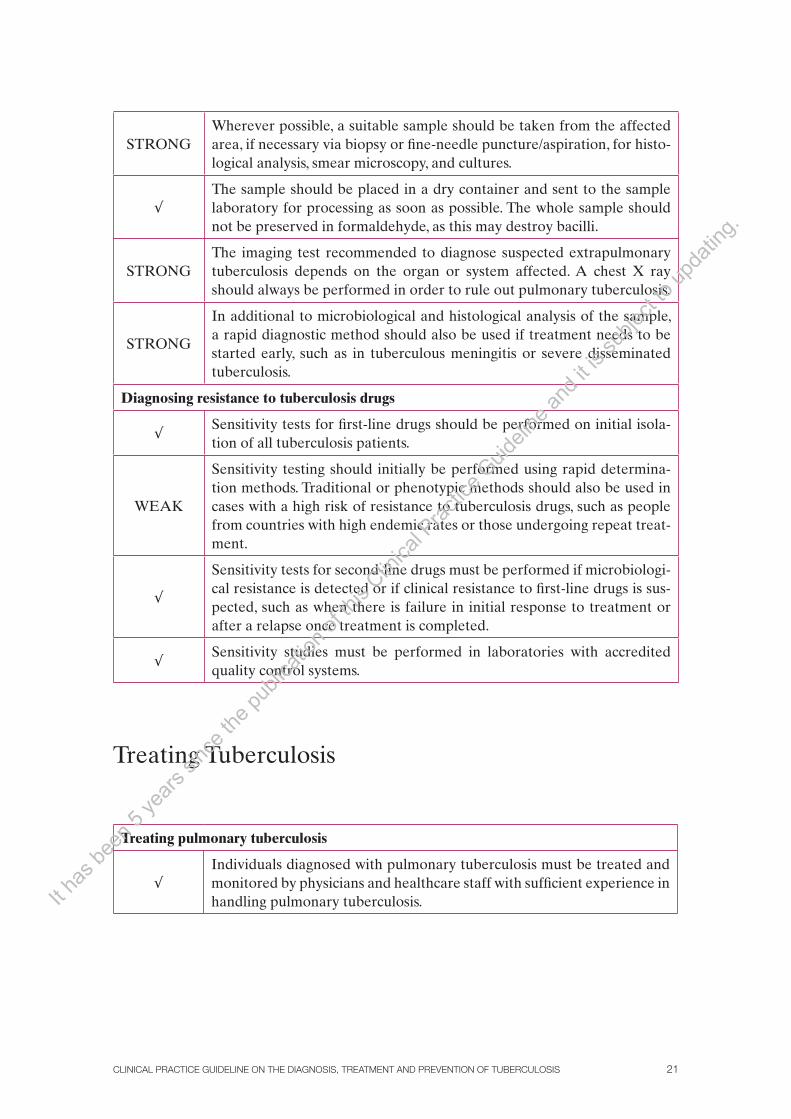
STRONGWherever possible, a suitable sample should be taken from the affected area, if necessary via biopsy or fine-needle puncture/aspiration, for histo-logical analysis, smear microscopy, and cultures.
√The sample should be placed in a dry container and sent to the sample laboratory for processing as soon as possible. The whole sample should not be preserved in formaldehyde, as this may destroy bacilli.
STRONGThe imaging test recommended to diagnose suspected extrapulmonary tuberculosis depends on the organ or system affected. A chest X ray should always be performed in order to rule out pulmonary tuberculosis.
STRONG
In additional to microbiological and histological analysis of the sample, a rapid diagnostic method should also be used if treatment needs to be started early, such as in tuberculous meningitis or severe disseminated tuberculosis.
Diagnosing resistance to tuberculosis drugs
√Sensitivity tests for first-line drugs should be performed on initial isola-tion of all tuberculosis patients.
WEAK
Sensitivity testing should initially be performed using rapid determina-tion methods. Traditional or phenotypic methods should also be used in cases with a high risk of resistance to tuberculosis drugs, such as people from countries with high endemic rates or those undergoing repeat treat-ment.
√
Sensitivity tests for second-line drugs must be performed if microbiologi-cal resistance is detected or if clinical resistance to first-line drugs is sus-pected, such as when there is failure in initial response to treatment or after a relapse once treatment is completed.
√Sensitivity studies must be performed in laboratories with accredited quality control systems.
Treating Tuberculosis
Treating pulmonary tuberculosis
√Individuals diagnosed with pulmonary tuberculosis must be treated and monitored by physicians and healthcare staff with sufficient experience in handling pulmonary tuberculosis.It h
as be
en 5
years
sinc
e the
publi
catio
n of th
is Clin
ical P
ractic
e Guid
eline
and i
t is su
bject
to up
datin
g.

CLINICAL PRACTICE GUIDELINE IN THE SNHS22
STRONG
Most patients with pulmonary tuberculosis not previously treated should be treated using a short, 6 month regimen consisting of an initial 2 month phase of isoniazid, rifampicin, pyrazinamide and ethambutol and a 4 month maintenance phase of isoniazid and rifampicin.
WEAKOther treatment regimens for pulmonary tuberculosis are also recom-mended.
WEAKTreatment should be extended to 9 months in patients with cavitary pul-monary tuberculosis who still have positive cultures at the end of the initial (2 month) phase of treatment.
√Treatment compliance must be assessed if there is a positive culture at the end of the initial (2 month) phase of treatment.
STRONGFor initial treatment of tuberculosis in children, the same treatment regi-mens (at suitable doses) are recommended as for the adult population unless there are specific contraindications.
WEAKIn children and adults, intermittent treatment (three times a week) may be considered during the maintenance phase if it is directly observed and if a culture taken after 2 months of treatment is negative.
STRONG Twice-weekly intermittent treatment regimens are not recommended.
WEAKTo reduce the development of drug resistance and the number of drugs taken daily, adults should be treated with fixed-dose combinations of the tuberculosis drugs currently on the market.
WEAKRifabutin is a reasonable option if rifampicin is not tolerated or if there is a high risk of interaction with other drugs, particularly antiretrovirals.
WEAKAdjuvant corticosteroid treatment may be considered in certain cases of extensive forms of tuberculosis.
STRONGOther adjuvant treatments, such as diets rich in vitamins or oligoelements, immunotherapy or laser radiation, are not recommended for tuberculosis.
√Liver toxicity must be closely monitored in patients receiving tuberculo-sis treatment, particularly those with known liver disease.
Treating extrapulmonary tuberculosis
STRONGTreatment regimens (drugs and duration) for patients with pleural, lym-phatic, osteal, spinal or pericardial tuberculosis should be no different from treatment regimens for pulmonary tuberculosis.
WEAKCorticosteroid treatment is not recommended for all patients with pleu-ral tuberculosis.
WEAKFor pleural tuberculosis, corticosteroid treatment should be considered in order to improve symptoms rapidly.
STRONGSurgery should not be performed routinely in all patients with osteal tu-berculosis.
It has
been
5 ye
ars si
nce t
he pu
blica
tion o
f this
Clinica
l Prac
tice G
uideli
ne an
d it is
subje
ct to
upda
ting.

CLINICAL PRACTICE GUIDELINE ON THE DIAGNOSIS, TREATMENT AND PREVENTION OF TUBERCULOSIS 23
WEAKIn patients with spinal tuberculosis, corrective or orthopaedic surgery should be considered in cases with a high risk of damage to the spinal cord or spinal instability, in order to achieve mechanical stability.
WEAKA longer treatment regimen, lasting up to 9 months, may be considered for patients with osteoarticular tuberculosis, depending on their clinical and radiological development.
STRONGPatients with tuberculous meningitis must follow a longer treatment regi-men, lasting up to 12 months.
STRONGIn patients with stage II or III tuberculous meningitis, adjuvant corticos-teroid treatment is recommended during the initial phase (prednisolone 60 mg/day for 4 weeks).
WEAKIn children with tuberculous meningitis, adjuvant corticosteroid treat-ment is recommended during the initial phase (prednisolone 60 mg/day for 4 weeks).
WEAKIn children with tuberculous meningitis and hydrocephalus, ventricular drainage should be considered.
STRONGIn patients with pericardialtuberculosis, adjuvant corticosteroid treat-ment is recommended during the initial phase (prednisolone 60 mg/day for 4 weeks).
STRONGRoutine pericardiocentesis is not recommended for patients with tuber-culous pericarditis and any degree of pericardial effusion.
WEAKIn patients with tuberculous pericarditis, evacuating pericardiocentesis can be considered for cases where there is risk of pericardial tamponade or functional compromise.
Monitoring treatment
√Responsibility for successful treatment must be shared between the healthcare professionals in charge of patients and the healthcare authori-ties that provide the necessary resources.
√The potential level of treatment compliance must be assessed and moni-tored in all tuberculosis patients who begin tuberculosis treatment.
√It is important to motivate patients and to highlight the importance of fully complying with treatment, both for latent infection and active tu-berculosis.
√The strategies available for improving compliance must be tailored to each case and agreed upon with the patient.
STRONGThe generalised use of directly observed treatment for all patients receiv-ing tuberculosis treatment is not recommended.It h
as be
en 5
years
sinc
e the
publi
catio
n of th
is Clin
ical P
ractic
e Guid
eline
and i
t is su
bject
to up
datin
g.

CLINICAL PRACTICE GUIDELINE IN THE SNHS24
STRONG
Directly observed treatment regimens are recommended under certain circumstances, such as for patients living in poverty, those with no fixed address, cases with significant grounds to suspect poor compliance, pa-tients with a history of poor compliance, and children.
STRONGVarious strategies are recommended for improving compliance. These in-clude reminder letters, phone calls, education, and home visits.
Treatment in challenging situations
√Tuberculosis treatment for HIV-positive individuals must be provided by a physician who specialises in both infections.
WEAK
In HIV-positive adults and children with pulmonary tuberculosis that has not been treated previously, a 6 month isoniazid and rifampicin treatment regimen is recommended, supplemented with pyrazinamide and etham-butol for the first 2 months.
STRONGIn treatment regimens for HIV-positive patients with tuberculosis, ri-fampicin should be maintained whenever possible.
√The beginning of antiretroviral treatment for a patient receiving tuber-culosis treatment must be considered on an individual basis according to the patient’s immune status, in order to prevent treatment interactions.
√In HIV-positive patients with CD4 counts above 350, tuberculosis treat-ment should be given first. Antiretroviral treatment must be introduced once tuberculosis treatment is complete.
√In HIV-positive patients with CD4 counts between 200 and 350, antiret-roviral treatment should begin after the first 2 months of tuberculosis treatment.
√In HIV-positive patients with CD4 counts below 200, antiretroviral treat-ment should begin after between 2 and 8 weeks of tuberculosis treatment if the latter is well tolerated.
√
In children, it is reasonable to begin antiretroviral treatment 2 to 8 weeks after the beginning of tuberculosis treatment. The patient’s immune sta-tus and the appropriateness of combining treatments must be considered on a case-by-case basis. In severe cases, both treatments may begin simul-taneously.
WEAKReplacing rifampicin with rifabutin is recommended in an 18 month tu-berculosis treatment regimen if there is a high risk of interactions with antiretroviral treatment.
√Patients with chronic liver disease must be treated by a specialist, particu-larly in advanced clinical stages.
It has
been
5 ye
ars si
nce t
he pu
blica
tion o
f this
Clinica
l Prac
tice G
uideli
ne an
d it is
subje
ct to
upda
ting.

CLINICAL PRACTICE GUIDELINE ON THE DIAGNOSIS, TREATMENT AND PREVENTION OF TUBERCULOSIS 25
√
Liver function must be tested before the beginning of tuberculosis treat-ment and at regular intervals, particularly in patients with chronic alco-hol consumption, those being treated with other hepatotoxic drugs, those who are HIV-positive, or those who have chronic hepatitis virus infection or known liver disease.
√Streptomycin and ethambutol doses must be adjusted for patients with kidney failure.
√In most cases, pregnant women should be given standard tuberculosis treatment.
General principles for treating drug-resistant cases
√Patients with multi-drug resistant tuberculosis must be treated by a spe-cialist.
√Treatment regimens for multi-drug resistant tuberculosis must consist of at least four drugs to which the patient has shown no resistance.
STRONGA directly observed treatment regimen lasting at least 18 months is rec-ommended for patients with multi-drug resistant tuberculosis.
√A patient with multi-drug resistant tuberculosis can be considered cured if he/she has completed the first year of treatment and gives at least five negative cultures (taken monthly).
STRONG A sensitivity test should be performed in cases of repeat treatment.
√In patients whose treatment is interrupted for less than 1 month and who have been fully monitored, treatment should be resumed until the treat-ment regimen has been completed.
√In patients whose treatment is interrupted for more than 1 month or who give a positive smear microscopy during the interruption, the treatment regimen should be restarted, from the beginning.
Monitoring patients
√If there are sufficient resources available, treatment, monitoring and iso-lation of most patients with pulmonary tuberculosis can be performed at the primary care level.
√In some clinical situations, specific monitoring by specialists, and even hospitalisation, is recommended.
√It is important to identify the main specialised institutions in each area or region to which patients must be referred if indicated.
It has
been
5 ye
ars si
nce t
he pu
blica
tion o
f this
Clinica
l Prac
tice G
uideli
ne an
d it is
subje
ct to
upda
ting.

CLINICAL PRACTICE GUIDELINE IN THE SNHS26
√
Monitoring of individuals who begin tuberculosis treatment must consist of clinical, analytical, and microbiological monitoring during the first 2 weeks. It then must be followed by monthly clinical monitoring, analyti-cal and bacteriological monitoring every 2 months, and radiological and bacteriological monitoring at the end of treatment.
√Clinical monitoring must be even closer if there are analytical changes or positive cultures after the second month, if complications are suspected, and in children.
√
If a patient presents liver enzyme values five times higher than normal, or signs and symptoms of cholestasis, all potentially hepatotoxic medica-tion must be suspended. The patient must then be monitored closely to see whether treatment can be resumed or whether a treatment regimen involving non-hepatotoxic drugs must be used instead.
WEAKIn most patients, clinical monitoring is not recommended after treatment has been correctly completed.
Preventing Tuberculosis
Isolation measures
STRONGPatients with pulmonary or laryngeal tuberculosis must remain in respira-tory isolation until they are no longer suspected of being infectious.
WEAKPatients may be placed in respiratory isolation at home if this is feasible, unless the disease is severe or there are complications.
√All medical establishments must have a set of measures (organisational and structural) designed to reduce hospital transmission of tuberculosis.
√In addition to these measures, tuberculosis patients suspected of being infectious must wear surgical masks when they are in communal areas of medical establishments.
√Healthcare staff working in high-risk areas must undergo tuberculin tests when they are hired, and at regular intervals if the initial test is negative.
Conventional contact studies
STRONG
Contact studies should begin promptly when pulmonary, pleural or laryn-geal tuberculosis is diagnosed. This is particularly important in the most infectious forms, such as cavitary pulmonary forms and/or cases with posi-tive sputum smear microscopy.
It has
been
5 ye
ars si
nce t
he pu
blica
tion o
f this
Clinica
l Prac
tice G
uideli
ne an
d it is
subje
ct to
upda
ting.
CLINICAL PRACTICE GUIDELINE ON THE DIAGNOSIS, TREATMENT AND PREVENTION OF TUBERCULOSIS 27
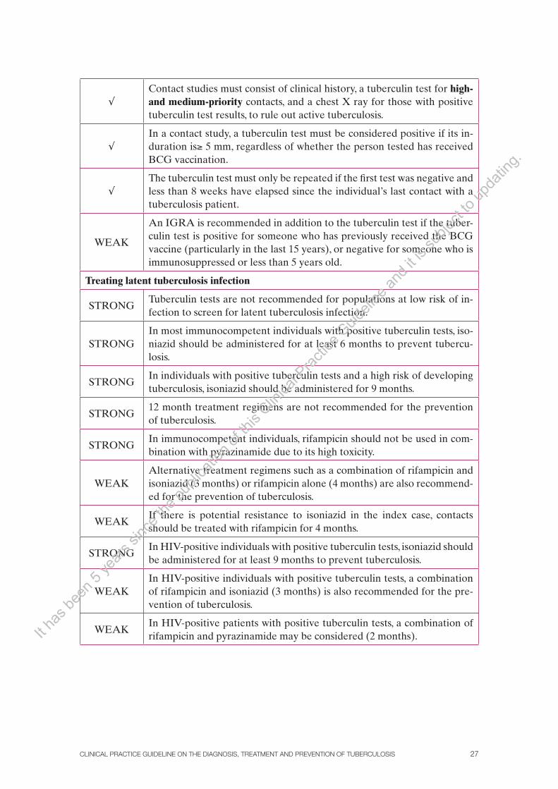
√Contact studies must consist of clinical history, a tuberculin test for high- and medium-priority contacts, and a chest X ray for those with positive tuberculin test results, to rule out active tuberculosis.
√In a contact study, a tuberculin test must be considered positive if its in-duration is≥ 5 mm, regardless of whether the person tested has received BCG vaccination.
√The tuberculin test must only be repeated if the first test was negative and less than 8 weeks have elapsed since the individual’s last contact with a tuberculosis patient.
WEAK
An IGRA is recommended in addition to the tuberculin test if the tuber-culin test is positive for someone who has previously received the BCG vaccine (particularly in the last 15 years), or negative for someone who is immunosuppressed or less than 5 years old.
Treating latent tuberculosis infection
STRONGTuberculin tests are not recommended for populations at low risk of in-fection to screen for latent tuberculosis infection.
STRONGIn most immunocompetent individuals with positive tuberculin tests, iso-niazid should be administered for at least 6 months to prevent tubercu-losis.
STRONGIn individuals with positive tuberculin tests and a high risk of developing tuberculosis, isoniazid should be administered for 9 months.
STRONG12 month treatment regimens are not recommended for the prevention of tuberculosis.
STRONGIn immunocompetent individuals, rifampicin should not be used in com-bination with pyrazinamide due to its high toxicity.
WEAKAlternative treatment regimens such as a combination of rifampicin and isoniazid (3 months) or rifampicin alone (4 months) are also recommend-ed for the prevention of tuberculosis.
WEAKIf there is potential resistance to isoniazid in the index case, contacts should be treated with rifampicin for 4 months.
STRONGIn HIV-positive individuals with positive tuberculin tests, isoniazid should be administered for at least 9 months to prevent tuberculosis.
WEAKIn HIV-positive individuals with positive tuberculin tests, a combination of rifampicin and isoniazid (3 months) is also recommended for the pre-vention of tuberculosis.
WEAKIn HIV-positive patients with positive tuberculin tests, a combination of rifampicin and pyrazinamide may be considered (2 months).It h
as be
en 5
years
sinc
e the
publi
catio
n of th
is Clin
ical P
ractic
e Guid
eline
and i
t is su
bject
to up
datin
g.

CLINICAL PRACTICE GUIDELINE IN THE SNHS28
WEAKTo prevent tuberculosis in children and adolescents with positive tubercu-lin tests, treatment with any treatment regimen routinely used in adults, at appropriate doses, is recommended.
√
In children born to mothers with pulmonary tuberculosis and positive smear microscopies, 6 month prophylactic treatment with isoniazid is sug-gested, in addition to a mask until the mother is no longer infectious, or separation of the neonate from the mother if drug resistance is suspected.
√Regardless of gestational age, isoniazid and vitamin B6 supplements are suggested for pregnant women with recent positive tuberculin tests (less than 2 years ago) after contact with a smear-positive patient.
WEAKTreatment for latent infection should not be begun in contacts of patients with multi-drug resistant tuberculosis.
√A proactive attitude must be taken to assess and promote compliance throughout treatment. If intermittent treatment regimens are used, direct observation of medication intake should be used.
√Analytical monitoring of liver function should be performed every 2 months in individuals receiving treatment for latent tuberculosis infec-tion, particularly those receiving isoniazid.
WEAK
Primary prophylaxis with isoniazid (300 mg/day or 5 mg/kg/day) is rec-ommended for 8 12 weeks in children less than 5 years old, HIV-positive individuals, and those with immune system alterations, if they have come into contact with infectious patients.
Vaccination
STRONG Routine BCG vaccination is not recommended in Spain.
√BCG vaccination is suggested for healthcare staff and those contacts of multi-drug resistant tuberculosis and in whom other control strategies cannot be implemented or have failed.
√The BCG vaccine must not be administered to those who have already been infected.
√A diagnosis of tuberculosis must not be ruled out in a vaccinated indi-vidual in whom clinical findings suggest tuberculosis.
It has
been
5 ye
ars si
nce t
he pu
blica
tion o
f this
Clinica
l Prac
tice G
uideli
ne an
d it is
subje
ct to
upda
ting.

CLINICAL PRACTICE GUIDELINE ON THE DIAGNOSIS, TREATMENT AND PREVENTION OF TUBERCULOSIS 29
It has
been
5 ye
ars si
nce t
he pu
blica
tion o
f this
Clinica
l Prac
tice G
uideli
ne an
d it is
subje
ct to
upda
ting.

CLINICAL PRACTICE GUIDELINE IN THE SNHS30
It has
been
5 ye
ars si
nce t
he pu
blica
tion o
f this
Clinica
l Prac
tice G
uideli
ne an
d it is
subje
ct to
upda
ting.

CLINICAL PRACTICE GUIDELINE ON THE DIAGNOSIS, TREATMENT AND PREVENTION OF TUBERCULOSIS 31
1. Introduction
Tuberculosis is a contagious, infectious disease caused by species of the complex Myco-bacterium tuberculosis. It progresses chronically and is characterised by the formation of granulomas. It is most commonly located in the lung, although it can affect any organ. The International Classification of Diseases assigns tuberculosis codes 010 to 018, depending on its location. All cases that fulfil any of the three definitions for suspected, probable or confirmed tuberculosis must be reported (Appendix 4)1.
1.1 Scale of the Problem Worldwide
Tuberculosis remains one of the main causes of illness and death in many countries, and a major public health problem worldwide. Despite the progress made in the fight against tuberculosis in recent decades, there are still marked regional and national differences. Ac-cording to World Health Organisation (WHO) estimates, there were around 9.27 million new cases in 2007, most of which were reported in Asia (55%) and Africa (31%); in the eastern Mediterranean, Europe and the Americas the corresponding figures were 6%, 5%, and 3% respectively. The estimated number of deaths was 1.7 million, 456,000 in people who were HIV-positive. In 2007 alone there were approximately half a million multi-drug resistant cases, most of them in a group of 27 countries of which 15 are in the WHO Euro-pean Region. The worldwide burden of tuberculosis is slowly falling, and at least three of the six WHO regions are well on the way to meeting the worldwide targets for reducing the numbers of cases and deaths established for 20152. However, in low-income countries, tuberculosis is the main cause of infection-related death in those aged 19 to 49, and ac-counts for approximately 25% of all potentially preventable deaths. These countries ac-count for 95% of tuberculosis cases and 98% of deaths due to tuberculosis3. Almost all the world’s tuberculosis patients are concentrated in 22 countries. Moreover, 75% of cases affect these countries’ working population, in whom tuberculosis is a major medical and economic problem4.
In the 53 countries within the WHO European Region, tuberculosis remains a major public health problem, varying greatly between countries and with an increasing incidence from West to East. The situation is particularly worrying in the Eastern Europe and some countries of the former Soviet Union due to the high rates of resistant and multi-drug re-sistant tuberculosis, the increased incidence of HIV infection, socioeconomic deterioration and insufficiently developed health services2.
Childhood tuberculosis accounts for 11% of cases; in other words, there are around 1 million new cases of tuberculosis in children every year. Approximately one third will die.
Again, the proportion of children with tuberculosis is higher in low-incomecountries than in high-incomecountries.5- 6.
It is estimated that one third of the world’s population is infected with M. tuberculosis. This is a reservoir thatwill continue to generate new cases for many years. In countries low-
It has
been
5 ye
ars si
nce t
he pu
blica
tion o
f this
Clinica
l Prac
tice G
uideli
ne an
d it is
subje
ct to
upda
ting.
CLINICAL PRACTICE GUIDELINE IN THE SNHS32
income countries, almost the whole population is infected and 80% of infected individuals are under 50 years of age. In contrast, in countries with high incomes less than 20% of the population is infected and most of these are aged over503.
1.2 The Scale of the Problem in Spain
The figures on tuberculosis in Spain are unreliable. This is may be due to the fact that until 1995, only the numbers of cases of respiratory tuberculosis had to be reported, and that in 1996 and 1997, only respiratory and meningeal tuberculosis were reported. The data avail-able indicate that Spain has a higher endemic rate of tuberculosis than other countries of similar socioeconomic status, with the exception of Portugal7-8. The situation has been gradually improving since the 1980s, and today mortality is concentrated in the very young and very old, the immunosuppressed, those living in extreme poverty and those with ex-trapulmonary forms of tuberculosis that are diagnosed very late.
According to WHO figures, Spain’s rate of tuberculosis incidence (in any location) in 2007 was 30 cases per 100,000 inhabitants. This figure is higher than that reported by Spain’s National Epidemiology Surveillance Network (18.4 cases per 100,000 inhabitants in 2006) and that supplied by the European Tuberculosis Surveillance Network (www.eurotb.org), which in its 2006 annual report gives a report rate of 18.3 cases per 100,000 inhabitants2,9-10.
A recent study on data published by the WHO (the Global Health Atlas database) shows the evolution of the incidence of this diseasebased on the number of cases reported in 52 European countries from 1980 to 2006. According to the estimates analysed, the situation in Spain worsened in the period from 1992 to2006. This deterioration is attributed to the high prevalence of HIV-positive people, parenteral drug addicts, and the effects of migration11.
Mass migration from countries with high endemic rates of tuberculosis and the living conditions to which these immigrants are exposed have led to an increase in tuberculosis in many EU countries. This increase has been limited to this section of the population, and there is no evidence that it may be affecting the epidemiology of tuberculosis in the auto-chthonous population. This new situation makes it necessary to reinforce efforts using con-trol programmes and activities that ensure early diagnosis, the availability of appropriate treatment, follow-up of treatment and treatment completion, as well as actions that target vulnerable groups at high risk of infection or those with poor living conditions1.
1.3 The Aetiopathogenesis of Tuberculosis Infection
The pathogen that causes tuberculosis belongs to the Mycobacterium genre, a small, im-mobile, non-sporulated, Gram-positive bacillus. The Mycobacterium genre includes more than 100 species. Those that cause tuberculosis are M. tuberculosis, M. bovis and M. afri-canum; M. microti, which causes tuberculosis in rats and was once used as a tuberculosis vaccine, is also included.
It has
been
5 ye
ars si
nce t
he pu
blica
tion o
f this
Clinica
l Prac
tice G
uideli
ne an
d it is
subje
ct to
upda
ting.

CLINICAL PRACTICE GUIDELINE ON THE DIAGNOSIS, TREATMENT AND PREVENTION OF TUBERCULOSIS 33
When infectious particles are inhaled, only the smallest evade the defences of surfaces of the airways and reach the alveoli, in the lungs. Within the alveoli, macrophages success-fully engulf most infectious particles via phagocytosis. The bacilli multiply within the mac-rophages, and once these have been destroyed and the bacilli are in the extracellular space, they travel in the lymph to the mediastinal lymph nodes, and in the blood to many body systems. The bacilli have a particular affinity for well-oxygenated organs with abundant reticuloendothelial systems. Acquired or specific immunity halts the bacilli multiplication, but this does not become fully established until 6 14 weeks after infection12-13.
An individual presents latent tuberculosis infection when tuberculosis infection does not progress to disease; the person is healthy (has no signs or symptoms of disease), but his or her body contains live tuberculosis bacilli. Some individuals’ specific immunity is insufficient to prevent active tuberculosis, and between 10% and 15% of these will develop it at some point in their lives. Between 50% and 80% of cases of active tuberculosis ap-pear within 2 years of infection. In children the disease is called primary tuberculosis if it develops within 5 years of primary infection. When the disease develops a long time after primary infection, it is called post-primary tuberculosis, secondary tuberculosis or adult tuberculosis. The deterioration of the immune system allows bacilli from primary infection to develop (tuberculosis due to endogenous reactivation). An individual may also experi-ence manifold exposure, repeated exposure or exposure to particularly virulent strains (tuberculosis due to exogenous reinfection)3.
1.4 Transmission
The main reservoir of the bacillus is a human who is ill or infected. Almost the sole source of contagion is an individual with respiratory (pulmonary, bronchial or laryngeal) tuber-culosis. Transmission usually occurs when a person with smear-positive tuberculosis expels particles of respiratory secretions containing bacilli when he or she coughs, sneezes, laughs, sings or speaks. The patient’s degree of infectiouness depends on how well connected his/her lesions are to the airways, and is higher in patients whose lesions contain more micro-organisms and who expel large quantities of them in their respiratory secretions. There are factors that increase the risk of infection, including living with a infectious individual and the age of the people exposed3.
1.5 Clinical Manifestation
The clinical presentation of pulmonary tuberculosis is non-specific: its signs and symptoms de-pend on its location and appear late, sometimes after the patient has become infectious. Pulmo-nary tuberculosis should be suspected when a patient presents febrile syndrome of unknown origin or a productive cough lasting longer than 3 weeks, particularly if it is haemoptoic.
Chronic pulmonary sequelae after acute infection has been cured are responsible for most of the deterioration in patients’ quality of life15.
It has
been
5 ye
ars si
nce t
he pu
blica
tion o
f this
Clinica
l Prac
tice G
uideli
ne an
d it is
subje
ct to
upda
ting.
CLINICAL PRACTICE GUIDELINE IN THE SNHS34
1.6 Principles of Diagnosis
Once clinical diagnosis of suspected tuberculosis has been established, medical care ad-dresses radiological, immunological and microbiological diagnosis. Microbiological diag-nosis provides confirmation via isolation and identification of the strain and determina-tion of its sensitivity. Histological analysis or a high degree of clinical and epidemiological suspicion enables diagnosis, sometimes without bacteriological confirmation. Imaging tests are fairly sensitive but not very specific, and are useful for extrapulmonary forms in par-ticular. In addition to the tuberculin test, there are now interferon-gamma release assays to diagnose tuberculosis infection14.
1.7 Principles of Treatment
1.7.1 Treating the Disease
Tuberculosis can be cured if it is treated for long enough and if enough drugs are taken regularly and at the correct doses. In general, tuberculosis treatment is based on the fol-lowing principles:
• Theuseofdrugstowhichthebacillusissensitive,forlongenoughtoeliminatethewhole population of bacilli16.
• The treatment regimen for cases not previously treated lasts between 6 and 9months (depending on the patient’s characteristics, the disease’s characteristics and loca-tion, and disease progression during treatment). It consists of a 2 month “intensive phase” involving four drugs (isoniazid [H], rifampicin [R], pyrazinamide [Z] and ethambutol [E]), followed by a 4 month “maintenance phase” involving two drugs (isoniazid [H] and ri-fampicin [R]). Treatment for meningitis lasts 12 months1,16,17.
• ObtaininganantibiograminallcasesinwhichM. tuberculosis has been isolated1.
• Tailor-madetreatmentforpatientswithresistancetotuberculosisdrugs16,18.
Directly observed treatment is used in some tuberculosis patients to provide satisfac-tory treatment, ensure treatment compliance, and cure the disease. This involves verifying that the patient takes his/her medication19.
1.7.2 Treating the Infection
It is estimated that he bacillus population in the lesions of people carrying tuberculosis infection is small. The use of one drug, usually isoniazid, is therefore sufficient to cure the infection and reduce the risk of developing the disease20.It h
as be
en 5
years
sinc
e the
publi
catio
n of th
is Clin
ical P
ractic
e Guid
eline
and i
t is su
bject
to up
datin
g.
CLINICAL PRACTICE GUIDELINE ON THE DIAGNOSIS, TREATMENT AND PREVENTION OF TUBERCULOSIS 35
1.8 Principles of Prevention
The main aim of preventing and controlling tuberculosis is to eliminate sources of infec-tion. Early diagnosis and isolation of cases of patients with respiratory tuberculosis are essential. Active searches must be performed in at-risk groups with higher average inci-dences than the community as a whole. Screening involves a tuberculin test, chest X ray, smear microscopy and culture respiratory secretions1,7,20. An individual who has received suitable treatment for 3 weeks is considered to be no longer infectious. A patient must remain in isolation until there is no risk of contagion.
After a case of pulmonary or respiratory tuberculosis has been diagnosed, a contact study must be performed to find other people with tuberculosis, and to identify and treat those infected to prevent progression to active tuberculosis1. The infectious period begins roughly 3 months before diagnosis, and transmission requires close contact, although oc-casional contact may also be sufficient to transmit the infection21-22. Isoniazid is also used to prevent infection in individuals who present negative tuberculin tests even after contact with a infectious (smear-positive) tuberculosis patient23.
It has
been
5 ye
ars si
nce t
he pu
blica
tion o
f this
Clinica
l Prac
tice G
uideli
ne an
d it is
subje
ct to
upda
ting.

CLINICAL PRACTICE GUIDELINE IN THE SNHS36
It has
been
5 ye
ars si
nce t
he pu
blica
tion o
f this
Clinica
l Prac
tice G
uideli
ne an
d it is
subje
ct to
upda
ting.

CLINICAL PRACTICE GUIDELINE ON THE DIAGNOSIS, TREATMENT AND PREVENTION OF TUBERCULOSIS 37
2. Scope and Aims
This CPG covers issues relating to the diagnosis, treatment and prevention of both tuber-culosis infection and pulmonary and extrapulmonary active tuberculosis. For definitions concerning active tuberculosis, this CPG has followed internationally-accepted criteria, and Spanish criteria where there are discrepancies. The target population includes both adults and children of any age with the most common clinical presentations. The cases not covered by this document are detailed below.
No recommendations have been made on the organisation of medical services, con-trol programmes or aspects of epidemiology surveillance for the disease. Also excluded are recommendations for certain situations or specific locations such as boat or plane travel, customs or prisons, as these can be applied only with the cooperation of public authorities.
In addition, complex clinical situations that require highly specialised care have not been covered in depth. These include the specific handling of concurrent HIV infection, those with other immune system changes, tuberculosis in neonates, rare forms of extrapulmonary tuber-culosis, recurrent tuberculosis, and extremely resistant tuberculosis. These issues, while cer-tainly important, are the subjects of other CPGs, both in Spain and abroad (Appendix 6).
The target audience of this CPG consists of any medical professionals active in pri-mary care, mainly physicians who specialise in family and community medicine, pulmon-ology, paediatrics, infectious diseases, internal medicine, preventive medicine and public health, microbiology; as well as nurses and laboratory staff. All these professionals deal with tuberculosis patients or community control of tuberculosis at some point. Finally, this CPG is also intended for patients and their relatives, whose involvement is very important in the treatment and control of this disease.
Tuberculosis-related terminology is complex, and often differs between documents and between organisations. For the terms used in this CPG, which are also those accepted in Spain, we recommend consulting the glossary (Appendix 4).
2.1. Aims
This CPG aims to establish a set of recommendations for the diagnosis, treatment and pre-vention of tuberculosis, based on the best scientific evidence available and the consensus of experts in the field. The ultimate aim of these recommendations is to reduce the burden of tuberculosis in Spain through standardised, high-quality medical practice, in line with Spanish healthcare strategies for the control of tuberculosis.
It has
been
5 ye
ars si
nce t
he pu
blica
tion o
f this
Clinica
l Prac
tice G
uideli
ne an
d it is
subje
ct to
upda
ting.

CLINICAL PRACTICE GUIDELINE IN THE SNHS38
It has
been
5 ye
ars si
nce t
he pu
blica
tion o
f this
Clinica
l Prac
tice G
uideli
ne an
d it is
subje
ct to
upda
ting.
CLINICAL PRACTICE GUIDELINE ON THE DIAGNOSIS, TREATMENT AND PREVENTION OF TUBERCULOSIS 39
3. Methods
The methods used are described in detail in the Methodology Manual for Developing Clin-ical Practice Guidelines of the Spanish National Healthcare System24.
The steps taken were as follows:
The group that would develop the guideline was established, consisting of primary care professionals and specialists (in preventive medicine and public health, infectious diseases, general and community medicine, pulmonology, paediatrics, microbiology and clinical parasitology, and clinical pharmacology). These professionals were contacted via the various scientific societies interested in the subject of the CPG. Various users of the healthcare system inspected materials for patients.
Clinical questions were formulated according to the PICO model: Patient, Interven-tion, Comparison, Outcome.
A search of the literature was carried out, prioritising the identification of system-atic reviews (SRs) and other documents that provided a critical synthesis of the scientific literature, such as health technology assessment reports. For this purpose, an initial phase comprised a search of other CPGs on the subject, in order to ascertain which SRs they had considered as a basis for their recommendations. The main CPGs used as secondary sources are listed in Appendix 6. Additional SRs were identified subsequently, following the date on which the selected CPGs were searched. The following electronic databases were consulted in this initial stage:
• TRIPDatabase
• NHSNationalLibraryofGuidelines
• AHRQNationalGuidelineClearinghouse
• CochraneDatabaseofSystematicReviews(theCochraneLibrary)
• DatabaseofAbstractsofReviewsofEffects(DARE)
• HealthTechnologyAssessment(HTA)Database
• NHSEconomicEvaluationDatabase(NHSEED)
• MEDLINE(accessedviaPubMed)
• EMBASE(accessedviaOvid)
To complete this stage, the publications of a number of technology assessment agen-cies were also consulted. These included the National Institute for Clinical Excellence (NICE); agencies that issue CPGs, such as the Scottish Intercollegiate Guidelines Network (SIGN); and international societies.
In the second stage, an extended search of individual studies was conducted, in order to update the relevant SRs and answer the questions of the CPG. The main aim was to identify randomised clinical trials (RCTs) and observational studies, respecting the origi-nal search strategy of the relevant SRs. When these were not available, a specific strategy
It has
been
5 ye
ars si
nce t
he pu
blica
tion o
f this
Clinica
l Prac
tice G
uideli
ne an
d it is
subje
ct to
upda
ting.

CLINICAL PRACTICE GUIDELINE IN THE SNHS40
was designed for each question, adding validated filters in each case to identify RCTs and observational studies. The following electronic databases were consulted in this phase: the Cochrane Central Register of Controlled Trials (CENTRAL) (the Cochrane Library), MEDLINE, EMBASE and the Cumulative Index to Nursing and Allied Health Literature (CINAHL) (accessed via Ovid).
No language restriction was established for the searches carried out, but most of the studies considered were written in Spanish, English, or French. Searches covered a period from any date (varying according to the database in question) to September 2007, although relevant studies were identified in the highest-profile biomedical journals during the entire duration of the CPG development process.
Meta-analysis of results: in some sections for which the only evidence available con-sists of several individual RCTs, meta-analysis was performed when possible and if the availability of a joint result was judged clinically relevant. In sections for which there is an SR and the search of the literature provided one or more subsequent individual RCTs, the main meta-analysis of the SR was updated, providing a new joint figure if possible. This was the method following for studies (SRs or RCTs) in the treatment and prevention sec-tions. The free-access program RevMan 5 (http://www.cc-ims.net/revman) was used when performing or updating meta-analysis.
Evaluation of evidence quality and grading of recommendations was conducted fol-lowing the parameters of the GRADE (Grading of Recommendations of Assessment Development and Evaluations) system, using GRADEpro (http://www.cc-ims.net/revman/gradepro/gradepro), the free-access program of the GRADE Working Group. Controver-sial recommendations or those with no evidence were resolved by consensus of the group that developed the CPG.
External review: External reviewers reviewed the second draft (May 2009). The vari-ous scientific societies involved, which are also represented by members of the group that compiled the CPG and external reviewers, were contacted.
– Materials detailing the methodological process used for the CPG (search strategies for each clinical question, critical reading files for the studies selected, tables summarising the evidence and formal evaluation tables) are available in Spanish at www.guiasalud.es.
It is intended that the CPG will be updated every 3 years. More frequent updating of the electronic version is not ruled out, should this prove necessary.
3.1 The GRADE System for Formulating Recommendations
When recommendations are formulated, the group developing the guideline must decide to what extent it can be assumed that implementing a recommendation will result in more benefit than harm. This is not a simple decision, and it is affected by many factors, making this stage one of the most complex in guideline development.
It has
been
5 ye
ars si
nce t
he pu
blica
tion o
f this
Clinica
l Prac
tice G
uideli
ne an
d it is
subje
ct to
upda
ting.
CLINICAL PRACTICE GUIDELINE ON THE DIAGNOSIS, TREATMENT AND PREVENTION OF TUBERCULOSIS 41
Proposals for systems to formulate recommendations began more than twenty years ago. Even at the outset these systems distinguished between the level of scientific evidence (how suitable different study designs were for answering the various types of questions) and the strength of recommendations. Since then, the various systems have gradually de-veloped and incorporated issues other than study design, which must be taken into account when formulating recommendations.
The methods of the GRADE working group have been followed when classifying evi-dence quality and grading the strength of recommendations. The GRADE working group aims to establish an explicit, transparent method to develop recommendations thatis easy for teams that develop CPGs to use, in order to overcome the disadvantages of other rec-ommendation development systems25.
The main stages of the GRADE system are described below:
A) Classification of outcome variables’ relative importance
In this stage, the GRADE system recommends that during the initial phase of formu-lating clinical questions, the group developing the CPG explicitly establishes the outcome variables that are relevant to the questions, and classifies their relative importance. Impor-tance should be classified using the following nine-point scale:
1-3: An unimportant outcome variable. It should not be included in the table evaluat-ing quality to the results table. These outcome variables will not play an important role in formulating recommendations.
4-6: The outcome variable is important, but not a key to decision-making.
7-9: A key outcome variable in decision-making.
The relative importance of outcome variables is established by consensus.
B) Evaluating the quality of scientific evidence
Quality is evaluated for each selected outcome variable. This means that for a single clinical question there will probably be outcome variables with various different levels of quality. Initially, scientific evidence must be evaluated on the basis of study design and studies’ suitability for answering each type of question in the guideline. RCTs are assessed as “high-quality”, and observational studies as “low-quality”. However, there are a number of issues that may reduce the quality of RCTs or increase that of observational studies. They are outlined below. Finally, the quality of scientific evidence will be assessed as high, moderate, low or very low (Appendix 1).
The following issues many reduce the quality of RCTs:
1. Shortcomings in design or conduct: possible examples are failure to hide the ran-domisation sequence, inadequate blinding, major losses, the absence of analysis by intention to treat and early study termination due to benefit.
2. Inconsistent results: widely differing estimates of the effect of treatment (het-erogeneity or variation in results) in available studies suggest real differences between these estimates. The differences may be due to differences in the popu-lation, the intervention, the outcome variables or the quality of the studies. Het-erogeneity that cannot be reasonably explained reduces quality.
It has
been
5 ye
ars si
nce t
he pu
blica
tion o
f this
Clinica
l Prac
tice G
uideli
ne an
d it is
subje
ct to
upda
ting.

CLINICAL PRACTICE GUIDELINE IN THE SNHS42
3. Lack of direct scientific evidence: Evidence is considered indirect if there are no direct comparisons of two treatments (comparison of each treatment with a pla-cebo, but not of the two treatments). Also, if there is extrapolation of the results of a study with a particular drug to all other drugs in the same class in the absence of a demonstrated class effect. When there are major differences between the population to whom the recommendations will be applied and the population recruited in the studies evaluated, indirectness can also be an issue. Last but not least, aspects of the potential applicability of recommendations in one context must be assessed against other contexts, as well as the external validity of the scientific evidence
4. Imprecision: when the studies available include relatively few events and few patients, and therefore have broad confidence intervals, the scientific evidence is considered to be of poorer quality.
5. Reporting bias: quality may be reduced if there is reasonable doubt as to whether the authors have included all studies (e.g. publication bias in the context of an SR) or all relevant outcome variables (reporting bias).
The following issues many increase the quality of observational studies:
1. Significant effect: when the effect observed shows a strong (RR > 2 or < 0.5) or very strong (RR > 5 or < 0.2), consistent association on the basis of studies with no confounding factors. In such cases, quality may be considered moderate, or even high.
2. A dose-response slope.
3. Situations in which all possible confounding factors may have reduced the as-sociation observed. If patients who receive the intervention being studied have a worse prognosis than the control group and nevertheless present better out-comes, the real effect observed is likely to be greater.
C) Grading the strength of recommendations
Grading the strength of recommendations is relatively simple, as only two categories are considered: strong recommendations and weak recommendations (Appendix 1).
With strong recommendations, the group compiling the CPG are sure that the ben-efits of the intervention exceed its harm or, in contrast, that the harm exceeds the benefits. In the former case, the recommendation is strongly for the intervention. In the latter, it is strongly against it.
Weak recommendations can also be for or against an intervention. A recommenda-tion is weakly in favour of an intervention if the group compiling the CPG concludes that the benefits of implementing the recommendation probably exceed the harm, although this is not absolutely certain. In contrast, a recommendation is weakly against an interven-tion if its harmful effects probably exceed its benefits.
A number of other factors must also be considered when grading recommendations:
1. Risk/benefit balance: in order to give a suitable assessment of the balance be-tween benefits and risks, the baseline risk of the population for whom the recom-
It has
been
5 ye
ars si
nce t
he pu
blica
tion o
f this
Clinica
l Prac
tice G
uideli
ne an
d it is
subje
ct to
upda
ting.
CLINICAL PRACTICE GUIDELINE ON THE DIAGNOSIS, TREATMENT AND PREVENTION OF TUBERCULOSIS 43
mendation is intended and the effect in both relative and absolute terms must be considered.
2. Quality of scientific evidence: before a recommendation is implemented, the lev-el of certainty of the estimate of the observed effect must be ascertained. If the quality of the scientific evidence is not high, confidence must decrease even if the scale is large, and therefore so must the strength with which a recommendation is made.
3. Values and preferences: uncertainty as to the values and preferences of the CPG’s target population must also be taken into account. The values and preferences of healthcare staff, the patient population and the general public must be reflected and should affect the grading of recommendations.
4. Cost: costs vary much more over time, between geographical areas, and according to various other factors than other outcome variables. This means that although a high cost reduces the probability of a recommendation being graded as strong, context will be essential to the final assessment.
It has
been
5 ye
ars si
nce t
he pu
blica
tion o
f this
Clinica
l Prac
tice G
uideli
ne an
d it is
subje
ct to
upda
ting.

CLINICAL PRACTICE GUIDELINE IN THE SNHS44
It has
been
5 ye
ars si
nce t
he pu
blica
tion o
f this
Clinica
l Prac
tice G
uideli
ne an
d it is
subje
ct to
upda
ting.

CLINICAL PRACTICE GUIDELINE ON THE DIAGNOSIS, TREATMENT AND PREVENTION OF TUBERCULOSIS 45
4. Diagnosis
4.1. Diagnosing the Infection
Mycobacterium tuberculosis infection is considered to be latent if there are no clinical, bac-teriological or radiological signs of active tuberculosis. In these individuals, infection has traditionally been detected by a positive tuberculin test. More recently, infection detection methods such as immunological in vitro blood tests known as IGRAs (Interferon-Gamma Release Assays) have appeared.
4.1.1 The Tuberculin Test
The tuberculin test, which is widely used, is performed using the Mantoux technique. Its advantages are its low cost and ease of use, although it also has some disadvantages. The tuberculin test proce-dure is detailed in point 6.2.4, and test interpretation in point 6.2.5 and Appendix 11.
This test can yield false positives due to immune system sensitisa-tion caused by previous BCG vaccination or exposure to non-tubercu-lous mycobacteria. One SR of prospective studies (26 studies involving more than 115,000 participants) showed that the probability of testing positive (induration >10 mm) to the tuberculin test is 2 3 times higher in people who have been vaccinated, regardless of the type of antigen used for the test. Subgroup analyses showed that in those vaccinated 15 or more years before the test, this vaccination had no effect on the percentage of positive results. Likewise a tuberculin test induration with a diameter greater than 15 mm was associated with tuberculosis infection rather than an effect of BCG vaccination26.
False negatives can also occur in individuals with immune sys-tem alterations, particularly individuals who are HIV-positive, those receiving immunosuppressant treatment, and in cases of severe viral infections or disseminated tuberculosis20. The reliability of the tu-berculin test in detecting tuberculosis infection in children less than 6 months old is lower, as the immune response mechanism has not yet fully developed27.
Quality: MODERATE
Questions to Answer
• Howusefulisthetuberculintestindiagnosinglatenttuberculosisinfection?
• Whatisthediagnosticperformanceofinterferon-gammareleaseassaysindiagnos-ing latent tuberculosis infection?
It has
been
5 ye
ars si
nce t
he pu
blica
tion o
f this
Clinica
l Prac
tice G
uideli
ne an
d it is
subje
ct to
upda
ting.
CLINICAL PRACTICE GUIDELINE IN THE SNHS46
4.1.2 The Interferon-Gamma Release Assay (IGRA)
New techniques intended to improve diagnosis of latent tuberculo-sis infection have appeared in recent years. These techniques detect interferon-gamma produced by T cells previously stimulated (sensi-tised) by specific M. tuberculosis antigens such as ESAT 6 (early se-cretory antigen target 6) and the CFP 10 (culture filtrate protein 10) and TB7.7. There are two tests on the market: QFT (QuantiFERON TB Gold or QuantiFERON TB Gold In-Tube, made by Cellestis) and T SPOT.TB (Oxford Immunotec, Ltd). QFT Gold In-Tube also contains a third antigen, the TB7.7,in addition to the two mentioned. QFT stimulates all the incubated blood with the antigens and uses the ELISA method to determine the quantity of interferon (in pg/ml or IU/ml). For T SPOT.TB, mononuclear cells must first be separated, stimulated with antigens, and read using the ELISPOT technique, in which each point represents a T cell that secretes interferon-gamma.
Features that may give IGRA tests an advantage over the tuber-culin test include greater specificity and fewer cross-reactions with the BCG vaccine. Also, interpretation is less subjective and results are ob-tained quickly and confidentially. However, IGRA tests are expensive and require samples to be processed in laboratories.
One SR showed the results of 38 diagnosis studies for the IGRA tests currently on the market28. The SR is an update of an earlier review that examined both IGRA tests on the market and those not commercially available29. This most recent SR did not include studies with fewer than ten participants or those including only participants with immune alterations. Sensitivity was assessed only in a population with confirmed tuberculosis. Specificity was assessed by sustaining healthy populations with no previous known contact, or populations with very low incidences of tuberculosis.
Quality:LOW
The overall estimates of sensitivity for QFT Gold and QFT Gold In-Tube were 0.78 (CI 95%, 0.73 to 0.82) and 0.70 (CI 95%, 0.63 to0.78) respectively (36 studies, 2,095 participants), and 0.90 (CI 95%, 0.86 to 0.93) for T SPOT.TB.
It has
been
5 ye
ars si
nce t
he pu
blica
tion o
f this
Clinica
l Prac
tice G
uideli
ne an
d it is
subje
ct to
upda
ting.

CLINICAL PRACTICE GUIDELINE ON THE DIAGNOSIS, TREATMENT AND PREVENTION OF TUBERCULOSIS 47
The combined specificity of the QFT tests was 0.99 (CI 95%, 0.98 to 1.0) in individuals who had not received the BCG vaccine and 0.96 (CI 95%, 0.94 to 0.98) in those who had been vaccinated (for a total of 16 studies and 1,624 participants). For the T SPOT.TB technique, specificity was 0.93 (CI 95%, 0.86 to 1.0) in the population that had been vaccinated. Only one study included a non-vaccinated popula-tion, which showed a specificity of 100%. In studies comparing the tuberculin test with these new diagnosis methods, the tuberculin test showed an overall sensitivity of 0.77 (CI 95%, 0.71 to 0.82) in a total of 20 studies with 1,193 participants. Specificity varied markedly be-tween the non-vaccinated population (0.97 [CI 95%, 0.95 to 0.99]) and the vaccinated population (0.59 [CI 95%, 0.46 to 0.73])28.
The SR above analysed studies that included an immunosup-pressed or HIV-positive population. Although the results are not compared, they showed that the performance of IGRA tests is lower in patients with altered immunity. In these patients, IGRA tests were more sensitive than the tuberculin skin test29. It should be noted that in some studies that included an HIV-positive population, the degree of immunosuppression was often low30.
Although the evidence for the diagnosis of latent infection is in-direct, IGRA tests have a good sensitivity and, among the vaccinated population, a high specificity. The evidence is based mainly on small, cross-sectional studies with widely varying inclusion criteria. As yet there is little evidence regarding those at high risk, such as the eld-erly31 or immunosuppressed32..
One recent SR included 59 studies, of which 7 were conducted in children. The overall estimates of sensitivity for QTF and T SPOT.TB were 66% and 62% respectively, compared to 55% for the tuber-culin test. In the studies that provided this information, the agree-ment between the two tests was greater than 50% for negative results. The population analysed was heterogeneous and included paediatric, school, hospitalised and healthy contacts, both with and without pre-vious BCG vaccination, and populations from both high- and low-income countries. The number of people included in each study also varied widely. Added to these shortcomings was the difficulty in tak-ing blood and indeterminate results in this age group29.
Quality:LOW
It has
been
5 ye
ars si
nce t
he pu
blica
tion o
f this
Clinica
l Prac
tice G
uideli
ne an
d it is
subje
ct to
upda
ting.

CPGs have proposed two-stage diagnosis strategies in which both types of test are used sequentially for certain cases17,20.
In the light of the results, a single negative result to either one of these tests should not rule out latent infection in a population prone to yielding negative results, such as children under 5 or those with major immunosuppression. In these cases, the consequences of not begin a treatment, based on a false negative result, could be particu-larly serious33.
Summary of Evidence
–There is no reference test with ideal performance for the diagnosis of latent tuberculosis infection. The tuberculin test is the most widely used. It is cheap and easy to interpret.
Quality: MODERATE
The tuberculin test can yield false positives after BCG vaccination or exposure to non-tuberculous mycobacteria. False negatives can oc-cur in patients with various types of immune system alteration, par-ticularly those who are HIV-positive or receiving immunosuppressive treatment.
Quality: LOW
IGRA tests have high specificity, particularly among those who have received the BCG vaccine.
Quality: LOW
The diagnostic performance of IGRA tests is low in the elderly and those with immune system alterations, although it is more sensitive than the tuberculin test.
Quality: LOW
The diagnostic performance of the tuberculin test and of IGRA tests to detect latent infection in children aged under 6 months is not ideal.
Recommendations
STRONGThe tuberculin test is always recommended for the diagnosis of latent tuberculosis infection.
√The tuberculin test must be performed by trained staff in order to prevent errors in either performance or reading. It can be performed in children from the age of 6 months.
CLINICAL PRACTICE GUIDELINE IN THE SNHS48
It has
been
5 ye
ars si
nce t
he pu
blica
tion o
f this
Clinica
l Prac
tice G
uideli
ne an
d it is
subje
ct to
upda
ting.
WEAK
If the tuberculin test is positive for someone who has previously re-ceived BCG vaccination (particularly in the last 15 years) or negative for someone who is immunosuppressed or a child less than 5 years old, an IGRA must be considered as a complementary test.
√In patients in whom it is suspected that the tuberculin test will be im-possible to read, an IGRA test and chest X ray are suggested in order to rule out active tuberculosis.
√IGRA tests must be performed in laboratories with accredited quality control systems.
4.2. Diagnosing Active Pulmonary Tuberculosis
4.2.1. Clinical and Radiological Diagnosis of Pulmonary Tuberculosis
Primary tuberculosis infection is often asymptomatic. The first contact with the bacillus causes two different immune responses in the body, at different times: protective cellular immunity, and delayed cellular hy-persensitivity. The latter takes the form of conversion of the tuberculin test, which can sometimes remain positive for the rest of the individu-al’s life.
Questions to Answer
• What are the clinical and radiological characteristics of pulmonary tuberculosis inadults?
• Whataretheclinicalandradiologicalcharacteristicsofpulmonarytuberculosis inchildren?
• What is thediagnosticperformanceof thevarious samplingmethodsavailable indiagnosing pulmonary tuberculosis in adults and children?
• Whatisthediagnosticperformanceofmicroscopicsputumexamination(smearmi-croscopy) and its various forms in diagnosing pulmonary tuberculosis?
• WhatisthediagnosticperformanceofculturesandthevariouswaystodetectM. tu-berculosis directly in sputum in diagnosing pulmonary tuberculosis?
• Whatisthediagnosticperformanceofserologicalmethodsindiagnosingpulmonarytuberculosis?
CLINICAL PRACTICE GUIDELINE ON ACUTE BRONCHIOLITIS 49
It has
been
5 ye
ars si
nce t
he pu
blica
tion o
f this
Clinica
l Prac
tice G
uideli
ne an
d it is
subje
ct to
upda
ting.

Following exposure to the tuberculosis bacillus, most people’s cellular immune system successfully controls the infection. In the first phases of contact, only 5% of people present any kind of symptoms, which are non-specific. Occasionally, initial hypersensitivity to tuber-culosis proteins is manifested early as erythema nodosum, usually in the anterior portion of the lower extremity. It can also take the form of phlyctenularkeratoconjunctivitis34.
Inhalation of the bacillus can lead to the Ghon complex (primary site of infection and enlarged lymph nodes). As part of the primary infec-tion complex, mediastinal nodules may appear, which cause obstructive symptoms depending on their location. This may progress to contain-ment of the infection with no disease, or to post-primary or reactivat-ed pulmonary tuberculosis. Only 10% of those initially infected later present reactivation of the infection, or post-primary tuberculosis.
There is no specific clinical description of pulmonary tuberculosis. The symptoms that occur after primary infection are mainly constitutional and respiratory. Constitutional symptoms often include asthenia, weight loss, fever, and night sweats. The most common respiratory symptom is a cough, sometimes accompanied by haemoptoic expectoration. Dysp-noea occurs in advanced stages if there is significant destruction of the pulmonary parenchyma, and chest pain is common when there is lung involvement34.
Table 1 presents the most common signs and symptoms
Primary pulmonary tuberculosis
Post-primary tuberculosis (reactivation of pulmonary tuberculosis).
Symptoms
Cough and expectoration - +++
Asthenia + ++
Weight loss + ++
Night sweats - ++
Haemoptysis - +
Chest pain + +
Signs
Fever ++ ++
Signs of pulmonary consolidation - +
Tuberculin test +++ +++
Chest X ray
CLINICAL PRACTICE GUIDELINE IN THE SNHS50
It has
been
5 ye
ars si
nce t
he pu
blica
tion o
f this
Clinica
l Prac
tice G
uideli
ne an
d it is
subje
ct to
upda
ting.

Apical involvement - +++
Cavitation - +++
Involvement of bases of the lungs ++ -
Widening of hila of the lungs ++ -
+++/++/+/-: Frequency, ranging from common (> 50%) to isolated cases Adapted from Brandli O: The clinical presentation of tuberculosis, Respiration 1998; 65(2): 97-105 (34)
There are a number of risk factors relating to biology (genetics, HIV infection, immune alterations); lifestyle (smoking, alcoholism or other drug addictions); social factors (overcrowding, poverty, extended care institutions) and environmental factors (silicosis, asbestosis). They increase the risk of developing tuberculosis. These situations must be considered when assessing a patient with suspected tuberculosis infec-tion or disease.
One SR (9 studies, 2,194 patients) evaluated the reliability of vari-ous signs, symptoms and initial tests (such as X rays) in predicting ac-tive tuberculosis in patients when they were hospitalised, comparing them with microbiological diagnosis. Both prospective and retrospec-tive observational studies were included. Although the results of the studies were not analysed jointly, the SR shows that chest X ray (with infiltration in the upper lung fields) and a patient’s report of a positive tuberculin test were strongly associated with a positive M. tuberculosis culture. Among patient characteristics, HIV infection was also associ-ated with a positive culture36.
Quality: LOW
Many subsequent observational studies have been published eval-uating the diagnostic performance of various signs and symptoms, and of chest X rays. In all of these, the population studied had clinically-suspected tuberculosis, and the aim was to identify smear-positive pa-tients using a scale that awarded points for various signs, symptoms and simple tests. The results were similar to those of the previous review: alterations in the apical area of the chest X ray showed a high diag-nostic performance in most studies. Dyspnoea and expectoration were identified as negative predictors of active tuberculosis, probably due to a positive association with other pathologies37-41.
A retrospective study showed that the length of time elapsing be-tween infection and development of the disease was not significantly associated with a differentiated pattern for patients suffering from primary infection or reactivation (post-primary tuberculosis). In HIV-positive patients, X ray findings were mostly atypical42. The results were similar to those of a previous study43.
Quality: LOW
CLINICAL PRACTICE GUIDELINE ON ACUTE BRONCHIOLITIS 51
It has
been
5 ye
ars si
nce t
he pu
blica
tion o
f this
Clinica
l Prac
tice G
uideli
ne an
d it is
subje
ct to
upda
ting.
CLINICAL PRACTICE GUIDELINE IN THE SNHS52
No studies of sufficient quality have been conducted to evaluate the diagnostic performance of computed tomography (CT) in children with suspected tuberculosis or confirmed active tuberculosis. The infor-mation available is taken from case series thatdescribed the findings in certain clinical situations. CT scans may detect intrathoracic lymph nodes, infiltrations, and granulomas more easily in up to 60% of chil-dren with positive tuberculin test, no symptoms, and normal chest X rays, particularly in children less than 4 years old. However, in such cases CT scans have not been found to be able to distinguish infec-tion from active tuberculosis. There are no studies relating CT results in children with infection and no apparent disease with microbiological confirmation of tuberculosis, or subsequent efficacy of treatment. The main problems in all the studies were variations in the parameter used and in the definition of pathological findings44.
In asymptomatic children, meanwhile, moderate enlargement of pul-monary lymph nodes is a natural part of primary tuberculosis infection and, in the vast majority of cases, improves spontaneously. Adenopathies contain few bacilli, which makes it difficult for isoniazid-resistant mutant forms to appear44. These cases should therefore be treated using the same regimens as the ones used for treatment of latent tuberculosis infection.
4.2.1.1 Children Childhood tuberculosis differs from adult tuberculosis in some respects. The age at which infection occurs and immune status are two of the most important factors affecting pro-gression to active tuberculosis. Like adults, most immunocompetent children infected with M. tuberculosis will not become ill. However, if infection occurs before the age of 2 the probability of progression to active tuberculosis rises to 50%, mainly for extrapulmonary forms. The risk is lower in children between 5 and 10 years of age.
Inhalation of the bacillus can lead to the Ghon complex (primary site of infection and enlarged lymph nodes). The disease may appear between 2 and 12 months after primary infection, pulmonary forms being the most common (accounting for between 60% and 80% of cases). Extrapulmonary manifestations are the following, in decreasing order of frequency: lymphadenopathy, central nervous system involvement, pleural involvement, miliary/disseminated forms and skeletal involvement.
Half of children between 5 and 10 years of age may be asymptomatic even if they present chest X ray alterations such as intrathoracic adenopathies, segmental pneumonia or calcifications. The most common symptoms are fever, coughing, dyspnoea, anorexia and lack of growth. It is difficult to distinguish between infection and active tuberculosis in children, partly because of tuberculin test anergy. Even when they are ill, children with tuberculosis are rarely infectious, as the bacillus population is usually smaller.
Post-primary tuberculosis generally occurs during adolescence, in areas with endemic tuberculosis or in HIV-positive individuals. The most common signs and symptoms are weight loss, fever, a productive cough, haemoptysis, and night sweats. Cavities may be iden-tified on chest X ray.
It has
been
5 ye
ars si
nce t
he pu
blica
tion o
f this
Clinica
l Prac
tice G
uideli
ne an
d it is
subje
ct to
upda
ting.

CLINICAL PRACTICE GUIDELINE ON THE DIAGNOSIS, TREATMENT AND PREVENTION OF TUBERCULOSIS 53
Because it is difficult to obtain bacteriological confirmation of primary tuberculosis, in communities with low endemic rates tuberculosis diagnosis in children is based on known contact with an adult diagnosed with tuberculosis, a positive tuberculin (and/or IGRA) test and a chest X ray compatible with tuberculosis5,45 47.
4.2.2 Microbiological Diagnosis of Pulmonary Tuberculosis
To diagnose pulmonary tuberculosis, all tests performed when there is clinical sus-picion must be considered. The diagnostic performance of each test in comparison with methods involving cultures can be ascertained, but the performance of tests in normal us-age conditions, used together, is unknown.
4.2.2.1 Sampling Methods
The aim of microbiological laboratory techniques is to isolate and identify pathogenic microorganisms, and to perform tests for sensitiv-ity to antimicrobial drugs. According to international standards, two, or preferably three, sputum samples must be obtained for microscopic examination from all patients (adult or child able to produce sputum) with suspected pulmonary tuberculosis, on three consecutive days. Where possible, at least one of these should be obtained first thing in the morning, although current recommendations advocate taking all three sputum samples at this time48.
Samples should be placed in standardised, sterile, appropriately labelled containers. Analysis should take place as soon as possible after procurement. If it is more than one hour later, the sample must be pre-served in a refrigerator at around 4°C, suitably protected from light49.
Alternative techniques are sometimes needed to obtain suitable samples of respiratory secretion. These include sputum induction using nebulised hypertonic saline solution, gastric aspiration, or fiberoptic bronchoscopy. In children, obtaining a suitable sample of respiratory secretion is even more difficult, and it is usually impossible to obtain a sample unless one of these methods is used.
It has
been
5 ye
ars si
nce t
he pu
blica
tion o
f this
Clinica
l Prac
tice G
uideli
ne an
d it is
subje
ct to
upda
ting.

CLINICAL PRACTICE GUIDELINE IN THE SNHS54
The diagnostic performance of various techniques for obtaining sputum in patients in whom a respiratory sample could not be obtained has been studied. Only one study compared the results of induced spu-tum and bronchoscopy with normal practice50. This study, conducted in 101 individuals with radiologically suspected pulmonary tuberculo-sis, showed that the sensitivity corresponding to sputum obtained by spontaneous expectoration, induced sputum, and bronchoscopy was 49%, 52%, and 63% of all the positive cultures obtained respective-ly (using any method). The combined sensitivity for two samples ob-tained via expectoration was 61%. Other studies described the number of cultures with positive results as a percentage of the total number of samples obtained using various methods. The results were varied, largely because the population studied was also heterogeneous. For induced sputum, the studies show a sensitivity range of between 87% and 39%51 57. The sensitivity of fiberoptic bronchoscopy varied be-tween 90% and 21%51,53,56,58 68. For gastric aspiration, sensitivity was generally lower, yielding results between 40% and 11% for posi-tive cultures52,55,60-61..
Quality: LOW
Various studies with solely paediatric populations were found. A study involving 355 children with clinically and radiologically diag-nosed tuberculosis and positive tuberculin tests showed positive cul-tures in 52% of sputum samples obtained via spontaneous expectora-tion in children more than 10 years old. Obtaining several samples of gastric aspirate in children less than 5 years old increased sensitivity by 6% in absolute terms69. However, other studies yielded lower results for gastric aspirate, with positive cultures in 12% to 50% of cases70 76. The diagnostic performance of sampling by fibre-optic bronchoscopy was low, with positive cultures in 10% to 32% of cases70,74-75.
Quality: LOW
Only one study conducted in Spain that evaluated the diagnos-tic performance of gastric aspirate in children was found71. It was a prospective, controlled, blind study of 139 children with suspected tu-berculosis aged between 1 and 15 years. This study showed that the sensitivity of cultures of gastric aspirate was 32.6%, while that of smear microscopy was 13%.
Quality: MODERATE
4.2.2.2 Sputum smear Microscopy
Smear microscopy, or microscopic examination of sputum, is widely used to diagnose pulmonary tuberculosis in low- and middle-income countries. In many of these countries the prevalence of tuberculosis is high, therefore, the results of many studies have shown very variable diagnostic performances.It h
as be
en 5
years
sinc
e the
publi
catio
n of th
is Clin
ical P
ractic
e Guid
eline
and i
t is su
bject
to up
datin
g.

CLINICAL PRACTICE GUIDELINE ON THE DIAGNOSIS, TREATMENT AND PREVENTION OF TUBERCULOSIS 55
Smear microscopy is a quick, simple, cheap method. Traditional staining methods are the Ziehl–Neelsen method and the Kinyoun method, which are evaluated using reflected natural light or artificial light. Another widely used method is auramine staining and evaluation using ultraviolet light, which detects bacilli using fluorescence. This screening method allows multiple preparations to be examined faster and with less fatigue for the observer.
In addition, sputum is processed in order to achieve homogenisa-tion and higher concentrations, which increases the detection capac-ity of these methods. Also, the sensitivity of this test is higher when examining respiratory secretions (which are normally rich in bacilli) than other samples from other parts of the body (often low in bacilli), particularly sterile fluids. The sensitivity of smear microscopy in com-parison to cultures is very variable49..
An SR of 45 studies compared the diagnostic performance of tra-ditional techniques (Ziehl–Neelsen and Kinyoun) and fluorescence smear microscopy and compared them against cultures. The quality and design of the studies varied, as well as their results. Heterogeneity was not assessed numerically, but results were analysed in subgroups. The sensitivity of conventional methods ranged from 0.32 to 0.94, while that of fluoroscopy varied between 0.52 and 0.97. Joint analysis was not performed.
Quality: LOW
On average, fluoroscopic methods were 10% more sensitive, al-though this average was not weighted for study size. Overall, specificity was similar. The seven studies that used techniques that involved spu-tum processing showed better diagnostic performance, but the differ-ences between traditional and fluoroscopic methods were similar. In the only study included that involved HIV-positive patients, the sensi-tivity of traditional smear microscopy was 0.36, while that of fluoros-copy was 0.76, with similar specificities77. After the publication of this SR, a study that evaluated traditional methods and compared them to fluoroscopy was located.It showed no differences between methods, with a sensitivity of approximately 45% when compared to culture78.
There are various methods of processing sputum samples. They are designed to increase the sensitivity of bacillus detection during microscopic examination. An SR of 83 studies showed that centrifu-gation combined with any chemical homogenisation method achieves greater sensitivity than sedimentation. The average increase was 18%, although this average was not weighted for study size79. Various studies that aimed to boost the performance of sputum samples using various processing techniques were found, although as a whole they do not change the direction of the effect observed in the SR mentioned here.
Quality: LOW
It has
been
5 ye
ars si
nce t
he pu
blica
tion o
f this
Clinica
l Prac
tice G
uideli
ne an
d it is
subje
ct to
upda
ting.

CLINICAL PRACTICE GUIDELINE IN THE SNHS56
The WHO classifies a case of pulmonary tuberculosis as confirmed when a positive M. tuberculosis culture result is obtained. In countries in which cultures are not routinely available, it is defined as the pres-ence of two positive smear microscopies80. This definition reinforces the need to obtain three samples for microscopic examination as estab-lished by international standards.
An SR of 37 studies evaluated the diagnostic performance of tak-ing a series of sputum samples. Joint weighted analysis of the studies in which this method was compared with cultures (20 studies) showed a sensitivity of 53.8% for the first sputum sample. When the second and third samples were obtained, sensitivity increased by an average of 11.1% and 3.1% respectively. The studies included were of hetero-geneous design and quality, and the results of the studies with the most similar characteristics were grouped together. Nevertheless, the crite-rion for positive smear microscopy was the one that had been estab-lished in each study. The increase in sensitivity ranged from 2% to 5%, depending on the subgroups used for analysis81.
Quality: LOW
4.2.2.3 Cultures and Direct Detection in Clinical Samples
A Mycobacterium tuberculosis-positive culture is considered the standard criterion for diagnosing tuberculosis. According to the WHO, a single cul-ture is sufficient to establish tuberculosis. A sample of respiratory secretion can be used for smear microscopy and cultures, which can recover myco-bacteria. Cultures are used to obtain the microorganism’s sensitivity profile for various antimicrobial drugs. Traditionally, samples were cultivated in a solid medium: the Löwenstein–Jensen medium. In this culture medium most results are obtained after 2 to 4 weeks, while negative results take 6 to 8 weeks. Cultures can also yield false positives and false negatives due to cross-contamination. There are culture-based methods that use liquid media, in order to increase financial viability and obtain results as quickly as possible (BACTEC MGIT 960, BacT/ALERT 3D, ESP II).
The authors found a technology assessment report that evaluated various tuberculosis diagnosis systems. For each method of diagnosis, an SR was conducted with joint calculation of the results of the studies lo-cated. The report includes an assessment of automated liquid-medium culture systems and compares them with solid or non-automated sys-tems (including a liquid-medium culture method, BACTEC 460). The review located 19 diagnostic studies, most of which had patients with pulmonary forms of tuberculosis. The percentages of false negatives of each automated liquid-medium culture system were compared against those of solid-medium culture systems and against each other, although no joint estimate was given. Liquid-medium systems had significantly fewer false negatives than solid-medium methods, except for ESP II, which was no different. All liquid-medium systems significantly reduced detection time (5 10 days) when compared to solid-medium systems82.
Quality: LOW
It has
been
5 ye
ars si
nce t
he pu
blica
tion o
f this
Clinica
l Prac
tice G
uideli
ne an
d it is
subje
ct to
upda
ting.

CLINICAL PRACTICE GUIDELINE ON THE DIAGNOSIS, TREATMENT AND PREVENTION OF TUBERCULOSIS 57
Molecular methods based on the identification of specific DNA sequences of mycobacteria have recently been developed. These are intended to increase accuracy and provide results much faster than cultures. The best-known technique for amplifying nucleic acids is the polymerase chain reaction (PCR), using either commercially available tests or protocols that involve tests that are not in the market83.
A recent SR that included 125 diagnostic studies assessed the di-agnostic performance of commercially available tests for nucleic acid amplification compared to cultures for the diagnosis of pulmonary tuberculosis using samples of respiratory secretion. Given the differ-ences found among included studies in design and methodological quality, the results had high heterogeneity, which was explored using subgroups and meta-regression techniques. Overall sensitivity was 0.85 (CI 95%, 0.847 to 0.86) and specificity 0.968 (CI 95% 0.967 to 0.969). Although these methods have the advantage of yielding rapid results, their diagnostic performance is not ideal. The tests used on samples of respiratory secretion (induced or obtained via bronchoscopy) had a significantly higher performance than those used on sputum samples. The performance of these nucleic acid amplification methods is highest for samples with high bacillus contents84.
An earlier SR that included 50 diagnostic studies evaluated the overall results of nucleic acid amplification tests involving PCR (wheth-er commercially available or otherwise) in smear-negative patients. The authors believed that due to their high variability the results should be interpreted with care. The overall sensitivity for the 16 studies that provided sufficient data for combination was 0.72, and their specificity 0.96 when compared to a standard test (culture and/or clinical criteria). The sampling method also had a major effect on results: the test used for samples obtained by expectoration had higher performances when used in combination with other methods of obtaining samples of respi-ratory secretion than when applied to sputum alone85.
Quality: LOW
Other techniques used to detect M. tuberculosis are those based on the detection of specific viruses that infect bacteria (bacteriophag-es). This technique was evaluated in an SR that included 13 diagnostic studies that were highly heterogeneous. The population studied had clinically suspected tuberculosis, and the test used for comparison was sample cultivation (three studies included samples from extrapulmo-nary locations). The results were not analysed jointly, but the authors conclude that overall these tests have high specificity but low sensitiv-ity. On the one hand, some of the studies include analysis of samples af-ter the beginning of treatment, which may have reduced the number of viable bacilli. Also, specificity is reduced in areas with high incidences of non-tuberculous mycobacteria86.
Quality: LOW
It has
been
5 ye
ars si
nce t
he pu
blica
tion o
f this
Clinica
l Prac
tice G
uideli
ne an
d it is
subje
ct to
upda
ting.

CLINICAL PRACTICE GUIDELINE IN THE SNHS58
One of the studies included in the review had been conducted in Spain. It included 2,048 consecutive samples of respiratory secretion from two sites; M. tuberculosis was detected in 144 of them. The sen-sitivity and specificity of the bacteriophage detection technique was 0.58 (CI 95%, 0.50 to 0.66) and 0.99 (CI 95%, 0.99 to 0.99) respectively. Sensitivity was higher in samples with smear microscopies that were more strongly positive. The authors concluded that the low diagnostic performance of this type of test reduces its usefulness in Spain 87.
Quality: LOW
4.2.2.4 Other Methods
Indirect diagnostic methods, which are immunological methods based on the detection of antibodies, antigens and immune complexes, have also been studied. Those that have been assessed the most are designed to detect a humoral immune response (IgG, IgA, IgM) to mycobacteria using the enzyme-linked immunosorbent assay (ELISA).
An SR included 68 diagnostic studies involving immunological methods that compared them against either cultures or smear micros-copy. This SR did not provide joint results for sensitivity or specificity, as the results were very variable, probably due to the characteristics of the patients included and variable study quality. Because of this, sensi-tivity ranged from 10% to 90%, while specificity varied between 47% and 100%. The authors concluded that overall diagnostic performance is modest. Sensitivity was higher in smear-positive patients, whereas there were few data from studies involving smear-negative patients. Specificity was higher in studies that involved healthy volunteers88.
Summary of Evidence
Quality:LOW
A persistent cough, fever and apical infiltration or cavities on a chest X ray suggest pulmonary tuberculosis.
Quality:LOW:
In children with suspected tuberculosis, computed tomography is more sensitive than X rays to detect images with potential pathological sig-nificance, although X-rays have not been associated with microbiological confirmation. Computed tomography cannot distinguish between latent infection and active tuberculosis.
Quality:LOW:
A Mycobacterium tuberculosis-positive culture of a sample of respiratory secretion indicates pulmonary tuberculosis. This test may present false positives due to cross-contamination.
Quality:LOW
For patients, mainly children, from whom a sputum sample cannot be obtained by expectoration, there are methods such as induced sputum, gastric aspiration or fibre-optic bronchoscopy thathave been found to be useful in diagnosing pulmonary tuberculosis.
It has
been
5 ye
ars si
nce t
he pu
blica
tion o
f this
Clinica
l Prac
tice G
uideli
ne an
d it is
subje
ct to
upda
ting.

CLINICAL PRACTICE GUIDELINE ON THE DIAGNOSIS, TREATMENT AND PREVENTION OF TUBERCULOSIS 59
Quality:LOW
Although the diagnostic performance of traditional smear microscopy techniques (Ziehl–Nielsen and Kinyoun) is not ideal, it can be improved using sample concentration and fluorescence detection techniques. It is a quick, simple, and cheap method of detecting tuberculosis bacilli in res-piratory secretions.
Quality:LOW
Molecular diagnosis techniques that amplify nucleic acids or detect bac-teriophages are highly specific and provide results faster than culture techniques. They are particularly useful in confirming diagnoses of pul-monary tuberculosis. However, their limited sensitivity means they can-not be used to rule out tuberculosis, particularly in samples with low ba-cillus contents.
Quality:LOW
Immunological methods of antibody detection have been shown to be of little use in diagnosing tuberculosis.
Recommendations
STRONGPulmonary tuberculosis must be clinically suspected in patients with a cough lasting more than 2 weeks, haemoptoic expectoration, and fever of unknown origin.
STRONGPatients with a persistent cough lasting more than 3 weeks must undergo a chest X ray to rule out pulmonary tuberculosis and other illnesses.
WEAK
In children who have been in contact with a smear-positive patient and have a positive tuberculin test, clinical symptoms and a normal chest X ray, CT scans may be considered on a case-by-case basis.
In children who have been in contact with a smear-positive patient and have a positive tuberculin test, no clinical symptoms and a doubtful chest X ray, CT scans may be considered on a case-by-case basis.
STRONG
In patients with clinically and radiologically suspected pulmonary tuber-culosis, at least three samples of respiratory secretion (sputum) must be obtained, preferably in the mornings, and sent as soon as possible to a microbiology laboratory for smear microscopy, sample culture, identifica-tion and sensitivity tests.
WEAKIf a sputum sample cannot be obtained, sputum induction or gastric aspira-tion should be performed to obtain a sample. Fibre-optic bronchoscopy is recommended for cases in which other methods have proved ineffective.
√
Clinical and radiological suspicion of pulmonary tuberculosis is sufficient grounds to begin treatment. There is no need to wait for culture results, although it is advisable thatsputum samples be obtained before begin-ning treatment.
WEAK Sputum samples should be centrifuged and chemically homogenised.
It has
been
5 ye
ars si
nce t
he pu
blica
tion o
f this
Clinica
l Prac
tice G
uideli
ne an
d it is
subje
ct to
upda
ting.

CLINICAL PRACTICE GUIDELINE IN THE SNHS60
WEAKFor smear microscopy of sputum, traditional staining methods are recom-mended in addition to fluorescence methods of analysis.
WEAKAutomated liquid-medium cultivation methods are recommended in ad-dition to traditional methods using solid media.
√Molecular or bacteriophage-based diagnosis techniques must be consid-ered secondary to conventional techniques such as smear microscopy and cultures.
WEAKIf there are major clinical grounds to suspect tuberculosis, molecular di-rect detection techniques must be considered for sputum samples in ad-dition to traditional cultivation methods.
STRONGSerological diagnosis methods are not recommended for the diagnosis of pulmonary tuberculosis.
√Molecular diagnosis techniques must be performed only in recognised laboratories with accredited quality control systems.
4.3. Diagnosing Extrapulmonary Tuberculosis
Most forms of extrapulmonary tuberculosis are associated with a low bacillary load (forms with low bacillus numbers). As a result, smear mi-croscopies are rarely positive and the viability of cultures, and even of molecular amplification techniques, is usually low. Cytological analysis using a sample or direct biopsy can yield data that are highly suggestive of tuberculosis, such as the presence of caseating granulomas. Imaging tests on the organ or system in which tuberculosis is suspected can also be helpful. This means that the diagnosis of extrapulmonary tubercu-losis is often an assumption based on clinical, radiological, anatomical/pathological and treatment-response data. As a result the sensitivity to tuberculosis drugs of the strain involved is frequently unknown.
Questions to Answer
• Whataretheclinicalandradiologicalcharacteristicsofextrapulmonarytuberculosis?
• Whatisthediagnosticperformanceofthevariousmethodsavailabletodiagnoseex-trapulmonary (pleural, meningeal, pericardial, lymphatic, abdominal) tuberculosis?
It has
been
5 ye
ars si
nce t
he pu
blica
tion o
f this
Clinica
l Prac
tice G
uideli
ne an
d it is
subje
ct to
upda
ting.

CLINICAL PRACTICE GUIDELINE ON THE DIAGNOSIS, TREATMENT AND PREVENTION OF TUBERCULOSIS 61
The bacillus spreads to organs or systems through the blood or lymph. When extrapulmonary tuberculosis is diagnosed, the lung may also be affected. Obtaining a sputum sample for smear microscopy or cultiva-tion is essential for the detection of these cases35.
Quality: LOW
The most common signs and symptoms of the various types of extrapul-monary tuberculosis are listed in Table 2.
Table 2: Signs and symptoms of extrapulmonary tuberculosis
Local Systemic
Pleural tuberculosis
Pleuritic pain -
Unilateral exudative lymphocytic pleural -
effusion
Febricula -
Dyspnoea -
Lymphatic tuberculosis
Adenopathies, mainly cervical and supra- -
clavicular
Inflammation, pain, ulceration and suppu- -
ration of the lymph node
Constitutional symptoms in HIV- -
positive individuals
Uncommon in HIV-negative indi- -
viduals
Osteoarticular tuber-
culosis
Osteomyelitis, arthritis -
Pain and abscesses in surrounding tis- -
sues
Spondylitis and spondylodiscitis -
Radicular compression, paraplegia -
Curvature of the spine, osteoarticular de- -
formities
Constitutional symptoms uncom- -
mon
Tuberculosis of the
central nervous
system (meningitis,
tuberculomas)
Cephalalgia, meningeal signs -
Involvement of cranial nerves -
Hydrocephalus -
Thrombosis of cerebral vessels, ictus -
Hemiparesis, monoparesis -
Extrapyramidalism -
Fever -
Anorexia, vomiting, malaise, per- -
sonality changes
Confusion, stupor, coma -
Convulsions -
Pericardial tuberculosis
Exudative or constrictive pericarditis -
Pericardial tamponade -
Constitutional symptoms -
Hypotension -
Dyspnoea -
Genitourinary tuber-
culosis
Pyuria, haematuria, dysuria, pollakiuria, tradi- -
tional cultures negative
Colicky pain -
Interstitial nephritis -
Epididymitis, hydrocele, prostatitis -
Metrorrhagia, amenorrhoea -
Uncommon -
It has
been
5 ye
ars si
nce t
he pu
blica
tion o
f this
Clinica
l Prac
tice G
uideli
ne an
d it is
subje
ct to
upda
ting.

CLINICAL PRACTICE GUIDELINE IN THE SNHS62
Gastrointestinal tuber-
culosis
Abdominal mass, ascites -
Ulcers and bleeding of the digestive tract, -
usually the lower digestive tract
Intestinal obstruction -
Fever, weight loss, diarrhoea -
Disseminated/miliary
tuberculosis
Symptoms are usually systemic. They may -
be accompanied by meningitis, particularly
in children, with clinical presentation
Malaise, fever, anorexia, weight -
loss, night sweats, weakness
Rarely, shock and acute respira- -
tory distress
Cutaneous tubercu-
losis
Recurrent nodules with signs of inflamma- -
tion
-
Table 3 presents the methods most commonly used to diagnose extrapulmonary tuberculosis, by location. They include imaging, biopsy and culture techniques.
Organ/system Imaging test Biopsy Cultures Other
Pleural Simple X ray Pleura Pleural fluid ADA
Lymphatic Simple X ray, CT Lymph node Lymph node aspirate
Osteoarticular Simple X ray, CT MRI Affected area Paravertebral abscess
Synovial fluid
Central nervous system
Brain CTMRI
Tuberculoma Cerebrospinal fluid ADA
Pericardial ECG Pericardium Pericardial fluid ADA
Genitourinary PyelogramUltrasound
Affected area Urine cultureEndometrial material
Gastrointestinal Ultrasound Abdominal CT
Intestine Ascitic fluid
Cutaneous Affected area Affected area
Disseminated
High-resolution chest CTAbdominal ultrasound
Lung Liver Bone marrow aspirate
Samples of respiratory secretionBlood culture Urine cultureBone marrow aspirate
Peritoneal Abdominal CT Peritoneum Ascitic fluid or biopsy
Adapted from National Collaborating Centre for Chronic Conditions, Tuberculosis: clinical diagnosis and management of tuber-culosis, and measures for its prevention and control, CPG: Royal College of Physicians, 2006 (20)It h
as be
en 5
years
sinc
e the
publi
catio
n of th
is Clin
ical P
ractic
e Guid
eline
and i
t is su
bject
to up
datin
g.
CLINICAL PRACTICE GUIDELINE ON THE DIAGNOSIS, TREATMENT AND PREVENTION OF TUBERCULOSIS 63
4.3.1 Assessment of the Methods Used to Diagnose Pleural Tuberculosis
There are various tests available to diagnose pleural tuberculosis. Genu-inely pleural tuberculosis is extrapulmonary and does not include a pul-monary component, but the pleura is often affected at some point during pulmonary tuberculosis. Various biological markers have been studied, such as adenosine deaminase (ADA) and various interleukins, including interferon-gamma. Detecting interferon-gamma in the pleural fluid has been found to be the best-performing test89.
ADA is an enzyme involved in the maturation of mononuclear phagocytes. It is found in pleural fluid infected by M. tuberculosis. One SR included 63 diagnostic studies on the usefulness of detecting ADA in the pleural fluid. The studies included had major methodological flaws, given that the comparator methods used varied widely and, in a third of the studies, were unsuitable. Also, the disease spectrum included patients selected in hospitals with other possible causes of damage to the pleura. Despite the variation between the parameters used, the SR does provide a joint result for the studies. Detecting ADA in pleural fluid had a sensi-tivity of 92% and a specificity of 90%. The sensitivity in individual stud-ies ranged from 47% to 100%, while specificity varied between 41% and 100%90.
Quality: LOW
An earlier SR that included31 diagnostic studies calculated the prob-ability of presenting pleural involvement in tuberculosis after the use of a test according to the prevalence of tuberculosis in the population and the estimated maximum diagnostic performance obtained in the studies. Thus in countries with low prevalences (around 5%) the probability of diagnosing pleural tuberculosis after detecting pleural ADA is only 41%. In countries with high prevalences (85%) this probability rises to 99%91. This result is similar to that of an SR published in the same year, which in-cluded 40 studies92. The results of two additional reviews are not shown, because one of them considered only studies conducted in Brazil, and the other was published in 1990 and contains studies that were included in subsequent reviews93-94.
A recent retrospective case and control study of 197 patients diag-nosed with pleural tuberculosis (using cultures or histological analysis) and with a high prevalence of HIV infection, tested for pleural ADA and related it to degree of immunosuppression. The sensitivity and specificity of ADA were 94% and 95% respectively. The performance of ADA was not affected by immunosuppression status, taking 50 CD4 cells per micro-litre as the cut-off point95.
Quality: LOW
It has
been
5 ye
ars si
nce t
he pu
blica
tion o
f this
Clinica
l Prac
tice G
uideli
ne an
d it is
subje
ct to
upda
ting.

CLINICAL PRACTICE GUIDELINE IN THE SNHS64
Nucleic acid amplification techniques (NAATs) have been evaluated in various studies involving patients with pleural tuberculosis. One SR in-cluded 40 diagnostic studies involving NAAT techniques, both commer-cially and non-commerciallyavailable97. Study quality and design varied. The overall result for the non-commercially available tests was very vari-able. The tests that were on the market showed a joint sensitivity of 62% and a specificity of 98%, although these results were heterogeneous. In all techniques, the test used for comparison included at least culture de-velopment. These results are of value in confirming pleural tuberculosis, although they are of limited use in ruling it out.
Quality: LOW
Techniques that diagnose pleural tuberculosis by detecting interfer-on-gamma produced by T cells that had been previously stimulated (sen-sitised) with specific antigens have been investigated in various studies. One recent SR included 22 studies, most of which compared the perform-ance of interferon detection tests with cultures or histological analysis of the pleural fluid. The overall sensibility and specificity results were 0.89 (CI 95%, 0.87 to 0.91) and 0.97 (CI 95%, 0.96 to 0.98) respectively. Al-though the results varied between studies, no clear source of heteroge-neity was found96. The results are similar to those of a previous SR that included 13 studies, most of which were included in the SR91.
Quality: LOW
Nucleic acid amplification techniques (NAATs) have been evaluated in various studies of patients with pulmonary tuberculosis. One SR includ-ed 40 diagnostic studies involving commercially and non-commercially availableNAATs97. The quality and design of the studies varied greatly. The combined result for non-commercially available tests was very vari-able. The tests on the market showed a combined sensitivity of 62% and a specificity of 98%, although these results also varied. The comparator tests in all the studies included at least cultures. These results can be used to confirm tuberculosis, but are of limited value in ruling it out.
4.3.2 Assessment of the Methods Used to Diagnose Meningeal Tuberculosis
The use of NAAT tests to diagnose meningeal tuberculosis has been eval-uated in many studies. One SR identified a total of 49 diagnostic studies, 14 of which evaluated tests that were commercially available. These studies were relatively recent, although most involved small numbers of patients (fewer than ten), and study quality was generally low. Overall, the results showed relatively low sensitivity levels (71%) and good specificity (95%), though there was a great deal of variability in these results. The results for commercially available tests alone were less varied but showed less sensi-tivity (56%) and specificity of 98%98.
Quality: LOW
It has
been
5 ye
ars si
nce t
he pu
blica
tion o
f this
Clinica
l Prac
tice G
uideli
ne an
d it is
subje
ct to
upda
ting.

CLINICAL PRACTICE GUIDELINE ON THE DIAGNOSIS, TREATMENT AND PREVENTION OF TUBERCULOSIS 65
A technology assessment report that assessed various systems used to diagnose tuberculosis included 8 studies that evaluated ADA and com-pared them with the results obtained from cultures, clinical findings or histological analysis, although the patient spectrum used in most studies was unrepresentative. The results were very varied, with sensitivity rang-ing from 36% to 100% and specificity ranging from 63% to 99%82.
Quality: LOW
A recent study showed that the diagnostic performance of IGRA techniques may be lower for tuberculous meningitis99.
4.3.3 Assessment of the Methods Used to Diagnose Pericardial Tuberculosis
A recent SR evaluated the results of diagnostic studies that assessed the validity of ADA in diagnosing pericardial tuberculosis. The SR in-cluded only prospective studies that assessed a specific cut-off value (40 IU/l). Five studies of moderate–high quality were analysed, and their combined results consistently showed a sensitivity of 88% and a specificity of 83%. The test used for comparison was bacillus detection. When it was not possible to conduct this test, clinical progression was used instead100.
Quality: MODERATE
4.3.4 Assessment of the Methods Used to Diagnose Lymphatic Tuberculosis
One SR evaluated the results of 49 diagnostic studies involving NAAT techniques (commercially and non-commercially available) in pa-tients with lymphatic tuberculosis. The studies had major shortcomings caused by great variations in their results, which were not analysed jointly. Studies that only used clinical methods for comparison were excluded. Sensitivity ranged from 2% to 100%, and specificity from 28% to 100%. The studies that evaluated commercially available tests performed better, although the results were not compared101.
Quality: LOW
4.3.5 Assessment of Methods Used to Diagnose Abdominal Tuberculosis
It has
been
5 ye
ars si
nce t
he pu
blica
tion o
f this
Clinica
l Prac
tice G
uideli
ne an
d it is
subje
ct to
upda
ting.

CLINICAL PRACTICE GUIDELINE IN THE SNHS66
Detection of ADA in the ascitic fluid has also been used to diagnose abdominal tuberculosis. One SR that evaluated the diagnostic perform-ance of ADA detection in ascitic fluid included prospective studies in patients diagnosed microbiologically (smear microscopy or cultures) and/or histologically with peritoneal tuberculosis. Four studies that de-tected ADA using the Giusti method (with various different cut-off points) were analysed, two of which were conducted in Spain. In all, these studies included 50 patients (19%) with peritoneal tuberculo-sis. The other cases of ascites were due to other disorders. Individual studies evaluated the diagnostic performance of various cut-off points and obtained values of 100% and 97% for sensitivity and sensitivity respectively. Combined analysis of the studies showed that the high-est sensitivity and specificity were obtained using ADA detection with a cut-off point > 39 IU/l, with little variation in the results. Although ADA detection in the ascitic fluid may avoid aggressive examinations such as laparoscopy, false positives may be caused by serious illnesses, such as various types of neoplasia102.
Quality: MODERATE
4.3.6 Assessment of the Methods Used to Diagnose Extrapulmonary Tuberculosis in Other Locations
Indirect diagnosis methods such as assessment of the humoral immune response (detection of immunoglobulins) to mycobacteria have been evaluated. One SR that included 21 studies showed major variations in the performance of these tests; the authors conclude that there is insuf-ficient evidence to justify their use103.
Quality: LOW
The bacteriophage detection technique was evaluated in an SR (13 studies) that had results that varied widely. Although no combined result was provided, the authors conclude that overall these tests have high specificity but low sensitivity86. The only study in the SR that evaluated extrapulmonary samples alone showed overall sensitivity and specificity of 90.9% and 88% respectively when compared to liq-uid-medium cultures104.
Quality: LOW
4.4 Reporting Cases of Tuberculosis
All cases of tuberculosis (suspected, probable or confirmed) must be reported (Appendix 4). Clinicians must report them as soon as possible, within a week of diagnosis.
Previously treated cases do not need to be reported again unless 12 months or more have elapsed since the patient last completed tuberculosis treatment (Appendix 4).
It has
been
5 ye
ars si
nce t
he pu
blica
tion o
f this
Clinica
l Prac
tice G
uideli
ne an
d it is
subje
ct to
upda
ting.

CLINICAL PRACTICE GUIDELINE ON THE DIAGNOSIS, TREATMENT AND PREVENTION OF TUBERCULOSIS 67
Summary of Evidence
Quality:LOW
The signs and symptoms of extrapulmonary tuberculosis are less clear than those of pulmonary forms. Extrapulmonary tuberculosis is therefore often diagnosed late. Concomitant pulmonary involvement is common.
Quality:LOW
Diagnosis is confirmed by taking a suitable sample from the affected area, via biopsy or fine-needle puncture/aspiration if necessary, for histological analysis, smear microscopy, and cultures.
Quality:LOW
Available techniques, such as adenosine deaminase (ADA) detection, bacte-riophage detection, interferon-gamma detection, and nucleic acid amplifica-tion techniques (NAATs), have suboptimum sensitivity and high specificity overall in diagnosing tuberculosis in various extrapulmonary locations.
Recommendations
√A high degree of clinical suspicion is needed for not delaying diagnosis of extrapulmonary tuberculosis.
√
Extrapulmonary tuberculosis must always be considered if a patient presents constitutional symptoms (asthenia, anorexia, weight loss), fever, night sweats and signs and symptoms of local organ involvement, with al-tered immunity or a history of pulmonary tuberculosis.
STRONGWherever possible, a suitable sample should be taken from the affected area, if necessary via biopsy or fine-needle puncture/aspiration, for histo-logical analysis, smear microscopy, and cultures.
√The sample should be placed in a dry container and sent to the sample laboratory for processing as soon as possible. The whole sample should not be preserved in formaldehyde, as this may destroy bacilli.
STRONGThe imaging test recommended to diagnose suspected extrapulmonary tu-berculosis depends on the organ or system affected. A chest X ray should always be performed in order to rule out pulmonary tuberculosis.
STRONGIn additional to microbiological and histological analysis of the sample, a rapid diagnostic method should also be used if treatment needs to be started early, such as in tuberculous meningitis or severe disseminated tuberculosis.
4.5. Diagnosing Resistance to Tuberculosis Drugs
Questions to Answer
• What is thediagnosticperformanceof thevariousmethodsavailable todiagnoseresistance to tuberculosis drugs?
It has
been
5 ye
ars si
nce t
he pu
blica
tion o
f this
Clinica
l Prac
tice G
uideli
ne an
d it is
subje
ct to
upda
ting.

CLINICAL PRACTICE GUIDELINE IN THE SNHS68
The development of drug resistance in M. tuberculosis is a serious problem for worldwide control of tuberculosis. The appearance and spread of strains resistant to multiple tuberculosis drugs in recent years is particularly wor-rying. According to the WHO, in 2006 there were almost half a million new cases of tuberculosis resistant to at least isoniazid and rifampicin (multi-resistance or multi-drug resistant tuberculosis, MDR-TB). Strains that are also resistant to fluoroquinolones and to some injectable second-line drugs have also developed. These extremely resistant strains (extremely or extensive drug-resistant tuberculosis, XDR-TB) have been reported in 45 countries, with highest incidences in the poorest of these countries. How-ever, today’s large-scale migration also poses a growing threat to more developed countries. All of this makes it essential to determine the drug resistance rates of tuberculosis and its development worldwide105.
Resistant tuberculosis can occur in both new patients (primary resistance) and those who have been treated previously (secondary resistance). In patients who have not been treated before, resistance is acquired directly through contagion from other patients who carry resistant strains. In previously-treated patients, resistance develops dur-ing treatment which is inadequate as a result of poor compliance, errors in treatment or even poor absorption in the digestive tract. Factors such as lack of access to treatment or poor drug condition, which result from poverty in countries with low incomes, also play a role. Clinically, resist-ance is suspected in any case in which initial treatment fails or there is a relapse after treatment has been completed105.
There are various ways to study sensitivity to tuberculosis drugs. The traditional (phenotypic) method involves proportions in a solid medium, according to the number of colony-forming units that grow in a medium that contains the drug(s) being studied and in a control me-dium. A strain is considered to be resistant when 1% of the inoculum of the bacteria population being studied is resistant to a pre-established concentration of a particular drug106.
Automated methods using liquid culture media (MGIT 960, MB/BacT ALERT 3D and VersaTREK) are currently the most widely-used option, as they are quick and reliable. Although phenotypic sensitivity tests can be performed directly on smear-positive clinical samples, it is recommended that they be performed on pure M. tuberculosis cultures. These methods correctly identify multi-resistance to isoniazid and ri-fampicin, although there is some variation between laboratories in the detection of resistance to other first-line drugs such as pyrazinamide, ethambutol and streptomycin. Detection of resistance to second-line drugs, meanwhile, is particularly difficult, as there are no standard crite-ria for evaluation or interpretation80,106 -107. In most developed countries, tests for sensitivity to first-line drugs are performed on initial isolation of all tuberculosis patients.
It has
been
5 ye
ars si
nce t
he pu
blica
tion o
f this
Clinica
l Prac
tice G
uideli
ne an
d it is
subje
ct to
upda
ting.

CLINICAL PRACTICE GUIDELINE ON THE DIAGNOSIS, TREATMENT AND PREVENTION OF TUBERCULOSIS 69
More recently, rapid detection methods have been developed to identify drug resistance on the basis of a clinical sample. In comparison to conventional methods that involve cultures, they have advantages in key aspects such as detection time (2 to 4 days following cultiva-tion). They include bacteriophage detection; the LRP (luciferase re-porter phage assay) and the MAB (mycobacteriophage-based assay) have shown the greatest clinical usefulness109-110. These methods correctly identify resistance to rifampicin, but their performance in de-tecting resistance to isoniazid, ethambutol, streptomycin, and pyrazina-mide is very variable. Molecular techniques based on the detection of one or more genetic mutations associated with phenotypic resistance currently seem to be the best alternative for rapid detection of resist-ance to tuberculosis drugs, particularly rifampicin, which is a marker of multi-resistance. These methods are not particularly complex, they are fast and they have a high diagnostic performance. The main disadvan-tage of molecular methods is that not all genetic mutations associated with resistance to first-line drugs, and especially to second-line drugs, are known107.
Summary of Evidence
-Resistance to tuberculosis drugs can occur in any patient. This is a public health problem that is becoming more and more common.
–In developed countries, automated methods involving cultures in liquid media are the most widely used techniques to detect resistance to first-line tuberculosis drugs.
–There are no standard criteria for evaluating or interpreting tests for sen-sitivity to second-line drugs.
Quality: LOW
Rapid detection methods performed on clinical samples perform well in diagnosing resistance to rifampicin, and are used for early detection of multi-resistance.
Recommendations
√Sensitivity tests for first-line drugs should be performed on initial isolation of all tuberculosis patients.
WEAK
Sensitivity testing should initially be performed using rapid determination methods. Traditional or phenotypic methods should also be used in cases with a high risk of resistance to tuberculosis drugs, such as people from countries with high endemic rates or those undergoing repeat treatment.
It has
been
5 ye
ars si
nce t
he pu
blica
tion o
f this
Clinica
l Prac
tice G
uideli
ne an
d it is
subje
ct to
upda
ting.

CLINICAL PRACTICE GUIDELINE IN THE SNHS70
√
Sensitivity tests for second-line drugs must be performed if microbiological resistance is detected or if clinical resistance to first-line drugs is suspected, such as when there is failure in initial response to treatment or after a re-lapse once treatment is completed.
√Sensitivity studies must be performed in laboratories with accredited qual-ity control systems.
It has
been
5 ye
ars si
nce t
he pu
blica
tion o
f this
Clinica
l Prac
tice G
uideli
ne an
d it is
subje
ct to
upda
ting.

CLINICAL PRACTICE GUIDELINE ON THE DIAGNOSIS, TREATMENT AND PREVENTION OF TUBERCULOSIS 71
It has
been
5 ye
ars si
nce t
he pu
blica
tion o
f this
Clinica
l Prac
tice G
uideli
ne an
d it is
subje
ct to
upda
ting.

CLINICAL PRACTICE GUIDELINE IN THE SNHS72
It has
been
5 ye
ars si
nce t
he pu
blica
tion o
f this
Clinica
l Prac
tice G
uideli
ne an
d it is
subje
ct to
upda
ting.

CLINICAL PRACTICE GUIDELINE ON THE DIAGNOSIS, TREATMENT AND PREVENTION OF TUBERCULOSIS 73
5. Treating Tuberculosis
5.1. Available Forms of Tuberculosis Treatment
Many different tuberculosis drugs are commercially available in Spain. Rifapentine, a semi-synthetic rifampicin derivative,is not available. Other active substances, such as some fluoroquinolones (levofloxacin, moxifloxacin), aminoglucosides (amikacin) and linezolid, are available underother marketing indications.
The drugs sold in Spain and indicated for the treatment of tuberculosis are as follows:
• Isoniazid
• Rifampicin
• Pyrazinamide
• Ethambutol
• Streptomycin
• Others:otherrifampicins,quinolones,prothionamide,para-aminosalicylicacid,linezolid
The main characteristics of these drugs are described below111-114.
IsoniazidIsoniazid has marked bactericidal activity against fast-dividing microorganisms. It acts spe-cifically on the M. tuberculosis complex and some non-tuberculous mycobacteria. It has no effect on other bacteria, fungi, or viruses. The primary action of isoniazid is inhibition of the biosynthesis of mycolic acids, specific lipid components of the mycobacterium mem-brane. Isoniazid easily penetrates cells and thus acts on intracellular bacilli. The surround-ing pH does not affect isoniazid’s pharmacotherapeutic action, so it can act on caseating granulomas and tuberculous cavities.
Oral absorption of isoniazid is rapid. After approximately 3 hours it reaches an ef-fective plasma concentration, between 1 and 2 µg/ml. Due to its low molecular weight, it spreads rapidly in the cerebrospinal, pleural, and ascitic fluids, as well as in organs and tissues. It crosses the placental barrier easily, and its concentration in breast milk is similar to its concentration in plasma. It is metabolised mainly by the liver, via acetylation. Its half-life can range from 1 to 6 hours, depending on the patient’s acetylation capacity, which is determined genetically. This does not significantly alter the efficacy of isoniazid, but it can increase plasma concentrations and thus its toxicity. Up to 95% of a dose of isoniazid is eliminated in the urine within 24 hours. A small quantity is eliminated in the faeces.
Its side effects affect mainly the liver, and to a lesser extent the nervous system. He-patic side effects can take the form of self-limiting increases in liver enzymes at any time
It has
been
5 ye
ars si
nce t
he pu
blica
tion o
f this
Clinica
l Prac
tice G
uideli
ne an
d it is
subje
ct to
upda
ting.
CLINICAL PRACTICE GUIDELINE IN THE SNHS74
during treatment, mainly duringthe first 4 months (10 to 20% of cases). Peripheral neu-ropathy is dose-related and affects mainly patients with some predisposition (malnutri-tion, alcoholism, diabetes, HIV infection, kidney failure) (2%). To prevent this, dosage forms are often combined with pyridoxine (vitamin B6). Individuals with slow acetylation capacity are also at greater risk. The cause of liver toxicity is the metabolite of isoniazid. Less commonly, isoniazid can cause haematological reactions (agranulocytosis, aplastic anaemia, eosinophilia), and hypersensitivity reactions (up to 20% of patients may present antinuclear antibodies), sometimes with cutaneous symptoms.
Rifampicin Rifampicin blocks the multiplication of many Gram-positive and Gram-negative bacte-ria. It also has a bactericidal effect on fast-dividing microorganisms, as well as on bacteria at intermediate or slow stages of division, which means that rifampicincan sterilise them. Its main mechanism of action is on bacteria’s polymerase RNA enzyme. It acts on both intracellular and extracellular bacteria. Small mutations in sensitive microorganisms can give rise to drug-resistant strains fairly easily. It is an essential part of any short treatment regimen.
Oral absorption of rifampicin is rapid, though it falls considerably (by up to 30%) if it is administered with food. It spreads easily in organs and tissues, and crosses the blood-brain barrier. Concentration in the cerebrospinal fluid can be up to 20% of plasma concen-tration, and may increase if there is meningeal inflammation. It is metabolised mainly by the liver via deacetylation, producing its active metabolite. Its half-life after repeated oral administration is 2 3 hours, although it can be higher in patients with liver dysfunction. It is a powerful enzyme inducer for the P450 complex (1A2, 2C9, 2C19, 3A4), and it is elimi-nated mainly in the bile, with up to 30% eliminated in the urine.
Pruriginous skin reactions, with or without an associated rash, occur in up to 6% of cases. They are generally self-limiting, and are rarely serious hypersensitivity reactions. Gastrointestinal reactions include nausea, anorexia, and abdominal pain, occasionally se-vere. It can cause transient bilirubin increases; liver toxicity is more common if rifampicin is combined with isoniazid. Thrombocytopenia and pseudoinfluenza have been reported during intermittent treatment regimens. It typically causes orange discoloration of bodily fluids (sputum, urine, tears, etc.), of which patients should be warned.
PyrazinamidePyrazinamide is a first-line drug for all forms of tuberculosis. It acts mainly on slow-di-
viding microorganisms and in an acid medium, and thus on bacilli residing in macrophages. Its mechanism of action is not fully known, although it may consist of the inhibition of the synthesis of mycolic acid. Pyrazinamide presents rapid oral absorption and good distribu-tion in the organs, tissues, and cerebrospinal fluid. Its concentration in the cerebrospinal fluid reaches levels similar to its plasma concentrations. It is metabolised by the liver via hydrolysis (producing an active metabolite) and hydroxylation. Its half-life is 9 10 hours, and may be higher in patients with kidney failure. During the first 24 hours, 70% of the product is eliminated in the urine, either in the form of metabolites or as an unaltered ac-tive substance.
It has
been
5 ye
ars si
nce t
he pu
blica
tion o
f this
Clinica
l Prac
tice G
uideli
ne an
d it is
subje
ct to
upda
ting.
CLINICAL PRACTICE GUIDELINE ON THE DIAGNOSIS, TREATMENT AND PREVENTION OF TUBERCULOSIS 75
The main side effect of pyrazinamide is liver toxicity, which is dose-related. At normal doses (25 mg/kg), the rate of liver toxicity is less than 1%. It is also a frequent cause of pol-yarthralgia and asymptomatic hyperuricaemia, although dose adjustment and suspension of treatment are rarely necessary. Other side effects include nausea, anorexia, and skin rash.
EthambutolEthambutol is considered a first-line drug for all forms of tuberculosis. It is part of the initial phases of many treatment regimens, as it suppresses the multiplication of isoniazid-resistant bacilli. Resistance to ethambutol is very slow to develop. Its mechanism of action is based on alteration of the biosynthesis of bacillus cell walls.
Oral absorption of ethambutol is rapid, and therapeutic concentrations are reached within 2 4 hours of administration. 75% of the product is eliminated unaltered in the urine in the first 24 hours. Its half-life is around 2 4 hours.
The main side effect of ethambutol is optic neuritis with reduced visual acuity or reduced colour perception, affecting one or both eyes. This effect is associated with daily doses above 15 mg/kg. An intermittent treatment regimen may reduce the risk of this side effect. Patients beginning a treatment regimen that includes ethambutol must undergo a visual acuity and colour perception test, which must be repeated on a monthly basis if treatment lasts more than 2 months, if it involves high doses, or if the patient has any degree of kidney failure. Occasionally, ethambutol may cause skin reactions. In children, ethambutol doses of 15 25 mg/kg may be administered during the first 2 months if plasma levels of ethambutol are lower115. Visual evoked potentialtests can be conducted in small children who do not cooperate with the examination116.
StreptomycinThis was the first active drug used to treat tuberculosis. The emergence of drugs that are effective when taken orally and of combined treatment, made it possible to reduce the use of streptomycin.
Other Tuberculosis DrugsThere are other drugs with anti-M. tuberculosis activity. Their use is limited to combina-tions with other first-line drugs and to replacing those that have not proved effective. They present greater toxicity and are much more expensive than the drugs described above. Also, their activity against M. tuberculosis is lower than that of first-line drugs, with the ex-ception of fluoroquinolones. Resistance to these drugs can develop easily, and they there-fore require closer monitoring by a specialist.
Other RifampicinsThis is a group of antimicrobial drugs that are particularly effective in treating infections caused by mycobacteria. Their main mechanism of action is inhibition of RNA synthesis in prokaryotic organisms. They are usually considered to be tuberculosis drugs for specific situations, particularly rifampicin intolerance or in patients receiving antiretroviral treat-ment, as they are less likely to cause drug interactions. Oral absorption of rifabutin is also rapid, and due to its high degree of lipophilia it is found preferentially in the organs and tis-
It has
been
5 ye
ars si
nce t
he pu
blica
tion o
f this
Clinica
l Prac
tice G
uideli
ne an
d it is
subje
ct to
upda
ting.

CLINICAL PRACTICE GUIDELINE IN THE SNHS76
sues. It is metabolised mainly by the liver and is eliminated both by the kidneys and in the bile. Its average half-life is high, 45 hours. Its liver enzyme induction capacity is much lower than that of rifampicin. Rifabutin can cause severe neutropoenia (up to 2%), particularly at high doses, in daily treatment regimens and in patients who are HIV-positive. When combined with macrolide antimicrobials or other drugs that can reduce the elimination of rifabutin, it can cause uveitis. Like rifampicin, rifabutin is associated with asymptomatic liver enzyme increases and, in under 1% of cases, with clinical hepatitis. It can also cause gastrointestinal side effects, skin reactions, polyarthralgia, pseudojaundice, and pseudoin-fluenza, as well as orange coloration of bodily fluids.
Quinolones, particularly moxifloxacin and levofloxacin, have a bactericidal effect against M. tuberculosis. When it develops, resistance to quinolones is cross-resistance, af-fecting the whole of this class of antimicrobial drugs. Quinolones have therefore only been used in combination with other drugs and in treatment when the standard initial regimen cannot be used.
Prothionamide, which is similar to ethionamide, is a bacteriostatic drug that has main-ly been used in cases in which other drugs have proved ineffective or are contraindicated. There is a risk of cross-resistance to isoniazid.
Cycloserine is another drug that is effective against M. tuberculosis, particularly in an alkaline medium. There is no cross-resistance to any other tuberculosis drugs, and its use is limited to combination treatment when first-line drugs prove ineffective.
Para-aminosalicylic acid (PAS) has a specific bacteriostatic effect on M. tuberculosis. The appearance of strains that are highly resistant to this drug and the emergence of new drugs that are effective in treating pulmonary tuberculosis have greatly reduced the use of PAS.
Clinical experience with linezolid is very limited, but it is highly active against various mycobacteria in vitro.
Table 4 shows the classification of tuberculosis drugs adopted by the WHO107.
Group 1: ORAL first-line tuberculosis drugs Isoniazid (H), rifampicin (R), pyrazinamide (Z), ethambutol (E)
Group 2: INJECTABLE tuberculosis drugsDetermining CRP and/or PCT levels may be useful in patients with acute bronchiolitis and fever in whom a potentially serious bacterial infection is suspected.
Group 3: Fluoroquinolones Moxifloxacin (Mfx), gatifloxacin (Gfx), levofloxacin (Lfx
Group 4: Second-line oral bacteriostatic tuberculosis drugs
Prothionamide (Pto), ethionamide (Eto), cycloserine (Cs), para-aminosalicylic acid (PAS)
Group 5: Other potentially useful drugs wich eficacy has not been proved.
Clofazimine (Cfz), linezolid (Lzd), clarithromycin (Clr), thiacetazone (Th), amoxicillin/clavulanate (Amx/Clv)
It has
been
5 ye
ars si
nce t
he pu
blica
tion o
f this
Clinica
l Prac
tice G
uideli
ne an
d it is
subje
ct to
upda
ting.

CLINICAL PRACTICE GUIDELINE ON THE DIAGNOSIS, TREATMENT AND PREVENTION OF TUBERCULOSIS 77
5.2. Treating Pulmonary Tuberculosis
Questions to Answer
• Inpatients(adultsandchildren)withpulmonarytuberculosis,whatistheoptimumdu-ration of tuberculosis treatment?
• Inpatients(adultsandchildren)withpulmonarytuberculosis,areintermittenttreat-ment regimens as effective as daily regimens?
• Arefixed-dosecombinationsoftuberculosisdrugsaseffectiveasindividualdrugsintreating pulmonary tuberculosis?
• Inpatientswithpulmonarytuberculosis,aretreatmentregimensthatincluderifabutinas effective as those that include rifampicin?
• Inpatientswithpulmonarytuberculosis,arecorticosteroidseffectiveasanadditiontotuberculosis treatment?
• Arethereanyothertreatments,drug-basedorotherwise,whichareeffectiveintreatingpulmonary tuberculosis?
5.2.1 Treating Tuberculosis with First-Line Drugs
Any physician treating someone with tuberculosis must be able to prescribe an appropriate, standard treatment regimen; achieve patient compliance with treatment; and provide appropriate monitoring48. In countries with low endemic rates, such as Spain, it is becoming more and more common for doctors not to have sufficient experience to pro-vide correct treatment and monitoring. In such cases patients should be treated at specialised care units.
The basic principles for treating a patient with tuberculosis or sus-pected tuberculosis are the same in all countries. First, diagnosis must be established swiftly and accurately; then, the standard treatment reg-imens that have proved effective must be used; and finally, response to treatment must be monitored. Accurate diagnosis and treatment are key parts of public health strategies to control tuberculosis48.
It has
been
5 ye
ars si
nce t
he pu
blica
tion o
f this
Clinica
l Prac
tice G
uideli
ne an
d it is
subje
ct to
upda
ting.
CLINICAL PRACTICE GUIDELINE IN THE SNHS78
The main aim of all tuberculosis treatment is to eliminate the ba-cillus swiftly and prevent the emergence of drug resistance117. The main treatment regimens that were effective for pulmonary tubercu-losis were established in the early 1950s and involved a combination of isoniazid, streptomycin, and para-aminosalicylic acid (PAS)118. Etham-butol subsequently replaced PAS119. Later trials aimed to reduce the duration of the initial combined regimens, which lasted for between 18 and 24 months. The first large multicentre RCT conducted in the 1970s showed that the combination of rifampicin or pyrazinamide with streptomycin and isoniazid treatment increased the proportion of pa-tients with smear-negative sputum after 2 months of treatment, and significantly reduced the numbers of recrudescences120.
It was subsequently established that a 9 month treatment regimen involving isoniazid and rifampicin, supplemented with ethambutol or streptomycin for the first 2 months, achieved a recrudescence rate of 1% after 3 years. The RCT showed good results when this supple-menting lasted only 2 months121. A later RCT showed that ethambu-tol was as effective as streptomycin122. Finally, one RCT showed that adding pyrazinamide (with ethambutol or streptomycin) for the first 2 months of a treatment regimen involving isoniazid and rifampicin successfully reduced the duration of treatment to 6 months, with re-sults similar to a 9 month regimen123.
One SR (7 RCTs, 4,100 patients) showed the results of studies that compared a short treatment regimen lasting less than 6 months, with another, longer regimen in treating pulmonary tuberculosis. With 2 month treatment regimens there were more recurrences after one year (OR 6.1; CI 95%, 2.19 to 17.01) that with longer regimens; there were also more recurrences with 3 and 4 month treatment regimens than with longer regimens (OR 3.7; CI 95%, 2.4 to 5.6 and OR 3.6; CI 95%, 1.7 to 7.8 respectively). Only one RCT compared the number of recurrences with a 5 month regimen against another, longer regimen. It found no significant differences. In terms of side effects requiring changes to or suspension of treatment, there were no differences be-tween short and longer regimens124.
Quality: MODERATE
It has
been
5 ye
ars si
nce t
he pu
blica
tion o
f this
Clinica
l Prac
tice G
uideli
ne an
d it is
subje
ct to
upda
ting.

CLINICAL PRACTICE GUIDELINE ON THE DIAGNOSIS, TREATMENT AND PREVENTION OF TUBERCULOSIS 79
After the SR mentioned above, one RCT was found(1,355 pa-tients). It was conducted in Asia and Africa and compared two 8 month treatment regimens (consisting of a 2 month intensive phase of daily or intermittent HRZE treatment, followed by a maintenance phase of HE treatment) against a 6 month regimen consisting of a 2 month initial phase involving the same drugs daily and a 4 month maintenance phase using HR. Follow-up after one year of the end of treatment showed a higher rate of recurrence with the 8 month regi-mens than with the 6 month regimens (OR 2.7; CI 95%, 1.6 to 4.7). In addition, the cure rate was higher with 6 months of treatment (91%) than with 8 months of treatment (83.5%)125.
In paediatric patients, most studies that have evaluated the op-timum duration of treatment for pulmonary tuberculosis have been case series. One RCT that involved a total of 137 children up to the age of 12 with respiratory tuberculosis was found. The children were randomised to receive treatment for 6 or 9 months. At the five-year follow-up, clinical and radiological development was similar for both treatment groups126.
Quality: LOW
The most recent studies have evaluated the efficacy of various proposed intermittent treatment regimens or of fixed-dose combi-nations of tuberculosis drugs. They have also assessed the efficacy of other rifamycins, such as rifabutin or fluoroquinolones, in addition to other treatment or as replacement for one of the drugs.
One SR (57 RCTs, 21,472 patients) evaluated the efficacy of dura-tion and of daily or intermittent use of rifampicin in standard treatment regimens for tuberculosis patients. Combined analysis of the studies involving direct comparisons showed that the treatment failure rates were significantly higher with rifampicin regimens lasting 1 or 2 months than with rifampicin regimens lasting 3 or 4 months, although the ab-solute difference was 0.3% (0.9% to 1.4%), with consistent results. For relapses, the results showed that longer treatment regimens using ri-fampicin were significantly better than shorter regimens, although the results were inconsistent. Comparison of all the studies showed that the risk of failure, recrudescence, and drug resistance was significantly higher with regimens lasting 1 or 2 months than with those lasting 5 7 months. The rate of failure, recrudescence, and drug resistance fell as duration increased. In general, intermittent regimens were not as-sociated with a higher risk of failure, recrudescence, or drug resistance compared with daily regimens, except the intermittent regimen involv-ing treatment 3 times a week throughout treatment, which showed a risk of drug resistance 2.4 times higher than the daily regimen127.
Quality: MODERATE
It has
been
5 ye
ars si
nce t
he pu
blica
tion o
f this
Clinica
l Prac
tice G
uideli
ne an
d it is
subje
ct to
upda
ting.

CLINICAL PRACTICE GUIDELINE IN THE SNHS80
A recent SR analysed the risk of recurrence for a total of 5,000 patients in several different treatment cohorts. Intermittent regimens involving treatment twice a week throughout treatment showed more recurrences than daily regimens (OR 2.8; CI 95%, 1.3 to 6.1; OR 2.8; CI 95%, 1.4 to 5.7 respectively).In the presence of cavitation, only dai-ly regimens and intermittent regimens involving treatment 3 times a week in the consolidation phase achieved recurrence rates of less than 5%130.
Quality: LOW
Evidence for paediatric patients is even scarcer; different forms of tuberculosis are mixed together and in most cases the evidence takes the form of prospective studies of case series. One fairly recent RCT involved 206 children 3 years of age or less with pulmonary tuberculo-sis. They were randomised to receive daily or intermittent treatment. There were no differences in clinical and radiological development or treatment compliance between the two regimens131.
Quality: LOW
Various RCTs have analysed the efficacy of fixed-dose combina-tions in comparison to individual treatments for pulmonary tubercu-losis. The results were inconsistent, and overall the number of patients was low. All the studies were open-label, and patient monitoring was inadequate.
Quality: LOW
An initial RCT that presented only preliminary results after 8 weeks showed that the speed and proportion of negative sputum tests was higher with combination treatment132. A subsequent RCT involv-ing different types of intermittent regimen compared the efficacy of combined and individual treatment in 892 patients aged over 15 who had been diagnosed with pulmonary tuberculosis. In the first 2 months, the proportions of patients with negative cultures were similar, as was the number of relapses after 30 months’ monitoring133.
Another open-label RCT (205 patients) also failed to show any differences in smear microscopies or cultures after 2 or 6 months of combined or individual treatment. At two-year follow-up, only two re-lapses were detected after combined treatment (102 patients) and two after individual treatment (103 patients). The two groups’ side effect and compliance rates were similar134.
One RCT in a total of 307 patients aged over 15 with pulmonary tuberculosis showed that treatment failure after completing a 6 month regimen was 0.65% in patients who received individual treatment and 1.3% in those who received combined treatment. At five-year follow-up there were 15 relapses: 3 after individual treatment and 12 after combined treatment. Although this difference was significant, only patients with sensitive strains and complete follow-up (approximately 50% of patients) were analysed135.
It has
been
5 ye
ars si
nce t
he pu
blica
tion o
f this
Clinica
l Prac
tice G
uideli
ne an
d it is
subje
ct to
upda
ting.
CLINICAL PRACTICE GUIDELINE ON THE DIAGNOSIS, TREATMENT AND PREVENTION OF TUBERCULOSIS 81
Another RCT involving only a small number of patients showed no differences between combined and individual treatment in terms of compliance or negative sputum tests at the end of treatment. The only case of a relapse was a patient who received combination treatment136.
More recently, an RCT showed that a combination of four drugs (isoniazid, rifampicin, pyrazinamide and ethambutol) was similar to in-dividual treatments (treatment following national guidelines) in terms of the results of sputum cultures after 2 months, cure, or non-treatment compliance137. Five-year follow-up of the cohort of patients who were cured showed that relapses were more common in patients who re-ceived fixed-combination treatment (10 of 99: 10.1%) than in those who received individual treatment (2 of 73: 2.7%), although the differ-ences were not significant138.
One RCT studied 1,159 patients with pulmonary tuberculosis who were randomised to receive combined treatment or treatment with individual drugs (isoniazid, rifampicin, pyrazinamide and etham-butol) for 2 months, followed by isoniazid and rifampicin for 4 months. The percentage of patients with negative sputum tests during follow-up lasting up to 1 year was no lower with fixed-dose combinations than with single-drug treatments. Tolerability was also similar, while accept-ance by patients was higher with the fixed-dose combinations139.
One SR140 (5 RCTs, 924 patients) assessed the efficacy and safety of treatment regimens for pulmonary tuberculosis including rifabutin or rifampicin. All the regimens investigated lasted 6 months or longer. The review showed no differences between the two types of treatment in terms of cure rates after 6 months (RR 1; CI 95%, 0.96 to 1.04) (2 RCTs, 553 patients) or recurrence after 24 months (RR 1.2; CI 95%, 0.5 to 3.4) (2 RCTs, 448 patients). The percentage of patients who experienced se-rious side effects was higher with rifabutin (11.6%) than with rifampicin (7.1%). Because of its pharmacological characteristics, rifabutin has less potential to cause drug interactions, particularly interactions with antiretroviral drugs. Most patients, however, were not HIV-positive.
Quality: LOW
The treatment regimens that are widely recommended and rec-ognised by the WHO are based on these initial RCTs, which were conducted 20 to 30 years ago. Table 5 summarises the four treatment regimens recommended for patients with tuberculosis that have not been treated previously and that is caused by drug-sensitive microor-ganisms. These regimens can be used for the vast majority of patients, although there are modifications for challenging situations, which are addressed in this CPG. The RCTs were conducted almost exclusively in adults. The evidence for paediatric patients is derived from these studies, which formed the basis of the recommendations of various ex-isting CPGs on the treatment of tuberculosis in children5.
It has
been
5 ye
ars si
nce t
he pu
blica
tion o
f this
Clinica
l Prac
tice G
uideli
ne an
d it is
subje
ct to
upda
ting.
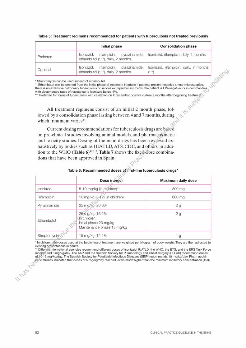
CLINICAL PRACTICE GUIDELINE IN THE SNHS82
Table 5: Treatment regimens recommended for patients with tuberculosis not treated previously
Initial phase Consolidation phase
PreferredIsoniazid, rifampicin, pyrazinamide, ethambutol (*,**), daily, 2 months
Isoniazid, rifampicin, daily, 4 months
Optional Isoniazid, rifampicin, pyrazinamide, ethambutol (*,**), daily, 2 months
Isoniazid, rifampicin, daily, 7 months (***)
* Streptomycin can be used instead of ethambutol. ** Ethambutol can be omitted from the initial phase of treatment in adults if patients present negative smear microscopies, there is no extensive pulmonary tuberculosis or serious extrapulmonary forms, the patient is HIV-negative, or in communities with documented rates of resistance to isoniazid below 4%. *** Preferred for forms of tuberculosis with cavitation on X ray and/or positive culture 2 months after beginning treatment.
All treatment regimens consist of an initial 2 month phase, fol-lowed by a consolidation phase lasting between 4 and 7 months, during which treatment varies48.
Current dosing recommendations for tuberculosis drugs are based on pre-clinical studies involving animal models, and pharmacokinetic and toxicity studies. Dosing of the main drugs has been reviewed ex-haustively by bodies such as IUATLD, ATS, CDC, and others, in addi-tion to the WHO (Table 6)48,117. Table 7 shows the fixed-dose combina-tions that have been approved in Spain.
Table 6: Recommended doses of first-line tuberculosis drugs*
Dose (range) Maximum daily dose
Isoniazid 5-10 mg/kg (in children)** 300 mg
Rifampicin 10 mg/kg (8 12) (in children) 600 mg
Pyrazinamide 25 mg/kg (20 30) 2 g
Ethambutol
25 mg/kg (15 25)In children:Initial phase 20 mg/kgMaintenance phase 15 mg/kg
2 g
Streptomycin 15 mg/kg (12 18) 1 g
* In children, the doses used at the beginning of treatment are weighted per kilogram of body weight. They are then adjusted to existing presentations in adults. ** Different international agencies recommend different doses of isoniazid. IUATLD, the WHO, the BTS, and the ERS Task Force recommend 5 mg/kg/day. The AAP and the Spanish Society for Pulmonology and Chest Surgery (SEPAR) recommend doses of 10 15 mg/kg/day. The Spanish Society for Paediatric Infectious Diseases (SEIP) recommends 10 mg/kg/day. Pharmacoki-netic studies indicated that doses of 5 mg/kg/day reached levels much higher than the minimum inhibitory concentration (150).
It has
been
5 ye
ars si
nce t
he pu
blica
tion o
f this
Clinica
l Prac
tice G
uideli
ne an
d it is
subje
ct to
upda
ting.
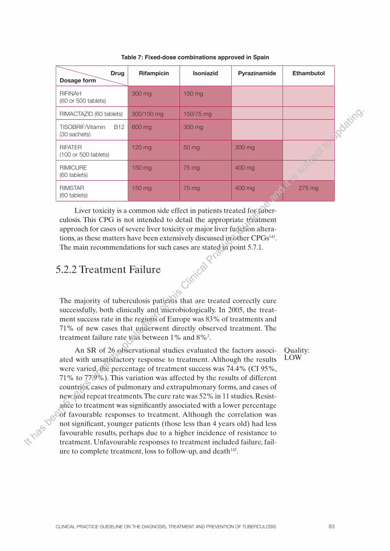
CLINICAL PRACTICE GUIDELINE ON THE DIAGNOSIS, TREATMENT AND PREVENTION OF TUBERCULOSIS 83
Table 7: Fixed-dose combinations approved in Spain
Drug Dosage form
Rifampicin Isoniazid Pyrazinamide Ethambutol
RIFINAH(60 or 500 tablets)
300 mg 150 mg
RIMACTAZID (60 tablets) 300/150 mg 150/75 mg
TISOBRIF/Vitamin B12 (30 sachets)
600 mg 300 mg
RIFATER(100 or 500 tablets)
120 mg 50 mg 300 mg
RIMICURE(60 tablets)
150 mg 75 mg 400 mg
RIMSTAR(60 tablets)
150 mg 75 mg 400 mg 275 mg
Liver toxicity is a common side effect in patients treated for tuber-culosis. This CPG is not intended to detail the appropriate treatment approach for cases of severe liver toxicity or major liver function altera-tions, as these matters have been extensively discussed in other CPGs141. The main recommendations for such cases are stated in point 5.7.1.
5.2.2 Treatment Failure
The majority of tuberculosis patients that are treated correctly cure successfully, both clinically and microbiologically. In 2005, the treat-ment success rate in the regions of Europe was 83% of treatments and 71% of new cases that underwent directly observed treatment. The treatment failure rate was between 1% and 8%2.
An SR of 26 observational studies evaluated the factors associ-ated with unsatisfactory response to treatment. Although the results were varied, the percentage of treatment success was 74.4% (CI 95%, 71% to 77.9%). This variation was affected by the results of different countries, cases of pulmonary and extrapulmonary forms, and cases of new and repeat treatments. The cure rate was 52% in 11 studies. Resist-ance to treatment was significantly associated with a lower percentage of favourable responses to treatment. Although the correlation was not significant, younger patients (those less than 4 years old) had less favourable results, perhaps due to a higher incidence of resistance to treatment. Unfavourable responses to treatment included failure, fail-ure to complete treatment, loss to follow-up, and death142.
Quality: LOW
It has
been
5 ye
ars si
nce t
he pu
blica
tion o
f this
Clinica
l Prac
tice G
uideli
ne an
d it is
subje
ct to
upda
ting.
CLINICAL PRACTICE GUIDELINE IN THE SNHS84
The results of a cohort of more than 13,000 cases in England, Wales and Ireland were published at a later date. In general, between 76.8% and 86.6% of cases (depending on the definition used) showed favour-able results after treatment. The factors associated with treatment fail-ure were female sex, age over 15, pulmonary forms of tuberculosis, and drug resistance143. The results were similar to those of a cohort in northern Italy144.
5.2.3 Corticosteroid Treatment
It has been suggested that the anti-inflammatory effect of corticoster-oids may be beneficial in treating some tuberculosis patients. Some symptoms, such as fever, weight loss, and tissue damage have been as-sociated with cytokines or tumour necrosis factor, which are released by lymphocytes when inflammation occurs145. On the basis of this hy-pothesis several RCTs have been conducted in which steroids, mainly prednisolone, were used as coadjuvant treatment, in addition to the usual treatment for pulmonary tuberculosis. They have been found to be beneficial in the treatment of various extrapulmonary forms of tu-berculosis, such as tuberculous meningitis.
Most of the studies located are not recent and used treatment regimens that did not include rifampicin. A recent narrative review showed the results of 11 RCTs involving a total of 1,814 patients with pulmonary tuberculosis, who were being treated with various different regimens of tuberculosis drugs and adjuvant steroids.
Quality: LOW
In general, adjuvant steroid treatment did not show a long-term benefit in terms of mortality or other clinically relevant variables. Two RCTs that evaluated bacteriological relapses showed no benefit asso-ciated with steroids. Three RCTs showed that steroids were associated with faster negative results to sputum smear microscopy, one indi-cated the opposite, and seven showed no differences between treat-ment groups. The participants involved generally presented extensive, serious forms of tuberculosis with disseminated pulmonary infiltrates. These extensive forms are often associated with toxicity or constitu-tional symptoms and can cause pulmonary obstruction. In this context, corticosteroid treatment has been found to be beneficial in improving patients’ overall condition, promoting weight gain, and accelerating the improvement of infiltrates on X rays146.
It has
been
5 ye
ars si
nce t
he pu
blica
tion o
f this
Clinica
l Prac
tice G
uideli
ne an
d it is
subje
ct to
upda
ting.

CLINICAL PRACTICE GUIDELINE ON THE DIAGNOSIS, TREATMENT AND PREVENTION OF TUBERCULOSIS 85
This CPG does not address one of the possible complications of pri-mary tuberculosis: Addison’s disease. Destruction of the adrenal gland due to tuberculosis, prolonged metabolic stress during infection, and rifampicin treatment are potential causes of this syndrome following primary tuberculosis infection. If this occurs, corticosteroids are not intended to cure the infection and must be considered as substitute treatment.
5.2.4 Other Treatments
Various studies have been conducted to evaluate the efficacy of nu-tritional interventions (vitamins or oligoelements) in patients being treated for pulmonary tuberculosis. In general their results have been uncertain, and it is not believed that tuberculosis patients should fol-low any particular type of diet. Neither are dietary supplements con-sidered as a matter of routine147. Other initiatives, such as immune therapy and laser radiation, have also failed to yield positive results in several studies, all of which were low-quality. These alternative treat-ments should not be included in tuberculosis treatment148-149.
Quality: LOW
Summary of Evidence
Quality: MODERATE
6 month daily treatment regimens involving first-line tuberculosis drugs have shown the greatest efficacy in treating pulmonary tubercu-losis, in both adults and children.
Quality:LOW
Some factors have been associated with treatment failure or relapses after treatment. These include drug resistance, poor treatment compli-ance, age, female sex, HIV infection, and forms that are cavitary on chest X rays.
Quality:MODERATE
Regimens that are intermittent throughout treatment present a higher risk of drug resistance.
Quality:LOW
The overall efficacy of fixed-dose drug combinations is no different from that of drugs administered individually.
Quality:LOW
The efficacy and safety of treatment regimens that include rifabutin are similar to those of regimens with rifampicin. Also, given its phar-macological characteristics,it may cause fewer interactions with other drugs.
Quality:LOW
Although adjuvant corticosteroid treatment has not shown lower mortality rates and does not increase the probability of cure, it is as-sociated with clinical and radiological improvement in extensive and serious forms of pulmonary tuberculosis.
It has
been
5 ye
ars si
nce t
he pu
blica
tion o
f this
Clinica
l Prac
tice G
uideli
ne an
d it is
subje
ct to
upda
ting.

CLINICAL PRACTICE GUIDELINE IN THE SNHS86
Quality:LOW
Adjuvant treatments for tuberculosis such as diets rich in vitamins or oligoelements, immunotherapy and laser radiation have not been found to be beneficial.The main side effect of tuberculosis treatment in adults and children is liver toxicity.
Recommendations
√Individuals diagnosed with pulmonary tuberculosis must be treated and monitored by physicians and healthcare staff with sufficient expe-rience in handling pulmonary tuberculosis.
STRONG
Most patients with pulmonary tuberculosis not previously treated should be treated using a short, 6 month regimen consisting of an ini-tial 2 month phase of isoniazid, rifampicin, pyrazinamide and etham-butol and a 4 month maintenance phase of isoniazid and rifampicin.
WEAKOther treatment regimens for pulmonary tuberculosis are also recom-mended (Table 1).
WEAKTreatment should be extended to 9 months in patients with cavitary pulmonary tuberculosis who still have positive cultures at the end of the initial (2 month) phase of treatment.
√Treatment compliance must be assessed if there is a positive culture at the end of the initial (2 month) phase of treatment.
STRONGFor initial treatment of tuberculosis in children, the same treatment regimens (at suitable doses) are recommended as for the adult popu-lation unless there are specific contraindications.
WEAKIn children and adults, intermittent treatment (three times a week) may be considered during the maintenance phase if it is directly ob-served and if a culture taken after 2 months of treatment is negative.
STRONG Twice-weekly intermittent treatment regimens are not recommended.
WEAKTo reduce the development of drug resistance and the number of drugs taken daily, adults should be treated with fixed-dose combina-tions of the tuberculosis drugs currently on the market.
WEAKRifabutin is a reasonable option if rifampicin is not tolerated or if there is a high risk of interaction with other drugs, particularly antiret-rovirals.
WEAKAdjuvant corticosteroid treatment may be considered in certain cases of extensive forms of tuberculosis.
STRONGOther adjuvant treatments, such as diets rich in vitamins or oligoele-ments, immunotherapy or laser radiation, are not recommended for tuberculosis.
It has
been
5 ye
ars si
nce t
he pu
blica
tion o
f this
Clinica
l Prac
tice G
uideli
ne an
d it is
subje
ct to
upda
ting.

CLINICAL PRACTICE GUIDELINE ON THE DIAGNOSIS, TREATMENT AND PREVENTION OF TUBERCULOSIS 87
√Liver toxicity must be closely monitored in patients receiving tuber-culosis treatment, particularly those with known liver disease.
5.3. Treating Extrapulmonary Tuberculosis
Questions to Answer
• Inpatients(adultsandchildren)withextrapulmonarytuberculosisinvariousloca-tions, what is the optimum duration of treatment?
• Inpatients(adultsandchildren)withextrapulmonarytuberculosisinvariousloca-tions, do corticosteroids reduce mortality or increase the likelihood of cure when used as an addition to tuberculosis treatment?
• Inpatientswithosteoarticulartuberculosis,whatarethebenefitsofsurgeryassoci-ated with tuberculosis treatment?
• Inpatientswithpericardialtuberculosis,ispericardialintervention(pericardiocente-sis or pericardiectomy) beneficial?
Tuberculosis can affect any organ or tissue. When the bacillus infects non-pulmonary tis-sues, this is known as extrapulmonary tuberculosis. One practical classification of tuber-culosis distinguishes between respiratory and extra-respiratory forms. The former include pulmonary, trachaeobronchial, and laryngeal tuberculosis49.
Extrapulmonary involvement is more common in children and patients with altered immunity. It is usually a reactivation of a latent form of infection. In recent years the most common cause of reactivation and the development of extrapulmonary tuberculosis has been changes in immunity caused by HIV infection, AIDS and other types of immunosup-pression49.
In general, the basic principles for deciding on the duration of pulmonary tuberculosis treatment also apply to extrapulmonary tuberculosis, although there are some individual differences, such as in the treatment of tuberculosis of the central nervous system. There are no studies that have analysed intermittent treatment regimens, and as a result these should not be considered for the treatment of extrapulmonary forms117.
Tuberculosis is very rare in some locations, and as a result no RCTs evaluating the effi-cacy of various durations or combinations of tuberculosis drugs have been conducted regarding these sites. Although there are case series describing progress with various different treatment regimens, all these studies are of poor methodological quality. In many cases the evidence is indirect and based on studies involving patients with nearby or similar involvement, or simply on studies of pulmonary tuberculosis. Nevertheless, once diagnosis has been confirmed there is little doubt that the benefits of treating these patients with a regimen lasting at least 6 months and including isoniazid, rifampicin, and pyrazinamide far outweigh the risks49.
The evidence relating to the most common forms of extrapulmonary tuberculosis for which such evidence has been located in the form of RCTs is described below.
It has
been
5 ye
ars si
nce t
he pu
blica
tion o
f this
Clinica
l Prac
tice G
uideli
ne an
d it is
subje
ct to
upda
ting.

CLINICAL PRACTICE GUIDELINE IN THE SNHS88
5.3.1 Pleural Tuberculosis
Pleural tuberculosis is the involvement of the pleura in tuberculosis infection, caused when a small number of bacilli enter the pleura from the lung, causing a hypersensitivity reaction, irritation and pleural ef-fusion. Rupture of a tuberculous cavity or the presence of a broncho-pleural fistula can also result in large numbers of bacilli entering the pleural space, causing empyema.
Various studies have been conducted to evaluate the regimens established for the treatment of pulmonary tuberculosis that are ac-cepted or standard in the place where the studies are conducted. The duration of the treatment regimens ranged from 6 months, in the most recent studies, to 9 months. Most of the studies included patients with pleural involvement in tuberculosis, although primary pulmonary ori-gin is not excluded.
Quality: MODERATE
Like other extrapulmonary locations involving serous mem-branes, adjuvant use of corticosteroids has been considered for pleu-ral tuberculosis. A recent SR (6 RCTs, 633 patients) evaluated its ef-ficacy in patients (adults and children) with pleural tuberculosis. The corticosteroids used were prednisone or prednisolone, administered orally or intramuscularly, for periods of up to 2 months. In the short term the steroids were associated with a decrease in pleural effusion and thickness of the pleura as shown on chest X rays, and with faster resolution of symptoms. These results did not translate into reduced pleural effusion at 2 months, respiratory function or overall mortality (RR 0.9; CI 95%, 0.7 to 1.3, 75 events). Failure to complete treatment as a result of side effects was between 1 and 3 times more common in patients receiving corticosteroids (5.5%). The only RCT that included only HIV-positive patients produced similar results, but six patients who had received corticosteroids developed Kaposi’s sarcoma151.
A common complication of empyema is pleural fibrosis, which often requires aggressive treatment including thoracotomy and drain-age, fibrinolytic drugs or even thoracotomy to remove the infected tis-sue152.
It has
been
5 ye
ars si
nce t
he pu
blica
tion o
f this
Clinica
l Prac
tice G
uideli
ne an
d it is
subje
ct to
upda
ting.

CLINICAL PRACTICE GUIDELINE ON THE DIAGNOSIS, TREATMENT AND PREVENTION OF TUBERCULOSIS 89
5.3.2 Lymphatic Tuberculosis
There is only a limited amount of evidence available on the treatment of patients with lymphatic tuberculosis. An SR that included eight stud-ies with varying designs and methodological quality was located, cov-ering a total of 634 patients (adults and children). This SR combined the results of RCTs and case series of patients who produced efficacy results for various different treatment regimens that lasted between 6 and 9 months. The rate of recurrence after completing treatment (con-firmation of tuberculosis infection in a new lymph node after a period of clinical remission) was 3.3% (CI 95%, 1.7 to 5.5, 13 events) for 6 month regimens, and 2.7% (CI 95%, 0.6 to 7.8, 3 events) for 9 month regimens. Although these results were not compared statistically, the absolute difference in risk indicates that treating approximately 160 pa-tients with 9 month regimens would prevent one relapse as compared to 6 month regimens153.
A subsequent RCT (268 patients) evaluated the comparative ef-ficacy of two 6 month regimens, one intermittent and the other daily, in a cohort of patients with lymphatic tuberculosis, both adults and chil-dren. In those receiving intermittent treatment, treatment was directly observed. The percentages of favourable responses in the two groups were similar. The percentages of lymphatic relapses were also similar, although the number of events was very low (2 with the daily regimens, 3 with the intermittent regimens). Side effects were significantly more common with intermittent regimens (11% versus 1%)154.
Various studies have evaluated relapses and the appearance of new lymph nodes during or after treatment. This is a common occur-rence, possibly related to underlying immune responses to treatment, and in most cases cannot be considered treatment failure. Only cases confirmed microbiologically should be considered to be relapses.
Quality: LOW
5.3.3 Osteoarticular Tuberculosis
The most common type of osteal involvement in tuberculosis is spinal involvement, which is potentially serious. It can cause pain and destroy bone tissue, causing vertebral collapse. Bone destruction in turn causes curvature of the spine, compression of nerve roots, neurological deficits and occasionally paraplegia155.
It has
been
5 ye
ars si
nce t
he pu
blica
tion o
f this
Clinica
l Prac
tice G
uideli
ne an
d it is
subje
ct to
upda
ting.

CLINICAL PRACTICE GUIDELINE IN THE SNHS90
Spinal tuberculosis has some specific features, and treatment is con-sidered successful when curvature of the spine is corrected, neurological deficits are corrected, pain is prevented, spinal fusion is achieved, loss of bone mass is prevented, and future local recurrences are prevented. The disease is monitored clinically and radiologically, preferably using nuclear magnetic resonance images.
Surgery has often been considered as adjuvant treatment to tuber-culosis drugs. In general, the surgical approach consists of debriding in-fected material or debriding and stabilising the spine (reconstruction). Stabilising the spine involves bone implants taken from the patient or synthetic materials.
One SR (2 RCTs, 331 patients) analysed the efficacy of tubercu-losis treatment with and without surgery in adults and children with spinal tuberculosis. Both RCTs were conducted by the British Medi-cal Research Council (BMRC) and began in the 1960s and 1970s. They had major methodological shortcomings; in addition, the international standards when they were conducted were different from those in place today. The SR did not detect any statistically significant differences in the number of patients who presented increased curvature of the spine, progression of neurological deficits, or cure defined as spinal fusion. Monitoring of these variables was incomplete. Surgery associated with treatment did not reduce overall mortality, although the number of events was very low156.
Quality: LOW
The optimum duration of tuberculosis treatment was evaluated by BMRC in a series of international studies conducted in India, Korea, and Hong Kong. Various publications have shown results for long treat-ment regimens (18 months) and short treatment regimens (6 9 months) with various durations of follow-up. The first studies involving isoniazid and para-aminosalicylic acid for 18 months became obsolete after it was shown that short treatment regimens involving isoniazid and rifampicin were effective in treating pulmonary tuberculosis. The studies had sub-stantial methodological shortcomings, with major losses to follow-up and no analysis by intention to treat.
Quality: LOW
It has
been
5 ye
ars si
nce t
he pu
blica
tion o
f this
Clinica
l Prac
tice G
uideli
ne an
d it is
subje
ct to
upda
ting.
CLINICAL PRACTICE GUIDELINE ON THE DIAGNOSIS, TREATMENT AND PREVENTION OF TUBERCULOSIS 91
Only one RCT presented results that had been compared statistically. These were the results of a 10 year follow-up of the RCT conducted in India. A total of 304 patients, both adults and children, with tuberculo-sis of the thoracic, lumbar, or sacral spine and with no paralysis were randomised to treatment with a regimen that included isoniazid and rifampicin for 6 or 9 months. The study did not show any significant differences in the percentage of patients with favourable clinical/radio-logical development with the 6 month (94%) and the 9 month (99%) regimen. Neither were there any differences between the 9 month and 6 month regimens in terms of the percentage of patients with spinal fu-sion (85% versus 81%). Curvature of the spine increased to a similar extent in both groups157.
The RCT conducted in Korea by the same group (the BMRC) showed similar efficacy for short treatment regimens (6 or 9 months) with rifampicin and for longer regimens (18 months) without rifampicin. The results were not compared statistically. The same publication shows the results of an RCT conducted in Hong Kong. Clinical/radiological status and the percentage of patients with spinal fusion was similar for the 6 month and the 9 month regimens that included isoniazid and ri-fampicin. This study also lacked statistical comparison158.
When osteoarticular tuberculosis affects the spine or other loca-tions, patients often undergo surgery. In a retrospective series of 53 cas-es in Spain, the main location was axial, followed by involvement of the knee and ankle joints. Although positive cultures were obtained in most cases, tuberculosis was diagnosed late, after an average of 8 months. 38% of patients required surgical debridement or arthrodesis159.
Quality: LOW
5.3.4 Tuberculosis of the Central Nervous System
The most common tuberculous involvement of the central nervous system is meningeal tuberculosis, and less frequently cerebral tuber-culoma. Although the incidence of tuberculosis of the central nervous system is very low, it has high morbidity and mortality rates. It can cause irreversible neurological damage, cerebral palsy, mental retardation or epilepsy.
Tuberculosis of the meninges has characteristic features. Age and clini-cal status when the patient presents with the disease are known to be relevant to prognosis. Prognosis is worse if the illness occurs at an early age and with advanced clinical presentations160.It h
as be
en 5
years
sinc
e the
publi
catio
n of th
is Clin
ical P
ractic
e Guid
eline
and i
t is su
bject
to up
datin
g.
CLINICAL PRACTICE GUIDELINE IN THE SNHS92
Traditionally, tuberculous meningitis has been classified in three stages161:
Stage I: no focal neurological signs or deterioration in level of con-sciousness
Stage II: focal neurological signs and/or deterioration in level of consciousness
Stage III: coma
The drugs available to treat meningeal tuberculosis are the same as those available for the treatment of pulmonary tuberculosis. Because some of the tuberculosis drugs available do not enter the central nerv-ous system well through the blood-brain barrier, the treatment regi-mens have been prolonged. Also, unlike with pulmonary tuberculosis, adjuvant corticosteroids play an important role in the treatment of this serious illness. Despite the drugs available today and longer treatment regimens, mortality rates are considerable.
There are no RCTs comparing different treatment regimens or durations in patients diagnosed with tuberculous meningitis. The little evidence there is on the subject comes from case series or small cohorts of patients, with the methodological limitations implicit in this type of design. None of the studies is recent, and all of them were conducted in countries with low per capita incomes162-172.
One SR evaluated the results of the studies that provided numeri-cal data, although no meta-analysis was performed and the results were not compared statistically. The studies included involved a total of 872 patients, 75% of whom were children or teenagers aged under 16. Most of the studies involved patients with a probable diagnosis of tubercu-lous meningitis, and fewer than a quarter of patients had diagnoses confirmed by positive cultures or staining for M. tuberculosis. The stud-ies considered were those that provided results for treatment lasting 6 months and treatment lasting more than 6 months. Most patients had clinical stage I or II tuberculous meningitis at the beginning of treat-ment (with more or less focal neurological involvement or deteriora-tion in level of consciousness).
Treatment follow-up rates were above 80% with both regimens. The mortality rate during treatment was 16% in 6 month regimens and 6% in longer regimens. In follow-up after treatment, 1.5% of patients who had been treated according to 6 month regimens suffered a re-crudescence, compared to none of those treated according to longer regimens. Cure could not be accurately evaluated in many patients, as the definition of cure is not well established173.
Quality: LOW
It has
been
5 ye
ars si
nce t
he pu
blica
tion o
f this
Clinica
l Prac
tice G
uideli
ne an
d it is
subje
ct to
upda
ting.

CLINICAL PRACTICE GUIDELINE ON THE DIAGNOSIS, TREATMENT AND PREVENTION OF TUBERCULOSIS 93
A recent SR (7 RCTs, 1,140 patients) evaluated the efficacy of cor-ticosteroids in combination with conventional tuberculosis treatment in patients of any age with tuberculous meningitis, and compared the results with those of a regimen with no corticosteroids. The studies ana-lysed used prednisolone (60 mg/day in adults, 2 mg/kg/day in children) and dexamethasone (12 16 mg/day in adults, 0.4 mg/kg/day in children). Follow-up of the studies ranged from 2 months to nearly 4 years.
Quality: HIGH
Overall, corticosteroids reduced the risk of death by 20% (RR 0.78; CI 95%, 0.67 to 0.91, 429 events). The mortality rate in the group that did not receive corticosteroids was more than 40%. Corticosteroids provided an absolute reduction of 8.6%, which is clinically significant. The number of patients needed to treat to avoid one death was ap-proximately 10. The benefit of treatment did not depend on the length of follow-up in the study.
The information available on neurological sequelae comes from three studies that assessed neurological them in different ways. Treat-ment significantly reduced the risk of neurological deficits by 18% (RR 0.82; CI 95%, 0.70 to 0.97, 318 events). Most of the evidence was taken from studies that included patients with stage II or III (advanced or serious) meningitis, and the overall efficacy was similar for both these clinical stages. Only one study showed results for HIV-positive and HIV-negative patients: although the differences were not significant, efficacy tended to be higher in HIV-negative patients174.
For children with hydrocephalus, early insertion of an external ventricular drain has been suggested to improve the hydrocephalus. This may also improve patients’ final prognosis175.
Quality: VERY LOW
5.3.5 Pericardial Tuberculosis
Although pericardial tuberculosis is very rare in developed countries (less than 5% of pericarditis cases), in low-income countries, particular-ly since the beginning of the HIV pandemic, it is relatively common176. Tuberculosis of the pericardium is a particularly serious situation, and can be fatal.
One SR (4 RCTs, 469 patients) evaluated the efficacy of the vari-ous treatment options available in patients with pericardial tuberculo-sis, although in a significant percentage of patients the diagnosis was not confirmed. The review did not locate any study that compared 6 month treatment regimens and longer regimens in patients with tuberculous pericarditis, or any RCTs that evaluated pericardiectomy.
Quality: MODERATE
It has
been
5 ye
ars si
nce t
he pu
blica
tion o
f this
Clinica
l Prac
tice G
uideli
ne an
d it is
subje
ct to
upda
ting.

CLINICAL PRACTICE GUIDELINE IN THE SNHS94
The evidence available on the efficacy of pericardial drainage comes from a single RCT conducted in adults and children. All the pa-tients enrolled were randomised to receive corticosteroids or a placebo, in addition to standard tuberculosis treatment. A subgroup of 122 pa-tients were also randomised to receive pericardiocentesis, either elec-tive or deferred according to their physicians’ judgement. No patient who underwent elective pericardiocentesis required a second operation due to pericardial tamponade, while 12 of the 55 patients who did not receive initial surgery did undergo pericardiocentesis during follow-up (RR 0.04; CI 95%, 0 to 0.6, 12 events). This did not translate into a re-duction in overall mortality (RR 0.9; CI 95%, 0.3 to 3.1, 10 events) or in deaths due to pericarditis (RR 1.3; CI 95%, 0.3 to 5.5, 7 events).
Three RCTs included in the SR evaluated the efficacy of corticos-teroids (mostly prednisolone) in combination with a standard regimen of tuberculosis drugs in HIV-negative patients. Although overall corti-costeroids are associated with a beneficial effect on all the variables an-alysed, the wide confidence interval and small number of events make it impossible to draw firm conclusions. Therefore, corticosteroids were associated with a significant 31% reduction in death or persistence of tuberculosis after 2 years (RR 0.69; CI 95%, 0.48 to 0.98, 96 events) but not with a decrease in overall mortality (RR 0.6; CI 95%, 0.4 to 1.2, 43 events) or in the number of operations to drain or remove the pericar-dium. A single RCT in HIV-positive patients also failed to show dif-ferences between the treatment groups177. A subsequent publication that analysed the efficacy of corticosteroids in tuberculous pericarditis included the same studies and produced the same results178.
A subsequent RCT randomised 57 patients with suspected or confirmed pericardial tuberculosis (all of whom underwent pericardi-ocentesis) to receive corticosteroids or a placebo via intrapericardial infusion. All the patients enrolled were aged 17 or over and 37% were HIV-positive. No deaths were recorded at the one-year follow-up. Two patients who received corticosteroids developed constrictive pericardi-tis due to pericardial effusion179.
In general, the studies did not follow the patients long enough to allow the evaluation of the effect of treatment on constrictive pericardi-tis, which is a chronic complication of pericardial effusion. This, together with small study size, may be one of the reasons corticosteroid treatment is associated with therapeutic benefit, though not significantly. One of the studies included in the SR showed that in the short term (less than 3 months) corticosteroid treatment improved the signs and symptoms that suggest constrictive pericarditis, indicating a rapid improvement in pericardial effusion180.
It has
been
5 ye
ars si
nce t
he pu
blica
tion o
f this
Clinica
l Prac
tice G
uideli
ne an
d it is
subje
ct to
upda
ting.
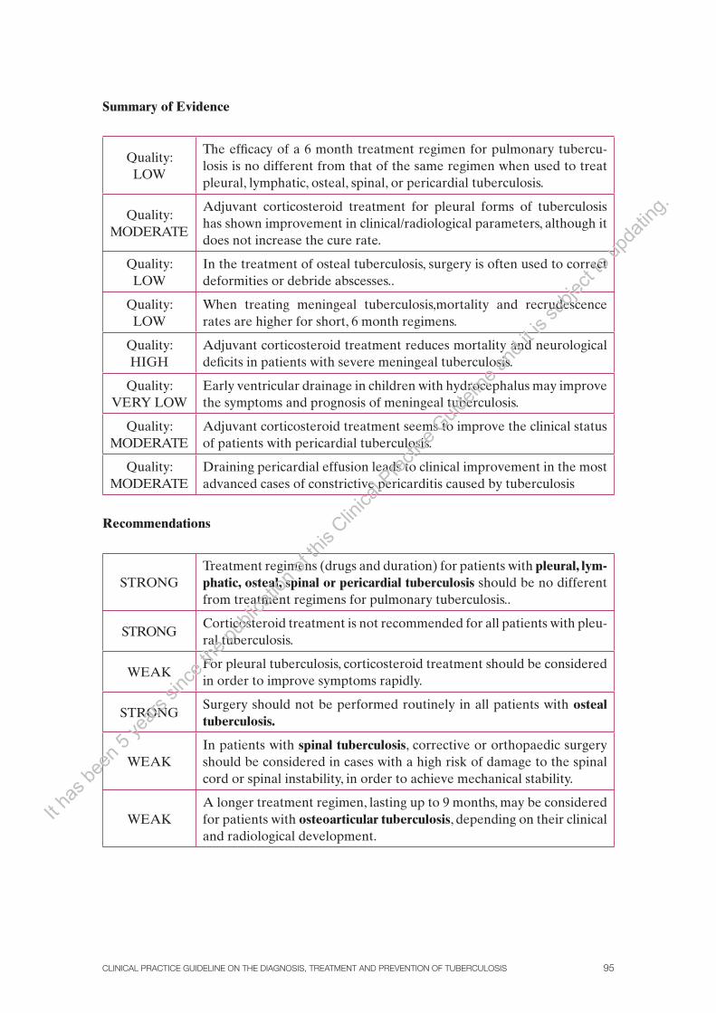
CLINICAL PRACTICE GUIDELINE ON THE DIAGNOSIS, TREATMENT AND PREVENTION OF TUBERCULOSIS 95
Summary of Evidence
Quality:LOW
The efficacy of a 6 month treatment regimen for pulmonary tubercu-losis is no different from that of the same regimen when used to treat pleural, lymphatic, osteal, spinal, or pericardial tuberculosis.
Quality:MODERATE
Adjuvant corticosteroid treatment for pleural forms of tuberculosis has shown improvement in clinical/radiological parameters, although it does not increase the cure rate.
Quality:LOW
In the treatment of osteal tuberculosis, surgery is often used to correct deformities or debride abscesses..
Quality:LOW
When treating meningeal tuberculosis,mortality and recrudescence rates are higher for short, 6 month regimens.
Quality:HIGH
Adjuvant corticosteroid treatment reduces mortality and neurological deficits in patients with severe meningeal tuberculosis.
Quality:VERY LOW
Early ventricular drainage in children with hydrocephalus may improve the symptoms and prognosis of meningeal tuberculosis.
Quality:MODERATE
Adjuvant corticosteroid treatment seems to improve the clinical status of patients with pericardial tuberculosis.
Quality:MODERATE
Draining pericardial effusion leads to clinical improvement in the most advanced cases of constrictive pericarditis caused by tuberculosis
Recommendations
STRONGTreatment regimens (drugs and duration) for patients with pleural, lym-phatic, osteal, spinal or pericardial tuberculosis should be no different from treatment regimens for pulmonary tuberculosis..
STRONGCorticosteroid treatment is not recommended for all patients with pleu-ral tuberculosis.
WEAKFor pleural tuberculosis, corticosteroid treatment should be considered in order to improve symptoms rapidly.
STRONGSurgery should not be performed routinely in all patients with osteal tuberculosis.
WEAKIn patients with spinal tuberculosis, corrective or orthopaedic surgery should be considered in cases with a high risk of damage to the spinal cord or spinal instability, in order to achieve mechanical stability.
WEAKA longer treatment regimen, lasting up to 9 months, may be considered for patients with osteoarticular tuberculosis, depending on their clinical and radiological development.
It has
been
5 ye
ars si
nce t
he pu
blica
tion o
f this
Clinica
l Prac
tice G
uideli
ne an
d it is
subje
ct to
upda
ting.

CLINICAL PRACTICE GUIDELINE IN THE SNHS96
STRONGPatients with tuberculous meningitis must follow a longer treatment regimen, lasting up to 12 months.
STRONGIn patients with stage II or III tuberculous meningitis, adjuvant cor-ticosteroid treatment is recommended during the initial phase (pred-nisolone 60 mg/day for 4 weeks).
WEAKIn children with tuberculous meningitis, adjuvant corticosteroid treat-ment is recommended during the initial phase (prednisolone 60 mg/day for 4 weeks).
WEAKIn children with and hydrocephalus, ventricular drainage should be considered.
STRONGIn patients with pericardial tuberculosis, adjuvant corticosteroid treat-ment is recommended during the initial phase (prednisolone 60 mg/day for 4 weeks).
STRONGRoutine pericardiocentesis is not recommended for patients with tu-berculous pericarditis and any degree of pericardial effusion.
WEAKIn patients with tuberculous pericarditis, evacuating pericardiocentesis can be considered for cases where there is risk of pericardial tampon-ade or functional compromise.
5.4. Monitoring Treatment
Questions to Answer
• In patients with tuberculosis,what strategies are effective in increasing treatmentcompliance?
• Inpatientswithpulmonarytuberculosis,doesdirectlyobservedtreatmentimprovetreatment compliance, increase the likelihood of cure,or reduce the risk of resistance to treatment?
The World Health Assembly (WHA) described tuberculosis as a worldwide public health problem in 1991181. In 1994, it adopted the DOTS (directly observed treatment, short course) strategy to tackle this challenge, and in 2002 its content was extended to cover a total of 182 countries19,182. In 2005, worldwide mortality, diagnosis of tuberculosis, and treatment success had all improved183.
The DOTS strategy consists of the following five key points:
1. A political commitment to a gradual, significant increase in funding
2. Correct diagnosis
3. Standard short-course treatment in all confirmed cases, together with appropriate monitoring
It has
been
5 ye
ars si
nce t
he pu
blica
tion o
f this
Clinica
l Prac
tice G
uideli
ne an
d it is
subje
ct to
upda
ting.

CLINICAL PRACTICE GUIDELINE ON THE DIAGNOSIS, TREATMENT AND PREVENTION OF TUBERCULOSIS 97
4. Guaranteed uninterrupted supply of treatment
5. Establishment of systems for continual evaluation of results
The WHO sponsors and leads the Stop TB Partnership, which consists of more than 1000 governmental and non-governmental organisations. These organisations are currently promoting a strategy set out in a document published in 2006184. Its most recent updates can be found on the WHO website (http://www.who.int/tb/strategy/en/). The ultimate aim of the strategy is to reduce the global burden of tuberculosis by 2015. Its specific objectives are in line with the United Nations’ Millennium Development Goals. To achieve them, it integrates the DOTS strategy and proposes a set of measures that should be taken by countries’ national programmes, such as the following:
• EnhancetheDOTSstrategy
• AddressTB/HIVandMDR-TB
• Strengthenhealthsystemsviaprimarycare
• Engageallhealthcareproviders
• Providepatientsandcommunitieswithmoreresources,throughassociations
• Driveresearch
Treatment monitoring by health services means identifying and acting on the factors that may lead a patient to abandon or interrupt his/her treatment. The aim of this is that the patient will complete treatment and be cured, consequently reducing the appearance of resistance to treatment. Treatment monitoring programmes may include directly ob-served treatment (DOT), which consists of ensuring that patients take their medication, although it involves various different strategies depending on the environment that are often complex and not always easy to apply. The most promising results have been found in studies in which treatment monitoring strategies have been applied most rigorously. For now, there is little available information to evaluate the impact of DOT on acquired resist-ance to treatment185-187.
The regularity with which patients take medication can be evaluated by inspecting their urine, because of its orangey colour in patients taking rifampicin, or by the pres-ence of drugs or their metabolites in the urine of patients treated for active tuberculosis or latent tuberculosis infection. Treatment monitoring is possible for all or most drugs, although only isoniazid plasma concentrations have been associated clearly to treatment response188. This monitoring may help rule out incorrect medication intake, interactions with other drugs, or poor absorption as probable causes of treatment failure189.
5.4.1 Assessment of Methods to Increase Compliance
It has
been
5 ye
ars si
nce t
he pu
blica
tion o
f this
Clinica
l Prac
tice G
uideli
ne an
d it is
subje
ct to
upda
ting.

CLINICAL PRACTICE GUIDELINE IN THE SNHS98
One recent SR (11 RCTs, 5,609 patients) compared the results for cure and treatment compliance in patients with pulmonary tuberculosis with whom any kind of DOT strategy had been used. Only four trials (1,603 patients) compared a DOT strategy with self-administered treatment. The SR did not find any differences regarding cure (RR 1.02; CI 95%, 0.86 to 1.21) or for the combined outcome of cure plus completion of treatment . The results varied between the studies, partly due to the studies that exam-ined a home-based DOT strategy. For home-based DOT, the three RCTs included (1,365 patients) showed an increase in the percentage of cures, al-though the difference was slight (RR 1.10; CI 95%, 1.02 to 1.18). The trials that evaluated different DOT strategies (monitoring performed by a rela-tive, in hospital or by a member of the community) found no differences between the strategies. Eight of the 11 trials were conducted in countries with low or medium per capita incomes. The differences between similar programmes applied locally in countries with very different circumstances certainly contributed to the heterogeneity and the lack of differences be-tween the groups analysed190. No subsequent RCTs that analyse the self-administered treatment for pulmonary tuberculosis were identified.
Quality:
LOW
One SR (9 RCTs, 5,257 patients) analysed controlled studies (RCTs and before-and-after controlled studies) that evaluated various strategies to improve compliance with tuberculosis diagnosis or treatment processes (home visits, sending letters, reminders). Overall the results were favourable for these strategies, although no joint result for the studies was provided191. An additional RCT included 480 participants with pulmonary tuberculosis who had not been treated previously. The results showed that home visits significantly reduced non-treatment compliance (RR 0.21; CI 95%, 0.11 to 0.43) and the percentage of patients who were smear-positive at the end of treatment (RR 0.28; CI 95%, 0.17 to 0.47) when compared to self-adminis-tration of medication. The mortality rates of the two groups were similar192.
Quality:
LOW
Another SR (5 RCTs, 2,179 patients) assessed the efficacy of various strategies intended to improve treatment compliance (or prophylaxis) in RCTs and observational studies, although the results were not analysed joint-ly193. The two RCTs conducted to assess treatment compliance used different techniques. One of them showed that 88% of the patients who received a reminder letter completed their treatment, compared to 73% of those who did not (RR 1.2; CI 95%, 1.1 to 1.4)194. The other RCT showed that motiva-tional programmes and monitoring the groups receiving DOT resulted in more patients completing their treatment (RR 1.2; CI 95%, 1.1 to 1.3)195. An RCT that evaluated a combined strategy of financial incentives and educa-tion for patients also showed that more patients completed their treatment or prophylaxis after the intervention (RR 2.4; CI 95%, 1.5 to 3.7)196. The SR mentioned above included a single study conducted in Spain, with children receiving prophylactic treatment. Education (performed at several different levels) increased the number of patients who completed their treatment by 20% (RR 1.2; CI 95%, 1.1 to 1.4) in comparison to the control group197.
Quality:
LOW
It has
been
5 ye
ars si
nce t
he pu
blica
tion o
f this
Clinica
l Prac
tice G
uideli
ne an
d it is
subje
ct to
upda
ting.

CLINICAL PRACTICE GUIDELINE ON THE DIAGNOSIS, TREATMENT AND PREVENTION OF TUBERCULOSIS 99
The RCTs and SRs located did not evaluate the appearance of resist-ance to treatment, which is one of the main aims of treatment monitoring198. This evaluation was performed in observational cohort studies in which the percentage of resistance to tuberculosis treatment (mainly rifampicin) was obtained following general implementation of treatment monitoring pro-grammes. This percentage was compared with retrospective records made before treatment monitoring programmes were implemented. Three co-hort studies that involved a significant number of patients and included the evaluation of resistance to treatment as a study aim were been found. They show a significant decrease in drug resistance after these programmes were implemented. The studies were conducted in low-income countries and in populations that are substantially different from each other199-201.
Quality:
LOW
5.4.2 Assessment of the DOTS Strategy
A narrative review highlighted the heterogeneity of programmes used to apply the DOTS strategy. It identified 32 studies (RCTs and observational studies) describing a total of 30 different programmes, all of which included DOT. Only 13 included continual evaluation systems, 11 included guaran-teed supply of treatment, 9 included improvements in laboratory tests to diagnose tuberculosis, and 8 included a political commitment.
Quality:
LOW
These studies described other initiatives to improve compliance, such as reminder letters, financial incentives, transport vouchers, donations of clothing or food, the involvement of social workers, educational pro-grammes, and many others202. Another recent SR evaluated perceptions of the factors affecting compliance by patients, healthcare staff and other individuals involved. The complexity and the large number of factors that affect compliance were highlighted, and it was concluded that improving compliance required more patient-centred strategies and more attention to structural obstacles to access or availability of treatment203.
The WHO has made a commitment to implementing DOT for all patients, while other organisations that target environments more similar to Spain, such as the CDC and the Barcelona Tuberculosis Research Unit, have agreed to suggest the use of DOT for groups with compliance levels below 90%204-205.
Summary of Evidence
Quality:LOW
Direct observation of medication intake has not consistently shown higher cure rates or better treatment compliance in patients with pulmonary tuber-culosis.
It has
been
5 ye
ars si
nce t
he pu
blica
tion o
f this
Clinica
l Prac
tice G
uideli
ne an
d it is
subje
ct to
upda
ting.

CLINICAL PRACTICE GUIDELINE IN THE SNHS100
Quality:LOW
Direct observation of medication intake may reduce resistance to tuberculo-sis drugs, particularly rifampicin.
Quality:LOW
Various treatment-monitoring strategies have been shown to increase over-all compliance.
Recommendations
√Responsibility for successful treatment must be shared between the health-care professionals in charge of patients and the healthcare authorities that provide the necessary resources.
√The potential level of treatment compliance must be assessed and moni-tored in all tuberculosis patients who begin tuberculosis treatment.
√It is important to motivate patients and to highlight the importance of fully complying with treatment, both for latent infection and active tuberculosis.
√The strategies available for improving compliance must be tailored to each case and agreed upon with the patient.
STRONGThe generalised use of directly observed treatment for all patients receiv-ing tuberculosis treatment is not recommended.
STRONG
Directly observed treatment regimens are recommended under certain circumstances, such as for patients living in poverty, those with no fixed ad-dress, cases with significant grounds to suspect poor compliance, patients with a history of poor compliance, and children.
STRONGVarious strategies are recommended for improving compliance. These in-clude reminder letters, phone calls, education, and home visits.
5.5. Treating Challenging Groups
Questions to Answer
• DoHIV-positiveindividualspresentdifferenttuberculosischaracteristicsandpro-gression from those who are HIV-negative?
• WhenHIV-positiveindividualsaretreatedfortuberculosis,dotheysuffermorere-lapses than those who are HIV-negative?
• DoHIV-positiveindividuals(adultsandchildren)withtuberculosisbenefitfromalonger tuberculosis treatment regimen?
• InHIV-positiveindividuals(adultsandchildren)withtuberculosis,whorequiretuber-culosis and antiretroviral treatment, what is the best way to handle these treatments?
• Whatisthebestwaytotreattuberculosisinchallengingsituations(liverdysfunction,kidney dysfunction, pregnancy)?
It has
been
5 ye
ars si
nce t
he pu
blica
tion o
f this
Clinica
l Prac
tice G
uideli
ne an
d it is
subje
ct to
upda
ting.

CLINICAL PRACTICE GUIDELINE ON THE DIAGNOSIS, TREATMENT AND PREVENTION OF TUBERCULOSIS 101
5.5.1 Concurrent HIV Infection
Treating concurrent tuberculosis and HIV infection is complex. In Spain, such cases are handled by specialists in both infections17. This CPG does not aim to examine the treatment of this complex situation in detail, as there are other guidelines dedicated exclusively to this subject48,206-207.
The risk of concurrent infection is increased as soon as HIV sero-conversion occurs, and it increases as the disease progresses. The clinical presentation of tuberculosis (often extrapulmonary forms), symptoms, radiological features, laboratory diagnostic tests, and frequent infection by mycobacteria other than those that cause tuberculosis all lend tuber-culosis specific features in patients with greater immune system altera-tion. It is also known that there is great potential for drug interactions between rifamycins (particularly rifampicin) and antiretrovirals. These two groups of drugs are essential to any treatment regimen for pulmo-nary tuberculosis and HIV infection, respectively208.
The various studies that evaluate the efficacy and safety of vari-ous treatments regimens including tuberculosis drugs have included a certain proportion of HIV-positive patients, generally in small numbers, which has not been sufficient to allow the drawing of firm conclusions regarding this subgroup of patients. Also, many of these studies were conducted before the development and generalised use of highly active antiretroviral therapy (HAART)140.
Quality: LOW
In general, there is no difference between the efficacy of tuber-culosis treatment in patients who are HIV-positive or HIV-negative. Various Spanish and international bodies recommend standard 6 month treatment (2HRZE/4HR) with fixed-dose drug combinations for patients with pulmonary tuberculosis who have not previously been treated17,48,117,207.
The relapse rate for active tuberculosis in HIV-positive patients is higher than that of HIV-negative patients. As a result, treatment with longer regimens has been suggested, although this has not been ade-quately assessed in RCTs.
One SR evaluated relapses after pulmonary tuberculosis treatment with regimens that included rifampicin in HIV-positive and HIV-nega-tive patients. A total of 47 experimental and observational studies were included. At the end of the follow-up period there were more relapses in HIV-positive patients (7.0%) than in HIV-negative patients (4.2%) (p=0.013). Relapses were also more common in regimens with shorter rifampicin treatment. In treatment regimens lasting up to 3 months and including rifampicin, relapses were 3.4 times more common in HIV-positive patients than in HIV-negative patients209.
Quality: LOW
It has
been
5 ye
ars si
nce t
he pu
blica
tion o
f this
Clinica
l Prac
tice G
uideli
ne an
d it is
subje
ct to
upda
ting.

CLINICAL PRACTICE GUIDELINE IN THE SNHS102
A recent retrospective cohort study of 6934 HIV-positive individ-uals analysed results from 1127 patients diagnosed with tuberculosis. Survival of patients who began tuberculosis and HAART treatment si-multaneously was compared to that of patients who did not receive the two treatments simultaneously. Simultaneous treatment was associated with a significant decrease in mortality (HR 0.37; CI 95%, 0.17 to 0.66) after adjustment for various prognosis factors210.
Quality: LOW
In patients whose immune status has not deteriorated (CD4 counts above 350), the main recommendations agree that the beginning of treatment should not be delayed and that 6 month tuberculosis treat-ment should be completed before HAART is begun, to prevent immune reconstitution syndrome and interactions with HAART drugs17,48,207,211.
The clinical situation is more complex in HIV-positive patients with deteriorated immunity (CD4 counts below 350). In these cases, the mortality rate among patients with pulmonary tuberculosis is high211. The results of a recent retrospective cohort study in a total of 700 pa-tients with pulmonary tuberculosis showed higher mortality in HIV-positive patients (23.5%) than in HIV-negative patients (4.5%). Mor-tality attributed to tuberculosis was also higher in HIV-positive patients (15.3%) than in HIV-negative patients (7.4%). In the study, only 12% of HIV-positive patients received HAART, and most had CD4 counts below 200 per microlitre213.
Quality: LOW
Patients with CD4 counts between 200 and 350 per microlitre re-quire antiretroviral treatment. To reduce the number and severity of interactions, it is accepted that HAART should be begun after the first 2 months of tuberculosis treatment. In patients with CD4 counts below 200 per microlitre, it is accepted that antiretroviral treatment should begin once it has been established that tuberculosis treatment is toler-ated (after 2 8 weeks of treatment)17,48,207,211.
The number of drugs and drug groups involved in antiretroviral treatment is constantly growing, and dealing with them is very complex. Nevertheless, it is useful to know that nucleoside (and nucleotide) ana-logue reverse transcriptase inhibitors (zidovudine, lamivudine, didano-sine, emtricitabine, stavudine, abacavir and tenofovir) do not interact with rifampicin, while reverse transcriptase inhibitors, non-analogues, and protease inhibitors do interact to a greater or lesser extent with rifamycins, particularly rifampicin.
One alternative to rifampicin is rifabutin, which interacts less with antiretroviral treatment. A single RCT, conducted in HIV-positive pa-tients, compared the efficacy of rifabutin with that of rifampicin in 6 month treatment regimens for pulmonary tuberculosis. The overall ef-ficacy of the two treatments was similar, although rifabutin achieved negative smear microscopies more quickly than rifampicin214.
Quality: LOW
It has
been
5 ye
ars si
nce t
he pu
blica
tion o
f this
Clinica
l Prac
tice G
uideli
ne an
d it is
subje
ct to
upda
ting.

CLINICAL PRACTICE GUIDELINE ON THE DIAGNOSIS, TREATMENT AND PREVENTION OF TUBERCULOSIS 103
In cases in which a rifamycin (rifampicin or rifabutin) cannot be used, treatment can be extended to 18 months using isoniazid and ethambutol, with pyrazinamide for the first 2 months of the intensive phase17,48,207,211
In children, the optimum duration of tuberculosis treatment and the ideal time to begin antiretroviral treatment are unknown. Some consensuses suggest that HIV-positive children with pulmonary tuber-culosis should be treated with a 6 month treatment regimen5. Others suggest longer treatment, lasting up to 9 months for pulmonary tuber-culosis and up to 12 months for extrapulmonary tuberculosis215. Begin-ning antiretroviral treatment requires a fine balance between disease progression towards immune alteration, the toxicity of both treatments, interactions, the risk of immune reconstitution syndrome, the child’s age, and the suitability of both treatments in view of the large number of drugs that have to be taken every day. An additional problem is that CD4 counts in children less than 5 years old differ from those of adults5.
Usual practice involves beginning antiretroviral treatment after 2 to 8 weeks of tuberculosis treatment, once it has been confirmed that tuberculosis treatment is adequately tolerated5,216.
5.5.1.1 Drug Interactions Between Tuberculosis Drugs and Antiretroviral Treatment
The potential for pharmacokinetic drug interactions between tuber-culosis drugs, particularly rifamycins, and many antiretrovirals is well known. Rifamycins can induce synthesis and therefore the activity of various enzymes in the P450 complex, mainly CYP3A4, CYP2C8 and CYP2C9, and to a lesser extent CYP2C19 and CYP2D6. The potential to activate these enzymes also varies between rifamycins. Rifampicin has the highest capacity to induce the metabolism of other drugs (re-ducing their plasma concentration and sometimes their efficacy), fol-lowed by rifapentine and rifabutin. The latter has a much lower enzyme induction capacity.
The US Center for Disease Control (CDC) recently (in 2007) pub-lished a document containing summaries of the main interactions be-tween antiretrovirals and rifampicin, and the most appropriate dosing and treatment regimen adjustments. An adapted version of this infor-mation is provided in Appendix 9.
It has
been
5 ye
ars si
nce t
he pu
blica
tion o
f this
Clinica
l Prac
tice G
uideli
ne an
d it is
subje
ct to
upda
ting.

CLINICAL PRACTICE GUIDELINE IN THE SNHS104
Despite the complexity of interactions, the key role played by ri-famycims in tuberculosis treatment makes it imperative to take these interactions into account in HIV-positive patients receiving antiretrovi-ral treatment. This must not be a reason to exclude rifamycins system-atically from tuberculosis treatment regimens or not to begin antiretro-viral treatment as indicated.
5.5.1.2 Paradoxical Reactions
Paradoxical reactions of the immune system (immune reconstitution syndrome) have been recorded during tuberculosis treatment. They are characterised by fever, increased pulmonary infiltrate or increased pleural exudate and enlarged lymph nodes in the neck, thorax or abdo-men after treatment has begun. This type of reaction, which can be seri-ous, has been recorded in more than a third of patients receiving both antiretroviral and tuberculosis treatment217-218. However, paradoxical reactions after the beginning of tuberculosis treatment have also been recorded in HIV-negative individuals.
5.5.2 Treating Patients with Liver Dysfunction
Isoniazid, rifampicin and pyrazinamide can cause a liver toxicity of var-ying severity and frequency. The risk of hepatic side effects is increased by combined administration, alcohol abuse, and age. In addition, RCTs have not included participants with established liver disease, although various observational studies have shown a higher incidence of liver toxicity for tuberculosis treatment in children, women and, carriers of the hepatitis B virus (HBV) or hepatitis C virus (HCV)141.
Treatment for patients with chronic liver disease, particularly in advanced clinical stages, and patients with acute hepatitis, is complex and must be performed by a specialist. In general, the main recom-mendations aim to avoid the use of pyrazinamide and to use longer treatment regimens involving isoniazid and rifampicin (provided they are tolerated) in combination with drugs with less potential for liver toxicity, such as streptomycin, ethambutol, or fluoroquinolones. These patients must be closely monitored, clinically and analytically17,141.
It has
been
5 ye
ars si
nce t
he pu
blica
tion o
f this
Clinica
l Prac
tice G
uideli
ne an
d it is
subje
ct to
upda
ting.

CLINICAL PRACTICE GUIDELINE ON THE DIAGNOSIS, TREATMENT AND PREVENTION OF TUBERCULOSIS 105
5.5.3 Treating Patients with Kidney Failure
Isoniazid and rifampicin are metabolised by the liver. This means that in most patients with altered renal function, the standard 6 month treat-ment regimen can be used at normal doses. However, the kidneys do play a major role in eliminating pyrazinamide and ethambutol (either their metabolites or unaltered), so doses must be adjusted to the pa-tient’s age and degree of kidney failure.
Streptomycin, ethambutol and many second-line drugs can cause renal toxicity. If they need to be administered due to intolerance or re-sistance, dosing must be adjusted according to the glomerular filtration rate. In patients undergoing kidney dialysis, drugs must be administered after dialysis, as dialysis eliminate them17,117.
5.5.4 Treating Pregnant Women
Pregnancy and breastfeeding should not affect standard tuberculosis treatment. There is extensive experience of using standard treatment regimens, and they are currently indicated for pregnant or breastfeed-ing women, thanks to their low potential for altering human foetal de-velopment at the doses and for the duration of standard treatment.
Before beginning treatment with other second-line drugs, however, the benefits and potential risks must be weighed up, as with cycloserine or fluoroquinolones. Streptomycin, kanamycin, prothionamide/ethiona-mide, amikacin and capreomycin are contraindicated during pregnancy. Various Spanish societies (SEPAR) and international societies (the WHO) agree on this17,207.
Summary of Evidence
Quality: LOW
The efficacy of treatment regimens for HIV-positive tuberculosis patients who have not been treated previously is the same as for HIV-negative pa-tients.
Quality: LOW
HIV-positive individuals with pulmonary tuberculosis suffer more relapses following treatment. Short treatment regimens involving rifampicin lead to high numbers of relapses.It h
as be
en 5
years
sinc
e the
publi
catio
n of th
is Clin
ical P
ractic
e Guid
eline
and i
t is su
bject
to up
datin
g.

CLINICAL PRACTICE GUIDELINE IN THE SNHS106
Quality: LOW
In patients who are HIV-positive, the efficacy of tuberculosis treatment regi-mens involving rifampicin and rifabutin is similar.Rifampicin has great potential for interacting with antiretroviral drugs. Ri-fabutin’s potential for interactions is lower.Isoniazid, rifampicin and pyrazinamide can cause liver toxicity, particularly in individuals who are older or have existing altered liver function.The kidneys play a major role in metabolising streptomycin, ethambutol, and some second-line tuberculosis drugs.Standard treatments regimens have little potential for altering foetal devel-opment. Their administration during pregnancy is considered safe.
Recommendations
√Tuberculosis treatment for HIV-positive individuals must be provided by a physician who specialises in both infections.
WEAK
In HIV-positive adults and children with pulmonary tuberculosis that has not been treated previously, a 6 month isoniazid and rifampicin treatment regimen is recommended, supplemented with pyrazinamide and ethambutol for the first 2 months.
STRONGIn treatment regimens for HIV-positive patients with tuberculosis, rifampicin should be maintained whenever possible.
√The beginning of antiretroviral treatment for a patient receiving tuberculosis treatment must be considered on an individual basis according to the pa-tient’s immune status, in order to prevent treatment interactions.
√In HIV-positive patients with CD4 counts above 350, tuberculosis treatment should be given first. Antiretroviral treatment must be introduced once tu-berculosis treatment is complete.
√In HIV-positive patients with CD4 counts between 200 and 350, antiretro-viral treatment should begin after the first 2 months of tuberculosis treat-ment.
√In HIV-positive patients with CD4 counts below 200, antiretroviral treat-ment should begin after between 2 and 8 weeks of tuberculosis treatment if the latter is well tolerated.
√
In children, it is reasonable to begin antiretroviral treatment 2 to 8 weeks af-ter the beginning of tuberculosis treatment. The patient’s immune status and the appropriateness of combining treatments must be considered on a case-by-case basis. In severe cases, both treatments may begin simultaneously.
WEAKReplacing rifampicin with rifabutin is recommended in an 18 month tuber-culosis treatment regimen if there is a high risk of interactions with antiret-roviral treatment.
√Patients with chronic liver disease must be treated by a specialist, particu-larly in advanced clinical stages.
It has
been
5 ye
ars si
nce t
he pu
blica
tion o
f this
Clinica
l Prac
tice G
uideli
ne an
d it is
subje
ct to
upda
ting.

CLINICAL PRACTICE GUIDELINE ON THE DIAGNOSIS, TREATMENT AND PREVENTION OF TUBERCULOSIS 107
√
Liver function must be tested before the beginning of tuberculosis treatment and at regular intervals, particularly in patients with chronic alcohol con-sumption, those being treated with other hepatotoxic drugs, those who are HIV-positive, or those who have chronic hepatitis virus infection or known liver disease.
√Streptomycin and ethambutol doses must be adjusted for patients with kid-ney failure.
√In most cases, pregnant women should be given standard tuberculosis treat-ment.
5.6. General Principles for Treating Drug-Resistant Cases
Questions to Answer
• Inmulti-drugresistanttuberculosis,isstandardtreatmentmorebeneficialthantai-lor-made treatment?
Treating patients with multi-drug resistant tuberculosis is complex and requires a multidisciplinary approach. A political commitment (guide-lines for action), rapid detection and diagnosis strategies, suitable treat-ment strategies, and appropriate recorded are needed. Establishing diagnosis, treatment, prevention, and follow-up guidelines for patients with multi-drug resistant tuberculosis exceeds the scope of this CPG. There are widely accepted and disseminated guidelines that have ad-dressed this subject in depth219. In addition, multi-drug resistant tuber-culosis (in any form) must be treated by specialists.
Resistance of M. tuberculosis to tuberculosis drugs is generally due to a spontaneous genetic mutation. The genes involved in the ap-pearance of most types of resistance to first-line drugs are known. This has allowed the development of laboratory tests that quickly diagnose resistance, mainly to rifampicin and isoniazid. Resistance is classified as either primary, in patients who have never been treated for tuber-culosis and who are assumed to have been infected by resistant micro-organisms, or secondary or acquired, in patients who have previously received incorrect treatment.
Whether or not a patient has drug-resistant tuberculosis can only be determined using in vitro laboratory confirmation. The definition of confirmed multi-drug resistant tuberculosis is tuberculosis in which strains isolated from the patient demonstrate resistance (in vitro) to at least isoniazid and rifampicin219.
It has
been
5 ye
ars si
nce t
he pu
blica
tion o
f this
Clinica
l Prac
tice G
uideli
ne an
d it is
subje
ct to
upda
ting.

CLINICAL PRACTICE GUIDELINE IN THE SNHS108
According to the WHO, the best way to prevent drug resistance is ap-propriate first-line treatment of sensitive cases. Primary transmission could be halted by prompt identification of drug-resistant cases and the use of suitable treatment regimens. The use of DOT strategies during treatment of drug-resistant cases would be of further benefit in eliminating most po-tential sources of transmission of drug-resistant tuberculosis219.
In broad terms, there are three types of treatment strategy, though all cases must undergo sensitivity analysis (in vitro). These are:
1. A standard treatment regimen based on the categories estab-lished in records of drug resistance (with no knowledge of drug resistance in individual patients). All patients in the same situa-tion must receive the same treatment.
2. Empirical treatment based on the patient’s history and the re-sults of a recording drug resistance in the individual patient.
3. Tailor-made treatment based on the patient’s history and the results of drug sensitivity tests.
Standard regimens provide most patients with access to treatment, while tailor-made treatment requires considerable infrastructure for labo-ratory diagnosis even for second-line drugs, although it can prevent expo-sure to potentially toxic, expensive drugs to which a patient is resistant.
Multi-drug resistant tuberculosis is often treated using a combina-tion of strategies. Where no rapid tests are available to diagnose drug sensitivity, empirical treatment is begun while waiting for test results, and standard or tailor-made treatment follows. In Spain, where the re-sults of rapid tests are usually available within a few days, standard or tailor-made treatment is begun after results are obtained.
In general, all treatment regimens must consist of at least four drugs to which the patient has demonstrated sensitivity. Ideally, they should be ad-ministered daily and for at least 18 months. Patients must receive DOT219.
A recent SR (33 studies, 8,506 participants) evaluated the results ob-tained using standard and tailor-made treatment regimens that included second-line drugs in patients with multi-drug resistant tuberculosis. Stud-ies involving extremely resistant cases were excluded. All the studies in-cluded were retrospective cohort studies, with major differences between them (in terms of treatment duration and previous treatment, inclusion of HIV-positive individuals and definition of cure). Overall, the cure rate was 62% (CI 95%, 58% to 67%). The cure rate for tailor-made treat-ment regimens (64%, 29 studies) was not significantly different from that for standard regimens (54%, 5 studies), although there was very signifi-cant variation in the results. In contrast, regimens lasting longer than 18 months and involving DOT (12 studies) showed a cure rate (69%; CI 95%, 64% to 73%) significantly higher than that of shorter regimens or regimens without DOT (22 studies) (58%; CI 95%, 52% to 64%)220.
Quality: LOW
It has
been
5 ye
ars si
nce t
he pu
blica
tion o
f this
Clinica
l Prac
tice G
uideli
ne an
d it is
subje
ct to
upda
ting.
CLINICAL PRACTICE GUIDELINE ON THE DIAGNOSIS, TREATMENT AND PREVENTION OF TUBERCULOSIS 109
First-line tuberculosis drugs, namely oral isoniazid, rifampicin, pyrazinamide, and ethambutol, are the most potent and best tolerated drugs. They must form the basis of treatment once resistance has been determined.
Injectable drugs, such as streptomycin, kanamycin, amikacin, and capreomycin are often chosen to supplement treatment regimens.
Fluoroquinolones (preferably moxifloxacin or levofloxacin) must be included in all treatment regimens for patients in whom it is deter-mined that there is no resistance to this group of drugs either.
In some cases, second-line drugs such as prothionamide, cycloser-ine, thioacetazone, clofazimine, or PAS are required to supplement a treatment regimen.
A patient with multi-drug resistant tuberculosis who has com-pleted the first 12 months of treatment is considered to be curedif he/she presents at least five negative cultures taken at intervals of at least 1 month. If one of these cultures is positive but there are no clinical indications of tuberculosis, he/she is considered to be cured if he/she subsequently presents three consecutive negative cultures, also taken at intervals of at least 1 month. Treatment failure is considered if two or more of the five cultures are positive or one of the last three cultures is positive, as well as if treatment has to be withdrawn due to intolerance. If treatment is interrupted for a minimum of two consecutive months for any reason, this is considered incomplete treatment. A patient who has completed the established treatment but cannot be considered cured according to the definition above is considered to have received complete treatment.
5.6.1. Retreatment
A previously treated patient is one who has received tuberculo-sis treatment (excluding chemoprevention) for at least 1 month. This includes recrudescence, treatment following failure to complete treat-ment, treatment failures, and other cases such as chronic cases.
Quality: MODERATE
The main problem in repeated treatment is drug resistance, either to isoniazid alone or to multiple drugs.
Due to this risk, if a patient requires repeat treatment his/her pre-vious treatment must be investigated and sensitivity must be diagnosed using at least traditional methods (phenotypic methods or fluid cultiva-tion), and ideally also using rapid diagnosis methods (see point 4.5).
It has
been
5 ye
ars si
nce t
he pu
blica
tion o
f this
Clinica
l Prac
tice G
uideli
ne an
d it is
subje
ct to
upda
ting.
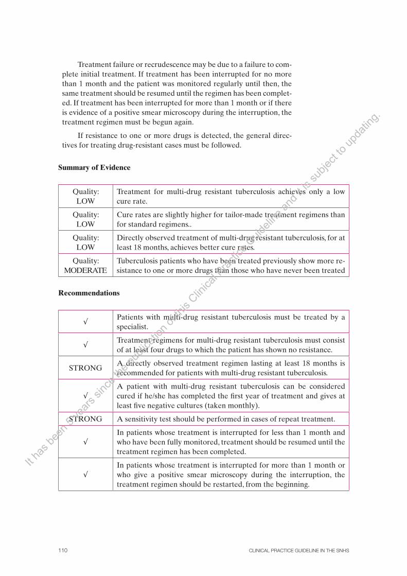
CLINICAL PRACTICE GUIDELINE IN THE SNHS110
Treatment failure or recrudescence may be due to a failure to com-plete initial treatment. If treatment has been interrupted for no more than 1 month and the patient was monitored regularly until then, the same treatment should be resumed until the regimen has been complet-ed. If treatment has been interrupted for more than 1 month or if there is evidence of a positive smear microscopy during the interruption, the treatment regimen must be begun again.
If resistance to one or more drugs is detected, the general direc-tives for treating drug-resistant cases must be followed.
Summary of Evidence
Quality: LOW
Treatment for multi-drug resistant tuberculosis achieves only a low cure rate.
Quality: LOW
Cure rates are slightly higher for tailor-made treatment regimens than for standard regimens..
Quality: LOW
Directly observed treatment of multi-drug resistant tuberculosis, for at least 18 months, achieves better cure rates.
Quality: MODERATE
Tuberculosis patients who have been treated previously show more re-sistance to one or more drugs than those who have never been treated
Recommendations
√Patients with multi-drug resistant tuberculosis must be treated by a specialist.
√Treatment regimens for multi-drug resistant tuberculosis must consist of at least four drugs to which the patient has shown no resistance.
STRONGA directly observed treatment regimen lasting at least 18 months is recommended for patients with multi-drug resistant tuberculosis.
√A patient with multi-drug resistant tuberculosis can be considered cured if he/she has completed the first year of treatment and gives at least five negative cultures (taken monthly).
STRONG A sensitivity test should be performed in cases of repeat treatment.
√In patients whose treatment is interrupted for less than 1 month and who have been fully monitored, treatment should be resumed until the treatment regimen has been completed.
√In patients whose treatment is interrupted for more than 1 month or who give a positive smear microscopy during the interruption, the treatment regimen should be restarted, from the beginning.
It has
been
5 ye
ars si
nce t
he pu
blica
tion o
f this
Clinica
l Prac
tice G
uideli
ne an
d it is
subje
ct to
upda
ting.
CLINICAL PRACTICE GUIDELINE ON THE DIAGNOSIS, TREATMENT AND PREVENTION OF TUBERCULOSIS 111
5.7. Monitoring Patients
Questions to Answer
• Whatisthebestwaytomonitorapatientwhobeginstuberculosistreatment?
In most cases, pulmonary tuberculosis can be treated and monitored in primary care, and isolation can be provided at home. However, if there are not sufficient resources available for suitable monitoring, if the physician does not have sufficient experience (a more and more common problem in countries with low endemic rates), or in certain cases such as those detailed in Figure 1, patients should be monitored in outpatient specialist care units, or may even need to be hospitalised for monitoring.
Prescribing appropriate treatment and strict treatment compli-ance should ensure that tuberculosis infection is eliminated or active tuberculosis is cured. Because of the repercussions of treatment failure for the patient and the public, clinical, analytical, bacteriological, and radiological monitoring are required throughout treatment.
Clinical monitoring: there should be an initial clinical visit during the first 2 weeks and one visit per month during the initiation phase. The frequency of subsequent visits depends on the patient’s develop-ment. The purpose of clinical monitoring is early detection of any tox-icity caused by treatment that might lead to failure to complete treat-ment, and to record treatment efficacy and compliance. Dosing must be adjusted if necessary, and potential interactions with any concomitant medication must be assessed.
Monitoring using blood tests (blood count and blood chemistry) must be performed early (at the clinical visit during the first 2 weeks) and approximately every 2 months during treatment. If there are treat-ment-related analytical alterations, tests must be performed more often until the parameters in question return to normal.
Radiological monitoring of patients with pulmonary tuberculosis is performed 2 months after the beginning of treatment and at the end of treatment, or at any time a complication is suspected. For extrapulmonary tuberculosis, monitoring involves the imaging tests that led to diagnosis.
Bacteriological monitoring in patients with pulmonary tuberculo-sis in whom samples can be obtained must be performed in the first, second, and fourth months and at the end of treatment. If the culture remains positive in the second month, the patient must be monitored monthly.
It has
been
5 ye
ars si
nce t
he pu
blica
tion o
f this
Clinica
l Prac
tice G
uideli
ne an
d it is
subje
ct to
upda
ting.

CLINICAL PRACTICE GUIDELINE IN THE SNHS112
Relapses, or recurrences, are defined as patients who develop ac-tive tuberculosis following completed treatment but who presented negative cultures during treatment (Appendix 4). The various RCTs conducted using standard, 6 month treatment regimens have shown rel-atively low relapse rates (1 2%) at the two-year follow-up123, and up to 3.4% at the five-year follow-up122. Many factors relating to the host, treatment, and disease are associated with a lower risk of relapse.
Quality: LOW
Various recent observational studies have evaluated the factors associated with a higher risk of relapse. The factors observed include extrapulmonary tuberculosis, pulmonary forms with cavitation on the initial X ray, HIV infection, low treatment compliance, drug resistance and intermittent treatment221-227.
The results of an SR of cohort studies showed similar results for intermittent regimens, with a risk of relapse almost 3 times higher than for daily treatment regimens130. More recently, an SR of RCTs and observational studies showed great variation in relapse rates following 6 month tuberculosis treatment and DOTS programmes (0 14%). The marked heterogeneity of the studies prevented the observation of fac-tors associated with recurrence (185). A recent retrospective study con-ducted in Spain calculated estimated the recurrence rateat 0.53 cases per 100 people per year of follow-up. Factors significantly associated with a higher risk of recurrence were male sex (HR 4.3; CI 95%, 1.3 to 14.6), immigrants (HR 3.2; CI 95%, 1.2 to 9), and parenteral drug abuse (HR 2.9; CI 95%, 1.3 to 6.4)228.
5.7.1 Monitoring Treatment Toxicity
Before treatment begins, patients must be informed about tuberculosis infection and disease, highlighting the importance of good treatment compliance and of avoiding alcohol. Appendix 12 outlines the main ac-tions that must be taken when monitoring patients treated for tubercu-losis who present side effects to treatment. Figure 2 presents a proposal for handling possible liver toxicity.
In general, analytical monitoring of liver function is performed be-fore the beginning of treatment and at regular intervals during treat-ment, particularly in those aged over 35, or with suspected toxicity re-gardless of age.
Patients must be informed as to how to recognise the signs of po-tential liver toxicity due to treatment. If necessary, they must interrupt their treatment and consult a physician. The appearance of fever, ma-laise, persistent vomiting, jaundice, or general deterioration with no ap-parent cause is grounds for suspecting liver toxicity..
It has
been
5 ye
ars si
nce t
he pu
blica
tion o
f this
Clinica
l Prac
tice G
uideli
ne an
d it is
subje
ct to
upda
ting.

CLINICAL PRACTICE GUIDELINE ON THE DIAGNOSIS, TREATMENT AND PREVENTION OF TUBERCULOSIS 113
Isoniazid and rifampicin, and to a lesser extent pyrazinamide, are causes of liver toxicity during tuberculosis treatment. Up to 33% of people who receive treatment may present some kind of liver involvement. Other factors have been associated with increased toxicity, mainly age (over 35). Various observational studies have found inconsistent results re-garding the risk of liver toxicity for women, alcohol consumption, slow acetylators, HIV-positive patients, and those with HVB or HCV141.
Quality: LOW
Summary of Evidence
Quality: LOW
The risk of recrudescence in patients who complete treatment accord-ing to standard regimens is fairly low.
Quality: LOW
Certain factors, such as cavitary pulmonary forms, extrapulmonary forms, HIV infection, poor treatment compliance, drug resistance, and intermittent treatment, increase the risk of recrudescence..
Quality: LOW:
Liver involvement is common during tuberculosis treatment, particu-larly in those aged over 35.
Recommendations
√If there are sufficient resources available, treatment, monitoring and isolation of most patients with pulmonary tuberculosis can be per-formed at the primary care level.
√In some clinical situations, specific monitoring by specialists, and even hospitalisation, is recommended(Figure 1).
√It is important to identify the main specialised institutions in each area or region to which patients must be referred if indicated.
√
Monitoring of individuals who begin tuberculosis treatment must con-sist of clinical, analytical, and microbiological monitoring during the first 2 weeks. It then must be followed by monthly clinical monitoring, analytical and bacteriological monitoring every 2 months, and radio-logical and bacteriological monitoring at the end of treatment.
√Clinical monitoring must be even closer if there are analytical changes or positive cultures after the second month, if complications are sus-pected and in children.
√
If a patient presents liver enzyme values five times higher than normal, or signs and symptoms of cholestasis, all potentially hepatotoxic medi-cation must be suspended. The patient must then be monitored closely to see whether treatment can be resumed or whether a treatment regi-men involving non-hepatotoxic drugs must be used instead.
It has
been
5 ye
ars si
nce t
he pu
blica
tion o
f this
Clinica
l Prac
tice G
uideli
ne an
d it is
subje
ct to
upda
ting.

CLINICAL PRACTICE GUIDELINE IN THE SNHS114
It has
been
5 ye
ars si
nce t
he pu
blica
tion o
f this
Clinica
l Prac
tice G
uideli
ne an
d it is
subje
ct to
upda
ting.

CLINICAL PRACTICE GUIDELINE ON THE DIAGNOSIS, TREATMENT AND PREVENTION OF TUBERCULOSIS 115
6. Preventing Tuberculosis
6.1. Isolation MeasuresQuestions to Answer
• How should a infectious patient with tuberculosis be placed in respiratory isola-tion?
• Whatmeasuresshouldbetakentoreducehospitaltransmissionoftuberculosis?
6.1.1 Transmission of the Disease
Tuberculosis is mainly transmitted through the air and is less infectious than other infec-tious diseases. Contagion requires close, sustained contact. When patients cough, sneeze, speak or sing, they expel tiny droplets (1 5 µm) which may contain one or several bacilli that are easily disseminated in rooms and air conditioning pipes. When inhaled, these droplets easily penetrate the airways, enabling bacilli to reach the alveoli. A well-ventilated room successfully eliminates most of these particles3.
If treated appropriately and early, patients with tuberculosis that is sensitive to first-line drugs cease to be infectious after approximately 2 weeks, when their cough begins to lessen and the bacillus concentration in their sputum falls. Identifying and treating cases of tuberculosis are therefore the most effective ways to control the disease229.
6.1.2 Nosocomial Infection Control Measures
Hospital transmission of tuberculosis, both to patients and to staff, was clearly documented even before antibiotics were used, but the develop-ment of effective treatment successfully reduced it, so many hospital control programmes were discontinued. Hospital outbreaks of multi-drug resistant tuberculosis in the 1980s caused many cases, particularly in HIV-positive patients, with high mortality rates. These outbreaks stimulated more research into administrative, structural and individual control measures, which led to a significant reduction in hospital trans-mission of tuberculosis, although the isolated impact of each measure is unknown230,231.
It has
been
5 ye
ars si
nce t
he pu
blica
tion o
f this
Clinica
l Prac
tice G
uideli
ne an
d it is
subje
ct to
upda
ting.

CLINICAL PRACTICE GUIDELINE IN THE SNHS116
A recent SR of observational studies (cohorts, records) showed that in high-income countries, 24% of healthcare staff had latent tuber-culosis infection (positive tuberculin tests), although the percentages varied a great deal (between 4% and 46%). Working in internal medi-cine or pulmonology units, length of employment history, and number of in-patients with tuberculosis at the establishment, whether with or without concomitant HIV infection, were considered to be risk factors. All the studies highlighted that healthcare staff exposed to this type of patients had a higher risk of becoming infected with M. tuberculosis than the general public230.
Quality: LOW
A panel of experts developed a narrative review on the association between ventilation (airflows) and aerial transmission of infections in buildings. A total of 40 studies were included, all of which were obser-vational and 18 of which concerned tuberculosis. The conclusion of this review was that there is insufficient information on the minimum venti-lation requirements in hospitals and non-hospital settings232.
Quality: LOW
Nowadays there is a considerable structure of measures intended to reduce hospital transmission of tuberculosis. Control measures must be applied to all patients suspected of having laryngeal or pulmonary tuberculosis, and the standard precautions that are a part of care for all patients must never be overlooked233.
6.1.2.1 Organisational Measures Organisational measures are designed to reduce the risk of exposure and infection.
Administrative controls designed to provide rapid detection, isolation, diagnosis, and treat-ment of tuberculosis patients are very important. They consist of:
1. Identifying, isolating, diagnosing, and beginning treatment promptly for individu-als with signs or symptoms that suggest tuberculosis, in order to avoid unnecessary delays.
2. Appropriate respiratory isolation of patients with pulmonary or laryngeal tubercu-losis. Patients must wear surgical masks while they are in communal areas, for:
• Shortoutpatientconsultations
• Transfers (including transport by healthcare services) between medical estab-lishments for diagnostic tests
• Waitingareasforpatientswithsuspectedtuberculosis.
3. Patients with the same kind of drug-sensitive tuberculosis may share isolation wards. Visits and entries of healthcare into isolation wards must be kept to a minimum.
4. Scheduling medical procedures at the end of the day for patients with suspected or confirmed tuberculosis.
5. Determining the risk of transmission of tuberculosis by area. The areas with the
It has
been
5 ye
ars si
nce t
he pu
blica
tion o
f this
Clinica
l Prac
tice G
uideli
ne an
d it is
subje
ct to
upda
ting.

CLINICAL PRACTICE GUIDELINE ON THE DIAGNOSIS, TREATMENT AND PREVENTION OF TUBERCULOSIS 117
highest risk are emergency units, intensive care units, bronchoscopy rooms, sputum induction rooms, inhaled treatment rooms, operating rooms, microbiology labora-tories, autopsy rooms, inpatient wards areas, and outpatient clinics33,233-234.
6.1.2.2 Structural Measures Structural measures are as follows:
1. Suitable ventilation and air circulation systems in areas with a risk of transmission, with at least six air changes per hour.
2. Air isolation wards, suitably prepared with negative pressure. The differential pressure must be 2.5 Pa when compared to the pressure outside the wards(Appendix 10).
3. Correct management and removal of waste generated by tuberculosis patients.
4. Cleaning and disinfecting critical, semi-critical, and non-critical material without taking additional measures, except for bronchoscopes, as these have been associ-ated with hospital outbreaks of tuberculosis. A high-performance disinfectant must be used for these items, following effective cleaning to remove organic material. Both at home and in hospitals, bedrooms, and materials used by patients must be cleaned and disinfected according to general procedures; no additional measures are needed. Tuberculosis is not transmitted through bedding, clothing, personal hy-giene equipment, or food.
5. There must be one air-isolated room for every 120 hospital beds, although this number may rise, depending on the number of patients hospitalised with tuberculo-sis each year. If there are no such rooms available, all patients in whom tuberculosis is suspected or confirmed must wear surgical masks. They must also be instructed to change their masks if they become damp. Basic hygiene measures such as cover-ing the mouth with a tissue when coughing while not wearing masks must also be observed233.
6.1.2.3 Personal Protection Measures 1. Personal protection equipment to prevent inhalation of infectious particles. These
are additional measures that should be taken when the risk of contagion cannot be controlled using organisational measures. Staff exposed to tuberculosis patients must use FFP3 respirators (Appendix 10) harmonised according to European regu-lations. These must be used in the following circumstances:
• Whenbronchoscopies,sputuminductiontechniques,secretionaspirationoraer-osol treatment are administered to patients with suspected or confirmed tuber-culosis
• Autopsiesofpatientswithclinicalindicationsoftuberculosisorconfirmedtuber-culosis
• Drainageoftuberculousabscesses
• AmbulancetransfersofthesepatientsIt h
as be
en 5
years
sinc
e the
publi
catio
n of th
is Clin
ical P
ractic
e Guid
eline
and i
t is su
bject
to up
datin
g.

CLINICAL PRACTICE GUIDELINE IN THE SNHS118
• Entryintoisolationroomsduringthefirst15daysofcorrecttreatment(prefer-ably using DOT)
• Laboratoriesthatprocesssamplestoidentifymycobacteria
6.1.2.4 Specific Tuberculosis Prevention and Control Measures for Healthcare Staff
Preventive measures for healthcare staff are as follows:
1. Tuberculin tests for healthcare staff who work in high-risk areas, when they are hired and regularly if tests are negative. The frequency of testing depends on whether or not staff usually work in areas with higher risks of infection. Testing is twofold: an initial test, and if this is negative a second test performed 1 week later. If the first tuberculin test is positive, the employee is considered to be infected already. Con-version is then ruled out, and treatment for latent infection must be considered once active tuberculosis has been ruled out. If the second tuberculin test is positive, this is a booster effect, and conversion is ruled out233. No studies evaluating the performance of IGRA tests in employees have been conducted.
2. Ongoing training for healthcare staff, concentrating particularly on identifying the signs and symptoms of tuberculosis, how it is transmitted how to prevent it.
3. Healthcare staff with any kind of immunosuppression must be deployed in areas at low risk of tuberculosis infection (Appendix 10).
4. Healthcare staff with suspected tuberculosis must be examined meticulously. They must not return to work until a diagnosis of tuberculosis has been ruled out, or until there is written confirmation that they have responded to treatment and are no longer infectious. An antibiogram must always be performed to investigate the development of drug resistance. The medical professional in charge (the company physician) must take charge of prescribing the correct treatment and conducting a contact study for the employee233.
6.1.3 Treating Patients in Hospitals
Patients with suspected pulmonary or laryngeal tuberculosis must have as little close contact with other patients and other individuals in medical establishments as possible. Medical establishments them-selves must minimise the risk of transmission and should only propose hospitalisation in cases in which this is essential or home isolation is impossible20,234.
It has
been
5 ye
ars si
nce t
he pu
blica
tion o
f this
Clinica
l Prac
tice G
uideli
ne an
d it is
subje
ct to
upda
ting.

CLINICAL PRACTICE GUIDELINE ON THE DIAGNOSIS, TREATMENT AND PREVENTION OF TUBERCULOSIS 119
A patient with suspected or confirmed tuberculosis is considered to be infectious in either of the following two situations:
1. If the patient has a spontaneous cough or coughs during sputum induction, or presents a positive smear microscopy
2. If the patient is not receiving treatment, has just begun treat-ment or does not present clinical or bacteriological improve-ment despite medication
However, there are also other factors that may be grounds for sus-pecting that a patient is infectious. These include the spread the disease in the body, cavities on a chest X ray, the level of positivity shown in a smear microscopy, frequency and severity of coughing, probability of drug-resistant tuberculosis, and the nature and circumstances of the in-dex case.
In contrast, a patient is considered no longer infectious if20,233:
1. There is clinical improvement and
2. Correct standard treatment has been followed for 3 weeks and/or
3. Three consecutive negative smear microscopies of sputum are obtained at intervals of up to 24 hours, at least one taken in the morning
A negative sputum smear microscopy alone does not mean that a patient is not infectious. Rates of up to 17% have been recorded for secondary cases associated with patients with pulmonary tuberculosis and a negative smear microscopy, using molecular epidemiology tech-niques235.
The criteria for air isolation and the time at which isolation can be ended are the result of consensus. A prospective study conducted in Spain, involving 184 patients, showed that 22% of them showed negative smear microscopies and cultures 2 weeks after hospitalisation and the beginning of treatment, and 53% showed these results after 4 weeks236. Table 8 suggests some criteria for beginning and ending air isolation.
Quality: LOW
It has
been
5 ye
ars si
nce t
he pu
blica
tion o
f this
Clinica
l Prac
tice G
uideli
ne an
d it is
subje
ct to
upda
ting.

CLINICAL PRACTICE GUIDELINE IN THE SNHS120
Table 8: Criteria for beginning and ending isolation
Criteria for beginning air isolation
Clinical symptoms and signs and apical infiltration or cavities on chest X ray
A patient diagnosed with pulmonary tuberculosis who stops taking medication during the initial phase or does not comply well with treatment, until the results of a smear microscopy is available
Children with pulmonary cavities
Patient diagnosed with extrapulmonary tuberculosis but with respiratory signs or symptoms
Criteria for ending air isolation
Tuberculosis diagnosis not confirmed according to:Two smear microscopies of spontaneous sputum on two different days, both negativeA single negative smear microscopy of sputum obtained by induction, fibre-optic bronchoscopy, or endotracheal tubeTwo negative cultures taken one month apart in patients with multi-drug resistant tuberculosis
Adapted from Centers for Disease Control and Prevention: Guidelines for preventing the transmission of Mycobacterium tu-berculosis in healthcare settings, MMWR, 2005 Dec 30; 54 (RR-17): 1-141 and John Hopkins Hospital: Interdisciplinary Clinical Practice Manual for Tuberculosis Control, 2001 (233, 238)
6.1.4 Home Isolation Measures
Patients who are isolated at home must be informed of how tuberculosis transmitted in order to achieve maximum cooperation in the hygiene measures that must be taken. As in the hospital, the simplest way to prevent the spread of bacilli is to cover the mouth with a tissue when coughing or sneezing. Attempts must also be made to ensure that the patient stays in a sunny, well-ventilated room. Patients must wear surgical masks when they go outside. This precaution should usually be taken until sputum tests are negative234,237.
Summary of Evidence
-Tuberculosis is mainly transmitted through the air. Contagion requires close, sustained contact.
Quality:LOW
In most cases of pulmonary tuberculosis, smear microscopies turn negative between the second and fourth week of standard treatment that is performed correctly. However, by themselves,these do not in indicate that patients are no longer infectious.
Quality: LOW
There is no conclusive evidence on the minimum ventilation requirements for hospitals or non-hospital settings.
Quality: LOW
Healthcare staff are at greater risk of becoming infected with M. tuberculosis than the general public.
It has
been
5 ye
ars si
nce t
he pu
blica
tion o
f this
Clinica
l Prac
tice G
uideli
ne an
d it is
subje
ct to
upda
ting.
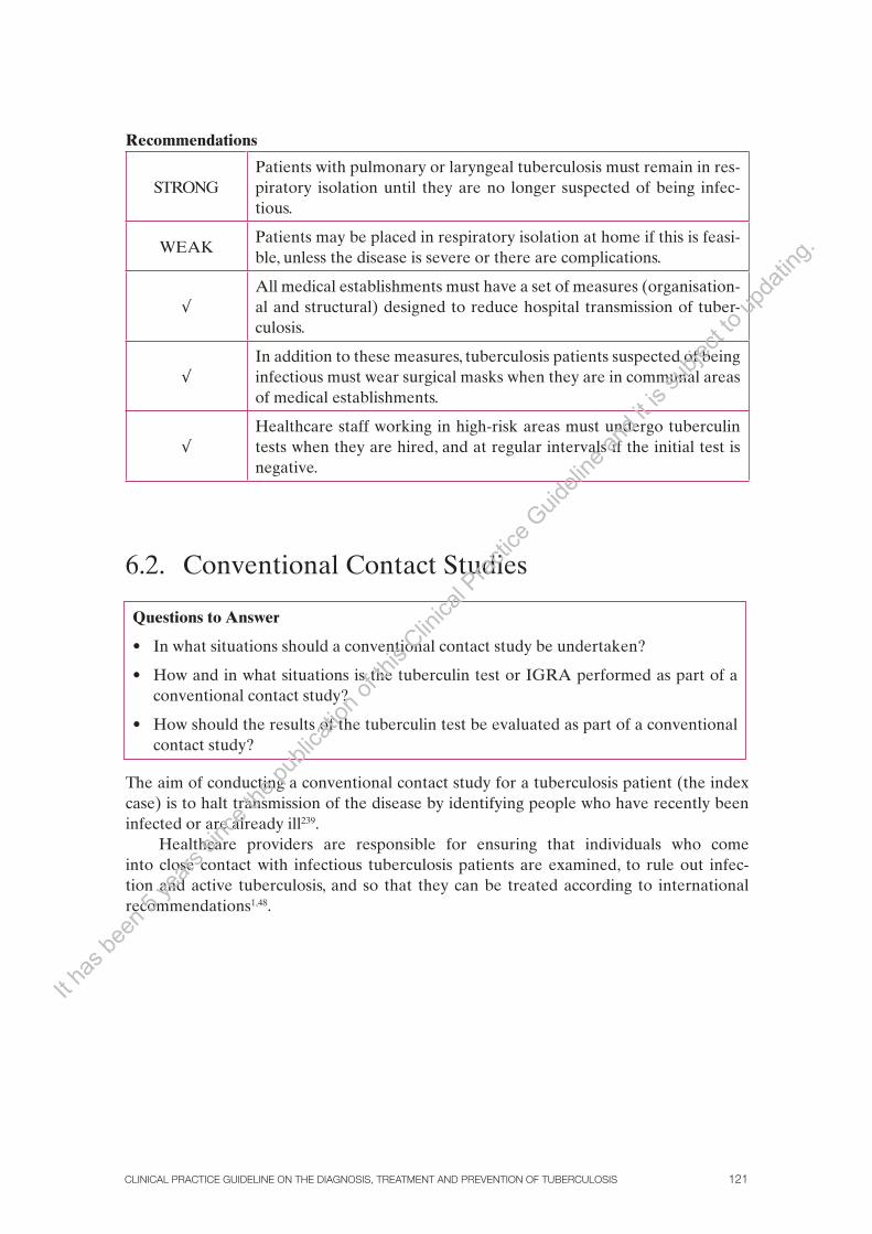
CLINICAL PRACTICE GUIDELINE ON THE DIAGNOSIS, TREATMENT AND PREVENTION OF TUBERCULOSIS 121
Recommendations
STRONGPatients with pulmonary or laryngeal tuberculosis must remain in res-piratory isolation until they are no longer suspected of being infec-tious.
WEAKPatients may be placed in respiratory isolation at home if this is feasi-ble, unless the disease is severe or there are complications.
√All medical establishments must have a set of measures (organisation-al and structural) designed to reduce hospital transmission of tuber-culosis.
√In addition to these measures, tuberculosis patients suspected of being infectious must wear surgical masks when they are in communal areas of medical establishments.
√Healthcare staff working in high-risk areas must undergo tuberculin tests when they are hired, and at regular intervals if the initial test is negative.
6.2. Conventional Contact Studies
The aim of conducting a conventional contact study for a tuberculosis patient (the index case) is to halt transmission of the disease by identifying people who have recently been infected or are already ill239.
Healthcare providers are responsible for ensuring that individuals who come into close contact with infectious tuberculosis patients are examined, to rule out infec-tion and active tuberculosis, and so that they can be treated according to international recommendations1,48.
Questions to Answer
• Inwhatsituationsshouldaconventionalcontactstudybeundertaken?
• HowandinwhatsituationsisthetuberculintestorIGRAperformedaspartofaconventional contact study?
• Howshouldtheresultsofthetuberculintestbeevaluatedaspartofaconventionalcontact study?
It has
been
5 ye
ars si
nce t
he pu
blica
tion o
f this
Clinica
l Prac
tice G
uideli
ne an
d it is
subje
ct to
upda
ting.

CLINICAL PRACTICE GUIDELINE IN THE SNHS122
6.2.1 Performance of Contact Studies
The potential performance of contact studies depends on the incidence and prevalence of tuberculosis in the environment in question. In coun-tries with low incidences (raw incidence less than 10 cases per 100,000 inhabitants), between five and ten contacts are found for each new case of tuberculosis18. Of these, approximately 30% have latent tuberculosis infection, and between 1% and 4% have active tuberculosis240 241. In contrast, in countries with high incidences, around 50% of family con-tacts are infected, and between 10% and 20% already have active tu-berculosis when the study begins48,239. The prevalence of tuberculosis infection increases with age (approximately 9% per year) regardless of sex, exposure to the index case, or BCG vaccination242.
Quality:LOW
No SRs of RCTs evaluating the performance of contact studies have been located. The evidence available is taken from various obser-vational studies.
Quality:LOW
A recent SR of observational studies (41 prospective or cross-sec-tional studies) analysed the performance of contact studies in 17 develop-ing countries. The diagnosis rates for active tuberculosis and latent infec-tion obtained from the study of 13,602 home contacts of 3218 index cases of pulmonary tuberculosis were calculated. The studies were conducted in countries in Africa (49%), Asia (29%) and Central and South America (22%). The definitions of active tuberculosis and latent infection varied between studies. Specifically, tuberculin test measurements of 5 mm, 8 mm, and 10 mm were used to diagnose latent infection.
Of all the patients studied, 4.5% (CI 95%, 4.3% to 4.8%) pre-sented active tuberculosis when the research was conducted, whereas for the 23 studies that included clinically and microbiologically con-firmed cases, performance was 2.3% (CI 95%, 2.1% to 2.5%). A total of 51.4% (CI 95%, 50.6% to 52.2%) (19 studies) of the contacts studied presented latent tuberculosis infection. In four studies that evaluated performance in HIV-positive patients, the result was similar, and study results were homogeneous. It is noteworthy that patients less than 5 years old had a higher rate of active tuberculosis (8.5%) and a lower rate of latent infection (30.4%). The performance of contact studies in these countries is high, although diagnosis is confirmed in only half of them. People who live in the same household as someone with active tuberculosis are an at-risk group for active tuberculosis or latent infec-tion, particularly if they are less than 5 years old243.
It has
been
5 ye
ars si
nce t
he pu
blica
tion o
f this
Clinica
l Prac
tice G
uideli
ne an
d it is
subje
ct to
upda
ting.

CLINICAL PRACTICE GUIDELINE ON THE DIAGNOSIS, TREATMENT AND PREVENTION OF TUBERCULOSIS 123
In Spain, a prospective analysis of the contact studies conducted in the healthcare district of Biscay over 10 years (1995-2004) to determine the incidence of tuberculosis among contacts of the index case, and the risk factors associated with becoming ill, was published in 2007. The study showed a tuberculosis incidence of 1.1% (66 secondary cases in 5,444 contacts studied), most of them diagnosed during the first year. Untreated contacts’ risk of developing the disease was higher in the first and second years, with incidences of 864 per 100,000 and 90 per 100,000 respectively. From the second year onwards the incidence fell significantly. The study showed a significant correlation between the following risk factors and becoming ill: close contact with the index case, positive smear microscopy, tuberculin test diameter greater than 10 mm, and age under 30244. An earlier study, also conducted in Spain, involving 3,071 contacts with 6 years’ monitoring showed a higher tu-berculosis incidence (5.7%), although the factors associated with the risk of becoming ill were very similar245.
Quality:LOW
6.2.2 Conducting Contact Studies
Contact studies are begun when pulmonary, laryngeal or pleural tuberculosis is suspected or confirmed. It takes particularly high priority when smear microscopy of sputum is posi-tive and/or when a pulmonary cavity is detected on a patient’s chest X ray33. Children, par-ticularly very small children, suffer from tuberculosis but rarely transmit it246. Figure 2 shows the criteria for beginning a contact study.
Table 9 shows the length of time before diagnosis of the index case during which the patient in question is considered potentially infectious, according to the form of tubercu-losis and the characteristics of the case. This time window is when the potential contacts of the index case must be studied33.
A contact study of a patient’s family and friends (the contacts at greatest risk of con-tagion) can be conducted by the same medical team that diagnoses and treats the index case. Every medical establishment must know the identities of the expert professionals that will be in charge of handling this situation. Public healthcare services must coordinate contact studies conducted both in medical establishments and in the public234,247.
Diagnosis of three or more cases of tuberculosis close together in space or time, or one or more cases of tuberculosis following the first case detected (the index case), is considered to be an epidemic. Any case in a child may also indicate an outbreak, and so the relevant investigation must be conducted. The risk of transmitting tuberculosis is considered to be higher in all these circumstances. As a result, extremely meticulous, strict diagnostic and preventive measures must be taken1,234.
It has
been
5 ye
ars si
nce t
he pu
blica
tion o
f this
Clinica
l Prac
tice G
uideli
ne an
d it is
subje
ct to
upda
ting.
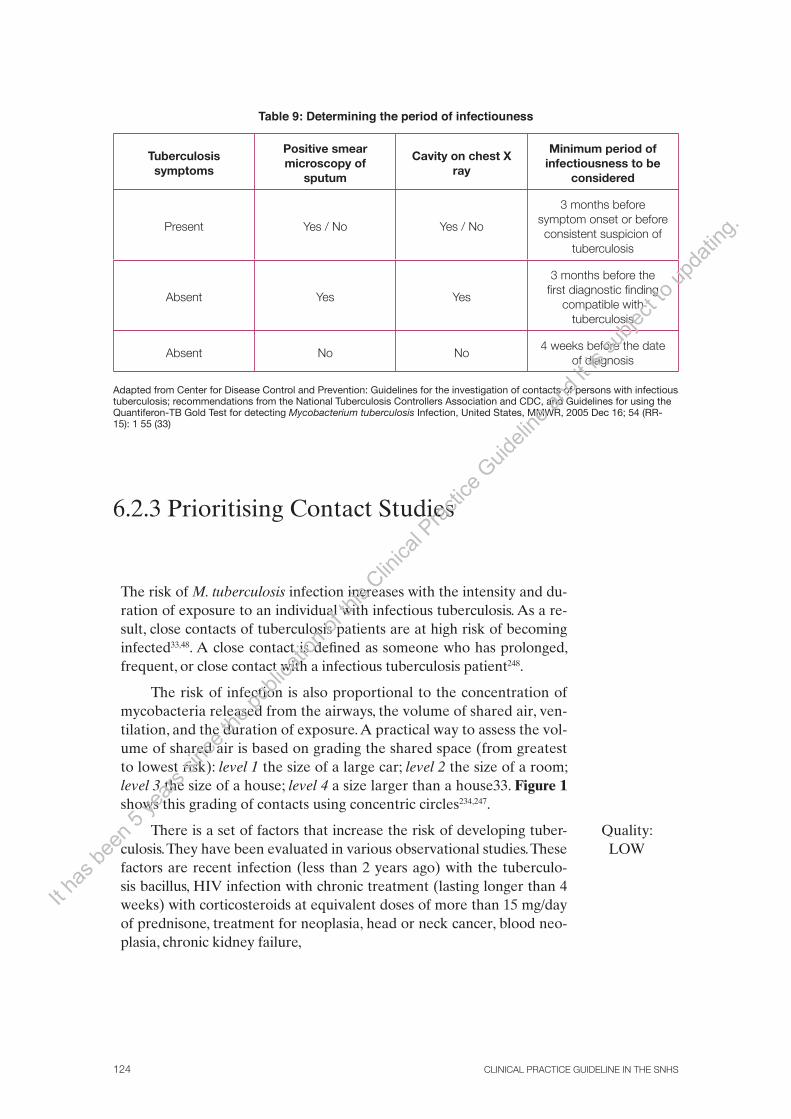
CLINICAL PRACTICE GUIDELINE IN THE SNHS124
Table 9: Determining the period of infectiouness
Tuberculosis symptoms
Positive smear microscopy of
sputum
Cavity on chest X ray
Minimum period of infectiousness to be
considered
Present Yes / No Yes / No
3 months before symptom onset or before consistent suspicion of
tuberculosis
Absent Yes Yes
3 months before the first diagnostic finding
compatible with tuberculosis
Absent No No4 weeks before the date
of diagnosis
Adapted from Center for Disease Control and Prevention: Guidelines for the investigation of contacts of persons with infectious tuberculosis; recommendations from the National Tuberculosis Controllers Association and CDC, and Guidelines for using the Quantiferon-TB Gold Test for detecting Mycobacterium tuberculosis Infection, United States, MMWR, 2005 Dec 16; 54 (RR-15): 1 55 (33)
6.2.3 Prioritising Contact Studies
The risk of M. tuberculosis infection increases with the intensity and du-ration of exposure to an individual with infectious tuberculosis. As a re-sult, close contacts of tuberculosis patients are at high risk of becoming infected33,48. A close contact is defined as someone who has prolonged, frequent, or close contact with a infectious tuberculosis patient248.
The risk of infection is also proportional to the concentration of mycobacteria released from the airways, the volume of shared air, ven-tilation, and the duration of exposure. A practical way to assess the vol-ume of shared air is based on grading the shared space (from greatest to lowest risk): level 1 the size of a large car; level 2 the size of a room; level 3 the size of a house; level 4 a size larger than a house33. Figure 1 shows this grading of contacts using concentric circles234,247.
There is a set of factors that increase the risk of developing tuber-culosis. They have been evaluated in various observational studies. These factors are recent infection (less than 2 years ago) with the tuberculo-sis bacillus, HIV infection with chronic treatment (lasting longer than 4 weeks) with corticosteroids at equivalent doses of more than 15 mg/day of prednisone, treatment for neoplasia, head or neck cancer, blood neo-plasia, chronic kidney failure,
Quality:LOW
It has
been
5 ye
ars si
nce t
he pu
blica
tion o
f this
Clinica
l Prac
tice G
uideli
ne an
d it is
subje
ct to
upda
ting.

CLINICAL PRACTICE GUIDELINE ON THE DIAGNOSIS, TREATMENT AND PREVENTION OF TUBERCULOSIS 125
malabsorption syndrome, post-transplant immunosuppression, use of drugs such as tumour necrosis factor-alpha blockers, silicosis, diabetes mellitus, gastrectomy and/or jejunoileal bypass, low weight (more than 10% below ideal weight), and parenteral drug abuse33,242,248. Any con-tact with an index case who falls into any of these high-risk categories must be classified as a high-priority contact and be examined to rule out active tuberculosis and latent infection. Asymptomatic individuals with chest X ray images indicating old, untreated tuberculosis form a significant group33.
Tables 10 and 11 show a proposal for prioritising contact studies (high, medium, low) for patients with respiratory tuberculosis and a positive or negative smear microscopy.
Figure 1: Classification of contacts using concentric circles (adapted from Contact Study Group of the Barcelona Tuberculosis Research Unit (BTRU): Consensus document on contact studies in
tuberculosis patients, Med Clin (Barc) 1999; 112: 151-156) (247)
Work and school
TB
12
3
Concentric circles, location of contact
Leisure area
Home
1st circle: close daily contact >6 hours2nd circle: frequent daily contact <6 hours3rd circle: sporadic contact, not daily
It has
been
5 ye
ars si
nce t
he pu
blica
tion o
f this
Clinica
l Prac
tice G
uideli
ne an
d it is
subje
ct to
upda
ting.

CLINICAL PRACTICE GUIDELINE IN THE SNHS126
Table 10: Priorities of contact studies for patients with pulmonary, laryngeal, or pleural tuberculo-sis and positive smear microscopies of sputum or cavities on chest X rays
Situation Priority
Contacts in the same household or close daily contact (first circle) HIGH
Contacts less than 5 years old HIGH
Contacts with risk factors* HIGH
Contacts in residential institutions HIGH
Contacts exposed during medical procedures (bronchoscopies, autopsies, etc.) HIGH
Contacts aged 5-15 MEDIUM
Less frequent contact (second circle) MEDIUM
Any contact not classified as high- or medium-priority LOW
*HIV infection or other medical circumstances such as silicosis, diabetes mellitus, chronic kidney failure, blood diseases (leu-kaemias, lymphomas), head and neck neoplasia, weight loss greater than 10%, gastrectomy, jejunoileal bypass.
Table 11: Priorities of contact studies for patients with pulmonary, laryngeal, or pleural tuberculo-sis with negative smear microscopies of sputum and no cavities on chest X rays
Situation Priority
Contacts less than 5 years old HIGH
Contacts with risk factors* HIGH
Contacts exposed during medical procedures (bronchoscopies, autopsies, etc.) HIGH
Contacts in the same household or close daily contact (first circle) MEDIUM
Contacts in residential institutions MEDIUM
Any contact not classified as high- or medium-priority LOW
*HIV infection or other medical circumstances such as silicosis, diabetes mellitus, chronic kidney failure, blood diseases (leu-kaemias, lymphomas), head and neck neoplasia, weight loss greater than 10%, gastrectomy, jejunoileal bypass
It has
been
5 ye
ars si
nce t
he pu
blica
tion o
f this
Clinica
l Prac
tice G
uideli
ne an
d it is
subje
ct to
upda
ting.
CLINICAL PRACTICE GUIDELINE ON THE DIAGNOSIS, TREATMENT AND PREVENTION OF TUBERCULOSIS 127
It is generally thought that contact studies must also be conducted in low-priority contacts when recent transmission is suspected in the following cases:
1. A high rate of tuberculosis infection or active tuberculosis is detected among the high-priority contacts studied
2. Tuberculin converters are present
3. Infection is detected in contacts under 5 years old33
6.2.4 Conducting Contact Studies: The Tuberculin Test
Until a few years ago the tuberculin test was the only test available to diagnose M. tu-berculosis infection. The tuberculin test has been in use since 1890 and is relatively cheap, easy to use and accessible. It is based on a delayed hypersensitivity reaction to a group of bacillus antigens obtained from a purified protein derivative (PPD). Many of these antigens are common to M. tuberculosis, M. bovis (bacillus Calmette–Guérin, or BCG) and various other mycobacteria that do not cause tuberculosis. As a result, the specificity of the tuberculin test is low in (BCG ) vaccinated populations and/or those exposed to non-tuberculous mycobacteria. Its sensitivity can also be low in individuals with altered immunity, patients with severe forms of tuberculosis and/or malnutrition and other groups. In general, its sensitivity and specificity are around 90 100%, although in environments with low tuberculosis prevalences it has a high false positive rate22,251.
The tuberculin test must be performed using the Mantoux technique, in which PPD is injected into the dermis. Other procedures are less reliable than the Mantoux technique. Administration and interpretation of the test are subject to variation, which is why it must be performed by trained staff.
A brief description of the Mantoux technique for the tuberculin test is given below248,250:
1. In Spain, PPD RT 23 is used with Tween 80. The dose of 2 units per 0.1 ml is the bioequivalent of the international tuberculin standard (PPD S)17,252.
2. Inject 0.1 ml just below the top layer of the skin (intradermally, not subcutane-ously) on the anterior surface of the forearm. The injection will cause a slight rising, or bump, on the skin, 6 10 mm in diameter. Disposable syringes and needles must be used, and all infection control procedures must be followed, including the use of gloves and suitable containers to dispose of needles.
3. After administration, patients must be instructed not to rub or scratch the test area or cover it with a plaster. The area may be washed and dried if necessary, but not using irritating substances.
The tuberculin test must be performed as soon as possible after the index case has been diagnosed, and must be repeated 8 or 12 weeks later if the first test is negative (Table 12). It cannot be administered to patients already known to be positive33.
It has
been
5 ye
ars si
nce t
he pu
blica
tion o
f this
Clinica
l Prac
tice G
uideli
ne an
d it is
subje
ct to
upda
ting.

CLINICAL PRACTICE GUIDELINE IN THE SNHS128
Table 12: Timeframes to begin monitoring contacts exposed to tuberculosis
Type of contact Working days until initial
examination (examination and tuberculin test)
Working days from initial examination to end of
study
HIGH priority: contact with index case, positive smear microscopy or image of cavity in lung
7 5
HIGH priority: contact with index case, negative smear microscopy
7 10
MEDIUM priority 14 10
6.2.5 Interpreting Tuberculin Test Results
Results must be interpreted according to an individual’s risk of developing tuberculosis if infected, and according to his/her level of exposure. The three cut-off points usually ap-plied (≥5 mm, ≥10 mm and ≥15 mm) are used to improve the test’s sensitivity and specifi-city when screening at-risk groups (Appendix 11).For contacts, the tuberculin test should be considered positive if induration is ≥5 mm22. The appearance of vesicles or necrosis is highly suggestive of tuberculosis infection17.
In individuals who have been vaccinated against tuberculosis it is impossible to tell whether the reaction is due to M. tuberculosis infection or immune memory. For practical purposes, vaccination history should be ignored in groups at high risk of infection. The tu-berculin test is of no use in individuals with a prior diagnosis of tuberculosis or those who have completed tuberculosis treatment. It is generally well tolerated, although it should not be performed on areas of skin affected by burns or eczema. No teratogenic effect has been recorded following administration during pregnancy26,33,253.
MMR vaccination can cause false negative results to the test. It is generally recom-mended that the tuberculin test be administered either on the same day as MMR vaccina-tion or 6 weeks later. False negatives can also be caused by poor inoculation technique, ill-nesses, and situations that cause immunosuppression, including tuberculosis. Also, it takes 8 12 weeks following M. tuberculosis infection for sensitised T cells in the blood to recog-nise the tuberculin inoculated into the dermis33,253.
Tuberculin tests must be read by staff trained to do so. The test is read 48 72 hours after intradermal injection of PPD, using a flexible ruler to measure the induration around the injection site. The greatest diameter of the induration (at its widest point) must be measured, excluding any reddened area around the injection site. The measurement ob-tained (in millimetres), not just whether the test is positive or negative, must always be recorded. The test is interpreted in the same way if between 72 hours and 1 week have elapsed since the injection.
Figure 4 shows how to examine, treat and monitor contacts.
Table 13 describes the stages of a complete contact study.It has
been
5 ye
ars si
nce t
he pu
blica
tion o
f this
Clinica
l Prac
tice G
uideli
ne an
d it is
subje
ct to
upda
ting.

CLINICAL PRACTICE GUIDELINE ON THE DIAGNOSIS, TREATMENT AND PREVENTION OF TUBERCULOSIS 129
Table 13: Stages of a conventional contact study
Examination and classification of the index case
Select contacts to be examined (electoral roll). Take detailed clinical history regarding tuberculosis
Tuberculin test in high- and medium-priority contacts during the first week
Chest X rays of individuals who test positive to the tuberculin test to rule out active tuberculosis
Prescribe treatment for latent tuberculosis infection (once active tuberculosis has been ruled out) or primary prophylaxis
Decide whether or not to expand the study to other contacts, depending on the results obtained
Tuberculin test repeated 8 12 weeks later in contacts who test negative to the first tuberculin test
Rule out active tuberculosis in converters and end primary prophylaxis in those who test negative to the tuberculin test again and are no longer at risk of contagion
Monitor treatment for latent tuberculosis infection indicated prescribed according to a treatment regimen
End of study, quantitative evaluation of study performance
In recent years new techniques have emerged, designed to improve diagnosis of latent tuberculosis infection. These techniques are based on the detection of interferon-gamma and are known as IGRAs (interferon-gamma release assays). There are currently two tests on the market: QFT (Quantiferon Tuberculosis Gold or Quantiferon Tuberculosis Gold In-Tube) and T SPOT.TB. They have not yet been systematically introduced in standard clinical practice. Results for this type of test are described in detail in point 4.1.2 on the diagnosis of tuberculosis infection.
6.2.6 The Booster Effect
The booster effect occurs when an individual has come into contact with (is sensitised to) atypical mycobacteria, the BCG vaccine, or the tuberculosis bacillus itself. The tuber-culin test response of those who have come into contact with the tuberculosis bacillus and are infected with M. tuberculosis decreases over time. As this is a temporary phenomenon, it is more common in those aged over 55, which may lead to false negative readings.
In these individuals, an initial test may yield a borderline or negative result, and a second test may yield a clearly positive result (due to reactivation of immunity). This can be confused with tuberculin conversion.
To distinguish between the booster effect and tuberculin conversion due to recent infection, two tests are performed 1 week apart. This two-step tuberculin test is useful in those aged over 55 who have received the BCG vaccine, and those in whom the test has to be repeated regularly, such as healthcare staff and those living in residential institu-tions. This procedure is not recommended for high- or medium-priority contacts in con-tact studies33.
It has
been
5 ye
ars si
nce t
he pu
blica
tion o
f this
Clinica
l Prac
tice G
uideli
ne an
d it is
subje
ct to
upda
ting.
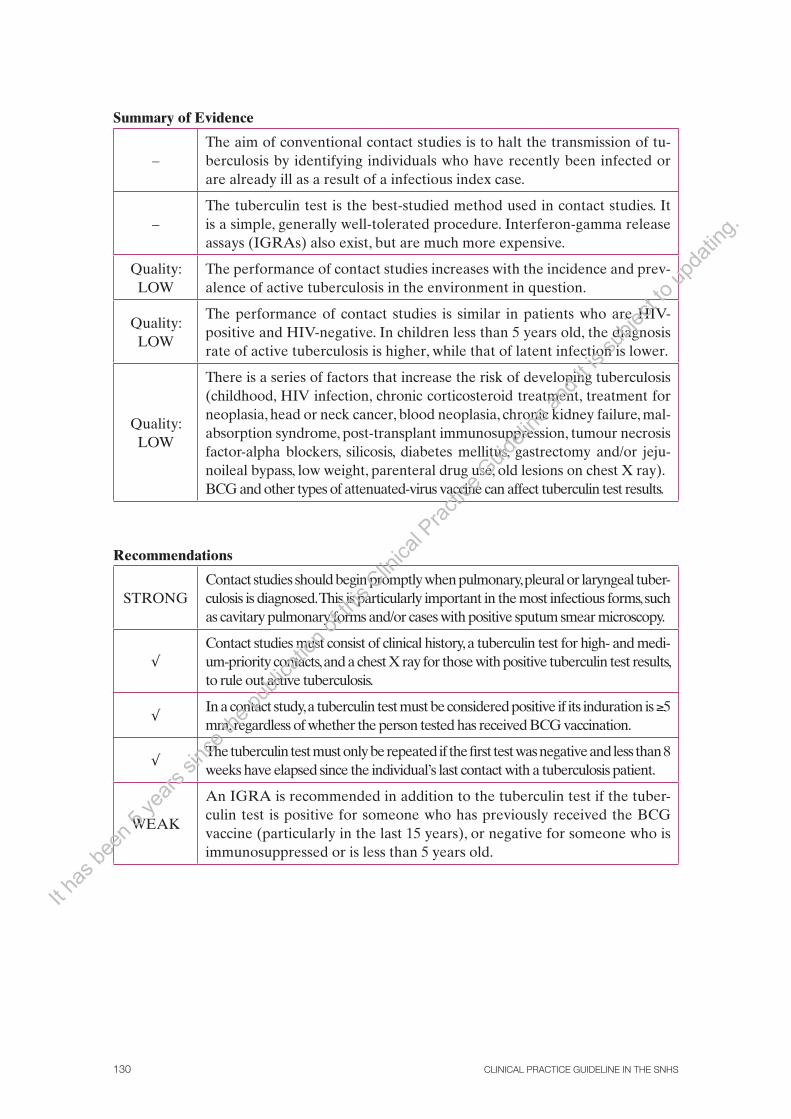
CLINICAL PRACTICE GUIDELINE IN THE SNHS130
Summary of Evidence
–The aim of conventional contact studies is to halt the transmission of tu-berculosis by identifying individuals who have recently been infected or are already ill as a result of a infectious index case.
–The tuberculin test is the best-studied method used in contact studies. It is a simple, generally well-tolerated procedure. Interferon-gamma release assays (IGRAs) also exist, but are much more expensive.
Quality: LOW
The performance of contact studies increases with the incidence and prev-alence of active tuberculosis in the environment in question.
Quality: LOW
The performance of contact studies is similar in patients who are HIV-positive and HIV-negative. In children less than 5 years old, the diagnosis rate of active tuberculosis is higher, while that of latent infection is lower.
Quality: LOW
There is a series of factors that increase the risk of developing tuberculosis (childhood, HIV infection, chronic corticosteroid treatment, treatment for neoplasia, head or neck cancer, blood neoplasia, chronic kidney failure, mal-absorption syndrome, post-transplant immunosuppression, tumour necrosis factor-alpha blockers, silicosis, diabetes mellitus, gastrectomy and/or jeju-noileal bypass, low weight, parenteral drug use, old lesions on chest X ray).BCG and other types of attenuated-virus vaccine can affect tuberculin test results.
Recommendations
STRONGContact studies should begin promptly when pulmonary, pleural or laryngeal tuber-culosis is diagnosed. This is particularly important in the most infectious forms, such as cavitary pulmonary forms and/or cases with positive sputum smear microscopy.
√Contact studies must consist of clinical history, a tuberculin test for high- and medi-um-priority contacts, and a chest X ray for those with positive tuberculin test results, to rule out active tuberculosis.
√In a contact study, a tuberculin test must be considered positive if its induration is ≥5 mm, regardless of whether the person tested has received BCG vaccination.
√The tuberculin test must only be repeated if the first test was negative and less than 8 weeks have elapsed since the individual’s last contact with a tuberculosis patient.
WEAK
An IGRA is recommended in addition to the tuberculin test if the tuber-culin test is positive for someone who has previously received the BCG vaccine (particularly in the last 15 years), or negative for someone who is immunosuppressed or is less than 5 years old.
It has
been
5 ye
ars si
nce t
he pu
blica
tion o
f this
Clinica
l Prac
tice G
uideli
ne an
d it is
subje
ct to
upda
ting.
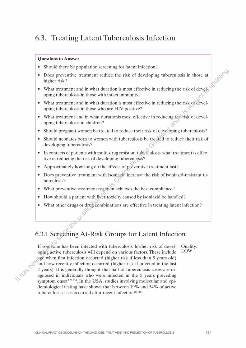
CLINICAL PRACTICE GUIDELINE ON THE DIAGNOSIS, TREATMENT AND PREVENTION OF TUBERCULOSIS 131
6.3. Treating Latent Tuberculosis Infection
6.3.1 Screening At-Risk Groups for Latent Infection
If someone has been infected with tuberculosis, his/her risk of devel-oping active tuberculosis will depend on various factors. These include age when first infection occurred (higher risk if less than 5 years old) and how recently infection occurred (higher risk if infected in the last 2 years). It is generally thought that half of tuberculosis cases are di-agnosed in individuals who were infected in the 5 years preceding symptom onset3,20,254. In the USA, studies involving molecular and epi-demiological testing have shown that between 19% and 54% of active tuberculosis cases occurred after recent infection255-257
Quality: LOW
Questions to Answer
• Shouldtherebepopulationscreeningforlatentinfection?
• Does preventive treatment reduce the risk of developing tuberculosis in those athigher risk?
• Whattreatmentandinwhatdurationismosteffectiveinreducingtheriskofdevel-oping tuberculosis in those with intact immunity?
• Whattreatmentandinwhatdurationismosteffectiveinreducingtheriskofdevel-oping tuberculosis in those who are HIV-positive?
• Whattreatmentandinwhatdurationismosteffectiveinreducingtheriskofdevel-oping tuberculosis in children?
• Shouldpregnantwomenbetreatedtoreducetheirriskofdevelopingtuberculosis?
• Shouldneonatesborntowomenwithtuberculosisbetreatedtoreducetheirriskofdeveloping tuberculosis?
• Incontactsofpatientswithmulti-drugresistanttuberculosis,whattreatmentiseffec-tive in reducing the risk of developing tuberculosis?
• Approximatelyhowlongdotheeffectsofpreventivetreatmentlast?
• Doespreventivetreatmentwithisoniazidincreasetheriskofisoniazid-resistanttu-berculosis?
• Whatpreventivetreatmentregimenachievesthebestcompliance?
• Howshouldapatientwithlivertoxicitycausedbyisoniazidbehandled?
• Whatotherdrugsordrugcombinationsareeffectiveintreatinglatentinfection?
It has
been
5 ye
ars si
nce t
he pu
blica
tion o
f this
Clinica
l Prac
tice G
uideli
ne an
d it is
subje
ct to
upda
ting.

CLINICAL PRACTICE GUIDELINE IN THE SNHS132
Various observational studies have investigated the risk of developing tuberculosis in patients with various risk factors, but results have varied wide-ly. For some factors there is insufficient evidence to make a recommendation for the treatment of latent infection on the basis of tuberculin test results. In these cases, the advice given is based on expert consensus (Table 14)20.
Table 14: Stages of a conventional contact study
Clinical situation Relative risk (RR) NNT
HIV infection 2.1-9.9 2-25
Contact with a smear-positive patient 5-10 7-177
Silicosis 1.2-30 3-119
Diabetes mellitus 1.7-41 18-89
Gastrectomy 1.3-2.5 11-36
TNF-alpha blocker treatment 2-22.6 NC
NNT: Number needed to treat to prevent one case of tuberculosis in 10 years; RR> 1 indicates a greater risk for a clinical situa-tion than for individuals without that situation; NC: not calculated Adapted from National Collaborating Centre for Chronic Conditions: Tuberculosis: Clinical diagnosis and management of tuberculosis, and measures for its prevention and control, CPG: Royal College of Physicians, 2006; and Rose DN: Benefits of screening for latent Mycobacterium tuberculosis infection, Arch Intern Med, 2000 May 22; 160(10): 1513-21 (20, 296)
Screening involves the tuberculin test (point 6.2.4) for close con-tacts of smear-positive patients. In contrast, in a low-risk population, the rate of false positives is very high1. Performing the tuberculin test im-plies the intention to treat the patient if the result is positive, so there must be sufficient resources available to prescribe and follow treatment for latent infection. Indications for screening are presented in Table 15.
Table 15: Stages of a conventional contact study
When recent tuberculosis infection is suspected
Close contacts of patients with infectious tuberculosis (contact study)
Adults living with a child less than 5 years old who has active tuberculosis
Children and adolescents exposed to at-risk adults
(poorly-controlled HIV infection, drug abusers, homelessness, those living in long-term residential institutions, prisoners, immigrants from countries with high tuberculosis incidences, farmworkers)
Immigrants from areas with high endemic rates (>50 cases per 100,000 inhabitants) who have immigrated in the last 5 years
Employees and residents of medical or residential institutions (hospitals, prisons, care homes, etc.), microbiology laboratory employees, social workers who work with at-risk groups, teachers, aid workers in developing countries, security staff
It has
been
5 ye
ars si
nce t
he pu
blica
tion o
f this
Clinica
l Prac
tice G
uideli
ne an
d it is
subje
ct to
upda
ting.

CLINICAL PRACTICE GUIDELINE ON THE DIAGNOSIS, TREATMENT AND PREVENTION OF TUBERCULOSIS 133
When there is a clinical situation that poses a risk of progression to active tuberculosis
Those with HIV (annually from infection onwards)
Those with inactive tuberculosis lesions on chest X rays
Those who use parenteral drugs
Those receiving immunosuppressant treatment (equivalent to >15 mg/day prednisone for more than 1 month)
Those receiving tumour necrosis factor-alpha inhibitor treatment (rule out active tuberculosis before beginning medication, treat latent infection)
6.3.2 Principles for Treating Latent Infection
Treatment for latent infection is treatment administered to individuals who have been infected with M. tuberculosis but are not ill, in order to prevent the disease from developing subsequently. Data from longitu-dinal studies, all conducted many years ago, that evaluated the clinical development of contacts of tuberculosis patients suggest that approxi-mately 5-10% of individuals with latent infection will develop the ac-tive tuberculosis between 1 and 2 years after becoming infected. An additional 5% will do so over the course of their lives. This risk is much greater in children and in those who are HIV-positive22,248,258.
The indications for treatment of latent tuberculosis infection (di-agnosed by a contact study or screening of at-risk groups) are stated in Table 16.
Table 16: Stages of a conventional contact study
High-priority contacts of a infectious tuberculosis patient
At-risk contacts:Receiving tumour necrosis factoralpha inhibitor treatment Receiving prolonged corticosteroid or immunosuppressant treatment With associated disorders such as diabetes mellitus, chronic kidney failure, head or neck neoplasia, parenteral drug use withno HIV infection
Children less than 5 years old
Those who are HIV positive
Tuberculin test converters*
Those with silicosis
Those with an imaging test suggesting untreated residual fibrosis (once active tuberculosis has been ruled out)
Patients on transplant waiting lists
It has
been
5 ye
ars si
nce t
he pu
blica
tion o
f this
Clinica
l Prac
tice G
uideli
ne an
d it is
subje
ct to
upda
ting.

CLINICAL PRACTICE GUIDELINE IN THE SNHS134
*Definition of tuberculin test converter: in screening outside contact studies, someone is considered to have a higher prob-ability of having been infected if the tuberculin test induration increases in diameter by between 6 10 mm after a maximum of 2 years. In contact studies tuberculin test conversion is considered to have occurred if a first test is negative (<5 mm) and a second is positive (≥5mm). Adapted from National Tuberculosis Controllers Association; Center for Disease Control and Prevention; Guidelines for the investigation of contacts of persons with infectious tuberculosis; recommendations from The National Tuberculosis Controllers Association and CDC, and Guidelines for using the Quantiferon-TB Gold Test for detecting Mycobacterium tuberculosis infec-tion, United States, MMWR, 2005 Dec 16; 54 (RR-15): 1-55 (33)
Contraindications to treatment include acute hepatitis, severe chronic liver disease, and serious adverse reactions to treatment. Mild or moderate chronic liver disease, history of adverse reactions to treat-ment that are not serious, and pregnancy are relative contraindications to treatment for latent tuberculosis infection.
In low-income countries, treatment of latent infection is rarely used to control tuberculosis, as diagnosis and treatment of active tuberculosis take priority. However, in middle- and high-income countries, treatment of latent infection is a major, effective part of any tuberculosis control programme. In 2002 alone it has been calculated that between 291,000 and 433,000 in-dividuals were treated for latent tuberculosis infection in the USA, and it is estimated that progression to active tuberculosis was prevented in a total of between 4000 and 11,000 individuals259. Several organisations acknowledge the role played by treatment for latent infection and actively promote it22,260.
Isoniazid has been used for 35 years as the sole treatment for la-tent tuberculosis infection. There are many studies that have evaluated the efficacy of isoniazid in treatment regimens lasting up to 12 months.
Between 1951 and 1970, several studies were conducted to assess the use of isoniazid to treat latent tuberculosis infection. More than 100,000 people took part in these studies, which were conducted in several differ-ent countries. The participants included individuals at risk of developing active tuberculosis, contacts of tuberculosis patients, those with positive tuberculin test results, people with inactive tuberculosis, and children with primary tuberculosis. Most of the studies compared a 12-month isoniazid regimen with placebo. The results showed that in each study, the incidence of tuberculosis ranged from 25% to 92% in those treated with isoniazid, although treatment efficacy was 90% in patients who were compliant with the treatment or even if pill taking was irregular. 22,254.
Quality:LOW
In 1965, the ATS recommended isoniazid as the drug of choice for the treatment of latent tuberculosis infection in all individuals with old untreat-ed tuberculosis lesions and in recent tuberculin test converters, particularly children aged under 3. In 1967 this recommendation was extended to indi-viduals with a tuberculin test induration diameter greater than 9 mm22.
IUATLD conducted a large-scale RCT to assess the efficacy of iso-niazid treatment (lasting 12, 24 or 52 weeks) in individuals with pulmo-nary fibrotic lesions and positive tuberculin tests. It included a total of 27,830 participants from seven European countries.
Quality:MODERATE
It has
been
5 ye
ars si
nce t
he pu
blica
tion o
f this
Clinica
l Prac
tice G
uideli
ne an
d it is
subje
ct to
upda
ting.

CLINICAL PRACTICE GUIDELINE ON THE DIAGNOSIS, TREATMENT AND PREVENTION OF TUBERCULOSIS 135
The results showed that when compared to placebo, isoniazid ad-ministered for 52 weeks reduced the incidence of confirmed tubercu-losis by 75%. The decreases were 65% and 21% for 24 week and 12 week treatments respectively. In patients whose treatment compliance was above 80%, the decrease was even greater (93% for the 52-week treatment regimen, 69% for the 24 week regimen, and 31% for the 12 week regimen)261.
A cost analysis conducted in the USA concluded that the 6 month treatment regimen was more cost-effective than regimens lasting 9 or 3 months. From 1986 onwards, the 6 month regimen was adopted for the treatment of latent infection in the USA262. However, reanalysis of community studies conducted in Alaska showed that the 9 month regi-men provided greater protection than the 6 month regimen. Extending treatment to 12 months provided so little additional benefit that such extension was not justified263.
Quality:LOW
It was hoped that large-scale use of isoniazid, which has a low cost and an acceptable toxicity profile, would successfully reduce the inci-dence of tuberculosis in high-risk groups by between 50% and 75%. This aim has not been completely achieved, probably because the regi-men has often been applied incorrectly22.
The introduction of rifampicin, which is highly effective, provid-ed the possibility of using it to treat latent tuberculosis infection using shorter, more effective treatment regimens (point 6.3.12).
6.3.3 Treating Latent Infection in Individuals with Intact Immunity
One SR (11 RCTs, 73,375 patients of all ages) evaluated the efficacy of various isoniazid treatment regimens against placebo, in preventing new cases of tuberculosis among HIV-negative patients at risk of developing active tuberculosis. RCTs in patients with tuberculosis currently or in the past were excluded.
Quality:MODERATE
The review showed that isoniazid treatment reduced the risk of active tuberculosis by 60% (11 RCTs, 796 events) with follow-up last-ing two or more years. According to the results of a single RCT, the reduction was even greater in patients who complied with treatment. Treatment did not significantly reduce tuberculosis-related mortality, and caused hepatitis in 0.55% of patients, compared to 0.1% with pla-cebo (1 RCT, 84 events). 6 month treatment regimens (2 RCTs) and 12 month regimens (10 RCTs) showed a similar decrease in the risk of developing active tuberculosis (56% and 62% respectively)264.
It has
been
5 ye
ars si
nce t
he pu
blica
tion o
f this
Clinica
l Prac
tice G
uideli
ne an
d it is
subje
ct to
upda
ting.

CLINICAL PRACTICE GUIDELINE IN THE SNHS136
A single RCT directly compared 6 month and 12 month isoniazid treatment regimens. The risk of developing active tuberculosis was 41% higher with the 6 month regimen than with the 12 month regimen (RR 1.41; CI 95%, 0.84 to 2.37, 58 events), although the result was not signifi-cant and the number of events was small261.
Given that the risk of developing active tuberculosis within 5 years is 5% for recent contact patients with a relative who has positive tu-berculin test, prevention of one case of active tuberculosis in the next 5 years requires treating 32 people using a 12 month treatment regimen, or 36 using a 6 month regimen.
6.3.4 Treating Latent Infection in HIV-Positive Individuals
In various observational studies conducted in the 1990s, isoniazid showed a decrease in mortality and progression to AIDS in HIV-positive indi-viduals with positive tuberculin tests. In these patients, isoniazid admin-istered for between 6 and 12 months reduced the incidence of tubercu-losis by up to 70%. Treatment regimens involving other drugs, such as rifampicin combined with pyrazinamide or isoniazid, were found to be as effective as isoniazid alone.
Three SRs of RCTs showed a reduction between 36% and 43% in the risk of developing active tuberculosis in patients receiving pre-ventive treatment when compared to those receiving placebo. The de-creases were greater in individuals with positive tuberculin tests258,265 266. The various regimens are shown in Appendix 14.
Quality:MODERATE
The most recent SR evaluated the efficacy of treatment for latent tuberculosis infection in individuals with HIV. The SR showed that when compared to placebo, preventive treatment with any tuberculosis drug reduced the risk of developing active tuberculosis by 36% (RR 0.64; CI 95%, 0.51 to 0.81). For tuberculosis confirmed by cultures, the decrease was similar but not statistically significant. All drug regimens, regardless of type, frequency, or duration, reduced the incidence of ac-tive tuberculosis when compared to placebo.
The effects observed with the various treatment regimens were as follows:
Isoniazid alone (13 RCTs): RR 0.67; CI 95%, 0.51 to 0.87
Isoniazid/rifampicin (2 RCTs): RR 0.41; CI 95%, 0.21 to 0.81
Rifampicin/pyrazinamide (4 RCTs): RR 0.54; CI 95%, 0.34 to 0.86
Isoniazid/rifampicin/pyrazinamide (1 RCT): RR 0.48; CI 95%, 0.23 to 1.00It has
been
5 ye
ars si
nce t
he pu
blica
tion o
f this
Clinica
l Prac
tice G
uideli
ne an
d it is
subje
ct to
upda
ting.
CLINICAL PRACTICE GUIDELINE ON THE DIAGNOSIS, TREATMENT AND PREVENTION OF TUBERCULOSIS 137
In those with positive tuberculin tests, preventive treatment re-duced the risk of active tuberculosis by 62% (4 RCTs: RR 0.38; CI 95%, 0.25 to 0.57). The effect among those with negative tuberculin test re-sults was not significant.
Efficacy was similar for all treatment regimens, but side effects leading to interruption of treatment were more common in the RCTs in which treatment involved combinations of drugs258.
An earlier SR (7 RCTs, 4,529 participants) evaluated the efficacy of isoniazid in treating latent tuberculosis infection in HIV-positive people. The 6 month regimen reduced the incidence of tuberculosis sig-nificantly (RR 0.58; CI 95%, 0.43 to 0.8) only in patients with positive tuberculin tests. There was no decrease in overall mortality. Side effects were also more common with isoniazid than with placebo266.
Another SR (4 RCTs, 4,055 participants), also in HIV-positive individuals, produced similar results. Chemoprevention (various regi-mens) reduced the incidence of tuberculosis by almost half (RR 0.57; CI 95%, 0.41 to 0.79) when compared to placebo. In individuals with positive tuberculin tests, the decrease was more marked, and overall mortality also fell (RR 0.73; CI 95%, 0.57 to 0.95). More participants in the treatment group withdrew due to side effects. The studies included had methodological shortcomings265.
An additional RCT not included in the reviews described above compared the results of a 12 month isoniazid or placebo regimen in individuals with advanced AIDS, most of whom had negative tubercu-lin tests. Treatment compliance was 85%. The incidence of tuberculosis was 18 cases per 100 individuals per year in the isoniazid group, and 11.6 cases per 100 individuals per year in the placebo group. There were no significant differences. Treatment also failed to reduce mortality or hospital admissions267.
Another RCT conducted in Spain in the 1990s but not published until recently failed to show any differences in the incidence of tuber-culosis between regimens involving isoniazid alone (6 months) or com-bined with rifampicin (3 months) and regimens involving a combina-tion of rifampicin and pyrazinamide (2 months). Treatment compliance was low and 2 year follow-up was incomplete. Cases of liver toxicity were recorded in regimens with isoniazid268.
It has
been
5 ye
ars si
nce t
he pu
blica
tion o
f this
Clinica
l Prac
tice G
uideli
ne an
d it is
subje
ct to
upda
ting.
CLINICAL PRACTICE GUIDELINE IN THE SNHS138
In a recent cohort of 2,778 HIV-positive patients, the incidence of tuberculosis was assessed and compared according to the treatment received. During follow-up, a total of 267 cases of tuberculosis were recorded. In patients who received HAART and were treated with iso-niazid, the annual incidence of tuberculosis was 1.1%, 89% less than in patients who did not receive any treatment (RR 0.11; CI 95%, 0.02 to 0.78). The annual incidence among patients who were treated with HAART but did not receive isoniazid was 4.6%269.
Quality: LOW
Chemoprevention seems to be truly beneficial for HIV-positive individuals with positive tuberculin tests. Isoniazid treatment lasting between 6 and 12 months prevents between 60% and 70% of future cases of tuberculosis. Taking a combination of isoniazid and rifampicin for 3 months can be equally effective, whether administered daily or twice a week. A combination of rifampicin and pyrazinamide may be an alternative for these patients, and these patients alone, as it causes high toxicity in individuals with intact immunity. In contrast, in patients with negative tuberculin tests or anergy, isoniazid has not shown the same efficacy. All treatment regimens were generally well tolerated, and the level of treatment compliance was higher with shorter regimens (2-3 months)254.
6.3.5 Treating Latent Infection in Children
Several recommendations recommend treating latent infection in chil-dren and adolescents as a precautionary measure. This is partly because once infected, children (particularly those less than 5 years old) can progress rapidly to primary tuberculosis, and partly because someone infected as a child has many years ahead of him/her in which active tu-berculosis may develop23,270.
The risk of progression to active tuberculosis in this group is close-ly related to age on infection and immune status. Immunocompetent children aged between 5 and 10 have a lower risk of becoming ill, while those aged under 2 are a high-risk group242,271.
An SR that evaluated the efficacy of various isoniazid treatment regimens in preventing new cases of tuberculosis included RCTs con-ducted in children. A 12 month regimen showed a significant decrease in the risk of tuberculosis (10 RCTs; RR 0.38; CI 95%, 0.28 to 0.50, 708 events). The dose used was 5 15 mg/kg body weight, and no RCTs conducted in children and involving shorter treatment regimenswere found. The review concludes that the efficacy and suitability of regimens lasting 6 and 9 months in this population need to be confirmed264.
Quality:LOW
It has
been
5 ye
ars si
nce t
he pu
blica
tion o
f this
Clinica
l Prac
tice G
uideli
ne an
d it is
subje
ct to
upda
ting.

CLINICAL PRACTICE GUIDELINE ON THE DIAGNOSIS, TREATMENT AND PREVENTION OF TUBERCULOSIS 139
A quasi-randomised trial compared a 9 month isoniazid treatment regimen with other, longer regimens involving combined isoniazid and rifampicin, in a total of 926 children aged under 15 with latent tuber-culosis infection according to established international criteria. Partici-pants were recruited during two different time periods, and the results obtained during each period were compared.
Quality:VERY LOW
During the first period, 24% of patients treated with isoniazid (9 months) presented radiological findings compatible with active tuber-culosis. The percentage was significantly lower in those who received isoniazid and rifampicin for 4 months during the same period (11.8%). During the second period, 4 month and 3 month isoniazid and rifampicin regimens were compared against each other, with no differences regard-ing radiological findings compatible with active tuberculosis (13.6% and 11% respectively). No patients developed clinical tuberculosis during the minimum follow-up period of 3 years. Overall, general treatment compliance was good (91.8%) and even higher for short regimens. The doses administered were 10 mg/kg/day, up to a maximum of 300 mg for isoniazid and 600 mg for rifampicin272.
A recent SR273 designed to evaluate the efficacy of isoniazid in HIV-positive children found only one RCT. This involved 236 children aged over 8 weeks. The children were randomised to receive isoniazid or placebo for 9 months, and all participants received co-trimoxazole. The RCT was terminated early after a significant decrease, 54%, in mortality was observed in the treatment group, although the number of events was very small32. The incidence of tuberculosis was also lower with isoniazid (3.8%, 5 events) than with placebo (9.9%, 13 events)274.
Quality:LOW
There is no consensus regarding dosing or duration of treatment, which vary between scientific societies and healthcare organisations. The various doses and treatment regimens recommended are shown in Table 17.
Table 17: Treatment regimens for latent infection in children
DrugDose
(mg/kg/day)Frequency Duration
Isoniazid 5-10/15Daily Intermittent (3 times per week)
6-9 months
Rifampicin 10/15Daily Intermittent (3 times per week)
6 months
Isoniazid and rifampicin Same dosesDaily Intermittent (3 times per week)
3 months
Adapted from the Tuberculosis Working Group of the Spanish Society for Paediatric Infectious Diseases (SEIP). Consensus document on treatment for exposure to tuberculosis and latent tuberculosis infection in children.An Pediatr (Barc). 2006; 64(1): 59-65 (17).
It has
been
5 ye
ars si
nce t
he pu
blica
tion o
f this
Clinica
l Prac
tice G
uideli
ne an
d it is
subje
ct to
upda
ting.

CLINICAL PRACTICE GUIDELINE IN THE SNHS140
6.3.6 Treating Latent Infection in Pregnant Women
Pregnancy seems not to affect the risk of progression of tuberculosis infection. Tuberculosis drugs have been used extensively in pregnant women, and in general the benefits of treat-ing tuberculosis are considered to outweigh the potential risks to the foetus. It has been recorded that 3% of foetuses exposed to rifampicin in the womb, 2% of those exposed to ethambutol, and 1% of those exposed to isoniazid displayed abnormalities22,275. However, by consensus, treatment of latent infection begins around 2-3 weeks after birth, in order to prevent liver toxicity caused by isoniazid immediately postpartum22,248,254,270,276.
It is currently agreed that pregnant women must undergo treatment for latent infec-tion (even during the first trimester of pregnancy) in the following situations22,254:
− Positive tuberculin test (≥0.5 mm) and HIV infection (high-risk behaviour or re-fusal to take an HIV test if HIV status is unknown)
− Positive tuberculin test (≥0.5 mm) in a contact of a smear-positive patient
− Tuberculin test conversion recorded in the last 2 years
Treatment regimens involving isoniazid, whether daily or intermittent, are preferred, with vitamin B6 supplements and regular monitoring of liver enzymes22.
6.3.6.1 Preventing Congenital and Neonatal TuberculosisCongenital tuberculosis is a very rare but potentially very serious disease that can be trans-mitted from mother to foetus through the blood, via the umbilical cord. There is often placental involvement, with lesions that, when ruptured, reach the amniotic fluid and are aspirated by the foetus before or during birth. Tuberculosis has been associated with pre-maturity, low birth weight, and perinatal death of the baby. Extrapulmonary forms are common271,277.
A pregnant woman with pulmonary tuberculosis will cease to be infectious after 2 3 weeks of suitable treatment. If birth occurs after this, the neonate has a very low risk of contagion. The risk increases if the mother is diagnosed shortly before birth or during labour. In these cases, the neonate must be examined carefully and the placenta must be analysed to rule out congenital tuberculosis. Tuberculosis treatment must be begun if tu-berculosis is diagnosed, and those in the same household as the neonate must be examined exhaustively5.
Children born to mothers with pulmonary tuberculosis and a positive smear microsco-py have a high risk of developing active disease. Treatment must therefore not be delayed, and children should receive prophylactic isoniazid for 6 months5. To prevent infection in the baby the mother must also comply thoroughly with her treatment and wear a mask while she is known or suspected to be infectious. Also, if the mother is known or suspected to be suffering from multi-drug resistant tuberculosis, the baby must be separated from the mother until there is confirmation that the mother is no longer infectious44. Follow-up must always be conducted for at least 2 years278.It h
as be
en 5
years
sinc
e the
publi
catio
n of th
is Clin
ical P
ractic
e Guid
eline
and i
t is su
bject
to up
datin
g.
CLINICAL PRACTICE GUIDELINE ON THE DIAGNOSIS, TREATMENT AND PREVENTION OF TUBERCULOSIS 141
6.3.7 Treating Contacts of Patients with Multi-Drug Resistant Tuberculosis
A narrative review that evaluated treatment to prevent active tubercu-losis in children in contact with multi-drug resistant tuberculosis identi-fied two cohort studies279.
Quality:LOW
The most recent cohort included 105 children who had come into contact with patients with multi-drug resistant tuberculosis. One group received 6 months of treatment according to sensitivity to antibiotics in each individual case, while the other group acted as the control. After 30 months of follow-up, two cases of tuberculosis were diagnosed in the 41 children who had received chemoprevention, while 13 cases were diagnosed in the 64 children in the control group (OR 0.2; CI 95%, 0.04 to 0.94).
An earlier retrospective cohort study observed 190 children who had come into contact with patients with multi-drug resistant tuber-culosis who had positive tuberculin tests (≥10 mm). The differences in the development of active tuberculosis between a group that received 6 months of isoniazid treatment (2 of 45) and a group that received no treatment (3 of 145) were not significant. The two cases of tuberculosis in the group that received treatment were both multi-drug resistant.
Contacts of patients with multi-drug resistant tuberculosis must receive clinical monitoring for at least 2 years. If they develop tuber-culosis, treatment must be started promptly, using an effective regi-men that is used to treat the index case. The only drugs that have been found to be useful as preventive treatment are isoniazid and rifampicin, although neither of these has any effect against multi-drug resistant strains. There is no evidence in favour of beginning preventive treat-ment with second-line drugs219.
6.3.8 Duration of Protection
The use of isoniazid for the appropriate length of time to treat latent tuberculosis infection in immunocompetent individuals signifi-cantly reduces the risk of developing active tuberculosis. According to a study conducted in the 1960s, which monitored patients for a very prolonged period, this protection lasts for more than 20 years263. The major European study UICTER-IUTALD showed a significant effect for isoniazid after a5-year follow-up261.
Quality:MODERATE
It has
been
5 ye
ars si
nce t
he pu
blica
tion o
f this
Clinica
l Prac
tice G
uideli
ne an
d it is
subje
ct to
upda
ting.

CLINICAL PRACTICE GUIDELINE IN THE SNHS142
Several RCTs, all included in SRs, evaluated the efficacy of treat-ment for latent infection in individuals who are HIV-positive. One RCT showed a decrease in protection over time for treatment regimens that combined isoniazid or rifampicin with pyrazinamide. Another RCT in patients with positive tuberculin tests showed a benefit for treatment at 15 months but not at the 3 year follow-up, probably due to the small number of events and to losses to follow-up258,265 266.
6.3.9 The Impact of Treatment on Resistance to Isoniazid
A recent SR (12 RCTs, 1 cohort; 32,179 participants) evaluated the ef-fect of chemoprevention with isoniazid on the development of drug resistance in adult patients (HIV-positive or HIV-negative). It showed that there were more cases of drug resistance among patients treated with isoniazid (0.17%, 31 of 18,095) than in the control group (0.15%, 28 of 17,985), although the differences were not significant. The effect was consistent across the various studies included. The results were similar in the studies involving HIV-positive patients. The studies had shortcom-ings such as a low proportion of antibiograms, high losses to follow-up, and differences between prophylaxis regimens280.
Quality:LOW
An SR on the efficacy of isoniazid showed a higher percentage of drug resistance among patients receiving active treatment than in the control group (20.6% versus 6.6%) in 5 RCTs that evaluated antibio-grams, although the absolute number was very small (22 events)264.
6.3.10 Compliance with Treatment for Latent Infection
Before the beginning of treatment for latent infection, patients must be informed about tuberculosis infection and disease, highlighting the importance of good treatment compliance and of avoiding alcohol during treatment.
Monitoring of patients who have begun treatment for latent infec-tion must include a visit 2 weeks after treatment is prescribed. This is done to ensure that the patient has understood instructions and to as-sess treatment compliance237.
A series of direct and indirect measures have been proposed to improve compliance. These include regular visits, medical advice, ed-ucational talks, and incentives. Any patient following an intermittent treatment regimen must receive directly observed treatment (DOT), although the routine use of DOT is not justified due to its high cost and unpopularity among patients.
There are several ways to assess treatment compliance:
It has
been
5 ye
ars si
nce t
he pu
blica
tion o
f this
Clinica
l Prac
tice G
uideli
ne an
d it is
subje
ct to
upda
ting.

CLINICAL PRACTICE GUIDELINE ON THE DIAGNOSIS, TREATMENT AND PREVENTION OF TUBERCULOSIS 143
1. DIRECT methods:
Measuring drug metabolites in the urine. This can be used to check that the last dose was taken, although it does not guarantee that previous doses have been taken. It also entails high labora-tory costs, which are not affordable for most hospitals.
Regular clinical monitoring is less expensive and can be used in all clinical environments. Irregular attendance of or failure to attend vis-its are good indicators of poor or zero compliance, but the reverse, good attendance, is not well correlated with treatment compliance.
2. INDIRECT methods:
Interviews, during which the patients themselves can describe medication intake, can be used to estimate treatment compli-ance and investigate reasons for any non-compliance. This is a fast, cheap method. It can overestimate compliance, although it is believed to be better at identifying patients with poor compli-ance. Specific questionnaires on compliance, filled in by patients themselves and analysing short periods of time to prevent mem-ory bias, can be used.
Electronic monitoring is one of the most accurate methods, al-though it is expensive and does not verify actual drug intake. It cannot be used for combined treatment regimens. Counting tab-lets also fails to confirm intake at the prescribed times. It tends to overestimate compliance and is therefore recommended only for unscheduled home visits254,270,281.
In the UICTER-IUATLD’s study, the compliance percentages for isoniazid regimens lasting 3, 6, and 12 months were 87%, 78%, and 68% respectively. For regimens that were identical except for the replacement of isoniazid with a placebo, compliance was slightly higher (91%, 82% and 69% respectively)261. An SR that included studies in HIV-positive patients showed that 70 75% of patients completed 6 month isoniazid treatment correctly282.
Quality: LOW
A recent SR of 78 studies published between 1997 and 2007 draws different conclusions. Treatment compliance is suboptimum in all at-risk groups (contacts of patients with tuberculosis, prisoners, immigrants, parenteral drug users, etc.), with widely varying results regardless of the regimen used. It finds no consistent relationship between patient, hospi-tal or drug characteristics and treatment compliance. Compliance does not depend on patient age (in adults), sex, place of birth, or ethnic group. Results according to patients’ sex or place of origin are also heteroge-neous. Patients’ fears of possible side effects have been associated with lower compliance rates and more clinical abnormalities. Fear of blood tests has also been associated with worse treatment compliance281.
It has
been
5 ye
ars si
nce t
he pu
blica
tion o
f this
Clinica
l Prac
tice G
uideli
ne an
d it is
subje
ct to
upda
ting.
CLINICAL PRACTICE GUIDELINE IN THE SNHS144
Treatment regimens that included rifampicin showed better compli-ance results that isoniazid in retrospective studies281,283 and in one RCT284, with rates ranging from 72% to 91%. In one SR, a combined rifampicin and isoniazid regimen generally showed better compliance rates then isoniazid alone. In an RCT not included in this review, the combined regi-men achieved compliance of 82%285. A subsequent RCT in children also showed better compliance rates for combined rifampicin and isoniazid treatment (lasting 3 or 4 months) than for 9 month isoniazid treatment. The main reasons for non-compliance were reluctance to take medica-tion, nausea and epigastric discomfort, interruption of treatment by phy-sicians, and poor understanding of instructions272.
Patients’ preferences also affect treatment compliance. A cohort study involving 591 patients treated with a short rifampicin and isoni-azid regimen or a longer, 6 month isoniazid regimen showed that 78% of patients chose the short treatment regimen. Compliance was a higher in younger patients, those who were allowed to choose their treatment regi-men, and those who attended scheduled clinical check-ups286.
6.3.11 Liver Toxicity of Isoniazid
Isoniazid treatment can cause liver toxicity, particularly in those aged over 35 or if it is administered together with other hepatotoxic agents such as alcohol, carbamazepine, methotrexate, or acetaminophen. Liver involvement includes damage of varying severity: transient transami-nase increases (in 20% of cases), necrotic lesions (30% of cases) and extensive lesions similar to those of viral hepatitis (10% of cases). Tox-icity is reversible if treatment is interrupted in time, but fatal if treat-ment is continued. Patients must be informed as to how to recognise the signs of liver toxicity. If necessary, they must consult a physician276. Appendix 13 shows a proposal for overall assessment of the risk of liv-er toxicity in a patient due to begin treatment with isoniazid. Figure 2 contains a proposal for handling possible liver toxicity during isoniazid treatment for latent infection.
Most patients with mild liver toxicity do not present any clinical symptoms. Nausea, vomiting, and constitutional symptoms occur in between 50% and 75% of patients with severe liver toxicity. Jaundice, biliuria, and acholia are clear late signs of deterioration. Serious liver failure must be suspected if the patient presents altered coagulation, hypoalbuminaemia, and hypoglycaemia. Restoration of liver function generally takes several weeks and tends to be complete after treatment is suspended. In children there may be transient transaminase increases but clinical hepatitis and acute liver failure are rare141,270.
It has
been
5 ye
ars si
nce t
he pu
blica
tion o
f this
Clinica
l Prac
tice G
uideli
ne an
d it is
subje
ct to
upda
ting.

CLINICAL PRACTICE GUIDELINE ON THE DIAGNOSIS, TREATMENT AND PREVENTION OF TUBERCULOSIS 145
A study conducted in the USA in the 1970s found great variations in the incidence of hepatitis among patients treated with isoniazid, de-pending on patient age. There were no cases in those aged under 20, but up to 8 cases per thousand patients over 65. Age (>35 years) and daily alcohol intake were significantly associated with the highest rates of hepatitis. According to these data, the risk in adults of an increase in liver enzymes secondary to isoniazid treatment was between 10% and 20%, the risk of clinical hepatitis was around 1%, and the risk of death due to acute liver failure was around 0.1%287. An SR on the he-patic effects of isoniazid showed that of 200,000 patients treated, only two deaths were reported, and concluded that the actual rate of deaths attributable to isoniazid treatment is very low if patients are appropri-ately selected and monitored288.
Quality: LOW
6.3.12 Other Drugs Used to Treat Latent Infection
One of the difficulties associated with the standard isoniazid regimen used to treat latent infection is poor treatment compliance by patients, because treatment lasts so long. As a result, shorter regimens involving two drugs have been proposed as an alternative to the standard regimen (Table 18).
Table 18: Treatment regimens recommended for latent infection in adults
DrugDose (maximum
dose, mg) Frequency Duration Comments
Isoniazid 5 mg/kg/day (300) Daily 6-9 months First-line treatment
Isoniazid 5 mg/kg/day (300) Daily 9 months
First-line treatment for HIV-positive patients and those with inactive tuberculosis lesions shown on chest X rays.
Isoniazid 15 mg/kg/day (900) Twice a week 6-9 months Only if DOT is used
Rifampicin 10 mg/kg/day (600) Daily 4 months
Contacts of patients with isoniazid-resistant tuberculosis or cases of isoniazid intolerance
Isoniazid and rifampicin
5 mg/kg/day (300),10 mg/kg/day (600)
Daily 3 monthsEfficacy and safety equivalent to 6 month isoniazid regimen
DOT: Directly observed treatment Adapted from Blasco D, Carbó RM, González F, Momparler P, Peñuelas J A, Pérez E et al.: Guideline for Monitoring and Controlling Tuberculosis, Regional Government of Valencia, Regional Health Ministry, 200; and Landry J, Menzies D: Preventive Chemotherapy. Where has it got us? Where to go next? Int J Tuberc Lung Dis 2008; 12 (12): 1352-64 (237, 297)
It has
been
5 ye
ars si
nce t
he pu
blica
tion o
f this
Clinica
l Prac
tice G
uideli
ne an
d it is
subje
ct to
upda
ting.

CLINICAL PRACTICE GUIDELINE IN THE SNHS146
6.3.12.1 Combined Isoniazid and Rifampicin
An SR (5 RCTs, 1,926 patients) compared isoniazid treatment regi-men (6 12 months) with combined isoniazid and rifampicin treatment (3 months). No differences were found between the two regimens in terms of the incidence of tuberculosis (4.2% for combined treatment, 4.1% for isoniazid alone, in a total of 80 events). There were no differ-ences in overall mortality. The combined treatment regimen, which was shorter, showed better compliance but higher toxicity282.
Quality:LOW
A subsequent, quasi-randomised trial in 926 children showed that combined treatment administered for 3 or 4 months compared to treat-ment with isniazid alone during 9 months reduced clinical and radio-logical progression to active tuberculosis 272.
The results of two cohorts of paediatric patients (aged up to 15) from a region of England with large numbers of immigrants showed that after isoniazid and rifampicin chemoprevention (6 9 months) was introduced, the number of cases of tuberculosis reported in the next 5 years fell. The decrease in reported tuberculosis cases was maintained with shorter treatment regimens (3 and 4 months). Their overall toxicity profile, and in particular their liver toxicity profile, was also acceptable, and was comparable to that of isoniazid monotherapy regimens289.
6.3.12.2 Combined Rifampicin and Pyrazinamide
The main current recommendations do not include a combined ri-fampicin and pyrazinamide treatment regimen, due to its high risk of liver toxicity. There are reservations regarding its use in HIV-positive patients. One SR (6 RCTs, 4053 patients) evaluated the efficacy and safety results for this combined treatment regimen and compared them to those of isoniazid monotherapy. The results were presented sepa-rately for the HIV-positive and HIV-negative populations. The com-bined treatment regimen proved equally effective in preventing active tuberculosis, although there were significantly more serious side effects and liver toxicity, particularly in HIV-negative individuals, given that serious liver toxicity was reported in up to 8.2% of these patients, com-pared to 1.7% of those treated with isoniazid alone290-291.
Quality:LOW
6.3.12.3 Rifampicin
It has
been
5 ye
ars si
nce t
he pu
blica
tion o
f this
Clinica
l Prac
tice G
uideli
ne an
d it is
subje
ct to
upda
ting.

CLINICAL PRACTICE GUIDELINE ON THE DIAGNOSIS, TREATMENT AND PREVENTION OF TUBERCULOSIS 147
Another alternative is a 4 month rifampicin monotherapy regimen, al-though there is little evidence concerning this treatment. One RCT com-pared regimens involving rifampicin (3 months), isoniazid (6 months), both medications combined (3 months), and placebo in 652 patients with silicosis. All treatment regimens were more effective than the placebo, and all showed similar results in preventing active tuberculosis. Patients receiving rifampicin alone did not develop any drug resistance, and also presented a very low risk of liver toxicity (0.08%)292.
Quality: LOW
An open-label RCT evaluated the safety of rifampicin (4 months) and isoniazid (9 months) in adults treated for latent infection. A total of 4% of patients treated with isoniazid experienced serious side events that led to treatment withdrawal, compared to 1.7% of those treated with rifampicin, with a total of 24 events. Of the 17 cases in the group receiving isoniazid, 16 were due to liver toxicity. Treatment compliance was also higher with the rifampicin treatment regimen (78% versus 60%). The au-thors concluded that rifampicin treatment has a better safety profile than isoniazid treatment, and believe that an RCT is needed to estimate the efficacy of this treatment regimen284. Two cohort studies subsequently reported better compliance rates and less liver toxicity with 4 month ri-fampicin treatment than with 9 month isoniazid treatment283,293.
A cohort of 157 adolescent contacts of patients with isoniazid-re-sistant tuberculosis and positive tuberculin test results (> 4 mm) docu-mented for the first time were treated with rifampicin (10 mg/kg/day, maximum dose 600 mg) for 6 months. After a two-year follow-up, 26% of participants had experienced a side effect. No cases of tuberculosis were detected in the 137 participants in whom follow-up was completed294.
6.4. Treating Probable Infection (Primary Chemoprevention)
Primary chemoprevention provides temporary protection to contacts of patients with a infectious form of tuberculosis. It is intended to pre-vent contacts from becoming infected or from developing the disease. Primary chemoprevention must therefore be administered during the time window before a second, potentially positive, tuberculin test after an initial negative tuberculin test.
Questions to Answer
• Underwhatcircumstancesshouldhealthyindividualsincontactwithpatientswithactive tuberculosis be treated to prevent latent tuberculosis infection?
It has
been
5 ye
ars si
nce t
he pu
blica
tion o
f this
Clinica
l Prac
tice G
uideli
ne an
d it is
subje
ct to
upda
ting.
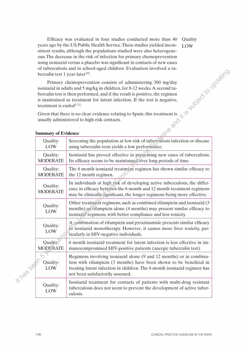
CLINICAL PRACTICE GUIDELINE IN THE SNHS148
Efficacy was evaluated in four studies conducted more than 40 years ago by the US Public Health Service. These studies yielded incon-sistent results, although the populations studied were also heterogene-ous. The decrease in the risk of infection for primary chemoprevention using isoniazid versus a placebo was significant in contacts of new cases of tuberculosis and in school-aged children. Evaluation involved a tu-berculin test 1 year later295.
Quality
LOW
Primary chemoprevention consists of administering 300 mg/day isoniazid in adults and 5 mg/kg in children, for 8-12 weeks. A second tu-berculin test is then performed, and if the result is positive, the regimen is maintained as treatment for latent infection. If the test is negative, treatment is ended17,23.
Given that there is no clear evidence relating to Spain, this treatment is usually administered to high-risk contacts.
Summary of Evidence
Quality:LOW
Screening the population at low risk of tuberculosis infection or disease using tuberculin tests yields a low performance.
Quality:MODERATE
Isoniazid has proved effective in preventing new cases of tuberculosis. Its efficacy seems to be maintained over long periods of time.
Quality:MODERATE
The 6 month isoniazid treatment regimen has shown similar efficacy to the 12 month regimen.
Quality:MODERATE
In individuals at high risk of developing active tuberculosis, the differ-ence in efficacy between the 6 month and 12 month treatment regimens may be clinically significant, the longer regimens being more effective.
Quality:LOW
Other treatment regimens, such as combined rifampicin and isoniazid (3 months) or rifampicin alone (4 months) may present similar efficacy to isoniazid regimens, with better compliance and less toxicity.
Quality:LOW
A combination of rifampicin and pyrazinamide presents similar efficacy to isoniazid monotherapy. However, it causes more liver toxicity, par-ticularly in HIV-negative individuals.
Quality:MODERATE
6 month isoniazid treatment for latent infection is less effective in im-munocompromised HIV-positive patients (anergic tuberculin test).
Quality:LOW
Regimens involving isoniazid alone (9 and 12 months) or in combina-tion with rifampicin (3 months) have been shown to be beneficial in treating latent infection in children. The 6 month isoniazid regimen has not been satisfactorily assessed.
Quality:LOW
Isoniazid treatment for contacts of patients with multi-drug resistant tuberculosis does not seem to prevent the development of active tuber-culosis.
It has
been
5 ye
ars si
nce t
he pu
blica
tion o
f this
Clinica
l Prac
tice G
uideli
ne an
d it is
subje
ct to
upda
ting.
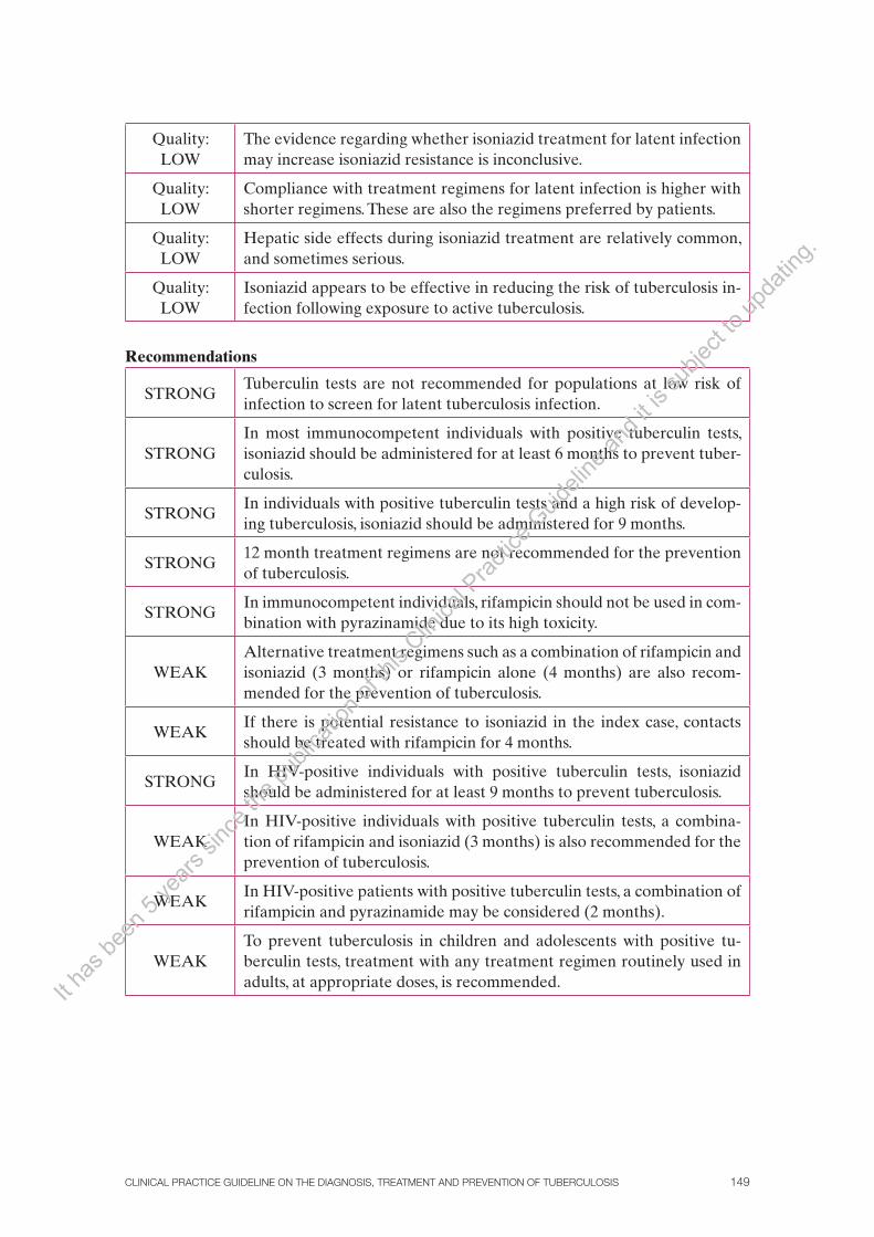
CLINICAL PRACTICE GUIDELINE ON THE DIAGNOSIS, TREATMENT AND PREVENTION OF TUBERCULOSIS 149
Quality:LOW
The evidence regarding whether isoniazid treatment for latent infection may increase isoniazid resistance is inconclusive.
Quality:LOW
Compliance with treatment regimens for latent infection is higher with shorter regimens. These are also the regimens preferred by patients.
Quality:LOW
Hepatic side effects during isoniazid treatment are relatively common, and sometimes serious.
Quality:LOW
Isoniazid appears to be effective in reducing the risk of tuberculosis in-fection following exposure to active tuberculosis.
Recommendations
STRONGTuberculin tests are not recommended for populations at low risk of infection to screen for latent tuberculosis infection.
STRONGIn most immunocompetent individuals with positive tuberculin tests, isoniazid should be administered for at least 6 months to prevent tuber-culosis.
STRONGIn individuals with positive tuberculin tests and a high risk of develop-ing tuberculosis, isoniazid should be administered for 9 months.
STRONG12 month treatment regimens are not recommended for the prevention of tuberculosis.
STRONGIn immunocompetent individuals, rifampicin should not be used in com-bination with pyrazinamide due to its high toxicity.
WEAKAlternative treatment regimens such as a combination of rifampicin and isoniazid (3 months) or rifampicin alone (4 months) are also recom-mended for the prevention of tuberculosis.
WEAKIf there is potential resistance to isoniazid in the index case, contacts should be treated with rifampicin for 4 months.
STRONGIn HIV-positive individuals with positive tuberculin tests, isoniazid should be administered for at least 9 months to prevent tuberculosis.
WEAKIn HIV-positive individuals with positive tuberculin tests, a combina-tion of rifampicin and isoniazid (3 months) is also recommended for the prevention of tuberculosis.
WEAKIn HIV-positive patients with positive tuberculin tests, a combination of rifampicin and pyrazinamide may be considered (2 months).
WEAKTo prevent tuberculosis in children and adolescents with positive tu-berculin tests, treatment with any treatment regimen routinely used in adults, at appropriate doses, is recommended.It h
as be
en 5
years
sinc
e the
publi
catio
n of th
is Clin
ical P
ractic
e Guid
eline
and i
t is su
bject
to up
datin
g.

CLINICAL PRACTICE GUIDELINE IN THE SNHS150
√
Children born to mothers with pulmonary tuberculosis and positive smear microscopies, 6 month prophylactic treatment with isoniazid is suggested, in addition to a mask until the mother is no longer infectious, or separa-tion of the neonate from the mother if drug resistance is suspected.
√Regardless of gestational age, isoniazid and vitamin B6 supplements are suggested for pregnant women with recent positive tuberculin tests (less than 2 years ago) after contact with a smear-positive patient.
WEAKTreatment for latent infection should not be begun in contacts of pa-tients with multi-drug resistant tuberculosis.
√A proactive attitude must be taken to assess and promote compliance throughout treatment. If intermittent treatment regimens are used, di-rect observation of medication intake should be used.
√Analytical monitoring of liver function should be performed every 2 months in individuals receiving treatment for latent tuberculosis infec-tion, particularly those receiving isoniazid.
WEAK
Primary prophylaxis with isoniazid (300 mg/day or 5 mg/kg/day) is rec-ommended for 8-12 weeks in children less than 5 years old, HIV-positive individuals, and those with immune system alterations, if they have come into contact with infectious patients.
6.5. Vaccination
After Robert Koch demonstrated that tuberculosis is an infectious disease in 1882, work began on developing a vaccine. However, the only tuberculosis vaccine that has been widely used worldwide is the vaccine obtained from M. bovisby Albert Calmette and Camille Guérin at the Pasteur Institute in Lille. In honour of its discoverers, it was named the BCG vaccine. Calmette and Guérin began their research in 1906, on the basis of a strain of the bovine bacillus isolated in the milk of a cow with tuberculous mastitis. In 1919, after 13 years and 230 consecutive passages of the culture, they obtained an attenu-ated strain of M. bovis.
Today there are four strains. The Pasteur Institute’ strain is considered the standard strain for vaccination, as laboratories all over the world have introduced genetic variations which have affected its immunogenicity and possibly the protection it provides against tu-berculosis. It has been suggested that the first vaccines are clearly superior to later ones, which are much more widely used298 299. Also, repeat cultivation has led to the loss of the RD1 region of its genome, which is responsible for synthesising the proteins ESAT 6 and CFP 10.
Questions to Answer
• HoweffectiveandsafeistheBCGvaccineinadultsandchildren?
• ShouldtheBCGvaccinebeadministeredtomedicalstaff?
It has
been
5 ye
ars si
nce t
he pu
blica
tion o
f this
Clinica
l Prac
tice G
uideli
ne an
d it is
subje
ct to
upda
ting.

CLINICAL PRACTICE GUIDELINE ON THE DIAGNOSIS, TREATMENT AND PREVENTION OF TUBERCULOSIS 151
Some of these proteins remain in the M. tuberculosis complex and are identified by IGRAs, which makes it easy to distinguish between infection and vaccination. The BCG vaccine is a highly complex immunogen, and induces an essentially cellular response20,49,299 300.
The BCG vaccine was first used in 1921, in a neonate whose mother had tuberculosis. The child did not develop the disease or suffer any side effects. From that point onwards the vaccine was widely used in Europe, in the 1920s and 1930s. However, 72 children died in Lübeck, Germany in 1930 after 251 had received the vaccine. They had been given an oral form of the vaccine contaminated with a virulent strain. This had a negative impact on vaccination programmes. Intradermal administration of the vaccine was introduced in 1927, followed by multiple injection (1939), and scarification. After World War II, mass use of the BCG vaccine was implemented in order to prevent tuberculosis. By 1948, some 5 million people had been vaccinated in 35 countries, and this was considered the only way to achieve effective immunity to tuberculosis. In the 1970s vaccination was performed in 169 countries, and the number of people vaccinated was estimated at 2 billion. In 1988 the WHO included the BCG vaccine in its Expanded Vaccination Program, and as a result, by 2002 approximately 80% of all children less than 1 year old had been vaccinated300.
Vaccination in Spain began in Barcelona in 1924, with a strain donated by Calmette. It was extended to the rest of Spain in 1927. 1965 saw the launch of the National Tuberculo-sis Eradication Plan. The strategies within the plan included a mass vaccination campaign in neonates, schoolchildren and adolescents with negative tuberculin tests. The Eradica-tion Plan ended in November 1973, with the recommendation of continuing to vaccinate neonates87. This systematic BCG vaccination of neonates was abandoned in 1980 (1974 in Catalonia), and continued only in the Basque Country. In 1991 the National Consen-sus for Tuberculosis Control in Spain decided that systematic BCG vaccination was not justified in Spain and should be suspended. Today, there are seven European countries that do not systematically vaccinate neonates: Andorra, Austria, Germany, Luxembourg, Spain, Belgium, and Denmark. Countries with low incidences of tuberculosis, such as the UK, Sweden, Canada, the USA, and the Netherlands, vaccinate high-risk groups such as medical professionals who work in endemic areas, children exposed to patients with multi-drug resistant tuberculosis, and the homeless (this is not done routinely in the USA or the Netherlands)234,301.
Although more than 3 trillion doses have been administered worldwide to date, the efficacy of the vaccine has been under scrutiny for decades. Both RCTs and case and con-trol studies have shown that the vaccine provides effective protection against miliary tu-berculosis and tuberculous meningitis in children up to 4 years old, but its efficacy against pulmonary tuberculosis in adolescents and adults is very variable49,300,302 303.
The vaccine does not prevent M. tuberculosis infection. In infected subjects, the vac-cine helps prevent the uncontrolled multiplication and spread of M. tuberculosis, but in general it does not prevent pulmonary tuberculosis from developing. This means that a diagnosis of tuberculosis cannot be ruled out in someone who has been vaccinated but presents clinical findings that suggest tuberculosis49,299,300 302.
The WHO recommends vaccination for all neonates (before the age of 1 month) in countries with high prevalence or tuberculosis. This is not recommended in countries with low endemic rates. However, before a country that performs systematic BCG vaccination
It has
been
5 ye
ars si
nce t
he pu
blica
tion o
f this
Clinica
l Prac
tice G
uideli
ne an
d it is
subje
ct to
upda
ting.

CLINICAL PRACTICE GUIDELINE IN THE SNHS152
suspend this policy, the following criteria must be met5:
− Average annual rate of pulmonary tuberculosis reported over the last 3 years is <5 cases per 100,000 inhabitants
− Average annual rate of tuberculous meningitis reported in children under 5 over the last 7 years <1 case per 100,000 inhabitants
− Average annual risk of infection ≤0.1
According to a recently-published theoretical model, a universal BCG vaccination programme would be beneficial in countries with prevalences above 30 smear-positive people per 100,000 inhabitants. With prevalences below 15 smear-positive people per 100,000 inhabitants, BCG vaccination must be considered carefully, as the risks of vaccina-tion may outweigh its benefits304.
The efficacy of the BCG vaccine varies greatly between studies and populations (from 0% to 80%), for many different reasons. These include factors relating to vaccination (technique, dose, route, age on administration), the vaccine itself (manufacture, storage, viability, type), the host (immunosuppression, HIV infection, malnutrition), infection by non-tuberculous mycobacteria or highly virulent strains of M. tuberculosis, and differences at the population level49,87,300.
6.5.1 Efficacy of the BCG Vaccine
One SR (26 studies, 367,844 patients) showed a protective effect for the vaccine when compared to individuals who had not been vaccinat-ed in RCTs (RR 0.49; CI 95%, 0.34 to 0.70) (13 RCTs, 2,575 events), and an effect of a very similar scale in observational studies. Vaccina-tion reduced tuberculosis-related deaths by 71% (7 RCTs, 143 events) and tuberculous meningitis by 64% (5 RCTs), which is also significant. Although the protection provided by the vaccine was observed for all forms of tuberculosis and in various different populations, there was great variation between studies305. A subsequent SR showed slightly lower efficacy in better-quality RCTs. Efficacy did not depend on which strain of BCG was used. Similar levels of protection were achieved us-ing various different preparations and strains in a single population, while genetically identical BCG preparations achieved different results when used in different populations. The efficacy of the BCG vaccine increased with distance from the equator, probably due to exposure to non-pathogenic mycobacteria, which is greater in hot climates and induces a certain level of protective immunity in exposed populations, masking the effect of the BCG vaccine. When efficacy analysis was lim-ited to cases of tuberculosis with confirmed diagnosis, the effect of vac-cination was clearly greater (OR 0.17; CI 95%, 0.07 to 0.53)302.
Quality LOW
It has
been
5 ye
ars si
nce t
he pu
blica
tion o
f this
Clinica
l Prac
tice G
uideli
ne an
d it is
subje
ct to
upda
ting.

CLINICAL PRACTICE GUIDELINE ON THE DIAGNOSIS, TREATMENT AND PREVENTION OF TUBERCULOSIS 153
The efficacy of the vaccine in children was evaluated in an SR that included 16 RCTs and case and control studies. The studies involved only children less than 1 year old. The BCG vaccine reduced the number of cases of tuberculosis by 74% (RR 0.26; CI 95%, 0.17 to 0.38) in joint analysis of four RCTs; for case and control studies the effect was smaller (52%) but still significant. A total of five RCTs evaluated tuberculosis-related deaths and found a significant decrease, 65%, in the group that received the vaccine. Various observational studies showed efficacy for tuberculous meningitis (64% decrease in five studies) and disseminated tuberculosis (78% reduction in three studies)306.
Quality LOW
A wide range of efficacy was observed in different environments. For pulmonary tuberculosis, efficacy varied between 10% and 66%, while protection against disseminated tuberculosis and meningitis was above 50% in all cases.
One SR that evaluated the cost-effectiveness of the BCG vaccine in various WHO regions has been located. According to the results of the study, vaccination of neonates would prevent approximately one case of tuberculous meningitis for every 3500 vaccinations, and one case of miliary tuberculosis for every 9300 people vaccinated before the age of 5. It was estimated that most of these cases would be prevented in countries with high incidences of tuberculosis. Although the authors suggest that overall vaccination is cost-effective for the most serious cases of tuberculosis, the cost for each case prevented is up to 10 times higher in high-income areas than in low-income areas303.
Quality LOW
Similarly, a cost study conducted in Finland concluded that BCG vaccination is only cost-effective if a selective vaccination strategy is adopted in the population with the highest baseline risk307.
6.5.2 Duration of Effect and Repeat Vaccination
One SR that included 10 RCTs or quasi-randomised trials, all con-ducted between the 1930s and the 1960s, analysed the duration of the protective effect of BCG vaccination in uninfected children and adults. The duration of follow-up in the studies was up to 23 years. The authors calculated only a joint estimate of the efficacy of vaccination after the first 10 years, and there were no significant differences when compared to placebo for the diagnosis of tuberculosis in 7 RCTs. However, the possibility that the individuals lost to follow-up might have affected the possibility of identifying differences between the treatment groups can-not be ruled out. The results for the first and second years were not ana-lysed jointly, due to the great variations between the results of different studies. These variations were probably caused by children and adults being mixed together, by different clinical forms of tuberculosis, and by great variations in study size and quality308.
Quality: LOW
It has
been
5 ye
ars si
nce t
he pu
blica
tion o
f this
Clinica
l Prac
tice G
uideli
ne an
d it is
subje
ct to
upda
ting.

CLINICAL PRACTICE GUIDELINE IN THE SNHS154
One SR that included only studies conducted in children less than 1 year old showed that the protective effect remained throughout fol-low-up lasting more than 10 years. It should be highlighted that the results of the studies with follow-up lasting more than 5 years are based on a very small number of events306.
A large-scale study involving more than 50,000 adolescents and young adults, conducted in the UK in the 1950s with 20 years’ follow-up, showed a protective effect for the first 2 and 10 years, but the effect was insignificant more than 10 years after vaccination. The incidence of tuberculosis among those who had been vaccinated was 0.23 cases per 1000 people per year, compared to 0.98 cases per 1000 people per year among those who had not been vaccinated. This study was included in the review mentioned above309. Other studies have shown some efficacy after longer periods, lasting up to 40 years, although these studies were conducted more than 70 years ago and in populations very different from Spain’s population today310.
The few RCTs that have been conducted to date to evaluate the effica-cy of repeat vaccination have not shown favourable results. The WHO currently recommends single BCG vaccination, as there is no firm evi-dence on repeat vaccination. Some countries, such as Russia, Chile, and Hungary, use repeat doses of BCG in order to counteract the decrease in the vaccine’s effect over time310-313.
6.5.3 Vaccinating Healthcare Staff
Healthcare staff are at greater risk of M. tuberculosis infection than those employed in oth-er areas314. Some scientific societies recommend BCG vaccination for healthcare staff who are at high risk of catching multi-drug resistant tuberculosis and in whom other control strategies cannot be used or have failed. However, it should be stressed that the efficacy of the BCG vaccine in adults in these circumstances has not been proved49,300.
6.5.4 Side Effects of Vaccination
The UICTER-IUATLD conducted two studies to investigate the in-cidence of complications following BCG vaccination, the first (1975-1976) retrospective, and the second (1979-1981) prospective. The latter involved six European countries and monitored approximately 5.5 mil-lion vaccinated children. The risk of local complications and suppura-tive lymphadenitis observed was 387 per million in children less than 1 year old, 93 per million of whom had positive histological or bacterio-logical results for M. bovis. In the older group (ages 1 20 years), the risk was 25 per million and confirmation was achieved in 18 cases per mil-lion. The risk of disseminated infections and hypersensitivity reactions differed greatly between countries315-316.
Quality LOW
It has
been
5 ye
ars si
nce t
he pu
blica
tion o
f this
Clinica
l Prac
tice G
uideli
ne an
d it is
subje
ct to
upda
ting.

CLINICAL PRACTICE GUIDELINE ON THE DIAGNOSIS, TREATMENT AND PREVENTION OF TUBERCULOSIS 155
Between 1 and 2% of vaccinated children may experience side ef-fects. These are generally local, benign reactions, correlated to the con-centration of bacilli in the vaccine, the child’s age, the strain used, and the vaccination method used. Most adverse reactions occur in the first 5 months following BCG vaccination.
Reducing the dose administered to neonates reduces the risk of side effects. The most common side effects are ulcers at the site of inoc-ulation, caused by incorrect administration (e.g. subcutaneous instead of intradermal), excessively high doses, or secondary contamination at the injection site. However, side effects to the vaccine are considered uncommon. The risk of local reaction ranges from 0.01 to 6 per thou-sand live births. Disseminated infection 6 to 12 months after vaccina-tion is much rarer (between 0.19 and 1.56 per million vaccinations), but can be fatal. This effect has been observed in vaccinated children who presented congenital or acquired immunodeficiency syndrome and in AIDS patients. Regional suppurative lymphadenitis is a rare side ef-fect, with an incidence of 0.1 per thousand vaccinated children. Osteitis is another side effect, occurring in up to 46 children per million vacci-nated. It can appear up to 12 years after vaccination5,49,301,310.
Although no teratogenicity has been described for the vaccine, in pregnant women vaccination is usually postponed until after the birth.
6.5.5 Rules for Correct Administration
Since 1960, the WHO has recommended stabilising cultures by lyophili-sation and freezing, in order to try to reduce the possible immunogenic differences observed in different strains of the vaccine. There are cur-rently four strains distributed by UNICEF:
1. French strain 1173: P2 Pasteur vaccines.
2. Danish strain 1331, Glaxo strain 1077: obtained in Copenhagen in the 1950s (derived from the strain Danish 1331 but slightly different). Two varieties of this are used: Mérieux in France and Evans in the UK.
3. Japanese strain 172: selected for its high resistance to lyophilisa-tion. It is also more heat-resistant.
In addition to the four varieties distributed by UNICEF, there are also others that are widely used worldwide, such as Moreau (Brazil), Montreal (Canada Connaught), Russian (Russia), and Tice (USA).
A fresh vaccine contains approximately 108 bacilli per mg of BCG, although it will only provide between 5x10⁶ and 45×106 colony-form-ing units. The proportion of viable BCG bacilli may fall by as much as half after lyophilisation and freezing.
It has
been
5 ye
ars si
nce t
he pu
blica
tion o
f this
Clinica
l Prac
tice G
uideli
ne an
d it is
subje
ct to
upda
ting.
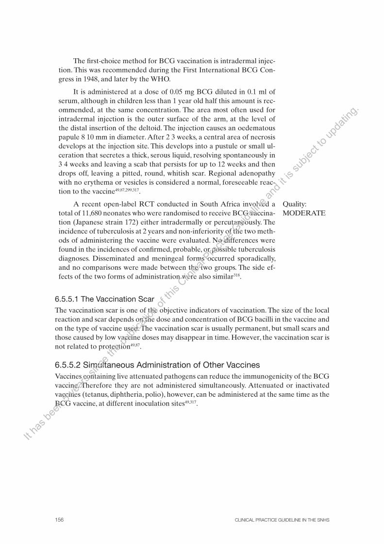
CLINICAL PRACTICE GUIDELINE IN THE SNHS156
The first-choice method for BCG vaccination is intradermal injec-tion. This was recommended during the First International BCG Con-gress in 1948, and later by the WHO.
It is administered at a dose of 0.05 mg BCG diluted in 0.1 ml of serum, although in children less than 1 year old half this amount is rec-ommended, at the same concentration. The area most often used for intradermal injection is the outer surface of the arm, at the level of the distal insertion of the deltoid. The injection causes an oedematous papule 8 10 mm in diameter. After 2 3 weeks, a central area of necrosis develops at the injection site. This develops into a pustule or small ul-ceration that secretes a thick, serous liquid, resolving spontaneously in 3 4 weeks and leaving a scab that persists for up to 12 weeks and then drops off, leaving a pitted, round, whitish scar. Regional adenopathy with no erythema or vesicles is considered a normal, foreseeable reac-tion to the vaccine49,87,299,317.
A recent open-label RCT conducted in South Africa involved a total of 11,680 neonates who were randomised to receive BCG vaccina-tion (Japanese strain 172) either intradermally or percutaneously. The incidence of tuberculosis at 2 years and non-inferiority of the two meth-ods of administering the vaccine were evaluated. No differences were found in the incidences of confirmed, probable, or possible tuberculosis diagnoses. Disseminated and meningeal forms occurred sporadically, and no comparisons were made between the two groups. The side ef-fects of the two forms of administration were also similar318.
Quality: MODERATE
6.5.5.1 The Vaccination Scar The vaccination scar is one of the objective indicators of vaccination. The size of the local reaction and scar depends on the dose and concentration of BCG bacilli in the vaccine and on the type of vaccine used. The vaccination scar is usually permanent, but small scars and those caused by low vaccine doses may disappear in time. However, the vaccination scar is not related to protection49,87.
6.5.5.2 Simultaneous Administration of Other VaccinesVaccines containing live attenuated pathogens can reduce the immunogenicity of the BCG vaccine. Therefore they are not administered simultaneously. Attenuated or inactivated vaccines (tetanus, diphtheria, polio), however, can be administered at the same time as the BCG vaccine, at different inoculation sites49,317.
It has
been
5 ye
ars si
nce t
he pu
blica
tion o
f this
Clinica
l Prac
tice G
uideli
ne an
d it is
subje
ct to
upda
ting.

CLINICAL PRACTICE GUIDELINE ON THE DIAGNOSIS, TREATMENT AND PREVENTION OF TUBERCULOSIS 157
Summary of Evidence
Quality: LOW
The efficacy demonstrated varies between studies. Factors relating to vacci-nation, the vaccine itself, the host, infection by non-tuberculous mycobacte-ria or highly virulent strains of M. tuberculosis, and population differences all play a role.
Quality: LOW
Vaccination has a good cost-efficacy balance, particularly in countries with high incidence of tuberculosis.
Quality: LOW
The vaccine reduces the number of new cases and mortality caused by tu-berculosis and tuberculous meningitis, particularly in children.
Quality: LOW
The vaccine does not seem to be effective in HIV-positive children.
Recommendations
STRONG Routine BCG vaccination is not recommended in Spain.
√BCG vaccination is suggested for healthcare staff and those contacts of multi-drug resistant tuberculosis, and in whom other control strategies can-not be implemented or have failed.
√The BCG vaccine must not be administered to those who have already been infected.
√A diagnosis of tuberculosis must not be ruled out in a vaccinated individual in whom clinical findings suggest tuberculosis.
It has
been
5 ye
ars si
nce t
he pu
blica
tion o
f this
Clinica
l Prac
tice G
uideli
ne an
d it is
subje
ct to
upda
ting.

CLINICAL PRACTICE GUIDELINE IN THE SNHS158
It has
been
5 ye
ars si
nce t
he pu
blica
tion o
f this
Clinica
l Prac
tice G
uideli
ne an
d it is
subje
ct to
upda
ting.

CLINICAL PRACTICE GUIDELINE ON THE DIAGNOSIS, TREATMENT AND PREVENTION OF TUBERCULOSIS 159
7. Dissemination and Implementation
Guideline Formats, Dissemination, and ImplementationThere are various different versions of CPGs: full, summary, information for patients, and quick-consultation tool. All these versions are available in HTML and PDF format from the GuíaSalud website (www.guiasalud.es). A hard copy of the summary version contain-ing a CD ROM of the full version is also published.
Dissemination and implementation strategies include the following:
Official presentation of the guideline by healthcare authorities.•
Copies sent individually to professionals and potential users.•
Distribution of patient guideline.•
Dissemination of the guideline in electronic format on the websites of healthcare •services and scientific societies involved in the project.
Presentation of the guideline at scientific events (conferences, meetings).•
Publication of the guideline in medical journals.•
Proposed Evaluation IndicatorsAny organisation that provides medical services must establish ongoing improvement strategies. This is the basis of quality plans, which require objective evaluation indicators. Healthcare indicators are a quantitative (and sometimes qualitative) measure that al-low for objective tracking of the quality of the important processes involved, in this case, healthcare. This means that indicators are not a direct measure of quality but rather tools that require ongoing use, assessment of changes in outcomes, and reading in the context of each individual organisation providing the service being evaluated. The quality of health-care is a gradient of probability, and improvements to healthcare can improve patients’ health (or that of the public).
Indicators do not measure a single component of healthcare quality. Instead, many aspects such as the accessibility, efficacy, and suitability of healthcare are interrelated and affect the final outcome. However, it is very important for indicators to have both internal validity (they must identify what is to be measured) and external validity (results must be interpretable), and to be based on up-to-date knowledge.
Defining indicators is not easy, and still less so in Spain, where various different organ-isations provide often complementary medical services within a single healthcare process. In addition to proposing a formula for calculation, identifying the updated theoretical ba-sis supporting it, and identifying the sources of information from which cases will be taken, the underlying factors that explain possible variations in results must also be outlined.
The working group has considered the complexity of healthcare provided for tuber-culosis and the many different situations in Spain, and has chosen to select a series of indicators already developed by the WHO, which can be adapted to Spain. Appendix 7
It has
been
5 ye
ars si
nce t
he pu
blica
tion o
f this
Clinica
l Prac
tice G
uideli
ne an
d it is
subje
ct to
upda
ting.

CLINICAL PRACTICE GUIDELINE IN THE SNHS160
presents and explains a list of indicators; comprehensive descriptions can be found in the full WHO document319. Finally, this clinical practice guideline aims to provide a tool for interested clinicians and managers, which may be useful in the specific design of healthcare evaluation.
It has
been
5 ye
ars si
nce t
he pu
blica
tion o
f this
Clinica
l Prac
tice G
uideli
ne an
d it is
subje
ct to
upda
ting.

CLINICAL PRACTICE GUIDELINE ON THE DIAGNOSIS, TREATMENT AND PREVENTION OF TUBERCULOSIS 161
8. Diagnosis and Treatment Strategies
Figure 1: Patient monitoring: referral criteria
MONITORING IN PRIMARY CARE
Positive culture 2 months after the beginning of treatment or √positive culture following previous negative culturesIrregular treatment for longer than 1 month √Intolerance to treatment (mainly liver toxicity) √Associated disorders that make it difficult to monitor treatment √(severe liver or kidney disease)Extrapulmonary forms or forms caused by drug-resistant √mycobacteria or in individuals who are HIV-positivePregnant women √
REFERRAL TO SPECIALIST CARE
Need of complementary explorations √Smear-positive patient for whom isolation at home is problematic √Patient whose overall condition is severely affected or with severe √forms of tuberculosis (tuberculous meningitis, disseminated forms, extensive pulmonary forms with respiratory failure)Severe toxicity caused by tuberculosis drugs √Interactions with drugs such as anticoagulants or others with a √narrow therapeutic margin
HOSPITALISATIONIt has
been
5 ye
ars si
nce t
he pu
blica
tion o
f this
Clinica
l Prac
tice G
uideli
ne an
d it is
subje
ct to
upda
ting.
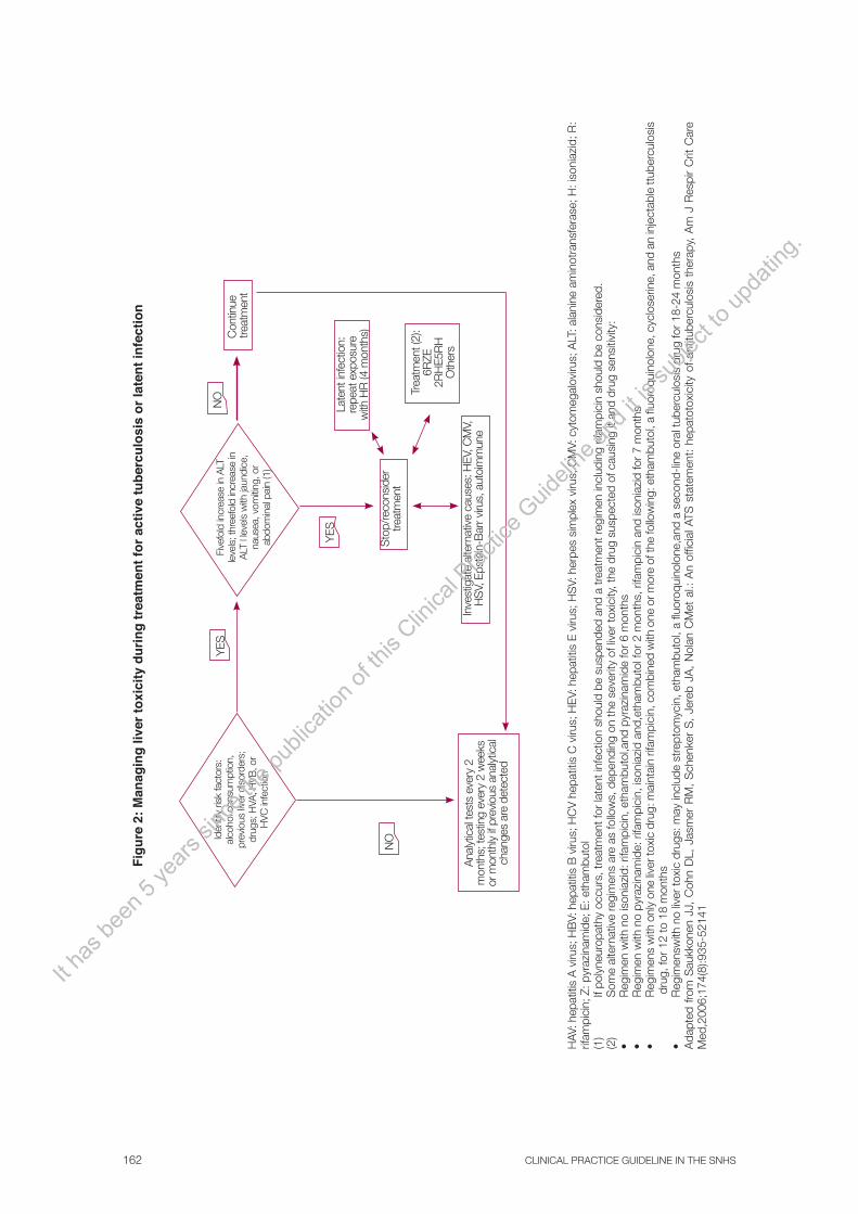
CLINICAL PRACTICE GUIDELINE IN THE SNHS162
HAV
: hep
atiti
s A
viru
s; H
BV:
hep
atiti
s B
viru
s; H
CV
hep
atiti
s C
viru
s; H
EV:
hep
atiti
s E
viru
s; H
SV:
her
pes
sim
plex
viru
s; C
MV:
cyt
omeg
alov
irus;
ALT
: ala
nine
am
inot
rans
fera
se; H
: iso
niaz
id; R
: rif
ampi
cin;
Z: p
yraz
inam
ide;
E: e
tham
buto
l(1
) If
poly
neur
opat
hy o
ccur
s, tr
eatm
ent f
or la
tent
infe
ctio
n sh
ould
be
susp
ende
d an
d a
trea
tmen
t reg
imen
incl
udin
g rif
ampi
cin
shou
ld b
e co
nsid
ered
. (2
) S
ome
alte
rnat
ive
regi
men
s ar
e as
follo
ws,
dep
endi
ng o
n th
e se
verit
y of
live
r to
xici
ty, t
he d
rug
susp
ecte
d of
cau
sing
it a
nd d
rug
sens
itivi
ty:
• R
egim
en w
ith n
o is
onia
zid:
rifa
mpi
cin,
eth
ambu
tol,a
nd p
yraz
inam
ide
for
6 m
onth
s•
Reg
imen
with
no
pyra
zina
mid
e: r
ifam
pici
n, is
onia
zid
and,
etha
mbu
tol f
or 2
mon
ths,
rifa
mpi
cin
and
ison
iazi
d fo
r 7
mon
ths
• R
egim
ens
with
onl
y on
e liv
er to
xic
drug
: mai
ntai
n rif
ampi
cin,
com
bine
d w
ith o
ne o
r mor
e of
the
follo
win
g: e
tham
buto
l, a
fluor
oqui
nolo
ne, c
yclo
serin
e, a
nd a
n in
ject
able
ttub
ercu
losi
s
dru
g, fo
r 12
to 1
8 m
onth
s•
Reg
imen
swith
no
liver
toxi
c dr
ugs:
may
incl
ude
stre
ptom
ycin
, eth
ambu
tol,
a flu
oroq
uino
lone
,and
a s
econ
d-lin
e or
al tu
berc
ulos
is d
rug
for
18-2
4 m
onth
sA
dapt
ed f
rom
Sau
kkon
en J
J, C
ohn
DL,
Jas
mer
RM
, S
chen
ker
S,
Jere
b JA
, N
olan
CM
et a
l.: A
n of
ficia
l ATS
sta
tem
ent:
hepa
toto
xici
ty o
f an
titub
ercu
losi
s th
erap
y, A
m J
Res
pir
Crit
Car
e M
ed,2
006;
174(
8):9
35-5
2141
Figu
re 2
: Man
agin
g liv
er to
xici
ty d
urin
g tr
eatm
ent f
or a
ctiv
e tu
berc
ulos
is o
r lat
ent i
nfec
tion
Iden
tify
risk
fact
ors:
al
coho
l con
sum
ptio
n,
prev
ious
liver
dis
orde
rs;
drug
s; H
VA, H
VB, o
r H
VC in
fect
ion
Five
fold
incr
ease
in A
LT
leve
ls; t
hree
fold
incr
ease
in
ALT
l le
vels
with
jaun
dice
, na
usea
, vom
iting
, or
abdo
min
al p
ain
(1)
Con
tinue
tr
eatm
ent
Late
nt in
fect
ion:
re
peat
exp
osur
e w
ith H
R (4
mon
ths)
Sto
p/re
cons
ider
tre
atm
ent
Trea
tmen
t (2)
:6R
ZE2R
HE5
RH
Oth
ers
Inve
stig
ate
alte
rnat
ive
caus
es: H
EV, C
MV,
H
SV,
Eps
tein
-Bar
r viru
s, a
utoi
mm
une
Ana
lytic
al te
sts
ever
y 2
mon
ths;
test
ing
ever
y 2
wee
ks
or m
onth
ly if
pre
viou
s an
alyt
ical
ch
ange
s ar
e de
tect
ed
NO
NO
YES
YES
It has
been
5 ye
ars si
nce t
he pu
blica
tion o
f this
Clinica
l Prac
tice G
uideli
ne an
d it is
subje
ct to
upda
ting.

CLINICAL PRACTICE GUIDELINE ON THE DIAGNOSIS, TREATMENT AND PREVENTION OF TUBERCULOSIS 163
Figure 3: Beginning a conventional contact study according to form of tuberculosis and confirmation of diagnosis
NO contact study
Index case of tuberculosis
Pulmonary/pleural/laryngeal (confirmed)
Suspected pulmonary TB (pending proof)
Begin CONTACT STUDY
Cavity on chest X ray: HIGH suspicion of
tuberculosis
Abnormal chest X ray with no cavity: COMPATIBLE
with tuberculosis
Begin CONTACT STUDY
Contact study in order of priority
Abnormal chest X ray INCOMPATIBLE with
tuberculosis
NEGATIVE smear microscopy (or no smear microscopy performed)
POSITIVE smear microscopy (respiratory secretion)
Begin CONTACT STUDY
Extrapulmonary
Adapted from National Tuberculosis Controllers Association; Center for Disease Control and Prevention: Guidelines for the investigation of contacts of persons with infectious tuberculosis; recommendations from the Natio¬nal Tuberculosis Controllers Association and CDC, and Guidelines for using the Quantiferon-TB Gold Test for detecting Mycobacterium tuberculosis infection, United States, MMWR 2005 Dec 16; 54 (RR-15): 1-5533
It has
been
5 ye
ars si
nce t
he pu
blica
tion o
f this
Clinica
l Prac
tice G
uideli
ne an
d it is
subje
ct to
upda
ting.

CLINICAL PRACTICE GUIDELINE IN THE SNHS164
NO
Repeat tuberculin test 8 10 weeks after last exposure
NO
Full examination to diagnose pulmonary tuberculosis
Chest X ray
Treat latent infection
END of evaluation
Are symptoms
compatible with tuberculosis?
Does image suggest tuberculosis?
8-10 weeks since last
exposure?
TT > 5 mm
TT: tuberculin test
3.1 If the patient displays any signs or symptoms that suggest tuberculosis, he/she must undergo a full examination to rule out or confirm the diagnosis of active tuberculosis.
3.2 If a chest X ray image is compatible with tuberculosis in an individual with a positive tuberculin test (even with no symptoms), he/she must undergo a full examination to rule out or confirm the diagnosis of active tuberculosis.
3.3 If a chest X ray is normal (or incompatible with tuberculosis) in an individual with a positive tuberculin test (even with no symptoms), treatment for latent infection must be begun.
3.4 The study must be ended if the tuberculin test is <5 mm (if there are no clinical symptoms and a chest X ray is normal) and more than 8 weeks have elapsed since the last exposure, or if a second tuberculin test is <5 mm 8 weeks after the last exposure.
Adapted from National Tuberculosis Controllers Association; Center for Disease Control and Prevention: Guidelines for the investigation of contacts of persons with infectious tuberculosis; recommendations from The National Tuberculosis Controllers Association and CDC, and Guidelines for using the Quantiferon-TB Gold Test for detecting Mycobacterium tuberculosis infection, United States, MMWR 2005 Dec 16; 54 (RR-15): 1-5533
YES
YES
YES
YES
YES
NONO
NO
3.1
3.2
3.3
3.4
3.4
Figure 4: Examination and treatment of high- and medium-priority contacts (immunocompetent adults, children aged 5 or above)
TT > 5 mm
Clinical examination, examination of exposure and examination of tuberculin test
It has
been
5 ye
ars si
nce t
he pu
blica
tion o
f this
Clinica
l Prac
tice G
uideli
ne an
d it is
subje
ct to
upda
ting.

CLINICAL PRACTICE GUIDELINE ON THE DIAGNOSIS, TREATMENT AND PREVENTION OF TUBERCULOSIS 165
Appendices
It has
been
5 ye
ars si
nce t
he pu
blica
tion o
f this
Clinica
l Prac
tice G
uideli
ne an
d it is
subje
ct to
upda
ting.

CLINICAL PRACTICE GUIDELINE IN THE SNHS166
It has
been
5 ye
ars si
nce t
he pu
blica
tion o
f this
Clinica
l Prac
tice G
uideli
ne an
d it is
subje
ct to
upda
ting.

CLINICAL PRACTICE GUIDELINE ON THE DIAGNOSIS, TREATMENT AND PREVENTION OF TUBERCULOSIS 167
Appendix 1: Quality of Evidence and Strength of Recommendations
GRADE classification of evidence quality
Quality of scientific evidence
Study design Quality reduced if Quality increased if
HIGH RCT Design shortcoming: Significant (-1)Very significant (-2)
Inconsistency (-1)
Direct evidence: Some uncertainty (-1)Great uncertainty as to whether evidence is direct (-2)
Inaccurate data (-1)
Reporting bias: High probability of (-1)
Association: scientific evidence of strong association (RR > 2 or < 0.5 and based on observational studies with no confounding factors) (+1)Scientific evidence of a very strong association (RR>5 or <0.2 and based on studies with no chance of bias) (+2)Dose-response slope (+1)
All the possible confounding factors may have reduced the effect observed (+1)
MODERATE
LOW
Observational study
VERY LOW Other designs
Implications of GRADE levels of recommendations
Implications of a strong recommendation:
Patients Clinicians Managers/Planners
The vast majority of people would agree with the action recommended, and only a few would disagree.
The action recommended should be implemented for most patients.
The recommendation can be adopted as healthcare policy in most situations.
Implications of a weak recommendation:
Patients Clinicians Managers/Planners
Most people would agree with the action recommended but a significant number would not.
Acknowledges that different options will be appropriate for different patients, and that clinicians must help each patient choose the option most consistent with his/her values and preferences.
Significant debate and stakeholder involvement are needed.
It has
been
5 ye
ars si
nce t
he pu
blica
tion o
f this
Clinica
l Prac
tice G
uideli
ne an
d it is
subje
ct to
upda
ting.

CLINICAL PRACTICE GUIDELINE IN THE SNHS168
It has
been
5 ye
ars si
nce t
he pu
blica
tion o
f this
Clinica
l Prac
tice G
uideli
ne an
d it is
subje
ct to
upda
ting.

CLINICAL PRACTICE GUIDELINE ON THE DIAGNOSIS, TREATMENT AND PREVENTION OF TUBERCULOSIS 169
Appendix 2: Information for Patients
What is tuberculosis and how is it transmitted?
Tuberculosis is a disease caused by a bacterium called the tuberculosis bacillus (Koch’s bacillus). It can affect any organ, but mainly affects the lungs (respiratory tuberculosis).
It is transmitted from one person to another in thethrough the air. Someone with respiratory tuberculosis expels bacilli into the air when he/she coughs, sneezes, speaks or sings. These can then be inhaled by other people, who become infected, who could become infected as a consequence.
Tuberculosis is only transmitted from someone whose lungs or airways are infected with it.
It is unlikely that someone will be infected following brief contact with tuberculosis patients in open spaces. Tuberculosis is not transmitted by shaking hands, sharing food, utensils or towels or having sex. However, people who live or work every day with some-one with untreated tuberculosis lesions in their lungs are more likely to become infected.
Latent Infection
Often, someone comes into contact with the bacillus, inhales it and becomes infected, but the bacillus does not multiply inside him/her and so he/she does not develop the disease. This is known as latent infection. In these cases, test results will be normal and the person will not have any signs or symptoms. He/She will also be unable to transmit tuberculosis to others.
9 of out of every 10 people infected will never develop the disease. The number of people who become ill is even lower among those who have taken preventive treatment.
If the following symptoms appear (all or some):
• Persistentcoughlasting3ormoreweeks,whetherdryorproductive,withorwithout coughing up blood
• Fever(temperature>38°C[98.4°F])
• Nightsweats
• Weightloss
• Fatigue/malaise/lossofappetite
If this occurs, YOU MUST CONSULT YOUR DOCTOR. You may be suffering from tuberculosis. The earlier tuberculosis is diagnosed, the better it will be treated and the sooner it will be cured.
It has
been
5 ye
ars si
nce t
he pu
blica
tion o
f this
Clinica
l Prac
tice G
uideli
ne an
d it is
subje
ct to
upda
ting.

CLINICAL PRACTICE GUIDELINE IN THE SNHS170
Tuberculosis is curable. If someone has been exposed to it, he/she should consult a doctor to rule out the disease and decide what steps to take. Depending on your circum-stances (and those of the person who is ill), your doctor will prescribe you preventive treat-ment so that you do not develop tuberculosis in the future.
What should I know about tuberculosis treatment and the prognosis?
Treatment with appropriate drugs is the only effective way to cure tuberculosis. It is also the most effective way to prevent other people becoming infected.
Tuberculosis treatment is long: it has to last several months (6 or more) in order to be effective and prevent relapses. It is very important that you take your pills as you are instructed by your doctor/nurse at the beginning of treatment.
Tuberculosis symptoms disappear quickly, but the disease can recur if the patient does not take the medication for long enough.
Possible Side Effects of Tuberculosis Treatment
The tuberculosis drugs used today are safe and effective, and most people do not experi-ence any problems while taking them. However, some people may suffer side effects. It is important to be monitored by a doctor while you are taking this treatment.
You must take your tuberculosis drugs regularly.
It is very important that you take all your medication as instructed by your doctor/ medi-cal staff. Do not stop taking your treatment even if you notice an improvement! Treatment that has not been taken properly or that has been stopped early may make the disease worse, cause a relapse and make it harder or even impossible to cure. If this happens more treatment will be needed, and it will be much longer, complicated and harder to tolerate.
Directly Observed Treatment
Sometimes it can be difficult to remember when and how to take each dose of tuberculosis medication. Directly observed treatment, also called DOT, makes it easier to take your drugs regularly. A group of medical professionals will make sure that you are taking the right medication, that you are taking it correctly and that it is not causing you any health-related problems.
It has
been
5 ye
ars si
nce t
he pu
blica
tion o
f this
Clinica
l Prac
tice G
uideli
ne an
d it is
subje
ct to
upda
ting.
CLINICAL PRACTICE GUIDELINE ON THE DIAGNOSIS, TREATMENT AND PREVENTION OF TUBERCULOSIS 171
How is tuberculosis diagnosed?
If it is suspected that you have tuberculosis (because of a history of contact with a infec-tious tuberculosis patient, or because of symptoms or signs such as those described above), your doctor will give you one of the tests described below.
The Mantoux Test, or Tuberculin Skin Test
A small amount of fluid is injected under the skin using a sterile needle. You will be asked to return 2-3 days later so that the site of the test can be examined and you can be given the results. This test tells us whether someone has already come into contact with the bacillus (positive test) or not (negative test, or a time window, as it takes 8-12 weeks after the last contact with a tuberculosis patient to be sure that the result is negative).
If the test is negative, this means that you do not have the tuberculosis bacillus in your body. This test sometimes gives unreliable results, if the body’s defences have been weak-ened. If this is the case, a chest X ray may be needed.
The test is positive, you will need a chest X ray to rule out active tuberculosis. Some-one who has been exposed to the tuberculosis bacillus will have a positive test result for the rest of his/her life, so a positive result may be the result of an old infection or vaccina-tion. Because of this, a positive result does not necessarily mean that you are ill. Neverthe-less, active tuberculosis must still be ruled out using other tests (a chest X ray).
If your chest X ray is normal there is treatment available to prevent active tuberculo-sis from developing. Preventive treatment is prescribed after examination by a doctor, to be sure that you are healthy and that this treatment will be beneficial to you.
If your chest X ray shows pulmonary lesions the doctor will continue to examine you, using sputum tests, to confirm that you have tuberculosis and determine how infectious you are. Not all forms of tuberculosis are equally infectious.
Can tuberculosis be prevented?
Yes. Any patient with infectious tuberculosis will be asked by his/her doctor to name the people with whom he/she spends a lot of time. This is a normal part of tuberculosis control, and is the best way to ensure that others do not develop tuberculosis. These people include relatives, friends and colleagues, and are known as contacts. Contacts must be examined, initially using tuberculin tests, to see whether they need preventive treatment.
Some people may have a higher risk of developing active the disease tuberculosis after they have been infected. They should therefore also be examined, and preventive treatment should be considered if they test positive to the tuberculin test.It h
as be
en 5
years
sinc
e the
publi
catio
n of th
is Clin
ical P
ractic
e Guid
eline
and i
t is su
bject
to up
datin
g.

CLINICAL PRACTICE GUIDELINE IN THE SNHS172
What precautions should a patient with infectious tuberculosis take?
Someone with infectious tuberculosis must stay at home, in isolation, for the length of time indicated by his/her doctor (usually 2-3 weeks). He/She must not go to work or any public places. In some cases he/she may have to wear a disposable breathing mask (when with other people). He/She must stay in a single room, which should be sunny (if possible) and have a window so that it can be aired several times a day with the door closed. During the isolation period children must not go into this room. When the patient coughs, sneezes or expectorates, he/she must cover his/her mouth and nose with a disposable tissue. Both the mask and the tissues must be placed in a plastic bag, and this must be sealed and then thrown out with the household rubbish. The patient’s room must be cleaned every day us-ing a mop that has been wetted with detergent, so that no dust is stirred up. It must never be swept with a broom, or vacuumed.
It has
been
5 ye
ars si
nce t
he pu
blica
tion o
f this
Clinica
l Prac
tice G
uideli
ne an
d it is
subje
ct to
upda
ting.
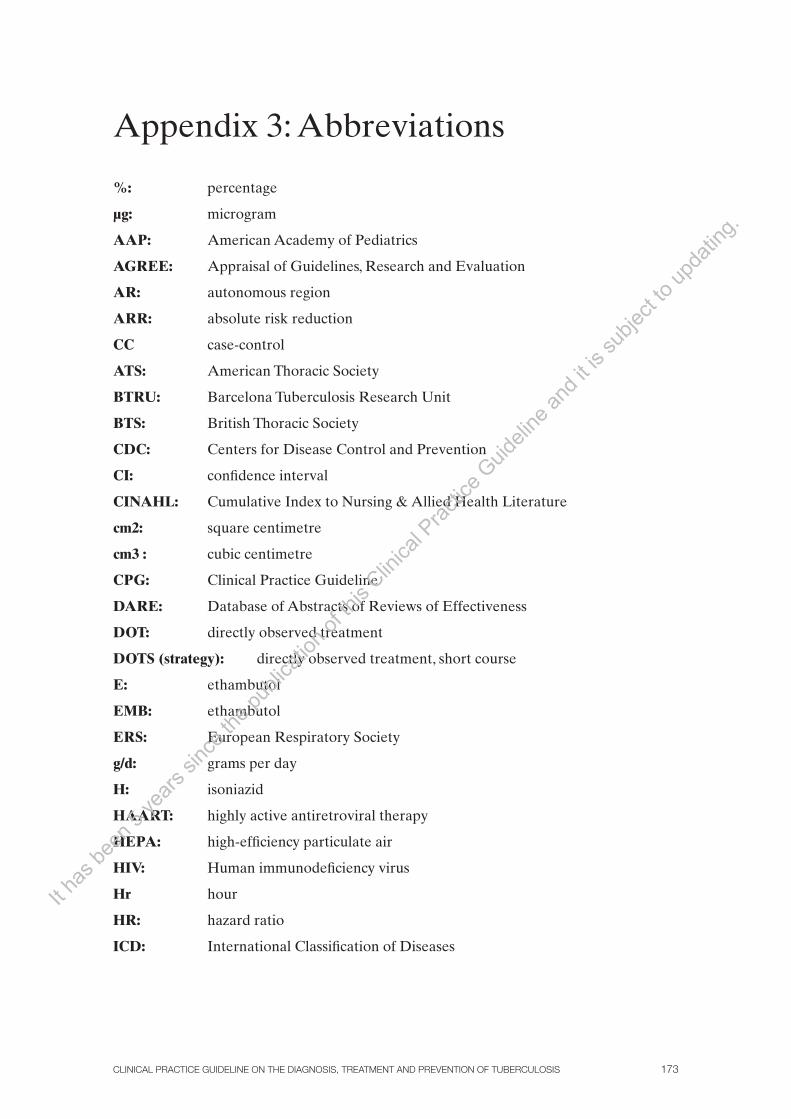
CLINICAL PRACTICE GUIDELINE ON THE DIAGNOSIS, TREATMENT AND PREVENTION OF TUBERCULOSIS 173
Appendix 3: Abbreviations
%: percentage
µg: microgram
AAP: American Academy of Pediatrics
AGREE: Appraisal of Guidelines, Research and Evaluation
AR: autonomous region
ARR: absolute risk reduction
CC case-control
ATS: American Thoracic Society
BTRU: Barcelona Tuberculosis Research Unit
BTS: British Thoracic Society
CDC: Centers for Disease Control and Prevention
CI: confidence interval
CINAHL: Cumulative Index to Nursing & Allied Health Literature
cm2: square centimetre
cm3 : cubic centimetre
CPG: Clinical Practice Guideline
DARE: Database of Abstracts of Reviews of Effectiveness
DOT: directly observed treatment
DOTS (strategy): directly observed treatment, short course
E: ethambutol
EMB: ethambutol
ERS: European Respiratory Society
g/d: grams per day
H: isoniazid
HAART: highly active antiretroviral therapy
HEPA: high-efficiency particulate air
HIV: Human immunodeficiency virus
Hr hour
HR: hazard ratio
ICD: International Classification of Diseases
It has
been
5 ye
ars si
nce t
he pu
blica
tion o
f this
Clinica
l Prac
tice G
uideli
ne an
d it is
subje
ct to
upda
ting.
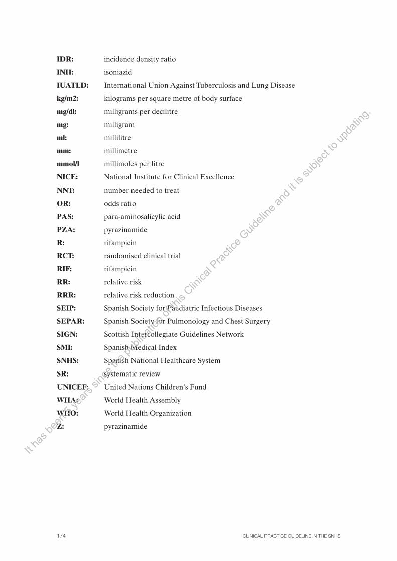
CLINICAL PRACTICE GUIDELINE IN THE SNHS174
IDR: incidence density ratio
INH: isoniazid
IUATLD: International Union Against Tuberculosis and Lung Disease
kg/m2: kilograms per square metre of body surface
mg/dl: milligrams per decilitre
mg: milligram
ml: millilitre
mm: millimetre
mmol/l millimoles per litre
NICE: National Institute for Clinical Excellence
NNT: number needed to treat
OR: odds ratio
PAS: para-aminosalicylic acid
PZA: pyrazinamide
R: rifampicin
RCT: randomised clinical trial
RIF: rifampicin
RR: relative risk
RRR: relative risk reduction
SEIP: Spanish Society for Paediatric Infectious Diseases
SEPAR: Spanish Society for Pulmonology and Chest Surgery
SIGN: Scottish Intercollegiate Guidelines Network
SMI: Spanish Medical Index
SNHS: Spanish National Healthcare System
SR: systematic review
UNICEF: United Nations Children’s Fund
WHA: World Health Assembly
WHO: World Health Organization
Z: pyrazinamide
It has
been
5 ye
ars si
nce t
he pu
blica
tion o
f this
Clinica
l Prac
tice G
uideli
ne an
d it is
subje
ct to
upda
ting.
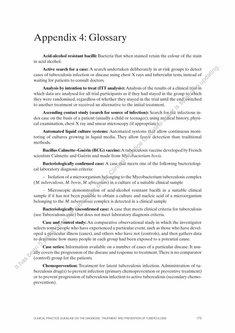
CLINICAL PRACTICE GUIDELINE ON THE DIAGNOSIS, TREATMENT AND PREVENTION OF TUBERCULOSIS 175
Appendix 4: Glossary
Acid-alcohol resistant bacilli: Bacteria that when stained retain the colour of the stain in acid alcohol.
Active search for a case: A search undertaken deliberately in at-risk groups to detect cases of tuberculosis infection or disease using chest X rays and tuberculin tests, instead of waiting for patients to consult doctors.
Analysis by intention to treat (ITT analysis): Analysis of the results of a clinical trial in which data are analysed for all trial participants as if they had stayed in the group to which they were randomised, regardless of whether they stayed in the trial until the end, switched to another treatment or received an alternative to the initial treatment.
Ascending contact study (search for source of infection): Search for the infectious in-dex case on the basis of a patient (usually a child or teenager), using medical history, physi-cal examination, chest X ray and smear microscopy (if appropriate).
Automated liquid culture systems: Automated systems that allow continuous moni-toring of cultures growing in liquid media. They allow faster detection than traditional methods.
Bacillus Calmette–Guérin (BCG) vaccine: A tuberculosis vaccine developed by French scientists Calmette and Guérin and made from Mycobacterium bovis.
Bacteriologically confirmed case: A case that meets one of the following bacteriologi-cal laboratory diagnosis criteria:
− Isolation of a microorganism belonging to the Mycobacterium tuberculosis complex (M. tuberculosis, M. bovis, M. africanum) in a culture of a suitable clinical sample
− Microscopic demonstration of acid-alcohol resistant bacilli in a suitable clinical sample if it has not been possible to obtain a culture and nucleic acid of a microorganism belonging to the M. tuberculosis complex is detected in a clinical sample
Bacteriologically unconfirmed case: A case that meets clinical criteria for tuberculosis (see Tuberculosis case) but does not meet laboratory diagnosis criteria.
Case and control study: An comparative observational study in which the investigator selects some people who have experienced a particular event, such as those who have devel-oped a particular illness (cases), and others who have not (controls), and then gathers data to determine how many people in each group had been exposed to a potential cause.
Case series: Information available on a number of cases of a particular disease. It usu-ally covers the progression of the disease and response to treatment. There is no comparator (control) group for the patients.
Chemoprevention: Treatment for latent tuberculosis infection. Administration of tu-berculosis drug(s) to prevent infection (primary chemoprevention or preventive treatment) or to prevent progression of tuberculosis infection to active tuberculosis (secondary chemo-prevention).
It has
been
5 ye
ars si
nce t
he pu
blica
tion o
f this
Clinica
l Prac
tice G
uideli
ne an
d it is
subje
ct to
upda
ting.

CLINICAL PRACTICE GUIDELINE IN THE SNHS176
Chemotherapy: Regimens involving multiple drugs used to treat active tuberculosis by acting on the bacilli that cause it.
Cochrane review: A systematic review of the evidence from studies related to a par-ticular health problem or medical treatment, produced by Cochrane Collaboration.
Cohort study: A retrospective or prospective monitoring study. The group of people being monitored is defined on the basis of whether or not they have been exposed to some-thing thought to be a risk factor, or whether or not they have received treatment. A cohort study can be comparative, in which case two or more groups are selected on the basis of differences in their exposure to the factor concerned.
Compliance: The patient correctly follows the treatment regimen agreed on with his/her doctor.
Compliance: the degree to which a patient complies with the treatment regime recom-mended.
Confidence interval (CI): The range of values that according to calculations based on a sample of the population contains the actual value for the population with a pre-established level of accuracy or “confidence” (conventionally 95%). 95% confidence means that if the study and method used to calculate the time interval are repeated many times, 95% of the intervals calculated will contain the true value for the population as a whole .
Contact: An individual who has spent a certain amount of time with another person who has infectious tuberculosis.
Conventional contact study: On the basis of an index case, the relevant tests are per-formed to a) diagnose other people who are ill (secondary cases) or infected; b) treat those who are ill and those who are infected and require treatment early; and c) reconstruct the chain of transmission to identify the true index case whenever possible.
Correctly completed treatment: The patient has taken at least 80% of the doses pre-scribed or has taken medication corresponding to a 6 month treatment regimen in a maxi-mum of 9 months. If no final negative culture is taken, a patient is placed in this category if he/she has finished taking his/her medication with no incidents
Country with a high incidence of active tuberculosis: Any country with an annual inci-dence equal to or greater than 40 cases per 100,000 inhabitants.
Country with a low incidence of active tuberculosis: A country with a low incidence of tuberculosis is defined as a country with fewer than 10 reported cases of active tuberculosis per 100,000 inhabitants. In Europe it is defined as a country with fewer than 20 cases per 100,000 inhabitants, in order to include as many countries as possible.
Culture: Tuberculosis bacteria taken from sputum or other samples are grown for iden-tification and diagnosis.
Cure: The patient has completed treatment and a) if diagnosis was confirmed by a cul-ture: the culture of a sample taken at the end of treatment and at least one earlier sample are negative ; b) if diagnosis was based on smear microscopy alone: the smear microscopies of a sample taken at the end of treatment and at least one earlier sample are negative.
Descriptive study: An observational study designed to quantify current service provi-
It has
been
5 ye
ars si
nce t
he pu
blica
tion o
f this
Clinica
l Prac
tice G
uideli
ne an
d it is
subje
ct to
upda
ting.

CLINICAL PRACTICE GUIDELINE ON THE DIAGNOSIS, TREATMENT AND PREVENTION OF TUBERCULOSIS 177
sion or service provision in certain clinical situation. These studies are not designed to test hypotheses regarding data.
Directly observed treatment (DOT): Direct observation of intake of medication. Pa-tients are usually given their medication by trained healthcare staff, who also promote pa-tients’ compliance with their treatment. This can also be done by a relative or close friend of the patient. DOT is highly recommended by the WHO.
Disseminated (or miliary) tuberculosis: Tuberculosis affecting more than two organs or systems. Isolation of the M. tuberculosis complex in the blood must also be classified as disseminated tuberculosis.
DOTS strategy: DOTS is the strategy recommended internationally to cure tuberculo-sis. It is based on five principles: government support for the control of tuberculosis; identi-fication of patients (passive search) using smear microscopies of patients who consult their doctors due to symptoms that suggest active tuberculosis; standard drug use (short regimen) in at least all patients with positive smear microscopies; regular, uninterrupted supply of essential tuberculosis drugs and standardised recording of reports and the treatment out-comes in each case.
Elimination and eradication: A country is considered to have entered the elimination phase when actual annual incidence is less than 20 cases per 100,000 inhabitants. Tuberculo-sis is considered to have been virtually eliminated when there is one smear-positive person per 1,000,000 inhabitants. Eradication would mean that there were no cases of tuberculosis infection worldwide.
Extrapulmonary (non-respiratory) tuberculosis: Tuberculosis affecting any location other than those described under Pulmonary tuberculosis. Pleural and intrathoracic lym-phatic tuberculosis are also classified as extrapulmonary if they do not involve the pulmo-nary parenchyma.
Failure to complete treatment: A patient interrupts his/her treatment for 2 months or longer and this has not been decide don by a physician; or a patient becomes impossible to monitor before he/she has completed his/her treatment, except when a patient is trans-ferred.
Gastric aspiration: Some patients (particularly children) with suspected tuberculosis are unable to produce sputum by coughing. As an alternative, gastric acid is aspirated by inserting saline solution into the stomach using a probe, and the content is examined for mycobacteria.
Good Clinical Practice (GCP): A recommendation based on the clinical experience of the group that develops the guideline, in the absence of reliable clinical evidence.
Grade of recommendation: Each recommendation is assigned a strength of confidence (strong or weak) with which it is expected to cause more benefit than harm. These grades are based on the quality of the evidence from the tests on which the recommendation is based, risk/benefit analysis, values and preferences, and cost.
Group developing a CPG: A multidisciplinary group of professionals who decide on the clinical issues addressed in the CPG, read the evidence critically and formulate recom-mendations.
It has
been
5 ye
ars si
nce t
he pu
blica
tion o
f this
Clinica
l Prac
tice G
uideli
ne an
d it is
subje
ct to
upda
ting.

CLINICAL PRACTICE GUIDELINE IN THE SNHS178
High-risk group: A segment of the population with a higher risk of developing tuber-culosis (arbitrarily, with more than 100 reported cases of active tuberculosis per 100,000 inhabitants.).
Histological testing: Microscopic analysis of cells and clinical samples.
Index case: The first case of tuberculosis identified; the case which gave rise to other cases.
Infected person: An individual with a positive tuberculin test, demonstrating that he/she has developed an immune response to Mycobacterium tuberculosis, in whom active tuberculosis has been ruled out.
Interferon-gamma release assay: A blood test used to diagnose latent tuberculosis (it can be used instead of or as well as the tuberculin test). It is based on the detection of a white blood cell response to tuberculosis antigens.
Latent infection: Subclinical infection caused by the tuberculosis bacillus with no clini-cal, bacteriological or radiological signs or symptoms of the disease.
Level of evidence: This is the level of confidence (high, moderate, low or very low) with which it is asserted that the results of a study or group or studies represent the “true” value of the intervention(s) evaluated for an issue under research.
Liquid medium culture: A culture grown in a liquid medium. This allows faster detec-tion than traditional solid media.
Mantoux technique: The most widely-used technique for the TT is intradermal reac-tion, known as the Mantoux technique. This involves intradermal injection into the ventral side of the forearm of 0.1 ml PPD, at a dose of 2 TU. The reaction is read 48 72 hours later, although it may remain valid for 7 days.
Meta-analysis: A statistical technique to combine the results of several different stud-ies on the same subject, the results of which are of interest. The aim is to obtain more ac-curate, clearer information from a set of independent data.
Methodological shortcomings: Design or presentation characteristics of a clinical trial which are known to be associated with a risk of bias or lack of validity.
Molecular test: A process used to detect a particular genetic sequence in cells. In the specific case of tuberculosis, genetic sequences can confirm the presence of the mycobacte-rium or of certain mutations that indicate drug resistance.
Multi-drug resistant tuberculosis: Simultaneous resistance to at least isoniazid and ri-fampicin, two of the first-line drugs.
Mycobacterium tuberculosis complex: The following species of mycobacterium: M. tu-berculosis, M. bovis, M. africanum, M. microti, M. canetti, M. capraeand M. pinnipedii. These can cause tuberculosis in humans.
Negative predictive value: The proportion of individuals with a negative test result who do not have the disease in question.
Negative pressure room: These have been used to isolate some patients with respira-tory tuberculosis. The pressure must be 10 Pa below that of the surrounding area.
It has
been
5 ye
ars si
nce t
he pu
blica
tion o
f this
Clinica
l Prac
tice G
uideli
ne an
d it is
subje
ct to
upda
ting.
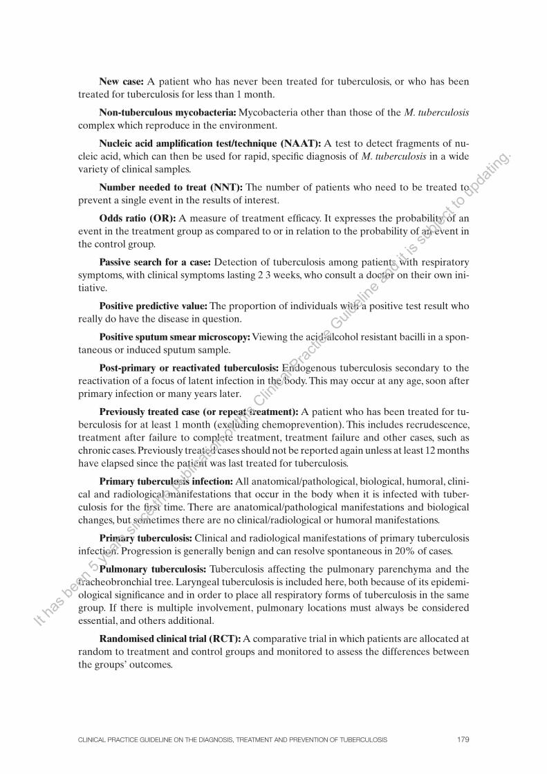
CLINICAL PRACTICE GUIDELINE ON THE DIAGNOSIS, TREATMENT AND PREVENTION OF TUBERCULOSIS 179
New case: A patient who has never been treated for tuberculosis, or who has been treated for tuberculosis for less than 1 month.
Non-tuberculous mycobacteria: Mycobacteria other than those of the M. tuberculosis complex which reproduce in the environment.
Nucleic acid amplification test/technique (NAAT): A test to detect fragments of nu-cleic acid, which can then be used for rapid, specific diagnosis of M. tuberculosis in a wide variety of clinical samples.
Number needed to treat (NNT): The number of patients who need to be treated to prevent a single event in the results of interest.
Odds ratio (OR): A measure of treatment efficacy. It expresses the probability of an event in the treatment group as compared to or in relation to the probability of an event in the control group.
Passive search for a case: Detection of tuberculosis among patients with respiratory symptoms, with clinical symptoms lasting 2 3 weeks, who consult a doctor on their own ini-tiative.
Positive predictive value: The proportion of individuals with a positive test result who really do have the disease in question.
Positive sputum smear microscopy: Viewing the acid-alcohol resistant bacilli in a spon-taneous or induced sputum sample.
Post-primary or reactivated tuberculosis: Endogenous tuberculosis secondary to the reactivation of a focus of latent infection in the body. This may occur at any age, soon after primary infection or many years later.
Previously treated case (or repeat treatment): A patient who has been treated for tu-berculosis for at least 1 month (excluding chemoprevention). This includes recrudescence, treatment after failure to complete treatment, treatment failure and other cases, such as chronic cases. Previously treated cases should not be reported again unless at least 12 months have elapsed since the patient was last treated for tuberculosis.
Primary tuberculosis infection: All anatomical/pathological, biological, humoral, clini-cal and radiological manifestations that occur in the body when it is infected with tuber-culosis for the first time. There are anatomical/pathological manifestations and biological changes, but sometimes there are no clinical/radiological or humoral manifestations.
Primary tuberculosis: Clinical and radiological manifestations of primary tuberculosis infection. Progression is generally benign and can resolve spontaneous in 20% of cases.
Pulmonary tuberculosis: Tuberculosis affecting the pulmonary parenchyma and the tracheobronchial tree. Laryngeal tuberculosis is included here, both because of its epidemi-ological significance and in order to place all respiratory forms of tuberculosis in the same group. If there is multiple involvement, pulmonary locations must always be considered essential, and others additional.
Randomised clinical trial (RCT): A comparative trial in which patients are allocated at random to treatment and control groups and monitored to assess the differences between the groups’ outcomes.
It has
been
5 ye
ars si
nce t
he pu
blica
tion o
f this
Clinica
l Prac
tice G
uideli
ne an
d it is
subje
ct to
upda
ting.

CLINICAL PRACTICE GUIDELINE IN THE SNHS180
Reactivation: An increase in the activity of a focus of latent infection in the body which is no longer controlled by the immune system. This may occur at any age, soon after primary infection or many years later.
Recrudescence: Worsening of a disease following clinical improvement, due to inade-quate bacteriological cure of the disease. In tuberculosis, for example, molecular techniques make it possible to determine whether a case of recrudescence is caused by the same strain of the bacillus (endogenous reactivation).
Relapse/recurrence: A second episode of a disease, in this case tuberculosis, after the initial episode was considered cured.
Relative risk (RR): A measure of treatment efficacy. It shows how much more or less likely an event is in the treatment group than in the control group.
Repeat infection: A second Mycobacterium tuberculosis infection. A previously in-fected host acquires a certain level of resistance to repeat infection. This resistance is main-tained by repeated exposure to the bacillus over the course of the individual’s life. However, if the level of contagion is high (large-scale inoculation), if there are factors that alter the host’s immunity or if resistance to infection falls because of a lack of repeated antigenic stimulation, the host becomes vulnerable to exogenous reinfection by a strain other than that involved in primary infection.
Reservoir: The main reservoir is the infected human, who may develop active tubercu-losis and expel bacilli in all respiratory movements, particularly coughing or sneezing, thus becoming a source of infection. In areas where bovine tuberculosis is common, cattle may also be a reservoir.
Sensitivity (of a test): The proportion of individuals classified as positive by a test who are correctly identified by the test.
Short treatment regimen: A treatment regimen for active tuberculosis that lasts 6 months.
Smear-positive case: When direct microscopic analysis of a spontaneous or induced sputum sample shows that it contains acid-alcohol resistant bacilli.
Specificity (of a test): The proportion of individuals classified as negative by a test who are correctly identified by the test.
Standard test (gold standard): A test that is considered to be the main or standard test, with which other treatments or tests must be compared.
Standard treatment (short regimen): This is the treatment regimen used for tubercu-losis patients that cures more than 95% of patients and causes severe intolerance requiring alterations to treatment in fewer than 5% of cases. Its minimum duration is currently 6 months, and it involves a combination of four first-line drugs.
Successful treatment: This includes both cured patients (with bacteriological confirma-tion) and those who have correctly completed the treatment regimen prescribed.
Systematic review (SR): An investigation that summarises the evidence on a particular, clearly worded issue, using systematic, explicit methods to identify, select and evaluate the relevant studies and to extract, collate and report on their conclusions. Statistical methods of meta-analysis may or may not be used.
It has
been
5 ye
ars si
nce t
he pu
blica
tion o
f this
Clinica
l Prac
tice G
uideli
ne an
d it is
subje
ct to
upda
ting.

CLINICAL PRACTICE GUIDELINE ON THE DIAGNOSIS, TREATMENT AND PREVENTION OF TUBERCULOSIS 181
Treatment failure: A patient that has not attained bacteriological conversion 5 months after beginning treatment and following it correctly, or a patient that attains bacteriological conversion but this is thenthat is reversed and, asa consequence, the patient needs second-line treatment instead of first-line treatment. Bacteriological conversion is not considered to have been attained when cultures remain positive with no significant reduction in the number of colonies; conversion is considered to have been reversed when there are two consecutive positive cultures with an increasing number of colonies following two previous consecutive negative cultures.
Treatment of latent tuberculosis infection: Administration of one or more tuberculosis drugs to prevent progression of infection to active tuberculosis (secondary chemoprevention).
Tuberculin skin test (TST): The standard test for diagnosis of tuberculosis infection. It uses an extract obtained from the filtrate of a culture of tuberculosis bacilli which is then sterilised and concentrated. The type of antigen currently used in tuberculin and is PPD (purified protein derivative). In Spain the variant RT 23 is used, with Tween 80 as an anti-absorbent. The immune response is assessed 48 72 hours later, on the basis of the size of the induration at the injection site. Results are expressed in millimetres.
Tuberculin test conversion: A tuberculin test is considered to have converted if the di-ameter of the induration is > 5 mm up to 2 years after a test with induration < 5 mm and the difference between the two measurements is 6 10 mm. This margin has been established to prevent errors when tuberculin tests are read. Establishing conversion is very important, as it reflects a high risk of becoming ill up to 2 years after infection. This individual will there-fore be a candidate for treatment for latent infection, regardless of his/her age.
Tuberculosis case: Any patient who meets one of the following requirements:
1) Signs, symptoms and/or X ray findings consistent with active tuberculosis in any location and prescription of a full course of tuberculosis treatment
2) Post-mortem diagnosis with abnormal findings consistent with active tuberculosis which would have required tuberculosis treatment if it had been diagnosed before death
Tuberculosis control (strategies): Strategies that aim to reduce the incidence of new My-cobacterium tuberculosis complex infections by identifying the sources of infection as quickly as possible and applying curative treatment so that these sources cease to be infectious.
Tuberculosis elimination (strategies): Strategies that aim to reduce the prevalence of latent tuberculosis, such as treatment of individuals at high risk of developing active tuberculosis.
Tuberculosis outbreak: The Spanish Health Authorities’ current surveillance protocol defines an outbreak as the appearance of one or more cases of tuberculosis after the first case detected (Tuberculosis Prevention and Control Plan for Spain, 2007).
Tuberculosis: A disease caused by infection by bacteria of the M. tuberculosis complex.
It has
been
5 ye
ars si
nce t
he pu
blica
tion o
f this
Clinica
l Prac
tice G
uideli
ne an
d it is
subje
ct to
upda
ting.

CLINICAL PRACTICE GUIDELINE IN THE SNHS182
It has
been
5 ye
ars si
nce t
he pu
blica
tion o
f this
Clinica
l Prac
tice G
uideli
ne an
d it is
subje
ct to
upda
ting.
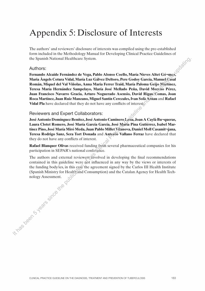
CLINICAL PRACTICE GUIDELINE ON THE DIAGNOSIS, TREATMENT AND PREVENTION OF TUBERCULOSIS 183
Appendix 5: Disclosure of Interests
The authors’ and reviewers’ disclosure of interests was compiled using the pre-established form included in the Methodology Manual for Developing Clinical Practice Guidelines of the Spanish National Healthcare System.
Authors:Fernando Alcaide Fernández de Vega, Pablo Alonso Coello, María Nieves Altet Gó¬mez, Maria Àngels Cotura Vidal, María Luz Gálvez Deltoro, Pere Godoy García, Manuel Casal Román, Miquel del Val Viñolas, Anna Maria Ferrer Traid, María Paloma Geijo Martínez, Teresa María Hernández Sampelayo, María José Mellado Peña, David Moreno Pérez, Juan Francisco Navarro Gracia, Arturo Noguerado Asensio, David Rigau Comas, Joan Roca Martínez, Juan Ruiz Manzano, Miguel Santín Cerezales, Ivan Solà Arnau and Rafael Vidal Pla have declared that they do not have any conflicts of interest.
Reviewers and Expert Collaborators:José Antonio Domínguez Benítez, José Antonio Caminero Luna, Joan A Caylà Bu¬queras, Laura Clotet Romero, José María García García, José María Pina Gutiérrez, Isabel Mar-tínez Pino, José María Miró Meda, Juan Pablo Millet Vilanova, Daniel Moll Casamit¬jana, Teresa Rodrigo Sanz, Sera Tort Donada and Antonio Vallano Ferraz have declared that they do not have any conflicts of interest.
Rafael Blanquer Olivas received funding from several pharmaceutical companies for his participation in SEPAR’s national conference.
The authors and external reviewers involved in developing the final recommendations contained in this guideline were not influenced in any way by the views or interests of the funding body/ies, in this case the agreement signed by the Carlos III Health Institute (Spanish Ministry for Health and Consumption) and the Catalan Agency for Health Tech-nology Assessment.
It has
been
5 ye
ars si
nce t
he pu
blica
tion o
f this
Clinica
l Prac
tice G
uideli
ne an
d it is
subje
ct to
upda
ting.

CLINICAL PRACTICE GUIDELINE IN THE SNHS184
It has
been
5 ye
ars si
nce t
he pu
blica
tion o
f this
Clinica
l Prac
tice G
uideli
ne an
d it is
subje
ct to
upda
ting.

CLINICAL PRACTICE GUIDELINE ON THE DIAGNOSIS, TREATMENT AND PREVENTION OF TUBERCULOSIS 185
Appendix 6: Main Documents and Useful Resources
To compile this CPG on the diagnosis, treatment, and prevention of tuberculosis, some high-quality CPGs and consensus documents on the subject were consulted as secondary literature sources. When it came to guidelines, only The guidelines particularly used were those of sufficient quality according to the AGREE directive, i.e. those that could be con-sidered as recommended for use in clinical practice, were taken into consideration. The consensus documents consulted were those likely to be most applicable due to the similar-ity to Spain of the environments for which they were written to Spain.
Thanks to their rigour and clarity, some of these documents provided inspiration and served as examples for some sections. Below is a list and links to the full text of the main documents. Due to their high quality or recent publication date, these may constitute a major source of information for those using this CPG.
Clinical Practice Guidelines on Tuberculosis
Title (year): Diagnóstico y tratamiento de la tuberculosis [Diagnosis and treatment of tuberculosis] (2008)Author(s): Ruiz-Manzano J, Blanquer R et al.Reference: Arch Bronconeumol. 2008; 44(10): 551-66
Title (year): Tuberculosis Care with TB-HIV Co-management: Integrated Man-agement of Adolescent and Adult Illness (IMAI) (2007)Author(s): WHOWebsite: http://www.who.int/hiv/capacity/TBHIV/en/index.html
Title (year): Canadian tuberculosis standards (2007)Author(s): Tuberculosis Prevention and Control, Public Health Agency of Cana-da, and the Canadian Lung Association/Canadian Thoracic SocietyWebsite: http://www.phac-aspc.gc.ca
Title (year): Consensus document on the treatment of pulmonary tuberculosis in children(2007)Author(s): Tuberculosis Working Group of the Spanish Society for Paediatric Infectious Diseases (SEIP) Reference: An Esp Pediatr 2007; 66: 597-602
Title (year): Guidelines for the programmatic management of drug-resistant tu-berculosis (2006).Author(s): WHOWebsite: http://whqlibdoc.who.int/publications/2006/9241546956_eng.pdf
It has
been
5 ye
ars si
nce t
he pu
blica
tion o
f this
Clinica
l Prac
tice G
uideli
ne an
d it is
subje
ct to
upda
ting.

CLINICAL PRACTICE GUIDELINE IN THE SNHS186
Title (year): Clinical practice guideline of the Andalusian Society for Infectious Diseases (SAEI) (2006)Author(s): Domínguez-Castellano A et al.Reference: Enferm Infecc Microbiol Clin 2007; 25(8): 519-34
Title (year): Guidance for national tuberculosis programmes on the management of tuberculosis in children (2006)Author(s): WHOWebsite: http://whqlibdoc.who.int/hq/2006/WHO_HTM_TB_2006.371_eng.pdf
Title (year): Clinical diagnosis and management of tuberculosis, and measures for its prevention and control (2006)Author(s): NICE, Royal College of PhysiciansWebsite: http://www.rcplondon.ac.uk
Title (year): International Standards for Tuberculosis Care (2006)Author(s): Tuberculosis Coalition for Technical AssistanceWebsite: http://www.stoptb.org
Title (year): Controlling Tuberculosis in the United States (2005)Author(s): ATS, CDCReference: Am J Respir Crit Care Med. 2005; 172: 1169-1227
Title (year): Consensus document on directly observed treatmentsfortuberculosis (1999)Author(s): Study Group of the 1999 BTRU WorkshopReference: Med Clin (Barc) 2000; 115: 749-757
Title (year): Consensus document on contact studies in tuberculosis patients(1999)Author(s): BTRU Contact Study Group Reference: Med Clin (Barc) 1999; 112: 151-6
Title (year): Consensus document on tuberculosis prevention and control in Spain(1999)Author(s): BTRU Study Group Reference: Med Clin (Barc) 1999; 113: 710-5
It has
been
5 ye
ars si
nce t
he pu
blica
tion o
f this
Clinica
l Prac
tice G
uideli
ne an
d it is
subje
ct to
upda
ting.

CLINICAL PRACTICE GUIDELINE ON THE DIAGNOSIS, TREATMENT AND PREVENTION OF TUBERCULOSIS 187
Appendix 7: Proposed Evaluation Indicators
The WHO sponsors and leads the Stop TB Partnership, which consists of many govern-mental and non-governmental organisations and aims to reduce the global burden of tu-berculosis in line with the United Nations’ Millennium Development Goals.
In order to evaluate whether these goals have been attained, two main indicators are proposed concerning the detection and cure of tuberculosis (Table 1)319. The internation-ally accepted definitions for the control of tuberculosis, which are useful for formulating indicators, can be found in a document compiled jointly by the WHO, the IUATLD and the Royal Netherlands Tuberculosis Association (KNCV)80.
A group of priority indicators is also proposed to assess implementation of the Stop TB Partnership’s goals in national programmes320:
Tuberculosis incidence (total and with positive smear microscopy)
Number of cases of tuberculous meningitis in children aged 0-4
Prevalence of infection among schoolchildren aged 6-7
Treatment compliance (total and with positive smear microscopy)
Delay in diagnosis in those with positive smear microscopies
Percentage of high-risk patients receiving DOT
Rate of compliance with DOT
Percentage of cases for which contact studies have been performed
These indicators should be subject to annual evaluation in order to provide ongoing improvement321.
Table 1: Tuberculosis-related indicators and goals according to the UN’s Millennium Development Goals
Indicator Definition Goal
Prevalence of tuberculosis and
tuberculosis-related mortality
Number of positive smear microscopies (per 100,000 inhabitants)
50% decrease on 2000 figure by 2010
Number of tuberculosis-related deaths (per 100,000 inhabitants per year)
50% decrease on 2000 figure by 2010.
Proportion of tuberculosis cases
identified and cured using DOT
Proportion of the total number of tuberculosis cases with positive smear microscopies
identified using DOT (in 1 year) Detection of 70% of cases1
Proportion of the total number of tuberculosis cases with positive smear microscopies
successfully treated using DOT Cure of 85% of cases1
1. These goals were for the year 2005.
It has
been
5 ye
ars si
nce t
he pu
blica
tion o
f this
Clinica
l Prac
tice G
uideli
ne an
d it is
subje
ct to
upda
ting.

CLINICAL PRACTICE GUIDELINE IN THE SNHS188
It has
been
5 ye
ars si
nce t
he pu
blica
tion o
f this
Clinica
l Prac
tice G
uideli
ne an
d it is
subje
ct to
upda
ting.
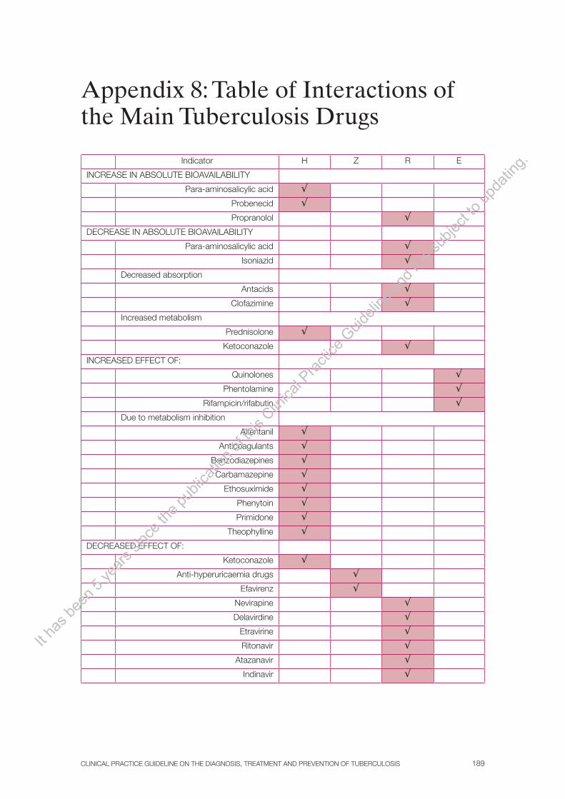
CLINICAL PRACTICE GUIDELINE ON THE DIAGNOSIS, TREATMENT AND PREVENTION OF TUBERCULOSIS 189
Appendix 8: Table of Interactions of the Main Tuberculosis Drugs
Indicator H Z R E
INCREASE IN ABSOLUTE BIOAVAILABILITY
Para-aminosalicylic acid √Probenecid √Propranolol √
DECREASE IN ABSOLUTE BIOAVAILABILITY
Para-aminosalicylic acid √Isoniazid √
Decreased absorption
Antacids √Clofazimine √
Increased metabolism
Prednisolone √Ketoconazole √
INCREASED EFFECT OF:
Quinolones √Phentolamine √
Rifampicin/rifabutin √Due to metabolism inhibition
Alfentanil √Anticoagulants √
Benzodiazepines √Carbamazepine √
Ethosuximide √Phenytoin √Primidone √
Theophylline √DECREASED EFFECT OF:
Ketoconazole √Anti-hyperuricaemia drugs √
Efavirenz √Nevirapine √Delavirdine √
Etravirine √Ritonavir √
Atazanavir √Indinavir √
It has
been
5 ye
ars si
nce t
he pu
blica
tion o
f this
Clinica
l Prac
tice G
uideli
ne an
d it is
subje
ct to
upda
ting.

CLINICAL PRACTICE GUIDELINE IN THE SNHS190
Nelfinavir √Saquinavir √Maraviroc √
Raltegravir √Due to increased metabolism
Beta-blockers √Calcium-channel blockers √
Cloramphenicol √Clofibrate √
Coumarin anticoagulants √Cyclosporine √
Dapsone √Diazepam √
Digoxin/digitoxin √Disopyramide √
Haloperidol √Barbiturates √
Fluconazole, itraconazole √Lorcainide, tocainide √
Methadone √Mexiletine √
Amitriptyline/nortriptyline √Oral contraceptives √
Oral anti-hyperglycaemia drugs √Phenytoin √
Prednisolone √Propafenone √
Quinidine √Theophylline √
Zidovudine √CUMULATIVE TOXICITY
Cycloserine √Disulfiram √Enflurane √
Levodopa √Paracetamol √
Pyridoxine √Rifampicin √
Ethionamide √Neurotoxic drugs √
Saquinavir + ritonavir √Lopinavir + ritonavir (Kaletra) √
It has
been
5 ye
ars si
nce t
he pu
blica
tion o
f this
Clinica
l Prac
tice G
uideli
ne an
d it is
subje
ct to
upda
ting.

CLINICAL PRACTICE GUIDELINE ON THE DIAGNOSIS, TREATMENT AND PREVENTION OF TUBERCULOSIS 191
Appendix 9: Combined Administra-tion of Rifampicin or Rifabutin and Antiretrovirals
RIFAMPICIN
Non-nucleoside reverse transcriptase inhibitors
Change to recommended dose of antiretroviral
Change to recommended
dose of rifampicin Comments
EfavirenzNone (some experts recommend 800
mg for patients weighing > 60 kg)No change (600
mg/day)
Efavirenz AUC ↓ 22%; no change in rifampicin concentration. Efavirenz
must not be administered during the first trimester of
pregnancy
Nevirapine No change No change
(600 mg/day)
Nevirapine AUC ↓ 37% 58% and Cmin ↓68% when 200 mg twice/day administered
Delavirdine Do not combine rifampicin and delavirdine Delavirdine AUC ↓ 95%
Etravirine Do not combine rifampicin and etravirine
Probably substantial decrease in etravirine concentration due
to possible interaction with con rifabutin
Protease inhibitors
Change to recommended dose of antiretroviral
Change to recommended
dose of rifampicinComments
Ritonavir No change No change
(600 mg/day)
Use with care. Ritonavir AUC↓ 35%; no change in rifampicin
concentration. Monitor antiretroviral activity of ritonavir
Fosamprenavir Do not administer rifampicin and fosamprenavir together
Atazanavir Do not administer rifampicin and atazanavir together Atazanavir AUC ↓ more than 95%
Indinavir Do not administer rifampicin and atazanavir together Indinavir AUC ↓ 89%
Nelfinavir Do not administer rifampicin and atazanavir together Nelfinavir AUC ↓ 82%
Saquinavir Do not administer rifampicin and atazanavir together Saquinavir AUC ↓ 84%
Combinations with dual protease inhibitors
Change to recommended dose of antiretroviral
Change to recommended
dose of rifampicinComments
It has
been
5 ye
ars si
nce t
he pu
blica
tion o
f this
Clinica
l Prac
tice G
uideli
ne an
d it is
subje
ct to
upda
ting.

CLINICAL PRACTICE GUIDELINE IN THE SNHS192
Saquinavir/ ritonavir
Saquinavir 400 mg + ritonavir 400 mg twice/day
No change (600 mg/day)
Use with care: a combination of saquinavir (1,000 mg twice/day) ritonavir (100 mg twice/
day) and rifampicin was associated with high rates of hepatitis in healthy volunteers
Lopinavir/ritonavir (Kaletra™)
Increase dose of lopinavir/ritonavir (Kaletra™): 4 capsules (200 mg
lopinavir, 50 mg ritonavir) twice/day
No change (600 mg/day)
Use with care: in a preliminary study this
combination caused some degree of hepatitis in
healthy volunteers
Lopinavir/ritonavir (Kaletra™): high
doses
Lopinavir/ritonavir (Kaletra™): 2 capsules (200 mg lopinavir, 50 mg
deritonavir) + 300 mg ritonavir twice/day
No change (600 mg/day)
Use with care: in a preliminary study this combination caused some degree of
hepatitis in healthy volunteers. However, there are favourable pharmacokinetic and clinical
results in children
CCR5 receptor antagonists
Change to recommended dose of antiretroviral
Change to recommended
dose of rifampicinComments
MaravirocIncrease maraviroc to
600 mg twice/dayNo change
(600 mg/day)
Maraviroc Cmin ↓ 78%. There is no clinical experience, or none has been found, of an increased maraviroc dose
with rifampicin
Integrase inhibitors
Change to recommended dose of antiretroviral
Change to recommended
dose of rifampicinComments
Raltegravir No changeNo change
(600 mg/day)
No clinical experience; raltegravir concentrations
around 40-61%
AUC: area under curve; Cmin: minimum blood plasma concentration Adapted from CDC: Managing Drug Interactions in the Treatment of HIV-Related Tuberculosis [online], 2007. Available at http://www.cdc.gov/tb/TB_HIV_Drugs/default.htm. Consulted on 14 November 2008.
It has
been
5 ye
ars si
nce t
he pu
blica
tion o
f this
Clinica
l Prac
tice G
uideli
ne an
d it is
subje
ct to
upda
ting.

CLINICAL PRACTICE GUIDELINE ON THE DIAGNOSIS, TREATMENT AND PREVENTION OF TUBERCULOSIS 193
RIFABUTIN
Non-nucleoside reverse transcriptase inhibitors
Change to recommended dose of antiretroviral
Change to recommended
dose of rifabutin Comments
Efavirenz No change↑ to 400 600 mg (daily or
intermittently) )
Rifabutin AUC ↓ 38%. The effect of combined efavirenz
and protease inhibitors on rifabutin concentration has not been researched.
Efavirenz must not be administered during the first
trimester of pregnancy
Nevirapine No changeNo change (300
mg/day or 3 times/week)
No significant changes in AUC of rifabutin or
nevirapine
Delavirdine Do not combine rifabutin and delavirdineDelavirdine AUC ↓ 80%; rifabutin AUC ↓ 100%
Etravirine No changeNo change (300
mg/day or 3 times/week)
No clinical experience; Cmin of etravirine ↓ 45% but a
change in dose would not be justified
Protease inhibitors
Change to recommended dose of antiretroviral
Change to recommended
dose of rifabutin Comments
Fosamprenavir No change↓ to 150 mg/
day or 300 mg 3 times/week
No clinical experience published
Atazanavir No change↓ to 150 mg every
other day or 3 times/week
No clinical experience published. Rifabutin AUC ↓
250%
Indinavir 1,000 mg every 8 hours ↓ to 150 mg/
day or 300 mg 3 times/week
Rifabutin AUC ↓ 170%; indinavir concentration ↓ 34%
Nelfinavir No change↓ to 150 mg/
day or 300 mg 3 times/week
Rifabutin AUC ↓ 207%; insignificant changes in indinavir concentration
Combinations with dual protease inhibitors
Change to recommended dose of antiretroviral
Change to recommended
dose of rifabutin Comments
Lopinavir/ritonavir
(Kaletra™)No change
↓ to 150 mg every other day or 3
times/week
Rifabutin AUC ↓ 303%; AUC of one of its metabolites
↓ 47.5%
It has
been
5 ye
ars si
nce t
he pu
blica
tion o
f this
Clinica
l Prac
tice G
uideli
ne an
d it is
subje
ct to
upda
ting.

CLINICAL PRACTICE GUIDELINE IN THE SNHS194
Ritonavir (any dose) with saquinavir, indinavir,
amprenavir, fosamprenavir,
atazanavir, tipranavir or darunavir
No change↓ to 150 mg every
other day or 3 times/week
AUC of rifabutin or one of its metabolites ↓ by different
amounts depending on combination
CCR5 receptor antagonists
Change to recommended dose of antiretroviral
Change to recommended
dose of rifabutin Comments
Maraviroc No change No changeNo clinical experience,
although clinically significant interaction is unlikely
Integrase inhibitors
Change to recommended dose of antiretroviral
Change to recommended
dose of rifabutin Comments
Raltegravir No change No changeNo clinical experience,
although clinically significant interaction is unlikely
AUC: area under curve; Cmin: minimum blood plasma concentration Adapted from CDC: Managing Drug Interactions in the Treatment of HIV-Related Tuberculosis [online], 2007. Available at http://www.cdc.gov/tb/TB_HIV_Drugs/default.htm. Consulted on 14 November 2008.
It has
been
5 ye
ars si
nce t
he pu
blica
tion o
f this
Clinica
l Prac
tice G
uideli
ne an
d it is
subje
ct to
upda
ting.
CLINICAL PRACTICE GUIDELINE ON THE DIAGNOSIS, TREATMENT AND PREVENTION OF TUBERCULOSIS 195
Appendix 10: Respiratory Isolation
Features of RESPIRATORY isolation wards:
1. Lower pressure than in corridors. This is achieved by extracting more air than is injected into the wards (pressure in ward at least 2.5 Pa lower than in corridor).
2. Changing air: six or more air changes per hour in wards (ideally twelve in newly-built rooms) and more than twelve air changes per hour in bronchoscopy rooms, autopsy rooms and sputum induction rooms.
3. The amount of air extracted must be at least 10% more than is injected into rooms. En-gineering certificates confirming that air circulation equipment is in good working order must be obtained before it is used, and there must be alarmed electronic sensors while it is in use.
4. Air injection and extraction grilles must be several metres apart, on either side of the head of the bed.
5. The door must be kept closed, but not hermetically. Automatic closure systems are recommended.
6. Air must be either released into the atmosphere or filtered through a high-efficien-cy particulate air (HEPA) filter before being recirculated.
7. There must be at least the following numbers of isolation wards in each at-risk unit: one in the intensive care unit, one in the accident and emergency unit, two in inpatient wards. However, the total number will depend on the overall assessment of occupational risk.
8. The number of people on a ward must be kept to a minimum at all times, and visits must be restricted. Children must not be allowed to enter.
Ventilation systems must not include recirculation, and must release 100% of air into the atmosphere, far away from communal areas. Systems that recirculate 100% of their air must be altered to use a certain proportion (10 30%) of outside air. To increase protec-tion, HEPA filters and germicidal ultraviolet radiation lamps may be installed (ultraviolet radiation lamps must not be used in environments with relative humidity above 60%, and can cause skin and eye alterations). HEPA filters may be installed on contaminated air extraction pipes, wards’ internal recirculation systems, pipes that recirculate air back to the general ventilation system (if there is no alternative) or the extracted air tubes of patients with suspected pulmonary or laryngeal tuberculosis who are receiving assisted ventilation. In exceptional circumstances a portable HEPA filter may be installed at the head of the patient’s bed49.
Reusing Wards
Before a ward previously occupied by a infectious tuberculosis patient is reused, there is a compulsory idle period. The length of this idle period depends on ventilation capacity. The table below shows the number of minutes needed for 99% and 99.9% of contaminat-ing particles to be removed, depending on the number of air changes per hour49.
It has
been
5 ye
ars si
nce t
he pu
blica
tion o
f this
Clinica
l Prac
tice G
uideli
ne an
d it is
subje
ct to
upda
ting.

CLINICAL PRACTICE GUIDELINE IN THE SNHS196
Minutes required for correct ventilation (removal of 99% and 99.9% of contaminating particles)
Air changes per hour 99% removed 99.9% removed
2 138 207
4 69 104
6 46 69
12 23 35
15 18 28
20 7 14
50 3 6
400 < 1 1
Adapted from Centers for Disease Control and Prevention: Guidelines for Preventing the Transmission of Mycobacterium tuber-culosis in Healthcare Settings, 2005, RR-17 [54]233
Classification of At-Risk Areas in Hospitals
HIGH RISK
Two or more tuberculin test conversions among healthcare staff in the last year, or high-risk aerosol-generating work such as cough-inducting procedures, bronchoscopies or work in mycobacteria laboratories or autopsy rooms.
MEDIUM RISK
Fewer than 10 healthcare employees for each patient with active tuberculosis cared for each year; or more than 3 tuberculosis patients hospitalised in hospitals with fewer than 200 beds, or more than 6 tuberculosis patients hospitalised in larger hospitals
LOW RISK
Minimum or sporadic contact with tuberculosis patients
The FFP3 Mask
It has
been
5 ye
ars si
nce t
he pu
blica
tion o
f this
Clinica
l Prac
tice G
uideli
ne an
d it is
subje
ct to
upda
ting.

CLINICAL PRACTICE GUIDELINE ON THE DIAGNOSIS, TREATMENT AND PREVENTION OF TUBERCULOSIS 197
Appendix 11: Reading Tuberculin Tests in Population Screening
Cut-off points for interpreting tuberculin tests as POSITIVE in population screening of at-risk groups
Tuberculin test induration > 5 mm
HIV infection
Recent close contact with patients with suspected or confirmed tuberculosis
Patients in whom clinical findings suggest tuberculosis
Imaging evidence of a history of pulmonary tuberculosis
Patients who have received solid organ transplants
Immunosuppressed patients (being treated with ≥ 15 mg/day prednisone for 1 month or longer)
Children less than 5 years old
Tuberculin test induration > 10 mm
Immigrants from countries with high tuberculosis incidences who immigrated less than 5 years ago
Parenteral drug users
Residents and employees of the following enclosed residential institutions: prisons, care homes, hospitals, medical centres, homeless shelters
People with an increased risk of active tuberculosis if there is infection, such as those with silicosis, diabetes mellitus, chronic kidney failure, blood diseases (leukaemia, lymphoma), head or neck neoplasia, weight loss above than 10%, gastrectomy, jejunoileal bypass
Children and adolescents aged 5 15 with frequent exposure to adults with poorly-controlled HIV infection, illegal drug addicts, the homeless, those living in long-term residential institutions, prisoners, immigrants from countries with high tuberculosis incidences, farmworkers
Those who have received BCG vaccination
Tuberculin test induration > 15 mm
General public with no risk factors or known contact with tuberculosis patients
It has
been
5 ye
ars si
nce t
he pu
blica
tion o
f this
Clinica
l Prac
tice G
uideli
ne an
d it is
subje
ct to
upda
ting.

CLINICAL PRACTICE GUIDELINE IN THE SNHS198
It has
been
5 ye
ars si
nce t
he pu
blica
tion o
f this
Clinica
l Prac
tice G
uideli
ne an
d it is
subje
ct to
upda
ting.

CLINICAL PRACTICE GUIDELINE ON THE DIAGNOSIS, TREATMENT AND PREVENTION OF TUBERCULOSIS 199
Appendix 12: Side Effects and Monitor-ing of Treatment for Latent Infection
Drug Side effects Monitoring/Treatment Comments
Isoniazid Rash, nausea, vomiting, persistent fatigue, abdominal pain, arthralgiaIncreased liver enzymes (10 20% of those treated), hepatitis (1%), acute liver failure (0.1%)Effects on central nervous system; peripheral neuropathy and paraesthesia. Increased phenytoin levels.
Initial liver function test for all patients. Monthly testing for high-risk groups (pregnant women, those with a history of liver problems, etc.).Tests in months 1, 3 and 6 for other patients, depending on clinical development. Liver toxicity is reversible if isoniazid is suspended in time.Isoniazid may make it more difficult to control blood sugar levels in diabetics.Low doses of pyridoxine prevent and treat neurotoxicity. High doses may reduce the efficacy of isoniazid.
The risk of hepatitis increases with age and alcohol consumption.Vitamin B6 supplements (10 mg/day) preferable for those with diabetes, kidney failure, chronic liver disease, cancer, HIV infection, substantial alcohol consumption and pregnant women.
Rifampicin Rash, itching of the skin, nausea, vomiting, abdominal pain, fever, anorexia.Hepatitis or increased transaminase levels. Increased bilirubin (asymptomatic).Moderate pseudoinfluenza with intermittent treatment regimen only.Thrombocytopoenia, purpura, haemolytic anaemia, respiratory distress, shock.Toxicity greater when administered intermittently.
Complete blood count and liver enzyme tests in weeks 2, 4 and 8.Cutaneous symptoms generally appear during the first month, gastrointestinal symptoms may occur at any time during treatment. Both require symptomatic treatment. Medication should be taken with food.Asymptomatic increases in liver enzymes are common and resolve spontaneously, rarely developing into hepatitis.Treatment must be suspended in the event of purpura, respiratory distress, acute kidney failure, haemolytic anaemia or hypersensitivity reactions.
Do not combine with protease inhibitors.Interactions with multiple drugs.Patients’ bodily fluids and secretions take on a reddish/orange colour (this may stain things such as contact lenses permanently).
Pyrazinamide Liver toxicity at high doses.Arthralgia, particularly with a daily treatment regimen.Asymptomatic hyperuricaemia, rarely gout.Hypersensitivity reactions are rare.
Asymptomatic hyperuricaemia does not require treatment; gout may
require colchicine.
Adapted from Frieden T, Espinal M: What is the therapeutic effect and what is the toxicity of antituberculosisdrugs? In: Frieden T, World Health Organization, editors; Toman’s Tuberculosis: Case detection, treatment, and monitoring - questions and an-swers, 2nd edition, Geneva: World Health Organization 2004; 2004, p. 110-21 p. 110-21276
It has
been
5 ye
ars si
nce t
he pu
blica
tion o
f this
Clinica
l Prac
tice G
uideli
ne an
d it is
subje
ct to
upda
ting.

CLINICAL PRACTICE GUIDELINE IN THE SNHS200
It has
been
5 ye
ars si
nce t
he pu
blica
tion o
f this
Clinica
l Prac
tice G
uideli
ne an
d it is
subje
ct to
upda
ting.
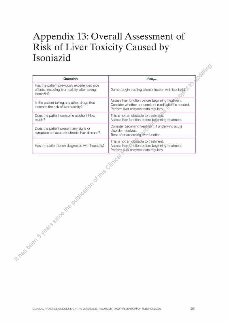
CLINICAL PRACTICE GUIDELINE ON THE DIAGNOSIS, TREATMENT AND PREVENTION OF TUBERCULOSIS 201
Appendix 13: Overall Assessment of Risk of Liver Toxicity Caused by Isoniazid
Question If so,…
Has the patient previously experienced side effects, including liver toxicity, after taking isoniazid?
Do not begin treating latent infection with isoniazid.
Is the patient taking any other drugs that increase the risk of liver toxicity?
Assess liver function before beginning treatment.Consider whether concomitant medication is needed.Perform liver enzyme tests regularly.
Does the patient consume alcohol? How much?
This is not an obstacle to treatment.Assess liver function before beginning treatment.
Does the patient present any signs or symptoms of acute or chronic liver disease?
Consider beginning treatment if underlying acute disorder resolves.Treat after assessing liver function.
Has the patient been diagnosed with hepatitis?This is not an obstacle to treatment.Assess liver function before beginning treatment.Perform liver enzyme tests regularly.
It has
been
5 ye
ars si
nce t
he pu
blica
tion o
f this
Clinica
l Prac
tice G
uideli
ne an
d it is
subje
ct to
upda
ting.

CLINICAL PRACTICE GUIDELINE IN THE SNHS202
It has
been
5 ye
ars si
nce t
he pu
blica
tion o
f this
Clinica
l Prac
tice G
uideli
ne an
d it is
subje
ct to
upda
ting.

CLINICAL PRACTICE GUIDELINE ON THE DIAGNOSIS, TREATMENT AND PREVENTION OF TUBERCULOSIS 203
Appendix 14: Treatment Regimens Evaluated in Latent Tuberculosis Infection and HIV Infection
Author (year of study)
Country Treatment regimens proposed, comparator SR (ref)1
Pape (1986-1992) HaitiH 300 mg/day (12 months) Placebo (12 months)
(258, 266)
Wadhawan (1988-1992) ZambiaH 300 mg/day (6 months) Placebo (6 months)
(258, 266)
Halsey (1990-1994) Haiti H 600-800 mg (6 months)R 450-600 mg + Z 1,500-2,500 mg (2 months) Intermittent treatment (twice a week)
(258)
Whalen (1993-1997) UgandaH 300 mg/day (6 months)H 300 mg + R 600 mg/day (3 months) HR + Z 2,000 mg/day (3 months) Placebo (6 months)
(258, 266)
Jonson (2001) UgandaSame treatment regimens as previous study, efficacy at 3 years
(258)
Mwinga (1992-1996) Zambia
H 900 mg (6 months)R 600 mg + Z 3,500 mg (3 months)Placebo (3 months)Intermittent treatment (twice a week)
(258, 266)
Hawken (1992-1996) Kenya H 300 mg/day (6 months) Placebo (6 months)
(258, 266)
Gordin (1991-1996) USAH 300 mg/day (6 months) Placebo (6 months)
(258, 266)
Fitzgerald (1998-1999) Haiti H 300 mg/day (12 months) Placebo (12 months)
(258)
Rivero (1994-1998) Spain
H 300 mg/day (6 months)HR (3 months)R 600 mg/day + Z 1,500-2,500 mg/day (2 months) No treatment
(258)
Mohamed (2003-2004) South Africa H 800-900 mg/day (12 months) Placebo (12 months) (267)
Zar (2003-2004)H 8 a 12 mg/kg/day (9 months)PlaceboAll received co-trimoxazole as prophylaxis
(274)
Rivero (1994-1998) Spain H 5 mg/kg/day (6 months)H + R 5 + 10 mg/kg/day (3 months)R + Z 10 mg/kg/day + 1,500-2,500 mg/day (2 months)
(268)
H: isoniazid; R: rifampicin; Z: pyrazinamide; SR: systematic review 1. References for systematic review including original studies or reference of studies published after review.
It has
been
5 ye
ars si
nce t
he pu
blica
tion o
f this
Clinica
l Prac
tice G
uideli
ne an
d it is
subje
ct to
upda
ting.

CLINICAL PRACTICE GUIDELINE IN THE SNHS204
It has
been
5 ye
ars si
nce t
he pu
blica
tion o
f this
Clinica
l Prac
tice G
uideli
ne an
d it is
subje
ct to
upda
ting.

CLINICAL PRACTICE GUIDELINE ON THE DIAGNOSIS, TREATMENT AND PREVENTION OF TUBERCULOSIS 205
Bibliography
1 Ministerio de Sanidad y Política Social, editor. Plan para la Prevención y el Control de la Tuberculosis en España [internet]. Madrid; 2008 [Consultado el 19 de Octubre de 2009]. Disponible en: http://www.msps.es
2 World Health Organization, editor. Global tuberculosis control 2009: epidemiology, strategy, financing [internet]. Switzerland: WHO Press; 2009. [Consultado el 19 de Octubre de 2009]. Disponible en: http://www.who.int/tb/publications/global_report/en/
3. Union Internacional contra la Tuberculosis y Enfermedades Respiratorias, editor. Bases epidemiológicas del control de la tuberculosis (traducción al español) [internet]. París: UICTER; 1999. [Consultado el 19 de Octubre de 2009]. Disponible en: http://www.theunion.org4. Scottish Intercollegiate Guidelines Network. Bronquiolitis in children. SIGN publication number 91. 2006, p. 1-41.
4. Altet MN, Alcaide J, Hernández I. Tabaquismo y tuberculosis: ¿otro dúo maldito?. Prev Tab. 2004;6:113-24.
5 World Health Organization, editor. Guidance for national tuberculosis programmes on the management of tuberculosis in children [internet]. Switzerland: WHO Press; 2009. [Consultado el 19 de Octubre de 2009]. Disponible en: http://www.who.int/child_adolescent_health/documents/htm_tb_2006_371/en/index.html
6. Marais BJ, Obihara CC, Warren RM, Schaaf HS, Gie RP, Donald PR. The burden of childhood tuberculosis: a public health perspective. Int J Tuberc Lung Dis. 2005;9:1305-13.
7. Altet MN, Alcaide J. Control y eliminación de la tuberculosis en España: las estrategias para el Siglo XXI. An Pediatr (Barc). 2006;64(1):66-73.
8. World Health Organization, editor.Global tuberculosis control 1997 [internet]. Switzerland: WHO Press; 1997. [consultado el 19 de Octubre de 2009]. Disponible en: http://www.who.int/ tb/publications/global_report/archive/en/index.html
9. Institut de veille sanitaire, editor. EuroTB and the national coordinators for tuberculosis surveillance in the WHO European Region.Surveillance of Tuberculosis in Europe. Report on Tuberculosis cases notified in 2005 [internet]. France; 2007. [consultado el 19 de Octubre de 2009]. Disponible en: http://www.eurotb.org/rapports/2005/full_report.pdf
10. E. Rodríguez, G. Hernández, S. Villarrubia, O. Díaz, O. Tello (Área de Vigilancia de la Salud Pública. Centro Nacional de Epidemiología. Instituto de Salud Carlos III.) Casos de tuberculosis declarados a la Red Nacional de Vigilancia Epidemiológica. España, 2008. Ministerio de Ciencia e Innovación. Instituto de Salud Carlos III. Boletín epidemiológico semanal. 2009 Vol. 17 nº 11/121-132. ISSN: 1135-6286.
11. Ríos M, Monleón-Getino T. A graphical study of tuberculosisi incidence and trends in the WHO’s European region (1980-2006). Europ J Epidemiol. 2009;24:381-7.
12. Rouillon A, Perdrizet S, Parrot R. Transmission of tubercle bacilli: the effects of chemotherapy. Tubercle. 1976;57:275-9.
13. Alcaide J, Altet MN, Salleras L. Vacuna BCG. En: Masson, editor. Vacunaciones preventivas. Principios y aplicaciones. 2ª ed. Barcelona: Elsevier; 2003. Pág. 529-74.
14. Altet MN, de Souza ML, Milá C. Infecciones por Micobacterias. En: Masson, editor. Vacunaciones preventivas. Principios y aplicaciones. 2ª ed. Barcelona: Elsevier; 2003. Pág. 479-515.
15. Miller TL, McNabb SJ, Hilsenrath P, Pasipanodya J, Weis SE. Personal and Societal Health Quality Lost to Tuberculosis. PLoS ONE. 2009;4(4): e5080.
16. Mitchison DA.Basic mechanisms of chemoteraphy. Chest 1979;76:771-81.
17. Rossi GA, Medici MC, Arcangeletti MC, Lanari M, Merolla R, Paparatti UD, et al.;
It has
been
5 ye
ars si
nce t
he pu
blica
tion o
f this
Clinica
l Prac
tice G
uideli
ne an
d it is
subje
ct to
upda
ting.

CLINICAL PRACTICE GUIDELINE IN THE SNHS206
Osservatorio Ruiz.ManzanoJ, Blanquer R, Calpe JL, Caminero JA, Caylá J, Dominguez JA, García JM, Vidal R. Normativa SEPAR. Diagnóstico y tratamiento de la tuberculosis. Arch Bronconeumol. 2008;44(10):551-66.
18. Broekmans JF, Migliori GB, Rieder HL, Lees J, Ruutu P, Loddenkemper R et al. European framework for tuberculosis control and elimination in countries with a low incidence. Recom mendations of the World Health Organization (WHO), International Union Against Tuberculosis and Lung Disease (IUATLD) and Royal Netherlands Tuberculosis Association (KNCV) Working Group. Eur Respir J. 2002;19:765-775.
19. An expanded DOTS Framework for effective tuberculosis control. Int J Tuberc Lung Dis. 2002;6:378-88.
20. National Collaborating Centre for Chronic Conditions. Tuberculosis: clinical diagnosis and management of tuberculosis, and measures for its prevention and control. London: Royal College of Physicians, 2006.
21. Joint Tuberculosis Committee of the British Thoracic Society. Control and prevention of tuberculosis in the United Kingdom: Code of Practice 2000. Thorax. 2000;55:887-901.
22. Targeted Tuberculin Testing and Treatment of Latent Tuberculosis Infection.Am J Respir Crit Care Med. 2000;161:S221-S247.
23. Grupo de trabajo de Tuberculosis de la Sociedad Española de Infectología Pediátrica (SEIP). Documento de consenso sobre el tratamiento de la exposición a tuberculosis y de la infección tuberculosa latente en niños. An Pediatr (Barc). 2006;64(1): 59-65.
24. Grupo de trabajo sobre GPC. Elaboración de Guías de Práctica Clínica en el Sistema Nacional de Salud. Manual Metodológico. Madrid: Plan Nacional para el SNS del MSC. Instituto Aragonés de Ciencias de la Salud-I+CS; 2007. Guías de Práctica Clínica en el SNS: I+CS Nº 2006/0I.
25. Schünemann HJ, Jaeschke R, Cook DJ, Bria WF, El-Solh AA, Ernst A, et al. ATS Documents Development and Implementation Committee. An official ATS statement: grading the quality of evidence and strength of recommendations in ATS guidelines and recommendations. Am J Respir Crit Care Med. 2006; 174(5):605-14.
26. Wang L, Turner MO, Elwood RK, Schulzer M, FitzGerald JM.A meta-analysis of the effect of Bacille Calmette Guérin vaccination on tuberculin skin test Measurements. Thorax. 2002; 57: 804-9.
27. Centers for Disease Control and Prevention [internet].Atlanta: CDC [actualizada a 1 Junio de 2009]. Fact Sheets: Tuberculin Skin Testing. [Consultado el 19 de Octubre de 2009]. Disponible en: http://www.cdc.gov/tb/publications/factsheets/testing/skintesting.htm
28. Pai M, Zwerling A, Menzies D Systematic Review: T-Cell–based Assays for the Diagnosis of Latent Tuberculosis Infection: An Update. Ann Intern Med. 2008;149:177-84.
29. Menzies D, Pai M, Comstock G. Meta-analysis: new tests for the diagnosis of latent tuberculosis infection: areas of uncertainty and recommendations for research. Ann Intern Med. 2007;146(5):340-54.
30. Stephan C, Wolf T, Goetsch U, Bellinger O, Nisius G, Oremek G, et al. Comparing QuantiFERON-Tuberculosis Gold, T-SPOT Tuberculosis and Tuberculin Skin Test in HIV-Infected Individuals from a Low Prevalence Tuberculosis Country. AIDS. 2008;22(18):2471-9.
31. Kobashi Y, Mouri K, Yagi S, Obase Y, Miyashita N, Okimoto N, et al. Clinical Utility of the QuantiFERON TB-2G Test for Elderly Patients with Active Tuberculosis. Chest. 2008; 133(5): 1196-202.
32. Raby E, Moyo M, Devendra A, Banda J, De Haas P, Ayles H, et al. The Effects of HIV on the Sensitivity of a Whole Blood IFN-Gamma Release Assay in Zambian Adults with Active Tuberculosis. PLoS ONE. 2008;3(6): e2489.
33. National Tuberculosis Controllers Association; Center for Disease Control and Prevention. Guidelines for the investigation of contacts of persons with infectious
It has
been
5 ye
ars si
nce t
he pu
blica
tion o
f this
Clinica
l Prac
tice G
uideli
ne an
d it is
subje
ct to
upda
ting.

CLINICAL PRACTICE GUIDELINE ON THE DIAGNOSIS, TREATMENT AND PREVENTION OF TUBERCULOSIS 207
tuberculosis;recommendations from The National Tuberculosis Controllers Assotiation and CDC, and Guidelines for using the Quantiferon-TB Gold Test for detecting Mycobacterium tuberculosis Infection, United States. MMWR 2005 Dec 16;54(RR-15):1-55.
34. Brandli O.The clinical presentation of tuberculosis.Respiration. 1998;65(2):97-105.
35. Parimon T, Spitters CE, Muangman N, Euathrongchit J, et al. Unexpected Pulmonary Involvement in Extrapulmonary Tuberculosis Patients. Chest. 2008;134(3):589-94.
36. Wisnivesky JP, Serebrisky D, Moore C, Sacks HS, Iannuzz MC, McGinn T. Validity of Clinical Prediction Rules for Isolating Inpatients with Suspected Tuberculosi.A Systematic Review. J Gen Intern Med. 2005; 20:947-52.
37. Soto A, Solari L, Agapito J, Acuna-Villaorduna C, Lambert ML, Gotuzzo E. Development of a clinical scoring system for the diagnosis of smear-negative pulmonary tuberculosis. Braz J Infect Dis. 2008;12(2):128-32.
38. Solari L, Acuna-Villaorduna C, Soto A, Agapito J, Perez F, Samalvides F.A clinical predictionrule for pulmonary tuberculosis in emergency departments. Int J Tuberc Lung Dis. 2008;12(6): 619-24.
39. Wang CS, Chen HC, Chong IW, Hwang JJ, Huang MS. Predictors for identifying the most infectious pulmonary tuberculosis patient. J Formos Med Assoc. 2008;107(1):13-20.
40. Rakoczy KS, Cohen SH, Nguyen HH. Derivation and validation of a clinical prediction score for isolation of inpatients with suspected pulmonary tuberculosis. Infect Control Hosp Epidemiol. 2008;29(10):927-32.
41. den Boon S, White NW, van Lill SW, Borgdorff MW, Verver S, Lombard CJ. An evaluation ofsymptom and chest radiographic screening in tuberculosis prevalence surveys.Int J Tuberc Lung Dis. 2006;10(8):876-82.
42. Geng E, Kreiswirth B, Burzynski J, Schluger NW. Clinical and Radiographic Correlates of Primary and Reactivation Tuberculosis A Molecular Epidemiology Study. JAMA. 2005;293: 2740-45.
43. Jones BE, Ryu R, Yang Z, Cave MD, Pogoda JM, Otaya M, et al. Chest radiographic findings in patients with tuberculosis with recent or remote infection. Am J Respir Crit Care Med. 1997;156:1270-73.
44. Gómez-Pastrana D, Carceller-Blanchard A. ¿Debe realizarse una tomografía computarizada torácica a los niños con infección tuberculosa sin enfermedad aparente? An Pediatr (Barc). 2007;67(6):585-93.
45. American Academy of Pediatrics. [Summaries of infectious diseases: Tuberculosis]. In: Pickering LK, Baker CJ, Kimberlin DW, Long SS, eds. Red Book: 2009 Report of the Committee on Infectious Diseases. 28th ed. Elk Grove Village, IL: American Academy of Pediatrics; 2009: [680-701].
46. Marais BJ, Gie RP, Schaaf S, Beyers N, Donald PR, Starke JR. Childhood Pulmonary Tuberculosis. Old Wisdom and New Challenges. Am Respir Crit Care Med. 2006;173:1078-90.
47. Cruz AT, Starke JR. Clinical manifestations of tuberculosis in chidren. Paediatr Respir Rev. 2007;8:107-17.
48. Tuberculosis Coalition for Technical Assistance: International Standards for Tuberculosis Care (ISTC) [internet]. The Hague: Tuberculosis Coalition for Technical Assistance, 2006. [Consultado el 19 de Octubre de 2009]. Disponible en: www.who.int/tb/publications/2006/istc/ en
49. Long R, Ellis E, editors. Canadian Tuberculosis Standards, 6ª ed [internet]. Tuberculosis Prevention and Control, Public Health Agency of Canada, Canadian Lung Association/Canadian Thoracic Society, 2007.[Consultado el 19 de Octubre de 2009]. Disponible en: http://www.phacaspc.gc.ca/tbpc-latb/pubs/tbstand07-eng.php
50. Schoch OD, Rieder P, Tueller C et al. Diagnostic yield of sputum, induced sputum, and bronchoscopy after radiologic tuberculosis screening. Am J Respir Crit Care Med. 2007;
It has
been
5 ye
ars si
nce t
he pu
blica
tion o
f this
Clinica
l Prac
tice G
uideli
ne an
d it is
subje
ct to
upda
ting.
CLINICAL PRACTICE GUIDELINE IN THE SNHS208
175(1): 80-6.
51. Anderson C, Inhaber N, Menzies D. Comparison of sputum induction with fiber-optic bronchoscopy in the diagnosis of tuberculosis. Am J Respir Crit Care Med. 1995;152:1570-4.
52. Brown M, Varia H, Bassett P, Davidson RN, Wall R, Pasvol G. Prospective study of sputum induction, gastric washing, and bronchoalveolar lavage for the diagnosis of pulmonary tuberculosis in patients who are unable to expectorate. Clin Infect Dis. 2007;44(11):1415-20.
53. Conde MB, Soares SL, Mello FC, Rezende VM, Almeida LL, Reingold AL, et al. Comparison of sputum induction with fiberoptic bronchoscopy in the diagnosis of tuberculosis: experience at an acquired immune deficiency syndrome reference center in Rio de Janeiro, Brazil. Am J Respir Crit Care Med 2000; 162(6):2238-40.
54. Conde MB, Loivos AC, Rezende VM, Soares SL, Mello FC, Reingold AL, et al. Yield of sputum induction in the diagnosis of pleural tuberculosis. Am J Respir Crit Care Med. 2003;167(5): 723-5.
55. Morse M, Kessler J, Albrecht S, Kim R, Thakur R, Nthobatsang R, et al. Induced sputum improves the diagnosis of pulmonary tuberculosis in hospitalized patients in Gaborone, Botswana. Int J Tuberc Lung Dis. 2008;12(11):1279-85.
56. Saglam L, Akgun M, Aktas E. Usefulness of induced sputum and fibreoptic bronchoscopy specimens in the diagnosis of pulmonary tuberculosis. J Int Med Res. 2005;33(2):260-5.
57. Parry CM, Kamoto O, Harries AD, Wirima JJ, Nyirenda CM, Nyangulu DS, et al. The use of sputum induction for establishing a diagnosis in patients with suspected pulmonary tuberculosis in Malawi. Tuber Lung Dis. 1995;76:72-6.
58. Baughman RP, Dohn MN, Loudon RG, Frame PT. Bronchoscopy with bronchoalveolar lavage in tuberculosis and fungal infections. Chest. 1991;99(1):92-7.
59. Chen NH, Liu YC, Tsao TC, Wu TL, Hsieh MJ, Chuang ML, et al. Combined bronchoalveolar lavage and polymerase chain reaction in the diagnosis of pulmonary tuberculosis in smearnegative patients. Int J Tuberc Lung Dis. 2002;6(4):350-5.
60. Norrman E, Keistinen T, Uddenfeldt M, Rydstrom PO, Lundgren R. Bronchoalveolar lavage is better than gastric lavage in the diagnosis of pulmonary tuberculosis. Scand J Infect Dis. 1988;20(1):77-80.
61. Rizvi N, Rao NA, Hussain M. Yield of gastric lavage and bronchial wash in pulmonary tuberculosis. Int J Tuberc Lung Dis. 2000;4(2):147-51.
62. Tan YK, Lee AS, Khoo KL, Ong SY, Wong SY, Ong YY. Rapid mycobacterial tuberculosis detection in bronchoalveolar lavage samples by polymerase chain reaction in patients with upper lobe infiltrates and bronchiectasis.Ann Acad Med Singapore. 1999;28(2):205-8.
63. Tueller C, Chhajed PN, Buitrago-Tellez C, Frei R, Frey M, Tamm M. Value of smear and PCR in bronchoalveolar lavage fluid in culture positive pulmonary tuberculosis. Eur Respir J. 2005;26(5):767-72.
64. Willcox PA, Benatar SR, Potgieter PD. Use of the flexible fiberoptic bronchoscope in diagnosis of sputum-negative pulmonary tuberculosis. Thorax. 1982;37:598-601.
65. Chawla R, Pant K, Jaggi OP, Chandrashekhar S, Thukral SS. Fibreoptic bronchoscopy in smear negative pulmonary tuberculosis. Eur Respir J. 1998;1:804-806.
66. Charoenratanakul S, Dejsomritrutai W, Chaiprasert A. Diagnostic role of fiberoptic bronchoscopy in suspected smear negative pulmonary tuberculosis. Respir Med. 1995;89(9):621-3.
67. Schoch OD, Rieder P, Tueller C et al. Diagnostic yield of sputum, induced sputum, and bronchoscopy after radiologic tuberculosis screening. Am J Respir Crit Care Med. 2007; 175(1): 80-6.
68. Wongthim S, Udompanich V, Limthongkul S, Charoenlap P, Nuchprayoon C. Fiberoptic bronchoscopy in diagnosis of patients with suspected active pulmonary tuberculosis. J Med
It has
been
5 ye
ars si
nce t
he pu
blica
tion o
f this
Clinica
l Prac
tice G
uideli
ne an
d it is
subje
ct to
upda
ting.

CLINICAL PRACTICE GUIDELINE ON THE DIAGNOSIS, TREATMENT AND PREVENTION OF TUBERCULOSIS 209
Assoc Thai. 1989;72:154-9.
69. Berggren-Palme I, Gudetta B, Bruchfeld J, Eriksson M, Giesecke J. Detection of Mycobacterium tuberculosis in gastric aspirate and sputum collected from Ethiopian HIV-positive and HIV-negative children in a mixed in- and outpatient setting. Acta Paediatr. 2004;93(3):311-5.
70. Abadco DL, Steiner P. Gastric lavage is better than bronchoalveolar lavage for isolation of Mycobacterium tuberculosis in childhood pulmonary tuberculosis. Pediatr Infect Dis J. 1992; 11(9):735-8.
71. Gomez-Pastrana D, Torronteras-Santiago R, Caro-Mateo P, et al. [Effectiveness of smears and cultures in gastric aspirate samples in the diagnosis of tuberculosis]. An Esp Pediatr.2000;53(5):405-11.
72. Laven GT. Diagnosis of tuberculosis in children using fl uorescence microscopic examination of gastric washings. Am Rev Respir Dis. 1977;115(5):743-9.
73. Shata AM, Coulter JB, Parry CM, Ching’ani G, Broadhead RL, Hart CA. Sputum induction for the diagnosis of tuberculosis. Arch Dis Child. 1996;74(6):535-7.
74. Singh M, Moosa NV, Kumar L, Sharma M. Role of gastric lavage and broncho-alveolar lavage in the bacteriological diagnosis of childhood pulmonary tuberculosis. Indian Pediatr. 2000;37(9):947-51.
75. Somu N, Swaminathan S, Paramasivan CN et al. Value of bronchoalveolar lavage and gastric lavage in the diagnosis of pulmonary tuberculosis in children. Tuber Lung Dis. 1995; 76(4): 295-9.
76. Zar HJ, Hanslo D, Apolles P, Swingler G, Hussey G. Induced sputum versus gastric lavage for microbiological confirmation of pulmonary tuberculosis in infants and young children: a prospective study. Lancet. 2005;365:130-4.
77. Steingart KR, Henry M, Ng V, Hopewell PC, Ramsay A, Cunningham J, et al. Fluorescence versus conventional sputum smear microscopy for tuberculosis: a systematic review. Lancet Infect Dis. 2006; 6:570-81.
78. Van Deun A, Chuquiyauri R, Torrea G, Agapito J, Verdonck K, Gotuzzo E. Yield of fluorescence microscopy versus culture for tuberculosis at a middle-income country referral hospital. Trans R Soc Trop Med Hyg. 2008;102(6):564-9.
79. Steingart KR , Ng V, Henry M, Hopewell PC, Ramsay A, Cunningham J, et al. Sputum processing methods to improve the sensitivity of smear microscopy for tuberculosis: a systematic review. Lancet Infect Dis. 2006;6(10):664-74.
80. WHO, IUATLD, Royal Netherlands Tuberculosis Association. Revised international definitions in tuberculosis control. Int J Tuberc Lung Dis. 2001;5(3):213-5.
81. Mase SR, Ramsay A, Ng V, Henry M, Hopewell PC, Cunningham J, et al. Yield of serial sputum specimen examinations in the diagnosis of pulmonary tuberculosis: a systematic review. Int J Tuberc Lung Dis. 2007;11(5):485-95.
82. Dinnes J, Deeks J, Kunst H, Gibson A, Cummins E, Waugh N, Drobniewski F, Lalvani A. A systematic review of rapid diagnostic tests for the detection of tuberculosis infection.Health Technol Assess. 2007;11(3):1-196.
83. Domínguez J, Blanco S, Lacoma A, García-Sierra N, Prat C, Ausina V. Utilidad de la biología molecular en el diagnóstico microbiológico de las infecciones por micobacterias. Enferm Infecc Microbiol Clin. 2008;Suppl 9:33-41.
84. Ling DI, Flores LL, Riley LW, Pai M. Commercial nucleic-acid amplification tests for diagnosis of pulmonary tuberculosis in respiratory specimens: meta-analysis and meta-regression. PLoS ONE. 2008;3(2):e1536.
85. Sarmiento OL, Weigle KA, Alexander J, Weber DJ, Miller WC. Assessment by meta-analysis of PCR for diagnosis of smear-negative pulmonary tuberculosis. J Clin Microbiol. 2003;41(7): 3233-40.
It has
been
5 ye
ars si
nce t
he pu
blica
tion o
f this
Clinica
l Prac
tice G
uideli
ne an
d it is
subje
ct to
upda
ting.

CLINICAL PRACTICE GUIDELINE IN THE SNHS210
86. Kalantri S, Pai M, Pascopella L, Riley L, Reingold A. Bacteriophage- based tests for the detection of Mycobacterium tuberculosis in clinical specimens: a systematic review and metaanalysis. BMC Infect Dis. 2005;5:59.
87. Alcaide F, Galí N, Domínguez J, Berlanga P, Blanco S, Orús P, et al. Usefulness of a new mycobacteriophage-based technique for rapid diagnosis of pulmonary tuberculosis. J Clin Microbiol. 2003;41(7):2867-71.
88. Steingart KR, Henry M, Laal S, Hopewell PC, Ramsay A, Menzies D, et al. Commercial serological antibody detection tests for the diagnosis of pulmonary tuberculosis: a systematic review. PLoS Med. 2007;4(6):e202.
89. Hiraki A, Aoe K, Eda R, Maeda T, Murakami T, Sugi K, et al. Comparison of six biological markers for the diagnosis of tuberculous pleuritis. Chest. 2004;125:987-9.
90. Liang QL, Shi HZ, Wang K, Qin SM, Qin XJ. Diagnostic accuracy of adenosine deaminase intuberculous pleurisy: a meta-analysis. Respir Med. 2008;102(5):744-54.
91. Greco S, Girardi E, Masciangelo R, Capoccetta GB, Saltini C. Adenosine deaminase and interferon gamma measurements for the diagnosis of tuberculous pleurisy: a meta-analysis. Int J Tuberc Lung Dis. 2003;7(8):777-86.
92. Goto M, Noguchi Y, Koyama H, Hira K, Shimbo T, Fukui T. Diagnostic value of adenosine deaminase in tuberculous pleural effusion: a meta-analysis. Ann Clin Biochem. 2003; 40: 374- 381.
93. Morisson P, Neves DD. Evaluation of adenosine deaminase in the diagnosis of pleural tuberculosis: a Brazilian meta-analysis. J Bras Pneumol. 2008;34(4):217-24.
94. Ena J, Valls V, Pérez de Oteyza C, Enríquez de Salamanca R. Utilidad y limitaciones de la adenosina desaminasa en el diagnóstico de la pleuresía tuberculosa. Estudio metaanalítico. Med Clin (Barc).1990; 95:333-5.
95. Baba K, Hoosen AA, Langeland N, Dyrhol-Riise AM. Adenosine Deaminase Activity is a Sensitive Marker for the Diagnosis of Tuberculous Pleuritis in Patients with very Low CD4 Counts. PLoS ONE. 2008;3(7):e2788.
96. Jiang J, Shi HZ, Liang QL Qin SM, Qin XJ. Diagnostic Value of Interferon gamma in Tuberculous Pleurisy. A Metaanalysis. Chest. 2007;131:1133-41.
97. Pai M, Flores LL, Hubbard A, Riley LW, Colford JM Jr. Nucleic acid amplification tests in the diagnosis of tuberculous pleuritis: a systematic review and meta-analysis. BMC Infect Dis. 2004;23:4:6.
98. Pai M, Flores LF, Pai N, Hubbard A, Riley LW, Colford JM. Diagnostic accuracy of nucleic acid amplification tests for tuberculous meningitis: A systematic review and meta-analysis. Lancet Infect Dis. 2003;3:633-43.
99. Chen X, Yang Q, Zhang M, Graner M, Zhu X, Larmonier N, et al. Diagnosis of active tuberculosis in China using an in-house gamma interferon enzyme-linked immunospot assay. Clin Vaccine Immunol. 2009;16(6):879-84.
100. Tuon FF, Litvoc MN, Lopes MI. Adenosine deaminase and tuberculous pericarditis: a systematic review with meta-analysis. Acta Trop. 2006;99(1):67-74.
101. Daley P, Thomas A, Pai M. Nucleic acid amplification tests for the diagnosis of tuberculous lymphadenitis: a systematic review. Int J Tuberc Lung Dis. 2007;11(11):1166-76.
102. Riquelme A, Calvo M, Salech F, Valderrama S, Pattillo A, Arellano M, et al. Value of adenosine deaminase (ADA) in ascitic fluid for the diagnosis of tuberculous peritonitis: a meta-analysis. J Clin Gastroenterol. 2006;40(8):705-10.
103. Steingart KR, Henry M, Laal S, Hopewell PC, Ramsay A, Menzies D, et al. A systematic review of commercial serological antibody detection tests for the diagnosis of extrapulmonary tuberculosis. Thorax. 2007;62(10):911-8.
104. Shenai S, Rodrigues C, Mehta AP. Evaluation of a new phage amplification technology for rapid diagnosis of tuberculosis. Indian J Med Microbiol. 2002;20(4):194-9.
105. World Health Organization, editor.Anti-tuberculosis drug resistance in the
It has
been
5 ye
ars si
nce t
he pu
blica
tion o
f this
Clinica
l Prac
tice G
uideli
ne an
d it is
subje
ct to
upda
ting.
CLINICAL PRACTICE GUIDELINE ON THE DIAGNOSIS, TREATMENT AND PREVENTION OF TUBERCULOSIS 211
World, Report Nº4.The WHO/IUATLD Global project on anti-tuberculosis drug resistance surveillance 2002-2007. Fourth Global Report [internet]. Switzerland: WHO Press; 2009. [Consultado el 19 de Octubre de 2009]. Disponible en: www.who.int/tb/publications/2008/drs_report4_26feb08.pdf
106. National Committee for Clinical Laboratory Standards (US).Susceptibility testing for mycobacteria, nocardiae, and other aerobic actinomycetes; approved standard. NCCLS document M24-A. National Committee for Clinical Laboratory Standards; Wayne, Pe, 2003.
107. Alcaide F, Santín M Tuberculosis multiresistente. Enferm Infecc Microbiol Clin. 2008;26(Supl 13):54-60.
108. World Health Organization, editor.Guidelines for drug susceptibility testing for second-line anti-tuberculosis drugs for DOTS-plus [internet].Switzerland: WHO Press; 2001. [Consultado el 19 de Octubre de 2009]. Disponible en: http://whqlibdoc.who.int/hq/2001/WHO_CDS_TB_2001.288.pdf
109. Galí N, Domínguez J, Blanco S, Prat C, Quesada MD, Matas L, Ausina V. Utility of an in-house mycobacteriophage-based assay for rapid detection of rifampin resistance in Mycobacterium tuberculosis clinical isolates. J Clin Microbiol. 2003;41(6):2647-9.
110. Galí N, Domínguez J, Blanco S, Prat C, Alcaide F, Coll P, et al. Use of a mycobacteriophagebased assay for rapid assessment of susceptibilities of Mycobacterium tuberculosis isolates to isoniazid and infl uence of resistance level on assay performance. J Clin Microbiol. 2006;44(1): 201-5..
111. Hardman JG, Limbrid LE, Goodman A. Goodman & Gilman’s.The Pharmacological Basis of Therapeutics. 10ª Edición. McGraw-Hill, 2001.
112. Royal Pharmaceutical Society of Great Britain, The Pharmaceutical Press (UK). Martindale: The Complete Drug Referente. 44ª Edición. Londres; 2005.
113. Thomson PDR (US).Physicians’ Desk Referente. 59ª Edición. Montvale;2005.114. World Health Organization, editor. Treatment of tuberculosis: guidelines for national
programmes, 3rd ed [internet]. Switzerland: WHO Press; 2003. [Consultado el 19 de Octubre de 2009]. Disponible en: http://whqlibdoc.who.int/hq/2003/WHO_CDS_TB_2003.313_eng.pdf
115. World Health Organization, editor. Etambutol efficacy and toxicity: literature review and recommendations for daily and intermittent dosage in children [internet]. Switzerland: WHO Press; 2006. [Consultado el 19 de Octubre de 2009]. Disponible en: http://whqlibdoc.who.int/ hq/2006/WHO_HTM_TB_2006.365_eng.pdf
116. Méndez-Echevarría A, Baquero-Artigao F, García-Miguel MJ, Pascual SI, Velásquez R, del Castillo F. Visual evoked responses in children with tuberculosis treated with ethambutol. Pediatr Infect Dis. J 2007;26:92-3.
117. American Thoracic Society/Centers for Disease Control and Prevention/Infectious Diseases Society of America: treatment of tuberculosis. Am J Respir Crit Care Med. 2003;167:603-62.
118. Medical Research Council.Long-term chemotherapy in the treatment of chronic pulmonary tuberculosis with cavitations. Tubercle. 1962;43:201.
119. Bobrowitz ID, Robins DE.Ethambutol-isoniazid versus PAS-isoniazid in original treatment of pulmonary tuberculosis. Am Rev Respir Dis. 1966;96:428-38.
120. East African/British Medical Research Council. Controlled clinical trial of four short-course (6-month) regimens of chemotherapy for treatment of pulmonary tuberculosis. Lancet. 1973;1:1331-39.
121 Short-course chemotherapy in pulmonary tuberculosis: a controlled trial by the British Thoracic and Tuberculosis Association. Lancet. 1976;2:1102-4.
122 Hong Kong Chest Service/British Medical Research Council.Five-year follow-up of a controlled trial of five 6-month regimens of chemotherapy for pulmonary tuberculosis. Am Rev Respir Dis. 1987;136:1339-42.
123 A controlled trial of six months chemotherapy in pulmonary tuberculosis: second report-results during the 24 months after the end of chemotherapy. British Thoracic Association. Am Rev Respir Dis. 1982;126:460-2.
It has
been
5 ye
ars si
nce t
he pu
blica
tion o
f this
Clinica
l Prac
tice G
uideli
ne an
d it is
subje
ct to
upda
ting.
CLINICAL PRACTICE GUIDELINE IN THE SNHS212
124 Gelband H. Regimens of less than six months for treating tuberculosis. Cochrane Database of Systematic Reviews 1999,Issue4.Art.No.:CD001362.DOI: 10.1002/14651858.CD001362.
125 Jindani A, Nunn AJ, Enarson DA. Two 8-month regimens of chemotherapy for treatment of newly diagnosed pulmonary tuberculosis: international multicentre randomised trial. Lancet. 2004;364:1244-51.
126 Swaminathan S, Raghavan A, Duraipandian M, Kripasankar AS, Ramachandran P. Shortcourse chemotherapy for paediatric respiratory tuberculosis: 5-year report. Int J Tuberc Lung Dis. 2005;9(6):693-6.
127 Menzies D, Benedetti A, Paydar A, Martin I, Royce S, Pai M, et al. Effect of duration and intermittency of rifampin on tuberculosis treatment outcomes: a systematic review and metaanalysis. PLoS Med. 2009;6(9):e1000146.
128 Mwandumba HC, Squire SB. Fully intermittent dosing with drugs for treating tuberculosis in adults. Cochrane Database of Systematic Reviews 2001, Issue 4. Art. No.: CD000970. DOI: 10.1002/14651858.CD000970.
129 Mitchison DA, Aber V, Allen BW. Controlled trial of four thrice-weekly regimens and a dailyregimen all given for 6 months for pulmonary tuberculosis. Lancet. 1981;1(8213):171-4.
130 Chang KC, Leung CC, Yew WW, Chan SL, Tam CM. Dosing Schedules of 6-Month Regimens
and Relapse for Pulmonary Tuberculosis. Am J Respir Crit Care Med. 2006;174: 1153-8.
131 Naude JMTW, Donald PR, Hussey GD, Kibel MA, Louw A, Perkins DR, Schaaf HS. Twice weekly vs. daily chemotherapy for childhood tuberculosis. Pediatr Infect Dis. J 2000;19(5): 405-10.
132 Geiter LJ, O’Brien RJ, Combs DL, Snider DE. United States Public Health Service Tuberculosis Therapy Trial 21: preliminary results of an evaluation of a combination tablet of isoniazid, rifampin and pyrazinamide. Tubercle. 1987;68(Suppl 2):41-6.
133 Girling DJ, Chan SL. Controlled trial of 2, 4, and 6 months of pyrazinamide in 6-month, threetimes weekly regimens for smear-positive pulmonary tuberculosis, including an assessment of a combined preparation of isoniazid, rifampin, and pyrazinamide: results at 30 months. Am Rev Respir Dis. 1991;143:700-6.
134 Zhang LX, Kan GQ, Tu DH, Wan LY, Faruqi AR. Fixed-dose combination chemotherapy versus multiple, single-drug chemotherapy for tuberculosis. Curr Ther Res Clin Exp. 1996;57: 849-56.
135 Teo SK. Assessment of a combined preparation of isoniazid, rifampicin and pyrazinamide (Rifater) in the initial phase of chemotherapy in three 6-month regimens for smear-positive pulmonary tuberculosis: a five-year follow-up report. Int J Tub Lung Dis. 1999;3(2):126-32.
136 Su WJ, Perng RP. Fixed-dose combination chemotherapy (Rifater/Rifinah) for active pulmonary tuberculosis in Taiwan: a two-year follow-up. Int J Tub Lung Dis. 2002;6(11):1029-32.
137 Gravendeel JMT, Asapa AS, Becx-Bleumink M, Vrakking HA. Preliminary results of an operational field study to compare side-effects, complaints and treatment results of a single-drug short-course regimen with a four-drug fixed-dose combination (4FDC) regimen in South Sulawesi, Republic of Indonesia.Tuberculosis. 2003;83:183-6.
138 Suryanto AA, van den Broek J, Hatta M, de Soldenhoff R, van der Werf MJ. Is there an increased risk of TB relapse in patients treated with fixed-dose combination drugs in Indonesia? Int J Tuberc Lung Dis. 2008;12(2):174-9.
139 Bartacek A, Schütt D, Panosch B, Borek M; Rimstar 4-FDC Study Group.Comparison of a four-drug fixed-dose combination regimen with a single tablet regimen in smear-positive pulmonary tuberculosis. Int J Tuberc Lung Dis. 2009;13(6):760-6.
140 Davies G, Cerri S, Richeldi L. Rifabutin for treating pulmonary tuberculosis. Cochrane Data-base of Systematic Reviews. 2007,Issue 4.Art. No.:CD005159. DOI: 10.1002/14651858. CD005159.pub2.
141 Saukkonen JJ, Cohn DL, Jasmer RM, Schenker S, Jereb JA, Nolan CM, et al. An official ATS statement: hepatotoxicity of antituberculosis therapy. Am J Respir Crit Care Med. 2006;174(8): 935-52.
It has
been
5 ye
ars si
nce t
he pu
blica
tion o
f this
Clinica
l Prac
tice G
uideli
ne an
d it is
subje
ct to
upda
ting.

CLINICAL PRACTICE GUIDELINE ON THE DIAGNOSIS, TREATMENT AND PREVENTION OF TUBERCULOSIS 213
142 Faustini A, Hall AJ, Perucci CA. Tuberculosis treatment outcomes in Europe: a systematic review. Eur Respir J. 2005;26(3):503-10.
143 Ditah IC, Reacher M, Palmer C, Watson JM, Innes J, Kruijshaar ME, et al. Monitoring tuberculosis treatment outcome: analysis of national surveillance data from a clinical perspective.Thorax. 2008;63(5):440-6.
144 Baussano I, Pivetta E, Vizzini L, Abbona F, Bugiani M. Predicting Tuberculosis Treatment Outcome in a Low-Incidence Area. Int J Tuberc Lung Dis. 2008;12(12):1441-8.
145 Seah G T, Scott G M, Rook G A W. Type 2 cytokine gent activation and its relationship to extent of disease in patients with tuberculosis. Clin Infect Dis. 2000;181:385-9.
146 Smego RA, Ahmed N. A systematic review of the adjunctive use of systemic corticosteroids for pulmonary tuberculosis. Int J Tuberc Lung Dis. 2003;7(3):208-13.
147 Abba K, Sudarsanam TD, Grobler L, Volmink J. Nutritional supplements for people being treated for active tuberculosis. Cochrane Database of Systematic Reviews. 2008, Issue 4. Art. No.:CD006086. DOI:10.1002/14651858.CD006086.pub2.
148 de Bruyn G, Garner P.Mycobacterium vaccae immunotherapy for treating tuberculosis.Cochrane Database of Systematic Reviews.2003, Issue 1. Art. No.:CD001166. DOI: 10.1002/ 14651858.CD001166.
149 Vlassov VV, MacLehose HG. Low level laser therapy for treating tuberculosis. Cochrane Database of Systematic Reviews. 2006, Issue 2. Art. No.:CD003490. DOI: 10.1002/ 14651858.CD003490.pub2.
150 Roy V, Tekur U, Chopra K. Pharmacokinetics of isoniacid in pulmonary tuberculosis –a comparative study at two dose levels. Indian Pediatrics. 1996;33:287-91.
151 Engel ME, Matchaba PT, Volmink J. Corticosteroids for tuberculous pleurisy. Cochrane Database of Systematic Reviews. 2007, Issue 4. Art. No.: CD001876. DOI: 10.1002/ 14651858.CD001876.pub2.
152 Chapman SJ, Davies RJ. Themanagement of pleural space infections. Respirology. 2004;9(1): 4-11.153 Loenhout-Rooyackers JH, Laheij RJ, Richter C, Verbeek AL. Shortening the duration of
treatment for cervical tuberculous lymphadenitis. Eur Respir J. 2000;15(1):192-5.154 Jawahar MS, Rajaram K, Sivasubramanian S, Paramasivan CN, Kamaludeen MN, Thirithuvathas
AJ, et al. Treatment of lymph node tuberculosis – a randomized clinical trial of two 6- month regimens. Trop Med Int Health. 2005;10(11):1090-98.
155 Smith IE, Laroche CM, Jamieson SA, Shneerson JM.Kyphosis secondary to tuberculous osteomyelitis as a cause of ventilatory failure. Clinical features,mechanisms, andmanagement. Chest. 1996;110(4):1105-10.
156 Jutte PC, Van Loenhout-Rooyackers JH. Routine surgery in addition to chemotherapy for treating spinal tuberculosis. Cochrane Database of Systematic Reviews. 2006, Issue 1. Art. No.: CD004532. DOI: 10.1002/14651858.CD004532.pub2.
157 Parthasarathy R, Sriram K, Santha T, Prabhakar R, Somasundaram PR, Sivasubramanian S. Short-course chemotherapy for tuberculosis of the spine. A comparison between ambulant treatment and radical surgery - Ten-year report. J Bone Joint Surg Br. 1999;81(3):464-71.
158 Five-year assessment of controlled trials of short-course chemotherapy regimens of 6, 9 or 18 months’ duration for spinal tuberculosis in patients ambulatory from the start or undergoing radical surgery. Fourteenth report of the Medical Research Council Working Party on Tuberculosis of the Spine.Int Orthop. 1999;23:73-81.
159 Mateo L, Ruiz Manzano J, Olivé A, Manterola JM, Pérez R, Tena X, et al. [Ostearticular tuberculosis. Study of 53 cases] Med Clin (Barc). 2007;129(13):506-9.
160 Humphries M.The management of tuberculosis meningitis. Thorax. 1992;47(8):577-81.161 Medical Research Council Report.Streptomycin treatment of tuberculous meningitis. Lancet.
1948;1:582-96.162 Chotmongkol V. Treatment of tuberculosis meningitis with 6-month course of chemotherapy.
Southeast Asian J Trop Med Public Health. 1991;22:372-374.163 Alarcón F, Escalante L, Pérez Y, Banda H, Chacón G, Dueñas G. Tuberculous meningitis. Arch
Neurol. 1990;47:1313-8.
It has
been
5 ye
ars si
nce t
he pu
blica
tion o
f this
Clinica
l Prac
tice G
uideli
ne an
d it is
subje
ct to
upda
ting.

CLINICAL PRACTICE GUIDELINE IN THE SNHS214
164 Jacobs R F, Sunakorn P, Chotpitayasunonah T, Pope S, Kelleher K. Intensive short course chemotherapy for tuberculous meningitis. Pediatr Infect Dis J. 1992;11:194-8.
165 Donald PR, Schoeman JF, van Zyl LE, de Villiers JN, Pretorius M, Springer P. Intensive short course chemotherapy in the management of tuberculous meningitis. Int J Tuberc Lung Dis. 1998;2:704-711.
166 Doganay M, Calangu S, Turgut H, Bakir M, Aygen B. Treatment of tuberculous meningitis in Turkey. Scand J Infect Dis. 1995;27:135-8.
167 Phuapradit P, Vejjajiva A. Treatment of tuberculous meningitis; role of short-course chemotherapy. Quarterly J Med. 1987;239:249-58.
168 Goel A, Pandya S K, Satoskar A R. Whither short-course chemotherapy for tuberculous meningitis? Neurosurgery. 1990;27:418-21.
169 Humphries M J, Teoh R, Lau J, Gabriel M. Factors of prognostic significance in Chinese children with tuberculous meningitis. Tubercle. 1990;71:161-8.
170 Ramachandran P, Duraipandian M, Nagarajan M, Prabhakar R, Ramakrishnan C V, Tripathy S P. Three chemotherapy studies of tuberculous meningitis in children. Tubercle. 1986;67:17-29.
171 Ramachandran P, Duraipandian M, Reetha A M, Mahalakshmi S M, Prabhakar R. Long-term status of children treated for tuberculous meningitis in south India Tubercle. 1989;70:235-9.
172 Sunakorn P, Pongparit S, WongrunS. Short course in tuberculous meningitis: a pilot trial. J Med Assoc Thai. 1980;63:340-5.
173 Loenhout-Rooyackers JH, Keyser A, Laheij RJ, Verbeek AL, van der Meer JW. Tuberculous meningitis: is a 6-month treatment regimen sufficient? Int J Tub Lung Dis. 2001;5(11):1028-35.
174 Prasad K, SinghMB.Corticosteroids for managing tuberculous meningitis. Cochrane Database of Systematic Reviews. 2008, Issue 1. Art. No.: CD002244. DOI: 10.1002/14651858.CD002244.pub3.
175 Grupo de trabajo de Tuberculosis de la Sociedad Española de Infectología Pediátrica. Documento de consenso sobre el tratamiento de la tuberculosis extrapulmonar y formas complicadas de tuberculosis pulmonar. An Pediatr (Barc). 2008;69:271-8.
176 Syed FF, Mayosi BM. A modern approach to tuberculosis pericarditis. Prog Cardiovasc Dis 2007;50(3):218-36.
177 Mayosi BM, Ntsekhe M, Volmink JA, Commerford PJ.Interventions for treating tuberculous pericarditis. Cochrane Database of Systematic Reviews. 2002, Issue 4. Art. No.: CD000526. DOI: 10.1002/14651858.CD000526.
178 Ntsekhe M, Wiysonge C, Volmink JA, Commerford PJ, Mayosi BM. Adjuvant corticosteroids for tuberculous pericarditis: promising, but not proven. QJM. 2003;96(8):593-9.
179 Reuter H, Burgess LY, Louw VJ, Doubell AF. Experience with adjunctive corticosteroids in managing tuberculous pericarditis. Cardiovasc J South Afr. 2006;17:233-8.
180 Dooley DP, Carpenter JL, Rademacher S. Adjunctive corticoesteroid therapy for tuberculosis: a critical reappraisal of the literature. Clin Infec Dis. 1997;25:872-87.
181 World Health Organization, editor.Handbook of resolutions and decisions of the World Health Assembly and the Executive Board. Volumen III, 3ª ed. (1985–1992). Tuberculosis control programme.Geneva, World Health Organization, 1993.
182 World Health Organization, editor. Tuberculosis Programme: framework for effective tuberculosis control. Switzerland: WHO Press; 1994. [Consultado el 19 de Octubre de 2009]. Disponible en: http://whqlibdoc.who.int/hq/1994/WHO_TB_94.179.pdf
183 World Health Organization, editor. Global tuberculosis control 2005: surveillance, planning, financing [internet]. Switzerland: WHO Press; 2005. [Consultado el 19 de Octubre de 2009]. Disponible en: http://www.who.int/tb/publications/global_report/2005/en/index.html
184 World Health Organization, editor. The STOP TB Strategy. Building on and enhancing DOTS to meet the TB-related Milleniumk Development Goals. Switzerland: WHO Press; 2005. [Consultado el 19 de Octubre de 2009]. Disponible en: http://whqlibdoc.who.int/hq/2006/WHO_HTM_STB_2006.368_eng.pdf
It has
been
5 ye
ars si
nce t
he pu
blica
tion o
f this
Clinica
l Prac
tice G
uideli
ne an
d it is
subje
ct to
upda
ting.

CLINICAL PRACTICE GUIDELINE ON THE DIAGNOSIS, TREATMENT AND PREVENTION OF TUBERCULOSIS 215
185 Cox HS, Morrow M, Deutschmann PW. Long term efficacy of DOTS regimens for tuberculosis: systematic review. BMJ. 2008;336(7642):484-7.
186 Davies GR, Squire SB. Boubts about DOTS: It’s too soon to say that direct observation of short courses of tuberculosis treatment is failing. BMJ. 2008;336(7642): 457-8.
187 Frieden TR, Sbarbaro JA. Promoting adherence to treatment for tubercuoisis: the importance of direct observation. Bulletin of the World Health Organization. 2007;85(5):407-9.
188 Vernon A, for the TB Trials Consortium. TBTC Study 22 (Rifapentine Trial): Preliminary Results in HIV-negative Patients [abstract]. Am J Respir Crit CareMed. 2000; 161:A252.
189 Peloquin CA. Therapeutic drug monitoring in the treatment of tuberculosis. Drugs. 2002;62(15):2169-83.
190 Volmink J, Garner P. Directly observed therapy for treating tuberculosis. Cochrane Database of Systematic Reviews 2007; (4):CD003343. DOI: 10.1002/14651858.CD003343.pub3.
191 Liu Q, Abba K, Alejandria MM, Balanag VM, Berba RP, Lansang MAD. Reminder systems and late patient tracers in the diagnosis and management of tuberculosis. Cochrane Database of Systematic Reviews 2008, Issue 4. Art. No.: CD006594. DOI: 10.1002/14651858. CD006594.pub2.
192 Mohan A, Nassir H, Niazi A. Does routine home visiting improve the return rate and outcome of DOTS patients who delay treatment? East Med Health J. 2003;9(4):702-8.
193 Volmink J, Garner P. Systematic review of randomised controlled trials of strategies to pro-mote adherence to tuberculosis treatment. BMJ. 1997;315:1403-6.
194 Paramasivan R, Parthasarathy RT, Rajasekaran S. Short course chemotherapy: A controlled study of indirect defaulter retrieval method. Indian J Tuberc. 1993;40:185-90.
195 Jin BW, Kim SC, Shimao T. The impact of intensified supervisory activities on tuberculosis treatment. Tubercle Lung Dis. 1993;74:267-72.
196 Morisky DE, Malotte CK, Choi P, Davidson P, Rigler S, Sugland B, et al. A patient education program to improve adherence rates with antituberculosis drug regimens. Health Educ Q. 1990;17:253-67.
197 Salleras L, Alcaide J, Altet MN, Canela J, Navas E, Suñé MR, et al. Evaluation of the efficacy of health education on the compliance with antituberculous chemoprophylaxis in school children. A randomized clinical trial. Tubercle Lung Dis. 1993;74:28-31.
198 Rusen ID, Aït-Khaled N, Alarcón E, Billo N, Bissell K, Boillot F, et al. Cochrane systematic review of directly observed therapy for treating tuberculosis: good analysis of the wrong outcome.Int J Tuberc Lung Dis. 2007;11(2):120-1.
199 Weis SE, Slocum PC, Blais FX, King B, Nunn M, Matney GB et al. The effect of directly observed therapy on the rates of drug resistance and relapse in tuberculosis. N Engl J Med. 1994;330:1179-84.
200 Ledru S, Chauchoix B, Yameogo M, Zoubga A, Lamande-Chiron J, Portaels F et al. Impact of short-course therapy on tuberculosis drug resistance in southwest Burkina Faso. Tuber Lung Dis. 1996;77:429-36.
201 Zhang LX, Tu DH, Enarson DA. The impact of directly-observed treatment on the epidemiology of tuberculosis in Beijing. Int J Tuberc Lung Dis. 2000;4:904-10.
202Volmink J, Matchaba P, Garner P. Directly observed therapy and treatment adherence. Lancet. 2000;355:1345-50.
203 Munro SA, Lewin SA, Smith HJ, Engel ME, Fretheim A, Volmink J. Patient adherence to tuberculosis treatment: a systematic review of qualitative research. PLoS Med. 2007;4(7):e238.
220 Orenstein EW, Basu S, Shah NS, Andrews JR, Friedland GH, Moll AP, et al.Treatment outcomes among patients with multidrug-resistant tuberculosis: systematic review and meta-analysis. Lancet Infect Dis. 2009;9:153-61.
221 Faustini A, Hall AJ, Mantovani J, Sangalli M, Perucci CA, Regional group for the Survey of TB Treatment Outcomes. Treatment outcomes and relapses of pulmonary tuberculosis in Lazio, Italy, 1999-2001: a six-year follow-up study. Int J Infect Dis. 2008;12(6):611-21.
222 Picon PD, Bassanesi SL, Caramori ML, Ferreira RL, Jarczewski CA, Vieira PR. Risk factors for recurrence of tuberculosis. J Bras Pneumol. 2007;33(5):572-8.
It has
been
5 ye
ars si
nce t
he pu
blica
tion o
f this
Clinica
l Prac
tice G
uideli
ne an
d it is
subje
ct to
upda
ting.

CLINICAL PRACTICE GUIDELINE IN THE SNHS216
223 Chiang CY, Enarson DA, Yu MC, Bai KJ, Huang RM, Hsu CJ, et al. Outcome of pulmonarymultidrug-resistant tuberculosis: a 6-yr follow-up study. Eur Respir J. 2006;28(5):980-5.
224 Li J, Munsiff SS, Driver CR, Sackoff J. Relapse and acquired rifampin resistance in HIV-infected patients with tuberculosis treated with rifampin- or rifabutin-based regimens in New York City, 1997-2000. Clin Infect Dis. 2005;41(1):83-91.
225 Thomas A, Gopi PG, Santha T, Chandrasekaran V, Subramani R, Selvakumar N, et al. Predictors of relapse among pulmonary tuberculosis patients treated in a DOTS programme in South India. Int J Tuberc Lung Dis. 2005;9(5):556-61.
226 Chang KC, Leung CC, Yew WW, Ho SC, Tam CM.A Nested Case–Control Study on Treatment-related Risk Factors for Early Relapse of Tuberculosis. Am J Respir Crit Care Med. 2004;170:1124–30.
227 Hamilton CD, Stout JE, Goodman PC, Mosher A, Menzies R, Schluger NW, et al. Tuberculosis Trials Consortium.The Value of End-of-Treatment Chest Radiograph in Predicting Pulmonary Tuberculosis Relapse. Int J Tuberc Lung Dis. 2008;12(9):1059-64.
228 Millet JP, Orcau A, García de Olalla P, Casals M, Rius C, Cayla JA. Tuberculosis recurrence and its associated risk factors among successfully treated patients. J Epidemiol Community Health. 2009;63(10):799-804.
229 Fitzgerald D, Haas Dw. Mycobacterium tuberculosis.Mandel’s principle. 2006. p. 2852-86.230 Menzies D, Joshi R, Pai M. Risk of tuberculosis infection and disease associated with work in
health care settings. Int J Tuberc Lung Dis. 2007;11(6):593-605.231 Moro ML, Errante I, Infuso A, Sodano L, Gori A, Orcese CA, et al. Effectiveness of infection
control measures in controlling a nosocomial outbreak of multidrug-resistant tuberculosis among HIV patients in Italy. Int J Tuberc Lung Dis. 2000;4:61-8.
232 Li Y, Leung GM, Tang JW, Yang X, Chao CY, Lin JZ, et al. Role of ventilation in airborne transmission of infectious agents in the built environment a multidisciplinary systematic review. Indoor Air. 2007;17(1):2-18.
233 Centers for Disease Control and Prevention.Guidelines for Preventing the Transmission of Mycobacterium tuberculosis in Health-Care Settings.MMWR. 2005 Dec 30;54(RR-17): 1-141.
234 Grupo de Trabajo del área TIR de SEPAR. Recomendaciones SEPAR.Normativa sobre la prevención de la tuberculosis. Arch Bronconeumol. 2002;38(9): 441-451.
235 Behr MA, Warren SA, Salamon H, Hopewell P C, De Leon A, Daley CL, et al. Transmission of from patients smear-negative for acid-fast bacilli.Lancet. 1999;353(9151):444-9.
236 Fortun J, Martin-Davila P, Molina A, Navas E, Hermida JM, Cobo J, et al. Sputum conversion among patients with pulmonary tuberculosis: are there implications for removal of respiratory isolation? J Antimicrob Chemother. 2007 Apr 1;59(4):794-8.
237 Blasco D, Carbó RM, González F, Momparler P, Peñuelas J A, Pérez E, et al. Guía para la vigilancia y el control de la tubeculosis. Generalitat Valenciana, Conselleria de Sanitat; 2007.
238 John Hopkins Hospital. Interdisciplionary clinical practice manual for Tuberculosis Control. 2001.
239 Rieder HL.Contacts of tuberculosis patients in high-incidence countries. Int J Tuberc Lung. 2003;7(Supl 3):S333-S336.
240 Reichler M R, Reves Randall, Bur S, Thompson V, Mangura B T, Ford J, et al. Evaluation of Investigation Conducted to Detect and Prevent Transmission of Tuberculosis. JAMA. 2002;287(8):991-5.
241 Etkind Sue C, Veen Jap. The Role of Contact tracing in tuberculosis. En: Raviglione MC, editor. Reichman and Hershfield’s Tuberculosis. A comprehenesive, International Approach.3era ed. New York: Informa Health Care; 2006. p. 555-77.
242 Rieder HL.Interventions for Tuberculosis Control and Elimination. Paris: International Union Against Tuberculosis and Lung Disease; 2002.
243 Morrison J, Madhukar P, Hopewell PC. Tuberculosis and latent tuberculosis infection in close contacts of people with pulmonary tuberculosis in low-income and middle-income countries: a systematic review and meta-analysis. Lancet Infect Dis. 2008;8(6):359-68.
It has
been
5 ye
ars si
nce t
he pu
blica
tion o
f this
Clinica
l Prac
tice G
uideli
ne an
d it is
subje
ct to
upda
ting.
CLINICAL PRACTICE GUIDELINE ON THE DIAGNOSIS, TREATMENT AND PREVENTION OF TUBERCULOSIS 217
244 Salinas Carlos, Capelastegui A., Lander Altube, España P.P., Díez Rosa, Oribe Mikel, et al. Incidencia longitudinal de la tuberculosis en una cohorte de contactos: factores asociados a la enfermedad. Arch Bronconeumol.2007;43(6):317-23.
245 Vidal R, Miravitlles M, Caylá JA, Torrela M, Martín N, De Gracia J. Estudio del contagio en 3.071 contactos familiares de enfermos con tuberculosis. Med Clin (Barc). 1997;108:361-65.
246 American Thoracic Society/Centers for Disease Control and Prevention/Infectious Diseases Society of America: controlling tuberculosis in the United States. Am J Respir Crit Care Med.2005;172:1169-227.
247 Grupo de Estudio de Contactos de la Unidad de Investigación en Tuberculosis de Barcelona (UITB). Documento de consenso sobre el estudio de contactos en los pacientes tuberculosos. Med Clin (Barc). 1999;112:151-156.
248 US Department of Health and Human Services, editor. Contact investigation for tuberculosis. En: Self-Study Modules on Tuberculosis [intrnet]. Atlanta;1999. [Consultado el 19 de Octubre de 2009]. Disponible en: http://www.cdc.gov/tb/education/ssmodules/pdfs/6.pdf
249 RufiG, García-Rodríguez JA, Solera J, Moreno S. Tuberculosis. En: Aguado JM, Almirante B, Fortín J, editores. Protocolos Clínicos de la SEIMC. Madrid: SEIMC; 2002. pág: 1-42
250 The WHO standard tuberculin test. Geneva:World Health Organization Document; 1963. WHO/TB/Techn. Guide/3 1-19.
251 Reznik M, O.Ozuah Ph. Tuberculin Skin Testing in Children. Emerg Infect Dis. 2006;12(5): 725-8.
252 Comstock GW, Edwards LB, Philip RN, Winn WA.A comparison in the United States of America of two tuberculins, PPD-S and RT 23. Bull World Health Org. 1964; 31:161-170.
253 World Health Organization. BCG Vaccine. WHO position paper.Weekly epidemiological record 2004; 79: 27-38.
254 Cohn DL, El-Sadr W. Treatment of Latent TB Infection. En: Raviglione MC, editor. Reichman and Hershfield’s Tuberculosis. A comprehenesive, International Approach. 3era ed. New York: Informa Health Care; 2006. p. 265-305.
255 Alland D, Kalkut GE, Moss AR, McAdam RA, Hahn JA, Bosworth W, et al. Transmission of Tuberculosis in New York City -- An Analysis by DNA Fingerprinting and Conventional Epidemiologic Methods. New Engl J Med. 1994;330(24):1710-6.
256 Barnes PF, Yang Z, Preston-Martin S, Pogoda JM, Jones BE, Otaya M, et al. Patterns of tuberculosis transmission in Central Los Angeles. JAMA. 1997;278(14):1159-63.
257 Sall PM, Hopewell PC, Singh SP, Paz A, Parsonnet J, Ruston DC, et al.The Epidemiology of Tuberculosis in San Francisco -- A Population-Based Study Using Conventional and Molecular Methods. N Eng J Med. 1994;330(24):1703-9.
258 Woldehanna S, Volmink J. Treatment of latent tuberculosis infection in HIV infected persons. Cochrane Database of Systematic Reviews 2004, Issue 1. Art. No.: CD000171. DOI: 10.1002/ 14651858.CD000171.pub2.
259 Sterling TR, Bethel J, Goldberg S, Weinfurter P, Yun L, Horsburgh R C, et al. The Scope and Impact of Treatment of Latent Tuberculosis Infection in the United States and Canada. Am J Respir Crit Care Med. 2006;173:927-31.
260 Recommendations of the Advisory Council for the Elimination of Tuberculosis, CDC.Essential Components of a Tuberculosis Prevention and Control Program Screening for Tuberculosis and Tuberculosis Infection in High-Risk Populations. MMWR. 1995;44(RR-11):8-9.
261 International Union Against Tuberculosis and Lung Disease, Comittee on Prophylaxis. Effi- cacy of various durations of isoniazid preventive therapy for tuberculosis: five years of followup in the IUAT trial. Bull World Health Org. 1982; 60[4]: 555-64.
262 Snider DE Jr, Caras GJ, Koplan JP. Preventive therapy with isoniazid.Cost-effectiveness of different durations of therapy. JAMA. 1986;255:1579-83.
263 Comstock GW, Baum C, Snider DE Jr. Isoniazid prophylaxis among Alaskan Eskimos: a final report of the bethel isoniazid studies. Am Rev Respir Dis. 1979;119(5):827-30.
264 Smieja MJ, Marchetti CA, Cook DJ, Smaill FM. Isoniazid for preventing tuberculosis in non-HIV infected persons. Cochrane Database of Systematic Reviews 1999, Issue 1. Art. No.:
It has
been
5 ye
ars si
nce t
he pu
blica
tion o
f this
Clinica
l Prac
tice G
uideli
ne an
d it is
subje
ct to
upda
ting.

CLINICAL PRACTICE GUIDELINE IN THE SNHS218
CD001363. DOI: 10.1002/14651858.CD001363.265 Wilkinson D, Squire SB, Garner P. Effect of preventive treatment for tuberculosis in adults
infected with HIV: systematic review of randomised placebo controlled trials. BMJ. 1998;31(7): 625-9.
266 Bucher HC, Griffith L, Guyatt GH, Sudre P, Naef M, Sendi P, et al. Isoniazid prophylaxis for tuberculosis in HIV infection: a meta-analysis of randomized controlled trials. AIDS. 1999 11;13(4):501-7.
267 Mohammed A, Myer L, Ehrlich R, Wood R, Cilliers F, Maartens G. Randomised controlled trial of isoniazid preventive therapy in South African adults with advanced HIV disease. Int J Tuberc Lung Dis. 2007;11:1114-20.
268 Rivero A, López-Cortés L, Castillo R, Verdejo J, García MA, Martínez-Marcos FJ, et al. [Randomized clinical trial investigating three chemoprophylaxis regimens for latent tuberculosis infection in HIV-infected patients] Enferm Infecc Microbiol Clin. 2007;25(5):305-10.
269 Golub JE, Pronyk P, Mohapi L, Thsabangu N, Moshabela M, Struthers H, et al. Isoniazid Preventive Therapy, HAART and Tuberculosis Risk in HIV-Infected Adults in South Africa: A Prospective Cohort. AIDS 2009; 23(5): 631-6.
270 Pediatric Tuberculosis Collaborative Group. Targeted Tuberculin Skin Testing and Treatment of Latent Tuberculosis Infection in Children and Adolescents. Pediatrics. 2004;114:1175-201.
271 Cruz AT, Starke JR. Clinical manifestations of tuberculosis in chidren. Paediatr Respir Rev. 2007;8:107-17.
272 Spyridis N, Panayotis G, Gelesme A, Sypsa V, Valianatou M, Metsou F, et al. The Effectiveness of a 9-Month Regimen of Isoniazid Alone versus 3-and 4-Month Regimens of Isoniazid plus Rifampin for Treatment of Latent Tuberculosis Infection in Children: Results of an 11-Year Randomized Study. Clin Infect Dis. 2007;45:715-22.
273 Gray DM, Zar H, Cotton M. Impact of tuberculosis preventive therapy on tuberculosis and mortality in HIV-infected children. Cochrane Database of Systematic Reviews 2009, Issue 1. Art. No.: CD006418. DOI:10.1002/14651858.CD006418.pub2.
274 Zar HJ, Cotton MF, Strauss S, Karpakis J, Hussey G, Schaaf HS. Effect of isoniazid prophylaxis on mortality and incidence of tuberculosis in children with HIV: randomised controlled trial. BMJ. 2007;334(7585):136.
275 Snider, DE, Layde PM, Johnson MW, Lyle MA..Treatment of tuberculosis during pregnancy.Am Rev Respir Dis. 1980;122:65-69.
276 Frieden T, Espinal M. What is the therapeutic effect and what is the toxicity of autituberculosis drugs? In: Frieden T, World Health Organization, editors. Toman’s Tuberculosis.Case de tection, treatment, and monitoring - questions and answers. 2ª ed. Geneva: World Health Organization 2004; 2004. p.110-21.
277 Ormerod P. Tuberculosis in pregnancy and the puerperium. Thorax. 2001;56:494-9.278 Whittaker E, Kampmann B. Perinatal tuberculosis: new challenges in the diagnosis and
treatment of tuberculosis in infants and the newborn. Early Hum Dev 2008; 84:795-9. 279 Fraser A, Paul M, Attamna A, Leibovici L. Treatment of latent tuberculosis in persons at risk
for multidrug-resistant tuberculosis: systematic review. Int J Tuberc Lung Dis. 2006;10:19-23.280 Balcells ME, Thomas SL, Godfrey-Fausset P, Grant AD. Isoniazid preventive therapy and risk
for resistant tuberculosis. Emerg Infect Dis. 2006;12(5):744-751.281 Hirsch-Moverman Y, Daftary A, Franks J, Colson PW. Adherence to treatment for latent
tuberculosis infection: systematic review of studies in the US and Canada [Review article]. Int J Tuberc Lung Dis. 2008;12:1235-54.
282 Ena J, Valls V. Short-Course Therapy with Rifampin plus Isoniazid, Compared with Standard Therapy with Isoniazid, for Latent Tuberculosis Infection: A Meta-analysis. Clin Infect Dis. 2005;40: 670-676.
283 Page K R, Sifakis F, Montes de Oca R, Cromin W A, Doherty M C, et al. Improved Adhe rence and Less Toxicity With Rifampin vs Isoniazid for Treatment of Latent Tuberculosis. Arch Intern Med. 2006;166:1863-70.
It has
been
5 ye
ars si
nce t
he pu
blica
tion o
f this
Clinica
l Prac
tice G
uideli
ne an
d it is
subje
ct to
upda
ting.
CLINICAL PRACTICE GUIDELINE ON THE DIAGNOSIS, TREATMENT AND PREVENTION OF TUBERCULOSIS 219
284 Menzies D, Dion MJ, Rabinovitch B, Mannix S, Brassard P. Treatment Completion and Costs of Randomized Trial of Rifampicin for 4 Months versus Isoniazid for 9 Months. Am J Respir Crit Care Med. 2004;170:445-9.
285 Mc Nab BD, Marciniuk DD DD, Alvi RA, Tan L, Hoeppner VH. Twice Weekly Isoniazid and Rifampin Treatment of Latent Tuberculosis Infection in Canadian Plains Aborigines. Am J Respir Crit Care Med. 2000;162(3):989-93.
286 Rennie Tw, Bothamiey GH, Engova D, Bates IP. Patient choice promotes adherence in preventive treatment for latent tuberculosis. Eur Respir J. 2007;30(4):728-35.
287 Kopanoff DE, Snider DE. Isoniazid-Related Hepatitis. Am Rev Respir Dis. 1978;117:991- 1001.
288 Salpeter SR. Fatal Isoniazid-Induced Hepatitis.Its Risk During Chemoprophylaxis. West J Med. 1993;159:560-4.
289 Ormerod LP.Rifampin and isoniazid prophylactic chemotherapy for tuberculosis.Arch Dis Child. 1998;78:169-71.
290 Center for Disease Control and Prevention (CDC), American Thoracic Society. Update: Ad-verse Event Data and Revised American Thoracic Society/CDC Recommendations Against the Use of Rifampin and Pyrazinamide for Treatment of Latent Tuberculosis Infection - United States, 2003. MMWR. 2003 Aug 8;52(31):735-9
291 Gao XF, Wang L, Liu GJ, Wen J, Sun X, Xie Y, et al. Rifampicin plus pyrazinamide versus isoniazid for treating latent tuberculosis infection: a meta-analysis. Int J Tuberc Lung Dis. 2006;10(10):1080-90.
292 A double-blind placebo-controlled clinical trial of three antituberculosis chemoprophylaxis regimens in patients with silicosis in Hong Kong. Hong Kong Chest Service/Tuberculosis Research Centre, Madras/British Medical Research Council. Am Rev Respir Dis 1992;145(1): 36-41.
293 Lardizabal A, Passanante M, Kojakali F, Hayden C, Reichman LB. Enhancement of treatment completion for latent tuberculosis infection with 4 months of rifampin. Chest. 2006;130(6): 1638-40.
294 Villarino ME, Ridzon R, Weismuller PC, Elcock M, Maxwell RM, Meador J, et al. Rifampin preventive therapy for tuberculosis infection: experience with 157 adolescents. Am J Respir Crit Care Med. 1997;155(5):1735-8.
295 Ferebee SH. Controlled chemoprophylaxis trials in tuberculosis.A general review. Adv Tuberc Res. 1969;17:28-106.
296 Rose DN. Benefits of Screening for Latent Mycobacterium tuberculosis Infection. Arch Intern Med. 2000;160(10):1513-21.
297 Landry J, Menzies D. Preventive Chemotherapy.Where has it got us?.Where to go next? Int J Tuberc Lung Dis. 2008;12(12):1352-64.
298 Brosch R, Gordon SV, Garnier T, Eiglmeier K, Frigui W, Valenti P, et al. Genome plasticity ofBCG and impact on vaccine efficacy. Proc Natl Acad Sci U S A. 2007;104(13):5596-601.
299 Asociación Española de Vacunología [internet]. [actualizado en Mayo 2007, consultado el 19 de Octubre de 2009] Iglesias Gonzalo MJ. Presente y futuro de la vacuna de la tuberculosis; [10 pantallas]. Disponible en: http://www.vacunas.org
300 Caminero Luna JA. Guía de la Tuberculosis para Médicos Especialistas. París: UICTER; 2003. [Consultado el 19 de Octubre de 2009]. Disponible en: http://www.theunion.org.
301 Pereira S. BCG against tuberculosis, its protective effect and vaccination policies. Rev Saude Publica. 41[Suppl 1]. 2007.
302 Brewer TF. Preventing Tuberculosis with Bacillus Calmette-Guerin Vaccine: A Meta-Analysis of the Literature. Clin Infect Dis. 2000;31(s3):S64-S67.
303 Bourdin Trunz B, Fine PEM, Dye C. Effect of BCG vaccination on childhood tuberculous meningitis and miliary tuberculosis worldwide: a metaanalysis and assessment of cost-effectiveness. Lancet. 2006;367:1173-80.
304 Manissero D, Lopalco PL, Levy-Bruhl D CiofiDegli Atti ML, Giesecke J. Assessing the impact of different BCG vaccination strategies on severe childhood TB in low-intermediate
It has
been
5 ye
ars si
nce t
he pu
blica
tion o
f this
Clinica
l Prac
tice G
uideli
ne an
d it is
subje
ct to
upda
ting.

CLINICAL PRACTICE GUIDELINE IN THE SNHS220
prevalence settings. Vaccine. 2008;26(18):2253-9.305 Colditz GA, Brewer TF, Berkey CS, Wilson ME, Burdick E, Fineberg HV, et al. Efficacy of
BCG vaccine in the prevention of tuberculosis. Meta-analysis of the published literature.JAMA. 1994;271(9):698-702.
306 Colditz GA, Berkey CS, Mosteller F, Brewer TF, Wilson ME, Burdick E, et al. The Efficacy of Bacillus Calmette-Guerin Vaccination of Newborns and Infants in the Prevention of Tuberculosis: Meta-Analyses of the Published Literature. Pediatrics. 1995;96(1):29-35.
307 Hersh AL, Tala-Heikkilä M, Tala E, Tosteson AN, Fordham von Reyn C A cost-effectiveness analysis of universal versus selective immunization with Mycobacterium bovis bacille Calmette-Guérin in Finland. Int J Tuberc Lung Dis. 2003;7(1):22-9.
308 Sterne JAC, Rodrigues L.C, Guedes N. Does the efficacy of BCG decline with time since vaccination? Int J Tuberc Lung Dis. 1998;2(3):200-7.
309 Hart PD, Sutherland I. BCG and Vole Bacillus Vaccines in the prevention of tuberculosis in adolescence and early adult life: Final report to the Medical Research Council. BMJ. 1977;2: 293-5.
310 Barreto M, Pereira S, Ferreira A. BCG vaccine: efficacy and indications for vaccination and revaccination. J Pediatr (Rio J). 2006;82[3]:S45-54.
311 Karonga Prevention Trial Group. Randomised controlled trial of single BCG, repeated BCG, or combined BCG and killed Mycobacterium leprae vaccine for prevention of leprosy and tuberculosis in Malawi. Lancet. 1996;348:17-24.
312 Barreto ML, Rodrigues LC, Cunha SS, Pereira S, Hijjar MA, Ichihara MY, et al. Design of the Brazilian BCG REVAC trial against tuberculosis: a large, simple randomized community trial to evaluate the impact on tuberculosis of BCG revaccination at school age. Control Clin Trials. 2002;23:540-53.
313 Rodrigues LC, Pereira SM, Cunha SS, Genser B, Ichihara MY, de Brito SC, et al. Effect of BCG revaccination on incidence of tuberculosis in school-aged children in Brazil: the BCGREVACcluster-randomised trial. Lancet. 2005;366:1290-5.
314 Diel R, Seidler A, Nienhaus A, Rusch-Gerdes S, Niemann S. Occupational risk of tuberculosis transmission in a low incidence area. Respir Res 2005;6(1):35.
315 Lotte A, Wasz-Hoockert O, Poisson N, Dumitrescu N, Verron M. Complicaciones ocasionadas por la vacunación BCG: Estudio retrospectivo. Bull Unión Int Tuber (ed. español). 1980;55: 57-66.
316 Lotte A, Wasz-Hoockert O, Poisson N, Engbaek H, Landmann H, Quast U, et al. Segundo estudio de la UICTER sobre las complicaciones de la vacunación intradérmica con BCG. Bull Union Int Tuberc (ed español). 1988, 63: 51-63.
317 Martín Montañes C, Iglesias Gozalo MJ, Altet Gómez MN. Tuberculosis. En: Asociación Española de Pediatría, editor. Manual de Vacunas en Pediatría (4ª edición); 2008. p. 583-97.
318 Hawkridge A, Hatherill M, Little F, Goetz MA, Barker L, Mahomed H, et al. Efficacy of Percutaneous versus Intradermal BCG in the Prevention of Tuberculosis in South African Infants: Randomised Trial. BMJ. 2008;337:a2052.doi: 10.1136/bmj.a2052.
319 World Health Organization, editor.Compendium of Indicators for Monitoring and Evaluating National Tuberculosis Programs [internet].Switzerland: WHO Press; 1997. [consultado el 19 de Octubre de 2009]. Disponible en: http://www.who.int/tb/publications/tb_compendium_of_indicators/en/
320 World Health Organization, editor. Implementing the WHO Stop TB Strategy: A Handbook for national tuberculosis control programmes [internet]. Switzerland: WHO Press; 2008. [consultado el 19 de Octubre de 2009]. Disponible en: http://www.who.int/tb/publications/2008/who_htm_tb_2008_401_eng.pdf
321 Rodrigo T, Caylà JA, Galdós-Tangüis H, García de Olalla P, Brugal MT, Jansà JM. Proposing indicators for evaluation of tuberculosis control programmes in large cities based on the experience of Barcelona. Int J Tuberc Lung Dis. 2001;5(5):432-40.
It has
been
5 ye
ars si
nce t
he pu
blica
tion o
f this
Clinica
l Prac
tice G
uideli
ne an
d it is
subje
ct to
upda
ting.

CLINICAL PRACTICE GUIDELINE ON THE DIAGNOSIS, TREATMENT AND PREVENTION OF TUBERCULOSIS 221
It has
been
5 ye
ars si
nce t
he pu
blica
tion o
f this
Clinica
l Prac
tice G
uideli
ne an
d it is
subje
ct to
upda
ting.

It has
been
5 ye
ars si
nce t
he pu
blica
tion o
f this
Clinica
l Prac
tice G
uideli
ne an
d it is
subje
ct to
upda
ting.

It has
been
5 ye
ars si
nce t
he pu
blica
tion o
f this
Clinica
l Prac
tice G
uideli
ne an
d it is
subje
ct to
upda
ting.