Clinical Practice Guideline : Improving Voice Outcomes after ...
37
Otolaryngology– Head and Neck Surgery 148(6S) S1–S37 © American Academy of Otolaryngology—Head and Neck Surgery Foundation 2013 Reprints and permission: sagepub.com/journalsPermissions.nav DOI: 10.1177/0194599813487301 http://otojournal.org Sponsorships or competing interests that may be relevant to content are disclosed at the end of this article. Abstract Objective. Thyroidectomy may be performed for clinical indica- tions that include malignancy, benign nodules or cysts, suspi- cious findings on fine needle aspiration biopsy, dysphagia from cervical esophageal compression, or dyspnea from airway compression. About 1 in 10 patients experience temporary laryngeal nerve injury after surgery, with longer lasting voice problems in up to 1 in 25. Reduced quality of life after thyroid surgery is multifactorial and may include the need for lifelong medication, thyroid suppression, radioactive scanning/treat- ment, temporary and permanent hypoparathyroidism, tempo- rary or permanent dysphonia postoperatively, and dysphagia. This clinical practice guideline provides evidence-based rec- ommendations for management of the patient’s voice when undergoing thyroid surgery during the preoperative, intraop- erative, and postoperative period. Purpose. The purpose of this guideline is to optimize voice outcomes for adult patients aged 18 years or older after thy- roid surgery. The target audience is any clinician involved in managing such patients, which includes but may not be lim- ited to otolaryngologists, general surgeons, endocrinologists, internists, speech-language pathologists, family physicians and other primary care providers, anesthesiologists, nurses, and others who manage patients with thyroid/voice issues. The guideline applies to any setting in which clinicians may interact with patients before, during, or after thyroid surgery. Children under age 18 years are specifically excluded from the target population; however, the panel understands that many of the findings may be applicable to this population. Also excluded are patients undergoing concurrent laryngectomy. Although this guideline is limited to thyroidectomy, some of the recom- mendations may extrapolate to parathyroidectomy as well. Results. The guideline development group made a strong rec- ommendation that the surgeon should identify the recurrent laryngeal nerve(s) during thyroid surgery.The group made rec- ommendations that the clinician or surgeon should (1) docu- ment assessment of the patient’s voice once a decision has been made to proceed with thyroid surgery; (2) examine vo- cal fold mobility, or refer the patient to a clinician who can examine vocal fold mobility, if the patient’s voice is impaired and a decision has been made to proceed with thyroid sur- gery; (3) examine vocal fold mobility, or refer the patient to a clinician who can examine vocal fold mobility, once a decision has been made to proceed with thyroid surgery if the patient’s voice is normal and the patient has (a) thyroid cancer with suspected extrathyroidal extension, or (b) prior neck surgery that increases the risk of laryngeal nerve injury (carotid end- arterectomy, anterior approach to the cervical spine, cervical esophagectomy, and prior thyroid or parathyroid surgery), or (c) both; (4) educate the patient about the potential impact of thyroid surgery on voice once a decision has been made to proceed with thyroid surgery; (5) inform the anesthesiolo- gist of the results of abnormal preoperative laryngeal assess- ment in patients who have had laryngoscopy prior to thyroid surgery; (6) take steps to preserve the external branch of the surperior laryngeal nerve(s) when performing thyroid surgery; (7) document whether there has been a change in voice between 2 weeks and 2 months following thyroid sur- gery; (8) examine vocal fold mobility or refer the patient for examination of vocal fold mobility in patients with a change in voice following thyroid surgery; (9) refer a patient to an oto- laryngologist when abnormal vocal fold mobility is identified after thyroid surgery; (10) counsel patients with voice change or abnormal vocal fold mobility after thyroid surgery on op- tions for voice rehabilitation. The group made an option that the surgeon or his or her designee may monitor laryngeal electromyography during thyroid surgery. The group made no recommendation regarding the impact of a single intraopera- tive dose of intravenous corticosteroid on voice outcomes in patients undergoing thyroid surgery. 487301OTO XX X 10.1177/0194599813487301Otola ryngology–Head and Neck SurgeryChandrasekhar et al 2013© The Author(s) 2010 Reprints and permission: sagepub.com/journalsPermissions.nav Clinical Practice Guideline: Improving Voice Outcomes after Thyroid Surgery Sujana S. Chandrasekhar, MD 1 , Gregory W. Randolph, MD 2 , Michael D. Seidman, MD 3 , Richard M. Rosenfeld, MD, MPH 4 , Peter Angelos, MD, PhD 5 , Julie Barkmeier-Kraemer, PhD, CCC-SLP 6 , Michael S. Benninger, MD 7 , Joel H. Blumin, MD 8 , Gregory Dennis, MD 9 , John Hanks, MD 10 , Megan R. Haymart, MD 11 , Richard T. Kloos, MD 12 , Brenda Seals, PhD, MPH 13 , Jerry M. Schreibstein, MD 14 , Mack A.Thomas, MD 15 , Carolyn Waddington, MS, FNP 16 , Barbara Warren, PsyD, Med 17 , and Peter J. Robertson, MPA 18 Clinical Practice Guideline Supplement
-
Upload
khangminh22 -
Category
Documents
-
view
1 -
download
0
Transcript of Clinical Practice Guideline : Improving Voice Outcomes after ...
Otolaryngology–Head and Neck Surgery148(6S) S1 –S37© American Academy of Otolaryngology—Head and Neck Surgery Foundation 2013Reprints and permission: sagepub.com/journalsPermissions.navDOI: 10.1177/0194599813487301http://otojournal.org
Sponsorships or competing interests that may be relevant to content are disclosed at the end of this article.
Abstract
Objective. Thyroidectomy may be performed for clinical indica-tions that include malignancy, benign nodules or cysts, suspi-cious findings on fine needle aspiration biopsy, dysphagia from cervical esophageal compression, or dyspnea from airway compression. About 1 in 10 patients experience temporary laryngeal nerve injury after surgery, with longer lasting voice problems in up to 1 in 25. Reduced quality of life after thyroid surgery is multifactorial and may include the need for lifelong medication, thyroid suppression, radioactive scanning/treat-ment, temporary and permanent hypoparathyroidism, tempo-rary or permanent dysphonia postoperatively, and dysphagia. This clinical practice guideline provides evidence-based rec-ommendations for management of the patient’s voice when undergoing thyroid surgery during the preoperative, intraop-erative, and postoperative period.
Purpose. The purpose of this guideline is to optimize voice outcomes for adult patients aged 18 years or older after thy-roid surgery. The target audience is any clinician involved in managing such patients, which includes but may not be lim-ited to otolaryngologists, general surgeons, endocrinologists, internists, speech-language pathologists, family physicians and other primary care providers, anesthesiologists, nurses, and others who manage patients with thyroid/voice issues. The guideline applies to any setting in which clinicians may interact with patients before, during, or after thyroid surgery. Children under age 18 years are specifically excluded from the target population; however, the panel understands that many of the findings may be applicable to this population. Also excluded are patients undergoing concurrent laryngectomy. Although this guideline is limited to thyroidectomy, some of the recom-mendations may extrapolate to parathyroidectomy as well.
Results. The guideline development group made a strong rec-ommendation that the surgeon should identify the recurrent laryngeal nerve(s) during thyroid surgery. The group made rec-ommendations that the clinician or surgeon should (1) docu-ment assessment of the patient’s voice once a decision has been made to proceed with thyroid surgery; (2) examine vo-cal fold mobility, or refer the patient to a clinician who can examine vocal fold mobility, if the patient’s voice is impaired and a decision has been made to proceed with thyroid sur-gery; (3) examine vocal fold mobility, or refer the patient to a clinician who can examine vocal fold mobility, once a decision has been made to proceed with thyroid surgery if the patient’s voice is normal and the patient has (a) thyroid cancer with suspected extrathyroidal extension, or (b) prior neck surgery that increases the risk of laryngeal nerve injury (carotid end-arterectomy, anterior approach to the cervical spine, cervical esophagectomy, and prior thyroid or parathyroid surgery), or (c) both; (4) educate the patient about the potential impact of thyroid surgery on voice once a decision has been made to proceed with thyroid surgery; (5) inform the anesthesiolo-gist of the results of abnormal preoperative laryngeal assess-ment in patients who have had laryngoscopy prior to thyroid surgery; (6) take steps to preserve the external branch of the surperior laryngeal nerve(s) when performing thyroid surgery; (7) document whether there has been a change in voice between 2 weeks and 2 months following thyroid sur-gery; (8) examine vocal fold mobility or refer the patient for examination of vocal fold mobility in patients with a change in voice following thyroid surgery; (9) refer a patient to an oto-laryngologist when abnormal vocal fold mobility is identified after thyroid surgery; (10) counsel patients with voice change or abnormal vocal fold mobility after thyroid surgery on op-tions for voice rehabilitation. The group made an option that the surgeon or his or her designee may monitor laryngeal electromyography during thyroid surgery. The group made no recommendation regarding the impact of a single intraopera-tive dose of intravenous corticosteroid on voice outcomes in patients undergoing thyroid surgery.
487301OTOXXX10.1177/0194599813487301Otolaryngology–Head and Neck SurgeryChandrasekhar et al2013© The Author(s) 2010
Reprints and permission:sagepub.com/journalsPermissions.nav
Clinical Practice Guideline: Improving Voice Outcomes after Thyroid Surgery
Sujana S. Chandrasekhar, MD1, Gregory W. Randolph, MD2, Michael D. Seidman, MD3, Richard M. Rosenfeld, MD, MPH4, Peter Angelos, MD, PhD5, Julie Barkmeier-Kraemer, PhD, CCC-SLP6, Michael S. Benninger, MD7, Joel H. Blumin, MD8, Gregory Dennis, MD9, John Hanks, MD10, Megan R. Haymart, MD11, Richard T. Kloos, MD12, Brenda Seals, PhD, MPH13, Jerry M. Schreibstein, MD14, Mack A. Thomas, MD15, Carolyn Waddington, MS, FNP16, Barbara Warren, PsyD, Med17, and Peter J. Robertson, MPA18
Clinical Practice Guideline Supplement
S2 Otolaryngology–Head and Neck Surgery 148(6S)
Keywords
evidence-based medicine, clinical practice guideline, thyroid surgery, voice outcomes, laryngoscopy, recurrent laryngeal nerve, intraoperative nerve monitoring
Received November 21, 2012; revised March 20, 2013; accepted April 2, 2013.
IntroductionThyroidectomy (surgical removal of all or part of the thyroid gland) may be performed for clinical indications that include malignancy, benign nodules or cysts, suspicious findings on fine needle aspiration biopsy, dysphagia from cervical esoph-ageal compression, or dyspnea from airway compression. Other indications for thyroidectomy include multinodular goiter, Hashimoto’s and other types of thyroiditis, and thyro-megaly with significant cosmetic compromise. Additional surgery may involve neck dissection or completion thyroidec-tomy, based on the extent of disease and final pathology results. Surgeons performing thyroidectomy include otolaryn-gologists and general surgeons.
Thyroid surgery rates have tripled over the past 3 decades. Between 118,000 and 166,000 patients in the United States undergo thyroidectomy per year for benign or malignant dis-ease.1 Thyroidectomy is performed on patients of both gen-ders, but more commonly on women. Thyroid cancer is the most common malignancy of the endocrine system and the cancer with the fastest growing incidence among women. It is estimated that 36,550 women and 11,470 men (48,020 total) in the United States were diagnosed with thyroid cancer in 2011,2 with 56,000 projected in 2012.3 Palpable thyroid nod-ules occur in 3% to 7% of the population; ultrasound indicates that the actual prevalence of thyroid nodules is up to 50%. On fine needle aspiration biopsy (FNAB), 5% of thyroid nodules are malignant and 10% are suspicious. FNAB has increased the identification of malignancy in nodules from 15% to 50%, predominantly due to increased detection of small papillary cancers.4 The incidence of thyroid cancer in the United States rose from 3.6 per 100,000 in 1973 to 8.7 per 100,000 in 2002—a 2.4-fold increase.5 It is the fifth most diagnosed cancer in women, whom it affects over 3 times more commonly than it does men. Although peak incidence is
between ages 45 and 49 in women and 65 and 69 in men, thy-roid cancer accounts for 10% of all malignancies diagnosed in young people between the ages of 15 and 29.6 Mortality from thyroid cancer remains low at 0.5 per 100,000.5 The overall numbers of thyroid surgery continue to increase: in 2007, US Agency for Healthcare Research and Quality (AHRQ) statis-tics indicated that 37.4 thyroidectomies were performed per 100,000 population. Both increased detection and growing US population (from 281 million in 2000 to 309 million in 2010) enable estimates of thyroid surgery in 2012 of between 118,000 and 166,000.
The goals of thyroid surgery remain: complete removal of the abnormal thyroid and any involved lymph nodes, preser-vation of parathyroid gland function, and maintenance or improvement of voice and swallowing. Reduction in quality of life (QOL) after thyroid surgery is multifactorial and may include need for lifelong medication, thyroid suppression, radioactive scanning/treatment, temporary and permanent hypoparathyroidism, temporary or permanent dysphonia post-operatively, and dysphagia.7-11 Voice disturbance may be iden-tified at least temporarily in up to 80% of patients after thyroid surgery, but prevention, evaluation, and management are incompletely defined.8 About 1 in 10 patients experience tem-porary laryngeal nerve injury after surgery, with longer lasting voice problems in up to 1 in 25.12 Although temporary hoarse-ness is not uncommon in any surgery that involves general anesthesia, the potential for laryngeal nerve injury in thyroid surgery mandates greater concern when hoarseness occurs after this type of procedure.13
The most common site of injury is damage to 1 or both recurrent laryngeal nerves (RLN), which are close to the thy-roid gland and are the main nerves that control vocal fold (VF) mobility. The other nerves of major interest, and frequently less directly addressed during thyroid surgery, are the bilateral superior laryngeal nerves (SLN), injury to which can impair the ability to change pitch and reduce voice projection.14 Another less common surgical cause for post-thyroidectomy voice change is cervical strap muscle injury.15,16 Nonsurgical causes may include laryngeal irritation, edema, or injury from airway management.9
Between 1993 and 2007 the performance of total (over par-tial) thyroidectomy more than doubled to nearly 40% of cases,
1New York Otology, New York, New York, USA; 2Department of Otolaryngology, Massachusetts Eye and Ear Infirmary, Boston, Massachusetts, USA; 3Department of Otolaryngology, Henry Ford Medical Center, West Bloomfield, Michigan, USA; 4Department of Otolaryngology, State University of New York Downstate Medical Center, Brooklyn, New York, USA; 5University of Chicago Medical Center, Chicago, Illinois, USA; 6Voice & Swallowing Center, University of California-Davis, Sacramento, California, USA; 7Head and Neck Institute, Cleveland Clinic, Cleveland, Ohio, USA; 8Department of Otolaryngology and Communication Sciences, Medical College of Wisconsin, Milwaukee, Wisconsin, USA; 9Human Genome Sciences, Rockville, Maryland, USA; 10University of Virginia, School of Medicine, Charlottesville, Virginia, USA; 11Department of Internal Medicine, Metabolism, Endocrinology & Diabetes, University of Michigan, Ann Arbor, Michigan, USA; 12Veracyte, Inc., South San Francisco, California, USA; 13Native American Cancer Research, Denver, Colorado, USA; 14Ear, Nose and Throat Surgeons of Western New England LLC, Springfield, Massachusetts, USA; 15Ochsner Health System, New Orleans, Louisiana, USA; 16The Methodist Hospital System, Houston, Texas, USA; 17LGBT Health Services, Beth Israel Medical Center, New York, New York, USA; 18American Academy of Otolaryngology—Head and Neck Surgery Foundation, Alexandria, Virginia, USA.
Corresponding Author:Sujana S. Chandrasekhar, MD, New York Otology, 1421 Third Avenue, 4th Floor, New York, NY 10028, USA. Email: [email protected]

Chandrasekhar et al S3
and that will continue to grow.17 Total (or bilateral) thyroidec-tomy puts twice the number of SLNs and RLNs at risk. This clinical practice guideline (CPG) seeks to provide guidance to minimize post-thyroidectomy voice impairment in the setting of the increasing number and extensiveness of thyroidecto-mies being performed by diversely trained and experienced surgeons.
This document is intended for all clinicians who diagnose or manage adult patients with thyroid disease for whom sur-gery is indicated, contemplated, or has been performed. Key terms used in this guideline are as follows:
• Thyroidectomy is defined as a surgical procedure per-formed to partially or completely remove the thyroid gland. This term may include total thyroidectomy or partial thyroidectomy, which includes subtotal thy-roidectomy and hemithyroidectomy.
• Voice outcomes include the patients’ own perceptions of their vocal quality, the perceptions of others, and objective voice-related measurements.
• Vocal folds, also known as the vocal cords, are twin infoldings of mucous membrane covering the upper surface of each vocalis (or thyroarytenoid) muscle, which extend from the midline, anterior attach-ment to the thyroid cartilage projecting posteriorly to the vocal process of the arytenoid cartilage.18 The vocal folds vibrate, modulating the flow of air being expelled from the lungs during phonation. They con-sist of epithelium and lamina propria overlying the vocalis muscle.
• Vocal fold mobility disorders as used in this docu-ment include paresis or hypomobility, which are syn-onymous with vocal fold weakness, and paralysis, which is immobility of the fold.
• Voice impairment can range from aphonia, which is absence of phonation, to dysphonia, which could include persistent or intermittent breathiness, hoarse-ness, reduced volume, vocal fatigue, and/or pitch change.
Although thyroidectomy procedures may be performed in all age groups, this guideline is limited to adults (aged 18 and older). In a review of AHRQ’s Healthcare Cost and Utilization Project (HCUP) Nationwide Inpatient Sample (NIS) data from 2003-2004, the majority of adult patients (78.8%) under-going thyroid surgery were between 18 and 64 years old, 17.9% were between ages 65 and 79 years, and 3.3% were 80 years old or older.19
PurposeAs defined by the Institute of Medicine (IOM), CPGs are “statements that include recommendations intended to optimize patient care that are informed by a systematic review of evi-dence and an assessment of the benefits and harms of alterna-tive care options.” They are based on a thorough review of the best evidence available at the time of writing, as evaluated by a
multidisciplinary panel with representation by as many stake-holders as possible. CPGs are intended to enhance clinician and patient decision making by collating current best evidence into an explicit and transparent action plan.20
The purpose of this guideline is to optimize voice outcomes for adult patients aged 18 years or older after thyroid surgery. The target audience is any clinician involved in managing such patients, which includes but may not be limited to otolar-yngologists, general surgeons, endocrinologists, internists, speech-language pathologists, family physicians and other primary care providers, anesthesiologists, nurses, and others who manage patients with thyroid/voice issues. The guideline applies to any setting in which clinicians may interact with patients before, during, or after thyroid surgery. Children under age 18 years are specifically excluded from the target population; however, the panel understands that many of the findings may be applicable to this population. Also excluded are patients undergoing concurrent laryngectomy. Although this guideline is limited to thyroidectomy, some of the recom-mendations may extrapolate to parathyroidectomy as well.
Actions considered by the Guideline Development Group (GDG) were broadly classified into laryngeal examination, voice assessment, nerve management, and interventions. A full list of issues discussed when planning the scope of the guideline is shown in Table 1, but not all of these were included in the final document. The group agreed that voice outcomes could potentially be improved:
1. preoperatively, with examination of the larynx, baseline preoperative voice assessment, and appro-priate counseling and education for realistic expec-tations;
2. intraoperatively, with targeted communication among the members of surgical team, proper anesthetic preparation including avoidance of laryngeal trauma during intubation and avoidance of paralytic agents where indicated, surgical techniques geared to opti-mize voice outcomes by preventing injury as well as by recognizing and managing injury, use of adjuvant medications during surgery, and defining a role for intraoperative nerve monitoring; and
3. postoperatively, with baseline postoperative laryn-geal examination and voice assessment, setting expectations for recovery, knowing when and to whom to refer, and discussion of options for reha-bilitation of voice impairment.
This guideline is intended to focus on quality improvement opportunities judged most important by the GDG. It is not intended to be a comprehensive guide for managing patients undergoing thyroid surgery. In this context, the purpose is to define useful actions for clinicians, regardless of discipline, to improve quality of care and voice outcomes. Conversely, the statements in this guideline are not intended to limit or restrict care provided by clinicians based on the assessment of indi-vidual patients.
S4 Otolaryngology–Head and Neck Surgery 148(6S)
Although there is evidence to guide management of many aspects of thyroid surgery, there is no evidence-based, multi-disciplinary CPG that specifically deals with improving voice outcomes. This guideline is warranted because of known prac-tice variations in the care of patients who undergo thyroid sur-gery and the large impact resulting voice impairment can have on a patient’s QOL and functional health status.
Health Care BurdenThyroid nodules are a major reason for thyroid surgery and are present in 50% of adults in the United States when assessed by ultrasound.21 In addition, thyroid cancer rates have been increasing over the past several decades, with age-adjusted incidence for women more than doubling to 14.9 per 100,000 individuals from 1988 to 2005. In the United States, currently there are between 118,000 and 166,000 thyroidectomies per-formed per year.1 As a conservative estimate, 5% to 10% of thyroid surgical patients experience RLN damage.12,22-24 As many as 30% of patients undergoing revision thyroid surgery experience impaired RLN function postoperatively.24,25 Impaired function of the RLN results in impaired function of laryngeal muscles causing onset of difficulties with breathing during daily activities in 75% of those with unilateral vocal fold immobility (UVFI), dysphagia in as many as 56% of those with UVFI including observed aspiration in 44%, and dysphonia, affecting as many as 80% of individuals with UVFI after thy-roid surgery.8,26-28 The most common sign of UVFI, dysphonia, significantly impacts individuals’ ability to work and their QOL, whether or not their occupation relies heavily on voice production.10,11,29 Individuals suffering from dysphonia may require more days off to recover or may need to change their job to accommodate a permanent dysphonia.
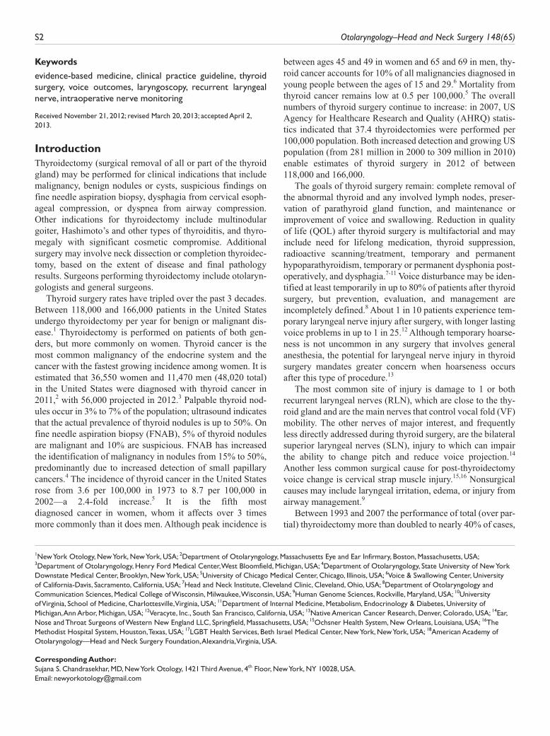
Surgical AnatomyThe thyroid gland sits in the lower anterior portion of the neck, deep to the cervical strap muscles and anterior to the trachea and
esophagus, and inferior to the thyroid cartilage (Figure 1). Nerves of concern during thyroid surgery are the RLN and SLN, which are the main focus of the current discussion.
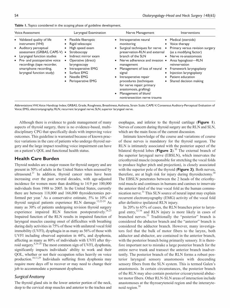
Intimate knowledge of the course and variations of course of these nerves is mandatory for the thyroid surgeon. The RLN is intimately associated with the posterior aspect of the bilateral thyroid lobes (Figure 2).30 The external branch of the superior laryngeal nerve (EBSLN), which innervates the cricothyroid muscle (responsible for stretching the vocal folds to produce higher pitch and projection), is closely associated with the superior pole of the thyroid (Figure 3). Both nerves, therefore, are at high risk for injury during thyroidectomy.30 The EBSLN penetrates between the 2 heads of the cricothy-roid muscle and continues in humans and canines to innervate the anterior third of the true vocal fold as the human commu-nication nerve.31 This SLN source of neural input may explain recurrent electromyography (EMG) activity of the vocal fold after definitive ipsilateral RLN injury.
In 20% to 65% of cases, the RLN branches prior to laryn-geal entry,32-34 and RLN injury is more likely in cases of branched nerves.35 Traditionally the “posterior” branch is considered the abductor branch and the “anterior” branch is considered the adductor branch. However, many investiga-tors feel that the bulk of motor fibers to the larynx, both adductor and abductor, are contained in the anterior branch, with the posterior branch being primarily sensory. It is there-fore important not to mistake a large posterior branch for the entire nerve trunk and transect the anterior branch inadver-tently. The posterior branch of the RLN forms a robust pos-terior laryngeal sensory anastomosis with descending sensory fibers from the SLN system. This is termed Galen’s anastomosis. In certain circumstances, the posterior branch of the RLN may also contain posterior cricoarytenoid abduc-tor motor fibers. Other RLN-SLN areas of interaction include anastomoses at the thyroarytenoid region and the interaryte-noid region.30
Table 1. Topics considered in the scoping phase of guideline development.
Voice Assessment Laryngeal Examination Nerve Management Interventions
• Validated quality of life instrument (VHI)
• Auditory perceptual assessment (GRBAS, CAPE-V)
• Laryngeal function studies • Pre- and postoperative voice
recordings (tape recorder, smartphone recording, laryngeal function study)
• Flexible fiberoptic • Rigid telescopic • High speed exam • Stroboscopy • Indirect mirror exam • Operative (direct)
laryngoscopy • Intraoperative EMG • Surface EMG • Needle EMG • Perioperative EMG
• Intraoperative neural monitoring
• Surgical techniques for nerve preservation-RLN and external branch of the SLN
• Nerve adherence and invasion management
• Management of loss of neural signal
• Intraoperative repair procedures (techniques for nerve repair; primary anastomosis, grafting)
• Management of blunt/nontransection nerve trauma
• Medical (steroids) • Voice therapy • Primary versus revision surgery
(as a modifying factor) • Nerve re-anastomosis • Ansa hypoglossi—RLN
reinnervation • Framework laryngoplasty • Injection laryngoplasty • Patient education • Shared decision making
Abbreviations: VHI, Voice Handicap Index; GRBAS, Grade, Roughness, Breathiness, Asthenia, Strain Scale; CAPE-V, Consensus Auditory-Perceptual Evaluation of Voice; EMG, electromyography; RLN, recurrent laryngeal nerve; SLN, superior laryngeal nerve.
Chandrasekhar et al S5
Nonrecurrent RLN occurs in less than 1% of cases, is seen during right-sided thyroidectomy when it occurs, and arises directly from the cervical vagus. Given its aberrant course, such a nerve may be more likely injured during thyroidec-tomy 36 ( Figure 4 ). Often, the nonrecurrent RLN occurs in conjunction with an anomalous (retro-esophageal) right sub-clavian artery. If a CT scan is performed during evaluation of thyroid/neck mass and a retro-esophageal subclavian artery is noted, then the surgeon should be on the lookout for a nonre-current laryngeal nerve. It behooves the thyroidectomy sur-geon to be intimately familiar with the course and potential aberrations of these nerves.
The neurolaryngology of phonation, swallowing, and res-piration is complex. Cortical representation of the larynx proj-ects to bilateral brainstem nuclei (including nucleus ambiguous), which then projects to the ipsilateral larynx. The RLN carries branchial efferents to the inferior constrictor, cri-copharyngeus, and all laryngeal intrinsic muscles except the cricothyroid muscle. Laryngeal motor fibers within the RLN have a 4 to 1 adductor to abductor ratio. The RLN also
contains afferent fibers that mediate sensation from the vocal folds and below including the upper esophagus and trachea. 30
Neural Injury and Voice Change Early recognition of neural injury, whether temporary or per-manent, may offer opportunities for intervention to improve short- and long-term vocal outcomes, with improved QOL. These issues are covered in detail later in the guideline, but knowing the incidence and prevalence offers additional per-spective on their importance.
In a recent review of 27 articles and 25,000 patients, the average incidence of temporary or permanent vocal fold paral-ysis after surgery was 9.8%, with a wide range from 2.3% to 26%, in part related to the timing and method of laryngeal examination. 12 The Scandinavian quality register reported a vocal fold paralysis rate of 4.3% nerves at risk, based on 3660 thyroid operations performed in 2008 in 26 endocrine surgical units from Sweden and Denmark. 37 , 38 Further, the detection of vocal fold paralysis doubled when patients were submitted to routine laryngeal exam after surgery as compared to laryngos-copy performed only in patients with persistent and severe voice changes.
Figure 1. Location of the thyroid gland. From Surgery of the Thyroid and Parathyroid Glands Edition 2 , Greg W. Randolph, editor, Elsevier Saunders Philadelphia 2012, reprinted with permission.
Figure 2. Relationship of recurrent laryngeal nerves (RLN) and superior laryngeal nerves (SLN) to thyroid lobe and tracheoesophageal groove. From Surgery of the Thyroid and Parathyroid Glands Edition 2 , Greg W. Randolph, editor, Elsevier Saunders Philadelphia 2012, reprinted with permission.

S6 Otolaryngology–Head and Neck Surgery 148(6S)
The 3rd British Association of Endocrine and Thyroid Surgeons (BAETS) audit reported a 2.5% rate of RLN palsy and 4.9% incidence of voice changes in a sample of 10,814 cases of thyroid surgery. For first-time surgery, the reported incidence of RLN palsy was 1.4% after lobectomy and 3.7% after total thy-roidectomy. These figures increased to 5.4% and 6.9%, respec-tively, in revision surgery. 39 Such data are derived from self-reporting by selected surgeons and as such might be too optimistic for extrapolation to the overall practice of thyroid sur-gery. 40 , 41 Administrators of these 2 national databases deem the rates of temporary and permanent RLN paralysis to be severely underestimated, due to lack of routine laryngeal exam. 38 , 39
Vocal fold immobility symptoms vary widely and may range from minimal or no symptoms to acute airway distress. For example, in a recent study of 98 patients with unilateral vocal fold immobility, the voice was judged to be normal in 20% of subjects and improved to normal in an additional 8%. Therefore nearly one-third of patients with unilateral vocal fold immobility were, or later became, asymptomatic. 42 In contrast, bilateral vocal fold immobility is typically associated with profound and immediate respiratory distress, may require tracheotomy, and if initially not recognized and treated promptly, can be associated with anoxic brain injury and death. 43
Voice changes may also occur after thyroid surgery through a variety of mechanisms, including those that are non-neural and without vocal fold immobility. In several large studies of patients without vocal fold immobility, subjective voice com-plaints occurred in 30% to 87% of patients. 8,15,37,44-46 Voice change is not unique to thyroidectomy, but is often observed after any surgery that involves general anesthesia and manipu-lation of the larynx. 13 Appropriately timed laryngeal examina-tion after thyroidectomy helps determine both the cause of voice change and the optimal management.
Overall Cost of Vocal Fold Immobility Vocal fold immobility can be the source of significant morbid-ity and may elicit symptoms profound enough to warrant changing vocation. 47 Unilateral vocal fold immobility can also be associated with significant dysphagia, most noticeably to liquids, and may be associated with aspiration pneumo-nia. 27 , 28 , 48 - 50 The general impact of dysphagia within a hospital setting using an estimate of an average length of stay of 1.64 days is calculated to cost $547 million each year. 51 Vocal fold paresis (VFP) specific dysphagia costs are not available. Permanent bilateral vocal fold immobility can be associated with airway distress and need for tracheostomy or other airway interventions/glottic widening procedures, which themselves significantly and negatively impact both voice and QOL. 52 , 53
Post-thyroidectomy vocal fold immobility may result in substantial postoperative costs including repeated office vis-its, multiple laryngoscopic evaluations, formal voice labora-tory evaluations, voice therapy, one or more VF medialization injection procedures, vocal fold reinnervation procedures, sur-gical thyroplasty, and then additional post-thyroplasty voice therapy sessions. The economic impact of assessing and man-aging individuals suffering a laryngeal disorder, in general, has been estimated to total between $179 million to $295 mil-lion in total annual direct costs. The average direct annual cost to such individuals was estimated to average between $577.18 to $953.21, with the proportion of direct claims associated with pharmaceutical, procedure, and medical encounter claims accounting for 20.1% to 33.3%, 50.4% to 69.9%, and 8.6% to
Figure 3. Course and branches of recurrent laryngeal nerves (RLN) and superior laryngeal nerves (SLN). From Surgery of the Thyroid and Parathyroid Glands Edition 2 , Greg W. Randolph, editor, Elsevier Saunders Philadelphia 2012, reprinted with permission.
Figure 4. Variations of nonrecurrent recurrent laryngeal nerve. From Stewart, Mountain, and Colcock. Nonrecurrent laryngeal nerve. Brit J Surg . 1972;59:379-381.

Chandrasekhar et al S7
16.3% of the annual total costs, respectively.54 A 20-year-old study evaluating the average cost of an otolaryngology evalu-ation in those with unilateral vocal fold paralysis estimated the average cost to be $1706.18 (range, $112.56-$3439.52).55 Current Medicare fee schedules for otolaryngology evaluation and flexible laryngoscopy average $244; most providers use a multiplier to reflect cost of practice in their areas, and that is often 3 to 4 times that amount, per visit.
Vocal fold paralysis is also the source of substantial medi-colegal activity and represents three-quarters of litigation in surgical endocrine disease.56-58
MethodsThis guideline was developed using an explicit and transpar-ent a priori protocol for creating actionable statements based on supporting evidence and the associated balance of benefit and harm.59 The guideline development panel was comprised of representatives from the fields of otolaryngology, laryngol-ogy, head and neck surgery, nursing, speech-language pathol-ogy, endocrinology, internal medicine, general surgery, anesthesiology, and consumer advocacy.
All literature searches were performed by an information specialist through January 2012. Three initial searches were performed to identify clinical practice guidelines, systematic reviews, and randomized controlled trials (RCT). The searches were performed in multiple databases including the National Guidelines Clearinghouse (NGC) (www.guideline.gov), the Cochrane Library, the Cumulative Index to Nursing and Allied Health Literature (CINAHL), EMBASE, PubMed, Web of Science, BIOSIS, the Cochrane Central Register of Controlled Trials (CENTRAL), CMA Infobase, NHS Evidence ENT and Audiology, National Library of Guidelines, National Institute of Clinical Excellence (NICE), Scottish Intercollegiate Guidelines Network (SIGN), New Zealand Guidelines Group (NZGG), Australian National Health and Medical Research Council (ANHMRC), and the TRIP database.
1. Clinical practice guidelines were identified by a PubMed, NGC, CMA Infobase, NHS Evidence, NZGG, ANHMRC, TRIP database, and the G-I-N library search using guideline as a publication type or title word. The search identified 7 guidelines after removing duplicates, clearly irrelevant references, and non–English language articles.
2. Systematic reviews were identified through Medline, EMBASE, the Cochrane Library, CINAHL, AMED, AHRQ, and the TRIP database. The final data set included 50 systematic reviews or meta-analyses that were distributed to the panel members. Articles were excluded if they were not available in English and did not meet the panels’ quality criteria, namely, the review had a clear objective and method, an explicit search strategy, and a valid method of data extraction.
3. RCTs were identified through MEDLINE, EMBASE, CINAHL, and CENTRAL and totaled 285 trials.
Results of all literature searches were distributed to guideline panel members including electronic listings with abstracts (if available) of the searches for clinical guidelines, RCTs, sys-tematic reviews, and other studies. This material was supple-mented, as needed, with targeted searches to address specific needs identified in writing the guideline through May 2012.
In a series of conference calls, the working group defined the scope and objectives of the proposed guideline. During the 12 months devoted to guideline development, the guideline development group met twice, with in-person meetings fol-lowing the format previously described, using electronic deci-sion support (BRIDGE-Wiz, Yale Center for Medical Informatics, New Haven, Connecticut) software to facilitate creating actionable recommendations and evidence profiles.59 Internal electronic review and feedback on each guideline draft was used to ensure accuracy of content and consistency with standardized criteria for reporting clinical practice guidelines.60
American Academy of Otolaryngology—Head and Neck Surgery Foundation (AAO-HNSF) staff used the Guideline Implementability Appraisal and Extractor (GLIA)61 to appraise adherence of the draft guideline to methodological standards, improve clarity of recommendations, and predict potential obstacles to implementation. Guideline panel mem-bers received summary appraisals in May 2012 and modified an advanced draft of the guideline.
The final guideline draft underwent extensive external peer review. Comments were compiled and reviewed by the pan-el’s chair, and a modified version of the guideline was distrib-uted and approved by the guideline development panel. The recommendations contained in the guideline are based on the best available data published through May 2012. Where data were lacking, a combination of clinical experience and expert consensus was used. A scheduled review process will occur at 5 years from publication, or sooner if new compelling evi-dence warrants earlier consideration.
Classification of Evidence-Based StatementsGuidelines are intended to produce optimal health outcomes for patients, minimize harm, and reduce inappropriate variations in clinical care. The evidence-based approach to guideline devel-opment requires that the evidence supporting a policy be identi-fied, appraised, and summarized and that an explicit link between evidence and statements be defined. Evidence-based statements reflect both the quality of evidence and the balance of benefit and harm that is anticipated when the statement is followed. The definitions for evidence-based statements are listed in Table 2 and Table 3.62 As much of the guideline dealt with evidence relating to diagnostic tests, Table 3 was adapted to include current recommendations from the Oxford Centre for Evidence-Based Medicine.63
Guidelines are not intended to supersede professional judg-ment; rather, they may be viewed as a relative constraint on individual clinician discretion in a particular clinical circum-stance. Less frequent variation in practice is expected for a “strong recommendation” than might be expected with a

S8 Otolaryngology–Head and Neck Surgery 148(6S)
“recommendation.”“Options” offer the most opportunity for practice variability.64 Clinicians should always act and decide in a way that they believe will best serve their patients’ inter-ests and needs, regardless of guideline recommendations. They must also operate within their scope of practice and according to their training. Guidelines represent the best judg-ment of a team of experienced clinicians and methodologists addressing the scientific evidence for a particular topic.62
Making recommendations about health practices involves value judgments on the desirability of various outcomes asso-ciated with management options. Values applied by the guide-line panel sought to minimize harm and diminish unnecessary and inappropriate therapy. A major goal of the panel was to be transparent and explicit about how values were applied and to document the process.
Financial Disclosure and Conflicts of InterestThe cost of developing this guideline, including travel expenses of all panel members, was covered in full by the AAO-HNSF. Potential conflicts of interest for all panel members in the past 5 years were compiled and distributed before the first conference call. After review and discussion of these disclosures,65 the panel concluded that individuals with potential conflicts could remain on the panel if they: (1) reminded the panel of potential conflicts before any related discussion, (2) recused themselves from a related discussion if asked by the panel, and (3) agreed not to discuss any aspect of the guideline with industry before publica-tion. Lastly, panelists were reminded that conflicts of interest extend beyond financial relationships and may include personal experiences, how a participant earns a living, and the partici-pant’s previously established “stake” in an issue.66
Guideline Key Action StatementsEach evidence-based statement is organized in a similar fash-ion: an evidence-based key action statement in bold, followed by the strength of the recommendation in italics. Each key action statement is followed by an “action statement profile” of aggregate evidence quality, benefit-harm assessment, and statement of costs. Additionally, there is an explicit statement of any value judgments, the role of patient preferences, clari-fication of any intentional vagueness by the panel, and a repeat statement of the strength of the recommendation. Several paragraphs subsequently discuss the evidence base supporting the statement. An overview of the evidence-based statements in the guideline is shown in Table 4.
The role of patient preferences in making decisions deserves further clarification. For some statements, where the evidence base demonstrates clear benefit, although the role of patient preference for a range of treatments may not be rele-vant (eg, with intraoperative decision making), clinicians should provide patients with clear and comprehensible infor-mation on the benefits in order to facilitate patient understand-ing and shared decision making.67 In cases where evidence is weak or benefits unclear, the practice of shared decision mak-ing, again where the management decision is made by a col-laborative effort between the clinician and an informed patient, is extremely useful. Factors related to patient preference
include (but are not limited to) absolute benefits (numbers needed to treat), adverse effects (number needed to harm), cost of drugs or procedures, and frequency and duration of treatment.59
STATEMENT 1. BASELINE VOICE ASSESSMENT: The surgeon should document assessment of the patient’s voice once a decision has been made to pro-ceed with thyroid surgery. Recommendation based on observational studies with a preponderance of benefit over harm.
Action Statement Profile
• Aggregate evidence quality: Grade C • Benefit: Establish a baseline, improve the detection
of preexisting voice impairment, establish expecta-tions about voice outcomes, educating the patient, facilitates shared decision making, prioritize the need for preoperative laryngeal assessment and more in-depth voice assessment
• Risk, harm, cost: Anxiety, cost of assessment tool, patient and provider time
• Benefit-harm assessment: Preponderance of benefit • Value judgments: Perception by the GDG of a cur-
rent underassessment of voice prior to surgery • Intentional vagueness: The proximity of the assess-
ment to the day of surgery is not specified because there was no consensus among the guideline group and there were no data to support the choice of one time point over another. The group agreed that any change in voice would warrant a new assessment.
• Role of patient preferences: Selection of assessment methods
• Exclusions: None • Policy level: Recommendation
Supporting text. The purpose of this recommendation is to improve quality of care by increasing awareness of the impor-tance of assessing voice due to the potential impact of thyroid surgery on voice quality. Patients with an abnormal voice should have additional evaluation to document the extent of impairment and should have preoperative assessment of the larynx performed as described in Statement 2.
At a minimum, subjective assessment of voice by the surgeon, patient, and family should be done. A simple way to accomplish this is to specifically ask the patient and his or her family mem-bers if they consider the patient’s voice to be abnormal, impaired, or less than satisfactory. The response to these questions should be documented in the medical record. The surgeon should also indicate his or her own subjective opinion as to the overall degree of voice quality aberrance and document this in the medical record.68,69 If there is any detectable voice impairment, if the patient gives a past history of voice disorder, or if there is uncer-tainty, more thorough voice investigation is indicated, which may include a validated QOL measure administered by the surgeon or his or her designee, referral to an otolaryngologist, and/or assess-ment by a speech and language pathologist. In addition, any
Chandrasekhar et al S9
patient with a preoperative voice abnormality should have laryn-geal examination as described in the next section.
Importance of voice assessment. Voice impairment can sig-nificantly impact the ability of an individual to work, social-ize, and perform many activities of daily living.10,11 It is important to establish the presurgical status of the patient’s voice characteristics and function for comparison postsurgi-cally, alert the surgeon to possible increased extent of disease, and determine the existence of preoperative voice problems that may remain postsurgically. Although the goal of this guideline is to optimize voice outcomes postoperatively, up to 33% of individuals may exhibit voice impairment preopera-tively.70-72 Preoperative voice problems may result from tumor invasion of, or compression injury to, the RLN (as seen with,
eg, edema or large goiter), or from preexisting or non–thyroid-related causes. In addition, vocal fold edema or other tissue changes may be seen in endocrine abnormalities associated with thyroid problems.73,74 One study demonstrated that indi-viduals identified with presurgical RLN impairment due to tumor invasion exhibited improved voice function outcomes after a subsequent voice surgery compared to those who were not so identified.72 That same study also reported on 1 patient exhibiting preoperative unilateral vocal fold immobility who developed postoperative impairment of the previously normal vocal fold, resulting in bilateral vocal fold paralysis. Thus, baseline assessment of the patient’s voice prior to thyroid sur-gery serves the purpose of identifying those with preoperative impairment as well as establishing a preoperative reference
Table 2. Guideline definitions for evidence-based statements.
Statement Definition Implication
Strong recommendation A strong recommendation means the benefits of the recommended approach clearly exceed the harms (or that the harms clearly exceed the benefits in the case of a strong negative recommendation) and that the quality of the supporting evidence is excellent (Grade A or B).a In some clearly identified circumstances, strong recommendations may be made based on lesser evidence when high-quality evidence is impossible to obtain and the anticipated benefits strongly outweigh the harms.
Clinicians should follow a strong recommendation unless a clear and compelling rationale for an alternative approach is present.
Recommendation A recommendation means the benefits exceed the harms (or that the harms exceed the benefits in the case of a negative recommendation), but the quality of evidence is not as strong (Grade B or C). In some clearly identified circumstances, recommendations may be made based on lesser evidence when high-quality evidence is impossible to obtain and the anticipated benefits outweigh the harms.
Clinicians should also generally follow a recommendation, but should remain alert to new information and sensitive to patient preferences.
Option An option means that either the quality of evidence that exists is suspect (Grade D) or that well-done studies (Grade A, B, or C) show little clear advantage to one approach versus another.
Clinicians should be flexible in their decision making regarding appropriate practice, although they may set bounds on alternatives; patient preference should have a substantial influencing role.
No recommendation No recommendation means there is both a lack of pertinent evidence (Grade D) and an unclear balance between benefits and harms.
Clinicians should feel little constraint in their decision making and be alert to new published evidence that clarifies the balance of benefit versus harm; patient preference should have a substantial influencing role.
aSee Table 3 for definition of evidence grades.
Table 3. Evidence quality for grades of evidence.
Grade Evidence Quality for Diagnosis Evidence Quality for Treatment and Harm
A Systematic review of cross-sectional studies with consistently applied reference standard and blinding
Well-designed randomized controlled trials performed on a population similar to the guideline’s target population
B Individual cross-sectional studies with consistently applied reference standard and blinding
Randomized controlled trials; overwhelmingly consistent evidence from observational studies
C Nonconsecutive studies, case-control studies, or studies with poor, nonindependent, or inconsistently applied reference standards
Observational studies (case control and cohort design)
D Mechanism-based reasoning or case reportsX Exceptional situations where validating studies cannot be performed and there is a clear preponderance of benefit over harm
S10 Otolaryngology–Head and Neck Surgery 148(6S)
for the patient’s voice characteristics and function for com-parison to his or her postoperative voice. In this way the clini-cian may be alerted to the presence of a preexisting vocal fold paralysis or paresis.
Common methods used for assessing a patient’s preopera-tive voice include patient self-report, audio-perceptual judg-ment, and acoustic measurement of audio recordings. A thorough assessment of a patient’s voice can be completed by a speech-language pathologist (SLP) or otolaryngologist. Such an individual can complete laryngeal function studies that typically consist of audio-perceptual ratings and acoustic and aerodynamic measures. However, when such a profes-sional is not available, the surgeon may be able to document the patient’s voice characteristics and function using less rig-orous methods.
Methods of voice assessment. The determination of the assessment tool should be based on the patient’s capacity to effectively participate and the examiner’s facility with the assessment tool (Table 5).
One method of determining preoperative status of patients’ voice is to ask them to report whether they have noticed changes in their voice pitch, loudness, quality, or endurance. Some examples of this approach have been described in the literature.70,71 The Voice Handicap Index (VHI) offers a stan-dardized method for gathering this type of information and offers substantial literature to support its use with this popula-tion. The VHI determines the degree of impaired vocal func-tion a patient experiences across 3 areas: emotional, physical, and functional. It is a validated 30-item questionnaire that can determine the presence or absence and severity of a self-identified voice problem and has been translated validly into at least 30 different languages.75-78 A total score higher than 18 points on the VHI 30-item instrument is considered indicative of a voice problem, with higher scores associated with increas-ing severity of the voice problem. The VHI has been success-fully used in several studies to determine pre- and postoperative
voice status in those undergoing thyroid surgery.37,74,79-81 Further, it has been shown to have high levels of diagnostic precision in predicting significant voice changes from pre- to post-thyroid surgery.74 The VHI-10 is a shorter, alternative version of the original VHI with only 10 questions that may be more practical for quick use. It has adequate reliability levels within and between raters,82 but concerns have been raised regarding the validity of the VHI-10 associated with its sensi-tivity and specificity for identifying individuals with voice disorders to the same degree as the VHI-30.83 Although the VHI-10 did not meet psychometric criteria for recommenda-tion as a tool for identifying individuals with voice disorders as determined by the University of North Carolina Evidence-Based Practice Center,84 subsequently, normative values for the VHI-10 have been established.85
There are a number of other validated instruments that can serve the purpose of patient self-report regarding voice prob-lems, including the Voice-Related Quality of Life instrument (V-RQOL). However, it is beyond the scope of this guideline to discuss all of them in detail. The reader is encouraged to peruse the references so as to have a basis to select from the many instruments that are available.76,86
Auditory perceptual assessment is a method judging a patient’s voice quality and describing any aberrant features. Two multidimensional rating scales used to complete auditory-perceptual evaluation of the voice include the GRBAS (Grade, Roughness, Breathiness, Asthenia, and Strain) and the Consensus Auditory-Perceptual Evaluation of Voice (CAPE-V). The GRBAS scale rates each feature using an ordinal 4-point rating scale.87 The CAPE-V provides visual analog scaling for rating the parameters of Overall Severity, Strain, Breathiness, Roughness, Pitch, and Loudness.88 Blank scales are also pro-vided on the CAPE-V form so that other voice quality features may be added and rated (eg, tremor). Two studies comparing audio-perceptual ratings from the GRBAS and CAPE-V have shown high reliability for both on the Overall Severity scale.
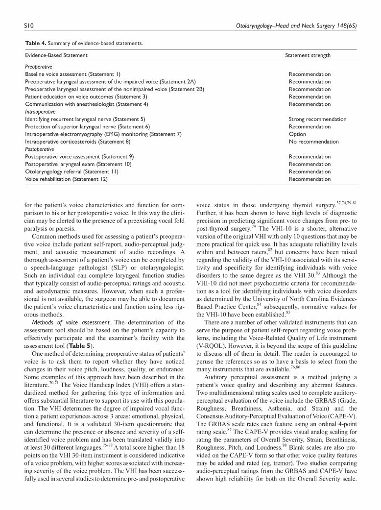
Table 4. Summary of evidence-based statements.
Evidence-Based Statement Statement strength
PreoperativeBaseline voice assessment (Statement 1) RecommendationPreoperative laryngeal assessment of the impaired voice (Statement 2A) RecommendationPreoperative laryngeal assessment of the nonimpaired voice (Statement 2B) RecommendationPatient education on voice outcomes (Statement 3) RecommendationCommunication with anesthesiologist (Statement 4) RecommendationIntraoperativeIdentifying recurrent laryngeal nerve (Statement 5) Strong recommendationProtection of superior laryngeal nerve (Statement 6) RecommendationIntraoperative electromyography (EMG) monitoring (Statement 7) OptionIntraoperative corticosteroids (Statement 8) No recommendationPostoperativePostoperative voice assessment (Statement 9) RecommendationPostoperative laryngeal exam (Statement 10) RecommendationOtolaryngology referral (Statement 11) RecommendationVoice rehabilitation (Statement 12) Recommendation
Chandrasekhar et al S11
The CAPE-V was also shown to have concurrent validity.89 These instruments were designed for use by professionals with expertise and training in audio-perceptual aspects of voice. For individuals without such training and expertise, a simple method for acquiring preoperative auditory-perceptual judg-ments of the patient’s voice can be achieved by completing an audio recording of the patient’s voice while they sustain a vowel sound such as “ah” or “ee” for 3 to 5 seconds and then while
they read standard sentences or spontaneously converse for 30 seconds to 1 minute. The audio recording should be made in an environment with minimal background noise and with a high-quality microphone placed in optimal proximity to the speaker if possible.90 With advancements in technology, audio record-ing devices are now more common and widely available. Although not ideal, many smartphones (which are readily avail-able to most practitioners) contain a recording application that
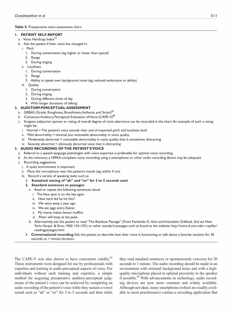
Table 5. Preoperative voice assessment chart.
1. PATIENT SELF-REPORT a. Voice Handicap Index75
b. Ask the patient if their voice has changed in: i. Pitch 1. During conversation (eg, higher or lower than typical) 2. Range 3. During singing ii. Loudness 1. During conversation 2. Range 3. Ability to speak over background noise (eg, reduced endurance or ability) iii. Quality 1. During conversation 2. During singing 3. During different times of day 4. With longer durations of talking2. AUDITORY-PERCEPTUAL ASSESSMENT a. GRBAS (Grade, Roughness, Breathiness, Asthenia, and Strain)87
b. Consensus Auditory-Perceptual Evaluation of Voice (CAPE-V)88
c. Surgeon subjective opinion or rating of overall degree of voice aberrance can be recorded in the chart. An example of such a rating might be:
i. Normal = The patient’s voice sounds clear and of expected pitch and loudness level ii. Mild abnormality = minimal, but noticeable abnormality in voice quality iii. Moderately abnormal = noticeable abnormality in voice quality that is sometimes distracting iv. Severely abnormal = obviously abnormal voice that is distracting3. AUDIO RECORDING OF THE PATIENT’S VOICE a. Referral to a speech-language pathologist with voice expertise is preferable for optimal voice recording b. At the minimum, a HIPAA-compliant voice recording using a smartphone or other audio recording device may be adequate c. Recording suggestions i. A quiet environment is important ii. Place the microphone near the patient’s mouth (eg, within 4 cm) iii. Record a variety of speaking tasks such as 1. Sustained voicing of “ah” and “ee” for 3 to 5 seconds each 2. Standard sentences or passages: a. Read or repeat the following sentences aloud i. The blue spot is on the key again ii. How hard did he hit him? iii. We were away a year ago. iv. We eat eggs every Easter. v. My mama makes lemon muffins. vi. Peter will keep at the peak. b. Alternatively, ask the patient to read “The Rainbow Passage” (From Fairbanks G. Voice and Articulation Drillbook. 2nd ed. New
York: Harper & Row; 1960: 124-139.) or other standard passages such as found at the website: http://www.d.umn.edu/~cspiller/readingpassages.html
3. Conversational recording: Ask the patient to describe how their voice is functioning or talk about a favorite vacation for 30 seconds to 1 minute duration.
S12 Otolaryngology–Head and Neck Surgery 148(6S)
may also suffice for this purpose. The health care provider should ensure that his or her smartphone, if used in this manner, is HIPAA compliant. The audio recording can be used postop-eratively to compare the patient’s voice to the preoperative recording to determine if changes in voice pitch, loudness, and quality are perceived.
STATEMENT 2A. PREOPERATIVE LARYNGEAL ASSESSMENT OF THE IMPAIRED VOICE: The sur-geon should examine vocal fold mobility, or refer the patient to a clinician who can examine vocal fold mobil-ity, if the patient’s voice is impaired (as determined by the assessment in Statement 1) and a decision has been made to proceed with thyroid surgery. Recommendation based on observational studies with a preponderance of benefit over harm.
Action Statement Profile
• Aggregate evidence quality: Grade C • Benefit: Assess mobility of vocal fold, potential
diagnosis of invasive thyroid cancer, influence the decision for surgery, extent of surgery, intraopera-tive technique, preoperative patient counseling, dis-tinguish iatrogenic from disease related paralysis/paresis
• Risk, harm, cost: Misdiagnosis (false positive/false negative), cost of examination, patient discomfort, resources, access, anxiety
• Benefit-harm assessment: Preponderance of benefit • Value judgments: None • Role of patient preferences: Limited • Exclusions: None • Policy level: Recommendation
Supporting text. The purpose of the statement is to improve quality of care by establishing baseline awareness of vocal fold mobility that may be important in perioperative manage-ment and outcome assessment.
At present, only 6.1% to 54% of thyroidectomy patients undergo a preoperative laryngeal exam.38,39,91 However, sev-eral international organizations are advocating for preopera-tive laryngeal exam. The BAETS and the German Association of Endocrine Surgery have recommended preoperative and postoperative laryngeal exam as requirements for all patients undergoing thyroid surgery.92,93 The international neural mon-itoring study group recommends pre- and postoperative laryn-goscopy in all patients undergoing thyroid surgery with use of intraoperative neural monitoring (IONM).94 Other groups offer less universal recommendations. The British Thyroid Association recommends laryngeal exam for preoperative patients with voice changes and for those undergoing surgery for cancer, and the National Comprehensive Cancer Network (NCCN) recommendations include preoperative laryngos-copy in all patients with thyroid malignancy.95,96
The decision to proceed with an examination of the larynx is often predicated on the initial perception of the quality of
the voice or the patient’s history of either having had a change in voice or generalized concerns with the voice. In cases where the patient/family or the physician notes a voice abnor-mality, it is recommended that a preoperative assessment of larynx and VF function be performed. Reduced movement of 1 vocal fold on preoperative examination of the patient with hoarseness suggests involvement of the RLN by the thyroid disease, which may prompt extra caution and evaluation.
In the general population, 1% of patients (and 2.5% of patients over age 75) seek evaluation and care for dysphonia, with 3% of those eventually diagnosed with VF paresis or paralysis.97 Among patients screened in primary care clinics for dysphonia, there was a point prevalence of 7.5% and a lifetime prevalence of 29.1%.98
Incidence rates for vocal fold paresis or paralysis for patients with benign thyroid disease is approximately 1%99 and for malignant thyroid disease is as high as 8%99 in patients who have not undergone a prior thyroid, neck, or chest sur-gery. A series of 200 patients with benign cervical and subster-nal goiter showed that 3.5% presented with vocal fold paralysis.100 Of 340 pre-thyroidectomy patients, VF motion abnormalities were found in 6.5%.101
A finding of VF paralysis on preoperative examination strongly suggests the presence of invasive thyroid malignancy. In 1 study, the rate of preoperative VF paralysis in a series of patients with invasive thyroid malignancy was over 70% ver-sus 0.3% in the control group of patients with noninvasive thyroid disease.102 The preoperative knowledge of invasive dis-ease allows for more robust surgical planning, more detailed pre-operative imaging, and more specific preoperative patient counseling. The NCCN guidelines describe preoperative VF paralysis as a “highly suspicious factor” for cancer and the need for surgery.
Identification of preoperative VF paralysis is also impor-tant because surgical algorithms in the management of inva-sive disease involving the nerve incorporate the degree to which the nerve is functional. Thus, preoperative functional information obtained via laryngeal exam greatly aids in tar-geted management of the invaded nerve.102,103 The Guideline Development Group emphasizes that examination of laryn-geal function both before (Statement 2A) and after (Statement 10) thyroid surgery is recommended. There is not enough evi-dence in the literature to make this either a strong recommen-dation or mandatory; however, there is no evidence against laryngeal examination. As stated previously, the preponder-ance of benefit over potential harm permits this key action statement to rise to the level of a recommendation.
The members of the GDG felt, overall, that examination of all larynges preoperatively would be of benefit to both physi-cian and patient, as diagnosis of asymptomatic vocal cord paresis and paralysis can be beneficial. However, guidelines are based on available literature, and there is not enough lit-erature to support a recommendation for examining all laryn-ges preoperatively. There are no standardized laryngeal examination methods that will fit all patients. Some of this will depend on the resources and equipment available in the
Chandrasekhar et al S13
community or in the individual practice. In general, there are 3 common techniques for examination of the larynx in the office setting.104 A simple and still reliable method of examin-ing the larynx is with the use of directed light, a head mirror, and a laryngeal mirror. The technique is commonly used in most otolaryngology practices and in some oncology and endocrine surgery practices. Gross vocal fold mobility can be observed in most patients, but assessment of minor alterations may be difficult, and there is no possibility of obtaining video recordings for repeat evaluation. In addition, some patients are unable to be adequately examined with a mirror.
A more reliable examination can be obtained with flexible laryngoscopy.105 Flexible laryngoscopy allows for easy access in almost all patients, allows for evaluation in running speech and with motion-directed tasks, and is better for an evaluation of subtle changes in vocal fold motion. It allows for evaluation in extremes of range and loudness, all of which may identify vocal fold motion aberrations. In addition, video recording can be obtained that allows for review and slow motion analy-sis. Common tasks performed with flexible laryngoscopy to try to isolate movement problems are “eee-sniff,” whistle, laugh, deep inspiration, cough, speaking, and singing. On average, for a novice, only 6 attempts are necessary in order to become competent in performing flexible laryngoscopy.106 Of course, diagnostic accuracy will improve with ongoing use of the flexible laryngoscope.
A more detailed functional method of examination of the larynx is video-strobo-laryngoscopy (VSL), but this tech-nology is not widely available. VSL can be performed with both rigid endoscopy through the mouth or flexible laryn-goscopy with an endoscope passed through the nose. Flexible stroboscopy is preferred when assessing vocal fold motion in normal, running speech and directed tasks, both of which cannot be done when a person has his or her tongue held during rigid laryngoscopy. Stroboscopic assessment of both gross motion and an in-depth assessment of vocal fold vibratory pliability and symmetry can help define subtle changes in vocal fold movement consistent with neuropa-thy. When compared to laryngeal electromyography as the standard, video stroboscopy has a sensitivity of 97.9%, a specificity of 63.2%, a positive predictive value of 95.9%, a negative predictive value of 77.42%, and a test efficiency of 94.41%.107
The panel understands that relative cost of laryngeal exam-ination modality must be considered. There is no added reim-bursable cost of indirect laryngoscopy using a laryngeal mirror; however, there is the cost incurred in educating one-self to perform that examination accurately and with maximal possible patient comfort. This technique is covered during the otolaryngology residency curriculum, and non-ENT thyroid surgeons have learned, or should consider learning, this tech-nique and using it regularly to maintain familiarity with it. There are additional health care costs in performing both flex-ible laryngoscopy and VSL; these are justified when the lar-ynx cannot be examined using the mirror, when the presence of VF movement abnormality is not clear after performing mirror exam, and in the case of identified VF abnormality, in
order to more accurately define that abnormality. The surgeon assessing laryngeal function preoperatively (as well as post-operatively) should strive to perform the most complete, cost-effective examination for the patient and document the examination accurately.
STATEMENT 2B. PREOPERATIVE LARYNGEAL ASSESSMENT OF THE NONIMPAIRED VOICE: The surgeon should examine vocal fold mobility, or refer the patient to a clinician who can examine vocal fold mobility, if the patient’s voice is normal and the patient has (a) thyroid cancer with suspected extra-thyroidal extension, or (b) prior neck surgery that increases the risk of laryngeal nerve injury (carotid endarterectomy, anterior approach to the cervical spine, cervical esophagectomy, and prior thyroid or parathyroid surgery), or (c) both, once a decision has been made to proceed with thyroid surgery. Recom-mendation based on observational studies with a prepon-derance of benefit over harm.
Action Statement Profile
• Aggregate evidence quality: Grade C • Benefit: Assess mobility of vocal fold, potential
diagnosis of invasive thyroid cancer, influence the decision for surgery, extent of surgery, intraopera-tive technique, preoperative patient counseling, dis-tinguish iatrogenic from disease related paralysis/paresis
• Risk, harm, cost: Misdiagnosis (false positive/false negative), cost of examination, patient discomfort, resources, access, anxiety
• Benefit-harm assessment: Preponderance of benefit • Value judgments: Even though the prevalence of pre-
operative vocal fold paresis is low, the consequence of not knowing this prior to surgery could result in substantial morbidity or mortality. For this reason, the GDG was willing to accept a large number of normal examinations in return for an occasional abnormal finding.
• Intentional vagueness: The timing of assessment relative to surgery is not stated to allow clinicians flexibility in decision making, although the Guide-line Development Group agreed that the assessment should take place as close to the surgery as possible. The word suspected is used due to the difficulty of identifying extrathyroidal extension through physi-cal exam and imaging.
• Role of patient preferences: Limited • Exclusions: None • Policy level: Recommendation
Supporting text. The purpose of the statement is to improve quality of care by establishing baseline awareness of vocal fold mobility that may be important in perioperative management and outcome assessment in certain groups of
S14 Otolaryngology–Head and Neck Surgery 148(6S)
high-risk patients.
There are papers reporting on patients with immobile vocal folds who have relatively normal voices. One-third of 340 such patients evaluated preoperatively had no voice complaints.101 Two studies reported limited sensitivity of vocal symptoms in the prediction of vocal fold paralysis ranging from 33% to 68%.101,102 In one study, one third of 98 patients with postopera-tive vocal fold paralysis were ultimately judged to be asymp-tomatic in terms of vocal symptoms.42 The members of the GDG felt that preoperative laryngeal assessment would be ideal in all patients undergoing thyroidectomy. However, the aggre-gate level of evidence is not high enough for the GDG to expand the current recommendation to preoperative larynx examina-tion in all thyroid patients, including those with normal voice and no prior neck/upper chest surgery.
The RLN may be injured by non-thyroid surgeries of the neck and chest, such as carotid endarterectomy, anterior approach to the cervical spine, cervical esophagectomy, and other neck/chest procedures, and by prior thyroidectomy or parathyroidectomy. In patients with a history of any of these RLN risk factors, preoperative laryngeal examination is indi-cated before planned thyroidectomy.
An evaluation of 1947 patients undergoing elective surgi-cal procedures with preoperative screening by laryngeal examination revealed 31 (1.5%) vocal palsies,108 and 1 asymp-tomatic patient was identified out of 50 (2%) patients screened prior to endarterectomy.109 A 20-year review of patients diag-nosed with vocal fold immobility identified 643 patients with unilateral and 189 with bilateral vocal fold immobility.110 In the unilateral group, 235, or 36.5%, were due to iatrogenic injury, with 80 (12.4%) following thyroid surgery. The remain-der of the iatrogenic injuries included anterior approaches to the cervical spine, carotid endarterectomy, and chest and neck surgeries. In the bilateral group, prior surgery accounted for 70 (37%) immobilities, with 56 (26.9%) being due to thyroid surgery.110
Incidence rates for injury to the RLNs from thyroid surgery range from 13% for thyroid cancer operations to 30% for revi-sion thyroid surgery.24,25 In individuals in whom the nerve is spared, incidence rates range from 0% to 5% based on the number of nerves at risk.22-24 Following carotid endarterec-tomy, the overall rate of injury to the RLN is 4% to 7%,109,111,112 with permanent injury in 3% to 4%.109,111 In anterior approaches to the cervical spine surgery, RLN injury occurs in 1.5% to 6.4%.112
Total thyroidectomy, commonly offered in the context of thy-roid cancer, imparts risk to bilateral recurrent and superior laryn-geal nerves. An underlying and undiagnosed preoperative laryngeal nerve dysfunction would convey significantly greater risk of postoperative bilateral nerve paralysis, a potentially cata-strophic event. While unilateral VFP is typically associated with a weakened hoarse voice, bilateral paralysis is associated with airway obstruction, respiratory distress, and the need for urgent life-saving interventions such as tracheotomy. Preoperative laryngoscopy may identify those individuals, with or without an impaired voice, who have preexisting VF weakness and who would therefore be at risk for a poor functional outcome. The
consensus of the panel was that any patient undergoing bilateral thyroid surgery should be evaluated with preoperative laryngeal exam even in this setting of normal preoperative voice, but there is not enough published evidence to elevate this statement to the level of a key action statement.
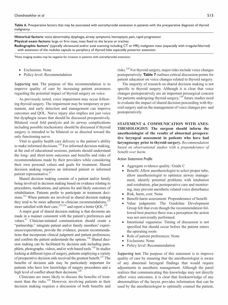
Approximately 10% to 15% of thyroid cancers present with extrathyroidal extension.113-116 The most common struc-tures involved in extrathyroidal extension include the strap muscles (53%), the RLN (47%), the trachea (30%), the esophagus (21%), and the larynx (12%).114 Preoperative imaging is not good enough in the routine detection of extra-thyroidal extension; ultrasonography (US) has sensitivities for tracheal invasion of 42% and for esophageal invasion, 29%,117 with accurate tumor staging of only 67%.118 The finding of preoperative vocal fold paralysis, however, tracks strongly with invasive disease in patients with cytologic diagnosis of thyroid cancer.102,119 Laryngeal examination is therefore recommended in patients with preoperative diag-nosis of thyroid cancer if there is evidence for extrathyroidal extension of cancer, even if the voice is normal. Factors that suggest extrathyroidal extension in the setting of a patient with preoperative diagnosis of malignancy may include his-torical, physical examination, and radiographic factors (see Table 6).
Given the incidence of paresis and paralysis in patients who have undergone prior thyroid, neck, or significant chest surgery, the evidence supports advocating for routine exami-nation of the vocal folds to assess the status of vocal fold mobility prior to surgery in these patients. This would allow for identification of potential problems that may arise, increase the diligence in relationship to management of the opposite nerve, or may prompt the use of nerve monitoring. All of these may reduce the risk of bilateral vocal fold paresis or paralysis as a result of thyroid surgery.
STATEMENT 3. PATIENT EDUCATION ON VOICE OUTCOMES: The clinician should educate the patient about the potential impact of thyroid surgery on voice once a decision has been made to proceed with thyroid surgery. Recommendation based on preponderance of benefit over harm.
Action Statement Profile
• Aggregate evidence quality: Grade B, RCTs on the value of patient education in general regarding sur-gery; Grade C, studies on the incidence of voice impairment following thyroid surgery in particular
• Benefit: Facilitate shared decision making, establish realistic expectations, help patients recognize voice changes postoperatively
• Risk, harm, cost: Anxiety • Benefit-harm assessment: Preponderance of benefit • Value judgments: Generalize evidence about the ben-
efits of patient education to this circumstance • Intentional vagueness: None • Role of patient preferences: Patient can decline
information
Chandrasekhar et al S15
• Exclusions: None • Policy level: Recommendation
Supporting text. The purpose of this recommendation is to improve quality of care by increasing patient awareness regarding the potential impact of thyroid surgery on voice.
As previously noted, voice impairment may occur follow-ing thyroid surgery. The impairment may be temporary or per-manent, and early detection and management can improve outcomes and QOL. Nerve injury also implies not just voice but dysphagia issues that should be discussed preoperatively. Bilateral vocal fold paralysis and its airway complications including possible tracheotomy should be discussed if thyroid surgery is intended to be bilateral or as directed toward the only functioning nerve.
Vital to quality health care delivery is the patient’s ability to make informed decisions.120 For informed decision making, at the end of educational sessions, patients should understand the long- and short-term outcomes and benefits and risks of recommendations made by their providers while considering their own personal values and goals for treatment. Shared decision making requires an informed patient or informed patient representative.67
Shared decision making consists of a patient and/or family being involved in decision making based on evidence relating to procedures, medications, and options for and likely outcomes of rehabilitation. Patients prefer to participate in treatment deci-sions.121 When patients are involved in shared decision making they tend to be more adherent to clinician recommendations,122 more satisfied with their care,123,124 and report a better QOL.125
A major goal of shared decision making is that decisions are made in a manner consistent with the patient’s preferences and values.67 Clinician-initiated communication should create a “partnership,” integrate patient and/or family members’ experi-ences/expectations, provide the evidence, present recommenda-tions that incorporate clinical judgment and patient preferences, and confirm the patient understands the options.126 Shared deci-sion making can be facilitated by decision aids including pam-phlets, photographs, videos, and/or web-based tools.120 In studies looking at different types of surgery, patients employing a variety of preoperative decision aids received the greatest benefit.127 The benefits of decision aids may be particularly important for patients who have low knowledge of surgery procedures and a high level of conflict about their decisions.128
Clinicians are more likely to discuss the benefits of treat-ment than the risks.129 However, involving patients in their decision making requires a discussion of both benefits and
risks.129 For thyroid surgery, major risks include voice changes postoperatively. Table 7 outlines critical discussion points for patient education on voice changes related to thyroid surgery.
The majority of research on shared decision making is not specific to thyroid surgery. Although it is clear that voice changes postoperatively are an important presurgical concern for patients undergoing thyroid surgery,130 future studies need to evaluate the impact of shared decision proceeding with thy-roid surgery and on the management of voice changes pre- and postoperatively.
STATEMENT 4. COMMUNICATION WITH ANES-THESIOLOGIST: The surgeon should inform the anesthesiologist of the results of abnormal preopera-tive laryngeal assessment in patients who have had laryngoscopy prior to thyroid surgery. Recommendation based on observational studies with a preponderance of benefit over harm.
Action Statement Profile
• Aggregate evidence quality: Grade C • Benefit: Allow anesthesiologist to select proper tube,
allow anesthesiologist to optimize airway manage-ment, identify potential problems with intubation and extubation, plan postoperative care and monitor-ing, may prevent anesthetic related voice disturbance
• Risk, harm, cost: None • Benefit-harm assessment: Preponderance of benefit • Value judgments: The Guideline Development
Group felt that even though the recommendation fol-lowed best practice there was a perception the action was not universally performed.
• Intentional vagueness: Timing of discussion is not specified but should occur before the patient enters the operating room
• Role of patient preferences: None • Exclusions: None • Policy level: Recommendation
Supporting text. The purpose of this statement is to improve quality of care by ensuring that the anesthesiologist is aware of any abnormal laryngeal findings that would require adjustments in anesthetic management. Although the panel realizes that communicating this knowledge may not directly affect voice outcomes, it is clear that foreknowledge of any abnormalities of the larynx provides information that can be used by the anesthesiologist to optimally counsel the patient,
Table 6. Preoperative factors that may be associated with extrathyroidal extension in patients with the preoperative diagnosis of thyroid malignancy.
Historical factors: voice abnormality, dysphagia, airway symptoms, hemoptysis, pain, rapid progressionPhysical exam factors: large or firm mass, mass fixed to the larynx or tracheaRadiographic factorsa (typically ultrasound and/or axial scanning including CT or MR): malignant mass (especially with irregular/blurred)
with extension of the nodules capsule to periphery of thyroid lobe especially posterior extension
aNote imaging studies may be negative for invasion in patients with extrathyroidal extension.
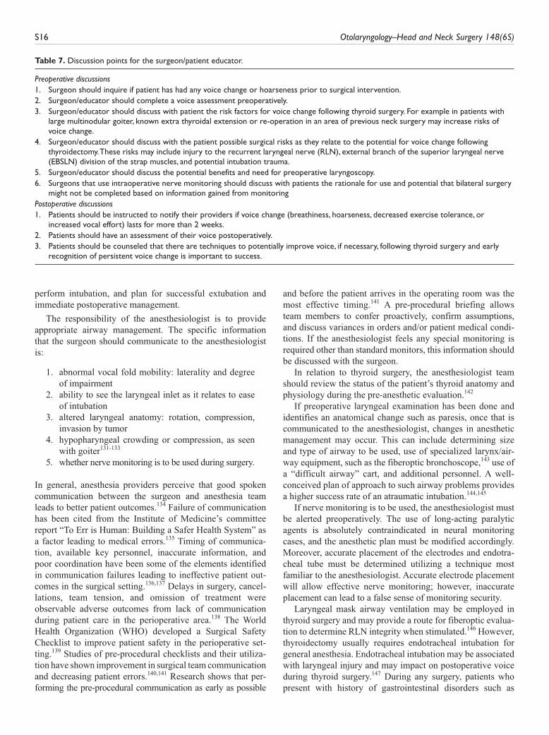
S16 Otolaryngology–Head and Neck Surgery 148(6S)
perform intubation, and plan for successful extubation and immediate postoperative management.
The responsibility of the anesthesiologist is to provide appropriate airway management. The specific information that the surgeon should communicate to the anesthesiologist is:
1. abnormal vocal fold mobility: laterality and degree of impairment
2. ability to see the laryngeal inlet as it relates to ease of intubation
3. altered laryngeal anatomy: rotation, compression, invasion by tumor
4. hypopharyngeal crowding or compression, as seen with goiter131-133
5. whether nerve monitoring is to be used during surgery.
In general, anesthesia providers perceive that good spoken communication between the surgeon and anesthesia team leads to better patient outcomes.134 Failure of communication has been cited from the Institute of Medicine’s committee report “To Err is Human: Building a Safer Health System” as a factor leading to medical errors.135 Timing of communica-tion, available key personnel, inaccurate information, and poor coordination have been some of the elements identified in communication failures leading to ineffective patient out-comes in the surgical setting.136,137 Delays in surgery, cancel-lations, team tension, and omission of treatment were observable adverse outcomes from lack of communication during patient care in the perioperative area.138 The World Health Organization (WHO) developed a Surgical Safety Checklist to improve patient safety in the perioperative set-ting.139 Studies of pre-procedural checklists and their utiliza-tion have shown improvement in surgical team communication and decreasing patient errors.140,141 Research shows that per-forming the pre-procedural communication as early as possible
and before the patient arrives in the operating room was the most effective timing.141 A pre-procedural briefing allows team members to confer proactively, confirm assumptions, and discuss variances in orders and/or patient medical condi-tions. If the anesthesiologist feels any special monitoring is required other than standard monitors, this information should be discussed with the surgeon.
In relation to thyroid surgery, the anesthesiologist team should review the status of the patient’s thyroid anatomy and physiology during the pre-anesthetic evaluation.142
If preoperative laryngeal examination has been done and identifies an anatomical change such as paresis, once that is communicated to the anesthesiologist, changes in anesthetic management may occur. This can include determining size and type of airway to be used, use of specialized larynx/air-way equipment, such as the fiberoptic bronchoscope,143 use of a “difficult airway” cart, and additional personnel. A well-conceived plan of approach to such airway problems provides a higher success rate of an atraumatic intubation.144,145
If nerve monitoring is to be used, the anesthesiologist must be alerted preoperatively. The use of long-acting paralytic agents is absolutely contraindicated in neural monitoring cases, and the anesthetic plan must be modified accordingly. Moreover, accurate placement of the electrodes and endotra-cheal tube must be determined utilizing a technique most familiar to the anesthesiologist. Accurate electrode placement will allow effective nerve monitoring; however, inaccurate placement can lead to a false sense of monitoring security.
Laryngeal mask airway ventilation may be employed in thyroid surgery and may provide a route for fiberoptic evalua-tion to determine RLN integrity when stimulated.146 However, thyroidectomy usually requires endotracheal intubation for general anesthesia. Endotracheal intubation may be associated with laryngeal injury and may impact on postoperative voice during thyroid surgery.147 During any surgery, patients who present with history of gastrointestinal disorders such as
Table 7. Discussion points for the surgeon/patient educator.
Preoperative discussions1. Surgeon should inquire if patient has had any voice change or hoarseness prior to surgical intervention.2. Surgeon/educator should complete a voice assessment preoperatively.3. Surgeon/educator should discuss with patient the risk factors for voice change following thyroid surgery. For example in patients with
large multinodular goiter, known extra thyroidal extension or re-operation in an area of previous neck surgery may increase risks of voice change.
4. Surgeon/educator should discuss with the patient possible surgical risks as they relate to the potential for voice change following thyroidectomy. These risks may include injury to the recurrent laryngeal nerve (RLN), external branch of the superior laryngeal nerve (EBSLN) division of the strap muscles, and potential intubation trauma.
5. Surgeon/educator should discuss the potential benefits and need for preoperative laryngoscopy.6. Surgeons that use intraoperative nerve monitoring should discuss with patients the rationale for use and potential that bilateral surgery
might not be completed based on information gained from monitoringPostoperative discussions1. Patients should be instructed to notify their providers if voice change (breathiness, hoarseness, decreased exercise tolerance, or
increased vocal effort) lasts for more than 2 weeks.2. Patients should have an assessment of their voice postoperatively.3. Patients should be counseled that there are techniques to potentially improve voice, if necessary, following thyroid surgery and early
recognition of persistent voice change is important to success.
Chandrasekhar et al S17
uncontrolled gastric reflux or large hiatal hernia are at high risk for aspiration; this may worsen, at least temporarily, peri-operative voice disturbance.148 Abnormal laryngeal structures, tracheal deviation or compression, intubation technique, endotracheal tube composition and size, difficult intubation, and preexisting patient medical conditions are some of the conditions that can contribute to an endotracheal intubation injury that may affect voice.
After any short-term intubation for any type of surgery, effects on voice can range from none to hoarseness, sore throat, vocal fatigue, loss of voice, throat clearing, and globus pharyngeus.149 Endotracheal cuff placement adjacent to the superior laryngeal or RLN may produce a period of voice dys-function that often lasts no more than 24 hours.149,150 In ante-rior cervical spine surgery, one of the causes of RLN injury is the placement of the retractor pushing against the cuff of the endotracheal tube. RLN injury has been minimized by deflat-ing the cuff at the time of retractor placement and then rein-flating gently.133
Any voice abnormalities noted in the immediate postoperative period should be evaluated by both anesthesiologist and the sur-geon. Higher risk individuals require higher anesthetic and post-anesthetic vigilance. A large, long-standing goiter may have deviated the trachea and may also impair swallowing, which can lead to retained secretions that also affect the airway. Removal of the goiter may cause collapse of the already tracheomalacic air-way that is identified only upon extubation and may require urgent re-intubation.150 Airway injury may occur during re-intu-bation due to distortion of the airway. A history of previous long-term intubation should alert the anesthesiologist to the possibility of some degree of glottic and/or tracheal stenosis. Patients who present with various airway impairments are likely to be at higher risk for postoperative airway obstruction due to bleeding and air-way edema causing stridor. If the patient does not respond to medical treatment, intubation should not be delayed. Intubations when such problems occur in the postoperative period may be associated with airway injury due to distortion of the airway structures. With a detailed pre-anesthetic evaluation that includes timely communication with the operating surgeon, many such problems can be identified and avoided, minimizing any further airway impairment.
STATEMENT 5. IDENTIFYING RECURRENT LARYNGEAL NERVE: The surgeon should identify the recurrent laryngeal nerve(s) during thyroid sur-gery. Strong recommendation based on a preponderance of benefit over harm.
Action Statement Profile
• Aggregate evidence quality: Grade B, RCTs and ret-rospective cohort studies
• Benefit: Optimize voice outcome, protect the RLN, preserve laryngeal function, reduce incidence of RLN injury
• Risk, harm, cost: Inadvertent RLN injury, extended operative time, false identification of another struc-ture as the RLN
• Benefit-harm assessment: Preponderance of benefit • Value judgments: None • Intentional vagueness: None • Role of patient preferences: None • Exclusions: Thyroid surgery limited to the isthmus • Policy level: Strong recommendation
Supporting text. The purpose of this statement is to reduce the incidence of inadvertent RLN injury during thyroidectomy by explicitly identifying the nerve during surgery. The concepts are discussed broadly, but this is not a surgical technique text and the reader is encouraged to look elsewhere for those details.
Prior to the twentieth century, most surgeons were taught to avoid injury to the RLN during thyroid surgery by intention-ally avoiding the nerve and keeping a “safe distance.” In the first half of the twentieth century, Lahey151 and subsequently Riddell152 independently described a technique for thyroidec-tomy in which the surgeon specifically attempted to identify the RLN in every case. This strategy of identifying the RLN as a means of preservation during thyroidectomy has become increasingly accepted.
In 2002, Hermann and colleagues provided evidence that identifying the nerve was a safer approach than avoidance.23 These investigators reviewed rates of RLN injury among patients undergoing thyroidectomy for benign diseases in 2 different eras: from 1979 to 1990 when nerves were not identified (n = 15,865) and from 1991 to 1998 when direct identification of the RLN was standard practice (n = 10,548). These authors demonstrated that the risk for permanent RLN injury in the former group was 1.1%, but in the latter group, where identification of the nerve became standard practice, the risk of permanent RLN injury decreased to 0.4%. This statistically significant reduction in rates of RLN injury showed the benefit of routine identification of the RLN rather than avoidance.
There are many ways to identify the RLN. The RLN may be identified below the level of the inferior pole of the thyroid gland as it courses up through the neck in the tracheoesophageal (TE) groove. The superior pole vessels of the thyroid may be taken down to lift the thyroid gland away from the trachea, thereby exposing the RLN as it enters the larynx. The GDG felt that it was beyond the scope of this document to delve into the nuances of surgical technique. Regardless of the method, the operative report should clearly state the location and integrity of the RLN and that the nerve was identified and protected during the dissection.
Despite the use of routine RLN identification, there are several circumstances during which the likelihood of RLN injury is increased. Thomusch and colleagues conducted a multivariate analysis of risk factors for RLN injury among patients undergoing thyroidectomy for benign disease.153 Larger extent of resection for recurrent goiter was found to be independent variables that contribute to the probability of RLN injury. In addition, Dralle and colleagues have identified abnormal anatomy, bulky disease, and surgeon inexperience as additional risk factors for RLN injury.154
S18 Otolaryngology–Head and Neck Surgery 148(6S)
At present, the extent to which thyroid surgeons routinely identify the RLN is not known. Historically, as noted previ-ously, the trend has been to identify the RLN, but this tech-nique has not been advocated by guidelines to date. Nonetheless, an increasing body of research shows that rou-tine RLN identification significantly decreases the risk of per-manent RLN injury and VF paralysis.
STATEMENT 6. PROTECTION OF SUPERIOR LARYNGEAL NERVE: The surgeon should take steps to preserve the external branch of the superior laryngeal nerve(s) when performing thyroid surgery. Recommendation based on preponderance of benefit over harm.
Action Statement Profile
• Aggregate evidence quality: Grade C • Benefit: Preserves vocal projection and high fre-
quencies • Risk, harm, cost: May leave superior pole thyroid tis-
sue • Benefit-harm assessment: Preponderance of benefit • Value judgments: None • Intentional vagueness: The steps taken to preserve
the nerve are purposefully not specified in the state-ment to emphasize the important issue is preserving the nerve, which may or may not be identifiable dur-ing surgery. Therefore, it is the attention to the nerve that is important.
• Role of patient preferences: None • Exclusions: None • Policy level: Recommendation
Supporting text. The purpose of this recommendation is to make the operating surgeon aware of the possibility and consequence of damage to this structure in order to promote surgical attention to the SLN and improved post-thyroidectomy voice outcomes.
The thyroid surgeon should exercise surgical techniques to preserve the EBSLN, which include identification and/or stimu-lation of the superior pedicle. At the very least, the surgeon should ensure that the EBSLN is not injured at the time of dividing tissue at the superior pole by identification of its course or excluding its presence in the divided tissue visually or by nerve monitoring. To that end, the surgeon should be aware of the variations of EBSLN location by thorough knowledge of the anatomy in proximity to the superior pole of the thyroid.
The SLN emerges from the main branch of the vagus nerve and divides into internal and external branches within 2 to 3 cm of the superior pole of the thyroid. The internal branch passes with the superior laryngeal artery through a foramen in the posterior-inferior portion of the thyroid membrane and provides sensation to the base of the tongue, both sides of the epiglottis, and the vestibule of the larynx to the level of the vocal folds. The EBSLN enters the cricothyroid muscle later-ally and provides motor fibers to the cricothyroid muscle. While the EBSLN may be difficult to see, it can be stimulated
using a nerve probe.155-158 The presence of the EBSLN in the superior pole pedicle can be excluded through visual inspec-tion and/or with the use of electrical nerve stimulation.
More attention in the literature has focused on the conse-quences of damage to the RLN, which will result in vocal fold paresis or paralysis, than on SLN injury. However, the damage that ensues with EBSLN injury is not trivial. Phonation and singing may be significantly affected. The laryngoscopic find-ings with EBSLN injury may be subtle and variable. The resultant dysphonia can have significant impact on patients and may have great significance to those who use their voice professionally.156,159,160
Identification and preservation of the EBSLN. The location of the EBSLN is largely predictable, with some variability. Oper-ative vigilance and careful dissection of the superior pole ves-sels should result in decreased EBSLN damage with good voice outcomes.160,161 However, in those cases where anatomy is altered by cancer or thyromegaly, overaggressive search in altered planes of dissection could jeopardize the EBSLN by stretching or severing it. There is some evidence that the nerve may be subfascial in a substantial fraction of cases and there-fore not always amenable to visual inspection.162
Neuromonitoring techniques have been used and reported especially over the past 15 years. Intraoperative neuromoni-toring of the RLN is a subject of a separate key action state-ment of this guideline. EBSLN stimulation can be accomplished through endotracheal tube–based monitoring systems or via simple handheld neurostimulation meth-ods.163,164 In 1992 Cernea et al reported a randomized con-trolled trial of 76 patients in which neuromonitoring of the EBSLN was associated with the best 30 day results.165 In 1995, this group reported a nonrandomized trial stating improved voice outcomes using EBSLN monitoring in patients with goiters.166 A randomized control trial done in 2009 showed that nerve monitoring of both RLN and EBSLN resulted in reduced risk of early phonation changes, but at 3 months, these changes become insignificant.163
STATEMENT 7. INTRAOPERATIVE EMG MONI-TORING: The surgeon or their designee may monitor laryngeal electromyography during thyroid surgery. Option based on 1 RCT and observational studies with a balance of benefit versus harm.
Action Statement Profile
• Aggregate evidence quality: Grade C • Benefit: Added information regarding neurophysi-
ologic status of the RLN (specifically when the nerve is injured), potential improved accuracy in nerve identification, potentially avoiding transient/tempo-rary nerve
• Risk, harm, cost: Cost of endotracheal tube and probe; capital equipment costs; education of key per-sonnel including anesthesia, nursing, surgeon, and technician; misinterpretation (both false positive/false negative); may instill a false sense of security in identifying nerve
Chandrasekhar et al S19
• Benefit-harm assessment: Equilibrium • Value judgments: None • Intentional vagueness: None • Role of patient preferences: None • Exclusions: None • Policy level: Option
Supporting text. The purpose of this recommendation is to make the operating surgeon aware of the benefits and harms of IONM and enable him or her to determine whether or not use of IONM is appropriate for the particular case at hand.
Over the past 35 years cranial nerve monitoring in head and neck surgery, including otologic procedures and parotid sur-gery, has been used to aid in the management of at-risk cranial nerves. EMG monitoring during neurotologic skull base sur-gery has been shown to improve facial nerve outcomes;167 lower cranial nerves may also be monitored in selected lateral skull base surgeries.168 Monitoring of the RLNs during thy-roidectomy was introduced in the mid-1990s and has now gained wide acceptance.169 Use is associated with being exposed to IONM in training and is more common among high-volume thyroid surgeons, defined as those performing more than 100 cases per year.170,171
German practice guidelines suggest IONM be considered for all cases of thyroid surgery and that monitoring has utility in RLN identification, prognostication of postoperative neural function, and avoidance of bilateral vocal fold paralysis and that there is proven utility of IONM in revision surgery.93 On the other hand, insurance companies in the United States, for the most part, consider IONM during thyroid and parathyroid surgery “experimental and investigational because its clinical value has not been established.” A European study showed that IONM adds 5% to 7% to the hospital costs for thyroidectomy.172
Reported rates of nerve paralysis with and without use of IONM in large series (more than 100 nerves at risk) show a nonsignificant trend toward decreased rates of paralysis when IONM was used. The average rate of nerve paralysis with IONM is 4.7%, whereas the average rate of nerve paralysis without monitoring is 5.7%.22,154,173-188 A meta-analysis of 44 studies also shows no significant difference between patients undergoing thyroidectomy with nerve identification compared to patients undergoing thyroidectomy with neural monitor-ing.189 The difficulty of showing IONM benefit may lie in issues of statistical power that would be necessary. Even a review of nearly 30,000 RLN at risk found that the risk of RLN injury was so low that no difference could be shown between visual nerve identification versus IONM.154
The sole randomized study investigating IONM demon-strated statistically lower rates of transient paralysis (nearly 3% lower) with neural monitoring as compared to visualiza-tion alone.190 Another study of 1000 nerves at risk showed no difference in primary surgery but a significant benefit in reduction of nerve paralysis with IONM in reoperation (19% vs 7.8%). These authors recommend IONM be considered in selected high-risk thyroidectomies. Also, there was a trend
toward lower rates of paralysis when nerve monitoring was used for cancer and for retrosternal goiter.187
IONM may be beneficial in:
1. Nerve identification/neural mapping.
IONM-based neural mapping is associated with rates of nerve identification between 98% and 100%.94 Another study reported 100% RLN identification rate including identifica-tion of the 25% of nerves overall that were regarded as diffi-cult to identify visually due to complex anatomy.191 A randomized study showed significant improvement in time for RLN identification with IONM.192
2. Aid in dissection once the nerve is identified and aid in elucidation of mechanism and site of nerve injury.
Stimulation and accurate delineation of the medial border of the RLN can be useful during ligament of Berry dissection. A study of 185 RLNs at risk during thyroid and parathyroid surgery showed that IONM assisted during neural dissection in 9.2% of cases, but there were 7 episodes (3.8%) of equip-ment malfunction.183 The mechanism of neural injury can also be informed through IONM and suggests that stretch at the ligament of Berry is the most likely cause for neuropraxic injury during thyroidectomy.193 IONM has been used to map out the segment of nerve that has been injured during surgery if there is EMG loss of signal, which may allow for nerve injury treatment (ie, the removal of an entrapping suture) and may significantly facilitate surgical learning.
3. Injury identification/postoperative nerve prognosti-cation.
IONM can provide information on neuropraxic nerve injury as well as nerve branch motor versus sensory fiber content. Such information is not available through visual assessment alone. IONM’s main function is in intraoperative prediction of postoperative function. Blunt (nontransection injury) and stretch injury to the nerve may not always be vis-ibly detectable. The nerve that appears structurally intact is not necessarily functional. This can be extremely important in bilateral thyroid surgery because both RLNs are placed at risk with 1 surgery. Several studies show how poor the surgeon is at visually judging RLN injury intraoperatively, with only 10% to 14% of injured nerves being identified intraopera-tively by the surgeon as being injured.24,194 Further, in only 1 out of 6 patients (16%) with bilateral nerve injury was nerve injury suspected.38 In comparison, existing studies show post-operative neural function prediction with IONM is associated with uniform and high negative predictive values ranging from 92% to 100%.173 However, studies using primarily audio only (non-EMG waveform) systems reveal positive predictive values that are low and highly variable, ranging from 9.2% to 92%.94 A retrospective analysis of 1333 patients undergoing surgery for benign bilateral disease using IONM showed that negative nerve stimulation on the first side of dissection
S20 Otolaryngology–Head and Neck Surgery 148(6S)
caused the surgical strategy to be changed in order to prevent bilateral RLN palsy in all patients when the information was used. This was compared to a group in which the neural monitoring information was not used and surgery was contin-ued; 17% of those patients developed bilateral vocal fold paralysis.195
Guidelines outlining standards for neural monitoring dur-ing thyroidectomy have been proposed and include algorithms for equipment setup, endotracheal tube placement, and stan-dardization of loss of signal troubleshooting algorithms.94 Many authors have demonstrated the safety of repetitive RLN stimulation during thyroidectomy.196-199 The International Neural Monitoring Study Group has, on the basis of their cumulative experience and review of the literature, stated that repetitive stimulation of the RLN or vagus nerve is not associ-ated with neural injury and has been applied safely in children and adults; they have also noted that vagal stimulation is not associated with cardiac arrhythmias or bronchospasm.94,200,201 After a brief initial learning curve, no increase in operating room time is associated with the use of IONM.202 One pro-spective study of 409 nerves at risk showed a threefold reduc-tion in operative time (and surgeon’s stress level) with the use of IONM likely due to increased speed of RLN identifica-tion.192 This may offset any additional operating room costs from the monitoring equipment. The surgeon is cautioned against relying on inadequate monitoring; as mentioned previ-ously, studies using primarily audio only (non-EMG wave-form) systems reveal positive predictive values that are low and highly variable, ranging from 9.2% to 92%.94
With equipoise between benefit and nonutility in the pre-vention of RLN injury, with established benefit in complex cases and utility in the avoidance of the significant complica-tion of bilateral VFP, and with secondarily reduced operating time probably negating any increased cost, the GDG feels it is reasonable for the surgeon to consider IONM and that neural monitoring emerging applications suggest value in (1) bilat-eral thyroid surgery, (2) revision thyroid surgery, and (3) sur-gery in the setting of an existing RLN paralysis.
STATEMENT 8. INTRAOPERATIVE CORTICOSTE-ROIDS: No recommendation can be made regarding the impact of a single intraoperative dose of intra-venous corticosteroid on voice outcomes in patients undergoing thyroid surgery. No recommendation based on observational studies with limitations and a balance of benefit versus harm.
Action Statement Profile
• Aggregate evidence quality: Grade D, observational studies with concerns over methodology and clinical importance
• Benefit: Uncertain effect on short-term voice improvement or shortening the duration of vocal fold paralysis or paresis.
• Risk, harm, cost: Hyperglycemia • Benefit-harm assessment: Balance of benefit versus
harm
• Value judgments: None • Intentional vagueness: None • Role of patient preferences: None • Exclusions: None • Policy level: No recommendation
Supporting text. The purpose of this statement is to highlight the uncertainty regarding the impact of a single dose of intraoperative steroids on voice outcomes after thyroid surgery. There may be other reasons to administer steroids (reduction of postoperative pain, nausea, and vomiting), but the evidence regarding voice outcomes is inconclusive and cannot serve as a primary indication for steroid use.
Voice changes after thyroid surgery occur frequently and may be due to multiple etiologies. Injury to or edema of the RLN, direct injury to the vocal folds from intubation, or abnormalities involving other anatomical structures (ie, strap muscle injury and/or soft tissue swelling) may impact voice. Preservation of voice is an important consideration after thy-roid surgery, particularly in view of the frequency with which thyroid surgery occurs and the potential impact it may have on the lives of those affected.
To reduce the likelihood of voice changes after thyroid sur-gery, one of the multiple measures to consider includes employing methods to prevent edema and injury to the nerves and other anatomical structures that are associated with voice change. Corticosteroid medications are potent inhibitors of inflammatory responses and have been used in single-dose administration during the perioperative period in thyroid sur-gery to significantly reduce postoperative nausea and pain.81,203 While the mechanism by which corticosteroids exert a benefi-cial effect on nausea and pain is likely to be different than how they might impact voice changes that occur after thyroid sur-gery, the possibility exists that corticosteroids may also improve voice outcomes by preventing the development of anatomical abnormalities that might result in abnormalities of voice.
Few studies have been conducted that assess the benefit of single-dose corticosteroid administration in the perioperative setting to prevent voice changes after thyroid surgery. In a prospective, case-controlled study of 295 patients, the effect of corticosteroid use was assessed on the recovery of the RLN after thyroid surgery.204 The first group consisted of 143 con-secutive patients who received a single dose of hydrocortisone 100 mg and compared to a second group in which 152 patients did not receive corticosteroids. Recovery time among 11 of 143 patients receiving corticosteroids intraoperatively who had temporary vocal palsy ranged from 10 to 36 days (mean = 28.6). In 12 of 152 non-steroid patients with temporary vocal palsy, the recovery time was 14 to 61 days (mean = 37.4). These differences were of borderline statistical significance (P = .045), but the difference of 8 days is of uncertain clinical importance. The cohorts in this study were historical (noncon-current) and had differences in management beyond steroid use. While the authors state that no patients reported compli-cations associated with the use of corticosteroids, no adverse events are reported.
Chandrasekhar et al S21
Two randomized controlled studies have been performed in which corticosteroids were administered before induction with anesthesia for thyroid surgery. One randomized, double-blind, controlled study administered a single preoperative dose of dexamethasone (8 mg) intravenously to 1 of 2 cohorts (n = 51) and compared the rate of postoperative nausea and vomiting (primary outcome measure), pain, and subjective vocal function (voice visual analog scale 0-100) to a second cohort (n = 51) after total thyroidectomy or lobectomy for benign disease.203 Potential adverse events associated with corticosteroid use including hyperglycemia, infection, delayed wound healing, and cardiovascular abnormalities were also collected, but not reported. Dexamethasone-treated patients had significantly less nausea and vomiting (P = .0001) and less pain (P = .008); however, no differences were noted in subjective voice analyses (P = .693).
In another double-blind, controlled thyroidectomy study, the effect of dexamethasone (8 mg) administered preopera-tively before induction with anesthesia was assessed in 72 patients after randomization into control group (n = 35) and a dexamethasone group (n = 37) and evaluated with respect to nausea, pain, and voice alteration.81 Voice function was assessed by fundamental voice frequency, the mean funda-mental frequency of a sustained “a,” as well as by subjective voice function using a modified VHI and by a voice visual analog scale. Dexamethasone significantly reduced the inci-dence and severity of the postoperative nausea and/or vomit-ing (P = .001) and significantly reduced postoperative pain (P = .009). In the first postoperative day, all patients assessed for vocal function experienced a reduced ability for lengthen-ing their vocal folds, which was more pronounced in the con-trol group versus the dexamethasone group when reading a standardized text and when pronouncing a sustained “a” (P = .018 and .015, respectively). No significant differences were noted between groups on the voice disturbance index; how-ever, significant differences were noted in subjective voice analysis with a nonvalidated outcome measure (P < .003). Of note, significant differences between groups were no longer apparent by 24 hours after surgery and no steroid-related com-plications were observed.
Evidence available in the literature supports using cortico-steroids when compared to placebo in the perioperative period to reduce postoperative nausea and vomiting and pain without increased adverse effects associated with corticosteroids. While the effect of corticosteroids on changes in voice after thyroid surgery appear to have their greatest impact on those that are temporary and early, methodological limitations in 2 relevant studies reduce the certainty regarding the clinical sig-nificance of the observed effects. Additional studies are needed to evaluate corticosteroid use to improve voice. More detailed assessments of potential adverse effects of corticoste-roids in patients undergoing thyroidectomy are necessary to allow a risk versus benefit determination.
STATEMENT 9. POSTOPERATIVE VOICE ASSESS-MENT: The surgeon should document whether there
has been a change in voice between 2 weeks and 2 months following thyroid surgery. Recommendation based on systematic reviews, clinical practice guidelines, and prospective, observational studies with a preponder-ance of benefit over harm.
Action Statement Profile
• Aggregate evidence quality: Grade C, cohort stud-ies on the prevalence and duration of voice changes after thyroid surgery and the underreporting of voice changes if not specifically sought
• Benefit: Identification of significant voice impair-ment and early institution of counseling and/or voice rehabilitation; avoidance of patient anxiety
• Risk, harm, cost: Cost of assessment tools/examina-tions
• Benefit-harm assessment: Preponderance of benefit • Value judgments: The Guideline Development
Group believes that postoperative voice assessment is not being performed universally, in the identified time frame.
• Intentional vagueness: The documentation time is stated as between 2 weeks and 2 months because there is no evidence on the optimal time, but the GDG suggests that the evaluation should be late enough to overcome transient postoperative changes but early enough to allow effective intervention.
• Role of patient preferences: No role in documenting the outcome, but a significant role in the choice and extent of outcome assessment
• Exclusions: None • Policy level: Recommendation
Supporting text. The purpose of this statement is to improve quality of care by making explicit an important aspect of outcome assessment that may or may not be routinely included in the care of patients after surgery. The result of this outcome assessment (change in voice vs no change in voice) is used subsequently (Statement 10) to prioritize patients for laryngeal examination after surgery. Inherent in this approach is the assumption that early detection of voice change and potential nerve injury after surgery allows early intervention and management that may improve outcomes.
The surgeon can document whether or not there has been a voice change by simply making a note in the patient’s medical record to this effect. The actual determination of voice change, or assessment of voice if done, does not need to be performed by the surgeon, but may be done by any clinician responsible for routine ongoing care, which includes the operating sur-geon or his or her designee, the endocrinologist, the primary care provider (internist, family physician, gynecologist), nurse practitioner, or physician’s assistant. The surgeon’s obligation is to ensure that the results of this assessment or screening process are documented in the medical record and refer as necessary.

S22 Otolaryngology–Head and Neck Surgery 148(6S)
Although there are few publications directly addressing this recommendation, perceptual documentation of voice changes from pre- to postsurgery noted by the physician and patient appears robust. A systematic review on diagnosis of RLN palsy (RLNP) after thyroidectomy identified a wide variation in reported temporary RLNP rates, from 1.4% to 38%.12 The heterogeneous rates were felt to be purely depen-dent upon the method of assessment, and that systematic review suggests that fiberoptic laryngoscopy become the gold standard.
Change in voice may be assessed in a number of ways:
1. contacting the patient or a family member to deter-mine if any persistent change in voice has occurred after the surgery
2. administration of a standardized assessment tool developed to identify the presence/absence of a voice problem and its impact (ideally before and after surgery)
3. completing measures of the voice and physiology by a speech-language pathologist.
The ideal timing of postoperative voice assessment is not well defined; assessing too early postoperatively may engender too many “false positive” referrals for speech/voice assessment and rehabilitation, may unnecessarily frighten the patient and their family, and might unnecessarily increase health care costs. Conversely, assessing too late may preclude the utility of early and perhaps more simple forms of intervention, leave the patient struggling with a poor voice for too long and secondarily increasing costs, or result in a loss of a certain percentage of patients for voice assessment purposes. Postanesthetic voice changes may last for up to 14 days,13 and because the ideal time period for vocal fold augmentation is less than 3 months following the injury, it is the recommenda-tion of the GDG that voice assessment be performed by the clinician between 2 weeks and 2 months after surgery.205-207 However, the panel understands that to minimize patient inconvenience, voice assessment may be made within the immediate postoperative period or at the 1-week postopera-tive visit. The clinician performing early voice assessment must be aware of potential overdiagnosis of otherwise self-limited postoperative voice change, as discussed previously.
The importance of assessing hoarseness or any voice change is emphasized in a prior AAO-HNSF CPG.208 In that document, hoarseness is defined on strictly clinical criteria reported by the patient or identified by the clinician. Patient reaction to a change in voice quality may range from ignoring changes in quality and function to seeking treatment due to a significant QOL impact. For example, 40% of patients with laryngeal cancer ignored their voice changes for 3 months, and 16.7% only sought care after encouragement from others. These findings suggest that the onset of voice change may not be reported by patients without prompting by their physician.
Methods of voice assessment have been detailed in Statement 1 (baseline voice assessment). The surgeon or des-ignee can use these measures as indicated. Significantly more
detailed voice analyses can be performed by a speech- language pathologist or otolaryngologist with expertise in voice and voice disorders. This is not recommended as a first-line for postoperative assessment. Laryngeal examination is easily performed using a laryngeal mirror or flexible fiberop-tic endoscope. Video-strobo-laryngoscopy uses a similar tech-nique with a different light source.
A prospective, single-arm, cohort trial of 50 patients under-going thyroid surgery with pre- and postoperative VHI and voice evaluations revealed 16% transient postoperative voice dysfunction, with 1 patient (2%) having permanent hoarse-ness, pitch loss, and voice fatigue after 6 months, attributed to transection of the RLN during surgery,74 and that VHI self-scores were predictive of objective vocal assessment. A study comparing 100 thyroid surgery patients (88 female, 12 male) to 30 female patients undergoing mastectomy used videolar-yngoscopy, VHI, GRBAS, and an acoustic voice measure ana-lyzed using the multidimensional voice program (MDVP).37 Post-thyroidectomy videolaryngoscopy showed alterations in 28%, including 10 with vocal fold immobility, 2 with vocal fold hypomobility, 6 with mild vocal fold edema, and 10 with interarytenoid edema—compared with none in the mastec-tomy group. VHI, GRBAS, and MDVP findings were consis-tent with visual findings. A prospective study of 67 patients undergoing thyroid and parathyroid surgery, utilizing multidi-mensional voice outcomes measures, showed that less than 1% of patients experienced long-term voice complications, and even in the presence of VF immobility, functional voicing was able to be achieved by 12 months.71 This result does not indicate that all patients will recover to functional voice use without treatment. A prospective study of 50 consecutive thy-roid surgery patients showed preoperative dysphonia in 33% and postoperative dysphonia in 22%, with 10% being perma-nent.70 A large study of 319 patients undergoing thyroidec-tomy for papillary carcinoma performed voice assessments preoperatively and at 1 week and at 1, 3, 6, and 12 months postoperatively.72 Fourteen patients had preoperative VF immobility. Of the remaining 305 patients, another 15 had VF immobility, but 11 recovered within 6 months postoperatively. VHI correlated with objective measures.
A prospective, controlled study of 32 patients (24 female, 8 male) undergoing unilateral thyroid lobectomy examined the patients 1 week pre- and postoperatively and performed voice analysis preoperatively and 1 and 3 months postoperatively. Controls were examined once. Examinations were all normal. Voice was slightly abnormal in females preoperatively and improved postoperatively. There was no change in male patients.209 A study examining the benefit of spectral/cepstral voice analysis in 70 patients (36 female, 34 male) prior to thy-roid surgery and at 2 weeks, 3 months, and 6 months postopera-tively showed that 29% of patients reported voice change at 2 weeks and the same patients had improved greatly by 6 months.210 Cepstral analysis using sentences correlated with self-reports. Sixty-two patients (34 female, 28 male) underwent evaluation preoperatively and at 1 to 4 weeks, 3 months, and 6 months postoperatively with acoustic analysis via MDVP, CAPE-V, VSL, VHI, and Dysphonia Severity Index (DSI).211
Chandrasekhar et al S23
At 6 months, 8 (13%) had negative (poor) voice outcomes (NVO), 6 subjectively and 2 objectively. Changes in DSI at the first postoperative visit (1-4 weeks) were highly predictive of long-term voice dysfunction after thyroidectomy.
A study of 27 patients (21 female, 6 male) undergoing total thyroidectomy without visible laryngeal nerve damage showed minimal and temporary changes in CAPE-V and acoustic analysis using MDVP.212 Another total thyroidec-tomy study of 39 female patients looked at VSL, acoustic analysis via MDVP, and subjective voice and swallowing evaluations, with a maximum of 3 months follow-up.213 Of these, 79.5% had voice/swallowing symptoms at 1 week post-operatively; the mean voice impairment score was signifi-cantly increased at 1 week and 1 month and increased without statistical significance at 3 months postoperatively. Mean swallowing impairment score was significantly increased at all 3 postoperative times. The discussants at that presentation pointed out that 3 months is too short of a time, these patients should be followed for at least 12 months, and the laryngeal examination is very important in these circumstances. The same researchers looked at 110 (97 female, 13 male) total thy-roidectomy patients preoperatively and at 1 week, 1 month, 3 months, and over 12 months postoperatively.214 “Functional post-thyroidectomy syndrome” was found to be frequent and could last for several months, but completely recovered to baseline in the long term.
Because even temporary voice and swallowing changes can dramatically diminish QOL, and because of the potential benefits of early identification and institution of voice therapy, the GDG recommends systematic assessment of voice after thyroid surgery. Intervals of assessment include preopera-tively and between 2 weeks and 2 months, with continued assessment of any patients with abnormalities after that time period. Documentation of voice evaluation in the medical record, which may be obtained by the surgeon’s designee, is an important outcome measure.
If the goal of the surgeon is only to identify the presence of vocal fold immobility, relying on a change in voice may not capture all patients. Routine postoperative laryngeal examina-tion by the operating surgeon or other qualified provider allows for optimal assessment of vocal fold immobility and self-assessment of the surgeon, and appropriate therapies can then be considered.38
STATEMENT 10. POSTOPERATIVE LARYNGEAL EXAMINATION: Clinicians should examine vocal fold mobility or refer the patient for examination of vocal fold mobility in patients with a change in voice fol-lowing thyroid surgery (as identified in Statement 9). Recommendation based on preponderance of benefit over harm.
Action Statement Profile
• Aggregate evidence quality: Grade C, QOL data, early intervention data, diagnostic maneuver
• Benefit: Detect nerve injury, gain information regard-ing prognosis, institute rehabilitation as needed
• Risk, harm, cost: Misdiagnosis (false positive/false negative), cost of examination, patient discomfort, resources, access, anxiety, by restricting this recom-mendation to only patients with a voice change some nerve injuries may be missed
• Benefit-harm assessment: Preponderance of benefit • Value judgments: None • Intentional vagueness: The timing of the examina-
tion is not specified but should occur expeditiously after the identification of a voice change, as identi-fied in Statement 9.
• Role of patient preferences: Moderate, based on patient self-perception of voice postoperatively, based on type of examination of larynx, based on physician determination and patient consent
• Exclusions: None • Policy level: Recommendation
Supporting text. The purpose of this statement is to identify a high-risk subset of patients after thyroid surgery (those with voice changes) that would benefit from laryngeal examination to assess vocal fold mobility. This statement builds upon the information obtained in the prior statement by linking examination of the larynx to patients with a documented change in voice after thyroid surgery. A preponderance of evidence indicates that intervention within 3 months of injury results in greatly improved voice outcomes.205,206 Examination of the larynx should include 1 or more of the following: mirror examination by a qualified examiner, flexible laryngoscopy, or stroboscopy.
One of the main concerns of potential morbidity related to thyroid surgery is injury to the RLN with a subsequent impact on voice and potentially on swallowing. Incidence rates for injury to the RLNs during thyroid surgery are dependent on the pathology, the involvement of the nerve with the tumor, or the need to resect or transect the nerve. Estimates of RLN injury would be somewhat dependent on the surgical practice and percentages of malignancy and can be as high as 13% for thyroid cancer operations and as high as 30% for revision thy-roid surgery.24,25 In patients where the nerve is spared, inci-dence rates range from 0% to 5% based on the number of nerves at risk.22-24 Following carotid endarterectomy, the rates of injury to the RLN range from 4% to 7%,109,111,112 with per-manent injury ranging 3% to 4%.109,111 Normal voice may occur despite persistent vocal fold paralysis.111 In anterior approaches to the cervical spine surgery, RLN injury occurs in 1.5% to 6.4% of patients.112
Traditionally, surgeons report low rates (1%) of vocal fold paralysis after thyroid surgery; however, this reported rate may be an underestimate. A recent analysis of 27 articles reviewing over 25,000 patients undergoing thyroidectomy found an average temporary vocal fold paralysis rate of 9.8%.12 Recent quality registers of European and UK endo-crine surgeons focusing on thyroid surgery have quoted rates between 2.5% and 4.3%. Administrators of these 2 databases deemed their rates of temporary and permanent vocal fold paralysis to be severely underestimated.38,39
S24 Otolaryngology–Head and Neck Surgery 148(6S)
Examination of vocal fold motion is appropriate as this allows for assessment of the cause of dysphonia and poten-tially to design treatment options and establish prognosis. Although many cases of RLN injury with paresis or paralysis may spontaneously resolve over time, this may take months to do so. Significant breathiness suggestive of vocal fold paraly-sis should lead to earlier laryngoscopy than minor roughness, which could potentially be observed for some time. Early identification offers significant advantages to the patient both in terms of resumption of more normal activities and improved QOL, but there is growing evidence that early management of vocal fold paralysis improves long-term prognosis for func-tional recovery with minimal morbidity.205-207
Patient QOL can be dramatically affected by dysphonia. It can impact their ability to work and results in negative impact on social, family, and vocational activities. There have been multiple studies exploring the impact of overall QOL in patients with dysphonia. A study evaluating 163 dysphonic patients with 744 age matched controls using the Short Form-36 QOL survey (SF-36) showed dysphonia had an obvious impact on all health status subscales as measured by the SF-36.10 Patient perception of dysphonia severity has corre-lated well with measures of function and voice-related QOL.11 QOL can be significantly affected by early laryngoscopy for identification and intervention to improve dysphonia. The expectation is that the patient would more quickly return to their normal social and occupational activities.215,216
Early evaluation, identification, and institution of surgical and nonsurgical treatment of vocal fold paralysis can have a notable impact on ultimate vocal function. Evaluations of patients who had a vocal fold injection either early (<6 months) or late (>6 months) after onset of their vocal fold paralysis showed that those who had received earlier injection had a decreased need for more invasive long-term therapy including transcervical vocal fold medialization.205 The authors of the study postulated that “early medialization cre-ates a more favorable vocal fold position for phonation that can be maintained by synkinetic reinnervation, in contrast to the final position of a lateralized vocal fold being determined solely by reinnervation.”205 Another study showed that patients with unrecovered vocal fold paralysis who had a temporary injection medialization were statistically less likely to ulti-mately require a permanent surgical intervention when com-pared to patients who initially were treated only with conservative management.206 A third study evaluated the long-term outcomes of people who were treated with temporary vocal fold injections for vocal fold paralysis and showed that the majority of subjects either had return of vocal fold motion or an adequate voice after injection without the need for per-manent intervention.207 It is likely that the afferent stimulation provided to the medialized vocal fold increases the rate and robustness of innervation as compared to a lateralized vocal fold.217,218
The other consideration for early intervention is the mor-bidity related to the procedures. Current injectables are largely resorbable, so they leave no long-term effect on vocal fold function if motion returns. Although traditionally performed
in the operating room with additional costs and risks related to anesthesia, there has been a gradual move to perform more of the injections in the office setting. Not only is this a cost-effective method, but most reports show good patient tolerance, mini-mal rates of complications, and successful outcomes.207,219 The low risk, high success, and acceptable costs of these office procedures would strongly support their use given the sub-stantive advantages in improvement in function and QOL and the ultimate success in limiting long-term, more invasive interventions.
Another important reason to evaluate the motion of the vocal folds in dysphonic patients after thyroid surgery is to identify RLN injury with paresis and paralysis, which may be important for patients who may require subsequent surgery. In all, 12.4% of cases of unilateral vocal fold paralysis and 26.9% of bilateral vocal fold paralysis occur secondary to thyroid surgery.110 Similar to identification after other procedures as discussed in Statement 2B (preoperative laryngeal assessment of the nonimpaired voice), identification will be important for patients who may subsequently require other neck procedures, such as revision thyroid or parathyroid surgery, carotid endar-terectomy, anterior cervical approaches to the spine, or other neck or major chest surgery, in an attempt to mitigate the risks of injury to the contralateral nerve. This is particularly true since some patients with injury to the RLN and paralysis who are initially dysphonic following their primary thyroid surgery may accommodate and compensate for their paralysis, leaving them a relatively normal voice some time after the surgery.
While laryngeal examination is necessary in patients with postoperative voice change, all patients can be offered laryngeal exam postoperatively as part of a surgical quality assessment evaluation. Postoperative vocal symptoms are not necessarily predictive of objective vocal fold function. Many patients with vocal fold paralysis or paresis who experienced intraoperative neural injury will be asymptomatic or minimally symptomatic. This is clearly shown in the experience of the Scandinavian Quality Registers study of over 3600 patients where the rate of vocal fold paralysis in patients undergoing routine postoperative laryngeal exam was twice that of patients who only underwent laryngeal exam in the setting of vocal symptoms.38 The panel rec-ognizes that in research studies investigating rates of neural injury and in surgical quality assessment that laryngeal exam is required for accurate surgical quality assessment as it relates to RLN injury rather than voice assessment alone (see Table 8). However, there is not enough evidence to permit this guideline to recom-mend routine examination of all larynges postoperatively.
The GDG emphasizes that examination of laryngeal func-tion both before (Statement 2A) and after (Statement 10) thy-roid surgery is recommended. There is not enough evidence in the literature to make this either a strong recommendation or mandatory; however, there is no evidence against laryngeal examination. As stated previously, the preponderance of ben-efit over potential harm permits this key action statement to rise to the level of a recommendation.
STATEMENT 11. OTOLARYNGOLOGY REFER-RAL: The clinician should refer a patient to an
Chandrasekhar et al S25
otolaryngologist when abnormal vocal fold mobility is identified after thyroid surgery. Recommendation based on observational studies with a preponderance of benefit over harm.
Action Statement Profile
• Aggregate evidence quality: Grade C, before and after studies showing voice improvement after surgi-cal intervention
• Benefit: Awareness of the opportunities for early surgical intervention, confirmation of the laryngeal findings, determination of appropriate treatment plan, facilitates shared decision making, facilitates coordination with speech-language pathologist in care of patient
• Risk, harm, cost: Cost, time, access • Benefit-harm assessment: Preponderance of benefit • Value judgments: None • Intentional vagueness: None • Role of patient preferences: None • Exclusions: None • Policy Level: Recommendation
Supporting text. The purpose of this statement is to increase awareness among health care providers of the importance of early referral of patients with suspected abnormal vocal fold mobility to an otolaryngologist. Patients who present to health care providers need to receive appropriate referrals for evaluation and management of abnormalities indicative of abnormal vocal folds to increase their chance of rehabilitation and improved voice outcomes. Referral for evaluation by otolaryngology may allow for confirmation of the laryngeal findings, establishment of a management plan, shared decision making and coordination of care with a speech-language pathologist with expertise in voice to provide voice therapy, and prompt surgical intervention to optimize voice outcome.216
Patients with vocal fold motion impairment may present for medical attention at any time in their clinical course with complaints of breathiness, dyspnea, and/or mild dysphagia. These abnormalities are associated with multiple causes; how-ever, a history of recent thyroid surgery strongly suggests the possibility that a complication related to thyroid surgery is the likely etiology. Early temporary vocal fold motion abnormali-ties resulting in voice changes may last for as long as 4 weeks postoperatively, but early evaluation and therapy ensures a greater likelihood of ultimately improving voice outcomes.
Otolaryngology evaluation in the postoperative course of impaired voice and/or VF mobility after thyroid surgery enables the patient access to a wide range of treatment options within the window of opportunity for maximizing long-term voice. Detailed VF movement analysis including stroboscopic evaluation is in the purview of the otolaryngologist or subspe-cialty laryngologist. Referral to speech therapy for complex vocal assessment including shimmer, jitter, and other vocal laboratory tests, and subsequent institution of voice therapy, is usually via the otolaryngologist. Speech therapy is discussed
in detail in Key Action Statement 12. Finally, surgical inter-ventions for VF immobility are in the scope of otolaryngology practice; early referral to this specialty provider reduces the time to institution of such procedures, when deemed necessary.
There are a number of office- and operating room–based surgical options for VF paresis and paralysis. These are focused around improving vocal fold approximation by improving the vocal folds’ position toward the midline. Surgical treatment options can be separated into temporary or permanent measures, appropriately chosen for a given situation.
In a patient that presents early after laryngeal nerve injury where the long-term outcome may still be uncertain, an injec-tion laryngoplasty can be performed with a temporary agent as a manner to bridge a patient during the healing period with improved laryngeal function and QOL.206,220 Recent studies suggest this maneuver can reduce the burden of long-term sur-gical management even if the larynx remains paralyzed.206 Injection laryngoplasty, which improves both VF position and bulk, can be performed in the ambulatory clinic with topical anesthesia and little patient downtime.219 Commonly injected agents include hyaluronic acid gels, autologous fat, collagen, micronized human dermis, methylcellulose gel, and calcium hydroxyapatite paste. The latter may be viewed as an interme-diate agent between temporary and permanent, as its effects may last for 18 months or longer.221 Some of these products are marketed as dermal fillers and used off-label in the larynx.222,223
Temporary vocal fold injection medialization is performed to place the immobile vocal fold into a more favorable posi-tion to improve glottic closure.224 In a retrospective chart review of 54 patients with unilateral vocal fold paresis (UVFP), the outcomes of 19 patients who underwent tempo-rary injection medialization were compared to 35 patients who received conservative management.206 Those undergoing temporary injection medialization were significantly less likely to require permanent medialization laryngoplasty (P = .0131). To evaluate the effect of timing of medialization on the need to perform surgery to restore vocal function, a retrospec-tive chart review was conducted in 112 patients with dyspho-nia resulting from UVFP who were injected as initial treatment within 1 year of onset of their paralysis.205 More than half (62.5%) of those undergoing early injection medialization (≤6 months from time of injury to medialization) maintained an adequate voice obviating the need for surgical reconstruction, whereas 100% (3/3) of those undergoing late injection medi-alization required surgical reconstruction.
Laryngeal framework operations and reinnervation are options to consider in long-term or permanent rehabilitation of UVFP.225 Framework procedures refer to adjustments of VF position by manipulation of laryngeal tissue. These are per-formed by mobilization of the hypomobile VF toward the midline with an implant226 (as in medialization thyroplasty) or by manipulation of the laryngeal cartilages (as in arytenoid adduction227,228 or arytenoidpexy229). These procedures can be performed in isolation or can be combined230 and require a
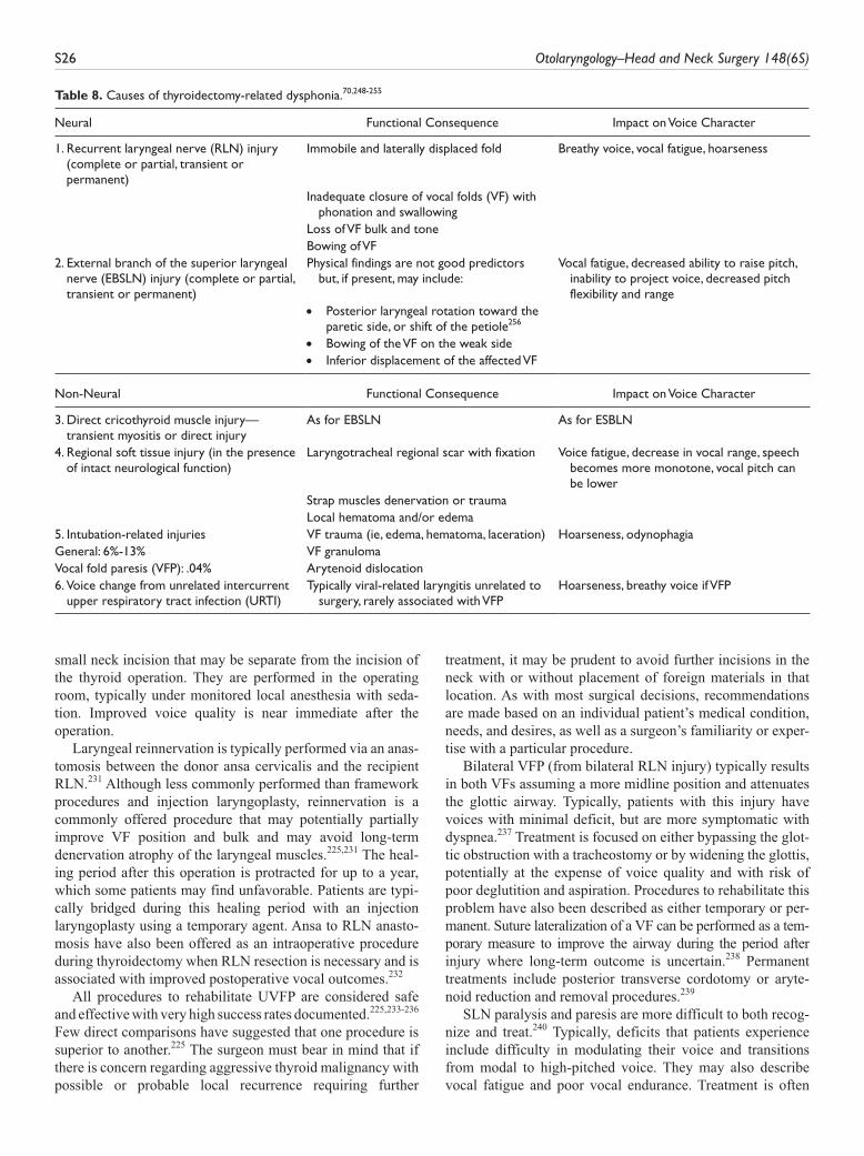
S26 Otolaryngology–Head and Neck Surgery 148(6S)
small neck incision that may be separate from the incision of the thyroid operation. They are performed in the operating room, typically under monitored local anesthesia with seda-tion. Improved voice quality is near immediate after the operation.
Laryngeal reinnervation is typically performed via an anas-tomosis between the donor ansa cervicalis and the recipient RLN.231 Although less commonly performed than framework procedures and injection laryngoplasty, reinnervation is a commonly offered procedure that may potentially partially improve VF position and bulk and may avoid long-term denervation atrophy of the laryngeal muscles.225,231 The heal-ing period after this operation is protracted for up to a year, which some patients may find unfavorable. Patients are typi-cally bridged during this healing period with an injection laryngoplasty using a temporary agent. Ansa to RLN anasto-mosis have also been offered as an intraoperative procedure during thyroidectomy when RLN resection is necessary and is associated with improved postoperative vocal outcomes.232
All procedures to rehabilitate UVFP are considered safe and effective with very high success rates documented.225,233-236 Few direct comparisons have suggested that one procedure is superior to another.225 The surgeon must bear in mind that if there is concern regarding aggressive thyroid malignancy with possible or probable local recurrence requiring further
treatment, it may be prudent to avoid further incisions in the neck with or without placement of foreign materials in that location. As with most surgical decisions, recommendations are made based on an individual patient’s medical condition, needs, and desires, as well as a surgeon’s familiarity or exper-tise with a particular procedure.
Bilateral VFP (from bilateral RLN injury) typically results in both VFs assuming a more midline position and attenuates the glottic airway. Typically, patients with this injury have voices with minimal deficit, but are more symptomatic with dyspnea.237 Treatment is focused on either bypassing the glot-tic obstruction with a tracheostomy or by widening the glottis, potentially at the expense of voice quality and with risk of poor deglutition and aspiration. Procedures to rehabilitate this problem have also been described as either temporary or per-manent. Suture lateralization of a VF can be performed as a tem-porary measure to improve the airway during the period after injury where long-term outcome is uncertain.238 Permanent treatments include posterior transverse cordotomy or aryte-noid reduction and removal procedures.239
SLN paralysis and paresis are more difficult to both recog-nize and treat.240 Typically, deficits that patients experience include difficulty in modulating their voice and transitions from modal to high-pitched voice. They may also describe vocal fatigue and poor vocal endurance. Treatment is often
Table 8. Causes of thyroidectomy-related dysphonia.70,248-255
Neural Functional Consequence Impact on Voice Character
1. Recurrent laryngeal nerve (RLN) injury (complete or partial, transient or permanent)
Immobile and laterally displaced fold Breathy voice, vocal fatigue, hoarseness
Inadequate closure of vocal folds (VF) with phonation and swallowing
Loss of VF bulk and tone Bowing of VF 2. External branch of the superior laryngeal
nerve (EBSLN) injury (complete or partial, transient or permanent)
Physical findings are not good predictors but, if present, may include:
Vocal fatigue, decreased ability to raise pitch, inability to project voice, decreased pitch flexibility and range
• Posterior laryngeal rotation toward the paretic side, or shift of the petiole256
• Bowing of the VF on the weak side • Inferior displacement of the affected VF
Non-Neural Functional Consequence Impact on Voice Character
3. Direct cricothyroid muscle injury— transient myositis or direct injury
As for EBSLN As for ESBLN
4. Regional soft tissue injury (in the presence of intact neurological function)
Laryngotracheal regional scar with fixation Voice fatigue, decrease in vocal range, speech becomes more monotone, vocal pitch can be lower
Strap muscles denervation or trauma Local hematoma and/or edema 5. Intubation-related injuries VF trauma (ie, edema, hematoma, laceration) Hoarseness, odynophagiaGeneral: 6%-13% VF granuloma Vocal fold paresis (VFP): .04% Arytenoid dislocation 6. Voice change from unrelated intercurrent
upper respiratory tract infection (URTI)Typically viral-related laryngitis unrelated to
surgery, rarely associated with VFPHoarseness, breathy voice if VFP
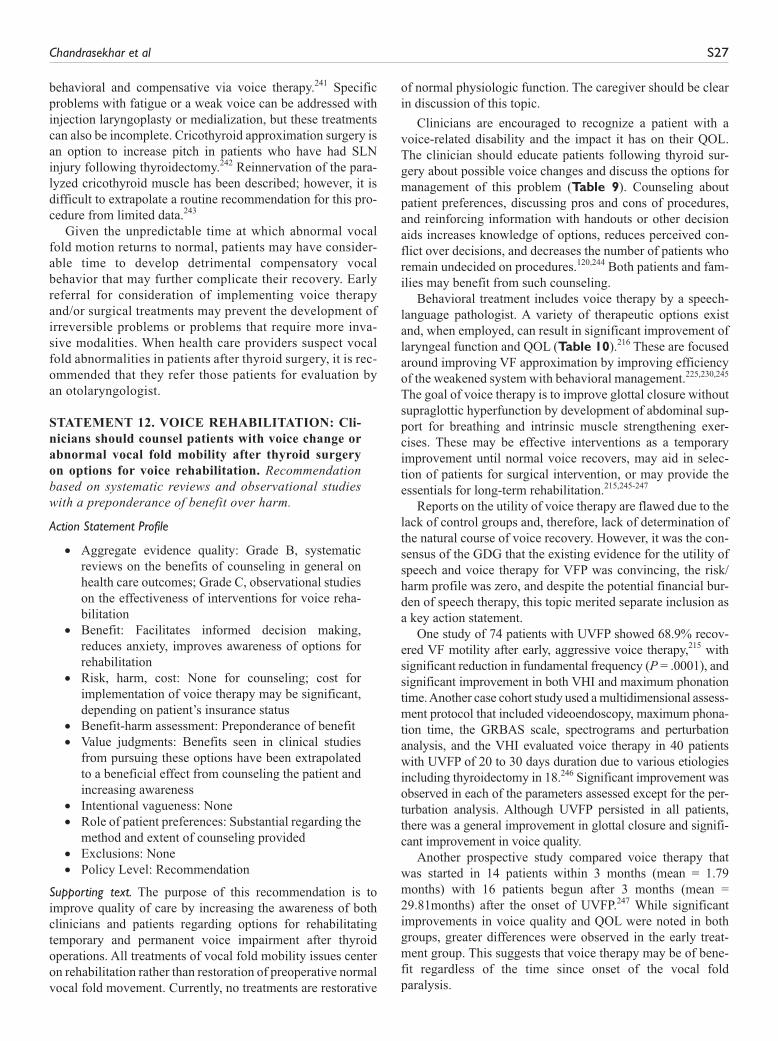
Chandrasekhar et al S27
behavioral and compensative via voice therapy.241 Specific problems with fatigue or a weak voice can be addressed with injection laryngoplasty or medialization, but these treatments can also be incomplete. Cricothyroid approximation surgery is an option to increase pitch in patients who have had SLN injury following thyroidectomy.242 Reinnervation of the para-lyzed cricothyroid muscle has been described; however, it is difficult to extrapolate a routine recommendation for this pro-cedure from limited data.243
Given the unpredictable time at which abnormal vocal fold motion returns to normal, patients may have consider-able time to develop detrimental compensatory vocal behavior that may further complicate their recovery. Early referral for consideration of implementing voice therapy and/or surgical treatments may prevent the development of irreversible problems or problems that require more inva-sive modalities. When health care providers suspect vocal fold abnormalities in patients after thyroid surgery, it is rec-ommended that they refer those patients for evaluation by an otolaryngologist.
STATEMENT 12. VOICE REHABILITATION: Cli-nicians should counsel patients with voice change or abnormal vocal fold mobility after thyroid surgery on options for voice rehabilitation. Recommendation based on systematic reviews and observational studies with a preponderance of benefit over harm.
Action Statement Profile
• Aggregate evidence quality: Grade B, systematic reviews on the benefits of counseling in general on health care outcomes; Grade C, observational studies on the effectiveness of interventions for voice reha-bilitation
• Benefit: Facilitates informed decision making, reduces anxiety, improves awareness of options for rehabilitation
• Risk, harm, cost: None for counseling; cost for implementation of voice therapy may be significant, depending on patient’s insurance status
• Benefit-harm assessment: Preponderance of benefit • Value judgments: Benefits seen in clinical studies
from pursuing these options have been extrapolated to a beneficial effect from counseling the patient and increasing awareness
• Intentional vagueness: None • Role of patient preferences: Substantial regarding the
method and extent of counseling provided • Exclusions: None • Policy Level: Recommendation
Supporting text. The purpose of this recommendation is to improve quality of care by increasing the awareness of both clinicians and patients regarding options for rehabilitating temporary and permanent voice impairment after thyroid operations. All treatments of vocal fold mobility issues center on rehabilitation rather than restoration of preoperative normal vocal fold movement. Currently, no treatments are restorative
of normal physiologic function. The caregiver should be clear in discussion of this topic.
Clinicians are encouraged to recognize a patient with a voice-related disability and the impact it has on their QOL. The clinician should educate patients following thyroid sur-gery about possible voice changes and discuss the options for management of this problem (Table 9). Counseling about patient preferences, discussing pros and cons of procedures, and reinforcing information with handouts or other decision aids increases knowledge of options, reduces perceived con-flict over decisions, and decreases the number of patients who remain undecided on procedures.120,244 Both patients and fam-ilies may benefit from such counseling.
Behavioral treatment includes voice therapy by a speech-language pathologist. A variety of therapeutic options exist and, when employed, can result in significant improvement of laryngeal function and QOL (Table 10).216 These are focused around improving VF approximation by improving efficiency of the weakened system with behavioral management.225,230,245 The goal of voice therapy is to improve glottal closure without supraglottic hyperfunction by development of abdominal sup-port for breathing and intrinsic muscle strengthening exer-cises. These may be effective interventions as a temporary improvement until normal voice recovers, may aid in selec-tion of patients for surgical intervention, or may provide the essentials for long-term rehabilitation.215,245-247
Reports on the utility of voice therapy are flawed due to the lack of control groups and, therefore, lack of determination of the natural course of voice recovery. However, it was the con-sensus of the GDG that the existing evidence for the utility of speech and voice therapy for VFP was convincing, the risk/harm profile was zero, and despite the potential financial bur-den of speech therapy, this topic merited separate inclusion as a key action statement.
One study of 74 patients with UVFP showed 68.9% recov-ered VF motility after early, aggressive voice therapy,215 with significant reduction in fundamental frequency (P = .0001), and significant improvement in both VHI and maximum phonation time. Another case cohort study used a multidimensional assess-ment protocol that included videoendoscopy, maximum phona-tion time, the GRBAS scale, spectrograms and perturbation analysis, and the VHI evaluated voice therapy in 40 patients with UVFP of 20 to 30 days duration due to various etiologies including thyroidectomy in 18.246 Significant improvement was observed in each of the parameters assessed except for the per-turbation analysis. Although UVFP persisted in all patients, there was a general improvement in glottal closure and signifi-cant improvement in voice quality.
Another prospective study compared voice therapy that was started in 14 patients within 3 months (mean = 1.79 months) with 16 patients begun after 3 months (mean = 29.81months) after the onset of UVFP.247 While significant improvements in voice quality and QOL were noted in both groups, greater differences were observed in the early treat-ment group. This suggests that voice therapy may be of bene-fit regardless of the time since onset of the vocal fold paralysis.
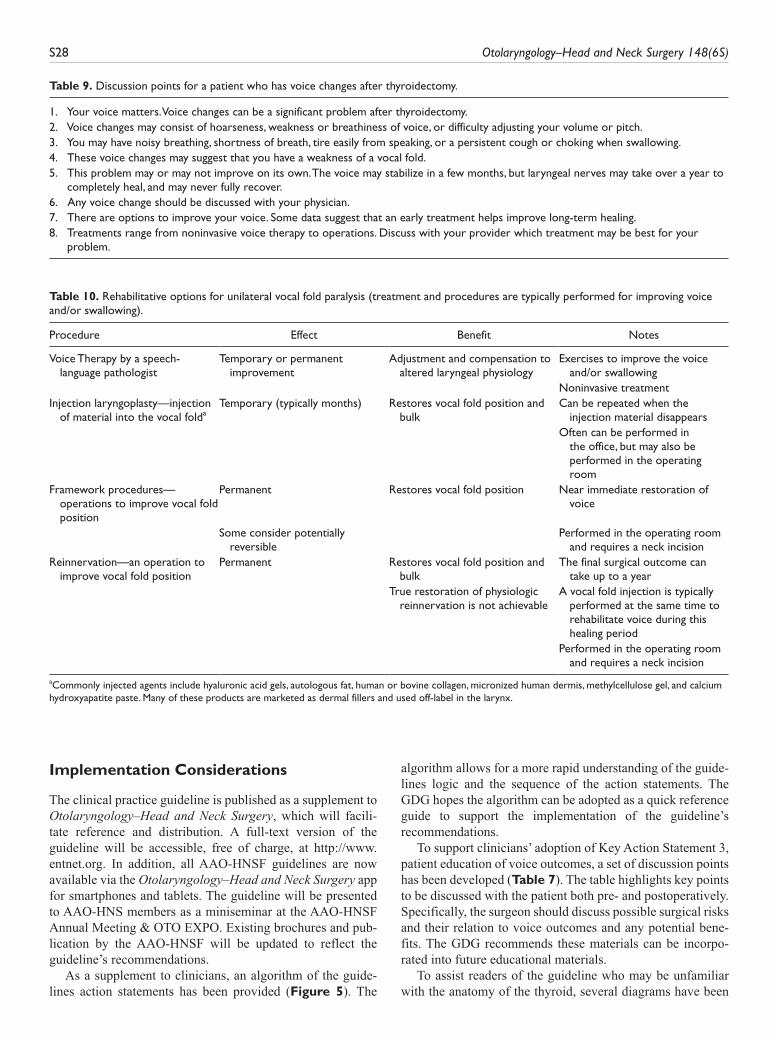
S28 Otolaryngology–Head and Neck Surgery 148(6S)
Implementation Considerations
The clinical practice guideline is published as a supplement to Otolaryngology–Head and Neck Surgery, which will facili-tate reference and distribution. A full-text version of the guideline will be accessible, free of charge, at http://www.entnet.org. In addition, all AAO-HNSF guidelines are now available via the Otolaryngology–Head and Neck Surgery app for smartphones and tablets. The guideline will be presented to AAO-HNS members as a miniseminar at the AAO-HNSF Annual Meeting & OTO EXPO. Existing brochures and pub-lication by the AAO-HNSF will be updated to reflect the guideline’s recommendations.
As a supplement to clinicians, an algorithm of the guide-lines action statements has been provided (Figure 5). The
algorithm allows for a more rapid understanding of the guide-lines logic and the sequence of the action statements. The GDG hopes the algorithm can be adopted as a quick reference guide to support the implementation of the guideline’s recommendations.
To support clinicians’ adoption of Key Action Statement 3, patient education of voice outcomes, a set of discussion points has been developed (Table 7). The table highlights key points to be discussed with the patient both pre- and postoperatively. Specifically, the surgeon should discuss possible surgical risks and their relation to voice outcomes and any potential bene-fits. The GDG recommends these materials can be incorpo-rated into future educational materials.
To assist readers of the guideline who may be unfamiliar with the anatomy of the thyroid, several diagrams have been
Table 9. Discussion points for a patient who has voice changes after thyroidectomy.
1. Your voice matters. Voice changes can be a significant problem after thyroidectomy.2. Voice changes may consist of hoarseness, weakness or breathiness of voice, or difficulty adjusting your volume or pitch.3. You may have noisy breathing, shortness of breath, tire easily from speaking, or a persistent cough or choking when swallowing.4. These voice changes may suggest that you have a weakness of a vocal fold.5. This problem may or may not improve on its own. The voice may stabilize in a few months, but laryngeal nerves may take over a year to
completely heal, and may never fully recover.6. Any voice change should be discussed with your physician.7. There are options to improve your voice. Some data suggest that an early treatment helps improve long-term healing.8. Treatments range from noninvasive voice therapy to operations. Discuss with your provider which treatment may be best for your
problem.
Table 10. Rehabilitative options for unilateral vocal fold paralysis (treatment and procedures are typically performed for improving voice and/or swallowing).
Procedure Effect Benefit Notes
Voice Therapy by a speech-language pathologist
Temporary or permanent improvement
Adjustment and compensation to altered laryngeal physiology
Exercises to improve the voice and/or swallowing
Noninvasive treatmentInjection laryngoplasty—injection
of material into the vocal foldaTemporary (typically months) Restores vocal fold position and
bulkCan be repeated when the
injection material disappears Often can be performed in
the office, but may also be performed in the operating room
Framework procedures—operations to improve vocal fold position
Permanent Restores vocal fold position Near immediate restoration of voice
Some consider potentially reversible
Performed in the operating room and requires a neck incision
Reinnervation—an operation to improve vocal fold position
Permanent Restores vocal fold position and bulk
The final surgical outcome can take up to a year
True restoration of physiologic reinnervation is not achievable
A vocal fold injection is typically performed at the same time to rehabilitate voice during this healing period
Performed in the operating room and requires a neck incision
aCommonly injected agents include hyaluronic acid gels, autologous fat, human or bovine collagen, micronized human dermis, methylcellulose gel, and calcium hydroxyapatite paste. Many of these products are marketed as dermal fillers and used off-label in the larynx.
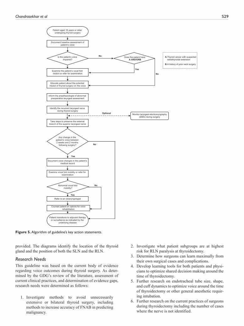
Chandrasekhar et al S29
provided. The diagrams identify the location of the thyroid gland and the position of both the SLN and the RLN.
Research NeedsThis guideline was based on the current body of evidence regarding voice outcomes during thyroid surgery. As deter-mined by the GDG’s review of the literature, assessment of current clinical practices, and determination of evidence gaps, research needs were determined as follows:
1. Investigate methods to avoid unnecessarily extensive or bilateral thyroid surgery, including methods to increase accuracy of FNAB in predicting malignancy.
2. Investigate what patient subgroups are at highest risk for RLN paralysis at thyroidectomy.
3. Determine how surgeons can learn maximally from their own surgical cases and complications.
4. Develop learning tools for both patients and physi-cians to optimize shared decision making around the time of thyroidectomy.
5. Further research on endotracheal tube size, shape, and cuff dynamics to optimize voice around the time of thyroidectomy or other general anesthetic requir-ing intubation.
6. Further research on the current practices of surgeons during thyroidectomy including the number of cases where the nerve is not identified.
Yes
No
Document baseline assessment ofpatient’s voice
Educate patient about the potential impact of thyroid surgery on the voice
Inform the anesthesiologist of abnormal preoperative laryngeal assessment
Identify the recurrent laryngeal nerve during thyroid surgery
Take steps to preserve the external branch of the superior laryngeal nerve
Patient aged 18 years or older undergoing thyroid surgery
Does the patient have:A AND/ORB
Patient transitions to adjuvant therapyor surveillance as indicated by the
underlying disease
No
Is the patient’s voice impaired?
Examine the patient’s vocal fold motion or refer for examination
A.Thyroid cancer with suspectedextrathyroidal extension
B.A history of prior neck surgery
Any change in the patient’s voice between 2 weeks and 2 months
following surgery?
Yes
No
Yes
No
Document voice changes in the patient’s medical record
Examine vocal fold mobility or refer for examination
Abnormal vocal fold mobility?
Refer to an otolaryngologist
Counsel patient on options for voicerehabilitation
Optional Monitor laryngeal electromyography(EMG) during surgery
Figure 5. Algorithm of guideline’s key action statements.

S30 Otolaryngology–Head and Neck Surgery 148(6S)
7. Further research on the utility of corticosteroids perioperatively, particularly their impact on voice outcomes.
8. Evaluate patterns and utility of otolaryngology refer-ral and treatment of vocal fold paralysis.
DisclaimerThis clinical practice guideline is provided for information and educa-tional purposes only. It is not intended as a sole source of guidance in managing voice outcomes after thyroid surgery. Rather, it is designed to assist clinicians by providing an evidence-based framework for decision-making strategies. The guideline is not intended to replace clinical judgment or establish a protocol for all individuals with this condition and may not provide the only appropriate approach to diag-nosing and managing this program of care. As medical knowledge expands and technology advances, clinical indicators and guidelines are promoted as conditional and provisional proposals of what is recom-mended under specific conditions, but they are not absolute. Guidelines are not mandates and do not and should not purport to be a legal standard of care. The responsible physician, in lights of all the circumstances presented by the individual patient, must determine the appropriate treat-ment. Adherence to these guidelines will not ensure successful patient outcomes in every situation. The American Academy of Otolaryngology—Head and Neck Surgery (AAO-HNS), Inc. emphasizes that these clinical guidelines should not be deemed to include all proper treatment decisions or methods of care, or to exclude other treatment decisions or methods of care reasonably directed to obtaining the same results.
© 2013 American Academy of Otolaryngology—Head and Neck Surgery Foundation. All rights reserved
Author ContributionsSujana S. Chandrasekhar, writer, chair; Gregory W. Randolph, writer, assistant chair; Michael D. Seidman, writer, assistant chair; Richard M. Rosenfeld, writer, consultant; Peter Angelos, writer; Julie Barkmeier-Kraemer, writer; Michael S. Benninger, writer; Joel H. Blumin, writer; Gregory Dennis, writer; John Hanks, writer; Megan R. Haymart, writer; Richard T. Kloos, writer; Brenda Seals, writer; Jerry M. Schreibstein, writer; Mack A. Thomas, writer; Carolyn Waddington, writer; Barbara Warren, writer; Peter J. Robertson, writer and AAO-HNSF staff liaison.
DisclosuresCompeting interests: Sujana S. Chandrasekhar, Scientific Development & Research Inc.—shareholder, board member, CMO, Med El Corporation—grant recipient; Michael D. Seidman, Body Language vitamins (founder), Visalus Sciences (director of product development), NIH (grant recipient), Save Your Hearing Now (author, no royalties); Peter Angelos, Olympus—consultant; Gregory Dennis, UCB Inc—employer, Human Genome Sciences—previous employer; Richard T. Kloos, Bayer, Eisai, Onyx, Exelixis—research investigator, Veracyte—stockholder and employee, Thompson Hine LLP—expert witness, Genzyme Corporation—consultant; Jerry M. Schreibstein, Sleep Management Solutions—medical director, Pioneer Valley Surgicenter—owner.
Sponsorships: American Academy of Otolaryngology—Head and Neck Surgery Foundation.
Funding source: American Academy of Otolaryngology—Head and Neck Surgery Foundation.
References
1. Agency for Healthcare Research and Quality. Statistical brief 86. Healthcare Cost and Utilization Project (HCUP). http://www .hcup-us.ahrq.gov/reports/statbriefs/sb86.jsp. Published 2010. Accessed June 4, 2012.
2. American Cancer Society. Cancer facts & figures 2011. http://www.cancer.org/acs/groups/content/@epidemiologysur veilance/documents/document/acspc-029771.pdf. Published 2011. Accessed March 28, 2012.
3. American Cancer Society. Cancer facts & figures 2012. http://www.cancer.org/acs/groups/content/@epidemiologysur-veilance/documents/document/acspc-031941.pdf. Published 2012. Accessed March 28, 2012.
4. Gharib H, Papini E, Valcavi R, et al. American Association of Clinical Endocrinologists and Associazione Medici Endo-crinologi medical guidelines for clinical practice for the diag-nosis and management of thyroid nodules. Endocr Pract. 2006;12(1):63-102.
5. Davies L, Welch HG. Increasing incidence of thyroid cancer in the United States, 1973-2002. JAMA. 2006;295(18):2164-2167.
6. Brown RL, de Souza JA, Cohen EE. Thyroid cancer: burden of illness and management of disease. J Cancer. 2011;2:193-199.
7. Husson O, Haak HR, Oranje WA, Mols F, Reemst PH, van de Poll-Franse LV. Health-related quality of life among thyroid cancer survivors: a systematic review. Clin Endocrinol (Oxf). 2011;75(4):544-554.
8. Stojadinovic A, Shaha AR, Orlikoff RF, et al. Prospective func-tional voice assessment in patients undergoing thyroid surgery. Ann Surg. 2002;236(6):823-832.
9. Soylu L, Ozbas S, Uslu HY, Kocak S. The evaluation of the causes of subjective voice disturbances after thyroid surgery. Am J Surg. 2007;194(3):317-322.
10. Wilson JA, Deary IJ, Millar A, Mackenzie K. The qual-ity of life impact of dysphonia. Clin Otolaryngol Allied Sci. 2002;27(3):179-182.
11. Jones SM, Carding PN, Drinnan MJ. Exploring the relationship between severity of dysphonia and voice-related quality of life. Clin Otolaryngol. 2006;31(5):411-417.
12. Jeannon JP, Orabi AA, Bruch GA, Abdalsalam HA, Simo R. Diagnosis of recurrent laryngeal nerve palsy after thyroidec-tomy: a systematic review. Int J Clin Pract. 2009;63(4):624-629.
13. Mendels EJ, Brunings JW, Hamaekers AE, Stokroos RJ, Kremer B, Baijens LW. Adverse laryngeal effects following short-term general anesthesia: a systematic review. Arch Otolaryngol Head Neck Surg. 2012;138(3):257-264.
14. Roy N, Smith ME, Dromey C, Redd J, Neff S, Grennan D. Explor-ing the phonatory effects of external superior laryngeal nerve paralysis: an in vivo model. Laryngoscope. 2009;119(4):816-826.
15. Sinagra DL, Montesinos MR, Tacchi VA, et al. Voice changes after thyroidectomy without recurrent laryngeal nerve injury. J Am Coll Surg. 2004;199(4):556-560.
16. Hong KH, Kim YK. Phonatory characteristics of patients under-going thyroidectomy without laryngeal nerve injury. Otolaryn-gol Head Neck Surg. 1997;117(4):399-404.
Chandrasekhar et al S31
17. Ho TW, Shaheen AA, Dixon E, Harvey A. Utilization of thy-roidectomy for benign disease in the United States: a 15-year population-based study. Am J Surg. 2011;201(5):570-574.
18. Graney D, Flint P. Anatomy of larynx, hypopharynx, trachea, bronchus, esopahgus. In: Cummings C, Fredrickson J, Harker L, Krause C, Richardson M, Schuller D, eds. Otolaryngology—Head and Neck Surgery. 3rd ed.St. Louis, MO: Mosby; 1998.
19. Sosa JA, Mehta PJ, Wang TS, Boudourakis L, Roman SA. A population-based study of outcomes from thyroidectomy in aging Americans: at what cost?J Am Coll Surg. 2008;206(3):1097-1105.
20. Institute of Medicine (U.S.). Committee on Standards for Developing Trustworthy Clinical Practice Guidelines. Clinical Practice Guidelines We Can Trust. Washington, DC: National Academies Press; 2011.
21. Mazzaferri EL. Management of a solitary thyroid nodule. N Engl J Med. 1993;328(8):553-559.
22. Shindo M, Chheda NN. Incidence of vocal cord paralysis with and without recurrent laryngeal nerve monitoring during thyroidectomy. Arch Otolaryngol Head Neck Surg. 2007;133(5):481-485.
23. Hermann M, Alk G, Roka R, Glaser K, Freissmuth M. Laryngeal recurrent nerve injury in surgery for benign thyroid diseases: effect of nerve dissection and impact of individual surgeon in more than 27,000 nerves at risk. Ann Surg. 2002;235(2):261-268.
24. Lo CY, Kwok KF, Yuen PW. A prospective evaluation of recur-rent laryngeal nerve paralysis during thyroidectomy. Arch Surg. 2000;135:204-207.
25. Jatzko GR, Lisborg PH, Muller MG, Wette VM. Recurrent nerve palsy after thyroid operations; principal nerve identification and literature review. Surgery. 1994;115:139-144.
26. Brunner E, Friedrich G, Kiesler K, Chibidziura-Priesching J, Gugatschka M. Subjective breathing impairment in unilateral vocal fold paralysis. Folia Phoniatr Logop. 2011;63(3):142-146.
27. Ollivere B, Duce K, Rowlands G, Harrison P, O’Reilly BJ. Swal-lowing dysfunction in patients with unilateral vocal fold paraly-sis: aetiology and outcomes. J Laryngol Otol. 2006;120(1):38-41.
28. Leder SB, Ross DA. Incidence of vocal fold immobility in patients with dysphagia. Dysphagia. 2005;20(2):163-167.
29. Titze IR, Lemke J, Montequin D. Populations in the U.S. work-force who rely on voice as a primary tool of trade: a preliminary report. J Voice. 1997;11(3):254-259.
30. Randolph GW. Surgery of the Thyroid and Parathyroid Glands. Philadelphia, PA: Saunders-Elsevier Science; 2003.
31. Wu BL, Sanders I, Mu L, Biller HF. The human communicating nerve. An extension of the external superior laryngeal nerve that innervates the vocal cord. Arch Otolaryngol Head Neck Surg. 1994;120(12):1321-1328.
32. Serpell JW, Yeung MJ, Grodski S. The motor fibers of the recur-rent laryngeal nerve are located in the anterior extralaryngeal branch. Ann Surg. 2009;249(4):648-652.
33. Beneragama T, Serpell JW. Extralaryngeal bifurcation of the recurrent laryngeal nerve: a common variation. ANZ J Surg. 2006;76(10):928-931.
34. Cernea CR, Hojaij FC, De Carlucci D Jr, et al. Recurrent laryn-geal nerve: a plexus rather than a nerve?Arch Otolaryngol Head Neck Surg. 2009;135(11):1098-1102.
35. Sancho JJ, Pascual-Damieta M, Pereira JA, Carrera MJ, Fontane J, Sitges-Serra A. Risk factors for transient vocal cord palsy after thyroidectomy. Br J Surg. 2008;95(8):961-967.
36. Defechereux T, Albert V, Alexandre J, Bonnet P, Hamoir E, Meurisse M. The inferior non recurrent laryngeal nerve: a major surgical risk during thyroidectomy. Acta Chir Belg. 2000;100(2):62-67.
37. de Pedro Netto I, Fae A, Vartanian JG, et al. Voice and vocal self-assessment after thyroidectomy. Head Neck. 2006;28(12):1106-1114.
38. Bergenfelz A, Jansson S, Kristoffersson A, et al. Complications to thyroid surgery: results as reported in a database from a multi-center audit comprising 3,660 patients. Langenbecks Arch Surg. 2008;393(5):667-673.
39. Scott-Coombes D. The British Association of Endocrine and Thyroid Surgeons—Third National Audit Report. Henley-on-Thames, UK: Dendrite Clinical Systems Ltd; 2009.
40. Lipsett PA, Harris I, Downing S. Resident self-other assessor agreement: influence of assessor, competency, and performance level. Arch Surg. 2011;146(8):901-906.
41. Dekutoski MB, Norvell DC, Dettori JR, Fehlings MG, Chapman JR. Surgeon perceptions and reported complications in spine sur-gery. Spine. 2010;35(suppl 9):S9-S21.
42. Sittel C, Stennert E, Thumfart WF, Dapunt U, Eckel HE. Prog-nostic value of laryngeal electromyography in vocal fold paraly-sis. Arch Otolaryngol Head Neck Surg. 2001;127(2):155-160.
43. Hassan-Smith ZK, Gopinath P, Mihaimeed F. A UK-wide survey of life-threatening thyroidectomy complications. J Thyroid Res. 2011;2011:329620.
44. Rosato L, Carlevato MT, De Toma G, Avenia N. Recurrent laryn-geal nerve damage and phonetic modifications after total thyroid-ectomy: surgical malpractice only or predictable sequence?World J Surg. 2005;29(6):780-784.
45. Musholt TJ, Musholt PB, Garm J, Napiontek U, Keilmann A. Changes of the speaking and singing voice after thyroid or para-thyroid surgery. Surgery. 2006;140(6):978-988.
46. Page C, Zaatar R, Biet A, Strunski V. Subjective voice assess-ment after thyroid surgery: a prospective study of 395 patients. Indian J Med Sci. 2007;61(8):448-454.
47. Munch S, deKryger L. A piece of my mind. Moral wounds: com-plicated complications. JAMA. 2001;285(9):1131-1132.
48. Fang TJ, Li HY, Tsai FC, Chen IH. The role of glottal gap in predicting aspiration in patients with unilateral vocal paralysis. Clin Otolaryngol Allied Sci. 2004;29(6):709-712.
49. Lundy DS, Smith C, Colangelo L, et al. Aspiration: cause and impli-cations. Otolaryngol Head Neck Surg. 1999;120(4):474-478.
50. Postma GN, McGuirt WF Sr, Butler SG, Rees CJ, Crandall HL, Tansavatdi K. Laryngopharyngeal abnormalities in hospitalized patients with dysphagia. Laryngoscope. 2007;117(10):1720-1722.
51. Altman KW, Yu GP, Schaefer SD. Consequence of dysphagia in the hospitalized patient: impact on prognosis and hospital resources. Arch Otolaryngol Head Neck Surg. 2010;136(8):784-789.
52. Dispenza F, Dispenza C, Marchese D, Kulamarva G, Saraniti C. Treatment of bilateral vocal cord paralysis following per-manent recurrent laryngeal nerve injury. Am J Otolaryngol. 2012;33(3):285-288.
53. Gilony D, Gilboa D, Blumstein T, et al. Effects of tracheostomy on well-being and body-image perceptions. Otolaryngol Head Neck Surg. 2005;133(3):366-371.
S32 Otolaryngology–Head and Neck Surgery 148(6S)
54. Cohen SM, Kim J, Roy N, Asche C, Courey M. Direct health care costs of laryngeal diseases and disorders. Laryngoscope. 2012;122:1582-1588.
55. Terris DJ, Arnstein DP, Nguyen HH. Contemporary evaluation of unilateral vocal cord paralysis. Otolaryngol Head Neck Surg. 1992;107(1):84-90.
56. Kern KA. Medicolegal analysis of errors in diagnosis and treat-ment of surgical endocrine disease. Surgery. 1993;114(6):1167-1173.
57. Kern KA. An overview of 711 general surgery liability cases. The anatomy of surgical malpractice claims. Bull Am Coll Surg. 1995;80(8):34-49.
58. Singer MC, Iverson KC, Terris DJ. Thyroidectomy-related malprac-tice claims. Otolaryngol Head Neck Surg. 2012;146(3):358-361.
59. Rosenfeld RM, Shiffman RN, Robertson P. Clinical practice guideline development manual, third edition: a quality-driven approach for translating evidence into action. Otolaryngol Head Neck Surg. 2013;148(suppl 1):S1-55.
60. Shiffman RN, Shekelle P, Overhage JM, Slutsky J, Grimshaw J, Deshpande AM. Standardized reporting of clinical practice guidelines: a proposal from the Conference on Guideline Stan-dardization. Ann Intern Med. 2003;139(6):493-498.
61. Shiffman RN, Dixon J, Brandt C, et al. The GuideLine Imple-mentability Appraisal (GLIA): development of an instrument to identify obstacles to guideline implementation. BMC Med Inform Decis Mak. 2005;5:23.
62. Classifying recommendations for clinical practice guidelines. Pediatrics. 2004;114(3):874-877.
63. Oxford Centre for Evidence-Based Medicine. OCEBM Levels of Evidence Working Group. The Oxford 2011 levels of evidence. http://www.cebm.net/index.aspx?o=5653. Published 2011. Accessed April 10, 2013.
64. Eddy D. A Manual for Assessing Health Practices and Design-ing Practice Policies: The Explicit Approach. Philadelphia, PA: American College of Physicians; 1992.
65. Choudhry NK, Stelfox HT, Detsky AS. Relationships between authors of clinical practice guidelines and the pharmaceutical industry. JAMA. 2002;287(5):612-617.
66. Detsky AS. Sources of bias for authors of clinical practice guide-lines. CMAJ. 2006;175(9):1033, 1035.
67. Kon AA. The shared decision-making continuum. JAMA. 2010;304(8):903-904.
68. De Bodt MS, Wuyts FL, Van de Heyning PH, Croux C. Test-retest study of the GRBAS scale: influence of experience and professional background on perceptual rating of voice quality. J Voice. 1997;11(1):74-80.
69. Eadie TL, Kapsner M, Rosenzweig J, Waugh P, Hillel A, Merati A. The role of experience on judgments of dysphonia. J Voice. 2010;24(5):564-573.
70. McIvor NP, Flint DJ, Gillibrand J, Morton RP. Thyroid surgery and voice-related outcomes. Aust N Z J Surg. 2000;70(3):179-183.
71. Meek P, Carding PN, Howard DH, Lennard TW. Voice change fol-lowing thyroid and parathyroid surgery. J Voice. 2008;22(6):765-772.
72. Roh JL, Yoon YH, Park CI. Recurrent laryngeal nerve paraly-sis in patients with papillary thyroid carcinomas: evaluation
and management of resulting vocal dysfunction. Am J Surg. 2009;197(4):459-465.
73. Melish JS. Thyroid disease. In: Walker HK, Hall WD, Hurst JW, eds. Clinical Methods: The History, Physicial and Laboratory Examinations. 3rd ed.Boston, MA: Butterworths; 1990.
74. Stojadinovic A, Henry LR, Howard RS, et al. Prospective trial of voice outcomes after thyroidectomy: evaluation of patient-reported and clinician-determined voice assessments in identify-ing postthyroidectomy dysphonia. Surgery. 2008;143(6):732-742.
75. Jacobson BH, Johnson A, Grywalski C, et al. The Voice Handi-cap Index (VHI): development and validation. Am J Speech Lang Pathol. 1997;6(3):66-70.
76. Franic DM, Bramlett RE, Bothe AC. Psychometric evaluation of disease specific quality of life instruments in voice disorders. J Voice. 2005;19(2):300-315.
77. Carrau RL, Khidr A, Gold KF, et al. Validation of a quality-of-life instrument for laryngopharyngeal reflux. Arch Otolaryngol Head Neck Surg. 2005;131(4):315-320.
78. Bogaardt HC, Hakkesteegt MM, Grolman W, Lindeboom R. Validation of the voice handicap index using Rasch analysis. J Voice. 2007;21(3):337-344.
79. Singh P, O’Connell D, Langille M, Dziegielewski P, Allegretto M, Harris J. LigaSure versus conventional hemostasis in thyroid surgery: prospective randomized controlled trial. J Otolaryngol Head Neck Surg. 2010;39(4):378-384.
80. Stevens K, Stojadinovic A, Helou LB, et al. The impact of recur-rent laryngeal neuromonitoring on multi-dimensional voice out-comes following thyroid surgery. J Surg Oncol. 2012;105(1):4-9.
81. Worni M, Schudel HH, Seifert E, et al. Randomized controlled trial on single dose steroid before thyroidectomy for benign dis-ease to improve postoperative nausea, pain, and vocal function. Ann Surg. 2008;248(6):1060-1066.
82. Carding PN, Wilson JA, MacKenzie K, Deary IJ. Measuring voice outcomes: state of the science review. J Laryngol Otol. 2009;123(8):823-829.
83. da Costa de Ceballos AG, Carvalho FM, de Araujo TM, Far-ias Borges dos Reis EJ. Diagnostic validity of Voice Handicap Index-10 (VHI-10) compared with perceptive-auditory and acoustic speech pathology evaluations of the voice. J Voice. 2010;24(6):715-718.
84. Biddle AK, Watson LR, Hooper CR, Lohr KN, Sutton SF. Crite-ria for determining disability in speech-language disorders. Evid Rep Technol Assess (Summ). 2002;(52):1-4.
85. Arffa RE, Krishna P, Gartner-Schmidt J, Rosen CA. Normative val-ues for the voice handicap index-10. J Voice. 2012;26(4):462-465.
86. Morzaria S, Damrose EJ. A comparison of the VHI, VHI-10, and V-RQOL for measuring the effect of botox therapy in adductor spasmodic dysphonia. J Voice. 2012;26(3):378-380.
87. Hirano M. Clinical Examination of Voice. Vienna: Springer-Ver-lag; 1981.
88. Kempster GB, Gerratt BR, Verdolini Abbott K, Barkmeier-Krae-mer J, Hillman RE. Consensus auditory-perceptual evaluation of voice: development of a standardized clinical protocol. Am J Speech Lang Pathol. 2009;18(2):124-132.
89. Zraick RI, Kempster GB, Connor NP, et al. Establishing valid-ity of the Consensus Auditory-Perceptual Evaluation of Voice (CAPE-V). Am J Speech Lang Pathol. 2011;20(1):14-22.
Chandrasekhar et al S33
90. Svec JG, Granqvist S. Guidelines for selecting microphones for human voice production research. Am J Speech Lang Pathol. 2010;19(4):356-368.
91. Hundahl SA, Cady B, Cunningham MP, et al. Initial results from a prospective cohort study of 5583 cases of thyroid carci-noma treated in the united states during 1996. U.S. and German Thyroid Cancer Study Group. An American College of Sur-geons Commission on Cancer Patient Care Evaluation study. Cancer. 2000;89(1):202-217.
92. Palazzo FF. Pre and post operative laryngoscopy in thyroid and parathyroid surgery. British Association of Endocrine and Thyroid Surgeons Consensus 2010. http://www.baets.org.uk/pages/Vocal_cord_check_consenus_document_2010_final.pdf. Published 2010. Accessed June 4, 2012.
93. Musholt TJ, Clerici T, Dralle H, et al. German Association of Endocrine Surgeons practice guidelines for the surgical treatment of benign thyroid disease. Langenbecks Arch Surg. 2011;396(5):639-649.
94. Randolph GW, Dralle H, Abdullah H, et al. Electrophysiologic recurrent laryngeal nerve monitoring during thyroid and para-thyroid surgery: international standards guideline statement. Laryngoscope. 2011;121(suppl 1):S1-16.
95. Guidelines for the management of thyroid cancer. 2nd ed. ed. London: Royal College of Physicians [in association with] British Thyroid Association; 2007.
96. Tuttle RM, Ball DW, Byrd D, et al. Thyroid carcinoma. Journal of the National Comprehensive Cancer Network : JNCCN. Nov 2010;8(11):1228-1274.
97. Cohen SM, Kim J, Roy N, Asche C, Courey M. Prevalence and causes of dysphonia in a large treatment-seeking population. Laryngoscope. 2012;122(2):343-348.
98. Cohen SM. Self-reported impact of dysphonia in a primary care population: an epidemiological study. Laryngoscope. 2010;120:2022-2032.
99. Rowe-Jones JM, Leighton SEJ, Rosswick RP. Benign thyroid disease and vocal cord palsy. Ann RCS Eng. 1993;75:241-244.
100. Shin JJ, Grillo HC, Mathisen D, et al. The surgical manage-ment of goiter: part I. Preoperative evaluation. Laryngoscope. 2011;121(1):60-67.
101. Farrag TY, Samlan RA, Lin FR, Tufano RP. The utility of eval-uating true vocal fold motion before thyroid surgery. Laryngo-scope. 2006;116(2):235-238.
102. Randolph GW, Kamani D. The importance of preoperative laryngoscopy in patients undergoing thyroidectomy: voice, vocal cord function, and the preoperative detection of invasive thyroid malignancy. Surgery. 2006;139(3):357-362.
103. Falk SA, McCaffrey TV. Management of the recurrent laryn-geal nerve in suspected and proven thyroid cancer. Otolaryngol Head Neck Surg. 1995;113(1):42-48.
104. Benninger MS, Crumley R, Ford CM, et al. Evaluation and treatment of the unilateral paralyzed vocal fold. Otolaryngol Head Neck Surg. 1994;111:497-508.
105. Grundfast KM, Harley E. Vocal cord paralysis. Otolaryngol Clin North Am. 1989;22(3):569-597.
106. Laeeq K, Pandian V, Skinner M, et al. Learning curve for competency in flexible laryngoscopy. Laryngoscope. 2010;120(10):1950-1953.
107. Sataloff RT, Praneetvatakul P, Heuer RJ, et al. Laryngeal elec-tromyography: clinical application. J Voice. 2010;24:228-234.
108. Curley JWA, Timms MS. Incidence of abnormalities in routine “vocal cord checks.”J Laryngol Otol. 1989;103:1057-1058.
109. Curran AJ, Smyth D, Sheehan SJ, Joyce W, Hayes DB, Walsh MA. Recurrent laryngeal nerve dsyfunction following carotid endarterectomy. J R Coll Surg Edinb. 1997;42:168-170.
110. Swibel Rosenthal LH, Benninger MS, Deeb RH. Vocal fold immobility: a longitudinal analysis of etiology over 20 years. Laryngoscope. 2007;117:1864-1870.
111. Sannell NA, Tober RL, Cipro RP, Pedicino JF, Donovan E, Gabriel N. Vocal cord paralysis following carotid endarter-ectomy: the paradox of return of function. Ann Vasc Surg. 1990;4:42-45.
112. Kriskovich MD, Apfelbaum RI, Haller JR. Vocal fold paraly-sis after anterior cervical spine surgery: incidence, mechanism, and prevention of injury. Laryngoscope. 2000;110:1467-1473.
113. Andersen PE, Kinsella J, Loree TR, Shaha AR, Shah JP. Dif-ferentiated carcinoma of the thyroid with extrathyroidal exten-sion. Am J Surg. 1995;170(5):467-470.
114. McCaffrey TV, Bergstralh EJ, Hay ID. Locally inva-sive papillary thyroid carcinoma: 1940-1990. Head Neck. 1994;16(2):165-172.
115. Czaja JM, McCaffrey TV. The surgical management of laryngo-tracheal invasion by well-differentiated papillary thyroid carci-noma. Arch Otolaryngol Head Neck Surg. 1997;123(5):484-490.
116. Price DL, Wong RJ, Randolph GW. Invasive thyroid cancer: management of the trachea and esophagus. Otolaryngol Clin North Am. 2008;41(6):1155-1168, ix-x.
117. Shimamoto K, Satake H, Sawaki A, Ishigaki T, Funahashi H, Imai T. Preoperative staging of thyroid papillary carcinoma with ultrasonography. Eur J Radiol. 1998;29(1):4-10.
118. Park JS, Oh KK, Kim EK, et al. Sonographic detection of thyroid cancer in breast cancer patients. Yonsei Med J. 2007;48(1):63-68.
119. Chiang FY, Lin JC, Lee KW, et al. Thyroid tumors with preop-erative recurrent laryngeal nerve palsy: clinicopathologic fea-tures and treatment outcome. Surgery. 2006;140(3):413-417.
120. O’Connor AM, Bennett CL, Stacey D, et al. Decision aids for people facing health treatment or screening decisions. Cochrane Database Syst Rev. 2009;(3):CD001431.
121. Feldman-Stewart D, Capirci C, Brennenstuhl S, et al. Informa-tion for decision making by patients with early-stage prostate cancer: a comparison across 9 countries. Med Decis Making. 2011;31(5):754-766.
122. Haynes RB, McKibbon KA, Kanani R. Systematic review of ran-domised trials of interventions to assist patients to follow prescrip-tions for medications. Lancet. 1996;348(9024):383-386.
123. Weiss SM, Wengert PA Jr, Martinez EM, Sewall W, Kopp E. Patient satisfaction with decision-making for breast cancer therapy. Ann Surg Oncol. 1996;3(3):285-289.
124. Moyer A, Salovey P. Patient participation in treatment decision making and the psychological consequences of breast cancer surgery. Womens Health. 1998;4(2):103-116.
125. Street RL Jr, Voigt B. Patient participation in deciding breast cancer treatment and subsequent quality of life. Med Decis Making. 1997;17(3):298-306.
S34 Otolaryngology–Head and Neck Surgery 148(6S)
126. Epstein RM, Alper BS, Quill TE. Communicating evidence for participatory decision making. JAMA. 2004;291(19):2359-2366.
127. Begum S, Grunfeld EA, Ho-Asjoe M, Farhadi J. An explora-tion of patient decision-making for autologous breast recon-structive surgery following a mastectomy. Patient Educ Couns. 2011;84(1):105-110.
128. Belkora JK, Volz S, Teng AE, Moore DH, Loth MK, Sepucha KR. Impact of decision aids in a sustained implementation at a breast care center. Patient Educ Couns. 2012;86(2):195-204.
129. Kuehn BM. Patient-centered care model demands better physician-patient communication. JAMA. 2012;307(5):441-442.
130. Abdul-Sater L, Henry M, Majdan A, et al. What are thyroidec-tomy patients really concerned about?Otolaryngol Head Neck Surg. 2011;144(5):685-690.
131. Agarwal A, Mishra AK, Gupta SK, Arshad F, Tripathi M, Singh PK. High incidence of tracheomalacia in longstanding goi-ters: experience from an endemic goiter region. World J Surg. 2007;31(4):832-837.
132. Findlay JM, Sadler GP, Bridge H, Mihai R. Post-thyroidectomy tracheomalacia: minimal risk despite significant tracheal com-pression. Br J Anaesth. 2011;106(6):903-906.
133. Audu P, Artz G, Scheid S, et al. Recurrent laryngeal nerve palsy after anterior cervical spine surgery: the impact of endotracheal tube cuff deflation, reinflation, and pressure adjustment. Anes-thesiology. 2006;105(5):898-901.
134. Helmio P, Blomgren K, Takala A, Pauniaho SL, Takala RS, Ikonen TS. Towards better patient safety: WHO Surgical Safety Checklist in otorhinolaryngology. Clin Otolaryngol. 2011;36(3):242-247.
135. Committee on Quality of Health Care in America, Institute of Medicine. To Err Is Human: Building a Safer Health System. Washington, DC: The National Academies Press; 2000.
136. Lingard L, Espin S, Whyte S, et al. Communication failures in the operating room: an observational classification of recurrent types and effects. Qual Saf Health Care. 2004;13(5):330-334.
137. Christian CK, Gustafson ML, Roth EM, et al. A prospec-tive study of patient safety in the operating room. Surgery. 2006;139(2):159-173.
138. Manser T. Teamwork and patient safety in dynamic domains of healthcare: a review of the literature. Acta Anaesthesiol Scand. 2009;53(2):143-151.
139. World Health Organization. Surgical Safety Checklist. Geneva: WHO; 2008.
140. Lingard L, Espin S, Rubin B, et al. Getting teams to talk: devel-opment and pilot implementation of a checklist to promote interprofessional communication in the OR. Qual Saf Health Care. 2005;14(5):340-346.
141. Awad SS, Fagan SP, Bellows C, et al. Bridging the communica-tion gap in the operating room with medical team training. Am J Surg. 2005;190(5):770-774.
142. American Society of Anesthesiologists. Guidelines for Patient Care in Anesthesiology. Committee of Origin: Surgical Anes-thesia. (Approved by the ASA House of Delegates on October 3, 1967, and last amended on October 19, 2011). http://www .asahq.org/~/media/For%20Members/Standards%20and%20Guidelines/2012/GUIDELINES%20FOR%20PATIENT%20
CARE%20IN%20ANESTHESIOLOGYREV.ashx. Published 2011. Accessed June 4, 2012.
143. Ovassapian A. Fiberoptic Endoscopy and the Difficult Airway. Philadelphia, PA: Lippincott-Raven; 1996.
144. Practice guidelines for management of the difficult airway: an updated report by the American Society of Anesthesiologists Task Force on Management of the Difficult Airway. Anesthesi-ology. 2003;98(5):1269-1277.
145. Voyagis GS, Kyriakos KP. The effect of goiter on endotracheal intubation. Anesth Analg. 1997;84(3):611-612.
146. Johnson S, Goldenberg D. Intraoperative monitoring of the recurrent laryngeal nerve during revision thyroid surgery. Oto-laryngol Clin North Am. 2008;41(6):1147-1154, ix.
147. Gravenstein JS, Kirby RR. General Anesthesia: Induction, Maintenance, and Emergence in Clinical Anesthesia Practice. Philadelphia, PA: W.B. Saunders Co; 1994.
148. Ovassapian A, Krejcie TC, Yelich SJ, Dykes MH. Awake fibre-optic intubation in the patient at high risk of aspiration. Br J Anaesth. 1989;62(1):13-16.
149. Hamdan AL, Sibai A, Rameh C, Kanazeh G. Short-term effects of endotracheal intubation on voice. J Voice. 2007;21(6): 762-768.
150. Flemming DC, Orkin FK, Kirby RR. Hazards of tracheal intubation. In: Gravenstein JS, Kirby RR, eds. Complica-tions in Anesthesiology. Philadelphia, PA: W.B. Saunders Co; 1996.
151. Lahey F. Routine dissection and demonstration of the recur-rent laryngeal nerve in subtotal thyroidectomy. Surg Gynecol Obstet. 1938;66:775-777.
152. Riddell VH. Injury to recurrent laryngeal nerves during thy-roidectomy; a comparison between the results of identification and non-identification in 1022 nerves exposed to risk. Lancet. 1956;271(6944):638-641.
153. Thomusch O, Machens A, Sekulla C, et al. Multivariate analy-sis of risk factors for postoperative complications in benign goiter surgery: prospective multicenter study in Germany. World J Surg. 2000;24(11):1335-1341.
154. Dralle H, Sekulla C, Haerting J, et al. Risk factors of paralysis and functional outcome after recurrent laryngeal nerve moni-toring in thyroid surgery. Surgery. 2004;136(6):1310-1322.
155. Hartl DM, Travagli JP, Leboulleux S, Baudin E, Brasnu DF, Sch-lumberger M. Clinical review: current concepts in the manage-ment of unilateral recurrent laryngeal nerve paralysis after thyroid surgery. J Clin Endocrinol Metab. 2005;90(5):3084-3088.
156. Randolph GW. Surgical anatomy of the recurrent laryngeal nerve. In: Randolph GW, ed. Surgery of the Thyroid and Parathyroid Glands. Philadelphia, PA: Saunders-Elsevier Science; 2003.
157. Pagedar NA, Freeman JL. Identification of the external branch of the superior laryngeal nerve during thyroidectomy. Arch Otolaryngol Head Neck Surg. 2009;135(4):360-362.
158. Kierner AC, Aigner M, Burian M. The external branch of the superior laryngeal nerve: its topographical anatomy as related to surgery of the neck. Arch Otolaryngol Head Neck Surg. 1998;124(3):301-303.
159. Crookes PF, Recabaren JA. Injury to the superior laryngeal branch of the vagus during thyroidectomy: lesson or myth? Ann Surg. 2001;233(4):588-593.
Chandrasekhar et al S35
160. Kark AE, Kissin MW, Auerbach R, Meikle M. Voice changes after thyroidectomy: role of the external laryngeal nerve. Br Med J (Clin Res Ed). 1984;289(6456):1412-1415.
161. Lennquist S, Cahlin C, Smeds S. The superior laryngeal nerve in thyroid surgery. Surgery. 1987;102(6):999-1008.
162. Cernea CR, Ferraz AR, Nishio S, Dutra A J., Hojaij FC, dos Santos LR. Surgical anatomy of the external branch of the superior laryngeal nerve. Head Neck. 1992;14(5):380-383.
163. Barczynski M, Konturek A, Stopa M, Honowska A, Nowak W. Randomized controlled trial of visualization versus neuromon-itoring of the external branch of the superior laryngeal nerve during thyroidectomy. World J Surg. 2012;36(6):1340-1347.
164. Jonas J, Bahr R. Neuromonitoring of the external branch of the superior laryngeal nerve during thyroid surgery. Am J Surg. 2000;179(3):234-236.
165. Cernea CR, Nishio S, Hojaij FC. Identification of the external branch of the superior laryngeal nerve (EBSLN) in large goi-ters. Am J Otolaryngol. 1995;16(5):307-311.
166. Cernea CR, Ferraz AR, Furlani J, et al. Identification of the external branch of the superior laryngeal nerve during thyroid-ectomy. Am J Surg. 1992;164(6):634-639.
167. Dong CC, Macdonald DB, Akagami R, et al. Intraoperative facial motor evoked potential monitoring with transcranial electrical stimulation during skull base surgery. Clinical Neu-rophysiol. 2005;116(3):588-596.
168. Topsakal C, Al-Mefty O, Bulsara KR, Williford VS. Intraoper-ative monitoring of lower cranial nerves in skull base surgery: technical report and review of 123 monitored cases. Neurosurg Rev. 2008;31(1):45-53.
169. Eisele DW. Intraoperative electrophysiologic monitoring of the recurrent laryngeal nerve. Laryngoscope. 1996;106(4):443-449.
170. Horne SK, Gal TJ, Brennan JA. Prevalence and patterns of intraoperative nerve monitoring for thyroidectomy. Otolaryn-gol Head Neck Surg. 2007;136(6):952-956.
171. Sturgeon C, Sturgeon T, Angelos P. Neuromonitoring in thy-roid surgery: attitudes, usage patterns, and predictors of use among endocrine surgeons. World J Surg. 2009;33(3):417-425.
172. Dionigi G, Bacuzzi A, Boni L, Rausei S, Rovera F, Dionigi R. Visualization versus neuromonitoring of recurrent laryngeal nerves during thyroidectomy: what about the costs?World J Surg. 2012;36:748-754.
173. Dralle H, Sekulla C, Lorenz K, Brauckhoff M, Machens A. Intraoperative monitoring of the recurrent laryngeal nerve in thyroid surgery. World J Surg. 2008;32(7):1358-1366.
174. Eltzschig HK, Posner M, Moore FD Jr. The use of readily available equipment in a simple method for intraoperative monitoring of recurrent laryngeal nerve function during thy-roid surgery: initial experience with more than 300 cases. Arch Surg. 2002;137(4):452-456.
175. Gavilan J, Gavilan C. Recurrent laryngeal nerve. Identifica-tion during thyroid and parathyroid surgery. Arch Otolaryngol Head Neck Surg. 1986;112(12):1286-1288.
176. Echeverri A, Flexon PB. Electrophysiologic nerve stimula-tion for identifying the recurrent laryngeal nerve in thyroid surgery: review of 70 consecutive thyroid surgeries. Am Surg. 1998;64(4):328-333.
177. Tomoda C, Hirokawa Y, Uruno T, et al. Sensitivity and speci-ficity of intraoperative recurrent laryngeal nerve stimulation
test for predicting vocal cord palsy after thyroid surgery. World J Surg. 2006;30(7):1230-1233.
178. Hamelmann WH, Meyer T, Timm S, Timmermann W. [A criti-cal estimation of intraoperative neuromonitoring (IONM) in thyroid surgery]. Zentralbl Chir. 2002;127(5):409-413.
179. Kunath M, Marusch F, Horschig P, Gastinger I. [The value of intraoperative neuromonitoring in thyroid surgery—a prospec-tive observational study with 926 patients]. Zentralbl Chir. 2003;128(3):187-190.
180. Jonas J, Bahr R. [Intraoperative neuromonitoring of the recur-rent laryngeal nerve—results and learning curve]. Zentralbl Chir. 2006;131(6):443-448.
181. Hemmerling TM, Schmidt J, Bosert C, Jacobi KE, Klein P. Intraoperative monitoring of the recurrent laryngeal nerve in 151 consecutive patients undergoing thyroid surgery. Anesth Analg. 2001;93(2):396-399.
182. Dackiw AP, Rotstein LE, Clark OH. Computer-assisted evoked electromyography with stimulating surgical instru-ments for recurrent/external laryngeal nerve identification and preservation in thyroid and parathyroid operation. Surgery. 2002;132(6):1100-1106.
183. Snyder SK, Hendricks JC. Intraoperative neurophysiology test-ing of the recurrent laryngeal nerve: plaudits and pitfalls. Sur-gery. 2005;138(6):1183-1191.
184. Brauckhoff M, Walls G, Brauckhoff K, Thanh PN, Thomusch O, Dralle H. Identification of the non-recurrent inferior laryn-geal nerve using intraoperative neurostimulation. Langenbecks Arch Surg. 2002;386(7):482-487.
185. Yarbrough DE, Thompson GB, Kasperbauer JL, Harper CM, Grant CS. Intraoperative electromyographic monitoring of the recurrent laryngeal nerve in reoperative thyroid and parathy-roid surgery. Surgery. 2004;136(6):1107-1115.
186. Robertson ML, Steward DL, Gluckman JL, Welge J. Con-tinuous laryngeal nerve integrity monitoring during thyroid-ectomy: does it reduce risk of injury?Otolaryngol Head Neck Surg. 2004;131(5):596-600.
187. Chan WF, Lang BH, Lo CY. The role of intraoperative neu-romonitoring of recurrent laryngeal nerve during thyroidec-tomy: a comparative study on 1000 nerves at risk. Surgery. 2006;140(6):866-872.
188. Cavicchi O, Caliceti U, Fernandez IJ, et al. Laryngeal neu-romonitoring and neurostimulation versus neurostimulation alone in thyroid surgery: a randomized clinical trial. Head Neck. 2011;34:141-145.
189. Higgins TS, Gupta R, Ketcham AS, Sataloff RT, Wadsworth JT, Sinacori JT. Recurrent laryngeal nerve monitoring versus iden-tification alone on post-thyroidectomy true vocal fold palsy: a meta-analysis. Laryngoscope. 2011;121(5):1009-1017.
190. Barczynski M, Konturek A, Cichon S. Randomized clinical trial of visualization versus neuromonitoring of recurrent laryngeal nerves during thyroidectomy. Br J Surg. 2009;96(3):240-246.
191. Chiang FY, Lu IC, Chen HC, et al. Intraoperative neuromoni-toring for early localization and identification of recurrent laryngeal nerve during thyroid surgery. Kaohsiung J Med Sci. 2010;26(12):633-639.
192. Sari S, Erbil Y, Sumer A, et al. Evaluation of recurrent laryngeal nerve monitoring in thyroid surgery. Int J Surg. 2010;8(6):474-478.
S36 Otolaryngology–Head and Neck Surgery 148(6S)
193. Chiang FY, Lu IC, Kuo WR, Lee KW, Chang NC, Wu CW. The mechanism of recurrent laryngeal nerve injury during thyroid surgery—the application of intraoperative neuromonitoring. Surgery. 2008;143(6):743-749.
194. Patow CA, Norton JA, Brennan MF. Vocal cord paralysis and reoperative parathyroidectomy. A prospective study. Ann Surg. 1986;203(3):282-285.
195. Goretzki PE, Schwarz K, Brinkmann J, Wirowski D, Lammers BJ. The impact of intraoperative neuromonitoring (IONM) on surgical strategy in bilateral thyroid diseases: is it worth the effort?World J Surg. 2010;34(6):1274-1284.
196. Mermelstein M, Nonweiler R, Rubinstein EH. Intraoperative identification of laryngeal nerves with laryngeal electromyog-raphy. Laryngoscope. 1996;106(6):752-756.
197. Rea J, Khan A. Recurrent laryngael nerve localisation in thy-roid and parathyroid surgery: use of an undwelling laryngeal surface electrode with evoked electromyography. Oper Tech Otolaryngol. 1994;(5):91.
198. Randolph G. Comparision of intraoperative recurrent laryngeal nerve monitoring techniques during recurrent laryngeal nerve surgery. Otolaryngol Head Neck Surg. 1996;(115):102.
199. Rea JL, Khan A. Clinical evoked electromyography for recur-rent laryngeal nerve preservation: use of an endotracheal tube electrode and a postcricoid surface electrode. Laryngoscope. 1998;108(9):1418-1420.
200. White WM, Randolph GW, Hartnick CJ, Cunningham MJ. Recurrent laryngeal nerve monitoring during thyroidectomy and related cervical procedures in the pediatric population. Arch Otolaryngol Head Neck Surg. 2009;135(1):88-94.
201. Meyer T, Hocht B. Recurrent laryngeal nerve monitoring during thyroid surgery in childhood. Eur J Pediatr Surg. 2006;16(3):149-154.
202. Bailey CW, Crosby LD, Johnson B, Lokey JS. Assessment of time and cost of anesthesia with versus without recurrent laryn-geal nerve monitoring in patients undergoing total thyroidec-tomy. Am Surg. 2011;77(8):E158-159.
203. Feroci F, Rettori M, Borrelli A, Lenzi E, Ottaviano A, Scatizzi M. Dexamethasone prophylaxis before thyroidectomy to reduce postoperative nausea, pain, and vocal dysfunction: a random-ized clinical controlled trial. Head Neck. 2011;33(6):840-846.
204. Wang LF, Lee KW, Kuo WR, Wu CW, Lu SP, Chiang FY. The efficacy of intraoperative corticosteroids in recurrent laryngeal nerve palsy after thyroid surgery. World J Surg. 2006;30(3):299-303.
205. Friedman AD, Burns JA, Heaton JT, Zeitels SM. Early versus late injection medialization for unilateral vocal cord paralysis. Laryngoscope. 2010;120(10):2042-2046.
206. Yung KC, Likhterov I, Courey MS. Effect of temporary vocal fold injection medialization on the rate of permanent medial-ization laryngoplasty in unilateral vocal fold paralysis patients. Laryngoscope. 2011;121(10):2191-2194.
207. Arviso LC, Johns MM, Mathison CC, Klein AM. Long-term outcomes of injection laryngoplasty in patients with potentially recoverable vocal fold paralysis. Laryngoscope. 2010;120:2237-2240.
208. Schwartz SR, Cohen SM, Dailey SH, et al. Clinical practice guideline: hoarseness (dysphonia). Otolaryngol Head Neck Surg. 2009;141(suppl 2):S1-S31.
209. Li C, Tao Z, Qu J, Zhou T, Xia F. A voice acoustic analysis of thyroid adenoma patients after a unilateral thyroid lobectomy. J Voice. 2012;26(1):e23-26.
210. Awan SN, Helou LB, Stojadinovic A, Solomon NP. Tracking voice change after thyroidectomy: application of spectral/ceps-tral analyses. Clin Linguist Phon. 2011;25(4):302-320.
211. Henry LR, Helou LB, Solomon NP, et al. Functional voice outcomes after thyroidectomy: an assessment of the Dsy-phonia Severity Index (DSI) after thyroidectomy. Surgery. 2010;147(6):861-870.
212. Santosh M, Rajashekhar B. Perceptual and acoustic analy-sis of voice in individuals with total thyriodectomy: pre-post surgery comparison. Indian J Otolaryngol Head Neck Surg. 2011;63(1):32-39.
213. Lombardi CP, Raffaelli M, D’Alatri L, et al. Voice and swal-lowing changes after thyroidectomy in patients without inferior laryngeal nerve injuries. Surgery. 2006;140(6):1026-1032.
214. Lombardi CP, Raffaelli M, De Crea C, et al. Long-term out-come of functional post-thyroidectomy voice and swallowing symptoms. Surgery. 2009;146(6):1174-1181.
215. Mattioli F, Bergamini G, Alicandri-Ciufelli M, et al. The role of early voice therapy in the incidence of motility recovery in unilateral vocal fold paralysis. Logoped Phoniatr Vocol. 2011;36(1):40-47.
216. Rubin AD, Sataloff RT. Vocal fold paresis and paralysis: what the thyroid surgeon should know. Surg Oncol Clin N Am. 2008;17(1):175-196.
217. Pavlov SP, Grosheva M, Streppel M, et al. Manually-stimulated recovery of motor function after facial nerve injury requires intact sensory input. Exp Neurol. 2008;211(1):292-300.
218. Skouras E, Ozsoy U, Sarikcioglu L, Angelov DN. Intrinsic and therapeutic factors determining the recovery of motor function after peripheral nerve transection. Ann Anat. 2011;193(4):286-303.
219. Sulica L, Rosen CA, Postma GN, et al. Current practice in injection augmentation of the vocal folds: indications, treat-ment principles, techniques, and complications. Laryngoscope. 2010;120(2):319-325.
220. Graboyes EM, Bradley JP, Meyers BF, Nussenbaum B. Effi-cacy and safety of acute injection laryngoplasty for vocal cord paralysis following thoracic surgery. Laryngoscope. 2011;121(11):2406-2410.
221. Carroll TL, Rosen CA. Long-term results of calcium hydrox-ylapatite for vocal fold augmentation. Laryngoscope. 2011;121(2):313-319.
222. Ford CN. Paradigms and progress in vocal fold restoration. Laryngoscope. 2008;118(9):1709-1713.
223. Rosen CA. Phonosurgical vocal fold injection: procedures and materials. Otolaryngol Clin North Am. 2000;33(5):1087-1096.
224. Mallur PS, Rosen CA. Vocal fold injection: review of indica-tions, techniques, and materials for augmentation. Clin Exp Otorhinolaryngol. 2010;3(4):177-182.
Chandrasekhar et al S37
225. Paniello RC, Edgar JD, Kallogjeri D, Piccirillo JF. Medial-ization versus reinnervation for unilateral vocal fold paraly-sis: a multicenter randomized clinical trial. Laryngoscope. 2011;121(10):2172-2179.
226. Isshiki N, Okamura H, Ishikawa T. Thyroplasty type I (lateral compression) for dysphonia due to vocal cord paralysis or atro-phy. Acta Otolaryngol. 1975;80(5-6):465-473.
227. Isshiki N, Tanabe M, Sawada M. Arytenoid adduction for unilat-eral vocal cord paralysis. Arch Otolaryngol. 1978;104(10):555-558.
228. Kraus DH, Orlikoff RF, Rizk SS, Rosenberg DB. Arytenoid adduction as an adjunct to type I thyroplasty for unilateral vocal cord paralysis. Head Neck. 1999;21(1):52-59.
229. Zeitels SM, Mauri M, Dailey SH. Adduction arytenopexy for vocal fold paralysis: indications and technique. J Laryngol Otol. 2004;118(7):508-516.
230. Mortensen M, Carroll L, Woo P. Arytenoid adduction with medialization laryngoplasty versus injection or medialization laryngoplasty: the role of the arytenoidopexy. Laryngoscope. 2009;119(4):827-831.
231. Aynehchi BB, McCoul ED, Sundaram K. Systematic review of laryngeal reinnervation techniques. Otolaryngol Head Neck Surg. 2010;143(6):749-759.
232. Smith ME, Roy N, Stoddard K. Ansa-RLN reinnervation for unilateral vocal fold paralysis in adolescents and young adults. Int J Pediatr Otorhinolaryngol. 2008;72(9):1311-1316.
233. Netterville JL, Stone RE, Luken ES, Civantos FJ, Ossoff RH. Silastic medialization and arytenoid adduction: the Vanderbilt experience. A review of 116 phonosurgical procedures. Ann Otol Rhinol Laryngol. 1993;102(6):413-424.
234. Bielamowicz S. Perspectives on medialization laryngoplasty. Otolaryngol Clin North Am. 2004;37(1):139-160, vii.
235. Young VN, Zullo TG, Rosen CA. Analysis of laryngeal frame-work surgery: 10-year follow-up to a national survey. Laryngo-scope. 2010;120(8):1602-1608.
236. Blumin JH, Merati AL. Laryngeal reinnervation with nerve-nerve anastomosis versus laryngeal framework surgery alone: a comparison of safety. Otolaryngol Head Neck Surg. 2008;138(2):217-220.
237. Rosenthal LH, Benninger MS, Deeb RH. Vocal fold immobil-ity: a longitudinal analysis of etiology over 20 years. Laryngo-scope. 2007;117(10):1864-1870.
238. Lichtenberger G. Reversible lateralization of the paralyzed vocal cord without tracheostomy. Ann Otol Rhinol Laryngol. 2002;111(1):21-26.
239. Bosley B, Rosen CA, Simpson CB, McMullin BT, Gartner-Schmidt JL. Medial arytenoidectomy versus transverse cordot-omy as a treatment for bilateral vocal fold paralysis. Ann Otol Rhinol Laryngol. 2005;114(12):922-926.
240. Sulica L. The superior laryngeal nerve: function and dysfunc-tion. Otolaryngol Clin North Am. 2004;37(1):183-201.
241. Dursun G, Sataloff RT, Spiegel JR, Mandel S, Heuer RJ, Rosen DC. Superior laryngeal nerve paresis and paralysis. J Voice. 1996;10(2):206-211.
242. Kitajima K, Tanabe M, Isshiki N. Cricothyroid distance and vocal pitch. Experimental surgical study to elevate the vocal pitch. Ann Otol Rhinol Laryngol. 1979;88(1 Pt 1):52-55.
243. El-Kashlan HK, Carroll WR, Hogikyan ND, Chepeha DB, Kileny PR, Esclamado RM. Selective cricothyroid muscle reinnervation by muscle-nerve-muscle neurotization. Arch Otolaryngol Head Neck Surg. 2001;127(10):1211-1215.
244. Stacey D, Bennett CL, Barry MJ, et al. Decision aids for people facing health treatment or screening decisions. Cochrane Data-base Syst Rev. 2011;(10):CD001431.
245. Miller S. Voice therapy for vocal fold paralysis. Otolaryngol Clin North Am. 2004;37(1):105-119.
246. Schindler A, Bottero A, Capaccio P, Ginocchio D, Adorni F, Ottaviani F. Vocal improvement after voice therapy in unilat-eral vocal fold paralysis. J Voice. 2008;22(1):113-118.
247. Cantarella G, Viglione S, Forti S, Pignataro L. Voice therapy for laryngeal hemiplegia: the role of timing of initiation of therapy. J Rehabil Med. 2010;42(5):442-446.
248. Pereira JA, Girvent M, Sancho JJ, Parada C, Sitges-Serra A. Prevalence of long-term upper aerodigestive symp-toms after uncomplicated bilateral thyroidectomy. Surgery. 2003;133(3):318-322.
249. Walts LF, Calcaterra T, Cohen A. Vocal cord function follow-ing short term endotracheal intubation. Clin Otolaryngol Allied Sci. 1980;5(2):103-105.
250. Roubeau B, Chevrie-Muller C, Lacau Saint Guily J. Electro-myographic activity of strap and cricothyroid muscles in pitch change. Acta Otolaryngol. 1997;117(3):459-464.
251. Peppard SB, Dickens JH. Laryngeal injury following short-term intubation. Ann Otol Rhinol Laryngol. 1983;92(4 Pt 1):327-330.
252. Jones MW, Catling S, Evans E, Green DH, Green JR. Hoarse-ness after tracheal intubation. Anaesthesia. 1992;47(3):213-216.
253. Echternach M, Maurer CA, Mencke T, Schilling M, Verse T, Richter B. Laryngeal complications after thyroidectomy: is it always the surgeon?Arch Surg. 2009;144(2):149-153.
254. Zimmert M, Zwirner P, Kruse E, Braun U. Effects on vocal function and incidence of laryngeal disorder when using a laryngeal mask airway in comparison with an endotracheal tube. Eur J Anaesthesiol. 1999;16(8):511-515.
255. Lombardi CP, Raffaelli M, D’Alatri L, et al. Video-assisted thyroidectomy significantly reduces the risk of early postthy-roidectomy voice and swallowing symptoms. World J Surg. 2008;32(5):693-700.
256. Roy N, Smith ME, Houtz DR. Laryngeal features of external superior laryngeal nerve denervation: revisiting a century-old controversy. Ann Otol Rhinol Laryngol. 2011;120(1):1-8.