Optimal dose of stereotactic radiosurgery for acoustic neuromas: a systematic review
Upload
independentCategory
view
6download
0
International Journal of
Radiation Oncologybiology physics
www.redjournal.org
Clinical Investigation: Gastrointestinal Cancer
Is Stereotactic Body Radiation Therapy an AttractiveOption for Unresectable Liver Metastases? A PreliminaryReport From a Phase 2 TrialMarta Scorsetti, MD,* Stefano Arcangeli, MD,* Angelo Tozzi, MD,*Tiziana Comito, MD,* Filippo Alongi, MD,* Pierina Navarria, MD,*Pietro Mancosu, MSc,* Giacomo Reggiori, MSc,* Antonella Fogliata, MSc,z
Guido Torzilli, MD,y Stefano Tomatis, MSc,* and Luca Cozzi, PhDz
*Radiotherapy and Radiosurgery Department and ySurgery Department, Humanitas Cancer Center, Istituto ClinicoHumanitas, Rozzano, Milano, Italy; and zMedical Physics Unit, Oncology Institute of Southern Switzerland,Bellinzona, Switzerland
Received Oct 23, 2012, and in revised form Dec 19, 2012. Accepted for publication Dec 21, 2012
Summary
Early results from a phase 2trial for stereotactic bodyradiation therapy (SBRT) of 1to 3 metastases in the liver arereported. A total of 61 patients(76 lesions) were treated in 3fractions up to 75 Gy usingvolumetric modulated arctherapy by RapidArc (Varian,PaloAlto,CA).After amedianof 12months, the in-field localcontrol rate was 94%; themedian overall survival was19 months; and the actuarialsurvival at 12 months was83.5%. No acute toxicityhigher than G3 (1 patient) andno radiation-induced liverdisease were observed.
Reprint requests to: Filippo Alongi, MD, H
Istituto Clinico Humanitas, Via Manzoni 56,
Italy. Tel: (39) 282246244; E-mail: filippo.alon
Int J Radiation Oncol Biol Phys, Vol. 86, No. 2
0360-3016/$ - see front matter � 2013 Elsevie
http://dx.doi.org/10.1016/j.ijrobp.2012.12.021
Purpose: To evaluate the feasibility of high-dose stereotactic body radiation therapy (SBRT) inthe treatment of unresectable liver metastases.Methods and Materials: Patients with 1 to 3 liver metastases, with maximum individual tumordiameters less than 6 cm and a Karnofsky Performance Status of at least 70, were enrolled andtreated by SBRT on a phase 2 clinical trial. Dose prescription was 75 Gy on 3 consecutive days.SBRT was delivered using the volumetric modulated arc therapy by RapidArc (Varian, PaloAlto, CA) technique. The primary end-point was in-field local control. Secondary end-pointswere toxicity and survival.Results: Between February 2010 and September 2011, a total of 61 patients with 76 lesionswere treated. Among the patients, 21 (34.3%) had stable extrahepatic disease at study entry.The most frequent primary sites were colorectal (45.9%) and breast (18%). Of the patients,78.7% had 1 lesion, 18.0% had 2 lesions, and 3.3% had 3 lesions. After a median of 12 months(range, 2-26 months), the in-field local response rate was 94%. The median overall survival ratewas 19 months, and actuarial survival at 12 months was 83.5%. None of the patients experiencedgrade 3 or higher acute toxicity. No radiation-induced liver disease was detected. One patientexperienced G3 late toxicity at 6 months, resulting from chest wall pain.Conclusions: SBRT for unresectable liver metastases can be considered an effective, safe, andnoninvasive therapeutic option, with excellent rates of local control and a low treatment-relatedtoxicity. � 2013 Elsevier Inc.
umanitas Cancer Center,
20089 Rozzano (Milano),
Conflict of interest: L.C. acts as Scientific Advisor to Varian Medical
Systems and is Head of Research, and Technological Development to
Oncology Institute of Southern Switzerland, IOSI, Bellinzona.
, pp. 336e342, 2013
r Inc. All rights reserved.
Volume 86 � Number 2 � 2013 SBRT for liver metastases 337
Introduction
The liver is a common site of metastases from most common solidmalignancies. For colorectal carcinoma liver metastases, selectedresection series have yielded 5-year survival rates of 50-60 (1),leading to a cure of selected cases, where the entire burden ofdisease might be recognized as a finite number of discrete lesions.In this “oligo-metastatic” scenario, local therapy may improveoverall survival by keeping the burden of disease below the lethalthreshold. However, most patients are unfit for surgery because ofunfavorable tumor factors or their poor general conditions. Forthese patients, a broad variety of alternative local therapeuticapproaches are available, besides conventional radio-chemotherapyapproaches. The most widely used are radiofrequency ablation(RFA) trans-catheter arterial chemo-embolization, cryotherapy,laser-induced thermotherapy, and high-intensity focal ultrasound.However, all of them have some grade of invasiveness and arecurrently limited to smaller tumors (commonly <3 cm) and faraway from critical structures. Because of these serious limitations,systemic therapy still remains the mainstay, even when patients arediagnosed with single or limited metastases. Although improve-ments in systemic therapy, including chemotherapy and molecu-larly targeted agents, have led to improved survival for patients withmetastatic colorectal cancer, these agents rarely eradicate metas-tases permanently. Historically, radiation therapy (RT) to the liverwas thought to be limited by its radiosensitivity, making it difficultto achieve the radiation doses necessary to eradicate gross tumorswithout causing radiation-induced liver disease (RILD) (2, 3). Inthe past decade, improvements in tumor imaging, RT planning anddelivery, and motion management allowed intensification of tightlyfocused external stereotactic body RT that targets small discreteliver lesions while significantly limiting dose to the surroundinghepatic parenchyma, challenging the concept of low radiationtolerance of the liver. Because the liver obeys the parallel archi-tecture model of radiobiology, the risk of RILD is generallyproportional to the mean dose of radiation delivered to normal livertissue. Therefore, it becomes feasible to safely treat small hepaticlesions with high doses of radiation by using SBRT, provided thatthe mean dose to normal liver can be kept well below the thresholdabove which severe RILD is observed (3). Several clinical experi-ences, accumulated over the past 10 to 15 years, have reportedoutcomes of stereotactic body radiation therapy (SBRT) fordifferent metastatic sites, showing that it can be considereda noninvasive tool to deliver ablative treatments on an outpatientbasis, resulting in decreasing morbidity and substantial cost savings(4). The purpose of the present article is to assess the efficacy and
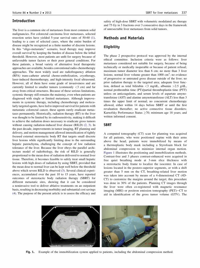
Fig. 1. Example of the immobilization system applied to
safety of high-dose SBRT with volumetric modulated arc therapyand 75 Gy in 3 fractions over 3 consecutive days in the frameworkof unresectable liver metastases from solid tumors.
Methods and Materials
Eligibility
The phase 2 prospective protocol was approved by the internalethical committee. Inclusion criteria were as follows: livermetastases considered not suitable for surgery, because of beingtechnically or medically inoperable or because of patient refusal;maximum tumor diameter less than 6 cm; no more than 3 liverlesions; normal liver volume greater than 1000 cm3; no evidenceof progressive or untreated gross disease outside of the liver; noprior radiation therapy to the targeted area; adequate liver func-tion, defined as total bilirubin <3 mg/dL, albumin >2.5 g/dL,normal prothrombin time (PT)/partial thromboplastin time (PTT)unless on anticoagulants, and serum levels of aspartate amyno-transferase (AST) and alanine amynotransferase (ALT) less than 3times the upper limit of normal; no concurrent chemotherapyallowed, either within 14 days before SBRT or until the firstrevaluation thereafter; no active connective tissue disorders;Karnofsky Performance Status �70; minimum age 18 years; andwritten informed consent.
SBRT
A computed tomography (CT) scan for planning was acquiredfor all patients, who were positioned supine with their armsabove the head; patients were immobilized by means ofa thermoplastic body mask including a Styrofoam block forabdominal compression to minimize internal organ motion.Figure 1 illustrates the positioning and immobilization methods.Contrast-free and 3 phases contrast-enhanced were acquired infree quiet breathing mode at 3-mm slice thickness witha stereotactic body frame to localize the isocenter. In case oflesions located in the postero-superior segments, or with a shiftgreater than 5 mm on the CT, breathing-related liver motionwas taken into account by means of a 4-dimensional CT (4D-CT) to customize the margins around the target; this procedurewas done in 30% of the patients. Planning CT images throughthe liver were often co-registered with magnetic resonanceimaging (MRI) or positron emission tomography (PET)eCT toaid in identification of the gross tumor volume (GTV). The
patients, including the abdominal compression method.

cm
Table 1 Baseline patient and treatment characteristics
Characteristic n %
No. of patients 61Male 26 42.6Female 35 57.4
Median age, y 65 -Range 39-87No. of liver lesions1 48 78.72 11 18.03 2 3.3
PrimaryColorectal 29 47.5Breast 11 18.0Gynecological 7 11.5Other 14 22.9
Time since diagnosis, mo�12 35 57.4>12 26 42.6
No. of prior systemic treatment regimens0 10 16.41 15 24.62 13 21.33 14 22.9�4 9 14.7
Presence of stable extrahepatic disease
Scorsetti et al. International Journal of Radiation Oncology � Biology � Physics338
clinical target volume (CTV) was defined as equal to the GTV.For patients with 4D-CT, an internal target volume was definedas the envelope of all GTVs in the different respiratory phases.The planning target volume (PTV) was generated from eitherthe GTV or the ITV by adding an overall isotropic margin of 5mm (from ITV) or of 7 to 10 mm in the cranial-caudal axis and4 to 6 mm in the anterior-posterior and lateral axes. Image-guided radiation therapy was performed by means of a mega-voltage CBCT before each daily session to account for set-upuncertainties.
The prescription dose was 75 Gy in 3 consecutive daily frac-tions of 25 Gy each, prescribed as the mean dose to PTV. Whenfull doses were not achievable because of OAR, the dose wascompromised (in 18% of the total of the lesions; Table 1), andpatients received 67.5 Gy in 8% of the lesions, 60 Gy in 5%, and52.5 in 5%. According to the studies by Schefter and Kavanagh(5), a critical doseevolume model was applied to fulfill theconstraints for organs at risk (OAR): V15 Gy (volume receiving15 Gy ) < (total liver volume � 700 cm3) for healthy liver, D0.1
3
for spinal cord <18 Gy (dose at a volume of 0.1 cm3 should be<18 Gy), V15 Gy <35% for both kidneys, V21 Gy <1% for theduodenum, small bowel, esophagus, and stomach, V30 Gy <1% forthe heart; D30 cm
3 <30 Gy for the ribs was considered asa secondary objective. In case of overlap between the PTV andduodenum or stomach, the priority was given to the OAR.
SBRT was delivered using volumetric modulated arc therapyby RapidArc (Varian, Palo Alto, CA) technique.
Yes 21 34.4No 40 65.6
Prior liver-directed therapyYes 28 45.9
Surgery 21 75RFA 2 7Both 5 19
No 33 54.1
Treatment No. of lesions %
Lesion diameter (mm)�30 mm 45 59.2>30 mm 31 40.8
CTV volume (cm3)Mean � SD 18.6 � 22.7Range 1.8-134.3
PTV volume (cm3)Mean 54.9 � 41.998Range 7.7-209.4
Dose prescription (per lesion)Full dose (75 Gy) 62 8290% (67.5 Gy) 6 880% (60 Gy) 4 570% (52.5 Gy) 4 5
Abbreviations: CTV Z clinical target volume; PTV Z planning
target volume; RFA Z radiofrequency ablation.
Study end points and follow-up
The primary end-point for this phase 2 study was to assess in-field local control. Secondary end points were to define radia-tion treatment-related toxicity and overall survival. Patientswere monitored by physical examination at the beginning andthe last day of the SBRT, and with basal blood chemistryanalyses. After conclusion of SBRT, these examinations wererequested 21 days after and then every 2 months. Imaging forfollow-up included CT scans every 3 months and, with thesame periodicity, PET-CT was also available for a subgroup of54% of patients. Acute and late toxicity were scored by theCommon Terminology Criteria for Adverse Events (CTCAEversion 3.0). Any increase in grade from baseline was consid-ered toxicity related to the treatment. RILD was defined byLawrence’s criteria as a subacute toxicity occurring within 4months after the completion of irradiation, and expressed aseither anicteric increase of the alkaline phosphatase value to atleast twice the upper normal level and nonmalignant ascites, orincrease of transaminases to at least 5 times the upper limit ofnormal or the pretreatment value. The diagnosis of RILD couldbe made only in the absence of progressive liver disease.Assessment of tumor response was based on European Orga-nization for Research and Treatment of Cancer ResponseEvaluation Criteria In Solid Tumors (EORTC-RECIST) criteriaversion 1.1 (6). Complete response (CR) was defined asdisappearance of all target lesions; partial response (PR) as atleast a 30% decrease in the sum of diameters of target lesions;and progressive disease (PD) as at least a 20% increase in thesum of diameters of target lesions. Progressive disease wasscored depending on whether it was intra- or extrahepatic. Theextrahepatic group was outside of the hepatic tissue or near tofield, whereas the intrahepatic group had progression occurring
within 1 cm of the field edges, or in the medium- to high-doseregions (greater than 20%e30% isodose).
Statistical considerations
Actuarial LC and OS curves were generated by using theKaplan-Meier method. All enrolled patients were included in

Fig. 2. Patient treated with stereotactic body radiation therapy for recurrence of liver metastasis after surgery. (a) Positron emissiontomography (PET)ecomputed tomography (CT) pretreatment image showing the lesion in the region of the surgical bed, defined by metalsurgical clips. (b) Visualization of dose distribution on the planning target volume. (c) PET-CT image at 3 months after radiation therapy,showing complete metabolic response.
Volume 86 � Number 2 � 2013 SBRT for liver metastases 339
the statistical evaluation. The influence of patient, disease, andtreatment characteristics on OS were evaluated by using Coxproportional hazards regression. Covariates evaluated in theCox model included primary tumor site, number of livermetastases, maximum lesion diameter, presence of extrahepaticdisease, and treatment with prior systemic therapy for meta-static disease.
Results
This ongoing study includes 61 enrolled patients with 76 livermetastases from solid tumors treated between February 2010and September 2011. The median follow-up was 12 months(range, 2-26 months). Baseline patients and treatment charac-teristics are summarized in Table 1. Figure 2 describes a typicalcase of patient with isolated liver metastasis included in theprotocol.
Mean of the mean dose to CTV was 73.2 � 5.6 Gy; the same toPTV was 69.6 � 6.7 Gy. The mean D98% was 71.0 � 6.2 Gy forCTV. The mean of the total liver mean dose was 12.7 � 5.1 Gy;
Fig. 3. (a) In-field local control after stereotactic body rad
the mean volume of total liver receiving less than 15 Gy was 1050� 318 cm3.
Local control
The radiographic/metabolic crude response rate of the radiatedlesions (complete response þ partial response þ stable disease)treatment was 94.7% (72/76); in-field progression was observedfor 4 lesions (5.3%). A summary of the analysis of patterns ofresponse is provided in Table 3. Of the 4 in-field progressions, 2occurred in the same patient, and none correlated with under-dosage to the CTV (V95% from 94% to 100%) Actuarial localcontrol rates for treated lesions at 6, 12, and 22 months were100%, 94.0% (95% confidence interval [CI] Z 0.82%-0.98%),and 90.6% (CI Z 0.76%-0.96%), respectively (Fig. 3a). Asubgroup analysis for lesions with maximum diameter of 3 cm orless, compared with those greater than 3 cm, revealed no statisticaldifferences in local control rates (PZ.90). A further subset anal-ysis revealed that tumors typically considered radio-resistant(including melanoma, renal carcinoma, pancreatic and biliary
iation therapy (SBRT). (b) Overall survival after SBRT.

Table 2 Overview of prospective trials of SBRT for liver metastases
First author (Reference) Design No. of patients with mets Tumor volume
Herfarth (8) Phase 1-2 35 1-132 mL (median, 10 mL)
Mendez Romero (20) Phase 1-2 (HCC and mets) 25 (17 liver mets) 1.1-322 mL (median, 22.2 mL)
Hoyer (21) Phase 2 (CRC oligomets) 64 (44 liver mets) 1-8.8 cm (median, 3.5 cm)
Rusthoven (11) Phase 1-2 47 0.75-97.98 mL (median, 14.93 mL)
Lee (9) Phase 1-2 68 1.2-3090 mL (median, 75.9 mL)
Ambrosino (12) Prospective cohort 27 20-165 mL (median, 69 mL)
Goodman (10) Phase 1 (HCC and mets) 26 (19 liver mets) 0.8-146.6 mL (median, 32.6 mL)
Abbreviations: CRC Z colorectal cancer; fx Z fractions; LC Z local control; mets Z metastases; NS Z not significant; OS Zoverall survival; RILD Z radiation-induced liver disease; RT Z radiation therapy; SBRT Z stereotactic body radiation therapy.
Scorsetti et al. International Journal of Radiation Oncology � Biology � Physics340
ducts adenocarcinoma) were not significantly associated withworse local control, compared with all the others (PZ.20).Multivariate analysis was not performed for LC, owing to thesmall number of events.
Survival
Progression was observed in 41 patients (67.2%) Analysis ofpatterns of failure is summarized in Table 3. Of the 33 extrahe-patic progressive patients, 19 (58%) had only hepatic metastasesat the time of SBRT, and 14 (42%) already had systemic disease.Of the latter, 13 progressed within 12 months (and 4 of thoseprogressed also to intrahepatic disease).
At the time of analysis, 16 of 61 patients (26.2%) died, ofthese, 13 (81%) died of disease-specific causes, whereas 3(19%) died of other causes. The median OS rate was 19 months(95% CIZ16.3-21.7 months). Actuarial survival at 12 monthswas 83.5% (with 29 patients at risk) and 65% at 18 months (10patients at risk). A survival curve is shown in Figure 3b.Neither primary tumor site nor cumulative number of priorsystemic treatment regimens were significantly related tosurvival on both univariate and multivariate analysis. Moreover,primary tumors from unfavorable primary sites were not foundto be associated with worse survival compared with tumors
from favorable primaries, including breast and colorectal(PZ.09).
Toxicity
Acute toxicity was low, despite a number of patients (26%)developing a G2 transient transaminase increase. In all cases, theincreased values were normalized within the 3 months after SBRT,according to published literature data. No cases of RILD have beendocumented. Post-treatment fatigue was common (65% ofpatients), but it was modest (no grade 2 or higher) and did notinterfere with daily activities. Nomajor (G4 or G5) late toxicity wasreported. One patient experienced a grade 3 late toxicity (chronicchest wall pain), which regressed within 1 year after treatment.
Discussion
To our knowledge, this study has the largest series addressing theuse of high doses of radiation by means of SBRT in the treatmentof liver metastases. Outcome data from a recent pooled analysis ina large cohort of patients with colorectal liver metastases showedthat local control is dose dependent, with a 3-fraction SBRT doserequired to achieve a 90% rate of local control at 1 year in the

First author (Reference) Type of mets RT dose Toxicity Outcomes
Herfarth (8) NA Dose escalation, 14-26Gy (1 fx)
NS 1-y LC, 71%18-mo LC, 67%1-y OS, 72%
Mendez Romero (20) CRC (14) 30-37.5 Gy (3 fx) 2 Grade 3 liver toxicity 2-y LC, 86%Lung (1) 2-y OS, 62%Breast (1)Carcinoid (1)
Hoyer (21) CRC (44) 45 Gy (3 fx) 1 Liver failure 2-y LC: 79% (by tumor),64% (by patient)2 Severe late GI toxicities
Rusthoven (11) CRC (15) Dose escalation, 36-60Gy (3 fx)
No RILD 1-y LC, 95%Lung (10) Late Grade 3-4 <2% 2-y LC, 92%Breast (4) Median survival, 20.5 moOvarian (3)Esophageal (3)HCC (2)Other (10)
Lee (9) CRC (40) 27.7-60 Gy (6 fx) No RILD 1-y LC, 71%Breast (12) Acute Grade 3-4 10% Median survival, 17.6 moGallbladder (4) No Grade 3-4 late toxicityLung (2)Anal canal (2)Melanoma (2)Other (6)
Ambrosino (12) CRC (11) 25-60 Gy (3 fx) NS Crude LC rate 74%Other (16)
Goodman (10) CRC (6) Dose escalation, 18-30Gy (1 fx)
No dose-limiting toxicity 2-y OS, 49% (mets only)Pancreatic (3)Gastric (2)Ovarian (2)Other (6)
Table 2 (continued)
Volume 86 � Number 2 � 2013 SBRT for liver metastases 341
range of 46 to 52 Gy (7). On the basis of a well recognizedrelationship between local control and survival in surgical seriesand at the observed dose response with SBRT (7), we designed thepresent phase 2 study of SBRT for unresectable liver metastases.
A number of prospective trials of SBRT for the treatment ofliver metastases have been published (Table 2), with follow-uptimes not exceeding 18 months in most series. Notwithstandingthe relatively short follow-up in our cohort, local control andsurvival rates seem consistent with those reported in literature(8-12), and confirm that a significant fraction of oligo-metastaticpatients may benefit from intensification of local therapy withhigher radiation doses by SBRT. A randomized study between themajor non-surgical ablative techniques, namely, SBRTand RFA, isstill lacking. However, outcomes in terms of local tumor recur-rence rates from the last published trials compare very favorablywith RFA (13). Prospective comparison of RFA versus SBRT isbeing addressed by a currently ongoing trial (RadiofrequencyAblation Versus Stereotactic Radiotherapy Trial) (14).
Unlike the findings from a recently published article (11)addressing the role of SBRT for liver lesions, in our cohort nocorrelations were found either between LC and lesions according tosize, or between LC and primary histology. Notwithstanding somepossible limitations due to the small number of events, the lack ofcorrelation in the former analysis may suggests that when higher
doses are used, even lesions greater than 3 cm can be fully ablated.The latter analysis revealed no relationships between LC and the so-called radio-resistant tumors (including melanoma, renal carci-noma, and pancreatic and biliary duct adenocarcinoma), whichmight be attributed to the synergistic tumoricidal effect of SBRT inthe high-dose range. Indeed, targeting the tumor vasculature forobliteration with high-dose radiation may be beneficial for tumorcontrol, as argued by Fuks and Kolesnick (15). Moreover, Kim et al(16) demonstrated that blood flow and microvascular densitydecrease very quickly after the administration of doses greater than20 Gy in a murine mode, confirming that vascular damage mayultimately enhance the tumoricidal potential of SBRT.
SBRT for liver lesions must be performed cautiously, given thechallenges of organ motion and the low toxicity tolerance of theneighboring liver tissue. In our experience, SBRT was deliveredusing RapidArc. This resulted in a strong reduction in deliverytime, with benefit in patient comfort and potential limitation ofintrafraction motion (17). This might have been a crucial factorthat strongly contributed, as well as an optimal management ofbreathing-related liver motion, to keep toxicity very low. Onepatient, only, experienced G3 chest wall pain approximately 6months after SBRT to a lesion very close to the ribs. Soon after-ward, we revised the soft tissue protocol dose constraints to keepthe volume of chest wall receiving 30 Gy (V30) to less than 10

Table 3 Patterns of treatment response and failure
In-field responseLesions
(NZ76), n (%)
Complete response 36 (47.4%)Partial response 16 (21.0%)Stable disease 20 (26.3%)Progressive disease 4 (5.3%)
Site of failurePatients
(NZ61), n (%)
No progression after SBRT 19 (31.1%)Progressive disease after SBRT 41 (67.2%)
Extra-field intrahepatic progression(different segment)
22 (36.0%)
Near-to-field intrahepatic progression(same segment)
3 (4.9%)
Extrahepatic progression 33 (54.1%)Extra-field intrahepatic progression only 8 (13.1%)Extrahepatic progression only 18 (29.5%)
Progressive disease in patients with previous extrahepaticdisease (21 patients)
Extrahepatic progression 13 (61.9%)Intrahepatic progression 11 (52.4%)
Scorsetti et al. International Journal of Radiation Oncology � Biology � Physics342
cm3, as it has been shown to be a reliable predictor of chest walltoxicity (18). Despite optimal local control rates, out-of-fieldmetastatic progression developed in a substantial proportion ofpatients, suggesting closer attention to patients who really belongto a relatively good prognostic group with longer survivalexpectancy, and offering a strong rationale for combining SBRTwith systemic treatments. An ongoing trial (Slim Trial) addressingthis relevant issue is currently recruiting patients (19). Neverthe-less, our study has some drawbacks: the main limitation is prob-ably the relatively short median survival (19 months) comparedwith that in surgery series (1). However, these data are most likelyrelated to the poor prognostic features of the patients enrolled,typically unsuitable for hepatic resection. Indeed, 36% of patientsalready had extrahepatic metastatic disease at the time of SBRT,and in 54% the time to progression was less than 12 months.Moreover, almost one-third (28%) had received chemotherapy formetastatic disease, and 45% had already undergone loco-regionaltreatment before study accrual.
Another limitation might be the lack of evaluation on theimpact of SBRTon the patient’s quality of life. So far, only 1 studyhas prospectively addressed the former issue (20), showing that nostatistical difference was observed between mean baseline valuesand those obtained at 1, 3, and 6 months after treatment.
Conclusion
The present SBRT trial is the first regimen to use doses in thehighest range among those reported in the literature for thetreatment of liver metastases. Our findings confirm that SBRTprovides an effective local ablation with a low toxicity profile inpatients with oligo-metastatic disease in whom disease control andsurvival should still be considered the aims of the treatment.Randomized trials to evaluate the efficacy of the various ablativetreatments are pending.
References
1. Shah SA, Bromberg R, Coates A, et al. Survival after liver resection
for metastatic colorectal carcinoma in a large population. J Am Coll
Surg 2007;205:676-683.
2. Garrean S, Hering J, Saied A, Helton WS, Espat NJ. Radiofrequency
ablation of primary and metastatic liver tumors: a critical review of the
literature. Am J Surg 2008;195:508-520.
3. Dawson LA, Normolle D, Balter JM, et al. Analysis of radiation-
induced liver disease using the Lyman NTCP model. Int J Radiat
Oncol Biol Phys 2002;53:810-821.
4. Timmerman RD, Kavanagh BD, Cho LC, et al. Stereotactic body radi-
ation therapy in multiple organ sites. J Clin Oncol 2007;25:947-952.
5. Schefter TE, Kavanagh BD. Radiation therapy for liver metastases.
Semin Radiat Oncol 2011;21:264-270. Review Erratum in: Semin
Radiat Oncol 2012;22(1):86.
6. Eisenhauer E, Therasse P, Bogaerts J, et al. New response evaluation
criteria in solid tumors: revised RECIST guideline (version 1.1). Eur J
Cancer 2009;45:228-247.
7. Chang DT, Swaminath A, Kozak M, et al. Stereotactic body radio-
therapy for colorectal liver metastases: a pooled analysis. Cancer
2011;117:4060-4069.
8. Herfarth KK, Debus J, Wannenmacher M. Stereotactic radiation
therapy of liver metastases: Update of the initial phase-I/ II trial. Front
Radiat Ther Oncol 2004;38:100-105.
9. Lee MT, Kim JJ, Dinniwell R, et al. Phase I study of individualized
stereotactic body radiotherapy of liver metastases. J Clin Oncol 2009;
27:1585-1591.
10. Goodman KA, Wiegner EA, Maturen KE, et al. Dose-escalation study
of single-fraction stereotactic body radiotherapy for liver malignan-
cies. Int J Radiat Oncol Biol Phys 2010;78:486-493.
11. Rusthoven KE, Kavanagh BD, Cardenes H, et al. Multi-institutional
phase I/II trial of stereotactic body radiation therapy for liver metas-
tases. J Clin Oncol 2009;27:1572-1578.
12. Ambrosino G, Polistina F, Costantin G, et al. Image-guided robotic
stereotactic radiosurgery for unresectable liver metastases: Prelimi-
nary results. Anticancer Res 2009;29:3381-3384.
13. Alongi F, Arcangeli S, Filippi AR, et al. Review and uses of stereo-
tactic body radiation therapy for oligometastases. Oncologist 2012;17:
1100-1107.
14. Radiofrequency Ablation Versus Stereotactic Radiotherapy in Colo-
rectal Liver Metastases (RAS01). ClinicalTrials.gov NCT01233544.
Last Updated: March 20, 2012.
15. Fuks Z, Kolesnick R. Engaging the vascular component of the tumor
response. Cancer Cell 2005;8:89-91.
16. Kim DW, Huamani J, Niermann KJ, et al. Noninvasive assessment of
tumor vasculature response to radiation-mediated, vasculature-tar-
geted therapy using quantified power Doppler sonography: Implica-
tions for improvement of therapy schedules. J Ultrasound Med 2006;
25:1507-1517.
17. Kragl G, Baier F, Lutz S, et al. flattening filter free beams in SBRT and
IMRT: dosimetric assessment of peripheral doses. Med Phys 2011;21:
91-101.
18. Dunlap N, Biederman G, Yang W, et al. Chest wall volume
receiving more than 30 Gy predicts risk of severe pain and/or rib
fracture after lung SBRT. Int J Radiat Oncol Biol Phys 2008;
72(Suppl 36):796-801.
19. Sorafenib-RT Treatment for Liver Metastasis (SLIM). ClinicalTrials.
gov NCT00892424. Last Updated: February 11, 2011.
20. Mendez Romero A, Wunderink W, van Os RM, et al. Quality of
life after stereotactic body radiation therapy for primary and
metastatic liver tumors. Int J Radiat Oncol Biol Phys 2008;70:
1447-1452.
21. Hoyer M, Roed H, Traberg HA, et al. Phase II study on stereotactic
body radiotherapy of colorectal metastases. Acta Oncol 2006;45:
823-830.
Copyright © 2022 FDOKUMEN