Investigating the Neural Correlates of Voice versus Speech-Sound Directed Information in Pre-School...
23
RESEARCH ARTICLE Investigating the Neural Correlates of Voice versus Speech-Sound Directed Information in Pre-School Children Nora Maria Raschle 1,2,3 *, Sara Ashley Smith 1 , Jennifer Zuk 1,2 , Maria Regina Dauvermann 1,2 , Michael Joseph Figuccio 1 , Nadine Gaab 1,2,4 1. Laboratories of Cognitive Neuroscience, Division of Developmental Medicine, Department of Developmental Medicine, Boston Children’s Hospital, Boston, Massachusetts, United States of America, 2. Harvard Medical School, Boston, Massachusetts, United States of America, 3. Psychiatric University Clinics Basel, Department of Child and Adolescent Psychiatry, Basel, Switzerland, 4. Harvard Graduate School of Education, Cambridge, Massachusetts, United States of America * [email protected] Abstract Studies in sleeping newborns and infants propose that the superior temporal sulcus is involved in speech processing soon after birth. Speech processing also implicitly requires the analysis of the human voice, which conveys both linguistic and extra- linguistic information. However, due to technical and practical challenges when neuroimaging young children, evidence of neural correlates of speech and/or voice processing in toddlers and young children remains scarce. In the current study, we used functional magnetic resonance imaging (fMRI) in 20 typically developing preschool children (average age 55.8 y; range 5.2–6.8 y) to investigate brain activation during judgments about vocal identity versus the initial speech sound of spoken object words. FMRI results reveal common brain regions responsible for voice-specific and speech-sound specific processing of spoken object words including bilateral primary and secondary language areas of the brain. Contrasting voice-specific with speech-sound specific processing predominantly activates the anterior part of the right-hemispheric superior temporal sulcus. Furthermore, the right STS is functionally correlated with left-hemispheric temporal and right- hemispheric prefrontal regions. This finding underlines the importance of the right superior temporal sulcus as a temporal voice area and indicates that this brain region is specialized, and functions similarly to adults by the age of five. We thus extend previous knowledge of voice-specific regions and their functional connections to the young brain which may further our understanding of the neuronal mechanism of speech-specific processing in children with developmental disorders, such as autism or specific language impairments. OPEN ACCESS Citation: Raschle NM, Smith SA, Zuk J, Dauvermann MR, Figuccio MJ, et al. (2014) Investigating the Neural Correlates of Voice versus Speech-Sound Directed Information in Pre-School Children. PLoS ONE 9(12): e115549. doi:10.1371/journal.pone.0115549 Editor: Antoni Rodriguez-Fornells, University of Barcelona, Spain Received: March 5, 2014 Accepted: November 24, 2014 Published: December 22, 2014 Copyright: ß 2014 Raschle et al. This is an open- access article distributed under the terms of the Creative Commons Attribution License, which permits unrestricted use, distribution, and repro- duction in any medium, provided the original author and source are credited. Data Availability: The authors confirm that all data underlying the findings are fully available without restriction. All relevant data are included within the paper and its Supporting Information files. fMRI images are available upon request from the corresponding author. Funding: This work was supported by the Eunice Kennedy Shriver National Institute of Child Health & Human Development (1R01HD065762-01/02/03 to N.G.); Charles H. Hood Foundation (to N.G.); Boston Children’s Hospital Pilot Grant (to N.G.); the Swiss National Foundation (to N.M.R.); the Janggen-Po ¨hn Stiftung (to N.M.R.); and the National Institute of Health Institutional Training Grant (NIH T32 DC000038-22 to J.Z.). The funders had no role in study design, data collection and analysis, decision to publish, or preparation of the manuscript. Competing Interests: The authors have declared that no competing interests exist. PLOS ONE | DOI:10.1371/journal.pone.0115549 December 22, 2014 1 / 23
-
Upload
independent -
Category
Documents
-
view
3 -
download
0
Transcript of Investigating the Neural Correlates of Voice versus Speech-Sound Directed Information in Pre-School...

RESEARCH ARTICLE
Investigating the Neural Correlates ofVoice versus Speech-Sound DirectedInformation in Pre-School ChildrenNora Maria Raschle1,2,3*, Sara Ashley Smith1, Jennifer Zuk1,2,Maria Regina Dauvermann1,2, Michael Joseph Figuccio1, Nadine Gaab1,2,4
1. Laboratories of Cognitive Neuroscience, Division of Developmental Medicine, Department ofDevelopmental Medicine, Boston Children’s Hospital, Boston, Massachusetts, United States of America, 2.Harvard Medical School, Boston, Massachusetts, United States of America, 3. Psychiatric University ClinicsBasel, Department of Child and Adolescent Psychiatry, Basel, Switzerland, 4. Harvard Graduate School ofEducation, Cambridge, Massachusetts, United States of America
Abstract
Studies in sleeping newborns and infants propose that the superior temporal sulcus
is involved in speech processing soon after birth. Speech processing also implicitly
requires the analysis of the human voice, which conveys both linguistic and extra-
linguistic information. However, due to technical and practical challenges when
neuroimaging young children, evidence of neural correlates of speech and/or voice
processing in toddlers and young children remains scarce. In the current study, we
used functional magnetic resonance imaging (fMRI) in 20 typically developing
preschool children (average age 55.8 y; range 5.2–6.8 y) to investigate brain
activation during judgments about vocal identity versus the initial speech sound of
spoken object words. FMRI results reveal common brain regions responsible for
voice-specific and speech-sound specific processing of spoken object words
including bilateral primary and secondary language areas of the brain. Contrasting
voice-specific with speech-sound specific processing predominantly activates the
anterior part of the right-hemispheric superior temporal sulcus. Furthermore, the
right STS is functionally correlated with left-hemispheric temporal and right-
hemispheric prefrontal regions. This finding underlines the importance of the right
superior temporal sulcus as a temporal voice area and indicates that this brain
region is specialized, and functions similarly to adults by the age of five. We thus
extend previous knowledge of voice-specific regions and their functional
connections to the young brain which may further our understanding of the
neuronal mechanism of speech-specific processing in children with developmental
disorders, such as autism or specific language impairments.
OPEN ACCESS
Citation: Raschle NM, Smith SA, Zuk J, DauvermannMR, Figuccio MJ, et al. (2014) Investigating the NeuralCorrelates of Voice versus Speech-Sound DirectedInformation in Pre-School Children. PLoS ONE 9(12):e115549. doi:10.1371/journal.pone.0115549
Editor: Antoni Rodriguez-Fornells, University ofBarcelona, Spain
Received: March 5, 2014
Accepted: November 24, 2014
Published: December 22, 2014
Copyright:� 2014 Raschle et al. This is an open-access article distributed under the terms of theCreative Commons Attribution License, whichpermits unrestricted use, distribution, and repro-duction in any medium, provided the original authorand source are credited.
Data Availability: The authors confirm that all dataunderlying the findings are fully available withoutrestriction. All relevant data are included within thepaper and its Supporting Information files. fMRIimages are available upon request from thecorresponding author.
Funding: This work was supported by the EuniceKennedy Shriver National Institute of Child Health& Human Development (1R01HD065762-01/02/03to N.G.); Charles H. Hood Foundation (to N.G.);Boston Children’s Hospital Pilot Grant (to N.G.); theSwiss National Foundation (to N.M.R.); theJanggen-Pohn Stiftung (to N.M.R.); and theNational Institute of Health Institutional TrainingGrant (NIH T32 DC000038-22 to J.Z.). The fundershad no role in study design, data collection andanalysis, decision to publish, or preparation of themanuscript.
Competing Interests: The authors have declaredthat no competing interests exist.
PLOS ONE | DOI:10.1371/journal.pone.0115549 December 22, 2014 1 / 23

Introduction
The human voice is omnipresent in our lives, conveying both linguistic and extra-
linguistic information and playing an integral role in our daily interactions. In
addition to delivering language content, the human voice conveys rich acoustic
information crucial for speaker identification, such as the fundamental frequency
of the speaker’s voice and the spectral formants produced through modification of
the vocal tract that characterize individual vowels and consonants [1–2]. The
latter carries the prosodic features of communication [3] as well as emotional tone
[4], and additionally provides cues to determine age [5] and gender [6–7].
Behavioral research has, for example, shown that infants as young as eight months
old are able to recognize male and female voices [8]. Voice perception carries
many different socially relevant features, demanding complex processes from our
brain. It has been proposed that the cerebral processing of vocal information (e.g.,
speaker identity or affect) may be organized in interacting, but functionally
dissociable pathways [9–11]. Neuropsychological evidence [12–13] suggests that
voice and speech-sound directed information may be processed differently.
Adults show a preference for general speech processing in bilateral temporal
brain regions, particularly in superior temporal gyrus (STG) and superior
temporal sulcus (STS; [12]). Using neuroimaging techniques such as functional
magnetic resonance imaging (fMRI), electroencephalography (EEG) or positron
emission tomography (PET), a human-specific voice-processing region has been
suggested in the upper bank of the right STS [9], [14–16]. It is of note that some
studies have identified voice processing areas in bilateral STS [11], [17–18].
However, the vast majority of publications report right-hemispheric neuronal
activation within the anterior part of the right STS, specifically when processing
human vocal sounds [9], [15], [17]. The idea of voice-specific sound processing in
humans is supported by studies comparing the neuronal correlates of vocal
sounds to activation patterns evoked by frequency modulated tones [19] or by
comparing human vocal sounds to those produced by animals [20–21]. Although
parts of the STS are activated in response to both vocal and non-vocal sounds
(e.g., environmental or artificial sounds), vocal sounds produce greater neural
responses than non-vocal sounds in most voice selective regions of the brain [7],
[16], [17], [22]. Furthermore, fMRI evidence shows that activity in the right STS
is greater when subjects perform a voice identity task (hearing several speakers say
the same word) as opposed to speech-sound specific tasks [10], [15], [19], [23],
providing evidence for the involvement of the anterior portion of the right STS in
processing voice identity.
The majority of research on the neural mechanisms of speech and voice
processing has been conducted in adult participants; however, infant studies
implementing passive listening experiments in the first years of life have been
reported. Behavioral research using the head-turn procedure in newborns could,
for example, show that newborns prefer human to non-human sounds, as well as
prosodic to non-prosodic speech [24]. Neuroimaging methods, such as near
infrared spectroscopy (NIRS) or fMRI, have shed light on hemispheric
Voice Processing within Human Speech in Pre-School Children
PLOS ONE | DOI:10.1371/journal.pone.0115549 December 22, 2014 2 / 23
specialization for speech, but provided mixed results. While some studies report
increased activation during human speech processing within right temporopar-
ietal brain regions (e.g., [25]; NIRS during human speech compared to flattened
speech sounds in three month olds), others have suggested a left-hemispheric
lateralization of human speech in newborns (e.g., [26]; optical topography during
speech processing in newborns). Left-hemispheric lateralization of speech
processing is further supported by fMRI evidence in three month old infants [27–
28].
Similar to adult studies, the anterior STS in infants was observed to be critically
involved during passive listening to human speech (e.g., comparing non-speech
human emotional sounds versus familiar non-voice background noises; [29]).
However, unlike in adults, infants did not show different activation patterns when
processing forward, as opposed to backward, speech, leading the authors to
conclude that the precursors to adult-like cortical language processing were
observable, but most likely not yet specialized [28], [30]. In line with this finding,
Grossmann and colleagues [3] reported that four month-old infants did not
display increased activation within bilateral superior temporal cortices when
contrasting human speech with non-vocal sounds. However, at seven months,
distinctive activation patterns can be observed during human voice and artificial
sounds processing in left and right superior temporal cortex, comparable to
results seen in adults [3]. To summarize, research so far has provided mixed
results regarding the activation pattern representing speech processing in infancy
(e.g., [25], [28]). However, the neuronal basis for speech is somehow similar to
that of adults, increasingly so with age. Improved specialization, as observed by
distinct neuronal activation to human speech sounds compared to a control
condition (backward speech in [28]; environmental sounds in [3]), takes place
between four and seven months- notably at a time when speech content is still
fairly irrelevant [3].
Though there is evidence for the neuronal basis of passive speech and vocal
information processing in infants, as well as plentiful studies in adults, a gap in
neuroimaging studies exists with toddler and preschool-aged participants.
However, technical improvements and increasingly more elaborate child-friendly
neuroimaging protocols allow for the extension of studies into younger age ranges
[31–33]. This is of utmost importance since previous developmental neuroima-
ging work has demonstrated that there are significant differences between children
and adults in regard to brain structure and function (e.g., [34–39]); thus
assumptions of a static model of the human brain are outdated [40]. Moreover,
even though studies in infancy are able to inform about crucial aspects of brain
organization and development closely after birth, a response-related cognitive
functional neuroimaging task including behavioral feedback is not yet possible,
and thus assumptions based on findings from research in sleeping infants may not
easily be applied to waking children. Finally, evidence that the neuronal circuits
for specific aspects of speech processing (e.g., phoneme discrimination) undergo
changes in the first year of life to attune to native language sounds underscores the
need for evaluation of the young brain before and after the onset of speech
Voice Processing within Human Speech in Pre-School Children
PLOS ONE | DOI:10.1371/journal.pone.0115549 December 22, 2014 3 / 23
production [41]. To our knowledge, only one exemplary study has examined
electrophysiological correlates of voice processing in a study of four and five year-
olds. Comparing voices to environmental sounds resulted in an early measurable
deflection (within 60 ms of onset) at right fronto-temporal sites, evidence for a
right-lateralized response to voices in children [42]. The authors have suggested
that this response may reflect activation of temporal voice areas in children.
To summarize, there is a general consensus regarding the neural location and
functional correlates of voice processing in adults [10–11], [17], [43–46] and there
is evidence for an early manifestation of this skill [3]. However, the precise
anatomical localization, neuronal correlates and functional connectivity of voice
processing in pre-school or school-aged children remains less well-understood.
While infant research has explored activation in response to ‘normal’ forward
speech compared to speech presented backwards, as well as between vocal and
non-vocal sounds, few studies with pre-school aged children have investigated
activation evoked specifically by varying aspects of native speech (for example,
vocal pitch as compared to speech-sound specific content). Therefore, the current
study employed whole brain fMRI in 20 typically developing pre-school children.
The objective of the present work was to investigate cortical response to two
auditory tasks in five year-old participants. The experimental task employed was
voice directed (voice matching (VM): ‘Is it the same person/voice talking or a
different person?’), while the second task was a speech-sound directed,
phonological processing task (first sound matching (FSM): ‘Do both words begin
with the same first sound?’). The same two voices, one male and one female, were
maintained throughout both tasks. In a comparable study in adults, Von
Kriegstein and colleagues [9] demonstrated that the right anterior STS is activated
during tasks requiring voice processing but not when content directed processing
was targeted. A follow-up fMRI study in adults [10] was furthermore able to
identify distinct but interacting areas along the right STS responding to acoustic
information stimuli, task demand and speaker-familiarity independently.
Furthermore, previous evidence from fMRI studies suggests the bilateral STS to be
crucial for processing human voices compared to non-speech sounds [17].
However, it has been suggested that the right STS alone is significantly more
activated for processing nonverbal components of human speech (e.g., voice
identity unrelated to verbal content; [9–10]). Therefore, we hypothesize that the
right STS will be recruited during the voice but not speech-sound directed task in
pre-schoolers, similar to the neuronal pattern observed in adult participants. To
test this hypothesis, we explicitly investigate patterns of neural activation as well as
functional connectivity during a voice identification task in right and left-
hemispheric STS regions.
Voice Processing within Human Speech in Pre-School Children
PLOS ONE | DOI:10.1371/journal.pone.0115549 December 22, 2014 4 / 23
Methods
Ethics Statement
This study and its consent procedures were approved by the Boston Children’s
Hospital Committee of Clinical Investigation (CCI). Written informed consent on
behalf of the child participants was obtained from guardians (first degree
relatives). Furthermore, research team members obtained verbal assent from child
participants. Verbal assent, and not written consent, was obtained from child
participants due to their young age (average age 5.8 y; children were non-readers
and could not write yet).
Participants
Twenty healthy, native English speaking children (average age at the time of
imaging: 5.8 years, age range 5.2 to 6.8 years) were included in the present
analysis. Nineteen children were right handed, whereas for one child handedness
could not be indicated yet (labeled as ambidextrous). All children were physically
healthy with no history of any neurological or psychological disorders, head
injuries, poor vision or poor hearing. All children scored within the normal or
above-average range on verbal and non-verbal IQ (Kaufman Brief Intelligence
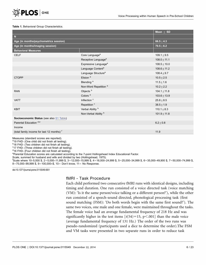
Test, 2nd edition [47]; Table 1). All children in the current study are part of the
Boston Longitudinal Study of Dyslexia (BOLD) at the Boston Children’s Hospital,
a study that aims to investigate neural and behavioral characteristics of typical
developing children compared to those with a familial risk for developmental
dyslexia. Participants are invited for one behavioral and one neuroimaging
session, including three functional experiments and structural image acquisition.
The results presented here are from a subgroup of typically developing children at
the first time point of neuroimaging (all children that had useful data obtained
during the voice- and speech-sound-directed task were included).
Behavioral Group Characteristics
Participants were characterized with a test battery of standardized assessments
examining language and pre-reading skills, such as expressive and receptive
vocabulary (Clinical Evaluation of Language Fundamentals (CELF Preschool 2nd
edition; [48]), phonological processing (Comprehensive Test of Phonological
Processing (CTOPP); [49] and Verb Agreement and Tense Test (VATT; [50]) and
rapid automatized naming (Rapid Automatized Naming Test; [51]). Additionally,
all participating families were given a socioeconomic background questionnaire
(questions adapted from the MacArthur Research Network: http://www.macses.
ucsf.edu/Default.htm; for a complete overview of SES questions see S1 Table) and
were asked questions regarding language development (see S2 Table). All children
were assessed for verbal and non-verbal IQ (KBIT average verbal IQ 5110.1¡8.3;
average non-verbal IQ5101.9¡11.8) and socioeconomic status (SES). Behavioral
testing and imaging were performed on different days, however, there were no
more than ¡42 days between the two sessions on average (less than 1.5 months).
Voice Processing within Human Speech in Pre-School Children
PLOS ONE | DOI:10.1371/journal.pone.0115549 December 22, 2014 5 / 23
fMRI - Task Procedure
Each child performed two consecutive fMRI runs with identical designs, including
timing and duration. One run consisted of a voice directed task (voice matching
(VM): ‘Is it the same person/voice talking or a different person?’), while the other
run consisted of a speech-sound directed, phonological processing task (first
sound matching (FSM): ‘Do both words begin with the same first sound?’). The
same two voices, one male and one female, were maintained throughout the tasks.
The female voice had an average fundamental frequency of 218 Hz and was
significantly higher in the test items [t(54)515, p,.001] than the male voice
(average fundamental frequency of 131 Hz.) The order of the two runs was
pseudo-randomized (participants used a dice to determine the order).The FSM
and VM tasks were presented in two separate runs in order to reduce task
Table 1. Behavioral Group Characteristics.
Mean ¡ SD
N
Age (in months/psychometrics session) 66.5¡4.3
Age (in months/imaging session) 70.5¡6.2
Behavioral Measures
CELF Core Languagea 109.1¡9.5
Receptive Languagea 108.0¡11.1
Expressive Languagea 108.5¡10.0
Language Contenta 108.6¡11.2
Language Structurea 108.4¡9.7
CTOPP Elision a 10.5¡2.5
Blending a 11.5¡1.6
Non-Word Repetition a 10.2¡2.2
RAN Objects b 104.1¡11.8
Colors a 103.6¡13.9
VATT Inflection c 25.8¡8.5
Repetition c 38.5¡1.9
KBIT Verbal Ability b 110.1¡8.3
Non-Verbal Ability b 101.9¡11.8
Socioeconomic Status (see also S1 Table)
Parental Education d,e 6.2¡0.8
Income
(total family income for last 12 months) f 11.9
Measures (standard scores are reported).a19 FHD- (One child did not finish all testing).b18 FHD- (Two children did not finish all testing).c17 FHD- (Three children did not finish all testing).d16 FHD- (Four children did not finish all testing).eParental Education scores are calculated according to the 7-point Hollingshead Index Educational Factor.Scale, summed for husband and wife and divided by two (Hollingshead, 1975).fScale where 10–5,000 $, 255,000–11,999 $, 3512,000–15,999 $, 4516,000–24,999 $, 5525,000–34,999 $, 6535,000–49,900 $, 7550,000–74,999 $,8575,000–99,999 $, 95100,000+$, 105 Don’t know, 115 No Response.
doi:10.1371/journal.pone.0115549.t001
Voice Processing within Human Speech in Pre-School Children
PLOS ONE | DOI:10.1371/journal.pone.0115549 December 22, 2014 6 / 23
demands (e.g., task switching) for the young participants, based on previous
experience carrying out neuroimaging studies in young populations (see also [32–
33], [52]). During the VM task all children listened to two subsequently presented
common object words spoken in a female or male voice via MR-compatible noise-
reducing headphones (two seconds per word). During both runs, corresponding
pictures were presented on the screen simultaneously in order to engage the
children and to reduce working memory demands. The object words were
followed by the presentation of a question mark, also displayed for two seconds.
Using two child-friendly buttons placed on either side of the participant, children
were asked to indicate via button-press whether the voice matched for the two
words presented. This task was contrasted with a rest condition, during which the
children were required to look at a fixation cross for the duration of the block.
During the FSM task, participants were again asked to listen to two common
object words, spoken in a female or male voice. Participants indicated via button
press whether the two words presented started with the same first sound (e.g., bed
and belt; ‘‘yes’’, or not (e.g., bird and ant; ‘‘no’’, for details see also [52]). This task
was again contrasted with a rest condition. Reaction time was measured from the
start of the second word on and the response window lasted until the start of the
consecutive trial for both tasks.
To avoid repetition effects (e.g., [53]), different word lists were created for the
VM and FSM tasks. However, all words between the two runs were kept as
comparable as possible by matching the two word lists for age of acquisition (e.g.,
when an average child recognizes a certain word; all words used here are
recognized before 4 years of age by typically developing children), Brown verbal
frequency, concreteness, imagery, numbers of letters, numbers of phonemes and
numbers of syllables (MRWC Psycholinguistic and the IPNP Database; http://
www.psy.uwa.edu.au/mrcdatabase/uwa_mrc.html and http://crl.ucsd.edu/
,aszekely/ipnp/). All pictures were adapted from the standardized Snodgrass
Picture System [54]. The same number of trials for VM and FSM matches were
included in each run (for further task descriptions see [52]). A sparse temporal
sampling design allowed for the presentation of the auditory stimuli without
scanner background noise interference [55–57]. A total of seven blocks of VM/
FSM and seven blocks of rest condition with an overall duration of 336 s seconds
for each run were employed. Each block lasted 24 seconds and each block
contained four trials. During the experimental and control tasks, 50% of the
words were spoken in a male/female voice and 50% of all items matched regarding
their first sound. The order of trials within a block was randomized, but kept
constant across participants.
Each child underwent extensive preparation and training in the mock MR
scanner area before the actual neuroimaging session. Participants were
familiarized with the voice and speech-sound directed task prior to the
neuroimaging session using unique practice items. Instructions for each task were
presented in separate short videos, which were shown in the MR scanner area and
repeated prior to actual scanning. To reduce movement during the scanning
procedure, cushions were used to stabilize the head and response buttons were
Voice Processing within Human Speech in Pre-School Children
PLOS ONE | DOI:10.1371/journal.pone.0115549 December 22, 2014 7 / 23
placed at arm’s length on each side of the child. A member of the research team
observed the child during in-scanner performance and provided a tactile reminder
to stay still during the session if needed (for a detailed description of the training
protocol see [32–33]).
fMRI - Acquisition and Analysis
For each run (experimental and control task), 56 functional whole-brain images
were acquired with a 32 slice EPI interleaved acquisition on a SIEMENS 3T Trio
MR scanner including the following specifications: TR 6000 ms; TA 1995 ms; TE
30 ms; flip angle 90 ; field of view 256 mm; voxel size 36364 mm, slice
thickness 4 mm. Prior to the start of the first block, additional functional images
were obtained and later discarded to allow for T1 equilibration effects. Stimuli
were presented using Presentation software (Version 0.70, www.neurobs.com).
The complete imaging session included 2 additional functional imaging tasks;
actual scan time per task was 12 minutes each.
Image processing and analyses were carried out using SPM5 (www.fil.ion.ucl.ac.
uk/spm) executed in MATLAB (Mathworks, Natick, MA). Prior to statistical
analysis, we first adjusted for movement artifacts within the acquired fMRI time
series by realigning all images using a least squares approach to the first image
(after discarding the first images to allow for T1 equilibration effects). In a second
step, all images were spatially normalized into standard space, as defined by the
ICBM, NIH-20 project [58]. It is to note that no customized child template was
used and that consequent reports of coordinates and activation pattern are
interpreted with caution due to the brain size differences of adults and children.
Finally, all images were smoothed with an 8 mm full width at half maximum
(FWHM) isotropic kernel to remove noise and effects due to residual differences
in functional and structural anatomy during inter-subject averaging (SPM5). Due
to the age of participants, a rigorous procedure for artifact detection was
implemented. We used the art-imaging toolbox (http://www.nitrc.org/projects/
artifact_detect) to visualize motion, plot potential movement artifacts and review
analysis masks for each subject. Upon visual inspection of all raw images,
preprocessed images were used to create an explicit mask excluding potential
artifactual brain volumes from the explicit mask through the art-imaging toolbox
for each child. The art-imaging toolbox was then used to plot differences in
motion between consecutive images and to review artifactual time-points: First,
we identified all images that exceeded a movement threshold of 2 mm and a
rotation threshold of 0.05 mm and checked that the analysis mask without said
images contained all voxels (this step is necessary to ensure that there are no
remaining outliers in the images within the defined threshold). Every image
exceeding this threshold was then visually inspected, and movement and outlier
regressors were added to the general linear model. Furthermore, volumes
containing visible artifacts were regressed out and not modeled in further
analyses. Prior to first level analysis, we ensured that the explicit mask was
complete (inclusion of all brain voxels). The general linear approach implemented
Voice Processing within Human Speech in Pre-School Children
PLOS ONE | DOI:10.1371/journal.pone.0115549 December 22, 2014 8 / 23
in SPM5 was used to analyze the data in a block design for each subject.
Movement regressors were modeled as cofounds within the general linear model
and explicit masking was performed during each subject’s first level analysis to
ensure inclusion of each voxel of the analysis mask. Contrast images (One sample
t-tests) for ‘VM.Rest’, ‘FSM.Rest’, ‘FSM.VM’ (contend directed contrast) and
‘VM.FSM’ (voice directed contrast) were obtained for the whole group of
children. Because of the lower signal-to-noise ratio in pediatric compared to adult
samples and the relatively high inter-individual variance in pediatric datasets (e.g.
[95]), results are reported at a threshold of p,0.005 with a cluster extent
threshold of k510, as similarly employed by other pediatric studies (e.g. [52],
[96]).
Region of Interest (ROI) Analysis
Previous research has shown involvement of the right anterior STS during voice
processing, particularly during the analysis of extra-linguistic features of speech
[9]. To investigate the role of the right anterior STS further, we defined an ROI for
the anterior part of the right STS based on evidence in adults [15] (4 mm-sphere
at Talairach coordinates of peak: 58,2,-8) using the MarsBaR toolbox [95]. Using
the MarsBaR transformation function, we flipped this right hemispheric ROI to
create a left-hemispheric analogue (left STS ROI). Mean parameter estimates were
extracted for the two resulting regions of interest for the conditions ‘VM.Rest’,
‘FSM.Rest’ and ‘VM,FSM’ and ‘VM.FSM’ to further characterize the
involvement of these regions during voice or speech-sound directed processing. A
paired two-samples T-Test was employed to test for lateralization effects during
‘VM.FSM’.
Functional Connectivity MRI (fcMRI) Analysis
A post-hoc seed-to-voxel bivariate correlation analysis was performed using the
MATLAB-based custom software package CONN [59]. Additional fcMRI
analysis-specific preprocessing steps included temporal band-pass filtering and
regression of nuisance variables including signal from white matter and
cerebrospinal fluid. Source seeds, defined as the right and left-hemispheric STS (as
extracted for the ROI analysis) were specified as multiple seeds. Seed-based
correlation maps were created by extracting the residual BOLD time series from
the seed regions, which were followed by Pearson’s correlation coefficients
between the average time series of each seed and the time series of all other voxels.
Seed-to-voxel correlation maps for the right and left STS for each subject and the
condition ‘VM.FSM’ were created. For the second-level seed-based fcMRI
analysis, results are reported at a significance level of p,0.005, uncorrected, and
an ET of 50 voxels.
Voice Processing within Human Speech in Pre-School Children
PLOS ONE | DOI:10.1371/journal.pone.0115549 December 22, 2014 9 / 23
In-Scanner Performance
Button presses were recorded during voice and speech-sound directed speech
processing tasks. Participants’ in-scanner performance was closely monitored to
ensure participation (for details see [32–33]). Children were instructed to indicate
their answer as soon as they saw a question mark appear on the screen (after the
presentation of the second word; for task design and figure see also [52]), and
responses were collected until the first word of the second trial was presented. Due
to the nature of the task, however, children were able to form their judgment soon
after the start of the presentation of the second word. Children were allowed to
correct their response until the first word of a consecutive trial was presented.
Task accuracy was calculated. The current study employs a block design; therefore
trials with in-scanner performance errors were included in the analysis.
Results
Demographics and Behavioral Group Characteristics
Demographics and behavioral group characteristics are listed in Table 1. All
children scored average or above average on standardized tests of pre-reading and
language skills, including expressive and receptive language skills, phonological
processing, rapid automatized naming, and verbal and non-verbal IQ.
In-Scanner Performance
Due to a technical problem, the behavioral response for one child is missing. Since
the child’s performance during training indicated that the child understood the
tasks, we decided to include the participant in consequent analyses. Behavioral
responses given by button presses during in-scanner performance indicate a
recognition rate for the speaker identification task (VM) of 79.8% (average raw
score of 22.3¡4.6 out of N528), 13.7% incorrect (average raw score of 3.8¡4.2)
and 6.6% misses (average raw score of 1.8¡1.7), and a recognition rate of 73.1%
(average raw score of 20.5¡5.1), 18.6% incorrect (average raw score of 5.2¡4.1)
and 8.3% misses (average raw score of 2.3¡2.5) during the speech-sound directed
task (FSM). Paired two sample t-tests showed that there was no difference in the
amount of correct responses between voice versus speech-sound directed tasks
(p.0.05). Furthermore, no difference in reaction times were observed between
the two tasks (p.0.05; VM RT52338.1 ms/FSM RT52305.4 ms).
fMRI results
Whole-brain analysis revealed increased activation for both voice directed (voice
matching (VM)) and speech-sound directed processing (first sound matching
(FSM)) in brain regions including bilateral middle occipital/fusiform gyrus,
middle/inferior frontal gyrus, superior/middle temporal gyrus and inferior/
superior parietal lobe when contrasted against rest (Fig. 1A,B; Table 2). Focusing
more on the initial speech sounds than speaker’s voice (VM,FSM) activated a
Voice Processing within Human Speech in Pre-School Children
PLOS ONE | DOI:10.1371/journal.pone.0115549 December 22, 2014 10 / 23

predominantly left-hemispheric language network including fusiform gyrus,
inferior occipital/lingual and middle occipital gyrus (Fig. 1C; Table 2; for an in
depth discussion regarding the greater activation during the FSM task when
compared to the VM task, see also [52]). However, when focusing more on
speaker identification compared to initial speech sounds (VM.FSM), brain
activation occurred within the right anterior middle/superior temporal gyrus
(Fig. 1D; Table 2).
ROI Analysis
Since both VM and FSM elicited activation within bilateral superior temporal
sulcus, we employed a region of interest analysis and further assessment of
bilateral STS activations using a systematic approach as suggested by Bosch [60].
In a first step, we defined an independent right-hemispheric functional ROI (a
region of interest was derived based on the right anterior STS in adults [15]) as
well as a flipped left-hemispheric analogous ROI. In a second step, mean
parameter estimates were extracted for these bilateral STS ROIs for the conditions
‘VM.Rest’, ‘FSM.Rest’, ‘FSM.VM’ and ‘VM.FSM’. There was significantly
more activation during the speaker identification or voice matching task
(‘VM.FSM’: mean parameter estimates 50.2) compared to the speech-sound
specific, or first sound matching task (‘FSM.VM’: mean parameter estimates
520.2; p.0.01) within right STS. Finally, we employed a paired two-samples T-
test to assess lateralization effects for voice identification (VM.FSM) in anterior
STS regions [60] and observed a significance of p50.036, with the right anterior
Fig. 1. Neuronal activation patterns during voice or speech-sound directed information processing.Cerebral regions activated when attending to (A) speakers voice (‘VM. Rest’) or (B) speech sounds (‘FSM.
Rest’). Brain regions activated when attending more on speech sounds of spoken object words than speakervoice (C; ‘VM ,FSM’) and regions activated when attending more to speaker voice than speech sounds ofspoken object words (D; ‘VM. FSM’ (p,0.005; k510).
doi:10.1371/journal.pone.0115549.g001
Voice Processing within Human Speech in Pre-School Children
PLOS ONE | DOI:10.1371/journal.pone.0115549 December 22, 2014 11 / 23
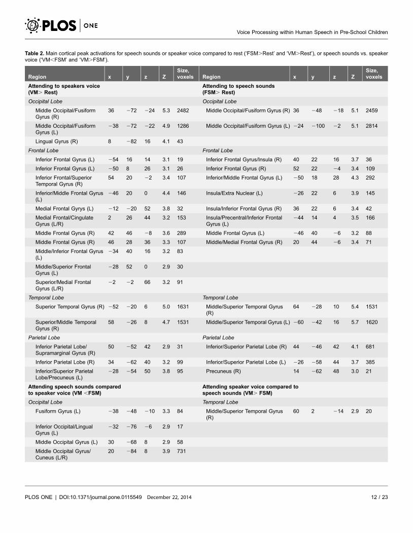
Table 2. Main cortical peak activations for speech sounds or speaker voice compared to rest (‘FSM.Rest’ and ‘VM.Rest’), or speech sounds vs. speakervoice (‘VM,FSM’ and ‘VM.FSM’).
Region x y z ZSize,voxels Region x y z Z
Size,voxels
Attending to speakers voice(VM. Rest)
Attending to speech sounds(FSM. Rest)
Occipital Lobe Occipital Lobe
Middle Occipital/FusiformGyrus (R)
36 272 224 5.3 2482 Middle Occipital/Fusiform Gyrus (R) 36 248 218 5.1 2459
Middle Occipital/FusiformGyrus (L)
238 272 222 4.9 1286 Middle Occipital/Fusiform Gyrus (L) 224 2100 22 5.1 2814
Lingual Gyrus (R) 8 282 16 4.1 43
Frontal Lobe Frontal Lobe
Inferior Frontal Gyrus (L) 254 16 14 3.1 19 Inferior Frontal Gyrus/Insula (R) 40 22 16 3.7 36
Inferior Frontal Gyrus (L) 250 8 26 3.1 26 Inferior Frontal Gyrus (R) 52 22 24 3.4 109
Inferior Frontal/SuperiorTemporal Gyrus (R)
54 20 22 3.4 107 Inferior/Middle Frontal Gyrus (L) 250 18 28 4.3 292
Inferior/Middle Frontal Gyrus(L)
246 20 0 4.4 146 Insula/Extra Nuclear (L) 226 22 6 3.9 145
Medial Frontal Gyrys (L) 212 220 52 3.8 32 Insula/Inferior Frontal Gyrus (R) 36 22 6 3.4 42
Medial Frontal/CingulateGyrus (L/R)
2 26 44 3.2 153 Insula/Precentral/Inferior FrontalGyrus (L)
244 14 4 3.5 166
Middle Frontal Gyrus (R) 42 46 28 3.6 289 Middle Frontal Gyrus (L) 246 40 26 3.2 88
Middle Frontal Gyrus (R) 46 28 36 3.3 107 Middle/Medial Frontal Gyrus (R) 20 44 26 3.4 71
Middle/Inferior Frontal Gyrus(L)
234 40 16 3.2 83
Middle/Superior FrontalGyrus (L)
228 52 0 2.9 30
Superior/Medial FrontalGyrus (L/R)
22 22 66 3.2 91
Temporal Lobe Temporal Lobe
Superior Temporal Gyrus (R) 252 220 6 5.0 1631 Middle/Superior Temporal Gyrus(R)
64 228 10 5.4 1531
Superior/Middle TemporalGyrus (R)
58 226 8 4.7 1531 Middle/Superior Temporal Gyrus (L) 260 242 16 5.7 1620
Parietal Lobe Parietal Lobe
Inferior Parietal Lobe/Supramarginal Gyrus (R)
50 252 42 2.9 31 Inferior/Superior Parietal Lobe (R) 44 246 42 4.1 681
Inferior Parietal Lobe (R) 34 262 40 3.2 99 Inferior/Superior Parietal Lobe (L) 226 258 44 3.7 385
Inferior/Superior ParietalLobe/Precuneus (L)
228 254 50 3.8 95 Precuneus (R) 14 262 48 3.0 21
Attending speech sounds comparedto speaker voice (VM ,FSM)
Attending speaker voice compared tospeech sounds (VM. FSM)
Occipital Lobe Temporal Lobe
Fusiform Gyrus (L) 238 248 210 3.3 84 Middle/Superior Temporal Gyrus(R)
60 2 214 2.9 20
Inferior Occipital/LingualGyrus (L)
232 276 26 2.9 17
Middle Occipital Gyrus (L) 30 268 8 2.9 58
Middle Occipital Gyrus/Cuneus (L/R)
20 284 8 3.9 731
Voice Processing within Human Speech in Pre-School Children
PLOS ONE | DOI:10.1371/journal.pone.0115549 December 22, 2014 12 / 23
STS more strongly activated during voice identification compared to the left (see
Fig. 2 for a complete overview; Notably, mean parameter estimates for higher
decimals reported are close to, but not exactly opposite, most likely because of
subtle masking differences between the two contrasts). Since we here investigate a
very young pediatric population, but have based our ROI analysis on adult
coordinates ([15] due to a lack of studies in younger children), we further
replicated our ROI findings using a right anterior STS region of interest based on
activation from our second level contrast during voice matching (‘VM.FSM’)
and achieved similar significant findings. We also performed a correlational
analysis between behavioral measures and activity within our regions of interest to
investigate the relationship between neuronal activation during voice matching
and behavioral performance, however, we did not find any significant results.
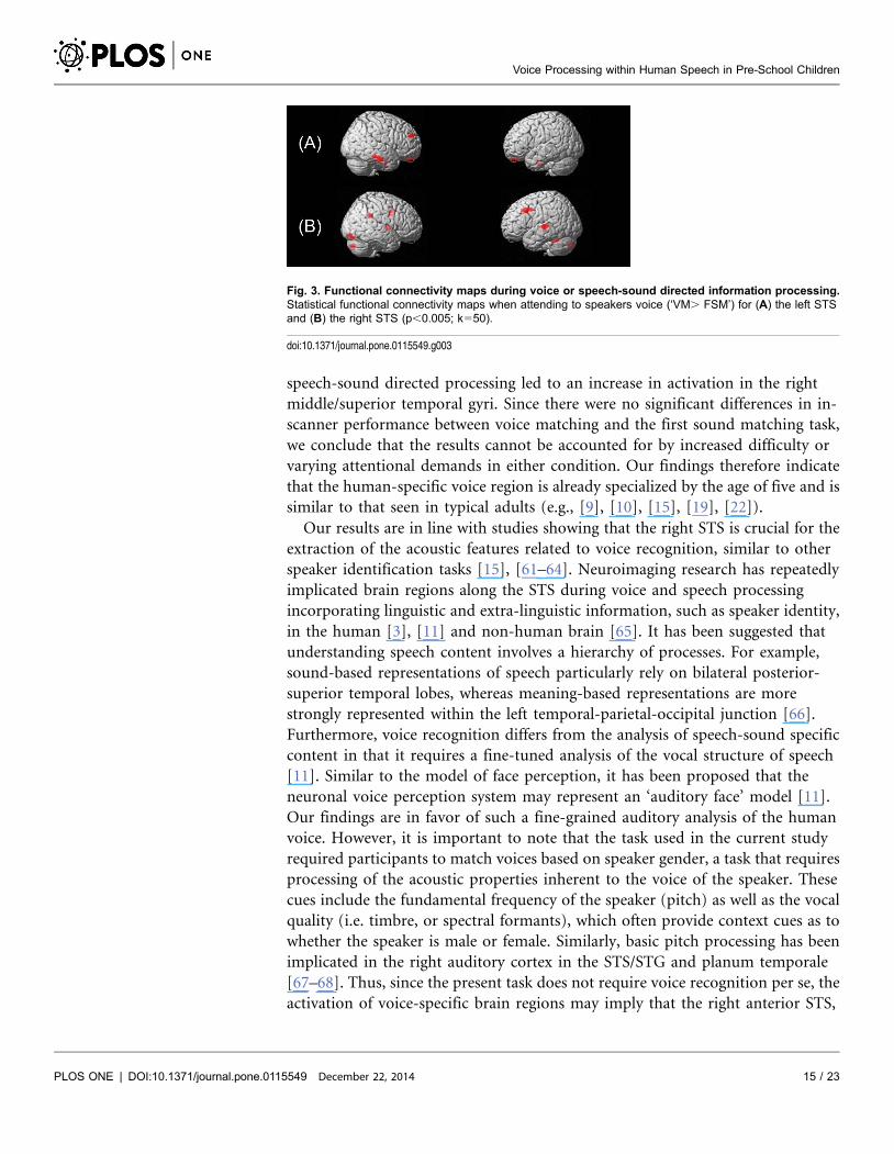
fcMRI Results
We applied a post-hoc seed-to-voxel bivariate correlation analysis to explore
networks of functionally connected regions with the seeds in the right and left STS
as extracted for the ROI analysis. The seed-based analysis was performed for the
contrast ‘VM.FSM’. Findings revealed positive correlations between right STS
and left superior temporal gyrus (STG) and right-hemispheric supramarginal
gyrus, middle frontal gyrus (MFG), putamen, middle occipital gyrus (MOG),
cingulate gyrus and inferior frontal gyrus (IFG). For the left STS, we observed
positive correlation with right-hemispheric superior frontal gyrus (SFG),
postcentral gyrus and inferior temporal gyrus (ITG) (Table 3). Fig. 3 shows the
correlation maps for the left (A) and right (B) STS seeds (‘VM.FSM’).
Discussion
The current paper investigates voice-specific compared to speech-sound specific
processing in preschool-aged children. When compared to rest, both voice and
speech-sound directed tasks activate bilateral primary and secondary auditory
language areas (e.g., bilateral superior and middle temporal gyri), but also
components of the language network (e.g., inferior frontal, temporoparietal and
occipitotemporal brain regions). Focusing on the speaker’s voice compared to
Table 2. Cont.
Region x y z ZSize,voxels Region x y z Z
Size,voxels
Limbic Lobe
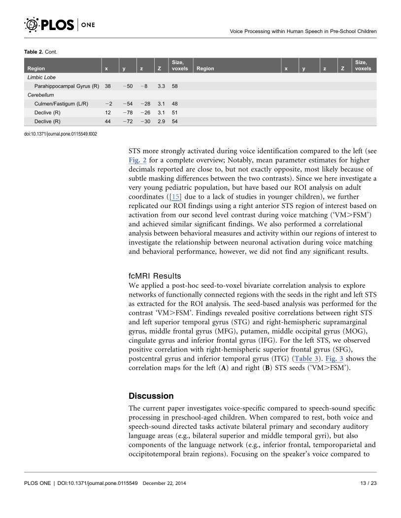
Parahippocampal Gyrus (R) 38 250 28 3.3 58
Cerebellum
Culmen/Fastigum (L/R) 22 254 228 3.1 48
Declive (R) 12 278 226 3.1 51
Declive (R) 44 272 230 2.9 54
doi:10.1371/journal.pone.0115549.t002
Voice Processing within Human Speech in Pre-School Children
PLOS ONE | DOI:10.1371/journal.pone.0115549 December 22, 2014 13 / 23
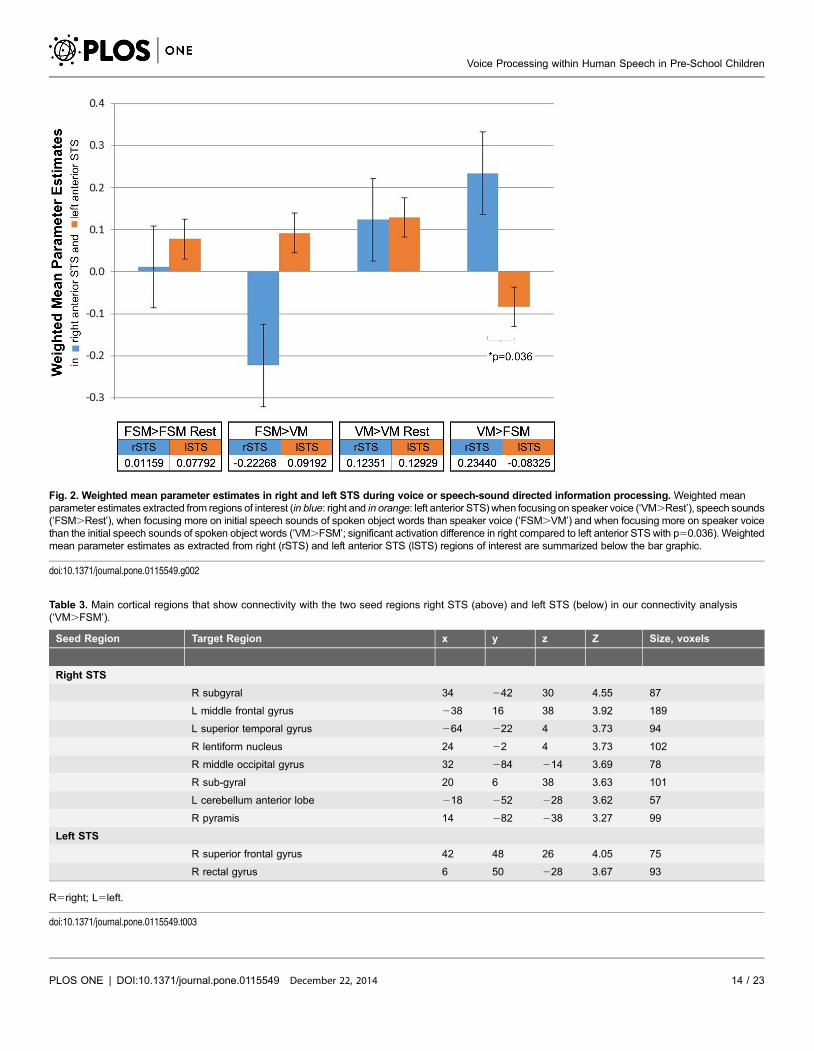
Fig. 2. Weighted mean parameter estimates in right and left STS during voice or speech-sound directed information processing. Weighted meanparameter estimates extracted from regions of interest (in blue: right and in orange: left anterior STS) when focusing on speaker voice (‘VM.Rest’), speech sounds(‘FSM.Rest’), when focusing more on initial speech sounds of spoken object words than speaker voice (‘FSM.VM’) and when focusing more on speaker voicethan the initial speech sounds of spoken object words (‘VM.FSM’; significant activation difference in right compared to left anterior STS with p50.036). Weightedmean parameter estimates as extracted from right (rSTS) and left anterior STS (lSTS) regions of interest are summarized below the bar graphic.
doi:10.1371/journal.pone.0115549.g002
Table 3. Main cortical regions that show connectivity with the two seed regions right STS (above) and left STS (below) in our connectivity analysis(‘VM.FSM’).
Seed Region Target Region x y z Z Size, voxels
Right STS
R subgyral 34 242 30 4.55 87
L middle frontal gyrus 238 16 38 3.92 189
L superior temporal gyrus 264 222 4 3.73 94
R lentiform nucleus 24 22 4 3.73 102
R middle occipital gyrus 32 284 214 3.69 78
R sub-gyral 20 6 38 3.63 101
L cerebellum anterior lobe 218 252 228 3.62 57
R pyramis 14 282 238 3.27 99
Left STS
R superior frontal gyrus 42 48 26 4.05 75
R rectal gyrus 6 50 228 3.67 93
R5right; L5left.
doi:10.1371/journal.pone.0115549.t003
Voice Processing within Human Speech in Pre-School Children
PLOS ONE | DOI:10.1371/journal.pone.0115549 December 22, 2014 14 / 23
speech-sound directed processing led to an increase in activation in the right
middle/superior temporal gyri. Since there were no significant differences in in-
scanner performance between voice matching and the first sound matching task,
we conclude that the results cannot be accounted for by increased difficulty or
varying attentional demands in either condition. Our findings therefore indicate
that the human-specific voice region is already specialized by the age of five and is
similar to that seen in typical adults (e.g., [9], [10], [15], [19], [22]).
Our results are in line with studies showing that the right STS is crucial for the
extraction of the acoustic features related to voice recognition, similar to other
speaker identification tasks [15], [61–64]. Neuroimaging research has repeatedly
implicated brain regions along the STS during voice and speech processing
incorporating linguistic and extra-linguistic information, such as speaker identity,
in the human [3], [11] and non-human brain [65]. It has been suggested that
understanding speech content involves a hierarchy of processes. For example,
sound-based representations of speech particularly rely on bilateral posterior-
superior temporal lobes, whereas meaning-based representations are more
strongly represented within the left temporal-parietal-occipital junction [66].
Furthermore, voice recognition differs from the analysis of speech-sound specific
content in that it requires a fine-tuned analysis of the vocal structure of speech
[11]. Similar to the model of face perception, it has been proposed that the
neuronal voice perception system may represent an ‘auditory face’ model [11].
Our findings are in favor of such a fine-grained auditory analysis of the human
voice. However, it is important to note that the task used in the current study
required participants to match voices based on speaker gender, a task that requires
processing of the acoustic properties inherent to the voice of the speaker. These
cues include the fundamental frequency of the speaker (pitch) as well as the vocal
quality (i.e. timbre, or spectral formants), which often provide context cues as to
whether the speaker is male or female. Similarly, basic pitch processing has been
implicated in the right auditory cortex in the STS/STG and planum temporale
[67–68]. Thus, since the present task does not require voice recognition per se, the
activation of voice-specific brain regions may imply that the right anterior STS,
Fig. 3. Functional connectivity maps during voice or speech-sound directed information processing.Statistical functional connectivity maps when attending to speakers voice (‘VM. FSM’) for (A) the left STSand (B) the right STS (p,0.005; k550).
doi:10.1371/journal.pone.0115549.g003
Voice Processing within Human Speech in Pre-School Children
PLOS ONE | DOI:10.1371/journal.pone.0115549 December 22, 2014 15 / 23
along with right STS regions such as the planum temporale, processes acoustic
voice identification features in general (such as pitch, vocal quality, and gender) in
our age group.
Seed-based fcMRI findings suggest that the right and left STS are correlated
with distinct functional networks during voice processing in pre-school children.
While the right STS correlates positively with left-hemispheric STG and right-
hemispheric temporal, occipital and frontal regions, the left STS correlates with
different right-hemispheric frontal and temporal regions. Three previous studies
investigating voice identity in adults have reported positive correlations between
contralateral STS and STG [10], [44–45]. The observed positive correlations
between the right STS and the right IFG and MFG in pre-school children are in
line with findings reported in adults [44–45], which may suggest a higher
cognitive involvement in voice identity matching based on individual vocal and
glottal parameters. Thus, we suggest that functional correlations between the right
STS and temporal/frontal regions during voice processing in pre-school children
may be comparable to functional networks previously observed in adults. Finally,
in line with our findings, both research groups reported positive correlations
between the right STS and ipsilateral frontal regions such as the IFG [45] and the
dorsolateral prefrontal cortex [44].
Notably, we employed a behavioral interleaved gradient design due to the
nature of our auditory discrimination task. Others have previously demonstrated
that functional networks can be observed by correlating sparse-sampled time-
series data [93–94], [97–99]. Though not optimal for fcMRI analysis, this design is
crucial for auditory experiments (e.g., in order to present auditory stimuli without
interference from scanner background noises [89–93]), especially in the context of
auditory selective attention. Scanner background noise (SBN) can increase BOLD
signal in auditory and language regions resembling a task-induced hemodynamic
response in a highly variable manner across subjects, and SBN during rest
conditions can further mask or alter the BOLD signal in a non-linear fashion [57].
Since fcMRI is inherently more sensitive to non-neuronal sources of noise than
traditional fMRI analysis, sparse temporal sampling may be warranted to avoid
spurious correlations due to scanner background noise. Although we collected
relatively fewer time-points with lower temporal resolution than typical of
continuous scanning designs, Van Dijk and colleagues have shown that fcMRI is
robust to long TRs [100]. Furthermore, the low-frequency fluctuations of interest
in fcMRI (typically ,0.1 Hz) should be captured within our 6 second TR, and we
sampled a consistent number of time points across all conditions.
Bilateral superior temporal sulci have shown to be recruited for a wide range of
pragmatic communicative tasks. Neuroimaging studies have implicated this brain
region during tasks targeting theory of mind and mentalization [69–71], motion
perception [72–73], person impressions [74], gestures [75], face [76] and speech
perception [77] as well as social attention [76]. Because of the diversity of roles of
the bilateral STS in social neuroimaging tasks, it has been argued that this region
may be responsible for interpreting social communicative significance in general
[78]. It has been hypothesized that the right STS may not be a voice-specific area
Voice Processing within Human Speech in Pre-School Children
PLOS ONE | DOI:10.1371/journal.pone.0115549 December 22, 2014 16 / 23
in the human brain per se, but rather represents an area that is responsible for
processing vocal sounds that are communicative in nature. For example, Shultz
and colleagues [79] employed an fMRI task to demonstrate that neuronal
activation within the right STS increases when presented with communicative
vocal sounds (e.g., speech and laughter, see [80]) in comparison with non-
communicative vocal sounds (e.g., coughing and sneezing) [79]. These findings
are in line with our results (where first-sound matching represents a non-
communicative and voice-matching a communicative task). Understanding the
role of the STS in differentiating between communicative and non-commu-
nicative sounds may be critical regarding implications for disorders of social
communication, such as ASD; disorders in which the region within the right STS
has been found to be hypoactivated (e.g., [81]). In addition, individuals with
social communication disorders show structural alterations in brain regions which
again include bilateral brain areas of the STS (e.g., [82–83]).
Although we observed significant differences when comparing the processing of
voice- specific and speech-sound directed speech stimuli within the right anterior
STS, we acknowledge certain limitations. It is noteworthy that only one female
and one male voice were used in this study. For example, it has been shown that
female voices may produce stronger neuronal responses than male voices, despite
a right hemispheric dominance in the STG for both male and female voices [7].
However, the use of male and female voices has been counterbalanced across our
experimental conditions. Future studies should include a wide range of different
speakers, particularly varying in gender, fundamental frequency, or age.
Furthermore, the current study employed a voice matching task, which does not
necessarily demand recognition of speaker voice. Thus, these findings reflect the
neural mechanisms involved in processing communicative vocal sounds, but need
to be interpreted carefully in relation to general processes required for voice
recognition. An additional potential limitation of the current study is the absence
of an adult participant control group. However, there is a robust body of existing
research demonstrating which regions are recruited in adults when completing
similar tasks [10] and activation peaks from these studies have been adapted and
further studied here. Still, we cannot rule out that there are not differences in
brain activation and functional connectivity between children and adults without
an adult control group. Finally, due to the aforementioned temporal restrictions
of our study design and the BOLD signal itself, our fcMRI results are not directly
comparable to connectivity work employing other neuroimaging modalities such
as EEG or MEG, and therefore should be interpreted with caution.
Impairments in speech perception or any of its related relevant features have
been reported in various disorders of social communications or perception,
including autism-spectrum disorder [14],[81], schizophrenia [84], Parkinson’s
disease and Alzheimer’s disease [85–86], as well as in patients with acquired brain
injuries, such as phonagnosia [12–13], ventral frontal lobe damage [87] and right
hemispheric dysfunctions [88]. Understanding the behavioral and neural basis of
these disorders first requires greater knowledge about speech processing in
typically developing populations. Due to technical and practical challenges, few
Voice Processing within Human Speech in Pre-School Children
PLOS ONE | DOI:10.1371/journal.pone.0115549 December 22, 2014 17 / 23
neuroimaging research studies include younger children and many conclusions
about infants and children with developmental disorders are based on the
assumption of a static adult brain [40]. However, modern neuroimaging tools,
such as EEG, MRI and NIRS, offer the means for research targeting abnormal
brain growth, development and function in pediatric populations (e.g [31–33]).
We suggest that the current findings in typically developing children may be
utilized to broaden understanding of neurophysiological findings in atypically
developing children, particularly within disorders of social communication.
In conclusion, the present study demonstrates neuronal differences between the
processing of voice versus speech-specific information in preschool-aged children
within right anterior STS. Our findings indicate that the human-specific voice
region within the right anterior STS is already specialized by the age of five and is
similar to that seen in typical adults. Additionally, positive functional correlations
between the right STS with left-hemispheric STG and right-hemispheric temporal,
occipital and prefrontal regions were observed. Our findings may have
implications within the fields of typical and atypical language and social
development. In particular, this work may guide future studies investigating
young children with speech impairments and disorders of social communication.
Supporting Information
S1 Table. Socioeconomic Status (SES).
doi:10.1371/journal.pone.0115549.s001 (DOC)
S2 Table. Language Development.
doi:10.1371/journal.pone.0115549.s002 (DOC)
Acknowledgments
We thank all participating families and the radiological team at Boston Children’s
Hospital in Waltham for their support in conducting this study. Furthermore, we
thank Yingying Wang and Danielle Sliva for their expertise and thoughtful input
during the review process.
Author ContributionsConceived and designed the experiments: NMR NG. Performed the experiments:
NMR. Analyzed the data: NMR SAS JZ MRD MF NG. Wrote the paper: NMR SAS
JZ MRD MF NG.
References
1. Lavner Y, Gath I, Rosenhouse J (2000) The effects of acoustic modifications on the identification offamiliar voices speaking isolated vowels. Speech Communication 30(1):9–26.
Voice Processing within Human Speech in Pre-School Children
PLOS ONE | DOI:10.1371/journal.pone.0115549 December 22, 2014 18 / 23
2. Van Dommelen WA (1990) Acoustic parameters in human speaker recognition. Language and Speech33(3):259–272.
3. Grossmann T, Oberecker R, Koch SP, Friederici AD (2010) The developmental origins of voiceprocessing in the human brain. Neuron 65: 852–858.
4. Scherer KR (1995) Expression of emotion in voice and music. J Voice 9: 235–248.
5. Hartman DE, Danahuer JL (1976) Perceptual features of speech for males in four perceived agedecades. J Acoust Soc Am 59: 713–715.
6. Lass NJ, Hughes KR, Bowyer MD, Waters LT, Bourne VT (1976) Speaker sex identification fromvoiced, whispered, and filtered isolated vowels. J Acoust Soc Am 59: 675–678.
7. Lattner S, Meyer ME, Friederici AD (2005) Voice perception: Sex, pitch, and the right hemisphere. HumBrain Mapp 24: 11–20.
8. Patterson ML, Werker JF (2003) Two-month old infants match phonetic information in lips and voice.Developmental Science 6(2):191–196.
9. Von Kriegstein K, Eger E, Kleinschmidt A, Giraud AL (2003) Modulation of neural responses tospeech by directing attention to voices or verbal content. Brain Res Cogn Brain Res 17: 48–55.
10. Von Kriegstein K, Giraud AL (2004) Distinct functional substrates along the right superior temporalsulcus for the processing of voices. NeuroImage 22: 948–955.
11. Belin P, Bestelmeyer PE, Latinus M, Watson R (2011) Understanding voice perception. Br J Psychol102: 711–725.
12. Van Lancker DR, Cummings JL, Kreiman J, Dobkin BH (1988) Phonagnosia: a dissociation betweenfamiliar and unfamiliar voices. Cortex 24: 195–209.
13. Van Lancker DR, Kreiman J, Cummings J (1989) Voice perception deficits: neuroanatomicalcorrelates of phonagnosia. J Clin Exp Neuropsychol 11: 665–674.
14. Ethofer T, Van De Ville D, Scherer K, Vuilleumier P (2009) Decoding of Emotional Information inVoice-Sensitive Cortices. Current Biology 19(12):1028–1033.
15. Belin P, Zatorre RJ (2003) Adaptation to speaker’s voice in right anterior temporal lobe. Neuroreport 14:2105–2109.
16. Petkov CI, Logothetis NK, Obleser J (2009) Where are the human speech and voice regions, and doother animals have anything like them? Neuroscientist 15: 419–429.
17. Belin P, Zatorre RJ, Lafaille P, Ahad P, Pike B (2000) Voice-selective areas in human auditory cortex.Nature 403: 309–312.
18. Linden DE, Thornton K, Kuswanto CN, Johnston SJ, van de Ven V, et al. (2011) The brain’s voices:comparing nonclinical auditory hallucinations and imagery. Cereb Cortex 21: 330–337.
19. Binder JR, Frost JA, Hammeke TA, Bellgowan PS, Springer JA, et al. (2000) Human temporal lobeactivation by speech and nonspeech sounds. Cereb Cortex 10: 512–528.
20. Altman NR, Bernal B (2001) Brain activation in sedated children: auditory and visual functional MRimaging. Radiology 221: 56–63.
21. Fecteau S, Armony JL, Joanette Y, Belin P (2004) Is voice processing species-specific in humanauditory cortex? An fMRI study. NeuroImage 23: 840–848.
22. Belin P, Fecteau S, Bedard C (2004) Thinking the voice: neural correlates of voice perception. TrendsCogn Sci 8: 129–135.
23. Belin P, Zatorre RJ, Ahad P (2002) Human temporal-lobe response to vocal sounds. Brain Res CognBrain Res 13: 17–26.
24. Ecklund-Flores L, Turkewitz G (1996) Asymmetric headturning to speech and nonspeech in humannewborns. Dev Psychobiol 29: 205–217.
25. Homae F, Watanabe H, Nakano T, Asakawa K, Taga G (2006) The right hemisphere of sleeping infantperceives sentential prosody. Neuroscience research 54(4): 276–280.
26. Pena M, Maki A, Kovacic D, Dehaene-Lambertz G, Koizumi Hi, et al. (2003) Sounds and silence: Anoptical-topography study of language recognition at birth. PNAS 100 (20): 11702–11705.
Voice Processing within Human Speech in Pre-School Children
PLOS ONE | DOI:10.1371/journal.pone.0115549 December 22, 2014 19 / 23
27. Mazoyer BM, Tzourio N, Frak V, Syrota A, Murayama N, et al. (1993) The cortical representation ofspeech. Journal of Cognitive Neuroscience, 5(4), 467–479.
28. Dehaene S, Dupoux E, Mehler J, Cohen L, Paulesu E, et al. (1997) Anatomical variability in thecortical representation of first and second language. Neuroreport, 8(17), 3809–3815.
29. Blasi A, Mercure E, Lloyd-Fox S, Thomson A, Brammer M, et al. (2011) Early Specialization for Voiceand Emotion Processing in the Infant Brain. Report 21: 1220–1224.
30. Dehaene-Lambertz G, Dehaene S, Hertz-Pannier L (2002) Functional neuroimaging of speechperception in infants. Science, 298, 2013–215.
31. Bookheimer SY (2000) Methodological issues in pediatric neuroimaging. Mental retardation anddevelopmental disabilities research reviews, 6(3):161–165.
32. Raschle N, Zuk J, Ortiz-Mantilla S, Sliva DD, Franceschi A, et al. (2012a) Pediatric neuroimaging inearly childhood and infancy: challenges and practical guidelines. Ann N Y Acad Sci 1252: 43–50.
33. Raschle NM, Lee M, Buechler R, Christodoulou JA, Chang M, et al. (2009) Making MR ImagingChild’s Play - Pediatric Neuroimaging Protocol, Guidelines and Procedure. JoVE 29.
34. Schlaggar BL, Brown TT, Lugar HM, Visscher KM, Miezin FM, et al. (2002) Functionalneuroanatomical differences between adults and school-age children in the processing of singlewords. Science 296: 1476–1479.
35. Nath AR, Fava EE, Beauchamp MS (2011) Neural correlates of interindividual differences in children’saudiovisual speech perception. J Neurosci 28: 13963–13971.
36. Berl MM, Duke ES, Mayo J, Rosenberger LR, Moore EN, et al. (2010) Functional anatomy of listeningand reading comprehension during development. Brain Lang 114: 115–125.
37. Lu LH, Dapretto M, O’Hare ED, Kan E, McCourt ST, et al. (2009) Relationships between brainactivation and brain structure in normally developing children. Cereb Cortex 11: 2595–2604.
38. Casey BJ, Jones RM, Hare TA (2008) The adolescent brain. Ann N Y Acad Sci 1124: 111–126.
39. Bitan T, Cheon J, Lu D, Burman DD, Gitelman DR, et al. (2007) Developmental changes in activationand effective connectivity in phonological processing. Neuroimage 38: 564–575.
40. Marsh M, Gerber AJ, Peterson BS (2008) Neuroimaging Studies of Normal Brain Development andTheir Relevance for Understanding Childhood Neuropsychiatric Disorders. Journal of the AmericanAcademy of Child & Adolescent Psychiatry 47(11): 1233–1251.
41. Dehaene-Lambertz G, Baillet S (1998) A phonological representation in the infant brain. NeuroReport,9(8), 1885–1888.
42. Rogier O, Roux S, Belin P, Bonnet-Brilhault F, Bruneau N (2010) An electrophysiological correlate ofvoice processing in 4- to 5-year-old children. Int J Psychophysiol 75: 44–47.
43. Charest I, Pernet C, Latinus M, Crabbe F, Belin P (2012) Cerebral Processing of Voice GenderStudied Using a Continuous Carryover fMRI Design. Cereb Cortex.
44. Von Kriegstein K, Smith DR, Patterson RD, Kiebel SJ, Griffiths TD (2010) How the human brainrecognizes speech in the context of changing speakers. J Neurosci 30(2):629–638.
45. McGettigan C, Eisner F, Agnew ZK, Manly T, Wisbey D, et al. (2013) T’ain’t what you say, it’s the wayyou say it: Left insula and inferior frontal cortex work in interaction with superior temporal regions tocontrol the performance of vocal impersonations. J Cogn Neurosci 25(11), 1875–1886.
46. Formisano E, De Martino F, Bonte M, Goebel R (2008) ‘‘Who’’ is saying ‘‘What’’? Brain-baseddecodingof human voice and speech. Science 322(5903):970–973.
47. Kaufman ASKNL (1997) KBIT-2: Kaufman Brief Intelligence Test. Minneapolis, MN: NCS Pearson, Inc.
48. Semel E, Wiig EH, Secord WA (2000) Child Evaluation of Language Fundamentals. San Antonia, USA:The Psychological Corporation.
49. Wagner RK, Torgesen JK, Rashotte CA (1999) The Comprehensive Test of Phonological Processing.Austin: PRO-ED, Inc.
50. Van der Lely HK (2000) Verb Agreement and Tense Test (VATT). London: Centre for DevelopmentalLanguage Disorders and Cognitive Neuroscience (DLDCN.COM).
Voice Processing within Human Speech in Pre-School Children
PLOS ONE | DOI:10.1371/journal.pone.0115549 December 22, 2014 20 / 23
51. Wolf M, Denckla MB (2005) RAN/RAS: Rapid Automatized Naming and Rapid Alternating. Austin, TX:PRO-ED, Inc.
52. Raschle NM, Zuk J, Gaab N (2012b) Functional characteristics of developmental dyslexia in left-hemispheric posterior brain regions predate reading onset. Proc Natl Acad Sci 109: 2156–2161.
53. Grill-Spector K, Henson R, Martin A (2006) Repetition and the brain: neural models of stimulus-specific effects. Trends in Cognitive Sciences 10(1):14–23.
54. Snodgrass JG, Vanderwart M (1980) A standardized set of 260 pictures: norms for name agreement,image agreement, familiarity, and visual complexity. J Exp Psychol Hum Learn 6: 174–215.
55. Hall DA, Haggard MP, Akeroyd MA, Palmer AR, Summerfield AQ, et al. (1999) "Sparse" temporalsampling in auditory fMRI. Hum Brain Mapp 7: 213–223.
56. Gaab N, Gaser C, Zaehle T, Jancke L, Schlaug G (2003) Functional anatomy of pitch memory-an fMRIstudy with sparse temporal sampling. Neuroimage 19(4): 1417–1426.
57. Gaab N, Gabrieli JD, Glover GH (2007a) Assessing the influence of scanner background noise onauditory processing. I. An fMRI study comparing three experimental designs with varying degrees ofscanner noise. Hum Brain Mapp 28: 703–720.
58. Talairach J, Tournoux P (1988) Co-planar Stereotaxie Atlas of the Human Brain: 3-dimensionalProportional System: An Approach to Cerebral Imaging. Stuttgart: Thieme Verlag.
59. Whitfield-Gabrieli S, Nieto-Castanon A (2012) Conn: A functional connectivitytoolbox for correlatedand anticorrelated brain networks. Brain Connect 2(3): 125–141.
60. Bosch V (2000) Statistical Analysis of Multi-Subject fMRI Data: Assessment of Focal Activations.J Magn Reson Imaging (11), 61–64.
61. Imaizumi S, Mori K, Kiritani S, Kawashima R, Sugiura M, et al. (1997) Vocal identification of speaker.
62. Andics A, McQueen JM, Petersson KM, Gal V, Rudas G, et al. (2010) Neuralmechanismsfor voicerecognition. NeuroImage 52: 1528–1540.
63. Latinus M, Crabbe F, Belin P (2011) Learning-induced changes in the cerebral processing of voiceidentity. Cereb Cortex 21: 2820–2828.
64. Scott SK, Blank CC, Rosen S, Wise RJ (2000) Identification of a pathway for intelligible speech in theleft temporal lobe. Brain 123 Pt 12: 2400–2406.
65. Petkov CI, Kayser C, Steudel T, Whittingstall K, Augath M, et al. (2008) A voice region in the monkeybrain. Nat Neurosci 11: 367–374.
66. Hickok G, Poeppel D (2000) Towards a functional neuroanatomy of speech perception. Trends CognSci 4: 131–138.
67. Hyde KL, Peretz I, Zatorre RJ (2008) Evidence for the role of the right auditory cortex in fine pitchresolution. Neuropsychologia, 46(2), 632–639.
68. Zatorre RJ (1988) Pitch perception of complex tones and human temporal-lobe function. The Journal ofthe Acoustical Society of America, 84, 566.
69. Saxe R, Baron-Cohen S (2006) The neuroscience of theory of mind. Soc Neurosci 1:i–ix.
70. Mier D, Sauer C, Lis S, Esslinger C, Wilhelm J, et al. (2010) Neuronal correlates of affective theory ofmind in schizophrenia out-patients: evidence for a baseline deficit. Psychol Med 40: 1607–1617.
71. Herholz SC, Halpern AR, Zatorre RJ (2012) Neuronal correlates of perception, imagery, and memoryfor familiar tunes. J Cogn Neurosci 24: 1382–1397.
72. Puce A, Perrett D (2003) Electrophysiology and brain imaging of biological motion. Philos Trans R SocLond B Biol Sci 358: 435–445.
73. Van der Wyk BC, Voos A, Pelphrey KA (2012) Action representation in the superior temporal sulcus inchildren and adults: An fMRI study. Dev Cogn Neurosci.
74. Mende-Siedlecki P, Cai Y, Todorov A (2012) The neural dynamics of updating person impressions. SocCogn Affect Neurosci.
75. Dick AS, Goldin-Meadow S, Solodkin A, Small SL (2012) Gesture in the developing brain. Dev Sci 15:165–180.
Voice Processing within Human Speech in Pre-School Children
PLOS ONE | DOI:10.1371/journal.pone.0115549 December 22, 2014 21 / 23
76. Iidaka T (2012) The role of the superior temporal sulcus in face recognition and perception. Brain Nerve64: 737–742.
77. Price CJ (2000) The anatomy of language: contributions from functional neuroimaging. J Anat 197 Pt 3:335–359.
78. Redcay E (2008) The superior temporal sulcus performs a common function for social and speechperception: implications for the emergence of autism. Neurosci Biobehav Rev 32: 123–142.
79. Shultz S, Vouloumanos A, Pelphrey K (2012) The superior temporal sulcus differentiatescommunicative and noncommunicative auditory signals. J Cogn Neurosci 24: 1224–1232.
80. Meyer M, Zysset S, Von Cramon DY, Alter K (2005) Distinct fMRI responses to laughter, speech, andsounds along the human peri-sylvian cortex. Cognitive Brain Research 24(2), 291–306.
81. Gervais H, Belin P, Boddaert N, Leboyer M, Coez A, et al. (2004) Abnormal cortical voice processingin autism. Nat Neurosci 7: 801–802.
82. Boddaert N, Chabane N, Gervais H, Good CD, Bourgeois M, et al. (2004) Superior temporal sulcusanatomical abnormalities in childhood autism: a voxel-
83. Greimel E, Nehrkorn B, Schulte-Ruther M, Fink GR, Nickl-Jockschat T, et al. (2012) Changes in greymatter development in autism spectrum disorder. Brain Struct Funct.
84. Borod JC, Welkowitz J, Alpert M, Brozgold AZ, Martin C, et al. (1990) Parameters of emotionalprocessing in neuropsychiatric disorders: conceptual issues and a battery of tests. Journal ofCommunication Disorders 23(4):247–271.
85. Hailstone JC, Ridgway GR, Bartlett JW, Goll JC, Buckley AH, et al. (2011) Voice processing indementia: a neuropsychological and neuroanatomical analysis. Brain 134(9): 2535–2547.
86. Pell MD, Leonard CL (2003) Processing emotional tone from speech in Parkinson’s disease: A role forthe basal ganglia. Cognitive, Affective, & Behavioral Neuroscience 3(4): 275–288.
87. Hornak J, Rolls ET, Wade D (1996) Face and voice expression identification in patients with emotionaland behavioural changes following ventral frontal lobe damage. Neuropsychologia, 34(4): 247–261.
88. Cohen MJ, Branch WB, Hynd GW (1994) Receptive prosody in children with left or right hemispheredysfunction. Brain and Language 47(2): 171–181.
89. Gaab N, Gabrieli JD, Glover GH (2007a) Assessing the influence of scanner background noise onauditory processing. I. An fMRI study comparing three experimental designs with varying degrees ofscanner noise. Hum Brain Mapp 28: 703–720.
90. Gaab N, Gabrieli JD, Glover GH (2007b) Assessing the influence of scanner background noise onauditory processing. II. An fMRI study comparing auditory processing in the absence and presence ofrecorded scanner noise using a sparse design. Hum Brain Mapp 28: 721–732.
91. Gaab N, Gabrieli JD, Glover GH (2008) Resting in peace or noise: Scanner background noisesuppresses default-mode network. Hum Brain Mapp 29: 858–867.
92. Hall DA, Haggard MA, Akeroyd AR, Palmer AR, Summerfield AQ, et al. (1999) ‘‘Sparse’’ temporalsampling in auditory fMRI. Hum Brain Mapp 7: 213–223.
93. Goncalves MS, Hall DA, Johnsrude IS, Haggard M (2001) Can Meaningful EffectiveConnectivities beObtained between Auditory Cortical Regions? Neuroimage 14: 13531360.
94. Kahn I, Andrews-Hanna JR, Vincent JL, Snyder AZ, Buckner RL (2008) Distinct Cortical AnatomyLinked to Subregions of the Medial Temporal Lobe Revealed by Intrinsic Functional Connectivity.J Neurophysiol 100: 129–139.
95. Thomason ME, Burrows BE, Gabrieli JD, Glover GH (2005) Breath holding reveals differences infMRI BOLD signal in children and adults. Neuroimage 25: 824–837.
96. Langer N, Benjamin C, Minas J, Gaab N (2013) The Neural Correlates of Reading Fluency Deficits inChildren. Cereb Cortex [Epub ahead of print].
97. Loui P, Zamm A, Schlaug G (2012) Enhanced functional networks in absolute pitch. Neuroimage 63(2):632–640.
98. Langers DRM, Melcher JR (2011) Hearing without listening: Functional connectivity reveals theengagement of multiple non-auditory networks during basic sound processing. Brain Connectivity 1(3):233–244.
Voice Processing within Human Speech in Pre-School Children
PLOS ONE | DOI:10.1371/journal.pone.0115549 December 22, 2014 22 / 23
99. Wang Y, Holland SK (2014) Comparison of functional network connectivity for passive-listening andactive-response narrative comprehension in adolescents. Brain Connectivity (4)4: 273–284.
100. VanDijk KRA, Hedden T, Venkataraman A, Evans KC, Lazar SW, et al. (2009) Intrinsic functionalconnectivity as a tool for human connectomics: Theory, properties, and optimization. Journal ofNeurophysiology 103 (1): 297–321.
Voice Processing within Human Speech in Pre-School Children
PLOS ONE | DOI:10.1371/journal.pone.0115549 December 22, 2014 23 / 23