Intraspecific comparative analysis of the species Salinibacter ruber
11
ORIGINAL PAPER Arantxa Pen˜a Maria Valens Fernando Santos Sandra Buczolits Josefa Anto´n Peter Ka¨mpfer Hans-Ju¨rgen Busse Rudolf Amann Ramon Rossello´ -Mora Intraspecific comparative analysis of the species Salinibacter ruber Received: 26 May 2004 / Accepted: 17 December 2004 / Published online: 15 March 2005 ȑ Springer-Verlag 2005 Abstract Salinibacter ruber is the first extremely halo- philic member of the Bacteria domain of proven environmental relevance in hypersaline brines at or approaching NaCl saturation, that has been brought to pure culture. A collection of 17 strains isolated from five different geographical locations (Mallorca, Alicante, Ebro Delta, Canary Islands, and Peruvian Andes) were studied following the currently accepted taxonomic approach. Additionally, random amplifica- tion of genomic DNA led to the phenetic analysis of the intraspecific diversity. Altogether the taxonomic study indicated that S. ruber remained highly homo- geneous beyond any geographical barrier. However, genomic fingerprints indicated that populations from different isolation sites could still be discriminated. Introduction Salinibacter ruber (Anto´n et al. 2002) is the first ex- tremely halophilic member of the Bacteria domain of proven environmental relevance (Anto´n et al. 2000) that has been cultured in the laboratory. Members of this species occur naturally in hypersaline brines at or approaching NaCl saturation. The use of molecular techniques allowed the discovery of this microorganism (Anto´n et al. 2000) and, shortly thereafter, its isolation and characterization (Anto´n et al. 2002). S. ruber affili- ates as a deep branch with the Bacteroidetes-Chlorobi superphylum, together with Rhodothermus marinus (Anto´n et al. 2002), and it has been classified within the Phylum BXX, Bacteroidetes, Class III Sphingobacteria, Order I Sphingobacteriales, Family V Crenotrichaceae, as a single new genus (Genus IV) and species (Garrity et al. 2003). Five strains were first isolated from the brines of solar salterns in Alicante and Mallorca that were the basis for the classification of the new genus and species (Anto´n et al. 2002). Since then, new information obtained from these isolates has shown a surprising similarity to the extremely halophilic Archaea. For example, like halo- archaea, S. ruber strains accumulate high intracellular amounts of potassium and chloride (Anto´n et al. 2002; Mu¨ller and Oren 2003; Oren et al. 2002). This feature has only been previously found in Bacteria for the members of the Haloanaerobiales and this is reflected by the high proportion of acidic amino acids in S. ruber proteins, another typical feature of haloarchaea (Oren and Mana 2002). Additionally, the proteins analysed show a low proportion of hydrophobic amino acids that are compensated for by the increase of serine (Oren and Mana 2002). Such modifications in amino acid compo- sition directly influence the requirements for inorganic anions. Several enzymes have been tested and most of them require chloride that has to be pumped into the cells (Mu¨ ller and Oren 2003). The high concentrations of chloride in haloarchaea are achieved by cotransport with Communicated by W.D. Grant A. Pen˜a F. Santos J. Anto´n Divisio´n de Microbiologı´a, Departamento de Fisiologı´a, Gene´tica y Microbiologı´a, Universidad de Alicante, Apto. 99, 03080 Alicante, Spain M. Valens R. Rossello´-Mora (&) Grup d’Oceanografia interdisciplinar, Departament de Recursos Naturals, Institut Mediterra`ni d’Estudis Avanc¸ats (CSIC-UIB), C/Miquel Marque´s 21, 07190 Esporles (Illes Balears), Spain E-mail: [email protected] Tel.: +34-971-611-826 Fax: +34-971-173-184 S. Buczolits H.-J. Busse Institut fu¨r Mikrobiologie und Genetik, Universita¨t Wien, 1030, Wien, Austria H.-J. Busse Institut fu¨r Bakteriologie, Mykologie und Hygiene, Veterina¨rmedizinische Universita¨t, 1210, Wien, Austria P. Ka¨mpfer Institut fu¨r Angewandte Mikrobiologie, Justus-Liebig Universita¨t Giessen, 35390 Giessen, Germany R. Amann Max-Planck-Institut fu¨r Marine Mikrobiologie, 28359 Bremen, Germany Extremophiles (2005) 9:151–161 DOI 10.1007/s00792-005-0430-y
-
Upload
independent -
Category
Documents
-
view
2 -
download
0
Transcript of Intraspecific comparative analysis of the species Salinibacter ruber

ORIGINAL PAPER
Arantxa Pena Æ Maria Valens Æ Fernando Santos
Sandra Buczolits Æ Josefa Anton Æ Peter Kampfer
Hans-Jurgen Busse Æ Rudolf Amann
Ramon Rossello-Mora
Intraspecific comparative analysis of the species Salinibacter ruber
Received: 26 May 2004 / Accepted: 17 December 2004 / Published online: 15 March 2005� Springer-Verlag 2005
Abstract Salinibacter ruber is the first extremely halo-philic member of the Bacteria domain of provenenvironmental relevance in hypersaline brines at orapproaching NaCl saturation, that has been broughtto pure culture. A collection of 17 strains isolatedfrom five different geographical locations (Mallorca,Alicante, Ebro Delta, Canary Islands, and PeruvianAndes) were studied following the currently acceptedtaxonomic approach. Additionally, random amplifica-tion of genomic DNA led to the phenetic analysis ofthe intraspecific diversity. Altogether the taxonomicstudy indicated that S. ruber remained highly homo-geneous beyond any geographical barrier. However,genomic fingerprints indicated that populations fromdifferent isolation sites could still be discriminated.
Introduction
Salinibacter ruber (Anton et al. 2002) is the first ex-tremely halophilic member of the Bacteria domain ofproven environmental relevance (Anton et al. 2000) thathas been cultured in the laboratory. Members of thisspecies occur naturally in hypersaline brines at orapproaching NaCl saturation. The use of moleculartechniques allowed the discovery of this microorganism(Anton et al. 2000) and, shortly thereafter, its isolationand characterization (Anton et al. 2002). S. ruber affili-ates as a deep branch with the Bacteroidetes-Chlorobisuperphylum, together with Rhodothermus marinus(Anton et al. 2002), and it has been classified within thePhylum BXX, Bacteroidetes, Class III Sphingobacteria,Order I Sphingobacteriales, Family V Crenotrichaceae,as a single new genus (Genus IV) and species (Garrityet al. 2003).
Five strains were first isolated from the brines of solarsalterns in Alicante and Mallorca that were the basis forthe classification of the new genus and species (Antonet al. 2002). Since then, new information obtained fromthese isolates has shown a surprising similarity to theextremely halophilic Archaea. For example, like halo-archaea, S. ruber strains accumulate high intracellularamounts of potassium and chloride (Anton et al. 2002;Muller and Oren 2003; Oren et al. 2002). This featurehas only been previously found in Bacteria for themembers of the Haloanaerobiales and this is reflected bythe high proportion of acidic amino acids in S. ruberproteins, another typical feature of haloarchaea (Orenand Mana 2002). Additionally, the proteins analysedshow a low proportion of hydrophobic amino acids thatare compensated for by the increase of serine (Oren andMana 2002). Such modifications in amino acid compo-sition directly influence the requirements for inorganicanions. Several enzymes have been tested and most ofthem require chloride that has to be pumped into thecells (Muller and Oren 2003). The high concentrations ofchloride in haloarchaea are achieved by cotransport with
Communicated by W.D. Grant
A. Pena Æ F. Santos Æ J. AntonDivision de Microbiologıa, Departamento de Fisiologıa,Genetica y Microbiologıa, Universidad de Alicante,Apto. 99, 03080 Alicante, Spain
M. Valens Æ R. Rossello-Mora (&)Grup d’Oceanografia interdisciplinar,Departament de Recursos Naturals,Institut Mediterrani d’Estudis Avancats (CSIC-UIB),C/Miquel Marques 21, 07190 Esporles (Illes Balears), SpainE-mail: [email protected].: +34-971-611-826Fax: +34-971-173-184
S. Buczolits Æ H.-J. BusseInstitut fur Mikrobiologie und Genetik, Universitat Wien,1030, Wien, Austria
H.-J. BusseInstitut fur Bakteriologie, Mykologie und Hygiene,Veterinarmedizinische Universitat, 1210, Wien, Austria
P. KampferInstitut fur Angewandte Mikrobiologie,Justus-Liebig Universitat Giessen,35390 Giessen, Germany
R. AmannMax-Planck-Institut fur Marine Mikrobiologie,28359 Bremen, Germany
Extremophiles (2005) 9:151–161DOI 10.1007/s00792-005-0430-y
sodium ions and/or using the light driven primarychloride pump halorhodopsin (Muller and Oren 2003).The presence of a homologous gene to halorhodopsinhas in fact been found in several strains of S. ruber(J.Anton et al. in preparation). Additionally, featureslike the high G+C content of the genome (Anton et al.2002) and pigmentation (Oren and Rodrıguez-Valera2001) clearly indicate that these organisms share manycharacteristics with the known haloarchaea.
Although the first five isolates were obtained fromsolar salterns in Alicante and Mallorca (Anton et al.2002), molecular microbial ecology methods showed thatS. ruber thrives in a broader geographical region thatcovers the whole Mediterranean (Anton et al. 2000). Theextreme nature of the environments that S. ruber inhabitsand the distant geographical position of such sampledsites are adequate conditions for allopatric speciation(Staley 2004). The aim of the present study was to isolatenew organisms from new sampling sites to understandthe geographical distribution and intraspecific diversityof S. ruber. We have characterised a collection of 17strains (including the five original strains used for speciesdescription) by studying several biochemical traits andchemotaxonomical markers such as fatty acid profiles,the quinone system, lipopolysaccharide and polar lipids,as well as the two genetic typing strategies randomlyamplified polymorphic DNA (RAPD; Sikorski et al.1999) and pulsed field gel electrophoresis analysis(PFGE; Grothues and Tummler 1991).
Methods
Strains, media and culture conditions
New S. ruber isolates were obtained from brine samplescollected from crystallizer ponds of salterns located atLa Palma (Canary Islands, Spain), San Carles de laRapita (Ebro Delta, Spain) and Maras (Peruvian Andes)during the years 2000–2002. All isolates were obtainedby plating samples or dilutions on salt solution 25% seawater (SW), containing (in gram per litre) NaBr, 0.65;NaHCO3, 0.167; KCl, 5; CaCl2, 0.723; MgSO4Æ7H2O,49.492; MgCl2Æ6H2O, 34.567; NaCl, 195; with 0.2%yeast extract (the pH was adjusted to 7.2 with NaOHprior to autoclaving), followed by incubation at 37�C.After growth, cells from colonies were examinedmicroscopically, with rods being selected and inoculatedagain on the same medium. In all cases, agar plates (SWsupplemented with yeast extract and 1.5% agar), or li-quid media (with shaking) were incubated at 37�C for atleast 14 days to obtain sufficient biomass for furtherexperiments.
Screening and selection of Salinibacter isolates
The universal primers used for PCR amplification of thebacterial and archaeal 16S rDNA were a forward primer
27f for Bacteria, (Lane 1991), or a forward primer 21Ffor Archaea (DeLong et al. 1992) and universal reverseprimer 1492r (Lane 1991) for both Bacteria and Ar-chaea. PCR was carried out as described previously(Acinas et al. 1999). Isolates belonging to the domainBacteria were submitted to a second PCR amplificationwith the specific primers for Salinibacter spp. EHB4F(5¢-ACACCCCTATGGGGCGTA-3¢, Anton et al.2002), and EHB9R (5¢-AGCGTCGAGCCTAGGTA-3¢). Alternatively, strains were identified as Salinibacterspp. by fluorescence in situ hybridisation (FISH) usingthe specific probe EHB412 labelled with the fluoro-chrome CY3 (5¢-TACGCCCCATAGGGTGT-3¢, Ant-on et al. 2000). Both the FISH probe and theamplification primers targeted specifically all thosehitherto known sequences of the genus Salinibacterwhich include sequences of all strains of S. ruber andthat of the hitherto uncultured EHB-2.
DNA isolation, DNA base compositionand DNA-DNA hybridisation
Genomic DNA was prepared following the method ofMarmur (1961). G+C content was analysed byhydrolysis of DNA to its nucleosides and quantifiedby HPLC following a modification of the method ofTamaoka and Komagata (1984). The experiments werecarried out as follows: 10 ll containing about 1 lg ofDNA were denatured by incubating 5 min at 100�Cand immediately ice chilled. Denatured DNA was thenmixed with 10 ll of nuclease buffer (sodium acetate40 mM; ZnSO4 2 mM; pH 5.3) containing 0.1 mg/mlof nuclease P1 (SIGMA), and incubated for 2 h at50�C. Once the DNA was digested, 10 ll of de-phosporylation buffer (Tris-HCl 0.1 M pH 8.1) con-taining 3 U of alkaline phosphatase (Roche) wereadded to the mixture and incubated for 2 h at 37�C.Those samples were then directly injected into theflow. Nucleoside chromatography was carried out witha Waters-Millipore HPLC apparatus, using a C18 re-versed-phase column (NOVA-PAK, Waters-Millipore).Nucleoside were eluted by using NH4H2PO4 0.1 MpH 4: acetonitrile (20:1, v:v), with a flow of 1 ml/minat room temperature. Chromatography was completedafter 10 min. Nucleosides were detected by UVabsorbance at 270 nm. Relative amounts of G+Cwere calculated by the use of the following standardDNAs: Staphylococcus aureus (strain CECT239,G+C: 34%; Hollander and Pohl 1980); Shewanellaputrefaciens (CECT5346, G+C: 43.9; Owen et al.1978); Escherichia coli B (strain CECT101, G+C:52%; Hollander and Pohl 1980); Pseudomonas aeru-ginosa (strain DSMZ 50071; G+C: 67.2%; Palleroni1984); and Agrococcuss jenensis (strain DSMZ 9580;G+C: 74%; Groth et al. 1996). DNA–DNA hy-bridisation experiments were carried out following amicrotiter plate non-radioactive method (Ziemke et al.1998).
152

Sequencing
In order to sequence the almost complete 16S rRNAgene, three primers were used: 533R (Amann et al.1995) EHB4F (Anton et al. 2002) and 1492r (Lane1991). PCR products were purified with the ConcertRapid PCR Purification System (Promega) accordingto the manufacturer’s protocol. The nucleotide se-quences of the products were determined using the BigDye Terminator Cycle Sequencing kit and an ABIPRISM 310 DNA sequencer (Applied Biosystems).Complete 16S rRNA sequences were compared ini-tially with reference sequences at NCBI (http://www.ncbi.nlm.nih.gov) using BLAST (Altschul et al.1997) and they were subsequently aligned with 16SrRNA reference sequences in the ARB package(Ludwig et al. 2004).
PCR genomic fingerprints
In order to randomly amplify genomic DNA, eightprimers were used, six of them especially designed forRAPD analysis of Pseudomonas strains, with additionalERIC and BOX primers (Sikorski et al. 1999). Thefollowing primers were used: RAPD1 (5¢-TGCGAAC-TGTTGGGAAGGG-3¢), RAPD2 (5¢-CGAGCTTCGCGTACCACCCC-3¢), RAPD3 (5¢-CGCTGCGGTTGCGCGCCGCC-3¢), RAPD4 (5¢-CTCAATGGCAGCG-GCTATGG-3¢), RAPD5 (5¢-GTTTCGCTCGATG-CGCTACC-3¢), RAPD6 (5¢-CGGCACACTG TTCC-TCGACG-3¢), BOX (5¢-CTACGGCAAGGCGAC-GCT-3¢), ERIC1R (5¢-ATGTAAGCTCCTGGG GA-TTCAC-3¢) and ERIC2 (5¢-AAGTAAGTGACTGGGGTGAGCG-3¢). Both ERIC primers (ERIC1Rand ERIC2) were simultaneously used for amplification.Thermocycling conditions were identical to those pre-viously published (Sikorski et al. 1999). A MastercyclerPersonal (Eppendorf) was used for amplifications: fourcycles at 94, 40, and 70�C for 5 min each followed by 30cycles at 94�C and 55�C for 1 min each and at 70�C for2 min with a final primer extension at 70�C for 5 min.Samples were run onto 1.5% agarose (SIGMA) gels,and the 100 base-pair ladder (Amersham Biosciences)was used as a molecular weight standard. Ethidiumbromide stained DNA gels were documented with theGel Printer Super II (T.D.I. S.A., Spain). Digitalizedpictures were analysed with the Lane Manager 2.2software for monodimensional gel analysis (T.D.I. S.A.,Spain). All positions where at least one band was presentin any of the lanes analysed were counted as singlecovariant characters. For each amplification experiment,a single binary matrix was constructed. Dendrogramswere drawn from the distance matrices using the un-weighted pair group method of analysis (UPGMA) ascontained in the Treecon software package (Van derPeer and De Wachter 1994).
Pulsed field gel electrophoresis (PFGE)
Twenty milliliters of cultures were grown at 37�C and170 rpm until an OD600 of 0.5–0.6 was reached, andwere then centrifuged for 10 min at 3939 g. Cell pelletswere washed and resuspended in 200 lL of buffer A(10 mM Tris, 3 M NaCl). The cell suspension wasequilibrated at 37�C and mixed with an equal volume ofmelted 1.6% low-melting-point agarose (Pronadisa).The mixture was distributed in 0.1 ml plastic moulds(BioRad) and allowed to solidify at room temperaturefor 15 min. Agarose blocks were removed from themoulds and incubated overnight at 50�C in ESP (0.5 MEDTA, pH 9–9.5; 1% N-laurylsarcosine; 0.5 mg/mlproteinase K). Proteinase K was inactivated with Pe-fabloc (Roche) according to the manufacturer’sinstructions. Restriction with endonucleases (XbaI fromRoche, and DraI from Gibco) was carried out as pre-viously described (Lopez-Garcıa et al. 1994). Contour-clamped homogeneous electric field electrophoresis ofdigested DNA was carried out in a CHEF-DR III Sys-tem (BioRad). Yeast Chromosome PFGE Marker andLow Range PFGE marker (New England BioLabs), andHansenula wingei and Schizosaccharomyces pombeCHEF DNA size markers (BioRad) were used as linearsize standards. Samples were loaded in 1% LE agarose(FMC Bioproducts) gels in 0.5· TBE. Electrophoresesof DNA digestions were carried out in 0.5· TBE at14�C, 6 V/cm, 120� field angle. For XbaI digestions,samples were run for 24 h with a 1–10 s pulse linearramp. DraI restriction products were separated usingtwo linear ramps: 1–17 s for 24 h followed by 1–8 s for20 h. The dendrogram was produced with the sameprocedure used for genomic fingerprints.
Southern analysis
Gels were transferred onto positively charged nylonmembranes (Hybond Amersham) as previously de-scribed (Smith et al. 1988). PCR amplified (see above)16S rDNA from S. ruber M31 was labelled and hybri-dised against the blotted gels using the DIG High PrimeDNA Labelling and Detection Starter Kit II (Roche)according to the manufacturer’s protocol.
Inter-spacer
The internal region DNA sequences between the 16SrRNA and 23S rRNA gene sequences were amplified,digested, and sequenced as previously described byGuasp et al. (2000). Complete and partial sequenceswere aligned with a selection of gene spacer regionscompiled in the RISSC database (Garcıa-Martınez et al.2001; http://www.miracle.umh.es/rissc/). Alignment ofthe sequences, tree reconstructions, and similarity cal-
153
Table
1Strainsused,theirorigin
andsomeofthedetermined
genetic
andphenotypic
properties
Origin
aM1
M8
M31
A1
A7
P13
P18
C4
C5
C9
C12
C16
C25A
E3
E7
E11
PR
11
11
22
33
44
44
44
55
56
Halorhodopsingene
present
Yes
Yes
Yes
Yes
Yes
Yes
Yes
No
No
No
No
No
No
No
No
No
No
G+C
mol%
69.8
(66,3)b
69.6
(67.0)70.2
(66.5)70.3
(66.5)70.3
(67.7)70.2
69.5
68.3
69.5
68.3
69.6
69
67.9
69.2
68.6
68.4
68.7
Reassociationto
M31
92.5
88.5
100
94.3
96.5
80.8
87.3
85.5
94.3
83.4
76.4
89.6
88.5
87
89.6
84.7
100
16SrD
NA
accession
number
AF323499
AF323501
AF323500
AY529491
AY531224
AF323503
AF323502
AY531225
AY531234
AY531230
AY531226
AY531229
AY531227
AY531232
AY531233
AY531235
AY531231
Intergenic
spacer
accessionnumber
AJ605300
––
AJ605294
AJ605296
AJ605296
AJ605293
AJ605298
AJ605301
––
––
––
–AJ605297
Quinonesystem
MK-7
(99.8%)
MK-7
MK-7
(98.2%)
MK-7
MK-7
MK-7
(99.4%)
MK-7
MK-7
MK-7
MK-7
MK-7
MK-7
MK-7
MK-7
MK-7
MK-7
MK-7
MK-8
(0.2%)
MK-8
(1.8%)
MK-8
(0.6%)
Polarlipids
DPG,PE,
DPG,
PE,
DPG,PE,
DPG,PE
DPG,PE
DPG,PE
DPG,PE
DPG,PE
DPG,PE
DPG,PE
DPG,PE
DPG,PE
DPG,PE
DPG,PE
DPG,PE
DPG,PE
DPG,PE
PL,GL1-3
PL,
GL1-3
PL,GL1-3
PL,GL1-3
PL,GL1-3
PL,GL1-3
PL,GL1-3
PL,GL1-3
PL,GL1-3
PL,GL1-3
PL,GL1-3
PL,GL1-3
PL,GL1-3
PL,GL1-3
PL,GL1-3
PL,GL1-3
PL,GL1-3
L1-5,
L7
L1-4,
L6-7
L2-4,
L7
L1-3,
L6-7
L1,
L3,
L7
L2-4,
L7
L1-4,
L7
L2-3,
L7
L1-3,
L7
L1-3,
L6,7
L1-3,
L7
L1-3,
L6,
L7
L1-3,
L6,
L7
L1,
L7
L1,
L7
L1-4,
L6-L7
L1-4,
L7
Hydrolysisofc
oNP-b-D
-Galactopyranoside–
––
+–
––
––
––
––
––
––
pNP-b-D
-Glucopyranoside
–+++
+++
+++
–+++
+++
++
++
+++
––
++
––
––
oNP-b-D
-Xylopyranoside
––
–+
––
––
––
––
––
––
–L-G
lutamate-c-pNA
+++
+++
+++
+++
++
+++
+++
+++
++
++
+++
++
++
+++
++
++
–L-G
lutamate-c-3-
carboxy-pNA
++
++
+++
++
++
++
+++
++
++
++
++
++
++
++
++
++
–
pNP-a-D
-Glucopyranoside
++
++
+++
++
++
++
++
+++
++
++
++
++
++
++
++
–pNP-a-D
-Mannopyranoside
+++
+++
+++
+++
++
++
–++
++
+++
++
++
++
++
+++
++
–pNP-b-D
-Glucopyranoside
–+++
+++
+++
-+++
+++
++
++
+++
-–
+–
–+++
–pNP-beta-L-Fucopyranoside+++
++
+++
+++
++++
+++
++
+++
++
++
+++
++
++
–pNP-N
-acetyl-b-D
-Glucosaminide
+++
+++
+++
+++
+++
+++
+++
+++
+++
+++
+++
+++
+++
+++
+++
–++
aOrigin
ofthestrains:1Mallorca,Levante
salterns;2Mallorca,s’Avallsalterns;3Alicante;4Canary
Islands;5Ebro
Delta;6Peru
bin
bracketsGC
contentmeasuredpreviously(A
ntonet
al.2002)Positiveforalltestswere:
L-Leucine-pNA,L-G
lycine-pNA,L-Proline-pNA,pNP-a-D-M
altoside,
pNP-phenyl-phosphonate,pNP-phosphate,pNP-Phosphorylcholine,
pNP-Thymidine-5
¢-monophosphate
ester
c+++,strongpositiveresults,++
positiveresults;+
weakpositiveresults;�
negativeresult
Negativeforalltestswere:
mNP-a-D
-Galactopyranoside,
pNP-a-L-A
rabinopyranoside,
pNP-a-L-Fucopyranoside,
pNP-a-L-R
hamnoside,
pNP-b-D-G
alactopyranoside,
pNP-b-D-G
lucuronide,
pNP-b-D-Lactopyranoside,
pNP-b-D-
Xylopyranoside,
pNP-N
-acetyl-a-D-G
lucosaminide
pNP,para-nitrophenyl;pNA,para-nitroanilide;
oNP
(ortho-nitrophenyl-);
mNP
(meta-nitrophenyl);DPG,diphosphatidylglycerol;PE,phosphatidylethanolamine;
PL,unknownphospholipid;GL1-3,unknownglycolipids;
Lx,
unknownlipids(theseunnownlipidswereonly
presentasminorcompounds.Strainsthatlack
oneortheother
mayhavethem
butthey
could
notbedetectedbecause
theconcentrationsmayhavebeentoolow
154

culations were performed by the use of the ARB pro-gram (Ludwig et al. 2004). Gene alignments were per-formed by the ClustalW tool contained in the ARBpackage. Tree reconstructions were performed by theuse of subsets of the imported database, with neighbourjoining, parsimony, and maximum likelihood algorithmsusing the same package. The presence and the positionof putative tRNAs within the sequences were checked bythe use of the tRNAscan-SE (Lowe and Eddy 1997;http://www.genetics.wustl.edu/eddy/tRNAscan-SE).
Biochemical tests with chromogenic substrates
The chromogenic substrates (2 mM) were dissolved in5 ml of SW medium and the pH adjusted to 7.0. Thecompleted media were sterilized by filtration. Aliquots of50 ll were filled into microtiter plates. Pre-inocula weregrown in the SW medium (with 0.2% yeast extract,pH 7.2) until turbidity was visible. Fifty microliters ofthese suspensions were added to the wells of the pre-pared microtiter plates and the plates were incubated at36�C for 24 h. Development of a yellow colour indicatedpositive results.
Chemotaxonomic markers
Quinone systems were extracted and analysed as previ-ously described (Tindall 1990; Altenburger et al. 1996).Polar lipids were extracted and analysed by two-dimensional TLC according to Ventosa et al. (1993).Fatty acid methyl esters were extracted and prepared bythe standard protocol of the Microbial IdentificationSystem (MIDI; Microbial ID) after cultivation of thestrains on SW agar. Extracts were analysed using aHewlett Packard model HP6890A GC equipped with a
FID, an automatic sampler, an integrator and a com-puter, as described previously (Kampfer and Kroppen-stedt 1996). Cell envelope lipopolysaccharides wereextracted following previously published protocols(Westphal and Jann 1963) with some modifications asrecommended by Bengoechea et al. (1996). Polyamineswere analysed as described recently (Altenburger et al.1997).
Results
Strain isolation and identification
In addition to the already studied strains M1, M8, M31,P13 and P18, 12 new strains were isolated from crys-tallizer ponds of several geographical origins (Table 1):Mediterranean (strains A1, A7, E3, E7 and E11),Atlantic (strains CAN4, CAN5, CAN9, CAN12,CAN16 and CAN25A) and even an Andean saltern(strain PR1). All strains were motile, straight or slightlycurved rods, and growth on agar plates was shown in allcases to be pigmented as previously reported for othermembers of the species (Anton et al. 2002). The identi-fication of the strains as putative members of Bacteriawas achieved after PCR amplification with domainspecific primers (pair 27f-1492r), and Salinibacter spp.(pair EHB4F-EHB9R) primer set.
Genetic traits
The 16S rRNA gene sequences of all new isolates wereanalysed and they were shown to be identical to those ofthe already sequenced Salinibacter ruber. No represen-tative of the second Salinibacter spp. phylotype EHB-2was obtained. The 16S-23S rRNA gene spacer regionswere amplified for all strains and enzymatically digestedwith Taq I. In all cases, the digestion patterns wereidentical for all tested strains. The same region of nineselected strains was sequenced to compare them with thetype strain M31 (see accession numbers in Table 1).Altogether, the sequences were very similar with valuesalways higher than 97% sequence identity. With these
Fig. 1 Maximum parsimony phylogenetic reconstruction of the16S-23S rRNA gene spacer region of a selection of sequencesavailable at the RISSC web site (Garcıa-Martınez et al. 2001). Thistree is in accordance with the various reconstructions carried out bythe use of different subsets of data, and the three differentalgorithms neighbour joining, maximum parsimony, and maximumlikelihood as contained in the ARB package (Ludwig et al. 2004).
155

similarities, no phylogenetic reconstruction could beperformed to infer reliable genealogies within S. ruber.Additionally, the phylogeny of the gene spacer region ofM31 was reconstructed with respect to a selection of theavailable homologous regions (Fig. 1). The closest rel-ative sequence was that of Rhodothermus marinus (se-quence X80994) in accordance with the 16S rRNA genesequence reconstructions (Anton et al. 2002). Blottedprofiles of enzymatically restricted fragments of genomicDNA separated by PFGE were hybridized with a 16SrDNA probe to reveal the number of rDNA operons. Inall cases a single copy of the rDNA operon was found.The G+C mol% of the strains was quite homogeneousand the values ranged from 67.9% to 70.3% (Table 1),which was slightly higher (maximum of 3.4%) thanthose calculated previously (Anton et al. 2002). DNA-DNA hybridisation experiments gave reassociation val-ues always higher than 76.4% with the genomic DNA ofthe type strain of the species.
Phenotypic traits
A total of 27 different chromogenic substrates were testedfor all strains (Table 1). Cleavage of L-leucine-pNA,L-glycine-pNA, L-proline-pNA, pNP-a-D-maltoside,pNP-phenyl-phosphonate, pNP-phosphate, pNP-phos-phorylcholine and pNP-thymidine-5¢-monophosphateester resulted positive for all strains tested. No strainrendered cleavage of mNP-a-D-galactopyranoside, pNP-a-L-arabinopyranoside, pNP-a-L-fucopyranoside, pNP-
a-L-rahmnoside, pNP-b-D-galactopyranoside, pNP-b-D-glucuronide, pNP-b-D-lactopyranoside, pNP-b-D-xylo-pyranoside and pNP-n-acetyl-a-D-glucosaminide. StrainPR1 was the most divergent strain since, apart from thecommon positive traits of the species, only pNP-n-acetyl-b-D-glucosaminide was cleaved by this strain. Apart fromPR1, all strains showed cleavage of l-glutamate-c-pNA, l-glutamate-c-3-carboxy-pNA, pNP-a-D-glucopyranosideand pNP-beta-l-fucopyranoside. Strain A1 showed aweak reaction of oNP-b-D-galactopyranoside and oNP-b-D-xylopyranoside for which the remaining strains werenegative. The only traits that could serve for discrimi-native intraspecific purposes were oNP-b-D-glucopyr-anoside, pNP-a-D-mannopyranoside, pNP-b-D-gluco-pyranoside and pNP-n-acetyl-b-D-Glucosaminide. Withthe exception of strain PR1 the isolates showed verysimilar physiological reactions.
Chemotaxonomic markers
All strains showed menaquinone 7 as the single or majorquinone component (Table 1). Only strains M1, M31and P13 showed traces of menaquinone 8 in amounts ofup to 1.8% of the total quinone present. S. ruber has amembrane lipid composition comprising glycerophosp-holipids containing ester-linked fatty acyl chains that aretypical of Bacteria (Harwood and Russell 1984). Themajor polar lipids in S. ruber were diphosphatidylglyc-erol and the unknown polar lipid L7. Phospatidyletha-nolamine, as well as three unknown glycolipids, several
Table 2 Fatty acid profiles of whole cell hydrolysates (%)
Strain M1 M8 M31 A1 A7 P13 P18 C 4 C 5 C 9 C 12 C 16 C25A E3 E7 E11 PR 1
Fatty acidunknown 13.565a 2.414:0 0.615:0 ISO 29.7 33.1 34.6 29 23.5 32.9 25.6 30 29.4 25.3 32.6 33.6 30.8 38 33 23.3 25.915:0 ANTEISO 6.5 7.4 8 9.3 6.7 8.7 8.1 5.7 5.5 6 6.5 7 6 7.6 7.3 5.3 4.915:1 x6c 1.115:0 1.3 1.5 2.1 1.5 1.5 1.5 1.1 1.4 1.3 2.2 1.1 1.916:0 ISO 2.1 3.2 3.9 5.3 4.4 5 2.7 3.9 3.7 2.4 2.9 3.5 3.3 1.8 3 1.6 2.6Sum in feature b
16: 1 x7c/15iso 2OH
30 33.3 27.5 35 35 35 38 33.3 33.9 30.5 28.6 26.2 35.8 29.6 31.1 37 30
16:1 x5c 1.1 0.916:0 5 5 5.2 5.7 3.9 3.8 2.7 4.1 4.5 5.8 4.2 5.4 4.3 3.3 4.8 3.7 6.317:1 x9c ISO 1.1 1.2 1.4 1.3 1 1.3 2.1 0.817:0 ANTEISO 0.9 1.3 1 1.2 1.1 1.217:1 x6c 0.9 1.2 1.3 0.9 1 0.916:0 ISO 3OH 1.116:0 3OH 0.716:0 10 methyl 1.417:0 CYCLO 1.818:1 x7c 14.8 12 12.1 15.7 16.1 9.9 13.8 12.7 12.9 19.8 14.6 10.8 15.1 8 15.4 20.5 23.417:0 ISO 3OH 5.8 3 4.1 4 2.8 4.2 3.9 4.1 3.8 5.1 5.1 4.6 5.1 5.4 3.4 2.817:0 2OH 1.8 1.6 1.6 1.6 1 1.1 1.3 1.7 1.5 1.6Total 99.9 99.9 100 99.9 100 100 100 100 100 100 100 91.8c 99.9 100 99.7 100 98.4
a This fatty acid could not be identified by the MIDI systemb These two fatty acids could not be separated on the basis of the analytical procedurec Isolate Can16 contained several fatty acids in minor amounts not identified by the MIDI system
156

polar lipids, and one phospholipid were usually foundfrom moderate to minor amounts. Contrarily to previ-ous observations (Oren et al. 2004) no phosphatidyl-glycerol was detected in any of the strains. The unknownpolar lipid L3 displayed a chromatographic behaviourmost similar to that expected for phosphatidylglycerolbut L3 did not react with the spray reagent specific forphosphorus groups. Thus, we assumed that this un-known L3 was not phosphatidylglycerol. Phosphatidyl-choline was also not detected in any of the extractsanalysed contrarily to previous observations (Oren et al.2004). Phospholipid PL, such as phosphatidylcholine,could be stained with the specific Dragendorff reagent,but the chromatographic behaviour and shape of thecorresponding spot differed from the egg-like shape ofthe phosphatidylcholine. Consequently, the unknownPL might be a derivative of phosphatidylcholine thatdiffers in structure from the authentic compound. Thewhole cell fatty acid profiles of all strains were mainlycomposed of 15:0 Iso (23.3–38%), 16:1 x7c/15:0 iso2OH (26.2–38%) and 18:1 x7c (8–23.4%). In addition,the fatty acids 16:0, (3.3–6.3%), 15:0 anteiso (4.9–9.3%),and 17:0 iso 3OH (2.8–5.8%) were detected in minoramounts (Table 2). No pronounced differences in thefatty acid profiles were found between the strains.Polyamine patterns were analysed for strains A1, A7and M8. All three strains contained putrescine, spermi-dine and sym-homospermidine in extremely low con-centrations that were slightly above the detection limit ofour system (0.2 lmol g�1 dry weight). Lipopolysaccha-ride profiles were identical for all studied strains (datanot shown) and no significant differences could be seenbetween the isolates.
Fingerprinting with PFGE and randomly amplifiedgenomic fragments
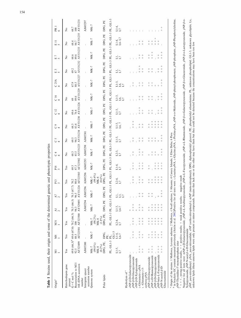
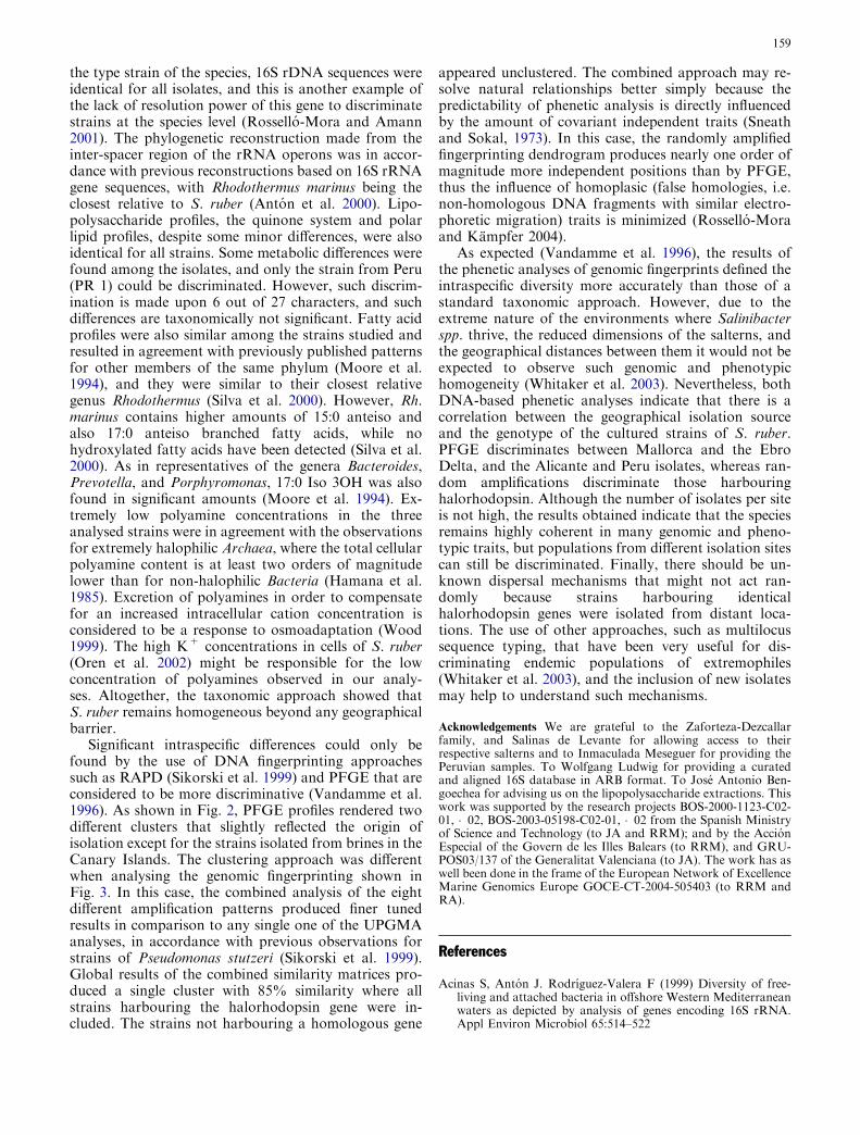
Two dendrograms were produced after UPGMA anal-ysis, one for the PFGE patterns and one as a result ofthe combination of eight independent random amplifi-cations with the primers cited. PFGE patterns resultedin about 30 different independent positions where asingle band (i.e. digestion product) was found in at leastone profile. In the derived dendrogram shown in Fig. 2two clusters were found with more than 55% bandingpattern similarity. Cluster 1 harboured all the isolatesfrom the Mallorca and Ebro Delta salterns (M1, M8,M31, A1, A7, E3, E7 and E11), whereas cluster 2 har-boured all strains isolated from Alicante and the Peru-vian Andes (P13, P18 and PR1). Strains isolated fromthe Canary Islands appeared to be spread across bothclusters (C25a and C4 in cluster 1, and C5, C9, C12 andC16 in cluster 2). Single randomly amplified genomicDNA patterns rendered between 20 (ERIC) and 48(RAPD4) independent positions (i.e. each single randomamplification product) that could be used for theanalysis. In Fig. 3, the eight independent analysesare shown, and a single dendrogram based on 260
independent positions resulted from the combination ofall amplification experiments. In the dendrogram basedon the combined results, one single independent clusterof seven strains sharing at least 85% pattern similaritycould be found. All strains isolated from the Alicanteand Mallorca salterns were grouped within the samecluster, whereas the rest of the strains appeared to bespread in four additional clusters. Additionally, thisunique cluster harboured all strains for which a copy ofa homologous halorhodopsin gene was found (J. Antonet al., in preparation; M31 halorhodopsin accessionnumber AY667579). No single independent amplifica-tion fingerprinting gave results similar to the simi-larity matrix generated after the combination of allexperiments.
Discussion
Shortly after the discovery of the existence and ecolog-ical relevance of Salinibacter spp. in brines close to sat-uration (Anton et al. 2000), members of this genus wereisolated leading to the classification of the new genusand species S. ruber (Anton et al. 2002). The firstdescription was made after five organisms that wereisolated from solar salterns of Alicante and Mallorca.Since then, the occurrence of this genus in brines ofgeographically distant sites has become more evident,and this led us to compile a larger strain collection fromdifferent locations in Europe and South America. Thecollection of isolates studied here includes organismsoccurring in brines from the Canary Islands, Ebro Deltaand the Peruvian Andes, in addition to the first sites
Fig. 2 Dendrogram generated after UPGMA analysis of PFGEpatterns. The analysis was carried out after a set of about 30covariant positions defined as the presence of at least one band inone of the analysed lanes. Black boxed strains are those notharbouring halorhodopsin
157
sampled. Despite the isolation efforts, all new isolateswere identified as S. ruber, and no isolation of a singlemember of the EHB-2 phylotype was successful. Thelack of success on the isolation of the hitherto uncul-tured EHB-2 phylotype might be due to either the cul-ture media used that do not satisfy their growthrequirements, or to the relative low abundance of thisphylotype in natural brines. In all cases studied EHB-2showed abundances lower than 2.4% of the total pro-karyotic population, whereas the phylotype EHB-1corresponding to S. ruber were always at least five foldhigher (Anton et al. 2000).
In a different study (J. Anton et al., in preparation), ahomologous gene to the archaeal halorhodopsin couldbe detected in the type strain of the genus after theannotation of the open reading frames detected during apartial genome-sequencing program. The presence ofthis gene was investigated for the same collection ofisolates and an identical copy was found in the genomesof a subset of the collection, but not in all of them (seeTable 1). The seven halorhodopsin-harbouring strainswere isolated from three different crystallizer ponds, onesituated in Alicante and two in Mallorca. In principle, it
seems that the presence of such a gene is related to theorigin of isolation. However, halorhodopsin-harbouringstrains in the Canary Island samples have been foundrecently (data not shown). Mallorca, Alicante and theEbro Delta are geographically equidistant, but this genehas not been found yet in the strains from the EbroDelta. The so called ‘polyphasic taxonomy’ (Vandammeet al. 1996) may reveal whether the geographical isola-tion of these extreme sites determines allopatric specia-tion phenomena (Staley, 2004).
S. ruber seems to be very homogeneous from thetaxonomic point of view, as indicated both by genomicand phenotypic data. We could not find solid trait dif-ferences to discriminate between the isolates from dis-tant geographical zones. All strains showed a singlerRNA operon with identical 16S rRNA gene sequencesand nearly identical inter-spacer sequences. G+C molpercent, and reassociation values were very similar to thetype strain of S. ruber, consistent for closely relatedstrains of a single species, and close enough to be con-sidered as a single genomovar within the species (Ros-sello-Mora and Amann 2001). Despite DNA-DNAreassociaton values ranged from 76.4% to 100% with
Fig. 3 Dendrograms generatedafter UPGMA analyses of eachsingle random amplificationprofile. The number ofcovariant positions variedbetween the differentfingerprints as follows: ERIC 20positions; BOX 31 positions;RAPD1 25 positions; RAPD241 positions; RAPD3 38positions; RAPD4 48 positions;RAPD5 30 positions; RAPD627 positions. The resultantglobal dendrogram wasproduced after the combinationof all single results into a singlesimilarity matrix; the number ofcovariant characters was 260.Results are expressed inpercentages of similarity. Blackboxed strains are those notharbouring halorhodopsin
158
the type strain of the species, 16S rDNA sequences wereidentical for all isolates, and this is another example ofthe lack of resolution power of this gene to discriminatestrains at the species level (Rossello-Mora and Amann2001). The phylogenetic reconstruction made from theinter-spacer region of the rRNA operons was in accor-dance with previous reconstructions based on 16S rRNAgene sequences, with Rhodothermus marinus being theclosest relative to S. ruber (Anton et al. 2000). Lipo-polysaccharide profiles, the quinone system and polarlipid profiles, despite some minor differences, were alsoidentical for all strains. Some metabolic differences werefound among the isolates, and only the strain from Peru(PR 1) could be discriminated. However, such discrim-ination is made upon 6 out of 27 characters, and suchdifferences are taxonomically not significant. Fatty acidprofiles were also similar among the strains studied andresulted in agreement with previously published patternsfor other members of the same phylum (Moore et al.1994), and they were similar to their closest relativegenus Rhodothermus (Silva et al. 2000). However, Rh.marinus contains higher amounts of 15:0 anteiso andalso 17:0 anteiso branched fatty acids, while nohydroxylated fatty acids have been detected (Silva et al.2000). As in representatives of the genera Bacteroides,Prevotella, and Porphyromonas, 17:0 Iso 3OH was alsofound in significant amounts (Moore et al. 1994). Ex-tremely low polyamine concentrations in the threeanalysed strains were in agreement with the observationsfor extremely halophilic Archaea, where the total cellularpolyamine content is at least two orders of magnitudelower than for non-halophilic Bacteria (Hamana et al.1985). Excretion of polyamines in order to compensatefor an increased intracellular cation concentration isconsidered to be a response to osmoadaptation (Wood1999). The high K+ concentrations in cells of S. ruber(Oren et al. 2002) might be responsible for the lowconcentration of polyamines observed in our analy-ses. Altogether, the taxonomic approach showed thatS. ruber remains homogeneous beyond any geographicalbarrier.
Significant intraspecific differences could only befound by the use of DNA fingerprinting approachessuch as RAPD (Sikorski et al. 1999) and PFGE that areconsidered to be more discriminative (Vandamme et al.1996). As shown in Fig. 2, PFGE profiles rendered twodifferent clusters that slightly reflected the origin ofisolation except for the strains isolated from brines in theCanary Islands. The clustering approach was differentwhen analysing the genomic fingerprinting shown inFig. 3. In this case, the combined analysis of the eightdifferent amplification patterns produced finer tunedresults in comparison to any single one of the UPGMAanalyses, in accordance with previous observations forstrains of Pseudomonas stutzeri (Sikorski et al. 1999).Global results of the combined similarity matrices pro-duced a single cluster with 85% similarity where allstrains harbouring the halorhodopsin gene were in-cluded. The strains not harbouring a homologous gene
appeared unclustered. The combined approach may re-solve natural relationships better simply because thepredictability of phenetic analysis is directly influencedby the amount of covariant independent traits (Sneathand Sokal, 1973). In this case, the randomly amplifiedfingerprinting dendrogram produces nearly one order ofmagnitude more independent positions than by PFGE,thus the influence of homoplasic (false homologies, i.e.non-homologous DNA fragments with similar electro-phoretic migration) traits is minimized (Rossello-Moraand Kampfer 2004).
As expected (Vandamme et al. 1996), the results ofthe phenetic analyses of genomic fingerprints defined theintraspecific diversity more accurately than those of astandard taxonomic approach. However, due to theextreme nature of the environments where Salinibacterspp. thrive, the reduced dimensions of the salterns, andthe geographical distances between them it would not beexpected to observe such genomic and phenotypichomogeneity (Whitaker et al. 2003). Nevertheless, bothDNA-based phenetic analyses indicate that there is acorrelation between the geographical isolation sourceand the genotype of the cultured strains of S. ruber.PFGE discriminates between Mallorca and the EbroDelta, and the Alicante and Peru isolates, whereas ran-dom amplifications discriminate those harbouringhalorhodopsin. Although the number of isolates per siteis not high, the results obtained indicate that the speciesremains highly coherent in many genomic and pheno-typic traits, but populations from different isolation sitescan still be discriminated. Finally, there should be un-known dispersal mechanisms that might not act ran-domly because strains harbouring identicalhalorhodopsin genes were isolated from distant loca-tions. The use of other approaches, such as multilocussequence typing, that have been very useful for dis-criminating endemic populations of extremophiles(Whitaker et al. 2003), and the inclusion of new isolatesmay help to understand such mechanisms.
Acknowledgements We are grateful to the Zaforteza-Dezcallarfamily, and Salinas de Levante for allowing access to theirrespective salterns and to Inmaculada Meseguer for providing thePeruvian samples. To Wolfgang Ludwig for providing a curatedand aligned 16S database in ARB format. To Jose Antonio Ben-goechea for advising us on the lipopolysaccharide extractions. Thiswork was supported by the research projects BOS-2000-1123-C02-01, �02, BOS-2003-05198-C02-01, �02 from the Spanish Ministryof Science and Technology (to JA and RRM); and by the AccionEspecial of the Govern de les Illes Balears (to RRM), and GRU-POS03/137 of the Generalitat Valenciana (to JA). The work has aswell been done in the frame of the European Network of ExcellenceMarine Genomics Europe GOCE-CT-2004-505403 (to RRM andRA).
References
Acinas S, Anton J. Rodrıguez-Valera F (1999) Diversity of free-living and attached bacteria in offshore Western Mediterraneanwaters as depicted by analysis of genes encoding 16S rRNA.Appl Environ Microbiol 65:514–522
159

Altenburger P, Kampfer P, Makristathis A, Lubitz W, Busse H-J(1996) Classification of bacteria isolated from a medieval wallpainting. J Biotech 47:39–52
Altenburger P, Kampfer P, Akimov V, Lubitz W, Busse H-J (1997)Polyamine distribution in actinomycetes with group B pepti-doglycan and species of the genera Brevibacterium, Coryne-bacterium, and Tsukamurella. Int J Syst Bacteriol 47:270–277
Altschul SF, Madden TL, Schaffer AA, Zhang J, Zhang Z, MillerW, Lipman DJ (1997) Gapped BLAST and PSI-BLAST: a newgeneration of protein database search programs. Nucleic AcidsRes 25:3389–3402
Amann RI, Ludwig W, Schleifer K-H (1995) Phylogenetic identi-fication and in situ detection of individual microbial cellswithout cultivation. Microbiol Rev 59:43–169
Anton J, Rossello-Mora R, Rodrıguez-Valera F Amann R (2000).Extremely halophilic Bacteria in crystallizer ponds from solarsalterns. Appl Environ Microbiol 66:3052–3057
Anton J, Oren A, Benlloch S, Rodrıguez-Valera F, Amann R,Rossello-Mora R (2002) Salinibacter ruber gen. nov., sp. nov., anovel, extremely halophilic member of the Bacteria from salterncrystallizer ponds. Int J Syst Evol Microbiol 52:485–491
Bengoechea JA, Dıaz R, Moriyon I (1996) Outer membrane dif-ferences between pathogenic and environmental Yersinia en-terocolitica biogroups probed with hydrophobic permeants andpolycationic peptides. Infect Immun 64:4891–4899
DeLong EF (1992) Archaea in coastal marine environments. ProcNatl Acad Sci USA 89:5685–5689
Garcıa-Martınez J, Bescos I, Rodrıguez-Sala JJ Rodrıguez-ValeraF (2001) RISSC: a novel database for ribosomal 16S-23S RNAgenes spacer regions. Nuc Acid Res 29:178–180
Garrity GM, Bell J, Lilburn TG (2003) Taxonomic outline of theProkaryotes. Bergey’s Manual of Systematic Bacteriology 2ndedn. fourth release, October 2003. DOI: http://dx.doi.org/10.1007/bergeysoutline200210
Groth I, Schumann P, Weiss N, Martin K, Rainey FA (1996)Agrococcus jenensis gen. nov., sp. nov., a new genus of actino-mycetes with diaminobutyric acid in the cell wall. Int J SystBacteriol 46:234–239
Grothues D, Tummler B (1991) New approaches in genome anal-ysis by pulsed-field gel electrophoresis: application to theanalysis of Pseudomonas species. Mol Microbiol 5:2763–2776
Guasp C, Moore ERB, Lalucat J, Bennasar A (2000) Utility ofinternally transcribed 16S-23S rDNA spacer regions for thedefinition of Pseudomonas stutzeri genomovars and otherPseudomonas species. Int J Syst Evol Microbiol 50:1629–1639
Hamana K, Kamekura M, Onishi H, Akazawa T, Matsuzaki S(1985) Polyamines in photosynthetic eubacteria and extreme-halophilic archaebacteria. J Biochem 97:1653–1558
Harwood JL, Russell NJ (1984) Lipids of plants and microbes.Allen and Unwin, London
Hollander R, Pohl S (1980) Deoxyribonucleic acid base composi-tion of bacteria. Zbl Bakt Hyg I Abt Orig A 246:236–273
Kampfer P, Kroppenstedt RM (1996) Numerical analysis of fattyacid patterns of coryneform bacteria and related taxa. Can JMicrobiol 42:989–1005
Lane DJ (1991) 16S/23S rRNA sequencing. In: Stackebrandt E,Goodfellow M (eds) Nucleic acid techniques in bacterial sys-tematics. John Wiley and Sons, Chichester United Kingdom, pp115–175
Lopez-Garcıa P, Anton J, Abad JP, Amils R (1994) Halobacterialmegaplasmids are negatively supercoiled. Mol Microbiol11:421–427
Lowe TM, Eddy SR (1997) tRNAscan-SE: a program for im-proved detection of transfer RNA genes in genomic sequence.Nuc Acids Res 25:955–964
Ludwig W, Strunk O, Westram R, Richter L, Meier H, Yadhuku-mar, Buchner A, Lai T, Steppi S, Jobb G, Forster W, Brettske I,Gerber S, Ginhart AW, Gross O, Grumann S, Hermann S, JostR, Konig A, Liss T, Lußmann T,MayM, Nonhoff B, Reichel B,Strehlow R, Stamatakis AP, Stuckmann N, Vilbig A, Lenke M,Ludwig T, Bode A, Schleifer K-H (2004) ARB: a softwareenvironment for sequence data. Nucl Ac Res 32:1363–1371
Marmur J (1961) A procedure for the isolation of DNA frommicroorganisms. J Mol Biol 3:208–218
Moore LV, Bourne DM, Moore WEC (1994) Comparative distri-bution and taxonomic value of cellular fatty acids in thirty-three genera of anaerobic gram-negative bacilli. Int J SystBacteriol 44:338–347
Muller V, Oren A (2003) Metabolism of chloride in halophilicprokaryotes. Extremophiles 7:261–266
Oren A, Mana L (2002) Amino acid composition of bulk proteinand salt relationships of selected enzymes of Salinibacter ru-ber, an extremely halophilic bacterium. Extremophiles 6:217–223
Oren A, Rodrıguez-Valera F (2001) The contribution of halophilicBacteria to the red coloration of saltern crystallizer ponds.FEMS Microbiol Ecol 36:123–130
Oren A, Heldal M, Norland S, Galinski EA (2002) Intracellular ionand organic solute concentrations of the extremely halophilicbacterium Salinibacter ruber. Extremophiles 6:491–498
Oren A, Rodrıguez-Valera F, Anton J, Benlloch S, Rossello-MoraR, Amann R, Coleman J, Russell NJ (2004) Red, extremelyhalophilic, but not archaeal: the physiology and ecology ofSalinibacter ruber, a bacterium isolated from saltern crystallizerponds. In: Ventosa A (ed) Halophilic Microorganisms. Hei-delberg, Springer, pp 63–76
Owen RJ, Legros RM, Lapage SP (1978) Base composition, sizeand sequence similarities of genome deoxyribonucleic acidsfrom clinical isolates of Pseudomonas putrefaciens. J GenMicrobiol 104:127–138
Palleroni NJ (1984) Pseudomonadaceae. In: Krieg NR, Holt, JG(eds) Bergey’s manual of systematic bacteriology. The Williamsand Wilkins Co, Baltimore, pp 141–199
Rossello-Mora R, Amann R (2001) The species concept for prok-aryotes. FEMS Microbiol Rev 25:39–67
Rossello-Mora R, Kampfer P (2004) Defining microbial diversity –the species concept for prokaryotic and eukaryotic microor-ganisms. In: Bull AT (ed) Microbial Diversity and Biopro-specting. D C ASM Press, Washington, pp 29–39
Sikorski J, Rossello-Mora R, Lorenz MG (1999) Analysis ofgenotypic diversity and relationships among Pseudomonasstutzeri strains by PCR-based genomic fingerprinting andmultilocus enzyme electrophoresis. Syst Appl Microbiol22:393–402
Silva Z, Horta C, da Costa MS, Chung AP, Rainey FA (2000)Polyphasic evidence for the reclassification of Rhodothermusobamensis Sako et al. 1996 as a member of the species Rhodo-thermus marinus Alfredsson et al. 1988. Int J Syst EvolMicrobiol 50:1457–1461
Smith CL, Klco ST, Cantor CR (1988) Pulsed field gel electro-phoresis and the technology of large DNA molecules. In: Da-vies K (ed) Genome Analysis: A Practical Approach. IRL Press,Oxford England, pp 41–72
Sneath PH, Sokal RR (1973) Numerical taxonomy, the principlesand practice of numerical classification. W H Freeman andCompany, San Francisco
Staley JT (2004) Speciation and bacterial phylospecies. In: Bull AT(ed) Microbial Diversity and Bioprospecting. ASM Press,Washington D.C, pp 40–48
Tamaoka J, Komagata K (1984) Determination of DNA basecomposition by reversed-phase high-performance liquid chro-matography. FEMS Microb Lett 25:125–128
Tindall BJ (1990) A comparative study of the lipid composition ofHalobacterium saccharovorum from various sources. Syst ApplMicrobiol 13:128–130
Vandamme P, Pot B, Gillis M, De Vos P, Kersters K, Swings J(1996) Polyphasic taxonomy, a consensus approach to bacterialsystematics. Microbiol Rev 60:407–438
Van de Peer Y, De Wachter Y (1994) TREECON for Windows: asoftware package for the construction and drawing of evolu-tionary trees for the Microsoft Windows environment. ComputAppl Biosci 10:569–570
Ventosa A, Marquez MC, Kocur M, Tindall BJ (1993) Compara-tive study of ‘‘Micrococcus sp.’’ strains CCM 168 and CCM
160

1405 and members of the genus Salinicoccus. Int J Syst Bacte-riol 43:245–248
Westphal O, Jann K (1963) Bacterial lipopolysaccharides extrac-tion with phenol-water and further applications of the proce-dure. Methods Carbohydr Chem 5:83–91
Whitaker RJ, Grogan DW, Taylor JW (2003) Geographic barriersisolate endemic populations of hyperthermophilic Archaea.Science 301:976–978
Wood JM (1999) Osmosensing by bacteria: Signals and membrane-based sensors. Microbiol Mol Biol Rev 63:230–262
Ziemke F, Hofle MG, Lalucat J, Rossello-Mora R (1998) Reclas-sification of Shewanella putrefaciens Owen’s genomic group IIas Shewanella baltica sp nov. Int J Syst Bacteriol 48:179–186
161