International Healthcare Research Journal (IHRJ)
33
International Healthcare Research Journal (IHRJ) E - I S S N : 2 4 5 6 - 8 0 9 0 Volume 4, Issue 7 (October 2020) Published: 25 th October, 2020
-
Upload
khangminh22 -
Category
Documents
-
view
8 -
download
0
Transcript of International Healthcare Research Journal (IHRJ)
International Healthcare Research Journal (IHRJ)
E - I S S N : 2 4 5 6 - 8 0 9 0
Volume 4, Issue 7 (October 2020)Published: 25th October, 2020
EDITOR-IN-CHIEF AND PUBLISHER Co-Editor
Dr. Vatsul Sharma, MDS(Public Health Dentistry)
Consultant Dental Specialist Ex-Senior Lecturer
Department of Public Health Dentistry
Sri Sukhmani Dental College Dera Bassi (SAS Nagar) 140507
Punjab India
+91 8607700075
[email protected] [email protected]
PUBLICATION ADDRESS
66 A Day Care Centre
Housing Board Colony Kalka (Panchkula)
Haryana, India-133302
Dr. Sahil Thakar, MDS(Public Health
Dentistry)
Consultant Dental Surgeon Senior Lecturer
Department of Public Health Dentistry
School of Dental Sciences Sharda University
Greater Noida -201310
+91 9990036390 [email protected]
Associate Editor
Dr. Ravneet Malhi, MDS(Public Health Dentistry) Department of Public Health Dentistry
DJ College of Dental Sciences and Research Modinagar
+91-98976601690 [email protected]
Editorial Coordinator Parul Chawla
Pharmacovigilance Specialist Masters in Systems Biology and
Bioinformatics +91-8288030921
Forensic Editor
Dr. Taruna Malhotra, BDS, M.Sc. (Forensic Odontology)
Consultant Dental Surgeon & Forensic Advisor
New Delhi +91-98189 37227
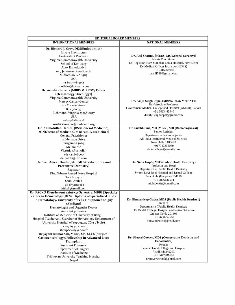
EDITORIAL BOARD MEMBERS
INTERNATIONAL MEMBERS NATIONAL MEMBERS
Dr. Richard J. Gray, DDS(Endodontics) Private Practitioner
Ex-Assistant Professor Virginia Commonwealth University
School of Dentistry Apex Endodontics
1149 Jefferson Green Circle Midlothian, VA 23113
USA +1 804-378-9152
Dr. Anil Sharma, [MBBS, MS(General Surgery)]
Private Practitioner
Ex-Registrar, Ram Manohar Lohia Hospital, New Delhi
Ex-Medical Officer Incharge (HCMS)
+91 9416264986
Dr. Arushi Khurana [MBBS,MD,PGY5 Fellow (Hematology/Oncology)]
Virginia Commonwealth University Massey Cancer Center
401 College Street Box 980037
Richmond, Virginia 23298-0037 USA
+1804-828-9726 [email protected]
Dr. Kuljit Singh Uppal,[MBBS, DLO, MS(ENT)]
Ex-Associate Professor
Government Medical College and Hospital (GMCH), Patiala
+91 9463443940 [email protected]
Dr. Naimatullah Habibi, [BSc(General Medicine), MD(Doctor of Medicine), MD(Family Medicine)]
General Practitioner 2, Merivale Drive Truganina 3029
Melbourne Victoria (Australia)
+61 424808900 [email protected]
Dr. Sulabh Puri, MD [MBBS, MD (Radiodiagnosis)]
Senior Resident
Department of Radiodiagnosis
All India Institute of Medical Sciences New Delhi 1100608
+917042202050 [email protected]
Dr. Syed Ameer Haider Jafri, MDS(Pedodontics and Preventive Dentistry)
Registrar King Salman Armed Force Hospital
Tabuk 47512 Saudi Arabia
+96 6534010567 [email protected]
Dr. Nidhi Gupta, MDS (Public Health Dentistry)
Professor and Head Department of Public Health Dentistry
Swami Devi Dyal Hospital and Dental College
Panchkula (Haryana) 134118 +91 9876136514
Dr. PACKO Dieu-le-veut saint-cyr Sylvestre, MBBS [Specialty career in Hématology (DES): Diploma of Specialized Study
in Hematology, University of Félix Houphouët Boigny (Abidjan)]
Hematologist and Urgentist Doctor Assistant professor
Institute of Medicine of University of Bangui Hospital Teacher and Searcher of Hematology Department of
University Hospital of Yopougon, Côte d’Ivoire +225 89 54 22 09
Dr. Bhuvandeep Gupta, MDS (Public Health Dentistry)
Reader
Department of Public Health Dentistry
ITS Dental College, Hospital and Research Centre Greater Noida 201308
+91 9650757561
Dr Jayant Kumar Sah, MBBS, MS, M.Ch (Surgical Gastroenterology), Fellowship in Advanced Liver
Transplant Assistant Professor
Department of Surgery Institute of Medicine
Tribhuvan University Teaching Hospital Nepal
Dr. Sheetal Grover, MDS (Conservative Dentistry and
Endodontics)
Reader Seema Dental College and Hospital
Rishikesh 249203
+91 8477981601 [email protected]
Dr Mayank Gahlot (MDS Orthodontics) Specialist Orthodontist
307, Block A Al Attar Center Karama 116440
Dubai +971 558096897
Dr. Nitin Gorwade, MDS (Periodontics)
Senior Resident PGIMER Chandigarh 160012
+91 7738477054
Dr. Vivek Vijay Gupta (MDS Periodontics) Senior Lecturer
Faculty of Dentistry, SEGi University Jalan Teknologi 9, PJU5, Kota Damansara
Petling Jaya-47810 Malaysia
+60 102924549 [email protected]
Dr. Gyanendra Mishra, MDS (Pedodontics)
Medical Officer Dental
Ministry of Health, Jharkhand
+91 8076597879 [email protected]
Dr. Ramya Madhuri, MDS (Oral Medicine and Radiology) Unit number 12
Building num 4277 Solumaniah
Riyadh Saudi Arabia
+966 555740418 [email protected]
Dr. Abhishek Bansal, MDS (Prosthodontics)
Consultant Prosthodontist & Private Practitioner H-32/62, Sector-3, Rohini, Delhi-110085
+91 9899236125
Dr. Nikhil Prakash, MDS (Prosthodontics)
Senior Lecturer Department of Prosthodontics
Yogita Dental College and Hospital
Khed, Ratnagiri- 415709 +91 7408814400
Dr. Khundrakpam Nganba MDS (Pedodontics and Preventive
Dentistry)
Senior Lecturer
Department of Pedodontics and Preventive Dentistry Maharana Pratap Dental College
Gwalior-475001, India
+91 8826355824 [email protected]
INTERNATIONAL HEALTHCARE RESEARCH JOURNAL
International Healthcare Research Journal
CONTENTS (VOLUME 4, ISSUE 7, OCTOBER 2020)
S.No TITLE AUTHOR NAMES PAGE NUMBERS
DOI
EDITORIAL COMMENT(S)
1.
World Mental Health Day 2020: Stress Levels at it Peak during Pandemic
Times?
Dr. Sahil Thakar
EC1-EC2
https://doi.org/10.26440/IHRJ/0407.10121
REVIEW(S)
2.
Verrucopapillary Lesions
of the Oral Cavity: A Review
Sana Khaled, Santosh R.
Bharadwaj, Bushra Anjum,
Satyanarayana D.
RV1-RV7
https://doi.org/10.26440/IHRJ/0407.10365
CASE REPORT(S)
3.
Cutaneous Manifestation of COVID-19: A Short
Clinical Case
Ratesh Bassi, Geetika Bassi
CR1-CR2
https://doi.org/10.26440/IHRJ/0407.10187
4.
Non-Syndromic Hypodontia: A Case
Report
Parul Uppal Malhotra,
Yagyeshwar Malhotra, Neera Ohri, Anindita
Mallik
CR3-CR6
https://doi.org/10.26440/IHRJ/0407.10287
ORIGINAL RESEARCH(S)
5.
Evaluating Knowledge, Awareness and Behaviour
Among Dental Interns Regarding Ergonomics in
Dentistry: A Cross-Sectional Survey
Rebecca Andrew,
Sonia Narang, Srishti Aggarwal,
Thongam S.
OR1-OR4
https://doi.org/10.26440/IHRJ/0407.10287
6.
The Relationship Between Maxillary and
Mandibular Base Lengths and Dental Crowding in Patients with True Class
II Malocclusions
Tanzin Palkit, Isha Aggarwal,
Yagyeshwar Malhotra, Mandeep
Uppal, Merry Goyal, Neetika
Singh
OR5-OR9
https://doi.org/10.26440/IHRJ/0407.10280
7.
Evaluating The Effect of pH of Dentin Bonding Agents on Dentin in
Relation to the Push-Out Bond Strength of
Composites in Class I Cavities in-vitro
Sofia Ganai, S. Vijay Singh, Saurabh Gupta, Poonam
Bogra
OR10-OR14
https://doi.org/10.26440/IHRJ/0407.10282
8.
Assessment of Micronuclei Frequency in Individuals with a Habit of Tobacco Chewing by
Means of Exfoliated Oral Buccal Cells
Swati Parhar, Amani Mahajan
OR15-OR18
https://doi.org/10.26440/IHRJ/0407.10277

International Healthcare Research Journal 2020;4(7):EC1-EC2.
The world is facing an unprecedented crisis in 2020: From Healthcare workers to teachers to the common labourer, all are facing the heat due to this so called “New Normal”. As per the World Health Organization, Bereavement, isolation, loss of income and fear are triggering mental health conditions or exacerbating existing ones and thus increasing the prevalence of mental health problems among people.1 The initiative to celebrate October 10 as the World Mental Health Day was done in the year 1994; and in 2020, the theme is aptly chosen as “Mental Health for All- Greater Investment – Greater Access”. Fear, stress, and worry were normal were the few responses people perceived in the context of the COVID-19 pandemic. During this pandemic, restricted movements, work from home, unemployment, no socialization has taken one’s mental and physical health for a toss. A person is also constantly trying to sanitize daily use items posing as an additional burden.2 A WHO survey states that current pandemic has led to either a disruption or halt in critical mental health services in 93% of countries, although the demand for mental health is increasing with every passing day. In addition, most healthcare services and workers are directed towards the prevention of COVID-19, leaving little focus on one’s mental health. Many countries (70%) have adopted telemedicine or teletherapy to overcome disruptions to in-person services, there are significant disparities in the uptake of these interventions, one of which includes disruption in internet services and poor network coverage. The three most common misconceptions are: “My problems are not serious enough to seek therapy and only weak people go to therapy”, “Once I begin therapy, I will be in it forever” and “I will be forced to take medications and will get addicted to them”. We all deserve to feel safe and supported when talking about our mental health. But too often, mental health
stigma leaves people feeling isolated and ashamed. At worst, it prevents people getting support, finding employment or having open conversations.3 The dilemma every year one adult in four, along with one child in ten, will have a mental health issue. These conditions can profoundly affect literally millions of lives, affecting the capability of these individuals to make it through the day, to sustain relationships, and to maintain work. There are however, a few ways to prevent stress:
1. Surround Yourself with Positivity 2. Exercise 3. Eat Mental Health Boosting Foods 4. Share Your Feelings 5. Sleep Enough
Last, but the least, a person should believe that “it is ok to seek help, either from friends or professional help”. One should remember that “there is not joy greater than life itself!!” In the end, I request all readers to be on the lookout of any such potentially depressed/stressed person and try to provide assistance in the best possible way, even if it means leaving the person alone for some time and raise concern, if the need arises. REFERENCES 1. Kumari J. World Mental Health Day 2020: History And Significance of The Day. Online Article Available from: https://www.india.com/lifestyle/world-mental-health-day-2020-history-and-significance-of-the-day-4168527/. [Last Accessed on 25th September, 2020] 2. Daniels I. Mental Health for All Greater Investment – Greater Access. Everyone, everywhere. (Message from the WFMH President) Online Article. Available from: https://wfmh.global/world-mental-health-day-2020/ [Last Accessed on 25th September, 2020] 3. World Health Organization. Analysing Disrupted Health Sectors. A Modular Manual. Online PDF.
World Mental Health Day 2020: Stress Levels at it’s Peak during Pandemic Times?
EDITORIAL COMMENT
ISSN: 2456-8090 (online) DOI: https://doi.org/10.26440/IHRJ/0407.10121
SAHIL THAKAR
QR CODE
EC1
© Sahil Thakar. This is an open access article distributed under the terms of the Creative Commons Attribution License CC-BY-NC 4.0, which permits unrestricted use, distribution and reproduction in any medium, provided the use is not commercial and the original author(s) and source are cited.
International Healthcare Research Journal 2020;4(7):EC1-EC2.
Available from: https://www.who.int/hac/techguidance/tools/disrupted_sectors/adhsm.pdf [Last Accessed on 26th
September, 2020]
Cite this article as: Thakar S. World Mental Health Day 2020: Stress Levels at it Peak during Pandemic Times?. Int Healthc Res J. 2020;4(7):EC1-EC2. https://doi.org/10.26440/IHRJ/0407.10121
e-mail id for correspondence: sahil.ihrj[at]gmail[dot]com
AUTHOR AFFILIATIONS: Co-Editor, International Healthcare Research Journal Assistant Professor, Department of Public Health Dentistry, School of Dental Sciences, Sharda University, Greater Noida (ORCID ID: https://orcid.org/0000-0002-8686-5309)
World Mental Health Day Editorial Comment Sahil Thakar
EC2
International Healthcare Research Journal 2020;4(7):RV1-RV7.
INTRODUCTION Most of the biopsied lesions of the oral mucosa have shown a unique proliferation of the stratified squamous epithelium, with or without inductive changes of the underlying stroma. These proliferations fall into three types: papillary exophytic masses, broad verruciform excesses of surface keratin and flat hyperplasias of spinous cell layer. The exophytic lesions represent as any pathologic growth that projects above the normal contours of the oral surface. The papillary lesions represent swelling with finger like projections imparting a cauliflower like appearance, these micro projections are rounded and blunt like fungiform papillae of the tongue. The verrucous lesions are similar to papillary lesions yet possess a more irregular surface. These papillary or verrucous type lesions are quite common in the oral and paraoral regions, representing 3% of biopsied oral lesions. Clinical information and an adequate biopsy are essential for making an accurate diagnosis of these lesions, but the primary objective must be to evaluate the epithelium for dysplastic features and signs of invasion. Hence, differentiation between verrucous and papillary lesion is based more on microscopic features rather than the clinical appearance. Biopsy is usually indicated to secure a definitive diagnosis and to follow a proper treatment plan.
PATHOGENESIS Majority of verrucous lesions are thought to be induced by viral infection of the epithelium especially Human Papilloma Virus (HPV). Human papilloma
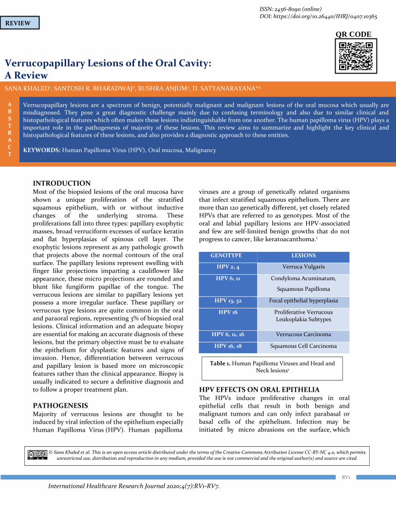
viruses are a group of genetically related organisms that infect stratified squamous epithelium. There are more than 120 genetically different, yet closely related HPVs that are referred to as genotypes. Most of the oral and labial papillary lesions are HPV-associated and few are self-limited benign growths that do not progress to cancer, like keratoacanthoma.1
HPV EFFECTS ON ORAL EPITHELIA The HPVs induce proliferative changes in oral epithelial cells that result in both benign and malignant tumors and can only infect parabasal or basal cells of the epithelium. Infection may be initiated by micro abrasions on the surface, which
GENOTYPE LESIONS
HPV 2, 4 Verruca Vulgaris
HPV 6, 11 Condyloma Acuminatum,
Squamous Papilloma
HPV 13, 32 Focal epithelial hyperplasia
HPV 16 Proliferative Verrucous Leukoplakia Subtypes
HPV 6, 11, 16 Verrucous Carcinoma
HPV 16, 18 Squamous Cell Carcinoma
Verrucopapillary Lesions of the Oral Cavity: A Review
REVIEW
A B S T R A C T
ISSN: 2456-8090 (online) DOI: https://doi.org/10.26440/IHRJ/0407.10365
SANA KHALED1, SANTOSH R. BHARADWAJ2, BUSHRA ANJUM3, D. SATYANARAYANA*4
Verrucopapillary lesions are a spectrum of benign, potentially malignant and malignant lesions of the oral mucosa which usually are misdiagnosed. They pose a great diagnostic challenge mainly due to confusing terminology and also due to similar clinical and histopathological features which often makes these lesions indistinguishable from one another. The human papilloma virus (HPV) plays a important role in the pathogenesis of majority of these lesions. This review aims to summarize and highlight the key clinical and histopathological features of these lesions, and also provides a diagnostic approach to these entities. KEYWORDS: Human Papilloma Virus (HPV), Oral mucosa, Malignancy
QR CODE
Table 1. Human Papilloma Viruses and Head and Neck lesions1
© Sana Khaled et al. This is an open access article distributed under the terms of the Creative Commons Attribution License CC-BY-NC 4.0, which permits unrestricted use, distribution and reproduction in any medium, provided the use is not commercial and the original author(s) and source are cited.
RV1
International Healthcare Research Journal 2020;4(7):RV1-RV7.
Verrucopapillary Lesions of the Oral Cavity Khaled S et al.
allows better access for this virus into the basal cells. When the virus initially enters host basal cells, it cannot replicate until the cell matures into a keratinocyte, as the host cell undergoes the normal differentiation, the virus also starts its replication. The virus starts its replication once the host cell mitosis occurs. Then virus expresses its the early proteins--E1, E2, E5, E6, and E7--in the lower spinous layers which occurs in the early phases of the infection. As the epithelial cells mature, the cell cycle is halted as part of forming a protective barrier; however, terminal differentiation is hindered by E7 and E6. This has most likely evolved to allow the host cell to continue to reproduce viruses. As the host cell life progresses to the upper spinous layer, gene expression of HPV changes. The late proteins--L1 and L2--and E4 are upregulated and at this point, virus assembly occurs and exfoliating cells of the epithelium now releases complete virions. These cells are resilient in dry environments and virions shed from cornified squames have a higher chance of survival. Cornified squames are the epithelial cells that have more keratin which is a protective agent that hardens the cell. HPV then adheres to a specific receptor protein on the keratinocytes membrane in order to be assimilated into the cell by a process known as endocytosis. Once the virus enters into the cell, it divests itself of its protein coat and the viral DNA and then utilizes host cell DNA building blocks to replicate. These viruses elaborate early gene proteins that are able to regulate the host cell cycle or mitotic capabilities. E6 and E7 proteins are the most important in this respect, as they bind to the host proteins that are regulators of the keratinocytes cell division cycle. E6 binds to a protein designated p53, a molecule that arrest cell division, however once bound, it is degraded and this causes inhibition of keratinocytes mitosis to be nullified. Likewise, E7 binds a protein termed Rb; and it leads to cell cycle regulation disruuption.2 E1 – Viral replication E2 – Regulates viral transcription and replication E4 – Interacts with cytoskeletal proteins E5 – Downregulation of MHC Class 1 molecules E6 – Oncoprotein, binds to tumor suppressor protein p53 E7 – Oncoprotein, binds to tumor suppressor protein retinoblastoma (Rb) L1 – Major viral caspid protein
L2 – Minor viral caspid protein
ANATOMICAL LANDMARKS RESEMBLING VERRUCOUS-PAPILLARY LESIONS Some of the normal anatomic structures in the oral cavity, presenting as a papillary pattern are accessory tonsillar tissue, filiform papillae, fungiform papillae, foliate papillae, circumvallate papillae, retrocuspid, retromolar papillae and stensens's papillae. Sometimes, these structures attain such a size that they are mistaken for pathoses. The anatomic locations of the structures, however, usually enable immediate recognition.3
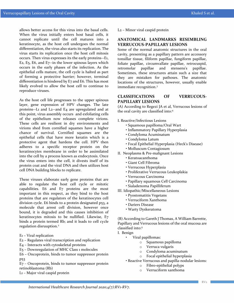
CLASSIFICATIONS OF VERRUCOUS-PAPILLARY LESIONS (A) According to Regezi JA et al, Verrucous lesions of the oral cavity are classified into:4
I. Reactive/Infectious Lesions
• Squamous papilloma/Oral Wart • Inflammatory Papillary Hyperplasia • Condyloma Acuminatum • Condyloma Latum • Focal Epithelial Hyperplasia (Heck's Disease) • Molluscum Contagiosum
II. Neoplasms & Pre-malignant Lesions • Keratoacanthoma • Giant Cell Fibroma • Verrucous Hyperplasia • Proliferative Verrucous Leukoplakia • Verrucous Carcinoma • Papillary squamous Cell Carcinoma • Sialadenoma Papilliferum
III. Idiopathic/Miscellaneous Lesions • Pyostomatitis Vegetans • Verruciform Xanthoma • Dariers Disease • Warty Dyskeratoma
(B) According to Gareth J Thomas, A William Barrette, Papillary and Verrucous lesions of the oral mucosa are classified into:5 I. Benign
• Viral papillomas: o Squamous papilloma o Verruca vulgaris o Condyloma acuminatum o Focal epithelial hyperplasia
• Reactive Verrucous and papilla-nodular lesions: o Fibro-epithelial polyps o Verruciform xanthoma
RV2
International Healthcare Research Journal 2020;4(7):RV1-RV7.
Verrucopapillary Lesions of the Oral Cavity Khaled S et al.
o Papillary hyperplasia o Pyostomatitis vegetans o Sialadenoma papilliferum o Acanthosis nigricans o Darier’s disease
II. Potentially Malignant: • Verrucous hyperplasia • Papillary dysplasia • Proliferative (verrucous) leukoplakia
III. Malignant: • Verrucous carcinoma • Papillary carcinoma:
o Non-invasive (synonymous with papillary dysplasia)
o Invasive (essentially a conventional squamous cell carcinoma requiring treatment as such)
• Carcinoma cuniculaturn (essentially a conventional, well differentiated squamous cell carcinoma requiring treatment as such)
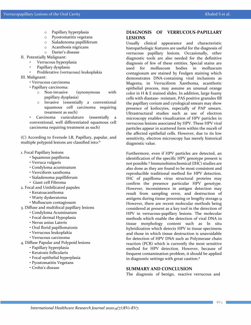
(C) According to Eversole LR, Papillary, papular, and multiple polypoid lesions are classified into:6 1. Focal Papillary lesions
• Squamous papillorna • Verruca vulgaris • Condyloma acuminatum • Verrciform xanthoma • Sialadenoma papilliferum • Giant cell Fibroma
2. Focal and Umbilicated papules • Keratoacanthoma • Warty dyskeratoma • Molluscum contagiosum
3. Diffuse and multifocal papillary lesions • Condyloma Acuminatum • Focal dermal Hypoplasia • Nevus unius Lateris • Oral florid papillomatosis • Verrucous leukoplakia • Verrucous carcinoma
4. Diffuse Papular and Polypoid lesions • Papillary hyperplasia • Keratosis follicularis • Focal epithelial hyperplasia • Pyostomatitis Vegetans • Crohn's disease
DIAGNOSIS OF VERRUCOUS-PAPILLARY LESIONS Usually clinical appearance and characteristic histopathologic features are useful for the diagnosis of verrucous papillary lesions. Occasionally, other diagnostic tools are also needed for the definitive diagnosis of few of these entities. Special stains are used for molluscum bodies in molluscum contagiosum are stained by Feulgen staining which demonstrates DNA-containing viral inclusions as Magenta, in Verruciform Xanthoma, acanthotic epithelial process, may assume an unusual orange color in H & E stained slides. In addition, large foamy cells with diastase- resistant, PAS positive granules fill the papillary corium and cytological smears may show presence of koilocytes, especially of PAP smears. Ultrastructural studies such as use of electron microscopy enables visualization of HPV particles in verrucous lesions associated by HPV. These HPV viral particles appear in scattered form within the nuceli of the affected epithelial cells. However, due to its low sensitivity, electron microscopy has merely historical diagnostic value. Furthermore, even if HPV particles are detected, an identification of the specific HPV genotype present is not possible.9 Immunohistochemical (IHC) studies are also done as they are found to be most consistent and reproducible traditional method for HPV detection. IHC of papilloma virus structural proteins may confirm the presence particular HPV genotype. However, inconsistence in antigen detection may result from sampling error, and destruction of antigens during tissue processing or lengthy storage.9 However, there are recent molecular methods being considered at present as a key tool in the detection of HPV in verrucous-papillary lesions. The molecular methods which enable the detection of viral DNA in tissue morphology content such as In situ hybridization which detects HPV in tissue specimens and those in which tissue destruction is unavoidable for detection of HPV DNA such as Polymerase chain reaction (PCR) which is currently the most sensitive method for HPV detection. However, because of frequent contamination problem, it should be applied in diagnostic settings with great caution.9
SUMMARY AND CONCLUSION The diagnosis of benign, reactive verrucous and
RV3
International Healthcare Research Journal 2020;4(7):RV1-RV7.
Verrucopapillary Lesions of the Oral Cavity Khaled S et al.
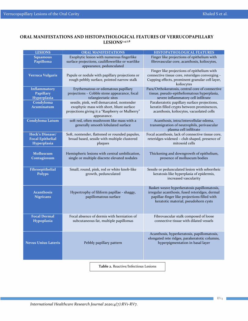
ORAL MANIFESTATIONS AND HISTOPATHOLOGICAL FEATURES OF VERRUCOPAPILLARY LESIONS2,4,7,8
LESIONS ORAL MANIFESTATIONS HISTOPATHOLOGICAL FEATURES
Squamous Papilloma
Exophytic lesion with numerous fingerlike surface projections, cauliflowerlike or wartlike
appearance, pedunculated
Finger like projections of epithelium with fibrovascular core, acanthosis, koliocytes,
Verruca Vulgaris
Papule or nodule with papillary projections or
rough pebbly surface, pointed narrow stalk
Finger like projections of epithelium with connective tissue core, reteridges converging - Cupping effects, prominent granular cell layer,
koliocytes
Inflammatory Papillary
Hyperplasia
Erythematous or edematous papillary projections – Cobble stone appearance, focal
telangiectatic sites
Para/Orthokeratosis, central core of connective tissue, pseudo-epitheliomatous hyperplasia,
severe inflammatory cell infiltrate
Condyloma Acuminatum
sessile, pink, well demarcated, nontender exophytic mass with short, blunt surface
projections giving it a “Raspberry or Mulberry” appearance.
Parakeratotic papillary surface projections, keratin filled crypts between prominences,
acanthosis, koliocytes, vacuolated cells
Condyloma Latum soft red, often mushroom like mass with a generally smooth lobulated surface
Acanthosis, intra/intercellular edema, transmigration of neutrophils, perivascular
plasma cell infiltrate
Heck’s Disease/ Focal Epithelial
Hyperplasia
Soft, nontender, flattened or rounded papules, broad based, sessile with multiple clustered
plaques
Focal acanthosis, lack of connective tissue core, reteridges widened – club shaped, presence of
mitosoid cells
Molluscum
Contagiosum
Hemispheric lesions with central umbilication,
single or multiple discrete elevated nodules
Thickening and downgrowth of epithelium,
presence of molluscum bodies
Fibroepithelial Polyps
Small, round, pink, red or white knob-like growth, pedunculated
Sessile or pedunculated lesion with seborrheic keratosis like hyperplasia of epidermis,
increased vascularity
Acanthosis Nigricans
Hypertrophy of filiform papillae - shaggy,
papillomatous surface
Basket-weave hyperkeratosis papillomatosis, irregular acanthosis, fused reteridges, dermal
papillae-finger like projections filled with keratotic material, pseudohorn cysts
Focal Dermal Hypoplasia
Focal absence of dermis with herniation of subcutaneous fat, multiple papillomas
Fibrovascular stalk composed of loose connective tissue with dilated vessels
Nevus Unius Lateris
Pebbly papillary pattern
Acanthosis, hyperkeratosis, papillomatosis, elongated rete ridges, parakeratotic columns,
hyperpigmentation in basal layer
Table 2. Reactive/Infectious Lesions
RV4
International Healthcare Research Journal 2020;4(7):RV1-RV7.
Verrucopapillary Lesions of the Oral Cavity Khaled S et al.
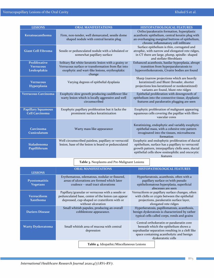
LESIONS ORAL MANIFESTATIONS HISTOPATHOLOGICAL FEATURES
Keratoacanthoma
Firm, non-tender, well demarcated, sessile dome
shaped nodule with central keratin plug
Ortho/parakeratin formation, hyperplastic acanthotic epithelium, central keratin plug with an overhanging marginal buttress of epithelium,
chronic inflammatory cell infiltrate
Giant Cell Fibroma
Sessile or pedunculated nodule with a lobulated or
somewhat papillary surface
Surface epithelium is thin, corrugated and atrophic, with narrow and elongated rete ridges,
in CT there are large, plump, spindle- shaped and stellate fibroblasts
Proliferative Verrucous
Leukoplakia
Solitary flat white keratotic lesion with a grainy or Verrucous surface or transformation from flat into
exophytic and wart-like lesions, erythroplakic change
Enhanced acanthosis, basilar hyperplasia, abrupt transition from hyperparakeratosis to
hyperorthokeratosis, Civatte bodies are found
Verrucous
Hyperplasia
Varying degrees of epithelial dysplasia
Sharp (narrow projections which are heavily keratinized) and Blunt (broader, shorter
projections less keratinzed or nonkeratinzed) variants are found, blunt rete ridges
Verrucous Carcinoma Exophytic slow growth producing cauliflower like warty lesion which is locally aggressive and well
circumscribed
Epithelial proliferation with downgrowth of epithelium into the connective tissue, dysplastic
features and parakeratin plugging are seen
Papillary Squamous Cell Carcinoma
Exophytic papillary proliferation but it lacks the prominent surface keratinization
Exophytic proliferation of malignant appearing squamous cells covering the papillae with fibro-
vascular cores
Carcinoma Cuniculatum
Warty mass like appearance
Keratinizing, endophytic and variably exophytic
epithelial mass, with a cohesive rete pattern invaginated into the tissues, microabscess
formation
Sialadenoma Papilliferum
Well circumscribed painless, papillary or verrucoid lesion, base of the lesion is board or pedunculated
Exophytic and endophytic proliferation of ductal epithelium, surface has a papillary to verrucoid
growth pattern, interpapillary clefts seen, ductal epithelial cells show eosinophilic and oncocytic
features
LESIONS
ORAL MANIFESTATIONS HISTOPATHOLOGICAL FEATURES
Pyostomatitis
Vegetans
Erythematous, edematous, nodular or fissured, areas of ulcerations are formed which later
coalesce – snail tract ulcerations
Hyperkeratosis, acanthosis, often with a papillary surface or/with pseudo
epitheliomatous hyperplasia, superficial abscesses are seen
Verruciform Xanthoma
Papillary/granular or verrucous with a sessile or pedunculated base, center of the lesion can appear
depressed, cup-shaped or crateriform with or without ulceration
Verruciform or papillary surface changes, often with clefts or crypts between the epithelial
projections, parakeratin surface layer, elongated rete ridges
Dariers Disease
Small whitish papules, producing an overall cobblestone appearance.
Hyperkeratosis, papillomatosis, acanthosis, benign dyskeratosis is characterized by rather
typical cells called corps, ronds and grains
Warty Dyskeratoma
Small whitish area of mucosa with central
depression
Central orthokeratin or parakeratin core beneath which the epithelium shows a
suprabasilar separation resulting in a cleft like space containing acantholytic and benign
dyskeratotic cells
Table 3. Neoplasms and Pre-Malignant Lesions
RV5
Table 4. Idiopathic/Miscellaneous Lesions
International Healthcare Research Journal 2020;4(7):RV1-RV7.
Verrucopapillary Lesions of the Oral Cavity Khaled S et al.
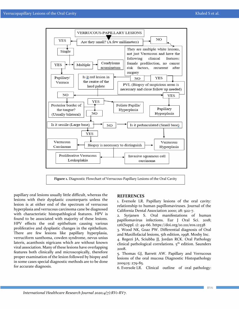
papillary oral lesions usually little difficult, whereas the lesions with their dysplastic counterparts unless the lesion is at either end of the spectrum of verrucous hyperplasia and verrucous carcinoma cane be diagnosed with characteristic histopathlogical features. HPV is found to be associated with majority of these lesions. HPV effects the oral epithelium causing various proliferative and dysplastic changes in the epithelium. There are few lesions like papillary hyperplasia, verruciform xanthoma, cowden syndrome, nevus unius lateris, acanthosis nigricans which are without known viral association. Many of these lesions have overlapping features both clinically and microscopically, therefore proper examination of the lesion followed by biopsy and in some cases special diagnostic methods are to be done for accurate diagnosis.
REFERENCES 1. Eversole LR. Papillary lesions of the oral cavity: relationship to human papillomaviruses. Journal of the California Dental Association 2000; 28: 922-7. 2. Syrjanen S. Oral manifestations of human papillomavirus infections. Eur J Oral Sci. 2018; 126(Suppl. 1): 49–66. https://doi.org/10.1111/eos.12538 3. Wood NK, Goaz PW. Differential diagnosis of Oral and Maxillofacial lesions, 5th edition, 1998. Mosby Inc. 4. Regezi JA, Sciubba JJ, Jordan RCK. Oral Pathology clinical pathological correlations. 5th edition. Saunders 2008. 5. Thomas GJ, Barrett AW. Papillary and Verrucous lesions of the oral mucosa Diagnostic Histopathology 2009;15: 279-85. 6. Eversole LR. Clinical outline of oral pathology:
RV6
Figure 1. Diagnostic Flowchart of Verrucous-Papillary Lesions of the Oral Cavity

International Healthcare Research Journal 2020;4(7):RV1-RV7.
Verrucopapillary Lesions of the Oral Cavity Khaled S et al.
Diagnosis and treatment, 3rd edition. 1992. Lea & Febiger, Philadelphia. 7. Rajendran R, Sivapathasundharam B. Shafer's text book of oral pathology. 8th edition. New Delhi: Elsevier; 2016 8. Neville BW, Damm DD, Allen CM, Bouquot JE. Oral & maxillofacial pathology. 1st South India Edition. New
Delhi: Elsevier; 2015. 9. Thompson LDR. Head and neck pathology a volume in series foundations in Diagnsotic pathology. Churchhill Livingstone, 2006: 34-6. 10. Gandolfo S, Scully C, Carrozzo M. Oral Medicine. Churchhill Livingstone Elsevier, 2006: 32-3.
AUTHOR AFFILIATIONS: (*Corresponding Author)
1. Assistant Professor, Master of Dental Surgery, Department of Oral and Maxillofacial Pathology, Sri Balaji Dental College, Hyderabad, Telangana 2. Assistant Professor, Master of Dental Surgery, Department of Orthodontics and Dentofacial Orthopedics, HKES, Nijalingappa Institute of Dental Sciences, Gulbarga, Karnataka 3. Assistant Professor, Master of Dental Surgery, Department of Oral and Maxillofacial Pathology, Panineeya Institute of Dental Sciences & RC Hyderabad, Telangana 4. Associate Professor, Master of Dental Surgery, Department of Public Health Dentistry, Panineeya Institute of Dental Sciences & RC Hyderabad, Telangana
Source of support: Nil, Conflict of interest: None declared
Cite this article as:
Khaled S, Bharadwaj SR, Anjum B, Satyanarayana D. Verrucopapillary Lesions of the Oral Cavity: A Review. Int Healthc Res J. 2020;4(7):RV1-RV7. https://doi.org/10.26440/IHRJ/0407.10365
Contact corresponding author at: satya.gullu[at]gmail[dot]com
RV7
International Healthcare Research Journal 2020;4(7):CR1-CR2.
INTRODUCTION The disease caused by the novel coronavirus SARS-CoV-2 (COVID- 19) was first described in China in December 2019 and is characterized by the appearance of symptoms such as fever, dry cough, dyspnoea, rhinorrhoea, anosmia, and ageusia. The horizon of respiratory involvement ranges from an upper airway catarrh, that can go unnoticed, even severe pneumonia or syndrome acute respiratory illness due to coronavirus.1 Although less frequently, dermatological manifestations have been described in association with COVID-19. In a study conducted by Recalcati in a sample of 88 cases, it was found that 18 patients had cutaneous manifestations, including 3 with generalized urticaria, 14 cases of erythematous rash, and one with varicelliform rash.2 These skin symptoms are not specific to COVID-19 and have characteristics similar to those that occur in other respiratory infections with common viral causes.
CASE REPORT We present the case of a 49-year-old man with no relevant medical history who presented to the emergency room with dry cough and dyspnea of moderate efforts for 7 days. By profession, the subject was a taxi driver but denied any recent contact with tourists or passengers due to lockdown. The subject also denied use of alcohol and tobacco and was not on any long-term medication. No chronic comorbidities were evident. On physical examination, the patient was afebrile, and pulmonary auscultation was normal.
He was able to pass air in and out of the lungs normally. Chest x-ray revealed peripheral and bilateral pulmonary opacities, predominantly in the lower lobes. The observed signs and symptoms were compatible with COVID-19. The polymerase chain reaction test for SARS-CoV-2 was done which came out to be positive. He was admitted and treatment with hydroxychloroquine and lopinavir /ritonavir was initiated. The subject showed clinical improvement and polymerase chain reaction for coronavirus SARS-CoV-2 gave negative result after two weeks. The subject was discharged but second day after discharge, he presented again with slightly itchy erythematous maculopapular rash with islets of healthy skin on the trunk, which appeared suddenly. Topical betamethasone ointment was prescribed along with oral levocetirizine, but after few hours, the lesions extended to cervical region, face and proximal region of upper limbs, oral prednisone was added to his treatment regimen. The clinical response was favorable, with disappearance of lesions in a few days.
DISCUSSION
Cutaneous symptoms are not too common in COVID-19 cases and in most patients appear after hospital discharge, as in our case, and are not associated with increased severity of COVID-19. In a previously conducted study, the skin lesions in COVID-19 patients mainly affected the trunk, were asymptomatic or slightly pruritic and then disappeared in a few days.2 Another previously
A B S T R A C T
K
Much is now known about the respiratory presentation of coronavirus SARS-CoV-2 (COVID-19) but it can also show up with clinical manifestations in other locations, such as on the skin. Herein, we describe a case with cutaneous symptoms that emerged during the recovery phase in a SARS-CoV-2 coronavirus infected patient. It is important for the healthcare professionals as well as the patients to know about such scenarios, so that appropriate action can be readily taken. KEYWORDS: Coronavirus, SARS-CoV-2, COVID-19
QR CODE
Cutaneous Manifestation of COVID-19: A Short Clinical Case
© Ratesh Bassi et al. This is an open access article distributed under the terms of the Creative Commons Attribution License CC-BY-NC 4.0, which permits unrestricted use, distribution and reproduction in any medium, provided the use is not commercial and the original author(s) and source are cited.
RATESH BASSI*1, GEETIKA BASSI2
ISSN: 2456-8090 (online) DOI: 10.26440/IHRJ/0407.10187
CASE REPORT
CR1
International Healthcare Research Journal 2020;4(7):CR1-CR2.
published case of COVID-19 was initially mistaken for dengue due to the similarity in cutaneous manifestations.3 Other dermatological manifestations to consider are those due to side effects of certain drugs used in the treatment of COVID-19. In particular, one must be aware of the possible adverse effects of hydroxychloroquine on the skin such as skin hyperpigmentation, pruritus, xerosis, alopecia, urticaria, morbilliform rashes or maculopapular and exfoliative dermatitis.4 Azithromycin can also cause skin rashes, itching or Stevens-Johnson syndrome. The most common adverse effects of the drug lopinavir/ritonavir are maculopapular rashes, pruritus, eczema and seborrheic dermatitis. Micro-thrombosis related to endothelial damage and vascular disorders in COVID-19 may also lead to other more serious skin lesions. They are ischemic lesions of sudden onset, characterized by cyanosis, blistering and dry gangrene of the fingers and toes.5
CONCLUSION It is evident from the literature that COVID-19 can lead to skin manifestations, more studies are needed to know all the forms of presentation and confirm the causal relationship with coronavirus
infection. It is important to rule out all other causes of skin lesions in COVID-19 affected subjects. For timely appropriate action, regular follow up of such patients should be scheduled for sure.
REFERENCES 1. Wang D, Hu B, Hu C, Zhu F, Liu X, Zhang J, et al. Clinical characteristics of 138 hospitalized patients with 2019 novel coronavirus-infected pneumonia in Wuhan, China. JAMA. 2020;323:1061–9. 2. Recalcati S. Cutaneous manifestations in COVID-19: A first perspective. J Eur Acad Dermatol Venereol. 2020;34(5):e212-e213. 3. Joob B, Wiwanitkit V. COVID-19 can present with a rash and be mistaken for dengue. J Am Acad Dermatol. 2020 May;82(5):e177. 4. Fernandez AP. Updated recommendations on the use of hydroxychloroquine in dermatologic practice. J Am Acad Dermatol. 2017;76:1176–82. 5. Zhang Y, Cao W, Xiao M, Li YJ, Yang Y, Zhao J, et al. Clinical and coagulation characteristics of 7 patients with critical COVID-2019 pneumonia and acro-ischemia. Zhonghua Xue Ye Xue Za Zhi. 2020;41(0):E006.
Cite this article as:
Bassi R, Bassi G. Cutaneous Manifestation of COVID-19: A Short Clinical Case. Int Healthc Res J. 2020;4(7):CR1-CR2. https://doi.org/10.26440/IHRJ/0407.10187
Contact Corresponding Author at: rateshbassi[at]gmail[dot]com
AUTHOR AFFILIATIONS: (*: Corresponding Author) 1. DNB Anesthesia 2. MDS, Oral and Maxillofacial Surgery Working in Rana Hospital, Kalka, Haryana, India
Source of support: Nil, Conflict of interest: None declared
Cutaneous Manifestation of COVID-19 Bassi R et al.
CR2
International Healthcare Research Journal 2020;4(7):CR3-CR6.
INTRODUCTION Hypodontia refers to the developmental failure of six or fewer teeth.1 Hypodontia is the most prevalent dentofacial malformation in humans.2 It can be associated with a recognised genetic syndrome or may occur as a nonsyndromic isolated trait.3 Tooth agenesis affects the maxilla and the mandible with similar prevelance4, whereas Wisth et al. (1974) suggested that the mandible is more frequently affected than the maxilla.5 Polder et al. (2004) in a metaanalysis on agenesis of tooth found that bilateral agenesis of maxillary lateral incisors occurs more often than unilateral agenesis. He too found that hypodontia affects females 1.4 times higher than males.4 Hypodontia causes deep bite and spacing in dentition. Missing posterior teeth also leads to nonworking interferences, overeruption of the opposing teeth and poor gingival contours. Laing et al. (2010) in a cross-sectional study concluded that if deciduous teeth are exfoliated which are associated with the missing permanent teeth, such patients with hypodontia will have more chewing difficulties.6 It is therefore plausible that hypodontia causes functional as well as esthetic limitations that affect an individual’s general well-being and quality of life in the process, although presently, evidence to support this is limited.
CASE REPORT
A 12-year-old girl reported in the dental clinic with chief complaint of non-eruption of teeth. Her past
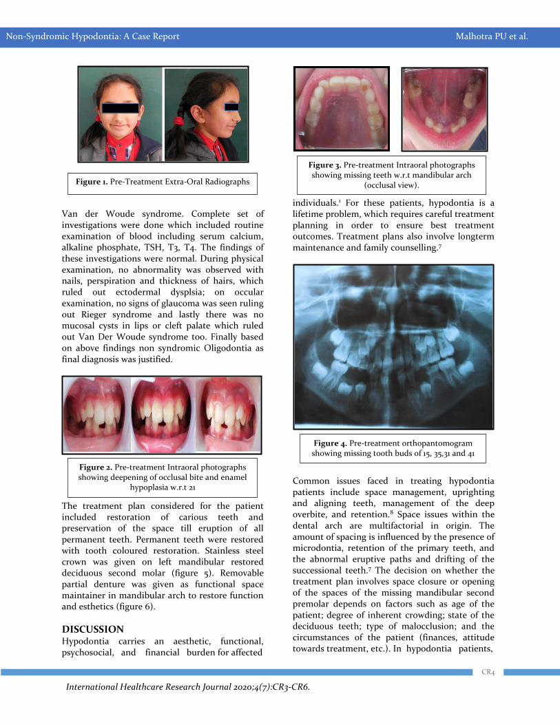
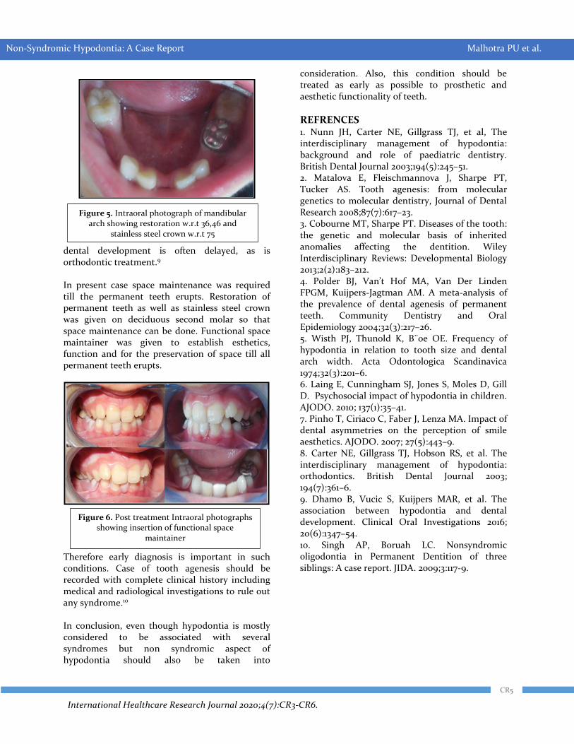
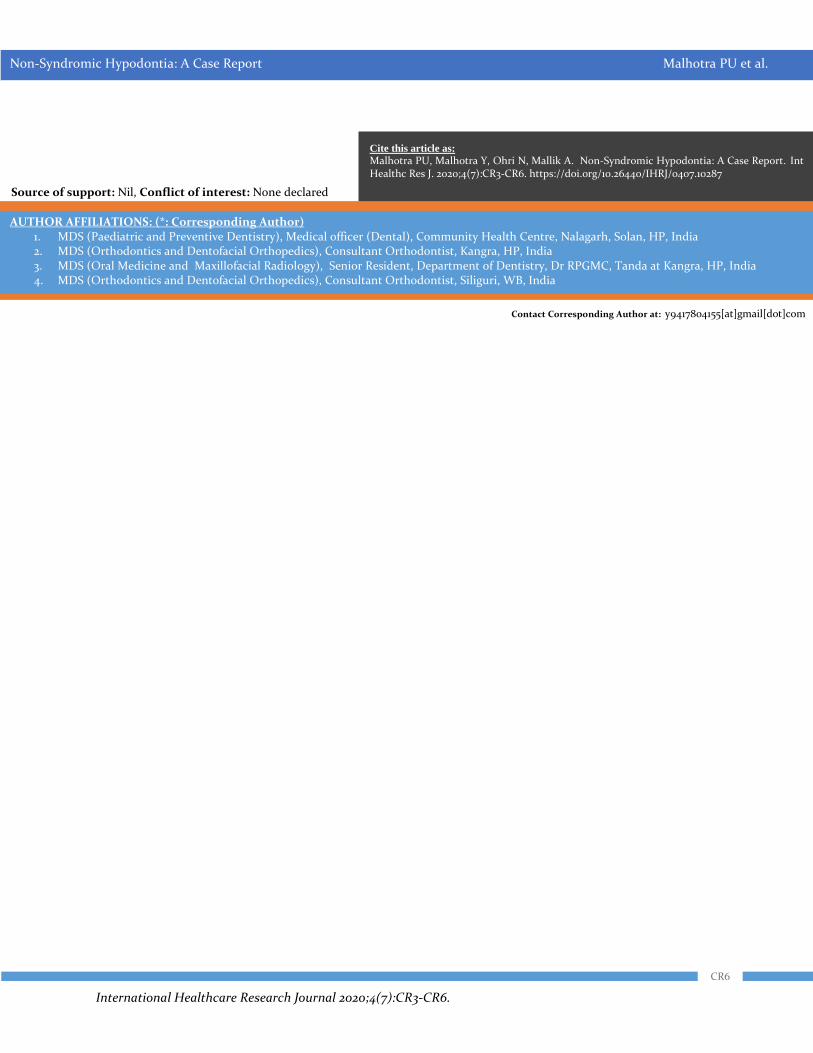
medical history was non-contributory and family history revealed that she was born to non-consanguineous marriage with normal delivery and no one in her family have congenitally missing teeth. The patient had no history of trauma or extractions. Extra oral examination revealed a face with normal facial profile and normal skeletal dental base relations (figure 1). Intra oral examination revealed, enamel hypoplasia in maxillary left central incisor and screw shaped left maxillary lateral incisor, retained right and left deciduous canines, right and left first deciduous molars, right and left deciduous second molars and right & left permanent first molars in maxillary arch. In mandibular arch, both permanent central incisors were missing. Permanent right & left lateral incisors and first molars were fully erupted. Deciduous right and left canines and deciduous left second molar were present. Permanent mandibular molars and deciduous left second molar were carious (figure 2 and 3). An orthopantomogram was advised which revealed missing tooth buds of tooth number 15, 35, 31 and 41 (figure 4). Due to prolonged absence of deciduous molars on right side and carious deciduous molar on left side in mandibular arch, there was deepening of occlusal bite as seen in figure 2. A provisional diagnosis of non-syndromic oligodontia was given with differential diagnosis of Ectodermal Dysplasia; Rieger syndrome and
A B S T R A C T
QR CODE
Non-Syndromic Hypodontia: A Case Report
PARUL UPPAL MALHOTRA1, YAGYESHWAR MALHOTRA*2, NEERA OHRI3, ANINDITA MALLIK4
ISSN: 2456-8090 (online) DOI: 10.26440/IHRJ/0407.10287
K
© Parul Uppal Malhotra et al. This is an open access article distributed under the terms of the Creative Commons Attribution License CC-BY-NC 4.0, which permits unrestricted use, distribution and reproduction in any medium, provided the use is not commercial and the original author(s) and source are cited.
CR3
CASE REPORT
Hypodontia is the most common dentofacial anomaly observed in humans. It can be syndromic or an isolated trait. Missing teeth not only affects functionality of dentition but also aesthetically looks unpleasing. In this case report, a 12 year old girl is presented with agenesis of four permanent teeth. On examination, patient was found to be suffering from non syndromic hypodontia. Restorative and Prosthetic treatment was done to rehabilitate the case KEYWORDS: Hypodontia, Non-syndromic, Agenesis
International Healthcare Research Journal 2020;4(7):CR3-CR6.
Van der Woude syndrome. Complete set of investigations were done which included routine examination of blood including serum calcium, alkaline phosphate, TSH, T3, T4. The findings of these investigations were normal. During physical examination, no abnormality was observed with nails, perspiration and thickness of hairs, which ruled out ectodermal dysplsia; on occular examination, no signs of glaucoma was seen ruling out Rieger syndrome and lastly there was no mucosal cysts in lips or cleft palate which ruled out Van Der Woude syndrome too. Finally based on above findings non syndromic Oligodontia as final diagnosis was justified.
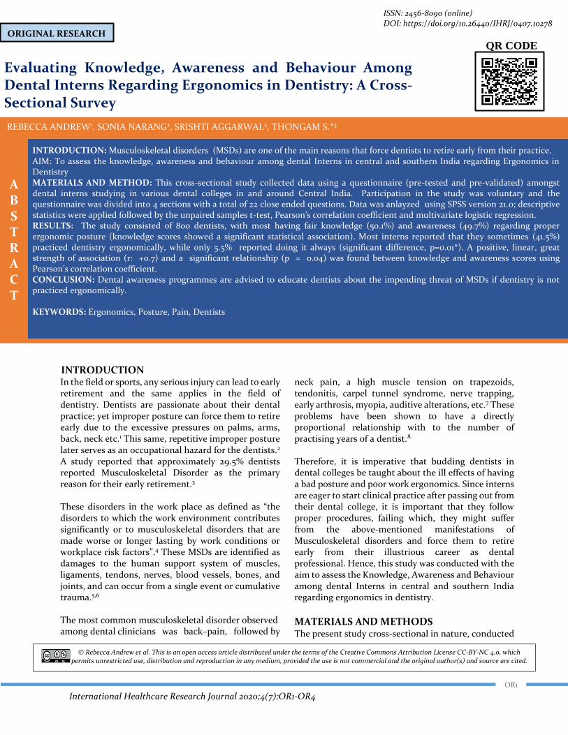
The treatment plan considered for the patient included restoration of carious teeth and preservation of the space till eruption of all permanent teeth. Permanent teeth were restored with tooth coloured restoration. Stainless steel crown was given on left mandibular restored deciduous second molar (figure 5). Removable partial denture was given as functional space maintainer in mandibular arch to restore function and esthetics (figure 6).
DISCUSSION Hypodontia carries an aesthetic, functional, psychosocial, and financial burden for affected
individuals.1 For these patients, hypodontia is a lifetime problem, which requires careful treatment planning in order to ensure best treatment outcomes. Treatment plans also involve longterm maintenance and family counselling.7
Common issues faced in treating hypodontia patients include space management, uprighting and aligning teeth, management of the deep overbite, and retention.8 Space issues within the dental arch are multifactorial in origin. The amount of spacing is influenced by the presence of microdontia, retention of the primary teeth, and the abnormal eruptive paths and drifting of the successional teeth.7 The decision on whether the treatment plan involves space closure or opening of the spaces of the missing mandibular second premolar depends on factors such as age of the patient; degree of inherent crowding; state of the deciduous teeth; type of malocclusion; and the circumstances of the patient (finances, attitude towards treatment, etc.). In hypodontia patients,
Figure 1. Pre-Treatment Extra-Oral Radiographs
Figure 2. Pre-treatment Intraoral photographs showing deepening of occlusal bite and enamel
hypoplasia w.r.t 21
Figure 3. Pre-treatment Intraoral photographs showing missing teeth w.r.t mandibular arch
(occlusal view).
Figure 4. Pre-treatment orthopantomogram showing missing tooth buds of 15, 35,31 and 41
CR4
Non-Syndromic Hypodontia: A Case Report Malhotra PU et al.
International Healthcare Research Journal 2020;4(7):CR3-CR6.
dental development is often delayed, as is orthodontic treatment.9 In present case space maintenance was required till the permanent teeth erupts. Restoration of permanent teeth as well as stainless steel crown was given on deciduous second molar so that space maintenance can be done. Functional space maintainer was given to establish esthetics, function and for the preservation of space till all permanent teeth erupts.
Therefore early diagnosis is important in such conditions. Case of tooth agenesis should be recorded with complete clinical history including medical and radiological investigations to rule out any syndrome.10 In conclusion, even though hypodontia is mostly considered to be associated with several syndromes but non syndromic aspect of hypodontia should also be taken into
consideration. Also, this condition should be treated as early as possible to prosthetic and aesthetic functionality of teeth.
REFRENCES
1. Nunn JH, Carter NE, Gillgrass TJ, et al, The interdisciplinary management of hypodontia: background and role of paediatric dentistry. British Dental Journal 2003;194(5):245–51. 2. Matalova E, Fleischmannova J, Sharpe PT, Tucker AS. Tooth agenesis: from molecular genetics to molecular dentistry, Journal of Dental Research 2008;87(7):617–23. 3. Cobourne MT, Sharpe PT. Diseases of the tooth: the genetic and molecular basis of inherited anomalies affecting the dentition. Wiley Interdisciplinary Reviews: Developmental Biology 2013;2(2):183–212. 4. Polder BJ, Van’t Hof MA, Van Der Linden FPGM, Kuijpers-Jagtman AM. A meta-analysis of the prevalence of dental agenesis of permanent teeth. Community Dentistry and Oral Epidemiology 2004;32(3):217–26. 5. Wisth PJ, Thunold K, B¨oe OE. Frequency of hypodontia in relation to tooth size and dental arch width. Acta Odontologica Scandinavica 1974;32(3):201–6. 6. Laing E, Cunningham SJ, Jones S, Moles D, Gill D. Psychosocial impact of hypodontia in children. AJODO. 2010; 137(1):35–41. 7. Pinho T, Ciriaco C, Faber J, Lenza MA. Impact of dental asymmetries on the perception of smile aesthetics. AJODO. 2007; 27(5):443–9. 8. Carter NE, Gillgrass TJ, Hobson RS, et al. The interdisciplinary management of hypodontia: orthodontics. British Dental Journal 2003; 194(7):361–6. 9. Dhamo B, Vucic S, Kuijpers MAR, et al. The association between hypodontia and dental development. Clinical Oral Investigations 2016; 20(6):1347–54. 10. Singh AP, Boruah LC. Nonsyndromic oligodontia in Permanent Dentition of three siblings: A case report. JIDA. 2009;3:117-9.
Figure 5. Intraoral photograph of mandibular arch showing restoration w.r.t 36,46 and
stainless steel crown w.r.t 75
Figure 6. Post treatment Intraoral photographs showing insertion of functional space
maintainer
CR5
Non-Syndromic Hypodontia: A Case Report Malhotra PU et al.
International Healthcare Research Journal 2020;4(7):CR3-CR6.
Cite this article as:
Malhotra PU, Malhotra Y, Ohri N, Mallik A. Non-Syndromic Hypodontia: A Case Report. Int Healthc Res J. 2020;4(7):CR3-CR6. https://doi.org/10.26440/IHRJ/0407.10287
Contact Corresponding Author at: y9417804155[at]gmail[dot]com
AUTHOR AFFILIATIONS: (*: Corresponding Author) 1. MDS (Paediatric and Preventive Dentistry), Medical officer (Dental), Community Health Centre, Nalagarh, Solan, HP, India 2. MDS (Orthodontics and Dentofacial Orthopedics), Consultant Orthodontist, Kangra, HP, India 3. MDS (Oral Medicine and Maxillofacial Radiology), Senior Resident, Department of Dentistry, Dr RPGMC, Tanda at Kangra, HP, India 4. MDS (Orthodontics and Dentofacial Orthopedics), Consultant Orthodontist, Siliguri, WB, India
CR6
Source of support: Nil, Conflict of interest: None declared
Non-Syndromic Hypodontia: A Case Report Malhotra PU et al.
International Healthcare Research Journal 2020;4(7):OR1-OR4
INTRODUCTION In the field or sports, any serious injury can lead to early retirement and the same applies in the field of dentistry. Dentists are passionate about their dental practice; yet improper posture can force them to retire early due to the excessive pressures on palms, arms, back, neck etc.1 This same, repetitive improper posture later serves as an occupational hazard for the dentists.2 A study reported that approximately 29.5% dentists reported Musculoskeletal Disorder as the primary reason for their early retirement.3 These disorders in the work place as defined as “the disorders to which the work environment contributes significantly or to musculoskeletal disorders that are made worse or longer lasting by work conditions or workplace risk factors”.4 These MSDs are identified as damages to the human support system of muscles, ligaments, tendons, nerves, blood vessels, bones, and joints, and can occur from a single event or cumulative trauma.5,6 The most common musculoskeletal disorder observed among dental clinicians was back–pain, followed by
neck pain, a high muscle tension on trapezoids, tendonitis, carpel tunnel syndrome, nerve trapping, early arthrosis, myopia, auditive alterations, etc.7 These problems have been shown to have a directly proportional relationship with to the number of practising years of a dentist.8 Therefore, it is imperative that budding dentists in dental colleges be taught about the ill effects of having a bad posture and poor work ergonomics. Since interns are eager to start clinical practice after passing out from their dental college, it is important that they follow proper procedures, failing which, they might suffer from the above-mentioned manifestations of Musculoskeletal disorders and force them to retire early from their illustrious career as dental professional. Hence, this study was conducted with the aim to assess the Knowledge, Awareness and Behaviour among dental Interns in central and southern India regarding ergonomics in dentistry.
MATERIALS AND METHODS The present study cross-sectional in nature, conducted
Evaluating Knowledge, Awareness and Behaviour Among Dental Interns Regarding Ergonomics in Dentistry: A Cross-Sectional Survey
ORIGINAL RESEARCH
A B S T R A C T
ISSN: 2456-8090 (online) DOI: https://doi.org/10.26440/IHRJ/0407.10278
REBECCA ANDREW1, SONIA NARANG2, SRISHTI AGGARWAL2, THONGAM S.*3
INTRODUCTION: Musculoskeletal disorders (MSDs) are one of the main reasons that force dentists to retire early from their practice. AIM: To assess the knowledge, awareness and behaviour among dental Interns in central and southern India regarding Ergonomics in Dentistry MATERIALS AND METHOD: This cross-sectional study collected data using a questionnaire (pre-tested and pre-validated) amongst dental interns studying in various dental colleges in and around Central India. Participation in the study was voluntary and the questionnaire was divided into 4 sections with a total of 22 close ended questions. Data was anlayzed using SPSS version 21.0; descriptive statistics were applied followed by the unpaired samples t-test, Pearson’s correlation coefficient and multivariate logistic regression. RESULTS: The study consisted of 800 dentists, with most having fair knowledge (50.1%) and awareness (49.7%) regarding proper ergonomic posture (knowledge scores showed a significant statistical association). Most interns reported that they sometimes (41.5%) practiced dentistry ergonomically, while only 5.5% reported doing it always (significant difference, p=0.01*). A positive, linear, great strength of association (r: +0.7) and a significant relationship (p = 0.04) was found between knowledge and awareness scores using Pearson’s correlation coefficient. CONCLUSION: Dental awareness programmes are advised to educate dentists about the impending threat of MSDs if dentistry is not practiced ergonomically. KEYWORDS: Ergonomics, Posture, Pain, Dentists
QR CODE
© Rebecca Andrew et al. This is an open access article distributed under the terms of the Creative Commons Attribution License CC-BY-NC 4.0, which permits unrestricted use, distribution and reproduction in any medium, provided the use is not commercial and the original author(s) and source are cited.
OR1
International Healthcare Research Journal 2020;4(7):OR1-OR4
Knowledge, Awareness and Behaviour Among Dental Interns Andrew R et al.
amongst dental interns studying in various dental colleges in and around Central India using convenience sampling from June 2019 to November 2019 after obtaining all necessary approvals (including ethical clearance) prior to start of the study. Data was collected using a pre-tested and pre-validated questionnaire filled by the interns and before they could answer the questionnaire, the first page informed them of the study objectives and that participation in the study was purely voluntary in nature. The interns were also informed that they could leave answering the questionnaire in between as per convenience. The questionnaire was divided pre-tested and pre-validated prior to the commencement of the study. It was divided into 4 sections and contained 22 questions (close ended) which took approximately 4-5 minutes to complete. Data was anlayzed using SPSS version 21.09 using descriptive statistics and the unpaired samples t-test, followed by Pearson’s correlation coefficient and multivariate logistic regression.
RESULTS Of a total of 1000 questionnaires distributed, we received 800 fully filled responses (response rate: 80%) which were further analyzed.
Knowledge and awareness and among dental interns regarding proper ergonomic posture (table 1) It was observed that most respondents has fair knowledge (50.1%) and awareness (49.7%) regarding proper ergonomic posture, with knowledge scores showing a significant statistical association. More respondents showed good knowledge (25% ) than awareness(19.3%).
Good Fair Poor P-value
Knowledge 200 (25%)
401 (50.1%)
199 (24.9%)
0.01
Awareness 154 (19.3%)
398 (49.7%)
248(31%) NS
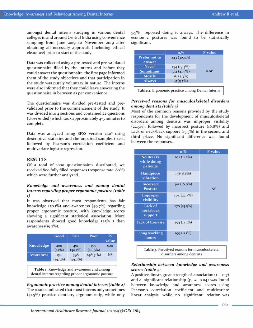
Ergonomic practice among dental interns (table 2) The results indicated that most interns only sometimes (41.5%) practice dentistry ergonomically, while only
5.5% reported doing it always. The difference in economic postures was found to be statistically significant.
n,% P-value
Prefer not to answer
243 (30.4%)
0.01* Never 154 (19.3%)
Sometimes 332 (41.5%)
Mostly 26 (3.3%)
Always 45(5.5%)
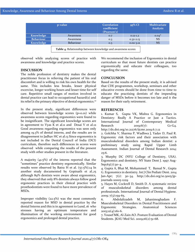
Perceived reasons for musculoskeletal disorders among dentists (table 3) Most of the common reasons provided by the study respondents for the development of musculoskeletal disorders among dentists was improper visibility (22.5%), followed by incorrect posture (16.8%) and Lack of neck/back support (15.5%) in the second and third place. No significant difference was found between the responses.
n,% P-value
No Breaks while doing
patients
202 (11.2%)
NS
Handpiece vibration
158(8.8%)
Incorrect Posture
301 (16.8%)
Improper visibility
404 (22.5%)
Lack of neck/back
support
278 (15.5%)
Lack of Exercise 254 (14.1%)
Long working hours
199 (11.1%)
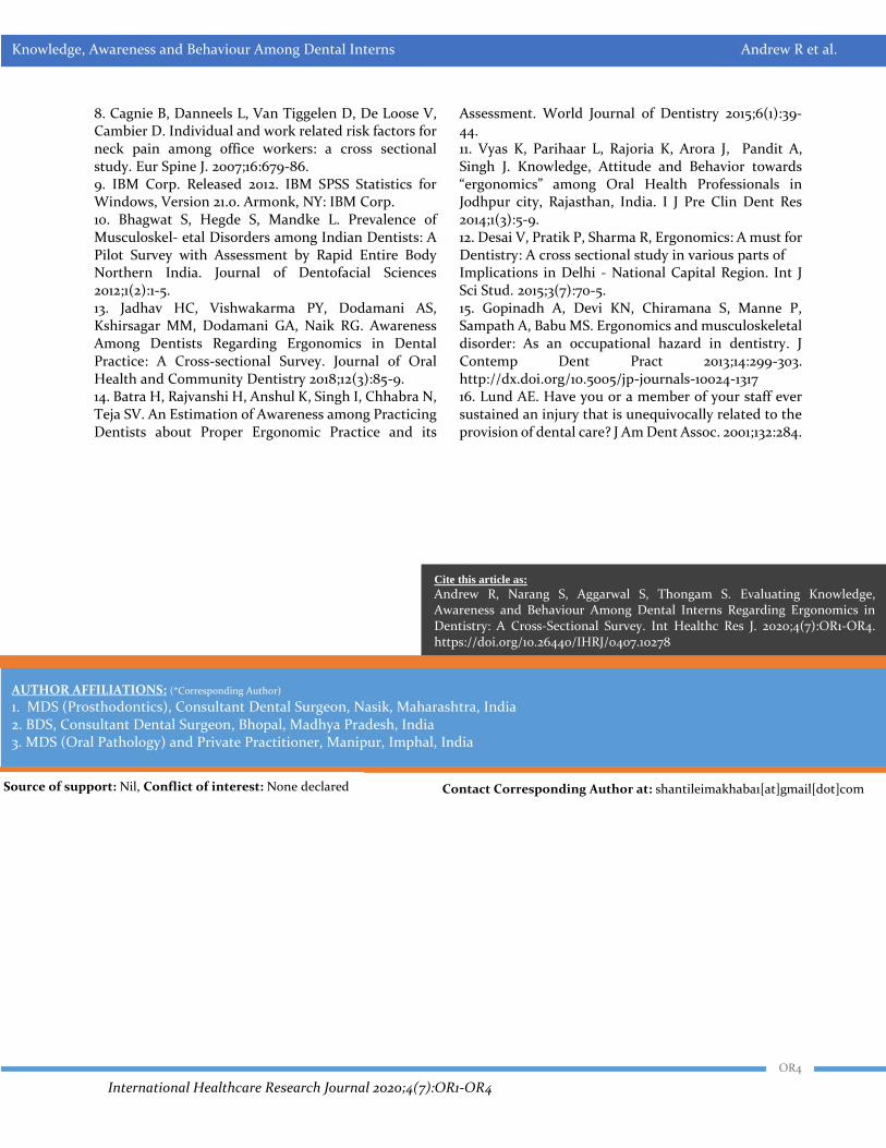
Relationship between knowledge and awareness scores (table 4) A positive, linear, great strength of association (r: +0.7) and a significant relationship (p = 0.04) was found between knowledge and awareness scores using Pearson’s correlation coefficient and multivariate linear analysis, while no significant relation was
Table 1. Knowledge and awareness and among dental interns regarding proper ergonomic posture
Table 2. Ergonomic practice among Dental Interns
Table 3. Perceived reasons for musculoskeletal disorders among dentists
OR2
International Healthcare Research Journal 2020;4(7):OR1-OR4
Knowledge, Awareness and Behaviour Among Dental Interns Andrew R et al.
p-value Correlation coefficient (Pearson’s)
95% CI Multivariate Analysis
Knowledge Awareness 0.7 0.12-1.2 0.04*
Behaviour Awareness 0.3 0.32-2.13 NS
Knowledge Behaviour 0.3 0.02-3.12 NS
observed while analyzing scores of practice with awareness and knowledge and practice scores.
DISCUSSION
The noble profession of dentistry makes the dental practitioner focus in relieving the patient of his oral discomfort and is willing to risk his own health for the same. This includes less breaks, lesser physical excercise, longer working hours and lesser time for self care. Repetitive small ranges of motion involved in dental practice can lead to occupational hazard(s) and its relief is the primary objective of dental ergonomics.10 In the present study, significant differences were observed between knowledge scores (p=0.01) while awareness scores regarding ergonomics were found to be insignificant. The significant knowledge scores are in agreement to Vyas K et al.11 and Desai V et al.12 Good awareness regarding ergonomics was seen only among 19.3% of dental interns, and the results are in disagreement to Jadhav HC et al.13 Since ergonomics is not included in the Dental Council of India (DCI) curriculum, therefore such differences in scores were observed while comparing the results of the present study with other studies present in the literature. A majority (41.5%) of the interns reported that the “sometimes” practice dentistry ergonomically. Similar results were observed by Batra H et al. (57.5%).14 In another study documented by Gopinath et al.,15 although 89% dentists were aware about ergonomics, they observed that only 8% dentists always follow good ergonomic practices in their clinical practice with prosthodontists were found to have more prevalence of MSDs. Improper visibility (22.5%) was the most commonly reported reason for MSD in dental practice by the dental Interns and this is in agreement to Lund, 16 who stresses having an optimum temperature and illumination of the working environment for good ergonomics and prolonged dental practice.
We recommend the inclusion of Ergonomics in dental curriculum so that most future dentists can practice ergonomically and educate their colleagues, too regarding the same.
CONCLUSION Based on the results of the present study, it is advised that CDE programmes, workshop, seminars and other educative events should be done from time to time to educate the practising dentists of the impending danger of MSDs before it becomes too late and it the reason for their early retirement.
REFERENCES 1. Kumar S, Gupta VK, Mishra G. Ergonomics in Dentistry: Really A Practice or Just a Tactics. International Journal of Contemporary Medical Research 2019;6(7): G1-G4. http://dx.doi.org/10.21276/ijcmr.2019.6.7.12 2. Golchha V, Sharma P, Wadhwa J, Yadav D, Paul R. Ergonomic risk factors and their association with musculoskeletal disorders among Indian dentist: a preliminary study using Rapid Upper Limb Assessment. Indian Journal of Dental Research 2014; 25:6. 3. Murphy DC (NYU College of Dentistry, USA). Ergonomics and dentistry. NY State Dent J. 1997 Aug-Sep;63(7):30–4. 4. Gupta A, Bhat M, Mohammed T, Bansal N , Gupta G. Ergonomics in dentistry. Int J Clin Pediatr Dent. 2014 Jan-Apr; 7(1): 30–34. http://dx.doi.org/10.5005/jp-journals-10005-1229 5. Hayes M, Cockrell D, Smith D. A systematic review of musculoskeletal disorders among dental professionals. International Journal of Dental Hygiene. 2009; 7(3):159-65. 6. Abdolalizadeh M, Jahanimoghadam F. Musculoskeletal Disorders in Dental Practitioners and Ergonomic Strategies. Anatomical Sciences 2015;12(4):161-6. 7. Yousef MK, Al-Zain AO. Posture Evaluation of Dental Students. JKAU Med Sci. 2009;16(2):51-68.
Table 4. Relationship between knowledge and awareness scores
OR3
International Healthcare Research Journal 2020;4(7):OR1-OR4
Knowledge, Awareness and Behaviour Among Dental Interns Andrew R et al.
8. Cagnie B, Danneels L, Van Tiggelen D, De Loose V, Cambier D. Individual and work related risk factors for neck pain among office workers: a cross sectional study. Eur Spine J. 2007;16:679-86. 9. IBM Corp. Released 2012. IBM SPSS Statistics for Windows, Version 21.0. Armonk, NY: IBM Corp. 10. Bhagwat S, Hegde S, Mandke L. Prevalence of Musculoskel‐ etal Disorders among Indian Dentists: A Pilot Survey with Assessment by Rapid Entire Body
Assessment. World Journal of Dentistry 2015;6(1):39‐44. 11. Vyas K, Parihaar L, Rajoria K, Arora J, Pandit A, Singh J. Knowledge, Attitude and Behavior towards “ergonomics” among Oral Health Professionals in Jodhpur city, Rajasthan, India. I J Pre Clin Dent Res 2014;1(3):5-9. 12. Desai V, Pratik P, Sharma R, Ergonomics: A must for Dentistry: A cross sectional study in various parts of
Northern India. Journal of Dentofacial Sciences 2012;1(2):1-5. 13. Jadhav HC, Vishwakarma PY, Dodamani AS, Kshirsagar MM, Dodamani GA, Naik RG. Awareness Among Dentists Regarding Ergonomics in Dental Practice: A Cross-sectional Survey. Journal of Oral Health and Community Dentistry 2018;12(3):85-9. 14. Batra H, Rajvanshi H, Anshul K, Singh I, Chhabra N, Teja SV. An Estimation of Awareness among Practicing Dentists about Proper Ergonomic Practice and its
Implications in Delhi - National Capital Region. Int J Sci Stud. 2015;3(7):70-5. 15. Gopinadh A, Devi KN, Chiramana S, Manne P, Sampath A, Babu MS. Ergonomics and musculoskeletal disorder: As an occupational hazard in dentistry. J Contemp Dent Pract 2013;14:299-303. http://dx.doi.org/10.5005/jp-journals-10024-1317 16. Lund AE. Have you or a member of your staff ever sustained an injury that is unequivocally related to the provision of dental care? J Am Dent Assoc. 2001;132:284.
Cite this article as:
Andrew R, Narang S, Aggarwal S, Thongam S. Evaluating Knowledge, Awareness and Behaviour Among Dental Interns Regarding Ergonomics in Dentistry: A Cross-Sectional Survey. Int Healthc Res J. 2020;4(7):OR1-OR4. https://doi.org/10.26440/IHRJ/0407.10278
AUTHOR AFFILIATIONS: (*Corresponding Author)
1. MDS (Prosthodontics), Consultant Dental Surgeon, Nasik, Maharashtra, India 2. BDS, Consultant Dental Surgeon, Bhopal, Madhya Pradesh, India 3. MDS (Oral Pathology) and Private Practitioner, Manipur, Imphal, India
Source of support: Nil, Conflict of interest: None declared
K
Contact Corresponding Author at: shantileimakhaba1[at]gmail[dot]com
OR4
International Healthcare Research Journal 2020;4(7):OR5-OR9
INTRODUCTION The prime objective of orthodontic treatment is to obtain better functional stability and aesthetics as well as good facial balance and harmony in an individual. It is possible through orthodontic intervention to achieve a better jaw relationship and a favourable relationship of the teeth to each other in the same and opposing arches and to their supporting bone and soft tissue. The occlusion and facial beauty are very much interdependent. Anterior crowding is one of the most common problems that motivate patients to seek orthodontic treatment. Dental crowding can be defined as a “discrepancy between tooth size and arch size that result in malposition and rotation of teeth”. Till date, many factors have been evaluated and found to be related to anterior dental crowding including dental arch width, arch length and mesiodistal tooth diameter. Studies show that smaller mandibular body lengths have been shown to be significantly associated with
crowding in permanent dentition.1-3 Hence, it can be put across that patients with class II malocclusion have a smaller mandibular length than subjects with normal occlusion and class I malocclusion. Another feature is the shape of facial profile which depends between the relationship between prognathism of the jaws. Facial profiling can also be done through dental pictures can also be analysed by comparing measurements on the tracing on the lateral skull- radiographs with known standards.4,5 To study the facial form and position of denture, it has been documented that upper and lower incisors to the APo plane is very useful guideline for determining cephalometric crowding of the anterior teeth, especially lower anteriors. This relation of the lower incisors to the APo plane is a key to communication of the problems with the anterior teeth.6 Although various researchers have tried to assess the relationship between facial profile and crowding, the
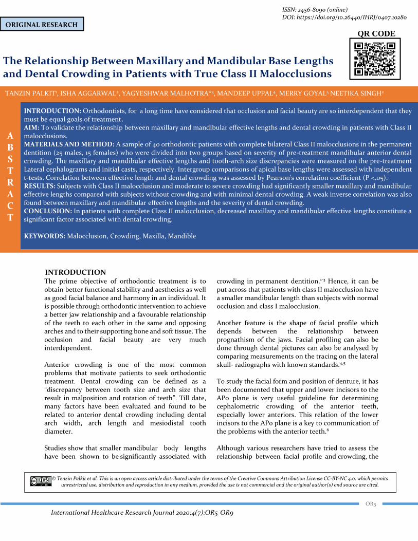
The Relationship Between Maxillary and Mandibular Base Lengths and Dental Crowding in Patients with True Class II Malocclusions
ORIGINAL RESEARCH
A B S T R A C T
ISSN: 2456-8090 (online) DOI: https://doi.org/10.26440/IHRJ/0407.10280
TANZIN PALKIT1, ISHA AGGARWAL2, YAGYESHWAR MALHOTRA*3, MANDEEP UPPAL4, MERRY GOYAL1, NEETIKA SINGH2
INTRODUCTION: Orthodontists, for a long time have considered that occlusion and facial beauty are so interdependent that they must be equal goals of treatment. AIM: To validate the relationship between maxillary and mandibular effective lengths and dental crowding in patients with Class II malocclusions. MATERIALS AND METHOD: A sample of 40 orthodontic patients with complete bilateral Class II malocclusions in the permanent dentition (25 males, 15 females) who were divided into two groups based on severity of pre-treatment mandibular anterior dental crowding. The maxillary and mandibular effective lengths and tooth-arch size discrepancies were measured on the pre-treatment Lateral cephalograms and initial casts, respectively. Intergroup comparisons of apical base lengths were assessed with independent t-tests. Correlation between effective length and dental crowding was assessed by Pearson's correlation coefficient (P <.05). RESULTS: Subjects with Class II malocclusion and moderate to severe crowding had significantly smaller maxillary and mandibular effective lengths compared with subjects without crowding and with minimal dental crowding. A weak inverse correlation was also found between maxillary and mandibular effective lengths and the severity of dental crowding. CONCLUSION: In patients with complete Class II malocclusion, decreased maxillary and mandibular effective lengths constitute a significant factor associated with dental crowding. KEYWORDS: Malocclusion, Crowding, Maxilla, Mandible
QR CODE
© Tenzin Palkit et al. This is an open access article distributed under the terms of the Creative Commons Attribution License CC-BY-NC 4.0, which permits unrestricted use, distribution and reproduction in any medium, provided the use is not commercial and the original author(s) and source are cited.
OR5
International Healthcare Research Journal 2020;4(7):OR5-OR9
Relationship Between Base Lengths and Dental Crowding Palkit T et al.
relationship between apical base length and dental crowding among patients in class II malocclusion has not been investigated exclusively. Therefore, the objective of this study was to evaluate the relationship of maxillary and mandibular effective length to the amount of anterior dental crowding in patients with complete class II malocclusion.
MATERIALS AND METHODS This cross-sectional study was carried out on subjects with class II malocclusion with the samples being retrospectively selected from the files of Orthodontic department of Dr. R. Ahmed Dental College and Hospital, Kolkata. Following an Ethical Approval, forty patients (25 males and 15 females) who satisfied the inclusion criteria were selected through convenience sampling. The inclusion criteria included presence of complete (full cusp) bilateral class II malocclusion (molar relationship), No open bite or cross bite, presence of all permanent teeth up to the first molar, absence of proximal decay or restoration, and absence of dental anomalies of number, size, form and position. The sample was be divided into two groups based on severity of pre-treatment mandibular anterior dental crowding. Group A consisted of 15 patients (10 males and 5 females) with a mean age of 12.81years and crowding >3mm and Group B had 25 patients (15 males and 10 females) with a mean age range 12.80 years and crowding < 3mm. Measurements were performed on pre-treatment dental casts and lateral head films. Tooth size and arch length discrepancy in the maxilla and mandible was calculated as the difference between arch perimeter and space required. Arch perimeter was measured on dental casts from the mesial aspect of first permanent molar to its antimere in millimetres with a brass wire. In a well aligned arch, arch perimeter were equal to the sum of tooth widths. Negative values indicated crowding. The space required was calculated by measuring mesio-distal width of each tooth from second premolar to contralateral second premolar in millimetres by a single examiner. Maxillary and mandibular effective lengths were measured on initial cephalogram. Mandibular effective length were measured from Co-Gn(Co- the most posterior superior point on the head of the condyle and Gn- is the most anterior inferior point on the symphysis of chin.) and the maxillary effective length were
measured from Co-point- A(Co-the most posterior superior point on the head of the condyle and pointA-deepest point in the midline between the anterior nasal spine and alveolar creast between the two incisors) then the Maxillomandibular differential is obtained by effective mandibular length-effective maxillary length. Inter group comparisons of apical base lengths were performed with independent t-tests. Correlation between base length and dental crowding was examined by means of Pearson’s correlation (p<.o5) using SPSS version 16.0.
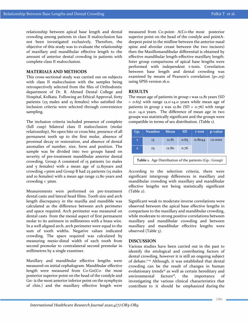
RESULTS The mean age of patients in group 1 was 12.81 years (SD = 0.65) with range 12.0-14.0 years while mean age of patients in group 2 was 12.80 (SD = 0.76) with range 12.0- 14.0 years. The difference of mean age in two groups was statistically significant and the groups were compatible in terms of sex distribution. (Table 1).
Gp. Number Mean SD t-test p-value
1 15 12.81 0.65 11.8044 <0.0001
2 25 12.80 0.76
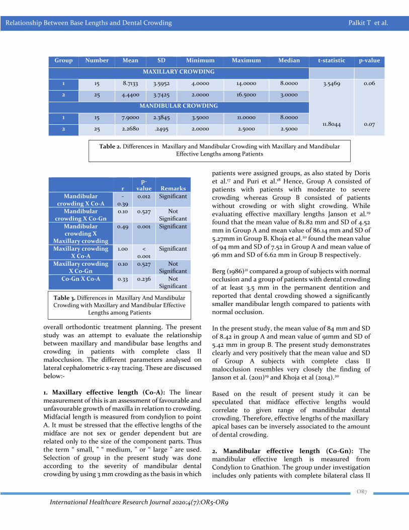
According to the selection criteria, there were significant intergroup differences in maxillary and mandibular crowding with maxillary and mandibular effective lengths not being statistically significant (Table 2). Significant weak to moderate inverse correlations were observed between the apical base effective lengths in comparison to the maxillary and mandibular crowding, while moderate to strong positive correlations between maxillary and mandibular crowding and between maxillary and mandibular effective lengths were observed (Table 3).
DISCUSSION Various studies have been carried out in the past to identify the etiological and contributing factors of dental crowding, however it is still an ongoing subject of debate.7-14 Although, it was established that dental crowding can be the result of changes in human evolutionary trends15 as well as certain hereditary and environmental factors16, the importance of investigating the various clinical characteristics that contribute to it should be emphasized during the
Table 1. Age Distribution of the patients (Gp.: Group)
OR6
International Healthcare Research Journal 2020;4(7):OR5-OR9
Relationship Between Base Lengths and Dental Crowding Palkit T et al.
Group Number Mean SD Minimum Maximum Median t-statistic p-value
MAXILLARY CROWDING
1 15 8.7133 3.5952 4.0000 14.0000 8.0000 3.5469
11.8044
0.06
0.07
2 25 4.4400 3.7425 2.0000 16.5000 3.0000
MANDIBULAR CROWDING
1 15 7.9000 2.3845 3.5000 11.0000 8.0000
2 25 2.2680 .2495 2.0000 2.5000 2.5000
r
p-value
Remarks
Mandibular crowding X Co-A
-0.39
0.012 Significant
Mandibular crowding X Co-Gn
0.10 0.527 Not Significant
Mandibular crowding X
Maxillary crowding
0.49 0.001 Significant
Maxillary crowding X Co-A
1.00 < 0.001
Significant
Maxillary crowding X Co-Gn
0.10 0.527 Not Significant
Co-Gn X Co-A 0.33 0.236 Not Significant
overall orthodontic treatment planning. The present study was an attempt to evaluate the relationship between maxillary and mandibular base lengths and crowding in patients with complete class II malocclusion. The different parameters analysed on lateral cephalometric x-ray tracing. These are discussed below:- 1. Maxillary effective length (Co-A): The linear measurement of this is an assessment of favourable and unfavourable growth of maxilla in relation to crowding. Midfacial length is measured from condylion to point A. It must be stressed that the effective lengths of the midface are not sex or gender dependent but are related only to the size of the component parts. Thus the term “ small, ” “ medium, ” or “ large ” are used. Selection of group in the present study was done according to the severity of mandibular dental crowding by using 3 mm crowding as the basis in which
patients were assigned groups, as also stated by Doris et al.17 and Puri et al.18 Hence, Group A consisted of patients with patients with moderate to severe crowding whereas Group B consisted of patients without crowding or with slight crowding. While evaluating effective maxillary lengths Janson et al.19 found that the mean value of 81.82 mm and SD of 4.52 mm in Group A and mean value of 86.14 mm and SD of 5.27mm in Group B. Khoja et al.20 found the mean value of 94 mm and SD of 7.52 in Group A and mean value of 96 mm and SD of 6.62 mm in Group B respectively. Berg (1986)21 compared a group of subjects with normal occlusion and a group of patients with dental crowding of at least 3.5 mm in the permanent dentition and reported that dental crowding showed a significantly smaller mandibular length compared to patients with normal occlusion. In the present study, the mean value of 84 mm and SD of 8.42 in group A and mean value of 91mm and SD of 5.42 mm in group B. The present study demonstrates clearly and very positively that the mean value and SD of Group A subjects with complete class II malocclusion resembles very closely the finding of Janson et al. (2011)19 and Khoja et al (2014).20 Based on the result of present study it can be speculated that midface effective lengths would correlate to given range of mandibular dental crowding. Therefore, effective lengths of the maxillary apical bases can be inversely associated to the amount of dental crowding. 2. Mandibular effective length (Co-Gn): The mandibular effective length is measured from Condylion to Gnathion. The group under investigation includes only patients with complete bilateral class II
Table 2. Differences in Maxillary and Mandibular Crowding with Maxillary and Mandibular Effective Lengths among Patients
Table 3. Differences in Maxillary And Mandibular Crowding with Maxillary and Mandibular Effective
Lengths among Patients
OR7
International Healthcare Research Journal 2020;4(7):OR5-OR9
Relationship Between Base Lengths and Dental Crowding Palkit T et al.
molar relationship so that class II malocclusions could be clearly characterised. Janson et al.19 while evaluating effective mandibular base length found that the mean value of 103 mm and SD of 5.01 mm in Group A. and mean value of 108 mm and SD of 6.04 mm in Group B. Khoja et al.20 found that the mean value of 97.8 mm and SD of 4.82 mm in Group A and mean value of 101 mm and SD of 5.42 mm in Group B. In the present study the mean value is 108.2 mm and SD of 22 mm in Group A and mean value of 111.6 mm and SD of 5.44 mm in group B. The present study demonstrates clearly and very positively that the mean value and SD of Group A subjects with complete class II malocclusion resembles very closely the finding of Janson et al.19 (2011) and Khoja et al. (2014).20 According to the study conducted by Ani GS et al.,(2014)22 it was found that the ach perimeter assessed with a brass wire had greater accuracy as compared to using vernier caliper. It was observed that patients with moderate to severe dental crowding had smaller mandibular effective lengths compared with subjects without crowding and with minimum dental crowding. In addition there was a significant weak to moderate inverse correlation between the amount of crowding and the mandibular effective lengths. Based on the result of present study, we speculate that mandibular effective lengths would correlate to given range of mandibular dental crowding and hence, effective lengths of the mandibular apical bases can be inversely associated to the amount of dental crowding.
CONCLUSION In the present study, groups selected according to mandibular crowding, maxillary crowding was also observed significantly in the severely crowded group and hence we conclude that severely crowded cases are more likely to present shorter effective apical base lengths and that the shorter the base lengths, greater is the likehood for crowding. These findings are specially applicable to subjects having complete class II malocclusion.
REFERENCES 1. Sakuda M, Kuroda Y, Wada K, Matsumoto M. Changes in crowding of teeth during adolescence and their relation to the growth of the facial skeleton. Trans
Eur Orthod Soc. 1976:93–104. 2. Leighton BC, Hunter WS. Relationship between lower arch spacing/crowding and facial height and depth. AJODO 1982;82(5):418-25. https://doi.org/10.1016/0002-9416(82)90191-9 3. Richardson ME. The etiology of late lower arch crowding alternative to mesially directed forces: a review. American Journal of Orthodontics and Dentofacial Orthopedics. 1994;105(6):592-7. 4. Björk A, Palling M. Adolescent age changes in sagittal jaw relation, alveolar prognathy, and incisal inclination. Acta odontologica Scandinavica 1955; 12(3-4):201–32. 5. Richardson ME. A review of changes in lower arch alignment from seven to fifty years. Semin Orthod. 1999;5:151–9. 6. Sampson WJ, Richards LC. Prediction of mandibular incisor and canine crowding changes in the mixed dentition. Am J Orthod. 1985;88:47–63. 7. Sayin MO, Turkkahraman H. Factors contributing to mandibular anterior crowding in the early mixed dentition. Angle Orthod 2004;74:754–8. 8. Radnzic D. Dental crowding and its relationship to mesiodistal crown diameters and arch dimensions. Am J Orthod Dentofacial Orthop 1988;94:50–6. 9. Bernabe E, Flores-Mir C. Dental morphology and crowding. A multivariate approach. Angle Orthod 2006;76:20–5 10. Sanin C, Savara BS. Factors that affect the alignment of the mandibular incisors: a longitudinal study. Am J Orthod 1973;64:248–57. 11. Bernabe E, del Castillo CE, Flores-Mir C. Intra-arch occlusal indicators of crowding in the permanent dentition. Am J Orthod Dentofacial Orthop. 2005;128(2):220–5. 12. Howe RP, McNamara JA, O'Connor KA. An examination of dental crowding and its relationship to tooth size and arch dimension. Am J Orthod Dentofacial Orthop 1983;83:363–73. 13. Shigenobu N, Hisano M, Shima S, Matsubara N, Soma K. Patterns of dental crowding in the lower arch and contributing factors. A statistical study. Angle Orthod 2007;77(2):303–10. 14. Lundstrom A. The etiology of crowding of teeth (based on studies of twins and on morphological investigations) and its bearing on orthodontic treatment (expansion or extraction). Trrans Eur Orthod Soc 1951:176–89. 15. Hooten EA. Up from the ape, New York. The Macmillan Company. 1947 16. Barber TK. The crowded arches. J South Calif Dent Assoc. 1967;35: 232–40.
OR8

International Healthcare Research Journal 2020;4(7):OR5-OR9
Relationship Between Base Lengths and Dental Crowding Palkit T et al.
17. Doris JM, Bernard BW, Kuftinec MM, Stom D. A biometric study of tooth size and dental crowding. Am J Orthod. 1981;79: 326–336. 18. Puri N, Pradhan L, Chandna A, Sehgal V, Gupta R. Biometric study of tooth size in normal, crowded, and spaced permanent dentitions. Am J Orthod Dentofacial Orthop. 2007 Sep;132(3):279.e7-14. 19. Janson G, Goizueta OEFM, Garib DG, Janson M. Relationship between maxillary and mandibular base lengths and dental crowding in patients with complete class II malocclusions. The Angle Orthodontist 2011; 81(2):217-21.
20. Khoja A, Fida M, Sheikh A. Association of maxillary and mandibular base lengths with dental crowding in different skeletal malocclusions; J Ayub Med Coll Abbottabad. 2014;26(6):428-33. 21. Berg R. Crowding of the dental arches : a longitudinal study of the age period between 6 and 12 years. Eur J Orthod. 1986:8; 43-9. 22. Ani GS, Babu EC and Soman PS. Assessment of dental crowding. Int J Appl Basic Med Res. 2014:4(1):52-5.
Cite this article as:
Palkit T, Aggarwal I, Malhotra Y, Goyal M, Uppal M, Singh N. The Relationship Between Maxillary and Mandibular Base Lengths and Dental Crowding in Patients with True Class II Malocclusions. Int Healthc Res J. 2020;4(7):OR5-OR9. https://doi.org/10.26440/IHRJ/0407.10280
AUTHOR AFFILIATIONS: (*Corresponding Author)
1. MDS (Orthodontics and Dentofacial Orthopedics), Senior Lecturer, Department of Orthodontics and Dentofacial Orthopedics, Bhojia Dental College, Baddi, H.P. 2. MDS (Orthodontics and Dentofacial Orthopedics), Reader, Department of Orthodontics and Dentofacial Orthopedics, Bhojia Dental College, Baddi, H.P. 3. MDS (Orthodontics and Dentofacial Orthopedics), Consultant Orthodontist, Kangra, HP, India (Corresponding Author). 4. MDS (Orthodontics and Dentofacial Orthopedics), Professor, Department of Orthodontics and Dentofacial Orthopedics, Bhojia Dental College, Baddi, H.P.
Source of support: Nil, Conflict of interest: None declared
K
Contact Corresponding Author at: y9417804155[at]outlook[dot]com
OR9
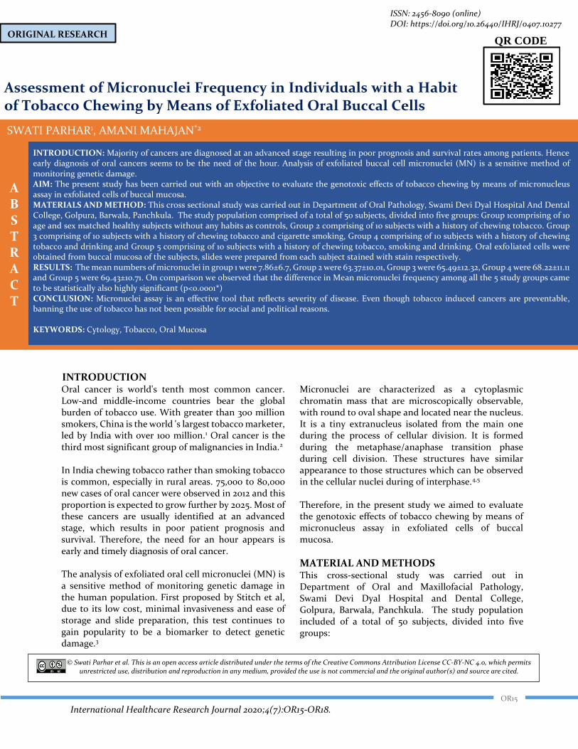
International Healthcare Research Journal 2020;4(7):OR15-OR18.
INTRODUCTION Oral cancer is world's tenth most common cancer. Low-and middle-income countries bear the global burden of tobacco use. With greater than 300 million smokers, China is the world 's largest tobacco marketer, led by India with over 100 million.1 Oral cancer is the third most significant group of malignancies in India.2 In India chewing tobacco rather than smoking tobacco is common, especially in rural areas. 75,000 to 80,000 new cases of oral cancer were observed in 2012 and this proportion is expected to grow further by 2025. Most of these cancers are usually identified at an advanced stage, which results in poor patient prognosis and survival. Therefore, the need for an hour appears is early and timely diagnosis of oral cancer. The analysis of exfoliated oral cell micronuclei (MN) is a sensitive method of monitoring genetic damage in the human population. First proposed by Stitch et al, due to its low cost, minimal invasiveness and ease of storage and slide preparation, this test continues to gain popularity to be a biomarker to detect genetic damage.3
Micronuclei are characterized as a cytoplasmic chromatin mass that are microscopically observable, with round to oval shape and located near the nucleus. It is a tiny extranucleus isolated from the main one during the process of cellular division. It is formed during the metaphase/anaphase transition phase during cell division. These structures have similar appearance to those structures which can be observed in the cellular nuclei during of interphase.4,5 Therefore, in the present study we aimed to evaluate the genotoxic effects of tobacco chewing by means of micronucleus assay in exfoliated cells of buccal mucosa.
MATERIAL AND METHODS This cross-sectional study was carried out in Department of Oral and Maxillofacial Pathology, Swami Devi Dyal Hospital and Dental College, Golpura, Barwala, Panchkula. The study population included of a total of 50 subjects, divided into five groups:
Assessment of Micronuclei Frequency in Individuals with a Habit of Tobacco Chewing by Means of Exfoliated Oral Buccal Cells
ORIGINAL RESEARCH
A B S T R A C T
ISSN: 2456-8090 (online) DOI: https://doi.org/10.26440/IHRJ/0407.10277
SWATI PARHAR1, AMANI MAHAJAN*2
INTRODUCTION: Majority of cancers are diagnosed at an advanced stage resulting in poor prognosis and survival rates among patients. Hence early diagnosis of oral cancers seems to be the need of the hour. Analysis of exfoliated buccal cell micronuclei (MN) is a sensitive method of monitoring genetic damage. AIM: The present study has been carried out with an objective to evaluate the genotoxic effects of tobacco chewing by means of micronucleus assay in exfoliated cells of buccal mucosa. MATERIALS AND METHOD: This cross sectional study was carried out in Department of Oral Pathology, Swami Devi Dyal Hospital And Dental College, Golpura, Barwala, Panchkula. The study population comprised of a total of 50 subjects, divided into five groups: Group 1comprising of 10 age and sex matched healthy subjects without any habits as controls, Group 2 comprising of 10 subjects with a history of chewing tobacco. Group 3 comprising of 10 subjects with a history of chewing tobacco and cigarette smoking, Group 4 comprising of 10 subjects with a history of chewing tobacco and drinking and Group 5 comprising of 10 subjects with a history of chewing tobacco, smoking and drinking. Oral exfoliated cells were obtained from buccal mucosa of the subjects, slides were prepared from each subject stained with stain respectively. RESULTS: The mean numbers of micronuclei in group 1 were 7.86±6.7, Group 2 were 63.37±10.01, Group 3 were 65.49±12.32, Group 4 were 68.22±11.11 and Group 5 were 69.43±10.71. On comparison we observed that the difference in Mean micronuclei frequency among all the 5 study groups came to be statistically also highly significant (p<0.0001*) CONCLUSION: Micronuclei assay is an effective tool that reflects severity of disease. Even though tobacco induced cancers are preventable, banning the use of tobacco has not been possible for social and political reasons. KEYWORDS: Cytology, Tobacco, Oral Mucosa
QR CODE
© Swati Parhar et al. This is an open access article distributed under the terms of the Creative Commons Attribution License CC-BY-NC 4.0, which permits unrestricted use, distribution and reproduction in any medium, provided the use is not commercial and the original author(s) and source are cited.
OR15
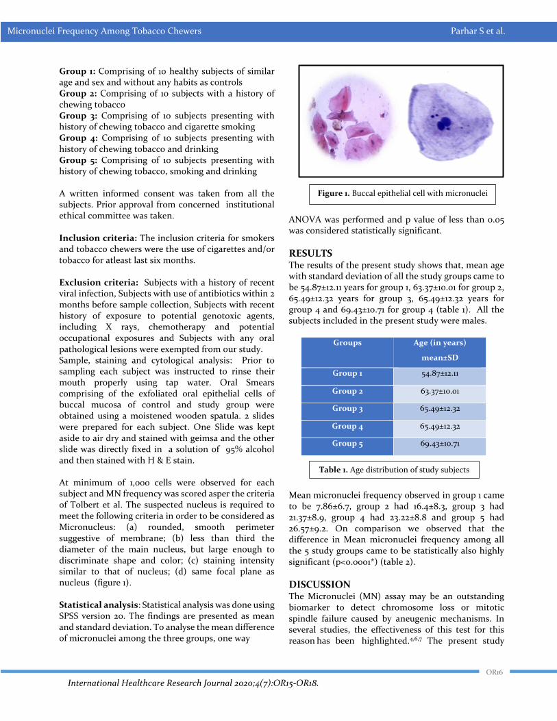
International Healthcare Research Journal 2020;4(7):OR15-OR18.
Micronuclei Frequency Among Tobacco Chewers Parhar S et al.
Group 1: Comprising of 10 healthy subjects of similar age and sex and without any habits as controls Group 2: Comprising of 10 subjects with a history of chewing tobacco Group 3: Comprising of 10 subjects presenting with history of chewing tobacco and cigarette smoking Group 4: Comprising of 10 subjects presenting with history of chewing tobacco and drinking Group 5: Comprising of 10 subjects presenting with history of chewing tobacco, smoking and drinking A written informed consent was taken from all the subjects. Prior approval from concerned institutional ethical committee was taken. Inclusion criteria: The inclusion criteria for smokers and tobacco chewers were the use of cigarettes and/or tobacco for atleast last six months. Exclusion criteria: Subjects with a history of recent viral infection, Subjects with use of antibiotics within 2 months before sample collection, Subjects with recent history of exposure to potential genotoxic agents, including X rays, chemotherapy and potential occupational exposures and Subjects with any oral pathological lesions were exempted from our study. Sample, staining and cytological analysis: Prior to sampling each subject was instructed to rinse their mouth properly using tap water. Oral Smears comprising of the exfoliated oral epithelial cells of buccal mucosa of control and study group were obtained using a moistened wooden spatula. 2 slides were prepared for each subject. One Slide was kept aside to air dry and stained with geimsa and the other slide was directly fixed in a solution of 95% alcohol and then stained with H & E stain. At minimum of 1,000 cells were observed for each subject and MN frequency was scored asper the criteria of Tolbert et al. The suspected nucleus is required to meet the following criteria in order to be considered as Micronucleus: (a) rounded, smooth perimeter suggestive of membrane; (b) less than third the diameter of the main nucleus, but large enough to discriminate shape and color; (c) staining intensity similar to that of nucleus; (d) same focal plane as nucleus (figure 1). Statistical analysis: Statistical analysis was done using SPSS version 20. The findings are presented as mean and standard deviation. To analyse the mean difference of micronuclei among the three groups, one way
ANOVA was performed and p value of less than 0.05 was considered statistically significant.
RESULTS
The results of the present study shows that, mean age with standard deviation of all the study groups came to be 54.87±12.11 years for group 1, 63.37±10.01 for group 2, 65.49±12.32 years for group 3, 65.49±12.32 years for group 4 and 69.43±10.71 for group 4 (table 1). All the subjects included in the present study were males.
Groups Age (in years)
mean±SD
Group 1 54.87±12.11
Group 2 63.37±10.01
Group 3 65.49±12.32
Group 4 65.49±12.32
Group 5 69.43±10.71
Mean micronuclei frequency observed in group 1 came to be 7.86±6.7, group 2 had 16.4±8.3, group 3 had 21.37±8.9, group 4 had 23.22±8.8 and group 5 had 26.57±9.2. On comparison we observed that the difference in Mean micronuclei frequency among all the 5 study groups came to be statistically also highly significant (p<0.0001*) (table 2).
DISCUSSION
The Micronuclei (MN) assay may be an outstanding biomarker to detect chromosome loss or mitotic spindle failure caused by aneugenic mechanisms. In several studies, the effectiveness of this test for this reason has been highlighted.4,6,7 The present study
Figure 1. Buccal epithelial cell with micronuclei
Table 1. Age distribution of study subjects
OR16
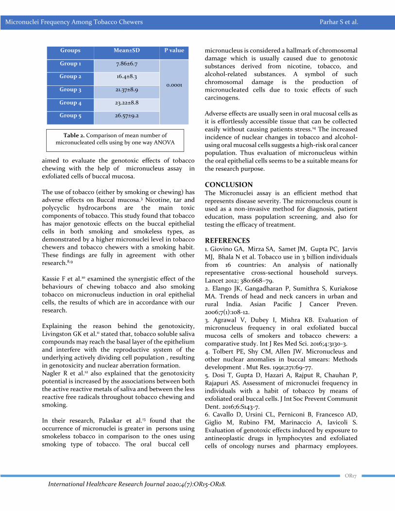
International Healthcare Research Journal 2020;4(7):OR15-OR18.
Micronuclei Frequency Among Tobacco Chewers Parhar S et al.
Groups Mean±SD P value
Group 1 7.86±6.7
0.0001
Group 2 16.4±8.3
Group 3 21.37±8.9
Group 4 23.22±8.8
Group 5 26.57±9.2
aimed to evaluate the genotoxic effects of tobacco chewing with the help of micronucleus assay in exfoliated cells of buccal mucosa. The use of tobacco (either by smoking or chewing) has adverse effects on Buccal mucosa.3 Nicotine, tar and polycyclic hydrocarbons are the main toxic components of tobacco. This study found that tobacco has major genotoxic effects on the buccal epithelial cells in both smoking and smokeless types, as demonstrated by a higher micronuclei level in tobacco chewers and tobacco chewers with a smoking habit. These findings are fully in agreement with other research.8,9 Kassie F et al.10 examined the synergistic effect of the behaviours of chewing tobacco and also smoking tobacco on micronucleus induction in oral epithelial cells, the results of which are in accordance with our research. Explaining the reason behind the genotoxicity, Livingston GK et al.11 stated that, tobacco soluble saliva compounds may reach the basal layer of the epithelium and interfere with the reproductive system of the underlying actively dividing cell population , resulting in genotoxicity and nuclear aberration formation. Nagler R et al.12 also explained that the genotoxicity potential is increased by the associations between both the active reactive metals of saliva and between the less reactive free radicals throughout tobacco chewing and smoking. In their research, Palaskar et al.13 found that the occurrence of micronuclei is greater in persons using smokeless tobacco in comparison to the ones using smoking type of tobacco. The oral buccal cell
micronucleus is considered a hallmark of chromosomal damage which is usually caused due to genotoxic substances derived from nicotine, tobacco, and alcohol-related substances. A symbol of such chromosomal damage is the production of micronucleated cells due to toxic effects of such carcinogens. Adverse effects are usually seen in oral mucosal cells as it is effortlessly accessible tissue that can be collected easily without causing patients stress.14 The increased incidence of nuclear changes in tobacco and alcohol-using oral mucosal cells suggests a high-risk oral cancer population. Thus evaluation of micronucleus within the oral epithelial cells seems to be a suitable means for the research purpose.
CONCLUSION The Micronuclei assay is an efficient method that represents disease severity. The micronucleus count is used as a non-invasive method for diagnosis, patient education, mass population screening, and also for testing the efficacy of treatment.
REFERENCES 1. Giovino GA, Mirza SA, Samet JM, Gupta PC, Jarvis MJ, Bhala N et al. Tobacco use in 3 billion individuals from 16 countries: An analysis of nationally representative cross-sectional household surveys. Lancet 2012; 380:668–79. 2. Elango JK, Gangadharan P, Sumithra S, Kuriakose MA. Trends of head and neck cancers in urban and rural India. Asian Pacific J Cancer Preven. 2006;7(1):108-12. 3. Agrawal V, Dubey I, Mishra KB. Evaluation of micronucleus frequency in oral exfoliated buccal mucosa cells of smokers and tobacco chewers: a comparative study. Int J Res Med Sci. 2016;4:3130-3. 4. Tolbert PE, Shy CM, Allen JW. Micronucleus and other nuclear anomalies in buccal smears: Methods development . Mut Res. 1991;271:69-77. 5. Dosi T, Gupta D, Hazari A, Rajput R, Chauhan P, Rajapuri AS. Assessment of micronuclei frequency in individuals with a habit of tobacco by means of exfoliated oral buccal cells. J Int Soc Prevent Communit Dent. 2016;6:S143-7. 6. Cavallo D, Ursini CL, Perniconi B, Francesco AD, Giglio M, Rubino FM, Marinaccio A, Iavicoli S. Evaluation of genotoxic effects induced by exposure to antineoplastic drugs in lymphocytes and exfoliated cells of oncology nurses and pharmacy employees.
Table 2. Comparison of mean number of micronucleated cells using by one way ANOVA
OR17
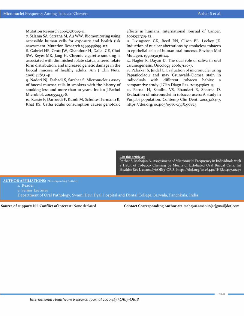
International Healthcare Research Journal 2020;4(7):OR15-OR18.
Micronuclei Frequency Among Tobacco Chewers Parhar S et al.
Mutation Research 2005;587:45-51. 7. Salama SA, Serrana M, Au WW. Biomonitoring using accessible human cells for exposure and health risk assessment. Mutation Research 1999;436:99-112. 8. Gabriel HE, Crott JW, Ghandour H, Dallal GE, Choi SW, Keyes MK, Jang H. Chronic cigarette smoking is associated with diminished folate status, altered folate form distribution, and increased genetic damage in the buccal mucosa of healthy adults. Am J Clin Nutr. 2006;41:835-41. 9. Naderi NJ, Farhadi S, Sarshar S. Micronucleus assay of buccal mucosa cells in smokers with the history of smoking less and more than 10 years. Indian J Pathol Microbiol. 2012;55:433-8. 10. Kassie F, Darroudi F, Kundi M, Schulte-Hermann R, Khat KS. Catha edulis consumption causes genotoxic
effects in humans. International Journal of Cancer. 2001;92:329-32. 11. Livingston GK, Reed RN, Olson BL, Lockey JE. Induction of nuclear aberrations by smokeless tobacco in epithelial cells of human oral mucosa. Environ Mol Mutagen. 1990;15:136-44. 12. Nagler R, Dayan D. The dual role of saliva in oral carcinogenesis. Oncology 2006;71:10-7. 13. Palaskar S, Jindal C. Evaluation of micronuclei using Papanicolaou and may Grunwald-Giemsa stain in individuals with different tobacco habits: a comparative study. J Clin Diagn Res. 2011;4:3607-13. 14. Bansal H, Sandhu VS, Bhandari R, Sharma D. Evaluation of micronuclei in tobacco users: A study in Punjabi population. Contemp Clin Dent. 2012;3:184-7. https://doi.org/10.4103/0976-237X.96825
Cite this article as:
Parhar S, Mahajan A. Assessment of Micronuclei Frequency in Individuals with a Habit of Tobacco Chewing by Means of Exfoliated Oral Buccal Cells. Int Healthc Res J. 2020;4(7):OR15-OR18. https://doi.org/10.26440/IHRJ/0407.10277
AUTHOR AFFILIATIONS: (*Corresponding Author)
1. Reader 2. Senior Lecturer Department of Oral Pathology, Swami Devi Dyal Hospital and Dental College, Barwala, Panchkula, India
Source of support: Nil, Conflict of interest: None declared Contact Corresponding Author at: mahajan.amani18[at]gmail[dot]com
OR18