
General Information - INTERNATIONAL JOURNAL OF ...
266
About The Journal International Journal of Scientific Study (IJSS) is a monthly journal publishing research articles after full peer review and aims to publish scientifically sound research articles in across all science like Medicine, Dentistry, Genetics, Pharmacy, etc. Each article submitted to us would be undergoing review in three stages: Initial Review, Peer Review & Final Review. All rights are reserved with journal owner. Without the prior permission from Editor, no part of the publication can be reproduced, stored or transmitted in any form or by any means. Abstracting & Indexing Information Index Medicus (IMSEAR), Global Index Medicus, Index Copernicus, Directory of Open Access Journals(DOAJ), Google Scholar, WorldCat, SafetyLit, WHO Hinari, Genamics Journal Seek Ulrichsweb Serials Solutions , International Committee of Medical Journal Editors(ICJME) Geneva Foundation for Medical Education & Research(GFMER), Socolar, Bielefeld Academic Search Engine(BASE) , Research Bible , Academic Journals Database, J-Gate, Jour Informatics, Directory of Research Journal Indexing(DRJI), Scientific Indexing Services(SIS) Rubriq-Beta, SHERPA RoMEO, New Jour, EIJASR), IndianScience.in, CiteFactor, Scientific Journal Impact Factor (SJIF), Journal Index.net, ROAD, Global Impact Factor(GIF) , International Society for Research Activity (ISRA), Advanced Science Index, OpenAccessArticles.com, etc Information for Authors The authors should follow “Instructions to Authors” which is available on website http://www.ijss-sn. com/instructions-to-authors.html. Authors should fill the Copyright Transfer form & Conflict of Interest form. Manuscripts should be submitted directly to: [email protected]. Publication Charges International Journal of Scientific Study aims to encourage research among all the students, professionals, etc. But due to costs towards article processing, maintenance of paper in secured data storage system, databases and other financial constraints, authors are required to pay. However discount will be provided for the non-funding quality research work upon request. Details about publication charges are mentioned on journal website at: http://www.ijss-sn.com/publication-charges.html. Advertising Policy The journal accepts display and classified advertising Frequency discounts and special positions are available. Inquiries about advertising should be sent to [email protected]. Publishing Details Publisher Name: International Research Organization for Life & Health Sciences (IROLHS) Registered Office: L 214, Mega Center, Magarpatta, Pune - Solapur Road, Pune, Maharashtra, India – 411028. Contact Number: +919759370871. Designed by: Tulyasys Technologies (www.tulyasys.com) Disclaimer The views and opinions published in International Journal of Scientific Study (IJSS) are those of authors and do not necessarily reflect the policy or position of publisher, editors or members of editorial board. Though the every care has been taken to ensure the accuracy and authenticity of Information, IJSS is however not responsible for damages caused by misinterpretation of information expressed and implied within the pages of this issue. No part of this publication may be reproduced without the express written permission of the publisher. General Information International Journal of Scientific Study
-
Upload
khangminh22 -
Category
Documents
-
view
0 -
download
0
Transcript of General Information - INTERNATIONAL JOURNAL OF ...

About The JournalInternational Journal of Scientific Study (IJSS) is a monthly journal publishing research articles after full peer review and aims to publish scientifically sound research articles in across all science like Medicine, Dentistry, Genetics, Pharmacy, etc.
Each article submitted to us would be undergoing review in three stages: Initial Review, Peer Review & Final Review.
All rights are reserved with journal owner. Without the prior permission from Editor, no part of the publication can be reproduced, stored or transmitted in any form or by any means.
Abstracting & Indexing InformationIndex Medicus (IMSEAR), Global Index Medicus, Index Copernicus, Directory of Open Access Journals(DOAJ), Google Scholar, WorldCat, SafetyLit, WHO Hinari, Genamics Journal Seek Ulrichsweb Serials Solutions , International Committee of Medical Journal Editors(ICJME) Geneva Foundation for Medical Education & Research(GFMER), Socolar, Bielefeld Academic Search Engine(BASE) , Research Bible , Academic Journals Database, J-Gate, Jour Informatics, Directory of Research Journal Indexing(DRJI), Scientific Indexing Services(SIS)Rubriq-Beta, SHERPA RoMEO, New Jour, EIJASR), IndianScience.in, CiteFactor, Scientific Journal Impact Factor (SJIF), Journal Index.net, ROAD, Global Impact Factor(GIF) , International Society for Research Activity (ISRA), Advanced Science Index, OpenAccessArticles.com, etc
Information for AuthorsThe authors should follow “Instructions to Authors” which is available on website http://www.ijss-sn.com/instructions-to-authors.html. Authors should fill the Copyright Transfer form & Conflict of Interest
form. Manuscripts should be submitted directly to: [email protected].
Publication ChargesInternational Journal of Scientific Study aims to encourage research among all the students, professionals, etc. But due to costs towards article processing, maintenance of paper in secured data storage system, databases and other financial constraints, authors are required to pay. However discount will be provided for the non-funding quality research work upon request. Details about publication charges are mentioned on journal website at: http://www.ijss-sn.com/publication-charges.html.
Advertising PolicyThe journal accepts display and classified advertising Frequency discounts and special positions are available. Inquiries about advertising should be sent to [email protected].
Publishing DetailsPublisher Name: International Research Organization for Life & Health Sciences (IROLHS)Registered Office: L 214, Mega Center, Magarpatta, Pune - Solapur Road, Pune, Maharashtra, India – 411028. Contact Number: +919759370871.Designed by: Tulyasys Technologies (www.tulyasys.com)
DisclaimerThe views and opinions published in International Journal of Scientific Study (IJSS) are those of authors and do not necessarily reflect the policy or position of publisher, editors or members of editorial board. Though the every care has been taken to ensure the accuracy and authenticity of Information, IJSS is however not responsible for damages caused by misinterpretation of information expressed and implied within the pages of this issue. No part of this publication may be reproduced without the express written permission of the publisher.
General Information
International Journal of Scientific Study

International Journal of Scientific Study
Dr. Swapnil S. Bumb – India (BDS, MDS, MPH, MSc, PGDHA, PDCR)Assistant Professor, ACPM Dental College, Dhule, Maharashtra, India
Dr. Dhairya Lakhani, India
Founder & Editor In Chief
Founder Editor
Dr. Stephen Cohen – United States of America (MA, DDS, FACD, FICD)Diplomate of the American Board of Endodontics
Senior editor for nine Editions of the definitive Endodontics Textbook - Pathways of the Pulp, and a Co-editor of the renamed 10 edition Cohen’s Pathways of the Pulp.
Dr. Abdel Latif Mohamed – Australia (MBBS, FRACP, MRCPCH, MPaeds, MPH, AFRACMA, MScEpi, MD)Professor in Neonatology, The Clinical School, Australian National University Medical School, Australia
Open Researcher and Contributor ID (ORCID): 0000-0003-4306-2933, Scopus ID: 13610882200
Dr. Bipin N. Savani – United States of America (M.D)Professor of Medicine Director, Vanderbilt University Medical Center and Veterans Affairs Medical Center, Vanderbilt- Ingram
Cancer Center, Nashville, TN, USA.Associate Editor (previously co-editor) of the journal “Bone Marrow Transplantation” (official journal of the European Group
for Blood and Marrow Transplantation- EBMT).Editorial advisory board: Biology of Blood and Marrow Transplantation (official journal of the American Society of
Blood and Marrow Transplantation.
Dr. Yousef Saleh Khader Al-Gaud, Jordan – (BDS, MSc, MSPH, MHPE, FFPH, ScD) Professor (Full) - Department of Community Medicine
Jordan University of Science and Technology, Jordan, Irbid
Dr. P. Satyanarayana Murthy – India (MBBS, MS, DLO)Professor and Head, Department of ENT and Head & Neck Surgery, Dr.Pinnamaneni Siddhartha Institute of Medical Sciences and
Research Center, Chinnaautapalli, GannavaramEditor - Indian journal of Otolaryngology (1991),
Editorial Chairman, Indian Journal of Otolaryngology and Head & Neck Surgery 2006-2009 & 2009-2012Editor, International Journal of Phonosurgery and Laryngology
Editor in Chief designate, International Journal of Sleep Science and SurgeryEditor in Chief Designate, Journal of Inadian Academy of Otorhinolaryngology and Head & Neck Surgery
Dr. Sidakpal S. Panaich – United States of America (M.D)Interventional Cardiology Fellow, Department of Cardiology, Michigan State University/Borgess Medical CenterCardiology Fellow, Department of Internal Medicine/Cardiology, Wayne State University/Detroit Medical Center
Associate EditorsDr. Silvana Beraj, Albania Dr. Mohannad Saleh Kiswani, Jordan
Dr. João Malta Barbosa, United States of America Dr. Safalya Kadtane, IndiaDr. Anastasia M. Ledyaeva, Russia Dr. Dorcas Naa Dedei Aryeetey, Kumasi, Ghana
Dr. Asfandyar Sheikh, Pakistan Dr. Animasahun Victor Jide, Sagamu, NigeriaDr. John Park, Scotland Dr. Hingi Marko C , Mwanza City, Tanzania
Senior Editorial Board Member
Editorial Board

International Journal of Scientific Study July2016•Vol4•Issue4
Contents
ORIGINAL ARTICLES
Surgical Apgar Score - A Simple Prognostic Tool in SurgeryS R Santoshsingh, B R Sathyakrishna 1
Clinical Study of Prolapse Vault – Anterior Fixation and Posterior ColpoperineoraphyB Surendra Babu, B Haritha, B Daasaradhi, B Radha Ramana 6
Effects of Tobacco Chewing on Serum Lipid Profile in South Indian PopulationR Haragopal, B M K Aruna 9
Computed Tomography Study of Paranasal Sinuses PathologiesManjit Bagul 12
Microbiology of Peritonsillar Abscess: A Prospective StudyC Shilpa 17
Morphometric Analysis of Mandibular Foramen in Dry Adult Human MandiblesB Lalitha, N S Sridevi, Ephraim K Vikram Rao 20
Detection of Candida Species by Hichrom Agar and Their Antimycotic Sensitivity in Hadoti RegionDivya Dadhich, Naveen Saxena, Anita E Chand, Ghanshyam Soni, Suchitra Morya 23
Clinico Pathological Study of Autoimmune Vesiculobullous Disorders: A Case Series from a Resource-poor Rural Tertiary Care Center in South Tamil NaduUma Selvaraj, Madhavan Ramamoorthy 27
Comparative Study of Analgesic Efficacy of Intravenous versus Intrathecal Fentanyl as an Adjuvant in Subarachnoid Block for Cesarean SectionGupta Parul, Nanda Harjeet Singh, Arora Neeharika 31

International Journal of Scientific Study July2016•Vol4•Issue4
Diagnostic Accuracy of Pre-operative Staging of Colorectal Carcinoma in Comparison to Post-operative Pathological StagingAnto J Richie, P Mellonie, H B Suresh 38
Pediatric Emergencies Seen in a Tertiary Hospital in Uyo, Akwa Ibom State of Nigeria: A two Year ReviewEno-Obong Udomobong Bassey, Echey Ijezie 42
Evaluation of Visual Evoked Potential in Migraine IndividualsT N Vijayalakshmi, Ratna Manjushree Jayaraman, Bhanu, Heber Anandan 46
Incidence, Mortality Pattern, and Outcome of Low Birth Weight Babies Admitted in a Rural Tertiary Care Center: A Retrospective StudyD Saminathan, B Mythili, E Ramesh, Ann Mary Zacharias 51
A Comparative Study of Intrauterine Contraceptive Device Utilization among Currently Married Women in a Rural Area of Rani Block and Urban Slums of Guwahati CityShaheen Rahman 55
High-resolution Computed Tomography Study of Temporal Bone PathologiesManjit Bagul 60
Outcome of Neodymium-doped Yttrium Aluminum Garnet Laser Posterior Capsulotomy for Posterior Capsular Opacification: A Prospective StudyC Suriyakumar, N Sharmila, K Kavitha, S Ganapathi Rajesh 66
Multidisciplinary Approach to Papilledema: A Prospective Case SeriesM Rita Hepsi Rani, D Anandhi, Heber Anandan, A Yogeswari, J Mohamed Ali 68
Hematological Analysis of Pancytopenia: A Prospective StudyAnushka Da Silva Pereira, Avril Dias 71
Cervical Cancer and its Demographic Factors at Central IndiaB K Sharma, Sanghmitra Singh 79
Visual Impairment Due to Refractive Errors among Female School Students Attending Tertiary Care Hospital, AnanthapuramuSatyanarayana Prasad Yellapu Maruthi, Sreenivasulu Palla 83
International Journal of Scientific Study July2016•Vol4•Issue4
Analysis of Results of Platelet-rich Plasma with Arthroscopic Acromioplasty and Arthroscopic Acromioplasty: A Comparative StudyPrasanta Kumar Saha 87
Reactive Hyperplastic Lesions of the Oral Cavity: A Retrospective Analysis in Jammu Region of Jammu and Kashmir State, IndiaMandeep Kaur, Suby Singh, Ravinder Singh, Abhiroop Singh, Romesh Singh 92
Assessment of Regenerate in Limbs by Ilizarov External FixationT Suresh Kumar, Swagat Mahapatra 97
Hemoglobin E in Marathwada Region of Maharashtra: Report of 14 CasesManjusha S Dhawle, Santosh Govind Rathod, Ashok Kumar Methwani, Mrunmayi M Awari, Ashwini R Tangde, R S Bindu 102
External Quality Assessment Scheme in Biochemistry: Four Years Experience as a Participating LaboratoryEkta Tiwari, Saurabh Mishra, Shilpi Singh, Madhubala Mishra 106
Acute Neurological Complications in Peripartum Period: A Retrospective StudyB Shanthirani, K Moogambigai 111
Mortality Profile and Timing of Death in Extremely Low Birth Weight Infants from 2013 to 2015 Admitted to Neonatal Intensive Care Unit, Government General Hospital, AnantapurN Praveen Deen Kumar, B Praveena 114
Efficacy of Saline Infusion Sonography in Diagnosing Intrauterine Pathology in Patients with Abnormal Uterine Bleeding: An Observational StudyT H Usha, M Gayathiri 118
Computed Tomography Scan Correlation between Anatomic Variations of Paranasal Sinuses and Chronic RhinosinusitisPartha Sarathi Sarkar, Pravin Ranganath Bhosale, Anjan R Bharthi, Rupa Ananthasivan 122
Occurrence of Tuberculosis in Patients Attending a Tertiary Care Hospital in Khanpur, Sonepat, HaryanaSumit Kumar, Surinder Kumar, Pallavi Kumari, Pallavi Sayal, Seema Garg, Raminder Sandhu 129

International Journal of Scientific Study July2016•Vol4•Issue4
Primary Adenoid Cystic Carcinoma of Head and Neck: Its Prognosis and Management - A Retrospective Analysis from a Tertiary Care CenterSubhalaxmi Rautray, Tapan Kumar Sahoo, Saroj Kumar Das, Saroj Kumar Das Majumdar, Dillip Kumar Parida 133
Diagnosis and Declaration of Death: A DilemmaNiranjan Kumar Verma, Ashutosh Ranjan, Alok Kumar Singh 138
Comparison of Oral Metoprolol and Oral Pregabalin for Suppression of Hemodynamic Responses to Laryngoscopy and Tracheal IntubationValli Sathyamoorthy, Nandhini Kumar, Bhavani Muthukrishnan, J Mohamed Ali 143
Pattern of Fractures and Dislocations in Road Traffic Accident Victims in a Tertiary Care Institute of Central IndiaNarendra Kumar 147
Oral Squamous Cell Carcinoma in Patients Younger than 40 Years: A 10 Year Retrospective StudyV T Beena, S S Binisree, T Ayswarya, Ismayil Paikkadan, S K Padmakumar, R Sivakumar 150
Comparative Study of Lateral Approach and Parascalene Approach of Brachial Plexus Block for Upper Limb Surgeries using Nerve StimulatorKundhavi Devi Ranganathan, Karthikeyan Natarajan, Gomathi Karmegam, Heber Anandan 154
Incidental Finding of Cervical Dysplasia in Hysterectomy Cases Done for Other CausesS Visalakshi, K Balakrishnan, K Uma Maheswari, G Hemalatha 157
Association of Angiotensin-converting Enzyme Gene Insertion/Deletion Polymorphism with Coronary Artery Disease in South Indian PopulationA Leena Devi, K Sangeetha 162
Pattern of Ocular Trauma in a Tertiary Referral Hospital in South Tamil NaduN Sharmila, K Kavitha, S Ganapathy Rajesh, C Suriya Kumar 167
Clinical and Histomorphological Profile of Breast NeoplasmsS Jenita Christiana, K Balakrishnan, G Hemalatha, K Uma Maheswari 170
Sudden Death Causes: An Autopsy Study in AdultsB Shanthi, S Saravanan, R Siva Elangovan, V Sudha 176
International Journal of Scientific Study July2016•Vol4•Issue4
Comparative Study of Coagulation Profile in Mild Pre-eclampsia, Severe Pre-eclampsia, and EclampsiaC Vijaya Lakshmi 180
Comparison between Perivascular and Perineural Ultrasound-guided Axillary Nerve Block for Forearm Surgeries: A Randomized Controlled StudyM Balamurugan, M Shanmugasundaram, R Priyadharshini 184
Effect of Sickle Cell Disease on Cardiovascular System: A 4.5 Years Autopsy Study Conducted in a Tertiary Care Center of Central IndiaVanita Bhaskar, Chandrakala Joshi 188
Clinical Study and Management of Parotid TumorsR Ashok Reddy, T S R S V Rajyalaxmi Godadevi 193
Association of Menopause, Reproductive Years, and Bone Mineral Density in Postmenopausal Women with Natural MenopauseBhavana Gupta 201
Analysis of Outcomes of Surgical Treatment for Varicocele in Primary Infertility Based on Seminal Parameters and Pregnancy RateRanjith Kumar, Hariharasudhan Sekar, Sriram Krishnamoorthy, Natarajan Kumaresan, Venkat Ramanan 205
Comparative Study between Oral Clonidine and Placebo in Laparoscopic Surgery to Attenuate the Hemodynamic Changes Due to Pneumoperitoneum and HypercarbiaGomathi Karmegam, Kundhavi Devi, Bhavani Vaidyanathan, Heber Anandan 211
Effect of HAART on the Oral Manifestations in Human Immunodeficiency Virus Positive Patients: A Clinical Study, Tiruchirappalli, Tamil NaduU Punitha Gnana Selvi, D Kamatchi, C Babu, S Keerthana 216
Low Cost Negative Pressure Wound Therapy for Treatment of Diabetic Foot UlcersN Arun Kumar, S Surya Nihar, K Chetan 220
Follicular Neoplasms Cytohistomorphological Aspects: A Case Study of 50 CasesK Balakrishnan, Velayutham Sumathi, R Siva Elangovan, G Hemalatha 225

International Journal of Scientific Study July2016•Vol4•Issue4
Comparison of Onset of Induction and Easiness of Laryngeal Mask Airway Insertion in Adults: Propofol versus Sevoflurane Single Vital Capacity Breath Technique-high Concentration (8%)A L Dharmalingam, B S Thamilselvi, M Sendil Murukan, Heber Anandan 231
Incidence of Microbial Contamination of Lenses in Long-term Soft Contact Lens WearersNita U Shanbhag, Priyadarshini Cholera, Ravi Karnam, Nadim Khatib 235
Functional Outcome of Patients Underwent Lumbar MicrodiscetomyToms Jacob, U Sudheer, C Jayaprakash 242
Significance of Fragmented QRS Complex in Acute Coronary Syndrome and its Correlation with Coronary Angiography to Identify the Culprit LesionGagan Sharma, T G Jayakumar, G Rupesh, Gopinath Rajesh, Nihas, Geofi George, Said Mohamed Abdulkhadar 246
REVIEW ARTICLE
Sleep Apnea: An OverviewNiketh DeSouza 253
CASE REPORT
Guidance of Eruption - Myth or Reality? A Case ReportT Sunil, Nishitha C Gowda, H A Amitha, Y C Kiran, B S Kavyashree 256

1 International Journal of Scientific Study | July 2016 | Vol 4 | Issue 4
Surgical Apgar Score - A Simple Prognostic Tool in SurgeryS R Santoshsingh1, B R Sathyakrishna2
1Post-graduate Student, Department of Surgery, St. Martha’s Hospital, Bengaluru, Karnataka, India, 2Senior Consultant and Unit Chief, Department of Surgery, St. Martha’s Hospital, Bengaluru, Karnataka, India
post-operative morbidity and mortality is by effective perioperative management of patients for which objective assessment of the patient is needed, which can be assessed with the risk scoring system. Risk scoring seeks to quantify a patient’s risk of adverse outcome based on the severity of illness derived from data available at an early stage of hospital stay. Ideally, risk-scoring systems should provide objectivity and mortality prediction enabling communication and understanding of the severity of illness. The possible outcome of surgical operation is needed to ensure appropriate resource allocation and for the evolution of more effective treatment regimens and also enable informed decision making by the recipient.
Surgeons have a need for predictive tools to assess perioperative risk. Several algorithms have been used or developed for risk stratification such as the American Society of Anesthesiologists Physical Status classification system (ASA classification),2-4 the physiologic and operative severity score for enumeration of mortality and morbidity (POSSUM),5,6 the Acute Physiology and Chronic Health
INTRODUCTION
The surgical Apgar score (SAS) is a simple score that uses intraoperative information on hemodynamic and blood loss to predict post-operative morbidity and mortality score on a scale of 0-10 calculated from three parameters collected during the operative procedure.11. Lowest heart rate (HR)2. Lowest mean arterial pressure (MAP)3. Estimated blood loss (EBL).
Post-operative morbidity and mortality reduction is the basic aim of any surgical procedure. The key to reduce
Original Article
AbstractIntroduction: In today’s era cost of health care is of growing importance and it is important to recognize patients at increased risk of post-operative morbidity and mortality and to find interventions to reduce the risk. Hence, there is a need of an objective prognostic tool to assess the post-operative outcome of patients, than the subjective gut feeling of surgeons. The surgical Apgar score (SAS) is a simple score that uses intraoperative information on hemodynamics and blood loss of patient to predict post-operative morbidity and mortality. Score on a scale of 0-10 calculated from three parameters collected during the operative procedure, lowest heart rate (HR), lowest mean arterial pressure (MAP), and estimated blood loss.
Materials and Methods: It is an 18 months prospective study done in St. Martha’s Hospital, Bengaluru. Emergency and elective major cases were included in this study. SAS calculated based on intraoperative parameters lowest MAP, lowest HR, and amount of blood loss.
Results: A total of 100 patients studied, age ranged from 18 to 70 years. 61 elective and 39 emergency surgeries, the majority were gastrointestinal surgeries. SAS was significantly associated with post-operative morbidity and mortality within 30 days (P < 0.001). Of 100 patients, 30 had SAS 4 or less. Complications noted in 16 out of 30 patients. By comparison among 5 patients with SAS 9 or 10 none experienced complications.
Conclusion: SAS is a simple prognostic tool for assessing post-operative outcome in general surgical patients.
Key words: Estimated blood loss, Mean arterial pressure, Surgical Apgar score
Access this article online
www.ijss-sn.com
Month of Submission : 05-2016 Month of Peer Review : 06-2016 Month of Acceptance : 07-2016 Month of Publishing : 07-2016
Corresponding Author: Dr. B R Sathyakrishna, #242, 1st B Main, 12th Cross, West of Cord Road, 2nd Stage, Mahalakshmipuram, Bengaluru - 560 086, Karnataka, India. Phone: +91-9845006775. E-mail: [email protected]
Print ISSN: 2321-6379Online ISSN: 2321-595X
DOI: 10.17354/ijss/2016/361

Singh and Sathyakrishna: Surgical Apgar Score
2International Journal of Scientific Study | July 2016 | Vol 4 | Issue 4
Evaluation (APACHE),1 and the simplified acute physiology score (SAPS).5,6 However, each of these systems has limitations and restricted uses. The ASA classification was initially intended as a means to stratify a patient’s systemic illness but not post-operative risk. Although the ASA classification has proved to be a predictive pre-operative risk factor in mortality models, its subjective nature and inconsistent scoring between providers make it less than ideal for performing evidence-based post-operative risk calculation. The POSSUM, APACHE, and SAPS and their later derivations (Portsmouth POSSUM, colorectal POSSUM, APACHE II and III, and SAPS II) are more accurate and objective predictive algorithms, but not all of the variables needed are easily and consistently attainable in an operating room setting, making them more practical in their initially intended role as critical care auditing tools rather than predictive tools.7-9
The SAS because of its availability in real time, simplicity, inexpensively collected in any hospital, and immediately usable for clinical decision has made it a powerful tool for broad safety improvement in surgery. SAS provides a readily available “Snapshot” of how an operation went by rating the condition of a patient after surgery from 0 (indicating heavy blood loss, hypotension, and an elevated HR or asystole) to 10 (indicating minimal blood loss, normal blood pressure, and a physiologically low to normal HR).
MATERIALS AND METHODS
This is a prospective study was undertaken at St. Martha’s Hospital over a period of 18-month, sample size 100 patients.
Study EndpointThe patient follow-up was up to the 30th post-operative day after surgery.
Inclusion Criteria1. Age-18-70 years2. Elective or emergency surgeries requiring intensive
perioperative monitoring3. Outpatient follow-up required4. ASA class two and above.
Exclusion Criteria1. Comorbid condition like ischemic heart disease,
patients on beta blockers, etc.,2. Surgeries under local anesthesia.
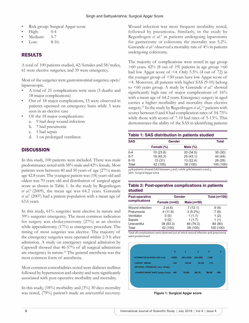
MethodologyUsing EBL, lowest HR, and lowest MAP during the surgical procedure, the SAS is calculated (Figure 1).1-3
(occurrence of pathologic bradyarrhythmia, including sinus arrest, atrioventricular block or dissociation, junctional or ventricular escape rhythms, asystole, and also receives 0 points for lowest HR). Scores are categorized into 0-4, 5-7, 8-10 for simplicity
Data such as lowest HR and lowest MAPs are noted intraoperatively are collected from an anesthesiologist’s records (manual/electronic).
Blood loss is calculated using the formula:10
Blood loss = EBV × (HBi-HBf) ÷ {(HBi + HBf)/2} + {500×Tu} where,
EBV = Estimated blood volume (body weight in kgs × 70 ml/kg)
HBi = Pre-operative hemoglobin (g/dl),HBf = Post-operative hemoglobin (g/dl) around 24 h after
surgeryTu = Sum of whole blood, packed red blood cell transfused.
Note: 500 constant changes according to hospital blood bank protocols.
Patients are followed up for the occurrence of any major complications or deaths within 30 days of surgery. The following events are considered major complications: Acute renal failure, bleeding that requires a transfusion of 4 U or more of red blood cells within 72 h after surgery, cardiac arrest requiring cardiopulmonary resuscitation, coma of 24 h or longer, deep vein thrombosis, myocardial infarction, unplanned intubation, ventilator use for 48 h or more, pneumonia, pulmonary embolism, stroke, wound disruption, deep or organ-space surgical site infection, sepsis, septic shock, systemic inflammatory response syndrome, and vascular graft failure. All deaths are assumed to include major complications.11 Superficial surgical site infection and urinary tract infection are not considered major complications.11 Other occurrences that involve complications of Clavien Class III and greater (those that require surgical, endoscopic, or radiological intervention or intensive care admission or are life threatening) are also considered major complications.2
The occurrence of major complications and mortality within 30 days postoperatively was based on follow-up data in admitting ward and surgical outpatient clinic notes. Major complications definitions were according national confidential enquiry into patient outcome and death classification.12,13 Patients were subsequently grouped into three categories based on their SAS for purposes of risk stratification. Thus,
Singh and Sathyakrishna: Surgical Apgar Score
3 International Journal of Scientific Study | July 2016 | Vol 4 | Issue 4
• Risk group: Surgical Apgar score• High: 0-4• Medium: 5-7• Low: 8-10.
RESULTS
A total of 100 patients studied, 42/females and 58/males, 61 were elective surgeries, and 39 were emergency.
Most of the surgeries were gastrointestinal surgeries; open/laparoscopic.• A total of 21 complications were seen (3 deaths and
18 major complications)• Out of 18 major complications, 15 were observed in
patients operated on emergency basis while 3 were seen in an elective case
• Of the 18 major complications:a. 9 had deep wound infectionb. 7 had pneumoniac. 1 had sepsisd. 1 on prolonged ventilator.
DISCUSSION
In this study, 100 patients were included. There was male predominance noted with 58% male and 42% female. Most patients were between 40 and 50 years of age (27%) mean age 42.8 years. The youngest patient was (18) years old and oldest was 70 years old and distribution of surgical apgar score as shown in Table 1. In the study by Regenbogen et al.2 (2009), the mean age was 64.2 years. Gawande et al.1 (2007) had a patient population with a mean age of 63.6 years.
In this study, 61% surgeries were elective in nature and 39% surgeries emergency. The most common indication for surgery was cholecystectomy (27%) as an elective while appendectomy (17%) as emergency procedure. The timing of most surgeries was elective. The majority of the emergency surgeries were operated within 2-3 h after admission. A study on emergency surgical admission by Capewell showed that 46-57% of all surgical admissions are emergency in nature.14 The general anesthesia was the most common form of anesthesia.
Most common comorbidities noted were diabetes mellitus followed by hypertension and obesity and were significantly associated with post-operative morbidity and mortality.
In this study, (18%) morbidity and (3%) 30 days mortality was noted, (79%) patient’s made an uneventful recovery.
Wound infection was most frequent morbidity noted, followed by pneumonia. Similarly, in the study by Regenbogen et al.3 in patients undergoing laparotomy for gastrectomy or colectomy the mortality was 5.2%. Gawande et al.1 observed a mortality rate of 4% in patients undergoing colectomy.
The majority of complications were noted in age group >60 years. 42% (8 out of 19) patients in age group >60 had low Apgar score of <4. Only 5.5% (4 out of 72) in the younger group of <50 years have low Apgar score of <4. Moreover, all patients with higher SAS (9-10) belong to <60 years group. A study by Gawande et al.1 showed significantly high rate of major complications of 16% with a mean age of 64.2 years. Emergency surgery in aged carries a higher morbidity and mortality than elective surgery.15 In the study by Regenbogen et al.,2,3 patients with scores between 0 and 4 had complication rates of 54-75% while those with scores of 7-10 had rates of 5-13%. This demonstrates the ability of the SAS in identifying patients
Table 1: SAS distribution in patients studiedSAS Gender Total
Female (%) Male (%)0-4 10 (23.8) 20 (34.5) 30 (30)5-7 19 (45.2) 25 (43.1) 44 (44)8-10 13 (31) 13 (22.4) 26 (26)Total 42 (100) 58 (100) 100 (100)44% patients showed SAS between 5 and 7 while 30% between 0 and 4. SAS: Surgical Apgar score
Table 2: Post-operative complications in patients studiedPost-operative complications
Gender Total (n=100)Female (n=42) Male (n=58)
Wound infection 2 (4.8) 7 (12.1) 9 (9)Pneumonia 4 (11.9) 3 (5.2%) 7 (8)Ventilator 0 (0) 1 (1.7) 1 (2)Sepsis 0 (0) 1 (1.7) 1 (1)Uneventful 38 (83.3) 46 (79.3) 84 (80)Total 42 (100) 58 (100) 100 (100)Total 18 complications were observed out of which wound infection and pneumonia were predominant
Figure 1: Surgical Apgar score

Singh and Sathyakrishna: Surgical Apgar Score
4International Journal of Scientific Study | July 2016 | Vol 4 | Issue 4
at higher than average risk of major post-operative complications.
Most common complication noted in this study (Table 2) was deep wound infection followed by pneumonia. Prolonged ventilator and sepsis are other complications. Three mortality noted out of three-two deaths secondary to septic shock and one secondary to cardiopulmonary arrest.
Of the 100 patients, there was (3%) 30 days mortality and (18%) major complications and (79%) no complication. The difference in surgical outcome between patients in different score group was also statistically significant. Among the patient with SAS 0-4, major complications occurred in (50%) 15 out of 29 patients and 30 days mortality in (10.3%). In contrast patients with SAS of >8 no major complications or mortality seen. A study by Regenbogen et al.2 showed among major surgeries, patient with score of 4 or less were 6.5 times more likely to have major complications (95% confidence interval [CI], 4.7-8.9, P < 0.001), Moreover, 112 times more likely to die within 30 days of surgery.
It was also noted that in every 2 point score category the incidence of both major complications and death was significantly greater than that of patients in next higher category (Graph 1). A similar result with relative risk of major complications among low scored operations was 16.1 (95% CI, 7.7-34, P < 0.0001), compared with those in high scored operations was noted in a study by Gawande et al.1
The long duration of surgery as a factor in the occurrence of major complication as has been established in most studies on the SAS.16,17 This may be a reflection complexity of surgery necessitated by possibly extensive disease.
However, long duration surgery was not associated with a lower mean SAS in our study.
The burgeoning literature on the SAS also identifies potential weakness of the scoring system. For example, calculation of the score relies on EBL, which critics have often tagged as imprecise. However, the previous studies have shown that the broad categories used to calculate the amount of blood loss (0-100 ml, 101-600 ml, 60-1000 ml, >1000 ml) are easily within observers’ range of precision.18,19 Another hypothetical weakness lies in the fact that intraoperative hemodynamics maybe affected by anesthetic medications and interventions such as induction and intubation, and therefore, alter the computation of the SAS. For example, a transient episode of hypotension associated with anesthetic induction would be treated the same as prolonged hypotension and resulting a lower (worse) SAS. On the other hand, a transient bradycardic episode would contribute to a higher (better) score. Nevertheless, several studies demonstrate that persistent HR elevation and hypotension are strongly associated with poorer outcomes, regardless of their cause.18,20 Finally, other potentially predictive perioperative variables such as coronary artery disease, volume of intravenous fluids administered, patient age, surgical time, functional status, renal function, and chronic steroid use are excluded from the SAS. The exclusion of these potentially predictive pre-operative risk factors could be interpreted as a weakness of the score. The prevalence of cardiovascular disease increases with age. Unfortunately, this is the same age group in which the largest number of surgical procedures is performed. However as previously mentioned, an important aspect of the usefulness of the SAS is its simplicity.
CONCLUSION
The SAS shows how intraoperative events affect post-operative outcomes. Calculating the SAS in the operating theater provides immediate, reliable, real-time feedback information about patient post-operative risk. Strengths of the SAS include the ability to calculate the score quickly and objectively. The provider could then anticipate the need for further or more aggressive interventions. Ultimately, the score may also prove useful in guiding preventive strategies such as optimizing intraoperative HR or blood pressure.
The SAS could be incorporated into electronic documentation packages for real-time calculation either during or at the end of surgery, providing an automated warning to clinicians. This prognostic value may alert the provider that additional diagnostic testing, further resuscitation, or more intensive monitoring is indicated.
Graph 1: Surgical Apgar score in relation to major complications and mortality. P < 0.001, significant, Fisher exact test. As the surgical Apgar score decreases more
complications and death seen

Singh and Sathyakrishna: Surgical Apgar Score
5 International Journal of Scientific Study | July 2016 | Vol 4 | Issue 4
1. The SAS is strongly associated with clinical decisions regarding immediate intensive care unit (ICU) admission after high-risk surgery.
2. The SAS, despite using simple and widely available intraoperative parameters, is adequate in stratification of post-operative risk of major complications following major surgery.
3. For patients with scores ≥7, very few complications noted hence can consider usual care. The patients with a score of 6 or less are high risk for major complication, and patients with a score of 4 or less are very high risk and should be considered at high risk of decompensation and monitored very closely, often in an ICU setting. It may also be useful to make nursing staff aware of these patients who are particularly high risk, so the care team can be notified early of any signs of decompensation.
4. Patients with comorbidities such as diabetes mellitus, hypertension, and anemia found to have a higher risk of complications.
5. Complication rates are higher in emergency surgeries as compared to elective surgeries.
6. Emergency surgery in elderly carries a higher morbidity than elective surgery, elderly should be strongly motivated to undergo elective surgery rather than put off surgery until the disease gets worse.
REFERENCES
1. Gawande AA, Kwaan MR, Regenbogen SE, Lipsitz SA, Zinner MJ. An Apgar score for surgery. J Am Coll Surg 2007;204:201-8.
2. Regenbogen SE, Ehrenfeld JM, Lipsitz SR, Greenberg CC, Hutter MM, Gawande AA. Utility of the surgical Apgar score: Validation in 4119 patients. Arch Surg 2009;144:30-6.
3. Regenbogen SE, Lancaster TR, Lipsitz SR, Greenberg CC, Hutter MM,
Gawande A, et al. Does the surgical Apgar score measure intraoperative performance? Ann Surg 2008;248:320-8.
4. Mak PH, Campbell RC, Irwin MG; American Society of Anesthesiologists. The ASA physical status classification: Inter-observer consistency. American Society of Anesthesiologists. Anaesth Intensive Care 2002;30:633-40.
5. Brooks MJ, Sutton R, Sarin S. Comparison of Surgical risk score, POSSUM and p-POSSUM in higher-risk surgical patients. Br J Surg 2005;92:1288-92.
6. Copeland GP, Jones D, Walters M. POSSUM: A scoring system for surgical audit. Br J Surg 1991;78:355-60.
7. Jones HJ, de Cossart L. Risk scoring in surgical patients. Br J Surg 1999;86:149-57.
8. Hariharan S, Zbar A. Risk scoring in perioperative and surgical intensive care patients: A review. Curr Surg 2006;63:226-36.
9. Chandra A, Mangam S, Marzouk D A review of risk scoring systems utilised in patients undergoing gastrointestinal surgery. J Gastrointest Surg 2009;13:1529-38.
10. McCullough TC, Roth JV, Ginsberg PC, Harkaway RC. Estimated blood loss underestimates calculated blood loss during radical retropubic prostatectomy; Urol Int 2004;72:13-6.
11. Dindo D, Demartines N, Clavien PA. Classification of surgical complications: A new proposal with evaluation in a cohort of 6336 patients and results of a survey. Ann Surg 2004;240:205-13.
12. A Report by National Confidential Enquiry Into Perioperative Death. Who Operates When II? 2003. Available from: http://www.ncepod.org.UK. [Last Accessed on 2016 Apr 25].
13. Faiz O, Banerjee S, Tekkis P, Papagrigoriadis S, Rennie J, Leather A. We still need to operate at night! World J Emerg Surg 2007;2:29.
14. Capewell S. The continuing rise in emergency admissions. BMJ 1996;312:991-2.
15. Greenburg AG, Saite RP, Coyle JJ. Mortality in BI surgeries in the aged. Arch Surg 1998;1116:788.
16. Zighelboim I, Kizer N, Taylor NP, Case AS, Gao F, Thaker PH, et al. “Surgical Apgar score” predicts postoperative complications after cytoreduction for advanced ovarian cancer. Gynecol Oncol 2010;116:370-3.
17. Prasad SM, Ferreria M, Berry AM, Lipsitz SR, Richie JP, Gawande AA, et al. Surgical Apgar outcome score: Perioperative risk assessment for radical cystectomy. J Urol 2009;181:1046-52.
18. Stav K, Zacci F, Bahar M, Leibovici D, Lindner A, Zisman A, et al. Intra cavernosal saline infusion decreases intraoperative blood loss during radical retropubic prostatectomy. Urol Oncol 2008;26:171-4.
19. Kajja I, Bimenya GS, Eindhoven B, Jan Ten Duis H, Sibinga CT. Blood loss and contributing factors in femoral fracture surgery. Afr Health Sci 2010;10:18-25.
20. Monk TG, Saini V, Weldon BC, Sigl JC. Anesthetic management and one-year mortality after noncardiac surgery. Anesth Analg 2005;100:4-10.
How to cite this article: Santoshsingh SR, Sathyakrishna BR. Surgical Apgar Score - A Simple Prognostic Tool in Surgery. Int J Sci Stud 2016;4(4):1-5.
Source of Support: Nil, Conflict of Interest: None declared.

6International Journal of Scientific Study | July 2016 | Vol 4 | Issue 4
Clinical Study of Prolapse Vault – Anterior Fixation and Posterior ColpoperineoraphyB Surendra Babu1, B Haritha2, B Daasaradhi3, B Radha Ramana4
1Professor and Head, Department of Urology, Rangaraya Medical College, Government General Hospital, Kakinada, Andhra Pradesh, India, 2Postgraduate, Department of General Surgery, M. V. J. Medical College, Bengaluru, Karnataka, India, 3Post Graduate, Department of General Medicine, M. V. J. Medical College, Bengaluru, Karnataka, India, 4Director, Haritha Hospital, Kakinada, Andhra Pradesh, India
As women live longer and healthier lives, pelvic floor disorders continue to become even more prevalent and are an important health and social issue. The lifetime risk of surgery for pelvic prolapse or incontinence has been estimated at 11%, with a reoperation rate for failure at 29%. The management of pelvic organ prolapse can be difficult because different support defects often coexist. The pelvic surgeon must be adept in the thorough evaluation and management of these issues. An understanding of the anatomy and the relationship of the vagina to surrounding structures is imperative6,7.
The true incidence of vaginal vault prolapsed is unknown. However, there is an overall perception that the number of procedures being performed for vaginal vault prolapsed is increasing. The main goal of any procedure aimed at suspending the vaginal vault should be to suspend the vaginal vault as near as possible to its normal anatomic position. This should reapproximate the upper vaginain the midline over the levator plate8. Distortion of the vaginal vault, whether in an anterior or posterior direction, can lead to a recurrent prolapsed opposite the vaginal vault in a significant number of patients.
INTRODUCTION
Vaginal vault prolapse is a condition in which the upper portion of the vagina loses its normal shape and sags or drops down into the vaginal canal or outside of the vagina1,2. This can occur either in conjunction with uterine prolapse or even after a hysterectomy3.
Levels of support defects (according to DeLancey classification)-Level 1: Apical defects caused by loss of support of the uterosacral ligaments, paracolpium, and parametrium: Level II: Disruption of the normal lateral at-rachments of the midvagina; and Level III: Lower vaginal defects in the perineal body or fusion of the distal urethral to the pubic bone4,5.
Original Article
AbstractIntroduction: The purpose of the clinical study was to assess the result whether the anterior fixation is better than posterior fixation in cases of vault prolapse for post hysterectomy patients.
Materials and Methods: Randomised prospective studies were perform in 20 cases of anterior fixation and posterior colpoperineoraphy. Technique is simple, without bleeding and reproducible with least post operative complications And the results were recorded.
Results: There were significant and marked improvement and the results after follow up of 3 years were very good. In one case we had recurrence and surgery was repeated with good results.
Conclusion: Posterior fixation for vault prolapse is standard procedure and we have achieved the results with our anterior fixation technique.
Key words: Prolapse vault, Anterior fixation, Posterior fixation, Proline suture & Posterior colpoperineoraphy.
Access this article online
www.ijss-sn.com
Month of Submission : 05-2016 Month of Peer Review : 06-2016 Month of Acceptance : 07-2016 Month of Publishing : 07-2016
Corresponding Author: Dr. B. Surendra Babu, Ms Mch, MAMS, Prof & HOD Urology, Haritha Hosptials (Multi Speciality), Opp: Government General Hospital, Kakinada - 533 001, Mobile: 09848528954/09441811442, Phone: 0884 2379292, 2375963, Email: [email protected]
Print ISSN: 2321-6379Online ISSN: 2321-595X
DOI: 10.17354/ijss/2016/362

Babu, et al.: Prolapse Vault – Anterior Fixation and Posterior Colpoperineoraphy
7 International Journal of Scientific Study | July 2016 | Vol 4 | Issue 4
The ultimate goal of pelvic reconstructive surgery is to restore anatomy, maintain or restore visceral function, and maintain or restore normal sexual function. It is extremely important to determine preoperatively whether lower urinary tract dysfunction, sexual dysfunction, and defecatory dysfunction exist. Urinary dysfunction may be masked in patients with advanced pelvic organ prolapsed by obstructing or kinking the urethra. Thus, reductive maneuvers aimed at simulating what surgery will accomplish should be used in the hope of identifying those patients who will require an anti incontinence procedure in conjunction with their pelvic reconstructive surgery. It is also important to initiate local estrogen therapy preoperatively in patients who have urogenital atrophy9,10.
Many operations have been described for suspending the prolapsed vaginal vault. There is no general consensus on what is the best procedure. The procedure that the surgeon ultimately chooses is influenced by many factors, including the comfort and skill of the surgeon performing the operation, whether the prolapse is primary or recurrent, the patients age state of health, anticipated outcome, sexual activity, and overall state of the tissue. We believe it is important for the surgeon to have a variety of operative approaches available for the individual patient11,12.
MATERIALS AND METHODS
The protocol was approved by the local Ethics committee and written informed consent was obtained from each patient.
Randomised prospective studies were perform in 20 cases of anterior fixation and posterior colpoperineoraphy in post hysterectomy patients were taken up for this study. Technique is simple, without bleeding and reproducible with least post operative complications the results were recorded. Grade I prolapse 10 cases, Grade II prolapse 7, Grade III prolapse 3 cases.
OPERATIVE TECHNIQUE
The patient is kept in modified lithotomy position and suprapubic transverse incision given. Perurethrally 16F Foleys catheter passed with 10 ml to balloon. Bladder neck is dissected and urethra with catheter was held. Sponge on stick passed vaginally and pushed right paraurethral area at vaginal vault apex. Proline 1 suture was taken bites and mesh was taken and it is suspended to periosteum retropubically. And same time it is repeated other side also.
By doing this the prolapse imaginarily looks like beaker. After correction it appears like inverted beaker. The vagina is inspected and evaluated for any remaining defects. Usually a posterior colporrhaphy and a perineoplasty are also required in all cases.
RESULTS
Overall the long term results from Colpopexy have been very good. Automatically cystocele is corrected in this repair. Intraoperative complications are unusual.
DISCUSSION
DeLencey divided the support of the vagina into three levels. This concept is helpful in understanding normal anatomic, in some patients and not in others. Level I support defects are apical defects caused by loss of support of the uterosacral ligaments, paracolpium, and paramettrium. Level II support defects result from disruption of the normal lateral attachment of the midvagina. Level III support defects result from defect in the perineal body or fusion of the distal urethra to the pubic bone. Although the exact indication were controversial many surgeons primarily try vaginal repair in all cases of posthysterectomy vault prolapse.
This procedure indicated due to average operative time, reproducible, in failed vaginal repairs and also in cases of fore shortened vagina.
Excellent results have been reported by us. The complications are very minimal
CONCLUSION
On the basis of our surgical technique and simplification this procedure has got advantage over posterior colpopexy in post hysterectomized patients.

Babu, et al.: Prolapse Vault – Anterior Fixation and Posterior Colpoperineoraphy
8International Journal of Scientific Study | July 2016 | Vol 4 | Issue 4
REFERENCES
1. Mant, J., Painter, R., Vessey, M. Epidemiology of genital prolapse: Observations from the Oxford family planning association study. BJOG. 1997;104:579-585.
2. Marchionni, M., Bracco, G., Checcucci, V. et al, True incidence of vaginal vault prolapse. J of Reproductive Medicine. 1999;44:679-684.
3. Hung, M.J., Liu, F.S., Shen, P.S. et al, Ho ES.Factors that affect recurrence after anterior colporrhaphy procedure reinforced with four-corner anchored polypropylene mesh. Int Urogynecol J Pelvic Floor Dysfunct. Nov-Dec. 2004;15:399-406.
4. Soderberg, M.W., Falconer, C., Bystrom, B. et al, Young women with genital prolapse have a low collagen concentration. Acta Obstet Gynecol Scand. Dec;. 2004;83:1193-1198.
5. Uustal Fornell, E., Wingren, G., Kjolhede, P. Factors associated with pelvic floor dysfunction with emphasis on urinary and fecal incontinence and genital prolapse: An epidemiological study. Acta Obstet Gynecol Scand. 2004;83:383-389.
6. Wiskind, A.K., Creighton, S.M., Stanton, S.L. The incidence of genital prolapse after the Burch colposuspension. Am J Obstet Gynecol. 1992;167:399-404.
7. Baden, W.F., Walker, T.A. Genesis of the vaginal profile: A correlated classification of vaginal relaxation. Clin Obstet Gynecol. 1972;15:1048-1054.
8. Bump, R.C., Mattiasson, A., Bo, K. et al, The standardization of terminology of female pelvic organ prolapse and pelvic floor dysfunction. Am J Obstet Gynecol. 1996;175:10-17.
9. Maher, C.F., Qatawneh, A.M., Dwyer, P.L. et al, Abdominal sacral colpopexy or vaginal sacrospinous colpopexy for vaginal vault prolapse: A prospective randomized study. Am J Obstet Gynecol. 2004;190:20-26.
10. Kearney, R., DeLancey, J.O.L. Selecting suspension points and excising the vagina during Michigan four-wall sacrospinous suspension. Obstet Gynecol. 2003;101:325-330.
11. Richter, K., Albrich, W. Long-term results following fixation of the vagina on the sacrospinal ligament by the vaginal route (vaginaefixatio sacrospinalis vaginalis). Am J Obstet Gynecol. 1981;141:811-816.
12. Arthure, H.G. Vault suspension. Proc R Soc Med. 1949;42:388–390.
How to cite this article: Babu BR, Haritha B, Daasaradhi B, Ramana BR. Clinical Study of Prolapse Vault – Anterior Fixation and Posterior Colpoperineoraphy. Surgical Apgar Score - A Simple Prognostic Tool in Surgery. Int J Sci Stud 2016;4(4):6-8.
Source of Support: Nil, Conflict of Interest: None declared.

9 International Journal of Scientific Study | July 2016 | Vol 4 | Issue 4
Effects of Tobacco Chewing on Serum Lipid Profile in South Indian PopulationR Haragopal1, B M K Aruna2
1Associate Professor, Department of Physiology, Kakatiya Medical College, Warangal, Telangana, India, 2Associate Professor, Department of Physiology, Government Medical College, Nizamabad, Telangana, India
the causation of coronary heart disease as the cause of coronary heart disease is a multifactorial. Thus, a strong synergistic interaction exists between hyper cholesterolemia and tobacco consumption in the genesis of coronary heart disease.4
Nicotine is the active ingredient in tobacco.5 Nicotine stimulates adrenal medulla to release catecholamine.6 Catecholamines are the only hormones which effectively stimulates lipolysis in humans.7 Tobacco smoking and its effects on lipid profile have been proved by several studies.8,9 There are very few studies regarding the effect of tobacco chewing on lipid profile. Hence, this study is conducted to determine the effect of tobacco chewing on lipid profile.
MATERIALS AND METHODS
This study was conducted on 40 male subjects in the age group ranging from 20 to 50 years. Twenty subjects were non-chewers of tobacco and 20 were chewers of tobacco. The chewers or tobacco users were divided into
INTRODUCTION
Tobacco was introduced by Portuguese merchants in the 16th century and now India is one among the world’s top five tobacco producers and consumers.1 The WHO attributed 4 million tobacco-related deaths every year and is expected to raise 8.4 million deaths by 2020.2 Various forms of smokeless tobacco products are available which include pan (piper betel leaf filled with sliced areca nut, lime, catechu, and other spices chewed with or without tobacco), pan-masala or gutkha (a chewable tobacco containing areca nut), and mishri (a powdered tobacco rubbed on the gums as toothpaste).3
Tobacco is pathogenetically a cholesterol-dependent risk factor and it acts synergistically with other risk factors for
Original Article
Abstract
Introduction: Nicotine which is an active ingredient in tobacco stimulates adrenal medulla to release catecholamine. Catecholamines activate the adenyl cyclase of adipose tissue which causes lipolysis of stored triglyceride (TG) and the release of free fatty acids into plasma.
Materials and Methods: A total of 40 healthy adult male participants were recruited in that 20 were non-chewers and 20 were chewers of tobacco. The chewers were again divided into the users of <10 years and users of more than 10 years. 5 ml of blood samples were collected and the serum was separated. The total cholesterol, TG, low-density lipoprotein (LDL), and high-density lipoprotein (HDL) were estimated by enzymatic and precipitation methods.
Results: A significant increase in the total cholesterol, LDL levels were observed in the long-term users of tobacco when compared with non-users and short-term users. However, HDL levels were similar in all the 3 groups. TGs were higher in the control group when compared with tobacco chewers.
Conclusion: Increased levels of total cholesterol and LDL could be considered as risk factors in the developing coronary heart disease. Tobacco chewing can be considered as one of the preventable risk factors of coronary heart disease.
Key words: Coronary artery disease, Lipid profile, Tobacco chewing
Access this article online
www.ijss-sn.com
Month of Submission : 05-2016 Month of Peer Review : 06-2016 Month of Acceptance : 07-2016 Month of Publishing : 07-2016
Corresponding Author: Dr. R Haragopal, Kakatiya Medical College, Warangal, Telangana, India. Phone: +91-9290036206. E-mail: [email protected]
Print ISSN: 2321-6379Online ISSN: 2321-595X
DOI: 10.17354/ijss/2016/363

Haragopal and Aruna: Serum Lipid Profile in Tobacco Chewers
10International Journal of Scientific Study | July 2016 | Vol 4 | Issue 4
2 groups again. They were tobacco chewers of <10 years (9 subjects) and tobacco chewers of more than 10 years (11 subjects). Thus, there were total 3 groups - non-users, users <10 years, and users more than 10 years. Care was taken to see the average age of controls and chewers were same. All the research participants were explained about the procedures and recruited after obtaining informed consent. Subjects with multiple tobacco habits, alcoholics, liver diseases, chronic renal failure, nephrotic syndrome, hypothyroidism, diabetes mellitus, drugs (β blockers, glucocorticoids, thiazide diuretics, and lipid lowering drugs), and also with other chronic illness were excluded from the study.
About 5 ml blood samples were collected after an overnight fasting and serum was separated from the blood. The serum lipid profile was studied and the lipid levels were calculated by Freidewald’s formula. Estimation of total cholesterol, triglycerides (TGs), low-density lipoprotein (LDL), and high-density lipoprotein (HDL) was done by standard methods.
RESULTS
The mean and standard deviations of total cholesterol levels of the non-users, users of <10 years and >10 years were 148.6 ± 32.63, 144.1 ± 23.84 and 169.6 ± 25.95, respectively. It is found that there is no significant difference in cholesterol values between non-users and users of <10 years. However, long-term users (<10 years) show increased cholesterol levels compared to short-term users. This may be due to the long-term effects of sustained blood nicotine values. These data are shown diagrammatically in Table 1 and Figure 1.
Serum TG levels in three groups were shown in Table 2. Remarkably non-users have higher levels than the other two groups. In fact, there is statistically significant difference
between non-users and users of <10 years. This may be an incidental finding as this is a cross-sectional study.
There seems to be no difference in the HDL cholesterols (HDL-C) of three groups, but the LDL cholesterol (LDL-C) levels seem to be increasing with chewing tobacco. There is gradual increase in the blood levels of LDL-C with the users of over 10 years showing maximum blood levels (Table 3). There is a statistically significant difference between non-users and users as well as between users of <10 years duration and the users of >10 years duration.
Figure 1: The parameters (triglyceride, high-density lipoprotein and low-density lipoprotein) in three groups
Table 1: Serum cholesterol levels in all three groupsMean, SD and SE
Non users Users <10 years Users >10 years
Number 20 9 11Mean 148.6 144.1 169.6SD 32.63 23.84 25.95SE of mean 7.30 7.95 7.83t=2.27, P<0.02 (between users of <10 years and >10 years). SD: Standard deviation, SE: Standard error
Table 2: Serum triglyceride levels in all three groupsMean, SD and SE
Non users Users <10 years Users >10 years
Number 20 9 11Mean 157.7 88.3 128.5SD 90.29 61.44 61.78SE of mean 20.19 20.48 18.63t=2.09, P<0.05 (between non‑users and users of <10 years). SD: Standard deviation, SE: Standard error
Table 3: HDL-cholesterol levelsMean, SD and SE
Non users Users <10 years Users >10 years
Number 20 9 11Mean 31.0 30.3 32.7SD 2.71 3.12 5.18SE of mean 0.61 1.04 1.56Not significant. SD: Standard deviation, SE: Standard error, HDL: High‑density lipoprotein
Table 4: Serum LDL-cholesterol levelsMean, SD and SE
Non users Users <10 years Users >10 years
Number 20 9 11Mean 91.1 96.1 111.2SD 28.07 12.07 13.68SE of mean 6.28 4.02 4.12t=2.22, P<0.05 (between non‑users and users of<10 years). t=2.59, P<0.02 (between users of <10 years and users of >10 years. SD: Standard deviation, SE: Standard error, HDL: High‑density lipoprotein

Haragopal and Aruna: Serum Lipid Profile in Tobacco Chewers
11 International Journal of Scientific Study | July 2016 | Vol 4 | Issue 4
This proves that there is gradual increase of LDL-C levels in tobacco chewers. These data are shown diagrammatically in Figure 1 and Table 4.
DISCUSSION
Nicotine which is an active ingredient in tobacco stimulates adrenal medulla to release catecholamine. Catecholamines activate the adenyl cyclase of adipose tissue which causes lipolysis of stored TG and the release of free fatty acids (FFAs) into plasma. The released FFAs are immediately bound to plasma albumin and are then transported to various tissues of the body particularly to the liver. Hepatic TG and very LDL-C (VLDL-C) synthesis is stimulated by increased influx of FFA. The increased levels of plasma FFAs could act to depress the plasma HDL-C and increases plasma TG and VLDL-C.1
In this study, total serum cholesterol was higher in long-term users when compared to non-users and users of <10 years. There was no significant difference between the non-users and the users of <10 years. This could be explained as due to long-term effects of sustained blood nicotine levels. This was also recorded by other Indian workers. Khurana et al. and Rao and Subash observed a rise in the levels of total cholesterol, TG, LDL, and VLDL with a decrease in the HDL level in smokers and tobacco chewers, which was in concurrence with the results of this study in relation to total cholesterol and LDL levels, whereas TGs were higher in non-users than users and the levels of HDL were similar in all the groups.4,10 Latha et al. administered nicotine to rats and found that the concentration of TGs increased in both serum and tissues.11 Our study results are consistent with the above two studies with reference to total serum cholesterol.
The serum TG levels in this study, however, show a different picture. It is higher in the non-users compared to users of more than 10 years. This is perhaps an incidental finding because this is a cross-sectional study and the average age of non-users is slightly higher than users. However, the
long-term users (<10 years) have higher levels of TGs compared to the short-term users (users of <10 years).
The HDL levels in the study and control groups did not show any difference. However, there was a notable gradual increase in the blood levels of LDL-C. The non-users have the lowest levels and the long-term users have the highest levels. This can be explained as one of the chronic effects of sustained blood nicotine.
CONCLUSION
With the limitations of this study, we could conclude that there is a definite impact of chewing tobacco on the serum lipid profile. Tobacco chewing causing increased total cholesterol and LDL levels in the blood serum which is harmful and may be responsible for the greater risk of developing atherosclerosis in the tobacco users than in the non-tobacco users.
REFERENCES
1. Gadpal RR, Deshpande KA, Waghmare MH. A study of lipid profile in tobacco chewers and smokers. J Contemp Med Dent 2015;3:39-43.
2. Bulliya G. Blood pressure and serum lipid profile in smokers and nonsmokers- A comparative study. Indian Pract 2002;55:363-8.
3. Rani M, Bonu S, Jha P, Nguyen SN, Jamjoum L. Tobacco use in India: Prevalence and predictors of smoking and chewing in a national cross sectional household survey. Tob Control 2003;12:e4.
4. Rao CS, Subash YE. The effect of chronic tobacco smoking and chewing on the lipid profile. J Clin Diagn Res 2013;7:31-4.
5. Benowitz NL, Jacob P 3rd. Metabolism of nicotine to cotinine studied by a dual stable isotope method. Clin Pharmacol Ther 1994;56:483-93.
6. Haass M, Kübler W. Nicotine and sympathetic neurotransmission. Cardiovasc Drugs Ther 1997;10:657-65.
7. Lafontan M, Berlan M. Fat cell adrenergic receptors and the control of white and brown fat cell function. J Lipid Res 1993;34:1057-91.
8. Sirisali K, Pomywarin N, Kalnam J. Serum lipids, lipoprotein cholesterol and Apo A-l and B of smoking and non-smoking male. J Med Assoc Thai 1992;75:709-13.
9. Gamit KS, Nanavati MG, Gohel PM, Gonsai RN. Effects of smoking on lipid profile. Int J Curr Res Rev 2013;5:36-42.
10. Khurana M, Sharma D, Khandelwal PD. Lipid profile in smokers and tobacco chewers - A comparative study. J Assoc Physicians India 2000;48:895-7.
11. Latha MS, Vijayammal PL, Kurup PA. Effect of nicotine administration on lipid metabolism in rats. Indian J Med Res 1993;98:44-9.
How to cite this article: Haragopal R, Aruna BMK. Effects of Tobacco Chewing on Serum Lipid Profile in South Indian Population. Int J Sci Stud 2016;4(4):9-11.
Source of Support: Nil, Conflict of Interest: None declared.

12International Journal of Scientific Study | July 2016 | Vol 4 | Issue 4
Computed Tomography Study of Paranasal Sinuses PathologiesManjit Bagul
Senior Resident, Department of Radiodiagnosis, RKDF Medical College, Bhopal, Madhya Pradesh, India
sinuses that still bears his name, “the Caldwell view,” which is a depiction of the ethmoid and frontal sinuses that include both orbits. In 1914, Waters and Waldron, two British radiologists, introduced a projection that defined the paranasal sinuses and facial bones to greater advantage. At the present time, the waters view is still being used to survey sinus disease and facial fractures.
An important historic achievement occurred in 1972 with the introduction of computed tomography (CT) by Godfrey Hounsfield of Great Britain. The foundation for CT was based on mathematic equations that had been formulated in 1963 and 1964 by Cormack, a Professor of Physics at Tufts University in Boston. The development of spiral CT in the past few years has allowed a shorter examination time and thinner sections, with the capability of three-dimensional reconstruction. Most recently, multi-detector row CT with increased spatial resolution, with
INTRODUCTION
The head and neck radiology, similar to that of other subspecialties in radiology, began with the discovery of the X-ray in 1895 by Wilhelm Konrad Roentgen (1845-1923). Another early investigator was Caldwell (1870-1918), who became fascinated by X-rays only 2 years after Roentgen’s discovery. In 1903, he wrote one of the first textbooks on diagnostic and therapeutic radiology. His interest in head and neck radiology is reflected by a view of the paranasal
Original Article
AbstractIntroduction: The application of computed tomography (CT) in the paranasal sinuses study has allowed the detail assessment of inflammation, cysts, benign, and malignant conditions. CT has increased the accuracy of patient management with a consequent decrease in morbidity and mortality. The purpose of this study is to determine the role and efficacy of CT scan in diseases of the paranasal sinus. This was the cross-sectional prospective study of 1½ years duration.
Aims and Objective: To evaluate the various pathologies affecting the paranasal sinuses on CT and study various physiological variants.
Materials and Methods: This was cross-sectional prospective study conducted at RKDF Medical College from June 2014 to December 2015. A total of 110 patients of varied age group presenting with symptoms and signs of paranasal sinus diseases were included in the study. Imaging diagnosis was confirmed either by histopathology or by positive response to treatment.
Results: In our study, paranasal sinuses pathologies were more common in male (62%) compare to female population (33%). Most common age group affected by the paranasal sinuses pathologies was 11-30 years age group (45.5%) and least common age group was less than 10 years (<2%). The most common paranasal sinuses pathologies in our study were inflammatory (60%) followed by neoplastic (33%) and miscellaneous (7%). Most commonly affected paranasal sinuses in descending order were maxillary (86%), ethmoidal (54%), frontal (31%), and sphenoidal (21%). Common anatomical variants observed in our study were deviated nasal septum (40%), concha bullosa (43%), agar nasi cells (59%), Haller cells (16%), and Onodi cells (31%).
Conclusion: CT scan depicts both soft tissue and bony details of nose and paranasal sinuses thereby accurately detect various pathologies affecting the paranasal sinuses. Various important anatomical variants can be easily detected on CT of paranasal sinuses.
Key words: Computed tomography, Histopathological diagnosis, Paranasal sinuses
Access this article online
www.ijss-sn.com
Month of Submission : 05-2016 Month of Peer Review : 06-2016 Month of Acceptance : 07-2016 Month of Publishing : 07-2016
Corresponding Author: Manjit Bagul, RKDF Medical College, Bhopal, Madhya Pradesh, India. E-mail: [email protected]
Print ISSN: 2321-6379Online ISSN: 2321-595X
DOI: 10.17354/ijss/2016/364

Bagul: CT Study of PNS Pathologies-Manjit Bagul
13 International Journal of Scientific Study | July 2016 | Vol 4 | Issue 4
a section thickness as small as 0.5 mm and acquisition capabilities of 16 images per second, has been developed.
Diseases of paranasal sinuses are a major health problem. Most of the times physical examination is nonspecific and radiological evaluation has been relied on as an aid in confirming the diagnosis. Traditionally, plain radiographs were the modality of choice in the evaluation of paranasal sinuses. In recent years, because of technologic advancements in imaging, CT has supplanted conventional radiography as the primary diagnostic modality and has also contributed in the change in therapeutic approach. Standard plain radiographs still have a limited role in the imaging of the paranasal sinuses and are used as the initial technique before the application of CT. The refinement of CT technology has resolved the traditionally difficult problem of identifying lesions of the paranasal sinuses. It has also allowed improved accuracy in evaluating the soft tissues about the sinuses. The improvement in tissue resolution that CT offers over plain films allows evaluation of subtle changes of soft tissues, bones and air containing spaces. The ability of CT to image the bony details as well as soft tissues is the greatest advantage over previous radiographic modalities. Furthermore, coronal and axial CT scanning has dramatically improved the imaging of the anatomy of the paranasal sinus. CT excellently displays the bony architecture and its mucosal covering as well as the narrow air channels of the osteomeatal complex. CT accurately depicts the boundaries between the paranasal sinuses, the orbit and the intracranial compartment and also the relationship between the optic nerve, cavernous sinus, carotid artery and fifth cranial and vidian nerves to the sphenoid sinuses. Contrast media helps evaluate the vascularity and contrast enhancing characteristics of lesions, giving clues to the histology and extent of abnormality.
The aims and objective of this study is to determine the role and efficacy of CT scan in diseases of paranasal sinus and study of various physiological variants.
MATERIALS AND METHODS
The present prospective study was conducted at RKDF Medical College from June 2014 to December 2015.
Study AreaThe study area includes Bhopal city and district with peripheral small towns/villages.
Study PopulationA total of 110 patients of varied age group presenting with symptoms and signs of paranasal sinus diseases were included in the study.
Inclusion Criteria• Patients referred for CT of paranasal sinuses, who
were suspected to have paranasal sinus disease.• Patients who were suspected to have some paranasal
sinus pathology on conventional radiographs and were then referred for CT of paranasal sinuses.
Exclusion Criteria• Patients presenting with trauma to face• Patients with contrast allergy• Patients who were lost to follow-up without a definite
diagnosis.
Equipment usedSpiral CT, Siemens Somatom, Siemens Medical Systems, Forchiem, Germany.
CT scan of paranasal sinuses (PNS) requires imaging of the anatomy into coronal and axial planes. A lateral 256 mm scout scan was first obtained at 120 kVp and 100 mA. Routinely axial scanning was done in supine position. Reformatting in coronal and sagittal planes was done using software provided.
Direct coronal imaging was done whenever deemed necessary either by referring physician or by the radiologist. For direct coronal imaging, the patient was kept in prone position or in supine position with the head of the patient free leading edge of the table of the scanner. The gantry angle used in case of coronal imaging was perpendicular to the plane of hard palate. 3 mm sections from anterior margin of nose to the posterior margin of sphenoid sinus were taken.
Final imaging diagnosis correlated with histopathological confirmation or treatment response.
OBSERVATIONS AND RESULTS
CT scan was performed in 110 patients who presented with history, symptoms, and signs of the paranasal sinuses pathologies. The results are enumerated in Tables 1-8.
DISCUSSION
The varied etiology of the diseases of PNS forms the basis of their evaluation. The lack of specificity in clinical examination and the imprecise result of conventional radiography render CT as the modality of choice other than magnetic resonance imaging.
In the present study, 110 patients were evaluated for their various symptoms pertaining to PNS. The gender ratio in this study was 2.05:1 (male:female) (Table 1).

Bagul: CT Study of PNS Pathologies-Manjit Bagul
14International Journal of Scientific Study | July 2016 | Vol 4 | Issue 4
The etiologic distribution of the lesions was inflammatory (60%), neoplastic (32.7%), and miscellaneous (7.3%). Thus, the inflammatory disease was found to be the most frequently occurring pathology affecting the PNS. The incidence of neoplasms increases sharply after age of 40 years (Table 4). There is another peak in teen age due to increase in incidence of angiofibroma and rhabdomyosarcoma at this age.
Acute sinusitis was diagnosed when there was air fluid level, enhancing mucosal thickening. Chronic sinusitis showed decrease in sinus size with sclerosis and thickening of the walls. Considering the inflammatory etiology of the various sinuses, the following was the percentage affection of individual sinuses. Maxillary (89.4%), frontal (31.8%),
sphenoids (15.2%), ethmoidal (50%). Thus, maxillary sinus was most commonly involved and sphenoid sinus was least involved in inflammatory conditions. In the study conducted by Smith and Brindley,1 maxillary sinus was involved in 55.5% of cases, ethmoidal air cells were involved in 46.5% of cases, frontal sinus in 30%, and sphenoid in 20%. Similarly, Maru and Gupta2 reported maxillary sinus to be the most frequently involved sinus in inflammatory lesions (70.4%) followed by ethmoids (52.4%), frontal (48.3%), and sphenoid sinuses (40.8%). Zinreich et al.3 published in his study that the maxillary sinus involvement was the most frequent in inflammatory lesions, i.e., 65% followed by ethmoid cells 40%, frontal sinus in
Table 1: Gender distributionGender N (%)Female 36 (32.73)Male 74 (67.27)Grand total 110 (100)
Table 2: Age and gender distributionAge distribution Female Male Total1-10 1 1 211-20 6 19 2521-30 6 19 2531-40 8 10 1841-50 6 10 1651-80 9 15 24Grand total 36 74 110
Table 3: Etiopathological distribution of caseEtiology Female Male Total PercentageInflammatory 21 45 66 60Sinusitis 14 26 40Polyposis 6 12 18Other 1 6 7Neoplastic 12 24 36 32.7Miscellaneous 3 5 8 7.3Grand total 36 74 110 100
Table 4: Age distribution of pathologiesAge range Inflammatory Neoplastic Miscellaneous
Benign Malignant Total1-10 0 0 1 1 111-20 14 5 3 8 321-30 19 3 0 3 331-40 13 1 4 5 041-50 8 2 6 8 051-80 12 1 10 11 1Grand Total 66 12 24 36 8
Table 5: Various sinuses involvedSinus involved N (%)Maxillary 95 (86.36)Ethmoidal 59 (53.63)Frontal 34 (30.9)Sphenoids 23 (20.9)
Table 6: Sinuses involved in various pathologiesSinus involved
Inflammatory Neoplastic MiscellaneousBenign Malignant Total
Maxillary 59 6 23 29 7Ethmoidal 33 8 16 24 2Frontal 21 4 7 11 2Sphenoids 10 5 6 11 2
Table 7: CT features of benign and malignant neoplasmsCT parameter Benign (n=12) (%) Malignant (n=24) (%)Sinus size increased 8 (66.6) 14 (58.3)Erosions 7 (58.3) 24 (100)Thinning 2 (16.6) 5 (20.8)Sclerosis 1 (8.3) 1 (4.1)Extensions in at least one region
4 (33.3) 23 (95.8)
CT: Computed tomography
Table 8: Anatomical variantsAnatomical variant Total (%)DNS 44 (40)Concha bullosa 48 (43.6)Agger nasi 65 (59.1)Haller cells 18 (16.3)Onodi cells 34 (30.9)EEB 9 (8.2)PMT 15 (13.6)DUP 12 (10.9)PUP 4 (3.6)DNS: Deviated nasal septum, EEB: Enlarged ethmoid bulla, PMT: Paradoxical middle turbinate, DUP: Deviated uncinate process, PUP: Pneumatized uncinate process
Bagul: CT Study of PNS Pathologies-Manjit Bagul
15 International Journal of Scientific Study | July 2016 | Vol 4 | Issue 4
extension of the tumors. Thus, helping in their exact staging and finally in the management of these tumors.
Considering the involvement of sinuses by various neoplasms in the present study, maxillary sinus was involved in 80.5% of cases, ethmoids in 66.6%, frontal in 30.5%, and sphenoid in 30.5% of cases. In a study conducted by Dolan and Smoker,9 they noted similar findings, wherein maxillary sinus was the most frequently involved sinus affected by intrinsic or nearby or metastatic neoplasms, as seen in our study. According to the study conducted by Parsons and Hodson,10 the tumor extension was most common into the region of orbit and into the pterygoid region. The two authors studied 15 cases of histologically proven malignancy, to evaluate their extension into adjacent anatomic structures. This was similar to our study where intraorbital and infratemporal fossa extension of the neoplastic lesions was found to be most common.
Two cases of inverted papilloma were diagnosed; CT showed middle meatus involvement along with the extension of the lesion to maxillary sinus, eroding the turbinates on the same side. Thus, we reemphasized the findings quoted by Lund and Lloyd.11 They studied 60 patients of histologically proved inverted papilloma retrospectively and concluded that mass in the middle meatus of nasal cavity extending into adjacent maxillary antrum is highly suggestive of the tumor.
Total six cases of histologically proven angiofibroma were studied by us. All of them were males of age group between 15 and 25 years and presented with epistaxis. It is similar to Barnes et al.,12 who stated that angiofibroma occurs almost exclusively in young males. Typical site of origin near pterygopalatine fossa and sphenopalatine foramen with widening of pterygopalatine fossa and strong contrast enhancement were the diagnostic criteria used by Som et al.13 We found these criteria to be diagnostic. However, in one of our patient, the pterygopalatine fossa was not widened. Many of these angiofibromas extended into infratemporal fossa (66.6%) and sphenoid sinus (66%). Apostol and Frazell,14 observed that sphenoid sinus is involved in 61% of the cases. Intracranial extension into middle cranial fossa was found in one patient (16.6%); similar was observed by Barnes et al.12 The differential diagnosis of angiofibroma includes fibrosed antrochoanal polyp and angiomatous polyp.
Abrahams and Glassberg15 observed that many of the maxillary sinus pathologies are related to dental disease. In the present study, four cases related to dental pathologies were encountered. All of them involved maxillary sinus. CT is diagnostic of osseous and fibro-osseous lesions. Four cases of fibrous dysplasia, two of osteomas and one of ossifying fibroma were diagnosed. Frontal sinus was
34%, and sphenoid sinus in 29% of cases. Kopp et al.4 in his study of 105 cases of aspergillosis of paranasal sinuses or nasal fossa detected the characteristic CT features of foci of increased attenuation in affected paranasal sinuses. He also found that mycosis was always unilateral, and the maxillary sinus was infected in almost all cases. On the basis of similar findings, we were able to diagnose one case of fungal sinusitis involving maxillary sinus.
Airless sinus filled with mucoid density material, expanding the sinus with thinning of sinus walls is diagnostic of mucocele. One case of frontal mucocele, one case of frontoethmoid and two cases of maxillary mucocele were observed in our study. Zizmor et al.5 found that mucocele most commonly occur in frontal sinus (60-65%) followed by ethmoid sinuses. In the present study, frontal and maxillary sinus involved in equal proportion.
We were able to diagnose allergic fungal sinusitis in two patients. There were characteristic findings of bilateral hyperdense polypoidal masses with expansion, remodeling and thinning of bony walls of sinuses. Mukherji et al.6 emphasized on similar findings. He studied 43 patients of allergic fungal polyposis and concluded that it is more common in young male patients and commonly has bilateral involvement.
In the present study, 36 cases were diagnosed to have neoplastic lesions. 24 of them were detected to be malignant in nature depending on the enhancement pattern of the lesion, the presence of bony destruction and finally the extension of the lesion into various adjacent vital anatomical structures. In the present study, bony erosion was seen in all the 24 (100%) malignant masses and thus found to be most valuable CT criteria for the diagnosis of malignancy. However, 58.3% of benign neoplasms also showed bone erosions (Table 7).
In 95% of malignant neoplasms extension to one of the adjacent regions was present, compared to 33.3% in benign neoplasms. Rests of the criteria like increase in size of the sinus, thinning of walls, sclerosis did not differed much between the benign from malignant masses and not found to be useful in differentiating the two. We were able to demonstrate the precise location and extension of the tumors. Histological types could not be discriminated on imaging (CT) in most of the cases. Similar was emphasized by many authors including Peter Som et al.,7 who concluded that it is not possible to diagnose tumor in the absence of bone destruction.
Graber et al.8 used similar criteria to detect malignant tumors in 15 patients. He characterized malignant tumors in paranasal sinuses on CT scan by their nonhomogeneous structure, destructed bony margins of the sinuses and infiltration into neighboring regions. Graber depicted the precise location and

Bagul: CT Study of PNS Pathologies-Manjit Bagul
16International Journal of Scientific Study | July 2016 | Vol 4 | Issue 4
involved in both the osteomas and is the most common site of osteoma as studied by Fu and Perzin,16 Both fibrous dysplasia and ossifying fibroma showed expansion and ground glass appearance. Because there is overlap in imaging appearance of Fibrous dysplasia and ossifying fibroma, Commins et al.,17 suggested the term benign fibro-osseous lesion.
In our study, we came across some anatomical variants. Almost all types of anatomical variants were diagnosed. In descending order of their occurrence, we found agger nasi in 59.1%, concha bullosa in 43.6%, deviated nasal septum (DNS) in 40%, Onodi in 30.9%, Haller cells in 16.3%, paradoxical middle turbinate in 13.6%, deviated uncinate process in 10.9%, enlarged ethmoid bulla in 8.2%, and pneumatized uncinate process (PUP) in 3.6% of cases.
DNS causes decrease in the critical area of osteomeatal complex predisposing it to obstruction and related complications. It was found to be the one of the common anatomical variants in our study (40%). It was more than that of 38% reported by Asruddin et al.18 Agger nasi cells lie anterior to the anterosuperior attachment of middle turbinate and frontal recess. This is the most common variant found in our study. These cells were present in 59.1% of cases in our study as compare to 48% of cases studied by Asruddin et al.18 Concha Bullosa has been implicated as an etiological factor in the causation of chronic sinusitis due to compromise in the space of middle meatus region as quoted by Tonai and Baba;19 Onodi cells are the extension of the posterior ethmoidal cells into the sphenoid sinus lying medial to optic nerve. The chances of injury to optic nerve are increased when the bony canal of the nerve is lying dehiscent. The incidence of Onodi cells was found to be 30.9% in our study. Another study that closely matched with the incidence in our study was conducted by Driben et al.,20 where the incidence was 39%. Haller cells may narrow the adjacent ostium of the maxillary sinus, especially when they become infected. The incidence of Haller cells in our study was 16.3% as compared to 16% reported by Dua et al.21
CONCLUSION
CT scan evaluates both soft tissue and bony details of nose and paranasal sinuses. Due to complex anatomy, radiographic evaluation of paransal sinuses has major limitations and hence cost-effective CT is most common and widely used investigation to study the various
PNS diseases. A wide spectrum of disease affecting the sinonasal cavities can be detected by CT with high accuracy in diagnosis of inflammatory conditions and their complications. It is also a very sensitive modality for detection, accurate localization and determination of exact extent of paranasal sinus neoplasms; hence is essential for preoperative evaluation. Various important anatomical variants can also be easily detected on CT of paranasal sinuses.
REFERENCES
1. Smith LF, Brindley PC. Indications, evaluation, complications, and results of functional endoscopic sinus surgery in 200 patients. Otolaryngol Head Neck Surg 1993;108:688-96.
2. Maru YK, Gupta V. Anatomic variations of the bone in sinonasal C.T. Indian J Otolaryngol Head Neck Surg 2001;53:123-8.
3. Zinreich SJ, Kennedy DW, Rosenbaum AE, Gayler BW, Kumar AJ, Stammberger H. Paranasal sinuses: CT imaging requirements for endoscopic surgery. Radiology 1987;163:769-75.
4. Kopp W, Fotter R, Steiner H, Beaufort F, Stammberger H. Aspergillosis of the paranasal sinuses. Radiology 1985;156:715-6.
5. Zizmor J, Noyek AM. Cysts, benign tumors, and malignant tumors of the paranasal sinuses. Otolaryngol Clin North Am 1973;6:487-508.
6. Mukherji SK, Figueroa RE, Ginsberg LE, Zeifer BA, Marple BF, Alley JG, et al. Allergic fungal sinusitis: CT findings. Radiology 1998;207:417-22.
7. Som PM, Lawson W, Biller HF, Lanzieri CF. Ethmoid sinus disease: CT evaluation in 400 cases. Radiology 1986;159:591-7.
8. Graber HR, Zaunbauer W, Haertel M. Computed tomographic diagnosis and differential diagnosis of malignant paranasal sinus tumors. Rofo 1986;144:405-12.
9. Dolan KD, Smoker WR. Paranasal sinus radiology, Part 4A: Maxillary sinuses. Head Neck Surg 1983;5:345-62.
10. Parsons C, Hodson N. Computed tomography of paranasal sinus tumors. Radiology 1979;132:641-5.
11. Lund VJ, Lloyd GA. Radiological changes associated with inverted papilloma of the nose and paranasal sinuses. Br J Radiol 1984;57:455-61.
12. Barnes L, Verbin R, Gnepp D. Diseases of the nose, paranasal sinuses and nasopharynx. In: Barnes L, editor. Surgical Pathology of the Head and Neck. Vol. 1. New York: Marcel Dekker: 1985. p. 403-51.
13. Som PM, Brandwein MS. Sinonasal cavities: Tumors and tumor-like conditions. In: Som PS, Curtin HD, editors. Head and Neck Imaging. 4th ed., Vol. 1. St. Louis: CV Mosby; 2003.
14. Apostol JV, Frazell EL. Juvenile nasopharyngeal angiofibroma. a clinical study. Cancer 1965;18:869-78.
15. Abrahams JJ, Glassberg RM. Dental disease: A frequently unrecognized cause of maxillary sinus abnormalities? AJR Am J Roentgenol 1996;166:1219-23.
16. Fu YS, Perzin K. Nonepithelial tumors of the nasal cavity, paranasal sinuses and nasopharynx: A clinicopathologic study. Cancer 1974;33:1289-305.
17. Commins DJ, Tolley NS, Milford CA. Fibrous dysplasia and ossifying fibroma of the paranasal sinuses. J Laryngol Otol 1998;112:964-8.
18. Asruddin, Yadav SP, Yadav RK, Singh J. Low dose CT in chronic sinusitis. Indian J Otolaryngol Head Neck Surg 1999;52:17-22.
19. Tonai A, Baba S. Anatomic variations of the bone in sinonasal CT. Acta Otolaryngol Suppl 1996;525:9-13.
20. Driben JS, Bolger WE, Robles HA, Cable B, Zinreich SJ. The reliability of computerized tomographic detection of the Onodi (sphenoethmoid) cell. Am J Rhinol 1998;12:105-11.
21. Dua K, Chopra H, Khurana AS, Munjal M. CT scan variations in chronic sinusitis. IJRI 2005;3:315-20.
How to cite this article: Bagul M. Computed Tomography Study of Paranasal Sinuses Pathologies. Int J Sci Stud 2016;4(4):12-16.
Source of Support: Nil, Conflict of Interest: None declared.
17 International Journal of Scientific Study | July 2016 | Vol 4 | Issue 4
Microbiology of Peritonsillar Abscess: A Prospective StudyC Shilpa
Assistant Professor, Department of ENT, JSS Medical College and Hospital, Mysore, Karnataka, India
important and requires surgical drainage as well as proper antimicrobial therapy.2
Peritonsillar abscess has to be treated immediately and cannot await the culture and sensitivity reports. Hence, it is desirable to know the organisms most commonly isolated from the cases of quinsy.
MATERIALS AND METHODS
A total of 30 patients of peritonsillar abscess were randomized prospectively into two groups; one group to be treated by incision and drainage and another to be treated by needle aspiration.
The patients to be treated by incision and drainage had application of 4% topical lidocaine hydrochloride over the oropharynx. Next, preliminary needle aspiration was done to get pus for culture and sensitivity. Then, incision and drainage of the abscess were done at the point of maximum fluctuance or bulging.
In patients to be treated with needle aspiration, aspiration was done sequentially at three different points with an
INTRODUCTION
A peritonsillar abscess remains the most common deep infection of the head and neck even today despite the liberal use of antimicrobial agents for pharyngeal infections. It is a common entity seen by otolaryngologists, occurring at a rate of approximately 30/100,000 persons-years.1
A peritonsillar abscess (quinsy) is a collection of pus between the fibrous capsule of the tonsil, usually at its upper pole, and the superior constrictor muscle of the pharynx. It usually occurs as a complication of acute tonsillitis or it may arise de novo with no preceding tonsillitis.
Peritonsillar abscess is a potential life-threatening infection. The proper management of this serious infection is
Original Article
AbstractIntroduction: Peritonsillar abscess remains a common deep infection of the head and neck. We performed culture and sensitivity studies on pus drained from a peritonsillar abscess. The objectives were to identify the bacterial flora and to compare the relative efficacies of three-point needle aspiration and incision and drainage in the initial management of peritonsillar abscess.
Materials and Methods: A total of 30 cases of peritonsillar abscess were randomly divided into two groups. One group underwent three-point needle aspiration and the other group incision and drainage. The pus obtained was sent to the laboratory for direct smear examination by Gram-stain and for culture and sensitivity studies.
Results: Bacteriologic studies showed positive culture in 73.3% of cases. The bacteria most commonly isolated was beta-hemolytic Streptococcus followed by Staphylococcus. Following intervention, immediate relief of pain in patients who underwent needle aspiration was 75%, and the same was 77.8% in patients who underwent incision and drainage.
Conclusion: The bacteria most commonly identified were beta-hemolytic Streptococcus. Three points needle aspiration of pus was found to be equally efficacious in relief of pain as incision and drainage in the initial management of peritonsillar abscess.
Key words: Culture, Gram-stain, Peritonsillar abscess
Access this article online
www.ijss-sn.com
Month of Submission : 05-2016 Month of Peer Review : 06-2016 Month of Acceptance : 07-2016 Month of Publishing : 07-2016
Corresponding Author: Dr. C Shilpa, Department of ENT, JSS Medical College and Hospital, Mysore, Karnataka, India. Phone: +91-9902287453. E-mail: [email protected]
Print ISSN: 2321-6379Online ISSN: 2321-595X
DOI: 10.17354/ijss/2016/365

Shilpa: Microbiology of Peritonsillar Abscess
18International Journal of Scientific Study | July 2016 | Vol 4 | Issue 4
18-gauge needle and syringe. The area lateral to the superior tonsillar pole was aspirated initially with two additional aspirations 1 and 2 cm inferior to the initial point.
After aspiration of pus, the needle is removed and the syringe is capped with a rubber cap to provide an air tight seal. The pus is then sent to the laboratory for direct smear examination by Gram-stain and for culture and sensitivity studies.
The patients with severe dysphagia, trismus, and fever were admitted to the hospital and put on parenteral antibiotics and analgesics and intravenous fluids. Patients who could take orally were sent home with oral antibiotics and analgesics. For comparison purposes, the time required for relief of pain after each procedure was documented.
RESULTS
Peritonsillar abscess was found to be more common in the age group between 21 and 30 years followed by 31-40 years group. There was a male to female ratio of roughly 2:1. There were no cases with bilateral peritonsillar abscess seen. The majority of cases were seen between the months of May to July.
Out of the 30 patients, 6 patients had a previous history of throat pain while in 24 patients it was de novo.
Bacteriologic studies showed positive culture in 73.3% of cases. Gram-positive growth was seen in 68.1% of cases and Gram-negative growth in 31.9%. The bacteria most commonly isolated was beta-hemolytic Streptococcus (40.9%) followed by Staphylococcus aureus (27.7%). Other isolates seen were alpha-hemolytic streptococci, Escherichia coli, Klebsiella, Pseudomonas, and Acinetobacter (Table 1).
Group-A beta-hemolytic Streptococcus was found to be 100% sensitive to ciprofloxacin. Sensitivity to amoxicillin was
66.66%. S. aureus showed 100% sensitivity to amikacin, ciprofloxacin, and ofloxacin. Klebsiella showed 100% sensitivity to ciprofloxacin and ofloxacin. Sensitivity to gentamicin was 50%. Pseudomonas aeruginosa showed 100% sensitivity to ciprofloxacin and cefotaxime.
Following intervention, immediate relief of pain in patients who underwent needle aspiration was 75%, and the same was 77.8% in patients who underwent incision and drainage. Chi-square value was 0.0311; P > 0.5 was not statistically significant. Hence, needle aspiration is as efficacious in producing relief of pain as incision and drainage.
DISCUSSION
A peritonsillar abscess (quinsy) is a collection of pus between the fibrous capsule of the tonsil, usually at its upper pole, and the superior constrictor muscle of the pharynx. It usually occurs as a complication of acute tonsillitis or it may arise de novo with no preceding tonsillitis.
Peritonsillar abscess is a potential life-threatening infection that often complicates acute tonsillitis. If not treated, it may rupture spontaneously with a risk of aspiration or progress to parapharyngeal space and along the neck vessels to the mediastinum. The proper management of this serious infection is therefore of paramount importance and requires surgical drainage as well as proper antimicrobial therapy.2
The drug of choice in the antimicrobial therapy should ideally be based on the culture and sensitivity reports of the pus drained from the abscess. However, peritonsillar abscess has to be treated immediately and cannot await the sensitivity reports. Hence, it is desirable to know the organisms most commonly isolated from cases of peritonsillar abscess so that the antibiotic to which most of these are sensitive can be used as standard drug.
According to Scott-Brown,3 the bacteriology of acute tonsillitis and peritonsillar abscess is different. Although beta-hemolytic Streptococcus is frequently isolated, it is rarely isolated on its own. Mixed aerobic and anaerobic flora are found. Beta-lactamase producing organisms are recovered. Snow et al., in their study,4 on 91 patients with peritonsillar abscess obtained a positive culture in 55 patients. Forty patients had pure growth of a single organism, the remaining 15 patients had mixed growth. The most common organism isolated was beta hemolytic streptococci.
Table 1: Culture results obtained from pus samples in 30 patients with peritonsillar abscessBacterial flora N (%)Beta hemolytic streptococci 9 (30.0)Staphylococcus aureus 5 (16.7)Escherichia coli 2 (6.7)Pseudomonas 2 (6.7)Klebsiella 2 (6.7)Acinetobacter 1 (3.3)Alpha hemolytic streptococci 1 (3.3)No growth 8 (26.7)Total 30 (100)

Shilpa: Microbiology of Peritonsillar Abscess
19 International Journal of Scientific Study | July 2016 | Vol 4 | Issue 4
Another important aspect is the type of intervention used in the initial management of peritonsillar abscess. Incision and drainage are the time honored and accepted form of treatment. But increasingly, surgeons have suggested that per mucosal needle drainage of pus is equally efficacious, less distressing for the patient and cost effective. In most patients, aspiration of pus can bring about immediate relief of odynophagia and trismus, so that hospitalization for intravenous administration of fluids is not necessary. In a study by Ophir et al.,5 only 12% of the patients who underwent aspiration had to be hospitalized. In 85% of patients, the abscess resolved without further therapy. They concluded that aspiration of pus, along with oral antibiotics is a reasonable alternative to incision and drainage or hot tonsillectomy.
Our study correlates well with these findings regarding per mucosal needle aspiration of pus being equally efficacious, less distressing for the patient and cost-effective compared to incision and drainage in the initial management of peritonsillar abscess.
CONCLUSION
The bacteria most commonly identified were beta-hemolytic Streptococcus followed by S. aureus. Per mucosal three points needle aspiration of pus was found to be equally efficacious in relief of pain as incision and drainage in the initial management of peritonsillar abscess.
REFERENCES
1. Cherukuri S, Benninger MS. Use of bacteriologic studies in the outpatient management of peritonsillar abscess. Laryngoscope 2002;112:18-20.
2. Brook I, Frazier EH, Thompson DH. Aerobic and anaerobic microbiology of peritonsillar abscess. Laryngoscope 1991;101:289-92.
3. Cowan DL, John H. Acute and chronic infection of the pharynx and tonsil. In: John H, editor. Scott-Brown’s Laryngology and Head and Neck Surgery. 6th ed., Vol. 5. Oxford: Butterworth and Co., Ltd.; 1997. p. 5/4/3, 5/4/4, 5/4/17.
4. Snow DG, Campbell JB, Morgan DW. The microbiology of peritonsillar sepsis. J Laryngol Otol 1991;105:553-5.
5. Ophir D, Bawnik J, Poria Y, Porat M, Marshak G. Peritonsillar abscess. A prospective evaluation of outpatient management by needle aspiration. Arch Otolaryngol Head Neck Surg 1988;114:661-3.
How to cite this article: Shilpa C. Microbiology of Peritonsillar Abscess: A Prospective Study. Int J Sci Stud 2016;4(4):17-19.
Source of Support: Nil, Conflict of Interest: None declared.

20International Journal of Scientific Study | July 2016 | Vol 4 | Issue 4
Morphometric Analysis of Mandibular Foramen in Dry Adult Human MandiblesB Lalitha1, N S Sridevi2, Ephraim K Vikram Rao1
1Assistant Professor, Department of Anatomy, Deccan College of Medical Sciences, Hyderabad, Telangana, India, 2Associate Professor, Department of Anatomy, Sri Devaraj Urs Medical College, Kolar, Karnataka, India
canal, it gives branches to mandibular teeth, gingiva of mandible and lower lip.1 IAN block is the most frequently used nerve block technique in dental practice. It is fundamental for achieving local anesthesia for mandibular restorative and surgical procedure.2 The technique IAN block anesthetize the nerve before entering into the MF. Approximately, 20-25% of failure of the IAN block is due to improper knowledge of the location of MF.3 Location of MF is clinically important in IAN anesthesia, dentoalveolar surgery planning, and endodontic treatments.4 This study was undertaken to find out the size and location of MF.
MATERIALS AND METHODS
The study was conducted at Deccan College of Medical Sciences, Hyderabad, Telangana. This study was a cross-sectional study consisting of 100 dry adult human mandibles consisting of 200 MFs (100 right and 100 left). Size was determined by measuring vertical and horizontal diameter of MF. Location was determined by measuring following parameters with digital vernier calipers: (Figure 1)
INTRODUCTION
The mandibular foramen (MF) is located above the center on the medial surface of the ramus of the mandible. The mandibular canal starts at the MF and descends obliquely forward in the ramus and later in the body of mandible containing the inferior alveolar neurovascular bundle. Inferior alveolar nerve (IAN) is a branch of posterior trunk of the mandibular nerve. After its origin, it descends behind lateral pterygoid muscle. At the lower border of lateral pterygoid, the nerve passes between the sphenomandibular ligament and ramus of the mandible. The IAN enters the mandibular canal via the MF. Before entering into MF, it gives off a small mylohyoid branch. Within the mandibular
Original Article
AbstractIntroduction: The proper knowledge about the location of mandibular foramen (MF) is very much essential for dentists to anesthetize mandibular teeth, mandibular gingival, and lower lip by blocking inferior alveolar nerve (IAN). This study aims to find out the size and location of MF.
Materials and Methods: 100 dry mandibles were collected for the study.
Results: Size of the MF was measured by taking vertical and horizontal diameters of the foramen. The mean vertical diameter on right side was 9.23 ± 1.81 mm and on left side 8.89 ± 1.81 mm. The mean horizontal diameter on right side was 3.54 ± 0.65 mm and on left side 3.40 ± 0.82 mm. The mean distance between MF to mandibular notch, posterior border of the ramus and inferior border of ramus is little higher on right side compared to left side. However, the distance between MF to anterior border of ramus was higher on left side. Statistically significant difference was not found between right and left sides which indicate bilateral asymmetry.
Conclusion: The results of this study may be helpful in developing new or alternative techniques of IAN block and may also be useful in reconstructive surgery and anthropological assessments.
Key words: Anesthesia, Inferior alveolar nerve, Mandible foramen
Access this article online
www.ijss-sn.com
Month of Submission : 05-2016 Month of Peer Review : 06-2016 Month of Acceptance : 07-2016 Month of Publishing : 07-2016
Corresponding Author: Dr. N S Sridevi, Department of Anatomy, Sri Devaraj Urs Medical College, Kolar, Karnataka, India. Phone: +91-09741243683. E-mail: [email protected]
Print ISSN: 2321-6379Online ISSN: 2321-595X
DOI: 10.17354/ijss/2016/366
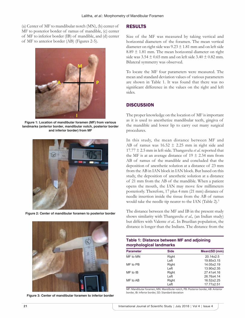
Lalitha, et al.: Morphometry of Mandibular Foramen
21 International Journal of Scientific Study | July 2016 | Vol 4 | Issue 4
Table 1: Distance between MF and adjoining morphological landmarksParameter Side Mean±SD (mm)MF to MN Right 20.14±2.5
Left 19.85±3.15MF to PB Right 14.05±2.19
Left 13.90±2.35MF to IB Right 27.41±4.16
Left 26.76±4.14MF to AB Right 16.52±2.25
Left 17.77±2.51MF: Mandibular foramen, MN: Mandibular notch, PB: Posterior border, AB: Anterior border, IB: Inferior border, SD: Standard deviation
Figure 1: Location of mandibular foramen (MF) from various landmarks (anterior border, mandibular notch, posterior border
and inferior border) from MF
Figure 2: Center of mandibular foramen to posterior border
Figure 3: Center of mandibular foramen to inferior border
(a) Center of MF to mandibular notch (MN), (b) center of MF to posterior border of ramus of mandible, (c) center of MF to inferior border (IB) of mandible, and (d) center of MF to anterior border (AB) (Figures 2-5).
RESULTS
Size of the MF was measured by taking vertical and horizontal diameters of the foramen. The mean vertical diameter on right side was 9.23 ± 1.81 mm and on left side 8.89 ± 1.81 mm. The mean horizontal diameter on right side was 3.54 ± 0.65 mm and on left side 3.40 ± 0.82 mm. Bilateral symmetry was observed.
To locate the MF four parameters were measured. The mean and standard deviation values of various parameters are shown in Table 1. It was found that there was no significant difference in the values on the right and left sides.
DISCUSSION
The proper knowledge on the location of MF is important as it is used to anesthetize mandibular teeth, gingiva of the mandible and lower lip to carry out many surgical procedures.
In this study, the mean distance between MF and AB of ramus was 16.52 ± 2.25 mm in right side and 17.77 ± 2.5 mm in left side. Thangavelu et al. reported that the MF is at an average distance of 19 ± 2.34 mm from AB of ramus of the mandible and concluded that the deposition of anesthetic solution at a distance of 23 mm from the AB in IAN block in IAN block. But based on this study, the deposition of anesthetic solution at a distance of 21 mm from the AB of the mandible. When a patient opens the mouth, the IAN may move few millimeters posteriorly. Therefore, 17 plus 4 mm (21 mm) distance of needle insertion inside the tissue from the AB of ramus would take the needle tip nearer to the IAN (Table 2).5
The distance between the MF and IB in the present study shows similarity with Thangavelu et al., (an Indian study) but differs with Valente et al.. In Brazilian population, the distance is longer than the Indians. The distance from the
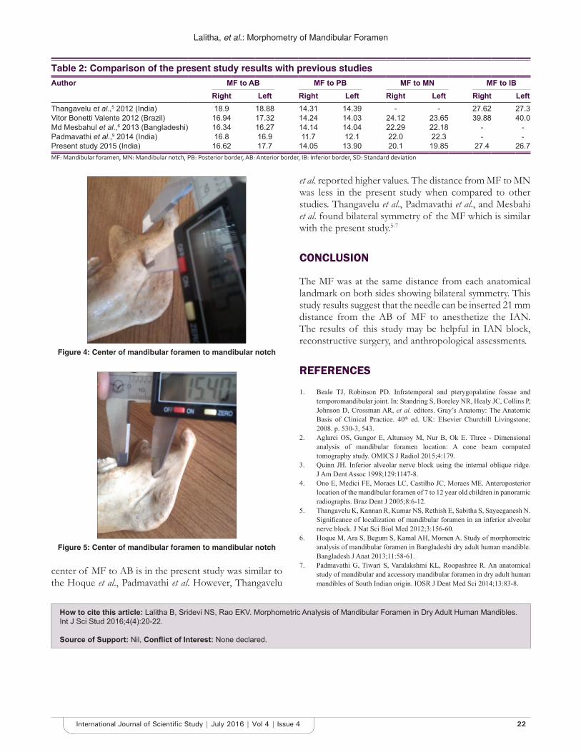
Lalitha, et al.: Morphometry of Mandibular Foramen
22International Journal of Scientific Study | July 2016 | Vol 4 | Issue 4
et al. reported higher values. The distance from MF to MN was less in the present study when compared to other studies. Thangavelu et al., Padmavathi et al., and Mesbahi et al. found bilateral symmetry of the MF which is similar with the present study.5-7
CONCLUSION
The MF was at the same distance from each anatomical landmark on both sides showing bilateral symmetry. This study results suggest that the needle can be inserted 21 mm distance from the AB of MF to anesthetize the IAN. The results of this study may be helpful in IAN block, reconstructive surgery, and anthropological assessments.
REFERENCES
1. Beale TJ, Robinson PD. Infratemporal and pterygopalatine fossae and temporomandibular joint. In: Standring S, Boreley NR, Healy JC, Collins P, Johnson D, Crossman AR, et al. editors. Gray’s Anatomy: The Anatomic Basis of Clinical Practice. 40th ed. UK: Elsevier Churchill Livingstone; 2008. p. 530-3, 543.
2. Aglarci OS, Gungor E, Altunsoy M, Nur B, Ok E. Three - Dimensional analysis of mandibular foramen location: A cone beam computed tomography study. OMICS J Radiol 2015;4:179.
3. Quinn JH. Inferior alveolar nerve block using the internal oblique ridge. J Am Dent Assoc 1998;129:1147-8.
4. Ono E, Medici FE, Moraes LC, Castilho JC, Moraes ME. Anteroposterior location of the mandibular foramen of 7 to 12 year old children in panoramic radiographs. Braz Dent J 2005;8:6-12.
5. Thangavelu K, Kannan R, Kumar NS, Rethish E, Sabitha S, Sayeeganesh N. Significance of localization of mandibular foramen in an inferior alveolar nerve block. J Nat Sci Biol Med 2012;3:156-60.
6. Hoque M, Ara S, Begum S, Kamal AH, Momen A. Study of morphometric analysis of mandibular foramen in Bangladeshi dry adult human mandible. Bangladesh J Anat 2013;11:58-61.
7. Padmavathi G, Tiwari S, Varalakshmi KL, Roopashree R. An anatomical study of mandibular and accessory mandibular foramen in dry adult human mandibles of South Indian origin. IOSR J Dent Med Sci 2014;13:83-8.
Table 2: Comparison of the present study results with previous studiesAuthor MF to AB MF to PB MF to MN MF to IB
Right Left Right Left Right Left Right LeftThangavelu et al.,5 2012 (India) 18.9 18.88 14.31 14.39 - - 27.62 27.3Vitor Bonetti Valente 2012 (Brazil) 16.94 17.32 14.24 14.03 24.12 23.65 39.88 40.0Md Mesbahul et al.,6 2013 (Bangladeshi) 16.34 16.27 14.14 14.04 22.29 22.18 - -Padmavathi et al.,9 2014 (India) 16.8 16.9 11.7 12.1 22.0 22.3 - -Present study 2015 (India) 16.62 17.7 14.05 13.90 20.1 19.85 27.4 26.7MF: Mandibular foramen, MN: Mandibular notch, PB: Posterior border, AB: Anterior border, IB: Inferior border, SD: Standard deviation
Figure 4: Center of mandibular foramen to mandibular notch
Figure 5: Center of mandibular foramen to mandibular notch
How to cite this article: Lalitha B, Sridevi NS, Rao EKV. Morphometric Analysis of Mandibular Foramen in Dry Adult Human Mandibles. Int J Sci Stud 2016;4(4):20-22.
Source of Support: Nil, Conflict of Interest: None declared.
center of MF to AB is in the present study was similar to the Hoque et al., Padmavathi et al. However, Thangavelu

23 International Journal of Scientific Study | July 2016 | Vol 4 | Issue 4
Detection of Candida Species by Hichrom Agar and Their Antimycotic Sensitivity in Hadoti RegionDivya Dadhich1, Naveen Saxena2, Anita E Chand3, Ghanshyam Soni2, Suchitra Morya4
1Assistant Professor, Department of Microbiology, Government Medical College, Kota, Rajasthan, India, 2Professor, Department of Microbiology, Government Medical College, Kota, Rajasthan, India, 3Senior Professor and Head, Department of Microbiology, Government Medical College, Kota, Rajasthan, India, 4Senior Demonstrator, Department of Microbiology, Government Medical College, Kota, Rajasthan, India
and female genital tract, particularly higher in the vagina during pregnancy.1 Carriage rate of Candida species tends to increase with age. Candida species are the fifth most common cause of blood stream infections and fourth common cause of nosocomial infections.1,2 Candida species produces various cutaneous, mucocutaneous, and systemic manifestations depending on the immune status of the host and underlying predisposing factors. In developed countries, Candida albicans accounts for 40-60% of yeasts isolates, whereas Indian reports show an increased predominance of non-C. albicans (NAC) isolates.3,4 Increase in the prevalence of non-albicans species such as Candida glabrata and Candida krusei5 has been noted during the past decade because of the extensive use of antimycotic drugs particularly azoles for prolonged periods. C. glabrata is associated with severe complications than other species.5
INTRODUCTION
Candida is the most common fungal infection found in the humans affecting mucosa, skin, nails, and internal organs. Candida species colonize the mucosal surfaces of all humans soon after birth, and the risk of endogenous infection is ever-present.1 Candida species are a component of the normal flora of human beings and commonly found on the skin, gastrointestinal tract,
Original Article
AbstractIntroduction: Hichrom agar is a differential culture medium which facilitates the isolation and identification of some clinically important species of Candida.
Materials and Methods: A total of 100 Candida species were isolated from various mucocutaneous clinical specimens including oral thrush, vaginitis, balanitis, and angular cheilitis. Speciation of Candida was done using Hichrom agar and conventional methods simultaneously. Antifungal susceptibility testing was done by the disc diffusion method to amphotericin B, fluconazole, nystatin, itraconazole, ketoconazole, and clotrimazole.
Results: Candida albicans (54%) was the predominant species isolated. Non-albicans Candida spp. isolated was Candida tropicalis (22%), Candida glabrata (12%), Candida krusei (06 %), Candida parapsilosis (04%), and Candida kefyr (02%). Antifungal susceptibility testing was done using antimycotic sensitivity testing by disc diffusion method. Overall antifungal drug resistance for Candida in the present study was 26% for fluconazole, 24% for itraconazole, 29% for clotrimazole, 18% for ketoconazole, and 10% for nystatin. No resistance was observed for amphotericin B.
Conclusion: The advantage of using Hichrom agar is that it helps in the isolation and identification of Candida to species level. The performance of Hichrom agar paralleled that of conventional methods. Use of this medium is rapid, technically simple, and cost-effective compared to time-consuming technically demanding expensive conventional method. Hichrom agar serves as a primary isolation and differentiation medium for clinical specimens that could allow mycology laboratories to rapidly identify Candida spp., enabling clinicians to choose appropriate antifungal agents, thus decreasing patient’s morbidity and mortality.
Key words: Antifungal susceptibility testing, Candida, Hichrom agar, Fluconazole
Access this article online
www.ijss-sn.com
Month of Submission : 05-2016 Month of Peer Review : 06-2016 Month of Acceptance : 07-2016 Month of Publishing : 07-2016
Corresponding Author: Dr. Anita E Chand, House No. 2-k-9 Rangbari Yojna, Kota - 324 005, Rajasthan, India. Phone: +91-09829577983. E-mail: [email protected]
Print ISSN: 2321-6379Online ISSN: 2321-595X
DOI: 10.17354/ijss/2016/367

Dadhich, et al.: Detection of Candida Species by Hichrom Agar and Their Antimycotic Sensitivity in Hadoti Region
24International Journal of Scientific Study | July 2016 | Vol 4 | Issue 4
Several brands of chromogenic media have been developed to produce rapid yeast identification. C. albicans produces an enzyme b-N-acetyl-galactosaminidase6 and incorporation of chromogenic or fluorogenic hexosaminidase substrates into the growth medium helps in identification of C. albicans isolates directly on primary isolation.7 Hichrom Candida differential agar is a selective and differential medium, which facilitates rapid isolation of yeasts from mixed cultures and allows differentiation of Candida species namely C. albicans, C. krusei, Candida Tropicalis, and C. glabrata on the basis of coloration and colony morphology.
It is necessary to identify Candida to species level as many NAC have decreased susceptibility to antifungal agents. The present study was undertaken to evaluate the advantages of Hichrom agar over conventional method for speciation of Candida isolates and their susceptibility to antifungal agents by disc diffusion method.
MATERIALS AND METHODS
The study was conducted at the Department of Microbiology, Government Medical College and M.B.S Hospital, Kota, from June 2012 to September 2013. A total of 100 strains of Candida were isolated from various mucocutaneous clinical samples of patients admitted in MBS Hospital and associated Group of Hospitals - JK Lone Hospital and NMC Hospital, Kota, Rajasthan.
Hichrom agar was prepared as per the instruction manual (Himedia India) (Table 3). Candida spp. isolated was inoculated simultaneously to Hichrom agar plates and Sabouraud dextrose agar (SDA) tubes. These were incubated at 37°C for 48 h. Species were identified on Hichrom agar by morphology and color of the colony. Growth on SDA was speciated by standard methods using germ tube, corn meal agar, sugar fermentation, and assimilation test. Appearances of Candida spp. on Hichrom agar were as follows:8• C. albicans - blue green• C. tropicalis - dark blue-gray center with pink halo• C. krusei - pink large rough spreading colonies with
pale edge• C. parapsilisis - pale cream colored colonies• C. glabrata - cream to white smooth colonies.
Antifungal susceptibility was performed by disc diffusion method using antimycotic sensitivity test agar. Discs used were amphotericin B (100 units), fluconazole (10 mcg), clotrimazole (10 mcg), nystatin (100 mcg), itraconazole (10 mcg), ketoconazole (10 mcg), and sensitivity zones were measured as for the instruction manual (Himedia).8,9 ATCC strain of C. albicans was used as control.
RESULTS
Table 1 shows Candida spp. isolated in various clinical samples. All isolates of candida grew on Hichrom agar after 48 h of incubation at 37°C.
Overall antifungal drug resistance for Candida in the present study was 26% for fluconazole, 24% for itraconazole, 29% for clotrimazole, 18% for ketoconazole, and 10% for nystatin. No resistance was observed for amphotericin B. The results of this study have been presented in Tables 1 and 2.
DISCUSSION
C. albicans (54%) was the most prevalent species of Candida reported in the present study (Table 1). This finding was consistent with the findings of other workers who reported that the incidence of C. albicans was 61.3% (Biradar et al.),10 49.3% (Feglo and Narkwa),11 and 47% (Dominic and Dharwad).12 However, Kashid et al.13 found that C. tropicalis was the most prevalent species accounted for 46.2% followed by C. albicans (29.2%). C. tropicalis (22%) was the second most common species reported in the present study. This finding was comparable with other workers, Bobade et al.14 (22.9%), Babin et al.15 (26.4%), and Khan and Baqai et al.16 (21%). However, C. glabrata was reported as second most common species by Feglo and Narkwa et al.11 (17.9%) and Saldhei et al.17 (11.9%).
Overall antifungal drug resistance for Candida in the present study was 26% for fluconazole, 24% for itraconazole, 29% for clotrimazole, 18% for ketoconazole, and 10% for nystatin. No resistance was observed for amphotericin B. In present study, 22.2% of C. albicans was found to be fluconazole resistant which is in consonance with Kashid et al.13 and Babin et al.15 However, a higher resistance was observed by Saldhei et al.17 (81.1%). For NAC, the resistance varies from 0% in Candida kefyr to 100% in C. krusei. For AmB, no resistance was observed in the present study. This was in consonance with Kashid et al.13 Bobade et al.14 reported 7.5% resistance in C. albicans.
The conventional methods and the CHROM agar method were compared and were found to give similar results. This was similar to the findings of Nayak et al.,18 who found that CHROM agar showed 100% specificity and 100% sensitivity when compared to SDA and conventional methods. The advantages of Hichrom agar are easy to prepare, i.e., boiling, facilitate the rapid isolation, and identification of yeast species. Hichrom agar facilitates identification between yeast spp. from specimens containing mixture of yeast spp. and do not affect the

Dadhich, et al.: Detection of Candida Species by Hichrom Agar and Their Antimycotic Sensitivity in Hadoti Region
25 International Journal of Scientific Study | July 2016 | Vol 4 | Issue 4
viability on subsequent subcultures.19 Hichrom agar has the advantage of rapid identification of Candida species, technically simple, rapid, and cost-effective compared to technically demanding time consuming and expensive conventional method.
CONCLUSION
Although the results on Hichrom agar exactly paralleled that of the conventional method, it is superior to SDA in terms of suppressing the bacterial growth. Use of Hichrom agar medium would allow mycology laboratories to identify rapidly, clinically important Candida spp. while potentially decreasing laboratory cost. Furthermore, the species level identification of the Candida isolates along with their antifungal susceptibility patterns can greatly influence the treatment options for the clinician and may have an impact on the patient care.
REFERENCES
1. Chander J. A Text Book of Medical Mycology, Candidiasis. 3rd ed. Ch. 20. New Delhi: Mehta Publishers; 2009. p. 266-83.
2. Koneman E, Allen S, Janda W. Colour Atlas and Textbook of Diagnostic Microbiology. 6th ed. Philadelphia, PA: Lippincott; 2006.
3. Coleman DC, Rinaldi MG, Haynes KA, Rex JH, Summerbell RC, Anaissie EJ, et al. Importance of Candida species other than Candida albicans as opportunistic pathogens. Med Mycol 1998;36 Suppl 1:156-65.
4. Moran GP, Sullivan DJ, Coleman D. Emergence of non - Candida albicans species as pathogens. In: Calderone RA, editor. Candida and Candidiasis. 4th ed. Ch. 4. Washington, DC: ASM Press; 2002. p. 37-53.
5. Topley WW, Wilson CS. Topley and Wilson’s Microbiology and Microbial Infections, Medical Mycology, Candidiasis. 10th ed. Ch. 30. Baltimore: Edward Arnold Publisher; 2005. p. 579-20.
6. Perry JL, Miller GR. Umbelliferyl-labeled galactosaminide as an aid in identification of Candida albicans. J Clin Microbiol 1987;25:2424-5.
7. Rousselle P, Freydiere AM, Couillerot PJ, de Montclos H, Gille Y. Rapid identification of Candida albicans by using Albicans ID and fluoroplate agar plates. J Clin Microbiol 1994;32:3034-6.
8. Vijaya D, Harsha TR, Nagaratnamma T. Candida speciation using CHROMagar. J Clin Diagn Res 2011;5:755-7.
9. Glasmacher A, Molitor E, Mezger J, Marklein G. Antifungal prophylaxis
Table 1: Candida species isolated from different clinical conditionsClinical condition C. albicans (%) C. tropicalis (%) C. glabrata (%) C. krusei (%) C. parapsilosis (%) C. kefyr (%) Total (%)Oral thrush 23 (42.5) 17 (31.4) 6 (11.1) 6 (11.1) 0 2 (3.7) 54 (54)Vaginitis 26 (68.4) 3 (7.8) 6 (15.7) 0 3 (7.8) 0 38 (38)Balanitis 4 (80) 0 0 0 1 (20) 0 5 (5)Angular cheilitis 1 (33.3) 2 (66.7) 0 0 0 0 3 (3)Total 54 (54) 22 (22) 12 (12) 6 (6) 4 (4) 2 (2) 100C. albicans: Candida albicans, C. tropicalis: Candida tropicalis, C. glabrata: Candida glabrata, C. krusei: Candida krusei, C. parapsilosis: Candida parapsilosis, C. kefyr: Candida kefyr
Table 2: Antifungal sensitivity profile of Candida isolates (in percentage)Antifungal drugs Fluconazole
(25 µg)Itraconazole
(10 µg)Clotrimazole
(10 µg)Ketoconazole
(10 µg)Nystatin
(100 U/disc)AmB
(100 U)S I R S I R S I R S I R S I R S I R
C. albicans 48.1 29.6 22.2 29.6 44.4 25.9 14.8 55.5 29.6 29.6 44.4 25.9 66.7 29.6 3.7 100 00 00C. tropicalis 54.5 31.8 13.6 54.5 31.8 13.6 40.9 36.3 22.7 90.9 9.1 00 40.9 36.3 22.7 100 00 00C. glabrata 50 16.6 33.4 50 16.6 33.4 16.6 33.4 50 50 33.4 16.6 33.4 50 16.6 100 00 00C. kefyr 100 00 00 100 00 00 100 00 00 100 00 00 100 00 00 100 00 00C. parapsilosis 50 25 25 50 25 25 25 50 25 50 25 25 25 50 25 100 00 00C. krusei 00 00 100 00 00 100 66.6 16.6 16.6 66.6 16.6 16.6 100 00 00 100 00 00Total (in percentage) 48 26 26 38 38 24 26 45 29 50 32 18 58 32 10 100 00 00S: Sensitive, I: Intermediate sensitive, R: Resistant, C. albicans: Candida albicans, C. tropicalis: Candida tropicalis, C. glabrata: Candida glabrata, C. krusei: Candida krusei, C. parapsilosis: Candida parapsilosis, C. kefyr: Candida kefyr
Table 3: Comparison of various studies showing Candida species recovered from clinical samples (in percentage)Candida species
Present study
Bobade et al.
201314
Saldhei et al.
201217
Vijaya et al. 20118
Kashid et al. 201113
Feglo and Narkwa 201111
Babin et al. 201115
Shivanand et al. 201121
Fouzia et al.
201016
Birader et al.
200910
Usharani et al.
200520
C. albicans 54 36.6 79.1 45.9 29.2 49.3 35.5 47 30 61.3 62.5C. tropicalis 22 22.9 5.9 35.29 46.2 11.9 22.9 30 21 18 15.6C. glabrata 12 13.7 11.9 - 6.12 17.9 20.6 09 08 10.6 9.3C. krusei 6 08.7 2.9 10.78 - 4.5 15.7 14 03 3.3 -C. parapsilosis 4 03.6 - 7.84 10.2 1.5 - - 10 - -C. kefyr 2 11.46 - - 1.36 - - - - - -

Dadhich, et al.: Detection of Candida Species by Hichrom Agar and Their Antimycotic Sensitivity in Hadoti Region
26International Journal of Scientific Study | July 2016 | Vol 4 | Issue 4
with itraconazole in neutropenic patients: Pharmacological, microbiological and clinical aspects. Mycoses 1996;39:249-58.
10. Biradar S, Amruthkishan U, Gangane R, Sheeba P, Amar N, Praveen D, et al. Prevalence and antifungal susceptibility of Candida species isolated from tertiary care Hospital in North East Karnataka. Int J Pharm Bio Sci 2013;4:1113-8.
11. Feglo PK, Narkwa P. Prevalence and antifungal susceptibility patterns of yeast isolates at the Komfo Anokye Teaching Hospital (KATH), Kumasi, Ghana. Br Micro Res J 2012;2:10-22.
12. Dominic RM, Dharwad S. Species identification of Candida isolates in various clinical specimens with their antifungal susceptibility patterns. J Clin Diagn Res 2011;5:1177-81.
13. Kashid RA, Belawadi S, Devi G, Dadich D. Characterisation and antifungal susceptibilty testing for Candida species in a tertiary care hospital. JOHSR 2011;2:1-7.
14. Bobade O, Waghmare M, Chhabrani P, Kaur I. Species distribution and antifungal susceptibility profile of Candida isolated from urine samples. Int J Med Sci Public Health 2013;2:867-70.
15. Babin D, Kotigadde S, Rao PS, Rao TV. Clinico-mycological profile of vaginal candidiasis in a tertiary care hospital in Kerala. Int J Res Biol Sci 2013;3:55-9.
16. Khan F, Baqai R. In vitro antifungal sensitivity of fluconazole, clotrimazole and nystatin against vaginal candidiasis in females of childbearing age. J Ayub Med Coll Abbottabad 2010;22:197-200.
17. Saldhei Z, Seifi Z, Mahmoudabadi AZ. Sensitivity of vaginal isolates of Candida to eight antifungal drugs isolated from Ahvaz, Iran. Jundishapur J Microbiol 2012;5:574-7.
18. Nayak S, Kavitha B, Sriram G, Saraswathi TR, Sivapathasundharam B, Dorothy AL. Comparative study of Candida by conventional and CHROMagar method in non-denture and denture wearers by oral rinse technique. Indian J Dent Res 2012;23:490-7.
19. Odds FC, Bernaerts R. CHROMagar Candida, a new differential isolation medium for presumptive identification of clinically important Candida species. J Clin Microbiol 1994;32:1923-9.
20. Usharani DM, Sulochana D, Singh L. A study on the prevalence, pattern of antifungal susceptibility and co-relation of CD4+T lymphocytes amongst the Candida isolates from HIV patients in Manipur. Nat J Basic Med Sci 2005;2:329-32.
21. Saldanha Dominic RM, Dharwad S. Species Identification of Candida Isolates in Various Clinical Specimens with Their Antifungal Susceptibility Patterns. J of Clin and Diag Res. 2011;5(6):1177-81.
How to cite this article: Dadhich D, Saxena N, Chand AE, Soni G, Morya S. Detection of Candida Species by Hichrom Agar and Their Antimycotic Sensitivity in Hadoti Region. Int J Sci Stud 2016;4(4):23-26.
Source of Support: Nil, Conflict of Interest: None declared.

27 International Journal of Scientific Study | July 2016 | Vol 4 | Issue 4
Clinico Pathological Study of Autoimmune Vesiculobullous Disorders: A Case Series from a Resource-poor Rural Tertiary Care Center in South Tamil NaduUma Selvaraj1, Madhavan Ramamoorthy2
1Senior Assistant Professor, Department of Dermatology, Government Theni Medical College, Theni, Tamil Nadu, India, 2Senior Assistant Professor, Department of STD, Coimbatore Medical College, Coimbatore, Tamil Nadu, India
formation. They are classified as epidermal and subepidermal. To arrive at a diagnosis, complete clinical examination, aided by Tzanck smear, histopathology of skin biopsy, and immunofluorescence studies need to be done. But in resource-poor centers like ours diagnosis could be achieved only with the available investigations. The aim of the study was to analyze the clinical and histopathological features of these disorders in our setup with the available resources.
MATERIALS AND METHODS
A retrospective analysis of all vesiculobullous disorders admitted to our ward over a span of 10 years between 2005 and 2015 was analyzed.
INTRODUCTION
Autoimmune vesiculobullous disorders are a rare group of disorders where auto antibodies are directed against specialized structures essential for maintaining the integrity of skin leading to blister formation. Erosions involving skin and mucous membranes depending on the level of blister
Original Article
AbstractBackground: Autoimmune vesiculobullous disorders are rare diseases that are characterized by blisters over skin and oral erosions. An accurate diagnosis is achieved by clinical examination, pathological correlation, and immunofluorescence. In resource-poor settings where immunofluorescence is not available, the diagnosis is made only with the available resources.
Aims and Objectives: To study the clinical and pathological features of autoimmune bullous disorders.
Materials and Methods: A total of 45 patients admitted to the Government Theni Medical College from 2005 to 2015 were analyzed retrospectively. Detailed clinical examination, Tzanck smear, and histopathological examination of skin biopsy were done for all patients. Immunofluorescence was not done due to lack of availability in our center.
Results: Out of 45 cases studied, 32 (71%) were pemphigus, 10 (22.22%) belonged to bullous pemphigoid group, 2 (4.44%) were chronic bullous dermatosis of childhood, one case (2.2%) was a bullous systemic lupus erythematosus. Out of 45 patients, 31 patients (68.8%) were in the age group of 41-60 years, 6 patients (13.33%) in the range of 21-40 years, 5 patients (11.11%) in the age range above 60 years, and 3 patients (6.66%) below 20 years. Out of 45 patients, 17 (37.77%) were males and 28 (62.22%) were females. All the bullous disorders showed a female preponderance. The clinical spectrum was consistent in all patients enrolled (100%) Tzanck smear was consistent in 95.5% of patients. Histopathology was consistent in 100% of cases.
Conclusion: Thorough clinical examination, aided by Tzanck smear and histopathology was helpful in arriving at a diagnosis of these vesiculobullous disorders in resource-poor center like ours where the gold standard immunofluorescence studies are not available.
Key words: Autoimmuno bullous disorders, Bullous pemphigoid, Pemphigus
Access this article online
www.ijss-sn.com
Month of Submission : 05-2016 Month of Peer Review : 06-2016 Month of Acceptance : 07-2016 Month of Publishing : 07-2016
Corresponding Author: Dr. Uma Selvaraj, 15/1/38A, Arjun Illam, Sivanantha Nagar, P.C. Patty, Theni - 625 531, Tamil Nadu, India. E-mail: [email protected]
Print ISSN: 2321-6379Online ISSN: 2321-595X
DOI: 10.17354/ijss/2016/368

Selvaraj and Ramamoorthy: Clinico Pathological Study of Autoimmune Vesiculobullous Disorders
28International Journal of Scientific Study | July 2016 | Vol 4 | Issue 4
Thorough clinical examination was done for all patients. Tzanck smear was done for all patients by deroofing an early vesicle and making a smear on a glass slide and staining with Leishman’s stain and examined under light microscope. A skin biopsy taken from an early vesicle was subjected to histopathological examination after staining with eosin and hematoxylin.
All data regarding the patients’ age, sex, duration of disease, reports of Tzanck smear, and histopathological findings were documented.
RESULTS
Out of 45 cases studied, 32 (71.1%) belonged to pemphigus group. 10 cases (22.2%) belonged to bullous pemphigoid Group 2 patients (4.4%) belonged to chronic bullous dermatosis of childhood (CBDC) group and one patient (2.2%) was a bullous systemic lupus erythematosus (SLE) (Table 1).
Out of 32 cases of pemphigus, 28 were pemphigus vulgaris and 4 were pemphigus foliaceus. Predominant age group affected was 51-60 (16 patients) followed by 15 patients in the age group of 41-50. Youngest age incidence was seen in 3 patients in the range 0-10. The highest age range was seen in 5 patients in the ranges 61-70 years (Table 2).
Out of 45 cases, 28 were females (62.14%) and 17 (37.6%) were males (Figure 1) female predominance was seen in all the bullors disorders noted.
The clinical diagnosis could be arrived in all pemphigus patients with flaccid blisters and erosions, a positive
Nickolsky sign in pemphigus vulgaris and no mucosal involvement in pemphigus foliaceus (Figure 2).
All bullous pemphigoid patients showed tense blisters and erosions over trunk and extremities. Mucosal erosions were seen in only 4 out of 10 patients (Figure 3). Both cases of CBDC showed, tense blisters with few lesions showing “cluster of jewels” appearance (Figure 4). No mucosal involvement was seen in both cases. Bullous SLE showed tense blisters over extremities and with other cutaneous and lab findings diagnostic of SLE.
Table 1: Bullous disorders prevalenceBullous disorder Number of cases (%)Pemphigus vulgaris 28 (62.2)Pemphigus foliaceus 4 (8.8)Bullous pemphigoid 10 (22.22)CBDC 2 (4.44)Bullous SLE 1 (2.2)Total 45CBDC: Chronic bullous dermatosis of childhood, SLE: Systemic lupus erythematosus
Table 2: Age wise statistics of bullous disordersType 0-10 11-20 21-30 31-40 41-50 51-60 61-70 TotalPemphigus 0 0 4 2 13 10 3 32Bullous Pemphigoid
0 0 0 0 2 6 2 10
CBDC 2 0 0 0 0 0 0 2Bullous SLE 1 0 0 0 0 0 0 1Total 45CBDC: Chronic bullous dermatosis of childhood, SLE: Systemic lupus erythematosus
Figure 1: Sex wise distribution
Figure 2: Flaccid blisters and erosions in pemphigus
Figure 3: Tense blisters in bullous pemphoid
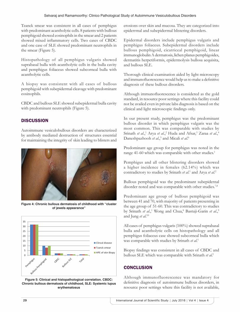
Selvaraj and Ramamoorthy: Clinico Pathological Study of Autoimmune Vesiculobullous Disorders
29 International Journal of Scientific Study | July 2016 | Vol 4 | Issue 4
Tzanck smear was consistent in all cases of pemphigus with predominant acantholytic cells. 8 patients with bullous pemphigoid showed eosinophils in the smear and 2 patients showed mixed inflammatory cells. Two cases of CBDC and one case of SLE showed predominant neutrophils in the smear (Figure 5).
Histopathology of all pemphigus vulgaris showed suprabasal bulla with acantholytic cells in the bulla cavity and pemphigus foliaceus showed subcorneal bulla with acantholytic cells.
A biopsy was consistent with all cases of bullous pemphigoid with subepidermal cleavage with predominant eosinophils.
CBDC and bullous SLE showed subepidermal bulla cavity with predominant neutrophils (Figure 5).
DISCUSSION
Autoimmune vesiculobullous disorders are characterized by antibody mediated destruction of structures essential for maintaining the integrity of skin leading to blisters and
erosions over skin and mucosa. They are categorized into epiderrmal and subepidermal blistering disorders.
Epidermal disorders include pemphigus vulgaris and pemphigus foliaceus. Subepidermal disorders include bullous pemphigoid, cicatriceal pemphigoid, linear immunoglobulin A dermatosis, lichen planus pemphigoides, dermatitis herpetiformis, epidermolysis bullosa acquisita, and bullous SLE.
Thorough clinical examination aided by light microscopy and immunofluorescence would help us to make a definitive diagnosis of these bullous disorders.
Although immunofluorescence is considered as the gold standard, in resource poor settings where this facility could not be availed even in private labs diagnosis is based on the clinical and light microscopic findings only.
In our present study, pemphigus was the predominant bullous disorder in which pemphigus vulgaris was the most common. This was comparable with studies by Srinath et al.,1 Arya et al.,2 Huda and Afsar,3 Zaraa et al.,4 Daneshpazhooh et al.,5 and Micali et al.6
Predominant age group for pemphigus was noted in the range 41-60 which was comparable with other studies.1
Pemphigus and all other blistering disorders showed a higher incidence in females (62.14%) which was contradictory to studies by Srinath et al.1 and Arya et al.2
Bullous pemphigoid was the predominant subepidernal disorder noted and was comparable with other studies.7,8
Predominant age group of bullous pemphigoid was between 41 and 70, with majority of patients presenting in the age group of 51-60. This was contradictory to studies by Srinath et al.,1 Wong and Chua,8 Bastuji-Garin et al.,9 and Jung et al.10
All cases of pemphigus vulgaris (100%) showed suprabasal bulla and acantholytic cells on histopathology and all pemphigus foliaceus case showed subcorneal bulla which was comparable with studies by Srinath et al.1
Biopsy findings was consistent in all cases of CBDC and bullous SLE which was comparable with Srinath et al.1
CONCLUSION
Although immunofluorescence was mandatory for definitive diagnosis of autoimmune bullous disorders, in resource poor settings where this facility is not available,
Figure 4: Chronic bullous dermatosis of childhood with “cluster of jewels appearance”
Figure 5: Clinical and histopathological correlation. CBDC: Chronic bullous dermatosis of childhood, SLE: Systemic lupus
erythematosus

Selvaraj and Ramamoorthy: Clinico Pathological Study of Autoimmune Vesiculobullous Disorders
30International Journal of Scientific Study | July 2016 | Vol 4 | Issue 4
diagnosis could be arrived only with the available investigations.
REFERENCES
1. Srinath M, Kambli, Madavamurthy P. Immunobullous disorders clinical histopathological and immunofluorescence study of 36 cases. Mullers J Med Res 2014:5:134-8.
2. Arya SR, Valand AG, Krishna K. A clinico-pathological study of 70 cases of pemphigus. Indian J Dermatol Venereol Leprol 1999;65:168-71.
3. Huda MM, Afsar MI. A clinicopathological study of pemphigus. Indian J Dermatol 2001;46:75-9.
4. Zaraa I, Kerkeni N, Ishak F, Zribi H, El Euch D, Mokni M, et al. Spectrum of autoimmune blistering dermatoses in Tunisia: An 11-year study and a review of the literature. Int J Dermatol 2011;50:939-44.
5. Daneshpazhooh M, Chams-Davatchi C, Payandemehr P, Nassiri S,
Valikhani M, Safai-Naraghi Z. Spectrum of autoimmune bullous diseases in Iran: A 10-year review. Int J Dermatol 2012;51:35-41.
6. Micali G, Musumeci ML, Nasca MR. Epidemiologic analysis and clinical course of 84 consecutive cases of pemphigus in Eastern Sicily. Int J Dermatol 1998;37:197-200.
7. Bernard P, Vaillant L, Labeille B, Bedane C, Arbeille B, Denoeux JP, et al. Incidence and distribution of subepidermal autoimmune bullous skin diseases in three French regions. Bullous diseases French study group. Arch Dermatol 1995;131:48-52.
8. Wong SN, Chua SH. Spectrum of subepidermal immunobullous disorders seen at the National Skin Centre, Singapore: A 2-year review. Br J Dermatol 2002;147:476-80.
9. Bastuji-Garin S, Joly P, Lemordant P, Sparsa A, Bedane C, Delaporte E, et al. Risk factors for bullous pemphigoid in the elderly: A prospective case-control study. J Invest Dermatol 2011;131:637-43.
10. Jung M, Kippes W, Messer G, Zillikens D, Rzany B. Increased risk of bullous pemphigoid in male and very old patients: A population-based study on incidence. J Am Acad Dermatol 1999;41:266-8.
How to cite this article: Selvaraj U, Ramamoorthy M. Clinico Pathological Study of Autoimmune Vesiculobullous Disorders: A Case Series from a Resource-poor Rural Tertiary Care Center in South Tamil Nadu. Int J Sci Stud 2016;4(4):27-30.
Source of Support: Nil, Conflict of Interest: None declared.

31 International Journal of Scientific Study | July 2016 | Vol 4 | Issue 4
Comparative Study of Analgesic Efficacy of Intravenous versus Intrathecal Fentanyl as an Adjuvant in Subarachnoid Block for Cesarean SectionGupta Parul1, Nanda Harjeet Singh2, Arora Neeharika3
1Senior Resident, Department of Anaesthesiology, Critical Care and Pain Management, Shri Ram Murti Smarak Institute of Medical Sciences, Bareilly, Uttar Pradesh, India, 2Professor and Head, Department of Anaesthesiology, Critical Care and Pain Management, Shri Ram Murti Smarak Institute of Medical Sciences, Bareilly, Uttar Pradesh, India, 3Associate Professor, Department of Anaesthesiology, Critical Care and Pain Management, Shri Ram Murti Smarak Institute of Medical Sciences, Bareilly, Uttar Pradesh, India
in less maternal and neonatal morbidity than general anesthesia.2,3
The local anesthetics have a relatively short duration of action, thereby limiting the technique for comparatively long duration surgery and increasing the requirement of analgesics in the early post-operative period.4,5 Furthermore, even the conventional recommended doses of the local anesthetic are unable to completely abolish visceral pain produced by the manipulation of the uterus and peritoneum, leading to intraoperative pain and nausea-vomiting,4,6 necessitating an increase in doses of the local
INTRODUCTION
Subarachnoid block for cesarean delivery is a preferred and widely used technique1 because it is safer and results
Original Article
AbstractIntroduction: Cesarean section under spinal anesthesia with local anesthetics leads to intraoperative visceral pain, which may require analgesic supplementation.
Objectives/Purpose: To compare analgesic efficacy in perioperative period between intravenous fentanyl and intrathecal fentanyl as an adjuvant in the subarachnoid block for cesarean section.
Materials and Methods: The study was carried out on 100 parturients, aged 18-45 years, ASA status I and II, scheduled for elective cesarean section, randomly allocated in two groups of 50 patients each: Group A (intravenous group): Intrathecal 0.5% hyperbaric bupivacaine 2.0 ml + normal saline 0.25 ml, and intravenous fentanyl 12.5 mcg immediately after administration of spinal bupivacaine. Group B (intrathecal group): Intrathecal 0.5% hyperbaric bupivacaine 2.0 ml (10 mg) + fentanyl 12.5 mcg (0.25 ml), and intravenous normal saline 0.25 ml (placebo). We compared the requirement for intravenous fentanyl supplementation during surgery, the intraoperative pain scores by visual analog scale, and the time to post-operative rescue analgesia.
Results: The intravenous group (Group A) has higher intraoperative pain scores than Group B (P < 0.05). Higher amount of intraoperative intravenous fentanyl supplementation was required in the intravenous fentanyl group as compared to the intrathecal group (mean intravenous fentanyl requirement = 19.5 ± 3.56 versus 4.5 ± 1.87 mcg, respectively; P = 0.002). The time to first request for post-operative analgesia was significantly longer in the intrathecal fentanyl group than in intravenous fentanyl group (133.9 ± 20.68 vs. 115.4 ± 16.28 min, respectively; P = 0.003).
Conclusion: Intrathecal fentanyl supplementation of spinal anesthesia with bupivacaine during cesarean delivery resulted in a better quality of spinal analgesia than the same dose of intravenous fentanyl supplementation.
Key words: Analgesia, Bupivacaine, Cesarean section, Fentanyl, Subarachnoid block
Access this article online
www.ijss-sn.com
Month of Submission : 05-2016 Month of Peer Review : 06-2016 Month of Acceptance : 07-2016 Month of Publishing : 07-2016
Corresponding Author: Dr. Parul Gupta, P.G Hostel, Room No. 09, Shri Ram Murti Smarak Institute of Medical Sciences, Bareilly - 243 001, Uttar Pradesh, India. Phone: +91-7500077643/9458705944. E-mail: [email protected]
Print ISSN: 2321-6379Online ISSN: 2321-595X
DOI: 10.17354/ijss/2016/369
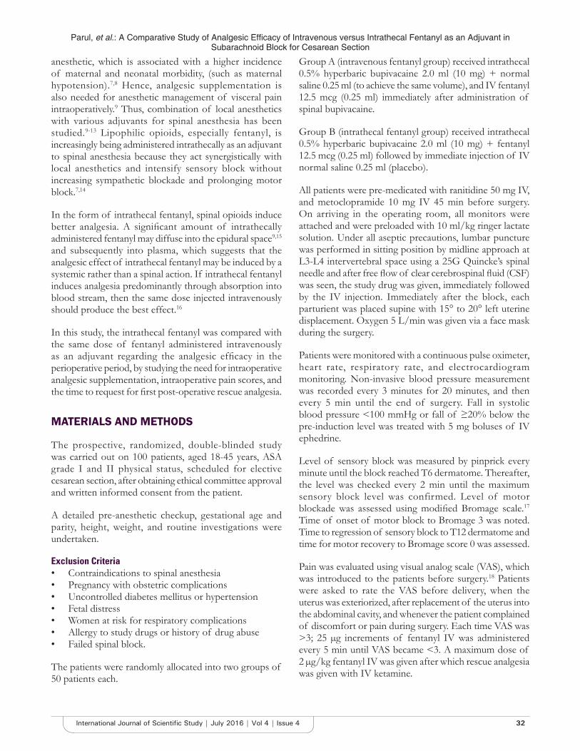
Parul, et al.: A Comparative Study of Analgesic Efficacy of Intravenous versus Intrathecal Fentanyl as an Adjuvant in Subarachnoid Block for Cesarean Section
32International Journal of Scientific Study | July 2016 | Vol 4 | Issue 4
anesthetic, which is associated with a higher incidence of maternal and neonatal morbidity, (such as maternal hypotension).7,8 Hence, analgesic supplementation is also needed for anesthetic management of visceral pain intraoperatively.9 Thus, combination of local anesthetics with various adjuvants for spinal anesthesia has been studied.9-13 Lipophilic opioids, especially fentanyl, is increasingly being administered intrathecally as an adjuvant to spinal anesthesia because they act synergistically with local anesthetics and intensify sensory block without increasing sympathetic blockade and prolonging motor block.7,14
In the form of intrathecal fentanyl, spinal opioids induce better analgesia. A significant amount of intrathecally administered fentanyl may diffuse into the epidural space9,15 and subsequently into plasma, which suggests that the analgesic effect of intrathecal fentanyl may be induced by a systemic rather than a spinal action. If intrathecal fentanyl induces analgesia predominantly through absorption into blood stream, then the same dose injected intravenously should produce the best effect.16
In this study, the intrathecal fentanyl was compared with the same dose of fentanyl administered intravenously as an adjuvant regarding the analgesic efficacy in the perioperative period, by studying the need for intraoperative analgesic supplementation, intraoperative pain scores, and the time to request for first post-operative rescue analgesia.
MATERIALS AND METHODS
The prospective, randomized, double-blinded study was carried out on 100 patients, aged 18-45 years, ASA grade I and II physical status, scheduled for elective cesarean section, after obtaining ethical committee approval and written informed consent from the patient.
A detailed pre-anesthetic checkup, gestational age and parity, height, weight, and routine investigations were undertaken.
Exclusion Criteria• Contraindications to spinal anesthesia• Pregnancy with obstetric complications• Uncontrolled diabetes mellitus or hypertension• Fetal distress• Women at risk for respiratory complications• Allergy to study drugs or history of drug abuse• Failed spinal block.
The patients were randomly allocated into two groups of 50 patients each.
Group A (intravenous fentanyl group) received intrathecal 0.5% hyperbaric bupivacaine 2.0 ml (10 mg) + normal saline 0.25 ml (to achieve the same volume), and IV fentanyl 12.5 mcg (0.25 ml) immediately after administration of spinal bupivacaine.
Group B (intrathecal fentanyl group) received intrathecal 0.5% hyperbaric bupivacaine 2.0 ml (10 mg) + fentanyl 12.5 mcg (0.25 ml) followed by immediate injection of IV normal saline 0.25 ml (placebo).
All patients were pre-medicated with ranitidine 50 mg IV, and metoclopramide 10 mg IV 45 min before surgery. On arriving in the operating room, all monitors were attached and were preloaded with 10 ml/kg ringer lactate solution. Under all aseptic precautions, lumbar puncture was performed in sitting position by midline approach at L3-L4 intervertebral space using a 25G Quincke’s spinal needle and after free flow of clear cerebrospinal fluid (CSF) was seen, the study drug was given, immediately followed by the IV injection. Immediately after the block, each parturient was placed supine with 15° to 20° left uterine displacement. Oxygen 5 L/min was given via a face mask during the surgery.
Patients were monitored with a continuous pulse oximeter, heart rate, respiratory rate, and electrocardiogram monitoring. Non-invasive blood pressure measurement was recorded every 3 minutes for 20 minutes, and then every 5 min until the end of surgery. Fall in systolic blood pressure <100 mmHg or fall of ≥20% below the pre-induction level was treated with 5 mg boluses of IV ephedrine.
Level of sensory block was measured by pinprick every minute until the block reached T6 dermatome. Thereafter, the level was checked every 2 min until the maximum sensory block level was confirmed. Level of motor blockade was assessed using modified Bromage scale.17 Time of onset of motor block to Bromage 3 was noted. Time to regression of sensory block to T12 dermatome and time for motor recovery to Bromage score 0 was assessed.
Pain was evaluated using visual analog scale (VAS), which was introduced to the patients before surgery.18 Patients were asked to rate the VAS before delivery, when the uterus was exteriorized, after replacement of the uterus into the abdominal cavity, and whenever the patient complained of discomfort or pain during surgery. Each time VAS was >3; 25 µg increments of fentanyl IV was administered every 5 min until VAS became <3. A maximum dose of 2 µg/kg fentanyl IV was given after which rescue analgesia was given with IV ketamine.

Parul, et al.: A Comparative Study of Analgesic Efficacy of Intravenous versus Intrathecal Fentanyl as an Adjuvant in Subarachnoid Block for Cesarean Section
33 International Journal of Scientific Study | July 2016 | Vol 4 | Issue 4
Any adverse effects, such as intraoperative nausea, vomiting, pruritus, shivering, and respiratory depression were recorded and treated. After delivery, the Apgar scores of the neonates were assessed at 1 and 5 min. Time to the first request for rescue post-operative analgesia for the patient was recorded. Rescue analgesia was available in the form of IV 75 mg diclofenac sodium aqueous.
The data were analyzed using SPSS software version 20 by inserting the data into MS Excel. Unpaired/independent t-test was used on the data to compare the significant difference between the two groups for different parameters under study, and qualitative data were analyzed using non-parametric tests of statistics, that is, Chi-square test and Fisher’s exact test, at 0.05 level of significance (that is, a P < 0.05 was considered significant). Data are presented as mean value ± standard deviation, median, mode, and incidence n (%).
RESULTS
The groups were comparable with respect to age, height, weight, gestational age, and parity (Tables 1 and 2). The sensory and motor block characteristics are summarized in Table 3. The time of onset to T6 sensory block, maximum sensory level achieved, and time required to achieve maximum sensory level is similar between the groups. In most patients, the maximum sensory level in both groups reached a level of T5. The difference between the groups in motor block onset, time for motor block recovery, and
time to the recession of sensory block to T12 did not reach statistical significance. There was no significant difference between groups with respect to intrathecal injection to delivery time and duration of surgery (Table 1).
Intraoperative pain scores are depicted in Table 4. Group A had a higher incidence (n = 10%) of moderate to severe pain (i.e., VAS > 3) than Group B (n = 2%) before delivery, which was of statistical significance (P = 0.004). There was also a statistically significant difference between the groups in the incidence of significant pain when uterus was exteriorized and when it was replaced in the abdomen. During exteriorization of the uterus, Group A had a significantly higher incidence of pain (n = 42%) than Group B (n = 10%) (P < 0.001); whereas when uterus was replaced in abdomen, 24% of Group A patients complained of moderate to severe pain as compared to only 4% of patients in Group B (P = 0.002). Group A patients required IV fentanyl intraoperatively for analgesic supplementation more frequently (n = 56%) than Group B (n = 14%), which was of statistical significance (P = 0.001) (Table 5). 22% patients needed IV fentanyl to be repeated intraoperatively in Group A as compared with only 4% patients in Group B (P < 0.05). A statistically significant higher amount of mean IV fentanyl supplementation was required intraoperatively in Group A (19.5 ± 3.56 µg) as compared with Group B (4.5 ± 1.87 µg) (P = 0.001) (Table 5). The time to first post-operative rescue analgesia showed a statistically significant difference between the two groups, with time being longer in Group B (133.9 ± 20.68 min) than Group A (115.4 ± 16.28 min) (P = 0.001) (Table 5). There was no statistically significant difference in the incidence of nausea, vomiting, pruritus, shivering, and respiratory depression between the two groups (Table 6 and Graph 1). The Apgar score of all neonates in both the groups was ≥8 at 1 min and ≥9 at 5 min (Table 6). The patients in both groups remained hemodynamically stable intraoperatively (Graphs 2 and 3).
Table 1: Demographic data, time to delivery, and duration of surgeryParameters Group A
(mean±SD)Group B
(mean±SD)P value
Age (years) 27.26±5.13 25.76±3.11 0.215 (P>0.05)Height (cm) 154.28±2.08 153.86±2.11 0.314 (P>0.05)Weight (kg) 55.38±4.81 55.62±3.47 0.349 (P>0.05)Gestational age in weeks 38.92±1.43 38.6±1.76 0.398 (P>0.05)Time to delivery (min) 13.18±2.28 12.86±2.72 0.074 (P>0.05)Duration of surgery (min) 53.3±5.68 52.7±7.09 0.231 (P>0.05)SD: Standard deviation
Table 2: ParityParity Number of patients P value
Group A Group B0 2 3 0.116 NS1 28 29 0.123 NS2 16 12 0.089 NS3 3 4 0.096 NS4 1 1 0.333 NS5 0 1 0.211 NSTotal 50 50NS: Non‑significant
Graph 1: Intraoperative respiratory rate (mean ± standard deviation)
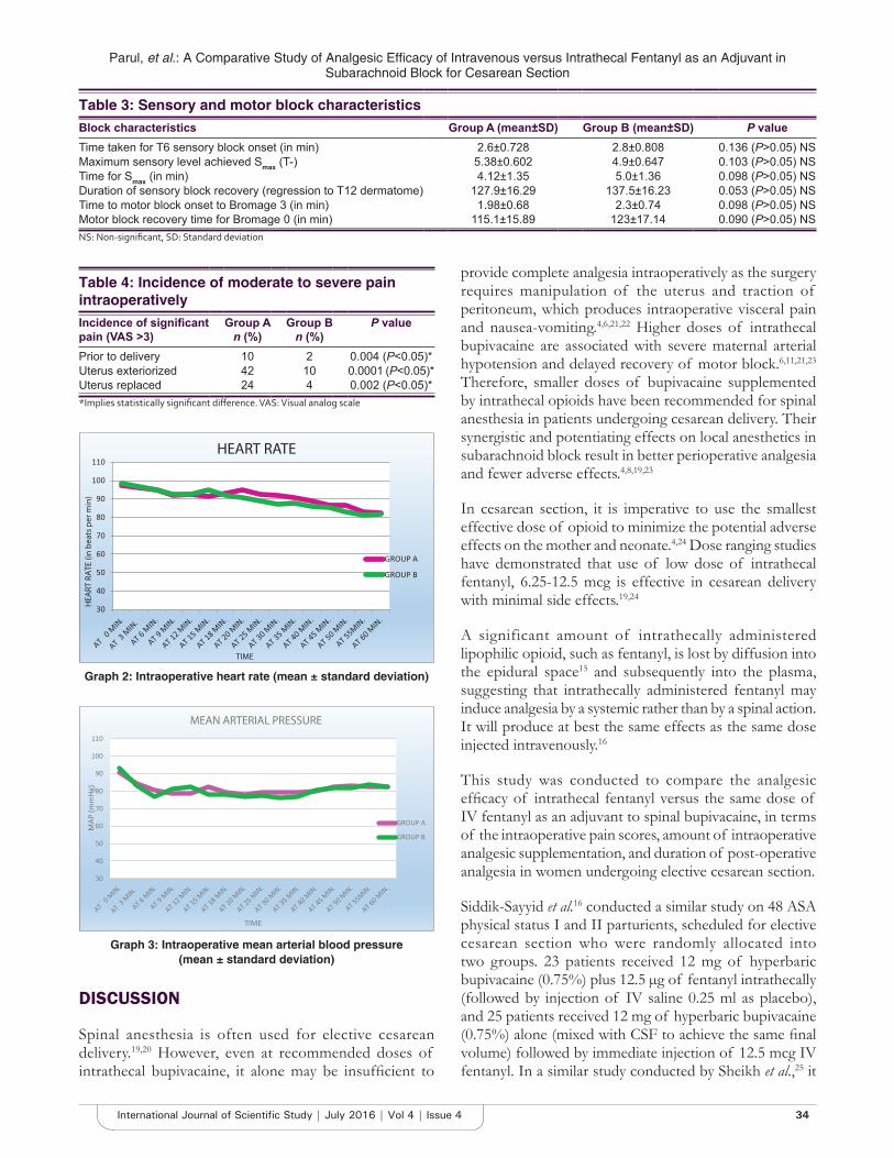
Parul, et al.: A Comparative Study of Analgesic Efficacy of Intravenous versus Intrathecal Fentanyl as an Adjuvant in Subarachnoid Block for Cesarean Section
34International Journal of Scientific Study | July 2016 | Vol 4 | Issue 4
DISCUSSION
Spinal anesthesia is often used for elective cesarean delivery.19,20 However, even at recommended doses of intrathecal bupivacaine, it alone may be insufficient to
provide complete analgesia intraoperatively as the surgery requires manipulation of the uterus and traction of peritoneum, which produces intraoperative visceral pain and nausea-vomiting.4,6,21,22 Higher doses of intrathecal bupivacaine are associated with severe maternal arterial hypotension and delayed recovery of motor block.6,11,21,23 Therefore, smaller doses of bupivacaine supplemented by intrathecal opioids have been recommended for spinal anesthesia in patients undergoing cesarean delivery. Their synergistic and potentiating effects on local anesthetics in subarachnoid block result in better perioperative analgesia and fewer adverse effects.4,8,19,23
In cesarean section, it is imperative to use the smallest effective dose of opioid to minimize the potential adverse effects on the mother and neonate.4,24 Dose ranging studies have demonstrated that use of low dose of intrathecal fentanyl, 6.25-12.5 mcg is effective in cesarean delivery with minimal side effects.19,24
A significant amount of intrathecally administered lipophilic opioid, such as fentanyl, is lost by diffusion into the epidural space15 and subsequently into the plasma, suggesting that intrathecally administered fentanyl may induce analgesia by a systemic rather than by a spinal action. It will produce at best the same effects as the same dose injected intravenously.16
This study was conducted to compare the analgesic efficacy of intrathecal fentanyl versus the same dose of IV fentanyl as an adjuvant to spinal bupivacaine, in terms of the intraoperative pain scores, amount of intraoperative analgesic supplementation, and duration of post-operative analgesia in women undergoing elective cesarean section.
Siddik-Sayyid et al.16 conducted a similar study on 48 ASA physical status I and II parturients, scheduled for elective cesarean section who were randomly allocated into two groups. 23 patients received 12 mg of hyperbaric bupivacaine (0.75%) plus 12.5 µg of fentanyl intrathecally (followed by injection of IV saline 0.25 ml as placebo), and 25 patients received 12 mg of hyperbaric bupivacaine (0.75%) alone (mixed with CSF to achieve the same final volume) followed by immediate injection of 12.5 mcg IV fentanyl. In a similar study conducted by Sheikh et al.,25 it
Table 3: Sensory and motor block characteristicsBlock characteristics Group A (mean±SD) Group B (mean±SD) P valueTime taken for T6 sensory block onset (in min) 2.6±0.728 2.8±0.808 0.136 (P>0.05) NSMaximum sensory level achieved Smax (T-) 5.38±0.602 4.9±0.647 0.103 (P>0.05) NSTime for Smax (in min) 4.12±1.35 5.0±1.36 0.098 (P>0.05) NSDuration of sensory block recovery (regression to T12 dermatome) 127.9±16.29 137.5±16.23 0.053 (P>0.05) NSTime to motor block onset to Bromage 3 (in min) 1.98±0.68 2.3±0.74 0.098 (P>0.05) NSMotor block recovery time for Bromage 0 (in min) 115.1±15.89 123±17.14 0.090 (P>0.05) NSNS: Non‑significant, SD: Standard deviation
Table 4: Incidence of moderate to severe pain intraoperativelyIncidence of significant pain (VAS >3)
Group An (%)
Group Bn (%)
P value
Prior to delivery 10 2 0.004 (P<0.05)*Uterus exteriorized 42 10 0.0001 (P<0.05)*Uterus replaced 24 4 0.002 (P<0.05)**Implies statistically significant difference. VAS: Visual analog scale
Graph 2: Intraoperative heart rate (mean ± standard deviation)
Graph 3: Intraoperative mean arterial blood pressure (mean ± standard deviation)

Parul, et al.: A Comparative Study of Analgesic Efficacy of Intravenous versus Intrathecal Fentanyl as an Adjuvant in Subarachnoid Block for Cesarean Section
35 International Journal of Scientific Study | July 2016 | Vol 4 | Issue 4
included patients with pregnancy-induced hypertension without proteinuria.
There were no significant differences between the intrathecal group and the IV fentanyl group with respect to age, height, weight, parity or gestational age, intrathecal injection to delivery time, and duration of surgery; which is in line with observations of Siddik-Sayyid et al.,16 Sheikh et al.,25 Biswas et al.,26 and others.4,19 Furthermore, the sensory and motor block characteristics showed no statistically significant difference between the groups. These findings are consistent with the findings of Siddik-Sayyid et al.,16 Biswas et al.,26 and others.4,19,25,27 It has been documented that intrathecal opioids potentiate the effects of local anesthetic without intensifying motor and sympathetic blockade.14
Similar to observations of Siddik-Sayyid et al.,16 the incidence of VAS > 3 (i.e. moderate-severe pain) before delivery was significantly lower in the IT fentanyl group as compared with IV fentanyl group. Furthermore, the incidence of significant pain (VAS > 3) during exteriorization of uterus and replacement of uterus into abdomen was found to be higher in the IV fentanyl group as compared with the intrathecal group in both the studies (Siddik-Sayyid et al);16 the difference was of statistical significance in our study (P < 0.05) but was not so in the study by Siddik-Sayyid et al.16 This may be explained by the fact that we used a lower dose and concentration (10 mg, 0.5%) of bupivacaine and a larger sample size as compared with Siddik-Sayyid et al.16 (12 mg, 0.75%) (n = 48). Our results corroborate with those of Hunt et al.20 and others,8,26-29 where administration of more than 10 mcg
of intrathecal fentanyl with 0.5% hyperbaric bupivacaine led to a marked decrease in the incidence of intraoperative pain or discomfort, indicating that intrathecal fentanyl effectively abolishes visceral pain. This high incidence of visceral pain in cesarean deliveries during manipulation of uterus and peritoneum under regional anesthesia has been demonstrated earlier.6,21 This can be attributed to the fact that similar to the tourniquet pain, visceral pain is believed to be conducted by unmyelinated C-fibers. Hence, once the dose of local anesthetic is reduced these fibers become unblocked, while the Aδ-fibers transmitting incisional pain are still blocked.6,30
There was a statistically significant difference in the need for intraoperative analgesic supplementation with IV fentanyl and also the mean fentanyl requirements between the groups in our study, in consonance with Siddik-Sayyid et al.,16 with greater requirement for supplementation in the IV group as compared with IT group. The decrease in the intraoperative analgesic requirement has been well documented in other studies as well.19,20,24,29 However, this is not in agreement with findings of Dahlgren et al.,4 as none of the patients in either group required intraoperative supplementary analgesics in their study. The varying local anesthetic concentrations, baricity, and doses may account for these differences in the requirement for intraoperative analgesic supplementation.
The duration of post-operative analgesia, i.e., the time to first request of post-operative analgesia was prolonged in the intrathecal fentanyl group as compared with the IV fentanyl group, and this difference was statistically significant, which is in agreement with Siddik-Sayyid et al.,16 and most of the studies using an intrathecal fentanyl dose of >10 mcg such as Biswas et al.26 and others.4,19,25,27,29 Even a moderate increase in the duration of post-operative analgesia must be considered beneficial, as it allows early mobilization of the mother and a sooner mother-baby interaction.
The hemodynamic parameters and the incidence of maternal side effects (such as nausea, vomiting, shivering, pruritus, and respiratory depression) showed no statistically significant difference between the groups, which in conformity with the observations of other workers.19,25-27
Table 5: Intraoperative fentanyl requirement and post-operative analgesiaNumber of patients n (%) Group A (%) Group B (%) P valuePatients requiring fentanyl
Once 28 (56) 7 (14) <0.001 (P<0.05)*Twice 11 (22) 2 (4) 0.0001 (P<0.05)*
Mean fentanyl requirement intraoperatively (micrograms) 19.5±3.56 4.5±1.87 0.001 (P<0.05)*Time to first rescue post-operative analgesia (in min) 115.4±16.28 133.9±20.68 0.003 (P<0.05)**Implies statistically significant difference
Table 6: Side effects and neonatal Apgar scoresNumber of patients n (%)
Group A (%) Group B (%) P value
Nausea 5 (10) 1 (2) 0.892 (P>0.05)Vomiting 2 (4) 0 (0) 0.112 (P>0.05)Pruritus 1 (2) 0 (0) 0.120 (P>0.05)Shivering 1 (2) 0 (0) 0.211 (P>0.05)Apgar score AT(median value)
1 min 8 (8-9) 8 (8-9) 0.125 (P>0.05)5 min 9 (9-10) 9 (9-10) 0.163 (P>0.05)

Parul, et al.: A Comparative Study of Analgesic Efficacy of Intravenous versus Intrathecal Fentanyl as an Adjuvant in Subarachnoid Block for Cesarean Section
36International Journal of Scientific Study | July 2016 | Vol 4 | Issue 4
However, a statistically significant lower incidence of intraoperative nausea and vomiting in the IT fentanyl group was seen by Siddik-Sayyid et al.,16 which was probably due to hypotension and was cured after administration of ephedrine (which corrected the hypotension). The Apgar scores of all neonates were ≥8 at 1 min and ≥9 at 5 min in both the studies.16 Studies by Frolich et al.31 and Eisele et al.32 found similar results for IV administration of fentanyl in doses up to 1 mcg/kg before delivery in cesarean section.
These results suggest that intraoperative analgesic supplementation of bupivacaine spinal anesthesia with intrathecal fentanyl results in a better quality of spinal anesthesia than IV fentanyl, as evidenced by the lower intraoperative VAS scores, as well as lesser need for additional intraoperative analgesics. In addition, the time to the first post-operative analgesic requirement was longer in the intrathecal fentanyl group as compared with the IV fentanyl group.
CONCLUSION
From our study, it can be concluded that:• Intrathecal fentanyl analgesic supplementation
was more efficacious than intravenous fentanyl supplementation of subarachnoid block in cesarean delivery, as intrathecal fentanyl supplementation was associated with lower intraoperative VAS scores, lower intraoperative analgesic requirements, and a longer time to first post-operative rescue analgesia than with intravenous fentanyl supplementation with comparable maternal adverse effects and fetal outcomes
• In view of better analgesia and lesser side effects, it can be concluded that intrathecal fentanyl supplementation of spinal anesthesia with bupivacaine during cesarean delivery resulted in a better quality of spinal analgesia than the same dose of intravenous fentanyl supplementation.
REFERENCES
1. Miller RD, Eriksson LI, Fleisher LA, Wiener-Kronish JP, Young WL. Miller’s Anesthesia. 7th ed. Philadelphia, PA: Churchill Livingstone; 2010. p. 341-60.
2. National Institute of Health and Clinical Excellence. Clinical Guideline 13: Cesarean Section. London: NICE; 2004.
3. Samuel CH, Gershon E, Mark AR, Sol MS. Shnider and Levinson’s Anesthesia for Obstetrics. 4th ed. Philadelphia, PA: Lippincott Williams & Wilkins; 2002. p. 249.
4. Dahlgren G, Hultstrand C, Jakobsson J, Norman M, Eriksson EW, Martin H. Intrathecal sufentanil, fentanyl, or placebo added to bupivacaine for Cesarean section. Anesth Analg 1997;85:1288-93.
5. Vyas N, Sahu DK, Parampill R. Comparative study of intrathecal sufentanil bupivacaine versus intrathecal bupivacaine in patients undergoing elective cesarean section. J Anaesthesiol Clin Pharmacol 2010;26:488-92.
6. Pedersen H, Santos AC, Steinberg ES, Schapiro HM, Harmon TW, Finster M. Incidence of visceral pain during cesarean section: The effect of varying doses of spinal bupivacaine. Anesth Analg 1989;69:46-9.
7. Ben-David B, Miller G, Gavriel R, Gurevitch A. Low-dose bupivacaine-fentanyl spinal anesthesia for cesarean delivery. Reg Anesth Pain Med 2000;25:235-9.
8. Choi DH, Ahn HJ, Kim MH. Bupivacaine-sparing effect of fentanyl in spinal anesthesia for cesarean delivery. Reg Anesth Pain Med 2000;25:240-5.
9. Al-Ghanem SM, Massad IM, Al-Mustafa MM, Al-Zaben KR, Qudaisat IY, Qatawneh AM, et al. Effect of adding dexmedetomidine versus fentanyl to intrathecal bupivacaine on spinal block characteristics in gynecological procedures: A double blind controlled study. Am J Appl Sci 2009;6:882-7.
10. Ben-David B, Solomon E, Levin H, Admoni H, Goldik Z. Intrathecal fentanyl with small-dose dilute bupivacaine: Better anesthesia without prolonging recovery. Anesth Analg 1997;85:560-5.
11. Kothari N, Bogra J, Chaudhary AK. Evaluation of analgesic effects of intrathecal clonidine along with bupivacaine in cesarean section. Saudi J Anaesth 2011;5:31-5.
12. Braga AA, Frias JA, Braga FS, Potério GB, Hirata ES, Torres NA. Spinal anesthesia for cesarean section. Use of hyperbaric bupivacaine (10mg) combined with different adjuvants. Rev Bras Anestesiol 2012;62:775-87.
13. Braga Ade F, Frias JA, Braga FS, Pereira RI, Titotto SM. Spinal anesthesia for elective ceasarean section: Use of different doses of hyperbaric bupivacaine associated with morphine and clonidine. Acta Cir Bras 2013;28:26-32.
14. Abouleish E, Rawal N, Fallon K, Hernandez D. Combined intrathecal morphine and bupivacaine for cesarean section. Anesth Analg 1988;67:370-4.
15. Ummenhofer WC, Arends RH, Shen DD, Bernards CM. Comparative spinal distribution and clearance kinetics of intrathecally administered morphine, fentanyl, alfentanil, and sufentanil. Anesthesiology 2000;92:739-53.
16. Siddik-Sayyid SM, Aouad MT, Jalbout MI, Zalaket MI, Berzina CE, Baraka AS. Intrathecal versus intravenous fentanyl for supplementation of subarachnoid block during cesarean delivery. Anesth Analg 2002;95:209-13.
17. Bromage PR. A comparison of the hydrochloride and carbon dioxide salts of lidocaine and prilocaine in epidural analgesia. Acta Anaesthesiol Scand Suppl 1965;16:55-69.
18. McCormack HM, Horne DJ, Sheather S. Clinical applications of visual analogue scales: A critical review. Psychol Med 1988;18:1007-19.
19. Hunt CO, Naulty JS, Bader AM, Hauch MA, Vartikar JV, Datta S, et al. Perioperative analgesia with subarachnoid fentanyl-bupivacaine for cesarean delivery. Anesthesiology 1989;71:535-40.
20. Randalls B, Broadway JW, Browne DA, Morgan BM. Comparison of four subarachnoid solutions in a needle-through-needle technique for elective caesarean section. Br J Anaesth 1991;66:314-8.
21. Alahuhta S, Kangas-Saarela T, Hollmén AI, Edström HH. Visceral pain during caesarean section under spinal and epidural anaesthesia with bupivacaine. Acta Anaesthesiol Scand 1990;34:95-8.
22. Dutta D, Naskar C, Wahal R, Bhatia V K, Singh V. Intrathecal clonidine for perioperative pain relief in abdominal hysterectomy. Indian J Pain 2013;27:26-32.
23. Goel S, Bhardwaj N, Grover VK. Intrathecal fentanyl added to intrathecal bupivacaine for day case surgery: A randomized study. Eur J Anaesthesiol 2003;20:294-7.
24. Belzarena SD. Clinical effects of intrathecally administered fentanyl in patients undergoing cesarean section. Anesth Analg 1992;74:653-7.
25. Sheikh F, Ahmed M, Ommid M, Gurcoo S, Shakoor N, Nazir S, et al. Comparative evaluation of low dose hyperbaric bupivacaine with or without fentanyl in spinal anaesthesia for caesarean section in patients with pregnancy induced hypertension. Int J Anesthesiol 2012;30:1-7.
26. Biswas BN, Rudra A, Bose BK, Nath S, Chakrabarty S. Intrathecal fentanyl with hyperbaric bupivacaine improves analgesia during caesarean delivery and in early postoperative period. Indian J Anaesth 2002;46:469-72.
27. Bogra J, Arora N, Srivastava P. Synergistic effect of intrathecal fentanyl and bupivacaine in spinal anesthesia for cesarean section. BMC Anesthesiol 2005;5:5.
28. Chu CC, Shu SS, Lin SM, Chu NW, Leu YK, Tsai SK, et al. The effect of intrathecal bupivacaine with combined fentanyl in cesarean section. Acta

Parul, et al.: A Comparative Study of Analgesic Efficacy of Intravenous versus Intrathecal Fentanyl as an Adjuvant in Subarachnoid Block for Cesarean Section
37 International Journal of Scientific Study | July 2016 | Vol 4 | Issue 4
Anaesthesiol Sin 1995;33:149-54.29. Shende D, Cooper GM, Bowden MI. The influence of intrathecal fentanyl on
the characteristics of subarachnoid block for caesarean section. Anaesthesia 1998;53:706-10.
30. Concepcion MA, Lambert DH, Welch KA, Covino BG. Tourniquet pain during spinal anesthesia: A comparison of plain solutions of tetracaine and
bupivacaine. Anesth Analg 1988;67:828-32.31. Frölich MA, Burchfield DJ, Euliano TY, Caton D. A single dose of fentanyl
and midazolam prior to cesarean section have no adverse neonatal effects. Can J Anaesth 2006;53:79-85.
32. Eisele JH, Wright R, Rogge P. New born and maternal fentanyl levels at Cesarean section. Anesth Analg 1982;61:179-80.
How to cite this article: Parul G, Singh NH, Neeharika A. Comparative Study of Analgesic Efficacy of Intravenous versus Intrathecal Fentanyl as an Adjuvant in Subarachnoid Block for Cesarean Section. Int J Sci Stud 2016;4(4):31-37.
Source of Support: Nil, Conflict of Interest: None declared.

38International Journal of Scientific Study | July 2016 | Vol 4 | Issue 4
Diagnostic Accuracy of Pre-operative Staging of Colorectal Carcinoma in Comparison to Post-operative Pathological StagingAnto J Richie1, P Mellonie2, H B Suresh3
1Assistant Professor, Department of Radiodiagnosis, Father Muller Medical College, Mangalore, Karnataka, India, 2Assistant Professor, Department of Pathology, Father Muller Medical College, Mangalore, Karnataka, India, 3Professor and Head, Department of Radiodiagnosis, Father Muller Medical College, Mangalore, Karnataka, India
computed tomography (CT) has become an increasingly routine part of the pre-operative staging of colon cancer. The treatment and prognosis of patients with colorectal carcinoma is dependent on the stage of disease at the time of diagnosis.
AimThe study aim at evaluating the accuracy of CT in the tumor staging of malignant lesions. The pre-operative CT findings are correlated with post-operative histopathological findings considering histopathological staging as the gold standard.
MATERIALS AND METHODS
This was a hospital-based retrospective correlative study conducted in Father Muller Medical College and hospital,
INTRODUCTION
Colorectal cancer is the third most common cancer in men and the second in women worldwide. Population-based time trend studies show a rising trend in the incidence of colorectal carcinoma in India.1 Pre-operative staging of colon cancers is important to plan the best possible treatment options for the patients, which may include for example, neoadjuvant drug therapy to allow for pre-operative downsizing of the primary tumor.2 Consequently,
Original Article
AbstractBackground: Pre-operative staging of primary tumor is critical for planning treatment and it is directly related to prognosis of the patient. Hence, assessing the accuracy of this pre-operative staging is essential.
Aim: To evaluate the accuracy of computed tomography (CT) in pre-operative tumor staging of colorectal malignancies by correlating with post-operative histopathological staging.
Materials and Methods: This is a retrospective correlative study. The medical records of 45 consecutive patients who underwent resection for a colonic carcinoma at Father Muller Medical College, Mangalore, over a period of 13-month from January 2015 to February 2016 were retrospectively reviewed. Pre-operative CT staging was correlated with post-operative histopathological staging.
Results: In the CT staging of malignant lesions, 9 of the 11 cases were correctly staged as T1 and T2 lesions. 16 of the 18 cases were correctly staged as T3 lesions, and all the 7 cases were correctly staged as T4 lesions.
Conclusion: CT proved to be an excellent modality for the staging of colorectal carcinomas which helps in the proper planning of surgery and further management of the patient.
Key words: Colon cancer, Computed tomography staging, Histopathology staging, Pre-operative computed tomography staging, Tumor node metastasis staging
Access this article online
www.ijss-sn.com
Month of Submission : 05-2016 Month of Peer Review : 06-2016 Month of Acceptance : 07-2016 Month of Publishing : 07-2016
Corresponding Author: Dr. Anto J Richie, Flat No. 304, Fredrose Enclave (Near Valencia Church), Valencia, Mangalore - 575 002, Karnataka, India. Phone: +91-9739681469. E-mail: [email protected]
Print ISSN: 2321-6379Online ISSN: 2321-595X
DOI: 10.17354/ijss/2016/370
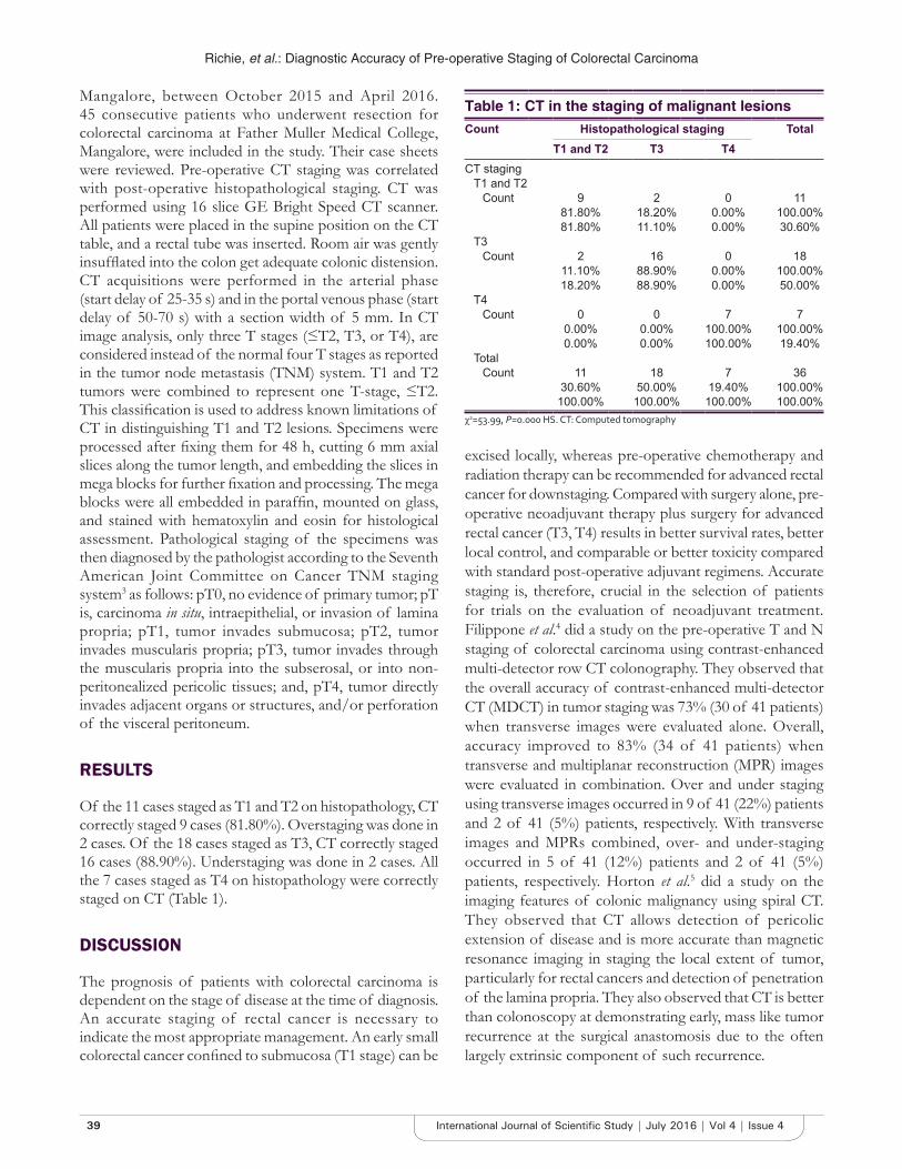
Richie, et al.: Diagnostic Accuracy of Pre-operative Staging of Colorectal Carcinoma
39 International Journal of Scientific Study | July 2016 | Vol 4 | Issue 4
Mangalore, between October 2015 and April 2016. 45 consecutive patients who underwent resection for colorectal carcinoma at Father Muller Medical College, Mangalore, were included in the study. Their case sheets were reviewed. Pre-operative CT staging was correlated with post-operative histopathological staging. CT was performed using 16 slice GE Bright Speed CT scanner. All patients were placed in the supine position on the CT table, and a rectal tube was inserted. Room air was gently insufflated into the colon get adequate colonic distension. CT acquisitions were performed in the arterial phase (start delay of 25-35 s) and in the portal venous phase (start delay of 50-70 s) with a section width of 5 mm. In CT image analysis, only three T stages (≤T2, T3, or T4), are considered instead of the normal four T stages as reported in the tumor node metastasis (TNM) system. T1 and T2 tumors were combined to represent one T-stage, ≤T2. This classification is used to address known limitations of CT in distinguishing T1 and T2 lesions. Specimens were processed after fixing them for 48 h, cutting 6 mm axial slices along the tumor length, and embedding the slices in mega blocks for further fixation and processing. The mega blocks were all embedded in paraffin, mounted on glass, and stained with hematoxylin and eosin for histological assessment. Pathological staging of the specimens was then diagnosed by the pathologist according to the Seventh American Joint Committee on Cancer TNM staging system3 as follows: pT0, no evidence of primary tumor; pT is, carcinoma in situ, intraepithelial, or invasion of lamina propria; pT1, tumor invades submucosa; pT2, tumor invades muscularis propria; pT3, tumor invades through the muscularis propria into the subserosal, or into non-peritonealized pericolic tissues; and, pT4, tumor directly invades adjacent organs or structures, and/or perforation of the visceral peritoneum.
RESULTS
Of the 11 cases staged as T1 and T2 on histopathology, CT correctly staged 9 cases (81.80%). Overstaging was done in 2 cases. Of the 18 cases staged as T3, CT correctly staged 16 cases (88.90%). Understaging was done in 2 cases. All the 7 cases staged as T4 on histopathology were correctly staged on CT (Table 1).
DISCUSSION
The prognosis of patients with colorectal carcinoma is dependent on the stage of disease at the time of diagnosis. An accurate staging of rectal cancer is necessary to indicate the most appropriate management. An early small colorectal cancer confined to submucosa (T1 stage) can be
excised locally, whereas pre-operative chemotherapy and radiation therapy can be recommended for advanced rectal cancer for downstaging. Compared with surgery alone, pre-operative neoadjuvant therapy plus surgery for advanced rectal cancer (T3, T4) results in better survival rates, better local control, and comparable or better toxicity compared with standard post-operative adjuvant regimens. Accurate staging is, therefore, crucial in the selection of patients for trials on the evaluation of neoadjuvant treatment. Filippone et al.4 did a study on the pre-operative T and N staging of colorectal carcinoma using contrast-enhanced multi-detector row CT colonography. They observed that the overall accuracy of contrast-enhanced multi-detector CT (MDCT) in tumor staging was 73% (30 of 41 patients) when transverse images were evaluated alone. Overall, accuracy improved to 83% (34 of 41 patients) when transverse and multiplanar reconstruction (MPR) images were evaluated in combination. Over and under staging using transverse images occurred in 9 of 41 (22%) patients and 2 of 41 (5%) patients, respectively. With transverse images and MPRs combined, over- and under-staging occurred in 5 of 41 (12%) patients and 2 of 41 (5%) patients, respectively. Horton et al.5 did a study on the imaging features of colonic malignancy using spiral CT. They observed that CT allows detection of pericolic extension of disease and is more accurate than magnetic resonance imaging in staging the local extent of tumor, particularly for rectal cancers and detection of penetration of the lamina propria. They also observed that CT is better than colonoscopy at demonstrating early, mass like tumor recurrence at the surgical anastomosis due to the often largely extrinsic component of such recurrence.
Table 1: CT in the staging of malignant lesionsCount Histopathological staging Total
T1 and T2 T3 T4CT staging
T1 and T2Count 9 2 0 11
81.80% 18.20% 0.00% 100.00%81.80% 11.10% 0.00% 30.60%
T3Count 2 16 0 18
11.10% 88.90% 0.00% 100.00%18.20% 88.90% 0.00% 50.00%
T4Count 0 0 7 7
0.00% 0.00% 100.00% 100.00%0.00% 0.00% 100.00% 19.40%
TotalCount 11 18 7 36
30.60% 50.00% 19.40% 100.00%100.00% 100.00% 100.00% 100.00%
χ2=53.99, P=0.000 HS. CT: Computed tomography

Richie, et al.: Diagnostic Accuracy of Pre-operative Staging of Colorectal Carcinoma
40International Journal of Scientific Study | July 2016 | Vol 4 | Issue 4
Freeny et al.6 evaluated the pre-operative staging and detection of post-operative recurrence in colonic cancers. Compared with the Duke’s classification, CT correctly staged only 47.5% of patients: 16.6% were upstaged, and 83.3% were downstaged.
Balthazar et al.7 did a pre-operative evaluation of 90 proved cases of colon carcinoma to know the detection rate and role of CT in the pre-operative evaluation. In this study, the overall detection rate was 84%; however, the rate varied from 68% in unprepared colons to 95% in clean colons that were adequately distended with air. Their study shows that CT had a sensitivity of 55% for local invasion, 73% for regional nodes, and 79% for liver metastases.
Ng et al.8 assessed the pathological significance of abnormal pericolic fat shown by CT in the context of colorectal carcinoma. According to their study abnormal pericolic fat had a sensitivity of 79%, specificity of 33%, positive predictive value of 91% in identifying extension of tumor infiltration beyond the muscle coat.
Harvey et al.9 evaluated the role of spiral CT pneumocolon for assessing colonic neoplasms. According to their study, CT pneumocolon clearly showed the primary tumor in all cases as an enhancing soft tissue mass, and was able to detect local extension and lymphadenopathy as well as assess the liver, peritoneum and remaining abdomen. CT depicted the morphology of the primary tumor more clearly than barium enema, and in one case also detected a 1 cm polyp which was not seen on the barium study because the patient was incontinent of barium and views were limited. There was a good correlation between the CT and pathological findings.
Of the 11 cases staged as T1 and T2 on histopathology, CT correctly staged 9 cases (81.80%) (Figure 1). Over staging was done in 2 cases. Filippone et al.6 in their study on the staging of colorectal carcinoma using MDCT were able to stage 93% of the lesions as T1 and T2 correctly. In our study, 16 cases (88.90%) were correctly staged as T3 (Figure 2). Under staging was done in 2 cases. Filippone et al. were able to correctly stage 90% of the cases as T3 lesions. All cases with T4 lesions were correctly staged in our study (Figure 3). Filippone et al. were able to correctly stage 98% of the lesions as T4.
CONCLUSION
CT is proved to have good diagnostic accuracy in pre-operative staging of malignant lesions which helps in the proper planning of surgery and further management of the patient.
Figure 1: Axial contrast-enhanced computed tomography showing wall thickening of the descending colon (arrows) confined to the
wall. No evidence of pericolic fat stranding - T2 tumor staging
Figure 2: Axial contrast-enhanced computed tomography showing wall thickening of the ascending colon (arrows) with nodular margin and peri colonic fat stranding suggestive of
malignancy - T3 tumor staging
Figure 3: Axial contrast-enhanced computed tomography showing heterogeneously enhancing asymetric wall thickening involving the descending colon with infilyration of the spleen
suggestive of T4 stage

Richie, et al.: Diagnostic Accuracy of Pre-operative Staging of Colorectal Carcinoma
41 International Journal of Scientific Study | July 2016 | Vol 4 | Issue 4
REFERENCES
1. Mohandas KM. Colorectal cancer in India: Controversies, enigmas and primary prevention. Indian J Gastroenterol 2011;30:3-6.
2. Wainberg Z, Hecht JR. A phase III randomized, open-label, controlled trial of chemotherapy and bevacizumab with or without panitumumab in the first-line treatment of patients with metastatic colorectal cancer. Clin Colorectal Cancer 2006;5:363-7.
3. Edge SB, Compton CC. The American Joint Committee on Cancer: The 7th edition of the AJCC cancer staging manual and the future of TNM. Ann Surg Oncol 2010;17:1471-4.
4. Filippone A, Ambrosini R, Fuschi M, Marinelli T, Genovesi D, Bonomo L. Preoperative T and N staging of colorectal cancer: Accuracy of contrast-enhanced multi-detector row CT colonography - Initial experience. Radiology 2004;231:83-90.
5. Horton KM, Abrams RA, Fishman EK. Spiral CT of colon cancer: Imaging features and role in management. Radiographics 2000;20:419-30.
6. Freeny PC, Marks WM, Ryan JA, Bolen JW. Colorectal carcinoma evaluation with CT: Preoperative staging and detection of postoperative recurrence. Radiology 1986;158:347-53.
7. Balthazar EJ, Megibow AJ, Hulnick D, Naidich DP. Carcinoma of the colon: Detection and preoperative staging by CT. AJR Am J Roentgenol 1988;150:301-6.
8. Ng CS, Doyle TC, Dixon AK, Miller R, Arends MJ. Histopathological correlates of abnormal pericolic fat on CT in the assessment of colorectal carcinoma. Br J Radiol 2002;75:31-7.
9. Harvey CJ, Amin Z, Hare CM, Gillams AR, Novelli MR, Boulos PB, et al. Helical CT pneumocolon to assess colonic tumors: Radiologic-pathologic correlation. AJR Am J Roentgenol 1998;170:1439-43.
How to cite this article: Richie AJ, Mellonie P, Suresh HB. Diagnostic Accuracy of Pre-operative Staging of Colorectal Carcinoma in Comparison to Post-operative Pathological Staging. Int J Sci Stud 2016;4(4):38-41.
Source of Support: Nil, Conflict of Interest: None declared.
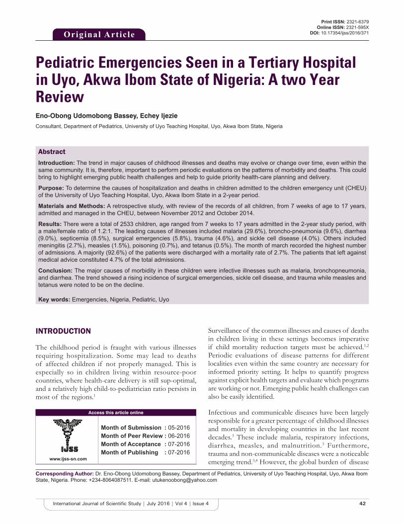
42International Journal of Scientific Study | July 2016 | Vol 4 | Issue 4
Pediatric Emergencies Seen in a Tertiary Hospital in Uyo, Akwa Ibom State of Nigeria: A two Year ReviewEno-Obong Udomobong Bassey, Echey Ijezie
Consultant, Department of Pediatrics, University of Uyo Teaching Hospital, Uyo, Akwa Ibom State, Nigeria
Surveillance of the common illnesses and causes of deaths in children living in these settings becomes imperative if child mortality reduction targets must be achieved.1,2 Periodic evaluations of disease patterns for different localities even within the same country are necessary for informed priority setting. It helps to quantify progress against explicit health targets and evaluate which programs are working or not. Emerging public health challenges can also be easily identified.
Infectious and communicable diseases have been largely responsible for a greater percentage of childhood illnesses and mortality in developing countries in the last recent decades.3 These include malaria, respiratory infections, diarrhea, measles, and malnutrition.3 Furthermore, trauma and non-communicable diseases were a noticeable emerging trend.3,4 However, the global burden of disease
INTRODUCTION
The childhood period is fraught with various illnesses requiring hospitalization. Some may lead to deaths of affected children if not properly managed. This is especially so in children living within resource-poor countries, where health-care delivery is still sup-optimal, and a relatively high child-to-pediatrician ratio persists in most of the regions.1
Original Article
AbstractIntroduction: The trend in major causes of childhood illnesses and deaths may evolve or change over time, even within the same community. It is, therefore, important to perform periodic evaluations on the patterns of morbidity and deaths. This could bring to highlight emerging public health challenges and help to guide priority health-care planning and delivery.
Purpose: To determine the causes of hospitalization and deaths in children admitted to the children emergency unit (CHEU) of the University of Uyo Teaching Hospital, Uyo, Akwa Ibom State in a 2-year period.
Materials and Methods: A retrospective study, with review of the records of all children, from 7 weeks of age to 17 years, admitted and managed in the CHEU, between November 2012 and October 2014.
Results: There were a total of 2533 children, age ranged from 7 weeks to 17 years admitted in the 2-year study period, with a male/female ratio of 1.2:1. The leading causes of illnesses included malaria (29.6%), broncho-pneumonia (9.6%), diarrhea (9.0%), septicemia (8.5%), surgical emergencies (5.8%), trauma (4.6%), and sickle cell disease (4.0%). Others included meningitis (2.7%), measles (1.5%), poisoning (0.7%), and tetanus (0.5%). The month of march recorded the highest number of admissions. A majority (92.6%) of the patients were discharged with a mortality rate of 2.7%. The patients that left against medical advice constituted 4.7% of the total admissions.
Conclusion: The major causes of morbidity in these children were infective illnesses such as malaria, bronchopneumonia, and diarrhea. The trend showed a rising incidence of surgical emergencies, sickle cell disease, and trauma while measles and tetanus were noted to be on the decline.
Key words: Emergencies, Nigeria, Pediatric, Uyo
Access this article online
www.ijss-sn.com
Month of Submission : 05-2016 Month of Peer Review : 06-2016 Month of Acceptance : 07-2016 Month of Publishing : 07-2016
Corresponding Author: Dr. Eno-Obong Udomobong Bassey, Department of Pediatrics, University of Uyo Teaching Hospital, Uyo, Akwa Ibom State, Nigeria. Phone: +234-8064087511. E-mail: [email protected]
Print ISSN: 2321-6379Online ISSN: 2321-595X
DOI: 10.17354/ijss/2016/371

Bassey and Ijezie: Paediatric Emergencies in a Tertiary Hospital in Uyo
43 International Journal of Scientific Study | July 2016 | Vol 4 | Issue 4
2010 reported substantial decreases in child mortality driven by reductions in diarrhea, lower respiratory tract infections, and more recently malaria.5 These health statistics, which are based on various studies and data generated from several countries, may be inadequate to serve every population. It, therefore, behaves that indigenous studies are undertaken, to reveal better, what diseases prevail within each community.
This study was aimed at reviewing the pattern of pediatric emergencies seen in children older than 6 weeks of age, managed in the children emergency unit (CHEU) of the University of Uyo Teaching Hospital, Uyo, Nigeria. Moreover, to compare the present result from an erstwhile study done in the same unit over 5 years ago,4 to assess if any changing trend. This would help re-prioritize interventions and health-care planning strategies.
MATERIALS AND METHODS
This study is retrospective and descriptive in design. It covered a period of 2-year, from November 2012 to October 2014. The records of all children aged 7 weeks to 17 years, as documented in the register were reviewed. The children from the 1st day of life to the age of 6 weeks are managed in the neonatal wards of this facility and were therefore excluded. Furthermore, any child with incomplete data entry was excluded. Information extracted from the records included age, gender, final diagnosis, duration, and outcome of the treatment.
The University of Uyo Teaching Hospital is one of the tertiary health-care facilities in Akwa Ibom State, located in the outskirts of Uyo about 6 km from the center of the city. The hospital is a 355 bed health-care facility and serves as a referral center, also accepting self-reported cases. The Department of Pediatrics provides in-patient and out-patient services for all children.
The CHEU has a 22 bed capacity with an average admission rate of 120 children/month. It is manned by a full complement of staff that includes consultants, resident doctors, interns, and nursing staff with 24 h shift duty coverage on all categories of staff. It undertakes basic resuscitation, treatment, and management of all children presenting there. A side room laboratory, diarrhea treatment unit, and pharmacy are also a functional part of the unit.
The definitive diagnosis documented was as made by the unit consultants. This was based on the presenting clinical features, with or without results of laboratory tests. The diagnosis of malaria, for instance, was supported
by the presence of malaria parasites in the blood film. Anemia was diagnosed on clinical grounds, supported by a hemoglobin estimation and sickle cell disease by hemoglobin electrophoresis. Measles was based on clinical features, and septicemia/meningitis was diagnosed based on clinical features, with or without a positive blood culture or abnormal cerebrospinal fluid analysis. The patients with pneumonia were diagnosed either clinically or with chest radiographs or both. HIV/AIDS were diagnosed based on positive ELISA test on a patient with features of the World Health Organization clinical case definition, confirmed by the western blot test. The good clinical response to certain medications by some patients was used to assign the final diagnosis in some cases.
The outcome was classified as discharged, left against medical advice (LAMA), and death.
The data obtained was analyzed using the bar chart, frequency tabulations, and cross tabulations of the Microsoft Excel.
RESULTS
A total of 2533 children were managed in the 2-year period. The total number of males being 1470 (54.7%) and females 1216 (45.3%), giving a male:female ratio of 1.2:1. The leading causes of illnesses included malaria 29.6%, broncho-pneumonia 9.6%, diarrhea 9.0%, septicemia 8.5%, surgical emergencies 5.8%, trauma 4.6%, and sickle cell disease 4.0%. Others included meningitis 2.7%, measles 1.5%, poisoning 0.7%, and tetanus (0.5%). This is represented in Table 1.
The number of admissions per month is represented in Figure 1, and this shows that the months of March (310), February (274), and January (260) recorded the highest number of admissions.
Table 2 shows that children <5 years constituted over half of the total admissions in this study (51.0%), and also had the highest burden of infective illnesses, especially bronchopneumonia (87.2%), diarrheal diseases (79.9%) and malaria (27.3%).
The outcome of admissions showed a greater percentage (92.6) being discharged in satisfactory condition, with a low mortality percentage (2.7) as seen in Table 3.
DISCUSSION
This study revealed infectious diseases, especially malaria as the highest cause for hospital admissions in the CHEU.

Bassey and Ijezie: Paediatric Emergencies in a Tertiary Hospital in Uyo
44International Journal of Scientific Study | July 2016 | Vol 4 | Issue 4
This is similar to observations from other tertiary hospitals across the eastern and western parts of Nigeria.5-8 It is also comparable to an erstwhile study in the same center and unit that was documented over 5 years ago.4 The difference between both studies in the same center lies in
the reduced percentage of malarial incidence in the past 2 years of study (29.6%), compared to the previous study, which was 56.6%. The reduction in the total incidence of malarial admissions may be related to the millions of long-lasting insecticide treated nets which have been distributed as part of the global malaria control strategy.9 In addition, over-the-counter usage of artemisinin-based combination drugs, which are widely available and user-friendly must be sustained to reduce malarial associated illnesses and deaths in children, especially under-fives.
Pneumonia constituted 9.6% of total admissions in this study which is higher than observations from some other centers in Nigeria.6,10-12 With the commencement of the pentavalent vaccine which gives immunity against some pneumococcal strains, this trend should be monitored and evaluated to assess the success of its recent inclusion in the schedule of the National Programme for Immunization. A reduction in the incidences of measles and tetanus may be as a result of improved vaccine coverage and uptake in the state.
Strikingly, childhood poisoning has remained at the rate of 0.7% of total admissions, as was recorded in the previous study in this center.4 There is still need for sustained community enlightenment campaigns on the prevention of accidental childhood poisoning.
The month of March recorded the highest number of admissions. The rainy season usually starts about this month in Uyo, Akwa Ibom State of Nigeria. This is the period during which an increase in the incidence of malaria, pneumonia, diarrheal disease, and water-borne diseases is observed.13,14
The children aged <5 years constituted over half of total admissions during the period under review in this
Table 1: Pattern of admission over the study periodDisease Number of patients (%)Malaria 750 (29.6)Bronchopneumonia 242 (9.6)Diarrhea 229 (9.0)Septicemia 216 (8.5)Surgical emergencies 147 (5.8)Trauma 117 (4.6)Sickle cell disease 102 (4.0)Meningitis 65 (2.7)Measles 39 (1.5)Poisoining 17 (0.7)Tetanus 13 (0.5)Others 596 (23.5)Total 2533 (100.0)
Table 2: Age distribution of the patientsDiagnosis N (%)
<5 years 5-10 years >10 years TotalMalaria 295 (39.3) 250 (33.3) 205 (27.3) 750 (100.0)Bronchopneumonia 211 (87.2) 31 (12.8) 0 (0.0) 242 (100.0)Diarrhea 183 (79.9) 31 (13.5) 15 (6.6) 229 (100.0)Septicemia 115 (53.2) 76 (35.2) 25 (11.6) 216 (100.0)Surgical emergencies
68 (46.3) 49 (33.3) 30 (20.4) 147 (100.0)
Trauma 70 (59.8) 31 (26.5) 16 (13.7) 117 (100.0)Sickle cell disease 36 (35.3) 51 (50.0) 15 (14.7) 102 (100.0)Meningitis 29 (44.6) 27 (41.5) 9 (13.9) 65 (100.0)Measles 37 (94.9) 2 (5.1) 0 (0.0) 39 (100.0)Poisoning 12 (70.6) 4 (23.5) 1 (5.9) 17 (100.0)Tetanus 1 (7.7) 11 (84.6) 1 (7.7) 13 (100.0)Others 236 (39.6) 202 (33.9) 158 (26.5) 596 (100.0)Total 1293 (51.0) 765 (30.2) 475 (18.8) 2533 (100.0)
Table 3: Outcome of admissionsDiagnosis N
Discharged LAMA Died TotalMalaria 720 24 6 750Bronchopneumonia 220 15 7 242Diarrhea 217 8 4 229Septicemia 183 19 14 21Surgical emergencies 134 7 6 147Trauma 101 9 7 117Sickle cell disease 97 5 0 102Meningitis 53 7 5 65Measles 37 0 2 39Poisoning 11 3 3 17Tetanus 10 1 2 13Others 520 22 16 596Total (%) 2346 (92.6) 119 (4.7) 68 (2.7) 2533 (100)LAMA: Left against medical advice
Figure 1: Number of admissions per month. Health workers industrial strike involving nurses and other paramedics
occurred during the months of July and August

Bassey and Ijezie: Paediatric Emergencies in a Tertiary Hospital in Uyo
45 International Journal of Scientific Study | July 2016 | Vol 4 | Issue 4
study. This pattern is similar to the findings from other centers.10,15,16
A majority of the patients were discharged with a mortality rate of 2.7% observed in this study. This is lower than the 5.1% reported by Toma et al. in Jos Nigeria,16 and the rate of 10.0% recorded by Anyanwu et al. in Abakaliki, Nigeria.17 The common causes of death in this study included septicemia, bronchopneumonia, and malaria. This agrees with reports that pneumonia, diarrhea, and malaria rank high among the causes of pediatric deaths, particularly in children aged 1-59 months.17,18
The rate of “LAMA” observed in this study (4.7%) is higher than the rates of 2.0% observed by Ndukwu and Onah19 2.1% by Toma et al.,16 and 3.8% observed by Anyanwu et al.,17 respectively. Some of the common reasons for LAMA include lack of acceptance of the treatment modalities20 and financial constraints.21
CONCLUSION
This study revealed that infectious diseases, particularly malaria and pneumonia, are common causes of morbidity and mortality in children presenting in our center. The outcome of the admissions in the CHEU of this facility is observed to be good, with the vast majority of children being discharged, and relatively low rates of mortality and LAMA. However, there is still need for continuous improvement and sustenance of the quality of health services provided, as well as community enlightenment on the common childhood diseases.
REFERENCES
1. Ekure EN, Esezobor CI, Balogun MR, Woo JG, Muktar-Yola M, Ojo OO, et al. Paediatrician workforce in Nigeria and impact on child health. Niger J Paed 2013;40:112-4.
2. Burden of disease unit. The global burden of diseases. Havard link title to 2000. Available from: http://www.hsps.havard.edu/organizations. [Last accessed on 2016 May 02].
3. Deen JL, Vos T, Huttly SR, Tulloch J. Injuries and noncommunicable diseases: Emerging health problems of children in developing countries. Bull World Health Organ 1999;77:518-24.
4. Enobong EI, Eno-Obong EU. Pattern of paediatric emergencies and outcome as seen in a tertiary hospital: A five year review. Sahel Med J 2009;12:73-7.
5. Naghavi M, Wang H, Lozano R, Davis A, Liang X, Zhou M, et al. Global, regional and national age-sex specificall cause and cause-specific mortality for 240 causes of death, 1990-2013: A systematic analysis for the global burden of disease study 2013. Lancet 2015;385:117-71.
6. Edelu BO, Eze BN, Oguonu T, Ndu IK. Morbidity and mortality pattern in the children emergency unit of the University of Nigeria Teaching Hospital, Enugu. Morbidity and mortality in children. Orient J Med 2014;26:73-5.
7. Elusiyan JB, Obiajunwa PO, Adejuyigbe EA, Olowu WA, Adeodu OO, Owa JA, et al. Pattern of morbidity and mortality among children hospitalized at the Obafemi Awolowo University Teaching Hospital, Ile-Ife. Niger J Paed 2009;36:22-8.
8. Ojukwu JN, Ogbu CN, Nnebe-Agumadu UH. Post neonatal medical admissions into the paediatric ward of Ebonyi State University Teaching Hospital, Abakaliki: The initial experience and outcome. Niger J Paed 2004;13:79-86.
9. Russell CL, Sallau A, Emukah E, Graves PM, Noland GS, Ngondi JM, et al. Determinants of bed net use in Southeast Nigeria following mass distribution of LLINs: Implications for social behavior change interventions. PLoS One 2015;10:e0139447.
10. Bilkisu GI, Aminu MS, Sunday OO, Bassey E, Smart A, Muyideen AB. Pattern of medical childhood morbidity and mortality in a new specialist hospital in Gusau, Nigeria. Ann Niger Med 2014;8:15-9.
11. Ugwu GIM. Pattern of morbidity and mortality in the newborn special care unit in a tertiary institution in the Niger Delta region of Nigeria: A two year prospective study. Glob Adv Res J Med Med Sci 2012;1:133-8.
12. Abhulimhen-Iyoha BI, Okolo AA. Morbidity and mortality of childhood illnesses at the emergency paediatric unit of the University of Benin Teaching Hospital, Benin City. Niger J Paed 2012;39:71-4.
13. Mouneke UV, Ibekwe RC, Eke CB, Ibekwe MU, Chinawa JM. Mortality among paediatric inpatients in Mile 4 Mission hospital Abakaliki, South-Eastern Nigeria: A retrospective study. Niger J Paed 2013;40:259-63.
14. Ijezie E, Okpokowuruk FS. Mortality audit in the paediatrics department of the University of Uyo teaching hospital, Uyo, Nigeria. Int J Res Med Sci 2016;4:615-20.
15. Chukwu BF, Chinawa JM, Ikefuna AN, Emodi IJ. Pattern and outcome of paediatric medical admissions at the University of Nigeria Teaching Hospital (UNTH), Ituku-Ozalla, Enugu: A five year retrospective review (2007-11). Niger J Paed 2013;40:354-3.
16. Toma BO, Gyang M, Abdu H, Shwe D, Ekere IA, Ihekaike M. Paediatric morbidity and mortality in a Suburban Hospital in Jos, North-Central Nigeria. Int J Trop Dis Health 2015;5:156-64.
17. Anyanwu OU, Ezeanosike OB, Ezeonu CT. Pattern and outcome of admissions at the children emergency room at the federal teaching hospital Abakaliki. Afr J Med Health Sci 2014;13:6-10.
18. Liu L, Oza S, Hogan D, Perin J, Rudan I, Lawn JE, et al. Global, regional, and national causes of child mortality in 2000-13, with projections to inform post-2015 priorities: An updated systematic analysis. Lancet 2015;385:430-40.
19. Ndukwu CI, Onah SK. Pattern and outcome of postneonatal pediatric emergencies in Nnamdi Azikiwe University Teaching Hospital, Nnewi, South East Nigeria. Niger J Clin Pract 2015;18:348-53.
20. Ndukwu CU, Ogbuagu CN, Ihegihu CC, Ugezu AI, Chukwuka CN. Discharge against medical advice amongst orthopaedic patients in Nnewi, South-East Nigeria, and its public health implications. Orient J Med 2014;26:9-15.
21. Onyiriuka AN. Pediatric discharge against medical advice: Experience from a Niger secondary healthcare institution. Med J Islam Repub Iran 2011;25:194-9.
How to cite this article: Bassey EU, Ijezie E. Pediatric Emergencies Seen in a Tertiary Hospital in Uyo, Akwa Ibom State of Nigeria: A two Year Review. Int J Sci Stud 2016;4(4):42-45.Source of Support: Nil, Conflict of Interest: None declared.

46International Journal of Scientific Study | July 2016 | Vol 4 | Issue 4
Evaluation of Visual Evoked Potential in Migraine IndividualsT N Vijayalakshmi1, Ratna Manjushree Jayaraman2, Bhanu3, Heber Anandan4
1Assistant Professor, Institute of Physiology and Experimental Medicine, Madras Medical College, Chennai, Tamil Nadu, India, 2Associate Professor, Department of Physiology, Tirunelveli Medical College, Tirunelveli, Tamil Nadu, India, 3Former Professor, Department of Neurology, Madras Medical College, Chennai, Tamil Nadu, India, 4Senior Clinical Scientist, Dr. Agarwal’s Healthcare Limited, Tirunelveli, Tamil Nadu, India
Headache Society (IHS), migraine is classified into headache with aura and headache without aura. Migraine is characterized by recurrent headache disorder manifesting in attacks lasting 4-72 h, with typical headache of unilateral location, pulsating quality, moderate or severe intensity, aggravation by routine physical activity, and association with nausea and/or photophobia and phonophobia. Visual disturbances are well-known clinical features of migraine. The migrainous aura is visual in 82% of cases, with symptoms such as flashes of light, stars, zig-zags, central or paracentral blind spot, and scotoma.1 The migraine patients are also more sensitive to environmental light stimuli.2 Apart from this, migraine in some can produce progressive brain damage. Pathophysiological studies in migraine patients and animal models of the migraine headache have identified the trigeminovascular system,3 brainstem,4,5 and the cerebral cortex6-8 as structures which may have primary causative roles. Modern neuroimaging studies9 have confirmed that migraine with aura symptoms are due to a cortical phenomenon similar to spreading depression. Various methods in electroneurophysiology are particularly
INTRODUCTION
Migraine is a common disabling primary headache disorder with a high prevalence, socio-economic, and personal impacts. The World Health Organization ranks migraine as one of the top 20 leading neurological causes of disability. The headache is a pervasive symptom and the most common problem that the neurologists encounter in their clinical practices. It is estimated that 12% of world’s population suffer from migraine and in India, of 1200 million populations, there are 150-200 million migraineurs under treatment. The gender prevalence of migraine is about 20% in females and 6% in males. According to the International
Original Article
AbstractBackground: Migraine is a multifactorial brain disorder characterized by recurrent disabling attacks of a headache. The migraine patients have visual disturbances during the attack as well as during the interictal period. In addition, even between attacks some aspects of cortical dysfunction are peculiar in migraineurs.
Aim: This study aims for knowing the pathogenesis of migraine in between attacks that lead to migraine disorder.
Materials and Methods: The study was conducted by evaluating the visual evoked potential of 30 migraine patients during the headache-free period and compared with 30 normal persons. The results were interpreted and statistically analyzed.
Results: There was statistically significant increase in amplitude of P100 wave of the migraine patients due to deficient habituation after a period of 15 min stimulation. In normal subjects, there was a decrease in amplitude of P100 wave due to the effect of habituation. The deficient habituation can be due to decreased serotonin levels leading to reduced pre-activation of the cortex.
Conclusion: The migraine patients has attributed to abnormal cortical processing in migraine with interictal hyperactivity leading to heightened responsiveness and lack of habituation and lack of intracortical inhibition.
Key words: Habituation, Migraine, Visual evoked potential
Access this article online
www.ijss-sn.com
Month of Submission : 05-2016 Month of Peer Review : 06-2016 Month of Acceptance : 07-2016 Month of Publishing : 07-2016
Corresponding Author: Heber Anandan, Dr. Agarwal’s Healthcare Limited, No.10, South By-pass Road, Vannarpettai, Tirunelveli - 627 003, Tamil Nadu, India. Mobile: +91-9894067910. E-mail: [email protected]
Print ISSN: 2321-6379Online ISSN: 2321-595X
DOI: 10.17354/ijss/2016/372
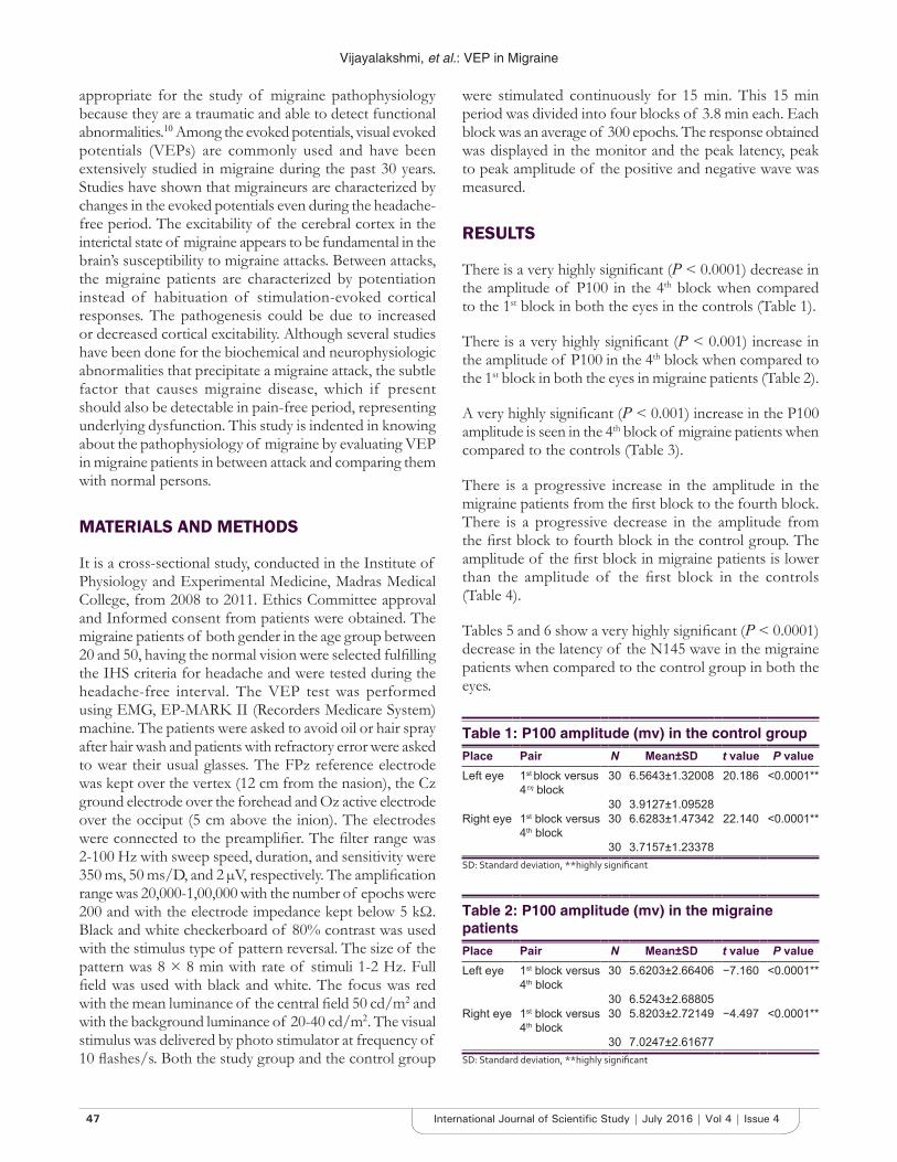
Vijayalakshmi, et al.: VEP in Migraine
47 International Journal of Scientific Study | July 2016 | Vol 4 | Issue 4
appropriate for the study of migraine pathophysiology because they are a traumatic and able to detect functional abnormalities.10 Among the evoked potentials, visual evoked potentials (VEPs) are commonly used and have been extensively studied in migraine during the past 30 years. Studies have shown that migraineurs are characterized by changes in the evoked potentials even during the headache-free period. The excitability of the cerebral cortex in the interictal state of migraine appears to be fundamental in the brain’s susceptibility to migraine attacks. Between attacks, the migraine patients are characterized by potentiation instead of habituation of stimulation-evoked cortical responses. The pathogenesis could be due to increased or decreased cortical excitability. Although several studies have been done for the biochemical and neurophysiologic abnormalities that precipitate a migraine attack, the subtle factor that causes migraine disease, which if present should also be detectable in pain-free period, representing underlying dysfunction. This study is indented in knowing about the pathophysiology of migraine by evaluating VEP in migraine patients in between attack and comparing them with normal persons.
MATERIALS AND METHODS
It is a cross-sectional study, conducted in the Institute of Physiology and Experimental Medicine, Madras Medical College, from 2008 to 2011. Ethics Committee approval and Informed consent from patients were obtained. The migraine patients of both gender in the age group between 20 and 50, having the normal vision were selected fulfilling the IHS criteria for headache and were tested during the headache-free interval. The VEP test was performed using EMG, EP-MARK II (Recorders Medicare System) machine. The patients were asked to avoid oil or hair spray after hair wash and patients with refractory error were asked to wear their usual glasses. The FPz reference electrode was kept over the vertex (12 cm from the nasion), the Cz ground electrode over the forehead and Oz active electrode over the occiput (5 cm above the inion). The electrodes were connected to the preamplifier. The filter range was 2-100 Hz with sweep speed, duration, and sensitivity were 350 ms, 50 ms/D, and 2 µV, respectively. The amplification range was 20,000-1,00,000 with the number of epochs were 200 and with the electrode impedance kept below 5 kΩ. Black and white checkerboard of 80% contrast was used with the stimulus type of pattern reversal. The size of the pattern was 8 × 8 min with rate of stimuli 1-2 Hz. Full field was used with black and white. The focus was red with the mean luminance of the central field 50 cd/m2 and with the background luminance of 20-40 cd/m2. The visual stimulus was delivered by photo stimulator at frequency of 10 flashes/s. Both the study group and the control group
were stimulated continuously for 15 min. This 15 min period was divided into four blocks of 3.8 min each. Each block was an average of 300 epochs. The response obtained was displayed in the monitor and the peak latency, peak to peak amplitude of the positive and negative wave was measured.
RESULTS
There is a very highly significant (P < 0.0001) decrease in the amplitude of P100 in the 4th block when compared to the 1st block in both the eyes in the controls (Table 1).
There is a very highly significant (P < 0.001) increase in the amplitude of P100 in the 4th block when compared to the 1st block in both the eyes in migraine patients (Table 2).
A very highly significant (P < 0.001) increase in the P100 amplitude is seen in the 4th block of migraine patients when compared to the controls (Table 3).
There is a progressive increase in the amplitude in the migraine patients from the first block to the fourth block. There is a progressive decrease in the amplitude from the first block to fourth block in the control group. The amplitude of the first block in migraine patients is lower than the amplitude of the first block in the controls (Table 4).
Tables 5 and 6 show a very highly significant (P < 0.0001) decrease in the latency of the N145 wave in the migraine patients when compared to the control group in both the eyes.
Table 1: P100 amplitude (mv) in the control groupPlace Pair N Mean±SD t value P valueLeft eye 1st block versus
4th block30 6.5643±1.32008 20.186 <0.0001**
30 3.9127±1.09528Right eye 1st block versus
4th block30 6.6283±1.47342 22.140 <0.0001**
30 3.7157±1.23378SD: Standard deviation, **highly significant
Table 2: P100 amplitude (mv) in the migraine patientsPlace Pair N Mean±SD t value P valueLeft eye 1st block versus
4th block30 5.6203±2.66406 −7.160 <0.0001**
30 6.5243±2.68805Right eye 1st block versus
4th block30 5.8203±2.72149 −4.497 <0.0001**
30 7.0247±2.61677SD: Standard deviation, **highly significant

Vijayalakshmi, et al.: VEP in Migraine
48International Journal of Scientific Study | July 2016 | Vol 4 | Issue 4
DISCUSSION
The present study deals with the changes in the VEPs in patients with migraine. The changes are compared with the controls. There was no statistically significant difference in the age, height, weight, and gender between study and the control group. In this study, both the controls and the migraine patients were stimulated continuously for 15 min. This 15 min period was divided into four blocks of 3.8 min each. The data were statistically analyzed, and their significance derived using independent samples t-test and paired t-test. VEP results were interpreted with respect to their latency and amplitude. Amplitude of the wave denotes the number of fibers recruited. Increase in the amplitude indicates more number of fibers is being recruited. Decrease in the amplitude indicates less number of fibers is being recruited. Latency denotes the time taken for the impulse to travel from the retina to the occipital striate area. In this study, when the P100 amplitude of the first block and the fourth block were compared, there was a very highly significant (P < 0.001) decrease in the amplitude of the fourth block (Table 1) in the controls which may be because of habituation, whereas there was a very highly significant (P < 0.001) increase in the amplitude of the fourth block of the migraine patients (Table 2) indicating potentiation. When the amplitude of the P100 of the fourth block of the migraine patient was compared with the amplitude of the fourth block of the controls, a very highly significant increase (P < 0.001) in the amplitude of the P100 was noted in the migraine patients (Table 3). When all the four blocks were compared, there was a progressive increase in the P100 amplitude from the first block to the fourth block in migraine patients indicating that more and more number of fibers were recruited (potentiation) during the continuous period of stimulation for 15 min. In the controls, there was a progressive decrease in the amplitude from the first block to the fourth block indicating that the fibers recruited were decreasing during continuous period of stimulation probably because of habituation (Table 4). This result was consistent with the study done by Gawel et al. (1983),11 Diener et al. (1989),12 and Khalil (1991),13 who have said that there was an increase in amplitude of P100 wave in migraine patients on pattern-reversal stimulus. Similarly, Afra et al. (1998),14 found that during repetitive pattern-reversal stimulation lasting 2 min, the amplitude of the P100 wave increased in migraineurs when tested interictally, whereas it decreased in healthy control subjects. Hartner and White (1970), and Peachy et al. (1994),15 said that check size (spatial frequency) influences the components of habituation behavior of pattern-reversal VEP, whereas Oelkers et al. (1999),16 notes that the peak to peak amplitudes were consistently higher in migraineurs
Table 3: Comparison of P100 amplitude (mv) in the 4th block between the migraine patients and controlsPlace Group N Mean±SD t value P value
Left4th block Migraine patients 30 6.524±2.6880 4.928 <0.0001**
Controls 30 3.913±1.0953Right
4th block Migraine patients 30 7.025±2.6168 6.265 <0.0001**Controls 30 3.716±1.2338
SD: Standard deviation, **highly significant
Table 4: Amplitude of P100 wave (mv) from 1st block to 4th blockPlace Mean values
Controls Migraine patients
Left eye1st block 6.56 5.622nd block 5.56 6.453rd block 4.75 6.574th block 3.91 6.52
Right eye1st block 6.63 5.822nd block 5.64 6.253rd block 4.71 6.584th block 3.72 7.02
Table 5: Comparison of VEP latency (ms) in the left eyeVariable Group N Mean±SD t value P valueN75 (ms) Controls 30 66.435±2.9452 −1.194 0.240
Migraine patients
30 68.048±6.7846
P100 (ms) Controls 30 95.5853±3.46872 1.518 0.136Migraine patients
30 93.6697±5.97927
N145 (ms) Controls 30 148.283±10.9468 3.925 <0.0001**Migraine patients
30 136.688±11.9122
SD: Standard deviation, VEP: Visual evoked potential, **highly significant
Table 6: Comparison of VEP latency (ms) in the right eyeVariable Group N Mean±SD t value P valueN75 (ms) Controls 30 66.251±3.0018 −0.903 0.371
Migraine patients
30 67.225±5.0854
P100 (ms) Controls 30 95.7847±4.42328 1.701 0.096Migraine patients
30 92.9780±7.87919
N145 (ms) Controls 30 149.017±10.6557 4.074 <0.0001**Migraine patients
30 134.440±16.4475
SD: Standard deviation, VEP: Visual evoked potential, **highly significant

Vijayalakshmi, et al.: VEP in Migraine
49 International Journal of Scientific Study | July 2016 | Vol 4 | Issue 4
at all spatial frequencies. Dienner et al. (1989),12 found that there was a decrease in P100 amplitude after treatment with Beta-blockers. Contrary to the above studies, Benna et al. (1985),17 Mariani et al. (1988),18 Drake et al. (1990),19 and Tagliati et al. (1995),20 have said that there was no difference in the VEP amplitude between the migraine patients and the normal subjects. However, there was no significant difference in the latency of N75 and P100 between the migraine patients and the controls. This result was similar to the study done by Benna et al. (1985),17 Mariani et al. (1988),18 Drake et al. (1990),19 and Tagliati et al. (1995).20 In another study done by Oelkers et al. (1999),16 migraineurs exhibited longer latencies than healthy controls when small checks were presented, i.e., high spatial frequency. A highly significant decrease (P < 0.001) in the latency of N145 wave (Tables 5 and 6) was observed in both the eyes of this study. This may be attributed to the increased excitability of the cerebral cortex in patients with migraine. Thus, habituation “a response decrement as a result of repeated stimulation” in VEP, which appears to be a physiological phenomenon in the visual cortex, is defective in migraineurs as evidenced by an increase in the amplitude of VEP. Habituation in the nervous system is a ubiquitous phenomenon with complex, region, and functional-dependent mechanisms. In cerebral cortex, it is likely to be modulated by excitatory neurons receiving thalamocortical input, intracortical inhibitory interneurons, and subcortical connections of the brainstem involving the neurotransmitters such as serotonin, dopamine, noradrenaline, and histamine that normally protects against cortical overstimulation.21,22 Serotonin has widespread innervation of sensory cortices and exhibits tonic pacemaker activity and thus plays a modulatory role in cortical information and processing.23 Since serotonin plays a pivotal role in migraine pathogenesis,24 low interictal activity in the serotonergic pathway could be responsible for a low pre-activation level of sensory cortices which causes both increased detection thresholds and a wider range of suprathreshold activation before reaching a saturation or “ceiling” effect.25-27 This leads to deficient habituation. Initial low amplitudes recorded in this study are due to low pre-activation of visual cortex (Table 4). Thus, habituation of the VEP, which appears to be a physiological phenomenon in the visual cortex, is defective in migraine patient’s in-between attacks. Defective habituation is not limited to the processing of visual information alone. It has also been demonstrated for cortical auditory evoked responses,28 event related potentials,29 as well as for contingent negative variation.30 Coppola et al. (2007),31 in his study further substantiated that the deficient habituation is purely cortical phenomena, and this is due to abnormal thalamic control. A change in thalamocortical activity due to anatomical and functional disconnection of the thalamus from its controlling inputs (e.g., aminergic brain stem nuclei) can favor hypoactivity at the cortical level causing
deficient habituation leading to thalamocortical dysrhythmia syndrome. Another explanation for an interictal habituation deficit might be due to lactate accumulation in sensory cortices during sustained activation. The abnormal cortical information processing in migraine during repetitive photic stimulation may have deleterious consequences on the metabolic homeostasis of the brain parenchyma. As habituation protects the cerebral cortex against sensory overload, repeated photic stimulation causes transient, excess of glycolysis accompanied by a significant rise in lactate levels.32,33 Sappey-Marinier et al. (1992),34 found that, during pattern-reversal visual stimulation in healthy control subjects, cortical lactate levels began to decrease only after the amplitude of the VEP had diminished by 50% and concluded that the response habituation was an adaptive mechanism prohibiting an excessive increase in cortical lactate levels. If this mechanism is defective, it induces metabolic instability leading to lactate accumulation thus triggering spreading depression (by Lauritzen, 199435) or a similar dysfunctional leading to attack. Such metabolic shifts demonstrated with spectroscopy was seen in the study conducted by Welch et al. (1989); Barbiroli et al. (1992);36 Montagna et al. (1994);37 Sangiorgi et al. (1994).38 Recent nuclear magnetic resonance (NMR) spectroscopy study performed interictally in migraine with aura patients disclosed elevated lactate levels in the occipital cortex Watanabe et al. (1996).39
CONCLUSION
Migraine patients have attributed to abnormal cortical processing in migraine with interictal hyperactivity leading to heightened responsiveness and lack of habituation and lack of intracortical inhibition. Further studies can be done to know the precise relationship between physiological and biochemical abnormalities of the cerebral cortex in migraine. This can be done by combining sensory activation with neurophysiological techniques for functional imaging such as positron emission tomography or NMR. The best insight into the nature of interictal cortical dysfunction will lead to novel therapeutic targets and may allow a better understanding of the mode of action of available therapies.
REFERENCES
1. Rasmussen BK, Olesen J. Migraine with aura and migraine without aura: An epidemiological study. Cephalalgia 1992;12:221-8.
2. Hay KM, Mortimer MJ, Barker DC, Debney LM, Good PA 1044 women with migraine: The effect of environmental stimuli. Headache 1994;34:166-8.
3. Reuter U, Sanchez del Rio M, Moskowitz MA. Experimental models of migraine. Funct Neurol 2000;15:9-18.
4. Weiller C, May A, Limmroth V, Jüptner M, Kaube H, Schayck RV, et al. Brain stem activation in spontaneous human migraine attacks. Nat Med 1995;1:658-60.

Vijayalakshmi, et al.: VEP in Migraine
50International Journal of Scientific Study | July 2016 | Vol 4 | Issue 4
5. Bahra A, Matharu MS, Buchel C, Frackowiak RS, Goadsby PJ. Brainstem activation specific to migraine headache. Lancet 2001;357:1016-7.
6. Schoenen J, Jamart B, Delwaide PJ. Electroencephalographic mapping in migraine during the critical and intercritical periods. Rev Electroencephalogr Neurophysiol Clin 1987;17:289-99.
7. Welch KM, Levine SR, D’Andrea G, Schultz LR, Helpern JA. Preliminary observations on brain energy metabolism in migraine studied by in vivo phosphorus 31 NMR spectroscopy. Neurology 1989;39:538-41.
8. Olesen J, Friberg L, Olsen TS, Iversen HK, Lassen NA, Andersen AR, et al. Timing and topography of cerebral blood flow, aura, and headache during migraine attacks. Ann Neurol 1990;28:791-8.
9. Cutrer FM, Sorensen AG, Weisskoff RM, Ostergaard L, Sanchez del Rio M, Lee EJ, et al. Perfusion-weighted imaging defects during spontaneous migrainous aura. Ann Neurol 1998;43:25-31.
10. Ambrosini A, Rossi P, De Pasqua V, Pierelli F, Schoenen J. Lack of habituation causes high intensity dependence of auditory evoked cortical potentials in migraine. Brain 2003;126:2009-15.
11. Gawel M, Connolly JF, Rose FC, Migraine patients exhibit abnormalities in the visual evoked potential. Headache 1983;23:49-52.
12. Diener HC, Scholz E, Dichgans J, Gerber WD, Jäck A, Bille A, et al. Central effects of drugs used in migraine prophylaxis evaluated by visual evoked potentials. Ann Neurol 1989;25:125-30.
13. Khalil NM. Investigations of Visual Function in Migraine using Visual Evoked Potentials and Visual Psychophysical Tests, (PhD Thesis). London: University of London; 1991.
14. Afra J, Cecchini AP, De Pasqua V, Albert A, Schoenen J. Visual evoked potentials during long periods of pattern-reversal stimulation in migraine. Brain 1998;121:233-41.
15. Peachey NS, DeMarco PJ Jr, Ubilluz R, Yee W. Short-term changes in the response characteristics of the human visual evoked potential. Vision Res 1994;34:2823-31.
16. Oelkers R, Grosser K, Lang E, Geisslinger G, Kobal G, Brune K, et al. Visual evoked potentials in migraine patients: Alterations depend on pattern spatial frequency. Brain 1999;122:1147-55.
17. Benna P, Bianco C, Costa P, Piazza D, Bergamasco B. Visual evoked potentials and brainstem auditory evoked potentials in migraine and transient ischemic attacks. Cephalalgia 1985;5:53-8.
18. Mariani E, Moschini V, Pastorino G, Rizzi F, Severgnini A, Tiengo M. Pattern-reversal visual evoked potentials and EEG correlations in common migraine patients. Headache 1988;28:269-71.
19. Drake ME, Pakalnis A, Hietter SA, Padamadan H. Visual and auditory evoked potentials in migraine. Electromyogr Clin Neurophysiol 1990;30:77-81.
20. Tagliati M, Sabbadini M, Bernardi G, Silvestrini M. Multichannel visual evoked potentials in migraine. Electroencephalogr Clin Neurophysiol 1995;96:1-5.
21. Sokolov EN. Perception and the Conditioned Reflex. Oxford: Pergamon; 1963.
22. Thompson RF, Berry SF, Rinaldi PC, Berger TW. Habituation and the orienting reflex: The dual - Process theory revised (review). In:
Kimmel HD, Vanolst EH, Orlebeke JF, editors. The Orienting Reflex in Humans. Hillsdale, NJ: Lawrence Erlbaum; 1979. p. 21-60.
23. Jacobs BL, Azmitia EC. Structure and function of the brain serotonin system. Physiol Rev 1992;72:165-229.
24. Ferrari MD. Biochemistry of migraine. Pathol Biol (Paris) 1992;40:287-92.25. Knott JR, Irwin DA. Anxiety, stress, and the contingent negative variation.
Arch Gen Psychiatry 1973;29:538-41.26. Hegerl U, Juckel G. Intensity dependence of auditory evoked potentials as
an indicator of central serotonergic neurotransmission: A new hypothesis. Biol Psychiatry 1993;33:173-87.
27. Schoenen J. Abnormal cortical information processing between migraine attacks. In: Sandler M, Ferrari M, Harnett S. editors. Migraine: Pharmacology and Genetics. London: Altman; 1996. p. 233-53.
28. Wang W, Timsit-Berthier M, Schoenen J. Intensity dependence of auditory evoked potentials is pronounced in migraine: An indication of cortical potentiation and low serotonergic neurotransmission? Neurology 1996;46:1404-9.
29. Wang W, Schoenen J, Timsit-Berthier M. Cognitive functions in migraine without aura between attacks: A psychophysiological approach using the “Oddball” paradigm. Neurophysiol Clin 1995;25:3-11.
30. Kropp P, Gerber WD. Is increased amplitude of contingent negative variation in migraine due to cortical hyperactivity or to reduced habituation? Cephalalgia 1993;13:37-41.
31. Coppola G, Ambrosini A, Di Clement L, Magis D, Fumal A, Gérard P, et al. Interictal abnormalities of gamma band activity in visual evoked responses in migraine: An indication of thalamocortical activity dysrhythmia? Cephalalgia 2007;27:1323-30.
32. Phelps ME, Kuhl DE, Mazziota JC. Metabolic mapping of the brain’s response to visual stimulation: Studies in humans. Sci 1981;211:1445-8.
33. Fox PT, Raichle ME, Mintun MA, Dence C. Nonoxidative glucose consumption during focal physiologic neural activity. Science 1988;241:462-4.
34. Sappey-Marinier D, Calabrese G, Fein G, Hugg JW, Biggins C, Weiner MW. Effect of photic stimulation on human visual cortex lactate and phosphates using 1H and 31P magnetic resonance spectroscopy. J Cereb Blood Flow Metab 1992;12:584-92.
35. Lauritzen M. Pathophysiology of the migraine aura. The spreading depression theory [see comments]. Brain 1994;117:199-210.
36. Barbiroli B, Montagna P, Cortelli P, Funicello R, Iotti S, Monari L, et al. Abnormal brain and muscle energy metabolism shown by 31P magnetic resonance spectroscopy in patients affected by migraine with aura. Neurology 1992;42:1209-14.
37. Montagna P, Cortelli P, Monari L, Pierangeli G, Parchi P, Lodi R, et al 31P-magnetic resonance spectroscopy in migraine without aura. Neurology 1994;44:666-9.
38. Sangiorgi S, Mochi M, Riva R, Cortelli P, Monari L, Pierangeli G, et al. Abnormal platelet mitochondrial function in patients affected by migraine with and without aura. Cephalalgia 1994;14:21-3.
39. Watanabe H, Kuwabara T, Ohkubo M, Tsuji S, Yuasa T. Elevation of cerebral lactate detected by localized 1H-magnetic resonance spectroscopy in migraine during the interictal period. Neurology 1996;47:1093-5.
How to cite this article: Vijayalakshmi TN, Jayaraman RM, Bhanu, Anandan H. Evaluation of Visual Evoked Potential in Migraine Individuals. Int J Sci Stud 2016;4(4):46-50.
Source of Support: Nil, Conflict of Interest: None declared.

51 International Journal of Scientific Study | July 2016 | Vol 4 | Issue 4
Incidence, Mortality Pattern, and Outcome of Low Birth Weight Babies Admitted in a Rural Tertiary Care Center: A Retrospective StudyD Saminathan1, B Mythili2, E Ramesh3, Ann Mary Zacharias4
1Professor and Head, Department of Pediatrics, KAP Viswanatham Government Medical College, Tiruchirappalli, Tamil Nadu, India, 2Professor, Department of Pediatrics, KAP Viswanatham Government Medical College, Tiruchirappalli, Tamil Nadu, India, 3Senior Resident, Department of Pediatrics, KAP Viswanatham Government Medical College, Tiruchirappalli, Tamil Nadu, India, 4Junior Resident, Department of Pediatrics, KAP Viswanatham Government Medical College, Tiruchirappalli, Tamil Nadu, India
the antenatal period, labor, and better management of the LBW babies.
Appropriate care of LBW infants including their feeding, temperature maintenance, hygienic cord and skin care, early detection, and treatment of infection and its complications can substantially reduce mortality and morbidity.
Kangaroo mother care is a method of care of preterm infants weighing <2 kg it includes exclusive breastfeeding in addition to skin to skin contact and support for the mother-infant dyad and has shown to reduce mortality in hospital-based studies in low and middle-income countries. The WHO document Kangaroo mother care: A practical guide provides guidance on how to organize services in health facilities and on what is needed to provide effective Kangaroo mother care.
INTRODUCTION
The global prevalence of low birth weight (LBW) is 15.5% which accounts to about 20 million LBW babies born each year, of them 96.5% belong to developing countries.
Neonatal and infant mortality rates can be reduced significantly by providing better care to the mother during
Original Article
AbstractBackground: The purpose of this retrospective study was to assess the incidence, Mortality pattern, and outcome of low birth weight (LBW) babies in Mahatma Gandhi Memorial Government Hospital, Trichirapalli for 1 year January 2015 to December 2015.
Materials and Methods: All newborn babies weighing <2500 g delivered and admitted to neonatal intensive care unit at MGM Government Hospital, Trichirapalli and outborn babies weighing <2500 g who were referred to our hospital were included in the study. The incidence, mortality, and outcome of these inborn and outborn babies were analyzed separately since there is difference in antenatal care and mortality in the two groups of babies. The delayed referral of outborn babies also necessitates our team to study their profile separately. The statistical test used are percentages and proportions.
Results: Out of 3582 babies <2500 g (2171 inborn, 1411 outborn), incidence of LBW was 44%. Incidence of prematurity was 29% (34-37 weeks) and 26.5% (<34 weeks). The majority of baby’s duration of stay was 4-7 days. Morbidity profile shows 43% with birth asphyxia, 41% for prematurity and its complications, 34% with respiratory distress syndrome. Out of 3582 babies admitted, the outcome of babies showed 89% (3216) of babies were discharged well and 6.72% (241) babies died in a period of 1-year.
Conclusion: Timely intervention, better awareness and interaction with obstetricians, timely resuscitation by trained personnel, adequate manpower, use of antenatal steroids, surfactant, continuous positive airway pressure ventilation, and close monitoring of LBW babies results in better outcome.
Key words: Extramural, Gestational age, Inborn, Intramural, Low birth weight babies, Morbidity, Mortality, Neonatal intensive care unit, Outborn, Preterm
Access this article online
www.ijss-sn.com
Month of Submission : 05-2016 Month of Peer Review : 06-2016 Month of Acceptance : 07-2016 Month of Publishing : 07-2016
Corresponding Author: Dr. D Saminathan, No. 1, Second Street, Kalyanasundaram Nagar, Karumandapam, Tiruchirappalli - 620 001, Tamil Nadu, India. Phone: +91-09443401736. E-mail: [email protected]
Print ISSN: 2321-6379Online ISSN: 2321-595X
DOI: 10.17354/ijss/2016/373

Saminathan, et al.: Incidence, Morbidity Pattern, and Outcome of Low Birth Weight Babies Admitted in a Rural Tertiary Care Center
52International Journal of Scientific Study | July 2016 | Vol 4 | Issue 4
Aim of the Study1. To study retrospectively the incidence of low birth
babies both inborn and outborn admitted at neonatal intensive care unit (NICU),1 Mahatma Gandhi Memorial Government Hospital attached to KAPV Government Medical College, Trichirapalli for a period of 1-year from January 2015 to December 2015
2. To assess the mortality pattern in these babies admitted to NICU2
3. To know the duration of hospital stay, mortality and outcome in these babies3
4. The study includes all newborn babies both intrauterine growth restriction as well as preterm babies weighing <2500 g4,5
5. The study was undertaken since large number of LBW babies were delivered in this part of rural Tamil Nadu and this hospital caters to the need of antenatal mothers of four districts.6,7
MATERIALS AND METHODS
It is a retrospective study done at Mahatma Gandhi Memorial Government Hospital, Trichirapalli for a period of 1-year, from January 2015 to December 2015. After obtaining the approval from Ethical Committee, all inborn babies weighing <2500 g delivered and admitted in NICU and those outborn babies weighing <2500 g referred to our hospital requiring admission in NICU were included in this study. The incidence, morbidity, and outcome of these inborn and outborn babies were analyzed separately since there is difference in antenatal care and morbidity in the two groups of babies.7 The delayed referral of outborn babies also necessitates our team to study their profile separately. The statistical test used are percentages and proportions.
Limitation of the StudyThe detailed morbidity profile of LBW babies is not studied since it is a large study. The study mainly focuses incidence and outcome of LBW babies. The study did not include all LBW babies delivered in MGM Government Hospital, Trichirapalli and only includes LBW babies admitted to NICU.
RESULTS
Out of 3582 babies <2500 g (2171 inborn, 1411 outborn), incidence of LBW was 44%. Incidence of prematurity was 29% (34-37 weeks) and 26.5% (<34 weeks). The majority of baby’s duration of stay was 4-7 days. Morbidity profile shows 43% with birth asphyxia, 41% for prematurity and its complications, 34% with respiratory distress syndrome (RDS). Out of 3582 babies admitted, the outcome of
babies showed 89% (3216) of babies were discharged well and 6.72% (241) babies died in a period of 1-year (Tables 1-5).
DISCUSSION
Out of 3582 babies admitted to NICU from January 2015 to December 2015, 44% (1576) were LBW babies. 34.4% (1233) were between 1500 and 2499 g, 7.73% (277) were between 1000 and 1499 g, and 1.84% (66) babies were <1000 g.
Table 1: Birth weight censusYear 2015 N (%)
IM EM TotalTotal admission 2171 (60.60) 1411 (39.39) 3582 (100)>2500 g 1230 (56.65) 776 (54.99) 2006 (56)1500-2499 g 763 (35.14) 470 (33.3) 1233 (34.4)1000-1499 g 156 (7.18) 121 (8.57) 277 (7.73)<1000 g 22 (1.01) 44 (3.11) 66 (1.84)IM: Intramural (inborn), EM: Extramural (outborn)
Table 2: Gestational age censusYear 2015 N (%)
IM EM TotalTotal admission 2171 (60.60) 1411 (39.39) 3582 (100)>37 weeks 1012 (46.61) 579 (41.3) 1591 (44.41)34-37 weeks 612 (28.18) 428 (30.33) 1040 (29.03)<34 weeks 547 (25.19) 404 (28.63) 951 (26.54)IM: Intramural (inborn), EM: Extramural (outborn)
Table 3: Outcome censusYear 2015 N (%)
IM EM TotalTotal admissions 2171 (60.60) 1411 (39.39) 3582 (100)Discharged 1982 (91.29) 1234 (87.45) 3216 (89.78)Referral 43 (1.98) 29 (2.05) 72 (2.01)Discharged against medical advice
23 (1.05) 35 (2.48) 58 (1.61)
Died 123 (5.66) 118 (8.36) 241 (6.72)IM: Intramural (inborn), EM: Extramural (outborn)
Table 4: Duration of hospital stayYear 2015 N (%)
IM EM TotalTotal admissions 2171 (60.60) 1411 (39.39) 3582 (100)<1 day 67 (3.08) 42 (2.97) 109 (3.04)1-3 days 419 (19-29) 299 (21.19) 718 (20.04)4-7 days 887 (40.85) 514 (36.42) 1401 (39.11)>7 days 798 (36.75) 556 (39.4) 1354 (37.8)IM: Intramural (inborn), EM: Extramural (outborn)

Saminathan, et al.: Incidence, Morbidity Pattern, and Outcome of Low Birth Weight Babies Admitted in a Rural Tertiary Care Center
53 International Journal of Scientific Study | July 2016 | Vol 4 | Issue 4
Classification according to gestational age showed 29% (1040) were between 34 and 37 weeks and 26.5% (951) babies were <34 weeks gestation. Their duration of stay showed, out of 3582 babies 39.1% (1401) babies stayed 4-7 days. 37.8% (1354) babies stayed more than 7 days. 20.4% (718) babies stayed between 1 and 3 days and 3% (109) babies stayed <1 day. Hence, the majority of baby’s duration of stay was between 4 and 7 days.
Analyzing the outcome of babies admitted to NICU out of 3582 babies 89% (3216) were discharged well, 2% (72) babies were referred to higher center for want of bed or surgery. 1.61% (58) babies were discharged against medical advice and 6.72% (241) babies could not be saved.
Hence, in the period of 1-year from January 2015 to December 2015, the death rate was only 6.72% which is well below the national average and indicated good neonatal care and outcome in our NICU.7
Showing the mortality profile of babies, 43 were admitted for birth asphyxia, 41 for prematurity and its complications, 34 were admitted for RDS, 26 were admitted for sepsis, pneumonia and meningitis, 11 were due to meconium aspiration syndrome, 42 were for other causes such as jaundice, anemia, metabolic disorders, and causes not mentioned above. In 13 of babies the causes could not be established, since post-mortem of the babies are not done in our setup.8,9
CONCLUSION
Analyzing the data for the previous year, it is found that 44.4% of total admission is constituted by LBW babies <2500 g and preterm babies constitute 55.57%, now it is a challenge both in manpower, material, and money to treat such large number of LBW babies in NICU.10,11 An average duration of hospital stay of 4-7 days of such large proportion of LBW babies and good outcome in 89.7% of LBW babies indicates good NICU care. Proper and
persistent antenatal care from primary health care level to Medical College has to be taken to reduce the birth of preterm and LBW babies. Early referral of high-risk pregnancies to the tertiary care centers will greatly reduce the mortality and morbidity in LBW babies.12,13 Managing the antenatal risk factors for LBW from the first trimester will greatly enhance the prospect of survival of these babies.14
Timely intervention, better awareness and interaction with obstetricians, timely resuscitation by trained personnel, adequate manpower, use of antenatal steroids, surfactant, continuous positive airway pressure ventilation, and close monitoring of LBW babies have mostly reduced the death rate of LBW babies in our center to 6.7%.15
Further proper antenatal care, correction of anemia, malnutrition will go a long-way in reducing the birth of LBW babies.16
REFERENCES
1. Choudhary AK, Choudhary A, Tiwari SC, Dwivedi R. Factors associated with low birth weight among newborns in an urban slum community in Bhopal. Indian J Public Health 2013;57:20-3.
2. Bhaskar RK, Deo KK, Neupane U, Chaudhary Bhaskar S, Yadav BK, Pokharel HP, et al. A case control study on risk factors associated with low birth weight babies in Eastern Nepal. Int J Pediatr 2015;2015:807373.
3. UNICEF. Low Birth Weight Country, Regional and Global Estimates. New York: UNICEF; 2004. p. 1-9.
4. Kiran A, Garg BS. A study of factors affecting LBW. Indian J Community Rev 2000;25;57-62.
5. Biswas R, Dasgupta A, Sinha RN, Chaudhuri RN. An epidemiological study of low birth weight newborns in the district of Puruliya, West Bengal. Indian J Public Health 2008;52:65-71.
6. Begum MR, Bhuiyan AB, Khanom ST. Incidence of low birth weight of newborn babies in Dhaka Medical College Hospital, Bangladesh. J Obstet Gynecol 1995;101:26-37.
7. WHO Care of the Preterm and/or Low Birth Weight Newborn. WHO 2016, Maleval, Newborn, Child and Adolescent Health. Available from: http://www.who.int/maternal_child_adolescent/topics/newborn/care_of_preterm/en. [Last accessed on 2016 Apr 25].
8. UCSF (University California San Francisco). Children hospital at UCSF medical center. in: Intensive Care Nursery House Staff Manual. The Regents of the University of California,; 2004.
9. Siva Subramanian KN, Barton M Aimee, Montazami Sepideh. Extremely Low Birth Weight Infant. Retrieved from http://emedicine.medscape.com/article/979717. [Last accessed on 2014 Dec 17].
10. UNICEF. Low Birth Weight: Country, Regional and Global Estimates. New York: UNICEF; 2004. p. 1-9.
11. Nayak RK, Metgud CS, Mallapm MD, Naik VA. Prevalence of LBW at primary health center of North Karnataka. Int J Pharm Med Bio Sci 2013;2:1-4.
12. World Health Organization. WHA global nutrition targets 2025.Low birth weight policy brief. Available from: http://www.who.int/nutrition/publications/globaltargets2025_policybrief_lbw/en. [Last accessed on 2016 Apr 25].
13. Sharma SR, Giri S, Timalsina U, Bhandari SS, Basyal B, Wagle K, et al. Low birth weight at term and its determinants in a tertiary hospital of Nepal: A case-control study. PLoS One 2015;10:e0123962.
14. Anthony S, Ouden L, Brand R, Verloove-Vanhorick P, Gravenhorst JB. Changes in perinatal care and survival in very preterm and extremely
Table 5: Causes of deathCauses of death TotalHIE/moderate–Severe birth asphyxia 43Prematurity with complications 41RDS 34Major congenital anomalies 31Sepsis/pneumonia/meningitis 26MAS 11Others (Hyperbilirubinemia/metabolic complications) 42Causes not established 13Total 241MAS: Meconium aspiration syndrome, RDS: Respiratory distress syndrome, HIE: Hypoxic‑ischemic encephalopathy

Saminathan, et al.: Incidence, Morbidity Pattern, and Outcome of Low Birth Weight Babies Admitted in a Rural Tertiary Care Center
54International Journal of Scientific Study | July 2016 | Vol 4 | Issue 4
preterm infants in The Netherlands between 1983 and 1995. Eur J Obstet Gynecol Reprod Biol 2004;112:170-7.
15. Anitha CJ, Nair MK, Rajamohanan K, Nair SM, Shenoy KT, Narendranathan M. Predictors of birthweight - A cross sectional study.
Indian Pediatr 2009;46 Suppl: s59-62.16. Narayan S, Aggarwal R, Upadhyay A, Deorari AK, Singh M, Paul VK.
Survival and morbidity in extremely low birth weight (ELBW) infant. Indian Pediatr 2003;40:130-5.
How to cite this article: Saminathan D, Mythili B, Ramesh E, Zacharias AM. Incidence, Morbidity Pattern, and Outcome of Low Birth Weight Babies Admitted in a Rural Tertiary Care Center: A Retrospective Study. Int J Sci Stud 2016;4(4):51-54.
Source of Support: Nil, Conflict of Interest: None declared.

55 International Journal of Scientific Study | July 2016 | Vol 4 | Issue 4
A Comparative Study of Intrauterine Contraceptive Device Utilization among Currently Married Women in a Rural Area of Rani Block and Urban Slums of Guwahati CityShaheen Rahman
Assistant Professor, Department of Community Medicine, Gauhati Medical College, Guwahati, Assam, India
to the present reproductive and child health program. This change was made particularly to emphasize adoption of family planning methods voluntarily without compulsion and also to increase the acceptance of contraceptive methods by reproductive age group people.
Due to emphasis on sterilization, spacing methods have not been actively promoted nor are they easily available to those who are willing to adopt them. Despite the many advantages of the intrauterine contraceptive device (IUCD) as a method of family planning, it generally suffers from unpopularity worldwide, with the exception of few countries such as China, Egypt, Mexico, and Turkey. The
INTRODUCTION
In India, family planning program was started in the year 1952; the first country in the world to do so. Since 1977, the program was renamed as family welfare program and lastly
Original Article
AbstractBackground: The Government of India as a part of its commitment toward the provision of quality spacing services in family planning introduced copper T (Cu-T) 380A in 2002 with an effective protection for 10 years replacing the earlier Cu-T 200.
Study Design: A cross-sectional, descriptive type of observation study.
Objectives: Rural-urban comparison of Cu-T utilization status and to determine the relevant factors influencing it.
Materials and Methods: A cross-sectional study was carried out in a rural block (Rani Block) and urban slums of Guwahati city between the periods from August 2012 to July 2013. Sample size was determined by 4 PQ/L2, based on 43.3% prevalence of contraceptive practice by the modern method according to the National Family Health Survey-III, 2005-2006 of Assam and 20% permissible error was considered. So, the sample size was calculated as 130 eligible couples. So, 130 eligible couples from each area were studied for which 260 currently married women constitute the study population. Statistical analysis was done by applying Chi-square test.
Results: Cu-T acceptance was more in age group (25-29 years) in urban area (66.7%) than in rural area (33.3%). The level of education had no influence on Cu-T acceptance which was more in rural (16.2%) than in urban (7.9%). Cu-T acceptance was more among household workers both in the urban and rural area. Cu-T acceptance was more among women who earned Rs. 228-500 as per capita per month both in urban and rural. Spacing and postponement of pregnancy (66.7%) reasons for Cu-T acceptance. Desire for a child was the main ground for discontinuation of Cu-T. Majority of women had no complaints (77.8%) on Cu-T insertion, but few (22.2%) complained of spontaneous expulsion.
Conclusion: Organization of awareness camp in both rural and urban area with involvement of community influencers (Panchayat members health functionaries, ICDS workers) and behavior change communication through interpersonal communication is needed.
Key words: Contraceptive device, Intrauterine, Utilization
Access this article online
www.ijss-sn.com
Month of Submission : 05-2016 Month of Peer Review : 06-2016 Month of Acceptance : 07-2016 Month of Publishing : 07-2016
Corresponding Author: Shaheen Rahman, Department of Community Medicine, Gauhati Medical College, Guwahati, Assam, India. E-mail: [email protected]
Print ISSN: 2321-6379Online ISSN: 2321-595X
DOI: 10.17354/ijss/2016/374
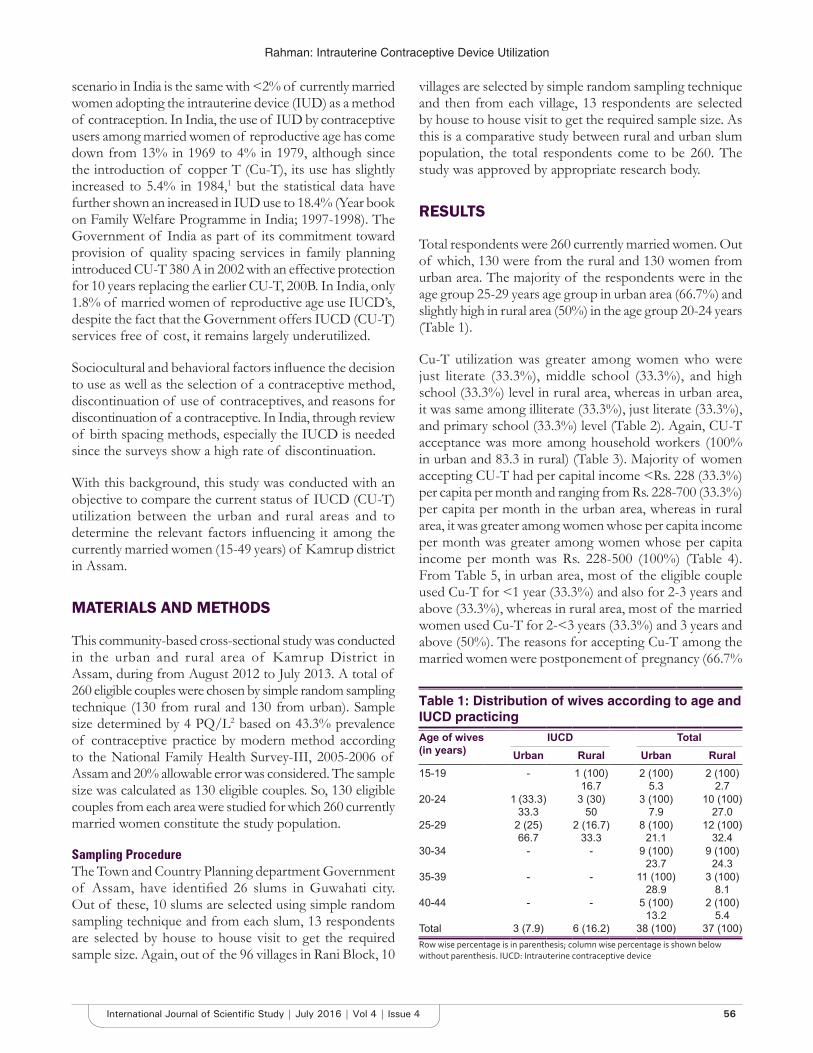
Rahman: Intrauterine Contraceptive Device Utilization
56International Journal of Scientific Study | July 2016 | Vol 4 | Issue 4
scenario in India is the same with <2% of currently married women adopting the intrauterine device (IUD) as a method of contraception. In India, the use of IUD by contraceptive users among married women of reproductive age has come down from 13% in 1969 to 4% in 1979, although since the introduction of copper T (Cu-T), its use has slightly increased to 5.4% in 1984,1 but the statistical data have further shown an increased in IUD use to 18.4% (Year book on Family Welfare Programme in India; 1997-1998). The Government of India as part of its commitment toward provision of quality spacing services in family planning introduced CU-T 380 A in 2002 with an effective protection for 10 years replacing the earlier CU-T, 200B. In India, only 1.8% of married women of reproductive age use IUCD’s, despite the fact that the Government offers IUCD (CU-T) services free of cost, it remains largely underutilized.
Sociocultural and behavioral factors influence the decision to use as well as the selection of a contraceptive method, discontinuation of use of contraceptives, and reasons for discontinuation of a contraceptive. In India, through review of birth spacing methods, especially the IUCD is needed since the surveys show a high rate of discontinuation.
With this background, this study was conducted with an objective to compare the current status of IUCD (CU-T) utilization between the urban and rural areas and to determine the relevant factors influencing it among the currently married women (15-49 years) of Kamrup district in Assam.
MATERIALS AND METHODS
This community-based cross-sectional study was conducted in the urban and rural area of Kamrup District in Assam, during from August 2012 to July 2013. A total of 260 eligible couples were chosen by simple random sampling technique (130 from rural and 130 from urban). Sample size determined by 4 PQ/L2 based on 43.3% prevalence of contraceptive practice by modern method according to the National Family Health Survey-III, 2005-2006 of Assam and 20% allowable error was considered. The sample size was calculated as 130 eligible couples. So, 130 eligible couples from each area were studied for which 260 currently married women constitute the study population.
Sampling ProcedureThe Town and Country Planning department Government of Assam, have identified 26 slums in Guwahati city. Out of these, 10 slums are selected using simple random sampling technique and from each slum, 13 respondents are selected by house to house visit to get the required sample size. Again, out of the 96 villages in Rani Block, 10
villages are selected by simple random sampling technique and then from each village, 13 respondents are selected by house to house visit to get the required sample size. As this is a comparative study between rural and urban slum population, the total respondents come to be 260. The study was approved by appropriate research body.
RESULTS
Total respondents were 260 currently married women. Out of which, 130 were from the rural and 130 women from urban area. The majority of the respondents were in the age group 25-29 years age group in urban area (66.7%) and slightly high in rural area (50%) in the age group 20-24 years (Table 1).
Cu-T utilization was greater among women who were just literate (33.3%), middle school (33.3%), and high school (33.3%) level in rural area, whereas in urban area, it was same among illiterate (33.3%), just literate (33.3%), and primary school (33.3%) level (Table 2). Again, CU-T acceptance was more among household workers (100% in urban and 83.3 in rural) (Table 3). Majority of women accepting CU-T had per capital income <Rs. 228 (33.3%) per capita per month and ranging from Rs. 228-700 (33.3%) per capita per month in the urban area, whereas in rural area, it was greater among women whose per capita income per month was greater among women whose per capita income per month was Rs. 228-500 (100%) (Table 4). From Table 5, in urban area, most of the eligible couple used Cu-T for <1 year (33.3%) and also for 2-3 years and above (33.3%), whereas in rural area, most of the married women used Cu-T for 2-<3 years (33.3%) and 3 years and above (50%). The reasons for accepting Cu-T among the married women were postponement of pregnancy (66.7%
Table 1: Distribution of wives according to age and IUCD practicingAge of wives(in years)
IUCD TotalUrban Rural Urban Rural
15-19 - 1 (100)16.7
2 (100)5.3
2 (100)2.7
20-24 1 (33.3)33.3
3 (30)50
3 (100)7.9
10 (100)27.0
25-29 2 (25)66.7
2 (16.7)33.3
8 (100)21.1
12 (100)32.4
30-34 - - 9 (100)23.7
9 (100)24.3
35-39 - - 11 (100)28.9
3 (100)8.1
40-44 - - 5 (100)13.2
2 (100)5.4
Total 3 (7.9) 6 (16.2) 38 (100) 37 (100)Row wise percentage is in parenthesis; column wise percentage is shown below without parenthesis. IUCD: Intrauterine contraceptive device

Rahman: Intrauterine Contraceptive Device Utilization
57 International Journal of Scientific Study | July 2016 | Vol 4 | Issue 4
in urban and 66.7% in rural), spacing (66.7% in urban and 66.7% in rural), and maternal health (33.3% in urban and 16.7% in rural) (Table 6). Discontinuation of Cu-T among the married women was mostly due to want of a child (55.6% in urban and 60% in rural) and problems faced due to Cu-T use (44.4% in urban and 40% in rural) (Table 7).
DISCUSSION
It is revealed from the Table 1 that in urban slums, majority of the respondents were in the age groups 25-29 years (66.7%), whereas in rural area, it was slightly high (50%) in the age group 20-24 years. Contraceptive acceptance increases with the increase in the age groups of the wives, which was shared by Zutshi and Dhar (1970),2 Raju et al. (1994),3 and Singh and Benera (1999).4 In Table 2, it is
seen that Cu-T utilization was greater among women who were just literate (33.3%), middle school (33.3%), and high school (33.3%) in rural area, whereas in urban area, it was same (33.3%) among illiterate, just literate, and primary school level literate women. The study findings hold good with the observations of Mazumdar (1955),5 Chandra (1959),6 Ranajit and Seal (1974),7 Ghosh and Mohapatra (1993),8 Upadhyay and Sharma (1995),9 Bhuyan (1996),10 and Sharma et al. (1997),11 wherein it was that the female literacy was positively and significantly associated with higher adoption of contraceptive methods. Table 3 shows that both in rural and urban area the acceptance of Cu-T was more among household workers (100% in urban slums and 83.3% in rural area). However, observations by Raju et al (1994)3 and Lingaraju (1998)12 contradicted the present study finding by stating that contraceptive acceptance was high in women who worked outside their homes (80%) than the housewives (60%). It is evident from Table 4, majority of women accepting Cu-T had per capital income <Rs. 228 (33.3%) per capita per month in urban area, whereas in rural area, it was greater among women whose per capita income per month was Rs. 228-500 (100%). Various study findings by Gandhi et al. (1969)13 stated that majority of the contraceptive acceptors belonged to the lower income group, whereas Awasthi (1973)14 stated that
Table 2: Distribution of the wives according to their literacy status and IUCD practicingLiteracy status IUCD Total
Urban Rural Urban RuralIlliterate 1 (5)
33.3- 20 (100)
52.6-
Just literate 1 (14.3)33.3
2 (28.6)33.3
7 (100)18.4
7 (100)18.9
Primary school 1 (20)33.3
- 5 (100)13.2
4 (100)10.8
Middle school - 2 (40)33.3
3 (100)7.9
5 (100)13.5
High school - 2 (22.2)33.3
1 (100)2.6
9 (100)24.3
HSLC - - - 8 (100)21.6
HS - - - 4 (100)10.8
Graduation and above - - 2 (100)5.3
-
Total 3 (7.9) 6 (16.2) 38 (100) 37 (100)Row wise percentage is in parenthesis; column wise percentage is shown below without parenthesis. IUCD: Intrauterine contraceptive device
Table 3: Distribution of the wives according to their occupation and IUCD practicingOccupation IUCD Total
Urban Rural Urban RuralHousehold work 3 (9.7)
1005 (19.2)
83.331 (100)
81.626 (100)
70.3Daily way - - 3 (100)
7.93 (100)
8.1Part time domestic work - - 1 (100)
2.62 (100)
5.4Service - - 2 (100)
5.31 (100)
2.7Business - 1 (20)
16.71 (100)
2.65 (100)
13.5Total 3 (7.9) 6 (16.2) 38 (100) 37 (100)Row wise percentage is in parenthesis; column wise percentage is shown below without parenthesis. IUCD: Intrauterine contraceptive device
Table 4: Distribution of eligible couples according to per capita income per month and IUCD practicingPer capital per month (Rs.) IUCD Total
Urban Rural Urban Rural<228 1 (33.3)
33.3- 3 (100)
7.92 (100)
5.4228-500 1 (6.3)
33.3 6 (23.1)
10016 (100)
42.126 (100)
70.3501-700 1 (10)
33.3- 10 (100)
26.32 (100)
5.4701-900 - - 5 (100)
13.23 (100)
8.1>901 - - 4 (100)
10.54 (100)
10.8Total 3 (7.9) 6 (16.2) 38 (100) 37 (100)IUCD: Intrauterine contraceptive device
Table 5: Distribution of eligible couples currently practicing contraceptive methods in relation to duration of useDuration of use in years IUCD Total
Urban Rural Urban Rural<1 1 (33.3) 1 (16.7) 4 (10.5) 5 (13.5)1-<2 - - 1 (2.6) 2 (5.4)2-<3 1 (33.3) 2 (33.3) 3 (7.9 4 (10.3)3 and above 1 (33.3) 3 (50) 30 (78.9) 26 (70.3)Total 3 (100) 6 (100) 38 (100) 37 (100)IUCD: Intrauterine contraceptive device
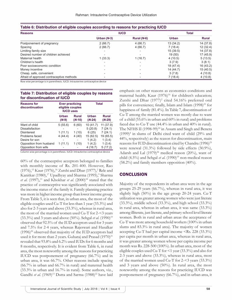
Rahman: Intrauterine Contraceptive Device Utilization
58International Journal of Scientific Study | July 2016 | Vol 4 | Issue 4
60% of the contraceptive acceptors belonged to families with monthly income of Rs. 201-400. However, Rao (1976),15 Kaur (1976),16 Zutshi and Dhar (1977),2 Rele and Kanitkar (1980),17 Upadhyay and Sharma (1995),9 Sharma et al. (1997),11 and Khokhar et al. (2000)18 stated that the practice of contraceptive was significantly associated with the income status of the family it. Family planning practice was more in higher income group than lower income group. From Table 5, it is seen that, in urban area, the most of the eligible couples used Cu-T for less than 1 year (33.3%) and also for 2-3 years and above (33.3%), whereas in rural area, the most of the married women used Cu-T for 2-<3 years (33.3%) and 3 years and above (50%). Sehgal et al. (1990)19 observed that 92.5% of the IUD acceptors used for 3 years and 7.5% for 2-4 years, whereas Rajeswari and Hasalkar (1996)20 observed that majority of the IUD acceptors had used it for more than 2 years. Guharaj and Prasad (1970)21 revealed that 93.8% and 6.2% used IUDs for 6 months and 8 months, respectively. It is evident from Table 6, in rural area, the most noteworthy among the reasons for practicing IUCD was postponement of pregnancy (66.7%) and in urban area, it was 66.7%. Other reasons include spacing (66.7% in urban and 66.7% in rural) and maternal health (33.3% in urban and 16.7% in rural). Some authors, viz., Gandhi et al. (1969)13 Dutta and Sarma (1988)22 have laid
emphasis on other reasons as economics conditions and maternal health; Kaur (1976)16 for children’s education; Zutshi and Dhar (1977)2 cited 54.16% preferred oral pills for convenience; finally, Islam and Islam (1998)23 for happiness of family (81.4%). In Table 7, discontinuation of Cu-T among the married women was mostly due to want of a child (55.6% in urban and 60% in rural) and problems faced due to Cu-T use (44.4% in urban and 40% in rural). The NFHS II (1998-99)24 in Assam and Singh and Benera (1999)4 in slums of Delhi cited want of child (29% and 44%, respectively) as the reason for discontinuation, main reasons for IUD discontinuation cited by Chandra (1996)25 were renewal (31.3%) followed by side effects (30.9%), Adarsh and Lal (1979)26 medical reason (20%), want of child (4.5%) and Sehgal et al. (1990)19 non-medical reason (58.2%) and family members opposition (40%).
CONCLUSION
Majority of the respondents in urban area were in the age groups 25-29 years (66.7%), whereas in rural area, it was slightly high (50%) in the age group 20-24 years. Cu-T utilization was greater among women who were just literate (33.3%), middle school (33.3%), and high school (33.3%) in rural area, whereas in urban area, it was same (33.3%) among illiterate, just literate, and primary school level literate women. Both in rural and urban areas the acceptance of Cu-T was more among household workers (100% in urban slums and 83.3% in rural area). The majority of women accepting Cu-T had per capital income <Rs. 228 (33.3%) per capita per month in urban area, whereas in rural area, it was greater among women whose per capita income per month was Rs. 228-500 (100%). In urban area, most of the eligible couples used Cu-T for <1 year (33.3%) and also for 2-3 years and above (33.3%), whereas in rural area, most of the married women used Cu-T for 2-<3 years (33.3%) and 3 years and above (50%). In rural area, the most noteworthy among the reasons for practicing IUCD was postponement of pregnancy (66.7%), and in urban area, it
Table 6: Distribution of eligible couples according to reasons for practicing IUCDReasons IUCD Total
Urban (N-3) Rural (N-6) Urban RuralPostponement of pregnancy 2 (66.7) 4 (66.7) 13 (34.2) 14 (37.8)Spacing 2 (66.7) 4 (66.7) 7 (18.4) 12 (32.4)Limiting family size - - 15 (39.5) 14 (37.8)Desired number of children achieved - - 19 (50) 17 (45.9)Maternal health 1 (33.3) 1 (16.7) 4 (10.5) 5 (13.5)Children’s health - - 3 (7.9) 3 (8.1)Poor socioeconomic condition - - 18 (47.4) 16 (43.2)Money incentive - - 14 (44.7) 15 (40.5)Cheap, safe, convenient - - 3 (7.8) 4 (10.8)Afraid of approved contraceptive methods - - 7 (18.4) 4 (10.8)Row wise percentage is in parenthesis. IUCD: Intrauterine contraceptive device
Table 7: Distribution of eligible couples by reasons for discontinuation of IUCDReasons for discontinuation
Ever practicing eligible couples
IUCD uses
Total
Urban(N-9)
Rural(N-10)
Urban(N-24)
Rural(N-29)
Want of child 5 (55.6) 6 (60) 10 (41.7) 11 (37.9)Dissatisfaction - - 5 (20.8) 7 (24.1)Disinterest 1 (11.1) 1 (10) 6 (25) 7 (24.1)Problems faced 4 (44.4) 4 (40) 15 (62.5) 19 (65.5)Desire for son - - 1 (4.2) 1 (3.4)Opposition from husband 1 (11.1) 1 (10) 1 (4.2) 1 (3.4)Opposition from wife - - 4 (16.7) 5 (17.2)Column wise percentage is in parenthesis. IUCD: Intrauterine contraceptive device

Rahman: Intrauterine Contraceptive Device Utilization
59 International Journal of Scientific Study | July 2016 | Vol 4 | Issue 4
was 66.7%. Other reasons include spacing (66.7% in urban and 66.7% in rural) and maternal health (33.3% in urban and 16.7% in rural). Discontinuation of Cu-T among the married women was mostly due to want of a child (55.6% in urban and 60% in rural) and problems faced due Cu-T use (44.4% in urban and 40% in rural).
REFERENCES
1. Nortman DL. A Population Council Fact Book. Population & Family Planning Programme. 12th ed. New York: The Population Council; 1985.
2. Dhar GM, Zutshi ML. Contraceptive practice among hospital attendants. Indian J Public Health 1977;21:8-15.
3. Raju KN, Bhat TN, Umamoni KS, Lingaraji M. Differential acceptance of family planning methods in a high performance district in Karnataka. J Fam Welfare 1994;40:31-9.
4. Singh P, Benera SK. Five percent post survey cheek of National Family Health Survey (NFHS II) in Orissa. Indian J Community Med 1999;XXIV:181-7.
5. Mazumdar DN. Report on Enquiry into Fertility and Family Planning among a Section of Married Women in Kanpur, Lucknow, Mamoria, Kitabmahal, Allahabad; 1955.
6. Chandrasekhar S. Report on a Survey of Attitude of Married Couples Towards Family Planning in the Putupakhan Area in City of Madras; 1959.
7. Ranajit D, Seal SC. A study of relationship of socioeconomic factors and family planning practice in a community in West Bengal. Indian J Public Health 1974;XVIII:78-91.
8. Ghosh S, Mahapatra P. A study of family planning devices used by the slum dweller of Sundarpur, Varanasi. Indian J Prev Soc Med 1993;24:56-8.
9. Upadhyay J, Sharma AK. Fertility patterns and family planning acceptance among slum dweller in Kanpur. J Fam Welfare 1995;41:61-8.
10. Bhuyan KC. Different fertility among adopter and nonadopter couples of differing social states and mobility pattern in rural Bangladesh - A case
study. J Fam Welfare 1996;42:51-60.11. Sharma AK, Grover V, Agarwal OP, Dubey KK, Sharma S. Pattern of
contraceptive use by resident of a village in South Delhi. Indian J Public Health 1997;XXXXI:75-8.
12. Lingaragu M. Utilization of MCH and family planning services by a schedule caste community in Mysore district. J Fam Welfare 1998;44:28-35.
13. Gandhi HS, Trivedi BK, Mishra VN. Awareness and acceptance of different contraceptive methods in a rural population. Indian J Public Health 1969;XIV:130-43.
14. Awasthi NM. Characteristics of acceptances of vasectomy with a probe into the motivational aspects. Indian J Prev Soc Med 1973;3:132-3.
15. Rao NB. Family Planning in India: A Case Study of Karnataka. New Delhi: Vikas; 1976.
16. Kaur S. Family Planning in Two Industrial Units: A Study. New Delhi: Sterling Publishers Pvt., Ltd.; 1976.
17. Rele JR, Jana K. Fertility and Family Planning in Greater Bombay. Bombay: International Institute for Population Studies, Popular Prakashan; 1980.
18. Khokhar A, Gulati N. A study of never users of contraception from an urban slum of Delhi. Indian J Community Med 2000;XXV:26-30.
19. Sehgal K, Sharma N, Garg S. A profile and one year follow up of IUCD copper - T-200 acceptors in a rural area of South Delhi. Health Popul Prospect Issues 1990;13:155-61.
20. Rajeswari NV, Hasalkar JB. IUD retention in Shimoga district of Karnataka. J Fam Welfare 1996;42:44-50.
21. Guharaj A, Prasad BG. A study of family planning by IUCD (Lippe’s Loop). Indian J Prev Soc Med 1970;1:197-203.
22. Dutta KK, Sarma N. A peep into knowledge attitude and practice of family planning among navel sailors family residing in Bombay. Indian J Public Health 1988;XXXII:154-5.
23. Islam MM, Islam NN. Contraceptive use among married adolescent girls in rural Bangladesh. J Fam Welfare 1998;44:32-9.
24. National Family Health Survey - III (NFHSIII), 2005-06, Assam & Preliminary Report.
25. Chandra S. A study of copper-T 200 acceptors. J Fam Welfare 1996;42:55-7.26. Adarsh R., Lal S. Use of effectiveness of copper-T as an IUD in family
welfare programmes. Indian J Community Med 1979;IV:21-3.
How to cite this article: Rahman S. A Comparative Study of Intrauterine Contraceptive Device Utilization among Currently Married Women in a Rural Area of Rani Block and Urban Slums of Guwahati City. Int J Sci Stud 2016;4(4):55-59.
Source of Support: Nil, Conflict of Interest: None declared.

60International Journal of Scientific Study | July 2016 | Vol 4 | Issue 4
High-resolution Computed Tomography Study of Temporal Bone PathologiesManjit Bagul
Senior Resident, Department of Radiodiagnosis, RKDF Medical College, Hospital & Research Centre, Jatkhedi, Bhopal, Madhya Pradesh, India
modalities available for the evaluation of temporal bone were plain radiograph, polytomography, angiography, and cisternography. Plain radiograph remains inexpensive tool of the study of temporal bone but has major limitations due to complex anatomy and overlapping of various bony structures.
High-resolution computed tomography (HRCT) offers excellent spatial and density resolution using special algorithms. It provides information not only about bony outline but also soft tissue changes making it possible to demonstrate the location and extent of disease as well as its complications. Furthermore, coronal and axial CT scanning together has dramatically improved the imaging of temporal bone. HRCT accurately depicts the boundaries between the external, middle and inner
INTRODUCTION
The temporal bone is a complex anatomic structure that contains the organs of hearing and balance. In addition, major vessels and nerves course through it and it also has a close proximity to the brain. Temporal bone has direct contact with brainstem, cerebellum and temporal lobe of brain. Before computed tomography (CT) imaging
Original Article
AbstractIntroduction: The application of high-resolution computed tomography (HRCT) in the temporal bone study has allowed the detailed assessment of complex bony anatomy and pathology. HRCT temporal bone accurately depicts bony erosion or destruction and associated soft tissue pathology. The purpose of this study is to determine the role and efficacy of HRCT temporal bone in evaluation of congenital, inflammatory, traumatic, and neoplastic conditions. This was the cross-sectional prospective study of 1½ year’s duration.
Aims and Objectives: To evaluate the congenital, inflammatory, traumatic, and neoplastic conditions of the temporal bone with the help of HRCT.
Materials and Methods: This was cross-sectional prospective study conducted at RKDF Medical College between December 2014 to March 2016. A total of 120 patients of varied age group presenting with symptoms and signs of temporal bone pathologies were included in the study. Imaging diagnosis was confirmed either by histopathology or follow-up and response to treatment.
Results: In our study, temporal bone pathologies were more common in male (66.66%) compare to female population (33.33%). The most common age group affected by the temporal bone pathologies was 11-20 years age group (38.33%) and least common age group was more than 60 years (<4%). The most common temporal bone pathologies in our study were inflammatory (50%) followed by traumatic (11.66), benign neoplasm (10%), congenital (6.66), and malignant neoplasm (5%). Approximately, 16.66% cases of HRCT temporal bone study revealed no abnormality. Most common inflammatory pathology in our study was cholesteatoma (73.3%) followed by otomastoiditis/otitis externa constitute (26.66%). Out of 14 traumatic cases of temporal bone, 57.14% cases were associated with intracranial injuries in our study. Out of 18 cases of neoplastic pathology, 66.66% of cases were benign and 33.33% were malignant etiology.
Conclusion: HRCT scan of temporal bone depicts complex bony details and associated soft tissue pathologies accurately. Due to various limitations of clinical examination and radiography, HRCT temporal bone is single most important imaging tool for pre-operative evaluation and management of various pathologies of the temporal bone.
Key words: High-resolution computed tomography, Histopathological diagnosis, Temporal bone
Access this article online
www.ijss-sn.com
Month of Submission : 05-2016 Month of Peer Review : 06-2016 Month of Acceptance : 07-2016 Month of Publishing : 07-2016
Corresponding Author: Manjit Bagul, RKDF Medical College, Bhopal, Madhya Pradesh, India. E-mail: [email protected]
Print ISSN: 2321-6379Online ISSN: 2321-595X
DOI: 10.17354/ijss/2016/375

Bagul: HRCT Study of Temporal Bone Pathologies
61 International Journal of Scientific Study | July 2016 | Vol 4 | Issue 4
ear cavities thereby localized the disease precisely and also greatly demarcate thin boundary between temporal bone and intracranial compartment with exact details of intracranial spread of primary temporal bone disease. Contrast media help to evaluate the vascularity and contrast enhancing characteristics particularly in soft tissue lesions of temporal bone giving clues to the histopathology.
Aims and objective of this study is to evaluate the congenital, inflammatory, traumatic, and neoplastic conditions affecting the temporal bone with the help of HRCT.
MATERIALS AND METHODS
The present prospective study was conducted at RKDF Medical College, Bhopal, Madhya Pradesh from December 2014 to March 2016.
Study AreaThe study area includes Bhopal city and district with peripheral small towns/villages.
Study PopulationA total of 120 patients of varied age group presenting with symptoms and signs of temporal bone diseases were included in the study.
Inclusion Criteria• Patients referred for HRCT, who were suspected to
have of temporal bone pathology.
Exclusion CriteriaPost-operative cases of temporal bone pathology.
Equipment UsedSpiral CT, Siemens Somatom, Siemens Medical Systems, Forchiem, Germany.
HRCT was done using thin section, high-resolution and bone algorithm technique. Sections in both axial and coronal planes were obtained. Coronal imaging was done by neck extension and prone position and axial imaging done in supine and neutral position of the neck. Iodine Based contrast used mainly in the neoplastic pathologies. Final imaging diagnosis correlated with histopathological confirmation or follow-up and treatment response.
OBSERVATIONS AND RESULTS
HRCT scan was performed in 120 patients who presented with history, symptoms, and signs of the temporal bone pathologies. The results are enumerated in Tables 1-8.
Table 1: Gender distributionGender Number of cases (%)Female 40 (33.33)Male 80 (66.66)Grand total 120 (100)
Table 2: Clinical presentationSign and symptoms Number of cases (%)Deafness 78 (65.00)Otorrhoea 70 (58.33)Headache 32 (26.66)Otalgia 48 (40.00)Fever 16 (13.33)Vertigo 20 (16.66)Fascial nerve palsy 12 (10.00)Ataxia 4 (3.33)Tinnitus 12 (10.00)Ear bleed 8 (6.66)
Table 3: Age distributionAge distribution Number of cases (%)1-10 14 (11.66)11-20 46 (38.33)21-30 18 (15.00)31-40 16 (13.33)41-50 12 (10.00)51-60 10 (8.33)61 and above 4 (3.33)Total 120
Table 4: Etiopathological distribution of casesEtiology Number of cases (%)Inflammatory 60 (50)Traumatic 14 (11.66)Congenital 8 (6.66)Benign 12 (10)Malignant 6 (5.0)Normal 20 (16.66)Grand total 120 (100)
Table 5: Distribution of various lesionsPathology Number of casesCongenital malformation
External and middle ear anomalies 6Inner ear abnormalities 2Temporal bone fractures 14
Inflammatory processOtitis externa 3Malignant otitis externa 1Otomastoiditis 12
CholesteatomaCongenital 2Acquired 42
NeoplasmBenign 12Malignant 6

Bagul: HRCT Study of Temporal Bone Pathologies
62International Journal of Scientific Study | July 2016 | Vol 4 | Issue 4
DISCUSSION
The varied temporal bone pathologies including congenital, inflammatory, traumatic, and neoplastic conditions were evaluated by HRCT. The lack of specificity in clinical examination and the imprecise result of conventional radiography renders CT as the modality of choice in the evaluation of temporal bone pathology.
In this study, 120 patients were evaluated for their various symptoms pertaining to PNS. The gender ratio in this study was 2:1 (male:female) (Table 1). A maximum number of patients presented with the chief complaints of hearing problem or deafness (65%) followed by otorrhea (58%) (Table 2). Other chief complaints were otalgia, vertigo, tinnitus, ataxia, and fascial nerve palsy. Patients with intracranial complications had headache, fever, vomiting in addition to above complaints. The most common age group involved was 11-20 years (38%) and least common
age group was 61 and above comprising (3%) of total cases (Table 3).
The et io logic distr ibut ion of the les ions was inflammatory (50%) followed by traumatic (11.6%), benign (10%), congenital (6.6%), and malignant (5%) (Table 4). Thus, the inflammatory disease was found to be the most frequently occurring pathology affecting the temporal bone. Inflammatory pathologies were common in younger age group (<30 years) and neoplastic pathologies were common in older age group (>50 years). Traumatic conditions equally distributed in all age group. Congenital disease frequently diagnosed in <10 years of age group.
Congenital malformation of the external and middle ear is more common than inner ear anomalies. Atresia or hypoplasia of the external auditory canal (EAC) is most common anomaly detected in our study.1,2 The degree of distortion range from web to small band of soft tissue covering EAC to complete absence.3 Ossicular chain abnormalities are commonly found in association with external meatal atresia.1,2 Isolated ossicular chain anomalies are less common.4 The main role of HRCT in atresia of HRCT to identify the type of anomaly and determine the surgical correctability.5 If surgical correction of EAC atresia is to be done to improve hearing normal inner ear structures and adequate middle ear cleft are necessary. It is important to evaluate the thickness of atresia plate, associated ossicular abnormalities, status of middle ear cleft, status of inner ear, course of fascial nerve canal and position of sigmoid sinus and mastoid pneumatisation.6 In our study, we came across 6 cases of external and middle ear anomaly, out of 6, four cases had unilateral EAC atresia and two cases had bilateral atresia. In two cases, there was soft tissue atresia and in four cases bony atresia
Table 6: Distribution of external and middle ear anomaliesHRCT findings Number of cases (%)Bony atresia 4 (66.66)Soft tissue atresia 2 (33.33)Ossicular deformity 4 (66.66)Thick atresia plate 4 (66.66)Small tympanic cavity 4 (66.66)Fascial canal 2 (33.33)Inner ear anomaly 2 (33.33)HRCT: High‑resolution computed tomography
Table 7: Distribution of CT findings in temporal bone fractureCT findings Number of cases (%)Longitudinal fracture 10 (71.42)Transverse fracture 2 (14.28)Complex fracture 2 (14.28)Haemotympanum 10 (71.42)Ossicular disruption 4 (28.57)Labyrinthine injury 2 (14.28)Fascial nerve canal injury 2 (14.28)Intracranial injury 8 (57.14)CT: Computed tomography
Table 8: CT features of cholesteatomaCT findings Number of cases (%)Soft tissue lesion 42 (100)Ossicular and scutum erosion 40 (95.23)Erosion of tympanic wall 38 (90.47)Erosion of sigmoid sinus plate 18 (42.85)Opacified mastoid 24 (57.14)Erosion of lateral semicircular canal 8 (19.04)Erosion of tegmen tympanii 8 (19.04)Erosion of vestibule and fascial canal 4 (9.52)CT: Computed tomography
Table 9: Various complications of cholesteatomaComplications Number of cases (%)Brain abscess 18 (42.85)Post auricular abscess 8 (19.04)Dural sinus thrombosis 6 (14.28)Meningitis 4 (9.52)
Table 10: Distribution of neoplastic lesionsNeoplasm Number of casesAcoustic neuroma 6Glomus tumor 3Epidermoid 2Osteoma 1Primary carcinoma 4Secondary metastasis 2

Bagul: HRCT Study of Temporal Bone Pathologies
63 International Journal of Scientific Study | July 2016 | Vol 4 | Issue 4
was found. Ossicular deformities and thick atresia plate were present in 4 cases and in 2 cases anteriorly placed descending fascial canal was found. The present study correlated well with findings of Swartz et al.4 Congenital anomalies of inner ear are relatively uncommon compare to middle and external ear anomalies. In this study, 2 cases (33.33%) of inner ear anomalies were detected on HRCT temporal bone. One case was unilateral enlarged vestibular aqueduct (EVA) or large vestibular aqueduct syndrome (LVAS) and the second case was bilateral EVA or LVAS with associated unilateral mondini dysplasia or also known as incomplete partition type II characterized by cystic cochlear apex with the normal basal turn. EVA has been reported to be the most common inner ear abnormality associated with sensorineural hearing loss and is also commonly associated with other inner ear abnormalities.7
Temporal bone fractures may be longitudinal, transverse or complex. Longitudinal fractures are common and comprise 70-90% of all temporal bone fractures.8 In our study, 71.42% of cases had the longitudinal type of temporal bone fractures. These type of fractures extend across roof or posterior wall of EAC into tympanic cavity and lead to rupture of the tympanic membrane with hemotympanum.8,9 Ossicular chain disruption is a common cause leading to hearing loss and incus is most vulnerable for dislocation due to lack of muscular anchor.10 Fascial nerve palsy occurs in 10-20% of cases and usually delayed and incomplete. A common site for fascial nerve injury is horizontal segment.10 Transverse fractures are less common and usually associated with temporomandibular joint or mandibular injury and fascial paralysis is more common in the transverse fractures.10 In our study of two cases of fascial nerve palsy one case (10%) was seen associated with longitudinal fracture and the second case (50%) was seen in a transverse fracture of temporal bone. We studied 10 cases (71.42%) of longitudinal, 2 cases (14.28%) of transverse and 2 cases (14.2%) of complex type of temporal bone fractures. Labyrinth injury involving destruction of the vestibule-cochlear complex is more common in transverse fracture and resulting into sensorineural hearing loss, vertigo, nystagmus and pneumolabryinth.10,11 In our study, out of two cases of labyrinth injury, one case was associated with transverse and the second case was associated with complex temporal bone fractures.
Otoscopic evaluation alone is usually sufficient to study external ear but HRCT temporal bone needed to rule out osteomyelitis, otomastoiditis or malignant otitis externa. In present study, one patient was found to have malignant otitis externa with soft tissue in EAC causing bony destruction and associated edematous pinna and
subcutaneous tissue of the scalp.12 Three cases of isolated otitis externa with opacified EAC and no bony erosion or destruction included in our study. Acute otitis media seen as middle ear opacification and concomitant mastoiditis seen as opacification or air fluid level in mastoid cavity on HRCT temporal bone.13,14 Coalescent otitis media seen as resorption and destruction of mastoid bony septii with the irregular mastoid cavity and it can penetrate through bony cortex to form subperiosteal abscess or break at the tip of mastoid bone to form abscess in the neck.15 In this study, we included 12 cases of otomastoiditis without cholesteatoma. Out of 12 cases of otomastoiditis 8 cases were without significant bony destruction or resorption and 4 cases were with resorption of bony septii and irregular mastoid cavity. 2 out of 4 cases of coalescent otomastoiditis were associated with subperiosteal abscess formation.
Cholesteatoma is the misnomer, it’s not true neoplasm but sac of stratified squamous epithelium filled with exfoliated keratin and subepithelial fibrous stroma producing proteolytic enzyme which in turn causes bone resorption.16-18 Congenital cholesteatoma accounts for 2% of cases.16 In our study, out of 44 cases of cholesteatoma, 2 cases were of congenital cholesteatoma. Cholesteatoma found behind the tympanic membrane in the patient with no history of otitis media is considered congenital. These lesions can erode internal auditory meatus and semicircular canal leading to hearing loss.19 In our study, one case of congenital cholesteatoma was present as a cystic lesion in the petrous apex with the destruction of internal auditory canal and the second case of congenital cholesteatoma present as small soft tissue lesion without bony destruction. Acquired cholesteatoma divided into two types primary arising from the pars flaccid and secondary arising from pars tensa. Pars flaccid cholesteatoma arising from the prussak space of epitympanum and extend into mastoid antrum with erosion of bony scutum.16,17 Pars tensa cholesteatoma arises from posterosuperior retraction of tympanic membrane and spread into surrounding recesses. Cholesteatoma is most frequent soft tissue mass in the middle ear.20 Expansion and scalloping of mastoid, erosion of lateral attic wall, erosion of the ossicles, tegmen tympani, sigmoid sinus plate, posterosuperior wall of EAC, labyrinth, and fascial nerve canal are signs indicating the presence of cholesteatoma.17-20 Pre-operative CT scan is helpful in relation to diagnosis and decision making for surgery in cases of cholesteatoma and ossicular erosion.21 In this study, the most common CT findings in the cholesteatoma was ossicular and scutum erosion (95%) followed by erosion of tympanic wall (90%), opacified mastoid (57%), erosion of sigmoid plate (42%), erosion of lateral semicircular canal wall and tegmen tympanii (19%) and erosion of vestibule and fascial canal (9.5%).
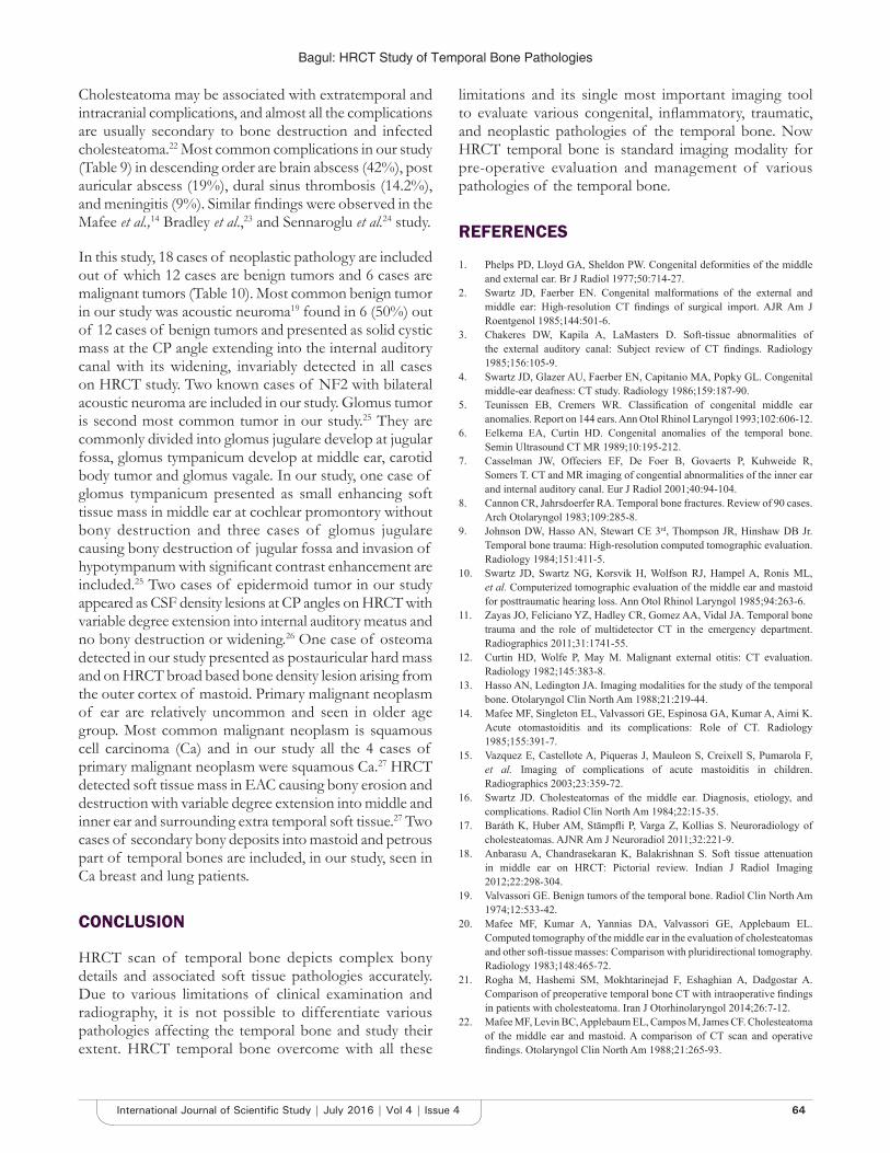
Bagul: HRCT Study of Temporal Bone Pathologies
64International Journal of Scientific Study | July 2016 | Vol 4 | Issue 4
Cholesteatoma may be associated with extratemporal and intracranial complications, and almost all the complications are usually secondary to bone destruction and infected cholesteatoma.22 Most common complications in our study (Table 9) in descending order are brain abscess (42%), post auricular abscess (19%), dural sinus thrombosis (14.2%), and meningitis (9%). Similar findings were observed in the Mafee et al.,14 Bradley et al.,23 and Sennaroglu et al.24 study.
In this study, 18 cases of neoplastic pathology are included out of which 12 cases are benign tumors and 6 cases are malignant tumors (Table 10). Most common benign tumor in our study was acoustic neuroma19 found in 6 (50%) out of 12 cases of benign tumors and presented as solid cystic mass at the CP angle extending into the internal auditory canal with its widening, invariably detected in all cases on HRCT study. Two known cases of NF2 with bilateral acoustic neuroma are included in our study. Glomus tumor is second most common tumor in our study.25 They are commonly divided into glomus jugulare develop at jugular fossa, glomus tympanicum develop at middle ear, carotid body tumor and glomus vagale. In our study, one case of glomus tympanicum presented as small enhancing soft tissue mass in middle ear at cochlear promontory without bony destruction and three cases of glomus jugulare causing bony destruction of jugular fossa and invasion of hypotympanum with significant contrast enhancement are included.25 Two cases of epidermoid tumor in our study appeared as CSF density lesions at CP angles on HRCT with variable degree extension into internal auditory meatus and no bony destruction or widening.26 One case of osteoma detected in our study presented as postauricular hard mass and on HRCT broad based bone density lesion arising from the outer cortex of mastoid. Primary malignant neoplasm of ear are relatively uncommon and seen in older age group. Most common malignant neoplasm is squamous cell carcinoma (Ca) and in our study all the 4 cases of primary malignant neoplasm were squamous Ca.27 HRCT detected soft tissue mass in EAC causing bony erosion and destruction with variable degree extension into middle and inner ear and surrounding extra temporal soft tissue.27 Two cases of secondary bony deposits into mastoid and petrous part of temporal bones are included, in our study, seen in Ca breast and lung patients.
CONCLUSION
HRCT scan of temporal bone depicts complex bony details and associated soft tissue pathologies accurately. Due to various limitations of clinical examination and radiography, it is not possible to differentiate various pathologies affecting the temporal bone and study their extent. HRCT temporal bone overcome with all these
limitations and its single most important imaging tool to evaluate various congenital, inflammatory, traumatic, and neoplastic pathologies of the temporal bone. Now HRCT temporal bone is standard imaging modality for pre-operative evaluation and management of various pathologies of the temporal bone.
REFERENCES
1. Phelps PD, Lloyd GA, Sheldon PW. Congenital deformities of the middle and external ear. Br J Radiol 1977;50:714-27.
2. Swartz JD, Faerber EN. Congenital malformations of the external and middle ear: High-resolution CT findings of surgical import. AJR Am J Roentgenol 1985;144:501-6.
3. Chakeres DW, Kapila A, LaMasters D. Soft-tissue abnormalities of the external auditory canal: Subject review of CT findings. Radiology 1985;156:105-9.
4. Swartz JD, Glazer AU, Faerber EN, Capitanio MA, Popky GL. Congenital middle-ear deafness: CT study. Radiology 1986;159:187-90.
5. Teunissen EB, Cremers WR. Classification of congenital middle ear anomalies. Report on 144 ears. Ann Otol Rhinol Laryngol 1993;102:606-12.
6. Eelkema EA, Curtin HD. Congenital anomalies of the temporal bone. Semin Ultrasound CT MR 1989;10:195-212.
7. Casselman JW, Offeciers EF, De Foer B, Govaerts P, Kuhweide R, Somers T. CT and MR imaging of congential abnormalities of the inner ear and internal auditory canal. Eur J Radiol 2001;40:94-104.
8. Cannon CR, Jahrsdoerfer RA. Temporal bone fractures. Review of 90 cases. Arch Otolaryngol 1983;109:285-8.
9. Johnson DW, Hasso AN, Stewart CE 3rd, Thompson JR, Hinshaw DB Jr. Temporal bone trauma: High-resolution computed tomographic evaluation. Radiology 1984;151:411-5.
10. Swartz JD, Swartz NG, Korsvik H, Wolfson RJ, Hampel A, Ronis ML, et al. Computerized tomographic evaluation of the middle ear and mastoid for posttraumatic hearing loss. Ann Otol Rhinol Laryngol 1985;94:263-6.
11. Zayas JO, Feliciano YZ, Hadley CR, Gomez AA, Vidal JA. Temporal bone trauma and the role of multidetector CT in the emergency department. Radiographics 2011;31:1741-55.
12. Curtin HD, Wolfe P, May M. Malignant external otitis: CT evaluation. Radiology 1982;145:383-8.
13. Hasso AN, Ledington JA. Imaging modalities for the study of the temporal bone. Otolaryngol Clin North Am 1988;21:219-44.
14. Mafee MF, Singleton EL, Valvassori GE, Espinosa GA, Kumar A, Aimi K. Acute otomastoiditis and its complications: Role of CT. Radiology 1985;155:391-7.
15. Vazquez E, Castellote A, Piqueras J, Mauleon S, Creixell S, Pumarola F, et al. Imaging of complications of acute mastoiditis in children. Radiographics 2003;23:359-72.
16. Swartz JD. Cholesteatomas of the middle ear. Diagnosis, etiology, and complications. Radiol Clin North Am 1984;22:15-35.
17. Baráth K, Huber AM, Stämpfli P, Varga Z, Kollias S. Neuroradiology of cholesteatomas. AJNR Am J Neuroradiol 2011;32:221-9.
18. Anbarasu A, Chandrasekaran K, Balakrishnan S. Soft tissue attenuation in middle ear on HRCT: Pictorial review. Indian J Radiol Imaging 2012;22:298-304.
19. Valvassori GE. Benign tumors of the temporal bone. Radiol Clin North Am 1974;12:533-42.
20. Mafee MF, Kumar A, Yannias DA, Valvassori GE, Applebaum EL. Computed tomography of the middle ear in the evaluation of cholesteatomas and other soft-tissue masses: Comparison with pluridirectional tomography. Radiology 1983;148:465-72.
21. Rogha M, Hashemi SM, Mokhtarinejad F, Eshaghian A, Dadgostar A. Comparison of preoperative temporal bone CT with intraoperative findings in patients with cholesteatoma. Iran J Otorhinolaryngol 2014;26:7-12.
22. Mafee MF, Levin BC, Applebaum EL, Campos M, James CF. Cholesteatoma of the middle ear and mastoid. A comparison of CT scan and operative findings. Otolaryngol Clin North Am 1988;21:265-93.

Bagul: HRCT Study of Temporal Bone Pathologies
65 International Journal of Scientific Study | July 2016 | Vol 4 | Issue 4
23. Bradley PJ, Manning KP, Shaw MD. Brain abscess secondary to otitis media. J Laryngol Otol 1984;98:1185-91.
24. Sennaroglu L, Sozeri B. Otogenic brain abscess: Review of 41 cases. Otolaryngol Head Neck Surg 2000;123:751-5.
25. Chakeres DW, LaMasters DL. Paragangliomas of the temporal bone: High-
resolution CT studies. Radiology 1984;150:749-53.26. Curtin HD. CT of acoustic neuroma and other tumors of the ear. Radiol Clin
North Am 1984;22:77-105.27. Phelps PD, Lloyd GA. The radiology of carcinoma of the ear. Br J Radiol
1981;54:103-9.
How to cite this article: Bagul M. High-resolution Computed Tomography Study of Temporal Bone Pathologies. Int J Sci Stud 2016;4(4):60-65.
Source of Support: Nil, Conflict of Interest: None declared.

66International Journal of Scientific Study | July 2016 | Vol 4 | Issue 4
Outcome of Neodymium-doped Yttrium Aluminum Garnet Laser Posterior Capsulotomy for Posterior Capsular Opacification: A Prospective StudyC Suriyakumar1, N Sharmila2, K Kavitha3, S Ganapathi Rajesh4
1Assistant Professor, Department of Ophthalmology, Government Theni Medical College and Hospital, Theni, Tamil Nadu, India, 2Associate Professor, Department of Ophthalmology, Government Theni Medical College, Theni, Tamil Nadu, India, 3Associate Professor, Department of Ophthalmology, Government Theni Medical College, Theni, Tamil Nadu, India, 4Senior Assistant Professor, Department of Ophthalmology, Government Theni Medical College, Theni, Tamil Nadu, India
capsular opacification following cataract surgery. This study was conducted in Government Theni Medical College and Hospital, Theni.
Eyes of 100 patients who underwent Nd:YAG laser posterior capsulotomy were studied. Eight patients were aphakic, 89 patients had posterior chamber intraocular lens, and 3 patients had anterior chamber intraocular lens. An informed consent was obtained from all patients.
The posterior capsulotomies were performed with Nd: YAG laser, usually starting with 1-2 mJ/pulse and gradually increased until the desired responses were obtained.
During each visit, the following data were recorded.2• Visual acuity with Snellen chart• Intraocular pressure with Schiotz tonometry• Slitlamp examination• Fundus examination with direct and indirect
ophthalmoscopy• Post-laser complications.
INTRODUCTION
A laser is the major technological advance in the medical field to give better results with less invasive method. Neodymium-doped yttrium aluminum garnet (Nd:YAG) laser posterior capsulotomy is one of the breakthroughs, for opening the posterior capsular opacities, the non-invasive characteristics of it provide an excellent outcome.1-4
MATERIALS AND METHODS
This prospective clinical study was done to analyze the results of Nd:YAG laser posterior capsulotomy in posterior
Original Article
AbstractIntroduction: Posterior capsular opacification is the opacity developing in posterior capsule of lens after cataract surgery commonly and rarely with crystalline lens primarily.
Aim: To study the visual outcome and analyze the complications in posterior capsular opacification treated with neodymium-doped yttrium aluminum garnet (Nd:YAG) laser posterior capsulotomy.
Materials and Methods: A prospective study of 100 patients attending our outpatient department with visual deterioration due to posterior capsular opacification after cataract surgery.
Results: Visual acuity improved in 96% of patients, 4% showed no improvement, which was related to previous retinal problems and thick posterior capsule.
Conclusion: Nd:YAG laser posterior capsulotomy is safe and effective procedure for creating capsular opening compared to invasive surgical techniques. The post-operative results are very good and the complication rate is low.
Key words: Posterior Capsular Opacification, Nd:YAG laser, Capsulotomy, Visual acuity
Access this article online
www.ijss-sn.com
Month of Submission : 05-2016 Month of Peer Review : 06-2016 Month of Acceptance : 07-2016 Month of Publishing : 07-2016
Corresponding Author: Dr. Sharmila. N., 22, second street, Kumaran Colony, Vadapalani, Chennai - 600 026, Phone: 9677032124. E-mail: [email protected]
Print ISSN: 2321-6379Online ISSN: 2321-595X
DOI: 10.17354/ijss/2016/376

Suriyakumar, et al.: Outcome of Nd:YAG laser posterior capsulotomy for posterior capsular opacification
67 International Journal of Scientific Study | July 2016 | Vol 4 | Issue 4
RESULTS
Visual acuity profile reveals that 75% of patients had visual acuity in the range between 6/60 and 3/60 and 10% of patients had visual acuity < 3/60 before doing YAG laser capsulotomy.
Immediately after doing YAG laser capsulotomy within 1 h, 57% of patients had improved. Visual acuity had improved gradually during follow-up period at 24 h, 1 week, 1 month, and 3 months. The vision of four patients not improved. Among four 2 patients had optic atrophy and other 2 patients had thick posterior capsule (Tables 1-3).5
DISCUSSION
The documented visual improvement of the subjects in this study confirms the efficacy of Nd:YAG laser for the production of posterior capsulotomy. This non-invasive surgical laser allows omission of primary intraoperative capsulotomy, with its increased risk of the intraocular lens and vitreous displacement, cystoid macular edema and retinal detachment.2
CONCLUSION
Following extracapsular cataract surgery, increased incidence of posterior capsular opacification has been noted. The opening of posterior capsule by surgical methods has been noted to cause certain complications such as cystoid macular edema and retinal detachment.3 The Nd:YAG laser is a non-invasive surgical tool that provides excellent posterior capsulotomies.
From the above study, it is clear that Nd: YAG laser posterior capsulotomy is a safe and effective procedure for creating capsular opening. The post-operative results are very good and the complication rate is low. 2
REFERENCES
1. Aron-Rosa D, Aron JJ, Griesemann M, Thyzel R. Use of the neodymium-YAG laser to open the posterior capsule after lens implant surgery: A preliminary report. J Am Intraocul Implant Soc 1980;6:352-4.
2. Chambless WS. Neodymium: YAG laser posterior capsulotomy results and complications. J Am Intraocul Implant Soc 1985;11:31-2.
3. Steinert RF, Puliafito CA, Kumar SR, Dudak SD, Patel S. Cystoid macular edema, retinal detachment, and glaucoma after Nd: YAG laser posterior capsulotomy. Am J Ophthalmol 1991;112:373-80.
4. Bath PE, Fankhauser F. Long-term results of Nd: YAG laser posterior capsulotomy with the Swiss laser. J Cataract Refract Surg 1986;12:150-3.
5. Aslam TM, Devlin H, Dhillon B. Use of Nd: YAG laser capsulotomy. Surv Ophthalmol 2003;48:594-612.
How to cite this article: Suriyakumar C, Sharmila N, Kavitha K, Rajesh SG. Outcome of Neodymium-doped Yttrium Aluminium Garnet Laser Posterior Capsulotomy for Posterior Capsular Opacification: A Prospective Study. Int J Sci Stud 2016;4(4):66-67.
Source of Support: Nil, Conflict of Interest: None declared.
Table 1: Laser power distributionTotal energy (mJ) Cases10-20 2921-40 3441-60 2661-80 781-100 3>100 1
Table 2: Post-laser visual acuity improvementFollow-up <3/60 6/60-3/60 6/18-6/36 6/6-6/121 h 4 12 57 2724 h 4 10 45 411 week 4 1 39 561 month 2 2 31 653 months 2 2 30 66
Table 3: Complications following Nd:YAG laser capsulotomyComplications CasesIntraocular pressure elevation 2Iris bleeding 4Intraocular lens damage 23Vitreous in anterior chamber 4Uveitis 1Cystoid macular edema 0Retinal detachment 0Endophthalmitis 0Nd: YAG: Neodymium‑doped yttrium aluminum garnet
68International Journal of Scientific Study | July 2016 | Vol 4 | Issue 4
Multidisciplinary Approach to Papilledema: A Prospective Case SeriesM Rita Hepsi Rani1, D Anandhi1, Heber Anandan2, A Yogeswari3, J Mohamed Ali4
1Assistant Professor, Department of Ophthalmology, Tirunelveli Medical College, Tirunelveli, Tamil Nadu, India, 2Senior Clinical Scientist, Department of Clinical Research, Dr. Agarwal’s Eye Hospital, Tirunelveli, Tamil Nadu, India, 3Professor, Department of Ophthalmology, Tirunelveli Medical College, Tirunelveli, Tamil Nadu, India, 4Research Associate, Department of Clinical Research, Dr. Agarwal’s Eye Hospital, Tirunelveli, Tamil Nadu, India
MATERIALS AND METHODS
Prospective, observational case series was performed in 75 patients. Prior approval from the Institutional Ethics Committee was obtained, and the patients were enrolled in the study after informed consent was obtained. The study was conducted in accordance with the principles of Declaration of Helsinki. Patients who were presented with symptoms of headache, nausea and vomiting, defective vision with ophthalmoscope findings of bilateral swollen optic disc were included in this study. Patients with inflammatory optic disc edema, unilateral papilledema, and pseudo papilledema were excluded from the study.
A thorough examination of symptoms, mode of onset, duration, associated features, and the pattern of presentation was done. History of patients was carefully sought to determine the presence or absence of risk
INTRODUCTION
Papilledema is swelling of optic disc due to elevated intracranial pressure.1 If untreated, it will lead to irreversible damage to the optic disc thereby affecting vision. The purpose of this study is to comprehensively analyze papilledema in a South Indian tertiary care hospital for a period of 2-year. Incidence, clinical features, and symptoms of the disease were analyzed thoroughly through this clinical study.
Original Article
AbstractIntroduction: Papilledema is swelling of optic disc due to elevated intracranial pressure. If untreated, it will lead to irreversible damage to the optic disc thereby affecting vision.
Aim: To analyze various causes for the papilledema at a tertiary care hospital in South India.
Materials and Methods: A total of 75 patients were recruited for the study after informed consent signed. All subjects underwent a comprehensive ophthalmic examination, including best-corrected visual acuity, color vision, visual field test, and intraocular pressure measurement with tonometer and fundus examination including ophthalmoscope. Computed tomography and magnetic resonance imaging were done in required cases for evaluating neurologic function.
Results: Papilledema is most common in the third decade of life and rare in the fifth decade of life with a female preponderance. A headache was the most common symptom, enlarged blind spot was the most common field defect, and idiopathic intracranial hypertension was the most common etiology for papilledema in this study.
Conclusion: A complete ophthalmic and neurological investigation is warranted to prevent the onset of secondary optic atrophy in chronic papilledema. It is also advisable to have an ophthalmic consultation in all the patients with a headache to rule out papilledema. If papilledema is detected later, it could lead to loss of vision. Hence, the early detection and management are essential to prevent progress of disease and loss of vision.
Key words: Headache, Idiopathic intracranial hypertension, Intra ocular pressure, Optic atrophy, Papilledema
Access this article online
www.ijss-sn.com
Month of Submission : 05-2016 Month of Peer Review : 06-2016 Month of Acceptance : 07-2016 Month of Publishing : 07-2016
Corresponding Author: Heber Anandan, Department of Clinical Research, Dr. Agarwal’s Eye Hospital, No. 10, South By-pass Road, Vannarpettai, Tirunelveli - 627 003, Tamil Nadu, India. Phone: +91-9894067910. E-mail: [email protected]
Print ISSN: 2321-6379Online ISSN: 2321-595X
DOI: 10.17354/ijss/2016/377

Rani, et al.: Demographic Profile of Papilledema
69 International Journal of Scientific Study | July 2016 | Vol 4 | Issue 4
factors such as hypertension, head trauma, brain tumor, use of oral contraceptives, steroids for the development of papilledema.
All subjects underwent a comprehensive ophthalmologic examination including best-corrected visual acuity, color vision, field’s test, intraocular pressure measurement, ophthalmoscope examination, and slit lamp biomicroscopy to examine the extent of severity. Computed tomography brain scan and magnetic resonance imaging brain scan were done in selected cases suspected of neurologic dysfunction. Complete blood count was performed in all patients to rule out the possibility of anemia as it may play a role in elevating intracranial pressure.
RESULTS
Of 75 patients, the majority of papilledema diagnosed patients (9 men and 20 women) were in between 30 and 40 years of age, constituting 40% of study population (Figure 1). The majority of patients presented to the hospital were in the early stage of papilledema thereby; it was managed early in most of the patients (Figure 2) 41% of patients had enlarged blind spot, 22% of patients had peripheral field constriction (Figure 3).
The most common symptom in maximum number of patients is headache (88%, 66 patients). Diplopia was the least reported symptom with only 9% (11 patients) experiencing it and it was due to VI nerve palsy (false localizing sign) in 9 patients while in 2 patients it was due to III and IV nerve palsy. The majority of the study population (77.4%, 58 patients) had a good visual acuity >6/18 at presentation. This can be attributed to acute onset of symptoms of papilledema in most of the patients. While, poor visual acuity was observed in 11 patients and various causes for poor visual acuity varied from dural venous thrombosis, cortical vein thrombosis, recurrent cerebellopontine angle tumor, neurocysticercosis, frontoparietal preisylvian anaplastic astrocytoma Grade 3, supratentorial space occupying lesion, and sphenoid ridge meningioma. Homonymous hemianopia was observed in 2 patients (2.6%), it is due to neurocysticercosis presenting with multiple cysts in the cerebellum and occipital cortex in one patient while it is due to mass in the parietal lobe presenting with right hemiplegia in another patient. The most common cause of papilledema is idiopathic intracranial hypertension in 24 patients with 32%. The patients with idiopathic intracranial hypertension were medically managed and showed drastic improvement in symptoms. Two patients underwent lumbar puncture to reduce the cerebrospinal fluid pressure. One was of unknown cause presenting with severe visual loss; another was a steroid induced benign intracranial hypertension,
presenting with severe headache. Papilledema in patients with iron deficiency anemia (6 patients, 8%) is mainly due to cerebral ischemia and cerebral edema and it is very rare. These results are in concurrence with the results obtained by Biousse et al.2 Out of the 75 patients in the study, one patient was a case of Behcet syndrome with deep vein
Figure 1: Bar diagram showing the age wise distribution of study population
Figure 2: Bar diagram depicts the distribution of papilledema stages in study population
Figure 3: Pie chart showing the field’s test measurements in study population

Rani, et al.: Demographic Profile of Papilledema
70International Journal of Scientific Study | July 2016 | Vol 4 | Issue 4
thrombosis who developed papilledema and recovered after treatment which is in concurrence with a case report by Pamir et al.3 4 patients with cerebral venous thrombosis in the study presented with a sudden visual loss in both eye and multiple cranial palsies. Absolute afferent pupillary defect in both eyes was noted in 9 cases of dural venous thrombosis.
DISCUSSION
In our study, the main cause of the development of papilledema is idiopathic intracranial hypertension, and if it is left untreated, it develops to progressive visual loss; hence, there is a need for careful examination of fundal changes and visual function in the patients who were having intracranial hypertension of known or idiopathic cause. The percentage of patients who were reported with a headache in our study is in concurrence with the results obtained by Vengala et al.,4 in the study by Jacobs,5 it was found that patients with severe iron deficiency anemia have low levels of cytochrome oxidase which is an iron-containing enzyme, in the buccal mucosa, however, it reaches back to normal levels following treatment with iron therapy. In our patients who were having severe iron deficiency anemia, we had administered 4-5 units of blood to improve hemoglobin levels and later were placed on iron medications. A gradual reduction in optic disc swelling was observed in the patients.
In the patient who was diagnosed with Behcet syndrome, non-parenchymal neurological involvement was observed. The patient was presented to our hospital with symptoms of oral and genital sores, diplopia and severe headache. Fundoscopy indicated bilateral optic disc swelling, and bilateral enlarged blind spot was observed through the visual field test.
Magnetic resonance venography revealed deep vein thrombosis. His intraocular pressure was <20 mm Hg in both eyes. After neurologic consultation, the diagnosis was confirmed as Behcet syndrome and treatment was initiated with prednisolone 30 mg/day, warfarin 5 mg/day, azathioprine 50 mg/day patient has shown a gradual reduction in the symptoms by the end of 4th day, and optic disc swelling resolved in few months.
CONCLUSION
A high degree of suspicion, with a headache being the most common indication of papilledema, a complete ophthalmic and neurologic examination, timely imaging and a multidisciplinary approach would give an accurate diagnosis and optimum visual results in patients with papilledema. The chronicity of the papilledema is directly proportional to the onset of secondary optic atrophy and visual loss is the outcome, hence early treatment is necessary to retain vision. The rare proven causes of papilledema like Behcet syndrome and iron deficiency anemia have to be borne in mind while examining a patient with indications of papilledema.
REFERENCES
1. Walsh F, Hoyt W. Clinical Neuro-ophthalmology. 3rd ed. Baltimore: Williams & Wilkins; 1969.
2. Biousse V, Rucker J, Vignal C, Crassard I, Katz B, Newman N. Anemia and papilledema. Am J Ophthalmol 2003;135:437-46.
3. Pamir MN, Kansu T, Erbengi A, Zileli T. Papilledema in Behçet’s syndrome. Arch Neurol 1981;38:643-5.
4. Vengala K, Rao M, Subrahmanyam B, Rao S. Papilloedema. Indian J Ophthalmol 1982;30:465-7.
5. Jacobs A. Iron-containing enzymes in the buccal epithelium. Lancet 1961;2:1331-3.
How to cite this article: Rani MRH, Anandhi D, Anandan H, Yogeswari A, Ali JM. Multidisciplinary Approach to Papilledema: A Prospective Case Series. Int J Sci Stud 2016;4(4):68-70.
Source of Support: Nil, Conflict of Interest: None declared.

71 International Journal of Scientific Study | July 2016 | Vol 4 | Issue 4
Hematological Analysis of Pancytopenia: A Prospective StudyAnushka Da Silva Pereira1, Avril Dias2
1Ex Post-graduate Student, Department of Pathology, Goa Medical College, Santa Cruz, Goa, India, 2Associate Professor, Department of Pathology, Goa Medical College, Santa Cruz, Goa, India
examination.3 Underlying pathology determines management and prognosis of patients, hence it is extremely important to study the etiology of pancytopenia.
MATERIALS AND METHODS
The present prospective study was conducted spanning a period of 2-year in the Department of Pathology at Goa Medical College, Goa. The patients of all age groups with hematological diagnosis of pancytopenia on peripheral smear and followed by bone marrow aspiration were included in the study. The other inclusion criteria were presence of all 3 of the following: Hemoglobin, ≤10 g/dl; total leukocyte count, ≤4000/mm3; platelet count, ≤100,000/mm3. Relevant clinical data were collected.
About 2 ml anticoagulated blood sent for complete hemogram by the respective departments was run on a coulter counter for the following results: Hemoglobin, total count, platelet count, packed cell volume, and red blood cell indices. Peripheral smear was studied after staining
INTRODUCTION
Pancytopenia is a disorder in which all three major formed elements of blood (red blood cells, white blood cells [WBC] and platelets) are below normal reference.1 It may be a manifestation of a wide variety of disorders which primarily/secondarily affect the bone marrow. The presenting symptoms are often attributed to anemia/thrombocytopenia. Leukopenia is an uncommon cause of initial presentation but can become the most serious threat to life during the course of the disorder.2
Pancytopenia is a serious hematological problem, the underlying cause of which is diagnosed by bone marrow
Original Article
AbstractBackground: Pancytopenia is manifestations of many illnesses which can be life threatening at times. The severity of pancytopenia and the underlying pathology determine the management and prognosis. This study was conducted to evaluate hematological and bone marrow findings in patients presenting with pancytopenia.
Materials and Methods: In this study, a total of 80 cases were studied by performing complete hemograms, examining peripheral smears, and bone marrow aspiration.
Results: Among the 80 cases studied the age of the patients ranged from 4 to 80, with a slight preponderance in the males. Most of the patients presented with generalized weakness and fever. The most common physical finding was pallor, followed by generalized weakness and fever. The most common cause for pancytopenia was megaloblastic anemia followed by aplastic anemia, subleukemic leukemia, and myelodysplastic syndrome. The most common bone marrow finding was hypercellularity with erythroid hyperplasia.
Conclusion: This study highlights the importance of detailed primary hematological investigations along with bone marrow aspiration in patients with pancytopenia which will help to identify the cause which is important for planning further investigation and management.
Key words: Bone marrow, Hematological parameters, Megaloblastic anemia, Pancytopenia
Access this article online
www.ijss-sn.com
Month of Submission : 05-2016 Month of Peer Review : 06-2016 Month of Acceptance : 07-2016 Month of Publishing : 07-2016
Corresponding Author: Dr. Anushka Da Silva Pereira, GHS-2, Gomeco Cooperative HSG Society, Bambolim, Goa, India. Phone: +91-8975277152. E-mail: [email protected]
Print ISSN: 2321-6379Online ISSN: 2321-595X
DOI: 10.17354/ijss/2016/378
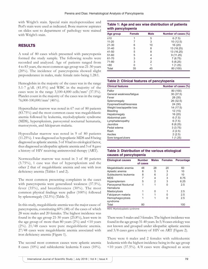
Pereira and Dias: Hematological Analysis of Pancytopenia
72International Journal of Scientific Study | July 2016 | Vol 4 | Issue 4
with Wright’s stain. Special stain myeloperoxidase and Perl’s stain were used as indicated. Bone marrow aspirates on slides sent to department of pathology were stained with Wright’s stain.
RESULTS
A total of 80 cases which presented with pancytopenia formed the study sample. The following results were recorded and analyzed. Age of patients ranged from 4 to 83 years, the most common age group was 21-30 years (20%). The incidence of pancytopenia showed slight preponderance in males, male: female ratio being 1.28:1.
Hemoglobin in the majority of the cases was in the range 5.1-7 g/dl. (41.0%) and WBC in the majority of the cases were in the range 3,100-4,000 cells/mm3 (37.5%). Platelet count in the majority of the cases was in the range 76,000-100,000/mm3 (46%).
Hypercellular marrow was noted in 67 out of 80 patients (83.75%) and the most common cause was megaloblastic anemia followed by leukemia, myelodysplastic syndrome (MDS), hypersplenism, paroxysmal nocturnal hematuria, mastocytosis, and falciparum malaria.
Hypocellular marrow was noted in 9 of 80 patients (11.25%). 1 was diagnosed as hypoplastic MDS and 8 being diagnosed as aplastic anemia. 5 of 8 had no etiological factor, thus diagnosed as idiopathic aplastic anemia and 3 of 8 gave a history of HIV receiving antiretroviral therapy (ART).
Normocellular marrow was noted in 3 of 80 patients (3.75%), 1 case was that of hypersplenism and the other 2 that of megaloblastic anemia and one with iron deficiency anemia (Tables 1 and 2).
The most common presenting complaints in the cases with pancytopenia were generalized weakness (37.5%), fever (35%), and breathlessness (30%). The most common physical findings were pallor (100%) followed by splenomegaly (32.5%) (Table 3).
In this study, megaloblastic anemia was the major cause of pancytopenia, constituting 60% (48) of the cases of which 28 were males and 20 females. The highest incidence was found in the age group 21-30 years (25.8%), least were in the age group of more than 80 years (2%) and <10 years (2%). 21/48 cases were pure megaloblastic anemia. 27/48 cases were megaloblastic anemia associated with iron deficiency anemia (Figure 1).
The second most common causes were aplastic anemia 8 cases (10%) and subleukemic leukemia 8 cases (10%).
There were 5 males and 3 females. The highest incidence was found in the age group 31-40 years. In 5/8 cases etiology was not known and grouped under idiopathic aplastic anemia and 3/8 cases gave a history of HIV on ART (Figure 2).
There were 6 males and 2 females with subleukemic leukemia with the highest incidence being in the age group <10 years (37.5%). 4/8 cases were diagnosed as acute
Table 1: Age and sex wise distribution of patients with pancytopeniaAge group Female Male Number of cases (%)≤10 1 5 6 (7.5)11-20 7 3 10 (12.5)21-30 6 10 16 (20)31-40 5 8 13 (16.25)41-50 5 8 13 (16.25)51-60 5 4 9 (11.25)61-70 3 4 7 (8.75)71-80 3 2 5 (6.25)>80 0 1 1 (1.25)Total 35 45 80 (100)
Table 2: Clinical features of pancytopeniaClinical features Number of cases (%)Pallor 80 (100)General weakness/fatigue 30 (37.5)Fever 28 (35)Splenomegaly 26 (32.5)Dyspnea/breathlessness 24 (30)Weight loss/appetite loss 14 (17.5)Bleeding 12 (15)Hepatomegaly 16 (20)Abdominal pain 6 (7.5)Lymphadenopathy 6 (7.5)Jaundice 8 (6.25)Pedal edema 3 (3.75)Rash 2 (2.5)Ascites 2 (2.5)Sore tongue/ulcers 1 (1.25)
Table 3: Distribution of the various etiological causes of pancytopeniaEtiological causes Number
of casesMales Females Percentage
Megaloblastic anemia 48 28 20 60Aplastic anemia 8 5 3 10Subleukemic leukemia 8 6 2 10MDS 7 4 3 8.75Hypersplenism 5 2 3 6.25Paroxysmal Nocturnal Hematuria
1 1 0 2.5
Mastocytosis 1 0 1 2.5Falciparum malaria 1 1 0 2.5Hemophagocytosis syndrome
1 1 0 2.5
Total 80 100MDS: Myelodysplastic syndrome

Pereira and Dias: Hematological Analysis of Pancytopenia
73 International Journal of Scientific Study | July 2016 | Vol 4 | Issue 4
myeloid leukemia (AML) and 4/8 cases were diagnosed as acute lymphoblastic leukemia (ALL) (Figure 3).
MDS was the third most common diagnosis with 7 cases (8.75%). There were 4 males and 3 females and the highest incidence was found in the age group 61-70 years (43%). 1/7 case was diagnosed as hypoplastic MDS and 6/7 cases were diagnosed as refractory cytopenia with multilineage dysplasia.
There were 5 cases of hypersplenism and one case each of paroxysmal nocturnal hematuria, mastocytosis, falciparum malaria and hemophagocytosis syndrome.
DISCUSSION
In this study done at the Goa Medical College over a period of 2 years, a total number of hemograms were 62,698 and bone marrow aspirations sent was 764; pancytopenia was found in 80 cases.
In this study, age of patients with pancytopenia ranged from 4 to 83 years. Most of the studies by Tilak and Jain4 Tariq et al.,5 Mussarrat et al.,6 Qamar and Aijaz7 Khodke et al.,8 and Gayathri and Rao9 reported patients to be in the same age range. Pancytopenia showed its highest incidence in the age group 21-30 years similar to the studies by Mussarrat et al.6 and Qamar and Aijaz.7
The most common cause of pancytopenia reported from various studies worldwide has been aplastic anemia. This is in sharp contrast with the results of the present study where the most common cause of pancytopenia was megaloblastic anemia. This seems to reflect the higher prevalence of nutritional anemia in Indian subjects as well as in developing countries3 (Table 4).
In this study, megaloblastic anemia was the most common cause of pancytopenia, the incidence being 60%. Incidence of 72% was reported by Khunger et al.,20 68% by Tilak and Jain4 and 74% by Gayathri and Rao,9 49% by Rangaswamy et al.,27 62% by Khanduri and Sharma,30 72.6% by Javalgi and Dombale,28 and 26.42% by Subrahmanyam and Padma.29
All the above-mentioned studies have been done in India, and they stress the importance of megaloblastic anemia as the major cause of pancytopenia. As facilities, for estimating folic acid and Vitamin B12 levels are not routinely available in most centers in India the exact deficiency is usually not identified.31
Age and Sex DistributionThe age of the patients with megaloblastic anemia ranged from 4 to 83 years and male:female ratio was 1.4:1. This
Figure 1: Megeloblastic anemia. (a) Bone marrow aspiration of a case of megaloblastic anemia showing erythroid hyperplasia
with magaloblastic maturation (×100), (b) peripheral smear case of megaloblastic anemia showing hypersegmented
neutrophil (×40)
a
b
Figure 2: Aplastic anemia. (a) Bone marrow aspirate of a case of aplastic anemia showing markedly hypocellular marrow (×100), (b) peripheral smear of a case aplastic anemia (×40)
a
b
Figure 3: Subleukemic leukemia. (a) Peripheral smear of a case of subleukemic leukemia presenting as pancytopenia (×40),
(b) myeloblast showing auer rods (×100)
a
b
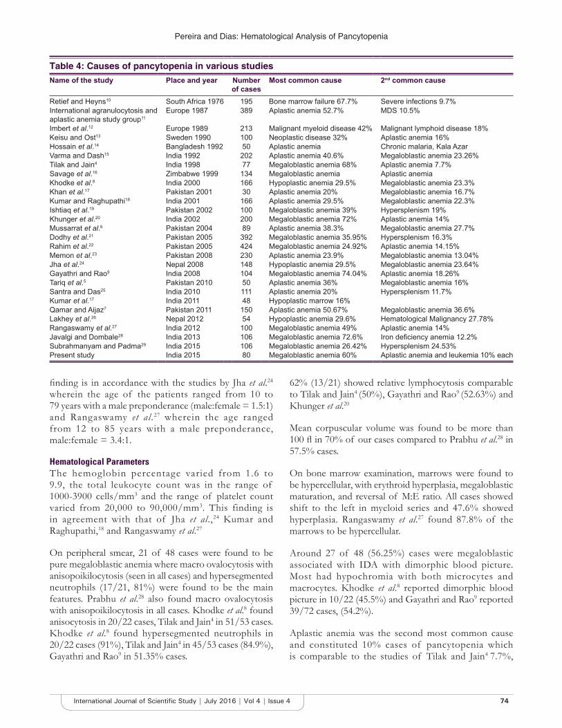
Pereira and Dias: Hematological Analysis of Pancytopenia
74International Journal of Scientific Study | July 2016 | Vol 4 | Issue 4
finding is in accordance with the studies by Jha et al.24 wherein the age of the patients ranged from 10 to 79 years with a male preponderance (male:female = 1.5:1) and Rangaswamy et al.27 wherein the age ranged from 12 to 85 years with a male preponderance, male:female = 3.4:1.
Hematological ParametersThe hemoglobin percentage varied from 1.6 to 9.9, the total leukocyte count was in the range of 1000-3900 cells/mm3 and the range of platelet count varied from 20,000 to 90,000/mm3. This finding is in agreement with that of Jha et al.,24 Kumar and Raghupathi,18 and Rangaswamy et al.27
On peripheral smear, 21 of 48 cases were found to be pure megaloblastic anemia where macro ovalocytosis with anisopoikilocytosis (seen in all cases) and hypersegmented neutrophils (17/21, 81%) were found to be the main features. Prabhu et al.28 also found macro ovalocytosis with anisopoikilocytosis in all cases. Khodke et al.8 found anisocytosis in 20/22 cases, Tilak and Jain4 in 51/53 cases. Khodke et al.8 found hypersegmented neutrophils in 20/22 cases (91%), Tilak and Jain4 in 45/53 cases (84.9%), Gayathri and Rao9 in 51.35% cases.
62% (13/21) showed relative lymphocytosis comparable to Tilak and Jain4 (50%), Gayathri and Rao9 (52.63%) and Khunger et al.20
Mean corpuscular volume was found to be more than 100 fl in 70% of our cases compared to Prabhu et al.28 in 57.5% cases.
On bone marrow examination, marrows were found to be hypercellular, with erythroid hyperplasia, megaloblastic maturation, and reversal of M:E ratio. All cases showed shift to the left in myeloid series and 47.6% showed hyperplasia. Rangaswamy et al.27 found 87.8% of the marrows to be hypercellular.
Around 27 of 48 (56.25%) cases were megaloblastic associated with IDA with dimorphic blood picture. Most had hypochromia with both microcytes and macrocytes. Khodke et al.8 reported dimorphic blood picture in 10/22 (45.5%) and Gayathri and Rao9 reported 39/72 cases, (54.2%).
Aplastic anemia was the second most common cause and constituted 10% cases of pancytopenia which is comparable to the studies of Tilak and Jain4 7.7%,
Table 4: Causes of pancytopenia in various studiesName of the study Place and year Number
of casesMost common cause 2nd common cause
Retief and Heyns10 South Africa 1976 195 Bone marrow failure 67.7% Severe infections 9.7%International agranulocytosis and aplastic anemia study group11
Europe 1987 389 Aplastic anemia 52.7% MDS 10.5%
Imbert et al.12 Europe 1989 213 Malignant myeloid disease 42% Malignant lymphoid disease 18%Keisu and Ost13 Sweden 1990 100 Neoplastic disease 32% Aplastic anemia 16%Hossain et al.14 Bangladesh 1992 50 Aplastic anemia Chronic malaria, Kala AzarVarma and Dash15 India 1992 202 Aplastic anemia 40.6% Megaloblastic anemia 23.26%Tilak and Jain4 India 1998 77 Megaloblastic anemia 68% Aplastic anemia 7.7%Savage et al.16 Zimbabwe 1999 134 Megaloblastic anemia Aplastic anemiaKhodke et al.8 India 2000 166 Hypoplastic anemia 29.5% Megaloblastic anemia 23.3%Khan et al.17 Pakistan 2001 30 Aplastic anemia 20% Megaloblastic anemia 16.7%Kumar and Raghupathi18 India 2001 166 Aplastic anemia 29.5% Megaloblastic anemia 22.3%Ishtiaq et al.19 Pakistan 2002 100 Megaloblastic anemia 39% Hypersplenism 19%Khunger et al.20 India 2002 200 Megaloblastic anemia 72% Aplastic anemia 14%Mussarrat et al.6 Pakistan 2004 89 Aplastic anemia 38.3% Megaloblastic anemia 27.7%Dodhy et al.21 Pakistan 2005 392 Megaloblastic anemia 35.95% Hypersplenism 16.3%Rahim et al.22 Pakistan 2005 424 Megaloblastic anemia 24.92% Aplastic anemia 14.15%Memon et al.23 Pakistan 2008 230 Aplastic anemia 23.9% Megaloblastic anemia 13.04%Jha et al.24 Nepal 2008 148 Hypoplastic anemia 29.5% Megaloblastic anemia 23.64%Gayathri and Rao9 India 2008 104 Megaloblastic anemia 74.04% Aplastic anemia 18.26%Tariq et al.5 Pakistan 2010 50 Aplastic anemia 36% Megaloblastic anemia 16%Santra and Das25 India 2010 111 Aplastic anemia 20% Hypersplenism 11.7%Kumar et al.17 India 2011 48 Hypoplastic marrow 16%Qamar and Aijaz7 Pakistan 2011 150 Aplastic anemia 50.67% Megaloblastic anemia 36.6%Lakhey et al.26 Nepal 2012 54 Hypoplastic anemia 29.6% Hematological Malignancy 27.78%Rangaswamy et al.27 India 2012 100 Megaloblastic anemia 49% Aplastic anemia 14%Javalgi and Dombale28 India 2013 106 Megaloblastic anemia 72.6% Iron deficiency anemia 12.2%Subrahmanyam and Padma29 India 2015 106 Megaloblastic anemia 26.42% Hypersplenism 24.53%Present study India 2015 80 Megaloblastic anemia 60% Aplastic anemia and leukemia 10% each

Pereira and Dias: Hematological Analysis of Pancytopenia
75 International Journal of Scientific Study | July 2016 | Vol 4 | Issue 4
Khunger et al.20 14%, Rahim et al.22 14.15%, Rangaswamy, Rangaswamy et al.27 14% and Khodke et al.8 14%.
The incidence of aplastic anemia quoted from the West is much higher than that observed by US. The increased incidence may be related to environmental factors.10 A few studies that quote aplastic anemia to be the most common cause in India as well as elsewhere are International Agranulocytosis and Aplastic Anemia study group11 52.7%, Varma and Dash15 40.6%, Khodke et al.8 29.5%, Khan et al.17 20%, Kumar et al.32 29.5%, Mussarrat et al.6 38.3%, Memon et al.25 23.9%, Jha et al.24 29.5%, Mohammad Tariq et al.5 36%, Lakhey et al.26 29.6%, Santra and Das25 50.67%, Kumar and Raghupathi18 16%, Qamar and Aijaz7 20%.
Age and Sex DistributionAge of patients with aplastic anemia ranged from 7 to 67 years and male: female being 1.7:1. studies by Kumar and Raghupathi18 Jha et al.,24 Rangaswamy et al.27 and Tilak and Jain4 reported similar age range and male:female ratio.
Hematological ParametersHemoglobin varied from 3.2 to 8.6 g/dl, total WBC count varied from 400 to 3900 cells/mm3 and platelet count varied from 20,000 to 90,000/mm3. Similar hematological parameters were reported by Tilak and Jain4 Jha et al.,24 Kumar and Raghupathi18 and Rangaswamy et al.27
Peripheral Smear and Bone Marrow Aspiration FindingsIn our study, five of the eight patients showed anisocytosis and three of eight showed relative lymphocytosis. In a study by Khodke et al.,8 three of seven patients showed anisocytosis and one of three showed relative lymphocytosis. In a study by Tilak and Jain,4 two of six patients showed anisocytosis and three of six showed relative lymphocytosis.
Bone marrow findings were the following: Markedly hypocellular with increased fat spaces, marked depression of all 3 series and increase in plasma cells, lymphocytes, and reticular cells. Mohler33 in their analysis of 50 cases reported 74% patients with hypocellular marrow, 16% with normocellular marrow which later became hypocellular and 10% were acellular.
5/8 cases etiology was not known, grouped under Idiopathic aplastic anemia.
3/8 cases gave a history of HIV on ART. Khodke et al.8 reported 1 case of HIV (50 cases). Ishtiaq et al.19 reported 1 case of AIDS presenting with pancytopenia, so did Rangaswamy et al.27
Leukemia was also the second most common cause and constituted 10% of the cases of our study, which is comparable to the studies of Tariq et al.5 (14%), Mussarrat et al.6 (13.6%) and Qamar and Aijaz7 11%.
Khoo and Keihani33 in a study of 188 cases of pancytopenia found leukemia to be the most common cause, most of the cases being AML.
In the study by Jha et al.24 acute leukemia alone constituted 19.59% of the total cases of pancytopenia, in contrast to the studies by Khodke et al.8 and Tilak and Jain4 where one case of AML was detected as a cause of pancytopenia. Khunger et al.20 reported incidence of 5% and so did Rangaswamy et al.27 Santra and Das25 reported incidence of 1.8% and Ishtiaq et al.19 1.8%.
Nevertheless, in the study of Bashawri34 the main indication for bone marrow examination in cases of pancytopenia was acute leukemia.
Pancytopenia can be seen in 30% of acute leukemia at the time of presentation.8
The highest incidence was found in the age group <10 years (37.5%) and the age ranged from 5 to 57 years with a male: female = 3:1, which is in agreement to that reported by Jha et al.24 years and Rangaswamy et al.27
Hemoglobin varied from 3.7 to 8.1 g/dl. Total WBC count varied from 400 to 3800 cells/mm3. Platelet count varied from 20,000 to 94,000/mm3 similar to the hematological parameters reported by Jha et al.,24 Kumar and Raghupathi18 and Rangaswamy et al.27
Bone marrow aspiration revealed all cases to be hypercellular, with decreased fat spaces and marked suppression of megakaryocytic and myeloid series. 4/8 cases were diagnosed as AML, showed presence of myeloblast and were myeloperoxidase positive. 4/8 cases were diagnosed as ALL, showed presence of lymphoblast and were myeloperoxidase negative. In the study by Rangaswamy et al.27 80% marrows were hypercellular and 20% hypocellular.
In the study by Tariq et al.5 ALL constituted 12% cases and AML 2% cases whereas Varma and Dash15 reported AML to be 12.8%. Gayathri and Rao9 reported 4 cases of leukemia, 3 being AML and 1 ALL and Kumar and Raghupathi18 reported 13 cases of AML and 5 cases of ALL.
The incidence of MDS in our study was 8.75%. Lakhey et al.26 reported an incidence of 7.4%, Kumar and

Pereira and Dias: Hematological Analysis of Pancytopenia
76International Journal of Scientific Study | July 2016 | Vol 4 | Issue 4
Raghupathi18 8.3% which is comparable to our study. Qamar and Aijaz7 reported incidence of 5.3%, Mussarrat et al.6 2.4%, Khunger et al.20 2%, Jha et al.24 0.94% and Tariq et al.5 14%.
In a study of 118 patients with MDS by Juneja et al.,35 age ranged from 48 to 95 years, study by Lakhey et al.26 age ranged from 30 to 79 years. In the present study, highest incidence was found in the age group 61-70 years (43%) and age of patients ranged from 28 to 65 years, male:female=1.3:1
6/5 cases showed hypochromia, anisocytosis, microcytes, macrocytes. Anemia associated with MDS can be microcytic, normocytic, or, most commonly, macrocytic.36
Six of seven cases were diagnosed as refractory cytopenia with multilineage dysplasia. Marrow was hypercellular, showed dysplastic features in erythroid series ranging from increased mitotic activity in normoblasts, abnormal mitosis, megaloblastoid change, nuclear hypoblasts, irregular hypersegmentation, ringed nuclei and abnormal segmentation. Dysgranulopoeis and dysmegakaryopoiesis were also noted.
In a study by Kumar and Raghupathi18 of the four cases of MDS, one was refractory anemia, two refractory cytopenia with multilineage dysplasia and one MDS unclassified.
Hypersplenism constituted 6.25% cases of pancytopenia. Age of patients ranged from 21 to 63 years male:female = 3:4.
Retief and Heyns10 found hypersplenism to be the cause of pancytopenia in 7.7% cases, which is comparable to our finding.
Rangaswamy et al.27 found hypersplenism to be a cause of pancytopenia in 10% cases, with male:female ratio of 1:1.5. Ishtiaq et al.19 in 12% with a female preponderance being 1:1.3.). In a study of 195 patients, Kumar and Raghupathi18 reported incidence of hypersplenism in 11%, age ranging 14-49 years. Momen et al.36 in 4.34%, Santra and Das25 11.7%, Tariq et al.5 in 10%.
Hemoglobin varied from 5.4 to 9.9 m/dl, total WBC count varied from 1100 to 3600 cells/mm3 and platelet count varied from 50,000 to 90,000/mm3. This is in conformity with the findings of Kumar and Raghupathi18 and Gayathri and Rao.9
A single case of paroxysmal nocturnal hematuria was encountered. Santra and Das25 in a study of 111 cases of pancytopenia encountered one case and so did Khan et al.17 in a study of 30 cases of pancytopenia.
A 55-year-old male presented with hematuria, jaundice and splenomegaly. Laboratory investigations revealed elevated lactic acid dehydrogenase levels, raised erythrocyte sedimentation rate and C-reactive protein, mildly elevated fetal hemoglobin, serum ferritin 37 µg/L, and total iron binding capacity 289/µg/dl.
We encountered a single case of falciparum malaria causing pancytopenia.
In the study of Hossain et al.,14 chronic malaria was the second most common cause of pancytopenia. In the study of Khunger et al.,20 incidence was 1%, Gayathri and Rao9 1.93%, Tilak and Jain4 3.9%, Kumar and Raghupathi18 3%, and Santra and Das25 1.8%.
We encountered a single case of mastocytosis causing pancytopenia.
None of the above studies of pancytopenia have encountered a case of mastocytosis. Systemic mast cell disease is characterized by bone marrow involvement by mast cells and frequently by peripheral blood cytopenias and may result in pancytopenia.37,38
A 16-year-old female presented with fever, macular-papular rash on face. Hemoglobin 5.4 g/dl total count 1900/mm3 platelet count: 48,000/mm3. Peripheral smear revealed moderate hypochromia, moderate anisocytosis. Bone marrow was hypercellular. Fat content was decreased. M:E ratio was reversed to 1:3.
Erythroid series were hyperplastic at all levels. Most showing megaloblastoid change. Myeloid series showed shift to left with mild increase in eosinophilic precursors. A Large number of mature mast cells, precursors were seen scattered in marrow, forming 40% of the nonerythroid cells. Megakaryocytic series were normal and iron staining was mildly increased +4.
A single case of hemophagocytosis was encountered in our study. Tilak and Jain4 also encountered one such case in a study of 77 cases of pancytopenia.
A 4-year-old boy presented with fever, breathlessness, bleeding and was found to have hepatomegaly. Erythrocyte sedimentation rate was raised.
Hemoglobin was 1.9 g/dl, the total count of 3500/mm3 and platelet count of 90,000/mm3. Peripheral smear examination revealed moderate hypochromia and moderate anisocytosis, 2 normoblasts/100 WBC’ S. Differential count showed shift to left and presence of toxic granules.

Pereira and Dias: Hematological Analysis of Pancytopenia
77 International Journal of Scientific Study | July 2016 | Vol 4 | Issue 4
Bone marrow aspiration revealed the following: No marrow fragments were aspirated, mainly sinusoidal blood. Few myeloid and erythroid cells seen which show normal morphology. Occasional megakaryocytes seen. Scattered among the marrow cells are few histiocytes showing phagocytosis of WBC’s.
We were not able to determine the exact etiology of hemophagocytosis syndrome.
CONCLUSION
Pancytopenia is not an uncommon hematological problem encountered in clinical practice and should be suspected on clinical grounds when a patient presents with unexplained anemia, prolonged fever and tendency to bleed.
Physical findings and peripheral blood picture provide valuable information in the workup of cytopenic patients.
Bone marrow aspiration is an important diagnostic tool in hematology which helps to evaluate various causes of cytopenia and to plan further investigations and management of the patients.
Since a large number of causes for pancytopenia are remediable and reversible, accurate diagnoses and timely intervention may be lifesaving and will definitely have an influence on the morbidity and mortality in these patients.
The early detection of the underlying conditions would also help to enhance the prognosis of a patient with pancytopenia.
Thus, a comprehensive, clinical, and hematological study of patients with pancytopenia will usually help in identifying the underlying cause.
Further research with a larger sample size is required to replicate the findings of this study.
ACKNOWLEDGMENT
Author has shown a sincere gratitude to the staff of Department of Pathology for their unstinted support and cooperation.
REFERENCES
1. Williams DM. Pancytopenia, aplastic anaemia and pure red cell aplasia. In: Lee GR, Bithell TC, Foerster J, Athens JW, Lukens JN, editors. Wintrobes Clinical Haematology. 10th ed. Philadelphia, PA: Williams and Wilkins; 1999. p. 1449-84.
2. Firkin F, Chesterman C, Pennigton D, Rush B. De Gruchy’s Clinical
Hematology in Medical Practice. 5th ed. London: Blackwell Science Ltd.; 1989. p. 119-36.
3. Segel GB, Lichtman MA. Aplastic anemia: Acquired and inherited. In: Lichtman MA, Beutler E, Kipps TJ, Seligsohm U, Kaushansky K, Prchal JT, editors. Williams Hematology. 7th ed. New York: McGraw-Hill Medical; 2006. p. 419-30.
4. Tilak V, Jain R. Pancytopenia - A clinico-hematologic analysis of 77 cases. Indian J Pathol Microbiol 1999;42:399-404.
5. Tariq M, Khan N, Basri R, Amin S. Aetiology of pancytopenia. Prof Med J 2010;17:252-6.
6. Mussarrat N, Fazal-e-R M, Mohammad TK. Clinical and hematological features of megaloblastic anemia alone or in combination with iron deficiency anemia - An analysis of 349 patients. J Med Sci 2009;17:81-4.
7. Qamar U, Aijaz J. Results of bone marrow examination in patients presenting with pancytopenia and high mean corpuscular volume. Gomal J Med Sci 2012;10:133-6.
8. Khodke K, Marwah S, Buxi G, Yadav RB, Chaturvedi NK. Bone marrow examination in cases of pancytopenia. J Indian Acad Clin Med 2001;2:56-9.
9. Gayathri BN, Rao KS. Pancytopenia: A clinico hematological study. J Lab Physicians 2011;3:15-20.
10. Retief FP, Heyns AD. Pancytopenia and aplastic anaemia: A retrospective study. S Afr Med J 1976;50:1318-22.
11. Risks of agranulocytosis and aplastic anemia. A first report of their relation to drug use with special reference to analgesics. The International agranulocytosis and aplastic anemia study. JAMA 1986;256:1749-57.
12. Imbert M, Scoazec JY, Mary JY, Jouzult H, Rochant H, Sultan C. Adult patients presenting with pancytopenia: A reappraisal of underlying pathology and diagnostic procedures in 213 cases. Hematol Pathol 1989;3:159-67.
13. Keisu M, Ost A. Diagnoses in patients with severe pancytopenia suspected of having aplastic anemia. Eur J Haematol 1990;45:11-4.
14. Hossain MA, Akond AK, Chowdhary MK, Singh KJ, Ahluwalia G, Sharma SK, et al. Pancytopenia - A study of 50 cases. Bangladesh J Pathol 1992;1:9-12.
15. Varma N, Dash S. A reappraisal of underlying pathology in adult patients presenting with pancytopenia. Trop Geogr Med 1992;44:322-7.
16. Savage DG, Allen RH, Gangaidzo IT, Levy LM, Gwanzura C, Moyo A, et al. Pancytopenia in Zimbabwe. Am J Med Sci 1999;317:22-32.
17. Khan NM, Ayub M, Nawaz KH, Naqi M, Hussain T, Shujaat H, et al. Pancytopenia: Clinicopathological study of 30 cases at Military hospital, Rawalpindi. Pak J Pathol 2001;12:37-41.
18. Kumar DB, Raghupathi AR. Clinicohematologic analysis of pancytopenia: Study in a tertiary care centre. Basic Appl Pathol 2012;5:19-21.
19. Ishtiaq O, Baqai HZ, Anwer F, Hussain N. Patterns of pancytopenia patients in a general medical ward and a proposed diagnostic approach. J Ayub Med Coll Abbottabad 2004;16:8-13.
20. Khunger JM, Arulselvi S, Sharma U, Ranga S, Talib VH. Pancytopenia - A clinico haematological study of 200 cases. Indian J Pathol Microbiol 2002;45:375-9.
21. Dodhy MA, Bokhari N, Hayat A. A etiology of Pancytopenia, a five-year experience. Ann Pak Inst Med Sci 2005;1:92-5.
22. Rahim F, Ahmad I, Islam S, Hussain M, Khattak TA, Bano Q. Spectrum of haematological diseases in children observed in 424 cases of bone marrow aspirations/biopsies. Pak J Med Sci 2005;21:433-6.
23. Memon S, Shaikh S, Nizamani MA. Etiological spectrum of pancytopenia based on bone marrow examination in children. J Coll Physicians Surg Pak 2008;18:163-7.
24. Jha A, Sayami G, Adhikari RC, Panta AD, Jha R. Bone marrow examination in cases of pancytopenia. JNMA J Nepal Med Assoc 2008;47:12-7.
25. Santra G, Das BK. A cross-sectional study of the clinical profile and aetiological spectrum of pancytopenia in a tertiary care centre. Singapore Med J 2010;51:806-12.
26. Lakhey A, Talwar OP, Singh VK, Raj SK. Clinico-hematological study of pancytopenia. J Pathol Nepal 2012;2:207-10.
27. Rangaswamy M, Prabhu, Nandini NM, Manjunath GV. Bone marrow examination in pancytopenia. J Indian Med Assoc 2012;110:560-2, 566.
28. Javalgi AP, Dombale VD. Bone marrow study in pancytopenia. Natl J Lab Med 2013;2:12-7.
29. Subrahmanyam Y, Padma M. Pancytopenia a three years evaluation. Int J Sci Res 2015;4:1.

Pereira and Dias: Hematological Analysis of Pancytopenia
78International Journal of Scientific Study | July 2016 | Vol 4 | Issue 4
30. Khanduri U, Sharma A. Megaloblastic anaemia: Prevalence and causative factors. Natl Med J India 2007;20:172-5.
31. Mohler DN, Leavell BS. Aplastic anemia: An analysis of 50 cases. Ann Int Med 1958;49:326-62.
32. Alter BP. Arms and the man or hands and the child: Congenital anomalies and hematologic syndromes. J Pediatr Hematol Oncol 1997;19:287.
33. Khoo HJ, Keihani M. Causes of pancytopenia. Tehran Univ Med J 2006;64:1-2.
34. Bashawri LA. Bone marrow examination. Indications and diagnostic value.
Saudi Med J 2002;23:191-6.35. Juneja SK, Imbert M, Jouault H, Scoazec JY, Sigaux F, Sultan C.
Haematological features of Primary Myelodysplastic Syndromes (PMDS) at initial presentation: A study of 118 cases. J Clin Pathol 1983;36:1129-35.
36. Parker RI. Hematologic aspects of systemic mastocytosis. Hematol Oncol Clin North Am 2000;14:557-68.
37. Valent P, Horny HP, Escribano L, Longley BJ, Li CY, Schwartz LB, et al. Diagnostic criteria and classification of mastocytosis: A consensus proposal. Leuk Res 2001;25:603-25.
How to cite this article: Pereira ADS, Dias A. Hematological Analysis of Pancytopenia: A Prospective Study. Int J Sci Stud 2016;4(4):71-78.
Source of Support: Nil, Conflict of Interest: None declared.

79 International Journal of Scientific Study | July 2016 | Vol 4 | Issue 4
Cervical Cancer and its Demographic Factors at Central IndiaB K Sharma1, Sanghmitra Singh2
1Associate Professor, Department of Pathology, Bundelkhand Medical College, Sagar, Madhya Pradesh, India, 2Resident Surgical Officer, Department of Obstetrics and Gynaecology, S.S. Medical College, Rewa, Madhya Pradesh, India
Saharan african countries, Latin America. About 84 per cent of cervical cancer cases occurred in less developed countries like India, Pakistan and most of the African countries where incidence ranges between 20 and 30 per 100, 000. Most of the developed world, China and Middle East have a low incidence of <8/100,000.
In India, the peak age for cervical cancer incidence is 55-59 years.4 Current data from the National Cancer Registry Program indicate that the most common sites of cancer among women are the breasts and the cervix.5 It can be detected at a very early stage by simple technique of exfoliative cytology. Early stage detection is important because early stage is 100% curable reducing the morbidity and mortality from invasive cancer cervix. However, the most common lesions of the female genital tract are inflammatory lesions and parasitic lesions. Some inflammatory lesions such as trichomonas and human papillomavirus are forerunners of malignancy underlining the importance of diagnosing these conditions.
INTRODUCTION
Cancer of the uterine cervix is the 7th most common cancer in the world, and it is the most common cancer causing death among women in developing countries.1 Globally, the annual estimated number of new cases is 352,414 which accounts for 9% of all cancer diagnosed in women. 86% of the cancers occur in the developing countries,2 whereas 20% in the undeveloped countries.3
The incidence of cancer of uterine cervix varies from part to part in the world. The highest incidence is seen in Sub
Original Article
AbstractIntroduction: Cancer of the uterine cervix is the 7th most common cancer in the world, and it is the most common cancer causing death among women in developing countries. Africa, Central America, India, Pakistan, and most of the African countries have an incidence between 20 and 30/100,000.
Objective: With the objective to determine different abnormal cervical pattern and age, parity, socioeconomic status, and literacy wise distribution of cancers.
Materials and Methods: The present study was carried out at the Sanjay Gandhi Hospital and SS Medical College, Rewa, in 1-year duration with 200 samples were taken in account.
Results: Reactive changes 39 cases (19.5%) show epithelial abnormalities including atypical squamous cells of undetermined significance and atypical glandular cells of undetermined significance (13 cases) 6.5%. 15 cases (7.5%) show Low-grade squamous intraepithelial lesion (SIL), 8 cases (4%) show high-grade SIL, and 3 cases (1.5%) show carcinoma.
Conclusion: Most of the cases with abnormal smear were seen from the parity more than three and above. Furthermore, it has been observed that literacy and socioeconomic conditions are also having a high impact on rate among carcinoma cervix as it has been seen that lower socioeconomic condition and lower rate of literacy have more incidences of abnormal smear.
Key words: Cervical cancer, High-grade squamous intraepithelial lesion, Low-grade squamous intraepithelial lesion, Smear
Access this article online
www.ijss-sn.com
Month of Submission : 05-2016 Month of Peer Review : 06-2016 Month of Acceptance : 07-2016 Month of Publishing : 07-2016
Corresponding Author: Dr. B K Sharma, Associate Professor, Department of Pathology, Bundelkhand Medical College, Sagar, Madhya Pradesh, India. E-mail id: [email protected]
Print ISSN: 2321-6379Online ISSN: 2321-595X
DOI: 10.17354/ijss/2016/379
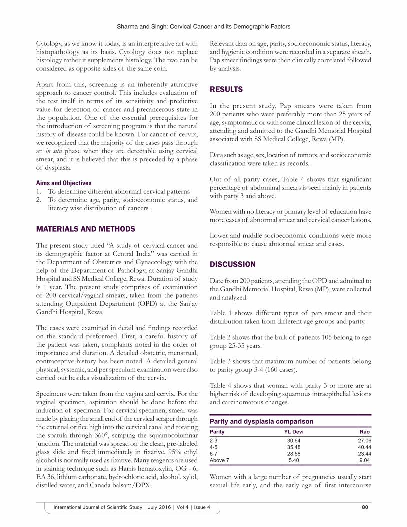
Sharma and Singh: Cervical Cancer and its Demographic Factors
80International Journal of Scientific Study | July 2016 | Vol 4 | Issue 4
Cytology, as we know it today, is an interpretative art with histopathology as its basis. Cytology does not replace histology rather it supplements histology. The two can be considered as opposite sides of the same coin.
Apart from this, screening is an inherently attractive approach to cancer control. This includes evaluation of the test itself in terms of its sensitivity and predictive value for detection of cancer and precancerous state in the population. One of the essential prerequisites for the introduction of screening program is that the natural history of disease could be known. For cancer of cervix, we recognized that the majority of the cases pass through an in situ phase when they are detectable using cervical smear, and it is believed that this is preceded by a phase of dysplasia.
Aims and Objectives1. To determine different abnormal cervical patterns2. To determine age, parity, socioeconomic status, and
literacy wise distribution of cancers.
MATERIALS AND METHODS
The present study titled “A study of cervical cancer and its demographic factor at Central India” was carried in the Department of Obstetrics and Gynaecology with the help of the Department of Pathology, at Sanjay Gandhi Hospital and SS Medical College, Rewa. Duration of study is 1 year. The present study comprises of examination of 200 cervical/vaginal smears, taken from the patients attending Outpatient Department (OPD) at the Sanjay Gandhi Hospital, Rewa.
The cases were examined in detail and findings recorded on the standard preformed. First, a careful history of the patient was taken, complaints noted in the order of importance and duration. A detailed obstetric, menstrual, contraceptive history has been noted. A detailed general physical, systemic, and per speculum examination were also carried out besides visualization of the cervix.
Specimens were taken from the vagina and cervix. For the vaginal specimen, aspiration should be done before the induction of specimen. For cervical specimen, smear was made by placing the small end of the cervical scraper through the external orifice high into the cervical canal and rotating the spatula through 360°, scraping the squamocolumnar junction. The material was spread on the clean, pre-labeled glass slide and fixed immediately in fixative. 95% ethyl alcohol is normally used as fixative. Many reagents are used in staining technique such as Harris hematoxylin, OG - 6, EA 36, lithium carbonate, hydrochloric acid, alcohol, xylol, distilled water, and Canada balsam/DPX.
Relevant data on age, parity, socioeconomic status, literacy, and hygienic condition were recorded in a separate sheath. Pap smear findings were then clinically correlated followed by analysis.
RESULTS
In the present study, Pap smears were taken from 200 patients who were preferably more than 25 years of age, symptomatic or with some clinical lesion of the cervix, attending and admitted to the Gandhi Memorial Hospital associated with SS Medical College, Rewa (MP).
Data such as age, sex, location of tumors, and socioeconomic classification were taken as records.
Out of all parity cases, Table 4 shows that significant percentage of abdominal smears is seen mainly in patients with party 3 and above.
Women with no literacy or primary level of education have more cases of abnormal smear and cervical cancer lesions.
Lower and middle socioeconomic conditions were more responsible to cause abnormal smear and cases.
DISCUSSION
Date from 200 patients, attending the OPD and admitted to the Gandhi Memorial Hospital, Rewa (MP), were collected and analyzed.
Table 1 shows different types of pap smear and their distribution taken from different age groups and parity.
Table 2 shows that the bulk of patients 105 belong to age group 25-35 years.
Table 3 shows that maximum number of patients belong to parity group 3-4 (160 cases).
Table 4 shows that woman with parity 3 or more are at higher risk of developing squamous intraepithelial lesions and carcinomatous changes.
Parity and dysplasia comparisonParity YL Devi Rao2-3 30.64 27.064-5 35.48 40.446-7 28.58 23.44Above 7 5.40 9.04
Women with a large number of pregnancies usually start sexual life early, and the early age of first intercourse
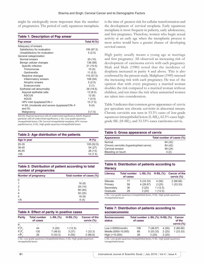
Sharma and Singh: Cervical Cancer and its Demographic Factors
81 International Journal of Scientific Study | July 2016 | Vol 4 | Issue 4
might be etiologically more important than the number of pregnancies. The period of early squamous metaplasia
is the time of greatest risk for cellular transformation and the development of cervical neoplasia. Early squamous metaplasia is most frequent in puberty, early adolescence, and first pregnancy. Therefore, women who begin sexual activity at an early age when the metaplastic process is most active would have a greater chance of developing cervical cancer.
High parity usually means a young age at marriage and first pregnancy. All observed an increasing risk of development of carcinoma cervix with each pregnancy. Shah and Shah (1980) noted that the incidence of dysplasia increased in parity 4 and above. This is also confirmed by the present study. Maliphant (1949) stressed the increasing risk with each pregnancy. He was of the opinion that with every pregnancy a married woman doubles the risk compared to a married woman without children, and ten times the risk when unmarried women are taken into consideration.
Table 5 indicates that common gross appearance of cervix per speculum was chronic cervicitis in abnormal smears. Chronic cervicitis was seen in 53.3% cases of low-grade squamous intraepithelial lesion (L-SIL), 62.5% cases high-grade SIL (H-SIL), and 33.33% cases carcinoma cervix.
Table 5: Gross appearance of cervixAppearance Total number of cases (%)Normal 64 (32)Chronic cervicitis (hypertrophied cervix) 84 (42)Cervical erosion 48 (24)Bleeding on touch 4 (2)
Table 1: Description of Pap smearPap smear Total N (%)Adequacy of smears
Satisfactory for evaluation 195 (97.5)Unsatisfactory for evaluation 5 (2.5)
General categorizationNormal smears 20 (10)Benign cellular changes 136 (68)
Specific infection 21 (10.5)Trichomonas 10 (5)Gardnerella 11 (5.5)
Reactive changes 115 (57.5)Inflammatory smears 108 (54)Atrophic smears 5 (2.5)Endocervicitis 2 (1)
Epithelial cell abnormality 39 (19.5)Atypical epithelial cells 13 (6.5)
ASCUS 12 (6)AGUS 1 (0.5)
HPV mild dysplasia/CIN–I 15 (7.5)H-SIL (moderate and severe dysplasia/CIN–II and III)
8 (4)
Malignancy 3 (1.5)ASCUS: Atypical squamous cells of undermined significance, AGUS: Atypical glandular cells of undermined significance, L‑SIL: Low‑grade squamous intraepithelial lesion, CIN: Cervical intraepithelial neoplasia, HPV: Human papillomavirus, H‑SIL: High‑grade squamous intraepithelial lesion
Table 2: Age distribution of the patientsAge in year N (%)25-35 105 (52.5)36-45 54 (27)46-55 26 (13)>55 15 (7.5)
Table 3: Distribution of patient according to total number of pregnanciesNumber of pregnancy Total number of cases (%)0 -1 16 (8)2 28 (14)3 80 (40)4 50 (25)5 18 (9)>/6 8 (4)
Table 4: Effect of parity in positive casesParity Total number
of casesL-SIL (%) H-SIL (%) Cancer of the
cervix (%)P0 - - - -P1P2 44 3 (20) 1 (12.5) -P3P4 130 7 (46.6) 3 (37) 1 (33.3)>/P5 26 5 (33.3) 4 (50) 2 (66.6)L‑SIL: Low‑grade squamous intraepithelial lesion, H‑SIL: High‑grade squamous intraepithelial lesion
Table 6: Distribution of patients according to literacyLiteracy Total number
of casesL-SIL (%) H-SIL (%) Cancer of the
cervix (%)Illiterate 77 5 (33.33) 4 (50) 2 (66.66)Primary 58 4 (26.67) 2 (25) 1 (33.33)Secondary 36 3 (20) 1 (12.5)Graduate 29 3 (20) 1 (12.5)L‑SIL: Low‑grade squamous intraepithelial lesion, H‑SIL: High‑grade squamous intraepithelial lesion
Table 7: Distribution of patients according to socioeconomicSocioeconomic status
Total number of cases
L-SIL (%) H-SIL (%) Cancer of the
cervix (%)Low (<5000/month) 106 7 (46.67) 4 (50) 2 (66.66)Middle (5000-10,000) 66 5 (53.33) 2 (25) 1 (33.33)High (>10,000) 28 3 (20) 2 (25) -L‑SIL: Low‑grade squamous intraepithelial lesion, H‑SIL: High‑grade squamous intraepithelial lesion

Sharma and Singh: Cervical Cancer and its Demographic Factors
82International Journal of Scientific Study | July 2016 | Vol 4 | Issue 4
of carcinoma diagnose by cytology, and also the result of present series of work differ much from results of others.
CONCLUSION
In the present study, 200 Pap smear analyzed, which shows the distribution pattern of various conditions, of which, 115 cases (57.5%) show reactive changes, 39 cases (19.5%) show epithelial abnormalities including atypical squamous cells of undetermined significance and atypical glandular cells of undetermined significance (13 cases) 6.5%. 15 cases (7.5%) show L-SIL, 8 cases (4%) show H-SIL, and 3 cases (1.5%) show carcinoma. Most of the cases with abnormal smear were seen from the parity more than 3 and above. Furthermore, it has been observed that literacy and socioeconomic conditions are also having a high impact on rate among carcinoma cervix as it has been seen that lower socioeconomic condition and lower rate of literacy have more incidences of abnormal smear.
REFERENCES
1. Denny L. Cervical cancer: Prevention and treatment. Discov Med 2012;14:125-31.
2. Arbyn M, Castellsagué X, de Sanjosé S, Bruni L, Saraiya M, Bray F, et al. Worldwide burden of cervical cancer in 2008. Ann Oncol 2011;22:2675-86.
3. Yeole BB, Kumar AV, Kurkure A, Sunny L. Population-based survival from cancers of breast, cervix and ovary in women in Mumbai, India. Asian Pac J Cancer Prev 2004;5:308-15.
4. World – Both Sexes Estimated Incidence by Age. Available from: http://www.globocan.iarc.fr/old/age_specific_table_r.asp? [Last accessed on 2015 Dec 30].
5. Nandakumar A, Ramnath T, Chaturvedi M. The magnitude of cancer cervix in India. Indian J Med Res 2009;130:219-21.
Cervical erosion was presenting 33.33% cases of L-SIL, 37.5% cases of H-SIL, and 66.66% cases of carcinoma cervix.
Table 6 shows the relationship of different level of literacy with the incidence of cervical cancer lesions, which clearly describe illiterate has maximum number of cases. Which also tells as literacy increases awareness increases towards health.
Table 7 describe relation of cases with Socio economic status in society. Low socio economic status has maximum no of cases which decline drastically as status increases.
A close association of dysplasia and malignancy with unhealthy cervix, e.g., cervical hypertrophy, cervical erosion has been observed by many people (Singh et al., Guard et al., Gupta et al., Panda et al., etc.) Therefore, all cases of suspected cervical lesion should be routinely subjected to Pap smears, and patients should be kept on regular follow-up.
Out of 200 smears and analyzed 7.5% were of L-SIL, 4% were of H-SIL, and 1.5% were malignant.
MacGregor and Baird (1963) screened 2683 cases and reported 18 (0.67%) to be malignant. Wahi et al. (1972) found rate of malignant smears to be 0.6% and that of dysplastic smears 6.7%.
From the above observation of different workers, it is concluded that there is a wide variability in the incidence
How to cite this article: Sharma BK, Singh S. Cervical Cancer and its Demographic Factors at Central India. Int J Sci Stud 2016;4(4):79-82.
Source of Support: Nil, Conflict of Interest: None declared.

83 International Journal of Scientific Study | July 2016 | Vol 4 | Issue 4
Visual Impairment Due to Refractive Errors among Female School Students Attending Tertiary Care Hospital, AnanthapuramuSatyanarayana Prasad Yellapu Maruthi1, Sreenivasulu Palla2
1Assistant Professor, Department of Ophthalmology, Government Medical College and Hospital, Ananthapuramu, Andhra Pradesh, India, 2Associate Professor, Department of Ophthalmology, Government Medical College and Hospital, Ananthapuramu, Andhra Pradesh, India
WHO globally for immediate attention to achieve the goals of Vision 2020.1
Blindness problem has gained importance globally; its magnitude is much higher in India. Of the estimated 45 million, India alone has 9 million blind people, which comes to one-fifth of the total in world.2
Refractive errors are of various types, predominantly affecting school going children. It is one of most common public health problem, which is treatable eye condition. Refractive errors are common cause of visual impairment, second treatable blindness. It is estimated that there are 35 million people in the world who require low vision care and 8 million (18%) are blind due to refractive errors.2 During survey period, blindness National Programme for Control of Blindness survey (2001-2002)3 reported 19.7% refractive
INTRODUCTION
Since the last three decades, recognition of concept of avoidable blindness (i.e., preventable or curable) is rising. For prevention of blindness, major global initiatives have taken. They are: WHO: Prevention of Blindness Programme (1978), Vision 2020 - The right to sight (1999) and vision for the future (2001). Refractive errors are the one among five major blinding eye conditions taken by
Original Article
AbstractIntroduction: Refractive errors are of various types, predominantly affecting school going children. Mostly in developing countries, refractive errors are responsible for either visual impairment of blindness. The aim of this study is to know the prevalence of refractive errors among female schoolchildren of 7-18 years age group and assessment of socio-demographic features among myopia children.
Materials and Methods: This is a prospective cross-sectional study conducted for 1 year (2014) among female schoolchildren attending the Department of Ophthalmology at Government General Hospital, Ananthapuramu. A total of 1600 female schoolchildren presenting with complaints of visual impairment were examined by standard ophthalmic procedures such as Snellen chart, slit-lamp examination, retinoscopy, and ophthalmoscopy to detect refractive errors.
Results: Out of 1600 schoolchildren, 1424 (89%) had myopia, 75 (4.6%) had hypermetropia, and 101 (6.3%) had astigmatism. Most of the female schoolchildren were presented to the outpatient department complaints of difficulty in seeking blackboard from back benches and headache/eye ache. Out of 1600 children, only 768 (48%) were attending routine eye checkups yearly once at various hospitals, 262 (16.3%) and 112 (7%) were attending 2 and 3 years once, respectively, and 458 (28.6%) did not have eye checkups at all.
Conclusion: Government and non-government organizations are helping a lot in India in reducing the incidence of visual impairment. Individuals should aware and support these organizations by seeking medical advice, which will reduce the visual impairment incidence.
Key words: Female schoolchild, Myopia, Refractive errors
Access this article online
www.ijss-sn.com
Month of Submission : 05-2016 Month of Peer Review : 06-2016 Month of Acceptance : 07-2016 Month of Publishing : 07-2016
Corresponding Author: Dr. Satyanarayana Prasad Yellapu Maruthi, Department of Ophthalmology, Government Medical College and Hospital, Ananthapuramu - 515 001, Andhra Pradesh, India. Phone: +91-9848453868. E-mail: [email protected]
Print ISSN: 2321-6379Online ISSN: 2321-595X
DOI: 10.17354/ijss/2016/380
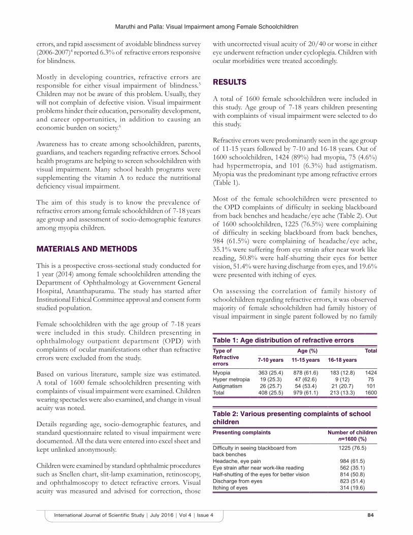
Maruthi and Palla: Visual Impairment among Female Schoolchildren
84International Journal of Scientific Study | July 2016 | Vol 4 | Issue 4
errors, and rapid assessment of avoidable blindness survey (2006-2007)4 reported 6.3% of refractive errors responsive for blindness.
Mostly in developing countries, refractive errors are responsible for either visual impairment of blindness.5 Children may not be aware of this problem. Usually, they will not complain of defective vision. Visual impairment problems hinder their education, personality development, and career opportunities, in addition to causing an economic burden on society.6
Awareness has to create among schoolchildren, parents, guardians, and teachers regarding refractive errors. School health programs are helping to screen schoolchildren with visual impairment. Many school health programs were supplementing the vitamin A to reduce the nutritional deficiency visual impairment.
The aim of this study is to know the prevalence of refractive errors among female schoolchildren of 7-18 years age group and assessment of socio-demographic features among myopia children.
MATERIALS AND METHODS
This is a prospective cross-sectional study conducted for 1 year (2014) among female schoolchildren attending the Department of Ophthalmology at Government General Hospital, Ananthapuramu. The study has started after Institutional Ethical Committee approval and consent form studied population.
Female schoolchildren with the age group of 7-18 years were included in this study. Children presenting in ophthalmology outpatient department (OPD) with complaints of ocular manifestations other than refractive errors were excluded from the study.
Based on various literature, sample size was estimated. A total of 1600 female schoolchildren presenting with complaints of visual impairment were examined. Children wearing spectacles were also examined, and change in visual acuity was noted.
Details regarding age, socio-demographic features, and standard questionnaire related to visual impairment were documented. All the data were entered into excel sheet and kept unlinked anonymously.
Children were examined by standard ophthalmic procedures such as Snellen chart, slit-lamp examination, retinoscopy, and ophthalmoscopy to detect refractive errors. Visual acuity was measured and advised for correction, those
with uncorrected visual acuity of 20/40 or worse in either eye underwent refraction under cycloplegia. Children with ocular morbidities were treated accordingly.
RESULTS
A total of 1600 female schoolchildren were included in this study. Age group of 7-18 years children presenting with complaints of visual impairment were selected to do this study.
Refractive errors were predominantly seen in the age group of 11-15 years followed by 7-10 and 16-18 years. Out of 1600 schoolchildren, 1424 (89%) had myopia, 75 (4.6%) had hypermetropia, and 101 (6.3%) had astigmatism. Myopia was the predominant type among refractive errors (Table 1).
Most of the female schoolchildren were presented to the OPD complaints of difficulty in seeking blackboard from back benches and headache/eye ache (Table 2). Out of 1600 schoolchildren, 1225 (76.5%) were complaining of difficulty in seeking blackboard from back benches, 984 (61.5%) were complaining of headache/eye ache, 35.1% were suffering from eye strain after near work like reading, 50.8% were half-shutting their eyes for better vision, 51.4% were having discharge from eyes, and 19.6% were presented with itching of eyes.
On assessing the correlation of family history of schoolchildren regarding refractive errors, it was observed majority of female schoolchildren had family history of visual impairment in single parent followed by no family
Table 2: Various presenting complaints of school childrenPresenting complaints Number of children
n=1600 (%)Difficulty in seeing blackboard from back benches
1225 (76.5)
Headache, eye pain 984 (61.5)Eye strain after near work-like reading 562 (35.1)Half-shutting of the eyes for better vision 814 (50.8)Discharge from eyes 823 (51.4)Itching of eyes 314 (19.6)
Table 1: Age distribution of refractive errorsType of Refractive errors
Age (%) Total7-10 years 11-15 years 16-18 years
Myopia 363 (25.4) 878 (61.6) 183 (12.8) 1424Hyper metropia 19 (25.3) 47 (62.6) 9 (12) 75Astigmatism 26 (25.7) 54 (53.4) 21 (20.7) 101Total 408 (25.5) 979 (61.1) 213 (13.3) 1600

Maruthi and Palla: Visual Impairment among Female Schoolchildren
85 International Journal of Scientific Study | July 2016 | Vol 4 | Issue 4
history of refractive errors (Figure 1). Among 1600 schoolchildren, 872 (54.5%) had family history of single parent, 453 (28.3%) had no family history, 218 (13.6%) had family history of siblings, and 57 (3.5%) had both parents refractive error. 132 (8.2%) children had family history of both siblings and single parent refractive error.
Refractive errors were assessed by correlating with various socio-demographic features. Socioeconomic status (SES) was assessed by modified Kuppuswamy scale. Out of 1600 children, 658 (41.1%) were upper lower class, and 565 (35.3%) were lower middle class. Many of the female children with refractive errors belong to lower SES (Table 3).
Out of 1600 children, only 768 (48%) were attending routine eye checkups yearly once at various hospitals, 262 (16.3%) and 112 (7%) were attending 2 and 3 years once, respectively, and 458 (28.6%) did not have eye checkups at all (Figure 2).
DISCUSSION
Refractive errors were the second most common cause of visual impairment. Refractive errors are treatable cause and can decrease its incidence by school health programs or health education in communities.
We have selected female gender of schoolchildren to investigate refractive errors in this study because female gender is usually neglected in few communities since birth, reasons for these may be low SES, illiteracy, bad beliefs, less food supplements, and also showing less interest in seeking to medical care toward female child. Many studies also reported that refractive errors were common in females.7-9
Refractive errors were predominantly seen in the age group of 11-15 years followed by 7-10 and 16-18 years in the present study. Khader et al.10 found lowest for the youngest age group (7.8%) and increased until it reaches 20.6% for 14 years children. After that age, prevalence decreased gradually. Lin et al.11 documented that among the schoolchildren in the age 7, 12, and 15 years, the prevalence of myopia was 20%, 61%, and 81%, respectively, and Kalikivayi et al.12 reported that the prevalence of myopia was significantly higher among children aged >10 years compared to those <10 years.
In this study, out of 1600 schoolchildren, 1424 (89%) had myopia, 75 (4.6%) had hypermetropia, and 101 (6.3%) had astigmatism. Myopia was the predominant type among refractive errors.
Kumari and Lakshmi13 documented that major refractive error was myopia (89.8%) followed by astigmatism (6.1%) and hypermetropia (4.1%). A study by Dulani and Dulani14 found myopia at 63.4% and astigmatism (25.8%) followed by hypermetropia (11.35%). Rohul et al.15 observed that myopia as 59.59% followed by astigmatism (35.23%) and hypermetropia (14.17%). In contrast to this study, lesser prevalence of myopia was observed by Kalikivayi et al.12 and Dandona and
Table 3: Socio‑demographic profile of female school childrenSocio-demographic features Number of schoolchildren (%)SES
Upper 79 (4.9)Upper middle 204 (12.75)Lower middle 565 (35.3)Upper lower 658 (41.1)Lower 94 (5.8)
ReligionHindu 804 (50.2)Muslim 518 (32.3)Christian 226 (14.1) Others 52 (3.2)
CasteST 216 (13.5)SC 442 (27.6)BC 357 (22.3)OC-General 585 (36.5)
SES: Socioeconomic status
Figure 1: Number of students with family history of refractive errors
Figure 2: Frequency of eye checkups by schoolchildren

Maruthi and Palla: Visual Impairment among Female Schoolchildren
86International Journal of Scientific Study | July 2016 | Vol 4 | Issue 4
Dandona6 was found to be 10%, 8.6%, and 4.44%, respectively.
As per this study, out of 1600 schoolchildren, 1225 (76.5%) were complaining of difficulty in seeking blackboard from back benches, 984 (61.5%) were complaining of headache/eye ache, 35.1% were suffering from eye strain after near work like reading, 50.8% were half-shutting their eyes for better vision, 51.4% were having discharge from eyes, and 19.6% were presented with itching of eyes.
Among 1600 schoolchildren, 872 (54.5%) had family history of single parent, 453 (28.3%) had no family history, 218 (13.6%) had family history of siblings, and 57 (3.5%) had both parents refractive error. 132 (8.2%) children had family history of both siblings and single parent refractive error. Studies done by Rohul et al.,15 Ip et al.,16 and Khader et al.10 found that among myopic children 39.9%, 58.5%, and 56.4%, respectively, had a family history of myopia.
SES was assessed by modified Kuppuswamy scale. Out of 1600 children, 658 (41.1%) were upper lower class, and 565 (35.3%) were lower middle class. Many of the female children with refractive errors belong to lower SES. Ahmed et al.8 observed that students from low socioeconomic conditions were having a higher prevalence of myopia when compared to students from higher SES.
Out of 1600 children, only 768 (48%) were attending routine eye checkups yearly once at various hospitals, 262 (16.3%) and 112 (7%) were attending 2 and 3 years once, respectively, and 458 (28.6%) did not have eye checkups at all as per this study. 28.6% of students did not have eye checkups, which is a quite significant; it signifies that less awareness regarding visual impairment.
Approach of vision 2020 toward refractive errors is to eliminate visual impairment (visual acuity <6/18) and blindness due to refractive errors or other causes of low vision.
CONCLUSION
From this study, we conclude that among refractive errors, myopia is the most common. Refractive errors were common in the age group of 7-15 years, students from low SES, with family history of myopia.
Health administrators or organizations should create awareness among schoolchildren, and their parents
regarding refractive errors and also should demand them to seek medical advice as refractive services. Screening is necessary, as a school health programs among individuals with poor vision, refractive services, optical services, low vision services, all those needed for to avoid blindness resulting due to refractive errors. Government and non-government organizations are helping a lot in India in reducing the incidence of visual impairment. Individuals should aware and support these organizations by seeking medical advice, which will reduce the visual impairment incidence.
ACKNOWLEDGMENTS
We are thankful to the staff of the Department of Ophthalmology, for helping in this study.
REFERENCES
1. WHO. Blindness information collected from various sources. Epidemiol Vital Stat Rep 1966;19:437.
2. Foster A, Gilbert C, Johnson G. Changing patterns in global blindness: 1988-2008. Community Eye Health 2008;21:37-9.
3. Government of India. National Survey on Blindness: 1999-2001. Report; 2002.
4. Neena J, Rachel J, Praveen V, Murthy GV; Rapid Assessment of Avoidable Blindness India Study Group. Rapid assessment of avoidable blindness in India. PLoS One 2008;3:e2867.
5. Ali A, Ahmad I, Ayub S. Prevalence of undetected refractive error among school children of Lahore. Biomedica 2007;23:96-101.
6. Dandona R, Dandona L. Refractive error blindness. Bull World Health Organ 2001;79:237-43.
7. Lin LL, Shih YF, Hsiao CK, Chen CJ, Lee LA, Hung PT. Epidemiologic study of the prevalence and severity of myopia among schoolchildren in Taiwan in 2000. J Formos Med Assoc 2001;100:684-91.
8. Ahmed I, Mian S, Mudasir S, Andrabi KI. Prevalence of myopia in students of Srinagar city of Kashmir, India. Int J Health Sci (Qassim) 2008;2:77-81.
9. Khalaj M, Gasemi MR, Zeid IM. Prevalence of refractive errors in primary school children [7-15 years] of Qazvin City. Eur J Sci Res 2009;28:174-85.
10. Khader YS, Batayha WQ, Abdul-Aziz SM, Al-Shiekh-Khalil MI. Prevalence and risk indicators of myopia among schoolchildren in Amman, Jordan. East Mediterr Health J 2006;12:434-9.
11. Lin LL, Shih YF, Hsiao CK, Chen CJ. Prevalence of myopia in Taiwanese schoolchildren: 1983 to 2000. Ann Acad Med Singapore 2004;33:27-33.
12. Kalikivayi V, Naduvilath TJ, Bansal AK, Dandona L. Visual impairment in school children in southern India. Indian J Ophthalmol 1997;45:129-34.
13. Kumari VK, Lakshmi MS. Screening for simple myopia among high school children in Hyderabad city. J Evid Based Med 2016;3:1097-101.
14. Dulani N, Dulani H. Prevalence of refractive errors among school children in Jaipur, Rajasthan. Int J Sci Study 2014;2.52-5.
15. Rohul J, Maqbool A, Hussain SA, Shamila H, Anjum F, Hamdani ZA. Prevalence of refractive errors in adolescents in out-patient attendees of the preventive ophthalmology clinic of community medicine, SKIMS, Kashmir, India. NUJHS 2013;3:17-20.
16. Ip JM, Rose KA, Morgan IG, Burlutsky G, Mitchell P. Myopia and the urban environment: Findings in a sample of 12-year-old Australian school children. Invest Ophthalmol Vis Sci 2008;49:3858-63.
How to cite this article: Maruthi SPY, Palla S. Visual Impairment Due to Refractive Errors among Female School Students Attending Tertiary Care Hospital, Ananthapuramu. Int J Sci Stud 2016;4(4):83-86.
Source of Support: Nil, Conflict of Interest: None declared.
87 International Journal of Scientific Study | July 2016 | Vol 4 | Issue 4
Analysis of Results of Platelet-rich Plasma with Arthroscopic Acromioplasty and Arthroscopic Acromioplasty: A Comparative StudyPrasanta Kumar Saha
Associate Professor, Department of Orthopaedics, Calcutta National Medical College and Hospital, Kolkata, West Bengal, India
be attributed to extrinsic and intrinsic mechanisms, as well as to environmental factors. Shoulder impingement is the main extrinsic cause of RCT. It causes mechanical compression of the external portion (bursal side) of the tendon. Upon repeated compression during movement, the coracoacromial ligament also thicken, decreasing the subacromial space. The anatomical variants (acromial shape, subacromial joint spurs, and acromioclavicular joint spurs) can lead to RCT. Furthermore, the angle and shape of the acromion could be a possible cause of the pathology. Overuse activity coupled with coracoacromial arch changes leads to decreasing the subacromial space which has a significant effect on tendon injury.
Intrinsic mechanisms are associated with the tendon itself and can be from aging, altered biology, microvascular blood supply, degeneration, tendon overload, overuse, or trauma. Histopathology studies reveal mainly degenerative changes with a fibroblastic and a vascular response known as angiofibroblastic degeneration of the rotator cuff tendons.7 The subacromial bursa is rich free nerve
INTRODUCTION
Rotator cuff tendinopathy (RCT) is an important condition of the upper extremity, affecting 1 in 50 adults.1 It is the third most common musculoskeletal problem over age of 65 years.2-4 It causes shoulder pain and loss of function. Its greatest impact is on workers with repetitive and high-load upper extremity tasks and on athletes leading to significant morbidity, affecting activities of daily living, recreation, and work life.5,6
The exact pathophysiology of pain in rotator cuff disease is not known.3 The patho-etiology of RCT can
AbstractIntroduction: Platelet-rich plasma (PRP) is widely used to treat a variety of clinical conditions. It has been hypothesized that PRP augments tendon healing. The mechanism of action of PRP is not fully elucidated. Our study is to compare the coapplication of PRP and arthroscopic acromioplasty (AA) with AA regarding clinical and tissue effect.
Methods: The study was conducted on 85 patients diagnosed as rotator cuff tendinopathy (RCT). The patients were randomized into AA (40 patients) and AA + PRP injection (45 patients). Patients were followed for a minimum 1 year. The results were evaluated with visual analog scale and Oxford shoulder score and Bonar score.
Results: There was significant improvement occurs in both groups from baseline symptom. We have found no statistical difference between the groups. When Bonar scoring for tendinopathy was compared, there was a statistically significant difference was found. The improved tendon healing was found in AA + PRP group.
Conclusion: AA significantly improves symptoms of RCT. In combination with PRP significantly improve tendon healing on histopathology. However, further studies with a larger and more varied ethnic and occupations group may be warranted to bring certainty in this dilemma.
Key words: Arthroscopic acromioplasty, Platelet-rich plasma, Rotator cuff tendinopathy
Access this article online
www.ijss-sn.com
Month of Submission : 05-2016 Month of Peer Review : 06-2016 Month of Acceptance : 07-2016 Month of Publishing : 07-2016
Corresponding Author: Dr. Prasanta Kumar Saha, Golden View Apartment, 2nd Floor, Room No - 305, T N Mukherjee Road, Monoharpur (Near HDFC Bank), Dankuni, Hooghly - 712 311, West Bengal, India. Phone: +91-9903151908. E-mail: [email protected]
Original Article DOI: 10.17354/ijss/2016/381
Print ISSN: 2321-6379Online ISSN: 2321-595X

Saha: Platelet-rich Plasma in Rotator Cuff Tendinopathy
88International Journal of Scientific Study | July 2016 | Vol 4 | Issue 4
endings and nociceptive agents.8,9 Proposed cellular changes associated with intrinsic mechanisms of RCT are increased matrix metalloproteinases (MMP) and reduced tissue inhibitors of MMP. The inhibitors are tendon cell apoptosis, insulin-like growth factor (IGF-1), nitric oxide synthetase, chondroid metaplasia of the tendon and matrix changes, fatty infiltration (following tears), cytokines, and caspases. Levy et al. showed that subjects with acute RCT had hypovascularity in the supraspinatus tendon in comparison to hypervascularity in chronic tendinopathy. Hence, the intrinsic factors have an influence on the morphology and performance of the tendon.
The tendon normally heals with scar tissue formation. The scar tissue is rich in Type 3 collagen. Type 3 collagen has excellent elastic properties but inferior strength property. As a result scar tissue is inferior to native tendon because of the structural organization and poor matrix formation. Therefore, scar tissue must thicken to make up for its mechanical insufficiency, resulting in a stiffer tendon.
Platelet-rich plasma (PRP) is a preparation of concentrated autologous platelets containing growth factors and bioactive substances essential to musculoskeletal healing.10-12 These growth factors are transforming growth factor β1 (TGF β1), platelet-derived growth factor, vascular endothelial growth factor (VEGF), hepatocyte growth factor, and IGF-1. These factors are biologically active and stimulate angiogenesis, epithelialization, cell differentiation, proliferation, and the formation of extracellular matrix and fibrovascular callus. PRP can be used to enhance extracellular matrix organization by enhanced cell proliferation and total collagen. In vitro and animal model studies suggest that PRP augments tissue healing and prevent the structural failure of the tendon in RCT by increasing tenocyte number and production of collagen (Types 1 and 3), which makes up a major portion of the tendon.13-15
Conservative management is the mainstay of treatment which includes relative rest, pain medication, physical therapy, and corticosteroid injections. However, many patients are refractory to standard care, and arthroscopic acromioplasty (AA) is done on those cases. Although most patients improve after AA, some fail to improve after surgery and many do not return to normal.10 Some studies fail to show improvement after AA. PRP has been suggested as a treatment option for refractory tendinopathies including RCT.10,16
In case of refractory RCT, AA fails to show improvement. This leads to increasingly widespread use of PRP in clinical practice with high expectations. The aim of this prospective study is to investigate the clinical and tissue effect of
coapplication PRP with AA in patients with chronic RCT. We hypothesized that PRP with AA would improve both clinical outcome and tissue characteristics.
METHODS
This was an institution-based, prospective longitudinal study. The study was conducted in our institution after getting ethical permission. All the patients were counseled about the advantages, disadvantages, and complications of the procedure. After getting written consent from patients, we performed procedure. The study period was from January 2012 to January 2015 (36 months duration).
Adult persons aged 40-70 years were recruited from a referral-based outpatient sports medicine practice. Inclusion criteria of our study were RCT with symptoms for 6 months or more, failed conservative treatment of at least 4 weeks of formal physical therapy (including rotator cuff strengthening and scapular and proprioceptive stabilization), at least one corticosteroid injection and single shoulder involvement. Exclusion criteria were joint instability, history of shoulder surgery, and corticosteroid injection within 3 months. Patients with labral lesion, complete rotator cuff tear, and adhesive capsulitis on magnetic resonance imaging (MRI) were excluded.
MRI was performed in all cases preoperatively and also on clinical follow-up period. MRI of rotator cuff was scored on a 0-5 severity scale (0: No tendinopathy; 1: Mild tendinopathy; 2: Moderate tendinopathy; 3: Moderate tendinopathy + partial thickness tear present; 4: Severe tendinopathy ± partial thickness tear present; 5: Severe tendinopathy + full thickness tear present). Patients with Grade 1 and Grade 5 were excluded from the study.
Arthroscopic procedure was done in lateral position under general anesthesia. During the procedure, shoulder joint was thoroughly examined to evaluate any other pathological condition-like labral lesion. Full thickness rotator cuff tear, frozen shoulder, or osteoarthritis and patients were withdrawn from the study. A standard AA was performed through the posterior and anterolateral portal.
PRP was prepared from the 50 ml whole blood taken from the patient. The blood was divided into five 10 ml syringes, and anticoagulant dextrose A (phosphogluconate dehydrogenase-A) solution was mixed 1:9 volume. The syringes were placed in centrifugation machine. The rotation speed and time was 3000 rpm × 3 min. It would separate red blood cell from plasma. The supernatant fluid was taken out and put in another five syringes, and 1 µg prostaglandin E1 in each syringe was mixed. The second centrifugation was performed at 4000 rpm for 15 min.

Saha: Platelet-rich Plasma in Rotator Cuff Tendinopathy
89 International Journal of Scientific Study | July 2016 | Vol 4 | Issue 4
The supernatant was discarded leaving 0.65 ml in syringe, and the sediment was mixed with-it using a vortex mixer. Finally, 0.65 ml of PRP solution was prepared from 10 ml of the whole blood in the syringe. The PRP was injected into the margin of partial tear and the subacromial space.
During the arthroscopic procedure, tendon biopsy specimen was taken at baseline under general anesthesia and at the 3-month follow-up under ultrasonography guidance.
Patients were followed up at 8, 12, and 52 weeks. The primary outcome measurements were visual analog scale (VAS) score and Oxford shoulder score (OSS). Secondary outcomes included functional tests (empty can exercise with dumbbell resistance, drop arm exercise with dumbbell resistance, side-lying external rotation with dumbbell resistance, full can exercise with dumbbell resistance, external rotation at 0 and 90° with Thera-Band resistance). Bonar scoring was used for histopathological grading.
RESULTS
DemographicAbout 130 patients were included in the study. Of these, 85 patients met the inclusion criteria. Among them, AA was done in 40 patients, and AA + PRP injection was given in 45 patients. The average platelet count was in 2.5 ± 3.1 AA group and 2.5 ± 3.8 in AA + PRP group. The baseline OSS in AA and AA + PRP group was 24.5 ± 3.2 and 23.6 ± 4.1, respectively. There was no statistical difference between the groups regarding demographic parameters (Table 1).
Functional OutcomeThe parameters for primary outcome functional measures (VAS score and OSS) were statistically significantly improved in both groups (Table 2). The VAS and OSS were improved >95% compare to baseline. In (AA + PRP) group, the VAS and OSS were improved from 7.5 ± 0.3 and 23.6 ± 4.1 to 0.34 ± 0.2 and 48 ± 2.3, respectively. Similarly, in AA group, the VAS and OSS were significantly improved from 7.6 ± 0.3 and
24.5 ± 3.2 to 0.4 ± 0.1 and 45 ± 2.1, respectively (P < 0.001). Secondary functional outcome measures were also improved significantly (Table 2). The maximum effect was found in Thera-Band external rotation at 90°. The average mean was improved from baseline 33.4 ± 5.7 (AA + PRP) and 34.2 ± 4.9 (AA) to 72.3 ± 1.7 (AA + PRP) and 72.3 ± 1.7, respectively. Another parameter drop arm test also improved dramatically from 44.3 ± 7.8 (AA + PRP) and 45.3 ± 6.7 (AA) to 83.4 ± 1.5 (AA + PRP) and 83.2 ± 1.2 (AA), respectively. However, when both the groups were compared regarding primary and secondary outcome measures, there was no statistical difference (Table 3).
MRI and HistopathologyGrade 2 MRI severity score was improved to Grade 1 in all patients in both the groups, and there was no statistical difference. In Grade 3 and Grade 4 group, there was statistically significant improvement was seen in (AA + PRP) group. Among the 22 patients, Grade 3 in (AA + PRP) group, 18 patients were improved to Grade 1. In comparison to (AA) group, where only 3 patients were improved to Grade 1 among 18 points. Similar result was found in Grade 4 in (AA + PRP) group, where among 13 patients of Grade 4 group, 11 patients were improved to Grade 1 (Table 4). According to histopathological parameters of Bonar scoring system, significant improvement was found in (AA + PRP) group (Table 5). Even after difference in histopathology, there was no difference in functional outcome measures.
There was no complication was found in both groups.
DISCUSSION
RCT is a common overuse injury in athletes and workers that occur because of the high chronic repetitive loading that surpasses the adaptive abilities of the tendon and causes micro tears and degeneration in the tendon substance. Many factors have been suspected to predispose patients to this condition by increasing the supraspinatus tendon overload. The high chronic repetitive loading stimulates the local release of cytokines, with an autocrine and paracrine modulation of cell activity that fails to adapt to continued abusive load and irritation and leads to intratendinous damage. The poor regeneration capacity of tendons, explained by the poor vascularity, oxygenation, and nutrition of this tissue, cannot cope with the applied forces and explains the low healing potential and the difficulties in the treatment of this chronic tendon disease.
In cases of injury, platelets are the first cells carried to the lesion site. In fact, they play a key role in mediating healing
Table 1: Demographic parametersParameters AA AA+PRP P valueAge 55.50±4.18 52.81±4.71 <0.01Gender: Female/male 22/18 25/20Length of symptoms 14±1.7 13±2.1Baseline OSS 24.5±3.2 23.6±4.1Pre-operative platelet count 2.5±3.1 2.5±3.8Pre-operative MRI findings
Grade 2 12 10Grade 3 18 22Grade 4 10 13
AA: Arthroscopic acromioplasty, PRP: Platelet‑rich plasma, MRI: Magnetic resonance imaging, OSS: Oxford shoulder score

Saha: Platelet-rich Plasma in Rotator Cuff Tendinopathy
90International Journal of Scientific Study | July 2016 | Vol 4 | Issue 4
source of growth factors which augment tissue healing. Among them, TGF β, VEGF, platelet-derived growth factor, IGF-1, and epidermal growth factor are important. So, PRP provides the potential for an autologous and relatively in expensive solution to facilitate tissue repair. In vitro studies demonstrated this mitogenic activity and that the stimulated tenocytes synthesize VEGF and hepatic growth factor, thus suggesting a beneficial effect for the treatment of tendon injuries by inducing cell proliferation and promoting the synthesis of angiogenic factors during the healing process. An animal model has also confirmed the usefulness of platelet concentrate for the treatment of tendon damage, with an increased tendon callus strength and stiffness after percutaneous injections in transacted tendons; a more rapid recovery in surgically repaired tendons has also been seen in a human study. Currently, PRP is widely used experimentally in different fields of medicine, but the evidence base for the clinical use of PRP is still in its infancy. Molloy et al. has applied PRP in various sports injury and concluded PRP augments tissue healing.12 Only a few articles specifically address treatment applications in the orthopedic field and, to our knowledge, only one study has been published regarding the treatment of tendinopathy through PRP injections. Mishra treated patients affected by severe chronic tennis elbow and reported promising result, with improvement in pain and function and no complications. In delayed and nonunion of bone, it has been used with bone marrow, and the result is encouraging. Now a days, the domain has been increased and applied in spinal surgery, periodontal, and craniofacial surgery.17,18
The mechanism of action of PRP is not fully understood. Recently, in anterior cruciate ligament reconstruction with semimembranosus quadruple graft is used with PRP and
Table 2: Primary and secondary outcome measurementsParameters Mean P value
Baseline Week 8 Week 12 Week 52VAS pain (AA+PRP) 7.5±0.3 3.3±0.4 0.7±0.3 0.34±0.2 <0.001VAS pain (AA) 7.6±0.3 3.8±0.38 0.8±0.3 0.4±0.1 <0.001Side-lying external rotation (number of repetitions) (AA+PRP) 17.5±1.6 23.8±0.9 32.3±3.5 38.2±1.6 <0.001Side-lying external rotation (number of repetitions) (AA) 18.1±1.5 24.7±0.7 31.3±3.6 37.5±1.2 <0.001Empty can (sec to fatigue) (AA+PRP) 59.1±7.9 71.8±4.7 76.5±10.5 80.4±2.5 0.65Empty can (sec to fatigue) (AA) 57.3±7.2 71.5±4.1 77.2±9.2 81.2±1.7 0.63Drop arm (sec to fatigue) (AA+PRP) 44.3±7.8 67.7±6.5 78.2±12.5 83.4±1.5 0.002Drop arm (sec to fatigue) (AA) 45.3±6.7 68.2±5.9 79.1±10.3 83.2±1.2 0.002Full can exercise (no. of repetitions) (AA+PRP) 27.4±3.9 38.6±4.0 47.1±5.2 51.3±2.4 <0.001Full can exercise (number of repetitions) (AA) 26.7±2.5 37.8±3.7 46.3±5.0 52.7±2.7 <0.001Thera-band external rotation at 0° (s) (AA+PRP) 37.7±4.3 54.8±5.6 62.1±3.3 68.1±2.1 <0.001Thera-band external rotation at 0° (s) (AA) 38.1±4.3 55.2±5.0 61.9±3.1 68.3±2.3 <0.001Thera-band external rotation at 90° (s) (AA+PRP) 33.4±5.7 47.9±3.2 61.5±3.0 72.3±1.7 <0.001Thera-band external rotation at 90° (s) (AA) 34.2±4.9 48.3±2.8 62.8±2.6 71.6±1.9 <0.001OSS (AA+PRP) 0.23.6±4.1 48±2.3 <0.001OSS (AA) 24.5±3.2 45±2.1 <0.001VAS: Visual analog scale, AA: Arthroscopic acromioplasty, PRP: Platelet‑rich plasma, OSS: Oxford shoulder score
Table 3: Comparison between AA and AA and PRP injectionParameters AA AA+PRP P valueVAS pain (0-10 points) 0.4±0.1 0.34±0.2 <0.001Side-lying external rotation (number of repetitions)
37.5±1.2 38.2±1.6
Empty can (sec to fatigue) 80.4±2.5 81.2±1.7Drop arm (sec to fatigue) 83.4±1.8 83.2±1.2Full can exercise (number of repetitions) 51.3±2.4 52.7±2.1Thera-Band external rotation at 0° (s) 68.3±2.3 68.1±2.1Thera-band external rotation at 90° (s) 71.6±1.9 72.3±1.7OSS 45±2.1 48±2.3AA: Arthroscopic acromioplasty, PRP: Platelet‑rich plasma, OSS: Oxford shoulder score
Table 4: Comparison of MRI gradingsParameters AA (pts) AA+PRP (pts) P valueGrade 2 12 10
Grade 1-12 Grade 1-10 <0.001Grade 3 18 22 >0.05
Grade 2-15 Grade 1-18Grade 1-3 Grade 2-4
Grade 4 10 13Grade 1-0 Grade 1-11Grade 2-2 Grade 2-1Grade 3-8 Grade 3-1
AA: Arthroscopic acromioplasty, PRP: Platelet‑rich plasma, MRI: Magnetic resonance imaging
Table 5: Comparison or Bonar scoreParameters Before operative
procedureAfter operative
procedureP value
AA 9.53±1.5 7.31±1.7 >0.05AA+PRP 9.48±1.4 2.3±1.29AA: Arthroscopic acromioplasty, PRP: Platelet‑rich plasma
of the damaged tissue because of the capacity to release growth factors from their α-granules. Platelets are a rich
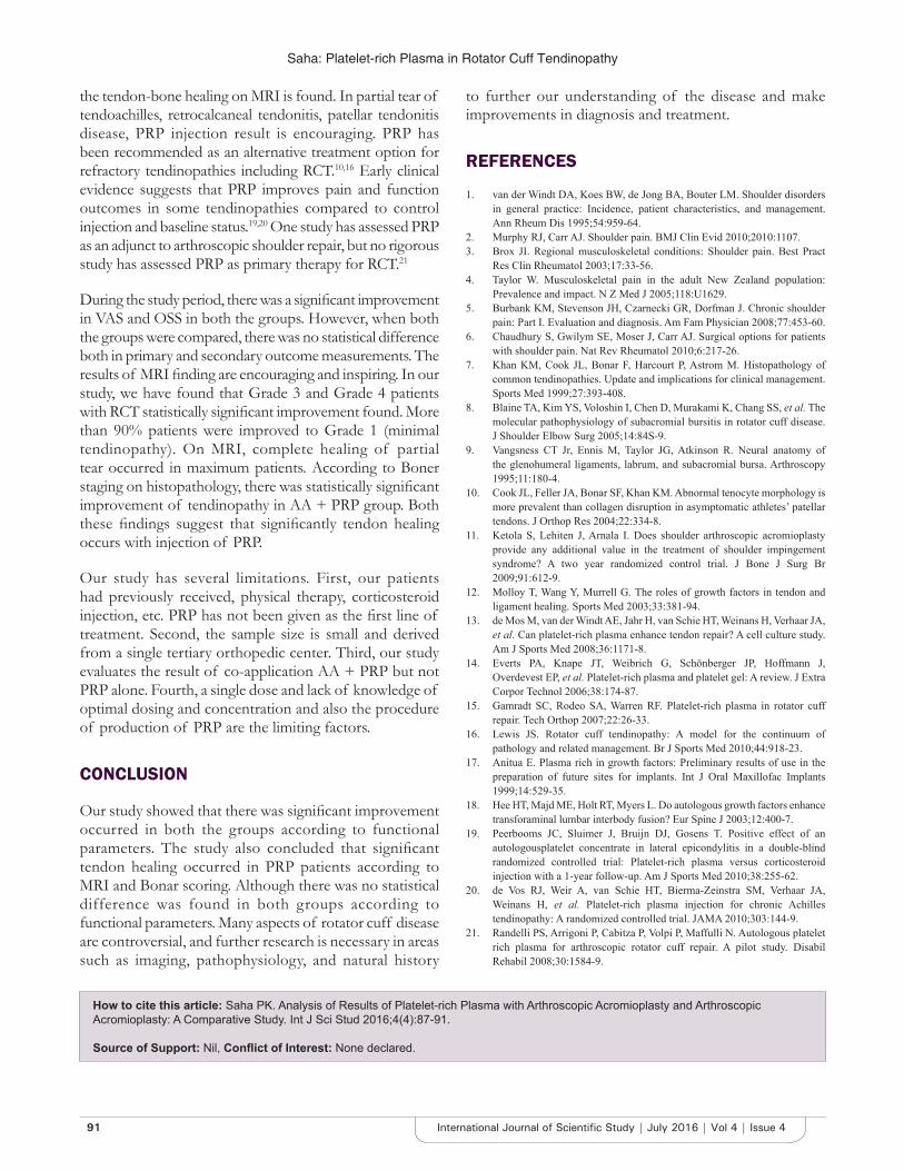
Saha: Platelet-rich Plasma in Rotator Cuff Tendinopathy
91 International Journal of Scientific Study | July 2016 | Vol 4 | Issue 4
the tendon-bone healing on MRI is found. In partial tear of tendoachilles, retrocalcaneal tendonitis, patellar tendonitis disease, PRP injection result is encouraging. PRP has been recommended as an alternative treatment option for refractory tendinopathies including RCT.10,16 Early clinical evidence suggests that PRP improves pain and function outcomes in some tendinopathies compared to control injection and baseline status.19,20 One study has assessed PRP as an adjunct to arthroscopic shoulder repair, but no rigorous study has assessed PRP as primary therapy for RCT.21
During the study period, there was a significant improvement in VAS and OSS in both the groups. However, when both the groups were compared, there was no statistical difference both in primary and secondary outcome measurements. The results of MRI finding are encouraging and inspiring. In our study, we have found that Grade 3 and Grade 4 patients with RCT statistically significant improvement found. More than 90% patients were improved to Grade 1 (minimal tendinopathy). On MRI, complete healing of partial tear occurred in maximum patients. According to Boner staging on histopathology, there was statistically significant improvement of tendinopathy in AA + PRP group. Both these findings suggest that significantly tendon healing occurs with injection of PRP.
Our study has several limitations. First, our patients had previously received, physical therapy, corticosteroid injection, etc. PRP has not been given as the first line of treatment. Second, the sample size is small and derived from a single tertiary orthopedic center. Third, our study evaluates the result of co-application AA + PRP but not PRP alone. Fourth, a single dose and lack of knowledge of optimal dosing and concentration and also the procedure of production of PRP are the limiting factors.
CONCLUSION
Our study showed that there was significant improvement occurred in both the groups according to functional parameters. The study also concluded that significant tendon healing occurred in PRP patients according to MRI and Bonar scoring. Although there was no statistical difference was found in both groups according to functional parameters. Many aspects of rotator cuff disease are controversial, and further research is necessary in areas such as imaging, pathophysiology, and natural history
to further our understanding of the disease and make improvements in diagnosis and treatment.
REFERENCES
1. van der Windt DA, Koes BW, de Jong BA, Bouter LM. Shoulder disorders in general practice: Incidence, patient characteristics, and management. Ann Rheum Dis 1995;54:959-64.
2. Murphy RJ, Carr AJ. Shoulder pain. BMJ Clin Evid 2010;2010:1107.3. Brox JI. Regional musculoskeletal conditions: Shoulder pain. Best Pract
Res Clin Rheumatol 2003;17:33-56.4. Taylor W. Musculoskeletal pain in the adult New Zealand population:
Prevalence and impact. N Z Med J 2005;118:U1629.5. Burbank KM, Stevenson JH, Czarnecki GR, Dorfman J. Chronic shoulder
pain: Part I. Evaluation and diagnosis. Am Fam Physician 2008;77:453-60.6. Chaudhury S, Gwilym SE, Moser J, Carr AJ. Surgical options for patients
with shoulder pain. Nat Rev Rheumatol 2010;6:217-26.7. Khan KM, Cook JL, Bonar F, Harcourt P, Astrom M. Histopathology of
common tendinopathies. Update and implications for clinical management. Sports Med 1999;27:393-408.
8. Blaine TA, Kim YS, Voloshin I, Chen D, Murakami K, Chang SS, et al. The molecular pathophysiology of subacromial bursitis in rotator cuff disease. J Shoulder Elbow Surg 2005;14:84S-9.
9. Vangsness CT Jr, Ennis M, Taylor JG, Atkinson R. Neural anatomy of the glenohumeral ligaments, labrum, and subacromial bursa. Arthroscopy 1995;11:180-4.
10. Cook JL, Feller JA, Bonar SF, Khan KM. Abnormal tenocyte morphology is more prevalent than collagen disruption in asymptomatic athletes’ patellar tendons. J Orthop Res 2004;22:334-8.
11. Ketola S, Lehiten J, Arnala I. Does shoulder arthroscopic acromioplasty provide any additional value in the treatment of shoulder impingement syndrome? A two year randomized control trial. J Bone J Surg Br 2009;91:612-9.
12. Molloy T, Wang Y, Murrell G. The roles of growth factors in tendon and ligament healing. Sports Med 2003;33:381-94.
13. de Mos M, van der Windt AE, Jahr H, van Schie HT, Weinans H, Verhaar JA, et al. Can platelet-rich plasma enhance tendon repair? A cell culture study. Am J Sports Med 2008;36:1171-8.
14. Everts PA, Knape JT, Weibrich G, Schönberger JP, Hoffmann J, Overdevest EP, et al. Platelet-rich plasma and platelet gel: A review. J Extra Corpor Technol 2006;38:174-87.
15. Gamradt SC, Rodeo SA, Warren RF. Platelet-rich plasma in rotator cuff repair. Tech Orthop 2007;22:26-33.
16. Lewis JS. Rotator cuff tendinopathy: A model for the continuum of pathology and related management. Br J Sports Med 2010;44:918-23.
17. Anitua E. Plasma rich in growth factors: Preliminary results of use in the preparation of future sites for implants. Int J Oral Maxillofac Implants 1999;14:529-35.
18. Hee HT, Majd ME, Holt RT, Myers L. Do autologous growth factors enhance transforaminal lumbar interbody fusion? Eur Spine J 2003;12:400-7.
19. Peerbooms JC, Sluimer J, Bruijn DJ, Gosens T. Positive effect of an autologousplatelet concentrate in lateral epicondylitis in a double-blind randomized controlled trial: Platelet-rich plasma versus corticosteroid injection with a 1-year follow-up. Am J Sports Med 2010;38:255-62.
20. de Vos RJ, Weir A, van Schie HT, Bierma-Zeinstra SM, Verhaar JA, Weinans H, et al. Platelet-rich plasma injection for chronic Achilles tendinopathy: A randomized controlled trial. JAMA 2010;303:144-9.
21. Randelli PS, Arrigoni P, Cabitza P, Volpi P, Maffulli N. Autologous platelet rich plasma for arthroscopic rotator cuff repair. A pilot study. Disabil Rehabil 2008;30:1584-9.
How to cite this article: Saha PK. Analysis of Results of Platelet-rich Plasma with Arthroscopic Acromioplasty and Arthroscopic Acromioplasty: A Comparative Study. Int J Sci Stud 2016;4(4):87-91.
Source of Support: Nil, Conflict of Interest: None declared.

92International Journal of Scientific Study | July 2016 | Vol 4 | Issue 4
Reactive Hyperplastic Lesions of the Oral Cavity: A Retrospective Analysis in Jammu Region of Jammu and Kashmir State, IndiaMandeep Kaur1, Suby Singh2, Ravinder Singh3, Abhiroop Singh4, Romesh Singh5
1Assistant Professor, Department of Oral Pathology and Microbiology, Indira Gandhi Government Dental College, Jammu, Jammu and Kashmir, India, 2Post-graduate Student, Department of Pathology, Government Medical College, Jammu, Jammu and Kashmir, India, 3Registrar, Department of General Surgery, Government Medical College, Jammu, Jammu and Kashmir, India, 4Post-graduate Student, Department of Oral and Maxillofacial Surgery, DAV College of Dental Sciences, Yamuna Nagar, Haryana, India, 5Principal and Head, Department of Periodontics, Indira Gandhi Government Dental College, Jammu, Jammu and Kashmir, India
tissue, multinucleated giant cells and proliferation of endothelial cells, and fibroblast.1 These lesions are of varying sizes and usually have no radiographic features. However, erosions and cup-shaped radiolucency can be seen. Clinically, it may be sessile or pedunculated masses with ulcerated or smooth surfaces.2 Surgical excision is the treatment of choice and elimination of chronic irritant is mandatory. Majority of these do not recur. However, if the source of trauma persists, frequent recurrences are possible. The purpose of this retrospective study was to survey and evaluate the frequency of various types of RHLs in Jammu province of Jammu and Kashmir state and to analyze the clinical features with histopathologic diagnosis treated at Indira
INTRODUCTION
Reactive hyperplastic lesions (RHLs) are common oral mucosal lesions caused by chronic and recurrent irritation of the mucosa and histologically represent chronic inflammation, granulation tissue, mineralized
Original Article
AbstractIntroduction: Oral mucosa is constantly under the influence of various stimuli, and thus exhibits a variety of inflammatory, reactive, and neoplastic lesions. Reactive lesions of the oral cavity are the common lesions faced by a dentist during routine examinations.
Aim: The aim of this study was to evaluate the prevalence of oral reactive hyperplastic lesions (RHLs) over a period of 5-year from 2010 to 2015 in Jammu province.
Materials and Methods: In this retrospective study, records were procured from the archives of the Department of Oral Pathology, Indira Gandhi Government Dental College, Jammu. Records with both clinical and histopathological diagnosis were analyzed. History of patient’s age, gender, and anatomic location lesions was extracted from the records.
Results: From total 402 specimens diagnosed from 2010 to 2015, 88 cases were diagnosed as RHL (21.8%). From a total of 88, RHL diagnosed fibroma was the most common lesion (32.9%), followed by inflammatory hyperplasia (27.2%), pyogenic granuloma (25%), peripheral ossifying fibroma (10.2%), and peripheral giant cell granuloma (3.4%). Of all the lesions evaluated, age of the patient ranged from 14 to 75 years, mean age being 45 years.
Conclusion: Findings in our study were broadly similar to the results of the previous studies with few differences. Knowledge of the frequency and distribution of these is beneficial during diagnosis and treatment plan in clinical practice and thus can reduce dentoalveolar complications.
Key words: Fibroma, Inflammatory hyperplasia, Peripheral giant cell granuloma, Peripheral ossifying fibroma, Pyogenic granuloma, Reactive hyperplastic lesions
Access this article online
www.ijss-sn.com
Month of Submission : 05-2016 Month of Peer Review : 06-2016 Month of Acceptance : 07-2016 Month of Publishing : 07-2016
Corresponding Author: Dr. Mandeep Kaur, Department of Oral Pathology, Indira Gandhi Government Dental College, Jammu, Jammu and Kashmir, India. E-mail: [email protected]
Print ISSN: 2321-6379Online ISSN: 2321-595X
DOI: 10.17354/ijss/2016/382

Kaur, et al.: Retrospective Analysis of Reactive Hyperplastic Lesions in Jammu Region
93 International Journal of Scientific Study | July 2016 | Vol 4 | Issue 4
Gandhi Government Dental College, within a span of 5 years from 2010 to 2015.
MATERIALS AND METHODS
RHL was classified into five groups: Fibroma (F), pyogenic granuloma (PG), peripheral giant cell granuloma (PGCG), peripheral ossifying fibroma (POF), and inflammatory hyperplasia (IH). A retrospective archive review of different RHL between 2010 and 2015 from the records of the Department of Oral Pathology, Indira Gandhi Government Dental College, Jammu, was carried out. Classification of the lesion was followed by the academic oral maxillofacial text. Histopathological slides, which were missing or not properly stained, were not included. Records with both clinical and histopathological diagnosis were selected. History of patient’s age, gender, and anatomic location lesions was extracted from the records. Clinical characteristics such as base, surface, size, and color of the lesions were also collected from the records. All the findings of our study were then compared with other studies conducted in different parts of the world.
RESULTS
Of the total 402 specimens diagnosed from 2010 to 2015 in the registers, 88 cases were diagnosed as RHL (21.8%). From a total of 88, RHL diagnosed during the assessed period of 5 years, fibroma was the most common lesion (32.9%), followed by IH (27.2%), PG (25%), POF (10.2%), and PGCG (3.4%) (Table 1 and Graph 1). Of all the lesions evaluated, age of the patient ranged from 14 years to 75 years, mean age being 45 years. Mean age of fibroma was 44 years, PG was 38 years, PGCG was 42 years, POF was 36 years, and IH was 38 years (Table 2 and Graph 2). Of these, 58 were females (65.9%) and 30 were males (34%) (Table 3 and Graph 3). Thus, male:female ratio was 1:1.5. The most common site was gingiva (70%) followed by buccal mucosa (14.7%), palate (7.9%), lips (3.4%), and retromolar region (3.4%) (Table 4 and Graph 4). No such lesions were
found in the tongue, vestibule, and alveolar mucosa. Chief complaint in the majority of lesions was a painless growth. History of burning sensation was given by few patients. The presence of local irritant was the most common cause and was seen in approximately 60-70% of cases. Others causes were overhanging restoration, abrasion, minor trauma, and hormonal imbalance. The size of the lesion varied between 6 mm and 2 cm (Table 5 and Graph 5). The duration of the lesion at the time of diagnosis ranged from 2 to 8 months approximately. Most of the lesions were firm with sessile base and smooth surface. Ulceration was seen in very few lesions. The color varied from normal to grayish to reddish brownish. All the lesions were surgically excised, and the diagnosis was confirmed by histopathological examination (Figures 1-5).
DISCUSSION
The reactive lesions are benign tumor-like proliferations in the oral cavity because oral mucosal tissues are frequently exposed to traumatic injuries. Chronic trauma can induce inflammation which produces granulation tissue with proliferation of endothelial cells and fibroblast and chronic
Graph:1 Distribution of patients with reactive hyperplasia according to prevalence (n-88)
Graph 2: Distribution of patients with reactive hyperplasia according to age
Graph 3: Distribution of patients with reactive hyperplasia according to Sex

Kaur, et al.: Retrospective Analysis of Reactive Hyperplastic Lesions in Jammu Region
94International Journal of Scientific Study | July 2016 | Vol 4 | Issue 4
In our study, fibroma was the most common RHL which constituted 32.9% of the total cases with 44 years as the
Graph 4: Distribution of patients with reactive hyperplasia according to location
Graph 5: Distribution of patients with reactive hyperplasia according to size
Figure 1: Peripheral ossifying fibroma 10X
inflammatory infiltrate resulting in a fibrous overgrowth called reactive hyperplasia.3
Figure 2: Inflammatory hyperplasia 10X
Figure 3: Peripheral giant cell granuloma 10X
Figure 4: Fibroma 10X

Kaur, et al.: Retrospective Analysis of Reactive Hyperplastic Lesions in Jammu Region
95 International Journal of Scientific Study | July 2016 | Vol 4 | Issue 4
Daley et al. in Canada and Zhang et al. in China.5,6 Studies reported by Ababneh et al., Kashyap et al., and Zarei et al. quoted PG to be most prevalent reactive lesion.7-9 In series of studies reported by Naderi et al., PGCG was the most prevalent lesion which was in agreement with the reports mentioned above including our study.10 Our findings were consistent with the data given by Buchner et al. except that PGCG comprised only 4.5% of the cases as compared to the findings reported by Buchner et al., where PGCG comprised of 18.7% of cases.11 In our study, IH comprised 27.2% of total cases which is high as compared to the findings reported by Zain and Fei which was 15.7%.12 Our percentage of POF was 10.2% which was higher than studies given by Maturana-Ramírez et al. 2.9% and Ababneh 7.2% and much lower than reported by Macleod and Soames 40%.13 The differences may be due to different classification and terminology of lesions and number of cases.
It was interested to note that all RH lesions occurred more in females with male:female ratio as 1:1.5. PG was seen two times more common in females as compared to males probably reflecting hormonal imbalance. Studies conducted by Zarei et al. and Ala Aghbali et al. male:female ratio was 1:1.8 and 1:1.4, respectively.1,7 Salum et al. and Kfir et al. also reported a higher RHL frequency in females except for PGCG, which was higher in males.14,15 Maturana-Ramírez et al. also reported a higher frequency in males.16 Ethic differences between studies could be the reason.
In the present study, the mean age of the patient was third and fourth decades which is comparable with the findings of other studies.6,17 The highest RHL prevalence was found in the group of 50-59 years as reported by Maturana-Ramírez et al.16 Few studies have shown.6,14-17 In our study, only one case of PG was female who was 14 years of age. According to location, the most common site was gingiva accounting for 70% of total cases. Anterior region was more commonly involved with slightly greater prevalence in the maxilla. This is comparable to few other studies reported in the literature.6,11,14 Buccal mucosa was the second most common site 14.7% followed by palate 7.9%, lips 3.4%, and retromolar region 3.4%. Thus, the present study supports previous assertion that traumatic fibroma and PG can involve any oral mucosal site with
Figure 5: Pyogenic granuloma 10X
Table 1: Distribution of patients with reactive hyperplasia according to prevalence (n-88)S.No Lesion No. of cases Age %1 Fibroma 29 32.92 Inflammatory hyperplasia 24 27.23 Pyogenic granuloma 22 254 Peripheral ossifying fibroma 9 10.25 Peripheral giant cell granuloma 4 4.5
Table 2: Distribution of patients with reactive hyperplasia according to ageS No. Lesion Mean age (in years)1 Fibroma 44 2 Peripheral giant cell granuloma 423 Pyogenic granuloma 384 Inflammatory hyperplasia 385 Peripheral ossifying fibroma 36
Table 3: Distribution of patients with reactive hyperplasia according to SexS No. Lesion Male Female1 Fibroma 11 182 Pyogenic granuloma 6 163 Peripheral giant cell granuloma - 44 Peripheral ossifying fibroma 3 65 Inflammatory hyperplasia 10 14
Table 4: Distribution of patients with reactive hyperplasia according to locationS No. Lesion Gingiva Buccal mucosa Lip Palate Retromolar region Tongue Vestibule1 Fibroma 20 3 3 3 - - -2 Pyogenic granuloma 18 - - 4 - - -3 Peripheral giant cell granuloma 4 - - - - - -4 Peripheral ossifying 9 - - - - - -5 Inflammatory hyperplasia 11 10 - - 3 - -
mean age and 37.9% in males and 62% in females. This finding is consistent with the finding given by Kadeh et al.4 Our results were also close to findings given by
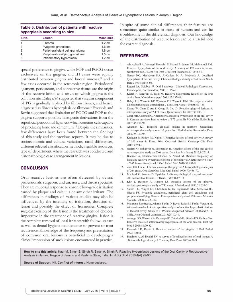
Kaur, et al.: Retrospective Analysis of Reactive Hyperplastic Lesions in Jammu Region
96International Journal of Scientific Study | July 2016 | Vol 4 | Issue 4
special preference to gingiva while POF and PGCG occur exclusively on the gingiva, and IH cases were equally distributed between gingiva and buccal mucosa,18 and a few cases occurred in the retromolar region. Periodontal ligament, periosteum, and connective tissues are the origin of the reactive lesion as a result of which gingiva is the common site. Daley et al. suggested that vascular component of PG is gradually replaced by fibrous tissues, and hence, diagnosed as fibrous hyperplasia or fibroma.5 Eversole and Rovin suggested that limitation of PGCG and POF to the gingiva supports possible histogenic derivations from the superficial periodontal ligament which contains cells capable of producing bone and cementum.19 Despite the similarities, few differences have been found between the findings of this study and the previous reports. It may be due to socioeconomic and cultural variations, racial differences, different selected classification methods, available resources, type of department, where the research was conducted and histopathologic case arrangement in lesions.
CONCLUSION
Oral reactive lesions are often detected by dental professionals, surgeons, and ear, nose, and throat specialist. They are mucosal response to chronic low-grade irritation caused by plaque and calculus or any other irritant. The differences in findings of various studies may also be influenced by the intensity of irritation, duration of lesion and possibly the effect of hormones. Complete surgical excision of the lesion is the treatment of choices. Imperative in the treatment of reactive gingival lesion is the complete removal of local irritants with follow-up care as well as dental hygiene maintenance to prevent or treat recurrence. Knowledge of the frequency and presentation of common oral lesions is beneficial in developing a clinical impression of such lesions encountered in practice.
In spite of some clinical differences, their features are sometimes quite similar to those of tumors and can be troublesome in the differential diagnosis. Our knowledge of the distribution of reactive lesion can be a useful tool for correct diagnosis.
REFERENCES
1. Ala Aghbali A, Vosough Hosseini S, Harasi B, Janani M, Mahmoudi SM. Reactive hyperplasia of the oral cavity: A survey of 197 cases in tabriz, Northwest iran. J Dent Res Dent Clin Dent Prospects 2010;4:87-9.
2. Nartey NO, Masadomr HA, Al-Cailani M, Al Mobeerik A. Localised hyperplasia of the oral cavity: Clinicopathological study of 164 cases. Saudi Dent J 1994;6:145-50.
3. Regazi JA, Sciubba JJ. Oral Pathology: Clinical-Pathologic Correlation. Philadelphia, PA: Saunders; 2008. p. 156-9.
4. Kadeh H, Saravani S, Tajik M. Reactive hyperplastic lesions of the oral cavity. Iran J Otorhinolaryngol 2015;27:137-44.
5. Daley TD, Wysocki GP, Wysocki PD, Wysocki DM. The major epulides: Clinicopathological correlations. J Can Dent Assoc 1990;56:627-30.
6. Zhang W, Chen Y, An Z, Geng N, Bao D. Reactive gingival lesions: A retrospective study of 2,439 cases. Quintessence Int 2007;38:103-10.
7. Zarei MR, Chamani G, Amanpoor S. Reactive hyperplasia of the oral cavity in Kerman province, Iran: A review of 172 cases. Br J Oral Maxillofac Surg 2007;45:288-92.
8. Ababneh KT. Biopsied gingival lesions in northern Jordanians: A retrospective analysis over 10 years. Int J Periodontics Restorative Dent 2006;26:387-93.
9. Kashyap B, Reddy PS, Nalini P. Reactive lesions of oral cavity: A survey of 100 cases in Eluru, West Godavari district. Contemp Clin Dent 2012;3:294-7.
10. Naderi NJ, Eshghyar N, Esfehanian H. Reactive lesions of the oral cavity: A retrospective study on 2068 cases. Dent Res J (Isfahan) 2012;9:251-5.
11. Buchner A, Shnaiderman-Shapiro A, Vered M. Relative frequency of localized reactive hyperplastic lesions of the gingiva: A retrospective study of 1675 cases from Israel. J Oral Pathol Med 2010;39:631-8.
12. Zain RB, Fei YJ. Fibrous lesions of the gingiva: A histopathologic analysis of 204 cases. Oral Surg Oral Med Oral Pathol 1990;70:466-70.
13. Macleod RI, Soames JV. Epulides: A clinicopathological study of a series of 200 consecutive lesions. Br Dent J 1987;163:51-3.
14. Kfir Y, Buchner A, Hansen LS. Reactive lesions of the gingiva. A clinicopathological study of 741 cases. J Periodontol 1980;51:655-61.
15. Salum FG, Yurgel LS, Cherubini K, De Figueiredo MA, Medeiros IC, Nicola FS. Pyogenic granuloma, peripheral giant cell granuloma and peripheral ossifying fibroma: Retrospective analysis of 138 cases. Minerva Stomatol 2008;57:227-32.
16. Maturana-Ramírez A, Adorno-Farías D, Reyes-Rojas M, Farías-Vergara M, Aitken-Saavedra J. A retrospective analysis of reactive hyperplastic lesions of the oral cavity: Study of 1149 cases diagnosed between 2000 and 2011, Chile. Acta Odontol Latinoam 2015;28:103-7.
17. Awange DO, Wakoli KA, Onyango JF, Chindia ML, Dimba EO, Guthua SW. Reactive localised inflammatory hyperplasia of the oral mucosa. East Afr Med J 2009;86:79-82.
18. Eversole LR, Rovin S. Reactive lesions of the gingiva. J Oral Pathol 1972;1:30-8.
19. Bataineh A, Al-Dwairi ZN. A survey of localized lesions of oral tissues: A clinicopathological study. J Contemp Dent Pract 2005;6:30-9.
How to cite this article: Kaur M, Singh S, Singh R, Singh A, Singh R. Reactive Hyperplastic Lesions of the Oral Cavity: A Retrospective Analysis in Jammu Region of Jammu and Kashmir State, India. Int J Sci Stud 2016;4(4):92-96.
Source of Support: Nil, Conflict of Interest: None declared.
Table 5: Distribution of patients with reactive hyperplasia according to sizeS No. Lesion Mean size1 Fibroma 1.2 cm2 Pyogenic granuloma 1.6 cm3 Peripheral giant cell granuloma 1.8 cm4 Peripheral ossifying granuloma 1.5 cm5 Inflammatory hyperplasia 1.2 cm

97 International Journal of Scientific Study | July 2016 | Vol 4 | Issue 4
Assessment of Regenerate in Limbs by Ilizarov External FixationT Suresh Kumar1, Swagat Mahapatra2
1Assistant Professor, Department of Orthopaedics, Vinayaka Missions Kirupananda Variyar Medical College and Hospital, Salem, Tamil Nadu, India, 2Assistant Professor, Department of Orthopaedics, Mahatma Gandhi Medical College and Research Institute, Puducherry, India
or lower extremities, that have resulted from various congenital or acquired abnormalities. The issue of limb lengthening was introduced in orthopedic surgery in the year 1950 and has been practiced up to the present time. Although clinical studies have proved the effectiveness of different methods of limb lengthening, there is still need for further research to study regeneration of the bone tissue. This study comprises an analysis of bone regeneration, especially the response of bone, periosteum and endosteum to distraction using ring fixators. Clinical studies include radiological and functional outcome, long-term results, and patient satisfaction after limb reconstruction.
MATERIALS AND METHODS
This was a prospective interventional study conducted in a high volume tertiary care teaching hospital in Southern India for almost 5 years from January 2010 to July 2015. This study involved all patients who fulfilled the inclusion criteria. The patients undergoing limb lengthening or
INTRODUCTION
Professor Gavriil Abramovich Ilizarov of U.S.S.R known as the “magician” of Kurgan invented the ring external fixator now known as Ilizarov external fixator. With this, he was also the pioneer in the most stunning discovery in orthopedics. The biological production of new tissues was termed as neo-osteogenesis or osteoneogenesis.1 When it encompassed all tissues, which were generated during limb lengthening, were coined as histoneogenesis or neo-histogenesis. Limb lengthening procedures are performed to correct significant length discrepancies, in the upper
AbstractIntroduction: Limb lengthening procedures are performed to correct significant length discrepancies, in the upper or lower extremities, that have resulted from various congenital or acquired abnormalities. The issue of limb lengthening was introduced in orthopedic surgery in the year 1950 and has been practiced up to the present time. This study comprises an analysis of bone regeneration, especially the response of bone, periosteum and endosteum to distraction limb lengthening.
Materials and Methods: This study involves all patients in a tertiary care teaching hospital from January 2010 to July 2015, where bone regenerate as been produced by Ilizarov methods. 131 regenerates have been analyzed. Ilizarov external fixator was applied, and corticotomy was done. Distraction was started at a rate of 1 mm/day after the latent period (7-10 days). X-rays were taken at intervals of 1 month. Occasionally, ultrasonography was used to assess the early regenerate.
Results: There were 105 normotrophic regenerate, 14 hypertrophic regenerate, and 14 hypotrophic regenerate. The lengthening achieved ranged between 3 cm and 23 cm. The average increase in length was 6.4 cm. The incidence of major complications, in our study, was 30.1 %, and overall complication was 60%.
Conclusion: Ilizarov external fixator is used in limb lengthening, deformity correction, and segmental long bone defect reconstruction. In our study, we achieved good to excellent results in 90% of our cases which is at par with many other studies. We achieved 10.7% excellent, 81.7% good, and 7.6% poor results. We had good to excellent or better results in the upper tibial metaphyseal area compared to any other area.
Key words: Corticotomy, Ilizarov, Limb lengthening, Regenerate
Access this article online
www.ijss-sn.com
Month of Submission : 05-2016 Month of Peer Review : 06-2016 Month of Acceptance : 07-2016 Month of Publishing : 07-2016
Corresponding Author: Dr. T Suresh Kumar, No-64, S N D Road, Tiruchengodu, Dt - Namakkal - 637 211, Tamil Nadu, India. E-mail: [email protected]
Original Article DOI: 10.17354/ijss/2016/383
Print ISSN: 2321-6379Online ISSN: 2321-595X

Suresh Kumar and Mahapatra: Regenerate in Ilizarov
98International Journal of Scientific Study | July 2016 | Vol 4 | Issue 4
bone transport using Ilizarov external fixation following congenital, post-traumatic, post-polio residual paralysis, infected nonunion of long bones, and tumors were included. Patients lost to follow-up, and limbs which were amputated before regenerate was consolidated were excluded from the study. The objectives of this study were to investigate bone regeneration after corticotomy and distraction lengthening, to assess treatment of posttraumatic shortening of limbs using the Ilizarov distraction device and to evaluate the results of limb lengthening. In our study, there was a total of 133 corticotomies in 113 patients. Two patients were lost to follow-up. Hence, 131 regenerates were finally analyzed. Ilizarov external fixator was applied, and corticotomy was done. Distraction was started at a rate of 1 mm/day after the latent period (7-10 days). X-rays were taken at intervals of 1 month. Occasionally, ultrasound was used to assess the early regenerate before it was visible on X-rays. Depending on the quality of regenerate in the X-rays and or ultrasound, if the quality of regenerate was poor, the distraction was either stopped and delayed or slowed down and distraction did at a rate of 0.5 mm/day. In some patients, compression was done to reduce the gap and distracted again (accordion technique). Bone marrow infiltration was done in 12 patients, where we saw either
poor regenerate or delayed consolidation. Results of regenerate were evaluated on the basis of four parameters, both clinically and radiologically. The parameters included consolidation of regenerate, absence of deformity, absence of limb length discrepancy, and absence of infection. The score was excellent - all four parameters; good - 3 parameters; poor - 2 or less.
OBSERVATIONS AND RESULTS
A total of 131 regenerates in 113 patients were analyzed. 83 (73.5%) were males and 30 were females, and the maximum number of patients (38) were in the age group of 21-30 years. Maximum affected and operated bone was the tibia, which was 92.5% of cases in our series (Graph 1). Anatomical site of corticotomy was proximal metaphysis in a maximum number of cases (Graph 2, Graph 5). A type of regenerate was normotrophic in 78.9% cases, hypertrophic in 10.5% cases, and hypotrophic in 10.5% cases (Graph 3). The residual shortening was maximum in post polio residual paralysis cases followed by acute fractures (Graph 4). The lengthening achieved ranged between 3 cm and 23 cm. The average increase in length
Graph 1: Bones involved in Ilizarov fixation
Graph 2: Site of corticotomy
Graph 3: Type of regenerate
Graph 4: Residual shortening according to diagnosis

Suresh Kumar and Mahapatra: Regenerate in Ilizarov
99 International Journal of Scientific Study | July 2016 | Vol 4 | Issue 4
Figure 1: Case of Congenital pseudoarthrosis of Tibia managed with Ilizarov
was 6.4 cm. Good to excellent results were achieved in 90% of cases (Graph 6). The incidence of minor complications in our study was 29%, and overall complication was 60%. The most common complication encountered was pin tract infection followed by deformity of the regenerate (Graph 7).
DISCUSSION
Ilizarov external fixator results in rapid advances in limb lengthening, deformity correction, and segmental long bone defect reconstruction.2-4 The mechanical features of and biologic response to using distraction osteogenesis with the circular external fixator are the unique aspects of Ilizarov’s contribution. Alan found in his study that the average increase in length was 5.9 cm and the rate for the lengthening phase was 12 days for one centimeter with the Ilizarov technique.5 Paley found in his study that the average increase in length was 5.6 cm. In our study, the
average increase in length was 6.4 cm and the lengthening phase was 10 days for one centimeter.6 Aronson found that
Graph 5: Assessment of outcome of regenerate according to corticotomy site

Suresh Kumar and Mahapatra: Regenerate in Ilizarov
100International Journal of Scientific Study | July 2016 | Vol 4 | Issue 4
Figure 2: Case of Giant cell tumor, post excision with shortening managed with Ilizarov
new bone formation was better and faster in metaphyseal than in diaphyseal bone.7 In our study, we had good to excellent results in the upper tibial metaphyseal area. Poor or hypotrophic regenerates seen in corticotomies performed in cortical bones and in post-polio residual paralysis patients whose bone diameter was less and were treated with the accordion technique of compression and distraction.
Paley found the incidence of minor complications in his study was 10.6%, major complications was 20.6% and altogether was 31.2%.6 In our study, minor complications were 29% and major complications were 30.1% and
altogether was 60.3%. Yun found good to excellent results in 78% of the cases in his study.8 In our study, we achieved good to excellent results in 90% of our cases which is at par with many other studies.
CONCLUSION
Ilizarov external fixator is used in limb lengthening, deformity correction, and segmental long bone defect reconstruction. In all metaphyseal corticotomies rate of distraction was 1 mm/day (0.25 mm four times a day),

Suresh Kumar and Mahapatra: Regenerate in Ilizarov
101 International Journal of Scientific Study | July 2016 | Vol 4 | Issue 4
which has been found good enough for a good regenerate to be obtained. In all other corticotomies of cortical bone,
the initial rate of distraction from 1 mm/day had to be reduced if necessary as per X-ray findings. In radius, ulna, post-polio residual paralysis of femur and tibia which are thin, and corticotomies in the shaft of such bones, the distraction rates were reduced to 0.5 mm/day. In our study, we achieved good to excellent results in 90% of our cases which is at par with many other studies. We achieved 10.7% excellent, 81.7% good and 7.6% poor results. We had good to excellent or better results in the upper tibial metaphyseal area compared to any other area.
REFERENCES
1. Ilizarov GA. The tension - Stress effect on the genesis and growth of tissues. Transosseous osteosynthesis. Clin Orthop Relat Res 1992;1:137-291.
2. Catagni MA, Lovisetti L, Guerreschi F, Combi A, Ottaviani G. Cosmetic bilateral leg lengthening: Experience of 54 cases. J Bone Joint Surg Br 2005;87:1402-5.
3. Wagner H. Operative technique in traumatic limb lengthening. Der Chir 1971;42:260-6.
4. Shevtsov VI, Makushin VD, Kuftryrev LM. Defects of the lower limb bones. Radiographical Dynamics in Formation of Regenerate Bones during Tibial-Fibular Synostosis. Db Misc, 2000. p. 483-7.
5. Codivilla A. On the means of lengthening, in the lower limbs, the muscles, and tissues which are shortened through deformity. Am J Orthop Surg 1905;2:353-69.
6. Paley D. Problems, obstacles, and complications of limb lengthening by the Ilizarov technique. Clin Orthop Relat Res 1990;250:81-104.
7. Aronson J. Limb-lengthening, skeletal reconstruction, and bone transport with the Ilizarov method. J Bone Joint Surg Am 1997;79:1243-58.
8. Yun AG, Severino R, Reinker K. Attempted limb lengthenings beyond twenty percent of the initial bone length: Results and complications. J Pediatr Orthop 2000;20:151-9.
How to cite this article: Kumar TS, Mahapatra S. Assessment of Regenerate in Limbs by Ilizarov External Fixation. Int J Sci Stud 2016;4(4):97-101.
Source of Support: Nil, Conflict of Interest: None declared.
Graph 6: Final outcome of regenerate in long bones treated by Ilizarov external fixation
Graph 7: Complications
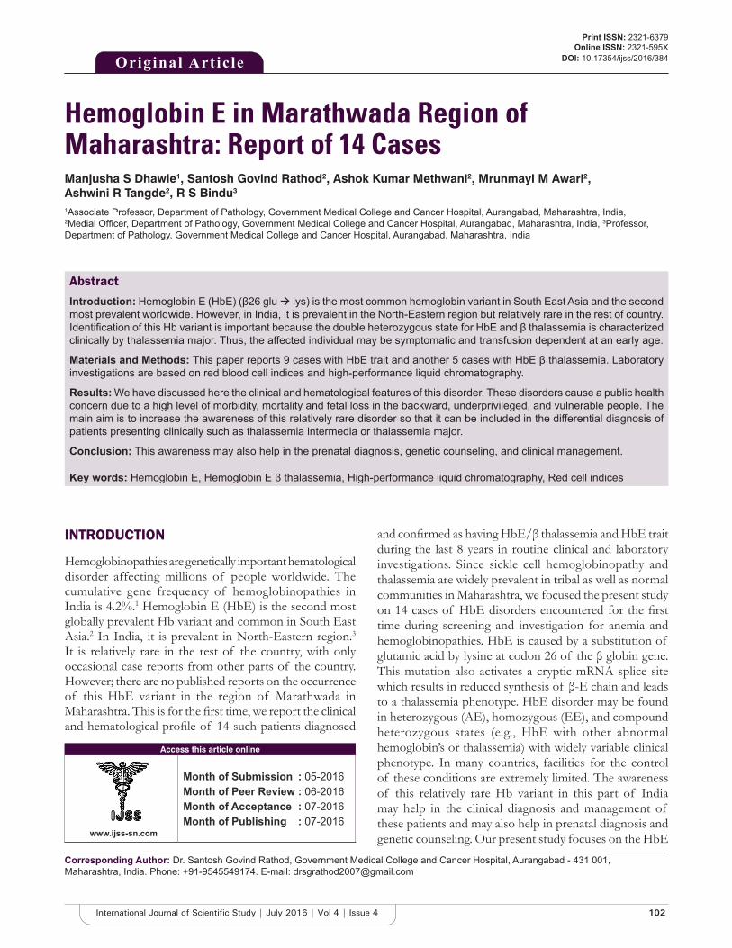
102International Journal of Scientific Study | July 2016 | Vol 4 | Issue 4
Hemoglobin E in Marathwada Region of Maharashtra: Report of 14 CasesManjusha S Dhawle1, Santosh Govind Rathod2, Ashok Kumar Methwani2, Mrunmayi M Awari2, Ashwini R Tangde2, R S Bindu3
1Associate Professor, Department of Pathology, Government Medical College and Cancer Hospital, Aurangabad, Maharashtra, India, 2Medial Officer, Department of Pathology, Government Medical College and Cancer Hospital, Aurangabad, Maharashtra, India, 3Professor, Department of Pathology, Government Medical College and Cancer Hospital, Aurangabad, Maharashtra, India
and confirmed as having HbE/β thalassemia and HbE trait during the last 8 years in routine clinical and laboratory investigations. Since sickle cell hemoglobinopathy and thalassemia are widely prevalent in tribal as well as normal communities in Maharashtra, we focused the present study on 14 cases of HbE disorders encountered for the first time during screening and investigation for anemia and hemoglobinopathies. HbE is caused by a substitution of glutamic acid by lysine at codon 26 of the β globin gene. This mutation also activates a cryptic mRNA splice site which results in reduced synthesis of β-E chain and leads to a thalassemia phenotype. HbE disorder may be found in heterozygous (AE), homozygous (EE), and compound heterozygous states (e.g., HbE with other abnormal hemoglobin’s or thalassemia) with widely variable clinical phenotype. In many countries, facilities for the control of these conditions are extremely limited. The awareness of this relatively rare Hb variant in this part of India may help in the clinical diagnosis and management of these patients and may also help in prenatal diagnosis and genetic counseling. Our present study focuses on the HbE
INTRODUCTION
Hemoglobinopathies are genetically important hematological disorder affecting millions of people worldwide. The cumulative gene frequency of hemoglobinopathies in India is 4.2%.1 Hemoglobin E (HbE) is the second most globally prevalent Hb variant and common in South East Asia.2 In India, it is prevalent in North-Eastern region.3 It is relatively rare in the rest of the country, with only occasional case reports from other parts of the country. However; there are no published reports on the occurrence of this HbE variant in the region of Marathwada in Maharashtra. This is for the first time, we report the clinical and hematological profile of 14 such patients diagnosed
AbstractIntroduction: Hemoglobin E (HbE) (β26 glu lys) is the most common hemoglobin variant in South East Asia and the second most prevalent worldwide. However, in India, it is prevalent in the North-Eastern region but relatively rare in the rest of country. Identification of this Hb variant is important because the double heterozygous state for HbE and β thalassemia is characterized clinically by thalassemia major. Thus, the affected individual may be symptomatic and transfusion dependent at an early age.
Materials and Methods: This paper reports 9 cases with HbE trait and another 5 cases with HbE β thalassemia. Laboratory investigations are based on red blood cell indices and high-performance liquid chromatography.
Results: We have discussed here the clinical and hematological features of this disorder. These disorders cause a public health concern due to a high level of morbidity, mortality and fetal loss in the backward, underprivileged, and vulnerable people. The main aim is to increase the awareness of this relatively rare disorder so that it can be included in the differential diagnosis of patients presenting clinically such as thalassemia intermedia or thalassemia major.
Conclusion: This awareness may also help in the prenatal diagnosis, genetic counseling, and clinical management.
Key words: Hemoglobin E, Hemoglobin E β thalassemia, High-performance liquid chromatography, Red cell indices
Access this article online
www.ijss-sn.com
Month of Submission : 05-2016 Month of Peer Review : 06-2016 Month of Acceptance : 07-2016 Month of Publishing : 07-2016
Corresponding Author: Dr. Santosh Govind Rathod, Government Medical College and Cancer Hospital, Aurangabad - 431 001, Maharashtra, India. Phone: +91-9545549174. E-mail: [email protected]
Original Article DOI: 10.17354/ijss/2016/384
Print ISSN: 2321-6379Online ISSN: 2321-595X

Dhawle, et al.: Hemoglobin E in Marathwada Region of Maharashtra: Report of 14 Cases
103 International Journal of Scientific Study | July 2016 | Vol 4 | Issue 4
disorders that were encountered during screening and investigation of hemolytic anemia.
MATERIALS AND METHODS
We studied a total 3707 patients from January 2008 to December 2015. Patients suspected of having thalassemia or other hemoglobinopathy were referred from the outpatient department or wards to the Department of Pathology, Government Medical College, Aurangabad. A detailed clinical and hematological evaluation and other investigations were performed in these cases. The clinical evaluations on the presence and absence of pallor, icterus, hepatosplenomegaly were carefully noted. Data pertaining to age, sex, place of origin, caste, history of blood transfusion, and hospitalization were also recorded. A complete hemogram with red blood cell (RBC) indices was performed on automated cell counter. High-performance liquid chromatography (HPLC) using variant β thalassemia short program (Biorad Laboratories) was carried out in all cases. HPLC was used as confirmatory test for identification of hemoglobinopathy in all cases. Each type of Hb has a characteristic retention time. HPLC utilizes the principle of cation exchange based on differential retention time. The introduction of HPLC for detection of hemoglobinopathies is an important advancement for hematology laboratories. All together 14 cases with HbE trait and HbE β thalassemia were identified. HbE has the same elution time as HbA2 on HPLC, but HbE can be distinguished by its higher concentration.
RESULTS
Here, we report total 14 cases, of which, 9 cases of HbE trait and 5 cases of HbE β thalassemia. All the cases of HbE β thalassemia had splenomegaly. The size of spleen varied from 1 to 8 cm below the left costal margin. Whereas 4 out of 5 cases had hepatomegaly (1-4 cm). Total Hb level varied from 5 to 7.2 g/dl with mean of 5.6 g/dl. Mild to moderate icterus was observed in 3 cases. History of multiple transfusion was given by 4 patients from the age of 1 year onward and needed 1-10 units of whole blood. The peripheral smear examination revealed a microcytic hypochromic blood picture with predominant target cells. The age variation of our patient was between 3 months and 33 years. The age of onset, clinical presentations and disease course of all the cases were such as β thalassemia major patients of this region. While the 9 cases of HbE trait did not have any symptoms. They had mild anemia with mean Hb level of 11.1 g/dl. Peripheral smear revealed mild hypochromia and microcytosis as seen with β thalassemia trait. There was no history of blood
Figure 1: Hemoglobin E β thalassemia
Table 1: Mean values of Hb, RBC, mean corpuscular volume, mean corpuscular Hb, HbA2, and HbF in a tabular formMean values Hb RBC count MCV MCH HbA2 HbFHbE trait 11.1 4.4 68.2 22.5 29.2 0.4HbE β thalassemia 5.6 2.5 66.4 20.9 47.5 22.8Hb: Hemoglobin, RBC: Red blood cell, MCV: Mean corpuscular volume, MCH: Mean corpuscular hemoglobin, HbA2: Hemoglobin A2, HbF: Hemoglobin F
transfusion. The RBC indices of all these patients were available, and the diagnosis was confirmed by HPLC (Figures 1 and 2).
Table 1 shows the mean levels of RBC indices, Hb A2/HbE, and HbF levels in the two groups of patients in HbE trait and HbE β thalassemia, and the same is represented in multiple bar charts (Figure 3).
The mean HbA2 level in HbE trait cases was 29.2 and that with HbE β thalassemia patients was 47.5, whereas the mean value of HbF in HbE trait patient was 0.4 and that with HbE β thalassemia patients was 22.8. It is known that a raised HbF value reduces the severity of symptoms.

Dhawle, et al.: Hemoglobin E in Marathwada Region of Maharashtra: Report of 14 Cases
104International Journal of Scientific Study | July 2016 | Vol 4 | Issue 4
DISCUSSION
Hemoglobinopathies are a growing global public health problem with an estimated 9.00,000 births of clinically significant thalassemia disorders expected to occur in next 20 years.4 This growth will occur in disorders previously uncommon in many parts of the world. Although hemoglobin disorders are of a worldwide occurrence, yet some communities and geological regions have high prevalence of specific hemoglobin variants either due
to practice of consanguinity or natural selection against malaria.5
HbE (Glu lys) is the most common Hb variant in South East Asia2 and second most prevalent Hb variant worldwide. In India, it is mostly prevalent in the North-Eastern states3 and Bengal. Case reports are also available from North India.6
Although the incidence of HbE disease is increasing which may be as a result of increased awareness, still little is known about the natural history, the reasons for clinical diversity or management of patients having this variant.
HbE is variant hemoglobin with a mutation in β globin gene causing substitution of glutamic acid for lysine at position 26 in β globin chain. HbE disease presents in 3 forms namely heterozygous state (genotype AE or HbE trait) homozygous state (genotype EE or HbE disease) and compound heterozygous state HbE β thalassemia (EB thalassemia), sickle cell/HbE disease (shared epitope genotype).7-9
Pathophysiology is complex which involves ineffective erythropoiesis, apoptosis, oxidative damage, and shortened red cell survival. HbF is the strongest predictor of morbidity.
Blood count, Hb, red cell indices, HPLC, and DNA analyses are the various diagnostic modalities which are used worldwide to assess the prevalence of thalassemia and hemoglobinopathies. HPLC provides effective separation of hemoglobin and detects a majority of hemoglobin variants.10
Clinical features of HBE β thalassemia range from that of β thalassemia minor through thalassemia intermedia to thalassemia major. It has been documented that the symptoms start usually before the age of 5 years. Most severely affected individuals are transfusion dependent and have hepato-splenomegaly, intermittent jaundice, and growth retardation.7-9 Death from infection in childhood is common, but some patients live until adult life.11 In our study, out of 5 cases, 4 cases were transfusion dependent from the age of 1 year onward and needed multiple transfusions varying from 1 to 10 units.
All of these patients had pallor, intermittent jaundice, fatigue, recurrent fever, splenomegaly, and 4 cases had hepatomegaly of varying grades and degrees. The clinical feature in our study is compatible with the other studies reported from other parts, especially from North-Eastern
Figure 2: Hemoglobin E trait
Figure 3: Multiple bar diagram in the two group of patients

Dhawle, et al.: Hemoglobin E in Marathwada Region of Maharashtra: Report of 14 Cases
105 International Journal of Scientific Study | July 2016 | Vol 4 | Issue 4
region of India where the frequency is reported to be very high.3,5,12,13 The peripheral blood smear examination revealed a hypochromic and microcytic picture with predominance of target cells.
Individuals with HbE trait are usually not anemic and have no symptoms. Hematological investigations of these individuals reveal mild microcytosis and hypochromia as seen with β thalassemia trait. Even in our study, the patients did not suffer from any symptoms.
However, identification of these individuals is of crucial importance as they may be the transmitters of the abnormal gene, giving rise to various combinations of hemoglobinopathies and thalassemia in their progeny.
These new insights into the knowledge of these diseases are important because they are gradually becoming a global health problem and impart diagnostic challenges to all the experts involved in the treatment of patients with thalassemia. Many cases have been misdiagnosed and misinterpreted as hemolytic anemia and met premature death without adequate diagnosis and management. The awareness of HbE, relatively rare Hb variant in this part of India, may have utility in clinical management and genetic counseling, and thus reducing the burden of this disease.
CONCLUSION
HPLC forms a rapid, accurate, and reproducible tool for early detection and management of hemoglobinopathies
and variants. Findings must be supplemented by hemogram findings, family studies, hemoglobin electrophoresis, and molecular studies.
REFERENCES
1. Sarnaik SA. Thalassemia and related hemoglobinopathies. Indian J Pediatr 2005;72:319-24.
2. Lukens JN. The abnormal hemoglobins: General principles. In: Lee GR, Foerster J, Lukens J, Paraskevas F, Greer JP, Rodgers GM, editors. Wintrobe’s Clinical Hematology. 10th ed. Baltimore: Williams and Wilkins; 1993. p. 1329-45.
3. Piplani S. Hemoglobin E disorders in the north east India. J Assoc Physicians India 2000;48:1082-4.
4. Vichinsky EP. Changing patterns of thalassemia worldwide. Ann N Y Acad Sci 2005;1054:18-24.
5. Balgir RS. Emerging trends in genetic epidemiology of hemoglobinopathy in the seven sister states of North Eastern India. In: Kanti DR, Debashish B, editors. North East India in Perspectives: Biology, Social Formation and Contemporary Problems. New Delhi: Akansha Publishing House; 2005. p. 17-37.
6. Kakkar N. Hemoglobin E-thalassaemia in a Sikh child: A case report. Indian J Pathol Microbiol 2005;48:408-10.
7. Weatherall DJ. Introduction to the problem of hemoglobin E-beta thalassemia. J Pediatr Hematol Oncol 2000;22:551.
8. Fucharoen S, Winichagoon P. Clinical and hematologic aspects of hemoglobin E beta-thalassemia. Curr Opin Hematol 2000;7:106-12.
9. Olivieri NF, Muraca GM, O’Donnel A, Premawardhena A, Fisher C, Weatherall DJ. Studies in haemoglobin E beta thalassaemia. Br J Haematol 2008;141:388-97.
10. Galanello R, Barella S, Gasperini D, Perseu L, Paglietti E, Sollaino C, et al. Evaluation of an automatic HPLC analyser for thalassemia and haemoglobin variants screening. J Automat Chem 1995;17:73-6.
11. Weatherall DJ, Clegg JB. Genetic disorders of hemoglobin. Semin Hematol 1999;36:24-37.
12. Pati AR, Bhargava M, Rath PK, Kochupillai V. Unusual features of haemoglobin-E thalassaemia. Indian J Med Res 1985;81:409-12.
13. Agarwal S, Gulati R, Singh K. Hemoglobin E-beta thalassemia in Uttar Pradesh. Indian Pediatr 1997;34:287-92.
How to cite this article: Dhawle MS, Rathod SG, Methwani AK, Awari MM, Tangde AR, Bindu RS. Hemoglobin E in Marathwada Region of Maharashtra: Report of 14 Cases. Int J Sci Stud 2016;4(4):102-105.
Source of Support: Nil, Conflict of Interest: None declared.
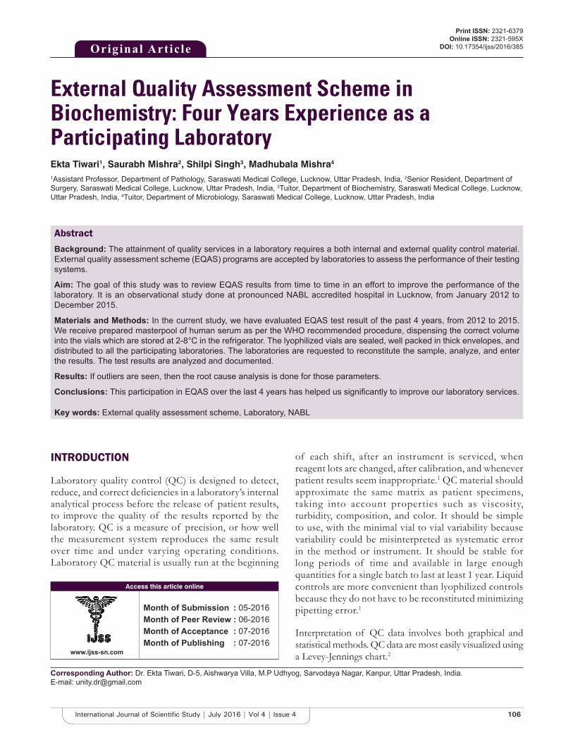
106International Journal of Scientific Study | July 2016 | Vol 4 | Issue 4
External Quality Assessment Scheme in Biochemistry: Four Years Experience as a Participating LaboratoryEkta Tiwari1, Saurabh Mishra2, Shilpi Singh3, Madhubala Mishra4
1Assistant Professor, Department of Pathology, Saraswati Medical College, Lucknow, Uttar Pradesh, India, 2Senior Resident, Department of Surgery, Saraswati Medical College, Lucknow, Uttar Pradesh, India, 3Tuitor, Department of Biochemistry, Saraswati Medical College, Lucknow, Uttar Pradesh, India, 4Tuitor, Department of Microbiology, Saraswati Medical College, Lucknow, Uttar Pradesh, India
of each shift, after an instrument is serviced, when reagent lots are changed, after calibration, and whenever patient results seem inappropriate.1 QC material should approximate the same matrix as patient specimens, taking into account properties such as viscosity, turbidity, composition, and color. It should be simple to use, with the minimal vial to vial variability because variability could be misinterpreted as systematic error in the method or instrument. It should be stable for long periods of time and available in large enough quantities for a single batch to last at least 1 year. Liquid controls are more convenient than lyophilized controls because they do not have to be reconstituted minimizing pipetting error.1
Interpretation of QC data involves both graphical and statistical methods. QC data are most easily visualized using a Levey-Jennings chart.2
INTRODUCTION
Laboratory quality control (QC) is designed to detect, reduce, and correct deficiencies in a laboratory’s internal analytical process before the release of patient results, to improve the quality of the results reported by the laboratory. QC is a measure of precision, or how well the measurement system reproduces the same result over time and under varying operating conditions. Laboratory QC material is usually run at the beginning
AbstractBackground: The attainment of quality services in a laboratory requires a both internal and external quality control material. External quality assessment scheme (EQAS) programs are accepted by laboratories to assess the performance of their testing systems.
Aim: The goal of this study was to review EQAS results from time to time in an effort to improve the performance of the laboratory. It is an observational study done at pronounced NABL accredited hospital in Lucknow, from January 2012 to December 2015.
Materials and Methods: In the current study, we have evaluated EQAS test result of the past 4 years, from 2012 to 2015. We receive prepared masterpool of human serum as per the WHO recommended procedure, dispensing the correct volume into the vials which are stored at 2-8°C in the refrigerator. The lyophilized vials are sealed, well packed in thick envelopes, and distributed to all the participating laboratories. The laboratories are requested to reconstitute the sample, analyze, and enter the results. The test results are analyzed and documented.
Results: If outliers are seen, then the root cause analysis is done for those parameters.
Conclusions: This participation in EQAS over the last 4 years has helped us significantly to improve our laboratory services.
Key words: External quality assessment scheme, Laboratory, NABL
Access this article online
www.ijss-sn.com
Month of Submission : 05-2016 Month of Peer Review : 06-2016 Month of Acceptance : 07-2016 Month of Publishing : 07-2016
Corresponding Author: Dr. Ekta Tiwari, D-5, Aishwarya Villa, M.P Udhyog, Sarvodaya Nagar, Kanpur, Uttar Pradesh, India. E-mail: [email protected]
Original ArticlePrint ISSN: 2321-6379
Online ISSN: 2321-595XDOI: 10.17354/ijss/2016/385

Tiwari, et al.: The External Quality Assessment Scheme in Biochemistry: Four Years Experience as a Participating Laboratory
107 International Journal of Scientific Study | July 2016 | Vol 4 | Issue 4
MATERIALS AND METHODS
External quality assessment scheme (EQAS) samples from the Christian Medical College, Vellore, are received and processed at our laboratory.
For each year, every month’s sample was shipped to our center for specific tests as recommended by the organizing laboratory. Each time, unknown samples packed with coolant were received within 3 days of dispatch. All the samples were handled as part of routine work samples, and recommended tests were performed by the concerned laboratory technician on duty. The tests are performed (Table 1) and mailed to the organizing laboratory within 1st week of the month.
Every year, a total of 12 samples for biochemistry are received. All tests were performed by dedicated staff using the conventional technique available in the department.
RESULTS
Our study reviewed EQAS result from a pronounced NABL accredited laboratory in Lucknow. The outliers seen month wise in 4 years are mentioned in Table 2.
As seen in Table 2 and Figure 1, most number of outliers were seen in the year 2014, i.e., 67 outliers and in the month of June, i.e., 11 outliers. The next most common year was 2015 with 53 outliers, then came 2013 with 40 outliers. Least number of cases was seen in the year 2012 with 38 outliers.
Outliers seen in the year 2012 are shown in Table 3. Alkaline phosphatase was seen to be most common parameter showing outliers, i.e., 9 times, then comes high-density lipoprotein (HDL) showing outlier 5 times. Next comes total protein, potassium, uric acid, and sodium, all showing outliers
3 times. Total bilirubin, creatinine, serum glutamic pyruvic transferase (SGPT), serum glutamic oxaloacetic transaminase (SGOT), calcium, and urea showed outlier 2 times.
Outliers seen in the year 2013 are shown in Table 4. In the year 2013, most number of outliers were seen in uric acid, i.e., 8. Then, comes total bilirubin showing 7 times outlier; next comes HDL showing 5 times outlier, sodium showed 4 times outlier. SGPT, glucose, alkaline phosphatase, SGOT, and triglyceride (TG) showed 2 times outlier. Total protein, urea, calcium, and cholesterol showed one time outlier.
Outliers seen in the year 2014 are shown in Table 5. In the year 2014, 9 times outliers were seen in HDL. Alkaline phosphatase, calcium, and uric acid showed 6 times outlier. Next comes total bilirubin and glucose which show 5 times outlier. SGPT, urea, and TG showed 4 times outlier. Next come sodium, potassium, SGOT, and albumin which show 3 times outlier. The least common outlier was seen in creatinine, i.e., 2.
Outliers seen in the year 2015 are shown in Table 6. In the year 2015, maximum number of an outlier was seen in total bilirubin, i.e., 8 times, then comes calcium showing 6 times outlier. 5 times outliers were seen in alkaline phosphatase, potassium, and glucose. 4 were seen in urea and uric acid.
Table 1: Tests with their methordsS No. Analyte Method 1. Glucose God-pod2. Urea Ned dye3. Creatinine Jaffe’s kinetic4. T.Bilirubin Jendrassik5. T-protein Biuret6. Albumin Bcg7. Calcium Arsenazo8. Uric acid Enzymatic9. Cholesterol Chod-pap10. Triglyceride Enzymatic11. Hdl cho Direct method12. Sodium ISE13. Potassium ISE14. AST UV kinetic15. ALT UV kinetic16. ALP PNP AMP kinetic
Table 2 : Outliers seen in 4 yearsMonth 2012 2013 2014 2015Jan 3 5 6 7Feb 3 3 2 4March 5 5 4 4April 3 1 6 9May 1 4 5 3June 3 1 11 4July 5 3 3 3August No entry 3 6 3Sept 2 1 7 2Oct 2 4 7 5Nov 5 6 6 4Dec 6 4 4 5
Figure 1: Outliers seen in 4 years
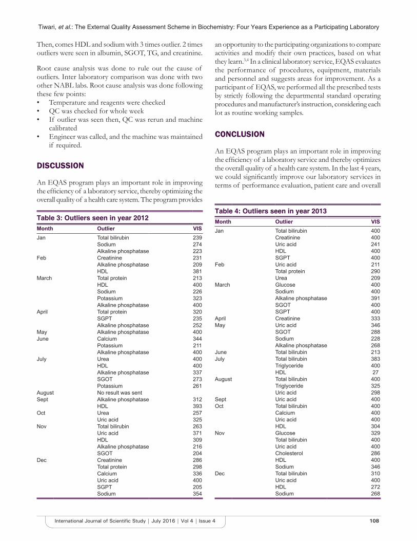
Tiwari, et al.: The External Quality Assessment Scheme in Biochemistry: Four Years Experience as a Participating Laboratory
108International Journal of Scientific Study | July 2016 | Vol 4 | Issue 4
Then, comes HDL and sodium with 3 times outlier. 2 times outliers were seen in albumin, SGOT, TG, and creatinine.
Root cause analysis was done to rule out the cause of outliers. Inter laboratory comparison was done with two other NABL labs. Root cause analysis was done following these few points:• Temperature and reagents were checked• QC was checked for whole week• If outlier was seen then, QC was rerun and machine
calibrated• Engineer was called, and the machine was maintained
if required.
DISCUSSION
An EQAS program plays an important role in improving the efficiency of a laboratory service, thereby optimizing the overall quality of a health care system. The program provides
an opportunity to the participating organizations to compare activities and modify their own practices, based on what they learn.3,4 In a clinical laboratory service, EQAS evaluates the performance of procedures, equipment, materials and personnel and suggests areas for improvement. As a participant of EQAS, we performed all the prescribed tests by strictly following the departmental standard operating procedures and manufacturer’s instruction, considering each lot as routine working samples.
CONCLUSION
An EQAS program plays an important role in improving the efficiency of a laboratory service and thereby optimizes the overall quality of a health care system. In the last 4 years, we could significantly improve our laboratory services in terms of performance evaluation, patient care and overall
Table 3: Outliers seen in year 2012Month Outlier VISJan Total bilirubin 239
Sodium 274Alkaline phosphatase 223
Feb Creatinine 231Alkaline phosphatase 209HDL 381
March Total protein 213HDL 400Sodium 226Potassium 323Alkaline phosphatase 400
April Total protein 320SGPT 235Alkaline phosphatase 252
May Alkaline phosphatase 400June Calcium 344
Potassium 211Alkaline phosphatase 400
July Urea 400HDL 400Alkaline phosphatase 337SGOT 273Potassium 261
August No result was sentSept Alkaline phosphatase 312
HDL 393Oct Urea 257
Uric acid 325Nov Total bilirubin 263
Uric acid 371HDL 309Alkaline phosphatase 216SGOT 204
Dec Creatinine 286Total protein 298Calcium 336Uric acid 400SGPT 205Sodium 354
Table 4: Outliers seen in year 2013Month Outlier VISJan Total bilirubin 400
Creatinine 400Uric acid 241HDL 400SGPT 400
Feb Uric acid 211Total protein 290Urea 209
March Glucose 400Sodium 400Alkaline phosphatase 391SGOT 400SGPT 400
April Creatinine 333May Uric acid 346
SGOT 288Sodium 228Alkaline phosphatase 268
June Total bilirubin 213July Total bilirubin 383
Triglyceride 400HDL 27
August Total bilirubin 400Triglyceride 325Uric acid 298
Sept Uric acid 400Oct Total bilirubin 400
Calcium 400Uric acid 400HDL 304
Nov Glucose 329Total bilirubin 400Uric acid 400Cholesterol 286HDL 400Sodium 346
Dec Total bilirubin 310Uric acid 400HDL 272Sodium 268

Tiwari, et al.: The External Quality Assessment Scheme in Biochemistry: Four Years Experience as a Participating Laboratory
109 International Journal of Scientific Study | July 2016 | Vol 4 | Issue 4
Contd...
Month Outlier VISDec Calcium 284
Uric acid 400HDL 400Alkaline phosphatase 381
Table 5: Contd...
Month Outlier VISJan Urea 400
Total bilirubin 400Calcium 336Uric acid 208Triglyceride 212HDL 400
Feb Total protein 249Calcium 383
March Creatinine 263Uric acid 202HDL 400Potassium 206
April Total bilirubin 400Calcium 206HDL 400Sodium 349Potassium 256Alkaline phosphatase 213
May Calcium 379Uric acid 206HDL 279Sodium 400Potassium 400
June Glucose 400Urea 250Creatinine 354Total bilirubin 400Total protein 276Albumin 400Calcium 400HDL 256SGOT 400SGPT 400Alkaline phosphatase 322
July Total bilirubin 326HDL 400SGPT 217
August Glucose 400Uric acid 400Triglyceride 276Sodium 201SGOT 400Alkaline phosphatase 400
Sept Glucose 210Urea 216Albumin 257Uric acid 395Triglyceride 216HDL 368SGPT 230
Oct Glucose 200Total bilirubin 273Total protein 312HDL 400SGOT 400SGPT 218Alkaline phosphatase 400
Nov Glucose 219Urea 387Total protein 333Albumin 391Triglyceride 268Alkaline phosphatase 254
Table 5: Outliers seen in year 2014
Table 6: Outliers seen in year 2015Month Outlier VISJan Urea 222
Total bilirubin 310Albumin 223Calcium 213Uric acid 400SGOT 400Alkaline phosphatase 400
Feb Total bilirubin 400Calcium 400HDL 350Alkaline phosphatase 400
March Total bilirubin 400Calcium 305Potassium 268Alkaline phosphatase 400
April Glucose 400Urea 400Total protein 209Albumin 220Calcium 400Uric acid 400Cholesterol 246HDL 400Alkaline phosphatase 295
May Glucose 274Total bilirubin 400Potassium 400
June Glucose 400Urea 400Calcium 300Uric acid 400
July Sodium 219Potassium 243
August Glucose 238Total bilirubin 400Potassium 400
Sept Total bilirubin 400Uric acid 319Triglyceride 215
Oct Glucose 400Urea 399Creatinine 226Sodium 331SGOT 400
Nov Total bilirubin 354Calcium 400Sodium 328Potassium 395
Dec Creatinine 236Total bilirubin 252Triglyceride 308HDL 211Alkaline phosphatase 386
Tiwari, et al.: The External Quality Assessment Scheme in Biochemistry: Four Years Experience as a Participating Laboratory
110International Journal of Scientific Study | July 2016 | Vol 4 | Issue 4
quality of laboratory practices.5,6 We believe that global participation in such an EQAS program will definitely improve the quality of a hospital service because no health care facility can be totally self-sufficient, and there is always a scope for improvement and development in a system.
REFERENCES
1. Tietz NW. Fundamentals of Clinical Chemistry. 3rd ed. Philadelphia, PA: WB Saunders Company; 1987.
2. Grant EL, Leavenworth RS. Statistical Quality Control. 6th ed. New York: McGraw-Hill Book Company; 1988.
3. Olson JD, Preston FE, Nichols WL. External quality assurance in thrombosis and hemostasis: An international perspective. Semin Thromb Hemost 2007;33:220-5.
4. Bejrachandra S, Saipin J, Nathalang O, Siriboonrit U, Rungroung E, Udee S. External quality assessment scheme in red blood cell serology: A 5-year experience in Thailand. Immunohematology 2006;22:1-5.
5. Uldall A. Origin of EQA programmers and multidisciplinary cooperation between EQA programme organizers within laboratory medicine. EQA News 1997;8:1-27.
6. Westgard JO. Internal quality control: Planning and implementation strategies. Ann Clin Biochem 2003;40:593-611.
How to cite this article: Tiwari E, Mishra S, Singh S, Mishra M. External Quality Assessment Scheme in Biochemistry: Four Years Experience as a Participating Laboratory. Int J Sci Stud 2016;4(4):106-110.
Source of Support: Nil, Conflict of Interest: None declared.

111 International Journal of Scientific Study | July 2016 | Vol 4 | Issue 4
Acute Neurological Complications in Peripartum Period: A Retrospective StudyB Shanthirani, K Moogambigai
Assistant Professor, Department of Obstetrics and Gynecology, Government Theni Medical College and Hospital, Theni, Tamil Nadu, India
MATERIALS AND METHODS
A retrospective study was conducted at Government Theni Medical College and Hospital in Department of Obstetrics and Gynecology during a 6-month period, from November 2015 to April 2016 involving a total of 30 patients. All patients with recent onset of neurological damage requiring neurological consultation were included in this study. Those who were known case of seizure disorder or cerebrovascular accident were excluded from study. The clinical presentation, imaging reports and prognosis of these 30 patients were followed up.
RESULTS
The total number of deliveries in this study period was 3208. Of this, only 30 patients developed acute neurological complications accounting for 0.01%. The results of this study are shown in Tables 1-6.
DISCUSSION
The most common age group affected by neurological complications is 20-25 years of age (40%). Complications
INTRODUCTION
Acute neurological conditions are rare in young age. A mean age of pregnancy in female has increased in recent years and thereby increases the risk of neurological complications. Conditions like pre-eclampsia cause no significant damage as long as it does not progress to eclampsia. Pre-eclampsia and eclampsia are directly related to pregnancy whereas conditions such as cerebral venous thrombosis (CVT), ischemia, and hemorrhage are indirectly related to pregnancy. Neurological abnormalities contribute significantly to maternal mortality in eclampsia. In this study, we will discuss the varied clinical presentation of neurological complications in the peripartum period.
Original Article
AbstractIntroduction: Acute neurological conditions are rare in young age. A mean age of pregnancy in female has increased in recent years and thereby increases the risk of neurological complications.
Objective: To study the clinical profile of patients presenting with neurological manifestations in a peripartum period.
Study Design: A retrospective study done at Government Theni Medical College and Hospital during November 2015 to April 2016. 30 cases studied. All patients in the peripartum period requiring neurological consultation were included in the study.
Results: A total of 30 women were included in this study. The incidence of neurological complications in the study period was 0.01%, but the mortality rate due to neurological complications was 6%. It occurred most commonly in primigravida.
Conclusion: This study concluded that proper management of pre-eclampsia can prevent eclampsia and reduce maternal mortality and morbidity.
Key words: Eclampsia, Hypertension, Mortality, Neurological
Access this article online
www.ijss-sn.com
Month of Submission : 05-2016 Month of Peer Review : 06-2016 Month of Acceptance : 07-2016 Month of Publishing : 07-2016
Corresponding Author: B Shanthirani, Asstistant Professor, Government Theni Medical College and Hospital, Theni, Tamil Nadu, India. E-mail: [email protected]
Print ISSN: 2321-6379Online ISSN: 2321-595X
DOI: 10.17354/ijss/2016/386

Shanthirani and Moogambigai: Acute neurological complications in peripartum period
112International Journal of Scientific Study | July 2016 | Vol 4 | Issue 4
occur more in primigravida (63%) than multigravida (27%). This incidence is comparable with the Al-Hayali et al. study where neurological complications occurred more in primigravida (85%) compared to multigravida (15%).1
This is because general incidence of preeclampsia is more common in primigravida. Risk factors for neurological complications include associated anemia and dehydration.2
A headache is the most common symptom of neurological conditions, and therefore, it is necessary to distinguish benign headache from headache due to complications in pregnancy. Moreover, the most common sign was seizure occurring in 76% of patients. In our study, 25 of the 30 patients had complaint of headache before the onset of neurological complications (83%). This is similar to Gupta study where a headache was the symptom in 90% of cases and seizure occurred in 92% of cases.3
The most patients presented in postpartum period (66%) with headache, seizures, visual disturbances, 23% on day of delivery and remaining 43% within a week of delivery, 1% in the antenatal period, <1% in intrapartum period.
Careful clinical evaluation of the patient can identify high-risk cases but it is the imaging studies that help us diagnose the disorder in the majority of cases. Hence, a computed tomography (CT) brain study was done for these 30 patients as per neurologist’s orders and reports showed 66% of CT reports were normal with 6.66% each of CVT and hemorrhagic infarct. 16% cases developed posterior reversible encephalopathy syndrome (PRES), a reversible encephalopathy caused due to an acute increase in blood pressure.
PRES has rapid onset in the postpartum period with symptoms of visual loss, seizures, and headache. If blood pressure is controlled, symptoms resolve within days to weeks. CT findings are usually present. Magnetic resonance imaging (MRI) shows fluid-attenuated inversion recovery abnormalities in parieto-occipital lobes, with occasional intracerebral hemorrhage.
CVT presents in the third trimester or postpartum with symptoms of headache at onset. Seizures can occur and CVT evolves over several days. CT and MRI reveal non arterial and territorial infarcts.
As mentioned earlier, the incidence of acute neurological complication in peripartum period is 0.1% in our hospital. Of this 2 patients died due to neurological complications, accounting for a mortality rate of 6%. Comparing to overall maternal mortality in our hospital in the same study period, 2 out of 7 deaths were due to neurological complications (28%). This is similar to the incidence of 20% maternal deaths due to neurological diseases as mentioned in Hosley and McCullough study.4 This is a significant proportion of maternal mortality stating the importance of monitoring neurological complications.
Table 1: Distribution of patients according to ageAge group (years) Number of patients<20 420-25 1225-30 830-35 5>35 1
Table 2: Distribution of patients according to parityParity Number of patientsPrimi 19Multi 10Grand multi (>5) 1
Table 3: Distribution of patients according to onset of acute neurological complicationsDay Complication occurredAntenatal 3Intrapartum 2PND 1 7Within PND 7 13>PND 7 5PND: Postnatal day
Table 4: Maternal morbidity following acute neurological complicationsComplication occurred Number of patientsSeizure 23Hemiplegia 2Unconsciousness 5
Table 5: Results of imagination studiesCT report Number of patientsNormal study 20CVT 2PRES 5Hemorrhagic infarct 2Granuloma 1CT: Computed tomography, CVT: Cerebral venous thrombosis, PRES: Posterior reversible encephalopathy syndrome
Table 6: Maternal outcomeRecovery of patients Number of patientsDischarged in good condition 25Referred out 3Death 2

Shanthirani and Moogambigai: Acute neurological complications in peripartum period
113 International Journal of Scientific Study | July 2016 | Vol 4 | Issue 4
CONCLUSION
The symptom of headache in a peripartum woman with or with our pregnancy induced hypertension must be taken seriously. A thorough neurological examination followed by imaging will pick up early lesion of CVT. Thus, the treatment could be planned and carried out which will reduce the complications significantly.
REFERENCES
1. Al-Hayali RM, Al-Habbo DJ, Hammo MK. Peripartum neurological emergencies in a Critical Care Unit. Neurosciences (Riyadh) 2008;13:155-60.
2. Edlow JA, Caplan LR, O’Brien K, Tibbles CD. Diagnosis of acute neurological emergencies in pregnant and post-partum women. Lancet Neurol 2013;12:175-85.
3. Gupta S, Rohatgi A, Sharma SK, Gurtoo A. A study of neurological disorders during pregnancy and puerperium. Ann Indian Acad Neurol 2006;9:152-7.
4. Hosley CM, McCullough LD. Acute neurological issues in pregnancy and the peripartum. Neurohospitalist 2011;1:104-16.
How to cite this article: Shanthirani B, Moogambigai K. Acute Neurological Complications in Peripartum Period: A Retrospective Study. Int J Sci Stud 2016;4(4):111-113.
Source of Support: Nil, Conflict of Interest: None declared.

114International Journal of Scientific Study | July 2016 | Vol 4 | Issue 4
Mortality Profile and Timing of Death in Extremely Low Birth Weight Infants from 2013 to 2015 Admitted to Neonatal Intensive Care Unit, Government General Hospital, AnantapurN Praveen Deen Kumar1, B Praveena2
1Assistant Professor, Department of Pediatrics, Government Medical College, Anantapur, Andhra Pradesh, India, 2Tutor, Department of Microbiology, Government Medical College, Anantapur, Andhra Pradesh, India
Causes of LBW are mainly preterm birth which is due to precocious fetal endocrine activation, uterine overdistension, decidual bleeding, and intrauterine inflammation/infection.3 Other causes include short gestational age (SGA) which may be constitutional or intrauterine growth restriction due to various reasons such as infections during pregnancy (TORCH), babies with congenital anomalies or chromosomal abnormalities, and environmental factors.4,5
Data regarding deaths in ELBW babies help to know the cause of death, assess disease patterns, and its complications, to implement necessity actions to reduce the mortality rate and also to plan and implement programs.
Many recent advances in perinatal care have resulted in a decrease in mortality rate for very LBW infants by antenatal steroids and surfactant therapy.6,7 However, few studies are concerning about problems by these advances in therapy such as prolong dying, extend suffering, or use resources for infants who will eventually die.8,9
INTRODUCTION
Infant mortality is a major public health problem worldwide, whereas many programs related to children have started to decrease the infant mortality rate. Among infant mortality, most of the data come from neonatal deaths, approximately two-third of all infant mortalities.1
Extreme low birth weight (ELBW) is defined as a birth weight of a liveborn infant of <1000 g. Most ELBW infants are also the youngest of premature newborns, usually born <28 weeks gestational age.2 ELBW infants are also at high risk of developing neonatal infections.
Original Article
AbstractIntroduction: Infant mortality is a major public health problem worldwide, whereas many programs related to children have started to decrease the infant mortality rate.
Materials And Methods: A retrospective study done to understand the mortality profile, etiology, and timing of death among ELBW infants in a tertiary care Neonatal Intensive Care Unit (NICU), Government General Hospital, Anantapur
Results: A total of 97 ELBW were died due to various causes from 2013 to 2015. Among 97 ELBW studied children, males and females were equally affected. Out of 97 ELBW children, 48 (49.4%) were males and 49 (50.5%) were female (Table 1). Gestational age of ELBW children was around 26.1 ± 1.5 weeks.
Conclusion: From this study, we conclude that there is a rise in mortality rate in ELBW babies, whereas most of the deaths due to multiple cause occurring within 7 h of hospital admission.
Key words: Mortality, Death, Low Birth Weight, Infants
Access this article online
www.ijss-sn.com
Month of Submission : 05-2016 Month of Peer Review : 06-2016 Month of Acceptance : 07-2016 Month of Publishing : 07-2016
Corresponding Author: Dr. N Praveen Deen Kumar, Department of Pediatrics, Government Medical College, Anantapur - 515 001, Andhra Pradesh, India. Phone: +91-9440655505. E-mail: [email protected]
Print ISSN: 2321-6379Online ISSN: 2321-595X
DOI: 10.17354/ijss/2016/387
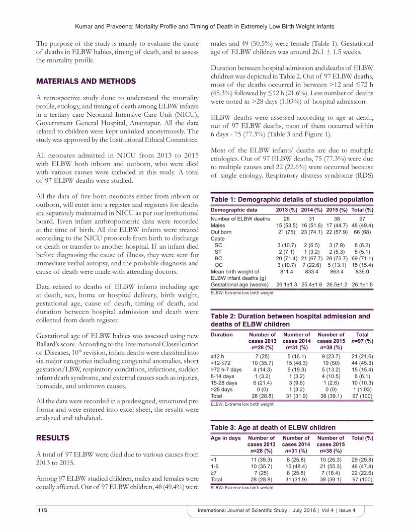
Kumar and Praveena: Mortality Profile and Timing of Death in Extremely Low Birth Weight Infants
115 International Journal of Scientific Study | July 2016 | Vol 4 | Issue 4
The purpose of the study is mainly to evaluate the cause of deaths in ELBW babies, timing of death, and to assess the mortality profile.
MATERIALS AND METHODS
A retrospective study done to understand the mortality profile, etiology, and timing of death among ELBW infants in a tertiary care Neonatal Intensive Care Unit (NICU), Government General Hospital, Anantapur. All the data related to children were kept unlinked anonymously. The study was approved by the Institutional Ethical Committee.
All neonates admitted in NICU from 2013 to 2015 with ELBW both inborn and outborn, who were died with various causes were included in this study. A total of 97 ELBW deaths were studied.
All the data of live born neonates either from inborn or outborn, will enter into a register and registers for deaths are separately maintained in NICU as per our institutional board. Even infant anthropometric data were recorded at the time of birth. All the ELBW infants were treated according to the NICU protocols from birth to discharge or death or transfer to another hospital. If an infant died before diagnosing the cause of illness, they were sent for immediate verbal autopsy, and the probable diagnosis and cause of death were made with attending doctors.
Data related to deaths of ELBW infants including age at death, sex, home or hospital delivery, birth weight, gestational age, cause of death, timing of death, and duration between hospital admission and death were collected from death register.
Gestational age of ELBW babies was assessed using new Ballard’s score. According to the International Classification of Diseases, 10th revision, infant deaths were classified into six major categories including congenital anomalies, short gestation/LBW, respiratory conditions, infections, sudden infant death syndrome, and external causes such as injuries, homicide, and unknown causes.
All the data were recorded in a predesigned, structured pro forma and were entered into excel sheet, the results were analyzed and tabulated.
RESULTS
A total of 97 ELBW were died due to various causes from 2013 to 2015.
Among 97 ELBW studied children, males and females were equally affected. Out of 97 ELBW children, 48 (49.4%) were
males and 49 (50.5%) were female (Table 1). Gestational age of ELBW children was around 26.1 ± 1.5 weeks.
Duration between hospital admission and deaths of ELBW children was depicted in Table 2. Out of 97 ELBW deaths, most of the deaths occurred in between >12 and ≤72 h (45.3%) followed by ≤12 h (21.6%). Less number of deaths were noted in >28 days (1.03%) of hospital admission.
ELBW deaths were assessed according to age at death, out of 97 ELBW deaths, most of them occurred within 6 days - 75 (77.3%) (Table 3 and Figure 1).
Most of the ELBW infants’ deaths are due to multiple etiologies. Out of 97 ELBW deaths, 75 (77.3%) were due to multiple causes and 22 (22.6%) were occurred because of single etiology. Respiratory distress syndrome (RDS)
Table 1: Demographic details of studied populationDemographic data 2013 (%) 2014 (%) 2015 (%) Total (%)Number of ELBW deaths 28 31 38 97Males 15 (53.5) 16 (51.6) 17 (44.7) 48 (49.4)Out born 21 (75) 23 (74.1) 22 (57.9) 66 (68)Caste
SC 3 (10.7) 2 (6.5) 3 (7.9) 8 (8.2)ST 2 (7.1) 1 (3.2) 2 (5.3) 5 (5.1)BC 20 (71.4) 21 (67.7) 28 (73.7) 69 (71.1)OC 3 (10.7) 7 (22.6) 5 (13.1) 15 (15.4)
Mean birth weight of ELBW infant deaths (g)
811.4 833.4 863.4 836.0
Gestational age (weeks) 26.1±1.3 25.4±1.6 26.5±1.2 26.1±1.5ELBW: Extreme low birth weight
Table 2: Duration between hospital admission and deaths of ELBW childrenDuration Number of
cases 2013 n=28 (%)
Number of cases 2014 n=31 (%)
Number of cases 2015 n=38 (%)
Total n=97 (%)
≤12 h 7 (25) 5 (16.1) 9 (23.7) 21 (21.6)>12-≤72 10 (35.7) 15 (48.3) 19 (50) 44 (45.3)>72 h-7 days 4 (14.3) 6 (19.3) 5 (13.2) 15 (15.4)8-14 days 1 (3.2) 1 (3.2) 4 (10.5) 6 (6.1)15-28 days 6 (21.4) 3 (9.6) 1 (2.6) 10 (10.3)>28 days 0 (0) 1 (3.2) 0 (0) 1 (1.03)Total 28 (28.8) 31 (31.9) 38 (39.1) 97 (100)ELBW: Extreme low birth weight
Table 3: Age at death of ELBW childrenAge in days Number of
cases 2013 n=28 (%)
Number of cases 2014 n=31 (%)
Number of cases 2015 n=38 (%)
Total (%)
<1 11 (39.3) 8 (25.8) 10 (26.3) 29 (29.8)1-6 10 (35.7) 15 (48.4) 21 (55.3) 46 (47.4)≥7 7 (25) 8 (25.8) 7 (18.4) 22 (22.6)Total 28 (28.8) 31 (31.9) 38 (39.1) 97 (100)ELBW: Extreme low birth weight

Kumar and Praveena: Mortality Profile and Timing of Death in Extremely Low Birth Weight Infants
116International Journal of Scientific Study | July 2016 | Vol 4 | Issue 4
was the most common cause responsible for ELBW infant deaths (37.1%), followed by sepsis (26.8%), which is the second most common cause (Table 4).
In 3 years, various causes were responsible for deaths including RDS - 36 (37.1%), sepsis - 26 (26.8%), birth asphyxia - 17 (17.5%), necrotizing enterocolitis (NEC) - 6 (6.1%), multiorgan dysfunction syndrome (MODS)/disseminated intravascular coagulation (DIC)/shock - 8 (8.2%), congenital anomalies – 2 (2.06%), and others - 2 (2.06%) (Figure 2).
DISCUSSION
ELBW or SGA is one of the causes for increase in neonatal mortality rate. On this, many other factors combined with ELBW play a major role in increasing the incidence of mortality rate. In this study, we have discussed the various factors which influence the mortality rate among ELBW children.
A total of 97 ELBW children were died due to various causes. All the studied children were premature children. ELBW deaths were increasing in recent years, as in 2015 deaths were 38. In this study, 30 ELBW deaths per year on an average were observed which is quite significant. Patel et al.10 observed a decrease in ELBW infant deaths from 2004 to 2011; it was 28.5% in 2004-2007, and 25.8% in 2008-2011.
Out of 97 ELBW infant deaths, males and females were equally affected. Out of 97 ELBW children, 48 (49.4%) were males and 49 (50.5%) were females (Table 1). Gestational age of ELBW children was around 27 weeks. Mean birth weight of ELBW babies was around 830 g in this study. Patel et al.10 reported that deaths occurred at mean gestational age of 24.3 ± 1.7 weeks and birth weight was around 660 g. Mukhopadhyay et al.11 reported mean birth weight was 843 ± 108 g and gestational age was 29.1 ± 2.6 weeks among ELBW neonates. Many studies
have found that neonates <750 g weight and <28 weeks gestational age were associated with higher mortality.12,13
Duration between hospital admission and deaths of ELBW children was depicted in Table 2. Out of 97 ELBW deaths, most of the deaths occurred in between >12 h and ≤72 h (45.3%) followed by ≤12 h (21.6%). Less number of deaths were noted in >28 days (1.03%) of hospital admission. Patel et al.10 documented that most common cause of death within 12 h after birth is immaturity, which was observed from 2000 to 2011 and also observed that deaths occurring after 12 h are due to RDS.
Most of ELBW deaths occurred within 72 h in the present study. Even with good hospital care, death rate is rising among ELBW children and as days in hospital increases death rate is decreasing, which indicates ELBW babies need care in hospitals from the time of birth to prevent various etiologies responsible for deaths. Patel et al.10 reported that 40.4% of deaths occurred within 12 h after birth and 17.3% occurred after 28 days.
ELBW deaths were assessed according to the age at death, out of 97 children, most of them occurred within 6 days – 75 (77.3%). In this study, within 6 days, highest death rate signifies the disease pattern which is more severe. Yasmin
Table 4: Etiologies of ELBW infant deathsCause of death Number of
cases 2013n=28 (%)
Number of cases 2014
n=31 (%)
Number of cases 2015
n=38 (%)
Totaln=97 (%)
RDS 10 (35.7) 11 (35.5) 15 (39.5) 36 (37.1)Sepsis 5 (17.8) 12 (38.7) 9 (23.7) 26 (26.8)Birth asphyxia 8 (28.6) 2 (6.4) 7 (18.4) 17 (17.5)NEC 2 (7.1) 3 (9.7) 1 (2.6) 6 (6.1)MODS/DIC/Shock 1 (3.6) 2 (6.4) 5 (13.2) 8 (8.2)Congenital anomalies
1 (3.6) 1 (3.2) 0 (0) 2 (2.0)
Others 1 (3.6) 0 (0) 1 (2.6) 2 (2.0)Total 28 (100) 31 (100) 38 (100) 97 (100)RDS: Respiratory distress syndrome, NEC: Necrotizing enterocolitis, MODS: Multiorgan dysfunction syndrome, DIC: Disseminated intravascular coagulation, ELBW: Extreme low birth weight
Figure 1: The number of extremely low birth weight children deaths at different ages from 2013 to 2015
Figure 2: Etiologies of extremely low birth weight infant deaths

Kumar and Praveena: Mortality Profile and Timing of Death in Extremely Low Birth Weight Infants
117 International Journal of Scientific Study | July 2016 | Vol 4 | Issue 4
et al.14 reported that 84% of neonatal deaths occurred in first 7 days and half of those within 48 h. Jacob et al.1 observed 59% of NICU deaths within 7 days of birth and 85% within 28 days after birth.
As per this study, out of 97 ELBW deaths, 75 (77.3%) were due to multiple causes and 22 (22.6%) were occurred because of single etiology. In 3 years, various causes were responsible for deaths including RDS - 36 (37.1%), sepsis - 26 (26.8%), birth asphyxia - 17 (17.5%), NEC - 6 (6.1%), MODS/DIC/shock - 8 (8.2%), congenital anomalies - 2(2.06%), and others - 2 (2.06%).
Mukhopadhyay et al.11 observed major causes of mortality were sepsis (46%), birth asphyxia (20%), and pulmonary hemorrhage (19%). Mukhopadhyay et al.11 and Kermorvant-Duchemin et al.15 reported 46% and 71% mortality due to sepsis. Yasmin et al.14 reported the predominant cause of NICU deaths was LBW (14%), followed by sepsis (12%), acquired bowel disease (11%), lung hypoplasia (9.5%), intraventricular hemorrhage (9.4%), RDS (8%), hypoxic ischemic encephalopathy (6.1%), genetic syndromes (5%), major heart defects (3.4%), bronchopulmonary (2.8%), hemorrhagic shock (2.2%), pulmonary hemorrhage (2%), renal failure (2%), congenital diaphragmatic hernia (1.7%), air leak syndrome (0.6%), and pulmonary hypertension (0.6%).
Evaluating the cause of death, age at death, and deaths in hospital even after treatment is quite significant to understand the reasons behind mortality rate. Nam et al and Carver et al.16,17 documented that reliability of finding the underlying cause of death is adversely affected because evaluating the disease differs among health-care providers.
Deaths of babies impart emotional stress to parents. Increase in mortality rate indicates poor health seeking behavior of mothers, poor diagnostic, infrastructure, and health-care facilities at institutions and overall signifies defects in societal health developments.
CONCLUSION
From this study, we conclude that there is a rise in mortality rate in ELBW babies, whereas most of the deaths due to
multiple cause occurring within 7 h of hospital admission. To reduce the incidence of ELBW deaths needs the utmost care of children at hospital by finding the cause earliest. Design of policies and implementing health education units, regular monitoring of mortality statistics, making preventive and corrective actions in various aspects helps to get mortality rate to baseline.
REFERENCES
1. Jacob J, Kamitsuka M, Clark RH, Kelleher AS, Spitzer AR. Etiologies of NICU deaths. Paediatr 2015;135:67-776.
2. Siva Subramanian KN. Extremely low birth weight infant. eMed 2007. p. 11-28.
3. Simhan HN, Caritis SN. Prevention of preterm delivery. N Engl J Med 2007;357:477-87.
4. Knopik VS. Maternal smoking during pregnancy and child outcomes: Real or spurious effect? Dev Neuropsychol 2009;34:1-36.
5. Salmasi G, Grady R, Jones J, McDonald SD; Knowledge Synthesis Group. Environmental tobacco smoke exposure and perinatal outcomes: A systematic review and meta-analyses. Acta Obstet Gynecol Scand 2010;89:423-41.
6. Jobe AH. Pulmonary surfactant therapy. N Engl J Med 1993;328:861-8.7. Guyer B, Martin JA, MacDorman MF, Anderson RN, Strobino DM. Annual
summary of vital statistics-1996. Pediatrics 1997;100:905-18.8. Kalter HD, Na Y, O’Campo P. Decrease in infant mortality in New York
City after 1989. Am J Public Health 1998;88:816-20.9. Meadow W, Reimshisel T, Lantos J. Birth weight-specific mortality
for extremely low birth weight infants vanishes by four days of life: Epidemiology and ethics in the neonatal intensive care unit. Pediatrics 1996;97:636-43.
10. Patel RM, Kandefer S, Walsh MC, Bell EF, Carlo WA, Laptook AR, et al. Causes and timing of death in extremely premature infants from 2000 through 2011. N Engl J Med 2015;372:331-40.
11. Mukhopadhyay K, Louis D, Mahajan R, Kumar P. Predictors of mortality and major morbidities in extremely low birth weight neonates. Indian Pediatr 2013;50:1119-23.
12. Tagare A, Chaudhari S, Kadam S, Vaidya U, Pandit A, Sayyad MG. Mortality and morbidity in extremely low birth weight (ELBW) infants in a neonatal intensive care unit. Indian J Pediatr 2013;80:16-20.
13. Narayan S, Aggarwal R, Upadhyay A, Deorari AK, Singh M, Paul VK. Survival and morbidity in extremely low birth weight (ELBW) infants. Indian Pediatr 2003;40:130-5.
14. Yasmin S, Osrin D, Paul E, Costello A. Neonatal mortality of low-birth-weight infants in Bangladesh. Bull World Health Organ 2001;79:608-14.
15. Kermorvant-Duchemin E, Laborie S, Rabilloud M, Lapillonne A, Claris O. Outcome and prognostic factors in neonates with septic shock. Pediatr Crit Care Med 2008;9:186-91.
16. Nam CB, Eberstein IW, Deeb LC, Terrie EW. Infant mortality by cause: A comparison of underlying and multiple cause designations. Eur J Popul 1989;5:45-70.
17. Carver JD, McDermott RJ, Jacobson HN, Sherin KM, Kanarek K, Pimentel B, et al. Infant mortality statistics do not adequately reflect the impact of short gestation. Pediatrics 1993;92:229-32.
How to cite this article: Kumar NPD, Praveena B. Mortality Profile and Timing of Death in Extremely Low Birth Weight Infants from 2013 to 2015 Admitted to Neonatal Intensive Care Unit, Government General Hospital, Anantapur. Int J Sci Stud 2016;4(4):114-117.
Source of Support: Nil, Conflict of Interest: None declared.

118International Journal of Scientific Study | July 2016 | Vol 4 | Issue 4
Efficacy of Saline Infusion Sonography in Diagnosing Intrauterine Pathology in Patients with Abnormal Uterine Bleeding: An Observational StudyT H Usha1, M Gayathiri2
1Senior Assistant Professor, Department of Obstetrics and Gynecology, Government Rajaji Hospital, Madurai Medical College, Madurai, Tamil Nadu, India, 2Associate Professor, Department of Obstetrics and Gynecology, Government Rajaji Hospital, Madurai Medical College, Madurai, Tamil Nadu, India
AUB inc ludes menor rhag i a , po lymenor rhea , menometrorrhagia, and metrorrhagia. In patients who do not respond to medical treatment, it is ideal to evaluate the endometrial status. It can be used in patients above 40 years also. Even though ovulation and breakthrough bleeding are considered normal, other forms of irregular uterine bleeding necessitate excluding local causes. The most common cause of AUB in premenopausal women is oligoanovulation, which reflects dysfunction in the hypothalamo-pituitary-ovarian axis. Without cyclic progesterone, endometrial lining remains proliferative and hyperplastic and present with non-cyclic menstrual blood flow with timing and amount being erratic. AUB can also be caused by anatomic conditions such as polyps, fibroids, hyperplasia, and even frank carcinomas, which needs appropriate evaluation.
Hysteroscopy combined with guided biopsy has been considered as gold standard in the evaluation of AUB.
INTRODUCTION
Abnormal uterine bleeding (AUB) is defined as any bleeding from the genital tract, which is a deviation of normal in frequency, cyclicity, and quantity. It is one of the most common disorders in gynecology and accounts for 30-40% of cases in the outpatient clinic. It can occur at any age but is most common in premenopausal age group. If the treatment not is instituted early, it can lead to severe anemia. If blood loss is acute, it can result in hypovolemic shock.
AbstractBackground: To describe the efficacy of saline infusion sonography (SIS) in the diagnosis of abnormal uterine bleeding (AUB).
Methods: This prospective observational study was conducted in patients with AUB admitted to our hospital gynecology ward. After obtaining consent from each patient, 100 women in the age group of 25-45 with H/O AUB from March 2015 to March 2016 (a period of 1-year). SIS was done using an 8 Fr size Foley’s catheter, and the results were analyzed.
Results: The most common lesion in the study group was adenomyosis followed by intramural fibroid, submucous myoma, and endometrial hyperplasia. Least common lesions found in our study were endometrial polyp and Mullerian anomalies. Positive predictive value of SIS in the diagnosis of AUB was 100% for Mullerian anomalies and submucous myoma. Positive predictive value for diagnosing adenomyosis was 90% and 88% for intramural fibroid. Positive predictive value for diagnosing endometrial polyps was 85%, and positive predictive value for endometrial hyperplasia was 85%.
Conclusion: SIS has the advantages of being non-invasive, cheap, affordable, shorter duration, and accurate method for uterine cavity evaluation. SIS has enhanced the diagnostic accuracy of transvaginal scan and can be an effective screening test before hysteroscopy.
Key words: Abnormal uterine bleeding, Intrauterine pathology, Saline infusion sonography
Access this article online
www.ijss-sn.com
Month of Submission : 05-2016 Month of Peer Review : 06-2016 Month of Acceptance : 07-2016 Month of Publishing : 07-2016
Corresponding Author: Dr. T H Usha, Department of Obstetrics and Gynecology, Government Rajaji Hospital, Madurai Medical College, Madurai, Tamil Nadu, India. Phone: +91-9443587008. E-mail: [email protected]
Original Article DOI: 10.17354/ijss/2016/388
Print ISSN: 2321-6379Online ISSN: 2321-595X

Usha and Gayathiri: Efficacy of Saline Infusion Sonography in Diagnosing Intrauterine Pathology
119 International Journal of Scientific Study | July 2016 | Vol 4 | Issue 4
Even though hysteroscopy is gold standard, it requires trained personnel, equipment, and anesthesia. It has its own complications and needs hospital stay. The search of a procedure, which is less invasive, cost-effective, and accurate in diagnosis led to the advent of endometrial imaging using saline as a contrast medium. Sonosalpingography otherwise called as saline infusion sonography (SIS) was first described by Nannini et al.,1 in 1981, it was Richman et al.,2 in 1984, used this technique first for evaluation of tubal patency in infertile women.
This test is easier, better, cost-effective, less time-consuming, and an efficient diagnostic modality with minimal morbidity in patients with AUB. The instillation of saline into uterine cavity provides a contrast that helps to localize abnormalities as intracavitary; endometrial or submucosal fluid represents an excellent medium for transmission of sound waves and provides a good contrast to examine the endometrial cavity, just as it is better to look at the fetus in case of polyhydramnios. We designed this study to analyze the efficacy, safety of SIS in the diagnosis of women with AUB.
MATERIALS AND METHODS
This prospective observational study was conducted in patients with AUB admitted to our hospital gynecology ward. Hospital ethical committee approval was obtained to conduct the study. After obtaining consent from each patient, 100 women with H/O AUB were included in the study. The inclusion criteria of women with age group of 25-45 years are:1. Irregular uterine bleeding2. Heavy and/or prolonged periods aged 40 years or more
after excluding fibroid in ultrasonography (USG)3. Heavy and/or prolonged periods below 40 years of
age, who do not respond to medical treatment4. Cystic, echogenic endometrium on USG5. Hyperechoic line around the endometrium6. Failure to visualize the endometrium on ultrasound.
Patients with unhealthy cervix, genital infection, tuberculosis, genital malignancy, suspected endocrinological abnormality, large myoma, cervical stenosis were excluded from the study. A detailed history was taken, and medical and gynecological examination was done to rule out the pelvic inflammatory disease. The patients had taken tablet doxycycline 200 mg and metronidazole 400 mg 1 h before the procedure.
Timing of the ProcedureProcedure is done on 8th or 9th day of the menstrual cycle in patients with regular cycles or just after cessation of the bleeding in those with irregular cycles. Patients with bleeding are best seen in proliferative phase, to rule out polyps and
with suspected fibroid, best in the secretory phase. In proliferative phase, since the endometrium is thickened, it provides a contrast to hypoechoic leiomyomata.
ProcedurePatients positioned in dorsal lithotomy position. Before SIS, routine standard transvaginal ultrasound is done with empty bladder. Transvaginal probe is removed. SIMS speculum is introduced to retract the anterior vaginal wall and cervix visualized. Size 8 Fr Foley’s catheter was introduced into the external Os, and the Foley bulb was inflated with 2 ml of sterile distilled water, and the catheter was withdrawn so that the Foley’s bulb is placed at the level of internal Os. The transvaginal probe is reintroduced into the vagina beneath the catheter, and 20 ml of sterile saline is slowly injected through the catheter and the uterine cavity distended. The uterus is scanned in longitudinal axis (sagittal plane) and transverse axis (coronal plane) and the intracavitary pathologies if present is detected. After the procedure is over, the probe is removed, Foley’s bulb is deflated and the catheter withdrawn and the patient is allowed to dress before discussing further management.
Criteria for diagnosis in SIS:1. Normal endometrium: Normal looking endometrium
with uniform thickness all around within the normal range
2. Endometrial hyperplasia: Diffuse and irregularly thickened endometrium, without any breech in surface, with intact endomyometrial interface; thickness >10 mm
3. Endometrial polyps: Not as round and regular in outline as fibroid polyp; smooth marginated echogenic mass with homogenous echotexture; sway with the movement of the fluid in the cavity
4. Endometritis: Bright echogenic foci with irregular surface
5. Submucous fibroid: Solid round structures of mixed echogenicity that cause bulge or protrusion of the endometrium that does not move with the medium
6. Intrauterine adhesions: Thread-like immobile strands7. Subseptate/bicornuate uterus.
OBSERVATION AND RESULTS
In our observational study, 100 patients were studied for the efficacy of SIS in detecting intracavitary lesions. Patients from 25 to 45 years patients were taken in this study. Out of 100 patients, 28 belong to 25-35 years age group and remaining 72 belong to 36-45 years age group. This shows the incidence of AUB more common around perimenopausal age (Table 1). In our study, nullipara and Para 1 were 2% only. 3 patients had one abortion. Patients with Para 4 were 3. Patients with Para 1, Para 2, and Para 3 totally occupied 90% out of 100 patients. Para 2 had

Usha and Gayathiri: Efficacy of Saline Infusion Sonography in Diagnosing Intrauterine Pathology
120International Journal of Scientific Study | July 2016 | Vol 4 | Issue 4
the highest incidence of AUB of about 53% followed by Para 3, which included 28% of patients. This clearly denoted that AUB more common in multiparous women.
About 27 patients out of total 72 patients in the age group between 36 and 45 were having menorrhea with 37% as the most common symptom. This was followed by polymenorrhagia with 25% of patients. Patients with polymenorrhea were 23%. Menometrorrhagia and metrorrhagia were the least common symptoms. Out of 100 patients, the most common symptom was polymenorrhea which included 35% of the patients, followed by menorrhagia which included about 32% of the patients. This was followed by polymenorrhagia with 20% of patients. Menometrorrhagia, metrorrhagia, and oligomenorrhea all constituted about 13% of patients (Table 2).
The most common lesion in the age group 36-45 years were adenomyosis with 25% of patients followed by fibroid with 21% of patients. Patients with submucous myoma were 15%. Patients with endometrial hyperplasia and polyps were least in number. There were no patients with Mullerian anomaly. 20 patients had a normal uterus. In age group between 25 and 35, all the pathologies were almost equal. Fibroid, polyp, and Mullerian anomaly were equal in number with 2 patients each. Submucous myoma and endometrial hyperplasia group patients were in equal in number with 3 patients. Patients with adenomyosis were 6 in number. 10 patients had a normal uterus (Table 3).
As routine transvaginal ultrasound is performed before SIS, scan findings were also observed. Out of 100 patients, 30 patients SIS test were normal. In remaining 70 patients, adenomyosis was the most common diagnosis with 18% of total 100 patients followed by patients with fibroid with 16%. Patients with endometrial hyperplasia and submucous myoma were 12%. Patients with endometrial polyp were 10%. The least common was Mullerian anomalies, which were only 2% (Table 4).
About 60 patients in this study underwent hysterectomy. Their uterine specimen were analyzed and found to be normal in 24 patients with 40%. Polyp was found in 9 patients with 15%. Patients with submucous myoma were found to be 19%.16 patients had intramural fibroid with 26%.
The positive predictive value of each condition varies from 85% to 100%. The positive predictive value was 85% for endometrial hyperplasia which was the least. The positive predictive value for fibroid was 88% followed by the positive predictive value of adenomyosis which was 90%. The positive predictive values for submucous myoma and Mullerian anomalies were 100%. The positive predictive value for diagnosing normal uterine cavity was 100% (Table 4).
DISCUSSION
AUB is often seen in peri- and post-menopausal women. The etiology varies from simple dysfunctional uterine bleeding to benign lesions such as polyps and even frank malignancies. Apart from the clinical diagnosis, various diagnostic methods are available to confirm the diagnosis. AUB can cause anemia, and in some cases, it can cause hypovolemic shock and the patient may collapse if the bleeding is too severe and acute. Various diagnosing tools are available to detect the pathology. In our study, the efficacy of SIS in the diagnosis of AUB studied because SIS is a cost-effective, easy, reliable, and an outpatient procedure. Even though, hysteroscopy is the
Table 1: Age incidenceAge group in years Number of patients (%)25-35 28 (28)36-45 72 (72)
Table 3: Incidence of lesions in study groupLesion Number of patients (%)
25-35 years of age 36-45 years of ageIntramural fibroid 2 (8) 15 (21)Endometrial polyp 2 (8) 2 (3)Submucous myoma 3 (11) 11 (15)Endometrial hyperplasia 3 (11) 5 (7)Adenomyosis 6 (14) 19 (25)Mullerian anomalies 2 (8) 0 (0)Normal 10 (40) 20 (29)Total 28 (100) 72 (100)
Table 4: Diagnosis by saline infusion sonography and positive predictive valveDiagnosis Saline infusion
sonographyPositive
predictive value (%)Number of
patients (%)Intramural fibroid 16 (16) 88Endometrial polyp 10 (10) 85Submucous myoma 12 (12) 100Endometrial hyperplasia 12 (12) 85Anomaly 2 (2) 100Adenomyosis 18 (18) 90Normal 30 (30) 100
Table 2: Various symptoms in study groupSymptoms Number of patients (%)Menorrhea 32 (32)Polymenorrhea 35 (35)Polymenorrhagia 20 (20)Menometrorrhagia 7 (7)Metrorrhagia 5 (5)Oligomenorrhea 1 (1)

Usha and Gayathiri: Efficacy of Saline Infusion Sonography in Diagnosing Intrauterine Pathology
121 International Journal of Scientific Study | July 2016 | Vol 4 | Issue 4
gold standard investigation of choice in evaluating patients with AUB, its disadvantages, does not make it the first line of investigation in these people.
As stated by de Kroon et al.,3 SIS can effectively replace hysteroscopy and reduces the cost of anesthesia, theater set up, disinfection, sterilization, and reserialization and guides the need for hysteroscopy in a particular patient. This technique is quite safe with occasional vasovagal reaction and exacerbation of dormant pelvic infection being very rare complications.
In our study, most of the patients present with history of menorrhagia (in 32% of patients), poly menorrhea (35% of patients), poly menorrhea being the most common symptom. In our study, when 100 patients are subjected to clinical examination, 50% of the patients were found to have normal size uterus and were diagnosed to be having dysfunctional uterine bleeding, and the next most common diagnosis being fibroid and adenomyosis. This finding was well correlated with the study conducted by Mathew et al.,4, who concluded that 48% of patients were normal uterine cavity, and fibroid and polyp were the next most common pathologies found in patients with AUB.
In our study, most of the patients (54%) have given birth to two children (parity two), and this is because of the increasing awareness of the family welfare programs and health education made available. Multiparous women suffer more from AUB.
In our study, most of the patients present in the perimenopausal age group. 72% of the patients belong to 36-45 years of age and with less incidence of the problem in the age group of 25-35 years (28%). These findings well correlated with study conducted by Pasrija et al.5
Grimbizis et al.6 compared the transvaginal sonography (TVS) and SIS and concluded that SIS detected more intrauterine abnormalities than TVS alone. This finding was well correlated with our study. The detection of intrauterine pathologies, such as polyp, submucous myoma, and endometrial hyperplasia, was more accurate and more in number with SIS when compared to an initial survey by TVS.
The diagnostic accuracy for SIS is highest for Mullerian anomalies, polyps, endometrial hyperplasia, and submucous
myoma. 60 patients out of 100 patients were subjected to hysterectomy, and the findings were correlated with SIS. In our study, the overall sensitivity was 95%, and the overall positive predictive value was 90%. These findings were comparable to Reddi Rani et al.7, and Ryu et al.8 Sharma et al.9 also showed similar sensitivity for SIS in their study.
Bonnamy et al.10 warned about possible side effect-like endometritis in SIS. In our study, there were no complications occurred.
CONCLUSION
SIS has the advantages of being non-invasive, cheap, affordable, shorter duration, and accurate method for uterine cavity evaluation. SIS has enhanced the diagnostic accuracy of transvaginal scan and can be an effective screening test before hysteroscopy.
REFERENCES
1. Nannini R, Chelo E, Branconi F, Tantini C, Scarselli GF. Dynamic echohysteroscopy: A new diagnostic technique in the study of female infertility. Acta Eur Fertil 1981;12:165-71.
2. Richman TS, Viscomi GN, deCherney A, Polan ML, Alcebo LO. Fallopian tubal patency assessed by ultrasound following fluid injection. Work in progress. Radiology 1984;152:507-10.
3. de Kroon CD, de Bock GH, Dieben SW, Jansen FW. Saline contrast hysterosonography in abnormal uterine bleeding: A systematic review and meta-analysis. BJOG 2003;110:938-47.
4. Mathew M, Gowri V, Rizvi SG. Saline infusion sonohysterography - an effective tool for evaluation of the endometrial cavity in women with abnormal uterine bleeding. Acta Obstet Gynecol Scand 2010;89:140-2.
5. Pasrija S, Trivedi SS, Narula MK. Prospective study of saline infusion sonohysterography in evaluation of perimenopausal and postmenopausal women with abnormal uterine bleeding. J Obstet Gynaecol Res 2004;30:27-33.
6. Grimbizis GF, Tsolakidis D, Mikos T, Anagnostou E, Asimakopoulos E, Stamatopoulos P, et al. A prospective comparison of transvaginal ultrasound, saline infusion sonohysterography, and diagnostic hysteroscopy in the evaluation of endometrial pathology. Fertil Steril 2010;94:2720-5.
7. Reddi Rani P, Lakshmikantha G. Transvaginal sonography (TVS) and Saline infusion sonohysterography (SIS) in the evaluation of abnormal uterine bleeding (AUB). J Obstet Gynecol India 2010;60:511-5.
8. Ryu JA, Kim B, Lee J, Kim S, Lee SH. Comparison of transvaginal ultrasonography with hysterosonography as a screening method in patients with abnormal uterine bleeding. Korean J Radiol 2004;5:39-46.
9. Sharma M, Joshi S, Sharma A, Nagar O, Khuteta R, Bagarahatta M. Role of saline infusion sonography in abnormal uterine bleeding. Int J Reprod Contracept Obstet Gynecol 2013;2:533-8.
10. Bonnamy L, Marret H, Perrotin F, Body G, Berger C, Lansac J. Sonohysterography: A prospective survey of results and complications in 81 patients. Eur J Obstet Gynecol Reprod Biol 2002;102:42-7.
How to cite this article: Usha TH, Gayathiri M. Efficacy of Saline Infusion Sonography in Diagnosing Intrauterine Pathology in Patients with Abnormal Uterine Bleeding: An Observational Study. Int J Sci Stud 2016;4(4):118-121.
Source of Support: Nil, Conflict of Interest: None declared.

122International Journal of Scientific Study | July 2016 | Vol 4 | Issue 4
Computed Tomography Scan Correlation between Anatomic Variations of Paranasal Sinuses and Chronic RhinosinusitisPartha Sarathi Sarkar1, Pravin Ranganath Bhosale2, Anjan R Bharthi3, Rupa Ananthasivan4
1Senior Resident, Department of Radiology, St John’s Hospital, Bangalore, Karnataka, India, 2Assistant Professor ,Department of Radiology, Government Medical College, Miraj, Maharastra, India, 3Consultant Radiologist, Department of Radiology, Manipal Hospital, Bangalore, Karnataka, India, 4Head of Department, Department of Radiology, Manipal Hospital, Bangalore, Karnataka, India
headaches, fever and nasal congestion and obstruction. It is almost always accompanied by concurrent nasal airway inflammation and is often preceded by rhinitis symptoms; thus, the term chronic rhinosinusitis has evolved to more accurately describe this condition.2
Several authors have assessed the relationship between sinonasal anatomic variants and the incidence of rhinosinusitis.3 There is now worldwide interest among otolaryngologists in the radiological definition of paranasal regional anatomy. Certain anatomic variations forming the lateral wall of the nose are very important because they can contribute to the blockage of the osteomeatal units, drainage, and ventilation and can thereby increase the risk of sinus mucosal disease. Moreover, anatomic variants with a potential impact on surgical safety occur frequently and need to be specifically sought as part of the pre-operative evaluation. Anatomic variations - such as deviation of the nasal septum, concha bullosa or paradoxical middle turbinate, ethmoidal bulla hypertrophic, agger nasi cell, lateral or medial bending of uncinate process (UP), and Haller cell - are common and emphasized in routine
INTRODUCTION
Chronic sinusitis is one of the more prevalent chronic illnesses worldwide, affecting persons of all age groups. The National Institute of Allergy and Infectious Diseases’ estimates approximately 134 million Indians to suffer from chronic sinusitis. One in eight Indians suffers from chronic sinusitis.1 Among Indians, this disease is more widespread than diabetes, asthma or coronary heart disease.
Chronic sinusitis is an inflammatory process that involves the paranasal sinuses and persists for 12 weeks or longer despite adequate medical treatment, the symptoms of which include but are not limited to debilitating
Original Article
AbstractIntroduction: The aim of this study was to analyze the incidence of anatomic variations of the paranasal sinuses in patients with persistent symptoms of rhinosinusitis and their correlation with paranasal sinus disease.
Materials and Methods: This retroprospective study of 500 patients between the age group 13 and 77 years was performed at Manipal Hospital, Bengaluru, from March 2013 to June 2014 in the Department of Radiodiagnosis.
Results: Multiparametric statistical analysis with Fischer’s exact test showed a correlation between left septal deviation and left maxillary sinusitis (MS) (P < 0.02). We also found a correlation between bilateral concha bullosa and bilateral MS (P < 0.002). In addition, there was a statistically significant difference between left agger nasi cell and homolateral frontal sinusitis (P < 0.02).
Conclusion: Computed tomography of the paranasal sinus has improved the visualization of paranasal sinus anatomy and variations and has allowed greater accuracy in evaluating paranasal sinus disease.
Key words: Anatomic variations, Chronic rhinosinusitis, Paranasal sinus
Access this article online
www.ijss-sn.com
Month of Submission : 05-2016 Month of Peer Review : 06-2016 Month of Acceptance : 07-2016 Month of Publishing : 07-2016
Corresponding Author: Dr. Partha Sarathi Sarkar, Department of Radiology, Manipal Hospital, Bengaluru, Karnataka, India. Phone: +91-8861522272. E-mail: [email protected]
Print ISSN: 2321-6379Online ISSN: 2321-595X
DOI: 10.17354/ijss/2016/389

Sarkar, et al.: CT Scan Correlation between Anatomic Variations of Paranasal Sinuses Chronic Rhinosinusitis
123 International Journal of Scientific Study | July 2016 | Vol 4 | Issue 4
evaluation of computed tomography (CT) images. Occasionally, some uncommon anatomic variations, in addition to those mentioned above, can increase the risk of surgical complications, associated with a residual disease or recurrence. Appropriate radiologic imaging and accurate interpretation play an important role in the diagnosis and management of these conditions.3
For many years, otorhinolaryngologist and radiologists have to rely on plain radiograph of the paranasal sinuses for assistance in the diagnosis and management of the paranasal sinus diseases. A plain film yields minimal information about delicate bony structures, mucosal changes, and anatomic variations.
The non-invasive cross-sectional imaging techniques of CT provide excellent resolution and good definition of the complete osteomeatal complex (OMC), subtle anatomic variations, soft-tissue abnormalities and to image distal structures such as the posterior ethmoid sinus, relationship of the sinuses to the orbit and the brain that cannot be viewed with direct endoscopy.4,5
Recently, the introduction of spiral CT and multidetector CT represents an interesting but incremental change in the way CT is performed. It minimizes patient motion, allows excellent coronal images.6 The coronal view is best correlated with findings from sinus surgery7 and allows any desired angle to be created from images made in the axial plane. In addition, this technique provides the advantage of speed and the ability to generate very thin images while producing least radiation dose. Technical advances like cone-beam tomography can further improve dose reduction as well as spatial resolution.8
MATERIALS AND METHODS
Study PeriodThis retroprospective study was performed at Manipal Hospital, Bengaluru, from March 2013 to June 2014 in the Department of Radiodiagnosis on patients who are referred for CT scan of paranasal sinus on clinical grounds of chronic sinusitis. The patients were of age group 13-77 years (average age 45 years). The study group included both urban and rural cases that fulfilled the inclusion criteria.
Inclusion Criteria for Study GroupAdult patients presenting with a history of nasal obstruction, nasal discharge, postnasal discharge and headache, clinically diagnosed to have chronic rhinosinusitis (symptoms for a period of 12-week or more despite adequate medical treatment) and are willing for CT evaluation.
Exclusion Criteria for Study GroupPatients with the previous alteration of the paranasal sinus anatomy due to facial trauma and surgery, patients with tumors, polyposis of the sinonasal cavity, complicated sinusitis, cystic fibrosis, osteomyelitis, and aggressive fungal infections.
Sample SizeThis retroperspective study included a sample volume of 500 patients diagnosed to have chronic rhinosinusitis and was willing to undergo CT evaluation.
TechniqueBefore subjecting the patients for radiographic examinations, informed consent, age, sex, and detailed clinical history will be obtained along with thorough ear, nose and throat examination. Chronic sinus disease is best scanned at 4-6 weeks after medical therapy and not during active infection. The CT examination will be performed with a general electric medical systems, 64-slice multidetector CT LightSpeed VCT. CT techniques include thin-section, axial scanning. The scanning parameters are as follows: Individual detector width 0.625 mm; gantry rotation time 600 ms; tube voltage 120 kVp; tube current, 250 mA; pitch 0.97. Axial images will be reconstructed using the following parameters: 1.25 mm section thickness, high-spatial-frequency reconstruction algorithm (bone preset), and an 18 cm field of view.
Statistical MethodThe statistical analysis to assess the relationship between anatomic variations and sinusitis was evaluated using the Fisher’s exact test.
RESULTS
A total of 310 of 500 patients showing symptoms of chronic rhinosinusitis who had at least one sign on CT anatomic variation were included in this study during period of 15-month. 202 (65.16%) patients were male and 108 (34.83%) were female (Chart 1) with ages ranging from 13 to 77 years (mean 45.5 years).
Regarding the CT prevalence of sinusal opacities in the group of 310 patients with sinusitis, 256 (82.5%) had maxillary sinusitis (MS), 136 (43.8%) anterior ethmoid sinusitis, 134 (43.2%) posterior ethmoid sinusitis, 89 (28.7%) frontal sinusitis, and 86 (27.7%) had sphenoid sinusitis (Chart 2). Pathology at OMC on CT scan was observed in 91 (29.3%) patients in this series (Table 1).
A detailed analysis of CT scans showed 310 of 500 (62%) patients had common or uncommon anatomic variations, 59.1% of patients were affected by common anatomic

Sarkar, et al.: CT Scan Correlation between Anatomic Variations of Paranasal Sinuses Chronic Rhinosinusitis
124International Journal of Scientific Study | July 2016 | Vol 4 | Issue 4
variations, and 9 patients (2.9%) with uncommon variations. CT scans showed that the affected side was right in 83 (26.7%) patients and left in 143 (46.1%). 190 (38%) patients had no signs of disease on CT.
The most common anatomic variation observed on CT scans (Chart 3) was a nasal septal deviation, which was presented by 232 patients (74.8%). Concha bullosa of the middle turbinate was the second most common variant, observed in 102 patients (32.9%). A total of 22 patients (9.1%) had hypertrophic ethmoid bulla, whereas agger nasi cell was observed in 11 (3.5%). Considering the UP, its lateral deviation was found in 5 patients (1.6%), whereas its medial deviation was presented by 9 patients (2.9%) and pneumatization UP was observed in 8 patients (2.6%). Haller cell was observed in 56 patients (18%) and onodi cell in 8 patients (2.6%), paradoxical middle turbinate was observed in only 21 patients (6.7%), hypoplastic MS was present in 1 patients (0.32%), and septated MS in 5 patients (1.6%). With respect to the level difference between the ethmoid and cribriform plate, Keros Type I was the most common and seen in 49 patients (15.8%), followed by Type II in 28 (9.03%) and Type III in 7 patients (2.2%); single large sphenoid sinus was seen in 8 patients (2.6%) (Table 2).Chart 1: Gender distribution in the study group
Chart 2: Distribution of sinusitis in the study group
Chart 3: Most common anatomic variations in the study population

Sarkar, et al.: CT Scan Correlation between Anatomic Variations of Paranasal Sinuses Chronic Rhinosinusitis
125 International Journal of Scientific Study | July 2016 | Vol 4 | Issue 4
A total of 4 uncommon anatomic variations were seen 9 patients (2.9%) in the 310 patients. These included atelectatic of UP in 3 patients (1%), hypoplastic MS in 1 patient (0.32%), pneumatization of crista galli was observed in 3 patients (1%), and pneumatization of the nasal septum in 2 patients (0.64%) (Table 3).
Multiparametric statistical analysis correlations are represented in Table 4. With regard to septal deviation, there was a statistically significant significance between left septal deviation and left MS (P < 0.02). We also found a correlation between bilateral concha bullosa and bilateral MS (P < 0.002). In addition, there was a statistically significant significance between left agger nasi cell and homolateral frontal sinusitis (P < 0.02).
No other statistically significant correlations were demonstrated between any other common and uncommon
anatomic variations and ipsilateral, contralateral or bilateral sinusitis (Figures 1 and 2).
DISCUSSION
Anatomic variations of paranasal sinus structures may predispose patients to recurrent sinusitis and, in selected cases, to a headache. However, the relative importance of anatomic variations is still a matter of discussion, and variable results have been reported. Kim et al., Lerdlum et al.,9 and Stallman et al. showed no specific association of anatomic variations in rhinosinusitis, and claimed that local, systemic, environmental factors or intrinsic mucosal disease were more significant in the pathogenesis of rhinosinusitis.9-11
The nasal septal deviation is present in 20-31% of the general population, and severe deviation has been noted as a contributing factor for sinusitis.12 However, some studies have not demonstrated a causal relationship between nasal septal deviation and sinusitis.13 We found a statistically significant correlation between left septal deviation and left MS (P < 0.02) (Table 4).
Normal ethmoid bulla was detected in 288 of 310 patients (92.9%), compared to 17-89% of cases in previous reports.14,15 Ethmoid bulla hypertrophic - prominent - was present in 7.1% of patients in our study (Table 2). Krzeski et al. reported a frequency of 26.75%,14 while Scribano estimated that it is only 3.5%.16 In our
Table 2: Common anatomic variations in CT scansCommon anatomic variations
N (%) Bilateral TotalUnilateral
Right Left TotalSeptal deviation 130 (41.9) 102 (32.9) 232 (74.8) - 232 (74.8)Hypertrophic ethmoidal bulla 4 (1.3) 3 (1) 7 (2.2) 15 (4.8) 22 (7.1)Large agger nasi cell 2 (0.64) 3 (1) 5 (1.6) 6 (1.9) 11 (3.5)Middle turbinate
Concha bullosa 39 (12.6) 22 (7.1) 61 (19.6) 41 (13.2) 102 (32.9)Paradoxical 12 (3.8) 4 (1.3) 16 (5.1) 5 (1.6) 21 (6.7)
Uncinate processLateral deviation 1 (0.32) 2 (0.64) 3 (1) 2(0.64) 5 (1.6)Medial deviation 1 (0.32) 3 (1) 4 (1.3) 5 (1.6) 9 (2.9)Pneumatization 1 (0.32) 5 (1.6) 6 (2) 2 (0.64) 8 (2.6)Haller’s cell 14 (4.5) 7 (2.2) 21 (6.7) 35 (11.3) 56 (18)Onodi cell 3 (1) 0 3 (1) 5 (1.6) 8 (2.6)
MSSeptated 1 (0.32) 2 (0.64) 3 (1) 2 (0.64) 5 (1.6)
Roof of ethmoidType I 49 (15.8) 84 (27.1)Type II 28 (9.03)Type III 7 (2.2)
Single large sphenoid sinus - - - - 8 (2.6)
Table 1: Prevalence of sinusal opacities of the study populationSinusitis N (%)
Unilateral Bilateral TotalRight Left Total
Maxillary 31 (10) 64 (20.6) 95 (30.6) 161 (51.9) 256 (82.5)Anterior ethmoid 13 (4.2) 21 (6.7) 34 (10.9) 102 (32.9) 136 (43.8)Frontal 12 (3.8) 26 (8.3) 38 (12.2) 51 (16.4) 89 (28.7)Posterior ethmoid 13 (4.2) 15 (4.8) 28 (9) 106 (43.2) 134 (43.2)Sphenoid 14 (4.5) 17 (5.5) 31 (10) 55 (17.7) 86 (27.7)Closed OMC 19 (6.1) 14 (4.5) 33 (10.6) 58 (18.7) 91 (29.3)OMC: Osteomeatal complex
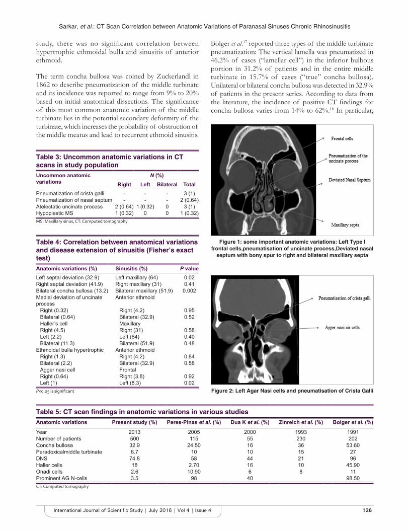
Sarkar, et al.: CT Scan Correlation between Anatomic Variations of Paranasal Sinuses Chronic Rhinosinusitis
126International Journal of Scientific Study | July 2016 | Vol 4 | Issue 4
Figure 1: some important anatomic variations: Left Type I frontal cells,pneumatisation of uncinate process,Deviated nasal
septum with bony spur to right and bilateral maxillary septa
Figure 2: Left Agar Nasi cells and pneumatisation of Crista Galli
Table 5: CT scan findings in anatomic variations in various studiesAnatomic variations Present study (%) Peres-Pinas et al. (%) Dua K et al. (%) Zinreich et al. (%) Bolger et al. (%)
Year 2013 2005 2000 1993 1991Number of patients 500 115 55 230 202Concha bullosa 32.9 24.50 16 36 53.60Paradoxicalmiddle turbinate 6.7 10 10 15 27DNS 74.8 58 44 21 96Haller cells 18 2.70 16 10 45.90Onadi cells 2.6 10.90 6 8 11Prominent AG N-cells 3.5 98 40 98.50CT: Computed tomography
Table 3: Uncommon anatomic variations in CT scans in study populationUncommon anatomic variations
N (%)Right Left Bilateral Total
Pneumatization of crista galli - - - 3 (1)Pneumatization of nasal septum - - - 2 (0.64)Atelectatic uncinate process 2 (0.64) 1 (0.32) 0 3 (1)Hypoplastic MS 1 (0.32) 0 0 1 (0.32)MS: Maxillary sinus, CT: Computed tomography
Table 4: Correlation between anatomical variations and disease extension of sinusitis (Fisher’s exact test)Anatomic variations (%) Sinusitis (%) P valueLeft septal deviation (32.9) Left maxillary (64) 0.02Right septal deviation (41.9) Right maxillary (31) 0.41Bilateral concha bullosa (13.2) Bilateral maxillary (51.9) 0.002Medial deviation of uncinate process
Anterior ethmoid
Right (0.32) Right (4.2) 0.95Bilateral (0.64) Bilateral (32.9) 0.52Haller’s cell MaxillaryRight (4.5) Right (31) 0.58Left (2.2) Left (64) 0.40Bilateral (11.3) Bilateral (51.9) 0.48
Ethmoidal bulla hypertrophic Anterior ethmoidRight (1.3) Right (4.2) 0.84Bilateral (2.2) Bilateral (32.9) 0.58Agger nasi cell FrontalRight (0.64) Right (3.8) 0.92Left (1) Left (8.3) 0.02
P<0.05 is significant
study, there was no significant correlation between hypertrophic ethmoidal bulla and sinusitis of anterior ethmoid.
The term concha bullosa was coined by Zuckerlandl in 1862 to describe pneumatization of the middle turbinate and its incidence was reported to range from 9% to 20% based on initial anatomical dissections. The significance of this most common anatomic variation of the middle turbinate lies in the potential secondary deformity of the turbinate, which increases the probability of obstruction of the middle meatus and lead to recurrent ethmoid sinusitis.
Bolger et al.17 reported three types of the middle turbinate pneumatization: The vertical lamella was pneumatized in 46.2% of cases (“lamellar cell”) in the inferior bulbous portion in 31.2% of patients and in the entire middle turbinate in 15.7% of cases (“true” concha bullosa). Unilateral or bilateral concha bullosa was detected in 32.9% of patients in the present series. According to data from the literature, the incidence of positive CT findings for concha bullosa varies from 14% to 62%.18 In particular,
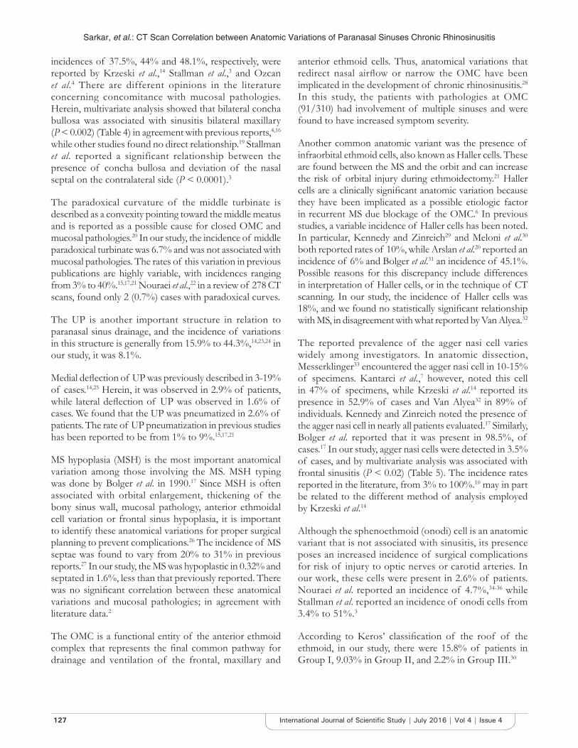
Sarkar, et al.: CT Scan Correlation between Anatomic Variations of Paranasal Sinuses Chronic Rhinosinusitis
127 International Journal of Scientific Study | July 2016 | Vol 4 | Issue 4
incidences of 37.5%, 44% and 48.1%, respectively, were reported by Krzeski et al.,14 Stallman et al.,3 and Ozcan et al.4 There are different opinions in the literature concerning concomitance with mucosal pathologies. Herein, multivariate analysis showed that bilateral concha bullosa was associated with sinusitis bilateral maxillary (P < 0.002) (Table 4) in agreement with previous reports,4,16 while other studies found no direct relationship.19 Stallman et al. reported a significant relationship between the presence of concha bullosa and deviation of the nasal septal on the contralateral side (P < 0.0001).3
The paradoxical curvature of the middle turbinate is described as a convexity pointing toward the middle meatus and is reported as a possible cause for closed OMC and mucosal pathologies.20 In our study, the incidence of middle paradoxical turbinate was 6.7% and was not associated with mucosal pathologies. The rates of this variation in previous publications are highly variable, with incidences ranging from 3% to 40%.15,17,21 Nouraei et al.,22 in a review of 278 CT scans, found only 2 (0.7%) cases with paradoxical curves.
The UP is another important structure in relation to paranasal sinus drainage, and the incidence of variations in this structure is generally from 15.9% to 44.3%,14,23,24 in our study, it was 8.1%.
Medial deflection of UP was previously described in 3-19% of cases.14,25 Herein, it was observed in 2.9% of patients, while lateral deflection of UP was observed in 1.6% of cases. We found that the UP was pneumatized in 2.6% of patients. The rate of UP pneumatization in previous studies has been reported to be from 1% to 9%.15,17,21
MS hypoplasia (MSH) is the most important anatomical variation among those involving the MS. MSH typing was done by Bolger et al. in 1990.17 Since MSH is often associated with orbital enlargement, thickening of the bony sinus wall, mucosal pathology, anterior ethmoidal cell variation or frontal sinus hypoplasia, it is important to identify these anatomical variations for proper surgical planning to prevent complications.26 The incidence of MS septae was found to vary from 20% to 31% in previous reports.27 In our study, the MS was hypoplastic in 0.32% and septated in 1.6%, less than that previously reported. There was no significant correlation between these anatomical variations and mucosal pathologies; in agreement with literature data.2
The OMC is a functional entity of the anterior ethmoid complex that represents the final common pathway for drainage and ventilation of the frontal, maxillary and
anterior ethmoid cells. Thus, anatomical variations that redirect nasal airflow or narrow the OMC have been implicated in the development of chronic rhinosinusitis.28 In this study, the patients with pathologies at OMC (91/310) had involvement of multiple sinuses and were found to have increased symptom severity.
Another common anatomic variant was the presence of infraorbital ethmoid cells, also known as Haller cells. These are found between the MS and the orbit and can increase the risk of orbital injury during ethmoidectomy.21 Haller cells are a clinically significant anatomic variation because they have been implicated as a possible etiologic factor in recurrent MS due blockage of the OMC.6 In previous studies, a variable incidence of Haller cells has been noted. In particular, Kennedy and Zinreich29 and Meloni et al.30 both reported rates of 10%, while Arslan et al.20 reported an incidence of 6% and Bolger et al.31 an incidence of 45.1%. Possible reasons for this discrepancy include differences in interpretation of Haller cells, or in the technique of CT scanning. In our study, the incidence of Haller cells was 18%, and we found no statistically significant relationship with MS, in disagreement with what reported by Van Alyea.32
The reported prevalence of the agger nasi cell varies widely among investigators. In anatomic dissection, Messerklinger33 encountered the agger nasi cell in 10-15% of specimens. Kantarci et al.,7 however, noted this cell in 47% of specimens, while Krzeski et al.14 reported its presence in 52.9% of cases and Van Alyea32 in 89% of individuals. Kennedy and Zinreich noted the presence of the agger nasi cell in nearly all patients evaluated.17 Similarly, Bolger et al. reported that it was present in 98.5%, of cases.17 In our study, agger nasi cells were detected in 3.5% of cases, and by multivariate analysis was associated with frontal sinusitis (P < 0.02) (Table 5). The incidence rates reported in the literature, from 3% to 100%.10 may in part be related to the different method of analysis employed by Krzeski et al.14
Although the sphenoethmoid (onodi) cell is an anatomic variant that is not associated with sinusitis, its presence poses an increased incidence of surgical complications for risk of injury to optic nerves or carotid arteries. In our work, these cells were present in 2.6% of patients. Nouraei et al. reported an incidence of 4.7%,34-36 while Stallman et al. reported an incidence of onodi cells from 3.4% to 51%.3
According to Keros’ classification of the roof of the ethmoid, in our study, there were 15.8% of patients in Group I, 9.03% in Group II, and 2.2% in Group III.30
Sarkar, et al.: CT Scan Correlation between Anatomic Variations of Paranasal Sinuses Chronic Rhinosinusitis
128International Journal of Scientific Study | July 2016 | Vol 4 | Issue 4
CONCLUSIONS
The results of this retrospective study highlight the statistically significant correlation between some anatomical variations of the paranasal sinuses and chronic rhinosinusitis. Therefore, knowledge of anatomic variations of the paranasal sinuses is important in all cases of chronic rhinosinusitis.
ACKNOWLEDGMENTS
The authors would like to thank Dr. Rupa Ananthsivan, Head of Department of Radiodiagnosis and Dr. Girish Rai Head of Department of ENT in Manipal Hospital, Bengaluru, for their extensive and extended help.
REFERENCES
1. The Times of India Mumbai, Section. April 11; 2012. p. 1.2. Selcuk A, Ozcan KM, Akdogan O, Bilal N, Dere H. Variations of maxillary
sinus and accompanying anatomical and pathological structures. J Craniofac Surg 2008;19:159-64.
3. Stallman JS, Lobo JN, Som PM. The incidence of concha bullosa and its relationship to nasal septal deviation and paranasal sinus disease. AJNR Am J Neuroradiol 2004;25:1613-8.
4. Ozcan KM, Selcuk A, Ozcan I, Akdogan O, Dere H. Anatomical variations of nasal turbinates. J Craniofac Surg 2008;19:1678-82.
5. Ercan I, Cakir BO, Sayin I, Basak M, Turgut S. Relationship between the superior attachment type of uncinate process and presence of agger nasi cell: A computer-assisted anatomic study. Otolaryngol Head Neck Surg 2006;134:1010-4.
6. Zinreich SJ, Kennedy DW, Rosenbaum AE, Gayler BW, Kumar AJ, Stammberger H. Paranasal sinuses: CT imaging requirements for endoscopic surgery. Radiology 1987;163:769-75.
7. Kantarci M, Karasen RM, Alper F, Onbas O, Okur A, Karaman A. Remarkable anatomic variations in paranasal sinus region and their clinical importance. Eur J Radiol 2004;50:296-302.
8. Danese M, Duvoisin B, Agrifoglio A, Cherpillod J, Krayenbuhl M. Influence of naso-sinusal anatomic variants on recurrent, persistent or chronic sinusitis. X-ray computed tomographic evaluation in 112 patients. J Radiol 1997;78:651-7.
9. Lund VJ, Kennedy DW. Staging for rhinosinusitis. Otolaryngol Head Neck Surg 1997;117:S35-40.
10. Fadda GL, Rosso S, Aversa S, Petrelli A, Ondolo C, Succo G. Multiparametric statistical correlations between paranasal sinus anatomic variations and chronic rhinosinusitis. Acta Otorhinolaryngol Ital 2012;32:244-51.
11. Tantilipikorn P. Accuracy of limited CT with the full CT as the standard evaluation for inflammatory disease of PNS and the identification of anatomical variations Department of Otorhinolaryngology. Siriraj Med J 2009;61:3-7.
12. Sarna A, Hayman LA, Laine FJ, Taber KH. Coronal imaging of the osteomeatal unit: Anatomy of 24 variants 2002;26:153-7.
13. Scribano E, Ascenti G, Loria G, Cascio F, Gaeta M. The role of the ostiomeatal unit anatomic variations in inflammatory disease of the maxillary sinuses. Eur J Radiol 1997;24:172-4.
14. Krzeski A, Tomaszewska E, Jakubczyk I, Galewicz-Zielinska A. Anatomic variations of the lateral nasal wall in the computed tomography scans of patients with chronic rhinosinusitis. Am J Rhinol 2001;15:371-5.
15. Earwaker J. Anatomic variants in sinonasal CT. Radiographics 1993;13:381-415.
16. Lam WW, Liang EY, Woo JK, Van Hasselt A, Metreweli C. The etiological role of concha bullosa in chronic sinusitis. Eur Radiol 1996;6:550-2.
17. Lerdlum S, Vachiranubhap B. Prevalence of anatomic variation demonstrated on screening sinus computed tomography and clinical correlation. J Med Assoc Thai 2005;88:S110-5.
18. Chao TK. Uncommon anatomic variations in patients with chronic paranasal sinusitis. Otolaryngol Head Neck Surg 2005;132:221-5.
19. Polavaram R, Devaiah AK, Sakai O, Shapshay SM. Anatomic variants and pearls – Functional endoscopic sinus surgery. Otolaryngol Clin North Am 2004;37:221-42.
20. Arslan H, Aydinlioglu A, Bozkurt M, Egeli E. Anatomic variations of the paranasal sinuses: CT examination for endoscopic sinus surgery. Auris Nasus Larynx 1999;26:39-48.
21. Basak S, Karaman CZ, Akdilli A, Mutlu C, Odabasi O, Erpek G. Evaluation of some important anatomical variations and dangerous areas of the paranasal sinuses by CT for safer endonasal surgery. Rhinology 1998;36:162-7.
22. Nouraei SA, Elisay AR, DiMarco A, Abdi R, Majidi H, Madani SA, et al. Variations in paranasal sinus anatomy: Implications for the pathophysiology of chronic rhinosinusitis and safety of endoscopic sinus surgery. J Otolaryngol Head Neck Surg 2009;38:32-7.
23. Bayram M, Sirikci A, Bayazit YA. Important anatomic variations of the sinonasal anatomy in light of endoscopic surgery: A pictorial review. Eur Radiol 2001;11:1991-7.
24. Zinreich J. Imaging of inflammatory sinus disease. Otolaryngol Clin North Am 1993;26:535-47.
25. Bolger WE, Woodruff WW Jr, Morehead J, Parsons DS. Maxillary sinus hypoplasia: Classification and description of associated uncinate process hypoplasia. Otolaryngol Head Neck Surg 1990;103:759-65.
26. Betts NJ, Miloro M. Modification of the sinus lift procedure for septa in the maxillary antrum. J Oral Maxillofac Surg 1994;52:332-3.
27. Jones NS, Strobl A, Holland I. A study of the CT findings in 100 patients with rhinosinusitis and 100 controls. Clin Otolaryngol Allied Sci 1997;22:47-51.
28. Meloni F, Mini R, Rovasio S, Stomeo F, Teatini GP. Anatomic variations of surgical importance in ethmoid labyrinth and sphenoid sinus. A study of radiological anatomy. Surg Radiol Anat 1992;14:65-70.
29. Kennedy DW, Zinreich SJ. The functional endoscopic approach to inflammatory sinus disease: Current perpectives and technique modifications. Am J Rhinol 1988;2:89-96.
30. Keros P. Uber. die praktische Bedeutung der Niveauunterschiede de Lamina cribrosa des Ethmoids (The practical significance of the differences in level de cribriform plate of the ethmoid). Laryngol Rhinol Otol (Stuttg) 1965;41:808-13.
31 Catalano GB, Conticello S, Galletti C, Madonia HT, Ottoboni A, Pignataro O. Clinica Otorinolaringoiatrica. Bologna: Monduzzi Editore; 1993.
32. Van Alyea OE. Ethmoid labyrinth anatomic study, with consideration of the clinical significance of its structural characteristics. Arch Otolaryngol 1939;29:881-901.
33. Messerklinger W. On the drainage of the normal frontal sinus of man. Acta Otolaryngol 1967;63:176-81.
34. Bolger WE, Butzin CA, Parsons DS. Paranasal sinus bony anatomic variations and mucosal abnormalities: CT analysis for endoscopic sinus surgery. Laryngoscope 1991;101:56-64.
35. Weinberger D, Anand V, Al-Rawi M, Cheng H, Messina A. Surgical anatomy and variations of the onodi cell. Am J Rhinol 1996;10:365-70.
36. Tonai A, Baba S. Anatomic variations of the bone in sinonasal CT. Acta Otolaryngol Suppl 1996;525:9-13.
How to cite this article: Sarkar PS, Bhosale PR, Bharthi AR, Ananthasivan R. Computed Tomography Scan Correlation between Anatomic Variations of Paranasal Sinuses and Chronic Rhinosinusitis. Int J Sci Stud 2016;4(4):122-128.
Source of Support: Nil, Conflict of Interest: None declared.

129 International Journal of Scientific Study | July 2016 | Vol 4 | Issue 4
Occurrence of Tuberculosis in Patients Attending a Tertiary Care Hospital in Khanpur, Sonepat, HaryanaSumit Kumar1, Surinder Kumar2, Pallavi Kumari3, Pallavi Sayal4, Seema Garg5, Raminder Sandhu5
1Associate Professor, Department of Microbiology, BPS Government Medical College for Women, Sonepat, Haryana, India, 2Professor & Head, Department of Microbiology BPS Government Medical College for Women, Sonepat, Haryana, India, 3Associate Professor, Department of Physiology, Teerthanker Mahaveer Medical College & Research Center, Moradabad, Uttar Pradesh, India, 4Tutor, Department of Microbiology BPS Government Medical College for Women, Sonepat, Haryana, India, 5Assistant Professor, Department of Microbiology BPS Government Medical College for Women, Sonepat, Haryana, India
India has more TB cases annually than any other country in the world, with an estimated disease prevalence of 256/100,000 population, incidence of 185/100,000 and deaths of 26/100,000.2 TB is responsible for as many as 29 deaths per year per 100,000 people. In a country with over a billion people, this translates to as many as 290,000 deaths in a year. With a reported prevalence of 283 cases per 100,000 population, India is among the 22 high TB burden countries in the world.4
Currently under Revised National TB Control Programme (RNTCP), any person presenting with a cough of more than 2 weeks is screened for pulmonary TB (PTB) by two sputum smear examinations, one spot and one overnight
INTRODUCTION
Tuberculosis (TB) is still a major public health problem in India, with an estimated 2.3 million new cases annually that making it the highest TB burden country in the world. In 2010, India alone accounted for an estimated one-quarter (26%) of all TB cases worldwide.1
Original Article
AbstractBackground: Tuberculosis (TB) is a major public health problem in India, estimated 2.3 million new cases annually makes it the highest TB burdened country. In 2010, India has estimated one-quarter (26%) of all TB cases worldwide. India has more TB cases annually than any other country globally, with estimated disease prevalence 256/100,000 population, the incidence of 185/100,000 and death of 26/100,000. The prevalence of TB is an important epidemiological index to measure the burden in a community.
Materials and Methods: A study was conducted on patients attending BPS, Government Medical College (GMC) (women), Khanpur, Sonepat, Hospital. Clinically TB suspected cases were investigated in the Department of Microbiology by staining the sputum samples by Ziehl–Neelsen staining and smears were examined by direct microscopy.
Results: The patients attended to BPS, GMC (women), Khanpur, Sonepat hospital during January 2015 to December 2015 were screened, and 4267 patients were suspected for TB. Out of 4267 patients, 528 (12.37%) were smear positive. Out of 528 positive cases, 402 (76.14%) were male and 126 (23.86%) were female. A maximum number of cases 101 (19.13%) were positive in 41-50 years group.
Conclusions: This study provides important information on TB status in Sonepat, Haryana. It can serve as baseline data for future evaluation, the impact of disease control measures and epidemiological trends. However, TB is a major public health problem, and there is need to maintain and further strengthen TB control measures on a sustained and long-term bais. Epidemiological information on TB has always been vital for planning control strategies and has now gained further importance for monitoring the impact of interventions against the disease.
Key words: Burden, Epidemiological, Occurrence, Tuberculosis
Access this article online
www.ijss-sn.com
Month of Submission : 05-2016 Month of Peer Review : 06-2016 Month of Acceptance : 07-2016 Month of Publishing : 07-2016
Corresponding Author: Dr. Sumit Kumar, Department of Microbiology, BPS Government Medical College for Women, Khanpur Kalan, Sonepat - 131 305, Haryana, India. Phone: +91-8396069442. E-mail: [email protected]
Print ISSN: 2321-6379Online ISSN: 2321-595X
DOI: 10.17354/ijss/2016/390

Kumar, et al.: Occurrence of Tuberculosis in Patients Attending a Tertiary Care Hospital
130International Journal of Scientific Study | July 2016 | Vol 4 | Issue 4
sample. The samples are collected and examined at designated microscopic centers. The treatment of TB patients is based on the internationally recommended directly observed treatment short course strategy.2
Different studies across the country have estimated a prevalence of smear-positive PTB between 60 and 760/100,000 population, culture positive TB between 170 and 980, and culture and/or smear-positive prevalence between 180 and 1270/100,000 people.4
The prevalence of TB disease is an important epidemiological index to measure the burden in a community and if it is measured periodically, will enable trends in disease prevalence to be observed over time. Epidemiological information on TB is also vital for the planning of control strategies and service delivery systems.1
In view of low health service coverage, a developing diagnostic network, and a weak disease notification system, it is not only difficult to determine the magnitude of TB from case notification alone but it is also impossible to monitor the effectiveness of control measures.4
Global targets for TB control now include a 95% reduction in TB deaths and <10 cases per 100,000 population by 2035. Such targets will be achieved by strategies to diagnose and treat people with active TB earlier in their disease course.4-10
This study was conducted in BPS Medical College, Khanpur, Sonepat to know the occurrence of TB.
MATERIALS AND METHODS
The study was conducted in the Department of Microbiology during January 2015 to December 2015 on patients attending BPS, Government Medical College (GMC) for women, Khanpur Kalan, Sonepat, Hospital. A history of the patient was taken. Individuals were questioned for chest symptoms relating to TB, namely: Persistent cough for 2 weeks or more, chest pain for 1 month or more, fever for 1 month or more, and hemoptysis anytime in last 6 months. History of TB treatment, alcohol consumption, tobacco smoking were collected. Persons with any of these symptoms (deemed as “chest symptomatics”), and also those with a previous history of anti-TB treatment, were considered eligible for sputum collection. Two sputum samples - one spot and one overnight - were collected from each eligible individual in a sterilized container.
Sputum smears were prepared directly from each sputum specimen, and also from concentrated samples. Two direct
smears were made from each specimen on new labeled slides under aseptic conditions in a bio-safety cabinet. Smears were stained using Ziehl–Neelsen stain and examined with oil-immersion microscopy for the presence of acid-fast bacilli (AFB). About 10% of randomly selected sputum smears were cross-examined by one of the investigators (SS), and quality assurance protocols were followed as per RNTCP guidelines. A TB case was defined as an individual in whom any of the two sputum specimens was positive for AFB by Ziehl–Neelsen microscopy. All bacteriologically positive cases were referred to the concerned health authorities for anti-TB treatment under the RNTCP using its standardized treatment regimens.
RESULTS
The patients attended to BPS, GMC for Women, Khanpur Kalan, Sonepat Hospital during January 2015 to December 2015 were screened for TB. A history of the patient was taken. Individuals were questioned for chest symptoms relating to TB, namely: Persistent cough for 2 weeks or more, chest pain for 1 month or more, fever for 1 month or more, and hemoptysis anytime in last 6 months. History of TB treatment was taken. 4267 patients were suspected for TB. Sputum samples of suspected patients were collected. Two sputum samples - one spot and one overnight - were collected from each eligible individual in sterilized container. Two direct smears were made from each specimen on new labeled slides under aseptic conditions in a bio-safety cabinet. Smears were stained using Ziehl–Neelsen stain and examined with oil-immersion microscopy for the presence of AFB.
Out of 4267 patients suspected for TB 528 (12.37%) cases were smear-positive. Out of 528 smear, positive cases 402 (76.14%) were males and 126 (23.86%) were females. In this study, a maximum number of cases 101 (19.13%) were positive in 41-50 years group.
DISCUSSION
TB is still a major public health problem in India. This study was conducted in BPS, Medical College, Khanpur, Sonepat, to know the occurrence of TB. The patients attended to BPS, GMC(women) Khanpur, Sonepat Hospital during January 2015 to December 2015 were screened for TB.
In this study, 4267 patients were suspected for TB. Out of 4267 patients, 528 (12.37%) were smear positive.
In a study done by Sharma et al., in 2015, a total of 81 (4.4%) sputum samples were found smear positive while 82 (4.4%) were culture positive. 63 (3.4%) subjects were both smear and culture positive.4 In a study done by

Kumar, et al.: Occurrence of Tuberculosis in Patients Attending a Tertiary Care Hospital
131 International Journal of Scientific Study | July 2016 | Vol 4 | Issue 4
Rao et al., in 2012, PTB was found to be 255.3/100,000 population.1 In a study done by Yadav et al., in 2010, TB prevalence was 146/100,000.1 In a study done by Rao et al., in 2010, TB prevalence was 1518/100,000.1 In a study done by Bhat et al., in 2009, TB prevalence was 387/100,000.6 In a study done by Chakraborty 2004, the prevalence of total PTB cases was 20.0/1000.12 In a study done by Chakma et al., in 1996, TB prevalence was 12.7/100,000 population of Morena district of Madhya Pradesh.1
TB occurrence rates could not be similar between areas in India. This is due to the regional diversities in terms of ethnic, economic, cultural complexities and variables, pervading the vast land masses and the population size of near continental dimensions.12
In this study out of 528 positive cases, 402 (76.14%) were male and 126 (23.86%) were female. In a study done by Aggarwal et al., in 2015, TB was more among men (34.5/100,000) as compared to women (14.2/100,000).5 In a study done by Bhat et al., in 2012, males contributed 76.9% of the total bacteriologically positive PTB cases.1 In a study done by Chadha et al., in 2012, male to female ratio in these surveys has been found to vary between 2:1 and 5:1. It was 6:1 during the present survey.3 In a study done by Gopi et al., in 2010, the prevalence of PTB in males (2156/100,000) was more than double that among females (933/100,000).9 In a study done by Bhat et al., in 2009, the prevalence of bacillary TB was more than double among males (554/100,000) than females (233/100,000).6
In general, the disease occurrence rates are about similar in both sexes, till the puberty in females. This is followed by a continuing widening of the gap between the sexes in favor of the females, the differences acutely accentuated past the 35-40 years age mark.12
In this study, a maximum number of cases 101 (19.13%) were positive in 41-50 years group. In a study done by Dhanaraj et al., in 2015, while the maximum prevalence of bacteriologically positive PTB 1241/100,000 was found in the age group of 55-64 years.2 In a study done by Bhat
et al., in 2012, the highest proportion of positive PTB cases (25.8%) was seen in the 35-44 years age group.1 In a study done by Rao et al., in 2010, the prevalence increased with age, being 546/100,000 in the 15-24 years age group, increasing to 3086/100,000 in the 55+ years age group.9 In a study done by Bhat et al., 2009, the prevalence increased with age being 174/100,000 in the 15-24 years age group to 990/100,000 in the 55+ years age group.6
It was observed that, of the prevalent cases in the community, the age-wise proportion of cases were substantially higher 35-44 years onward, to be at the peak for the age group 55-64 years (28.40%).12 The occurrence of TB rise with age, in both sexes (Table 1 and Graphs 1-3).12,13
CONCLUSIONS
This study provides important information on TB status in Khanpur Kalan, Sonepat, Haryana. The study indicates
Table 1: Occurrence of tuberculosis according to age and genderAge Male (%) Female (%) Total (%)11-20 21 (3.97) 22 (4.16) 43 (8.14)21-30 66 (12.50) 34 (6.44) 100 (18.94)31-40 65 (12.31) 11 (2.08) 76 (14.39)41-50 86 (16.28) 15 (2.84) 101 (19.13)51-60 69 (13.06) 17 (3.21) 86 (16.28)61-70 71 (13.44) 17 (3.21) 88 (16.66)71-80 15 (2.84) 8 (1.51) 23 (4.35)81-90 9 (1.70) 2 (0.38) 11 (2.08)Total 402 (76.14) 126 (23.86) 528 (100)
Graph 1: Tuberculosis according to gender
Graph 2: Tuberculosis in relation to gender and age
Graph 3: Tuberculosis in relation to age
Kumar, et al.: Occurrence of Tuberculosis in Patients Attending a Tertiary Care Hospital
132International Journal of Scientific Study | July 2016 | Vol 4 | Issue 4
high occurrence of TB in males they were affected more in 76.14% cases and female were affected in 23.86% cases. Maximum affected age group was between 41 and 50 years. The study results provide vital information on the TB disease situation among the population and can serve as baseline data for future evaluation of the impact of disease control measures and epidemiological trends. The findings suggest that the TB situation among the Khanpur, Sonipat, population is not that different from the situation among the population in the country.
However, TB remains a major public health problem among the population, and there is a need to maintain and further strengthen TB control measures on a sustained and long-term basis. Epidemiological information on TB has always been vital for planning control strategies and has now gained further importance for monitoring the impact of interventions against the disease.
REFERENCES
1. Rao VG, Bhat J, Yadav R, Gopalan GP, Nagamiah S, Bhondeley MK, et al. Prevalence of pulmonary tuberculosis - A baseline survey in central India. PLoS One 2012;7:e43225.
2. Dhanaraj B, Papanna MK, Adinarayanan S, Vedachalam C, Sundaram V,
Shanmugam S, et al. Prevalence and risk factors for adult pulmonary tuberculosis in a metropolitan city of South India. PLoS One 2015;10:e0124260.
3. Chadha VK, Kumar P, Anjinappa SM, Singh S, Narasimhaiah S, Joshi MV, et al. Prevalence of pulmonary tuberculosis among adults in a rural sub-district of South India. PLoS One 2012;7:e42625.
4. Sharma SK, Goel A, Gupta SK, Mohan K, Sreenivas V, Rai SK, et al. Prevalence of tuberculosis in Faridabad district, Haryana State, India. Indian J Med Res 2015;141:228-35.
5. Aggarwal AN, Gupta D, Agarwal R, Sethi S, Thakur JS, Anjinappa SM, et al. Prevalence of pulmonary tuberculosis among adults in a north Indian district. PLoS One 2015;10:e0117363.
6. Bhat J, Rao VG, Gopi PG, Yadav R, Selvakumar N, Tiwari B, et al. Prevalence of pulmonary tuberculosis amongst the tribal population of Madhya Pradesh, central India. Int J Epidemiol 2009;38:1026-32.
7. van der Werf MJ, Enarson DA, Borgdorff MW. How to identify tuberculosis cases in a prevalence survey. Int J Tuberc Lung Dis 2008;12:1255-60.
8. Jain SK, Ordonez A, Kinikar A, Gupte N, Thakar M, Mave V, et al. Pediatric tuberculosis in young children in India: A prospective study. Biomed Res Int 2013;2013:783698.
9. Rao VG, Gopi PG, Bhat J. Pulmonary tuberculosis: A public health problem amongst the Saharia, a primitive tribe of Madhya Pradesh, Central India. Int J Infect Dis 2010;14:e713-6.
10. Azman AS, Golub JE, Dowdy DW. How much is tuberculosis screening worth? Estimating the value of active case finding for tuberculosis in South Africa, China, and India. BMC Med 2014;12:216.
11. Thomas BE, Adinarayanan S, Manogaran C, Swaminathan S. Pulmonary tuberculosis among tribals in India: A systematic review and meta-analysis. Indian J Med Res 2015;141:614-23.
12. Chakraborty AK. Epidemiology of tuberculosis: Current status in India. Indian J Med Res 2004;120:248-76.
How to cite this article: Kumar S, Kumar S, Kumar P, Sayal P, Garg S, Sandhu R. Occurrence of Tuberculosis in Patients Attending a Tertiary Care Hospital in Khanpur, Sonepat, Haryana. A Retrospective Study. Int J Sci Stud 2016;4(4):129-132.
Source of Support: Nil, Conflict of Interest: None declared.

133 International Journal of Scientific Study | July 2016 | Vol 4 | Issue 4
Primary Adenoid Cystic Carcinoma of Head and Neck: Its Prognosis and Management - A Retrospective Analysis from a Tertiary Care CenterSubhalaxmi Rautray1, Tapan Kumar Sahoo2, Saroj Kumar Das2, Saroj Kumar Das Majumdar3, Dillip Kumar Parida4
1Assistant Professor, Department of Otorhinolaryngology, Sriram Chandra Bhanja Medical College, Cuttack, Odisha, India, 2Senior Resident, Department of Radiation Oncology, All India Institute of Medical Sciences, Bhubaneswar, Odisha, India, 3Assistant Professor, Department of Radiation Oncology, All India Institute of Medical Sciences, Bhubaneswar, Odisha, India, 4Professor, Department of Radiation Oncology, All India Institute of Medical Sciences, Bhubaneswar, Odisha, India
glands of the head and neck. The minor salivary glands of the oral cavity are the most common site.2 It can occur also in the breast, trachea, paranasal sinuses, lacrimal glands, larynx, tracheobronchial tree, external ear, skin, and vulva and are known as non-salivary ACC.3 ACC is a malignant tumor with benign histologic appearance. It is characterized by different histologic patterns, indolent, locally invasive growth with unpredictable clinical behavior, prolonged clinical course, and increased propensity for local recurrence and distant metastases.4 Regional lymph node involvement occurs infrequently in ACC.5 Aggressive surgical excision with adjunct radiotherapy is the required treatment modality.6 Six patients of ACC of head and neck were taken in this study.
INTRODUCTION
Adenoid cystic carcinoma (ACC) accounts for approximately 1% of all head and neck malignancies and 10-15 (22%) of all salivary gland neoplasms.1,2 ACC is arises within glands, most commonly from major and minor salivary
Original Article
AbstractBackground: Adenoid cystic carcinoma (ACC) is a rare malignant tumor of head and neck primary. It affects most commonly the major and minor salivary glands and rarely the paranasal sinuses, lacrimal gland, larynx, ear, vulva, etc. It is a slowly growing tumor, asymptomatic for a long period of time and usually presents with swelling and pain at the time of diagnosis. A wide radical resection of the tumor followed by radiotherapy is the main treatment modality.
Aim of the Study: To know the aggressiveness, diseases free interval and the prognosis of the ACC of head and neck.
Materials and Methods: Six cases of ACC of head and neck were taken. Three cases were having hard palate as the primary site, one case with nasopharynx as the primary site, one case with maxillary sinus, and one having the floor of mouth as the primary site. Cases were treated aggressively with surgery, adjuvant radiotherapy, palliative radiotherapy, and palliative chemotherapy (CT).
Results: Out of six cases, five cases were treated with surgery followed by radiotherapy. Four cases are now on regular follow-up without any disease recurrence or distant metastases. One case developed lung metastasis after 2-year of follow-up and is continuing palliative CT. One case presented with isolated temporal lobe metastasis and received radiotherapy to nasopharynx, its locoregional area and whole brain.
Conclusion: The behavior and survival status of ACC is not clear due to inefficient study data in literature. The study needs continuation with a more number of cases and longer duration of follow-up to draw significant data regarding disease-free interval and survival outcome of the diseases.
Key words: Adenoid cystic carcinoma, Head and neck primary, Management and prognosis
Access this article online
www.ijss-sn.com
Month of Submission : 05-2016 Month of Peer Review : 06-2016 Month of Acceptance : 07-2016 Month of Publishing : 07-2016
Corresponding Author: Tapan Kumar Sahoo, Department of Radiation Oncology, All India Institute of Medical Sciences, Bhubaneswar - 751 019, Odisha, India. Phone: +91-9437219525. E-mail: [email protected]
Print ISSN: 2321-6379Online ISSN: 2321-595X
DOI: 10.17354/ijss/2016/391

Rautray, et al.: Primary Adenoid Cystic Carcinoma of Head and Neck
134International Journal of Scientific Study | July 2016 | Vol 4 | Issue 4
MATERIALS AND METHODS
Six cases of ACC of head and neck were studied in between August 2013 and until date at All India Institute of Medical Sciences, Bhubaneswar, Odisha, India.
Out of 460 cases of head and neck cancers, only six cases (1.30%) were ACC. Five out of six cases presented at 30-50 years age group and one case presented in the age group of 60 years. Out of six cases, four cases are male and two cases are female. The oral cavity (hard palate and floor of the mouth) is the primary site of presentation in four cases. The nasopharynx and maxillary sinus as the primary site are seen in one case each. Pure cribriform type histology is seen in two hard palate cases and in one maxillary primary. Pure tubular, pure solid and mixed form seen in one cases each. All the six cases presented as locally advanced not less than T3 lesions and one case presented with isolated solitary temporal lobe brain metastasis. Three hard palate cases were treated with aggressive surgery (left extended maxillectomy with degloving approach) followed by adjuvant radiotherapy of 66 GY in 33 fractions to bilateral face and neck by CO60 in reduced portals after 45 GY. All the three cases are now on regular follow-up without any disease recurrence or distant metastases. One case of the floor of mouth primary underwent surgery (wide local excision + marginal mandibulectomy + bilateral modified neck dissections) with post-operative pathological staging of PT3N0MX with margin positivity and received adjuvant concurrent chemoradiation of 66 GY radiotherapy in 33 fractions along with weekly cisplatinum. After 2-year of follow-up, the patient developed bilateral lung metastasis and was treated with palliative chemotherapy (CT) with paclitaxel, cisplatin, and 5-flourouracil regimen up to six cycles with partial response, followed by oral tablet everolimus 10 mg once daily till date. The case with nasopharynx as the primary site found initially with isolated solitary temporal lobe metastasis was treated with palliative external beam radiotherapy (EBRT) to the brain of 30 GY in 10 fractions and local EBRT 66 GY in 33 fractions to bilateral face and neck. The ACC of maxillary sinus involved the inferior wall of orbit and was treated with surgery followed by EBRT of 66 GY in 33 fractions to bilateral face and neck and is now under regular follow-up since 26 months.
DISCUSSION
According to the World Health Organization (2005), ACC is defined as “A basaloid tumor consisting of epithelial and myoepithelial cells in various morphological configurations, including tubular, cribriform, and solid patterns. It has a relentless clinical course and usually a fatal outcome.”7 The
term ACC is still misleading as the tumor is not cystic.8-10 The most cases arise in the minor salivary glands (60%). Palate, buccal region, maxilla, retromolar region, and lips are the common sites of involvement in the oral cavity.11 The maxillary sinus ACC is the second most common sinonasal tumor.12 and accounts for 10% of all sinonasal tract malignancies.13 A recent review showed 74.3% of ACCs of the nasopharynx are presented at an advanced stage in the time of diagnosis.14
Gender predilection is an inconsistent feature in the literature. The peak age of incidence in ACC is 4th to 6th decades of life though cases have been reported in between ages of 10 and 96 years.15 There are equal male and female distributions in ACCs.5 Few data showed male predominance16 in ACCs, whereas few showed female predominance.1 Arsenic compounds, nickel, oak, or beech wood dust may be possible etiological factors.17
In this study, the majority were male, presented in the age group between 30 and 45 years, and hard palate as the primary. But, a large number of case studies are necessary for a significant data regarding the median age of presentation, incidence of the disease.
HistopathologyACCs are poorly encapsulated infiltrative lesions with grayish pink appearance.17 The characteristic histopathologic features of ACC is proliferation of round or cuboidal cells with scarce cytoplasm and large, oval and hyperchromatic nuclei, and cells are arranged in the form of islands or sheets surrounded by abundant hyaline stroma exhibiting pseudocystic structures.18 Microscopic examination of ACC comprises epithelial and myoepithelial cells.19 ACC has a distinct histopathological appearance. Histopathologically, it has three main growth patterns: (1) Cribriform, (2) tubular, and (3) solid.19
Histologically, a mixture of patterns is common. Classification is based on the predominant pattern. Tumor with more than 30% of the solid pattern is classified as the solid variant, indicating its more aggressive behavior.
The tubular patterns are well differentiated or Grade I and is characterized by slender tubules, solid cords, and glandular structures infiltrating a well-hyalinized background. Cribriform pattern is the most common pattern, moderately differentiated or Grade II and is characterized by invasive tumor islands with multiple holes (pseudocysts or pseudolumina) punched out in a “Swiss cheese” or sieve-like pattern. The solid pattern is poorly differentiated or Grade III and consists of large islands of carcinoma composed predominantly of myoepithelial cells with infrequent true lumina, lined with cuboidal epithelial

Rautray, et al.: Primary Adenoid Cystic Carcinoma of Head and Neck
135 International Journal of Scientific Study | July 2016 | Vol 4 | Issue 4
cells, and occasional pseudocysts. Stroma is usually fibrous and extensive hyalinization can occur. Mitotic figures and apoptotic cells are commonly seen in the high-grade or solid pattern. Necrosis is seen only in the solid pattern, often centrally located within cell nests and appearing as a come do appearance.20,21 Mitotic figures and apoptotic cells are occasionally seen in the cribriform pattern. Perineural spread is commonly seen in all patterns. The tubular pattern has best prognosis, whereas cribriform pattern has intermediate and solid pattern has worst prognosis.17
In this study, out of six cases, three cases were a purely cribriform pattern, one with mixed tubular and cribriform pattern, one with the purely tubular pattern, one with purely solid pattern supporting the literatures.
PresentationMost of the metastatic ACC remain asymptomatic for a long time.22 Clinical signs and symptoms of ACC depend on the primary site and extent of the lesion. It can present as a painless slow-growing mass in the face or mouth.
Locally advanced tumors may invade nerves, causing paresthesia and paralysis. ACC has a tendency to infiltrate neural structures and to spread perineurally.
Intracranial involvement is rarely seen.3,23 Tumors of the lacrimal gland may cause vision impairment and proptosis. ACC is the most common primary epithelial malignancy in the lacrimal glands.24 ACC is the second most common primary tumor of the trachea with a poor prognosis. ACC of the larynx may present with hoarseness and difficulty in breathing.25
Maxillary sinus primary may present with nasal obstruction, epistaxis, nasal discharge, swelling, facial pain, paresthesia, etc., mimicking inflammatory conditions resulting in a late diagnosis of the disease.12,26 ACCs have a prolonged natural history and slow growth even in local recurrence and distant metastatic situations.27 There is a high propensity of ACC for perineural invasion and early hematogenous spread.28,29 Brain involvement may occur by direct extension of tumor.17 Regional metastases are seen in <3% of cases,
Table 1: Demographic data with pre-treatment stagingAge in years Sex Site of the primary lesion Histopathological subtype TNM stage Stage Distant metastasis at presentation35 F Right hard palate Mixed cribriform and tubular type CT4N0M0 III No45 M Left hard palate Cribriform type CT4N0M0 III No30 M Right sided nasopharynx Tubular type CT4N0M1 IV Temporal lobe42 M Right sided floor of mouth Solid type CT3N1M0 II No40 F Left maxillary sinus Cribriform type CT4N0M0 III No60 M Left hard palate None CT3N0M0 II No
Table 2: Radiological findings and treatmentCase number
CT/MRI findings Initial treatment FU in months
Recurrence/metastasis
Palliative treatment
Case 1 MRI: Mass on the right maxilla involving adjacent soft palate and hard palate
EMDA (PT4N0MX)→EBRT of 66 GY in 33 fractions to bilateral face and neck
28 - -
Case 2 MRI: Left paramedian hard palate lesion, involving left pterygoid plates, pterygoid muscles, pterygomaxillary fissure, maxillary antrum
EMDA (PT4N0MX)→EBRT of 66 GY in 33 fractions to bilateral face and neck
18 - -
Case 3 CECT: Mass on right sided nasopharynx involving posterior nasal cavity causing obliteration of B/L nasopharyngeal airway, eustachean tube, torus tubaris and fossa of rosenmullerMRI: Metastasis in right temporal lobe
EBRT of 66 GY in 33 fractions to bilateral face and neck along with 30 GY in 10 fractions to whole brain
- - -
Case 4 MRI: Mass in right side floor of mouth involving anterior half of tongue, genioglossus, hyoglossus, geniohyoid muscles, mandible with right level IA, IB lymphadenopathy
WLE+mandibulectomy+MND PT4N0MX→concurrent CTRT of 66 Gy radiotherapy in 33 fractions along with weekly cisplatin
24 B/L lung metastasis
Palliative CT with paclitaxel+cisplatin→tablet everolimus
Case 5 CECT: Mass in the left maxillary sinus involving nasal septum, inferior wall of orbit
EMDA (PT4N0MX)→EBRT of 66 GY in 33 fractions to bilateral face and neck
26 - -
Case 6 CECT: Mass in the left maxillary sinus eroding posterolateral wall involving hard palate, left side nasopharyngeal wall
EMDA (PT4N0MX)→EBRT of 66 GY in 33 fractions to bilateral face and neck
15 - -
FU: Follow‑up, EMDA: Extended maxillectomy with degloving approach, CTRT: Chemoradiation, B/L: Bilateral, CT: Chemotherapy, MRI: Magnetic resonance imaging, EBRT: External beam radiotherapy, WLE: Wide local excision, MND: Modified neck dissections

Rautray, et al.: Primary Adenoid Cystic Carcinoma of Head and Neck
136International Journal of Scientific Study | July 2016 | Vol 4 | Issue 4
whereas distant metastases are comparatively common with the lung being the most common site followed by bones, liver, brain and omentum.30
The present study showed all the patients had T3 or T4 lesions, the majority with T4 lesions, one patient had distant metastasis as temporal lobe metastasis at initial presentation, and one had N1 disease clinico-radiological, but post-operative pathology showed PT4N0 with margin positive, the patient developed bilateral lung metastasis after 2 years of completion of treatment.
PrognosisACC of minor salivary glands has worse prognosis than those of major salivary glands possibly due to more easy infiltration of the lesion of the minor salivary gland to extra glandular soft tissues and bone resulting increased dissemination of the tumour.5 Solid histologic pattern, tumor size >4 cm, perineural invasion, delayed diagnosis, delayed treatment, surgical margin positive, recurrent local lesions, and distant metastases are associated with worse prognosis.5
ManagementACC is found in younger age groups in comparison to squamous cell carcinoma and is relatively resistant to treatment. Tumor markers have no significant role in determining the prognosis.6 Treatment modalities depend on the stage of the tumor. It is difficult to prevent and predict the late local recurrence and distant metastases in ACC. There is no standard guideline for optimal treatment and outcome of the disease due to lack of prospective randomized multicentric trials. Surgical resection with possible widest margins with or without neck node dissection is the cornerstone of the treatment.31 Neck node dissection depends on strong clinic-radiological suspicious of lymph node metastases. The majority cases present at an advanced stage during diagnosis and complete surgical resection remains difficult due to the larger size of the tumor and presence of nearby critical neurovascular structures.32 Late diagnosis of ACCs contributes to poor prognosis of the disease.33 Combination modality of treatment with surgery and radiotherapy is commonly required1 and results in better overall survival.16 The tumor cells extend well beyond the clinical or radiographic margins and undergo perineural invasion and spread. Therefore, surgery requires excision with widest possible margins.5 In maxillary sinus ACC, due to slow spread, late manifestations of the disease, and complex anatomy of the maxilla, complete surgical resection with widest margin is difficult. Rehabilitation options in huge maxillary defects still need further exploration.
ACCs are radiosensitive but not radio curable. Post-operative radiotherapy improves locoregional control
and overall outcome.31,34 A recent series data showed no difference in survival, the rate of recurrence, and time of recurrence between treatment with combination modality of surgery and radiotherapy or surgery alone.35 In the case of inoperable/unresectable tumor and patient refusal for surgery, primary radiotherapy is recommended. The role of CT is not established as an effective modality of treatment for ACC.26 As prolonged survival is not unusual even after distant metastasis, CT should be best withheld in inoperable cases with local recurrence and distant metastasis until symptoms appear.36 Clinical trials are going on for combination CT regimens treatment in local recurrent lesions or in distant metastases.37 In ACC of head and neck, survival curve drastically declines at 10- and 15-year of survival even after combination treatment of surgery and radiotherapy.14 Tumor growth rate and metastatic potential are independent tumor properties.38 The 5-, 10-, and 15-year survival rates are 75%, 20%, and 10%, respectively.38 5-year overall survival for maxillary sinus primary is 62.9%.26
Five out of six cases in this study treated with surgical resection with possible widest margins, but one case showed post-operative margin positive and all the five cases received adjuvant radiotherapy (Tables 1 and 2).
CONCLUSION
The combination of surgical resection and radiotherapy is the main modality of treatment. Complete surgical resection with widest possible margins is the cornerstone of treatment. The role of CT is not clear. ACCs of head and neck presented at an advanced stage (Stage III/IV) and complete surgical resection with clear margins is difficult due to late presentation, the larger size of tumor and nearby critical neurovascular structures. There is no clear data regarding prevention and prediction of local recurrence and distant metastases and it needs further clarification. This study needs continuation with more data and a longer period of follow-up to draw a possible survival outcome.
REFERENCES
1. Kokemueller H, Eckardt A, Brachvogel P, Hausamen JE. Adenoid cystic carcinoma of the head and neck – A 20 years experience. Int J Oral Maxillofac Surg 2004;33:25-31.
2. Dodd RL, Slevin NJ. Salivary gland adenoid cystic carcinoma: A review of chemotherapy and molecular therapies. Oral Oncol 2006;42:759-69.
3. Lee AG, Phillips PH, Newman NJ, Hayman LA, Schiffman JS, Segal SE, et al. Neuro-ophthalmologic manifestations of adenoid cystic carcinoma. J Neuroophthalmol 1997;17:183-8.
4. Jaso J, Malhotra R. Adenoid cystic carcinoma. Arch Pathol Lab Med 2011;135:511-5.
5. Singh S, Gokkulakrishnan, Jain J, Pathak S, Singh KT. Adenoid cystic carcinoma of buccal mucosa. J Maxillofac Oral Surg 2010;9:273-6.
Rautray, et al.: Primary Adenoid Cystic Carcinoma of Head and Neck
137 International Journal of Scientific Study | July 2016 | Vol 4 | Issue 4
6. Adwani D, Bhattacharya A, Arora RS, Soni R, Adwani N. Resection and reconstruction of maxillary Class IIIc defect in a case of adenoid cystic carcinoma: Cost-sensitive technique without microvascular grafts. Case Rep Dent 2013;2013:865010.
7. Barrett AW, Speight PM. Perineural invasion in adenoid cystic carcinoma of the salivary glands: A valid prognostic indicator? Oral Oncol 2009;45:936-40.
8. Barnes L. Surgical Pathology of the Head and Neck. New York: Marcel Dekker; 2001.
9. Myers EN, Ferris RL. Salivary Gland Disorders. New York: Springer; 2007.10. Martínez-Rodríguez N, Leco-Berrocal I, Rubio-Alonso L, Arias-Irimia O,
Martínez-González JM. Epidemiology and treatment of adenoid cystic carcinoma of the minor salivary glands: A meta-analytic study. Med Oral Patol Oral Cir Bucal 2011;16:e884-9.
11. Shankar VN, Prakash R, Sumalatha MN, Shankar A. Adenoid cystic carcinoma of tongue. Int J Acad Res 2011;3:580-2.
12. Rhee CS, Won TB, Lee CH, Min YG, Sung MW, Kim KH, et al. Adenoid cystic carcinoma of the sinonasal tract: Treatment results. Laryngoscope 2006;116:982-6.
13. Lin WY, Hsu WH. Tumor-to-tumor metastasis: Maxillary sinus adenoid cystic carcinoma metastasizing to double primary lung adenocarcinoma. Ann Thorac Surg 2010;90:e59-61.
14. Wiseman SM, Popat SR, Rigual NR, Hicks WL Jr, Orner JB, Wein RO, et al. Adenoid cystic carcinoma of the paranasal sinuses or nasal cavity: A 40-year review of 35 cases. Ear Nose Throat J 2002;81:510-4, 516-7.
15. da Cruz Perez DE, de Abreu Alves F, Nobuko Nishimoto I, de Almeida OP, Kowalski LP. Prognostic factors in head and neck adenoid cystic carcinoma. Oral Oncol 2006;42:139-46.
16. da Cruz Perez DE, Pires FR, Lopes MA, de Almeida OP, Kowalski LP. Adenoid cystic carcinoma and mucoepidermoid carcinoma of the maxillary sinus: Report of a 44-year experience of 25 cases from a single institution. J Oral Maxillofac Surg 2006;64:1592-7.
17. Monteiro BV, Grempel RG, Gomes DQ, Godoy GP, Miguel MC. Adenoid cystic carcinoma mimicking an oroantral fistula: A case report. Int Arch Otorhinolaryngol 2014;18:221-5.
18. Freitas VM, Scheremeta B, Hoffman MP, Jaeger RG. Laminin-1 and SIKVAV a laminin-1-derived peptide, regulate the morphology and protease activity of a human salivary gland adenoid cystic carcinoma cell line. Oral Oncol 2004;40:483-9.
19. El-Naggar AK, Huvos AG. Adenoid cystic carcinoma. In: Barnes L, Eveson JW, Reichart P, Sidransky D, editors. World Health Organization Classification of Tumours. Pathology and Genetics of Head and Neck Tumours. Lyon: IARC Press; 2005. p. 221-222.
20. Marx RE, Stern D. Oral and Maxillofacial Pathology. Hong Kong: Quintessence Publishing Co.; 2003.
21. Gnepp DR. Diagnostic Surgical Pathology of Head and Neck. Philadelphia, PA: WB Saunders Co.; 2009.
22. Dutta NN, Baruah R, Das L. Adenoid cystic carcinoma - Clinical
presentation and cytological diagnosis. Indian J Otolaryngol Head Neck Surg 2002;54:62-4.
23. Alleyne CH, Bakay RA, Costigan D, Thomas B, Joseph GJ. Intracranial adenoid cystic carcinoma: Case report and review of the literature. Surg Neurol 1996;45:265-71.
24. Terasaki M, Tokutomi T, Maruiwa H, Sugita Y, Harada H, Shigemori M. High-grade adenoid cystic carcinoma originating from the lacrimal gland. Brain Tumor Pathol 2000;17:159-63.
25. Khan AR, Jan A, Nawaz G, Zaman N. Adenoid cystic carcinoma of larynx. J Coll Physicians Surg Pak 2006;16:669-70.
26. Lupinetti AD, Roberts DB, Williams MD, Kupferman ME, Rosenthal DI, Demonte F, et al. Sinonasal adenoid cystic carcinoma: The M. D. Anderson Cancer Center experience. Cancer 2007;110:2726-31.
27. Cohen AN, Damrose EJ, Huang RY, Nelson SD, Blackwell KE, Calcaterra TC. Adenoid cystic carcinoma of the submandibular gland: A 35-year review. Otolaryngol Head Neck Surg 2004;131:994-1000.
28. Kumar VP, Rao PN, Kumar GA. Adenoid cystic carcinoma of nasal cavity - A case report. Indian J Otolaryngol Head Neck Surg 2003;55:43-6.
29. Gil Z, Carlson DL, Gupta A, Lee N, Hoppe B, Shah JP, et al. Patterns and incidence of neural invasion in patients with cancers of the paranasal sinuses. Arch Otolaryngol Head Neck Surg 2009;135:173-9.
30. Luna Ortiz K, Carmona Luna T, Herrera Gómez A, Cano Valdez AM. Macroglossia caused by adenoid cystic carcinoma. Case report. Med Oral Patol Oral Cir Bucal 2008;13:E395-7.
31. Gomez DR, Hoppe BS, Wolden SL, Zhung JE, Patel SG, Kraus DH, et al. Outcomes and prognostic variables in adenoid cystic carcinoma of the head and neck: A recent experience. Int J Radiat Oncol Biol Phys 2008;70:1365-72.
32. Bradley PJ. Adenoid cystic carcinoma of the head and neck: A review. Curr Opin Otolaryngol Head Neck Surg 2004;12:127-32.
33. Lloyd S, Yu JB, Wilson LD, Decker RH. Determinants and patterns of survival in adenoid cystic carcinoma of the head and neck, including an analysis of adjuvant radiation therapy. Am J Clin Oncol 2011;34:76-81.
34. Mano T, Wada N, Uchida K, Muraki Y, Nagatsuka H, Ueyama Y. Central adenoid cystic carcinoma of the mandible with multiple bone metastases: Case report. J Oral Maxillofac Surg 2010;68:446-51.
35. Oplatek A, Ozer E, Agrawal A, Bapna S, Schuller DE. Patterns of recurrence and survival of head and neck adenoid cystic carcinoma after definitive resection. Laryngoscope 2010;120:65-70.
36. Spiro RH. Distant metastasis in adenoid cystic carcinoma of salivary origin. Am J Surg 1997;174:495-8.
37. Terashima K, Shioyama Y, Nakamura K, Ohga S, Nomoto S, Yamaguchi T, et al. Long-term local control of recurrent adenoid cystic carcinoma in the parotid gland with radiotherapy and intraarterial infusion chemotherapy. Radiat Med 2006;24:287-91.
38. Marks RE, Stern D. Salivary gland neoplasm. In: Oral and Maxillofacial Pathology: A Rationale for Diagnosis and Treatment. 1st ed. New Malden: Quintessence books; 2003. p. 550-3.
How to cite this article: Rautray S, Sahoo TK, Das SK, Majumdar SKD, Parida DK. Primary Adenoid Cystic Carcinoma of Head and Neck: Its Prognosis and Management - A Retrospective Analysis from a Tertiary Care Center. Int J Sci Stud 2016;4(4):133-137.
Source of Support: Nil, Conflict of Interest: None declared.

138International Journal of Scientific Study | July 2016 | Vol 4 | Issue 4
Diagnosis and Declaration of Death: A DilemmaNiranjan Kumar Verma1, Ashutosh Ranjan2, Alok Kumar Singh3
1Professor and Head, Department of Anaesthesiology and Critical Care, JLN Medical College, Bhagalpur, Bihar, India, 2Senior Resident, Department of Anaesthesiology and Critical Care, JLN Medical College, Bhagalpur, Bihar, India, 3Assistant Professor, Department of Anaesthesiology and Critical Care, JLN Medical College, Bhagalpur, Bihar, India
challenged the concept that cardiac arrest is irreversible associated with death. Similarly, intervention in patients with terminal respiratory arrest secondary to an intracranial catastrophe has led to the emergence of a state of profound and irreversible apneic coma in patients whose heart continue to beat for as long as mechanical ventilation is continued. This second group of patients has led to the emergence of widely accepted criteria of brain death. Now, the key elements of brain death are irreversible loss of capacity to breathe, combined with the irreversible loss of capacity for consciousness.1,2 Hence, death has been divided into two groups: Brain death and cardiopulmonary or somatic death. To distinguish between the two is important as outcomes of transplantation from organs retrieved from “Heart beating brain dead donors are considered superior to those from asystolic donors.” Hence, nowadays, brain death has become inextricably linked with organ donations.
FUNDAMENTAL CONCEPT OF DEATH
The possibility of successful resuscitation in a patient who has recently suffered a cardiac arrest together with the maintained circulation and somatic physiology in an individual, who is brainstem dead, highlights the inadequacy of using only the cardio-respiratory criteria in the diagnosis of death. The diagnosis of death by cardio-respiratory criteria can be done with confirmation if there is continuous asystole for more than 5 min duration. This should be done by continuous electrocardiogram and intra-
INTRODUCTION
The diagnosis, confirmation, and certification of death are the vital necessary skills for any medical professional, especially those who are attached with big institutions and intensive care unit. Although confirmation of death is straightforward in the majority of cases, development in the field of advanced resuscitation techniques together with the need and benefits of cadaveric organ donation presents the clinicians working in the advanced center with the specific challenge to understand the essential distinctions between what is alive and what is dead. We have here tried to summarize the different accepted guidelines for clear cut diagnosis of brain death, such as to make it more precisely.
HISTORICAL PROSPECTS
A history is full of examples of failure to distinguish deep coma from death and phrases like, “graveyard shift” has emerged. The effectiveness of cardiopulmonary resuscitation in maintaining cerebral perfusion has
Original Article
AbstractDiagnosis and thereby declaration of death are one of the important duties of a clinician. Development in the field of advanced resuscitation techniques together with the benefits and need for cadaveric organ donation has made the need for scientific and correct diagnosis of brain deaths; where the brain stem is irreversibly damaged but heart is still beating and body is kept alive by ventilator, so as to make organ donation feasible. In the review we have tried to elaborate it in completeness. We have here tried to summarize the different accepted guidelines for clear cut diagnosis of brain death, such as to make it more precisely
Key words: Apnea testing, Asystolic donors, Brain death, Cadaveric organ donation, Cardiopulmonary resuscitation, Heart beating brain dead donors
Access this article online
www.ijss-sn.com
Month of Submission : 05-2016 Month of Peer Review : 06-2016 Month of Acceptance : 07-2016 Month of Publishing : 07-2016
Corresponding Author: Dr. Niranjan Kumar Verma, Mayaganj Doctor’s Colony, Opposite JLN Medical College and Hospital, Bhagalpur - 812 001, Bihar, India. Phone: +91-9431257415. E-mail: [email protected]
Print ISSN: 2321-6379Online ISSN: 2321-595X
DOI: 10.17354/ijss/2016/392

Verma, et al.: Diagnosis and Declaration of Death
139 International Journal of Scientific Study | July 2016 | Vol 4 | Issue 4
arterial pressure monitoring and/or an echocardiography examination.3 If there is return of signs of any cardiac or respiratory activity within this 5 min period, the check should be continued for another 5 min asystole period. At least two different sets of clinician, one of them being a neurologist or an anesthesiologist should confirm the diagnosis before the declaration of death.
Practical parameters for determining brain death in adults:4,5
A. Prerequisites: Brain death is the absence of clinical brain function when the proximate cause is unknown and demonstrable irreversible.1. Clinical or neuroimaging evidence of an acute
central nervous system (CNS) catastrophe that is compatible with the clinical diagnosis of brain death
2. Exclusion of complicating medical conditions that may confound clinical assessment 9 no severe electrolyte, acid-base, or endocrine disturbance)
3. No drug intoxication or poisoning4. Core temperature ≥32°C.
B. The three cardinal findings in brain death are coma or unresponsiveness, the absence of brainstem reflexes, and apnea.1. Coma or unresponsiveness - no cerebral motor
response to pain in all extremities (nail bed pressure and supraorbital pressure).
2. The absence of brainstem reflex. a. Pupil i. No response to bright light ii. Size: Mid position (4 mm) to dilated (8 mm). b. Ocular movement i. No oculocephalic reflex 9 tested only when
no fracture or instability of cervical spine is apparent)
ii. No deviation of eyes to irrigation of each ear with 50 ml of cold water (allow 1 min after injection and at least 5 min between testing of each ear).
c. Facial sensation and facial motor response i. No corneal reflex to touch with a cotton swab ii. No jaw reflex iii. No grimacing to deep pressure on nail bed,
supraorbital ridge or temporomandibular joint.
d. Pharyngeal and tracheal reflexes i. No response after stimulation of posterior
pharyngeal wall with tongue depressure ii. No cough response to bronchial suction.3. Apnea testing performed as follows: a. Prerequisites i. Core temperature ≥36.5°C ii. Systolic blood pressure ≥90 mm Hg iii. Euvolumia-positive fluid balance in the
previous 6 h iv. Normal PaCO2-arterial PaCO2 ≥40 mm Hg v. Normal PaO2-pre-oxygenation to obtain
arterial PaO2 ≥200 mm Hg. b. Connect a pulse oximeter and disconnect the
ventilator. c. Deliver 100% O2, 6 L/min, into the trachea by
placing a cannula at the level of the carina. d. Look closely for respiratory movements
(abdominal or chest rising). e. Measure arterial PaO2, PCO2, and pH after
approximately 8 min and reconnect the ventilator.
f. If respiratory movements are absent and arterial PCO2 is ≥60 mm Hg, the apnea test result is positive supporting the diagnosis of brain death.
g. If respiratory movements are observed, the apnea test result is negative, suggesting no death as yet.
h. Connect the ventilator if, during testing, the systolic blood pressure becomes ≥90 mm Hg or the pulse oximeter indicates significant oxygen desaturation and cardiac arrhythmias are present; immediately draw an arterial blood sample and analyze arterial blood gas. If PCO2 is ≥60 mm Hg or PCO2 increase is ≥20 mm Hg over baseline normal PCO2, the apnea test result is positive (it supports the clinical diagnosis of brain death); if PCO2 is ≤60 mm Hg or PCO2 increase is ≤20 mm Hg over baseline normal PCO2, the result is intermediate, and an additional confirmatory test can be considered.
Drug IntoxicationDrug intoxication represents a clinically significant reversible cause of coma and may complicate assessment on occasions where the patient has received an infusion of sedative drugs as part of their critical care treatment and when their brain injury is drug induced.6 The most problematic of circumstances are those where drug elimination is impaired by reduced hepatorenal function, or where agents with long half-lives have been used. A possible approach should be as follow:a. A period of observation of four times the elimination
half-life of the agent involved to allow effective drug elimination. This approach is best suited to circumstances where short-acting agents like propofol and alfentanil have been given to patients with normal hepato-renal functions.
b. The administration of specific antagonists such as flumazenil or naloxone in circumstances where the residual effects of opioids or benzodiazepines are required.
c. Plasma analysis to confirm that a suspected sedative

Verma, et al.: Diagnosis and Declaration of Death
140International Journal of Scientific Study | July 2016 | Vol 4 | Issue 4
is either not detected or at a sub-theraputic level. This opinion is particularly suited for agents with long or unpredictable half-lives, such as thiopental or phenobarbital.
d. A confirmatory test to demonstrate the absence of cerebral blood flow or perfusion, as cerebral angiography.
e. Despite this general guidance, the revised Academy of Medical Royal College Code remains permissive and give a clinician the liberty to dismiss the influence of sedative drugs in circumstances where there is independent evidence to suggest that the patient is brainstem dead as suggested by computed tomography of head or a prolonged period of malignant intracranial hypertension.
f. Other causes of apnea as some neuromuscular weakness from any cause, cervical spinal cord injury which causes respiratory paralysis. A nerve stimulator is used routinely to confirm the absence of residual drug related neuromuscular block.
g. Hypothalamic and anterior pituitary functions may be preserved to some extent for a certain period of times after the onset of brain death. The response of the immune system to stimulation is modified considerably after total and irreversible loss of CNS function.
Pitfalls in the Diagnosis of Brain DeathThe following conditions can interfere with the clinical diagnosis of brain death so that the diagnosis cannot be made with certainty on clinical grounds alone. In such cases, confirmatory tests are recommended.1. Severe facial trauma2. Pre-existing pupillary abnormalities3. The toxic level of any sedative drugs, aminoglycosides,
tricyclic antidepressants, anticholinergics, antiepileptic drugs, chemotherapeutic agents, or neuromuscular blocking agents 16
4. Sleep apnea or severe pulmonary diseases resulting in chronic retention of CO2.
Clinical Observations Still Compatible with the Diagnosis of Brain DeathThese manifestations are seen occasionally and should not be misinterpreted as evidence of normal brainstem function.1. Spontaneous spinal movements of limbs (not to
be confused with pathological flexion or extension response)7-9
2. Respiration like movements (shoulder elevation and adduction, back arching, intercostal expansion without significant tidal volume
3. Sweating, blushing or tachycardia4. Nor mal blood pressure in the absence of
pharmacological support
5. The absence of diabetes in sipidus (normal osmolar control mechanism)
6. Deep tendon reflexes, triple flexion responses or Babinski’s reflex.10,11
Confirmatory Laboratory Tests Supporting the Diagnosis of Brain DeathBrain death is a clinical diagnosis. Hence, repeat clinical evaluation after about 6 h. A confirmatory test is not mandatory but can be used as supportive data in which specific components of clinical testings cannot be reliably performed or evaluated. Remember to write down the names of the physicians interpreting the ancillary tests, as this will be needed in the declaration of death note.1. Conventional angiography: No intracerebral filling
at the level of the carotid bifurcation or circle of Willis is observed. The external carotid circulation is present, and filling of superior sagittal sinus may be delayed.12
2. Electroencephalography-no electrocerebral activity is present during at least 30 min of recording that adheres to the minimal technical criteria for electroencephalogram (EEG) recording in suspected brain death as adopted by American encephalographic society, including 16 channels EEG. It should include the absence of non-artefactual activities and no change should appear with auditory, visual or painful stimulation. Electrocardiographic artifact should be visible. No need is seen for the patient to be normothermic, but core body temperature should be above 90*F. If an EEG is obtained, the absence of EEG activity should be confirmed by a neurologist before the declaration of brain death. This should be noted in the patient’s medical record.13,14
3. Transcranial Doppler ultrasonography:15
a. Small systolic peaks in early systole occurring without diastolic flow or with the reverberating flow are indicative of very high vascular resistance associated with greatly increased intracranial pressure and lack of tissue blood flow.
b. Previously documented Doppler signals are lost, because 10% of patients may not have temporal window that permits insonation, however, the initial absence of Doppler signals cannot be interpreted as consistent with brain death.
c. Technetium-99-hexamethylpropyleneamineoxime brain scan; No uptake of isotope in brain parenchyma (hollow skull phenomenon) occurs, as interpreted by a nuclear medicine physician.
d. Somatosensory evoked potentials: The N20-P22 response with median nerve stimulation is absent bilaterally. The recording should adhere to the minimal technical criteria for somatosensory evoked potentials recording in suspected brain

Verma, et al.: Diagnosis and Declaration of Death
141 International Journal of Scientific Study | July 2016 | Vol 4 | Issue 4
death as adopted by American encephalographic society.
Diagnosis of Brainstem Death in ChildrenBrainstem death is diagnosed in the same way in children of more than 2 months old. Diagnosis is made by two clinicians among whom one must be a pediatrician. In infants younger than 2 months of age, the diagnosis of brainstem death becomes difficult. Coma in this age group is often multifactorial. Although hypoxic encephalopathy remains the most likely cause of massive brain injury. It is often difficult to demonstrate structural brain damage, and thus, the preconditions are rarely met. In preterm infants of gestational age below 37 weeks, there is little evidence regarding normal brainstem reflexes and as such their absence is difficult to demonstrate, and thus, the diagnosis of brainstem death is inappropriate. There are certain guidelines for brain death in children.16-18
A. History: Consider the cause of coma so as to eliminate reversible and remediable conditions
B. Criteria for physical examinations1. Coma and apnea.2. The absence of brainstem functions. a. Fully dilated pupil b. Absence of caloric-induced eye movement c. Absence of bulbar musculature, corneal, gag,
sucking, and rooting reflexes d. Absence of respiratory effort with standardized
Apnea testing.3. There should be no hypotension or hypothermia4. Flaccid tone and absence of spontaneous or
induced movements excluding activities initiated at spinal cord level
5. The examination should remain consistent throughout the predetermined observation period.
C. Observation period according to age:1. 7 days to 2 months age: Two examinations and
EEGs, 48 h apart.2. 2 months to 1 year age: Two examinations and
EEG 24 h apart and/or one examination and an initial EEG showing elecrocerebral silence combined with a radionuclide angiogram showing absence of cerebral blood flow.
3. Over 1 year age: Two examinations 12 and 24 h apart and EEG.
Although the guidelines are exhaustive, there are several fallacies:1. In case of intrauterine injury, the duration of assault
and its severity may be difficult to establish2. Normal systemic arterial pressure is difficult to
determine
3. The EEG and transcranial Doppler sonography may not be 100% reliable
4. The clinical examinations cannot be reliable due to immaturity.17
CONCLUSION
Diagnosis and thereby declaration of death are one of the important duties of a clinician. Social, professional, and sometimes legal complications are reported due to a misdiagnosed death. Hence, it should be carried out patiently and with proper documentations. Death consists of the loss of capacity for consciousness and the loss of the ability to breathe. Brainstem death occurs after neurological injury when the brain stem has been irreversibly damaged, but the heart is still beating and body is kept alive by a ventilator. Always two appropriately qualified clinicians are required to diagnose a brainstem death after exclusion of reversible cause of unconsciousness, confirmation of the absence of brainstem reflexes and completion of apnea testing. Cardiorespiratory death can be diagnosed after 5 min of observed asystole, long enough for irreversible damage to the brain stem to have occurred.
REFERENCES
1. Wijdicks EF. The diagnosis of brain death. N Engl J Med 2001;344:1215-21.
2. Van Norman GA. A matter of life and death: What every anesthesiologist should know about the medical, legal, and ethical aspects of declaring brain death. Anesthesiology 1999;91:275-87.
3. Power DJ, Darby JM, Grenvik A. Controver sies in brain death certification. Textbook of Critical Care. 3rd ed. Philadelphia, PA: WB Saunders; 1995. p. 1579.
4. A definition of irreversible coma. Report of the Ad Hoc Committee of the Harvard Medical School to Examine the Definition of Brain Death. JAMA 1968;205:337-40.
5. Practice parameters for determining brain death in adults (summary statement). The quality standards subcommittee of the American academy of Neurology. Neurology 1995;45:1012-4.
6. Segura T, Jiménez P, Jerez P, García F, Córcoles V. Prolonged clinical pattern of brain death in patients under barbiturate sedation: Usefulness of transcranial Doppler. Neurologia 2002;17:219-22.
7. Jorgensen EO. Spinal man after brain death. The unilateral extension-pronation reflex of the upper limb as an indication of brain death. Acta Neurochir (Wien) 1973;28:259-73.
8. Ropper AH. Unusual spontaneous movements in brain-dead patients. Neurology 1984;34:1089-92.
9. Ivan LP. Spinal reflexes in cerebral death. Neurology 1973;23:650-2.10. Saposnik G, Bueri JA, Mauriño J, Saizar R, Garretto NS. Spontaneous and
reflex movements in brain death. Neurology 2000;54:221-3.11. Facco E, Munari M, Baratto F, Behr AU, Giron GP. Multimodality evoked
potentials (auditory, somatosensory and motor) in coma. Neurophysiol Clin 1993;23:237-58.
12. Braum M, Ducrocq X, Huot JC, Audibert G, Anxionnat R, Picard L. Intravenous angiography in brain death: Report of 140 patients. Neuroradiology 1997;39:400-5.
13. Grigg MM, Kelly MA, Celesia GG, Ghobrial MW, Ross ER. Electroencephalographic activity after brain death. Arch Neurol 1987;44:948-54.

Verma, et al.: Diagnosis and Declaration of Death
142International Journal of Scientific Study | July 2016 | Vol 4 | Issue 4
14. Schwab RS, Potts F, Bonazzi A. EEG as aid in determining death in the presence of cardiac activity (ethical, legal and medical aspects), abstracted. Electroencephalogram Clin Neurophysiol 1962;15:147.
15. Pallis C. Further thoughts on brainstem death. Anaesth Intensive Care 1995;23:20-3.
16. Guidelines for the determination of brain death in children. Task force for
the determination of brain death in children. Neurology 1987;37:1077-8.17. Ashwal S. Brain death in early infancy. J Heart Lung Transplant
1993;12:S176-8.18. Braum M, Ducrocq X, Huot JC, Audibert G, Anxionnat R, Picard L.
Intravenous angiography in brain death: Report of 140 patients. Neuroradiology 1997;39:400-5.
How to cite this article: Verma NK, Ranjan A, Singh AK. Diagnosis and Decleration of Death: A Dilemma. Int J Sci Stud 2016;4(4):138-142.
Source of Support: Nil, Conflict of Interest: None declared.

143 International Journal of Scientific Study | July 2016 | Vol 4 | Issue 4
Comparison of Oral Metoprolol and Oral Pregabalin for Suppression of Hemodynamic Responses to Laryngoscopy and Tracheal IntubationValli Sathyamoorthy1, Nandhini Kumar2, Bhavani Muthukrishnan3, J Mohamed Ali4
1Professor, Department of Anaesthesiology, Government Kilpauk Medical College, Chennai, Tamil Nadu, India, 2Senior Resident, Department of Anaesthesiology, Government Kilpauk Medical College, Chennai, Tamil Nadu, India, 3Assistant Professor, Department of Anaesthesiology, Government Kilpauk Medical College, Chennai, Tamil Nadu, India, 4Research Associate, Department of Clinical Research, Dr. Agarwal's Health Care Limited, Tirunelveli, Tamil Nadu, India
Numerous attempts have been done previously and are continuing to find out the suitable drug or technique to attenuate this intubation response. This involves the use of opioids, volatile agents, lignocaine, beta blockers, vasodilators, and calcium channel blockers. Since none of these drugs prove to be the best choice for attenuating the pressor response, the quest continues. The most commonly used drugs are benzodiazepines and opioids. The effects like variability in patients response, respiratory complications and post-operative nausea and vomiting, delay in recovery of bowel function with benzodiazepines and opioids. It creates the need to find a much more suitable drug with limited side effects. Recently, an increasing emphasis has been made on the use of non-opioid drugs as a part of multimodal regimen for decreasing anxiety and the intubation response. Many recent studies show that drugs such as gabapentin and pregabalin are known to decrease
INTRODUCTION
Laryngoscopy and tracheal intubation are essential in providing general anesthesia, but produce sympathetic over drive by catecholamine release resulting in hypertension and tachycardia.1 This is usually tolerated by healthy individuals, but susceptible patients are likely to overreact to the hemodynamic fluctuations.2 Left ventricular compromise, myocardial ischemia, and cerebral hemorrhage can be precipitated by this sudden rise in blood pressure (BP).
Original Article
AbstractBackground: Hemodynamic responses of laryngoscopy, intubation, and pain are powerful noxious stimulus which should be attenuated by the appropriate premedication, smooth induction, and rapid intubation. Laryngoscopy and tracheal intubation are such a noxious stimuli causing intense sympathetic hemodynamic response.
Aim: The aim of this study to know about the efficacy of oral metoprolol and oral pregabalin in decreasing this stress response.
Methods: Prospective, randomized, double-blinded, placebo controlled study in which adult the American Society of Anesthesiology I and II patients undergoing elective surgery of both the sexes were divided into three groups. Group A received metoprolol 100 mg, Group B received oral pregabalin 150 mg, and Group C received placebo orally. Baseline heart rate (HR), systolic, diastolic blood pressure (BP), and mean arterial pressure and before induction, after induction, at the time of laryngoscopy and at the end of 0, 1, 3, 5 and 10 min were recorded.
Results: When compared to metoprolol and pregabalin, there was a significant increase in HR and BP in the control group after laryngoscopy and tracheal intubation.
Conclusion: Metoprolol is found to be better than pregabalin in blunting the cardiovascular stress response to laryngoscopy and intubation.
Key words: Hemodynamic response, Laryngoscopy, Metoprolol, Pregabalin, Tracheal intubation
Access this article online
www.ijss-sn.com
Month of Submission : 05-2016 Month of Peer Review : 06-2016 Month of Acceptance : 07-2016 Month of Publishing : 07-2016
Corresponding Author: Dr. Valli Sathyamoorthy, Professor of Anaesthesiology, Government Kilpauk Medical College, Kilpauk, Chennai -10, Tamil Nadu, India. Phone: 9840182189, E-mail: [email protected]
Print ISSN: 2321-6379Online ISSN: 2321-595X
DOI: 10.17354/ijss/2016/393

Sathyamoorthy, et al.: Oral Metoprolol vs Pregabalin for Attenuation of Intubation Responses
144International Journal of Scientific Study | July 2016 | Vol 4 | Issue 4
stress response due to laryngoscopy and intubation. Only minimal evidence is available in our literature related to the cardiovascular properties of pregabalin in patients undergoing surgery.3,4
AimLaryngoscopy and tracheal intubation are such a noxious stimuli causing intense sympathetic hemodynamic response. This study aims to know about the efficacy of oral metoprolol and oral pregabalin in decreasing this stress response.
METHODS
Prospective, randomized, double-blind, placebo-controlled study was conducted in the Department of Anesthesiology, Government Kilpauk Medical College Hospital, and Government Royapettah Hospital. 90 normotensive patients of both sexes in the American Society of Anesthesiology I and II physical status, between the age group of 18 and 45 years, who were posted for elective surgery under general anesthesia, were included in this study. Institutional Ethical Committee approval and written informed consent were obtained. They were divided into three groups: Group A - metoprolol group, Group B - pregabalin group, and Group C - placebo group. Exclusion criteria are patient refusal, Mallampati score >3, those with morbid obesity, end stage liver/renal disease, asthma, chronic obstructive pulmonary disease, hypertension, diabetes mellitus, and epilepsy. The two drugs were compared in terms of their effects on heart rate (HR), systolic BP (SBP), diastolic BP (DBP), and mean arterial pressure (MAP). All patients were assessed preoperatively by history, physical examination, routine laboratory tests, chest X-ray, and electrocardiogram. A pre-operative visit was made to allay the anxiety and to develop a good rapport. The patients were instructed to fast overnight and aspiration prophylaxis was advised. On the day of surgery, the patients were examined in the waiting room and the pulse rate, SBP, DBP, and MAP were recorded as the baseline value. An 18-gauge intravenous (IV) cannula was placed and the crystalloid infusion was started. All patients were premedicated with injection ranitidine 150 mg and injection ondansetron 4 mg IV Half an hour later patients were given injection glycopyrrolate 0.005 mg/kg IV, injection midazolam 0.05 mg/kg IV. On arrival in the operating room, the vitals parameters were monitored. All patients received fentanyl 0.02 mg/kg IV in the operating room. Then, induced with injection propofol 2 mg/kg IV and injection vecuronium 0.1 mg/kg was given to achieve muscle relaxation. Controlled positive pressure ventilation was done with 100% oxygen using bag and mask. A direct laryngoscopy was done 3 min after the injection of the muscle relaxant and the patients were intubated with
appropriate size cuffed oral endotracheal tube. The patients who strained on laryngoscopy, duration of laryngoscopy took more than 15 s, when the second attempt was required and patients in whom the surgical procedure lasted for more than 3 h were excluded from the study. HR and BP (systolic, diastolic, MAP) were recorded at the 1st, 2nd, 3rd, 5th, and 10th min following intubation and were recorded as T0, T1, T3, T5, and T10, respectively. All patients were ventilated using oxygen and nitrous oxide mixture in the ratio of 40:60%. Muscle relaxation was maintained with additional doses of injection vecuronium 0.02 mg/kg. At the end of surgery, neuromuscular paralysis was reversed with injection neostigmine 0.04 mg/kg IV and injection glycopyrrolate 0.01 mg/kg IV. Patients were extubated after thorough oral suctioning. The patients were then shifted to the post-operative ward and observed for up to 24 h.
Statistical MethodsDescriptive and inferential statistical analysis have been performed in this study. Results on continuous measurements are presented on mean ± standard deviation (min-max) and results on categorical measurements are presented in number (%). The significance is assessed at 5% level of significance. Analysis of variance has been used to find the significance of study parameters between three groups of patients. Chi-square/Fisher’s exact test has been used to find the significance of study parameters on categorical scale between two or more groups. Statistical software: SPSS.
RESULTS
A total of 90 patients were inducted into the study. In this study, there is no rise in HR and BP with a single dose of 100 mg metoprolol (in Group A) for the expected hemodynamic response to laryngoscopy and intubation and the HR and MAP (T0, T1, T3, T5, T10) are all below the baseline values. In Group B, with single dose of 150 mg pregabalin, the rise in HR, and MAP are not significant and reached the baseline within 10 min of intubation. Thus, it indicates that both the drugs were effective in blunting the pressor response to laryngoscopy and intubation when compared with the control group where there is highly significant rise in HR, BP, and MAP which are well above the baseline parameters until 10th min after intubation.
At the time of induction and at each minute of monitoring, HR in Group A is less than that of Group B which is less than that of Group C. At 5th min, HR in Group A and B are below baseline, but there is a significant rise in Group C (Table 1).
At the time of induction, Group A has less MAP than Groups B and C. At each minute of monitoring, mean

Sathyamoorthy, et al.: Oral Metoprolol vs Pregabalin for Attenuation of Intubation Responses
145 International Journal of Scientific Study | July 2016 | Vol 4 | Issue 4
arterial pressure maintained below baseline in Group A, whereas in Group B there is a rise from baseline but it’s insignificant. In Group C, there is a significant rise from baseline values (Table 2).
DISCUSSION
Although the patients are well anesthetized before performing laryngoscopy and tracheal intubation, reflex cardiovascular response to this noxious stimuli arises as described by Reid and Brace.1 Orotracheal intubation with the use of laryngoscope normally needs elevation of epiglottis, thereby exposing the glottic opening. This maneuver causes sympathetic activation leading to tachycardia and hypertension. The increase in pulse pressure of around 10 mmHg is associated with 20% or more risk of any events occurring in the renal, cardiovascular, and central nervous systems in both hypertensive and normotensive individuals.2 Many anesthetic techniques and different drugs such as opioids (fentanyl, remifentanil,3 alfentanil), beta blockers (esmolol, labetalol, metoprolol, earlier bunitrolol, propranolol), calcium channel blockers (verapamil, diltiazem, nicardipine), vasodilators (nitroglycerin, sodium nitroprusside), IV lignocaine, and newer drugs like gabapentin,6 pregabalin with various doses were tried to blunt this reflex pressor response, but none proved to attenuate this reflex fully. Many drugs used in the past to reduce this response is associated with some adverse effects.
Jakobsen et al. used 100 mg metoprolol for hysterectomy under general anesthesia and found it to be useful in reducing the cardiovascular stress response. Poupak rahimzadeh and coworkers used metoprolol specifically in nasal surgeries and found it to be effective not only in blunting the laryngoscopic and intubation response but also less bleeding and clear field for the surgeon to operate due to hypotension caused by metoprolol. They concluded that repetitive doses or large doses are needed to do such effect. Recently, gabapentin and pregabalin are being used along with preedicant drugs to blunt the cardiovascular stress response occuring to laryngoscopy and tracheal intubation but with different doses and conflicting results.5-7 Bockrader et al. did a comparison of the pharmacokinetics and pharmacodynamics of gabapentin and pregabalin. They found pregabalin to be distinct with better pharmacodynamics. In another study by Bashyam et al. between 600 mg of gabapentin with 150 mg of pregabalin, it has been found pregabalin to be better than gabapentin in maintaining the hemodynamics, sedation and anxiolysis without significant adverse effects. Hence, we took pregabalin 150 mg as another drug and compared its effect in blunting the response to laryngoscopy and intubation metoprolol 100 mg. In our study, there was a significant rise in HR and BP following laryngoscopy and intubation in the Group C. This is consistent with the study done by Mullet et al. In our study, we found there is no rise in HR and BP with a single dose of 100 mg metoprolol (in Group A) for the expected hypertensive and tachycardic response to laryngoscopy and intubation and the HR and MAP are all below the baseline values. This is consistent with the study done by Saarnivaara et al. In Group B, with single dose of 150 mg pregabalin, the rise in HR and MAP is not significant and reached the baseline within 10 min of intubation. This result is consistent with the study done by Talikoti et al. Similarly, Rastogi et al. used two different doses of pregabalin to find out the clinically effective and safe dose during airway manipulation. The response is found to be dose-dependent and 150 mg pregabalin reduced the stress response significantly and maintained stable hemodynamics intraoperatively without any prolongation in the recovery time. The mechanism by how pregabalin blunts the cardiovascular effects to laryngoscopy and intubation is not clear till now but it has been postulated to be due to its action against nociceptive receptors. This mechanism most likely modifies the calcium current selectively by binding to the voltage-gated Ca2+ channels and act in the same way as calcium channel blockers in maintaining the cardiovascular hemodynamics. As beta blockers are known to reduce HR and BP even in the intraoperative period also, there is no occurrence of bradycardia or hypotension which necessitates treatment. Though pregabalin is known to have sedative effect,3 there is no significant drowsiness in
Table 1: HR responseHR Group A Group B Group C P valuePre-operative 79.9±4.84 88.7±11.4 84.8±11.5 0.004*Pre-induction 79.9±4.84 88.7±11.4 84.8±11.5 0.004*T0 (scopy) 80.5±8.05 98.16±9.80 106.4±14.4 <0.0001**T1 83.4±6.6 100.6±10.25 109.7±10.77 <0.0001**T3 80.1±5.82 96.46±11.5 104.66±11.49 <0.0001**T5 76.0±6.65 88.1±8.57 96.2±9.15 <0.0001**T10 73.86±5.90 84.8±7.04 86.6±10.47 <0.0001**Data were expressed as mean±SD. *Means there was a significant difference in HR between the three groups (P<0.05). **Means there was a highly significant difference in HR between three groups (P<0.01). HR: Heart rate, SD: Standard deviation
Table 2: MAPMAP Group A Group B Group C P valuePre-operative 94.2±4.69 94.46±10.8 87±6.81 <0.0001**Pre-induction 82.6±8.73 83.3±6.30 92.7±7.00 0.000**T0 (scopy) 83.7±10.5 91.4±14.74 93.8±17.56 0.024*T1 84.6±9.7 95.1±10.10 113.2±10.98 <0.0001**T3 81.7±9.65 87.1±9.27 104.06±11.31 <0.0001**T5 82.9±8.3 85.3±8.76 93.5±12.8 <0.0001**T10 82.4±6.28 87.7±8.12 88.4±7.74 0.004*Data were expressed as mean±SD. *Means there was a significant difference in HR between the three groups (P<0.05). **Means there was a highly significant difference in HR between three groups (P<0.01). MAP: Mean arterial pressure, SD: Standard deviation, HR: Heart rate

Sathyamoorthy, et al.: Oral Metoprolol vs Pregabalin for Attenuation of Intubation Responses
146International Journal of Scientific Study | July 2016 | Vol 4 | Issue 4
patients who were given pregabalin. This is consistent with the study done by Gupta et al.
CONCLUSION
With this study, both metoprolol and pregabalin are found to be effective in attenuating the cardiovascular response to laryngoscopy and intubation when compared with the control group. The blunting of HR response is less with pregabalin than with metoprolol. Hence, we conclude that metoprolol is found to be better than pregabalin in blunting the cardiovascular stress to laryngoscopy and intubation.
REFERENCES
1. Reid LC, Brace DE. Irritation of respiratory tract and its reflex effect upon heart. Surg Gynaec Obst 1940;70:157-62.
2. Gupta K, Sharma D, Gupta PK. Oral premedication with pregabalin or clonidine for hemodynamic stability during laryngoscopy and laparoscopic cholecystectomy: A comparative evaluation. Saudi J Anaesth 2011;5:179-84.
3. White PF, Tufanogullari B, Taylor J, Klein K. The effect of pregabalin on preoperative anxiety and sedation levels: A dose-ranging study. Anesth Analg 2009;108:1140-5.
4. Sundar AS, Kodali R, Sulaiman S, Ravullapalli H, Karthekeyan R, et al. The effects of preemptive pregabalin on attenuation of stress response to endotracheal intubation and opioid- sparing effect in patients undergoing off-pump coronary artery bypass grafting. Ann Car Anaesth 2012;15:18-25.
5. O’Hare R, McAtamney D, Mirakhur RK, Hughes D, Carabine U. Bolus dose remifentanil for control of haemodynamic response to tracheal intubation during rapid sequence induction of anaesthesia. Br J Anaesth 1999;82:283-5.
6. Fassoulaki A, Melemeni A, Paraskeva A, Petropoulos G. Gabapentin attenuates the pressor response to direct laryngoscopy and tracheal intubation. Br J Anaesth 2006;96:769-73.
7. Rastogi B, Gupta K, Gupta PK, Agarwal S, Jain M, Chauhan H. Oral pregabalin premedication for attenuation of haemodynamic pressor response of airway instrumentation during general anaesthesia: A dose response study. Indian J Anaesth 2012;56:49-54.
How to cite this article: Sathyamoorthy V, Kumar N, Muthukrishnan B, Ali JM. Comparison of Oral Metoprolol and Oral Pregabalin for Suppression of Hemodynamic Responses to Laryngoscopy and Tracheal Intubation. Int J Sci Stud 2016;4(4):143-146.
Source of Support: Nil, Conflict of Interest: None declared.

147 International Journal of Scientific Study | July 2016 | Vol 4 | Issue 4
Pattern of Fractures and Dislocations in Road Traffic Accident Victims in a Tertiary Care Institute of Central IndiaNarendra Kumar
Assistant Professor, Department of Orthopaedics, Chirayu Medical College, Bhopal, Madhya Pradesh, India
importance of adequate data on traffic injuries. Indeed, accurate estimates of the public health burden of RTA can establish the priority of this public health problem and provide a rational basis for policy decisions.4,5
India has just 1% of the total vehicles in the world but it contributes to 6% of the global RTAs.6 Estimates suggest that Delhi has the highest number of road crash fatalities in India.7
Hence, this study was conducted to study the socio-demographic profile of victims with RTI and pattern of fractures and dislocations sustained.
MATERIALS AND METHODS
After Ethical and Research Committee clearance, the data were retrospectively analyzed. All trauma case files between January 2013 and December 2015 were retrieved from medical record section through a proper channel and all
INTRODUCTION
During 2008, road traffic injuries (RTI) ranked fourth among the leading causes of death in the world.1 Nearly, 1.3 million people die every year on the world’s roads and 20-50 million people suffer non-fatal injuries, with many sustaining a disability as a result of their injury.2 RTI are the leading cause of death among young people aged 15-29 years and cost countries 1-3% of the gross domestic product.2,3
The World Health Organization in its international conference on road traffic accident (RTA) noted the
Original Article
AbstractIntroduction: Due to the rapid economic transition, there is an increase in a number of automobiles on the road and rapid increase in road traffic accident (RTA).
Aim: The study was conducted with the aim to evaluate the pattern of different types of fractures and dislocations in the RTA victims in a tertiary care institute.
Materials and Methods: The study was conducted at Chirayu Medical College and Hospital situated on the Bhopal-Indore State highway with heavy traffic load. The data of 748 RTA victims admitted to the hospital were collected from January 2013 to December 2015 and analyzed.
Results: Most of the victims were young in the age group of 15-30 years - 254 (33.9%). Most of the accidents were due to the driving of two wheeler 440 (58.8%) patients.
Conclusion: As seen there is an alarming increasing trend of trauma cases in the last decade. Trauma causes significant financial and social burden requiring prioritized focus and attention. A cost-effective policy should be kept in mind in the triage of trauma patients. Similar studies help in making policy and planning for trauma patients and decrease the morbidity and mortality. Further research should be done to better understand RTA and prevention strategies.
Key words: Head injury, Road traffic accidents, Trauma
Access this article online
www.ijss-sn.com
Month of Submission : 05-2016 Month of Peer Review : 06-2016 Month of Acceptance : 07-2016 Month of Publishing : 07-2016
Corresponding Author: Narendra Kumar, Department of Orthopaedics, Chirayu Medical College, Bhopal, Madhya Pradesh, India. E-mail: [email protected]
Print ISSN: 2321-6379Online ISSN: 2321-595X
DOI: 10.17354/ijss/2016/394

Kumar: Fractures and Dislocations in RTA Victims
148International Journal of Scientific Study | July 2016 | Vol 4 | Issue 4
data’s mentioned in case record format were extracted from individual files. A total of 748 cases were found in which fracture or dislocation of bones or some joint occurred, depending on these cases observations and results were laid down. A detailed history and examination of all patients were done with regards to age, sex, injury type (blunt/penetrating), and mode. Apart from these injury patterns, causes of head and spinal cord injury were also noted. Any injury on the road without the involvement of a vehicle (e.g., a person slipping and falling on the road and sustaining injury) or injury involving a stationary vehicle (e.g., persons getting injured while washing or loading a vehicle) was excluded from the study.
RESULTS
In our study, the majority of the patients belong to the 15-30 and 31-50 years age group. A total number of male patient in our study was 598 (79.4%), whereas the number of female patients, in our study, was only 150 which comprise only 20.05% of the total patient (Table 1).
In our study, a maximum 440 (58.8%) cases were from fall from the motorcycle followed by injury from cycle accident which were 122 (16.3%), the least common mode of injury was hit by an animal which was only 1.5% in our study (Table 2).
In our study, multiple fractures in which more than two sites involved were found in majority 123 (21.39%) of the cases followed by fractures of the tibia and fibula 73 (12.69%). Spine and patella fractures were the least commonly found fracture in our study which accounts for only 08 (1.39%) and 6 (1.04%) cases, respectively (Table 3).
In our study, wrist joint was the most commonly dislocated joint found in 76 (61.78%) of the cases followed by elbow joint dislocation found in 32 (18.49%) of the cases. Ankle joint was the least commonly affected joint in our study which was found only in 15 (8.67%) of the cases (Table 4).
DISCUSSION
Due to the rapid economic transition, there is an increase in a number of automobiles on the road and rapid increase in RTA. In this study, most of the injuries were seen in age group of 15-30 years - 254 (33.9%). Similar findings were noted in a study which was done by Swarnkar et al. in a hospital in central India.8 This age group is the most productive age group, and trauma and its morbidity result in a huge economic setback for the country. A total number of male patient in our study was 598 (79.4%), whereas the number of female patients in our study was only 150
which comprise only 20.05% of the total patient. Similarly, a male predominance was seen in other studies which were done in India,9,10 which could have occurred due to the fact that in India, males are still the main working community
Table 1: Demographic profile of the patientsVariables N=748 (%)Age
<15 160 (21.3)15-30 254 (33.9)31-50 250 (33.4)>50 84 (11.2)
SexMale 598 (79.94)Female 150 (20.05)
Table 2: Mode of injuryMode of injury N=748 (%)Motor cycle fall 440 (58.8)Bicycle 122 (16.3)Pedestrian 72 (9.62)Car accident 72 (9.62)Heavy motor vehicle 30 (4.01)Animal 12 (1.60)
Table 3: Distribution of the patients according to the bones involved in fractureBone N=575 (%)Head 18 (3.13)Spine 12 (2.08)Radius and ulna 46 (8.00)Tibia and fibula 73 (12.69)Radius only 34 (5.91)Humerus 45 (7.82)Ulna only 28 (4.86)Tibia only 22 (3.82)Femur 16 (2.78)Hand 62 (10.78)Spine 8 (1.39)Foot 42 (7.30)Pelvis 14 (2.43)Fibula only 32 (5.56)Patella 06 (1.04)Multiple fractures 123 (21.39)Total 575 (100)
Table 4: Distribution of the patients according to the site of dislocationJoint dislocation N=173 (%)Shoulder 21 (12.13)Ankle 15 (8.67)Elbow 32 (18.49)Hip 12 (6.93)Wrist 76 (61.78)Knee 17 (13.82)Total 173 (100)

Kumar: Fractures and Dislocations in RTA Victims
149 International Journal of Scientific Study | July 2016 | Vol 4 | Issue 4
and are hence more exposed to work-related stress and workplace injuries.11
In our study, a maximum 440 (58.8%) cases were from fall from the motorcycle followed by injury from cycle accident, which were 122 (16.3%). Two wheeler fall injury represented the majority of victims sustaining injury as a result of being knocked by a vehicle, motorbike, or cycle. This shows the erratic behavior and reckless driving on the road besides this condition of road light is not adequate on the highway. Our findings are consistent with the study done by Mishra et al.12 In our study, multiple fractures in which more than two sites involved were found in majority 123 (21.39%) of the cases followed by fractures of the tibia and fibula 73 (12.69%). Spine and patella fractures were the least commonly found fracture in our study which accounts for only 8 (1.39%) and 6 (1.04%) cases, respectively. A similar result was obtained by Mishra et al. and Meena et al.12,13 In our study, wrist joint was the most commonly dislocated joint found in 76 (61.78%) of the cases followed by elbow joint dislocation found in 32 (18.49%) of the cases. Ankle joint was the least commonly affected joint in our study which was found only in 15 (8.67%) of the cases. A similar result was obtained by Lalwani et al.14
CONCLUSION
In India, trauma is a significant social and financial burden, which requires attention. An effort has been made to decrease the morbidity and mortality which are associated with fall and RTAs in this region. Improvement in infrastructure and behavior change can decrease the burden of RTAs in India. This study could assist in raising
the profile of RTAs as a public health problem which needs to be addressed as a preventable cause of RTAs.
REFERENCES
1. World Health Organization. Estimates of Mortality by Causes for WHO Member States for the Year 2008 Summary Tables. Geneva: WHO; 2011.
2. United Nations Decade of action for Road Safety 2011-2020. Available from: http://www.decadeofaction.org. [Last accessed on 2013 Jul 15].
3. World Health Organisation. Road Traffic Injuries Fact Sheet N 358, March 2013. Available from: http://www.who.int/mediacentre/factsheets/fs358/en/. [Last accessed on 2013 Jul 15].
4. Peden M, Scurfied R, Sleet D, Mohan D, Hyder AA, Jarawan E, et al. World Report on Road Traffic Injury Prevention. Geneva, Switzerland: WHO; 2004.
5. Jha N, Srinivasa DK, Roy G, Jagdish S, Minocha RK. Epidemiological study of road traffic accident cases: A study from South India. IJCM 2004;29:20-5.
6. Jacob R, Prabhakaran K, Jacob JS. Victims of road accidents - Assessment and management at field. J Indian Med Assoc 1999;97:171-5.
7. Singh V. Road Accidents in Delhi-2002. Annual Report, Delhi Traffic Police, New Delhi; 2003.
8. Swarnkar M, Singh PK, Dwivedi S. Pattern of trauma in central India: An epidemiological study with special reference to mode of injury. Int J Epidemiol 2010;9:1-3.
9. Bisht R, Pitchforth E, Murray SF. Understanding India, globalisation and health care systems: A mapping of research in the social sciences. Global Health 2012;8:32.
10. Murlidhar V, Roy N. Measuring trauma outcomes in India: An analysis based on TRISS methodology in a Mumbai university hospital. Injury 2004;35:386-90.
11. Sahdev P, Lacqua MJ, Singh B, Dogra TD. Road traffic fatalities in Delhi: Causes, injury patterns, and incidence of preventable deaths. Accid Anal Prev 1994;26:377-84.
12. Mishra B, Sinha Mishra ND, Sukhla S, Sinha A. Epidemiological study of road traffic accident cases from Western Nepal. Indian J Community Med 2010;35:115-21.
13. Meena RK, Singh AM, Singh CA, Chishti S, Kumar AG, Langshong R. Pattern of fractures and dislocations in a tertiary hospital in North - East India. Internet J Epidemiol 2013;11:1.
14. Lalwani S, Singh V, Trikha V, Sharma V, Kumar S, Bagla R, et al. Mortality profile of patients with traumatic spinal injuries at a level I trauma care centre in India. Indian J Med Res 2014;140:40-5.
How to cite this article: Kumar N. Pattern of Fractures and Dislocations in Road Traffic Accident Victims in a Tertiary Care Institute of Central India. Int J Sci Stud 2016;4(4):147-149.
Source of Support: Nil, Conflict of Interest: None declared.
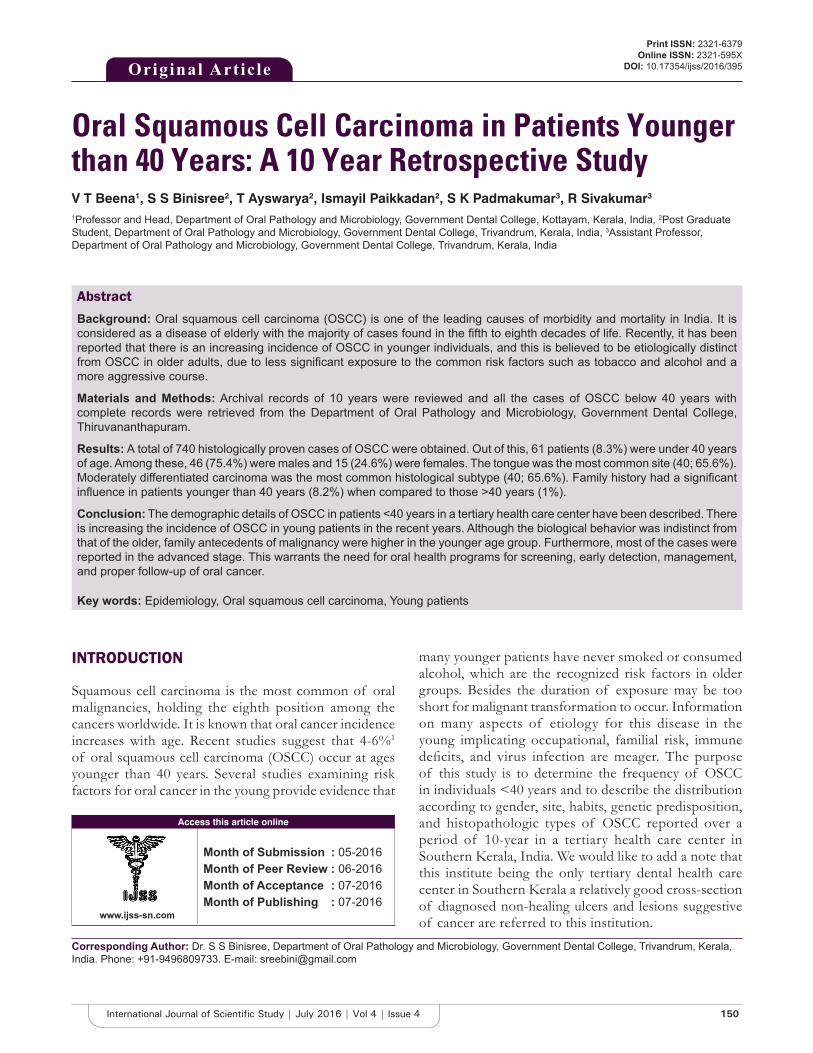
150International Journal of Scientific Study | July 2016 | Vol 4 | Issue 4
Oral Squamous Cell Carcinoma in Patients Younger than 40 Years: A 10 Year Retrospective StudyV T Beena1, S S Binisree2, T Ayswarya2, Ismayil Paikkadan2, S K Padmakumar3, R Sivakumar3
1Professor and Head, Department of Oral Pathology and Microbiology, Government Dental College, Kottayam, Kerala, India, 2Post Graduate Student, Department of Oral Pathology and Microbiology, Government Dental College, Trivandrum, Kerala, India, 3Assistant Professor, Department of Oral Pathology and Microbiology, Government Dental College, Trivandrum, Kerala, India
many younger patients have never smoked or consumed alcohol, which are the recognized risk factors in older groups. Besides the duration of exposure may be too short for malignant transformation to occur. Information on many aspects of etiology for this disease in the young implicating occupational, familial risk, immune deficits, and virus infection are meager. The purpose of this study is to determine the frequency of OSCC in individuals <40 years and to describe the distribution according to gender, site, habits, genetic predisposition, and histopathologic types of OSCC reported over a period of 10-year in a tertiary health care center in Southern Kerala, India. We would like to add a note that this institute being the only tertiary dental health care center in Southern Kerala a relatively good cross-section of diagnosed non-healing ulcers and lesions suggestive of cancer are referred to this institution.
INTRODUCTION
Squamous cell carcinoma is the most common of oral malignancies, holding the eighth position among the cancers worldwide. It is known that oral cancer incidence increases with age. Recent studies suggest that 4-6%1 of oral squamous cell carcinoma (OSCC) occur at ages younger than 40 years. Several studies examining risk factors for oral cancer in the young provide evidence that
Original Article
AbstractBackground: Oral squamous cell carcinoma (OSCC) is one of the leading causes of morbidity and mortality in India. It is considered as a disease of elderly with the majority of cases found in the fifth to eighth decades of life. Recently, it has been reported that there is an increasing incidence of OSCC in younger individuals, and this is believed to be etiologically distinct from OSCC in older adults, due to less significant exposure to the common risk factors such as tobacco and alcohol and a more aggressive course.
Materials and Methods: Archival records of 10 years were reviewed and all the cases of OSCC below 40 years with complete records were retrieved from the Department of Oral Pathology and Microbiology, Government Dental College, Thiruvananthapuram.
Results: A total of 740 histologically proven cases of OSCC were obtained. Out of this, 61 patients (8.3%) were under 40 years of age. Among these, 46 (75.4%) were males and 15 (24.6%) were females. The tongue was the most common site (40; 65.6%). Moderately differentiated carcinoma was the most common histological subtype (40; 65.6%). Family history had a significant influence in patients younger than 40 years (8.2%) when compared to those >40 years (1%).
Conclusion: The demographic details of OSCC in patients <40 years in a tertiary health care center have been described. There is increasing the incidence of OSCC in young patients in the recent years. Although the biological behavior was indistinct from that of the older, family antecedents of malignancy were higher in the younger age group. Furthermore, most of the cases were reported in the advanced stage. This warrants the need for oral health programs for screening, early detection, management, and proper follow-up of oral cancer.
Key words: Epidemiology, Oral squamous cell carcinoma, Young patients
Access this article online
www.ijss-sn.com
Month of Submission : 05-2016 Month of Peer Review : 06-2016 Month of Acceptance : 07-2016 Month of Publishing : 07-2016
Corresponding Author: Dr. S S Binisree, Department of Oral Pathology and Microbiology, Government Dental College, Trivandrum, Kerala, India. Phone: +91-9496809733. E-mail: [email protected]
Print ISSN: 2321-6379Online ISSN: 2321-595X
DOI: 10.17354/ijss/2016/395
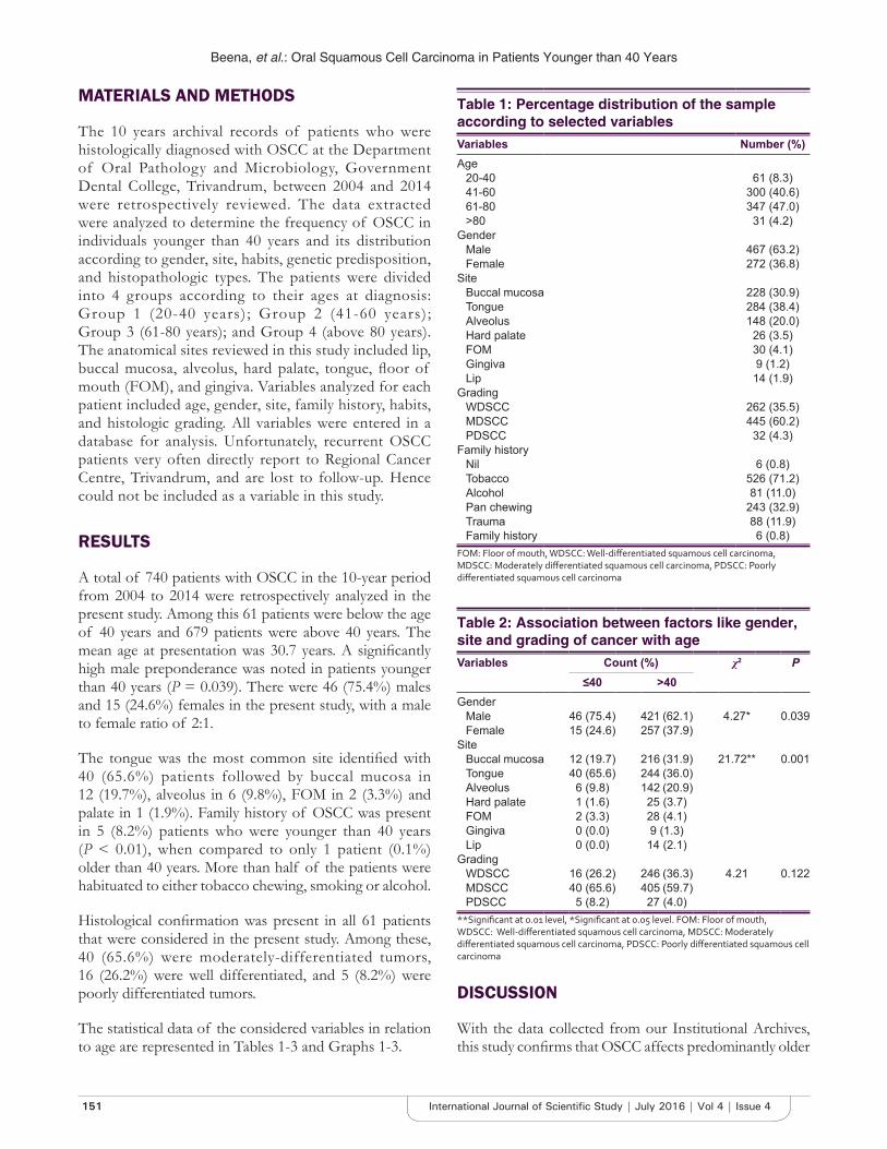
Beena, et al.: Oral Squamous Cell Carcinoma in Patients Younger than 40 Years
151 International Journal of Scientific Study | July 2016 | Vol 4 | Issue 4
MATERIALS AND METHODS
The 10 years archival records of patients who were histologically diagnosed with OSCC at the Department of Oral Pathology and Microbiology, Government Dental College, Trivandrum, between 2004 and 2014 were retrospectively reviewed. The data extracted were analyzed to determine the frequency of OSCC in individuals younger than 40 years and its distribution according to gender, site, habits, genetic predisposition, and histopathologic types. The patients were divided into 4 groups according to their ages at diagnosis: Group 1 (20-40 years); Group 2 (41-60 years); Group 3 (61-80 years); and Group 4 (above 80 years). The anatomical sites reviewed in this study included lip, buccal mucosa, alveolus, hard palate, tongue, floor of mouth (FOM), and gingiva. Variables analyzed for each patient included age, gender, site, family history, habits, and histologic grading. All variables were entered in a database for analysis. Unfortunately, recurrent OSCC patients very often directly report to Regional Cancer Centre, Trivandrum, and are lost to follow-up. Hence could not be included as a variable in this study.
RESULTS
A total of 740 patients with OSCC in the 10-year period from 2004 to 2014 were retrospectively analyzed in the present study. Among this 61 patients were below the age of 40 years and 679 patients were above 40 years. The mean age at presentation was 30.7 years. A significantly high male preponderance was noted in patients younger than 40 years (P = 0.039). There were 46 (75.4%) males and 15 (24.6%) females in the present study, with a male to female ratio of 2:1.
The tongue was the most common site identified with 40 (65.6%) patients followed by buccal mucosa in 12 (19.7%), alveolus in 6 (9.8%), FOM in 2 (3.3%) and palate in 1 (1.9%). Family history of OSCC was present in 5 (8.2%) patients who were younger than 40 years (P < 0.01), when compared to only 1 patient (0.1%) older than 40 years. More than half of the patients were habituated to either tobacco chewing, smoking or alcohol.
Histological confirmation was present in all 61 patients that were considered in the present study. Among these, 40 (65.6%) were moderately-differentiated tumors, 16 (26.2%) were well differentiated, and 5 (8.2%) were poorly differentiated tumors.
The statistical data of the considered variables in relation to age are represented in Tables 1-3 and Graphs 1-3.
DISCUSSION
With the data collected from our Institutional Archives, this study confirms that OSCC affects predominantly older
Table 1: Percentage distribution of the sample according to selected variablesVariables Number (%)Age
20-40 61 (8.3)41-60 300 (40.6)61-80 347 (47.0)>80 31 (4.2)
GenderMale 467 (63.2)Female 272 (36.8)
SiteBuccal mucosa 228 (30.9)Tongue 284 (38.4)Alveolus 148 (20.0)Hard palate 26 (3.5)FOM 30 (4.1)Gingiva 9 (1.2)Lip 14 (1.9)
GradingWDSCC 262 (35.5)MDSCC 445 (60.2)PDSCC 32 (4.3)
Family historyNil 6 (0.8)Tobacco 526 (71.2)Alcohol 81 (11.0)Pan chewing 243 (32.9)Trauma 88 (11.9)Family history 6 (0.8)
FOM: Floor of mouth, WDSCC: Well‑differentiated squamous cell carcinoma, MDSCC: Moderately differentiated squamous cell carcinoma, PDSCC: Poorly differentiated squamous cell carcinoma
Table 2: Association between factors like gender, site and grading of cancer with ageVariables Count (%) χ2 P
≤40 >40Gender
Male 46 (75.4) 421 (62.1) 4.27* 0.039Female 15 (24.6) 257 (37.9)
SiteBuccal mucosa 12 (19.7) 216 (31.9) 21.72** 0.001Tongue 40 (65.6) 244 (36.0)Alveolus 6 (9.8) 142 (20.9)Hard palate 1 (1.6) 25 (3.7)FOM 2 (3.3) 28 (4.1)Gingiva 0 (0.0) 9 (1.3)Lip 0 (0.0) 14 (2.1)
GradingWDSCC 16 (26.2) 246 (36.3) 4.21 0.122MDSCC 40 (65.6) 405 (59.7)PDSCC 5 (8.2) 27 (4.0)
**Significant at 0.01 level, *Significant at 0.05 level. FOM: Floor of mouth, WDSCC: Well‑differentiated squamous cell carcinoma, MDSCC: Moderately differentiated squamous cell carcinoma, PDSCC: Poorly differentiated squamous cell carcinoma

Beena, et al.: Oral Squamous Cell Carcinoma in Patients Younger than 40 Years
152International Journal of Scientific Study | July 2016 | Vol 4 | Issue 4
patients (91%); however, there is a much higher incidence of this disease in the young patients (8.3%) than that reported by other authors, which has varied from 0.4% to 3.6%.1,6,7 There is an overall male predominance in all intraoral subsites in patients younger than 40 years which is in agreement with most of earlier studies.2,4,16
The cause of OSCC is complex and multifactorial, and for young patients with OSCC possible risk factor seems to present a different pattern from that of the older patients.13,17 As a result, many authors have tried to investigate and propose risk factors for OSCC in young subjects. This was also the focus of this study, with factors such as tobacco chewing, smoking, pan chewing, alcohol consumption, trauma, and family history taken
into account. No statistical difference was found between the older and younger patients with regard to being a habitual consumer of tobacco and/or alcohol or not. Therefore, the higher occurrence of OSCC in patients over 40 years old seems to be due to a longer exposure and heavier consumption of tobacco and/or alcohol than to the habit itself. Although men and women in the older group were almost equally exposed to abnormal habits, men were more prone (60% vs. 7% for women) to consume tobacco, alcohol or pan chewing heavily, which explains the predominance of OSCC in older male patients.4,8,9
Table 3: Association between history and ageVariables Count (%) χ2 P
≤40 >40Any history
No 58 (95.1) 675 (99.6) 13.92** 0.000Yes 3 (4.9) 3 (0.4)
TobaccoNo 18 (29.5) 195 (28.8) 0.02 0.902Yes 43 (70.5) 483 (71.2)
AlcoholNo 51 (83.6) 607 (89.5) 2.01 0.156Yes 10 (16.4) 71 (10.5)
Pan chewingNo 52 (85.2) 444 (65.5) 9.9** 0.002Yes 9 (14.8) 234 (34.5)
TraumaNo 49 (80.3) 602 (88.8) 3.82 0.051Yes 12 (19.7) 76 (11.2)
Family historyNo 56 (91.8) 677 (99.9) 45.03** 0.000Yes 5 (8.2) 1 (0.1)
**Significant at 0.01 level
Graph 1: Percentage distribution of the sample according to selected variables
Graph 2: Association between factors like gender, site and grading of cancer with age
Graph 3: Association between history and age

Beena, et al.: Oral Squamous Cell Carcinoma in Patients Younger than 40 Years
153 International Journal of Scientific Study | July 2016 | Vol 4 | Issue 4
In contrast, OSCC of younger age group was almost equally distributed among those who were smokers and/or drinkers as compared to those who were not. Earlier, we had reported a case series of OSCC in non-habituate female patients younger than 40 years from our own department.5 This points out that other factors such as genetic susceptibility, viral infection, hormonal and immunologic modulations, and other systemic diseases could have an upper hand in initiating and promoting OSCC.
A significant correlation (P < 0.0001) between OSCC in young patients and familial history of malignant neoplasm was obtained. In this study, we could not do any genetic evaluation but it was reported earlier from our own department that p53 was mutated in 51.5% of tumors in young patients, and an intense p53 expression was associated with large sized tumors5 Such a relationship presumably indicate that this risk factor carries great weightage for young patients considering that the proposed risk factors like alcohol consumption and tobacco usage need a longer period of exposure for OSCC to develop.
Another important finding was that, in the younger patients, the disease occurred with greater frequency along the lateral border of the tongue (65.6%), whereas in the older patients this site was affected in only 36% of cases. This was in accordance with the study of Annertz et al., who reported that 5.5% of tongue cancers occurred in patients aged 20-39 years, posterolateral border being the most implicated site.15,18 To date, none of the studies have came up with any factor associated with the predominance of OSCC in the tongue of the young patients.10-12
Histopathological grading of tumors in this study showed that the majority of the tumors were moderately differentiated. This was not of much statistical significance and was consistent with the other studies done over the years.6,7,11-14
CONCLUSION
Recent studies have suggested that OSCC in young adults tends to be more frequently anaplastic resulting in a more aggressive behavior and poor prognosis.2,3,16 As the survival rates are good if diagnosed in early stages of the disease, the need for early detection and clinical staging should be stressed. More in-depth studies are needed to investigate the etiology of intraoral cancer in younger patients. Any ulcer or lesion at a younger age should not be dismissed
easily, even if it is not habit related. A high index of clinical suspicion should be attached in high incidence areas that would lead to further investigation and detection of the disease in an early stage, which is perhaps the only way to ensure a good prognosis.
REFERENCES
1. Llewellyn CD, Linklater K, Bell J, Johnson NW, Warnakulasuriya S. An analysis of risk factors for oral cancer in young people: A case-control study. Oral Oncol 2004;40:304-13.
2. Iype EM, Pandey M, Mathew A, Thomas G, Sebastian P, Nair MK. Oral cancer among patients under the age of 35 years. J Postgrad Med 2001;47:171-6.
3. Bodner L, Manor E, Friger MD, van der Waal I. Oral squamous cell carcinoma in patients twenty years of age or younger - Review and analysis of 186 reported cases. Oral Oncol 2014;50:84-9.
4. Hirota SK, Braga FP, Penha SS, Sugaya NN, Migliari DA. Risk factors for oral squamous cell carcinoma in young and older Brazilian patients: A comparative analysis. Med Oral Patol Oral Cir Bucal 2008;13:E227-31.
5. Beena VT, Chauhan I, Heera R, Rajeev R. Oral cancer in young non-habituè females: A report of four cases and review of the literature. Aust Dent J 2011;56:322-7.
6. Majchrzak E, Szybiak B, Wegner A, Pienkowski P, Pazdrowski J, Luczewski L, et al. Oral cavity and oropharyngeal squamous cell carcinoma in young adults: A review of the literature. Radiol Oncol 2014;48:1-10.
7. Cusumano RJ, Persky MS. Squamous cell carcinoma of the oral cavity and oropharynx in young adults. Head Neck Surg 1988;10:229-34.
8. Burzynski NJ, Flynn MB, Faller NM, Ragsdale TL. Squamous cell carcinoma of the upper aerodigestive tract in patients 40 years of age and younger. Oral Surg Oral Med Oral Pathol 1992;74:404-8.
9. Chen YK, Huang HC, Lin LM, Lin CC. Primary oral squamous cell carcinoma: An analysis of 703 cases in southern Taiwan. Oral Oncol 1999;35:173-9.
10. Siriwardena BS, Tilakaratne A, Amaratunga EA, Udagama MN, Ogawa I, Kudo Y, et al. Analysis of histopathological and immunohistochemical differences of oral squamous cell carcinoma in young and old patients in Sri Lanka. J Oral Pathol Med 2007;36:357-62.
11. Martínez-Conde R, Aguirre JM, Burgos JJ, Rivera JM. Clinicopathological factors in early squamous cell carcinoma of the tongue and floor of the mouth, in Biscay (the Basque Country, Spain). Med Oral 2001;6:87-94.
12. McGregor AD, Rennie JS. Intra-oral squamous cell carcinoma in patients under 40 years of age. A report of 13 cases and review of the literature. Br J Plast Surg 1987;40:270-3.
13. Mackenzie J, Ah-See K, Thakker N, Sloan P, Maran AG, Birch J, et al. Increasing incidence of oral cancer amongst young persons: What is the aetiology? Oral Oncol 2000;36:387-9.
14. Llewellyn CD, Johnson NW, Warnakulasuriya KA. Risk factors for squamous cell carcinoma of the oral cavity in young people - A comprehensive literature review. Oral Oncol 2001;37:401-18.
15. Venables CW, Craft IL. Carcinoma of the tongue in early adult life. Br J Cancer 1967;21:645-50.
16. Byers RM. Squamous cell carcinoma of the oral tongue in patients less than thirty years of age. Am J Surg 1975;130:475-8.
17. Son YH, Kapp DS. Oral cavity and oropharyngeal cancer in a younger population. Review of literature and experience at Yale. Cancer 1985;55:441-4.
18. Annertz K, Anderson J, Moller T, Wennerberg J, et al. Incidence and survival of squamous cell carcinoma of the tongue in Scandinavia, with special reference to young adults. Int J Cancer 2002;101:95-9.
How to cite this article: Beena VT, Binisree SS, Ayswarya T, Paikkadan I, Padmakumar SK, Sivakumar R. Oral Squamous Cell Carcinoma in Patients Younger than 40 Years: A 10 Year Retrospective Study. Int J Sci Stud 2016;4(4):150-153.
Source of Support: Nil, Conflict of Interest: None declared.
154International Journal of Scientific Study | July 2016 | Vol 4 | Issue 4
Comparative Study of Lateral Approach and Parascalene Approach of Brachial Plexus Block for Upper Limb Surgeries using Nerve StimulatorKundhavi Devi Ranganathan1, Karthikeyan Natarajan2, Gomathi Karmegam3, Heber Anandan4
1Professor, Department of Anaesthesiology, Government Kilpauk Medical College, Chennai, Tamil Nadu, India, 2Senior Resident, Department of Anaesthesiology, Government Kilpauk Medical College, Chennai, Tamil Nadu, India, 3Assistant Professor, Department of Anaesthesiology, Government Kilpauk Medical College, Chennai, Tamil Nadu, India, 4Senior Clinical Scientist, Department of Clinical Research, Dr. Agarwal’s Health Care Limited, Tirunelveli, Tamil Nadu, India
blocks and for reducing the complication have been described. The present study on brachial plexus blockade-a comparative study on supraclavicular lateral approach with parascalene approach was taken, as studies comparing both these approaches are much less and devoid of complications such as pneumothorax (usually associated with classical subclavian perivascular approach). Moorthy introduced the modified lateral paravascular approach of the supraclavicular block. Vongvises and Panijayanond described parascalene approach in 1979.
MATERIALS AND METHODS
A prospective, randomized, comparative study of 60 patients of American Society of Anesthesiologists Physical Status (ASA PS) I and II category of both sexes in the age group of 20-50 years posted for upper limb surgeries in the
INTRODUCTION
Brachial plexus blockade is gaining popularity day by day for upper extremity surgery because it lends a lot of advantages over general anesthesia.1,2 It is possible and desirable for the patient to remain awake intraoperatively and ambulatory postoperatively. Patients who present for surgery with an upper extremity injury may improve as soon as pain has been relieved with a successful blockade. Various approaches for successful performance of the
AbstractBackground: Upper limb surgery previously done under general anesthesia has now been revolutionized with the advent of various regional blocks. The advantages of two approaches - lateral and parascalene for brachial plexus blockade - are discussed in this study and compared.
Aim: A comparative evaluation of brachial plexus blockade using supraclavicular lateral approach and parascalene approach.
Materials and Methods: A prospective, randomized, comparative study with 60 patients of American Society of Anesthesiologists Physical Status I and II category of both sexes in the age group of 20-50 years posted for upper limb surgeries formed the study group. Patients were allocated into two groups (Group A - lateral approach and Group B - parascalene approach), and the block was performed using a nerve stimulator.
Result: Parascalene approach was found to have statistically significant advantages over the lateral approach in terms of less time to perform block, more success rate, and less vascular complications.
Conclusion: It can be concluded that supraclavicular brachial plexus block by parascalene approach is associated with minimal adverse events with high success rate in comparison to lateral approach.
Key words: Lateral approach, Nerve stimulator, Parascalene approach, Supraclavicular brachial plexus block
Access this article online
www.ijss-sn.com
Month of Submission : 05-2016 Month of Peer Review : 06-2016 Month of Acceptance : 07-2016 Month of Publishing : 07-2016
Corresponding Author: Kundhavi Devi Ranganathan, Professor, Department of Anaesthesiology, Government Kilpauk Medical College, Chennai, Tamil Nadu, Phone: 9841793135. E-mail: [email protected]
Original Article DOI: 10.17354/ijss/2016/396
Print ISSN: 2321-6379Online ISSN: 2321-595X

Ranganathan, et al.: Lateral Approach vs Parascalene Approach of BPB
155 International Journal of Scientific Study | July 2016 | Vol 4 | Issue 4
Department of Orthopedics and Department of Plastic Surgery, Government Kilpauk Medical College Hospital and Government Royapettah Hospital, were included in the study. Institutional Ethical Committee approval and informed consent were obtained. Patients were allocated into two groups: Group A (n = 30) receiving supraclavicular brachial plexus block using lateral approach and Group B (n = 30) receiving supraclavicular brachial plexus block using parascalene approach. Brachial plexus block was performed by supraclavicular block technique assisted with nerve stimulator. Inclusion criteria are all consented patients of both sexes weighing between 50 and 70 kg and aged between 20 and 50 years belonging to ASA PS I and II category undergoing upper limb surgeries. Exclusion criteria are patient refusal, those with pre-existing coagulation disorders, peripheral neuropathy, allergy to any of the drugs used in the study, any distortion of local anatomy, contractures, local infection, previous history of surgery involving brachial plexus, and patients on anticoagulant therapy. ASA PS III and IV and failed block were other exclusion criteria. Patients were evaluated preoperatively both clinically and with routine baseline investigations and assessed for fitness. Patients selected were counseled about the risks and benefits involved in performing the block. After getting informed and written consent, patients willing to be included in the study were enrolled. All patients were kept in nil per oral state at least for 8 h before the procedure. Intravenous access secured with 18-gauge intravenous cannula. Local anesthetic test dose was done. Injection ranitidine 50 mg and injection ondansetron 4 mg were given intravenously 30 min before the procedure and sedated with injection midazolam (0.02-0.05 mg/kg). Boyle’s machine, suctioning equipment, emergency intubation cart, and manual resuscitation bag with mask were kept ready. The procedure was carried out in the theater where facilities for resuscitation were available. Drugs used were 0.5% bupivacaine vial and 2% lignocaine with adrenaline (1:200,000) vial. Intra- and post-operative monitors used were pulse oximeter, non-invasive blood pressure (NIBP), and electrocardiogram (ECG). Initially, the pre procedure parameters were recorded, i.e., pulse rate, BP, SpO2, and ECG. Then, block was administered; these parameters were monitored continuously except the NIBP, which was recorded intermittently. Patients were observed vigilantly for the development of any complications. Surface landmarks: The needle puncture site in Group A was 1 cm above the clavicle at the junction of medial two-third and lateral one-third of the clavicle. After raising a skin wheal, a 22-gauge short bevel 50 mm insulated needle was directed medially and toward the plane of interscalene groove, parallel to clavicle. The needle insertion point in Group B was identified by drawing a line from Chassaignac’s tubercle to midpoint of the clavicle. The entry point of the block was at the junction of the upper two-thirds and lower one-third of the line drawn. The skin and subcutaneous tissue is
infiltrated with local anesthetic solution. A 22-gauge, 50 mm long insulated short bevel needle, directed posteriorly at right angle to the skin. In both groups, the block was performed using a nerve stimulator connected to the proximal end of the needle which is set at 1 mA. The patient may feel discomfort if more than 1 mA current is used. The needle position is adjusted while decreasing the current to 0.5 mA with a sustained distal motor contraction response. A cough from the patient is a warning sign that the pleura is being contacted by the needle. Incremental injection of 15 ml of 0.5% bupivacaine with 15 ml of 2% lignocaine with adrenaline (1:200,000) injected slowly with intermittent aspiration. After injecting the local anesthetic, the block is tested for both sensory (using pin prick) and motor (using muscle power) and is compared with same stimulation or power in the contralateral arm using the Hollmen scale. Onset of blockade means minimum Grade 2 and complete blockade means minimum Grade 3 of Hollmen scale. Motor block is evaluated by thumb abduction (radial nerve), thumb adduction (ulnar nerve), thumb opposition (median nerve), and flexion of the elbow in supination and pronation of the forearm (musculocutaneous). Rescue analgesia was achieved with injection fentanyl 1-2 mcg/kg. Patients with failed block are excluded from the study. Postoperatively patient was monitored for 24 h. Baseline vital signs pulse rate/BP/SpO2 were recorded and monitored. Time required for performing the block, onset, and completion of blockade, duration of blockade, level of sensory block to pinprick, successful blockade, complications of the block, and rescue analgesia was assessed. Data were analyzed using independent sample t-test performed in SPSS 17.
RESULTS
There was no statistically significant difference (P > 0.05) in population characteristics in lateral and parascalene approach group (Table 1).
Time to perform block is 4.7 ± 0.92 min and 2.9 ± 0.84 min in Group A and Group B, respectively. The difference was statistically significant (P = 0.0001). Time for onset of the sensory block is 6.13 ± 1.28 min and 6.2 ± 1.42 min in Group A and Group B, respectively. There was no significant difference (P = 0.8915). Time for onset of motor block is 11.87 ± 1.68 min and 11.93 ± 1.78 min. There was no significant difference (P = 0.8801). The procedure was more successful in the Group B nearly about 93.3% compared with 70% of the Group A. The difference was statistically significant (P = 0.0453) (Table 2).
No complications in the Group B and 7 cases of complications such as vessel injury in Group A. This difference was statistically significant (P = 0.0053). The

Ranganathan, et al.: Lateral Approach vs Parascalene Approach of BPB
156International Journal of Scientific Study | July 2016 | Vol 4 | Issue 4
rescue analgesia requirement in the Group B (6.7%) is less than compared with 30% of the Group A. This difference was statistically significant (P < 0.05). The level of sensory block to pinprick up to the level shoulder level in the Group B (76.7%) is more than compared with Group A. This difference was statistically significant (P < 0.05) (Table 3).
DISCUSSION
Supraclavicular technique was chosen for this study because it provides a rapid onset, dense, and predictable anesthesia with a high success rate. In this study, two approaches of the supraclavicular block are compared. Kulenkampff in Germany, in 1911, performed the first percutaneous supraclavicular approach. This technique was later published, in 1928, by Kulenkampff and Persky (classical) approach the subclavian perivascular approach by Winnie and Collins, in 1964, parascalene approach by Vongvises and Panijayanond in 1979. Vongvises and
Panijayanond described parascalene approach of brachial plexus block, conducted in 100 patients undergoing upper extremity surgery and found that it was a useful, simple, safe, and reliable technique for brachial plexus block, avoiding the complication of pneumothorax (1979).3 Dalens et al. prospectively evaluated parascalene approach with the subclavian perivascular approach in 120 children, 60 patients in each group. The parascalene approach proved to be easier and more reliable while also being almost free of complications, although both techniques produced a high degree of sensory blockade in almost all infraclavicular branches of the brachial plexus (1987).4 Sahu and Sahu found that supraclavicular brachial plexus block by lateral approach associated with minimal adverse effect in comparison to any other supraclavicular approach and more effective with high success rate.5 A new approach Dr. Kothari evaluated supraclavicular brachial plexus block by the lateral approach. Quick and complete analgesia and motor loss with no serious side effect were the main features of this approach.6
CONCLUSION
Supraclavicular block of brachial plexus by parascalene approach provides an adequate sensory blockade and motor blockade, with less time to perform block, level of sensory block is higher (up to shoulder), high success rate, and fewer complications when compared to lateral approach.
REFERENCES
1. Morgan EG Jr, Mikhail MS, Murray MJ. Clinical Anaesthesiology. New York: Lange Medical Books / McGraw-Hill Medical Publishing Division: Elsevier; 2005. p. 324-56.
2. Healy EJ, Knight Paul R. In: Wyle & Churchill-Davidson. Brachial Plexus Nerve Block- A Practice of Anaesthesia 2003;7: p. 599-628.
3. Vongvises P, Panijayanond T. A parascalene technique of brachial plexusanesthesia. Anesth Analg 1979;58:267-73.
4. Dalens B, Vanneuville G, Tanguy A. A new parascalene approach to the brachial plexus in children: Comparison with the supraclavicular approach.Anesth Analg 1987;66:1264-71.
5. Sahu D, Sahu A. Lateral approach for supraclavicular brachial plexus block. Indian J Anaesth 2010;54:215-8.
6. Kothari D. Supraclavicular Brachial plexus block: A new approach. Indian J Anaesth 2003;47:287-8.
Table 1: Population characteristicsAnalysis Group A Group B P valueAge (in years) 36.6±11.6 35.4±10.8 0.5385Sex (M: F) 80:20:00 60:40:00 0.159Weight (in kg) 59.4±6.3 57.1±7.0 0.1693
Table 2: Analysis of outcome of the blocksAnalysis Group A Group B P valueTime to perform block 4.7±0.92 2.9±0.84 0.0001Time for onset of sensory block 6.13±1.28 6.2±1.42 0.8915Time for onset of motor block 11.87±1.68 11.93±1.78 0.8801Success rate 70 93.3 0.0453
Table 3: Analysis of outcome of the blocksAnalysis Group A Group B P valueComplications 7 0 0.0053Rescue analgesia (%) 30 6.70 <0.05Level of sensory block to pin prick (shoulder level) (%)
0 76.70 <0.05
How to cite this article: Ranganathan KD, Natarajan K, Karmegam G, Anandan H. Comparative Study of Lateral Approach and Parascalene Approach of Brachial Plexus Block for Upper Limb Surgeries using Nerve Stimulator. Int J Sci Stud 2016;4(4):154-156.
Source of Support: Nil, Conflict of Interest: None declared.

157 International Journal of Scientific Study | July 2016 | Vol 4 | Issue 4
Incidental Finding of Cervical Dysplasia in Hysterectomy Cases Done for Other CausesS Visalakshi1, K Balakrishnan2, K Uma Maheswari1, G Hemalatha1
1Assistant Professor, Department of Pathology, KAPV Government Medical College, Tiruchirappalli, Tamil Nadu, India, 2Professor, Department of Pathology, KAPV Government Medical College, Tiruchirappalli, Tamil Nadu, India
Squamous cell carcinoma has been reduced steadily owing to effective cytological detection and eradication of its precursors.
MATERIALS AND METHODS
We performed a retrospective data analysis of hysterectomy cases which were done for other benign indications. Women between 30 and 70 years age group are included in this study. The details were taken from the databases maintained by the Pathology Department, KAPV Government Medical College. Institutional board approval was obtained.
All cases of hysterectomy performed for benign indication in the database from January 1, 2015, to December 10, 2015, were included in this analysis. Pre-operative surgical indications for hysterectomy were taken from the medical
INTRODUCTION
Invasive cervical cancers are usually preceded by a long phase of preinvasive disease. This is characterized by a spectrum of events progressing from cellular atypia to various grades of dysplasia or cervical intraepithelial neoplasia (CIN) before progression to invasive carcinoma.
Early diagnosis of this preinvasive dysplasia can save the life of the patient. Finding out the actual incidence of cervical dysplasia helps to improve the screening techniques.
Original Article
AbstractIntroduction: Cervical cancer is one of the leading causes of death in women. Squamous intraepithelial lesion (SIL) (cervical dysplasia) is usually diagnosed by cancer screening techniques. By keeping this in mind, we want to evaluate silent cells which have not been suspected or diagnosed previously, and the patient had hysterectomy for other cases. These findings will give us idea about the incidence of SIL in our population.
Materials and Methods: A retrospective data analysis of hysterectomy cases which were done for other benign indications. Women between 30 and 70 years age group are included in this study. The details were taken from the databases maintained by the Pathology Department, KAPV Government Medical College. Institutional board approval was obtained.
Results: During the study period, 100 hysterectomy cases in the age group of 30-70 years which were performed for benign causes were included. The most common indications for hysterectomy were leiomyoma and abnormal uterine bleeding. Among 100 cases, 14 cases were identified with SIL. All lesions are defined as squamous alterations in the cervical transformation zone that are induced by human papillomavirus infection.
Conclusion: Importance of screening procedures such as Papanicolaou smears and colposcopy not only for regular screening of individuals between age 30 and 70 years, these procedures are also to be followed before any hysterectomy procedures for other reasons. This one is important if cervical intraepithelial neoplasia III or high-grade squamous intraepithelial neoplasia or microinvasive disease identified to modify the surgical procedures. This will avoid further surgeries and complications due to metastasis or stump recurrence.
Key words: Cervical dysplasia, Cervical intraepithelial lesion, Papanicolaou smear
Access this article online
www.ijss-sn.com
Month of Submission : 05-2016 Month of Peer Review : 06-2016 Month of Acceptance : 07-2016 Month of Publishing : 07-2016
Corresponding Author: S Visalakshi, Department of Pathology, KAPV Government Medical College, Tiruchirappalli, Tamil Nadu, India. E-mail: [email protected]
Print ISSN: 2321-6379Online ISSN: 2321-595X
DOI: 10.17354/ijss/2016/397

Visalakshi, et al.: Incidental SILs in Hysterectomies.
158International Journal of Scientific Study | July 2016 | Vol 4 | Issue 4
record. The diagnosis of cervical dysplasia (squamous intraepithelial lesion [SIL]) and its categories were established based on the final pathology report abstracted from the clinical record. The previous HPE slides were also reviewed.
The criteria for the diagnosis of cervical dysplasia is based on guidelines established by the WHO.
Carcinoma cervix and SIL can be discovered on routine Papanicolaou (Pap) smear in an asymptomatic woman. However, the disadvantages are the mucosal surface may be covered by normal epithelium, and the underlying malignant cells may escape detection by cytological smear.
It was observed that some cases of SILs regressed, some persisted and others progressed to carcinoma in situ (CIS).
A direct correlation with progression and histological grade was observed. These observations held to the concept of a single, continuous disease process by which normal epithelium evolves into epithelial precursor lesions and to invasive cancer.
We conducted a data analysis of cervical dysplasia in hysterectomy cases which were done for other benign indications for a period of 1-year January 1, 2015, to December 31, 2015. Women in 30-70 years were included in this study. In this study, 100 hysterectomy cases in the age group of 30-70 years which were performed for benign causes were included. Among 100 cases, 14 cases were identified with SILs. 86 patients had normal cervical epithelium. Among SILs, the number of patients with CIN I is 11, CIN II is 2, and CIN III is 1. Incidence of CIN I is more 78%, whereas CIN II is 14.5% and CIN III 7.5%.
RESULTS
During the study period, 100 hysterectomy cases in the age group of 30-70 years which were performed for benign causes were included. The most common indications for hysterectomy were leiomyoma and abnormal uterine bleeding. Among 100 cases, 14 cases were identified with SILs.
All lesions are defined as squamous alterations in the cervical transformation zone that are induced by human papillomavirus (HPV) infection.
The features of the low-grade squamous intraepithelial lesion (LSIL) include the presence of conspicuous superficial cell atypia with binucleation, 2-fold nuclear
enlargement, variable nuclear chromatin, low N/C ratio with well-preserved polarity.
There are categories of LSIL that includes flat mature LSIL or flat Condyloma, mature exophytic LSIL or exophytic Condyloma, extensive exophytic LSIL or giant Condyloma, immature exophytic LSIL or immature Condyloma and immature flat metaplastic LSIL.
High-grade SILs (HSILs) exhibit atypia in all layers of epithelium. The features include less maturation, higher nuclear density, orderly transition from the immature to mature epithelial layers, nucleomegaly, hyperchromasia in the lower epithelial layers, loss of polarity, increased mitotic index, and abnormal mitotic Figures 1 and 2.5
DISCUSSION
We analyzed the data obtained from a sample of KAPV Government Medical College, Pathology Department. The incidence of SILs among 100 hysterectomy cases which were done for some other benign indications was
Figure 1: Cervical intraepithelial neoplasia I
Figure 2: Cervical intraepithelial neoplasia III

Visalakshi, et al.: Incidental SILs in Hysterectomies.
159 International Journal of Scientific Study | July 2016 | Vol 4 | Issue 4
14%. Among SILs, the number of patients with CIN I is 11, CIN II is 1, and CIN III is 1. Incidence of CIN I is more (85%), whereas CIN II is 7.5% and CIN III 7.5% (Table 1).
Cervical cancer is one of the leading causes of death in women. Significant public awareness must be created to enhance the early detection of cervical cancer. The mortality ratio of cervical cancer can be reduced by early screening techniques. The risk of cervical cancer increases with age.1,2
The replacement of areas of physiological columnar epithelium with mature stratified squamous epithelium beginning soon after birth. Before pubarche, the vaginal pH is neutral. After pubarche and before menarche, when plasma concentrations of estrogen rise and the vaginal and squamous cervical epithelia become glycogenated, lactobacilli are present in the normal flora and glycogenolysis causes the vagina to become acidic. The effect of this on columnar epithelium accustomed to conditions in the alkaline endocervical canal is thought to stimulate squamous metaplasia. Change of shape of the cervix, with exposure of the endocervical epithelium to the vaginal milieu, occurs on many occasions during adolescent and adult life and squamous metaplasia is, therefore, a recurring event.3
In early metaplasia, the simple columnar epithelium is retained above the proliferative squamous cells, but eventually, it is shed.
The histopathologist looking at a section of a cervical biopsy is faced with the task of deciding, whether a particular epithelium shows the changes of CIN and, if so, what degree of abnormality is present. Both these decisions may be fraught with difficulty. A number of benign physiological epithelial changes may be mistaken for CIN, and it is important that these are recognized for what they are. CIN is graded because of the belief that the degree of histological abnormality relates to the prognosis and so can be used as a guide for the management of the patient
Many of cervical dysplasia are undiagnosed, and the actual incidence is also unknown. The incidence of actual cases of cervical dysplasia is actually less than the original incidence.
Cervical precancerous conditions were reported using the categories of dysplasia and CIS, and still they are used in all countries. Many numbers of follow-up studied were conducted in women with such precancerous conditions.
The main observations are some cases of dysplasia regressed, some persisted, and other progressed to CIS. These observations led to the concept of a disease process by which normal epithelium evolves into epithelial precursor lesions and on to invasive cancer.4
CIN was divided into Grades 1, 2, and 3. CIN I - corresponds to mild dysplasia, CIN II - moderates dysplasia. CIN III - severe dysplasia and CIS. Invasive cervical cancers are usually preceded by a long phase of preinvasive disease. This is characterized microscopically as a spectrum of events progressing from cellular atypia to various grades of dysplasia or CIN before progression to invasive carcinoma. CIN may be suspected through cytological examination using the Pap technique or through colposcopic examination. Final diagnosis of CIN is established by the histopathological examination of a cervical punch biopsy or excision specimen. A judgment of whether or not a cervical tissue specimen reveals CIN, and to what degree, is dependent on the histological features concerned with differentiation, maturation and stratification of cells, and nuclear abnormalities. The proportion of the thickness of the epithelium showing mature and differentiated cells is used for grading CIN progression rates to invasive cancer. The diagnosis of HSIL on a Pap test means the presence of precancerous cells, not cancer. Although it is the most preventable type of cancer, each year cervical cancer kills about 4,000 women in the U.S. and about 300,000 women worldwide. Cervical cancer mortality rates are reduced now thanks to increased screening and early detection with the Pap test.5
In our study, 21% of CIN I diagnosis occur in women ages 30-50, and about 14% occur in women 41-50, 35% occurs in women 51-60, and 14% occurs in women 61-70 years of age. The median age of diagnosis is 48 years. About 7% of women had CIN II between the ages of 51-60. About 7% of women had CIN III between the ages of 61-70 (Table 2).
Table 1: Incidence of CINCIN I CIN II CIN III12 (85%) 1 (7.5%) 1 (7.5%)CIN: Cervical intraepithelial neoplasia
Table 2: Agewise incidence of CINAge CIN I CIN II CIN III30-40 3 - -41-50 2 1 -51-60 5 161-70 2 - -CIN: Cervical intraepithelial neoplasia

Visalakshi, et al.: Incidental SILs in Hysterectomies.
160International Journal of Scientific Study | July 2016 | Vol 4 | Issue 4
Cervical cancer is very rare in women younger than age 20. However, many young women become infected with HPV, which then can increase their risk of getting cervical cancer in the future. Young women with early abnormal changes who do not have regular examinations are at high risk for developing cervical cancer by their age 40 and for invasive cancer by age 50.
The best way to prevent cervical cancer is to avoid getting infected with HPV. CIN usually occurs after a woman becomes infected with the HPV. This is a virus that is spread through sexual contact. Some strains, such as HPV-16 and HPV-18, are more likely to infect the reproductive tract in women and cause CIN. A vaccine can protect against the major cancer-causing HPV strains in girls and young women who have not yet been exposed to the virus. Screening for cervical cancer includes regular Pap tests that remain the most effective way of identifying cervical cancer while it is in its earliest precancerous stages and preventing the development of invasive cervical cancer. About half of cervical cancer cases are diagnosed when the cancer is confined to the cervix. About 30-40% of cases are diagnosed after cancer has spread to adjacent areas or lymph nodes and about 10-20% of cases are diagnosed when cancer has already spread to distant regions. Low-grade cervical dysplasia (LGSIL and/or CIN1) often spontaneously resolves without treatment, but follow-up screening is recommended. Untreated high-grade cervical dysplasia may progress to cervical cancer over time. Surgical treatment of cervical dysplasia cures most women.5
Women who are sexually active are sometimes infected with HPV. About 50% of the HPV infections occur in women between the ages of 15 and 25. Mostly, the infections go away without causing any problems. According to Italian Group for Cervical Cancer Screening survey 2.4% of Pap tests were positive for cervical dysplasia. The problem with cervical screening is poor follow-up with abnormal pap smears. The problem with cervical screening is poor follow-up with abnormal Pap smears. The lack of symptoms, infrequent screening may lead the cervical dysplasia to develop into cervical cancer.6 If early detected cervical dysplasia is treated easily. If it progresses to cervical cancer, the treatment will be a prolonged one.
The well-known classification system has incorporated the finding of HPV as a preneoplastic lesion HPV lesions are now grouped with CIN I (mild dysplasia) as a single category, low-grade SIL (low-grade SIL). High-grade lesions are called high-grade SIL and include lesions previously known as CIN II and CIN III (moderate dysplasia, severe dysplasia, and CIS).
So, this system divides all lesions into two functional groups. CIN is characterized by a gradual progression of continuous derangements eventually culminating in a tumor capable of invasion.7-10 Detection of cervical epithelial cell abnormality helps predict the prognosis and can be used as a guide to manage the patient. The hallmarks of CIN are its defining nuclear abnormalities. Nuclei are enlarged, pleomorphic, and with the wrinkled nuclear membrane. There is hyperchromasia and irregularly clumped chromatin. Nucleoli are rare. The differentiation is reduced when the nuclear abnormality is increased. Nuclear polarity will be altered. Mitotic activity is increased there will be increase in the height of the epithelium. Abnormal configurations present. The definition of an in situ carcinoma requires that the surface epithelium lacks all differentiation. So, that immature and undifferentiated cells occupy the entire thickness of the epithelium.
CONCLUSION
About 14 cases being discovered incidentally in hysterectomy specimens done for other reasons may also be taken as the incidence of SILs in our population in silent way. Hence, we here stress the importance of screening procedures such as Pap smears and colposcopy not only for regular screening of individuals between age 30-70 years, these procedures are also to be followed before any hysterectomy procedures for other reasons. This one is important if CIN III or high-grade squamous intraepithelial neoplasia or microinvasive disease identified to modify the surgical procedures. This will avoid further surgeries and complications due to metastasis or stump recurrence.
REFERENCES
1. Schiffman M, Castle PE, Jeronimo J, Rodriguez AC, Wacholder S. Human papillomavirus and cervical cancer. Lancet 2007;370:890-907.
2. Fox H, Wells M. Haines and Taylor Obstetrical and Gynecological Pathology. 4th ed., Vol. 1. New York: Churchill Livingstone; 1995. p. 324-5, 1363-4, 335.
3. Martin-Hirsch PP, Paraskevaidis E, Bryant A, Dickinson HO, Keep SL. Surgery for cervical intraepithelial neoplasia. Cochrane Database Syst Rev 2010;CD001318.
4. Silverberg SG, DeLellis RA. Sliver Berg, Principles and practice of Histopathology and Cytopathology. Philadelphia, PA: Elsevier; 2006.
5. Robboy SJ. Pathology of Female Reproductive Tract. Edinburgh: Churchill Livingstone; 2002.
6. American College of Obstetricians and Gynecologists. ACOG practice bulletin. Diagnosis and treatment of cervical carcinomas. Number 35, May 2002. Int J Gynaecol Obstet 2002;78:79-91.
7. Naucler P, Ryd W, Törnberg S, Strand A, Wadell G, Elfgren K, et al. Human papillomavirus and Papanicolaou tests to screen for cervical cancer. N Engl J Med 2007;357:1589-97.
8. Park J, Sun D, Genest DR, Trivijitsilp P, Suh I, Crum CP. Coexistence of low

Visalakshi, et al.: Incidental SILs in Hysterectomies.
161 International Journal of Scientific Study | July 2016 | Vol 4 | Issue 4
and high grade squamous intraepithelial lesions of the cervix: Morphologic progression or multiple papillomaviruses? Gynecol Oncol 1998;70:386-91.
9. Ronco G, Cuzick J, Pierotti P, Cariaggi MP, Dalla Palma P, Naldoni C, et al. Accuracy of liquid based versus conventional cytology: Overall results of new technologies for cervical cancer screening: Randomised controlled
trial. BMJ 2007;335:28.10. Noller KL. Intraepithelial neoplasia of the lower genital tract (cervix,
vulva). In: Katz VL, Lentz GM, Lobo RA, Gershenson DM, editors. Comprehensive Gynecology. 5th ed. Ch. 28. Philadelphia, PA: Mosby, Elsevier; 2007.
How to cite this article: Visalakshi S, Balakrishnan K, Maheswari KU, Hemalatha G. Incidental Finding of Cervical Dysplasia in Hysterectomy Cases Done for Other Causes. Int J Sci Stud 2016;4(4):157-161.
Source of Support: Nil, Conflict of Interest: None declared.

162International Journal of Scientific Study | July 2016 | Vol 4 | Issue 4
Association of Angiotensin-converting Enzyme Gene Insertion/Deletion Polymorphism with Coronary Artery Disease in South Indian PopulationA Leena Devi1, K Sangeetha2
1Assistant Professor, Department of Biochemistry, Government Mohan Kumaramangalam Medical College, Salem, Tamil Nadu, India, 2Associate Professor, Department of Biochemistry, Government Theni Medical College, Theni, Tamil Nadu, India
cardiovascular disease. The RAS is present in circulating and tissue-based forms,4-6 and it is involved in sodium homeostasis, cardiovascular remodeling, and maintenance of vascular tone. Angiotensin I (Ang I)-converting enzyme (ACE) is a key component within the RAS, where it hydrolyzes Ang I to generate Ang II (vasoconstrictor)7 and the Kallikrein-Kinin system, where it inactivates bradykinin (vasodilator).8 The observation that ACE inhibitors reduce atherosclerosis in cholesterol-fed rabbits supports the potential role for ACE or its substrates in the development of atherosclerosis.9 ACE could affect smooth muscle cell and fibroblast migration and proliferation, low-density lipoprotein (LDL) oxidation, and endothelial cell function; these are all important factors in atherosclerosis. A polymorphic variant of the ACE gene correlates with higher circulating ACE levels and carries an increased risk of myocardial infarction and cardiomyopathies. The ACE gene located on chromosome 17q2310,11 contains a polymorphism in intron 1612-14 of an Insertion/Deletion (I/D) of a 287-bp alu repeat sequence. Insertion allele
INTRODUCTION
Atherosclerosis is the biggest killer of the 21st century.1 Mechanisms contributing to atherogenesis are multiple and complex. Multiple theories including the role of dyslipidemia, hypercoagulability, oxidative stress, inflammation, and endothelial dysfunction have been put forth.2,3 Coronary artery disease (CAD) is a polygenic disease whose phenotypic manifestation depends on the interaction of a number of environmental factors. The gene-encoding components of the renin-angiotensin system (RAS) present attractive candidates for
Original Article
AbstractIntroduction: The angiotensin-converting enzyme (ACE) gene contains a polymorphism in the form of either Insertion (I) or Deletion (D) of a 287 base pair alu repetitive sequences in intron 16. This polymorphism is shown to be associated with the interpersonal variability of ACE levels in circulating blood and DD genotype having approximately twice the level of ACE activity of II genotype and ID having an intermediate activity.
Aim: The aim of this work was to find the association between ACE gene I/D polymorphisms and coronary atherosclerosis in the South Indian population.
Materials and Methods: Genotype analysis was done on 61 patients with angiographically proven coronary atherosclerosis and 62 healthy controls by polymerase chain reaction (PCR) followed by agarose gel electrophoresis of PCR products.
Results: Results revealed that patients had significantly higher frequency of DD genotype than controls (65.7% vs. 39.3%; P = 0.001) with odds ratio of 3 (1.4-6.7); P = 0.01 between (II + ID) genotypes and DD genotype.
Conclusion: ACE DD genotype is found to be a significant risk factor for coronary atherosclerosis.
Key words: Angiotensin-converting enzyme gene, Coronary atherosclerosis, Insertion/Deletion polymorphism, Increased angiotensin-converting enzyme activity
Access this article online
www.ijss-sn.com
Month of Submission : 05-2016 Month of Peer Review : 06-2016 Month of Acceptance : 07-2016 Month of Publishing : 07-2016
Corresponding Author: Dr. A Leena Devi, Department of Biochemistry, Government Mohan Kumaramangalam Medical College, Salem - 636 030, Tamil Nadu, India. Phone: +91-8525052300. E-mail: [email protected]
Print ISSN: 2321-6379Online ISSN: 2321-595X
DOI: 10.17354/ijss/2016/398

Devi and Sangeetha: Angiotensin-converting Enzyme Gene Insertion/Deletion Polymorphism with CAD
163 International Journal of Scientific Study | July 2016 | Vol 4 | Issue 4
produces 490 bp product and deletion allele produces 190bp product, and this results in 3 possible genotypes of ACE gene as II, DD, and ID. It is said that ACE DD genotype is associated with twice the ACE activity of II genotype and ID with an intermediate activity.15 Our aim is to test the hypothesis that deletion allele is associated with an increased risk of atherosclerosis. In this study, we sought to determine the distribution of ACE genotypes and the frequency of allele D in patients undergoing coronary angiography at our institution.
MATERIALS AND METHODS
Study PopulationCasesThe study sample comprised 61 unrelated South Indian CAD patients (57 males, 4 females) with mean age of 55.4 years. Inclusion criteria were more than 50% stenosis of at least one of the major coronary arteries. Patients with <50% obstruction were excluded. All patients with acute myocardial infarction or unstable angina and patients with ischemic or idiopathic cardiomyopathy were also excluded.
ControlsTotally, 62 controls were studied, and they were recruited from outpatient department during their visit for non-cardiac cases. Age, sex, and other confounding factors such as diabetes mellitus (DM) and hypertension (HT) were matched. For all diabetic controls, treadmill test was done and only those with negative treadmill test were included in the study.
Biochemical MarkersTotal cholesterol, high-density lipoprotein cholesterol (HDL-C), and triglyceride (TGL) concentration were determined enzymatically using kits and XL-300 auto analyzer at the Centralized Biochemistry Laboratory at G.G.H, Chennai-3. LDL-cholesterol (LDL-c) was calculated using Friedewald’s formula.
ACE Gene Polymorphism ScreeningDNA was extracted from Buffy coat by high sa l t method , and us ing ACE gene, forward 5’-CTGGAGACCACTCCCATCCTTTCT–3’ and reverse 5’-GATGTGGCCATCAATTCGTCAGAT–3’ primers genomic DNA (1 μg) was amplified in 25μl reaction mixture containing 0.3 μmol/L of each primer and red dye master mix (Bangalore Genei) containing 100 μmol/L of each dNTP, 2.5 μL of ×10 reaction buffer and 0.6 unit of Taq DNA polymerase. After the DNA was denatured for 5 min at 94°C, the reaction mixture was subjected to 30 cycles of denaturation for 1 min at 94°C, 1 min of annealing at 58°C, and 1 min of extension at 72°C. Final extension was carried over at 72°C for 10 min. Amplification products
were separated by electrophoresis on a 2% agarose gel and visualized under ultraviolet light after ethidium bromide staining. The polymerase chain reaction (PCR) product is a 190 bp fragment in the presence of a deletion (D) allele and a 490 bp fragment in the absence of a deletion (I) allele. Thus, each DNA sample revealed one of the three possible patterns after electrophoresis: A 490 bp band (II genotype), a 190 bp band (DD genotype), or both 490 and 190 bp bands (I/D genotype).14,16 Analysis was done using a low molecular weight DNA ladder for Bangalore Genei (Figures 1 and 2).
Statistical Analysis1. Allele frequencies were calculated by allele counting.2. Age, body mass index (BMI), and serum lipid levels
were compared between controls and patients by Students’ t-test.
Figure 1: Extracted DNA (lane 2-8) was tested on 1% agarose gel using 1 kb ladder (lane 1) Ladder shows 10000, 8000, 7000,
6000, 5000, 4000, 3000, 2000, 1000 bp fragments
Figure 2: Agarose gel electrophoresis of polymerase chain reaction products. Deleted allele has 190 bp product and
inserted allele has 490 bp product. Lane 1 shows ladder (100, 200, 300, 400, 500, 600, 700, 800, 900, 1000 bp), lanes 2, 4, 5,
6, 8 show both the products and indicating ID genotype, lane 7 shows only 190 bp product indicating DD genotype, lane 3
shows only 490 bp product indicating II genotype
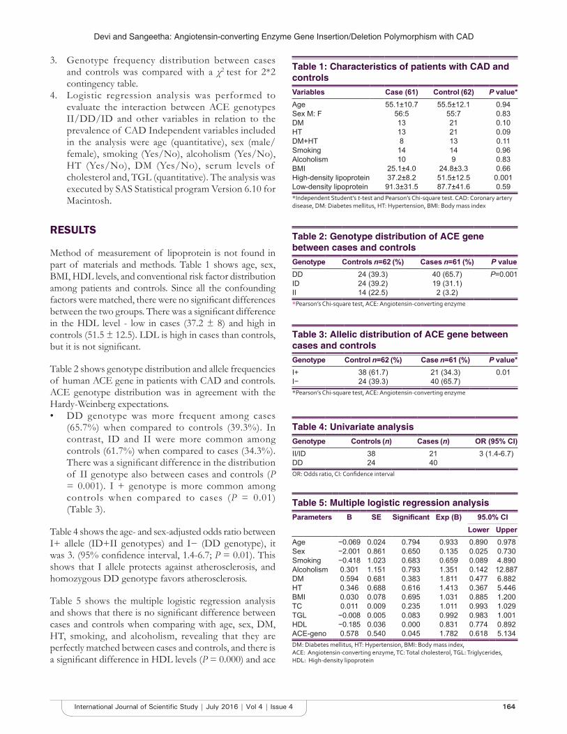
Devi and Sangeetha: Angiotensin-converting Enzyme Gene Insertion/Deletion Polymorphism with CAD
164International Journal of Scientific Study | July 2016 | Vol 4 | Issue 4
3. Genotype frequency distribution between cases and controls was compared with a χ2 test for 2*2 contingency table.
4. Logistic regression analysis was performed to evaluate the interaction between ACE genotypes II/DD/ID and other variables in relation to the prevalence of CAD Independent variables included in the analysis were age (quantitative), sex (male/female), smoking (Yes/No), alcoholism (Yes/No), HT (Yes/No), DM (Yes/No), serum levels of cholesterol and, TGL (quantitative). The analysis was executed by SAS Statistical program Version 6.10 for Macintosh.
RESULTS
Method of measurement of lipoprotein is not found in part of materials and methods. Table 1 shows age, sex, BMI, HDL levels, and conventional risk factor distribution among patients and controls. Since all the confounding factors were matched, there were no significant differences between the two groups. There was a significant difference in the HDL level - low in cases (37.2 ± 8) and high in controls (51.5 ± 12.5). LDL is high in cases than controls, but it is not significant.
Table 2 shows genotype distribution and allele frequencies of human ACE gene in patients with CAD and controls. ACE genotype distribution was in agreement with the Hardy-Weinberg expectations.• DD genotype was more frequent among cases
(65.7%) when compared to controls (39.3%). In contrast, ID and II were more common among controls (61.7%) when compared to cases (34.3%). There was a significant difference in the distribution of II genotype also between cases and controls (P = 0.001). I + genotype is more common among controls when compared to cases (P = 0.01) (Table 3).
Table 4 shows the age- and sex-adjusted odds ratio between I+ allele (ID+II genotypes) and I− (DD genotype), it was 3. (95% confidence interval, 1.4-6.7; P = 0.01). This shows that I allele protects against atherosclerosis, and homozygous DD genotype favors atherosclerosis.
Table 5 shows the multiple logistic regression analysis and shows that there is no significant difference between cases and controls when comparing with age, sex, DM, HT, smoking, and alcoholism, revealing that they are perfectly matched between cases and controls, and there is a significant difference in HDL levels (P = 0.000) and ace
Table 1: Characteristics of patients with CAD and controlsVariables Case (61) Control (62) P value*Age 55.1±10.7 55.5±12.1 0.94Sex M: F 56:5 55:7 0.83DM 13 21 0.10HT 13 21 0.09DM+HT 8 13 0.11Smoking 14 14 0.96Alcoholism 10 9 0.83BMI 25.1±4.0 24.8±3.3 0.66High-density lipoprotein 37.2±8.2 51.5±12.5 0.001Low-density lipoprotein 91.3±31.5 87.7±41.6 0.59*Independent Student’s t‑test and Pearson’s Chi‑square test. CAD: Coronary artery disease, DM: Diabetes mellitus, HT: Hypertension, BMI: Body mass index
Table 3: Allelic distribution of ACE gene between cases and controlsGenotype Control n=62 (%) Case n=61 (%) P value*I+ 38 (61.7) 21 (34.3) 0.01I− 24 (39.3) 40 (65.7)*Pearson’s Chi‑square test, ACE: Angiotensin‑converting enzyme
Table 4: Univariate analysisGenotype Controls (n) Cases (n) OR (95% CI)II/ID 38 21 3 (1.4-6.7)DD 24 40OR: Odds ratio, CI: Confidence interval
Table 5: Multiple logistic regression analysisParameters B SE Significant Exp (B) 95.0% CI
Lower UpperAge −0.069 0.024 0.794 0.933 0.890 0.978Sex −2.001 0.861 0.650 0.135 0.025 0.730Smoking −0.418 1.023 0.683 0.659 0.089 4.890Alcoholism 0.301 1.151 0.793 1.351 0.142 12.887DM 0.594 0.681 0.383 1.811 0.477 6.882HT 0.346 0.688 0.616 1.413 0.367 5.446BMI 0.030 0.078 0.695 1.031 0.885 1.200TC 0.011 0.009 0.235 1.011 0.993 1.029TGL −0.008 0.005 0.083 0.992 0.983 1.001HDL −0.185 0.036 0.000 0.831 0.774 0.892ACE-geno 0.578 0.540 0.045 1.782 0.618 5.134DM: Diabetes mellitus, HT: Hypertension, BMI: Body mass index, ACE: Angiotensin‑converting enzyme, TC: Total cholesterol, TGL: Triglycerides, HDL: High‑density lipoprotein
Table 2: Genotype distribution of ACE gene between cases and controlsGenotype Controls n=62 (%) Cases n=61 (%) P valueDD 24 (39.3) 40 (65.7) P=0.001ID 24 (39.2) 19 (31.1)II 14 (22.5) 2 (3.2)*Pearson’s Chi‑square test, ACE: Angiotensin‑converting enzyme

Devi and Sangeetha: Angiotensin-converting Enzyme Gene Insertion/Deletion Polymorphism with CAD
165 International Journal of Scientific Study | July 2016 | Vol 4 | Issue 4
genotype (P = 0.045) between cases and controls, proving our hypothesis.
DISCUSSION
Besides very well-known risk factors, genetic factors play a role in the development of CAD. Genetic factors differ in various populations. Among these, ACE gene polymorphism has most frequently been studied and proposed as a CAD risk factor. ACE gene polymorphism determines the serum and tissue ACE activity, which is high in subjects with DD genotype.17 ACE by causing high Ang II) and low bradykinin levels may increase the risk of CAD.18 Ang II increases the macrophage-derived growth factor and platelet-derived growth factor, which has a role in the genesis of atherosclerosis.19 Furthermore, Ang II leads to LDL-C oxidation and stimulates neutrophil, macrophage, and T-lymphocytes.19,20 ACE decreases nitric oxide release via the bradykinin-kallikrein system and causes endothelial dysfunction which has also an important role in the genesis of atherosclerosis. Homozygous deletion subset of the ACE I/D polymorphism is associated with deteriorated endothelial function.21 It has been demonstrated by various studies that the ACE D allele is associated with the risk of CAD in various populations. However, other studies show that ACE gene polymorphism is not associated with CAD and MI. A large case–control study by Guardsman et al.22 on Caucasian samples has shown that the D allele was associated with CAD in patients <61.7 years of age.
Hence, the clinical relevance of ACE gene polymorphism remains unclear. As CAD is a multifactorial disease, the ACE gene alone may not have a direct effect on the severity of CAD and premature death. However, the homozygous DD genotype is found to be significantly increased in our study samples.
In this study, we have not confirmed the DD genotypes using insertion-specific primers, as previous studies showed that there is a possibility of mistyping23 due to preferential amplification of D allele and all the DD genotype positive cases to be confirmed by using another insertion-specific primer. This is the drawback of our study. Moreover, we have not tested the phenotypic variation associated with ACE genotypes, which is one more limitation of our study.
The present study is important as there is a need for confirmation of the risk gene for CAD, even if the effect is small, so as to contribute to our understanding of the pathology of CAD, and determine potential therapeutic strategies.
CONCLUSION
In the present study, ACE homozygous DD genotype is significantly associated with atherosclerosis. The presence of I allele protects against atherosclerosis.
ACKNOWLEDGMENT
The author would sincerely like to acknowledge all his Professors, Associate Professors, and Assistant Professors, who have helped him in completing the study.
REFERENCES
1. Badimon JJ, Fuster V, Chesebro JH, Badimon L. Coronary atherosclerosis. A multifactorial disease. Circulation 1993;87:3-16.
2. Schoen FJ. The heart of inherited disease. Robbins Pathologic Basis of Disease. 5th ed. Ch. 12. New York: WB Saunders Co.; 1994. p. 517-82.
3. Braunwald E. Heart Disease. A Textbook of Cardiovascular Disease. 5th ed., Vol. 2. New York: WB Saunders Co.; 1997. p. 1105-60.
4. Caldwell PR, Seegal BC, Hsu KC, Das M, Soffer RL. Angiotensin-converting enzyme: Vascular endothelial localization. Science 1976;191:1050-1.
5. Ryan US, Ryan JW, Whitaker C, Chiu A. Localization of angiotensin converting enzyme (Kininase II). II. Immunocytochemistry and immunofluorescence. Tissue Cell 1976;8:125-45.
6. Erdös EG, Skidgel RA. The angiotensin I-converting enzyme. Lab Invest 1987;56:345-8.
7. Skeggs LT JR, Kahn JR, Shumway NP. The preparation and function of the hypertensin-converting enzyme. J Exp Med 1956;103:295-9.
8. Yang HY, Erdös EG, Levin Y. A dipeptidyl carboxypeptidase that converts angiotensin I and inactivates bradykinin. Biochim Biophys Acta 1970;214:374-6.
9. Schuh JR, Blehm DJ, Frierdich GE, McMahon EG, Blaine EH. Differential effects of renin-angiotensin system blockade on atherogenesis in cholesterol-fed rabbits. J Clin Invest 1993;91:1453-8.
10. Soubrier F, Alhenc-Gelas F, Hubert C, Allegrini J, John M, Tregear G, et al. Two putative active centers in human angiotensin I-converting enzyme revealed by molecular cloning. Proc Natl Acad Sci USA 1988;85:9386-90.
11. Mattei MG, Hubert C, Alhenc-Gelas F, Roeckel N, Corvol P, Soubrier F. Angiotensin-I converting enzyme gene is on chromosome 17. Cytogenet Cell Genet 1989;51:1041.
12. Rigat B, Hubert C, Alhenc-Gelas F, Cambien F, Corvol P, Soubrier F. An insertion/deletion polymorphism in the angiotensin I-converting enzyme gene accounting for half the variance of serum enzyme levels. J Clin Invest 1990;86:1343-6.
13. Hubert C, Houot AM, Corvol P, Soubrier F. Structure of the angiotensin I-converting enzyme gene. Two alternate promoters correspond to evolutionary steps of a duplicated gene. J Biol Chem 1991;266:15377-83.
14. Zintzaras E, Raman G, Kitsios G, Lau J. Angiotensin-converting enzyme insertion/deletion gene polymorphic variant as a marker of coronary artery disease: A meta-analysis. Arch Intern Med 2008;168:1077-89.
15. Fogarty Y, Fraser CG, Browning MC. Intra- and inter-individual variation of serum angiotensin converting enzyme: Clinical implications. Ann Clin Biochem 1989;26:201-2.
16. Danser AH, Schalekamp MA, Bax WA, van den Brink AM, Saxena PR, Riegger GA, et al. Angiotensin-converting enzyme in the human heart. Effect of the deletion/insertion polymorphism. Circulation 1995;92:1387-8.
17. Malik FS, Lavie CJ, Mehra MR, Milani RV, Re RN. Renin-angiotensin system: Genes to bedside. Am Heart J 1997;134:514-26.
18. Cambien F, Poirier O, Lecerf L, Evans A, Cambou JP, Arveiler D, et al. Deletion polymorphism in the gene for angiotensin-converting enzyme is a potent risk factor for myocardial infarction. Nature 1992;359:641-4
19. Keidar S, Brook JG, Aviram M. Angiotensin-II enhanced lipid peroxidation

Devi and Sangeetha: Angiotensin-converting Enzyme Gene Insertion/Deletion Polymorphism with CAD
166International Journal of Scientific Study | July 2016 | Vol 4 | Issue 4
of low-density lipoprotein. J Am Physiol Soc 1993;8:245-8.20. Farber HW, Center DM, Rounds S, Danilov SM. Components of the
angiotensin system cause release of a neutrophil chemoattractant from cultured bovine and human endothelial cells. Eur Heart J 1990;11:100-7.
21. Mulder HJ, van Geel PP, Schalij MJ, van Gilst WH, Zwinderman AH, Bruschke AV. PREFACE trial. DD ACE gene polymorphism is associated with increased coronary artery endothelial dysfunction: The PREFACE
trial. Heart 2003;89:557-8.22. Gardemann A, Weiss T, Schwartz O, Eberbach A, Katz N, Hehrlein FW,
et al. Gene polymorphism but not catalytic activity of angiotensin I-converting enzyme is associated with coronary artery disease and myocardial infarction in low-risk patients. Circulation 1995;92:2796-9.
23. Shanmugam V, Sell KW, Saha BK. Mistyping ACE heterozygotes. PCR Methods Appl 1993;3:120-1.
How to cite this article: Devi AL, Sangeetha K. Association of Angiotensin-converting Enzyme Gene Insertion/Deletion Polymorphism with Coronary Artery Disease in South Indian Population. Int J Sci Stud 2016;4(4):162-166.
Source of Support: Nil, Conflict of Interest: None declared.

167 International Journal of Scientific Study | July 2016 | Vol 4 | Issue 4
Pattern of Ocular Trauma in a Tertiary Referral Hospital in South Tamil NaduN Sharmila1, K Kavitha1, S Ganapathy Rajesh2, C Suriya Kumar3
1Associate Professor, Department of Ophthalmology, Theni Medical College, Theni, Tamil Nadu, India, 2Senior Assistant Professor, Department of Ophthalmology, Theni Medical College, Theni, Tamil Nadu, India, 3Assistant Professor, Department of Ophthalmology, Theni Medical College, Theni, Tamil Nadu, India
MATERIALS AND METHODS
A retrospective study was done at Government Theni Medical College from May 2015 to April 2016 on 300 patients who presented to the casualty, emergency, and trauma department. The patients who already had eye diseases such as glaucoma and operated eyes were excluded from the study. A thorough ophthalmic examination was done which included visual acuity measurement by Snellen’s chart, slit lamp examination to evaluate anterior segment injuries and fundus examination by IDO and slit lamp biomicroscopy. Intraocular pressure was measured except in open globe injuries. The data collected included age, sex, place, and date and time of trauma, cause of injury. The data were finally analyzed statistically by simple proportions.
RESULTS AND DISCUSSION
In our study, out of 300 patients, 80% of the patients belonged to the age group of 25-35 years. 71% of them
INTRODUCTION
Ocular injury is an avoidable cause of blindness and visual impairment. According to the WHO, 55 million eye injuries restricting activities occur for more than 1 day each year. 750,000 cases require hospitalization which includes 200,000 open globe injuries.1 Ocular trauma usually occurs in the younger individuals and road traffic accidents are a more common. Our Government Theni Medical College is situated in a National Highway. Hence, we designed this study to determine the pattern of ocular trauma, its causes, and effective management.
Original Article
AbstractIntroduction: Trauma is an important causative factor of ocular morbidity. It is one of the common causes of preventable blindness in our Indian population.
Aim: To assess the pattern of ocular trauma causes and management done at Government Theni Medical College Hospital.
Materials and Methods: A retrospective study was conducted from May 2015 to April 2016 in 300 patients with ocular trauma who presented to casualty at Government Theni Medical College, Theni. We recorded the data based on the nature and cause of injury, time of injury, and time interval between injury and reporting to the hospital. The data were analyzed statistically by simple proportion.
Results: Of 300 patient examined, the most common age group was 25-35 years, more predominant in males. The most common cause being road traffic accidents. Most of the patients were managed with suturing and conservatively.
Conclusion: Our study shows a male preponderance and the most common age group being 25-35 years. The most common cause of ocular trauma is road traffic accidents followed by assault. Hence, there is an urgent need for reinforcement of traffic rules and people should adhere to them. The urgent need for more ambulance services to bring the patients to the nearby hospital as soon as possible and first aid training to paramedical workers, enhancing the facilities available at the emergency centers will go a long way in preventing disability and visual loss to the public.
Key words: Ocular trauma, Road traffic accidents, Subconjunctival hemorrhage
Access this article online
www.ijss-sn.com
Month of Submission : 05-2016 Month of Peer Review : 06-2016 Month of Acceptance : 07-2016 Month of Publishing : 07-2016
Corresponding Author: Dr. N Sharmila, 22, 2nd Street, Kumaran Colony, Vadapalani, Chennai - 600 026, Tamil Nadu, India. Phone: +91-9444160167. E-mail: [email protected]
Print ISSN: 2321-6379Online ISSN: 2321-595X
DOI: 10.17354/ijss/2016/399

Sharmila, et al.: Pattern of Ocular Trauma
168International Journal of Scientific Study | July 2016 | Vol 4 | Issue 4
were males and 29% of them were females. As specified by the previous studies our analysis too showed a male preponderance (Table 1).2
The most common pattern of injury was found to be subconjunctival hemorrhage followed by lid trauma. A few cases of periorbital hematoma were noticed. A very few cases of open globe injury were recorded (Table 2).
Regarding the causes of ocular trauma, road traffic accidents formed the major bulk of causes, followed by assault (Table 3). This might be due to the reason that our Medical College is situated in a national highway.
About 16% of the patients were brought to the hospital within 24 h of injury. 52% of the patients were brought to the hospital within 24-48 h of injury and 28% of the patients were brought within 48 h - 1 week. 4% were brought after 1 week of injury (Table 4).
Most of the patients with subconjunctival hemorrhage (77%) were treated with topical antibiotics, steroid drops, and anti-inflammatory drugs and those who had laceration of lid (20%) were sutured, those with open globe injuries underwent repair with microsurgical techniques (3%) when compared to He et al.3 who managed 52.6% with surgery and 22.2% conservatively.
The majority of our patients presented with good vision (69%) and 3% of them had visual impairment (Table 5). This might be due to the reason that most of them were closed globe injuries and were brought to the hospital earlier.
However, according to Omolase et al.,4 50.8% had a visual acuity ranging from 6/18 to 6/6, 32.6% <3/60. As per the study conducted by Iqbal et al.,5 81.1% had a visual acuity <3/60 and 12.2% had a visual acuity 6/60-6/18. As our study had a lesser degree of penetrating injuries, patients presented to us with a better visual acuity. Our hospital is easily accessible to the patients, so most of them were brought without delay. This might be the reason for a lesser damage to the ocular system.
CONCLUSION
Our study shows a male preponderance and the most common age group being 25-35 years. The most common cause of ocular trauma is road traffic accidents followed by an assault. Hence, there is an urgent need for reinforcement of traffic rules and people should adhere to them. The urgent need for more ambulance services to bring the patients to the nearby hospital as soon as possible and
first aid training to paramedical workers, enhancing the facilities available at the emergency centers will go a long way in preventing disability and visual loss to the public. People can be educated using media and health education and awareness regarding first aid measures and safety precautions for ocular trauma will help us to bring down the visual impairment caused by ocular injuries.
REFERENCES
1. Desai P, MacEwen CJ, Baines P, Minassian DC. Epidemiology and implications of ocular trauma admitted to hospital in Scotland. J Epidemiol Community Health 1996;50:436-41.
Table 1: Age distributionAge group (years) Number of patients (%)<5 12 (4)5-15 12 (4)15-25 60 (20)25-35 120 (40)35-45 48 (16)45-55 24 (8)55-65 24 (8)>65 Nil (Nil)
Table 2: Pattern of traumaType of injury Number of patients (%)Contusion 12 (4)Periorbital hematoma 24 (8)Lid trauma 60 (20)Subconjunctival hemorrhage 195 (65)Open globe injury 9 (3)
Table 3: Causes of injuryCause Number of patients (%)Road traffic accident 180 (60)Assault 108 (36)Accidental fall 12
Table 4: Duration of presentationDuration Number of patients (%)<24 h 48 (16)24-48 h 156 (52)48 h-1 week 84 (28)>1 week 12 (4)
Table 5: Visual acuityVisual acuity at presentation Percentage<3/60 33/60-6/60 66/60-6/18 226/18-6/6 69

Sharmila, et al.: Pattern of Ocular Trauma
169 International Journal of Scientific Study | July 2016 | Vol 4 | Issue 4
2. Olurin O. Eye injuries in Nigeria. A review of 433 cases. Am J Ophthalmol 1971;72:159-66.
3. He MZ, Liping M. Epidemiology of patients hospitalised for ocular trauma in the Chaoshan Region of China 2001-2010. Vol. 10. Injury Prevention Research Centre Medical College of Shantou University; 2012. p. 1371.
4. Omolase CO, Omolade EO, Ogunleye OT, Omolase BO, Ihemedu CO, Adeosun OA. Pattern of ocular injuries in OWO, Nigeria. J Ophthalmic Vis Res 2011;6:114-8.
5. Iqbal CJ, Khan KR. Visual Outcome after Surgical Management in Penetrating Ocular Trauma. Lahore: King Eduard Medical University, Mayo Hospital; 2011.
How to cite this article: Sharmila N, Kavitha K, Rajesh SG, Kumar CS. Pattern of Ocular Trauma in a Tertiary Referral Hospital in South Tamil Nadu. Int J Sci Stud 2016;4(4):167-169.
Source of Support: Nil, Conflict of Interest: None declared.

170International Journal of Scientific Study | July 2016 | Vol 4 | Issue 4
Clinical and Histomorphological Profile of Breast NeoplasmsS Jenita Christiana1, K Balakrishnan2, G Hemalatha3, K Uma Maheswari3
1Associate Professor, Department of Pathology, KAPV Government Medical College, Tiruchirappalli, Tamil Nadu, India,2Professor, Department of Pathology, KAPV Government Medical College, Tiruchirappalli, Tamil Nadu, India,3Assistant Professor, Department of Pathology, KAPV Government Medical College, Tiruchirappalli, Tamil Nadu, India
death among women after lung cancer.2 In some of the studies, it occurs secondary to cervical cancer,3,4 presently 75,000 new cases occur in Indian women every year.5 Etiology of breast cancer is multifactorial. It includes diet, reproductive lifestyle, environmental and genetic factors.4 This study is intended to clarify the epidemiological and pathological features of breast cancer to adopt an adequate strategy of care in our area.
MATERIALS AND METHODS
A total number of 267 cases who attended the surgery outpatient department and for whom lumpectomy or modified radical mastectomy has been done were included in the study. Relevant clinical features such as age, parity, age of first childbirth, menstrual status, and family history were elicited. Location of lump, nodal enlargement, presence of nipple discharge, skin, and nipple changes were observed.
INTRODUCTION
Breast neoplasms encompass a heterogeneous group of lesions that may be presenting as a palpable mass, non-palpable abnormality detected on imaging analysis or an incidental microscopic finding. They constitute a wide spectrum of histological lesions ranging from a benign tumor at one end approximating to carcinoma at the other end. Breast cancer is the most common cancer of women worldwide representing approximately 16% of all female cancer.1 It represents the second leading cause of cancer
AbstractIntroduction: Breast neoplasms have a high degree of heterogeneity under several distinct viewpoints such as variable clinical presentations, diverse morphological features, and different therapeutic responses. Understanding the clinical prolife of breast cancer is important to develop early diagnosis efforts.
Aim: To evaluate clinical and pathological characteristics of breast neoplasms.
Materials and Methods: This study was conducted over a period of 2-year comprising 267 patients who underwent lumpectomy or modified radical mastectomy following a preliminary diagnosis on fine needle aspiration cytology or trucut biopsy specimens. The clinicopathologic evaluation was done in all of these cases following standard protocols.
Results: The study comprises 267 patients in the age range of 20-70 years. The majority of breast cancers occurred within the age range of 41-50 years with mean age of 45.5 years. Exceptionally one case of invasive ductal carcinoma was observed in the age 5 years. The most common benign tumor was fibroadenoma and in malignancy invasive ductal carcinoma was evident in 103 cases. Early menarche contributes to the major risk factor. Breast lump, nipple discharge, Peaud’ orange appearance, and axillary lymphadenopathy are the common clinical presentations. Predilection toward the left side and location of lump over the upper outer quadrant has been documented. Most of the cases presented with American Joint Committee on cancer pathologic Stage II A.
Conclusion: This study gave an insight to the clinicopathological prolife of breast cases in our area.
Key words: Breast neoplasms, Epidemiology, Histopathology cancer, Stage
Access this article online
www.ijss-sn.com
Month of Submission : 05-2016 Month of Peer Review : 06-2016 Month of Acceptance : 07-2016 Month of Publishing : 07-2016
*Corresponding Author: S Jenita Christiana, Department of Pathology, KAPV Government Medical College, Trichy, Tamil Nadu, India E-mail: [email protected]
Original ArticlePrint ISSN: 2321-6379
Online ISSN: 2321-595XDOI: 10.17354/ijss/2016/400

Christiana, et al.: Breast neoplasms: A profile
171 International Journal of Scientific Study | July 2016 | Vol 4 | Issue 4
The specimens were fixed in 10% formalin and usual techniques of histological processing, paraffin embedding, section cutting with microtome and hematoxylin and eosin staining were made. Histopathological evaluation was done with microscopy.
RESULTS
This study of breast neoplasms covers a total of 267 cases in which 120 cases were observed as benign and 147 cases as malignant neoplasms. The age range of patients was 20-70 with a median of 45 years.
As noted in Table 1, increased incidences of breast neoplasms were observed in 31-50 years (116 cases, 43.44%). The incidence is very low in pediatric age group <10 years (1 case, 0.37%) followed by 61-70 years (8 cases - 2.99%).
Bar diagram shows that benign lesions have a peak incidence in the age group of <20 years (55 cases - 45.84%) (Figure 1). Whereas malignancy reaches its peak in the age group 41-50 years (51 cases 34.93%). The findings of independent sample t-test are given in the following Table 2, which shows the difference in mean age between benign and malignant neoplasms.
Benign neoplasms occur at a relatively younger age than malignant neoplasms with statistical significance (P < 0.001). Clinical evaluation of malignant tumors is represented in Table 3.
All the cases presented with a breast lump. Risk factor like early menarche was seen in most cases (140 cases, 95.89%). Most of the women in our study were multiparous with a history of regular breastfeeding (141 cases, 96.57%). The tumor mostly involved the upper outer quadrant (85 cases, 58.21%) followed by diffuse involvement (36 cases 24.65%). Figure 2 illustrates the distribution of breast neoplasms. Malignant neoplasms predominate with (147 cases 54.66%) when compared with benign neoplasms (120 cases, 45.31%). Age incidence of individual benign neoplasms is given as shown in Table 4.
Most of the fibroadenomas are seen in the early reproductive age group, <20 years (53 cases, 50%) followed by 21-30 years (36 cases, 33.96%). Special forms of adenomas such as tubular adenoma and lactating adenoma were also observed in the same age group. One case of adenomyoepithelioma and myofibroblastoma was observed in the postmenopausal age group. Distribution of epithelial malignant tumors is shown in Table 5.
Table 1: Age incidence of breast neoplasmsAge group (years) Number of cases (%)<10 1 (0.37)10-20 54 (20.22)21-30 53 (19.85)31-40 58 (21.72)41-50 58 (21.72)51-60 35 (13.10)61-70 8 (2.99)>70 -
Table 2: Comparison of mean age of benign and malignant neoplasms by independent samples t-testVariable Age (Mean±SD) t-test value P valueBenign diseases 24.9±9.7 16.15 <0.001Malignant diseases 45.3±10.7SD: Standard deviation
Figure 1: Comparison of age incidence of benign/malignant neoplasms
Figure 2: Distribution of breast neoplasms
Infiltrating ductal carcinoma (IDC) not otherwise specified (NOS) was observed as the most common malignant breast tumor (103 cases - 70.06%). Other fibroepithelial and non-epithelial malignant tumors of the breast that were observed are represented as shown in Table 6.

Christiana, et al.: Breast neoplasms: A profile
172International Journal of Scientific Study | July 2016 | Vol 4 | Issue 4
their study of the evolution of evolution of cancer at Ivory Coast showed that cervical cancer was the first cancer of women followed by breast with a frequency of 10.52% of cases.4 As per Mohapatra et al’s., hospital data from South India breast cancer constitutes 15.05% next to cervical cancer.3 In our study, the prevalence of breast cancer is 18.91% ranking second to cervical cancer in accordance. Contrary to this in metropolitan cities such as New Delhi, Mumbai, Ahamadabad, Kolkotta, and Trivandrum breast cancer takes the lead.3
The age-specific incidence of breast neoplasms ranges from 20 to 70 years and shows increased incidence in 31-50 years. Pertaining to benign neoplasms the mean age of incidence is 24.9 and for malignant lesions, it is 45.3. This is comparable with the studies of Ranabhat et al.7
In a study conducted at Nepal,7 proportion of breast diseases was found to be 90.04% benign neoplasms and malignancy as 8.8%, whereas in our study they are almost equal comprising benign neoplasms to be 44.94% and malignancy 55.05%. Studies from Nepal, Lahore, Aurangabad, Mumbai,7 and Malawi,8 have found fibroadenoma as the most common benign breast lesion. In our study, fibroadenoma constitutes 89.16% with the peak incidence in the age group of <20 years. Next stands the
Table 3: Clinical evaluation of cases with malignant tumorsHistory/clinical features Number of cases (%)Early menarche 140 (95.89)Age of first child birth
Early 137 (93.83)Late 9 (6.16)
Parous women 141 (96.57)Nulliparous women 5 (3.42)Breast fed 141 (96.57)Menstrual status
Menopausal 93 (63.69)Menstruating 53 (36.30)
Family history 4 (2.73)Treatment history
Hormonal therapy 0 (0)Previous biopsy for benign disease 2 (1.36)
Breast lump 146 (100)Discharge 60 (41.09)Skin changes
Erythema 31 (21.23)Peaude orange appearance 46 (31.50)Ulceration 19 (13.01)
Nipple retraction 40 (27.39)Axillary nodes 45 (30.82)Location
Upper outer quadrant 85 (58.21)Upper inner quadrant 11 (7.53)Lower outer quadrant 8 (5.47)Lower inner quadrant 6 (4.10)Diffuse-involving all quadrants 36 (24.65)
Table 4: Incidences of individual benign neoplasmsType of benign neoplasm Age groups
<20 21-30 31-40 41-50 51-60Fibroadenoma 53 36 13 3 2Fibroadenoma with benign phyllodes
1 3 - - -
Benign phyllodes - - 1 1 -Others
Nipple adenoma - 1 - - -Tubular adenoma 1 1 - - -Lactating adenoma - 1 - - -Myofibroblastoma - - - 1 -Duct papilloma - - 1 - -Adenomyoepithelioma - - - - 1
Table 5: Distribution of epithelial malignant tumorsType of tumor Number of casesIDC with DCIS 12IDC-NOS 103Invasive lobular carcinoma 2Mucinous carcinoma 4Medullary carcinoma 1Invasive papillary carcinoma 5Invasive micropapillary carcinoma 1Infiltrating cribriform carcinoma 1Adenoid cystic carcinoma 1Neuroendocrine tumor 1Metaplastic carcinoma 4Adenomyoepithelioma with malignancy (epithelial)
1
IDC: Infiltrating ductal carcinoma, DCIS: Ductal carcinoma in situ, NOS: Not otherwise specified
Table 6: Distribution of fibroepithelial/non-epithelial malignant tumorsType of tumor Number of casesFibroepithelial
Malignant phyllodes 7Mesenchymal
Pleomorphic sarcoma 1Fibrosarcoma 1Angiosarcoma 1
LymphomaNon-Hodgkin lymphoma 1
DISCUSSION
Globally breast cancer is the most common cancer in females. Significant variations are noted in geographic, socio-demographic, and histomorphological profiles. International variations in incidence and mortality rates are a striking feature of breast cancer. Breast cancer is more common in Arab countries and in Malaysian women, but the incidence is higher in China and India compared to Malaysia. This difference could be due to reproductive, environmental, and dietary factors.6 Diomonde et al. in
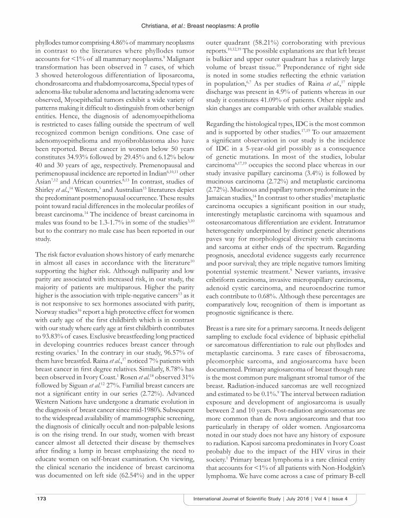
Christiana, et al.: Breast neoplasms: A profile
173 International Journal of Scientific Study | July 2016 | Vol 4 | Issue 4
phyllodes tumor comprising 4.86% of mammary neoplasms in contrast to the literatures where phyllodes tumor accounts for <1% of all mammary neoplasms.9 Malignant transformation has been observed in 7 cases, of which 3 showed heterologous differentiation of liposarcoma, chondrosarcoma and rhabdomyosarcoma, Special types of adenoma-like tubular adenoma and lactating adenoma were observed, Myoepithelial tumors exhibit a wide variety of patterns making it difficult to distinguish from other benign entities. Hence, the diagnosis of adenomyoepithelioma is restricted to cases falling outside the spectrum of well recognized common benign conditions. One case of adenomyoepithelioma and myofibroblastoma also have been reported. Breast cancer in women below 50 years constitutes 34.93% followed by 29.45% and 6.12% below 40 and 30 years of age, respectively. Premenopausal and perimenopausal incidence are reported in Indian6,10,11 other Asian7,12 and African countries.8,13 In contrast, studies of Shirley et al.,14 Western,3 and Australian15 literatures depict the predominant postmenopausal occurrence. These results point toward racial differences in the molecular profiles of breast carcinoma.14 The incidence of breast carcinoma in males was found to be 1.3-1.7% in some of the studies3,10 but to the contrary no male case has been reported in our study.
The risk factor evaluation shows history of early menarche in almost all cases in accordance with the literature10 supporting the higher risk. Although nulliparity and low parity are associated with increased risk, in our study, the majority of patients are multiparous. Higher the parity higher is the association with triple-negative cancers13 as it is not responsive to sex hormones associated with parity, Norway studies16 report a high protective effect for women with early age of the first childbirth which is in contrast with our study where early age at first childbirth contributes to 93.83% of cases. Exclusive breastfeeding long practiced in developing countries reduces breast cancer through resting ovaries.1 In the contrary in our study, 96.57% of them have breastfed. Raina et al.,17 noticed 7% patients with breast cancer in first degree relatives. Similarly, 8.78% has been observed in Ivory Coast.1 Rosen et al.18 observed 31% followed by Siguan et al.12 27%. Familial breast cancers are not a significant entity in our series (2.72%). Advanced Western Nations have undergone a dramatic evolution in the diagnosis of breast cancer since mid-1980’s. Subsequent to the widespread availability of mammographic screening, the diagnosis of clinically occult and non-palpable lesions is on the rising trend. In our study, women with breast cancer almost all detected their disease by themselves after finding a lump in breast emphasizing the need to educate women on self-breast examination. On viewing, the clinical scenario the incidence of breast carcinoma was documented on left side (62.54%) and in the upper
outer quadrant (58.21%) corroborating with previous reports.10,12,19 The possible explanations are that left breast is bulkier and upper outer quadrant has a relatively large volume of breast tissue.10 Preponderance of right side is noted in some studies reflecting the ethnic variation in population,6,7 As per studies of Raina et al.,17 nipple discharge was present in 4.9% of patients whereas in our study it constitutes 41.09% of patients. Other nipple and skin changes are comparable with other available studies.
Regarding the histological types, IDC is the most common and is supported by other studies.17,19 To our amazement a significant observation in our study is the incidence of IDC in a 5-year-old girl possibly as a consequence of genetic mutations. In most of the studies, lobular carcinoma6,17,19 occupies the second place whereas in our study invasive papillary carcinoma (3.4%) is followed by mucinous carcinoma (2.72%) and metaplastic carcinoma (2.72%). Mucinous and papillary tumors predominate in the Jamaican studies,14 In contrast to other studies3 metaplastic carcinoma occupies a significant position in our study, interestingly metaplastic carcinoma with squamous and osteosarcomatous differentiation are evident. Intratumor heterogeneity underpinned by distinct genetic alterations paves way for morphological diversity with carcinoma and sarcoma at either ends of the spectrum. Regarding prognosis, anecdotal evidence suggests early recurrence and poor survival; they are triple negative tumors limiting potential systemic treatment.9 Newer variants, invasive cribriform carcinoma, invasive micropapillary carcinoma, adenoid cystic carcinoma, and neuroendocrine tumor each contribute to 0.68%. Although these percentages are comparatively low, recognition of them is important as prognostic significance is there.
Breast is a rare site for a primary sarcoma. It needs deligent sampling to exclude focal evidence of biphasic epithelial or sarcomatous differentiation to rule out phyllodes and metaplastic carcinoma. 3 rare cases of fibrosacroma, pleomorphic sarcoma, and angiosarcoma have been documented. Primary angiosarcoma of breast though rare is the most common pure malignant stromal tumor of the breast. Radiation-induced sarcomas are well recognized and estimated to be 0.1%.9 The interval between radiation exposure and development of angiosarcoma is usually between 2 and 10 years. Post-radiation angiosarcomas are more common than de nova angiosarcoma and that too particularly in therapy of older women. Angiosarcoma noted in our study does not have any history of exposure to radiation. Kaposi sarcoma predominates in Ivory Coast probably due to the impact of the HIV virus in their society.1 Primary breast lymphoma is a rare clinical entity that accounts for <1% of all patients with Non-Hodgkin’s lymphoma. We have come across a case of primary B-cell

Christiana, et al.: Breast neoplasms: A profile
174International Journal of Scientific Study | July 2016 | Vol 4 | Issue 4
lymphoma of breast. The incidence of B-lymphoma is high in Sub-Saharan African countries probably an endemic Burkitt lymphoma; the spreading of tumor cells from an unknown site to the mammary vessels could be thought of.1 WHO stated that there are marked variations in the size ranging from under 10 mm to over 10 mm.19 Recording the measurements of size, T1 <2 cm constitutes 2.4% and 85.36% and 12.24%, respectively, for T2 and T3 in our study. African literatures13 report the presentation of tumor with stage III or IV in contrary to our study where stage II presentation is the most common.
Nodal positivity portrends the worst prognosis. The 10 years survival rate is 70-80% with node-negative carcinoma, 35-40% in carcinoma with 1-3 positive nodes and 10-15% in patients with more than 10 nodes.3 Nodal positivity was noted in 29.93% of cases in contrast to studies of Amr et al.11 which showed positivity in 61.7% of
cases. 21.76% of cases show <3 nodes positive and 8.16% have more than 3 nodes positivity.
Immunohistochemistry served as an adjunct tool for diagnosis of difficult cases. Smooth muscle actin shows positivity in a case of adenomyoepithelioma with suspicious malignancy (Figure 3a and b). Myoglobin positivity is observed in a case of malignant phyllodes with rhabdomyosarcomatous differentiation (Figure 4a and b) and a case of lymphoma was confirmed with CD 20 positivity (Figure 5a and b).
CONCLUSION
Breast cancer is the most common cancer in females posing a major health problem. There is a need for expedient evaluation of breast masses with an improved clinical and pathological characterization. The burden of breast cancer has to be brought down toward a declining trend. Hence, there is an urgent need to increase population screening program for early detection, training of women (breast self-examination), health worker, and medical practitioners. At present mammography serves as a screening tool but is less likely to be effective due to its insensitivity in high-density breast tissue at younger age. Moreover, most patients in our set up are unable to afford mammography due to their poor socio-economic background. Thus, there is a need for poverty alleviation programs, improved accessible and affordable health-care delivery services.
REFERENCES
1. Justin NK, Troh E, Kouakou EK, Doukoure B, Kouame AD, Abouna AD, et al. Epidemiology and histology aspects of breast cancers of women in ivory coast. J Cancer Ther 2012;3:782-6.
2. Dumitrescu RG, Cotarla I. Understanding breast cancer risk -- where do we stand in 2005? J Cell Mol Med 2005;9:208-21.
3. Mohapatra M, Satyanarayana S. Evaluation of clinico: Pathologic findings of breast carcinoma in a general hospital in Southern India. Indian J Cancer 2013;50:297-301.
4. Diomonde I, Horpock AF, Heroin P, Ette M, Dago A, Batesh F, et al. Evolution of cancer in ivory coast medical publication in ivory coast. Med Publication Ivory Coast 1988;75:81-4.
5. Chopra R. The Indian scene. J Clin Oncol 2001;19:106S-11S.6. Saha K, Raychandhuri G, Chattopadhyay BK. Clinico-pathological study
of breast carcinoma a prospective two year study in a tertiary care hospital. Clini Cancer Invest J 2013;2:34-40.
7. Ranabhat S, Subedi M, Bhandari A, Tiwari M, Maharjan S, Kshetri J, et al. Clinico - pathologic profile of women with palpable breast lumps in Chitwan medical college, Nepal. Int J Res Med Sci 2015;3:1611-6.
8. Kohler RE, Moses A, Krysiak R, Liomba NG, Gopal S. Pathologically confirmed breast cancer in Malawi: A descriptive study: Clinical profile of breast cancer. Malawi Med J 2015;27:10-2.
9. Fletcher CD. Diagnostic Histopathology of Tumours. 4th ed., Vol. 2. St. Louis: Churchill Livingstone; 2007.
10. Sandhu DS, Sandhu S, Karwasra RK, Marwah S. Profile of breast cancer patients at a tertiary care hospital in north India. Indian J Cancer 2010;47:16-22.
11. Amr SS, Sa’di AR, Ilahi F, Sheikh SS. The spectrum of breast diseases
Figure 3: (a) Adenomyoepithelioma shows tubules lined by epithelial and myoepithelial cells. (b) Smooth muscle actin
positivity
ba
Figure 4: (a) Malignant phyllodes with rhabdomyoblastic, (b) myoglobin positivity of differentiation rhabdomyoblasts
ba
Figure 5: (a) Lymphoma showing sheets of (b) CD 20 positivity monotonous cells interspersed with blasts
ba

Christiana, et al.: Breast neoplasms: A profile
175 International Journal of Scientific Study | July 2016 | Vol 4 | Issue 4
in Saudi Arab females: A 26 year pathological survey at Dhahran Health Center. Ann Saudi Med 1995;15:125-32.
12. Siguan SS, Hofilena JF, Ligo EL, Salutan SA, Baclig RY. Clinical profile and clinic - pathologic comparison of some clinical attributes of primary breast cancer admitted at an urban government medical center in the Philippines. Philipp J Surg Spec 2003;58:156-61.
13. Rahman GA, Olatoke SA, Agodirin SO, Adeniji KA. Socio-demographic and clinical profile of immuno-histochemically confirmed breast cancer in a resource limited country. Pan Afr Med J 2014;17:182.
14. Shirley SE, Sinclair PA, Stennett MA, Codrington G, Bhatt R, Escoffery CT. The pathology of breast cancer in Jamaica: The national public health laboratory study. West Indian Med J 2010;59:177-81.
15. Australian Institute of Health and Welfare 2014, Cancerin Australia an Overview 2014. Cancer Series No: 96 Cat No: CAN 88 Canberra: AIHW. Available from: http://www.bena. org.au/media/2089 Current statistics
bona brief summary of breast cancer statistics Jan - 15 - pdf. [Last assessed on 2015 Mar].
16. Fahro AE, Fateha BE, Al-Asheeri N, Al-Ekri SA. Breast cancer: Patient characteristics and survival analysis at salmaniya medical complex, Bahrain. East Mediterr Health J 1999;5:430-9.
17. Raina V, Bhutani M, Bedi R, Sharma A, Deo SV, Shukla NK, et al. Clinical features and prognostic factors of early breast cancer at a major cancer center in North India. Indian J Cancer 2005;42:40-5.
18. Rosen PP, Lesser ML, Senie RT, Kinne DW. Epidemiology of breast carcinoma III: Relationship of family history to tumor type. Cancer 1982;50:171-9.
19. Ellis IO, Schnitt SJ, Sastre-Garau X, Bussolati G, Tavassoli FA, Eusebi V, et al. Invasive breast carcinoma In: Tavassoli FA, Devilee P, editors. World Health Organization Classification of Tumours Pathology and Genetics of Tumorus of the Breast and Female Genital Organs. Lyon, Frnace: IARC Press; 2003. p. 13-59.
How to cite this article: Christiana SJ, Balakrishnan K, Hemalatha G, Uma Maheswari K. Clinical and Histomorphological Profile of Breast Neoplasms. Int J Sci Stud 2016;4(4):170-175.
Source of Support: Nil, Conflict of Interest: None declared.
176International Journal of Scientific Study | July 2016 | Vol 4 | Issue 4
Sudden Death Causes: An Autopsy Study in AdultsB Shanthi1, S Saravanan2, R Siva Elangovan1, V Sudha1
1Assistant Professor Department of Pathology, KAPV Government Medical College, Tiruchirappalli, Tamil Nadu, India, 2Assistant Professor, Department of Forensic Medicine, KAPV Government Medical College, Tiruchirappalli, Tamil Nadu, India
the presence of witness or died while asleep, their death was classified as SD.
The incidence of sudden cardiac death has been steadily increasing all over the world. When SD occurs in adults and elderly persons, coronary atherosclerosis is the usual cause. These diseases are frequently concealed and discovered with surprise only at post mortem using through macroscopic and microscopic examination.
This present study was done for 50 cases with the cause of SD with autopsy examination, with coordinance with Pathology Department and Forensic Medicine at KAPV Government Medical College, Tiruchirappalli, Tamil Nadu, India.
MATERIALS AND METHODS
This present study is based on autopsy observation carried out in 50 cases and analyzed for the cause of SD. The cases were chosen as per the definition of SD and autopsied. The heart was examined grossly and microscopically to observe various histomorphological changes and findings were correlated.
INTRODUCTION
Sudden death (SD) is defined as an unexpected natural death due to cardiac cause within a short period (usually within 1 h) with or without onset of symptoms and without any prior condition that would appear fatal.1 As the definition of SD varies, it is difficult task to compare one set of published data with another death from natural causes, in which interval between onset of signs and symptoms and death was not more than 24 h were regarded as SD.2-4 It is by definition natural and it excludes all deaths due to poison, trauma. Various workers in this field have given different definitions. Although SD is a relative concept, this concept is currently described as unexpected death occurring within 1 h of new symptoms.5 If the patient died instantaneously in
Original Article
AbstractBackground and Objectives: Incidence of sudden death (SD) is quiet frequent to determine the cause of SD and observe the morphological changes in the heart after death.
Materials and Methods: A study for 50 cases was carried out in KAPV Government Medical College, Tiruchirappalli, in Pathology Department, in coordination with forensic medicine department from October 2015 to May 2016. A detailed autopsy finding with histopathological examination was done to analyze the cause of SD. We studied only heart specimen to explore the cause of SD.
Results: All cases revealed to between the age group 31 and 75 years with male preponderance common; out of 50 cases, 23 cases showed features of lesions; about 8 cases showed severe atheromatous change in aorta followed by 4 cases with early myocardial infarction (MI) and 4 cases with MI and complications such as atheromatous aorta with calcification, thrombus both old and fresh and a case myocarditis and medial hypertrophy of left ventricle. Rest of the cases the cause of death was undetermined.
Conclusion: The major causes of death were due to atherosclerosis, whereas MI and acute coronary events (thrombus) observed in 4% of cases of SD.
Key words: Atheroma, Autopsy, Heart, Morphology, Sudden death
Access this article online
www.ijss-sn.com
Month of Submission : 05-2016 Month of Peer Review : 06-2016 Month of Acceptance : 07-2016 Month of Publishing : 07-2016
Corresponding Author: Dr. B Shanthi, Department of Pathology, KAPV Government Medical College, Trichy, Tamilnadu, India. E-mail: [email protected]
Print ISSN: 2321-6379Online ISSN: 2321-595X
DOI: 10.17354/ijss/2016/401

Shanthi, et al.: Autopsy Study in Sudden Death
177 International Journal of Scientific Study | July 2016 | Vol 4 | Issue 4
The randomized study on 50 cases was submitted for postmortem analysis; formalin fixed heart was inspected; sections were taken from right and left ventricular wall, stump of aorta and coronaries; multiple sections were taken at 4-5 mm interval; additional sections were made if needed. The tissue was processed, and paraffin sections were made at 4 mm thickness, stained with routine H and E staining.6
RESULTS
Out of 50 cases of SD, only 23 cases showed heart changes while rest 27 cases no known cause was made out as in Figure 1.
Our study was done for 50 cases with age range 25-80 years. The most common age group in our study with heart changes was in 51-60 years, and most predominance was in males as in Table 1.
The number of cases involved showed 21 male and 2 female cases; male: female ratio was 10.5:1.
The histomorphological changes were observed most common being solely atheromatous change in aorta (atherosclerosis) in 8 cases and next common being early changes in coronaries and aorta 4 cases; other complications such as calcification of plaques were also seen; thrombus formation was noted in 4 cases, of which one had an old thrombus formation and another with infected ball wall valve thrombus in aortic orifice; and rest one was reported to have focal myocardial infarction (MI) with atheromatous aorta and old thrombus formation. One case was noted with left ventricular hypertrophy with focal calcification of coronaries and one more case with medial wall hypertrophy of coronaries and one with myocarditis.
DISCUSSION
The term SD has no agreed universal definition; Goldstein proposed that SD should be defined as witnessed death within 1 h of the onset of symptoms, emphasizing the need for a uniform definition but saying that the definition of
SD may be expressed in different terms depending on the nature and scope of the investigation.7
In our study, out of the total 50 cases of SD, 47 cases (94%) were male cases and 3 (6%) cases were females as shown Figure 2; male:female ratio was 15.6:1 for SD in general. This finding was consistent with study of Sarkojia et al. (82%),8 Thomas et al. (73.9%),8 Nordrum et al. (79.67%),8 and Singh et al. (94.5%).8
The main aim of this study was to read out the cause of SD and the contribution of cardio vascular cause being the most common one. Data from post mortem of SD parallel the clinical observation on the prevalence of coronary disease, as the major structural etiological factor. More than 80% of cases have pathological findings of coronary heart disease; pathologic descriptions include a combination of long-standing atherosclerosis of the coronary arteries, and acute active coronary lesions, which include a combination of fissured or ruptured plaques, platelet aggregates, hemorrhage, and thrombosis.9
Although there are numerous causes of SD, cardiovascular causes are the principle cause among SD in the present
Table 1: Age and sex of occurrence in 23/50 casesAge group (years) Male Female25-30 - -31-40 4 -41-50 5 -51-60 8 161-70 3 -71-80 1 1Total 21 2
46%
�4%
disease heart
��r�a� heart
Figure 1: Autopsy changes in heart 50 cases
Figure 2: Age- and sex-wise distribution among sudden death cases (50 cases)

Shanthi, et al.: Autopsy Study in Sudden Death
178International Journal of Scientific Study | July 2016 | Vol 4 | Issue 4
study. In our study, autopsy of 50 cases SD revealed heart changes in 23 cases (46%) with atherosclerosis (35%) of aorta followed by MI with early changes (18%) and MI with complication (18%), thrombus in coronaries (18%), coronary medial wall hypertrophy, left ventricular wall hypertrophy, and myocarditis each (4%) as shown in Figure 3, and the Figures 6-8 depicting the histopathological and microscopic changes in H and E.
Kuller et al.10 studied showed that SDs are due to atherosclerotic disease, which correlated with our study.
A study by Zanjad and Nanadkar showed SD due to cardiac causes (49.55%); Reddy and Nandy8 stated that the most of the death were due to cardiovascular causes about 45-50%. Similar findings were seen in study of Kuller et al. (49.50%),8 Siboni et al. (46.20%),8 and Luke et al. (38%).8
Our study shows that incidence of male was high (21 cases male and 2 cases female) and average ratio of male: female was 10.5:1 which proved to be coherent with studies conducted by Farb et al.10 that SD was more predominant in male, and Framingham10 studies regarding male: female ratio.
The average age group of incidence was between 50 and 59 years in one study, and study series of Pentilla10 also showed age group 55-66 years which was compatible with our study and they were the average age group was 50-60 years as shown in Figure 7.
The risk of SD increases dramatically beyond age 35 years and greatest rate of increase is between 40 and 65 years. Among older than 30 years of age, with structural heart
Figure 3: Histomorphological changes in heart (23 cases)
Figure 5: Atheromatous plaque aorta (H and E, ×100)
Figure 6: Coronary vessel thrombus (H and E, ×100)
Figure 7: Age- and sex-wise incidence showing diseased heart changes in 23 cases (Total cases: 23, male - 21, female - 2)
Figure 4: (a and b) Myocardial infarct changes in cardiac myocytes (H and E, ×100)
a b

Shanthi, et al.: Autopsy Study in Sudden Death
179 International Journal of Scientific Study | July 2016 | Vol 4 | Issue 4
disease and markers of high risk for cardiac arrest, the event may exceed 25% per year and age-related risk
The non-cardiac causes for SD have been reported in the central nervous system as intracerebral hemorrhage due to ruptured berry aneurysm, primary intracerebral hemorrhage, and epilepsy. The respiratory causes could be pneumonia, pulmonary embolism, chronic bronchitis, pulmonary hypertension, and acute asthma and rest of the causes elicited to be acute alcoholic poisoning, alcoholic fatty liver.11 A variety of sedatives, tranquilizers, and mild analgesic drugs do contribute to causes of SD without cardiac change.11 Cigarette smoking also increases the risk for SD.
The cause of SD in rest of 27 cases (54%) are of non-cardiac origin (Figure 8), so SD is not a just heart attack, and there remains a group, in which no cause can ever be found to explain death or undetermined attenuates.
CONCLUSION
Ischemic heart disease may be the leading cause of SD, with coronary atherosclerosis being the most significant pathogenic mechanism. This factor is being emphasized by many authors, and in our study, the cases have proven to that atherosclerosis was the major cause of sudden cardiac death, and we received only heart specimen pertained to this study.
SD is source of concern and detail post mortem examination is mandatory to ascertain the cause. The study helped us to find out cardiac causes but the non-cardiac causes were not definite in our study. So, more emphasis has to be made by obtaining complete history data, autopsy details, and histopathological examination of other organs would help out to find the cause. No one putative explanation for SD could be made.
Sudden cardiac death accounts for approximately over half of the total death due to the cardiovascular cause, to prevent this SD in the population.
Certain strategies have to be followed to achieve major population impact; effective prevention of underlining disease and development of new epidemiologic and clinical probes, for better individual risk prediction and specific high-risk groups in the population in needed.
REFERENCES
1. Nisha M, Bhawna S, Sumiti G, Amrita D, Sunita S, Rajeev S. Histomorphological spectrum of various cardiac changes in sudden death: An autopsy study. Iran J Pathol 2011;6:179-86.
2. Kuller L, Cooper M, Perper J. Epidemiology of sudden death. Arch Intern Med 1972;129:714-9.
3. Tunstall-Pedoe H, Kuulasmaa K, Amouyel P, Arveiler D, Rajakangas AM, Pajak A. Myocardial infarction and coronary death in the World Health Organization MONICA Project: Registration procedures, event rated in 38 population from 21 countries in four continents. Circulation 1994;90:583-612.
4. Virmani R, Burke AP, Farb A. Sudden cardiac death. Cardiovasc Pathol 2001;10:275-82.
5. Torp-Pedersen C, Køber L, Elming H, Burchart H. Classification of sudden and arrhythmic death. Pacing Clin Electrophysiol 1997;20:2545-52.
6. Bancroft J, Gamble M. Theory and Practice. 3rd ed. New Jersey: Towata; 2004.
7. Goldstein S. The necessity of a uniform definition of sudden coronary death: Witnessed death within 1 hour of the onset of acute symptoms. Am Heart J 1982;103:156-9.
8. Zanjad NP, Nanadkar SD. Study of sudden unexpected deaths in medico-legal autopsies. JIAFM 2006;28:27-30.
9. Myerburg RJ, Castellanos A. Cardiovascular collapse, cardiac arrest, and sudden cardiac death. Harrison-Internal Medicine. 19th ed., Vol. I. Ch. 39. New York: McGraw Hill Education; 2012.
10. Rao D, Sood D, Pathak P, Dongre SD. A cause of sudden cardiac deaths on autopsy findings; A four-year report. Emerg (Tehran) 2014;2:12-7.
11. Thomas AC, Knapman PA, Krikler DM, Davies MJ. Community study of the causes of “natural” sudden death. BMJ 1988;297:1453-6.
Figure 8: Age‑wise and sex with normal heart findings (27 cases) in autopsy
How to cite this article: Shanthi B, Saravanan S, Elangovan RS, Sudha V. Sudden Death Causes: An Autopsy Study in Adults. Int J Sci Stud 2016;4(4):176-179.
Source of Support: Nil, Conflict of Interest: None declared.

180International Journal of Scientific Study | July 2016 | Vol 4 | Issue 4
Comparative Study of Coagulation Profile in Mild Pre-eclampsia, Severe Pre-eclampsia, and EclampsiaC Vijaya Lakshmi
Tutor, Department of Gynecology, Government Medical College, Anantapur, Andhra Pradesh, India
edema. Eclampsia is a severe form characterized by convulsions or coma.
PIH may also result in a variety of hematological aberrations.2 Thrombocytopenia is the most common hematological abnormality found in pre-eclampsia and eclampsia.3 It is a strong indicator of severity of PIH.2 Other coagulation abnormalities such as prothrombin time (PT), activated partial thromboplastin time (aPTT), fibronectin time, and antithrombin III level are more sensitive.1
Alteration of coagulation factors increases the risk of bleeding complications in pre-eclampsia and eclampsia. Hemorrhages are a major problem where it is the main cause of maternal mortality, which usually occur during operative delivery or regional anesthesia procedure.
To reduce the maternal morbidity and mortality need of accurate and rapid biochemical tests to detect the
INTRODUCTION
Pregnancy-induced hypertension (PIH) is hypertensive disorders usually appear after the 20th week of gestation, which often results in multiorgan failure.1 PIH plays a major role in perinatal mortality and morbidity. Globally, about 5-10% of all pregnancies are complicated by hypertensive disorders.
PIH can present in the form of pre-eclampsia or eclampsia. Pre-eclampsia is a multisystem hypertensive disorder characterized by triad of hypertension, proteinuria, and
Original Article
AbstractIntroduction: Alteration of coagulation factors increases the risk of bleeding complications in pre-eclampsia and eclampsia. To reduce the maternal morbidity and mortality need of accurate and rapid biochemical tests to detect the complications of pre-eclampsia and eclampsia. The aim of the present study is to detect the severity of hypertensive disorders during pregnancy and to compare the coagulation profile in mild pre-eclampsia, severe pre-eclampsia, and eclampsia patients.
Materials and Methods: A total of 200 pregnancy-induced hypertension (PIH) patients were selected for this study, among which 100 were presented with mild pre-eclampsia, 70 were severe pre-eclampsia, and 30 were eclampsia. Coagulation profile was investigated and analyzed.
Results: Platelet count was reduced in pre-eclampsia and eclampsia. Prothrombin time, activated partial thromboplastin time, bleeding time, and clotting time were prolonged in severe eclampsia and eclampsia. Statistical significance of coagulation profile was observed between mild pre-eclampsia and eclampsia, mild pre-eclampsia and severe pre-eclampsia.
Conclusion: Estimates of the biochemical parameters play an important role in the diagnosis of PIH and evaluation of risk factors, early detection, and effective antenatal services, prompt and proper management will decrease the maternofetal mortality, morbidity, and also perinatal mortality.
Key words: Coagulation profile, Eclampsia, Pre-eclampsia
Access this article online
www.ijss-sn.com
Month of Submission : 05-2016 Month of Peer Review : 06-2016 Month of Acceptance : 07-2016 Month of Publishing : 07-2016
Corresponding Author: Dr. C Vijaya Lakshmi, Department of Gynecology, Government Medical College, Anantapur, India. E-mail: [email protected]
Print ISSN: 2321-6379Online ISSN: 2321-595X
DOI: 10.17354/ijss/2016/402

Lakshmi: Coagulation Profile in Mild Pre-eclampsia, Severe Pre-eclampsia, and Eclampsia
181 International Journal of Scientific Study | July 2016 | Vol 4 | Issue 4
complications of pre-eclampsia and eclampsia including HELLP syndrome. Detecting the severity of PIH disorders, help in the better management of patients. Hence, the present study has undertaken to correlate coagulation parameters with the severity of PIH, which helped us in the early management of PIH before it worsens.
The aim of the present study is to detect the severity of hypertensive disorders during pregnancy and to compare the coagulation profile in mild pre-eclampsia, severe pre-eclampsia, and eclampsia patients.
MATERIALS AND METHODS
The study has done on pregnant women with PIH disorders for 1½ years at the Department of OBG, Government General Hospital, Anantapur, from April 2014 to December 2015. This prospective study has started after taking informed consent from all the studied patients. Ethical Committee has approved to do this study.
Inclusion CriteriaPregnant women with both mild and severe pre-eclampsia and eclampsia in the age group of 16-35 years.
Exclusion CriteriaPregnant women in labor or with abruptio placentae or with established disseminated intravascular coagulation (DIC) or anticoagulation therapy were excluded in this study.
A total of 200 PIH patients were selected for this study, among which 100 were presented with mild pre-eclampsia, 70 were severe pre-eclampsia, and 30 were eclampsia. Details of the study were explained to all PIH women. Data regarding age, sex, number of pregnancies, socioeconomic status, presenting complaints, significant past, and family history were noted. General, systemic, and obstetrical examination was done.
Patients were advised for blood investigations including routine blood and urine examination, renal and liver function tests, blood sugar, lipid profile, and coagulation profile. Coagulation parameters were assessed using following methods:1. Platelet count - using automated hematology analyzer2. PT and aPTT - using a coagulometer3. Bleeding time (BT) - by Dukes method4. Clotting time (CT) - by Wright’s capillary tube method.
All the data regarding coagulation profile were entered into excel sheet and analyzed.
RESULTS
A total of 200 patients with PIH disorders were included in the study. Peak incidence of PIH disorders was seen in 26-30 years followed by 21-25 years (Figure 1). 90 (45%) patients were in the age group of 26-30 years followed by 78 (39%) were in the age group of 21-25 years.
Out of 200 patients selected for doing this study, 100 were mild pre-eclampsia, 70 were severe pre-eclampsia, and 30 were severe pre-eclampsia. All the three PIH disorders were most commonly observed in the age group of 26-30 years followed by 21-25 years, 31-35 years, and 16-20 years (Table 1).
Mean of coagulation parameters was estimated and tabulated in Table 2. Platelet count reduced with increase in severity (mild pre-eclampsia to eclampsia). PT, aPTT, BT, and CT were prolonged in severe eclampsia and eclampsia.
Statistical significance of coagulation profile was observed between mild pre-eclampsia and eclampsia, mild pre-eclampsia, and severe pre-eclampsia (Table 3).
Prolonged PT and aPTT were observed in severe pre-eclampsia when compared to eclampsia and mild pre-eclampsia (Table 4). PT prolongation in between mild pre-eclampsia and severe pre-eclampsia showed statistically significant (P = 0.0001).
DISCUSSION
Pre-eclampsia is an idiopathic multisystem disorder specific to human pregnancy and puerperium. Hematological abnormalities such as thrombocytopenia and decrease in some plasma clotting factors may develop in pre-eclamptic women. Subtle changes suggesting DIC is one of the serious outcomes of pre-eclampsia.
During pregnancy, there is increase in the concentration of clotting factor II, V, VII, VIII, IX, X, XII. Plasma
Figure 1: Age-wise distribution of cases
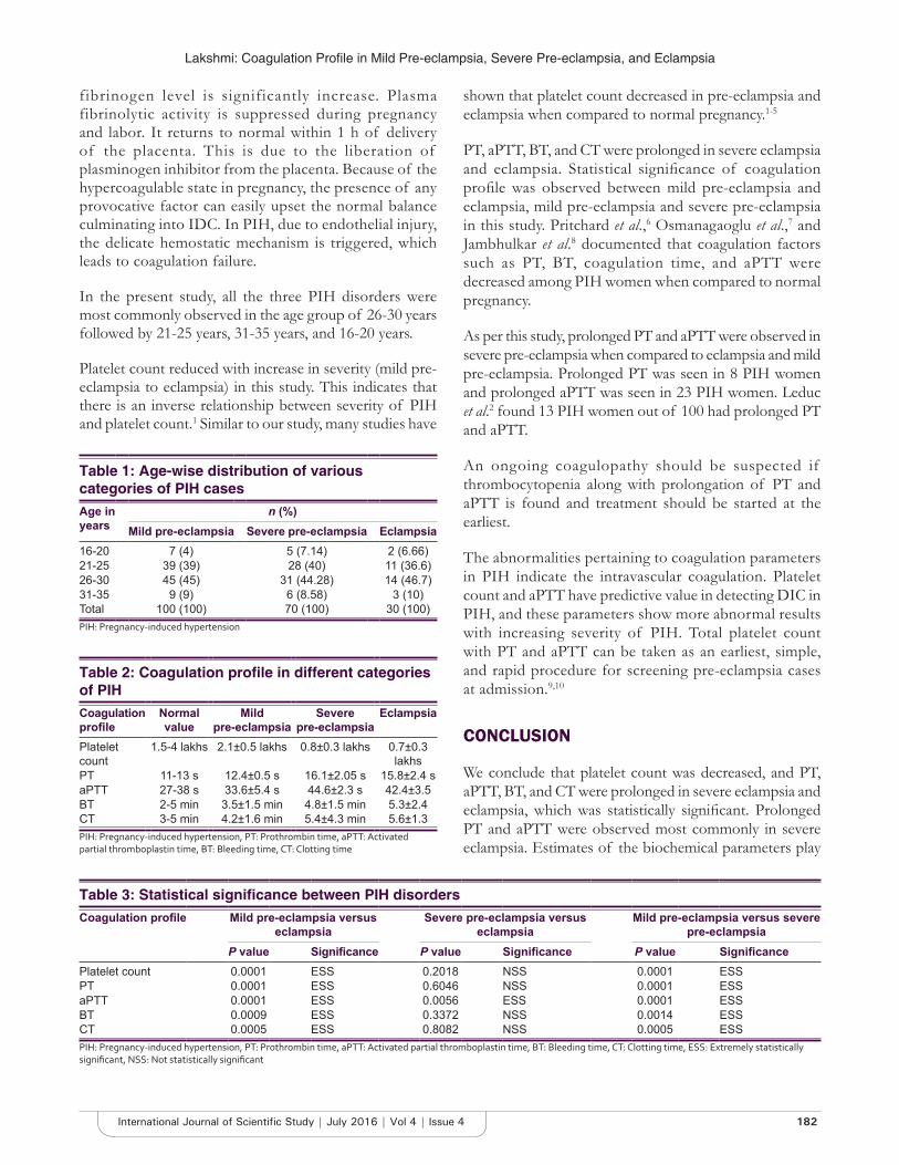
Lakshmi: Coagulation Profile in Mild Pre-eclampsia, Severe Pre-eclampsia, and Eclampsia
182International Journal of Scientific Study | July 2016 | Vol 4 | Issue 4
fibrinogen level is significantly increase. Plasma fibrinolytic activity is suppressed during pregnancy and labor. It returns to normal within 1 h of delivery of the placenta. This is due to the liberation of plasminogen inhibitor from the placenta. Because of the hypercoagulable state in pregnancy, the presence of any provocative factor can easily upset the normal balance culminating into IDC. In PIH, due to endothelial injury, the delicate hemostatic mechanism is triggered, which leads to coagulation failure.
In the present study, all the three PIH disorders were most commonly observed in the age group of 26-30 years followed by 21-25 years, 31-35 years, and 16-20 years.
Platelet count reduced with increase in severity (mild pre-eclampsia to eclampsia) in this study. This indicates that there is an inverse relationship between severity of PIH and platelet count.1 Similar to our study, many studies have
shown that platelet count decreased in pre-eclampsia and eclampsia when compared to normal pregnancy.1-5
PT, aPTT, BT, and CT were prolonged in severe eclampsia and eclampsia. Statistical significance of coagulation profile was observed between mild pre-eclampsia and eclampsia, mild pre-eclampsia and severe pre-eclampsia in this study. Pritchard et al.,6 Osmanagaoglu et al.,7 and Jambhulkar et al.8 documented that coagulation factors such as PT, BT, coagulation time, and aPTT were decreased among PIH women when compared to normal pregnancy.
As per this study, prolonged PT and aPTT were observed in severe pre-eclampsia when compared to eclampsia and mild pre-eclampsia. Prolonged PT was seen in 8 PIH women and prolonged aPTT was seen in 23 PIH women. Leduc et al.2 found 13 PIH women out of 100 had prolonged PT and aPTT.
An ongoing coagulopathy should be suspected if thrombocytopenia along with prolongation of PT and aPTT is found and treatment should be started at the earliest.
The abnormalities pertaining to coagulation parameters in PIH indicate the intravascular coagulation. Platelet count and aPTT have predictive value in detecting DIC in PIH, and these parameters show more abnormal results with increasing severity of PIH. Total platelet count with PT and aPTT can be taken as an earliest, simple, and rapid procedure for screening pre-eclampsia cases at admission.9,10
CONCLUSION
We conclude that platelet count was decreased, and PT, aPTT, BT, and CT were prolonged in severe eclampsia and eclampsia, which was statistically significant. Prolonged PT and aPTT were observed most commonly in severe eclampsia. Estimates of the biochemical parameters play
Table 1: Age-wise distribution of various categories of PIH casesAge in years
n (%)Mild pre-eclampsia Severe pre-eclampsia Eclampsia
16-20 7 (4) 5 (7.14) 2 (6.66)21-25 39 (39) 28 (40) 11 (36.6)26-30 45 (45) 31 (44.28) 14 (46.7)31-35 9 (9) 6 (8.58) 3 (10)Total 100 (100) 70 (100) 30 (100)PIH: Pregnancy‑induced hypertension
Table 2: Coagulation profile in different categories of PIHCoagulation profile
Normal value
Mild pre-eclampsia
Severe pre-eclampsia
Eclampsia
Platelet count
1.5-4 lakhs 2.1±0.5 lakhs 0.8±0.3 lakhs 0.7±0.3 lakhs
PT 11-13 s 12.4±0.5 s 16.1±2.05 s 15.8±2.4 saPTT 27-38 s 33.6±5.4 s 44.6±2.3 s 42.4±3.5BT 2-5 min 3.5±1.5 min 4.8±1.5 min 5.3±2.4CT 3-5 min 4.2±1.6 min 5.4±4.3 min 5.6±1.3PIH: Pregnancy‑induced hypertension, PT: Prothrombin time, aPTT: Activated partial thromboplastin time, BT: Bleeding time, CT: Clotting time
Table 3: Statistical significance between PIH disordersCoagulation profile Mild pre-eclampsia versus
eclampsiaSevere pre-eclampsia versus
eclampsiaMild pre-eclampsia versus severe
pre-eclampsiaP value Significance P value Significance P value Significance
Platelet count 0.0001 ESS 0.2018 NSS 0.0001 ESSPT 0.0001 ESS 0.6046 NSS 0.0001 ESSaPTT 0.0001 ESS 0.0056 ESS 0.0001 ESSBT 0.0009 ESS 0.3372 NSS 0.0014 ESSCT 0.0005 ESS 0.8082 NSS 0.0005 ESSPIH: Pregnancy‑induced hypertension, PT: Prothrombin time, aPTT: Activated partial thromboplastin time, BT: Bleeding time, CT: Clotting time, ESS: Extremely statistically significant, NSS: Not statistically significant
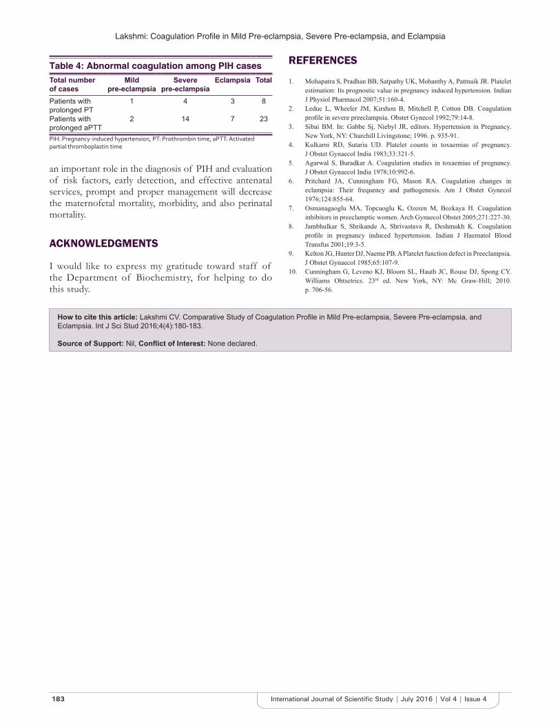
Lakshmi: Coagulation Profile in Mild Pre-eclampsia, Severe Pre-eclampsia, and Eclampsia
183 International Journal of Scientific Study | July 2016 | Vol 4 | Issue 4
an important role in the diagnosis of PIH and evaluation of risk factors, early detection, and effective antenatal services, prompt and proper management will decrease the maternofetal mortality, morbidity, and also perinatal mortality.
ACKNOWLEDGMENTS
I would like to express my gratitude toward staff of the Department of Biochemistry, for helping to do this study.
REFERENCES
1. Mohapatra S, Pradhan BB, Satpathy UK, Mohanthy A, Pattnaik JR. Platelet estimation: Its prognostic value in pregnancy induced hypertension. Indian J Physiol Pharmacol 2007;51:160-4.
2. Leduc L, Wheeler JM, Kirshon B, Mitchell P, Cotton DB. Coagulation profile in severe preeclampsia. Obstet Gynecol 1992;79:14-8.
3. Sibai BM. In: Gabbe Sj, Niebyl JR, editors. Hypertension in Pregnancy. New York, NY: Churchill Livingstone; 1996. p. 935-91.
4. Kulkarni RD, Sutaria UD. Platelet counts in toxaemias of pregnancy. J Obstet Gynaecol India 1983;33:321-5.
5. Agarwal S, Buradkar A. Coagulation studies in toxaemias of pregnancy. J Obstet Gynaecol India 1978;10:992-6.
6. Pritchard JA, Cunningham FG, Mason RA. Coagulation changes in eclampsia: Their frequency and pathogenesis. Am J Obstet Gynecol 1976;124:855-64.
7. Osmanagaoglu MA, Topcuoglu K, Ozeren M, Bozkaya H. Coagulation inhibitors in preeclamptic women. Arch Gynaecol Obstet 2005;271:227-30.
8. Jambhulkar S, Shrikande A, Shrivastava R, Deshmukh K. Coagulation profile in pregnancy induced hypertension. Indian J Haematol Blood Transfus 2001;19:3-5.
9. Kelton JG, Hunter DJ, Naeme PB. A Platelet function defect in Preeclampsia. J Obstet Gynaecol 1985;65:107-9.
10. Cunningham G, Leveno KJ, Bloom SL, Hauth JC, Rouse DJ, Spong CY. Williams Obtsetrics. 23rd ed. New York, NY: Mc Graw-Hill; 2010. p. 706-56.
Table 4: Abnormal coagulation among PIH casesTotal number of cases
Mild pre-eclampsia
Severe pre-eclampsia
Eclampsia Total
Patients with prolonged PT
1 4 3 8
Patients with prolonged aPTT
2 14 7 23
PIH: Pregnancy‑induced hypertension, PT: Prothrombin time, aPTT: Activated partial thromboplastin time
How to cite this article: Lakshmi CV. Comparative Study of Coagulation Profile in Mild Pre-eclampsia, Severe Pre-eclampsia, and Eclampsia. Int J Sci Stud 2016;4(4):180-183.
Source of Support: Nil, Conflict of Interest: None declared.

184International Journal of Scientific Study | July 2016 | Vol 4 | Issue 4
Comparison between Perivascular and Perineural Ultrasound-guided Axillary Nerve Block for Forearm Surgeries: A Randomized Controlled StudyM Balamurugan1, M Shanmugasundaram1, R Priyadharshini2
1Assistant Professor, Institute of Anaesthesiology, Government Rajaji Hospital, Madurai Medical College, Madurai, Tamil Nadu, India, 2Post Graduate, Institute of Anaesthesiology, Government Rajaji Hospital, Madurai Medical College, Madurai, Tamil Nadu, India
triangle, divisions behind the clavicle, and cords at the outer border of the first rib enter the axilla with the axillary artery.
Use of image guidance for locating the peripheral nerve and neurofascial plane improves the success of this block with fewer complications. There are various techniques of imaging for nerve blocks. Among that ultrasound technique seems to be most reliable for nerve blocks. Ultrasound use was first described, in 1978, for supraclavicular brachial plexus block with the help of Doppler to detect blood flow. The nerve structures were first identified by Kapral, in 1994, visualized brachial plexus under ultrasound guidance. Ultrasound technology improved dramatically in last few years and leads to better understanding of sonoanatomy. Ultrasound-guided technology provides a good anatomy
INTRODUCTION
Brachial plexus block has evolved in procedures for upper limb surgeries when William Halsted performed it for the first time in 1884. Brachial plexus is formed by anterior primary rami of C5 to C8 and T1 nerves which emerge from intervertebral foramina. They form roots between scalene muscles, trunks beneath the floor of posterior
Original Article
AbstractIntroduction: Brachial plexus block has evolved in procedures for upper limb surgeries when William Halsted performed it for the first time in 1884. Brachial plexus is formed by anterior primary rami of C5 to C8 and T1 nerves which emerge from intervertebral foramina.
Objective: The objective of this study was to compare ultrasound-guided perivascular (PV) and perineural (PN) axillary nerve block.
Materials and Methods: This prospective randomized comparative study was done in 50 patients posted for forearm surgery. This study was done to compare two techniques axillary block under ultrasound guidance. They were divided into two groups, namely, PN and PV group. In PN group, the median, ulnar, and radial nerves are blocked separately. In PV group, the local anesthetic is injected dorsal to the axillary artery. Intraoperatively, the imaging time, needling time, performance time, number of needle passes, vascular puncture, and onset of sensory and motor blockade were observed and recorded.
Results: According to this study, both PN and PV techniques have similar success rate. However, the performance time and number of needle passes were less in PV technique. Hence, the procedure-related pain was less in PV group.
Conclusion: The PV technique provides a simple alternative to axillary block under ultrasound guidance.
Key words: Axillary nerve block, Perivascular, Perineural, Ultrasound-guided
Access this article online
www.ijss-sn.com
Month of Submission :05-2016Month of Peer Review :06-2016Month of Acceptance :07-2016Month of Publishing :07-2016
Corresponding Author: Dr. M Balamurugan, Institute of Anaesthesiology, Government Rajaji Hospital, Madurai Medical College, Madurai, Tamil Nadu, India. Phone: 91-9443135904. E-mail: [email protected]
Print ISSN: 2321-6379Online ISSN: 2321-595X
DOI: 10.17354/ijss/2016/403

Balamurugan, et al.: Comparison of Two Approaches of Ultrasound-guided Axillary Nerve Block
185 International Journal of Scientific Study | July 2016 | Vol 4 | Issue 4
of the area of interest in real time.1 This imaging helps to visualize neural structures such as nerve plexus and peripheral nerves and the surrounding structures such as blood vessels and pleura help to pass the needle toward the target nerves or facial plane, and visualize the extent of local anesthetic spread.2 Ultrasound guidance allows visualization of the penetrating needle and nerve as well as a reasonable estimate of the spread of local anesthetic drugs.3 In this study, we compared ultrasonography-guided perineural (PN) and perivascular (PV) axillary brachial plexus block (AXB) using compare performance time, number of needle passes, complications, onset time, success rate.
MATERIALS AND METHODS
This prospective randomized study was done to compare the PN and PV AXB under ultrasound guidance. This study was approved by the Ethical Committee of Hospital, and written informed consent was obtained from parents of the each patient. 50 patients of either sex undergoing surgery of forearm, wrist, and hand. 25 patients in each group were randomly allocated into two groups, namely, PV and PN group. The patients on either sex with the age group of 18-65 years with American Society of Anesthesiologists (ASA) physical status I, II, and III posted for elective upper limb surgeries were included in the study. This study was done in patients posted for arteriovenous fistula surgery. Patients with history of bleeding disorders, patients on anticoagulant therapy, with local infection, with documented neuromuscular disorders, with respiratory compromise, with known H/O allergy to local anesthetic drugs, and H/O psychiatric illness were excluded from the study. The detailed pre-anesthetic check-up was done on all patients, and relevant hematological, biochemical, and radiological investigations were carried out for all patients as per surgical requirements. Anesthesia equipment was checked, and resuscitative equipment and drugs were kept ready. In the operation theater, monitors such as pulse oximetry and non-invasive blood pressure were attached. Baseline values of parameters such as mean arterial pressure (MAP), pulse rate, and oxygen saturation (SPO2) were recorded.
Sterile standard anesthesia tray prepared with the sterile towels, 4 × 4 gauze pack, local anesthetics mixture 12 ml of 0.5% bupivacaine, 6 ml of 2% lignocaine and 6 ml of sterile water, sterile gloves, one 2 cc 25 gauge needle for skin infiltration, one 23 gauge needle and 10 ml syringe for local anesthetic injection. Esaote ultrasound machine with probe frequency range of 10-15 MHz was used for the procedure.
ProcedurePatients were positioned supine with the shoulder abducted at 90° and the elbow flexed. The ultrasound probe was applied in a sterile fashion in the axilla. Betadine was used as a medium of interface to view the nerve structures. A high-frequency linear array probe of 10 MHZ frequencies was used for axillary block. Short axis views with sufficient compression to collapse the axillary vein was used to visualize neurovascular bundle. After obtaining a satisfactory image, a skin wheel was raised. In both groups, in-plane technique was used, in which the entire shaft and tip of needle were visible. In PV group, the needle is advanced until its tip is reached dorsal to axillary artery and the 24 ml of the local anesthetic mixture is deposited dorsal to the artery. A silhouette sign is sought to confirm proximity of needle tip to the artery. Silhouette sign is defined as blurring of arterial wall due to contiguity of blood and local anesthetic. In PN group, the median, ulnar, and radial nerves are blocked separately with 8 ml of local anesthetic mixture each.
The following parameters were assessed:1. Imaging time: Defined as the time required to visualize
axillary artery in PV group. In PN group, imaging time is defined as time needed to visualize all three nerves
2. Needling time: Time interval between the start of skin wheel and the end of local anesthetic injection
3. Performance time: Defined as the sum of imaging time and needling time
4. Number of needle passes5. Vascular puncture6. Time of sensory and motor blockade.
Motor and sensory blockade were assessed every 5 min up to 30 min. Sensory blockade of median, ulnar, and radial nerves was graded according to a 3 point scale using cold test (0 = No block; 1 = Analgesia - the patient can feel touch, not cold 2 = Anesthesia - the patient cannot feel touch). Sensory blockade was assessed in palmar aspect of the thumb for median nerve, in lateral aspect of the dorsum of the hand for radial nerve, and in palmar aspect of the fifth finger for ulnar nerve. Motor blockade of median, ulnar, and radial nerves was graded using 3 point scale (0 = no block; 1 = paresis; 2 = paralysis). Motor blockade was assessed by thumb abduction for radial nerve, thumb opposition for median nerve, and thumb adduction for ulnar nerve.
Sedation and rescue analgesics were not given during the procedure. If there was an inadequate motor or sensory block, the surgical procedure was completed with local infiltration. Intraoperatively, heart rate, MAP, and SPO2 were recorded throughout the operation and were monitored at an interval of 5 min.
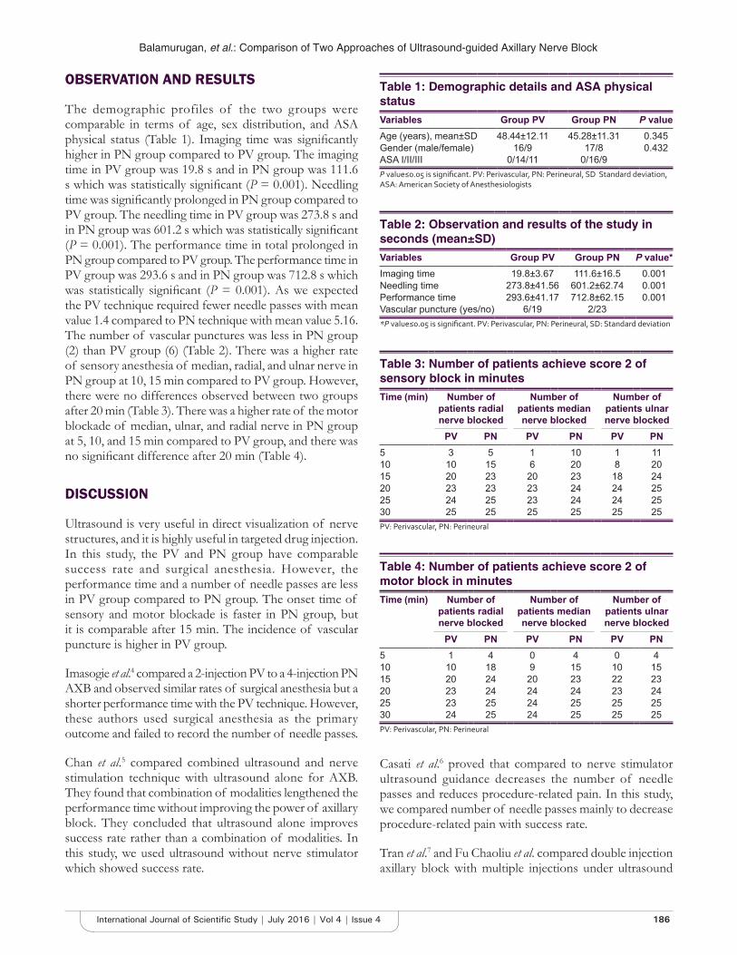
Balamurugan, et al.: Comparison of Two Approaches of Ultrasound-guided Axillary Nerve Block
186International Journal of Scientific Study | July 2016 | Vol 4 | Issue 4
OBSERVATION AND RESULTS
The demographic profiles of the two groups were comparable in terms of age, sex distribution, and ASA physical status (Table 1). Imaging time was significantly higher in PN group compared to PV group. The imaging time in PV group was 19.8 s and in PN group was 111.6 s which was statistically significant (P = 0.001). Needling time was significantly prolonged in PN group compared to PV group. The needling time in PV group was 273.8 s and in PN group was 601.2 s which was statistically significant (P = 0.001). The performance time in total prolonged in PN group compared to PV group. The performance time in PV group was 293.6 s and in PN group was 712.8 s which was statistically significant (P = 0.001). As we expected the PV technique required fewer needle passes with mean value 1.4 compared to PN technique with mean value 5.16. The number of vascular punctures was less in PN group (2) than PV group (6) (Table 2). There was a higher rate of sensory anesthesia of median, radial, and ulnar nerve in PN group at 10, 15 min compared to PV group. However, there were no differences observed between two groups after 20 min (Table 3). There was a higher rate of the motor blockade of median, ulnar, and radial nerve in PN group at 5, 10, and 15 min compared to PV group, and there was no significant difference after 20 min (Table 4).
DISCUSSION
Ultrasound is very useful in direct visualization of nerve structures, and it is highly useful in targeted drug injection. In this study, the PV and PN group have comparable success rate and surgical anesthesia. However, the performance time and a number of needle passes are less in PV group compared to PN group. The onset time of sensory and motor blockade is faster in PN group, but it is comparable after 15 min. The incidence of vascular puncture is higher in PV group.
Imasogie et al.4 compared a 2-injection PV to a 4-injection PN AXB and observed similar rates of surgical anesthesia but a shorter performance time with the PV technique. However, these authors used surgical anesthesia as the primary outcome and failed to record the number of needle passes.
Chan et al.5 compared combined ultrasound and nerve stimulation technique with ultrasound alone for AXB. They found that combination of modalities lengthened the performance time without improving the power of axillary block. They concluded that ultrasound alone improves success rate rather than a combination of modalities. In this study, we used ultrasound without nerve stimulator which showed success rate.
Casati et al.6 proved that compared to nerve stimulator ultrasound guidance decreases the number of needle passes and reduces procedure-related pain. In this study, we compared number of needle passes mainly to decrease procedure-related pain with success rate.
Tran et al.7 and Fu Chaoliu et al. compared double injection axillary block with multiple injections under ultrasound
Table 3: Number of patients achieve score 2 of sensory block in minutesTime (min) Number of
patients radial nerve blocked
Number of patients median nerve blocked
Number of patients ulnar nerve blocked
PV PN PV PN PV PN5 3 5 1 10 1 1110 10 15 6 20 8 2015 20 23 20 23 18 2420 23 23 23 24 24 2525 24 25 23 24 24 2530 25 25 25 25 25 25PV: Perivascular, PN: Perineural
Table 4: Number of patients achieve score 2 of motor block in minutesTime (min) Number of
patients radial nerve blocked
Number of patients median nerve blocked
Number of patients ulnar nerve blocked
PV PN PV PN PV PN5 1 4 0 4 0 410 10 18 9 15 10 1515 20 24 20 23 22 2320 23 24 24 24 23 2425 23 25 24 25 25 2530 24 25 24 25 25 25PV: Perivascular, PN: Perineural
Table 1: Demographic details and ASA physical statusVariables Group PV Group PN P valueAge (years), mean±SD 48.44±12.11 45.28±11.31 0.345Gender (male/female) 16/9 17/8 0.432ASA I/II/III 0/14/11 0/16/9P value≤0.05 is significant. PV: Perivascular, PN: Perineural, SD Standard deviation, ASA: American Society of Anesthesiologists
Table 2: Observation and results of the study in seconds (mean±SD)Variables Group PV Group PN P value*Imaging time 19.8±3.67 111.6±16.5 0.001Needling time 273.8±41.56 601.2±62.74 0.001Performance time 293.6±41.17 712.8±62.15 0.001Vascular puncture (yes/no) 6/19 2/23*P value≤0.05 is significant. PV: Perivascular, PN: Perineural, SD: Standard deviation
Balamurugan, et al.: Comparison of Two Approaches of Ultrasound-guided Axillary Nerve Block
187 International Journal of Scientific Study | July 2016 | Vol 4 | Issue 4
guidance, and they concluded with similar result in this study that double injection technique with fewer needle passes provides simple alternative to ultrasound-guided axillary block.
CONCLUSION
The PV and PN ultrasound-guided axillary block has comparable success rates. Because of fewer needle passes and less imaging, needling and performance time the PV technique can be preferred over a PN technique.
REFERENCES
1. Christophe JL, Berthier F, Boillot A, Tatu L, Viennet A, Boichut N, et al. Assessment of topographic brachial plexus nerves variations at the axilla
using ultrasonography. Br J Anaesth 2009;103:606-12.2. Tran QH, Clemente A, Doan J, Finlayson RJ. Brachial plexus blocks: A
review of approaches and techniques. Can J Anaesth 2007;54:662-74.3. O’Donnell BD, Iohom G. An estimation of the minimum effective
anesthetic volume of 2% lidocaine in ultrasound-guided axillary brachial plexus block. Anesthesiology 2009;111:25-9.
4. Imasogie N, Ganapathy S, Singh S, Armstrong K, Armstrong P. A prospective, randomized, double-blind comparison of ultrasound-guided axillary brachial plexus blocks using 2 versus 4 injections. Anesth Analg 2010;110:1222-6.
5. Chan VW, Perlas A, McCartney CJ, Brull R, Xu D, Abbas S. Ultrasound guidance improves success rate of axillary brachial plexus block. Can J Anaesth 2007;54:176-82.
6. Casati A, Danelli G, Baciarello M, Corradi M, Leone S, Di Cianni S, et al. A prospective, randomized comparison between ultrasound and nerve stimulation guidance for multiple injection axillary brachial plexus block. Anesthesiology 2007;106:992-6.
7. Tran DQ, Pham K, Dugani S, Finlayson RJ. A prospective, randomized comparison between double-, triple-, and quadruple-injection ultrasound-guided axillary brachial plexus block. Reg Anesth Pain Med 2012;37:248-53.
How to cite this article: Balamurugan M, Shanmugasundaram M, Priyadharshini R. Comparison between Perivascular and Perineural Ultrasound-guided Axillary Nerve Block for Forearm Surgeries: A Randomized Controlled Study. Int J Sci Stud 2016;4(4):184-187.
Source of Support: Nil, Conflict of Interest: None declared.

188International Journal of Scientific Study | July 2016 | Vol 4 | Issue 4
Effect of Sickle Cell Disease on Cardiovascular System: A 4.5 Years Autopsy Study Conducted in a Tertiary Care Center of Central IndiaVanita Bhaskar1, Chandrakala Joshi2
1Assistant Professor, Department of Pathology, Pt. Jawaharlal Nehru Memorial Medical College, Raipur, Chhattisgarh, India, 2Associate Professor, Department of Pathology, Pt. Jawaharlal Nehru Memorial Medical College, Raipur, Chhattisgarh, India
trait and 2.5% of population with fatal homozygous hemoglobin disease.5
Sickle cell disease (SCD) is highly prevalent among the triable of central, southern, and western India with frequency ranging from 10% to 23%.6,7 There is also increasing prevalence in nontribal communities in the above-mentioned region. Among all states, higher prevalence of this disease is found in Maharashtra, Madhya Pradesh, Chhattisgarh, and Tamil Nadu.8 The prevalence of SCA in Madhya Pradesh including Chhattisgarh is 1-40%.9 Central India region is a focus of sickle cell disorder.10 This disease has variable clinical presentation, and many of the Indian patients remain asymptomatic for a longer duration due to higher levels of HbF.
SCD produces considerable morbidity and mortality worldwide and also SCD presenting as death in clinically asymptomatic patients is not uncommon.8 However, only
INTRODUCTION
Sickle cell anemia (SCA) is the most common inherited hematological disorder worldwide.1,2 It is an autosomal recessive genetic disorder characterized by single point mutation at codon 6 of the globin gene on chromosome 11 resulting in replacement of valine for glutamic acid and thus formation of hemoglobin S.3 SCA was first described, in 1910, by Herrik.4 SCA is a common health problem in Chhattisgarh. Out of 26 million populations, about 27% of population is suffering with heterozygous hemoglobin
Original Article
AbstractBackground: In Central India, the sickle cell gene is distributed mainly in Madhya Pradesh, Chhattisgarh, Maharashtra, Orissa and Jharkhand and in Chhattisgarh prevalence is nearly 10%. It has variable clinical presentation and most patients remain asymptomatic for longer period. SCA is ignored by autopsy surgeon to be considered as cause of death despite of its high prevalence. While doing autopsy in cases of death with no apparent cause, autopsy surgeon must keep in mind the possibility of death due to vaso-occlusive crisis in SCD.
Materials & Methods: A cross sectional study of duration 4.5 years from January 2012 to May 2016 was conducted in Central India Tertiary care center. Total no. of whole heart autopsy specimen received within this duration was 427 out of which 12 cases were found to have sickle cell disease. Clinical and history detail were collected then thorough gross and microscopic examinations were performed.
Results: History of chest pain was present in 5/12 cases i.e 41.67%. On Histopathological examination, congestion of vessel with Sickle shaped Red Blood Cells was present in 100% cases, MI without atherosclerosis in 41.66%, MI with atherosclerosis in 33.33%, medial calcification in 16.66%, and myocardial hypertrophy in 8.33% of cases.
Conclusion: Sickle cell disease should be considered as one of the cause in case of unexplained and sudden death.
Key Words: Autopsy, Autosomal Recessive, Myocardial Infarction, Prevalence, Sickle Cell Disease.
Access this article online
www.ijss-sn.com
Month of Submission : 05-2016 Month of Peer Review : 06-2016 Month of Acceptance : 07-2016 Month of Publishing : 07-2016
Corresponding Author: Dr. Vanita Bhaskar, C-104, Avenue-144, Near Medishine Hospital, New Rajendra Nagar, Amlidih, Raipur - 492 001, Chhattisgarh, India. Mobile: +91-7697201574. E-mail: [email protected]
Print ISSN: 2321-6379Online ISSN: 2321-595X
DOI: 10.17354/ijss/2016/404
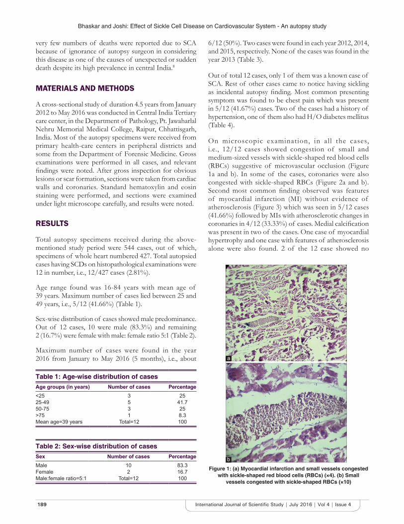
Bhaskar and Joshi: Effect of Sickle Cell Disease on Cardiovascular System - An autopsy study
189 International Journal of Scientific Study | July 2016 | Vol 4 | Issue 4
very few numbers of deaths were reported due to SCA because of ignorance of autopsy surgeon in considering this disease as one of the causes of unexpected or sudden death despite its high prevalence in central India.8
MATERIALS AND METHODS
A cross-sectional study of duration 4.5 years from January 2012 to May 2016 was conducted in Central India Tertiary care center, in the Department of Pathology, Pt. Jawaharlal Nehru Memorial Medical College, Raipur, Chhattisgarh, India. Most of the autopsy specimens were received from primary health-care centers in peripheral districts and some from the Department of Forensic Medicine. Gross examinations were performed in all cases, and relevant findings were noted. After gross inspection for obvious lesions or scar formation, sections were taken from cardiac walls and coronaries. Standard hematoxylin and eosin staining were performed, and sections were examined under light microscope carefully, and results were noted.
RESULTS
Total autopsy specimens received during the above-mentioned study period were 544 cases, out of which, specimens of whole heart numbered 427. Total autopsied cases having SCDs on histopathological examinations were 12 in number, i.e., 12/427 cases (2.81%).
Age range found was 16-84 years with mean age of 39 years. Maximum number of cases lied between 25 and 49 years, i.e., 5/12 (41.66%) (Table 1).
Sex-wise distribution of cases showed male predominance. Out of 12 cases, 10 were male (83.3%) and remaining 2 (16.7%) were female with male: female ratio 5:1 (Table 2).
Maximum number of cases were found in the year 2016 from January to May 2016 (5 months), i.e., about
6/12 (50%). Two cases were found in each year 2012, 2014, and 2015, respectively. None of the cases was found in the year 2013 (Table 3).
Out of total 12 cases, only 1 of them was a known case of SCA. Rest of other cases came to notice having sickling as incidental autopsy finding. Most common presenting symptom was found to be chest pain which was present in 5/12 (41.67%) cases. Two of the cases had a history of hypertension, one of them also had H/O diabetes mellitus (Table 4).
On microscopic examination, in al l the cases, i.e., 12/12 cases showed congestion of small and medium-sized vessels with sickle-shaped red blood cells (RBCs) suggestive of microvascular occlusion (Figure 1a and b). In some of the cases, coronaries were also congested with sickle-shaped RBCs (Figure 2a and b). Second most common finding observed was features of myocardial infarction (MI) without evidence of atherosclerosis (Figure 3) which was seen in 5/12 cases (41.66%) followed by MIs with atherosclerotic changes in coronaries in 4/12 (33.33%) of cases. Medial calcification was present in two of the cases. One case of myocardial hypertrophy and one case with features of atherosclerosis alone were also found. 2 of the 12 case showed no
Table 1: Age-wise distribution of casesAge groups (in years) Number of cases Percentage<25 3 2525-49 5 41.750-75 3 25>75 1 8.3Mean age=39 years Total=12 100
Table 2: Sex-wise distribution of casesSex Number of cases PercentageMale 10 83.3Female 2 16.7Male:female ratio=5:1 Total=12 100
Figure 1: (a) Myocardial infarction and small vessels congested with sickle-shaped red blood cells (RBCs) (×4). (b) Small
vessels congested with sickle-shaped RBCs (×10)
a
b

Bhaskar and Joshi: Effect of Sickle Cell Disease on Cardiovascular System - An autopsy study
190International Journal of Scientific Study | July 2016 | Vol 4 | Issue 4
heterozygous (sickle cell trait) or homozygous, SCD. The homozygous patients are symptomatic from an earlier age but heterozygous, i.e., trait patients are mostly asymptomatic and so ignorant about their disease and sickle cell crisis can occur in them with exposure to extreme hypoxic conditions.
Many studies have been done on various causes of deaths in SCA patients, but only a few studies focus on the effect of SCA on the cardiovascular system of the patient and causes of cardiac death in these patients.
Our study focused on the cardiovascular changes in these patients and tried to find out cardiac causes of death in them so that proper care or management could be provided to these vary group of patients to prevent morbidity and mortality among them. Our study found the mean age of death in patients with SCA was 39 years, ranging from 16
Table 3: Year-wise case distributionYears of study Number of
cases studied(with SCD)
Total number of specimen of heart
received (/year)2012 (January-December) 02 1012013 (January-December) 00 572014 (January-December) 02 1102015 (January-December) 02 1042016 (January-May) 06 55Total number of years=4 years 5 months
Total=12 Total=427
SCD: Sickle cell disease
Table 5: Microscopic findingsMicroscopic examination Number
of casesPercentage of
cases out of 12MI with atherosclerosis 4 33.33MI without atherosclerosis 5 41.66Atherosclerosis only 1 8.33Myocardial hypertrophy 1 8.33Vessels other then coronaries congested with sickle-shaped red blood cells
12 100
Medial calcification 2 16.66No specific pathology 2 16.66MI: Myocardial infarction
Figure 3: Myocardial infarction with medium-sized vessel congested with sickle-shaped red blood cells (×10)
specific pathology except for vessel congestion with sickle RBCs (Table 5).
DISCUSSION
SCA, the classical prototype of hereditary hemoglobinopathy, is an autosomal recessive disease manifesting in two forms
Table 4: Associated historyAssociated history Number of cases (%)History of HTN 2 (16.67)History of heart disease 1 (8.33)History of poisoning 1 (8.33)History of epilepsy 1 (8.33)History of chronic alcoholism 1 (8.33)History of chest pain 5 (41.67)History of DM 1 (8.33)Known case of SCD 1 (8.33)Total 12 (100)SCD: Sickle cell disease, DM: Diabetes mellitus, HTN: Hypertension
Figure 2: (a) Coronary artery filled with sickle‑shaped red blood cells (RBCs) (×4). (b) Coronary artery filled with sickle‑shaped
RBCs (×10)
a
b

Bhaskar and Joshi: Effect of Sickle Cell Disease on Cardiovascular System - An autopsy study
191 International Journal of Scientific Study | July 2016 | Vol 4 | Issue 4
to 84 years. It is similar to the study by Fitzhugh et al. as they also reported the mean age of death in SCD patients being 39 years.11 This is comparable with a study done by Darbari et al.,12 who found mean age to be 36 years.
Out of 12/427, 10 were male and only 2 were females with male to female ratio 5:1. Our observation shows discordance with the study conducted by Manci et al.,13 who found no significant differences in presentation among male and female. In their study, male: female ratio was 1:0.98 in patients with homozygous (SS) and 1:1.44 in the heterozygous state. Probable reason behind this could be due to less number of study population included in our study.
In our study, we found 12 patients having SCA among 427 patients undergoing autopsy, i.e., 2.8% cases. Out of these, only 1 patient was a known case of SCA and rest 11 cases were diagnosed incidentally after histopathological examination only.
In the present study, out of 12 patients, 5 of them had history of chest pain as presenting complaint, i.e., 41.66%. Martin et al. found chest pain in 6/72 patients, i.e., in 8.33% of cases,14 whereas Norris et al.15 found chest pain in 100% of cases out of total 19 cases he studied. This could be explained by various factors such as type of disease, i.e., trait or homozygous, associated health conditions and geographical differences.
On histopathological examination, 100% cases showed small and medium-sized vessel congestion with sickle-shaped RBCs. This could be cause of microvessel occlusion leading to cardiac changes and deaths in the study group.
5/12 cases (41.66%) had features of MI without any atherosclerotic changes followed by features of MI with atherosclerosis in 4/12 (33.33%) of cases. Manci et al. found in their study that in 2.5% of homozygous (SS) cases the immediate cause of death was directly related to vaso-occlusion.13 Martin et al. in their study found MI in 7/72 (9.2%) patients with SCD.14
In two of the cases, no specific pathology was noted during autopsy examination apart from the congestion of vessels with sickled RBCs.
Varies cardiac changes could be due to abnormal blood rheology in this disease that may result in vaso-occlusion, myocardial ischemia, and infarction.16
MI in SCD has been previously described in various case reports and series. These all have suggested microvascular disease related to vaso-occlusion as the likely cause of myocardial ischemia and injury in these patients.14,15,17-28
CONCLUSION
Clinical presentation of sickle cell patients in Central India is less severe as compared to African countries and is characterized by delayed presentation; patients are more asymptomatic, less incidence of vaso-occulsion, and low mortality due to which most of the patients remain undiagnosed. However, it is not uncommon in these patients presenting as death without any significant present or past history. Although many patients remain asymptomatic MI occurs in SCD and should be considered in differential diagnosis of patients with chest pain. Patients with SCD and chest pain who are suspected of having myocardial ischemia should be managed properly.
Furthermore, in case of unexplained death, during autopsy, it is important to keep in mind the possibility of SCD and so, proper histopathological examination along with hemoglobin electrophoresis, and molecular studies is needed to reach causes of death which would also be useful to their relatives to know the disease status in their family that could propel them to seek relevant management timely to prevent SCD related morbidity and mortality.
REFERENCES
1. Modell B, Darlison M. Global epidemiology of haemoglobin disorders and derived service indicators. Bull World Health Organ 2008;86:480-7.
2. Weatherall DJ. The inherited diseases of hemoglobin are an emerging global health burden. Blood 2010;115:4331-6.
3. Fertrin KY, Costa FF. Genomic polymorphisms in sickle cell disease: Implications for clinical diversity and treatment. Expert Rev Hematol 2010;3:443-58.
4. Savitt TL, Goldberg MF. Herrick’s 1910 case report of sickle cell anemia. The rest of the story. JAMA 1989;261:266-71.
5. Purnima DS, Gopinath A, Skaria LK. Analysis of liver dysfunction among sickle cell disease, population in bastar region of Chhattisgarh. J Pharm Biomed Sci (JPBMS) 2012;16:1-7.
6. Kate SL. Health problems of tribal population groups from the state of Maharashtra. Indian J Med Sci 2001;55:99-108.
7. Shukla RN, Solanki BR. Sickle-cell trait in Central India. Lancet 1958;1:297-8.
8. Manish BS, Ghormade PS, Chaitanya VT. Death due to sickle cell anaemia: Autopsy diagnosis. J Indian Acad Forensic Med 2013;35:383-5.
9. Balgir RS, Sharma SK. Distribution of sickle cell haemoglobin in India. Indian J Hematol 1988;6:1-14.
10. Balgir RS. The burden of haemoglobinopathies in India and challenges ahead. Curr Sci 2000;79:1536-47.
11. Fitzhugh CD, Lauder N, Jonassaint JC, Telen MJ, Zhao X, Wright EC, et al. Cardiopulmonary complications leading to premature deaths in adult patients with sickle cell disease. Am J Hematol 2010;85:36-40.
12. Darbari DS, Kple-Faget P, Kwagyan J, Rana S, Gordeuk VR, Castro O. Circumstances of death in adult sickle cell disease patients. Am J Hematol 2006;81:858-63.
13. Manci EA, Culberson DE, Yang YM, Gardner TM, Powell R, Haynes J Jr, et al. Causes of death in sickle cell disease: An autopsy study. Br J Haematol 2003;123:359-65.
14. Martin CR, Johnson CS, Cobb C, Tatter D, Haywood LJ. Myocardial infarction in sickle cell disease. J Natl Med Assoc 1996;88:428-32.
15. Norris S, Johnson CS, Haywood LJ. Sickle cell anemia: Does myocardial ischemia occur during crisis? J Natl Med Assoc 1991;83:209-13.
Bhaskar and Joshi: Effect of Sickle Cell Disease on Cardiovascular System - An autopsy study
192International Journal of Scientific Study | July 2016 | Vol 4 | Issue 4
16. Chien S, Usami S, Bertles JF. Abnormal rheology of oxygenated blood in sickle cell anemia. J Clin Invest 1970;49:623-34.
17. Pavlu J, Ahmed RE, O’Regan DP, Partridge J, Lefroy DC, Layton DM. Myocardial infarction in sickle-cell disease. Lancet 2007;369:246.
18. Assanasen C, Quinton RA, Buchanan GR. Acute myocardial infarction in sickle cell anemia. J Pediatr Hematol Oncol 2003;25:978-81.
19. Barrett O Jr, Saunders DE Jr, McFarland DE, Humphries JO. Myocardial infarction in sickle cell anemia. Am J Hematol 1984;16:139-47.
20. Dang NC, Johnson C, Eslami-Farsani M, Haywood LJ. Myocardial injury or infarction associated with fat embolism in sickle cell disease: A report of three cases with survival. Am J Hematol 2005;80:133-6.
21. de Montalembert M, Maunoury C, Acar P, Brousse V, Sidi D, Lenoir G. Myocardial ischaemia in children with sickle cell disease. Arch Dis Child 2004;89:359-62.
22. Deymann AJ, Goertz KK. Myocardial infarction and transient ventricular
dysfunction in an adolescent with sickle cell disease. Pediatrics 2003;111:E183-7.
23. Martin CR, Cobb C, Tatter D, Johnson C, Haywood LJ. Acute myocardial infarction in sickle cell anemia. Arch Intern Med 1983;143:830-1.
24. Maunoury C, Acar P, de Montalembert M, Sidi D. Myocardial perfusion in children with sickle cell disease. Am J Cardiol 2003;91:374-6.
25. McCormick WF. Massive nonatherosclerotic myocardial infarction in sickle cell anemia. Am J Forensic Med Pathol 1988;9:151-4.
26. Saad ST, Arruda VR, Junqueira OO, Schelini FA, Coelho OB. Acute myocardial infarction in sickle cell anaemia associated with severe hypoxia. Postgrad Med J 1990;66:1068-70.
27. Sherman SC, Sulé HP. Acute myocardial infarction in a young man with sickle cell disease. J Emerg Med 2004;27:31-5.
28. Wang H, Laslett LJ, Richman CM, Wun T. Acute myocardial infarction in hemoglobin SC disease. Ann Hematol 2004;83:622-4.
How to cite this article: Bhaskar V, Joshi C. Effect of Sickle Cell Disease on Cardiovascular System: A 4.5 Years Autopsy Study Conducted in a Tertiary Care Center of Central India. Int J Sci Stud 2016;4(4):188-192.
Source of Support: Nil, Conflict of Interest: None declared.

193 International Journal of Scientific Study | July 2016 | Vol 4 | Issue 4
Clinical Study and Management of Parotid TumorsR Ashok Reddy1, T S R S V Rajyalaxmi Godadevi2
1Professor, Department of General Surgery, Kakatiya Medical College, Mahatma Gandhi Hospital Warangal, Telangana, 2Assistant Professor, Department of General Surgery, Kakatiya Medical college/ Mahatma Gandhi Hospital Warangal, Telangana
advice. Because most of them are benign in nature and due to lack of health awareness in our setup, the number of patients seeking the treatment is less. Thus, the patients with malignant tumors present very late and need radical treatment which carries high morbidity.
Swelling is the most common symptom in parotid tumors. Most of the malignant tumors present with pain. Most of the benign tumors exhibit a slow growth pattern and malignant tumors Exhibit a rapid growth pattern.
Fine needle aspiration cytology (FNAC) is a good tool in diagnosing parotid gland tumors. Neural involvement is common in adenoid cystic carcinoma. Surgery is the main stay of the treatment for parotid tumors.
During the period from November 2012 to May2014, 30 cases of parotid tumors have been admitted to Mahatma
INTRODUCTION
The parotid gland is the most common site for salivary gland tumors.70-80% of Salivary gland neoplasms occur in the parotid gland, of which 80% are benign, 20% are malignant of which 80% of the benign tumors are pleomorphic adenomas.
Parotid tumors are generally slow growing and have been present for several years before the patient seeks medical
Original Article
AbstractIntroduction: The parotid gland is the most common site for salivary gland tumors. 70-80% of salivary gland neoplasms occur in the parotid gland, of which 80% are benign, 20% are malignant of which 80% of the benign tumors are pleomorphic adenomas.
Aims and Objectives: (1) To know the incidence of parotid gland tumors with respect to age and sex. (2) To study the various modes of clinical presentation of parotid tumors. (3) To evaluate the various modes of surgery and outcome of surgical management of the parotid gland tumors at Mahatma Gandhi Memorial (MGM) Hospital, Warangal.
Materials and Methods: This study was conducted from November 2012 to May 2014, over a period of 1-year and 6-month. 30 patients admitted to MGM Hospital, Warangal, with parotid gland neoplasms are included in this study. Inclusion criteria all patients with parotid swelling due to parotid tumors from 13 years of age of MGM Hospital, Warangal. Exclusion criteria all pediatric patients, tumor-like conditions and infectious causes of swelling are excluded.
Results: (1) Age incidence in parotid tumors, (2) clinical presentation of parotid tumors, (3) incidence in relation to duration of mass, (4) surgical treatment adopted in the study, (5) distribution of HPE diagnosis of tumors, (6) fine needle aspiration cytology (FNAC) and histopathology corelation, (7) correlation of FNAC with histopathological examination, (8) incidence of benign and malignant parotid tumors, (9) distribution of benign tumors, (10) distribution of malignant tumors.
Discussion: In this study, the most patients were in the 3rd and 5th decade of life. Malignant tumors were common in the 4th and 5th decade. Malignant tumors were encountered more in the older age group in comparison to benign ones.
Conclusion: A total of 30 cases admitted to MGM Hospital attached to Kakatiya Medical College from November 2012 to May 2014 over a period of 1½ years were included in this study.
Key words: Parotid Tumour,Fine Needle Aspiration, Histopathological Examination
Access this article online
www.ijss-sn.com
Month of Submission : 05-2016 Month of Peer Review : 06-2016 Month of Acceptance : 07-2016 Month of Publishing : 07-2016
Corresponding Author: Dr. R. Ashok Reddy, Professor, Department of General Surgery, Kakatiya Medical College/ Mahatma Gandhi Hospital Warangal. E-mail: [email protected]
Print ISSN: 2321-6379Online ISSN: 2321-595X
DOI: 10.17354/ijss/2016/405
Reddy and Godadevi: A Clinical Study and Management of Parotid Tumours
194International Journal of Scientific Study | July 2016 | Vol 4 | Issue 4
Gandhi Memorial (MGM) Hospital, Warangal. 12 cases are presented in detail for the purpose of this dissertation.
Aims and Objectives1 To know the incidence of parotid gland tumors with
respect to age and sex2 To study the various modes of clinical presentation of
parotid tumors3 To evaluate the various modes of surgery and outcome
of surgical management of parotid gland tumors at MGM Hospital, Warangal.
MATERIALS AND METHODS
This study was conducted from November 2012 to May 2014, over a period of 1-year and 6-month. 30 patients admitted to MGM Hospital, Warangal, with parotid gland neoplasms are included in this study.
Inclusion CriteriaAll patients with parotid swelling due to parotid tumors from 13 years of age of MGM Hospital, Warangal.
Exclusion CriteriaAll pediatric patients, tumor-like conditions and infectious causes of swelling are excluded from the study.
All patients admitted were evaluated by documenting the history, thorough clinical examination, routine laboratory investigations and specific investigations. In history, importance was given to presenting complaints, duration of lump, rapid increased in size, associated symptoms of facial nerve involvement, previous surgical treatment or any medical problem.
Regarding physical examination, particulars mentioned in the proforma were noted. Importance was given to the site, extent of the tumor, deep lobe enlargement and fixity to the surrounding structures, facial nerve involvement, and regional lymphadenopathy. Associated medical conditions such as diabetes, hypertension, and anemia were managed and controlled before surgery with physician’s advice.
As a part of general workup for surgery in all patients, hemoglobin level, bleeding time, clotting time, urine, sugar albumin, microscopy, and chest screening. Electrocardiogram, blood urea, serum creatinine, random blood sugar were estimated. Specific investigations like FNAC were done for all patients in the study group.
Sialography is not done for any of these patients because it may cause inflammation or infection. Extravasation of the dye may cause a severe inflammatory reaction preventing
a clear demarcation of tumor margins and may also delay the planned surgical procedure.
After evaluation of the tumor by clinical examination and specific investigations, a surgical plan was formulated. The final decision was taken per operatively by the surgeon. The specimen was sent for histopathology for final diagnosis. The adjuvant treatment was decided depending on the final histopathological report.
Different modalities of treatment adopted in this study are as follows:• Surgery alone• Surgery and postoperative radiotherapy.
Different surgical procedures adopted in this study are as:• Superficial parotidectomy• Total conservative parotidectomy.
The follow-up period of these patients ranged from 3 months to 1 year. Long-term follow-up is necessary to study the tumor recurrence, which was not possible in this study.
RESULTS
The following observations were made in 30 patients who presented with parotid gland neoplasms in this study.
Age Incidence in Parotid TumorsThe age incidence of the patients in the study group ranged from 13 to 72 years. The malignant tumors occurred between the age group of 36-72 years. Most patients in this series were in the 4th decade of life. The mean age was 37.6 years for benign tumors and 50.7 years for malignant tumors (Table 1).
Sex Distribution of Parotid Tumors (Table 2)Out of 30 patients parotid tumours,18 patients were Male, 12 Patients are female.
Side of the TumorAbout 60% of parotid tumors occurred in the left parotid gland in this study (Table 3).
Clinical Presentation of Parotid TumorsAll patients presented with swelling in the parotid region. Features of rapid growth, pain and associated facial paralysis were considered as signs of malignancy. Hard in consistency is noted mostly in a malignant tumor. Out of 30 patients, 5 patients presented with pain (16.67%) in swelling, out of which 5 were malignant.

Reddy and Godadevi: A Clinical Study and Management of Parotid Tumours
195 International Journal of Scientific Study | July 2016 | Vol 4 | Issue 4
Pain occurred in 100% of the patients with malignant tumors. Deep lobe enlargement was seen in two patients in this series. No patient had fixity to masseter/mandible. No patient had facial nerve paralysis at presentation (Table 4).
Incidence in Relation to Duration of MassAll patients presented with swelling in the parotid regions of which most cases (66.6%) presented within 5 years after noticing the swelling (Table 5).
FNAC Diagnosis of TumorsIn this study, the number of cases of pleomorphic adenoma cases diagnosed by FNAC was 26 which is the most common benign parotid tumor and among malignant tumors malignant mixed tumor has the highest incidence of two cases (Table 6).
Surgical Treatment Adopted in the StudySurgery was performed in 30 patients, the type of surgery was chosen according to clinical impression, FNAC and per-operative findings. Superficial parotidectomy was performed in 25 patients (83.33%), conservative total parotidectomy in five patients (16.67%). In this study, radical parotidectomy and RND was not done in any of the patients (Tables 7-9).
In this study, after subjecting the specimens of tumor tissue for HPE among the benign tumors the number of cases of pleomorphic adenoma was 21 followed by warthins tumor - 2 cases among malignant tumors malignant cases, malignant mixed tumor were four cases followed by acinic cell carcinoma 1 case (Table 10).
FNAC and Histopathology Co-relationAll 30 cases subjected to FNAC and were reported as parotid tumors. After surgical excision or biopsy, all
specimens were studied histopathologically and the table below shows corelation between FNAC reporting and histopathological diagnosis (Table 11).
FNAC showed 28 cases to be benign and 2 cases to be malignant but on HPE benign tumors are 24 and malignant tumors are 6 in number.
In this study, FNAC correctly diagnosed benign from malignant in 93.3% of the cases.
Table 1: Age incidence in parotid tumorsAge group
Total number of cases
Percentage Pleomorphic adenoma
Warthins tumor
Basal cell adenoma
Oncocytoma Malignant mixed tumor
Acinic cell carcinoma
13-20 2 6.67 2 121-30 8 26.67 5 131-40 7 23.33 7 141-50 9 30.0 6 1 2 151-60 2 6.67 161-70 1 3.33 171-80 1 3.33 1Total 30 100 21 2 1 1 4 1
Table 2: Sex distribution of parotid tumorSex Number
of casesPercentage Pleomorphic
adenomaWarthins
tumorBasal cell adenoma
Oncocytoma Malignant mixed tumor
Acinic cell carcinoma
Male 18 60 11 2 1 1 2 1Female 12 40 10 2Total 30 100 21 2 1 1 4 1
Table 3: Distribution of side of the tumorSide of the tumor Number of cases (n=30) PercentageRight side 12 40Left side 18 60
Table 4: Clinical Presentation of parotid tumours.Signs and symptoms Number of cases Overall (%)Swelling 30 100Pain 5 16.67Fungating mass 0 0Symptoms of facial palsy - -Cervical lymphadenopathy - -Deep lobe involvement 2 6.66Fixity to masseter/mandible - -
Table 5: Incidence in relation to duration of the massSigns and symptoms Number of cases Overall (%)1-12 months 5 16.671-5 years 15 506-10 years 9 3011-15 years 1 3.33

Reddy and Godadevi: A Clinical Study and Management of Parotid Tumours
196International Journal of Scientific Study | July 2016 | Vol 4 | Issue 4
Overall pleomorphic adenoma, constituted 70% of the tumor and among malignant tumor, malignant mixed tumor constituted 13% of the tumors in the series (Table 12).
In this study, among the benign tumors pleomorphic adenoma constituted 84% of the benign tumors and among the malignant tumors, malignant mixed tumor constituted 80% of the malignant parotid tumors (Tables 13 and 14).
Recurrent TumorOne recurrent tumor was operated in this series. It was acinic cell tumor.
Adjuvant TreatmentRadiotherapy was given to five patients, with a malignant tumor of the parotid gland, five patients were given postoperative radiotherapy. Out of theses, four patients had a malignant mixed tumor and another one had acinic cell tumor. One patient received radiotherapy developed xerostomia, which was treated conservatively. No patients were given chemotherapy in this series.
Follow upIn this series follow-up ranged from 3 months to 1 year. To know the recurrence of a tumor long-term follow-up is necessary which was not possible in this study. In spite of repeated postal reminders, most of the patients in this study did not respond. During the study period, none of the operated patients came back with recurrent diseases.
DISCUSSION
In this study, most patients were in the 3rd and 5th decade of life. Malignant tumors were common in the 4th and the 5th decade. Malignant tumors were encountered more in the older age group in comparison to benign ones. The mean age group was 37.6 years for benign tumors and 50.7 years for malignant tumors. Whereas mean age for benign tumors is 51 years and mean age for malignant tumors is 40 years in the study by Lim et al. (Table 15).
Males were affected more than females in both benign and malignant tumors. The duration of swelling was form 8 months to 12 years. The history of the duration of the swelling is not significant, as long-standing benign tumor may turn malignant.
Sex ratio of both benign and malignant tumors has been 3:2 in the present study, compared to a near equal distribution of tumor in both sexes in other studies. However, there a general male preponderance as seen in the study by Suwala et al. and Kawata et al. (Table 16).
In the present series, left sided tumors are more common than on the right side. Similar observations are made by Suwala et al. and Marcello Donati et al. (Table 17).
Discussion on Clinical Features of Parotid TumorsAll patients presented with a history of swelling in the parotid region. 16.67% of the patients presented with pain in the swelling. No patient presented with facial nerve palsy. In this series, 100% of the patients with malignant parotid disease presented with pain. No patient presented with cervical lymph node metastasis.
Table 6: Distribution of FNAC diagnosis of tumorsDiagnosis Number of patients PercentagePleomorphic adenoma 26 86.7Warthin’s tumor 1 3.33Acinic cell carcinoma 1 3.33Malignant mixed tumor 2 6.7FNAC: Fine needle aspiration cytology
Table 7: Types of surgical treatment adopted in the studyProcedure Number of cases PercentageSuperficial parotidectomy 25 83.33Conservative total parotidectomy 5 16.67
Table 8: Types of surgical treatment adopted in the studyProcedure Number of cases PercentageSuperficial parotidectomy 25 83.33Conservative total parotidectomy 5 16.67
Table 9: Complications following surgeryComplications Pleomorphic
adenomaWarthins
tumorBasal cell adenoma
Oncocytoma Malignant mixed tumor
Acinic cell carcinoma
Total
Immediate post-operative facial nerve weakness 4 1 0 1 1 7 (28.33)Permanent facial nerve weakness 3 0 1 1 5 (16.66)Parotid fistula 2 0 2 (6.66)Wound infection 2 2 4 (13.33)Frey’s syndrome 1 1 (3.3)Seroma - - - - - - 0Bleeding/hematoma - - - - - - 0Hypesthesia of cheek/ear lobule - - - - - - 0

Reddy and Godadevi: A Clinical Study and Management of Parotid Tumours
197 International Journal of Scientific Study | July 2016 | Vol 4 | Issue 4
The presence of facial nerve paralysis, skin infiltration or ulceration, and metastatic neck nodes were found only in patients with malignant tumors. A history of pain, hardness, and fixity, found in 30-50% of parotid cancers were significant indicators of malignancy as seen by Lam et al.7
Accuracy of FNACIn this study, FNAC correctly diagnosed benign from malignant in 93.3% of the cases.
Superficial parotidectomy was performed in 25 patients (83.33%), conservative total parotidectomy in 5 patients
(16.67%). In this study, radical parotidectomy and RND was not done in any of the patients (Table 18).
Preservation of the facial nerve and serious postoperative complications can be minimized following superficial and/or total conservative parotidectomy as seen in the study by al-Naqueeb et al.10
Radiotherapy was given to five patients with malignant tumors of the parotid gland. Five patients were given post-operative radiotherapy, and four patients are presented with malignant mixed carcinoma and one patient presented with recurrent Pleomorphic adenoma it turned out to be acinic cell carcinoma. In the management of malignant tumors, the usefulness of radiotherapy as an adjuvant to surgery has been accepted by all authors.
Chemotherapy is of doubtful benefit in the management of malignant parotid tumors and in this study it has not been given a trial (Table 19).
Table 10: Distribution of HPE diagnosis of tumorsDiagnosis Number of patients PercentagePleomorphic adenoma 21 70Warthins tumor 2 6.66Basal cell adenoma 1 3.33Oncocytoma 1 3.33Malignant mixed tumor 4 13.33Acinic cell carcinoma 1 3.33HPE: Histopathological examination
Table 11: Correlation of FNAC with histopathological examination
Diagnosed as benign Diagnosed as malignantFNAC Biopsy FNAC Biopsy28 24 2 6FNAC: Fine needle aspiration cytology
Table 12: Incidence of benign and malignant parotid tumorsIndividual tumors Number of patients (n=30) PercentagePleomorphic adenoma 21 70Warthins tumor 2 6.66Basal cell adenoma 1 3.33Oncocytoma 1 3.33Malignant mixed tumor 4 13.33Acinic cell carcinoma 1 3.33
Table 13: Distribution of benign tumorsBenign tumors Number of cases (n=25) PercentagePleomorphic adenoma 21 84Warthins tumor 2 8Basal cell adenoma 1 4Oncocytoma 1 4
Table 14: Distribution of malignant tumorsMalignant tumors Number of cases (n=5) PercentageMalignant mixed tumor 4 80Acinic cell carcinoma 1 20
Table 15: Discussion on age incidence in parotid tumorsAuthor Mean age for benign
tumors (years)Mean age for malignant
tumors (years)Lim et al.2 51 40Present study 37.6 50.7
Table 16: Discussion on sex incidence in parotid tumorsAuthor Males:female ratioSuwała et al.3 1.01:1Pietniczka-Załeska et al.4 0.7:1Kawata et al.5 1.06:1Present study 3:2
Table 17: Discussion on incidence of side of the tumorAuthor Side of the tumor
Right (%) Left (%)Suwala et al. 40 60Marcello Donati et al.6 43.7 56.3Present study 40 60
Table 18: Study on types of surgical treatments adopted in the studyAuthor Superficial
parotidectomy (%)Total conservative parotidectomy (%)
Corcione et al.8 12.7 50.9Patey et al.9 55.88 35.2Present study 83.3 16.7

Reddy and Godadevi: A Clinical Study and Management of Parotid Tumours
198International Journal of Scientific Study | July 2016 | Vol 4 | Issue 4
Temporary Facial WeaknessIn this study, post-operative facial nerve weakness occurred in 7 patients (23.33%) And in 3 patients facial nerve weakness recovered completely over 6 months. Reported incidence of immediate post-operative facial weakness varies between 6 and 23% as per western literature. Normal function usually returns within 3-6 months but it may take up to 1 year. It may be caused by nerve ischemia, fatigue from excessive stimulation, stretching or hematoma formation.
Permanent Facial WeaknessIn this study, postoperatively 16.6% (No. 5) of the patients developed permanent facial weakness, which is more compared with the western literature (3 patients with malignant mixed tumor and 1 patient with acinic cell pleomorphic adenoma). Reported incidence of permanent facial weakness in 2-9.8%, as per western literature.
Mehle et al. and Lacourrey et al. have reported 46% and 65% incidence of immediate post-operative facial weakness. Permanent facial weakness was 4% in both the series. Permanent facial weakness is slightly higher compared to western literature (Table 20).
Parotid FistulaParotid fistula occurred in 2 patients with pleomorphic adenoma who had undergone superficial parotidectomy, healed spontaneously within 3 months which is more compared to Klintworth et al. and Bova et al. As these cases are less frequently done in MGM hospital the number of parotid fistulas is more.
Wound InfectionWound infection occurred in 4 patients due to poor nutritional status of the patients due to low socio-economic status of the patients, incidence of infection is more compared to other studies. Infection healed with antibiotic treatment.
Frey’s SyndromeThere has been a single case of Frey’s syndrome which is less compared to Mantusopoulos et al. In the studies
carried out by Klintworth et al. and Bova et al., cases of Frey’s syndrome are nil as cases of parotid are done less frequently in MGM hospital. It was managed conservatively (Table 21).
Benign parotid tumors amount to 83.3% in the present study, whereas malignant parotid tumors amount to 16.7%. Similar observations were seen with other studies (Table 22).
Among benign tumors pleomorphic adenoma is the most common tumor amounting to 70% of all parotid tumors, which is slightly more compared to other studies. Whereas Warthin’s tumor is less compared to other studies as the incidence is more in older white men.
Among malignant tumors, the incidence of the malignant mixed tumor is more-13.3% and a similar observation is made by Przewoźny et al. which is 20.6%.
In this study, pleomorphic adenoma was the most common tumor encountered constituting 70% of the parotid tumors. Among the benign parotid tumors Pleomorphic adenoma constituted about 84%, Warthin’s tumor constituted about 8% of the benign parotid tumors. Among malignant tumors, the most common was malignant mixed tumor
Table 19: Discussion on complications following surgery in parotid tumorComplications Bova.R et al. Klintworth N et al. Mantsopoulos K et al. Present studyImmediate postoperative facial nerve weakness (%) 20 6 22.8 23.3Permanent facial nerve weakness (%) 3.5 2 9.8 16.6Parotid fistula (%) - 2 - 6.6Wound infection (%) 2.3 - - 13.3Frey’s syndrome (%) - - 11.3 3.3Seroma (%) 6.6 5 - -Bleeding/hematoma (%) 3.5 0.8/3 - -Hypoasthesia of cheek or ear lobule (%) - 10 - -
Table 20: Studies on permanent facial nerve weaknessAuthor Permanent facial
weakness (%)JMH Debetes and JDK Munting et al.1 7Mehle et al. and Laccourreye et al. 4Present study 16.67
Table 21: Discussion on distribution of benign and malignant tumorsAuthor Benign tumors (%) Malignant tumors (%)Van Niekerek et al.11 80.2 19.8Dunn et al.12 69.2 30.8Suwała et al.3 88 12Present study 83.3 16.7

Reddy and Godadevi: A Clinical Study and Management of Parotid Tumours
199 International Journal of Scientific Study | July 2016 | Vol 4 | Issue 4
constituting 80%, Acinic cell carcinoma constituting 20% of the malignant tumors.
CONCLUSION
• Thirty cases admitted to Mahatma Gandhi Memorial Hospital attached to Kakatiya Medical College from November 2012 to May 2014 over a period of 1½ years were included in this study
• Incidence of parotid tumors is highest in the 3rd to 5th decade constituting 65% of patients
• Male to female ratio for parotid tumors is 3:2• Benign tumors of the parotid constituted about
83.33% and malignant tumors constituted 16.66% of the parotid neoplasms in the study
• All patients presented with swelling in the parotid region
• Pain was the second most common symptom. The pain was noticed in 16.67% of the patients. Pain in the swelling occurred in 100% of the malignant tumors
• None of the patients in this series presented with facial nerve weakness or cervical lymph node metastasis
• Nearly, 6.66% of the patients presented with enlargement of the deep lobe of the parotid gland
• Pleomorphic adenoma was the most common tumor in this series constituting 70% of the overall parotid tumors
• Pleomorphic adenoma was the most common benign tumor constituting 84% of the benign tumors
• Malignant mixed tumor was the most common malignant tumor constituting 80% of the malignant parotid tumors
• FNAC was done in all patients. In this study, FNAC correctly diagnosed benign from malignant in 93.3% of the cases
• Sialography is seldom indicated in parotid gland neoplasms because it may cause inflammation or infection. Sialography was routinely done previously in patients with parotid gland neoplasms; Extravasation of the dye may cause a severe inflammatory reaction, preventing a clear demarcation of tumor margins and may also delay the planned surgical procedure
• The treatment of choice for parotid neoplasms is mainly superficial parotidectomy. This may be followed by radiotherapy if the tumor is malignant
• In this study, all patients with malignant tumors were given post-operative radiotherapy. No patient with benign tumors of the parotid was given radiotherapy
• No patients in the study were given chemotherapy• Most common post-operative complication is facial
nerve weakness• The incidence of permanent facial nerve weakness was
16.6%. This is more comparable to western standard (2-9.8%) This has occurred mainly in patients with malignant tumors. In this study, no form of facial nerve repair was done
• In view of the late presentation, in this series, which can adversely affect in malignant tumors, increased community awareness for early referral is mandatory
• The adequacy of treatment cannot be commented because of the short follow-up of these patients in the study. The study group in this series is small, as compared to large series in western literature; so statistical data in this series may not represent the actual data quoted in the western literature.
REFERENCES
1. Debets JM, Munting JD. Parotidectomy for parotid tumors. 19-year experience from Netherlands. Br J Surg 1992;79:1159-61.
2. Lim LH, Chao SS, Goh CH, Ng CY, Goh YH, Khin LW. Parotid gland surgery: 4-year review of 118 cases in an Asian population. Head Neck 2003;25:543-8.
3. Suwała P, Wilczyński K, Barnaś S. The parotid glands neoplasms in material of the otolaryngology clinical ward of 4th military clinical hospital with the polyclinic in Wrocław in the years 1992-2010. Otolaryngol Pol 2012;66:43-5.
4. Pietniczka M, Kukwa V. Guzy ślinianki przyusznej w materiale Kliniki Otolaryngologii w Warszawie w latach 1990-2006. Otolaryngol Pol 2008;62:395-9.
5. Kawata R, Lee K, Yoshimura K, Nishimawa S, Araki M. Review of 300 cases of parotidectomy for benign parotid tumors. Nihon Jibiinkoka Gakkai Kaiho 2012;115:618-24.
6. Donati M, Gandolfo L, Privitera A, Brancato G, Cardi F, Donati A. Superficial parotidectomy as first choice for parotid tumours. Chir Ital 2007;59:91-7.
8. Lam KH, Wei WI, Lau WF. Tumours of the parotid – The value of clinical assessment. Aust N Z J Surg 1986;56:325-9.
9. Corcione F, Califano L. Treatment of parotid gland tumors. Int Surg 1990;75:171-3.
10. Patey DH, Thackray AC. The treatment of parotid tumours in the light of a pathological study of parotidectomy material. Br J Surg 1958;45:477-87.
11. al-Naqeeb NI, Dashti H, al-Muhanna AH, Behbehani A. Parotid gland tumours: A 15-year experience. J R Coll Surg Edinb 1992;37:89-93.
12. van Niekerk JL, Wobbes T, Monstrey S, Bruaset I. The management of parotid tumors; A ten-year experience. Acta Chir Belg 1987;87:1-5.
13. Dunn EJ, Kent T, Hines J, Cohn I Jr. Parotid neoplasms: A report of 250
Table 22: Discussion on distribution of individual tumorsAuthor Pleomorphic
adenoma (%)Warthin’s tumor (%)
Basal cell adenoma (%)
Oncocytoma (%) Malignant mixed tumor (%)
Acinic cell carcinoma (%)
Przewoźny et al.13 65.5 24 4.2 - 20.6 -Lee et al.14 61.6 13.2 - - - -Eddey et al.15 60.14 15.94 - 2.63 - 2.63Present study 70 6.6 3.33 3.33 13.3 3.3

Reddy and Godadevi: A Clinical Study and Management of Parotid Tumours
200International Journal of Scientific Study | July 2016 | Vol 4 | Issue 4
cases and review of the literature. Ann Surg 1976;184:500-6.14. Przewoźny T, Stankiewicz C. Neoplasms of the parotid gland in northern
Poland, 1991-2000: An epidemiologic study. Eur Arch Otorhinolaryngol 2004;261:369-75.
15. Lee YM, Choi HJ, Kim JW, Kim JH. Management of parotid tumors. Eur Arch Otorhinolaryngol 2004;261:369-75.
16. Eddey HH. Parotid tumours: A review of 138 cases. Aust N Z J Surg 1970;40:1-14.
How to cite this article: Reddy RA, Godadevi TSR. Clinical Study and Management of Parotid Tumors. Int J Sci Stud 2016;4(4):193-200.
Source of Support: Nil, Conflict of Interest: None declared.

201 International Journal of Scientific Study | July 2016 | Vol 4 | Issue 4
Association of Menopause, Reproductive Years, and Bone Mineral Density in Postmenopausal Women with Natural MenopauseBhavana Gupta
Associate Professor, Department of Obstetrics and Gynaecology, Integral Institute of Medical Sciences and Research, Lucknow, Uttar Pradesh, India
associated negatively with BMD.3 The association is due to the amount or duration of estrogen exposure.4 The estrogen levels at a given point of time are influenced by underlying physiological and environmental factors.5 Some studies show age of menarche or menopause seems to be limited or of no importance for osteoporosis when subjects are of age 75 or older.6,7
Although dual-energy X-ray absorptiometry (DEXA) is the golden standard for diagnosis of osteoporosis, quantitative ultrasound (QUS) of calcaneum is a less expensive, portable, screening tool of sensitivity 67.6% as against DEXA 76.6%.6-8 Some studies show QUS calcaneum
INTRODUCTION
According to some studies, age at menopause has been found to be associated positively with bone mineral density (BMD),1,2 and the age at menarche has been found to be
Original Article
AbstractIntroduction: The age at menopause has been found to be associated positively with bone mineral density (BMD), and the age at menarche has been found to be associated negatively with BMD. The association is due to the amount or duration of estrogen exposure.
Materials and Methods: Initial study population consisted of postmenopausal women of 40-70 years reporting to the Integral Institute of Medical Sciences and Research, Dasauli, Kursi Road, Lucknow, as outpatient department patients. The final study group included only postmenopausal women with natural menopause. Duration of study was of 2 years from April 2014 to April 2016. The association of reproductive years and timing of menopause, BMD, age of menarche, and age of menopause were compared in the study. The continuous and categorical variables were compared by analysis of t-tests and Chi-square analysis, respectively.
Results: Out of 1022 postmenopausal women studied, 389 women had low BMD, and 633 women had high BMD. Out of 389 women, 49 (12.6%) women were <30 reproductive years, 302 (77.6%) women were between 30 and 39 years, and 38 (9.7%) were more than 40 reproductive years. Out of 633 women, 79 (12.5%) women, 490 (77.4%) women, and 63 (9.9%) were of <30, 30-39, and >40 reproductive years, respectively. Out of 389 women with low BMD, 88 (22.6%) and 301 (77.3%) were women with early menopause <49 years and late menopause >49 years, respectively. Out of 633 women with high BMD, 138 (21.8%) and 495 (78%) were women with early menopause <49 years and late menopause >49 years, respectively. The study showed the comparison in mean age, BMD, age of menarche, and age of menopause between Group 1 (early menopause <49 years) and Group 2 (late menopause >49 years). The mean age of Group 1 and Group 2 was 43.50 years (standard deviation [SD] 7.39) and 54.2 years (SD 4.8), respectively. P < 0.0001, extremely statistically significant. t-test = 25.91, df = 1020, 95% confidence interval −11.5112-−9.8888.
Conclusion: Longer the reproductive years, late the menopause, and earlier the menarche, there was positive association with bone density.
Key words: Bone mineral density, Menopause, Osteoporosis
Access this article online
www.ijss-sn.com
Month of Submission : 05-2016 Month of Peer Review : 06-2016 Month of Acceptance : 07-2016 Month of Publishing : 07-2016
Corresponding Author: Dr. Bhavana Gupta, 8/341, Vikas Nagar, Lucknow, Uttar Pradesh, India. Phone: +91-9554568668, 7897160391. E-mail: [email protected]
Print ISSN: 2321-6379Online ISSN: 2321-595X
DOI: 10.17354/ijss/2016/406

Gupta: To Study the Association of Menopause, Reproductive Years, and Bone Mineral Density in Postmenopausal Women with Natural Menopause
202International Journal of Scientific Study | July 2016 | Vol 4 | Issue 4
sensitivity 39.25%, specificity 91.71%, positive prediction 72.41%, and negative prediction of 73.14%.9,10
This study sought to examine the relationship of the timing of menopause and reproductive years with BMD.
AimTo study the association of menopause, reproductive years, and BMD in postmenopausal women with natural menopause.
ObjectiveTo study the relationship of timing of menopause, length of reproductive years (calculated by age of menopause-age of menarche) with BMD in postmenopausal women with natural menopause.
MATERIALS AND METHODS
Study design: Cross-sectional study.
The initial study population will consist of postmenopausal women of 40-70 years reporting to the Integral Institute of Medical Sciences and Research, Dasauli, Kursi Road, Lucknow, Uttar Pradesh, India, as outpatient department patients.
Final study group will include only postmenopausal women with natural menopause.
Duration of study: 2 years from April 2014 to April 2016.
Sample size: Complete enumeration.
Inclusion CriteriaSubjects are women of 40-70 year old, who reported suspension of menstruation for at least 1 year before the study, without any medical or surgical reason.
Exclusion Criteriaa. Medical conditions such as renal disease, insulin-
dependent diabetes mellitus, liver disease, rheumatoid arthritis, malignancy, chronic disease that affect skeleton, abnormalities of parathyroid, thyroid, and adrenal glands
b. Surgical conditions such as partial/total gastrectomy, hysterectomy with/without oophorectomy
c. Drugs-prior use of estrogen replacement therapy, corticosteroids, diuretics, cytotoxic drugs, anabolic steroids, bisphosphonates, calcitonin, or Vitamin D.
Data CollectionAll of the women will be counseled, and information sheet in patients own language will be given and written informed consent will be obtained from the subjects participating in the study.
The self-administered questionnaire will be used to collect information about lifestyle, past and present health condition, previous and current physical activities, dietary calcium intake, reproductive history, and breastfeeding practices were taken. To further reduce the influence of variables, menopausal women, who are non-smokers, parous, breastfed women with moderate physical activity and with no contraceptive history, were included in the selection criteria.
Investigation BMD analysis by calcaneal QUS.
RESULTS
The mean age of 1022 postmenopausal women was 48.85 years (standard deviation [SD] 6.14). The mean age of menopause and mean age at menarche for all women was 48.20 (SD3.61) and 14.76 years (SD2.24). The reproductive years is calculated as (age of menopause age of menarche). The reproductive period ranged from 12 to 52 years, with mean 32.0 years (SD4.53). The reproductive years were further grouped into <30 years, 30-39 years, and ≤40 years. The BMD results were categorized into osteopenia/higher BMD (T score −1.0-−2.5) and low BMD/osteoporosis (T score −2.5 and below).
The t-tests were used for continuous variables. The ANOVA test was used for more than two groups. The P value was calculated. Out of 1022 postmenopausal women studied, Table 1 shows that 389 women had low BMD, and 633 women had high BMD. Out of 389 women, 49 (12.6%) women were <30 years reproductive years, 302 (77.6%) women were between 30 and 39 years, and 38 (9.7%) were more than 40 reproductive years. Out of 633 women, 79 (12.5%) women, 490 (77.4%) women, and 63 (9.9%) were of <30, 30-39, and >40 reproductive years, respectively.
Out of 389 women with low BMD, 88 (22.6%) and 301 (77.3%) were women with early menopause <49 years and late menopause >49 years, respectively. Out of 633 women with high BMD, 138 (21.8%) and 495 (78%) were women with early menopause <49 years and late menopause >49 years, respectively. Table 2 shows the covariates and BMDs by timing of menopause and length of the reproductive period, and it shows the comparison in mean age, BMD, age of menarche, and age of menopause between Group 1 (early menopause <49 years) and Group 2 (late menopause >49 years). The mean age of Group 1 and Group 2 was 43.50 years (SD 7.39) and 54.2 years (SD 4.8), respectively. P < 0.0001, extremely statistically significant. t-test = 25.91, df = 1020, 95% confidence interval −11.5112-−9.8888.

Gupta: To Study the Association of Menopause, Reproductive Years, and Bone Mineral Density in Postmenopausal Women with Natural Menopause
203 International Journal of Scientific Study | July 2016 | Vol 4 | Issue 4
The mean age of menarche of Group 1 and Group 2 was 14.24 years (SD 2.24) and 15.28 years (SD 2.62), respectively. P < 0.0001, extremely statistically significant. t-test = 5.43, df = 1020, 95% confidence interval −1.4163-−0.6637. The mean age of menopause of Group 1 and Group 2 was 45.4 years (SD 3.61) and 51.10 years (SD 2.61), respectively. P < 0.0001, extremely statistically significant. t-test = 26.43, df = 1020, 95% confidence interval −6.1236-−5.2764. The BMD of Group 1 and Group 2 was −3.1 (SD1) and −2.3 (SD 1), respectively. P < 0.0001, extremely statistically significant. t-test = 10.61, df = 1020, 95% confidence interval −0.948-−0.652.
The women with three categories of reproductive years differed significantly by age at menarche and age at menopause. The mean age of 30, 30-39, and >40 was 54.07, 57.31, and 59.20 years, respectively. The F = 47.543, P = 0.000, highly significant, MS = 827.727, df = 2, SS = 1655.454. The age of menarche of 30, 30-39, and >40 was 16.20, 14.71, and 12.31 years, respectively. The F = 41.269, P = 0.000, highly significant, df=2, SS = 863.31, MS = 431.66. The age of menopause of 30, 30-39, and >40 was 43.56, 48.7, and 54.35 years, respectively. The F = 125.017, P = 0.000, highly significant, df = 2, SS = 6598.42, MS = 3299.21. The BMD of 30, 30-39, and >40 was −3.0, −2.6, and −2.1, respectively. The F = 22.865, P = 0.000, highly significant, df = 2, SS = 45.730, MS = 22.865.
DISCUSSION
Francucei et al.1 in 2010, Bulgakavo and Davydkin2 (2009) and Gallagher3 (2007) found age at menopause to be associated positively with bone mineral density.
Boonen et al.4 (2005) Panichkul et al.5 (2004) Juby6 (2004) and Tomkinson et al.7 (2003) found the age at menarche to be associated negatively with bone mineral density. Also Tomkinson et al.7 in 2003 showed the association is due to the amount or duration of estrogen exposure. Francucei et al.1 in 2007 and Gallagher3 in 2003 found the estrogen levels at a given point of time are influenced by underlying physiological and environmental factors.
However Francucei et al.1 in 2010 and Gallagher3 in 2003 found age of menarche or menopause seems to be limited or of no importance for osteoporosis when subjects are age 75 or older.
Quantitative ultrasound (QUS) of calcaneum is a less expensive, portable, screening tool of sensitivity 67.6% as against DEXA 76.6% as shown by Juby6 (2004), Tomkinson et al.7 2003 and Diez-Perez et al.8 2003. Some studies by Van den Berg et al.9 2001 and Langton and Langton10 2000, show QUS calcaneum sensitivity 39.25%, specificity 91.71%, positive prediction 72.41% and negative prediction of 73.14%.
The menopausal loss of ovarian estrogen is known to be associated with rapid decrease in bone mineral density, leading eventually to increased fracture risk.
During, pregnancy and lactation there is increased loss of calcium and inorganic phosphates from the mother’s body. The increased exposure to estrogen and progesterone, enhanced the absorption of calcium in the intestine and increased the conservation of calcium in the kidneys. The women with higher age had lower bone mass. The study showed, timing of menopause was statistically extremely significant. The BMD was low in <30 years reproductive years as compared to bone density in 30-39 years and >/40 years category.
Table 1: Distribution of variables in 1022 postmenopausal women according to bone mineral densityVariables Bone
Osteoporosis low BMD (n=389)
DensityOsteopenia higher BMD
(n=633)
All women(n=1022)
Reproductive period (years)
<30 49 (12.6) 79 (12.5) 128 (12.5)30-39 302 (77.6) 490 (77.4) 792 (77.4)>40 38 (9.7) 63 (9.9) 101 (9.8)
Timing of menopause (%)
Early (age <49 years) 88 (22.6) 138 (21.8) 226 (22.1)Late (age >49 years) 301 (77.3) 495 (78) 796 (77.9)
BMD: Bone mineral density
Table 2: Covariates and bone mineral densities in postmenopausal women according to timing of menopause and length of reproductive periodCovariables Timing of menopause Reproductive periods (years)
Early <49 (n=226) Late >49 (n=796) t-test P <30 30-39 >40 FMean age (years) 43.50 54.2 28.05 <0.0001 54.07 57.31 59.20 47.54BMD −3.1 −2.3 12.41 <0.0001 −3.0 −2.6 −2.1 41.26Age at menarche 14.24 15.28 6.50 <0.0001 16.20 14.71 12.31 125.01Age at menopause 45.4 51.10 29.20 <0.0001 43.56 48.7 54.35 22.86BMD: Bone mineral density

Gupta: To Study the Association of Menopause, Reproductive Years, and Bone Mineral Density in Postmenopausal Women with Natural Menopause
204International Journal of Scientific Study | July 2016 | Vol 4 | Issue 4
CONCLUSION
Thus, longer the reproductive years, late the menopause, and earlier the menarche, there was positive association with bone density. However, further studies may be needed to improve statistical significance in terms of risk analysis by including continuous variables and categorical variables such as weight, height, body mass index, number of pregnancies, number of births, smoking, use of oral contraceptives, breastfeeding, and calcium intake in the study.
REFERENCES
1. Francucci CM, Ceccoli L, Caudarella R, Rilli S, Boscaro M. Skeletal effect of natural early menopause. J Endocrinol Invest 2010;33 7 Suppl:39-44.
2. Bulgakova SV, Davydkin IL. Correlations between bone mass density and osteoporosis risk factors in postmenopausal women. Ter Arkh 2009;81:76-9.
3. Gallagher JC. Effect of early menopause on bone mineral density and fractures. Menopause 2007;14:567-71.
4. Boonen S, Nijs J, Borghs H, Peeters H, Vanderschueren D, Luyten FP. Identifying postmenopausal women with osteoporosis by calcaneal ultrasound, metacarpal digital X-ray radiogrammetry and phalangeal radiographic absorptiometry: A comparative study. Osteoporos Int 2005;16:93-100.
5. Panichkul S, Sripramote M, Sriussawaamorn N. Diagnostic performance of quantitative ultrasound calcaneus measurement in case finding for osteoporosis in Thai postmenopausal women. J Obstet Gynaecol Res 2004;30:418-26.
6. Juby AG. The use of calcaneal ultrasound evaluation of bone mineral density in cognitively impaired seniors. J Am Med Dir Assoc 2004;5:377-81.
7. Tomkinson A, Gibson JH, Lunt M, Harries M, Reeve J. Changes in bone mineral density in the hip and spine before, during, and after the menopause in elite runners. Osteoporos Int 2003;14:462-8.
8. Díez-Pérez A, Marín F, Vila J, Abizanda M, Cervera A, Carbonell C, et al. Evaluation of calcaneal quantitative ultrasound in a primary care setting as a screening tool for osteoporosis in postmenopausal women. J Clin Densitom 2003;6:237-45.
9. van den Bergh JP, Smals AG, Schweitzer DH, Hermus AR. Ultrasound measurement of calcaneus: A promising method for prediction of osteoporotic fractures. Ned Tijdschr Geneeskd 2001;145:1105-9.
10. Langton CM, Langton DK. Comparison of bone mineral density and quantitative ultrasound of the calcaneus: Site-matched correlation and discrimination of axial BMD status. Br J Radiol 2000;73:31-5.
How to cite this article: Gupta B. Association of Menopause, Reproductive Years, and Bone Mineral Density in Postmenopausal Women with Natural Menopause. Int J Sci Stud 2016;4(4):201-204.
Source of Support: Nil, Conflict of Interest: None declared.

205 International Journal of Scientific Study | July 2016 | Vol 4 | Issue 4
Analysis of Outcomes of Surgical Treatment for Varicocele in Primary Infertility Based on Seminal Parameters and Pregnancy RateRanjith Kumar1, Hariharasudhan Sekar2, Sriram Krishnamoorthy3, Natarajan Kumaresan3, Venkat Ramanan3
1Consultant Urologist, PPK Hospital, Marthandam, Tamil Nadu, India, 2Assistant Professor, Department of Urology, Sri Ramachandra Medical College and Research Institute, Chennai, Tamil Nadu, India, 3Professor, Department of Urology, Sri Ramachandra Medical College and Research Institute, Chennai, Tamil Nadu, India
internal spermatic vein.1 The percentage of clinically evidenced varicocele in young adult subjects varies from 9% to 23%, as reported by the most recent case studies.2,3 Furthermore, varicocele can be observed in over 40% of infertile males.4,5 The varicocele is a disease of puberty and is only rarely detected in boys <10 years of age.6 Many studies have been conducted to examine the etiology and pathophysiology of varicocele as well as the influence it can have on spermatogenesis. The exact pathophysiology of varicocele is not known for certain, but varicocele is thought to impair normal testicular function by elevating scrotal temperature via reflux of warm abdominal blood through incompetent valves of the spermatic veins.7-9 The veins that are most commonly involved are the internal
INTRODUCTION
Varicocele is the most commonly observed and correctable cause of male factor infertility. Varicocele, defined as dilated and tortuous pampiniform plexus of veins, is caused mainly by retrograde blood flow through the
Original Article
AbstractIntroduction: Varicocele is the most commonly observed and correctable cause of male factor infertility. The exact pathophysiology of varicocele is not known for certain. The purpose of our study was to analyze the effect of varicocele on the semen composition characteristics by semen analysis before and after varicocele ligation and also to assess the improvement in fertility status after varicocele ligation.
Patients and Methods: A prospective, non-randomized study for a study period of 2½ years with a total number (n) of 51 cases was carried out. Screening, identification, evaluation, and treatment were given to the patients attending our infertility clinic over a period from August 2010 to January 2013. A detailed semen analysis was done after 3 days of sexual abstinence. The specimen was examined within 1 h of collection. The history and clinical data of the patients were analyzed using SPSS software and comparison done using paired Student’s t-test.
Results: The median sperm concentration of 51 patients in baseline semen analysis was 8 million/ml, and the median total sperm count of 51 patients was 18 million. The median forward (A + B%) motility percentage of sperms in 51 cases was 20%. The median normal morphology of sperms in 51 patients was 8%. Except 4 patients (7.4%) who were smokers, others did not have any smoking history or exposure to tobacco in any form. On comparing the data, post-operative sperm count and motility improvement were slightly better in sub-inguinal varicocelectomy surgery as compared with other modalities such as inguinal surgery and laparoscopic surgery.
Conclusions: Varicocelectomy gives a statistically significant improvement in sperm concentration, motility, and overall morphology. All approaches to varicocelectomy (sub-inguinal, inguinal, or laparoscopic) have shown a significant improvement in seminal parameters, with sub-inguinal approach being good in improving motility parameter.
Key words: Male infertility, Oligospermia, Sperm motility, Varicocele, Varicocelectomy
Access this article online
www.ijss-sn.com
Month of Submission : 05-2016 Month of Peer Review : 06-2016 Month of Acceptance : 07-2016 Month of Publishing : 07-2016
Corresponding Author: Dr. Sriram Krishnamoorthy, Department of Urology and Renal Transplantation, Sri Ramachandra Medical College and Research Institute, Chennai - 600 116, Tamil Nadu, India. Phone: +91-8056139257. E-mail: [email protected]
Print ISSN: 2321-6379Online ISSN: 2321-595X
DOI: 10.17354/ijss/2016/407

Kumar, et al.: Varicocelectomy for Primary Infertility
206International Journal of Scientific Study | July 2016 | Vol 4 | Issue 4
spermatic veins, but the external spermatic veins and cremasteric veins have also been implicated.10,11 Varicoceles are currently the most common abnormality identified in men being evaluated for infertility.12 Improvements in semen quality after varicocele repair were first suggested by Barwell, in 1885; Bennett, in 1889; Macomber and Sanders, in 1929.13-15 In spite of these reports, surgical repair of the varicocele as a treatment for infertility was virtually forgotten until 1952, when the Edinburgh surgeon Selby Tulloch demonstrated the restoration of fertility following excision of bilateral varicocele in an azoospermic patient.16 Since then, thousands of studies on the diagnosis and surgical correction of varicoceles have appeared in the literature. Unfortunately, this entire body of experimental evidence has not been able to either identify the mechanism of spermatogenesis impairment or explain why surgical correction improves semen parameters.
Varicocelectomy, a commonly performed operation, is indicated in infertile males with varicocele who have oligospermia, asthenospermia, teratospermia, or a combination of these factors. It is not clear if varicocelectomy is indicated if the patients have normal sperm density associated with asthenospermia or teratospermia. Ligation of varicocele is known to cause marked improvement in semen parameters and also improve the fertility and conception rate.
This study has been undertaken with an idea to analyze the effect of varicocelectomy on semen parameters and fertility status. The purpose of our study was to analyze the effect of varicocele on the semen composition characteristics by semen analysis before and after varicocele ligation and also to assess the improvement in fertility status after varicocele ligation.
PATIENTS AND METHODS
A prospective, non-randomized study for a study period of 2-year and 6-month with a total number (n) of 51 cases was carried out. The screening, identification, evaluation, and treatment were given to the patients attending the infertility clinic of urology department over a period from August 2010 to January 2013. The following is the detailed description of the methodology adopted.
All those patients presenting with primary infertility with oligoasthenoteratospermia, with any grade of cl inical var icocele, subcl inical var icocele with oligoasthenoteratospermia diagnosed on Doppler study and in those men, who have a vein diameter >2.4 mm with reflux in Valsalva maneuver with no apparent female factor infertility were included in our study.
Those patients with secondary infertility, varicocele with normal semen parameters, secondary varicocele, recurrent varicocele, and those males with associated female factor infertility were excluded from our study.
All the patients selected for the study were evaluated by a detailed history exploring all aspects related to fertility. This was necessary to exclude other factors which could affect the fertility. Only patients with demonstrable varicocele (clinically or by investigations) and with no other causes of infertility were taken up for the study.
A detailed semen analysis was done after 3 days of sexual abstinence. The specimen was examined within 1 h of collection. Three specimens from each patient were examined over a period of 2-month to give an assessment of baseline spermatogenesis. All the patients in the study were subject to repair of varicocele by sub-inguinal approach, inguinal approach, or laparoscopic approach. In cases of bilateral varicoceles, both the sides were operated at the same sitting. Meticulous follow-up of all the patients was done. The following parameters were assessed: History and clinical examination at every 3 months’ interval, semen analysis at every 3 months’ interval, and pregnancy rate.
The history and clinical data of the patients were analyzed using SPSS software and comparison done using paired Student’s t-test.
RESULTS
During the study period from August 2010 to January 2013, a total (n) of 51 varicocele patients, who fulfilled our inclusion criteria were included in the study. The age of the patients ranged from 21 years to 40 years, majority of patients, and the median age was 30 years. About 33 patients (64.7%) belonged to 20-30 years age group. The median duration of infertility was 2.5 years with a range from 1 to 4 years. 29 patients (56.8%) had varicocele on the left side and 4 patients (7.8%) had varicocele on the right side. 18 patients (35.3%) had bilateral varicocele. Figure 1 demonstrates the pre-operative clinical and sonological findings of varicocele. Figure 1a depicts the Grade III varicoceles encountered in our study, which typically is described as a “Bag of Worms.” Figure 1b shows the intraoperative findings of dilated pampiniform plexus. Figure 1c and d depict the Doppler findings of Grade III varicocele.
Even though, the clinical guidelines do not advice varicocelectomy for sub-clinical varicoceles as a treatment for infertility, the palpatory method of examination and diagnosing varicocele is subjective, and accuracy may vary

Kumar, et al.: Varicocelectomy for Primary Infertility
207 International Journal of Scientific Study | July 2016 | Vol 4 | Issue 4
by 50% in low-grade varicocele. We considered patients with Doppler diagnosed varicocele to be included in the study if the physical examination is not satisfactory in view of body habitus of the patient.
The median sperm concentration of 51 patients in baseline semen analysis was 8 million/ml, and the median total sperm count of 51 patients was 18 million. The median forward (A + B%) motility percentage of sperms in 51 cases was 20%. The median normal morphology of sperms in 51 patients was 8%. Except 4 patients (7.4%)
who were smokers, others did not have any smoking history or exposure to tobacco in any form. 29 patients (69.1%) underwent inguinal varicocele ligation, 13 patients (30.9%) underwent sub-inguinal varicocele ligation, and 9 patients (17.6%) underwent laparoscopic varicocele repair. In inguinal varicocele repair, 9 cases were bilateral and 20 were unilateral. In sub-inguinal varicocele repair, 7 cases were unilateral varicocele and 8 cases were bilateral. In laparoscopic varicocele repair, 2 cases were bilateral and 7 were unilateral.
Figure 2 describes, in detail, the impact of the type of surgery on the ultimate outcome. In sub-inguinal varicocele repair (n = 13), the average pre-operative concentration of sperm is 11.22 million/ml, and post-operative average sperm concentration is 14.26 million/ml; this is statistically significant with P < 0.001. The average motility (A + B) in operative is 21.56% and that of in post-operative 38.27%, this is also statistically significant of P < 0.001. The average normal morphology of sperm in pre-operative is 8.65% and that of post-operative is 18.91%.
On comparing the above data, the post-operative sperm count and motility improvement is slightly better in sub-inguinal varicocelectomy surgery as compared with other modalities such as inguinal surgery and laparoscopic surgery, but it is statistically insignificant with P < 0.568.
Table 1 describes the percentage of improvement with the type of surgery. The percentage of improvement
Figure 2: Comparison of surgery and outcome
Figure 1: (a‑d) Pre‑operative clinical and sonological findings of varicocele
a
c
b
d

Kumar, et al.: Varicocelectomy for Primary Infertility
208International Journal of Scientific Study | July 2016 | Vol 4 | Issue 4
in inguinal, sub-inguinal, and laparoscopy in seminal parameters such as concentration, total sperm count, motility of sperms, and morphology of normal sperms was higher in sub-inguinal surgery with 78.2%, 74.3%, 63.4%, and 49.4%, respectively. The improvement in motility and morphology is evidently higher when compared with other types of surgeries.
ComplicationsAll were discharged after 48 h if they had no complications. The following were the complications noted. Post-operative fever on the 1st day was noted in 4 patients in laparoscopic repair. The fever subsided by oral antipyretics. Wound infection was noted on the 7th post-operative day in three patients in inguinal repair. This subsided by oral antibiotic and daily dressings as op basis.
Follow-upAll the patients were asked to come for follow-up investigations at 3, 6, 9, and 12 months after surgery. During the follow-up, the following parameters were assessed: A detailed history including the history of conception or pregnancy, a thorough clinical examination, investigations
including semen analysis, and Doppler study at 6 months postoperatively.
The findings of the follow-up study are presented below.
Pregnancy RateOf the 51 patients in our study, only 6 patients (11.8%) could successfully make his partner pregnant in the follow-up period. There were 3 patients of Grade II, 2 patients of sub-clinical, 1 patient of Grade 1 in the pregnancy.
Tables 2 and 3 illustrate the comparison of the percentage of improvement in successful pregnancy. On comparing the improvement of seminal parameters of successful pregnancy patients with the non-pregnant patients, there is statistically significant improvement in sperm concentration, total sperm count, motility, and morphology. On comparing the baseline values of the successful pregnant group with that of non-pregnant patients, the baseline values of pregnant were higher than as compared with baseline of non-pregnant.
DISCUSSION
Varicocele and its association with infertility have been recognized for many centuries. In De Medicina, written during the first century A.D., Celsus credits the Greeks with the first description of a varicocele and then remarks on veins that are swollen and twisted over the testicle, which becomes smaller than its fellow, in as much as its nutrition has become defective.17
It is generally accepted that treatment of varicocele improves semen parameters, with improvement rates ranging between 60% and 80%. The WHO study has clearly identified varicocele as an important detectable cause of male infertility.16 Improvement in seminal parameters following correction of varicocele has been variably reported in the literature ranging from 8% to 55%. Schlesinger et al. reviewed 16 studies that assessed the effect of varicocelectomy on sperm density and reported that post-operative significant improvements were demonstrated in 12 studies and that sperm motility was noted in 5 of these studies.1
Table 1: Percentage of improvement in type of surgeriesPercentage of improvement
Inguinal (%)
Sub-inguinal (%)
Laparoscopy (%)
Concentration (million/ml) 78.3 78.2 74.7Total sperm (million) 73.2 74.3 71.5Motility (A+B)% 60.6 63.4 56.2Morphology (normal %) 45.6 49.4 41.6
Table 2: Percentage of improvement in pregnant patientsParameters Pre-operative
(Avg)Post-operative
Avg)Percentage of improvement
in pregnant (%)Concentration(million/ml)
8.83 11.1 79.50
Total sperm(million)
18.5 24.5 75.50
Motility (A+B)% 38.3 48.25 79.40Morphology(normal %)
15.33 23.16 66.20
Table 3: Analysis of successful pregnant post-varicocelectomy patients with non-pregnantParameters Non-pregnant Percentage of improvement
in pregnant (%)Pre-operative (Avg) Post-operative (Avg) Percentage of improvement (%)Concentration (million/ml) 10.49 14.57 72.50 79.50Total sperm (million) 16.4 21.3 73.50 75.50Motility (A+B)% 22.6 35.56 67.50 79.40Morphology (normal %) 8.77 18.1 49.50 66.20
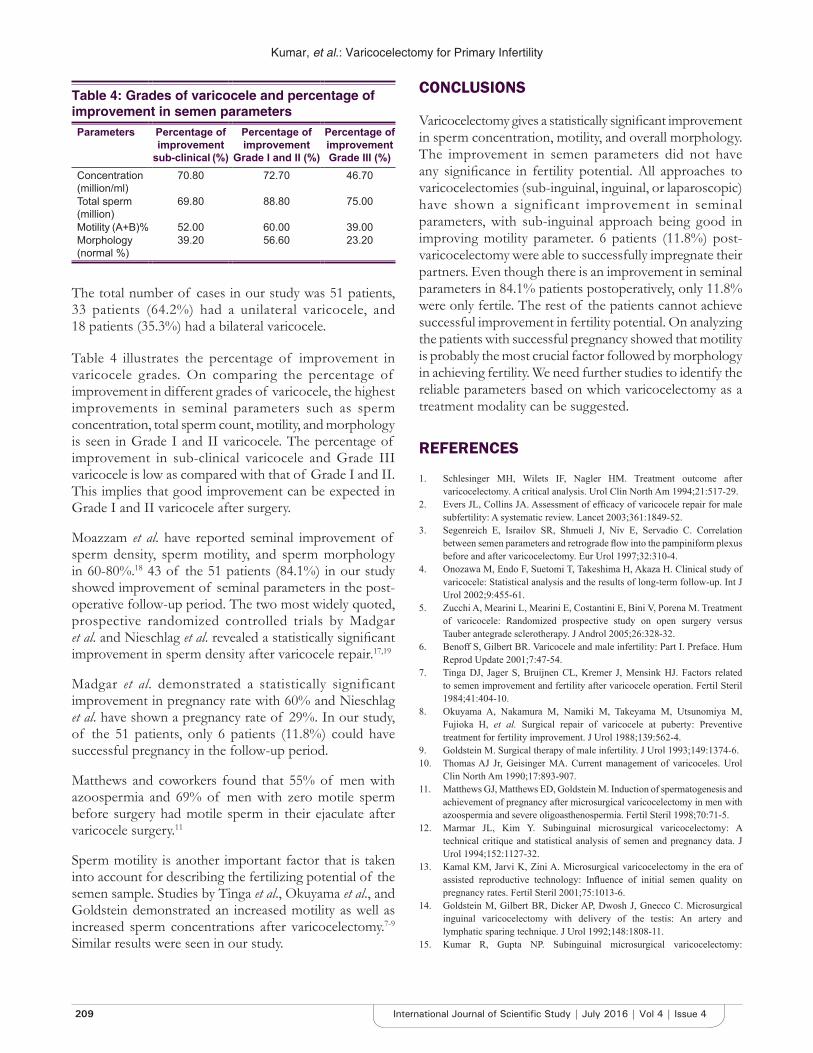
Kumar, et al.: Varicocelectomy for Primary Infertility
209 International Journal of Scientific Study | July 2016 | Vol 4 | Issue 4
The total number of cases in our study was 51 patients, 33 patients (64.2%) had a unilateral varicocele, and 18 patients (35.3%) had a bilateral varicocele.
Table 4 illustrates the percentage of improvement in varicocele grades. On comparing the percentage of improvement in different grades of varicocele, the highest improvements in seminal parameters such as sperm concentration, total sperm count, motility, and morphology is seen in Grade I and II varicocele. The percentage of improvement in sub-clinical varicocele and Grade III varicocele is low as compared with that of Grade I and II. This implies that good improvement can be expected in Grade I and II varicocele after surgery.
Moazzam et al. have reported seminal improvement of sperm density, sperm motility, and sperm morphology in 60-80%.18 43 of the 51 patients (84.1%) in our study showed improvement of seminal parameters in the post-operative follow-up period. The two most widely quoted, prospective randomized controlled trials by Madgar et al. and Nieschlag et al. revealed a statistically significant improvement in sperm density after varicocele repair.17,19
Madgar et al. demonstrated a statistically significant improvement in pregnancy rate with 60% and Nieschlag et al. have shown a pregnancy rate of 29%. In our study, of the 51 patients, only 6 patients (11.8%) could have successful pregnancy in the follow-up period.
Matthews and coworkers found that 55% of men with azoospermia and 69% of men with zero motile sperm before surgery had motile sperm in their ejaculate after varicocele surgery.11
Sperm motility is another important factor that is taken into account for describing the fertilizing potential of the semen sample. Studies by Tinga et al., Okuyama et al., and Goldstein demonstrated an increased motility as well as increased sperm concentrations after varicocelectomy.7-9 Similar results were seen in our study.
CONCLUSIONS
Varicocelectomy gives a statistically significant improvement in sperm concentration, motility, and overall morphology. The improvement in semen parameters did not have any significance in fertility potential. All approaches to varicocelectomies (sub-inguinal, inguinal, or laparoscopic) have shown a significant improvement in seminal parameters, with sub-inguinal approach being good in improving motility parameter. 6 patients (11.8%) post-varicocelectomy were able to successfully impregnate their partners. Even though there is an improvement in seminal parameters in 84.1% patients postoperatively, only 11.8% were only fertile. The rest of the patients cannot achieve successful improvement in fertility potential. On analyzing the patients with successful pregnancy showed that motility is probably the most crucial factor followed by morphology in achieving fertility. We need further studies to identify the reliable parameters based on which varicocelectomy as a treatment modality can be suggested.
REFERENCES
1. Schlesinger MH, Wilets IF, Nagler HM. Treatment outcome after varicocelectomy. A critical analysis. Urol Clin North Am 1994;21:517-29.
2. Evers JL, Collins JA. Assessment of efficacy of varicocele repair for male subfertility: A systematic review. Lancet 2003;361:1849-52.
3. Segenreich E, Israilov SR, Shmueli J, Niv E, Servadio C. Correlation between semen parameters and retrograde flow into the pampiniform plexus before and after varicocelectomy. Eur Urol 1997;32:310-4.
4. Onozawa M, Endo F, Suetomi T, Takeshima H, Akaza H. Clinical study of varicocele: Statistical analysis and the results of long-term follow-up. Int J Urol 2002;9:455-61.
5. Zucchi A, Mearini L, Mearini E, Costantini E, Bini V, Porena M. Treatment of varicocele: Randomized prospective study on open surgery versus Tauber antegrade sclerotherapy. J Androl 2005;26:328-32.
6. Benoff S, Gilbert BR. Varicocele and male infertility: Part I. Preface. Hum Reprod Update 2001;7:47-54.
7. Tinga DJ, Jager S, Bruijnen CL, Kremer J, Mensink HJ. Factors related to semen improvement and fertility after varicocele operation. Fertil Steril 1984;41:404-10.
8. Okuyama A, Nakamura M, Namiki M, Takeyama M, Utsunomiya M, Fujioka H, et al. Surgical repair of varicocele at puberty: Preventive treatment for fertility improvement. J Urol 1988;139:562-4.
9. Goldstein M. Surgical therapy of male infertility. J Urol 1993;149:1374-6.10. Thomas AJ Jr, Geisinger MA. Current management of varicoceles. Urol
Clin North Am 1990;17:893-907.11. Matthews GJ, Matthews ED, Goldstein M. Induction of spermatogenesis and
achievement of pregnancy after microsurgical varicocelectomy in men with azoospermia and severe oligoasthenospermia. Fertil Steril 1998;70:71-5.
12. Marmar JL, Kim Y. Subinguinal microsurgical varicocelectomy: A technical critique and statistical analysis of semen and pregnancy data. J Urol 1994;152:1127-32.
13. Kamal KM, Jarvi K, Zini A. Microsurgical varicocelectomy in the era of assisted reproductive technology: Influence of initial semen quality on pregnancy rates. Fertil Steril 2001;75:1013-6.
14. Goldstein M, Gilbert BR, Dicker AP, Dwosh J, Gnecco C. Microsurgical inguinal varicocelectomy with delivery of the testis: An artery and lymphatic sparing technique. J Urol 1992;148:1808-11.
15. Kumar R, Gupta NP. Subinguinal microsurgical varicocelectomy:
Table 4: Grades of varicocele and percentage of improvement in semen parametersParameters Percentage of
improvement sub-clinical (%)
Percentage of improvement
Grade I and II (%)
Percentage of improvement Grade III (%)
Concentration(million/ml)
70.80 72.70 46.70
Total sperm(million)
69.80 88.80 75.00
Motility (A+B)% 52.00 60.00 39.00Morphology(normal %)
39.20 56.60 23.20

Kumar, et al.: Varicocelectomy for Primary Infertility
210International Journal of Scientific Study | July 2016 | Vol 4 | Issue 4
Evaluation of the results. Urol Int 2003;71:368-72.16. The influence of varicocele on parameters of fertility in a large group of
men presenting to infertility clinics. World Health Organization. Fertil Steril 1992;57:1289-93.
17. Nieschlag E, Behre HM, Schlingheider A, Nashan D, Pohl J, Fischedick AR. Surgical ligation vs. angiographic embolization of the vena spermatica: A prospective randomized study for the treatment of varicocele-related
infertility. Andrologia 1993;25:233-7.18. Moazzam M, Siddiqui KM, Ather MH, Biyabani RS. Surgical ligation of
scrotal varicocele for male factor infertility is a valid option of treatment. JPMA 2006;56:364.
19. Madgar I, Weissenberg R, Lunenfeld B, Karasik A, Goldwasser B. Controlled trial of high spermatic vein ligation for varicocele in infertile men. Fertil Steril 1995;63:120-4.
How to cite this article: Kumar R, Sekar H, Krishnamoorthy S, Kumaresan N, Ramanan V. Analysis of Outcomes of Surgical Treatment for Varicocele in Primary Infertility Based on Seminal Parameters and Pregnancy Rate. Int J Sci Stud 2016;4(4):205-210.
Source of Support: Nil, Conflict of Interest: None declared.

211 International Journal of Scientific Study | July 2016 | Vol 4 | Issue 4
Comparative Study between Oral Clonidine and Placebo in Laparoscopic Surgery to Attenuate the Hemodynamic Changes Due to Pneumoperitoneum and HypercarbiaGomathi Karmegam1, Kundhavi Devi2, Bhavani Vaidyanathan3, Heber Anandan4
1Senior Assistant Professor, Government Kilpauk Medical College Hospital, Affiliated to the Tamil Nadu Dr. M. G. R. Medical University, Tamil Nadu, India, 2Professor, Government Kilpauk Medical College Hospital, Tamil Nadu, India, 3Assistant Professor, Indira Gandhi Medical College and Research Institute, Puducherry, India, 4Senior Clinical Scientist, Dr. Agarwal’s Health Care Limited, Tamil Nadu, India
due to minimal handling. Patients may face a stormy intraoperative course if anesthesia fails to maintain the hemodynamics within the normal range during pneumoperitoneum and hypercarbia. Numerous studies have evolved over the years to identify agents capable of maintaining hemodynamics in a balanced spectrum during laparoscopic surgery. Clonidine is a centrally acting alpha 2 agonist used for premedication as it decreases polypharmacy, post-operative nausea, vomiting, and shivering. It has antihypertensive property by decreasing sympathetic outflow.1 The use of drugs such as oral clonidine as a premedicant given before operation would be desirable to enhance the hypotensive action of inhalational agent without the disadvantages of intravenous (IV) vasodilators.
INTRODUCTION
Laparoscopy or minimally invasive surgery has made huge advances in the field of surgery. It has a plethora of advantages being reduced pain, therefore, decreased hospital stay, reduction in medical cost with day care admissions, reduction in morbidity and mortality
Original Article
AbstractBackground: Laparoscopic surgery has enormous advantages to the patient with regards to the post-operative pain and early discharge. However, it is not without disadvantages due to the hemodynamic swings that occur intraoperatively due to the intra-abdominal pressures. This study was undertaken to determine the efficacy of oral clonidine to attenuate these changes in response to pneumoperitoneum and hypercarbia.
Aim: A comparative study of oral clonidine and placebo in laparoscopic surgery to attenuate the hemodynamic response due to pneumoperitoneum and hypercarbia.
Materials and Methods: A prospective, randomized, double-blind, control study on 30 patients in two groups, one receiving placebo and the other receiving clonidine 5 µg/kg 90 min before induction was carried out. Intraoperative heart rate (HR) and non-invasive blood pressure (BP) were monitored along with post-operative complications such as nausea and vomiting were documented.
Results: Statistical analysis revealed oral clonidine was far beneficial maintaining intraoperative hemodynamic swings during laparoscopic surgery.
Conclusion: Oral clonidine was found beneficial with the ease of administration and control of HR and BP intraoperatively. Preoperatively, sedated and calm patient with a lesser incidence of polypharmacy intraoperatively was added advantages.
Key words: Clonidine, Hypercarbia, Laparoscopic surgery, Pneumoperitoneum, Stress response
Access this article online
www.ijss-sn.com
Month of Submission : 05-2016 Month of Peer Review : 06-2016 Month of Acceptance : 07-2016 Month of Publishing : 07-2016
Corresponding Author: Dr. Gomathi Karmegam, Department of Anaesthesiology, Kilpauk Medical College Hospital, Chennai - 600 010, Tamil Nadu, India. Phone: +91-9444005371. E-mail: [email protected]
Print ISSN: 2321-6379Online ISSN: 2321-595X
DOI: 10.17354/ijss/2016/408

Karmegam, et al.: Oral Clonidine vs. Placebo in lap surgery
212International Journal of Scientific Study | July 2016 | Vol 4 | Issue 4
AimComparative study between oral clonidine and placebo in laparoscopic surgery to attenuate the hemodynamic changes due to pneumoperitoneum and hypercarbia.
MATERIALS AND METHODS
A prospective, double-blind study was undertaken in 60 patients: 30 patients in two groups belonging to American Society of Anesthesiologist (ASA) PS I and II. After obtaining approval from the Institute Ethical Committee and due patient consent, Group P (placebo) was given vitamin C 90 min before surgery; Group C (oral clonidine) was given clonidine 5 µg/kg 90 min before surgery. Both the administrator and receiver were blind to the groups allocated. Inclusion criteria were patients aged 20-50 years, ASA PS I and II patients, those undergoing abdominal laparoscopic surgeries, patient weighing 50-65 kg. Exclusion criteria were hypertension/ischemic heart disease/rheumatic heart disease, diabetes mellitus, obesity, anticipated difficult airway, sinus bradycardia/heart blocks/conduction defects, patients on antipsychotics, patients on digitalis, calcium channel blockers and b-blockers, H/o cerebrovascular disease (cerebrovascular accident), chronic renal disease with increased renal parameters, pre-operative hypotension, and patient refusal. The sedation score was assessed preoperatively using the sedation score. Patients were assessed for level of sedation at 30 min, 60 min, and 90 min after premedication. The following score was used to assess the degree of sedation:Grade 1: Awake and alert patientGrade 2: Awake and calm lying down qualityGrade 3: Drowsy, arousable on oral commandsGrade 4: Drowsy, arousable on mild physical stimuliGrade 5: Drowsy, arousable on vigorous physical stimuli
only.
Intraoperative heart rate (HR) and blood pressure (BP) were recorded every 5 min from the period of insufflations to release. Intraoperative top ups of fentanyl, propofol, and deepening the plane with sevoflurane were documented.
Pre-operative AssessmentOnly those patients in the ASA Class I and II undergoing laparoscopic procedures were taken into this study. Nil oral duration was 8 h. Patients were given the oral premedicant, in a sealed cover 90 min before surgery and shifted to the operation theater with an 18G venflon and maintenance IV fluid. Sedation scoring was done to assess the efficacy of the premedicant. After shifting into the OT, monitors with non-invasive BP, SpO2, and electrocardiography were connected. Antacid prophylaxis was given with injection. Ranitidine 50 mg and injection
ondansetron 4 mg IV. Premedication was given with injection Glycopyrrolate 10 μ/kg, injection Midazolam 10 μ/kg and injection Fentanyl 2 μ/kg were given in IV. Preoxygenation with 100% O2 done for 3-5 min. Patient induced with injection. Propofol 2 mg/kg and injection atracurium 0.5 mg/kg after ascertaining good mask ventilation. Maintenance with N2O and O2 2:1 and sevoflurane 0.75-2 vol%. Titrated doses of muscle relaxant, fentanyl, and propofol were administered according to the need intraoperatively. Monitoring of HR and BP was done every 5 min during and after intubation, positioning, pneumoperitoneum and during extubation. Pneumoperitoneum was achieved with CO2 insufflation and intra-abdominal pressure was not allowed to exceed 12-15 mmHg. Time duration from insufflation to release was recorded. Rescue doses of fentanyl or propofol to deepen the plane of anesthesia was taken into consideration. Rescue with injection atropine was used if the HR <30%, injection ephedrine if the BP <30%, and injection nitroglycerin (NTG) infusion was used if the BP exceeded >30% of the patient’s baseline. The patient was extubated at the end of surgery. The patients were then shifted to the recovery room and were kept under observation for about 60 min during which the HR, BP, respiratory rate, and SpO2 were monitored at 30 min intervals for any signs of respiratory depression.
Statistics and AnalysisData were expressed as mean, standard deviation, or absolute values. Qualitative data were compared with the Chi-square test and Fisher’s exact test. Quantitative variables were compared with the Student t-test. The level of statistical significance was set at P < 0.05.
RESULTS
The mean age in Group C was 26.8 ± 4.40, and in the Group P, it was 26.3 ± 4.08 (P = 0.649). The mean weight in Group C was 53 ± 6.81. The mean weight in Group P was 51.13 ± 5.47 (P = 0.246). Sex distribution in each group was male = 17, female = 13. Thus, the demographic profile was comparable between the two groups. P value not significant.
At the end of 30 min following premedication, 25 patients in Group C were in the sedation score of 2, at the end of 60 min, 26 patients with the score of 3, and after 90 min, 23 patients had a score of 3. It was seen that all the patients in Group P were in the score of 1 till 90 min (Table 1).
9 (30%) patients in Group P supplemental doses of propofol and fentanyl when compared to 2 (6.66%) of Group C.

Karmegam, et al.: Oral Clonidine vs. Placebo in lap surgery
213 International Journal of Scientific Study | July 2016 | Vol 4 | Issue 4
i. It is hereby seen that a significantly higher consumption of sevoflurane >1 vol% almost 50% of patients in Group P was required when compared to Group C 2 (6.66%).
ii. 6 (20%) patients in Group P needed NTG infusion (0.1 mg/kg), whereas only 1 (3.33%) patients in Group C needed the same (Table 2).
Cardiovascular ParametersMean values of hemodynamic parameters (HR, systolic arterial pressure, diastolic arterial pressure, and mean arterial pressure) were taken at preinduction, at scopy 1 and 5 min thereafter, and with pneumoperitoneum every 5 min intraoperatively for the first 30 min and 15 min thereafter. Hemodynamic stability was remarkable with Group C as seen by the comparisons with Group P (Tables 3 and 4). The equivalent stability was maintained in Group P with additional propofol and fentanyl top-ups and sevo 1-2 vol% and NTG infusion. The pressor response to both intubation and extubation was blunted in the clonidine group Figure 1. Mean duration of surgery was comparable in both groups and not found significant Table 5.
There was no respiratory depression in both the groups, more so in the clonidine group. The patients in both groups had a stable cardiovascular status in the post-operative period.
DISCUSSION
Laparoscopy has been the mainstay in most abdominal surgeries in the past few years. Advantages being its minimal bowel handling and early ambulation of the patients, the disadvantages were CO2 insufflation, pneumoperitoneum, and hypercarbia. CO2 absorption causes a trigger in the HR, hypertension, and an increase in systemic vascular resistance. Pneumoperitoneum causes an increase in intra-abdominal pressure causing a biphasic fluctuation in BP.1,2 Positioning of the patient, namely, trendelenburg and reverse trendelenburg causes changes in the vascular physiology. CO2 elimination is usually achieved with hyperventilation and deepening the plane of anesthesia which may result in a delay in recovery.3 Pneumoperitoneum beyond
pressures of 18 mmHg may result in a compartment-like syndrome and a fall in venous return to the heart.4 Diaphragm being pushed upward reduces the FRC and ineffective ventilation due to increased airway pressures. Absorption of CO2 into the third spaces causes oozing from surgical sites and blood loss.5 Studies by Abe et al., conducted on patients were focused on delineating the
Table 1: Sedation score of both groupsScore Grade of sedation and anxiolysis 30 min (%) 60 min (%) 90 min (%)
Group C Group P Group C Group P Group C Group P1 Awake and alert 4 (13.33) 30 (100.00) 0 30 (100.00) 0 30 (100.00)2 Awake, calm, lying quietly 25 (83.33) 0 4 (13.33) 0 3 (10.00) 03 Drowsy arousable on oral commands 1 (3.33) 0 26 (86.67) 0 23 (76.67) 04 Drowsy arousable on mild physical stimuli 0 0 0 0 4 (13.33) 05 Drowsy arousable on vigorous physical stimuli only 0 0 0 0 0 0
Table 2: Reduction in requirements of anesthetic drugs and NTG seen in both groupsDrug 30 min %
Group C Group PPropofol/fentanyl 2 (6.67) 9 (30.00)Sevoflurane
1 vol% 2 (6.67) 5 (16.67)2 vol% 0 15 (50.00)>2 vol% 0 10 (33.33)
NTG 1 (3.33) 6 (20.00)Atropine 1 (3.33) 0Ephedrine 1 (3.33) 0
Table 3: HR during pneumoperitoneumTime of observation Group HR
(Bt/min)SD P value
Baseline Group C 81.166 4.026 0.154Group P 79.633 4.213
Preinduction Group C 72.8 5.821 <0.0001Group P 91.6 3.45
At Scopy Group C 75.566 6.463 <0.0001Group P 95.098 18.422
1 min after intubation Group C 76.601 6.499 <0.0001Group P 94.223 2.721
5 min after intubation Group C 80.333 7.871 0.130Group P 85.396 16.294
Average intraoperative value Group C 65.6 8.071 <0.0001Group P 79.6 8.298
During extubation Group C 69.66 14.406 0.632Group P 85.232 18.436
1 min after extubation Group C 72.1 10.121 <0.0001Group P 89.03 6.206
5 min after extubation Group C 75.533 11.578 0.001Group P 84.733 5.051
30 min after extubation Group C 73.066 15.097 0.052Group P 78.795 4.727
2 h after extubation Group C 78.333 10.346 0.946Group P 78.13 124.3
4 h after extubation Group C 80.366 8.176 0.026Group P 76.466 4.651
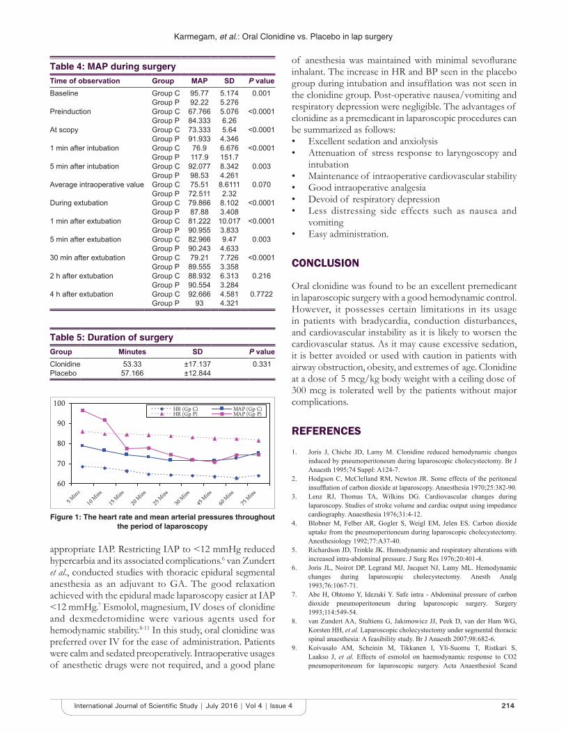
Karmegam, et al.: Oral Clonidine vs. Placebo in lap surgery
214International Journal of Scientific Study | July 2016 | Vol 4 | Issue 4
appropriate IAP. Restricting IAP to <12 mmHg reduced hypercarbia and its associated complications.6 van Zundert et al., conducted studies with thoracic epidural segmental anesthesia as an adjuvant to GA. The good relaxation achieved with the epidural made laparoscopy easier at IAP <12 mmHg.7 Esmolol, magnesium, IV doses of clonidine and dexmedetomidine were various agents used for hemodynamic stability.8-11 In this study, oral clonidine was preferred over IV for the ease of administration. Patients were calm and sedated preoperatively. Intraoperative usages of anesthetic drugs were not required, and a good plane
of anesthesia was maintained with minimal sevoflurane inhalant. The increase in HR and BP seen in the placebo group during intubation and insufflation was not seen in the clonidine group. Post-operative nausea/vomiting and respiratory depression were negligible. The advantages of clonidine as a premedicant in laparoscopic procedures can be summarized as follows:• Excellent sedation and anxiolysis• Attenuation of stress response to laryngoscopy and
intubation• Maintenance of intraoperative cardiovascular stability• Good intraoperative analgesia• Devoid of respiratory depression• Less distressing side effects such as nausea and
vomiting• Easy administration.
CONCLUSION
Oral clonidine was found to be an excellent premedicant in laparoscopic surgery with a good hemodynamic control. However, it possesses certain limitations in its usage in patients with bradycardia, conduction disturbances, and cardiovascular instability as it is likely to worsen the cardiovascular status. As it may cause excessive sedation, it is better avoided or used with caution in patients with airway obstruction, obesity, and extremes of age. Clonidine at a dose of 5 mcg/kg body weight with a ceiling dose of 300 mcg is tolerated well by the patients without major complications.
REFERENCES
1. Joris J, Chiche JD, Lamy M. Clonidine reduced hemodynamic changes induced by pneumoperitoneum during laparoscopic cholecystectomy. Br J Anaesth 1995;74 Suppl: A124-7.
2. Hodgson C, McClelland RM, Newton JR. Some effects of the peritoneal insufflation of carbon dioxide at laparoscopy. Anaesthesia 1970;25:382-90.
3. Lenz RJ, Thomas TA, Wilkins DG. Cardiovascular changes during laparoscopy. Studies of stroke volume and cardiac output using impedance cardiography. Anaesthesia 1976;31:4-12.
4. Blobner M, Felber AR, Gogler S, Weigl EM, Jelen ES. Carbon dioxide uptake from the pneumoperitoneum during laparoscopic cholecystectomy. Anesthesiology 1992;77:A37-40.
5. Richardson JD, Trinkle JK. Hemodynamic and respiratory alterations with increased intra-abdominal pressure. J Surg Res 1976;20:401-4.
6. Joris JL, Noirot DP, Legrand MJ, Jacquet NJ, Lamy ML. Hemodynamic changes during laparoscopic cholecystectomy. Anesth Analg 1993;76:1067-71.
7. Abe H, Ohtomo Y, Idezuki Y. Safe intra - Abdominal pressure of carbon dioxide pneumoperitoneum during laparoscopic surgery. Surgery 1993;114:549-54.
8. van Zundert AA, Stultiens G, Jakimowicz JJ, Peek D, van der Ham WG, Korsten HH, et al. Laparoscopic cholecystectomy under segmental thoracic spinal anaesthesia: A feasibility study. Br J Anaesth 2007;98:682-6.
9. Koivusalo AM, Scheinin M, Tikkanen I, Yli-Suomu T, Ristkari S, Laakso J, et al. Effects of esmolol on haemodynamic response to CO2 pneumoperitoneum for laparoscopic surgery. Acta Anaesthesiol Scand
Table 4: MAP during surgeryTime of observation Group MAP SD P valueBaseline Group C 95.77 5.174 0.001
Group P 92.22 5.276Preinduction Group C 67.766 5.076 <0.0001
Group P 84.333 6.26At scopy Group C 73.333 5.64 <0.0001
Group P 91.933 4.3461 min after intubation Group C 76.9 6.676 <0.0001
Group P 117.9 151.75 min after intubation Group C 92.077 8.342 0.003
Group P 98.53 4.261Average intraoperative value Group C 75.51 8.6111 0.070
Group P 72.511 2.32During extubation Group C 79.866 8.102 <0.0001
Group P 87.88 3.4081 min after extubation Group C 81.222 10.017 <0.0001
Group P 90.955 3.8335 min after extubation Group C 82.966 9.47 0.003
Group P 90.243 4.63330 min after extubation Group C 79.21 7.726 <0.0001
Group P 89.555 3.3582 h after extubation Group C 88.932 6.313 0.216
Group P 90.554 3.2844 h after extubation Group C 92.666 4.581 0.7722
Group P 93 4.321
Table 5: Duration of surgeryGroup Minutes SD P valueClonidine 53.33 ±17.137 0.331Placebo 57.166 ±12.844
Figure 1: The heart rate and mean arterial pressures throughout the period of laparoscopy

Karmegam, et al.: Oral Clonidine vs. Placebo in lap surgery
215 International Journal of Scientific Study | July 2016 | Vol 4 | Issue 4
1998;42:510-7.10. Jee D, Lee D, Yun S, Lee C. Magnesium sulphate attenuates arterial
pressure increases during laparoscopic cholecystectomy Br J Anaesth 2009;103:484-9.
11. Aho M, Scheinin M, Lehtinen AM, Erkola O, Vuorinen J, Korttila K. Intramuscularly administered dexmedetomidine attenuates hemodynamic and stress hormone responses to gynecologic laparoscopy. Anesth Analg 1992;75:932-9.
How to cite this article: Karmegam G, Devi K, Bavani V, Anandan H. Comparative Study between Oral Clonidine and Placebo in Laparoscopic Surgery to Attenuate the Hemodynamic Changes Due to Pneumoperitoneum and Hypercarbia. Int J Sci Stud 2016;4(4):211-215.
Source of Support: Nil, Conflict of Interest: None declared.
216International Journal of Scientific Study | July 2016 | Vol 4 | Issue 4
Effect of HAART on the Oral Manifestations in Human Immunodeficiency Virus Positive Patients: A Clinical Study, Tiruchirappalli, Tamil NaduU Punitha Gnana Selvi1, D Kamatchi2, C Babu3, S Keerthana3
1Professor and Head, Department of Dental Surgery, KAPV Government Medical College, Tiruchirappalli, Tamil Nadu, India, 2Assistant Professor, Department of Dental Surgery, KAPV Government Medical College, Tiruchirappalli, Tamil Nadu, India, 3Senior Intern, Department of Dental Surgery, KAPV Government Medical College, Tiruchirappalli, Tamil Nadu, India
viral infection characterized by profound impairment of the immune system that leads to opportunistic infections and secondary neoplasm.1 The mortality rate is decreasing due to improvements in treatment regimen and increased to access to anti-HIV drugs.2,3 The management modalities that have brought the mortality rates in the developed world down are the antiretroviral therapy (ART), especially the highly active ART (HAART).4-7
HIV is a retrovirus, which has a specific affinity for CD4 cells (T-helper cells).1 CD4 count helps in evaluating disease diagnosis, progression, prognosis, and making the decision for ART.
INTRODUCTION
Acquired immunodeficiency syndrome (AIDS) is an infectious disease caused by human immunodeficiency virus (HIV) and it is a highly lethal, progressively epidemic
Original Article
AbstractIntroduction: Human immunodeficiency virus (HIV) infection remains as a pandemic health problem. The development of highly active antiretroviral therapy (HAART) has significantly altered the course of HIV disease into a manageable disease with enhanced quality of life chiefly in the developed countries. Very limited studies have been made available concerning the effect of HAART on oral lesions in developing countries like India.
Aims and Objectives: (1) To compare the oral lesions in HIV-seropositive patients between those who are newly diagnosed and those actively undergoing HAART and (2) To determine the therapeutic effects of HAART on oral lesions in HIV-seropositive patients.
Materials and Methods: Screening was done among the patients attending the VCTC Centre at Government Hospital, Tiruchirappalli. Screening was conducted on 240 patients both male and females who were confirmed with HIV positive status with CD4 count <200 cells/mm3 were selected for the study. Out of these, 136 patients were already under HAART for the past 3 months and the rest 104 patients were newly diagnosed as HIV-patient, yet treatment to be started. The soft tissues of the oral cavity were examined thoroughly for the presence of any lesions. The results were tabulated in a standard form, and they were statistically analyzed using Chi-square test.
Results: The incidence of periodontal disease was decreased in patients under HAART after 3 months. There was strong decline in the severity of oral candidiasis, but there were increased the incidence of melanotic hyperpigmentation.
Conclusion: The oral manifestations of HIV infection have changed due to the advent of HAART. Many opportunistic infections have the fastest response to HAART as a result of an improved immune system. These lesions alongside immunologic parameters can be used as indicators of success or failure of antiretroviral therapy.
Key words: CD4 cell counts, Highly active antiretroviral therapy, Human immunodeficiency virus patients, Oral manifestations
Access this article online
www.ijss-sn.com
Month of Submission : 05-2016 Month of Peer Review : 06-2016 Month of Acceptance : 07-2016 Month of Publishing : 07-2016
Corresponding Author: Dr. D Kamatchi, 135 Madurai Road, Manaparai - 621 306, Tamil Nadu, India. Phone: +91-9842461740. E-mail: [email protected]
Print ISSN: 2321-6379Online ISSN: 2321-595X
DOI: 10.17354/ijss/2016/409

Selvi, et al.: Efficacy of HAART on Oral Lesions
217 International Journal of Scientific Study | July 2016 | Vol 4 | Issue 4
According to the new classification given by Center for Disease Control and Prevention, clinical AIDS is defined by a CD4 count of <200 and/or a CD4 <14%.8
One important factor to be considered to reduce the mortality and morbidity rates for patients with HIV is early diagnosis and identification of features with prognostic significance. In this regard, oral manifestations of AIDS have played a very important role and some of the oral lesions have both diagnostic and prognostic values.9-11 The occurrence of oral manifestations is favored by immune deterioration.12 Oral manifestations are seen in 30-80% of HIV patients. The more common of the oral lesions that occur along with HIV are the candidiasis, hyperpigmentation, angular cheilitis, gingivitis, periodontitis, aphthous ulcers, herpes simplex infections, and oral hairy leukoplakia of which oral candidiasis is of significance as far as prognostic indicators of immune suppression are concerned.13-15
The aim of our study is to compare the oral lesions in HIV-seropositive patients between those who are newly diagnosed and those actively undergoing HAART and to determine the therapeutic effects of HAART on oral lesions in HIV-seropositive patients.
MATERIALS AND METHODS
The screening was done among the patients attending the VCTC Centre at Mahatma Gandhi Memorial Government hospital, Tiruchirappalli. The screening was conducted on 240 patients both male and females of age group between 25 and 40 who were confirmed with HIV-positive status with CD4 count <200 cells/mm3 were selected for the study. Out of these, 136 patients were already under HAART for the last 3 months and the rest 104 patients were newly diagnosed as HIV positive, yet the treatment to be started. Their HIV status was confirmed by western blot. CD4+ T-lymphocyte counts were assessed by flow cytometry. The study was approved by the Institutional Review Board and all the participants provided with voluntary informed consent. All potential risks and benefits were explained to all the subjects in a language that they could comprehend. Demographic data were collected using a standard questionnaire. Present medical status, past medical history, family history, and drug history were recorded. Findings were recorded using internationally accepted presumptive clinical criteria of erythematous candidiasis (European Community)-clearing house (1991-1993) on oral problems related to HIV infection and WHO collaborating center on oral manifestations of the immunodeficiency virus.4 Oral examination was performed which included examining the individuals for lesions such as oral candidiasis, periodontitis, hyperpigmentation,
aphthous, and herpes simplex ulcers. HAART consisted of the combination of two nucleoside reverse transcriptase inhibitors (NRTI’s) (lamivudine + stavudine or lamivudine + zidovudine) and a non-NRTI (nevirapine orefavirenz). An intra-oral examination was performed clinically. Intra-orally the gingiva, periodontium, alveolar mucosa, buccal mucosa, lips, vestibule, dorsal, ventral, and lateral surfaces of the tongue and floor of the mouth were examined thoroughly. The results were tabulated in a standard form, and they were statistically analyzed using Chi-square test.
RESULTS
Out of 240 patients, 130 (54.16%) were males and 110 (45.83%) were females. When the age groups of the patients were compiled, the majority of patients were in the 25-40 (74%) years age group with the mean age of all the subjects being 34 years.
The distribution and comparison of oral lesions in individuals newly diagnosed with HAART were as follows. The occurrence of periodontal lesions was the most predominant (68.26%), followed by oral candidiasis (27.8%), hyperpigmentation (9.6%), aphthous ulcers (7.6%), and herpes (3.8%). During the intra-oral examination of individuals under active HAART, the incidence of periodontal diseases was still the most at 45%, followed by hyperpigmentation (38%), oral candidiasis (13%), aphthous ulcers (2%), and herpes (0.7).
Thus, it was observed that the occurrence of periodontal diseases was significantly reduced in patients under HAART (68.26% newly diagnosed and 33% under HAART). Interestingly, the occurrence of hyperpigmentation was higher in patients under HAART. Before HAART therapy, only 10 patients had hyperpigmentation whereas the total number of patients under HAART observed with hyperpigmentation was 38 patients which was statistically significant. The total number of patients who presented with oral candidiasis during diagnosis were 29 (27.8%) which had reduced to 13 (11.7%) in patients under HAART. Thus, the occurrence of oral candidiasis was significantly reduced in patients undergoing HAART actively. As far as the types of oral candidiasis observed in newly diagnosed patients, 14 patients exhibited erythematous candidiasis, 6 of them showed angular cheilitis, pseudomembranous candidiasis and chronic hyperplastic candidiasis were seen in 5 and 4 patients, respectively. During the examination of patients who are actively undergoing HAART, a number of patients exhibiting erythematous candidiasis reduced to 9, angular cheilitis to 1, whereas 2 patients had pseudomembranous candidiasis and only one patient exhibited chronic hyperplastic candidiasis (Tables 1 and 2).
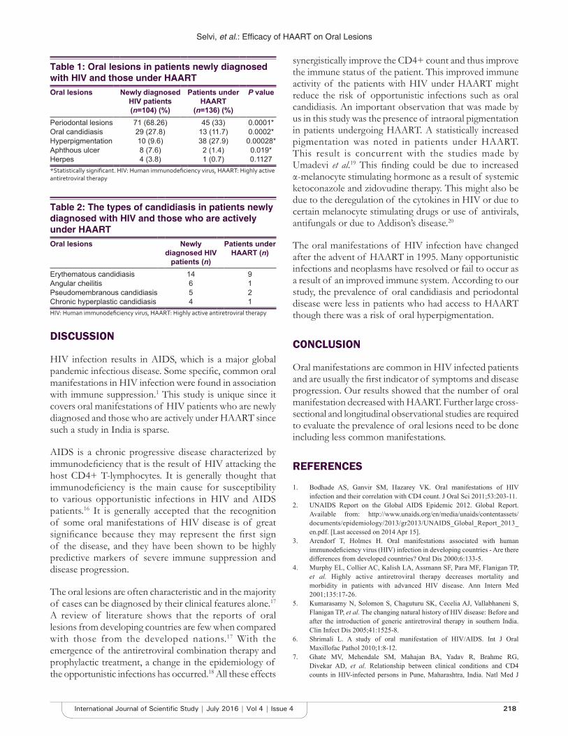
Selvi, et al.: Efficacy of HAART on Oral Lesions
218International Journal of Scientific Study | July 2016 | Vol 4 | Issue 4
DISCUSSION
HIV infection results in AIDS, which is a major global pandemic infectious disease. Some specific, common oral manifestations in HIV infection were found in association with immune suppression.1 This study is unique since it covers oral manifestations of HIV patients who are newly diagnosed and those who are actively under HAART since such a study in India is sparse.
AIDS is a chronic progressive disease characterized by immunodeficiency that is the result of HIV attacking the host CD4+ T-lymphocytes. It is generally thought that immunodeficiency is the main cause for susceptibility to various opportunistic infections in HIV and AIDS patients.16 It is generally accepted that the recognition of some oral manifestations of HIV disease is of great significance because they may represent the first sign of the disease, and they have been shown to be highly predictive markers of severe immune suppression and disease progression.
The oral lesions are often characteristic and in the majority of cases can be diagnosed by their clinical features alone.17 A review of literature shows that the reports of oral lesions from developing countries are few when compared with those from the developed nations.17 With the emergence of the antiretroviral combination therapy and prophylactic treatment, a change in the epidemiology of the opportunistic infections has occurred.18 All these effects
synergistically improve the CD4+ count and thus improve the immune status of the patient. This improved immune activity of the patients with HIV under HAART might reduce the risk of opportunistic infections such as oral candidiasis. An important observation that was made by us in this study was the presence of intraoral pigmentation in patients undergoing HAART. A statistically increased pigmentation was noted in patients under HAART. This result is concurrent with the studies made by Umadevi et al.19 This finding could be due to increased α-melanocyte stimulating hormone as a result of systemic ketoconazole and zidovudine therapy. This might also be due to the deregulation of the cytokines in HIV or due to certain melanocyte stimulating drugs or use of antivirals, antifungals or due to Addison’s disease.20
The oral manifestations of HIV infection have changed after the advent of HAART in 1995. Many opportunistic infections and neoplasms have resolved or fail to occur as a result of an improved immune system. According to our study, the prevalence of oral candidiasis and periodontal disease were less in patients who had access to HAART though there was a risk of oral hyperpigmentation.
CONCLUSION
Oral manifestations are common in HIV infected patients and are usually the first indicator of symptoms and disease progression. Our results showed that the number of oral manifestation decreased with HAART. Further large cross-sectional and longitudinal observational studies are required to evaluate the prevalence of oral lesions need to be done including less common manifestations.
REFERENCES
1. Bodhade AS, Ganvir SM, Hazarey VK. Oral manifestations of HIV infection and their correlation with CD4 count. J Oral Sci 2011;53:203-11.
2. UNAIDS Report on the Global AIDS Epidemic 2012. Global Report. Available from: http://www.unaids.org/en/media/unaids/contentassets/documents/epidemiology/2013/gr2013/UNAIDS_Global_Report_2013_en.pdf. [Last accessed on 2014 Apr 15].
3. Arendorf T, Holmes H. Oral manifestations associated with human immunodeficiency virus (HIV) infection in developing countries - Are there differences from developed countries? Oral Dis 2000;6:133-5.
4. Murphy EL, Collier AC, Kalish LA, Assmann SF, Para MF, Flanigan TP, et al. Highly active antiretroviral therapy decreases mortality and morbidity in patients with advanced HIV disease. Ann Intern Med 2001;135:17-26.
5. Kumarasamy N, Solomon S, Chaguturu SK, Cecelia AJ, Vallabhaneni S, Flanigan TP, et al. The changing natural history of HIV disease: Before and after the introduction of generic antiretroviral therapy in southern India. Clin Infect Dis 2005;41:1525-8.
6. Shrimali L. A study of oral manifestation of HIV/AIDS. Int J Oral Maxillofac Pathol 2010;1:8-12.
7. Ghate MV, Mehendale SM, Mahajan BA, Yadav R, Brahme RG, Divekar AD, et al. Relationship between clinical conditions and CD4 counts in HIV-infected persons in Pune, Maharashtra, India. Natl Med J
Table 1: Oral lesions in patients newly diagnosed with HIV and those under HAARTOral lesions Newly diagnosed
HIV patients(n=104) (%)
Patients under HAART
(n=136) (%)
P value
Periodontal lesions 71 (68.26) 45 (33) 0.0001*Oral candidiasis 29 (27.8) 13 (11.7) 0.0002*Hyperpigmentation 10 (9.6) 38 (27.9) 0.00028*Aphthous ulcer 8 (7.6) 2 (1.4) 0.019*Herpes 4 (3.8) 1 (0.7) 0.1127*Statistically significant. HIV: Human immunodeficiency virus, HAART: Highly active antiretroviral therapy
Table 2: The types of candidiasis in patients newly diagnosed with HIV and those who are actively under HAARTOral lesions Newly
diagnosed HIV patients (n)
Patients under HAART (n)
Erythematous candidiasis 14 9Angular cheilitis 6 1Pseudomembranous candidiasis 5 2Chronic hyperplastic candidiasis 4 1HIV: Human immunodeficiency virus, HAART: Highly active antiretroviral therapy

Selvi, et al.: Efficacy of HAART on Oral Lesions
219 International Journal of Scientific Study | July 2016 | Vol 4 | Issue 4
India 2000;13:183-7.8. Castro KG, Ward JW, Slutsker L, Buehler JW, Jaffe HW, Berkelman R,
et al. 1993 Revised classification system for HIV infection and expanded surveillance case definition for AIDS among adolescents and adults. Clin Infect Dis 1993;17:802-10.
9. Greenspan JS. Sentinels and signposts: The epidemiology and significance of the oral manifestations of HIV disease. Oral Dis 1997;3 Suppl 1:S13-7.
10. Kolokotronis A, Kioses V, Antoniades D, Mandraveli K, Doutsos I, Papanayotou P. Immunologic status in patients infected with HIV with oral candidiasis and hairy leukoplakia. Oral Surg Oral Med Oral Pathol 1994;78:41-6.
11. Glick M, Muzyka BC, Lurie D, Salkin LM. Oral manifestations associated with HIV-related disease as markers for immune suppression and AIDS. Oral Surg Oral Med Oral Pathol 1994;77:344-9.
12. Gaurav S, Keerthilatha PM, Archna N. Prevalence of oral manifestations and their association with CD4/CD8 Ratio and HIV viral load in South India. Int J Dent 2011;2011:964278.
13. Begg MD, Panageas KS, Mitchell-Lewis D, Bucklan RS, Phelan JA, Lamster IB. Oral lesions as markers of severe immunosuppression in HIV-infected homosexual men and injection drug users. Oral Surg Oral Med Oral Pathol Oral Radiol Endod 1996;82:276-83.
14. Greenspan D, Komaroff E, Redford M, Phelan JA, Navazesh M, Alves ME, et al. Oral mucosal lesions and HIV viral load in the Women’s Interagency
HIV Study (WIHS). J Acquir Immune Defic Syndr 2000;25:44-50.15. Kerdpon D, Pongsiriwet S, Pangsomboon K, Iamaroon A, Kampoo K,
Sretrirutchai S, et al. Oral manifestations of HIV infection in relation to clinical and CD4 immunological status in northern and southern Thai patients. Oral Dis 2004;10:138-44.
16. Liu X, Liu H, Guo Z, Luan W. Association of asymptomatic oral candidal carriage, oral candidiasis and CD4 lymphocyte count in HIV-positive patients in China. Oral Dis 2006;12:41-4.
17. Ranganathan K, Reddy BV, Kumarasamy N, Solomon S, Viswanathan R, Johnson NW. Oral lesions and conditions associated with human immunodeficiency virus infection in 300 south Indian patients. Oral Dis 2000;6:152-7.
18. Ranganathan K, Geethalakshmi E, Krishna Mohan Rao U, Vidya KM, Kumarasamy N, Solomon S. Orofacial and systemic manifestations in 212 paediatric HIV patients from Chennai, South India. Int J Paediatr Dent 2010;20:276-82.
19. Umadevi KM, Ranganathan K, Pavithra S, Hemalatha R, Saraswathi TR, Kumarasamy N, et al. Oral lesions among persons with HIV disease with and without highly active antiretroviral therapy in southern India. J Oral Pathol Med 2007;36:136-41.
20. Langford A, Pohle HD, Gelderblom H, Zhang X, Reichart PA. Oral hyperpigmentation in HIV-infected patients. Oral Surg Oral Med Oral Pathol 1989;67:301-7.
How to cite this article: Selvi UPG, Kamatchi D, Babu C, Keerthana S. Effect of HAART on the Oral Manifestations in Human Immunodeficiency Virus Positive Patients: A Clinical Study, Tiruchirappalli, Tamil Nadu. Int J Sci Stud 2016;4(4):216-219.
Source of Support: Nil, Conflict of Interest: None declared.
220International Journal of Scientific Study | July 2016 | Vol 4 | Issue 4
Low Cost Negative Pressure Wound Therapy for Treatment of Diabetic Foot UlcersN Arun Kumar1, S Surya Nihar2, K Chetan3
1Head, Department of General Surgery, St Martha’s Hospital, Bengaluru, Karnataka, India, 2Post-graduate Student, Department of General Surgery, St Martha’s Hospital, Bengaluru, Karnataka, India, 3Senior Consultant, St Martha’s Hospital, Bengaluru, Karnataka, India
a limb is lost somewhere in the world as a consequence of diabetes.1
In 1993, Fleischmann et al.2 described a new technique called negative pressure wound therapy (NPWT) where sub-atmospheric pressure was applied over the surface of the wound to promote wound healing.
The exact mechanism of action of NPWT is still debated. Negative pressure applies mechanical forces to the wound to promote wound healing; these are known as micro- and macro-strain.3-5 The visible changes that occur after the application of negative pressure is called as macro-strain, and the changes at the cellular level are called as micro-strain. Macro-strain includes drawing the wound edges together, removal of exudates, and even distribution of negative pressure. Micro-strain includes microdeformational changes occurring at the cellular level due to cell stretch resulting in a reduction of edema, enhancement of perfusion, and promotion of granulation tissue formation by facilitating cell migration.
INTRODUCTION
Many techniques have been tried over the centuries to heal chronic foot ulcers. Although there exists no ideal wound dressing, the management of chronic wounds especially diabetic wounds has seen many new developments.
Diabetic ulcers are the most common cause of chronic wounds.
The lifetime risk of a person with diabetes developing a foot ulcer is as high as 25% and it is said that every 30 s
Original Article
AbstractBackground: Many techniques have been tried in the treatment of chronic diabetic ulcers. Recent studies have shown that application of negative pressure wound therapy (NPWT) has got an important role in healing of such wounds. However, this technology is commercially available and is expensive. Cost-effective alternatives can, therefore, be of great value to patients. Hence, we conducted a study to evaluate the effects of NPWT on wound healing in diabetic ulcers using hospital based devices.
Objective: To determine the effectiveness of “low cost” NPWT in the treatment of diabetic foot ulcers.
Materials and Methods: A prospective observational study was conducted on 35 patients with diabetic foot ulcers. After an initial debridement, NPWT was applied using the standard wall suction apparatus and gauze - freely available in all hospital set-ups.
Results: Mean ulcer size was 49.4 cm2 at the start of therapy. The mean ulcer size at the end of 1, 2, and 3 weeks was 41.5 cm2, 30.2 cm2 and 24 cm2, respectively (P value is significant <0.05). The mean duration of healing was 22.4 days. Out of 35 wounds, 26 underwent split skin grafting, 6 wounds healed by secondary intention, and 3 wounds required local flaps for coverage. None of them needed amputation.
Conclusion: “Low cost” negative pressure therapy using wall suction is an effective treatment for faster healing of diabetic wounds.
Key words: Diabetes mellitus, Foot ulcers, Negative pressure wound therapy
Access this article online
www.ijss-sn.com
Month of Submission : 05-2016 Month of Peer Review : 06-2016 Month of Acceptance : 07-2016 Month of Publishing : 07-2016
Corresponding Author: Dr. N Arun Kumar, #214, 11th Main, Lakkasandra Extension, Bengaluru, Karnataka, India. Phone: +91-9845017098. E-mail: [email protected]
Print ISSN: 2321-6379Online ISSN: 2321-595X
DOI: 10.17354/ijss/2016/410

Kumar, et al.: Economical NPWT for Diabetic Foot Ulcers
221 International Journal of Scientific Study | July 2016 | Vol 4 | Issue 4
Based on this concept, commercial form of NPWT system also known as vacuum-assisted closure (VAC)6 (KCI, San Antonio) has become available since many years. After its introduction, it has evolved into a widely accepted treatment modality of chronic wounds.
However due to its high cost, the commercially available forms of NPWT cannot be used for unaffordable patients and hospitals with limited facilities, especially in rural places. Shalom et al.7 devised a “homemade” and cost-effective NPWT and the results were comparable to commercial NPWT.
Based on the same principles, we have applied NPWT to the wound surface using simple and easily available equipment available in any hospital settings.
MATERIALS AND METHODS
This study was a prospective observational study carried out to evaluate the effects of NPWT on diabetic foot ulcers and conducted in the Department of General Surgery from November 2013 to November 2015 at St. Martha’s Hospital, Bengaluru. 35 patients presenting with diabetic foot ulcers and consenting for the procedure were studied after Ethical and Scientific Committee approval. All the wounds underwent prior debridement and wound swab was sent for culture and sensitivity testing. The patients were treated with broad spectrum antibiotics initially, later according to the sensitivity pattern.
NPWT was applied using sterile gauzes as wound bed cover over which a Ryle’s tube was placed for the suction purpose. This dressing was secured with the help of sterile Ioban (iodinated drape). The Ryle’s tube was connected to sterile tubing which in turn was attached to wall suction with a continuous pressure of −125 mmHg. The presence of negative pressure was confirmed by frequently checking the readings of suction apparatus and also by the collapse of dressing material after application of suction (Figure 1a-c).
The dressing was changed twice every week. At every dressing change, the wound was washed with saline and assessed for relevant parameters. The results were analyzed weekly till the wound was satisfactorily covered with healthy granulation tissue. In our study, we used paired t-test to check the level of statistical significance. A P < 0.05 was considered significant. Physician consultation was taken regularly for the management of diabetes and other comorbidities.
Inclusion Criteria• Foot ulcers in diabetic patients aged more than
16 years.
Exclusion Criteria• Malignant ulcers, ulcers with exposed bone or blood
vessels, underlying osteomyelitis• Critical limb ischemia (ankle-brachial index <0.4).
RESULTS AND OBSERVATIONS
A total of 35 patients were studied of which 22 were males (63%). Most of the patients belonged to age group 45-54 years (46%). 21 patients (60%) had a duration of diabetes mellitus (DM) <10 years. Most patients (82%) had glycosylated hemoglobin levels >7.5%. Hypertension was the most commonly associated comorbidity (31%). Chronic kidney disease was seen in 4 cases and peripheral vascular disease was seen in 4 cases (ankle-brachial index of <0.8).
The majority of the ulcers presented were spontaneous in onset (63%). Left foot was the most commonly involved (42%). The mean duration of ulcer was 7.02 weeks.
About 32 cases had purulent discharge at the time of presentation (91%). Pus culture and sensitivity was done in all the patients on admission (Graph 1). Escherichia coli was the most common isolate 12 cases followed by Staphylococcus aureus 6 cases and Pseudomonas 4 cases. 7 cases showed mixed growth (both Gram-positive and negative). Out of the 6 cases of S. aureus, 4 cases were methicillin-resistant S. aureus (MRSA).
Most patients had ulcer size in the range of 31-50 cm2 (57%). The mean ulcer size at admission was 49.4 cm2. The mean ulcer size at the end of 1, 2, and 3 weeks was 41.5 cm2, 30.2 cm2, and 24 cm2, respectively. P value, calculated using paired t-test is significant (P < 0.01).
Evaluation of the granulation tissue at the time of application of NPWT showed that only unhealthy granulation tissue was seen in all wounds. At the end of 4 weeks, 28 wounds had attained 100% granulation and were taken up for skin grafting (Figure 2a-c). Rest 7 wounds had attained granulation >75% of wound area.
Figure 1: (a-c) Method of application of negative pressure wound therapy with wall suction, (b and c) The Ryle’s tube
being connected to the wall suction apparatus
a b c

Kumar, et al.: Economical NPWT for Diabetic Foot Ulcers
222International Journal of Scientific Study | July 2016 | Vol 4 | Issue 4
In our study time taken from the start of therapy to the time, they were ready for coverage ranged from 11 to 46 days. The mean duration of healing was 22.4 days.
About 26 wounds underwent split skin grafting, 3 wounds developed only partial granulation cover and these required local flap cover for wound closure and healing. In 6 patients not willing for surgery, the wounds had healed by complete epithelialization. No patient required any form of amputation.
DISCUSSION
Most of our patients are males in working age group, engaged in occupations like farming where they are always prone to trivial trauma. In our study, 21 of the 35 patients had duration of diabetes <10 years (60%). 29 patients (82%) had a poor glycemic control with the HbA1c levels more than 7.5%. Our results are comparable to Bansal et al.8 where 51.46% had DM for <10 years and another study by Alva et al.9 where glycosylated hemoglobin levels are more than 7% in 74% of patients. The above facts substantiate that majority of the patients were ignorant regarding foot care during the early years of DM treatment.
The mean duration of ulcer was 7.02 weeks, with most of the patients having ulcer for the duration of <6 weeks
(51%). In a study by Jeffcoate et al.,10 the mean ulcer duration was 15 days. In another study by Margolis,11 the mean duration of diabetic foot ulcers was 5.39 months.
Pus culture and sensitivity was done for all the patients on admission (Graph 1). In our study, the most common isolates were Gram-negative organisms (18 cases). E. coli was the most common isolate (12 patients, 34%) followed by S. aureus (6 patients, 17%). Of the 6 cases of S. aureus, 4 cases were MRSA. Our results are comparable to a study by Alva et al.9 in which Gram-negative organisms (55.8%) were the major isolates. After the completion of therapy, once the wound culture was negative the wounds were subsequently taken up for grafting or for flap cover.
All the patients showed a significant decrease of wound discharge including the MRSA wounds. Of the 4 MRSA, 3 patients were taken up for Split-thickness skin grafts and one patient healed by complete epithelization (Figure 3a-f). The role of NPWT in reducing the bacterial load has been proven in a study by Nain et al.12 where they found that 40% of wounds were sterile after 3 weeks of VAC therapy as compared to only 20% of the wounds treated by conventional dressing turning sterile after 3 weeks of treatment.
The mean ulcer size at admission was 49.4 cm2. In our study, measurements of the wound were taken at the end of 1st, 2nd, and 3rd week. These findings are comparable to the mean ulcer size observed in the patients undergoing NPWT in a study conducted by Nather13 which ranged from 6.9 cm2 to 124 cm2 with a mean size of 54.6 cm2. As the duration of treatment with NPWT increases, larger wounds showed satisfactory decrease in size and
Graph 1: Culture-organism isolated
Figure 2: (a-c) Status of wound on admission, 2 weeks after negative pressure wound therapy and before grafting,
respectively
a b cFigure 3: (a-f) Wound culture showed methicillin-resistant
Staphylococcus aureus. Patient was started on appropriate antibiotics and negative pressure wound therapy. Patient
was not willing for surgery. Wound had healed by complete epithelialization
a
d e f
b c

Kumar, et al.: Economical NPWT for Diabetic Foot Ulcers
223 International Journal of Scientific Study | July 2016 | Vol 4 | Issue 4
better granulation tissue. The mean ulcer size at the end of 1, 2, and 3 weeks was 41.5 cm2, 30.2 cm2, and 24 cm2, respectively. P < 0.001 was calculated using paired t-test and is statistically significant.
After the commencement of NPWT, the percentage of granulation at the end of every week was analyzed. At the end of the 3rd week, most of the wounds - 19 wounds (54%) had attained granulation of 100% and were taken up for grafting. Rest 16 patients had >75% granulation tissue over the wound. Lone et al.14 observed granulation tissue deposition in 92.85% of wounds by 2nd week and 100% granulation by the 5th week of treatment in patients treated with NPWT.
32 wounds, 91% of cases developed healthy granulation after being treated by NPWT before closure of the wound. 3 wounds that did not attain satisfactory granulation were taken up for local flap cover. Nather,13 Armstrong et al.,15 also observed early deposition of granulation tissue in patients treated with NPWT.
The end point or the time taken for wound closure was taken as the day the wounds were treated by graft, flap or by complete epithelialization. In our study, at the end of 2 weeks, 6 patients attained complete closure and 19 patients attained complete wound closure at the end of 3rd week. Split skin grafting was the most common treatment modality used for wound closure following NPWT. Of the 35 patients, 26 (74%) underwent skin grafting. 6 patients not willing for grafting had healed by complete epithelialization of the wounding regular dressing. 3 patients were closed with a flap. Nain et al.12 had similar findings where split skin grafting was the most common mode of closure employed (65% of patients). Lone et al.14 also had similar results where grafting was used in 86.4% of patients treated with NPWT.
In our study, the mean duration of healing is 22.4 days. A study by McCallon et al.16 observed the mean time for complete healing was 22.8 days. Vuerstaek et al.17 had similar results where the average time to heal in the study group was 29 days. Nather13 observed that the mean time to heal was 23.3 days. In our study out of the 35 ulcers, 30 healed within a month of initiation of the therapy.
Based on the principles of negative pressure therapy by Morykwas et al.6 we have used easily available materials like wall suction and gauze to perform NPWT. The usage of foam mandates gas sterilization of foam – not readily available in all hospitals. Shalom et al.7 had treated 15 patients successfully using wall suction and gelatin sponge. Gill et al.18 performed similar studies using wall suction and foam to treat complex traumatic wounds.
After foam contamination was noted with the usage of VAC system, Kiyokawa et al.19 used saline irrigation with continuous aspiration and negative pressure for treatment of intractable ulcers. We have not noted any instance of wound infection or gauze contamination after the initiation of therapy.
Commercial NPWT machines are marketed by various companies. The cost of each unit ranges from 2 to 7 lakh rupees. The cost of each dressing ranges from 8000 rupees to 12,000 rupees. Expenses per dressing for our “low cost” NPWT ranges from Rs. 250 to 350 (depending on the wound size) along with nominal general ward charges, which is affordable by most of the patients.
The present medical practice is not only advancing technologically but also rapidly inching toward the evolution of cost-effective solutions. Although the outcomes are promising, expensive equipment cannot be afforded by everyone.
CONCLUSION
NPWT therapy is a reliable modality of treatment for chronic wounds. NPWT requires training to ensure appropriate and competent use.
Our study shows that patients who cannot afford or utilize commercially available products for any reason can also benefit from NPWT using standard wall suction apparatus and dressing materials - freely available in all hospitals and at affordable costs. Hospitals in rural set up where commercial equipment are not available or cannot be affordable might benefit from “low cost” alternatives.
We recommend the present method of treatment as the first line management in resource challenged conditions especially when the options for reconstruction are also limited.
REFERENCES
1. International Diabetes Federation. Time to Act: Diabetes and Foot Care. Brussels: International Diabetes Federation; 2005.
2. Fleischmann W, Strecker W, Bombelli M, Kinzl L. Vacuum sealing as treatment of soft tissue damage in open fractures. Unfallchirurg 1993;96:488-92.
3. McNulty AK, Schmidt M, Feeley T, Kieswetter K. Effects of negative pressure wound therapy on fibroblast viability, chemotactic signaling, and proliferation in a provisional wound (fibrin) matrix. Wound Repair Regen 2007;15:838-46.
4. Orgill DP, Bayer LR, Neuwalder J, Felter RC. Microdeformational wound therapy - A new era in wound healing. Bus Brief Glob Surg Future Dir Surg 2005;2:22-5.
5. Saxena V, Hwang CW, Huang S, Eichbaum Q, Ingber D, Orgill DP. Vacuum-assisted closure: Microdeformations of wounds and cell proliferation. Plast

Kumar, et al.: Economical NPWT for Diabetic Foot Ulcers
224International Journal of Scientific Study | July 2016 | Vol 4 | Issue 4
Reconstr Surg 2004;114:1086-96.6. Morykwas MJ, Simpson J, Punger K, Argenta A, Kremers L, Argenta J.
Vacuum-assisted closure: State of basic research and physiologic foundation. Plast Reconstr Surg 2006;117 7 Suppl:121S-6.
7. Shalom A, Eran H, Westreich M, Friedman T. Our experience with a “homemade” vacuum-assisted closure system. Isr Med Assoc J 2008;10:613-6.
8. Bansal E, Garg A, Bhatia S, Attri AK, Chander J. Spectrum of microbial flora in diabetic foot ulcers. Indian J Pathol Microbiol 2008;51:204-8.
9. Alva KA, Aithala PS, Rai R, Rekha B. Clinical and microbiological profile of diabetic foot in patients admitted at a tertiary care center in Mangalore. Muller J Med Sci Res 2013;4:3.
10. Jeffcoate WJ, Chipchase SY, Ince P, Game FL. Assessing the outcome of the management of diabetic foot ulcers using ulcer-related and person-related measures. Diabetes Care 2006;29:1784-7.
11. Margolis DJ, Allen-Taylor L, Hoffstad O, Berlin JA. Diabetic neuropathic foot ulcers: The association of wound size, wound duration, and wound grade on healing. Diabetes Care 2002;25:1835-9.
12. Nain PS, Uppal SK, Garg R, Bajaj K, Garg S. Role of negative pressure wound therapy in healing of diabetic foot ulcers. J Surg Tech Case Rep 2011;3:17-22.
13. Nather A. Role of negative pressure wound therapy in healing of diabetic
foot ulcers. J Surg Tech Case Rep 2011;3:10-1.14. Lone AM, Zaroo MI, Laway BA, Pala NA, Bashir SA, Rasool A. Vacuum-
assisted closure versus conventional dressings in the management of diabetic foot ulcers: A prospective case-control study. Diabet Foot Ankle 2014;5.
15. Armstrong DG, Lavery LA; Diabetic foot study consortium. Negative pressure wound therapy after partial diabetic foot amputation: A multicentre, randomised controlled trial. Lancet 2005;366:1704-10.
16. McCallon SK, Knight CA, Valiulus JP, Cunningham MW, McCulloch JM, Farinas LP. Vacuum-assisted closure versus saline-moistened gauze in the healing of postoperative diabetic foot wounds. Ostomy Wound Manage 2000;46:28-32, 34.
17. Vuerstaek JD, Vainas T, Wuite J, Nelemans P, Neumann MH, Veraart JC. State-of-the-art treatment of chronic leg ulcers: A randomized controlled trial comparing vacuum-assisted closure (VAC) with modern wound dressings. J Vasc Surg 2006;44:1029-37.
18. Gill NA, Hameed A, Sajjad Y, Ahmad Z, Rafique Mirza MA. “Homemade” negative pressure wound therapy: Treatment of complex wounds under challenging conditions. Wounds 2011;23:84-92.
19. Kiyokawa K, Takahashi N, Rikimaru H, Yamauchi T, Inoue Y. New continuous negative-pressure and irrigation treatment for infected wounds and intractable ulcers. Plast Reconstr Surg 2007;120:1257-65.
How to cite this article: Kumar NA, Nihar SS, Chetan K. Low Cost Negative Pressure Wound Therapy for Treatment of Diabetic Foot Ulcers. Int J Sci Stud 2016;4(4):220-224.
Source of Support: Nil, Conflict of Interest: None declared.

225 International Journal of Scientific Study | July 2016 | Vol 4 | Issue 4
Follicular Neoplasms Cytohistomorphological Aspects: A Case Study of 50 CasesK Balakrishnan1, Velayutham Sumathi2, R Siva Elangovan2, G Hemalatha2
1Professor, Department of Pathology, KAPV Government Medical College, Trichy, Tamil Nadu, India, 2Assistant Professor, Department of Pathology, KAPV Government Medical College, Trichy, Tamil Nadu, India
Adults who initially present with a thyroid lump, which on scan is usually “cold,” sometimes “warm” and only rarely “hot.” However, the large majority of “hot” nodules are benign.3
A FC cannot be distinguished from a FA based on cytologic features alone. It is distinguished from a FA based on capsular invasion, vascular invasion extrathyroidal tumor invasion, lymph node metastasis, systemic metastasis. Follicular neoplasm with tumor invasion into but not through the entire capsule is considered a FA. Occasionally FA will contain bizarre cells sometimes multinuclear cells may be seen. These lesions are benign. Vascular invasion is defined as tumor penetration into a large caliber vessel within or outside the capsule. Vascular invasion is the most valuable sign of malignancy.4
The incidence of thyroid cancers has steadily increased in most countries over the past two decades, predominantly
INTRODUCTION
Follicular adenoma (FA) and follicular carcinoma (FC) of the thyroid gland are tumors of follicular cell differentiation that consists of a microfollicular architecture with follicles lined by cuboidal epithelial cells. A FA is a benign encapsulated tumor of the thyroid gland. About 5% of the thyroid nodules are malignant.1 Most patients with a FA are clinically and biochemically euthyroid. Approximately, 1% of FAs are “toxic adenomas.”2
AbstractBackground: Thyroid nodules are common clinically (prevalence about 5%) and even more common on ultrasound examination (about 25%). Most of the thyroid nodules are follicular neoplasms cytologically. Follicular neoplasms of the thyroid gland include benign follicular adenomas (FAs) and follicular carcinoma (FC).
Objectives: The objective of our study was to assess the histomorphological aspects of follicular neoplasms. The incidence of FAs and FCs was evaluated with respect to age, clinical presentation, and thyroid status. Our study also focuses on histopathology of the spectrum of follicular neoplasms.
Materials and Methods: This prospective study of 1 year included (June 2015-June 2016) 50 cases of thyroidectomy specimen received in the department of pathology. A careful sampling and multiple sections were taken to assess the histological aspects for differentiating between FAs and FCs. Clinical data, thyroid status, and fine needle aspiration (FNA) reports were retrieved from hospital records.
Results: About 50 thyroidectomy specimens studied showed 28 cases of follicular neoplasms, an incidence of 56% of which 25 were FAs and 3 diagnosed as carcinoma. The mean age of presentation was 41.5 years. The incidence of carcinoma in our study was 10.7%. Only one case had lymphnode metastasis.
Conclusion: FNA plays a significant role in thyroid nodules, but resection of the lobe is a definitive treatment as well as for the diagnosis between adenoma and carcinoma. Immunohistochemistry and molecular markers play a very small role in the diagnosis of follicular neoplasms.
Key words: Adenoma, Carcinoma, Histopathology, Thyroid nodule
Access this article online
www.ijss-sn.com
Month of Submission : 05-2016 Month of Peer Review : 06-2016 Month of Acceptance : 07-2016 Month of Publishing : 07-2016
Corresponding Author: Dr. Velayutham Sumathi, Department of Pathology, KAPV Government Medical College, Trichy, Tamil Nadu, India. E-mail: [email protected]
Original ArticlePrint ISSN: 2321-6379
Online ISSN: 2321-595XDOI: 10.17354/ijss/2016/411

Balakrishnan, et al.: Follicular Neoplasam in Thyroidectomy Specimens
226International Journal of Scientific Study | July 2016 | Vol 4 | Issue 4
attributed to increased detection of small tumors by imaging techniques.5
FC accounts for 10% of all cases of thyroid malignancy in iodine sufficient areas and 25-40% of thyroid malignancies in areas of iodine deficiency.2,6 Most follicular cancers are nonfunctional. FC is usually unifocal and <10% have lymph node metastasis. The most patients with follicular neoplasms present with a solitary thyroid nodule. The diagnostic evaluation of a patient who presents with a thyroid nodule consists of routine fine needle aspiration cytology (FNAC), ultrasound examination of neck, and serum thyroid-stimulating hormone level.
FNAC of follicular neoplasms is characterized by abundant follicular epithelial sheets with crowding, overlapping and microfollicular formation with scant or no colloid.
According to Bethesda system of reporting thyroid cytopathology, this cytological appearance is classified as follicular neoplasms or suspicious for follicular neoplasms. FNAC specimen consistent with a follicular neoplasm accounts for 20% of all fine needle aspiration FNA biopsy results and has a 15-30% risk of malignancy.7
The differential diagnosis for a patient with a thyroid nodule and FNAC result consistent with a follicular neoplasm is a FA, adenomatous hyperplasia, FC, follicular variant of papillary carcinoma, and classic papillary carcinoma. Cytomorphologic criteria alone cannot distinguish a FA from a FC.8,9
MATERIALS AND METHODS
Surgically treated patients between June 2015 and June 2016 were prospectively analyzed. The study included 50 cases of thyroidectomy specimens. Histopathological analysis was performed. Clinical details and thyroid profiles were noted for available cases. Most of the patients had undergone an FNAC and those diagnosed as follicular neoplasms were correlated with histopathology analysis.
Most of the cases were found in the age group of 41-50 years with female preponderance (Figure 1 and Table 1). Out of the 28 cases, only 2 cases were male and rest was females. All the cases presented with thyromegaly, only 9 presented a solitary nodule. Out of the 28 cases, 9 cases were unifocal and rest was multifocal. The thyroid profile tests for most of the patients were retrieved. Most of them were found to be euthyroid.
Out of the 50 cases, histopathological study revealed 28 cases were diagnosed as follicular neoplasms (25 - FAs and 3 - FCs). An incidence of 56% of all thyroidectomy specimens received in our institution (Figure 2).
The cytological reports were analyzed retrospectively for the 28 cases along with clinical information. 19 of the 28 cases were reported as follicular neoplasms, 5 as adenomatoid nodule and 4 as goiter with papillary hyperplasia cytologically.
Out of the 28 cases, 13 cases presented as a solitary nodule, 9 as multiple nodules, 4 were with diffuse swelling, and 2 of them had no swelling and were diagnosed having a nodule only on ultrasound examination (Figure 3). All the patients were euthyroid except for one who showed toxic features.
FC was diagnosed in 3 cases (2 females and 1 male) wherein there was a capsular and vascular invasion. One of the
Figure 1: Line diagram depicting the incidence of follicular adenomas in various age groups
Figure 2: Incidence of cytological diagnosis of thyroid nodules (50 cases)
Table 1: Age wise distribution of SNG diagnosed as follicular neoplasmsAge distribution (in years) Number of cases20-30 531-40 741-50 1151-60 461-70 1Total 28
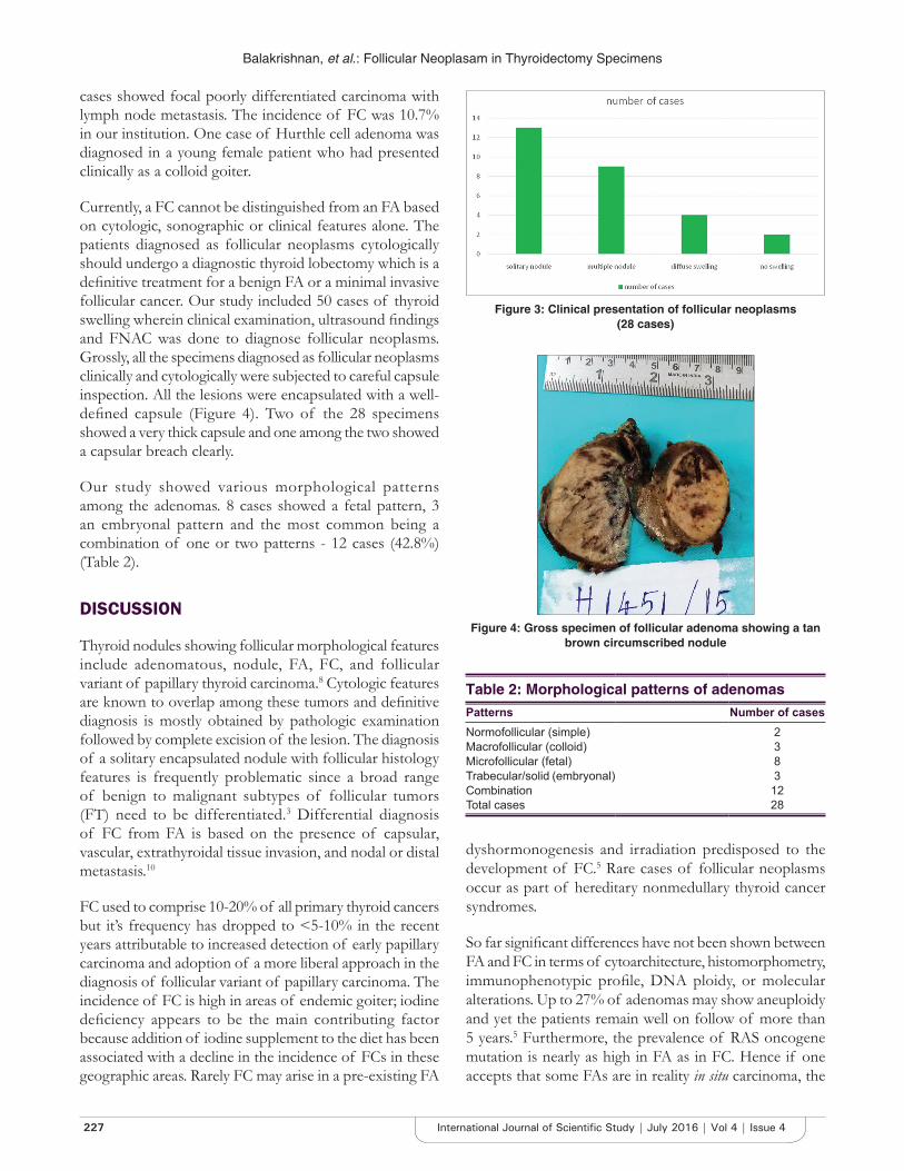
Balakrishnan, et al.: Follicular Neoplasam in Thyroidectomy Specimens
227 International Journal of Scientific Study | July 2016 | Vol 4 | Issue 4
cases showed focal poorly differentiated carcinoma with lymph node metastasis. The incidence of FC was 10.7% in our institution. One case of Hurthle cell adenoma was diagnosed in a young female patient who had presented clinically as a colloid goiter.
Currently, a FC cannot be distinguished from an FA based on cytologic, sonographic or clinical features alone. The patients diagnosed as follicular neoplasms cytologically should undergo a diagnostic thyroid lobectomy which is a definitive treatment for a benign FA or a minimal invasive follicular cancer. Our study included 50 cases of thyroid swelling wherein clinical examination, ultrasound findings and FNAC was done to diagnose follicular neoplasms. Grossly, all the specimens diagnosed as follicular neoplasms clinically and cytologically were subjected to careful capsule inspection. All the lesions were encapsulated with a well-defined capsule (Figure 4). Two of the 28 specimens showed a very thick capsule and one among the two showed a capsular breach clearly.
Our study showed various morphological patterns among the adenomas. 8 cases showed a fetal pattern, 3 an embryonal pattern and the most common being a combination of one or two patterns - 12 cases (42.8%) (Table 2).
DISCUSSION
Thyroid nodules showing follicular morphological features include adenomatous, nodule, FA, FC, and follicular variant of papillary thyroid carcinoma.8 Cytologic features are known to overlap among these tumors and definitive diagnosis is mostly obtained by pathologic examination followed by complete excision of the lesion. The diagnosis of a solitary encapsulated nodule with follicular histology features is frequently problematic since a broad range of benign to malignant subtypes of follicular tumors (FT) need to be differentiated.3 Differential diagnosis of FC from FA is based on the presence of capsular, vascular, extrathyroidal tissue invasion, and nodal or distal metastasis.10
FC used to comprise 10-20% of all primary thyroid cancers but it’s frequency has dropped to <5-10% in the recent years attributable to increased detection of early papillary carcinoma and adoption of a more liberal approach in the diagnosis of follicular variant of papillary carcinoma. The incidence of FC is high in areas of endemic goiter; iodine deficiency appears to be the main contributing factor because addition of iodine supplement to the diet has been associated with a decline in the incidence of FCs in these geographic areas. Rarely FC may arise in a pre-existing FA
dyshormonogenesis and irradiation predisposed to the development of FC.5 Rare cases of follicular neoplasms occur as part of hereditary nonmedullary thyroid cancer syndromes.
So far significant differences have not been shown between FA and FC in terms of cytoarchitecture, histomorphometry, immunophenotypic profile, DNA ploidy, or molecular alterations. Up to 27% of adenomas may show aneuploidy and yet the patients remain well on follow of more than 5 years.5 Furthermore, the prevalence of RAS oncogene mutation is nearly as high in FA as in FC. Hence if one accepts that some FAs are in reality in situ carcinoma, the
Figure 3: Clinical presentation of follicular neoplasms (28 cases)
Figure 4: Gross specimen of follicular adenoma showing a tan brown circumscribed nodule
Table 2: Morphological patterns of adenomasPatterns Number of casesNormofollicular (simple) 2Macrofollicular (colloid) 3Microfollicular (fetal) 8Trabecular/solid (embryonal) 3Combination 12Total cases 28
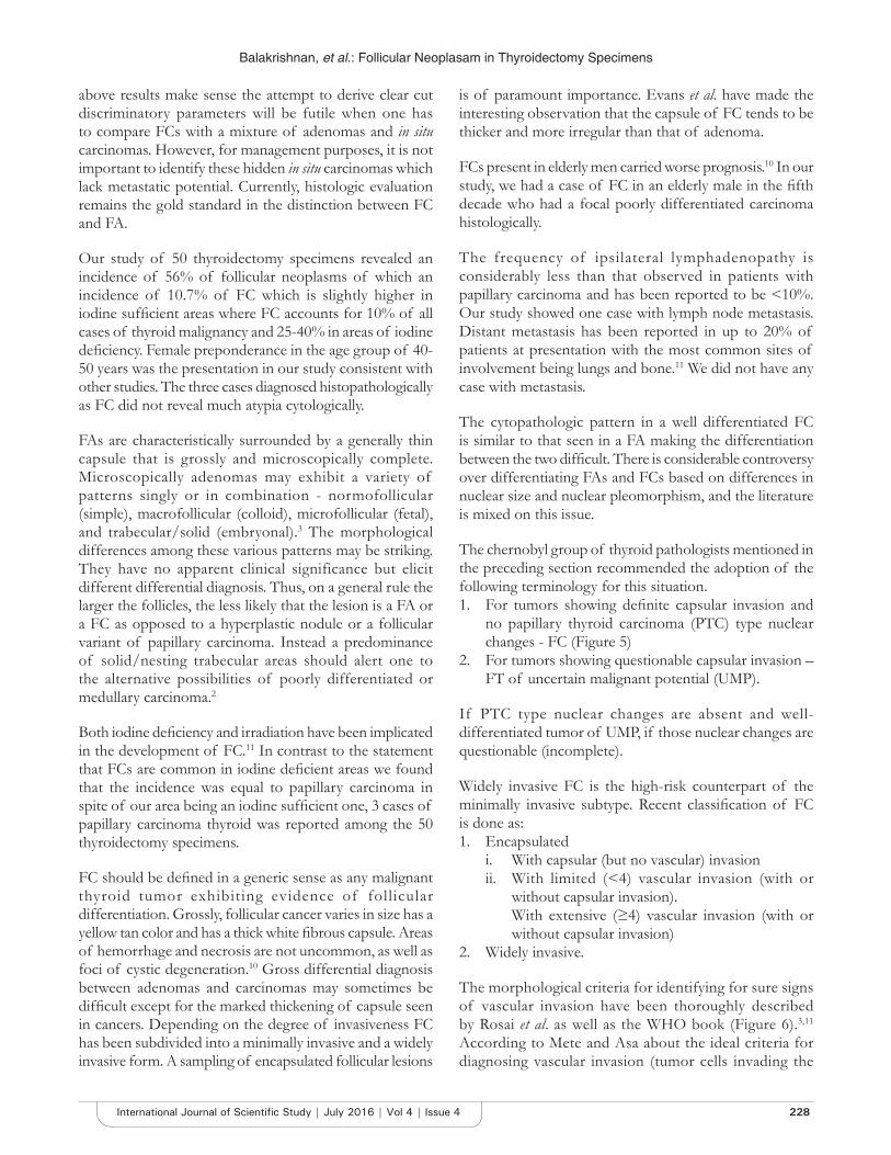
Balakrishnan, et al.: Follicular Neoplasam in Thyroidectomy Specimens
228International Journal of Scientific Study | July 2016 | Vol 4 | Issue 4
above results make sense the attempt to derive clear cut discriminatory parameters will be futile when one has to compare FCs with a mixture of adenomas and in situ carcinomas. However, for management purposes, it is not important to identify these hidden in situ carcinomas which lack metastatic potential. Currently, histologic evaluation remains the gold standard in the distinction between FC and FA.
Our study of 50 thyroidectomy specimens revealed an incidence of 56% of follicular neoplasms of which an incidence of 10.7% of FC which is slightly higher in iodine sufficient areas where FC accounts for 10% of all cases of thyroid malignancy and 25-40% in areas of iodine deficiency. Female preponderance in the age group of 40-50 years was the presentation in our study consistent with other studies. The three cases diagnosed histopathologically as FC did not reveal much atypia cytologically.
FAs are characteristically surrounded by a generally thin capsule that is grossly and microscopically complete. Microscopically adenomas may exhibit a variety of patterns singly or in combination - normofollicular (simple), macrofollicular (colloid), microfollicular (fetal), and trabecular/solid (embryonal).3 The morphological differences among these various patterns may be striking. They have no apparent clinical significance but elicit different differential diagnosis. Thus, on a general rule the larger the follicles, the less likely that the lesion is a FA or a FC as opposed to a hyperplastic nodule or a follicular variant of papillary carcinoma. Instead a predominance of solid/nesting trabecular areas should alert one to the alternative possibilities of poorly differentiated or medullary carcinoma.2
Both iodine deficiency and irradiation have been implicated in the development of FC.11 In contrast to the statement that FCs are common in iodine deficient areas we found that the incidence was equal to papillary carcinoma in spite of our area being an iodine sufficient one, 3 cases of papillary carcinoma thyroid was reported among the 50 thyroidectomy specimens.
FC should be defined in a generic sense as any malignant thyroid tumor exhibiting evidence of follicular differentiation. Grossly, follicular cancer varies in size has a yellow tan color and has a thick white fibrous capsule. Areas of hemorrhage and necrosis are not uncommon, as well as foci of cystic degeneration.10 Gross differential diagnosis between adenomas and carcinomas may sometimes be difficult except for the marked thickening of capsule seen in cancers. Depending on the degree of invasiveness FC has been subdivided into a minimally invasive and a widely invasive form. A sampling of encapsulated follicular lesions
is of paramount importance. Evans et al. have made the interesting observation that the capsule of FC tends to be thicker and more irregular than that of adenoma.
FCs present in elderly men carried worse prognosis.10 In our study, we had a case of FC in an elderly male in the fifth decade who had a focal poorly differentiated carcinoma histologically.
The frequency of ipsilateral lymphadenopathy is considerably less than that observed in patients with papillary carcinoma and has been reported to be <10%. Our study showed one case with lymph node metastasis. Distant metastasis has been reported in up to 20% of patients at presentation with the most common sites of involvement being lungs and bone.11 We did not have any case with metastasis.
The cytopathologic pattern in a well differentiated FC is similar to that seen in a FA making the differentiation between the two difficult. There is considerable controversy over differentiating FAs and FCs based on differences in nuclear size and nuclear pleomorphism, and the literature is mixed on this issue.
The chernobyl group of thyroid pathologists mentioned in the preceding section recommended the adoption of the following terminology for this situation.1. For tumors showing definite capsular invasion and
no papillary thyroid carcinoma (PTC) type nuclear changes - FC (Figure 5)
2. For tumors showing questionable capsular invasion – FT of uncertain malignant potential (UMP).
If PTC type nuclear changes are absent and well-differentiated tumor of UMP, if those nuclear changes are questionable (incomplete).
Widely invasive FC is the high-risk counterpart of the minimally invasive subtype. Recent classification of FC is done as:1. Encapsulated
i. With capsular (but no vascular) invasionii. With limited (<4) vascular invasion (with or
without capsular invasion). With extensive (≥4) vascular invasion (with or
without capsular invasion)2. Widely invasive.
The morphological criteria for identifying for sure signs of vascular invasion have been thoroughly described by Rosai et al. as well as the WHO book (Figure 6).3,11 According to Mete and Asa about the ideal criteria for diagnosing vascular invasion (tumor cells invading the

Balakrishnan, et al.: Follicular Neoplasam in Thyroidectomy Specimens
229 International Journal of Scientific Study | July 2016 | Vol 4 | Issue 4
vessel wall associated with the thrombus adherent to intravascular tumor), we diagnose vascular invasion if we detect well preserved neoplastic tissue within a vein. Whenever facing the dilemma “vascular invasion or not,” we do not rely on any immunostaining of endothelial cells. We search the additional sections of the capsule and stick to the following rule. If we do not detect any unequivocal signs of venous permeation we diagnose the tumor as non angioinvasive.12 The search for such signs was made for the 3 cases after multiple sampling of the capsule.
The prognosis of FC is directly related to the degree of encapsulation, hence the important distinction between minimally invasive and widely invasive types.
The most prominent molecular features of FC are the prominence of aneuploidy and the high prevalence of RAS mutations and of PAX8-peroxisome proliferator-activated
receptor γ (PPARγ) rearrangements, the latter as a result of a 2;3 translocation.13
Adenomas andfollicularcarcinomas
Follicular cell
RAS activation chromosomal instabilityIodine deficiency ( aneuploidy)
PAX8-PPARγ mutations
PAX8-PPARγ PTEN mutation
Other alterations
The prognosis of FC thyroid depends on several factors such as age of the patient, size and staging of the tumors, completeness of surgery and responsiveness to radioactive iodine. It also depends on the degree of invasiveness of tumors. Minimally invasive carcinomas carry a much better prognosis than do widely invasive carcinomas. The coexistence of less well-differentiated areas in some FCs raises the problem of differential diagnosis between FC and poorly differentiated carcinoma. In our study, we had one case with such diagnostic problems for which careful sampling was done and reported as FC with focal poorly differentiated carcinoma.
Immunohistochemistry of endothelial cells of the capsular vessels does not provide useful diagnostic data. There is also no immunohistochemical or molecular feature that may be needed as a reliable marker of invasion.14 Hence, no immunohistochemistry was used in our study and only careful histopathological analysis of our specimens was done to categorize the broad spectrum of follicular neoplasms.
CONCLUSION
Thyroid nodules whose FNA is diagnosed as follicular neoplasms should be resect unless there are significant contradictions to the surgical procedures. Our study of 50 thyroidectomy specimens of FAs and FCs has showed a slight increase in incidence 10.7% despite being an iodine sufficient area. Most of the patients were euthyroid and had come to the outpatient department only for a palpable thyroid swelling noted clinically. FNA does play an important role in the diagnosis of follicular neoplasms, but only histopathological examination can give the conclusive diagnosis.
For most thyroid tumors a diagnosis can be reached by morphologic assessment alone as with tumors in other endocrine organs the presence of nuclear atypia in a thyroid tumor is not necessarily synonymous with a diagnosis of malignancy.
Careful sampling of the specimen should be done to differentiate adenomas and carcinomas. Microscopically there should be a definite capsular and vascular invasion for the conclusive diagnosis of carcinoma.
Figure 5: H and E section showing tumor cells mushrooming the capsule (×10)
Figure 6: H and E section of follicular carcinoma showing definite vascular invasion (×40)

Balakrishnan, et al.: Follicular Neoplasam in Thyroidectomy Specimens
230International Journal of Scientific Study | July 2016 | Vol 4 | Issue 4
Immunohistochemistry and molecular markers play a very insignificant role in the categorization and diagnosis of follicular neoplasms.
REFERENCES
1. Mackenzie EJ, Mortimer RH. 6: Thyroid nodules and thyroid cancer. Med J Aust 2004;180:242-7.
2. Rosai J, Carcangiu ML, DeLellis RA. Tumours of thyroid gland. Atlas of Tumour Pathology. 3rd Series. Washington, DC: Armed Forces Institute of Pathology; 1992. p. 21-48.
3. Rosai J. Rosai and Ackerman’s Surgical Pathology. 9th ed., Vol. 1. London: Mosby; 2004. p. 1482.
4. Mills SE, Baloch ZW. Sternberg’s Diagnostic Surgical Pathology. 5th ed. Philadelphia, PA: Lippincott Williams & Wilkins; 2010. p. 510-1.
5. Dei Tos AP, Seregard S, Calonje E, Chan JK, Fletcher CD. Giant cell angiofibroma. A distinctive orbital tumor in adults. Am J Surg Pathol 1995;19:1286-93.
6. Correa P, Chen VW. Endocrine gland cancer. Cancer 1995;75 1 Suppl:338-52.7. Ali SZ, Cibas ES. The Bethesda System for Reprinting Thyroid
Cytopathology: Definitions, Criteria and Explanatory Notes. New York: Springer Science + Business Media; 2010.
8. Duggal R, Rajwanshi A, Gupta N, Vasishta RK. Interobserver variability amongst cytopathologists and histopathologists in the diagnosis of neoplastic follicular patterned lesions of thyroid. Diagn Cytopathol 2011;39:235-41.
9. Greaves TS, Olvera M, Florentine BD, Raza AS, Cobb CJ, Tsao-Wei DD, et al. Follicular lesions of thyroid: A 5-year fine-needle aspiration experience. Cancer 2000;90:335-41.
10. Silverberg SG, Deleius RA, Frable WJ. Surgical Pathology and Cytopathology. 3rd ed. New York: Churchill Livingstone; 1997. p. 2691.
11. Simoes MS, Asa SL, Kroll TG, Nifivorov Y, DeLellis R, Farid P, et al. Follicular Carcinoma WHO Classification of Tumours Pathology and Genetics. Tumours of Endocrine Glands. Lyon, France: IARC Press; 2004. p. 67-76.
12. Sobrinho-Simões M, Preto A, Rocha AS, Castro P, Máximo V, Fonseca E, et al. Molecular pathology of well-differentiated thyroid carcinomas. Virchows Arch 2005;447:787-93.
13. Nikiforova MN, Lynch RA, Biddinger PW, Alexander EK, Dorn GW 2nd, Tallini G, et al. RAS point mutations and PAX8-PPAR gamma rearrangement in thyroid tumors: Evidence for distinct molecular pathways in thyroid follicular carcinoma. J Clin Endocrinol Metab 2003;88:2318-26.
14. Prasad ML, Pellegata NS, Huang Y, Nagaraja HN, de la Chapelle A, Kloos RT. Galectin-3, fibronectin-1, CITED-1, HBME1 and cytokeratin-19 immunohistochemistry is useful for the differential diagnosis of thyroid tumors. Mod Pathol 2005;18:48-57.
How to cite this article: Balakrishnan K, Sumathi V, Elangovan RS, Hemalatha G. Follicular Neoplasms Cytohistomorphological Aspects: A Case Study of 50 Cases. Int J Sci Stud 2016;4(4):225-230.
Source of Support: Nil, Conflict of Interest: None declared.

231 International Journal of Scientific Study | July 2016 | Vol 4 | Issue 4
Comparison of Onset of Induction and Easiness of Laryngeal Mask Airway Insertion in Adults: Propofol versus Sevoflurane Single Vital Capacity Breath Technique-high Concentration (8%)A L Dharmalingam1, B S Thamilselvi2, M Sendil Murukan1, Heber Anandan3
1Assistant Professor, Department of Anaesthesiology, Government Kilpauk Medical College, Chennai, Tamil Nadu, India, 2Senior Resident, Department of Anaesthesiology, Government Kilpauk Medical College, Kilpauk, Tamil Nadu, India, 3Senior Clinical Scientist, Dr. Agarwal’s Health Care Limited, Tamil Nadu, Chennai, India
depth of anesthesia. This study is undertaken to compare the easiness of insertion of LMA using propofol/sevoflurane for induction. In recent times, inhalational induction with sevoflurane using single vital capacity breath (VCB) technique has been used. It is an alternate method to intravenous (IV) induction in adult patients. This method is rapid, with greater acceptances light excitatory phenomena and better hemodynamic profiles. LMA placement is more rapid after VCB induction using 8% of sevoflurane. This makes the sevoflurane sole drug for both maintenance and induction of anesthesia. It will make conversion period easier. Hence, this study is conducted to compare the consistency, excellence, and time to LMA insertion in adults after using sevoflurane induction and propofol induction.
INTRODUCTION
The laryngeal mask airway (LMA) is an airway device used frequently in anesthesia and critical care for airway management. It is an alternate and appropriate airway device to the facemask when endotracheal intubation is not mandatory. Acceptable placement of LMA needs enough
AbstractBackground: Laryngeal mask airway (LMA) insertion is an imperative tool in difficult airway scenarios. The ease of insertion in a spontaneously breathing patient and without the use of paralytic agents makes it a highly advantageous airway device.
Aims: In our study, we compare the onset of induction, to assess ease of LMA insertion, to assess number of attempts taken for correct placement, complications, if present during or following insertion of LMA, hemodynamic stability using sevoflurane high concentration inhalational technique and propofol intravenous (IV) induction technique in patients undergoing elective minor surgical procedures.
Materials and Methods: Prospective randomized study of American Society of Anesthesiologists physical status 1 or 2 patients was anesthetized with either a single vital capacity breath technique with sevoflurane 8% or IV propofol 2 mg/kg. Onset of induction and easiness of LMA insertion were studied, along with number of attempts taken for correct placement of LMA, hemodynamic stability, and complications.
Results: This study shows no significant difference between the two groups based on the demographic variables. The mean onset of induction was 44.40 s in the propofol group and 61.45 s in the sevoflurane group. The mean time for LMA insertion was 19.05 s in sevoflurane group and 12.88 s in propofol group.
Conclusion: Ease of insertion and placement of the LMA was found easier with propofol and prolonged in sevoflurane.
Key words: Laryngeal mask airway, Propofol, Sevoflurane, Vital capacity breath technique
Access this article online
www.ijss-sn.com
Month of Submission : 05-2016 Month of Peer Review : 06-2016 Month of Acceptance : 07-2016 Month of Publishing : 07-2016
Corresponding Author: Dr. A L Dharmalingam, Department of Anaesthesiology, Government Kilpauk Medical College, Kilpauk, Chennai - 600 010, Tamil Nadu, India. Phone: +91-9444220223. E-mail: [email protected]
Print ISSN: 2321-6379Online ISSN: 2321-595X
DOI: 10.17354/ijss/2016/412Original Article

Dharmalingam, et al.: Easiness of LMA insertion in adults between Propofol and sevoflurane induction
232International Journal of Scientific Study | July 2016 | Vol 4 | Issue 4
MATERIALS AND METHODS
A prospective, randomized, controlled trial was conducted in Government Kilpauk Medical College and Hospital, Chennai. Institutional Ethical Committee approval and written informed consent were obtained. 80 adult patients under American Society of Anesthesiologists (ASA) physical status 1 and 2 of either sex undergoing elective minor surgical procedures were enrolled for this study.
Inclusion CriteriaElective minor surgical procedures, both gender, ASA physical status 1-2, age from 18 years to 50 years, patients with normal body mass index from 18.5 to 25.
Exclusion CriteriaPatients not satisfying inclusion criteria, patients with cardiac disease, known case of malignant hyperthermia or suspected genetic propensity, patients with reactive airway disease. Boyles machine with circle CO2 absorber circuit, volatile anesthetic drug sevoflurane with vaporizer, propofol, classic LMA size 3 and 4. Resuscitation kit should be kept ready; approximate size endotracheal tubes, airways, suction apparatus. Patients in both the groups were IV cannulated with 18-gauge venflon. Monitors connected are non-invasive blood pressure (NIBP), electrocardiogram (ECG), and pulse oxymetry, end tidal CO2 (ETCO2), premedicated with IV injection glycopyrrolate 0.2 mg, fentanyl 2 µg/kg, ranitidine 50 mg, ondansetron 0.1 mg/kg, then preoxygenated for 3 min with 100% O2.
Propofol GroupPatients in the propofol group were preoxygenated with 100% oxygen for 3 min and anesthetized using propofol 2 mg/kg IV, given over a period of 30 s. The onset of induction (loss of eyelash reflex) was assessed. 30 s after the achievement of induction (i.e., 60 s after the start of propofol), jaw relaxation was assessed and, if achievable, LMA placement was attempted. If not possible, attempts were repeated every 30 s up to a max. 4 attempts, every time preceded by IV boluses of propofol about 0.5 mg/kg. NIBP, ECG, SPO2, and ETCO2 readings were recorded for 5 min in 1 min interval. Any failure of placement, defined as failure to insert the LMA after 4 attempts, they were rescued with suxamethonium 25 mg IV. The existence of difficulties correlated to induction and placement of the LMA was noted such as excitatory movement or withdrawal from pain, gagging, coughing, apnea, and laryngospasm. At the end of the surgery, the existence of blood on the LMA was noted.
Sevoflurane GroupA closed circuit with circle absorber for CO2 with a 2-L breathing bag was used. The closed circuit was primed with
8% sevoflurane in a 2:1 of N2O to O2 for 1 min at a rate of 6 L/min of fresh gas flow. Then, the patients were asked to take a deep breath after maximum exhalation and to hold as long as possible and then expire to residual volume. The onset of induction (loss of eyelash reflex) was assessed. 90 s after the induction, the jaw relaxation was assessed. 90 s was selected because it signifies the time at which all patients finished their VCB. If jaw relaxation was not possible, attempts were repeated every 30 s up to a max 4 attempts. At this time, anesthesia sustained with sevoflurane 8% and N2O 67% in O2. Once jaw relaxation was possible, LMA insertion was tried. Successful LMA insertion, time to LMA insertion (s), number of attempts (n), presence of blood on LMA, and hemodynamic parameters were observed.
The data were analyzed with SPSS software version 19.1 and P < 0.05 was considered statistically significant. Demographic data, the onset of induction, the time taken for LMA insertion, complications, and hemodynamic variables among the groups were analyzed with unpaired Student’s t-test. Chi-square analysis was used for comparing gender and number of attempts for insertion.
RESULTS
The number of attempts for LMA insertion was comparable in both the groups without any statistically significant difference. The only thing was time to LMA insertion was prolonged in sevoflurane group.
Compared between both groups, there was significant variation in the 1, 3 min post-insertion mean blood pressure, P < 0.0001 (Tables 1 and 2). This study shows no significant difference between the two groups based on the demographic variables. The time to LMA insertion in sevoflurane group was significantly longer than propofol group (P < 0.05) (Table 3). Onset of induction in sevoflurane group was longer than the propofol group (P < 0.05). The hemodynamic responses were more stable in the sevoflurane group (P < 0.05). There was no statistical difference between the two groups in number of attempts and complications for LMA insertion (Tables 4 and 5).
DISCUSSION
In our study, we observed sevoflurane single VCB inhalational induction takes more time for the onset than the propofol group which was statistically significant (Table 6).1,2 The time taken for LMA insertion was more with the sevoflurane group which was statistically significant. The hemodynamic stability was better with sevoflurane group. Placement of LMA after propofol
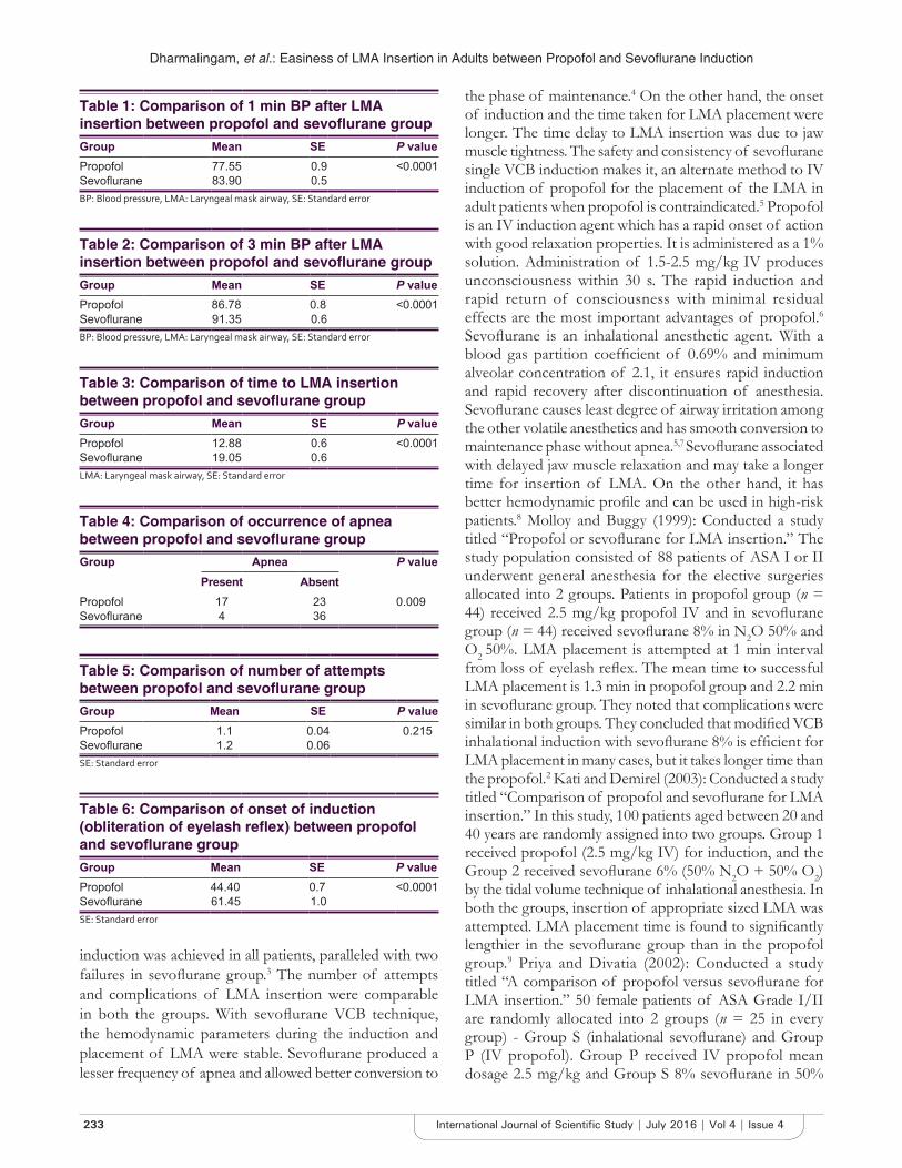
Dharmalingam, et al.: Easiness of LMA Insertion in Adults between Propofol and Sevoflurane Induction
233 International Journal of Scientific Study | July 2016 | Vol 4 | Issue 4
induction was achieved in all patients, paralleled with two failures in sevoflurane group.3 The number of attempts and complications of LMA insertion were comparable in both the groups. With sevoflurane VCB technique, the hemodynamic parameters during the induction and placement of LMA were stable. Sevoflurane produced a lesser frequency of apnea and allowed better conversion to
the phase of maintenance.4 On the other hand, the onset of induction and the time taken for LMA placement were longer. The time delay to LMA insertion was due to jaw muscle tightness. The safety and consistency of sevoflurane single VCB induction makes it, an alternate method to IV induction of propofol for the placement of the LMA in adult patients when propofol is contraindicated.5 Propofol is an IV induction agent which has a rapid onset of action with good relaxation properties. It is administered as a 1% solution. Administration of 1.5-2.5 mg/kg IV produces unconsciousness within 30 s. The rapid induction and rapid return of consciousness with minimal residual effects are the most important advantages of propofol.6 Sevoflurane is an inhalational anesthetic agent. With a blood gas partition coefficient of 0.69% and minimum alveolar concentration of 2.1, it ensures rapid induction and rapid recovery after discontinuation of anesthesia. Sevoflurane causes least degree of airway irritation among the other volatile anesthetics and has smooth conversion to maintenance phase without apnea.5,7 Sevoflurane associated with delayed jaw muscle relaxation and may take a longer time for insertion of LMA. On the other hand, it has better hemodynamic profile and can be used in high-risk patients.8 Molloy and Buggy (1999): Conducted a study titled “Propofol or sevoflurane for LMA insertion.” The study population consisted of 88 patients of ASA I or II underwent general anesthesia for the elective surgeries allocated into 2 groups. Patients in propofol group (n = 44) received 2.5 mg/kg propofol IV and in sevoflurane group (n = 44) received sevoflurane 8% in N2O 50% and O2 50%. LMA placement is attempted at 1 min interval from loss of eyelash reflex. The mean time to successful LMA placement is 1.3 min in propofol group and 2.2 min in sevoflurane group. They noted that complications were similar in both groups. They concluded that modified VCB inhalational induction with sevoflurane 8% is efficient for LMA placement in many cases, but it takes longer time than the propofol.2 Kati and Demirel (2003): Conducted a study titled “Comparison of propofol and sevoflurane for LMA insertion.” In this study, 100 patients aged between 20 and 40 years are randomly assigned into two groups. Group 1 received propofol (2.5 mg/kg IV) for induction, and the Group 2 received sevoflurane 6% (50% N2O + 50% O2) by the tidal volume technique of inhalational anesthesia. In both the groups, insertion of appropriate sized LMA was attempted. LMA placement time is found to significantly lengthier in the sevoflurane group than in the propofol group.9 Priya and Divatia (2002): Conducted a study titled “A comparison of propofol versus sevoflurane for LMA insertion.” 50 female patients of ASA Grade I/II are randomly allocated into 2 groups (n = 25 in every group) - Group S (inhalational sevoflurane) and Group P (IV propofol). Group P received IV propofol mean dosage 2.5 mg/kg and Group S 8% sevoflurane in 50%
Table 6: Comparison of onset of induction (obliteration of eyelash reflex) between propofol and sevoflurane groupGroup Mean SE P valuePropofol 44.40 0.7 <0.0001Sevoflurane 61.45 1.0SE: Standard error
Table 3: Comparison of time to LMA insertion between propofol and sevoflurane groupGroup Mean SE P valuePropofol 12.88 0.6 <0.0001Sevoflurane 19.05 0.6LMA: Laryngeal mask airway, SE: Standard error
Table 4: Comparison of occurrence of apnea between propofol and sevoflurane groupGroup Apnea P value
Present AbsentPropofol 17 23 0.009Sevoflurane 4 36
Table 5: Comparison of number of attempts between propofol and sevoflurane groupGroup Mean SE P valuePropofol 1.1 0.04 0.215Sevoflurane 1.2 0.06SE: Standard error
Table 1: Comparison of 1 min BP after LMA insertion between propofol and sevoflurane groupGroup Mean SE P valuePropofol 77.55 0.9 <0.0001Sevoflurane 83.90 0.5BP: Blood pressure, LMA: Laryngeal mask airway, SE: Standard error
Table 2: Comparison of 3 min BP after LMA insertion between propofol and sevoflurane groupGroup Mean SE P valuePropofol 86.78 0.8 <0.0001Sevoflurane 91.35 0.6BP: Blood pressure, LMA: Laryngeal mask airway, SE: Standard error

Dharmalingam, et al.: Easiness of LMA insertion in adults between Propofol and sevoflurane induction
234International Journal of Scientific Study | July 2016 | Vol 4 | Issue 4
N2O and 50% O2 for 30 s. After loss of eyelash reflex, LMA insertion was excellent in Group P (64%) than in Group S (32%). 72% of patients in Group P had complete jaw opening when compared to 44% of Group S. Hence, they concluded that propofol is better than sevoflurane for LMA insertion. Philip and Lambard (1999): Conducted a study titled “Comparison of vital capacity induction with sevoflurane to IV induction with propofol for adult ambulatory anesthesia.” In this study, there were 56 patients allocated randomly to receive either 8% sevoflurane in 75% N2O/O2 from already primed circuit (VC group n = 32 patients) or propofol 2 mg/kg bolus (IV group n = 24) and time to induction, loss of consciousness, and side effects are monitored. In the VC group patients, 59% have lost responsiveness in one breath taking 39 ± 3 s. All vital capacity patients finished the induction and all measures; induction time is appreciably shorter time for the vital capacity group than IV group. They concluded that vital capacity induction with sevoflurane is a satisfactory alternative to propofol IV induction of general anesthesia for the adult ambulatory anesthesia.10
CONCLUSION
In our study, we conclude that inhalational induction by VCB technique using 8% sevoflurane is an alternate to IV induction using propofol for insertion of LMA in adult patients. When compared to IV propofol induction, sevoflurane VCB technique had stable hemodynamic parameters and less incidence of apnea. It allowed smooth conversion to maintenance phase
and minimal occurrence of apnea. Even though onset of induction is more and extended jaw muscle tightness can delay LMA placement in patients with sevoflurane, it is a good alternative to propofol especially when it is contraindicated.
REFERENCES
1. Scanlon P, Carey M, Power M, Kirby F. Patient response to laryngeal mask insertion after induction of anaesthesia with propofol or thiopentone. Can J Anaesth 1993;40:816-8.
2. Sebel PS, Lowdon JD. Propofol: A new intravenous anesthetic. Anesthesiology 1989;71:260-77.
3. Thwaites A, Edmends S, Smith I. Inhalation induction with sevoflurane: A double-blind comparison with propofol. Br J Anaesth 1997;78:356-61.
4. Ruffle JM, Snider MT. Comparison of rapid and conventional inhalation inductions of halothane oxygen anesthesia in healthy men and women. Anesthesiology 1987;67:584-7.
5. Hall JE, Stewart JI, Harmer M. Single-breath inhalation induction of sevoflurane anaesthesia with and without nitrous oxide: A feasibility study in adults and comparison with an intravenous bolus of propofol. Anaesthesia 1997;52:410-5.
6. Drage MP, Nunez J, Vaughan RS, Asai T. Jaw thrusting as a clinical test to assess the adequate depth of anaesthesia for insertion of the laryngeal mask. Anaesthesia 1996;51:1167-70.
7. Inomata S, Nishikawa T. Determination of end-tidal sevoflurane concentration for tracheal intubation in children with the rapid method. Can J Anaesth 1996;43:806-11.
8. Taguchi M, Watanabe S, Asakura N, Inomata S. End-tidal sevoflurane concentrations for laryngeal mask airway insertion and for tracheal intubation in children. Anesthesiology 1994;81:628-31.
9. Muzi M, Robinson BJ, Ebert TJ, O’Brien TJ. Induction of anesthesia and tracheal intubation with sevoflurane in adults. Anesthesiology 1996;85:536-43.
10. Yurino M, Kimura H. Induction of anesthesia with sevoflurane, nitrous oxide, and oxygen: A comparison of spontaneous ventilation and vital capacity rapid inhalation induction (VCRII) techniques. Anesth Analg 1993;76:598-601.
How to cite this article: Dharmalingam AL, Thamilselvi BS, Murukan MS, Anandan H. Comparison of Onset of Induction and Easiness of Laryngeal Mask Airway Insertion in Adults: Propofol versus sevoflurane Single Vital Capacity Breath Technique-high Concentration (8%). Int J Sci Stud 2016;4(4):231-234.
Source of Support: Nil, Conflict of Interest: None declared.
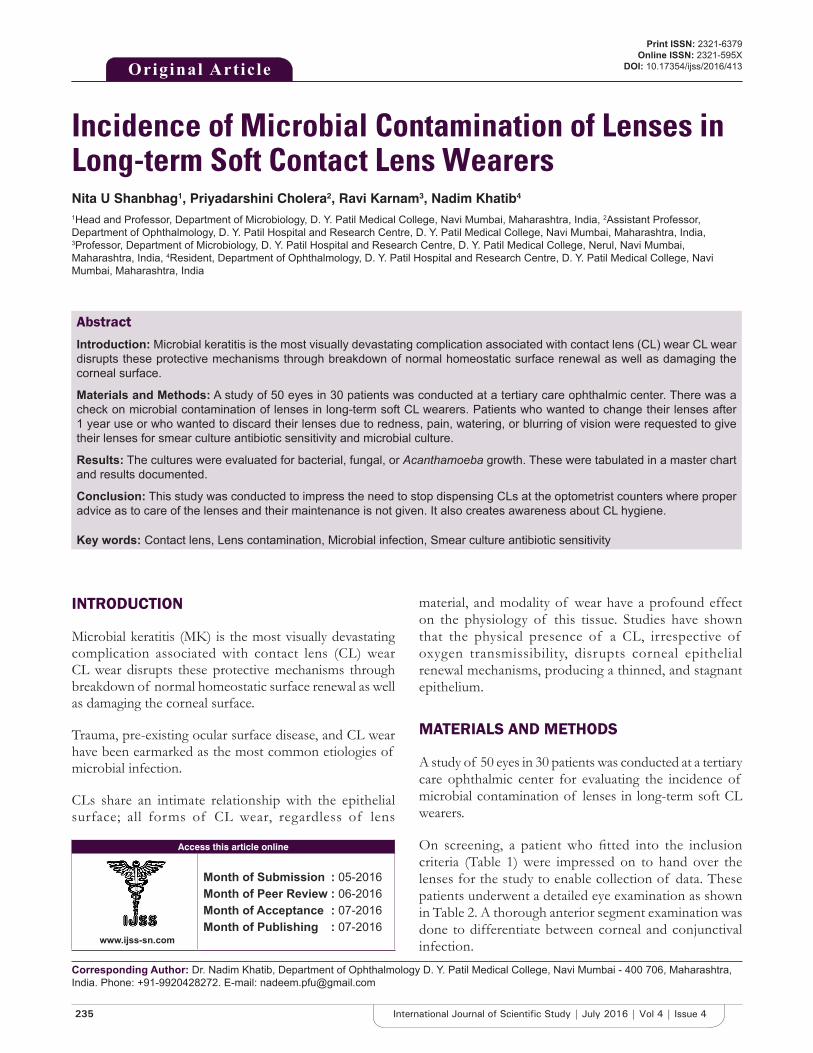
235 International Journal of Scientific Study | July 2016 | Vol 4 | Issue 4
Incidence of Microbial Contamination of Lenses in Long-term Soft Contact Lens WearersNita U Shanbhag1, Priyadarshini Cholera2, Ravi Karnam3, Nadim Khatib4
1Head and Professor, Department of Microbiology, D. Y. Patil Medical College, Navi Mumbai, Maharashtra, India, 2Assistant Professor, Department of Ophthalmology, D. Y. Patil Hospital and Research Centre, D. Y. Patil Medical College, Navi Mumbai, Maharashtra, India, 3Professor, Department of Microbiology, D. Y. Patil Hospital and Research Centre, D. Y. Patil Medical College, Nerul, Navi Mumbai, Maharashtra, India, 4Resident, Department of Ophthalmology, D. Y. Patil Hospital and Research Centre, D. Y. Patil Medical College, Navi Mumbai, Maharashtra, India
material, and modality of wear have a profound effect on the physiology of this tissue. Studies have shown that the physical presence of a CL, irrespective of oxygen transmissibility, disrupts corneal epithelial renewal mechanisms, producing a thinned, and stagnant epithelium.
MATERIALS AND METHODS
A study of 50 eyes in 30 patients was conducted at a tertiary care ophthalmic center for evaluating the incidence of microbial contamination of lenses in long-term soft CL wearers.
On screening, a patient who fitted into the inclusion criteria (Table 1) were impressed on to hand over the lenses for the study to enable collection of data. These patients underwent a detailed eye examination as shown in Table 2. A thorough anterior segment examination was done to differentiate between corneal and conjunctival infection.
INTRODUCTION
Microbial keratitis (MK) is the most visually devastating complication associated with contact lens (CL) wear CL wear disrupts these protective mechanisms through breakdown of normal homeostatic surface renewal as well as damaging the corneal surface.
Trauma, pre-existing ocular surface disease, and CL wear have been earmarked as the most common etiologies of microbial infection.
CLs share an intimate relationship with the epithelial surface; all forms of CL wear, regardless of lens
Original Article
AbstractIntroduction: Microbial keratitis is the most visually devastating complication associated with contact lens (CL) wear CL wear disrupts these protective mechanisms through breakdown of normal homeostatic surface renewal as well as damaging the corneal surface.
Materials and Methods: A study of 50 eyes in 30 patients was conducted at a tertiary care ophthalmic center. There was a check on microbial contamination of lenses in long-term soft CL wearers. Patients who wanted to change their lenses after 1 year use or who wanted to discard their lenses due to redness, pain, watering, or blurring of vision were requested to give their lenses for smear culture antibiotic sensitivity and microbial culture.
Results: The cultures were evaluated for bacterial, fungal, or Acanthamoeba growth. These were tabulated in a master chart and results documented.
Conclusion: This study was conducted to impress the need to stop dispensing CLs at the optometrist counters where proper advice as to care of the lenses and their maintenance is not given. It also creates awareness about CL hygiene.
Key words: Contact lens, Lens contamination, Microbial infection, Smear culture antibiotic sensitivity
Access this article online
www.ijss-sn.com
Month of Submission : 05-2016 Month of Peer Review : 06-2016 Month of Acceptance : 07-2016 Month of Publishing : 07-2016
Corresponding Author: Dr. Nadim Khatib, Department of Ophthalmology D. Y. Patil Medical College, Navi Mumbai - 400 706, Maharashtra, India. Phone: +91-9920428272. E-mail: [email protected]
Print ISSN: 2321-6379Online ISSN: 2321-595X
DOI: 10.17354/ijss/2016/413

Shanbhag, et al.: Incidence of Microbial Contamination of Lenses
236International Journal of Scientific Study | July 2016 | Vol 4 | Issue 4
CLs documenting g rowth or eye les ions were photographed for prognosis (Figures 1-6). The lenses collected and were transported to the laboratory in a sterile autoclaved lens cleaning solution to avoid cross contamination.
At the laboratory, the lens were transferred to glucose broth and incubated at 37° for 2 h. This broth was further cultured on blood agar, McConkey’s agar overnight at 37° and sabouraud’s at room temperature and 37° for 3 weeks. The growth observed was documented as bacterial, fungal and Acanthamoeba growth. Digital pictures were taken (Figures 7-12).
Results were tabulated and inference drawn (Tables 3 and 4).
The wear and tear of soft CL as it is creates microcracks. If handled roughly with nails then this forms earlier. Once the integrity of surface is lost, normal commensals or opportunist which will grow into the CL. During the wear of CL, if there is an inadvertent eye injury, these organisms will latch on the cornea causing corneal ulcer and subsequent loss of vision.
The cultures gave us a mixed basket of organisms with almost 1/3rd as no growth. Only those patients who had redness/conjunctival congestion or corneal defects were treated with antibiotics.
All were counseled as to CL hygiene (Table 5). They were reinforced on the corrective measures to be taken. In the case of a problem visit to the eye doctor was mandatory. Better to be safe than sorry.
DISCUSSION
The potential role of CL care solutions in MK has recently gained significant interest due to increased reports of fungal and Acanthamoeba keratitis. More recent reports support the view that corneal staining may be directly related to inflammation. This compromise includes the inhibition of apoptotic desquamation and a slowed renewal mechanism, producing a thinned, stagnant epithelial sheet. It appears that it is the cumulative breakdown of these collective processes that results in CL related MK and further illustrates the multifactorial nature of the disease process.1
Table 1: Inclusion and exclusion criteriaInclusion criteria Exclusion criteriaAge group: 15-35 years Age group: <15 or >35Lenses Lenses: RGP, bandage
soft CLSoft, yearly wear in power range of+5-−20 which were discarded after the yearly use or prior to that due to redness pain watering or blurring of vision not permitting the wearer to use them
Conjunctivitis causing redness pain watering
Asymptomatic patient wearing CL for duration more than the prescribed schedule
Iridocyclitis causing redness pain wateringPatient not willing for any eye procedure
Asymptomatic patient wearing CL and never having got them cleaned for >3 months in yearly wear schedule
Patient not willing to come for regular follow-up
Symptomatic patient wearing CL with redness pain watering
RGP: Rigid gas permeable, CL: Contact lens
Table 2: Detailed eye examination and investigationsCL history Clinical examination InvestigationCL usage at what age Visual acuity on logmar chart CL sent for SCABS and KOHDuration of CL usage in a day BCVA: Spectacle power/CL power BUT/SchirmersType of CL Slit lamp examination Sac syringingCleaning procedure for the CL CL details IOP measurement by a non
contact tonometerAny faulting in the usage Soft/RGP If corneal lesion present corneal
scrape for SCABS and KOHSleeping with the lenses in the eye CL fit tight or loose CBC/ESRCleaning lenses with tap water Lack of lustre on CL indicating over-usage Urine: (Routine & Microscopy)Washing the eye with tap water with the lenses in the eye Deposits on CL ENT/Dental focus of infectionUsing OTC drugs - Pyrimon for any redness of the eye with the lenses on
Cornea
Continuing the usage of lenses in spite of getting a FB sensation redness watering or discharge in the eye
Corneal lesion
Not visiting a doctor but an optician for CL dispensing Fluorescent stainingImproper cleaning schedule for the lenses Photo of the lesion
Corneal sensationSac status to r/o chronic dacryocystitisIOP to r/o glaucomaBUT to r/o dry eyes

Shanbhag, et al.: Incidence of Microbial Contamination of Lenses
237 International Journal of Scientific Study | July 2016 | Vol 4 | Issue 4
Increased risk of bacterial infection is a reality with which all CL wearers must live. Unfortunately, the most CLs wearers suffer from the “it-cannot-happen-to-me” syndrome. Those who have had a contact-lens-related bacterial infection now know better than to tempt fate.
Bacterial eye infections affecting the cornea are known as MK. MK is the most severe complication associated with CL wear. Pseudomonas aeruginosa is the most common bacteria involved in MK. It is also one of the most damaging. P. aeruginosa eats away at the cornea (causing
Figure 1: Tight fit lens causing hypoxia with ciliary congestion with corneal edema
Figure 2: Lack of luster depicting an aged lens
Figure 3: Lens deposit eroding into the lens
Figure 4: Multiple deposits on the contact lens causing FB sensation
Figure 5: Peripheral corneal infiltrate with vascularization
Figure 6: Corneal infiltrate with circumcorneal congestion following contact lens deposit
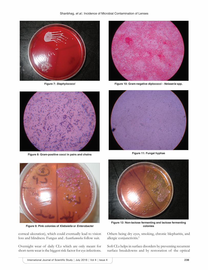
Shanbhag, et al.: Incidence of Microbial Contamination of Lenses
238International Journal of Scientific Study | July 2016 | Vol 4 | Issue 4
corneal ulceration), which could eventually lead to vision loss and blindness. Fungus and Acanthamoeba follow suit.
Overnight wear of daily CLs which are only meant for short-term wear is the biggest risk factor for eye infections.
Others being dry eyes, smoking, chronic blepharitis, and allergic conjunctivitis.2
Soft CLs helps in surface disorders by preventing recurrent surface breakdowns and by restoration of the optical
Figure 7: Staphylococci
Figure 8: Gram-positive cocci in pairs and chains
Figure 9: Pink colonies of Klebsiella or Enterobacter
Figure 10: Gram-negative diplococci - Neisseria spp.
Figure 11: Fungal hyphae
Figure 12: Non-lactose fermenting and lactose fermenting colonies
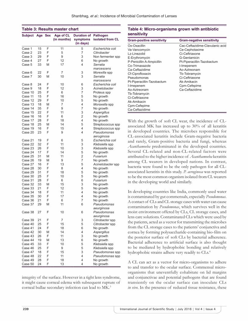
Shanbhag, et al.: Incidence of Microbial Contamination of Lenses
239 International Journal of Scientific Study | July 2016 | Vol 4 | Issue 4
integrity of the surface. However in a tight lens syndrome, it might cause corneal edema with subsequent rupture of corneal bullae secondary infection can lead to MK.3
With the growth of soft CL wear, the incidence of CL-associated MK has increased up to 30% of all keratitis in developed countries. The microbes responsible for CL-associated keratitis include Gram-negative bacteria and rarely, Gram-positive bacteria and fungi, whereas Acanthamoeba predominated in the developed countries. Several CL-related and non-CL-related factors were attributed to the higher incidence of Acanthamoeba keratitis among CL wearers in developed nations. In contrast, bacteria were found to be the only pathogen for all CL-associated keratitis in this study. P. aeruginosa was reported to be the most common organism isolated from CL wearers in the developing world and similarly.
In developing countries like India, commonly used water is contaminated by gut commensals, especially Pseudomonas. A contact of CLs and CL storage cases with water can cause contamination by Pseudomonas, which survives well in the moist environment offered by CLs, CL storage cases, and lens care solutions. Contaminated CLs which were used by the patients, acted as a vector for transmitting the microbes from the CL storage cases to the patients’ conjunctiva and cornea by forming polysaccharide-containing bio-film on the posterior surface of soft CLs by bacterial adherence. Bacterial adherence to artificial surface is also thought to be mediated by hydrophobic bonding and relatively hydrophobic strains adhere very readily to CLs.4
A CL can act as a vector for micro-organisms to adhere to and transfer to the ocular surface. Commensal micro-organisms that uneventfully cohabitate on lid margins and conjunctivae and potential pathogens that are found transiently on the ocular surface can inoculate CLs in vivo. In the presence of reduced tissue resistance, these
Table 3: Results master chartSubject Age Sex Age of CL
(in months)Duration of symptoms(in days)
Pathogen isolated from CL
Case 1 15 F 11 5 Escherichia coliCase 2 23 F 5 7 CitrobacterCase 3 29 F 9 3 Non fermenter sppCase 4 27 F 12 6 No growthCase 5 33 M 17 4 Serratia
marcescensCase 6 22 F 7 3 Morexilla sppCase 7 30 M 10 3 Serratia
marcescensCase 8 24 F 10 6 Escherichia coliCase 9 18 F 12 3 AcinetobacterCase 10 25 F 6 7 Proteus sppCase 11 15 F 16 5 No growthCase 12 29 F 10 5 No growthCase 13 18 M 7 4 Moraxella spp Case 14 35 F 9 7 No growthCase 15 22 F 11 3 AspergillusCase 16 16 F 6 2 No growthCase 17 28 F 18 4 No growthCase 18 25 M 14 5 Streptococcus sppCase 19 16 F 15 4 Streptococcus sppCase 20 23 F 9 4 Pseudomonas
aeruginosaCase 21 19 F 6 5 Escherichia coliCase 22 32 F 11 3 Klebsiella sppCase 23 26 F 10 3 Klebsiella sppCase 24 17 F 14 5 No growthCase 25 31 M 11 2 FusariumCase 26 19 M 9 7 No growthCase 27 16 F 13 5 Acinetobacter sppCase 28 30 F 8 4 No growthCase 29 31 F 18 5 No growth Case 30 25 F 10 5 No growthCase 31 28 F 6 6 FusariumCase 32 33 M 15 3 No growthCase 33 21 F 12 5 No growthCase 34 18 F 11 4 No growthCase 35 30 F 17 4 No growthCase 36 21 F 6 7 No growthCase 37 29 M 11 6 Pseudomonas
aeurginosaCase 38 27 F 10 6 Pseudomonas
aeurginosaCase 39 21 F 7 3 Citrobacter spp Case 40 25 F 15 5 Citrobacter sppCase 41 24 F 18 6 No growthCase 42 30 M 14 4 AspergillusCase 43 26 F 11 3 No growthCase 44 19 M 13 6 No growthCase 45 33 F 10 5 Klebsiella sppCase 46 25 F 9 5 Klebsiella sppCase 47 18 F 15 3 Pseudomonas spp Case 48 22 F 11 4 Pseudomonas sppCase 49 28 F 18 4 No growthCase 50 24 F 13 4 No growth
Table 4: Micro-organisms grown with antibiotic sensitivityGram-positive sensitivity Gram-negative sensitivityOx-Oxacillin Cac-Ceftazidime-Clavulanic acidVa-Vancomycin Ce-CephotaximeLz-Linezolid Ci-CeftriaxoneE-Erythromycin G-GentamicinP-Penicillin A-Ampicillin Pt-Piperacillin-TazobactumCo-Trimaxazole I-ImepenemCa-Ceftazidime Ao-AztreonamCf-Ciprofloxacin Tb-TobramycinPseudomonas Ci-CeftriaxonePt-Piperacillin-Tazobactum Ak-AmikacinI-Imepenem Cpm-CefepimeAo-Aztreonam Ca-CeftazidimeTb-TobramycinCi-CeftriaxoneAk-AmikacinCpm-CefepimeCa-Ceftazidime

Shanbhag, et al.: Incidence of Microbial Contamination of Lenses
240International Journal of Scientific Study | July 2016 | Vol 4 | Issue 4
resident micro-organisms or transient pathogens can invade and colonize the cornea or conjunctiva to produce inflammation or infection.5
Lens handling greatly increases the incidence of lens contamination, and the ocular surface has a tremendous ability to destroy organisms. Even when lenses are removed aseptically from the eye, 50% are found to harbor micro-organisms, almost exclusively bacteria. Coagulase-negative Staphylococci being most common and Gram-negative about 10%. In storage cases, the incidence of positive microbial bioburden is also typically >50%. All types of care solutions can become contaminated including up to 30% of preserved products. Thus, this detailed understanding of lens-related bioburden is important in the understanding of factors associated with infectious and inflammatory complications.5
Many complications arise when lenses are worn not as prescribed (improper wear schedule or lens replacement). Sleeping in lenses not designed or approved for extended wear is a common cause of complications. Many people go too long before replacing their lenses, wearing lenses designed for 1, 14, or 30 days of wear for multiple months or years. While this does save on the cost of lenses, it risks permanent damage to the eye and loss of sight. CL wear is the most important risk factor. The role of initial therapy for MK remains important.6
Severe MK with vision loss in CL wearers is more likely to be caused by an environmental pathogen, and to occur in tropical regions in association with high daytime
temperatures.6
One of the major factors that cause CL complications are that the lens is a barrier to oxygen. The cornea needs a supply of oxygen to function and it normally gets that oxygen from the surrounding air while awake and from the blood vessels in the back of the eyelid while asleep. The most prominent risks associated with long-term, chronic low oxygen to the cornea include corneal neovascularization increased epithelial permeability, bacterial adherence, micro cysts, corneal edema, endothelial polymegethism, and potential increase in myopia.
Mishandling of CLs can also cause problems. Corneal abrasions can increase the chances of infection. When combined with improper cleaning and disinfection of the lens, the risk of infection further increases. Decreased corneal sensitivity following extended CL wear may cause a patient to miss some of the earliest symptoms of such complications.7
Genetic mutations in the innate immune system may be involved in individual susceptibility to MK.8
Cytokine gene expression is tightly regulated, and aberrant expression from environmental and genetic polymorphism has been implicated in a range of diseases, susceptibility to infections, and responses to treatment. This review concentrates on the functionality of cytokine and cytokine receptor gene polymorphisms; it is through these variants that genuine disease-associations are based. Several mechanisms for single nucleotide polymorphism (SNP) functionality are present within cytokine genes.9
Table 5: CL hygiene and corrective measures to be takenFactors-lens hygiene Corrective measures
• No nail growth • When prescribing CL counsel on preventing injury to the lens• No cracks/rough pulp of index finger • Do not use lens in case of redness pain watering and if worn
then discard those lenses• Rub the CL with cleaning solution while inserting and after
removing• Do not use any unpreserved solution to clean or store lenses
in c/o emergency use a tear substitute• CL • Clean the lenses ultrasonically every 2 months as the rubbing
action is insufficient to remove accumulated deposits• Not to rub eyes immediately after CL removal. Least corneal
abrasion happens in case of post CL corneal edema• Storage of CL
• Clean the lens case with betadaine 5% solution when the patient comes for CL cleaning at the clinic
• Change solution every 2nd day • Visit eye doctor every 3 months to r/o tight lens syndrome or CL deposits on the lens
• Make sure CL borders are within well of CL storage box so that cap not put over CL
• Always get lenses from eye doctors and not optometrists
• Ultrasonic/enzyme cleaning of CL every 2 months• CL cleaning solution to be kept in fridge/not to be replaced by
tap water/use a transparent bottle so deposits can be cleaned• Do not wear CL in case of:
Redness of eyesWatering of eyesDropped visionDischarge
CL: Contact lens
Shanbhag, et al.: Incidence of Microbial Contamination of Lenses
241 International Journal of Scientific Study | July 2016 | Vol 4 | Issue 4
CONCLUSION
The trend to use lenses is more in the pre-presbyopic age group hence it formed our sample base. 90% of the sample was contributed by the female gender. Cosmesis as well as reduced corneal sensation compared to the male population was the basis of this rigid gas permeable (RGP) lenses are safer but soft lenses are user-friendly hence the increase in demand and shift from RGP lenses in recent years. The younger generation is careless about the hygiene of wearing, cleaning and maintenance of the lens. There seems to be no gender bias. The duration of wear seems to have a bearing on the incidence of infection. This may be due to the micro-cracks in the CL due to wear and tear. The presence of clinical symptoms like redness pain watering and blurring of vision seems to herald the onset of microbial infection in the lenses. Hence, continuing to use the CL in the presence of these symptoms may be a contributing factor in people developing corneal infections following CL wear. The presence of infiltrates is a sure shot marker for isolating pathogens in the lenses. Hence, a complete slit lamp examination at every follow-up should be mandatory. Seeing an infiltrate should be an absolute indication to discard usage of the lenses.
Deposit on the lens was the hallmark of finding pathogens as they grow into it. It highlights the need for ultrasonic cleaning of lenses and frequent change of CL solution in the lens case. Disinfecting the lens case once a fortnight with betadine 5% solution would prevent deposit formation on the lenses. Storing the solution in the refrigerator and handling the bottle cap aseptically would go a long way in preventing CL solution contamination.
Pathogens were isolated in 60% of cases. They came in a mixed basket of Gram-positive specially staphylococcus, Gram-negative especially Pseudomonas and Klebsiella, Fungal hyphae in 10% as listed in Table 3. We could not isolate
Acanthameoba in spite of literature reporting it as the most common organism in CL wearers.
The study emphasizes the need to clean hands with glycerine soap with clean water and dry them prior to handling lenses. Disinfecting sanitizer would definitely help. The need to emphasize cleaning of the CL case every week and rinsing them in betadiane solution 5% every fortnight before reuse. It is necessary to refrigerate the CL cleaning solution to prevent the growth of organism in them.
Immediate ophthalmic consultation in case of redness pain watering with blurred vision and refrain from wearing these lenses in these situations must be a compulsory mandate to prevent transfer of this infection to the cornea heralding the onset of infectious keratitis with significant loss of vision.
REFERENCES
1. Robertson DM, Cavanagh HD. The clinical and cellular basis of contact lens-related corneal infections: A review. Clin Ophthalmol 2008;2:907-17.
2. Cho P, Boost M, Cheng R. Non-compliance and microbial contamination in orthokeratology. Optom Vis Sci 2009;86:1227-34.
3. Dada VK, Kalra VK, Angra SK. Role of soft contact lens in ocular surface problems. Indian J Ophthalmol 1984;32:519-21.
4. Bharathi MJ, Ramakrishnan R, Meenakshi R, Kumar CS, Padmavathy S, Mittal S. Ulcerative keratitis associated with contact lens wear. Indian J Ophthalmol 2007;55:64-7.
5. Szczotka-Flynn LB, Pearlman E, Ghannoum M. Microbial contamination of contact lenses, lens care solutions, and their accessories: A literature review. Eye Contact Lens 2010;36:116-29.
6. Cruciani F, Cuozzo G, Di Pillo S, Cavallaro M. Predisposing factors, clinical and microbiological aspects of bacterial keratitis: A clinical study. Clin Ter 2009;160:207-10.
7. Levy B, Orsborn G. Clinical risks: Myths and truths - Interpreting the evidence-based data about contact lens care. Clin Refract Optometry 2008;19:165-71.
8. Stapleton F, Carnt N. Contact lens-related microbial keratitis: How have epidemiology and genetics helped us with pathogenesis and prophylaxis. Eye (Lond) 2012;26:185-93.
9. Smith AJ, Humphries SE. Cytokine and cytokine receptor gene polymorphisms and their functionality. Cytokine Growth Factor Rev 2009;20:43-59.
How to cite this article: Shanbhag NU, Cholera P, Karnam R, Khatib N. Incidence of Microbial Contamination of Lenses in Long-term Soft Contact Lens Wearers. Int J Sci Stud 2016;4(4):235-241.
Source of Support: Nil, Conflict of Interest: None declared.
242International Journal of Scientific Study | July 2016 | Vol 4 | Issue 4
Functional Outcome of Patients Underwent Lumbar MicrodiscetomyToms Jacob1, U Sudheer2, C Jayaprakash3
1Post-graduate Trainee, Department of Orthopedics, Amala Institute of Medical Science, Thrissur, Kerala, India, 2Associate Professor, Department of Orthopedics, Amala Institute of Medical Science, Thrissur Kerala, India, 3Head, Department of Orthopedics, Amala Institute of Medical Science, Thrissur, Kerala, India
The supine straight leg raise test (Laseugues test) and its variants (sitting straight leg raise test, bowstring test, contra lateral straight leg raise test) increase tension in sciatic nerve and are used to assess L5 and S1 nerve roots. The femoral nerve stretch test (reverse straight leg raise test) increases tension along femoral nerve and is used to assess the L2, L3, and L4 nerve roots.
Plain radiographs are not helpful in initial evaluation of suspected lumbar disc herniations. Although radiograph may show degenerative changes (disc space narrowing and osteophyte formation), there is poor correlation between these findings and clinical symptoms because these findings are also present in asymptomatic patients. Magnetic resonance imaging (MRI) is the preferred imaging test because it provides greatest amount of information about the lumbar region. About 90% of lumbar disc herniation occurs at L4 L5 and L5 S1 level. L3 L4 level is the next common level for symptomatic lumbar disc herniation.2
Disc herniation is described based on the circumference of the annulus fibrosus as central, posterolateral, foraminal,
INTRODUCTION
The life time prevalence of lumbar disc herniation is approximately 2%. The natural history of sciatica secondary to lumbar disc herniation is spontaneous improvement in the majority of cases. Among patients with radiculopathy secondary to lumbar disc herniation, approximately 10-25% experience persistent symptoms. Pain typically begins in the lumbar area and radiates to sacroiliac and buttocks region. Radicular pain extends below the knee in the region of involved nerve root. Radicular pain may be accompanied by paresthesia and weakness in the distribution of involved nerve root.1
Original Article
AbstractIntroduction: Inter vertebral disc prolapse (IVDP) is a very common cause for low back ache in younger population. When conservative treatment fails or when patient develops complications like neurological deficit, then the treatment is surgical discectomy. Open laminectomy and discectomy has a lot of morbidity when compared to micro discectomy which is the treatment of choice for such patients. In this study, we analyze the advantages and functional outcome of patients underwent micro lumbar discectomy.
Materials and Methods: This is an institution-based prospective study. We performed a prospective study of 15 microscopic discectomy in 15 patients who were non-responsive to conservative management. Their pre- and post-operative symptoms like pain and functional disabilities were evaluated over a period of 2-year using direct questionnaire method.
Results: About 95% of patients reported excellent pain relief on 1 year follow-up. Neurological recovery was 80% on 2 years follow-up. 2 patients had dural tear during the procedure which was treated by suturing with 5’0 prolene.
Conclusion: It is recommended that this procedure as the gold standard surgical method for the patients with IVDP who were failed with conservative treatment method.
Key words: Patients, Lumbar, Microdiscetomy
Access this article online
www.ijss-sn.com
Month of Submission : 05-2016 Month of Peer Review : 06-2016 Month of Acceptance : 07-2016 Month of Publishing : 07-2016
Corresponding Author: Dr. U Sudheer, Associate Professor, Department of Orthopedics, Amala Institute of Medical Science, Thrissur Kerala, India. E-mail: [email protected]
Print ISSN: 2321-6379Online ISSN: 2321-595X
DOI: 10.17354/ijss/2016/414

Jacob, et al.: Outcome of Patients Underwent Lumbar Microdiscetomy
243 International Journal of Scientific Study | July 2016 | Vol 4 | Issue 4
or extra foraminal. A posterolateral L4 L5 disc herniation compresses L5 nerve root. An L4 L5 foraminal or extra foraminal disc herniation compresses L4 nerve root. A central disc herniation compresses one or more of the caudal nerve roots.
Appropriate criteria for surgical intervention are follows:1. Functionally incapacitating leg pain extending below
the knee within a nerve root distribution2. Nerve root tension signs with or without neurologic
deficit3. Failure to improve with 4-8 weeks of nonsurgical
treatment4. Confirmatory imaging study which correlates with
patients’ physical findings and pain distribution.
Open lumbar discectomy using microsurgical techniques remains the gold standard for the treatment of symptomatic lumbar disc herniation. Although a variety of alternative procedures have been proposed, no procedure has demonstrated superior surgical outcome compared with microsurgical approach.3
Complications of microscopic lumbar discectomy include:1. Vascular injury2. Nerve root injury3. Dural tear4. Infection5. Increased back pain6. Recurrent disc herniation7. Cauda equina syndrome8. Medical complications such as thrombophlebitis,
urinary tract infection, etc.
The most common cause of surgical failure following lumbar discectomy is poor patient selection and wrong level of surgery. The incidence of recurrent disc herniation following non-surgical lumbar discectomy is 5-10%. If symptoms are predominantly radicular repeat lumbar discectomy may be beneficial. If symptoms include a combination of radicular pain and low back pain, discectomy combined with fusion may be considered in select patients.4
MATERIALS AND METHODS
Analysis of functional outcome of patients with herniated lumbar disc who have undergone microdiscectomy at Amala Institute of Medical Science, Thrissur over a period of 2-year. We performed a prospective study of 15 microscopic discectomy in 15 patients who were non responsive to conservative management. Pre- and post-
operative neurological status, pain, functional disability were evaluated. Other studied variables were mean age sex ratio, level of prolapse, mean hospital stay, and time to return to work.
All patients with sciatica caused by herniated lumbar discs who did not respond to conservative treatment were enrolled in the study to undergo microdiscectomy between 2011 and 2013. The inclusion criteria were the presence of a herniated lumbar disc observed on MRI scans and the persistence of sciatica after 4-8 weeks of conservative treatment with rest, analgesia, nonsteroidal anti-inflammatory drugs, and physical therapy. The exclusion criteria were as follows age older than 60 years, previous surgery, associated lumbar spine stenosis, foraminal, or extraforaminal disc herniations. Only those patients with a final post-operative follow-up period of at least 2-year were included in this study. After the inclusion criteria were met and an informed consent was obtained, the patients were included in the study. The surgical procedures were performed under general anesthesia with the patient in the prone position with hip flexed to 90°. Prophylaxis with the first-generation cephalosporin was introduced 1 h before anesthesia and kept for 8 h after the procedure.
Procedure: Patient in a prone position with hip flexed to 90° (Figures 1 and 2).
Level identification done under C-arm control which is the most crucial step.
Count the level from below upward as open spaces as seen in MRI.
For example if it is second open space from below upward in MRI, under C-arm guidance in lateral view identify the second open space. 1 inch (2.5 cm) incision made between adjacent spinous process from spinous process above to spinous process below slightly to the side depending on the predominant symptomatic side. Deep fascia and muscles retracted to either side. Self-retaining retractors applied. Base of the spinous process identified. Through the gap between the spinous process and lamina ligamentum flavum excised and reached the lateral side of spinal cord. Search for root extension from the cord. Cord is then retracted medially to see the disc underneath. Excision of the disc was done. Hemostasis attained and wound closed in layers.
Post-operative lumbar corset given. The 3rd day wound inspection done and the patient is discharged. Reviewed after 10 days for suture removal. Reviewed after 6 weeks for reassessment. On follow-up, pain is assessed using

Jacob, et al.: Outcome of Patients Underwent Lumbar Microdiscetomy
244International Journal of Scientific Study | July 2016 | Vol 4 | Issue 4
questionnaire method. Neurological status is assessed by clinical examination.
The surgical variables analyzed were the level and side of the herniated disc, side of root compression, pre- and post-operative neurological deficit, and post-operative pain relief. Pre- and post-operative evaluation consisted of a neurological examination, and questionnaire method for assessment of pain. The surgical wound pain was assessed 12 h after surgery using the questionnaire method. Clinical neurological status was evaluated using the Lasegue test, motor assessment by muscle strength, and testing of the sensory system (Figures 3 and 4).
Functional outcome was evaluated using the questionnaire method. The patients were reevaluated 1 week, 6 weeks, 6 months, 12 months, and 24 months after surgery. The time required for patients to return to work was also registered.
ResultsMean age: 591/155 = 39.4Sex: M/F - 3:2Level: L4 L5 level: 9 (60%)L5 S1 level: 6 (40%)
Pain relief:Pain relief is assessed by questionnaire method.
Pain relief 1 week 6 week 6 months 1 yearExcellent 10 12 13 14Good 4 2 1 0Fair 0 1 1 1Poor 1 0 0 0
Neurological status:Motor deficit:• Pre-operative: 10 (66.67%)• 1 week post-operative: 8 (53.3%)• 6 week post-operative: 5 (33.3%)• 6 months post-operative: 4 (26.6%)• 12 months post-operative: 4 (26.6%)• 24 months post-operative: 3 (20%).
Sensory deficit:• Pre-operative: 9 (60%)• 1 week post-operative: 6 (40%)• 6 week post-operative: 6 (40%)• 6 months post-operative: 5 (33.3%)• 12 months post-operative: 3 (20%)• 24 months post-operative: 3 (20%).
Figure 1: Patient in a prone position with hip flexed to 90°
Figure 2: Pre-operative image - Patient in a prone position with hip flexed to 90°
Figure 3: Excised part
Figure 4: Post-operative
Jacob, et al.: Outcome of Patients Underwent Lumbar Microdiscetomy
245 International Journal of Scientific Study | July 2016 | Vol 4 | Issue 4
RESULTS
A total of 15 patients were enrolled in the study. The mean post-operative follow-up period was 24 months. There were 9 men and 6 women with a mean age of 39.4. The vertebral level affected was L4 L5 in 9 patients, L5S1 in 6 patients. All patients is presented with pre-operative neurological impairment; 100% had a positive Lasegue sign (15 out of 15), 66.67% had motor deficits (10 out of 15), and 60% had sensory deficits (9 out of 15). After 6 months of follow-up, 26.6% had motor deficit and 33.3% had sensory deficit. After 12 months, these percentages changed to 26.67% and 20%, respectively, and after 24 months, they changed to 20% and 20%, respectively. Two patients had dural tear which was managed conservatively.
All 15 patients have full recovery from pain within 6 weeks and were able to resume their normal work.
DISCUSSION
Microscopic discectomy helps in faster post-operative mobilization, faster recovery, and resumption of work at the earliest. The development in recent years has made the treatment of herniated discs safer and less invasive.
By using microscopic discectomy approaches through small incisions, nerve root decompression is achieved with minimal risk of complication and preserving normal anatomy. The superiority of microdiscectomy over traditional discectomy has been widely proven. The most important step in microdiscectomy is correct level identification and adequate decompression.
CONCLUSION
It is recommended that this procedure as the gold standard surgical method for patients with Inter vertebral disc prolapse who were failed with conservative treatment method.
REFERENCES
1. Righesso O, Falavigna A, Avanzi O. Comparison of open discectomy with microendoscopic discectomyin lumbar disc herniations: Results of a randomized controlled trial. Neurosurgery 2007;61:545-9.
2. Wong DA, Transfeldt E. Macnab’ Backache. Lippincott: Williams and Wilkins; 2007.
3. Devlin VJ. Spine Secrets. Philadelphia, PA: Harley and Belfin, Elsevier; 2003.
4. Yorimitsu E, Chiba K, Toyama Y, Hirabayashi K. Long-term outcomes of standard discectomy for lumbar disc herniation: A follow-up study of more than 10 years. Spine 2001;26:652-7.
How to cite this article: Jacob T, Sudheer U, Jayaprakash C. Functional Outcome of Patients Underwent Lumbar Microdiscetomy. Int J Sci Stud 2016;4(4):242-245.
Source of Support: Nil, Conflict of Interest: None declared.

246International Journal of Scientific Study | July 2016 | Vol 4 | Issue 4
Significance of Fragmented QRS Complex in Acute Coronary Syndrome and its Correlation with Coronary Angiography to Identify the Culprit LesionSol- Gagan Sharma1, T G Jayakumar2, G Rupesh3, Gopinath Rajesh2, Sol- Nihas Chamanshaikh4, Geofi George5, Said Mohamed Abdulkhadar6
1Senior Resident, Department of Cardiology, Amala Institute of Medical Sciences, Amala, Thrissur, Kerela, India, 2Additional Professor, Department of Cardiology, Amala Institute of Medical Sciences, Amala, Thrissur, Kerela, India, 3Associate Professor, Department of Cardiology, Amala Institute of Medical Sciences, Amala, Thrissur, Kerela, India, 4Junior Resident, Department of Cardiology, Amala Institute of Medical Sciences, Amala, Thrissur, Kerela, India, 5Assistant Professor, Department of Cardiology, Amala Institute of Medical Sciences, Amala, Thrissur, Kerela, India, 6Professor and HOD, Department of Cardiology, Amala Institute of Medical Sciences, Amala, Thrissur, Kerela, India
to two-dimensional echocardiogram (2D echo/echo) to coronary angiogram (CAG) to comment on the culprit vessel involved in the MI. But still, the data are lacking in the correlation of non STEMI (NSTEMI) and the culprit vessel involved. In NSTEMI, there are ECG changes that may suggest that there is some ischemic activity going on in the heart but all are not very specific as compared to the STEMI. The researchers observed some slurring in the ECG in 1960. Investigators tried to correlate the same with the left ventricle (LV) dysfunction. It was Flowers et al., who first discovered the presence of fragmented QRS (fQRS) complex in the
INTRODUCTION
The diagnosis of ST-elevation myocardial infarction (STEMI) has evolved a lot from electrocardiogram (ECG)
Original Article
AbstractBackground: Electrocardiogram (ECG) is the first step in the evaluation of any acute coronary syndrome (ACS) followed by invasive coronary angiogram (CAG) if indicated. ECG plays a valuable role in diagnosis of ST elevation myocardial infarction (STEMI). But in non STEMI (NSTEMI), the role of ECG is not very specific as far as the area of ischemia is involved. Fragmented QRS (fQRS) is said to be a valuable ECG marker of ACS. fQRS can play an additional role in the diagnosis of NSTEMI.
Aim: To evaluate ECG in patients with NSTEMI ACS and to find out its correlation with culprit vessel by coronary angiography.
Materials and Methods: We studied a total of 450 patients with NTEMI ACS. STEMI patients were excluded. The patients were clinically evaluated for symptoms, risk factors followed by ECG. All of them underwent ECG analysis for fQRS. The patients also underwent CAG for evaluation of the culprit vessel. Further ECG and fQRS correlation with culprit vessel were done.
Results: A total of 450 patients including 308 male and 142 female patients were studied. Among them, 230 patients had fQRS in the ECG, whereas 220 had no fQRS in the ECG. P value of ST depressions in anterior leads (P = 0.003) and T inversions in lateral leads (P = 0.0001) was significant in fQRS group as compared to the non-fQRS group. Coronary angiography of positive fQRS patients showed significant lesions in culprit vessel in 91.3% of patients. The sensitivity of CAG in patients with a positive fQRS was 63.24 with a specificity of 83.05 for localizing the culprit vessel. The highest specificity of 96.74% was seen in patients with positive fQRS in anterior leads with left anterior descending lesions in CAG, followed by fQRS in lateral leads in left circumflex artery lesions (94.35%).
Conclusions: Analysis of fQRS could help in giving an indication of the culprit vessel involved with a high specificity and good sensitivity. Thus, fQRS analysis can be seen as a good adjunct with CAG for diagnosis of patients with NSTEMI.
Key words: Coronary angiography, Fragmented QRS, Non ST elevation myocardial infarction
Access this article online
www.ijss-sn.com
Month of Submission : 05-2016 Month of Peer Review : 06-2016 Month of Acceptance : 07-2016 Month of Publishing : 07-2016
Corresponding Author: Dr. Gagan Sharma, Department of Cardiology, Amala Institute of Medical Sciences, Amala, Thrissur - 680 555, Kerela, India. Phone: +91-9447786174. E-mail: [email protected]
Print ISSN: 2321-6379Online ISSN: 2321-595X
DOI: 10.17354/ijss/2016/415
Sharma, et al.: Significance of fQRS Complex in Acute Coronary Syndrome and its Correlation with Coronary Angiography to Identify the Culprit Lesion
247 International Journal of Scientific Study | July 2016 | Vol 4 | Issue 4
patients who already had an MI. It was thus reported as a high-frequency component.1 According to Friedman et al., there are persistent changes in the Purkinje fibers and myocardial fibrosis that will slow down the myocardial activation which he analyzed in the canine heart with induced MI.2
Varriale and Chryssos suggested that RSR’ complex unrelated to right bundle branch block or left bundle branch block could be associated with impaired depolarization within tissue surrounding the myocardial scar.3 It was de Luna who suggested that abnormalities in the second half of the QRS complex (i.e., terminal slurring, sometimes with RꞋ in lead V1) during MI might represent necrosis in late depolarized basal zones,4 It was Das et al. who defined the fQRS in 2006, as presence of an additional R-wave (RꞋ) or notching in the nadir of the S wave, or the presence of >1 RꞋ in 2 contiguous leads, corresponding to a major coronary artery territory on the resting 12-lead ECG with filter range 0.16-100 Hz, AC filter 60 Hz, paper speed 25 mm/s, and 10 mm/mV.5 As very little is known about this fQRS in NSTEMI, we planned to do a prospective study.
MATERIALS AND METHODS
Aim• To document the fQRS in the ECG of the acute
coronary syndrome (ACS) patients and to correlate with the coronary artery involved by doing CAG.
• To determine the culprit artery from the fQRS leads.
Objectives• To identify the incidence of fQRS in the ACS
(NSTEMI/UA).• To identify the sensitivity and specificity of the fQRS
in determining the culprit artery in ACS.• fQRS complex in ECG correlation with the CAG to
identify the culprit lesion in ACS patients.
HypothesisThe incidence of fQRS is around 60% which is due to the scarring of the myocardium.6 It has a strong correlation in determining the culprit artery lesion.
Inclusion Criteria• All patients with ACS undergoing CAG.
Exclusion Criteria• Patients with atrial fibrillation, cardiomyopathy,
myocarditis, valvular heart disease, congenital heart disease, and previous implantable cardioverter defibrillator.
• Patients with STEMI.
• All patients with bundle branch block.• Patients not willing for CAG.
Study Population• All the patients who were admitted to our hospital with
chest pain, effort angina (EA), dyspnea, palpitation, with ECG changes fulfilling the inclusion criteria within the period of March 2014-October 2015 were included in the study.
• We studied the mode of presentation, past history such as diabetes mellitus (DM), hypertension (HTN), dyslipidemia (DLP), and peripheral vascular disease (PVD), and the family history of DM, HTN, DLP, coronary artery disease (CAD), as well as the habits like smoking and alcohol consumption.
The ECG changes in the various leads, like fQRS, ST-T changes (depression and inversion) were also recorded. These ECG changes were correlated with CAG to find out any relation between the two.• This study was approved by the institutional ethics
committee, and an informed consent was obtained from all enrolled patients.
OutcomeWe recorded the ECG of all the patients who were admitted to the hospital on admission for CAG which was done within 24 h of admission. The ECG correlation was done with the CAG findings. No follow-up was done for the patients thereafter.
ElectrocardiographyAbout 12-lead ECG was done in all the patients on admission as well as when the patient complains of chest pain. The ECG criteria for fQRS were met according to the definition by Das et al.5 The resting 12-lead ECG filter range: 0.15-100 Hz; AC filter, 60 Hz, 25 mm/s, and 10 mm/mV.
Coronary AngiographyCAG were analyzed by two experienced interventional cardiologists. CAG was either done by radial or femoral route by standard technique. Coronary stenosis ≥70% was considered as significant.
Statistical AnalysisThe quantitative variables in the baseline data were expressed in mean ± standard deviation. Presenting complaints, past history, family history, ECG, 2D echo, and CAG findings were all divided into 2 groups, one group with fQRS and the other without fQRS. We used two-tailed Students t-test for comparing the continuous variables. Chi-square and fisher exact tests were done to compare the dichotomous data. Sensitivity, specificity,

Sharma, et al.: Significance of fQRS Complex in Acute Coronary Syndrome and its Correlation with Coronary Angiography to Identify the Culprit Lesion
248International Journal of Scientific Study | July 2016 | Vol 4 | Issue 4
likelihood ratio, and positive and negative predictive values (NPV) were recorded to evaluate the diagnostic value of fQRS complexes in each patient. To examine the influence of various clinical factors, logistic regression analysis was used by estimating the probability of fQRS occurrence on an ECG. Receiver operating characteristic curves were used to assess the relationship between fQRS complexes and ischemic T-wave inversion and ST segment depression for the diagnosis of NSTEMI. To identify the independent predictors of fQRS in various risk factors, CAG findings, step-wise multivariate logistic regression analyses were performed.
RESULTS
Study PatientsA total of 450 patients with NSTEMI were evaluated. Among them, 68.4% (308) were male and 31.6% (142) were females. fQRS complexes were identified in 230 patients (51.11%). The baseline characteristics of the patients were divided into fQRS (Group 1) and non-fQRS (Group 2) group. It was found that the EF and blood sugar levels (BSL) were found to be significant. Furthermore, patients with a history of chest pain, EA, dizziness, smokers and those with family history of systemic HTN were more likely to have fQRS. All other characteristics were insignificant.
Most of the patients presented with EA, i.e., 284, among which 156 were in fQRS group and 128 were in non-fQRS group. Chest pain was the next presenting complaint with 118 patients in fQRS group as compared to 80 patients in non-fQRS group. Rest of the patients presented with dyspnea (34 in fQRS vs. 28 in non-fQRS group), dyspnea on exertion (DOE) (58 in fQRS vs. 62 in non-fQRS group), and palpitation (28 in fQRS vs. 26 in non-fQRS group). The mode of the presentation was also studied according to the two groups. In which it was found that chest pain, EA, dizziness was found to be significant.
A total of 196 patients had HTN among which 96 were in fQRS group, whereas 100 were in non-fQRS group. Out of 188 patients who had DM, 106 were in fQRS whereas 82 were in non-fQRS group. DLP and PVD were equal in both the groups with 52 patients and 2 patients each, respectively. Among the past history nothing was statistically significant (P = 0.427). Family history of HTN, DM, DLP, and CAD was a more common in the fQRS group than non-fQRS group (16 vs. 6, 20 vs. 10, 4 vs. 2, and 48 vs. 32, respectively). Family history of HTN was found to be significant (P = 0.038) in the fQRS group. Smoking and alcohol intake was also more common in the fQRS group. Among personal history smoking and alcohol intake
was also more common in the fQRS group, but smoking was found to be significant (P = 0.0001).
Cardiac biomarkers like troponin I and CKMB were not only elevated in the fQRS group than non-fQRS group (44 vs. 22 and 16 vs. 0, respectively) but was also found to be significant (P = 0.001/0.0001). Mitral regurgitation (MR) was found to be more in non-fQRS group and was also found to be significant (P = 0.029). LV hypertrophy though more in non-fQRS group but was non-significant. Among the 450 patients, 4 patients were found to have raised JVP and presence of S3 which was also statistically insignificant (P = 0.438).
Regional wall motion abnormality (RWMA) was more in the fQRS group than in the non-fQRS group and was found to be significant in the anterior (P = 0.007) as well as inferior group (P = 0.023).
ECG correlation was done. In this ST depression in the anterior leads was found to be significant (P = 0.003), T-wave inversion in the lateral lead was found significant (P = 0.0001) in the fQRS group.
CAG was done in all the patients who were also found to be significant in all the coronaries except for left main coronary artery. Right coronary artery (RCA) was the most commonly involved artery (176) followed by left anterior descending (LAD) artery (172), followed by left circumflex artery (LCX) (130). And which was seen to be quite significant in the fQRS versus non-fQRS group.
In addition, non-fQRS group was more commonly associated with ectatic coronaries, normal coronaries, and minor CAD. Whereas fQRS group was more commonly associated with single vessel disease, double vessel disease (DVD) and triple vessel disease (TVD).
Similarly, the sensitivity, specificity, positive predictive value (PPV), NPV, likelihood ratio was calculated. The sensitivity of fQRS with CAG was 63.24 as compared to fQRS with ST status (52.23%) and fQRS with T status (58.87). The specificity of fQRS with CAG was again higher (83.05) as compared to the both.
On comparing individually fQRS in the various leads with the CAG corresponding vessels, it was found that the sensitivity of fQRS in the inferior leads in ECG with RCA lesion in CAG was highest with 59.69. Moreover, the specificity was the highest in fQRS in the anterior leads with LAD in CAG group (96.74) followed by fQRS in lateral leads in ECG with LCX in CAG (94.35).
The sensitivity and specificity of ST depression and T-wave inversion in various leads corresponding to their

Sharma, et al.: Significance of fQRS Complex in Acute Coronary Syndrome and its Correlation with Coronary Angiography to Identify the Culprit Lesion
249 International Journal of Scientific Study | July 2016 | Vol 4 | Issue 4
CAG vessels were very low as compared to the fQRS group.
DISCUSSION
Ventricular depolarization (QRS complex) and ventricular repolarization (ST-T wave) changes are easily, quickly and less expensively detected on the ECG. Therefore, any changes in the ECG can be easily dealt with. Among the 450 patients who had NSTEMI, 68% were male whereas 32% were females. The patients in fQRS group in the present study had a higher mean age as compared with non-fQRS, but this was not statically significant (0.475).5,7 However, Cetin et al.,8 Guo et al.,6 and Dabbagh Kakhki et al.,9 found the association of age with fQRS as significant. Probably because of this contradictions age may not be playing a significant role. The fQRS was found in 51% patients. In other studies, like Guo et al.6 fQRS was 60% whereas in another study by Li et al.10 fQRS was 56% which is almost comparable to our study. The most common mode of presentation in fQRS group was EA, followed by chest pain as compared to the non-fQRS group, which was very significant. Rest all the modes such as dyspnea (P = 0.57), DOE (P = 0.477), and palpitations (P = 0.0908) were not statistically significant. As we know that the fQRS complex is normally formed when there is scarring of the myocardium, but having angina is an indication of ongoing ischemia or still viable myocardium which should be dealt with more aggressively in order to prevent the further LV dysfunction. Past history of HTN, DM, and DLP was insignificant in both the groups. Although we know that the presence of these risk factors leads to CAD, here these risk factors are insignificant in both the groups, similar to the study by Li et al.10 The findings by Guo et al.6 and Dabbagh Kakhki et al.9 contrary to ours have significant DM patients in the fQRS group. In our study, the family history of HTN was significant which was not sought out in the studies like Li et al. and Guo et al. In our study, smoking was found to be significant in the fQRS group. Dabbagh Kakhki et al.9 also report increased incidence of smoking in their positive fQRS group. As we all know that smoking itself leads to atherosclerosis, this might be one of the reasons that the more smokers have more fQRS as well as more severe CAD. In Guo et al.6 study, it was tobacco they have compared, rather than smoking. According to Guo et al., tobacco was not significant in their findings. According to our study, alcohol consumption was not found to be significant (P = 0.052).
Very few patients presented in our study with raised JVP and S3, which was again clinically insignificant (P = 0.70). On comparing the fQRS and the non-fQRS groups, it was
found that differences in EF and BSL were statistically significant. The patients in the fQRS group had a wide variation in the EF, i.e., 58.67 ± 12.92 as compared to the non-fQRS group. The patients in fQRS group had low EF as compared to the non fQRS group. The reduced EF is due to more severe disease in the fQRS group as seen by CAG. This may need further evaluation. Dabbagh Kakhki et al.9 also report a reduction of resting ejection fraction using the myocardial perfusion imaging studies. Abdelrehman,11 Cheema et al.,7 Korhonen et al.,12 Li et al.,10 and Yan et al.13 have also reported reduced LV function in patients with fQRS.
Similarly, BSL variation was more in the fQRS group. Probably a better regulation of glucose levels may lead to improved outcomes. The association of fQRS with elevated BSL was also seen in the study by Çetin et al.8 The association of elevated BSL was significant (P = 0.006) in our study, but it was not significant in the study by Cetin et al. Rest of the other baseline characteristics were similar in both the group.
The cardiac enzymes were raised in the fQRS group which suggests the ongoing ischemia and was significant, similar to the Çetin et al.8 In Guo et al.6 study, troponin T-value is not very significant (P = 0.049), which was contrary to our results. This could be explained by the ongoing ischemia or chest pain in the patients in our study, which has not been mentioned in the other studies.
The percentage of MR was seen to be less in the fQRS group as compared to non-fQRS group which could not be explained. Abdelrehman11 has also reported a higher incidence of MR. We thought that the EF in the fQRS group was more variable and low as compared to the non-fQRS group, so the MR, if its ischemic should be more in the fQRS group, which was opposite to our prediction. As we had taken into account even the mild MR, we do not know who were the patients who previously had MR, so could be explained on that basis only.
Coming to ECG findings, patients with fQRS had associated findings like ST flattening, depression and T inversion in the anterior, inferior as well as lateral leads. ST depression in anterior leads (P = 0.003) and T inversion in lateral leads (P = 0.0001) in fQRS group was found to be significant. However, ST depression in inferior and lateral leads and T inversion in anterior and inferior leads were not significant. Guo et al.6 reported that specificity of fQRS complexes in identifying lesions in the left circumflex and RCA was lower for the inferior and lateral leads. These ECG findings had sensitivity of 52.23% for ST depression and 58.87% for T inversion. Das et al.14 report a sensitivity of 50% for fQRS in NSTEMI patients.
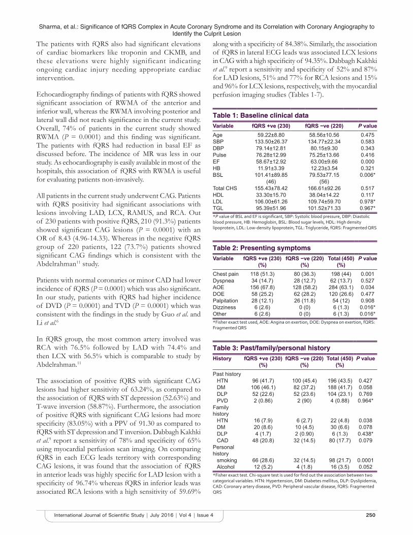
Sharma, et al.: Significance of fQRS Complex in Acute Coronary Syndrome and its Correlation with Coronary Angiography to Identify the Culprit Lesion
250International Journal of Scientific Study | July 2016 | Vol 4 | Issue 4
The patients with fQRS also had significant elevations of cardiac biomarkers like troponin and CKMB, and these elevations were highly significant indicating ongoing cardiac injury needing appropriate cardiac intervention.
Echocardiography findings of patients with fQRS showed significant association of RWMA of the anterior and inferior wall, whereas the RWMA involving posterior and lateral wall did not reach significance in the current study. Overall, 74% of patients in the current study showed RWMA (P = 0.0001) and this finding was significant. The patients with fQRS had reduction in basal EF as discussed before. The incidence of MR was less in our study. As echocardiography is easily available in most of the hospitals, this association of fQRS with RWMA is useful for evaluating patients non-invasively.
All patients in the current study underwent CAG. Patients with fQRS positivity had significant associations with lesions involving LAD, LCX, RAMUS, and RCA. Out of 230 patients with positive fQRS, 210 (91.3%) patients showed significant CAG lesions (P = 0.0001) with an OR of 8.43 (4.96-14.33). Whereas in the negative fQRS group of 220 patients, 122 (73.7%) patients showed significant CAG findings which is consistent with the Abdelrahman11 study.
Patients with normal coronaries or minor CAD had lower incidence of fQRS (P = 0.0001) which was also significant. In our study, patients with fQRS had higher incidence of DVD (P = 0.0001) and TVD (P = 0.0001) which was consistent with the findings in the study by Guo et al. and Li et al.6
In fQRS group, the most common artery involved was RCA with 76.5% followed by LAD with 74.4% and then LCX with 56.5% which is comparable to study by Abdelrahman.11
The association of positive fQRS with significant CAG lesions had higher sensitivity of 63.24%, as compared to the association of fQRS with ST depression (52.63%) and T-wave inversion (58.87%). Furthermore, the association of positive fQRS with significant CAG lesions had more specificity (83.05%) with a PPV of 91.30 as compared to fQRS with ST depression and T inversion. Dabbagh Kakhki et al.9 report a sensitivity of 78% and specificity of 65% using myocardial perfusion scan imaging. On comparing fQRS in each ECG leads territory with corresponding CAG lesions, it was found that the association of fQRS in anterior leads was highly specific for LAD lesion with a specificity of 96.74% whereas fQRS in inferior leads was associated RCA lesions with a high sensitivity of 59.69%
along with a specificity of 84.38%. Similarly, the association of fQRS in lateral ECG leads was associated LCX lesions in CAG with a high specificity of 94.35%. Dabbagh Kakhki et al.9 report a sensitivity and specificity of 52% and 87% for LAD lesions, 51% and 77% for RCA lesions and 15% and 96% for LCX lesions, respectively, with the myocardial perfusion imaging studies (Tables 1-7).
Table 3: Past/family/personal historyHistory fQRS +ve (230)
(%)fQRS −ve (220)
(%)Total (450)
(%)P value
Past historyHTN 96 (41.7) 100 (45.4) 196 (43.5) 0.427DM 106 (46.1) 82 (37.2) 188 (41.7) 0.058DLP 52 (22.6) 52 (23.6) 104 (23.1) 0.769PVD 2 (0.86) 2 (90) 4 (0.88) 0.964*
Family history
HTN 16 (7.9) 6 (2.7) 22 (4.8) 0.038DM 20 (8.6) 10 (4.5) 30 (6.6) 0.078DLP 4 (1.7) 2 (0.90) 6 (1.3) 0.438*CAD 48 (20.8) 32 (14.5) 80 (17.7) 0.079
Personal history
smoking 66 (28.6) 32 (14.5) 98 (21.7) 0.0001Alcohol 12 (5.2) 4 (1.8) 16 (3.5) 0.052
*Fisher exact test. Chi‑square test is used for find out the association between two categorical variables. HTN: Hypertension, DM: Diabetes mellitus, DLP: Dyslipidemia, CAD: Coronary artery disease, PVD: Peripheral vascular disease, fQRS: Fragmented QRS
Table 2: Presenting symptomsVariable fQRS +ve (230)
(%)fQRS −ve (220)
(%)Total (450)
(%)P value
Chest pain 118 (51.3) 80 (36.3) 198 (44) 0.001Dyspnea 34 (14.7) 28 (12.7) 62 (13.7) 0.527AOE 156 (67.8) 128 (58.2) 284 (63.1) 0.034DOE 58 (25.2) 62 (28.2) 120 (26.6) 0.477Palpitation 28 (12.1) 26 (11.8) 54 (12) 0.908Dizziness 6 (2.6) 0 (0) 6 (1.3) 0.016*Other 6 (2.6) 0 (0) 6 (1.3) 0.016**Fisher exact test used, AOE: Angina on exertion, DOE: Dyspnea on exertion, fQRS: Fragmented QRS
Table 1: Baseline clinical dataVariable fQRS +ve (230) fQRS −ve (220) P valueAge 59.22±8.80 58.56±10.56 0.475SBP 133.50±26.37 134.77±22.34 0.583DBP 79.14±12.81 80.15±9.30 0.343Pulse 76.28±12.99 75.25±13.66 0.416EF 58.67±12.92 63.00±9.66 0.000HB 11.91±3.39 12.23±3.54 0.321BSL 101.41±89.85 79.53±77.15 0.006*
(46) (56)Total CHS 155.43±78.42 166.61±92.26 0.517HDL 33.30±15.70 38.04±14.22 0.117LDL 106.00±61.26 109.74±59.70 0.978*TGL 95.39±51.96 101.52±71.33 0.967**P value of BSL and EF is significant, SBP: Systolic blood pressure, DBP: Diastolic blood pressure, HB: Hemoglobin, BSL: Blood sugar levels, HDL: High density lipoprotein, LDL: Low‑density lipoprotein, TGL: Triglyceride, fQRS: Fragmented QRS
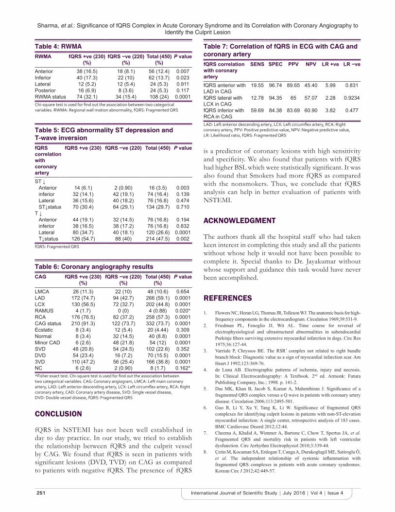
Sharma, et al.: Significance of fQRS Complex in Acute Coronary Syndrome and its Correlation with Coronary Angiography to Identify the Culprit Lesion
251 International Journal of Scientific Study | July 2016 | Vol 4 | Issue 4
is a predictor of coronary lesions with high sensitivity and specificity. We also found that patients with fQRS had higher BSL which were statistically significant. It was also found that Smokers had more fQRS as compared with the nonsmokers. Thus, we conclude that fQRS analysis can help in better evaluation of patients with NSTEMI.
ACKNOWLEDGMENT
The authors thank all the hospital staff who had taken keen interest in completing this study and all the patients without whose help it would not have been possible to complete it. Special thanks to Dr. Jayakumar without whose support and guidance this task would have never been accomplished.
REFERENCES
1. Flowers NC, Horan LG, Thomas JR, Tolleson WJ. The anatomic basis for high-frequency components in the electrocardiogram. Circulation 1969;39:531-9.
2. Friedman PL, Fenoglio JJ, Wit AL. Time course for reversal of electrophysiological and ultrastructural abnormalities in subendocardial Purkinje fibers surviving extensive myocardial infarction in dogs. Circ Res 1975;36:127-44.
3. Varriale P, Chryssos BE. The RSR’ complex not related to right bundle branch block: Diagnostic value as a sign of myocardial infarction scar. Am Heart J 1992;123:369-76.
4. de Luna AB. Electrographic patterns of ischemia, injury and necrosis. In: Clinical Electrocardiography: A Textbook. 2nd ed. Armonk: Futura Publishing Company, Inc.; 1998. p. 141-2.
5. Das MK, Khan B, Jacob S, Kumar A, Mahenthiran J. Significance of a fragmented QRS complex versus a Q wave in patients with coronary artery disease. Circulation 2006;113:2495-501.
6. Guo R, Li Y, Xu Y, Tang K, Li W. Significance of fragmented QRS complexes for identifying culprit lesions in patients with non-ST-elevation myocardial infarction: A single center, retrospective analysis of 183 cases. BMC Cardiovasc Disord 2012;12:44.
7. Cheema A, Khalid A, Wimmer A, Bartone C, Chow T, Spertus JA, et al. Fragmented QRS and mortality risk in patients with left ventricular dysfunction. Circ Arrhythm Electrophysiol 2010;3:339-44.
8. Çetin M, Kocaman SA, Erdogan T, Canga A, Durakoglugil ME, Satiroglu Ö, et al. The independent relationship of systemic inflammation with fragmented QRS complexes in patients with acute coronary syndromes. Korean Circ J 2012;42:449-57.
Table 4: RWMARWMA fQRS +ve (230)
(%)fQRS −ve (220)
(%)Total (450)
(%)P value
Anterior 38 (16.5) 18 (8.1) 56 (12.4) 0.007Inferior 40 (17.3) 22 (10) 62 (13.7) 0.023Lateral 12 (5.2) 12 (5.4) 24 (5.3) 0.911Posterior 16 (6.9) 8 (3.6) 24 (5.3) 0.117RWMA status 74 (32.1) 34 (15.4) 108 (24) 0.0001Chi‑square test is used for find out the association between two categorical variables. RWMA: Regional wall motion abnormality, fQRS: Fragmented QRS
Table 5: ECG abnormality ST depression and T-wave inversionfQRS correlation with coronary artery
fQRS +ve (230) fQRS −ve (220) Total (450) P value
ST ↓Anterior 14 (6.1) 2 (0.90) 16 (3.5) 0.003inferior 32 (14.1) 42 (19.1) 74 (16.4) 0.139Lateral 36 (15.6) 40 (18.2) 76 (16.8) 0.474ST↓status 70 (30.4) 64 (29.1) 134 (29.7) 0.710
T ↓Anterior 44 (19.1) 32 (14.5) 76 (16.8) 0.194inferior 38 (16.5) 38 (17.2) 76 (16.8) 0.832Lateral 80 (34.7) 40 (18.1) 120 (26.6) 0.0001T↓status 126 (54.7) 88 (40) 214 (47.5) 0.002
fQRS: Fragmented QRS
Table 6: Coronary angiography resultsCAG fQRS +ve (230)
(%)fQRS −ve (220)
(%)Total (450)
(%)P value
LMCA 26 (11.3) 22 (10) 48 (10.6) 0.654LAD 172 (74.7) 94 (42.7) 266 (59.1) 0.0001LCX 130 (56.5) 72 (32.7) 202 (44.8) 0.0001RAMUS 4 (1.7) 0 (0) 4 (0.88) 0.020*RCA 176 (76.5) 82 (37.2) 258 (57.3) 0.0001CAG status 210 (91.3) 122 (73.7) 332 (73.7) 0.0001Ecstatic 8 (3.4) 12 (5.4) 20 (4.44) 0.309Normal 8 (3.4) 32 (14.5) 40 (8.8) 0.0001Minor CAD 6 (2.6) 48 (21.8) 54 (12) 0.0001SVD 48 (20.8) 54 (24.5) 102 (22.6) 0.352DVD 54 (23.4) 16 (7.2) 70 (15.5) 0.00013VD 110 (47.2) 56 (25.4) 166 (36.8) 0.0001NC 6 (2.6) 2 (0.90) 8 (1.7) 0.162**Fisher exact test. Chi‑square test is used for find out the association between two categorical variables. CAG: Coronary angiogram, LMCA: Left main coronary artery, LAD: Left anterior descending artery, LCX: Left circumflex artery, RCA: Right coronary artery, CAD: Coronary artery disease, SVD: Single vessel disease, DVD: Double vessel disease, fQRS: Fragmented QRS
Table 7: Correlation of fQRS in ECG with CAG and coronary arteryfQRS correlation with coronary artery
SENS SPEC PPV NPV LR +ve LR −ve
fQRS anterior with LAD in CAG
19.55 96.74 89.65 45.40 5.99 0.831
fQRS lateral with LCX in CAG
12.78 94.35 65 57.07 2.28 0.9234
fQRS inferior with RCA in CAG
59.69 84.38 83.69 60.90 3.82 0.477
LAD: Left anterior descending artery, LCX: Left circumflex artery, RCA: Right coronary artery, PPV: Positive predictive value, NPV: Negative predictive value, LR: Likelihood ratio, fQRS: Fragmented QRS
CONCLUSION
fQRS in NSTEMI has not been well established in day to day practice. In our study, we tried to establish the relationship between fQRS and the culprit vessel by CAG. We found that fQRS is seen in patients with significant lesions (DVD, TVD) on CAG as compared to patients with negative fQRS. The presence of fQRS

Sharma, et al.: Significance of fQRS Complex in Acute Coronary Syndrome and its Correlation with Coronary Angiography to Identify the Culprit Lesion
252International Journal of Scientific Study | July 2016 | Vol 4 | Issue 4
9. Dabbagh Kakhki VR, Ayati N, Zakavi SR, Sadeghi R, Tayyebi M, Shariati F. Comparison between fragmented QRS and Q waves in myocardial scar detection using myocardial perfusion single photon emission computed tomography. Kardiol Pol 2015;73:437-44.
10. Li M, Wang X, Mi SH, Chi Z, Chen Q, Zhao X, et al. Short-term prognosis of fragmented QRS complex in patients with non-ST elevated acute myocardial infarction. Chin Med J (Engl) 2016;129:518-22.
11. Abdelrahman TM. QRS fragmentation as a prognostic test in acute coronary syndrome. World J Cardiovasc Surg 2013;3:42-51.
12. Korhonen P, Husa T, Konttila T, Tierala I, Mäkijärvi M, Väänänen H,
et al. Fragmented QRS in prediction of cardiac deaths and heart failure hospitalizations after myocardial infarction. Ann Noninvasive Electrocardiol 2010;15:130-7.
13. Yan GH, Wang M, Yiu KH, Lau CP, Zhi G, Lee SW, et al. Subclinical left ventricular dysfunction revealed by circumferential 2D strain imaging in patients with coronary artery disease and fragmented QRS complex. Heart Rhythm 2012;9:928-35.
14. Das MK, Michael MA, Suradi H, Peng J, Sinha A, Shen C, et al. Usefulness of fragmented QRS on a12-lead electrocardiogram in acute coronary syndrome for predicting mortality. Am J Cardiol 2009;104:1631-7.
How to cite this article: Sharma G, Jayakumar TG, Rupesh G, Rajesh G, Nihas, George G, Abdulkhadar SM. Significance of Fragmented QRS Complex in Acute Coronary Syndrome and its Correlation with Coronary Angiography to Identify the Culprit Lesion. Int J Sci Stud 2016;4(4):246-252.
Source of Support: Nil, Conflict of Interest: None declared.

253 International Journal of Scientific Study | July 2016 | Vol 4 | Issue 4
Sleep Apnea: An OverviewNiketh DeSouza
Private Practitioner, Mazgaon, Mumbai, Maharashtra, India
and it is most commonly diagnosed in middle-aged men.4 OSA can be temporary in those who have upper respiratory tract infections, nasal congestion, tonsillitis, or are under the influence of alcohol. Risk factors for developing OSA include increased body mass index (BMI), large neck size, hypertension, diabetes, chronic nasal congestion, male gender, ages 18-60, smoking and using alcohol. Most of the cases of OSA are thought to be due to decrease in muscle tone, old age, and brain injury. OSA in children is usually caused by overgrown tonsils and adenoids. Muscle tone decreases with alcohol and certain drugs. Women are at the highest risk of developing OSA during pregnancy and prevalence is higher in post-menopausal women than menstruating women. Syndromes with craniofacial abnormalities (such as Down’s syndrome) can increase the risk of developing OSA. OSA usually occurs in rapid eye movement (REM) sleep when the muscle tone in the neck, throat and skeletal muscles are decreased. This allows the oropharynx and tongue to relax and partially or completely block the flow of air. In some cases when the blood oxygen level decreases, a neurological stimulation can cause interruption of sleep which may cause awakening and will have a negative effect on the quality of sleep. This can occur in non-REM (NREM) Stage 3 as well which is the deepest stage of sleep and has a physically restorative effect on the body. In children, this stage of sleep (NREM Stage 3) is also responsible for the release of growth
INTRODUCTON
The three main types of sleep apnea are obstructive (84%), central (0.4%), and mixed or complex sleep apnea (15%) which is a combination of both types. Obstructive sleep apnea (OSA) is due to a physical blockage of airflow while central sleep apnea is due to a lack of respiratory effort. Both will present with excessive daytime sleepiness, lethargy, inability to focus and decreased alertness. In addition to these, OSA will have snoring. Below we will discuss each of the types of sleep apnea including how to diagnose and treat them.1-3
OSA
OSA is the most common type of sleep apnea and occurs when the muscles of the throat relax and block airflow during sleep. It can be a partial or complete obstruction of the upper airway. 2-4% of Americans are said to have OSA
Review Article
AbstractSleep apnea is a common and potentially dangerous sleep disorder characterized by one or more pauses in breathing or instances of shallow breathing during sleep. The episodes of apnea can last from a few seconds to several minutes. It is estimated that 1 in every 15 Americans has sleep apnea. Sleep apnea is associated with an increased risk of developing cardiovascular disease, stroke, hypertension, arrhythmias, diabetes, and causing accidents due to falling asleep while driving. A 2012 study showed that the hypoxia caused by sleep apnea promoted angiogenesis which increased tumor and vascular growth. This led to a 4.8 time increase in the incidence of cancer mortality. Sleep apnea can often go undiagnosed as patients are unaware of it while they sleep. It is estimated that the average untreated sleep apnea patient’s annual health-care costs 1336 dollars more than a person without sleep apnea which can cause 3.4 billion dollars a year in additional medical costs. It is a chronic condition which requires management throughout life. Lifestyle modifications, oral appliances, surgery, and breathing devices can treat sleep apnea in most patients. The introduction of the continuous positive airway pressure has greatly improved the management of sleep apnea.
Key words: Central sleep apnea, Complex sleep apnea, Mixed sleep apnea, Obstructive sleep apnea, Sleep apnea
Access this article online
www.ijss-sn.com
Month of Submission : 05-2016 Month of Peer Review : 06-2016 Month of Acceptance : 07-2016 Month of Publishing : 07-2016
Corresponding Author: NikethDeSouza, Court View, 33 Nesbit Road, Mazgaon, Mumbai - 400 010, Maharashtra, India. Phone: +91-920359448. E-mail: [email protected]
Print ISSN: 2321-6379Online ISSN: 2321-595X
DOI: 10.17354/ijss/2016/416

Desouza: An Overview of Sleep Apnea
254International Journal of Scientific Study | July 2016 | Vol 4 | Issue 4
hormone and OSA can result in failure to thrive in the affected children. Clinical symptoms can be divided into nocturnal and daytime symptoms. The nocturnal symptoms include loud snoring, witnessed apneas, insomnia, nocturia, sudden arousal from sleep accompanied by gasping and choking. The daytime symptoms include non-restorative sleep, morning headaches, dry mouth on awakening, daytime fatigue that worsens throughout the day, daytime sleepiness with the need to take frequent naps, decreased the ability to concentrate, mood and personality changes and decreased libido. The patient will usually present as an obese adult with a thick neck who is either brought in by the patient’s partner, who are alerted by the episodes of apnea and snoring, or by the patient themselves who complain of excessive daytime sleepiness. The daytime sleepiness usually begins with quiet activities such as watching TV or reading but can later progress to even activities which require alertness such as driving. A clinical diagnosis can be made by asking about the symptoms previously mentioned or by providing the patient with a questionnaire to fill which can then be scored to determine their likelihood of having OSA. Physical examination includes calculating the BMI (>30 is a risk factor) and measuring the neck circumference (>43 cm in men and >37 cm in women is a risk factor). Other findings on the physical exam can include an abnormal Mallampati score, enlarged tonsils, high-arched hard palate, large degree of overjet, and hypertension. The gold standard, however, to diagnose sleep apnea (obstructive or central) is with an overnight sleep study or polysomnography. This can either be done at home (less reliable) or at a sleep center. According to the results of the sleep study, sleep apnea can be graded. The number of events of apnea per hour is reported as the apnea-hypopnea index or AHI. An AHI of <5 is normal, 5-15 is mild, 15-30 is moderate, and >30 is severe. The treatment of sleep apnea includes lifestyle modifications such as weight loss, avoidance of sedatives, alcohol or tobacco, and sleeping on one’s side. Mild to moderate OSA can be treated with oral appliances which help keep the throat open by bringing the lower jaw forward. The most common method of treating moderate to severe OSA is by continuous positive airway pressure (CPAP) or a CPAP machine. The machine is connected to a mask which is fitted over the nose and mouth or just into the nose and delivers an airway pressure which prevents the upper airway from collapsing during sleep. This decreases the episodes of apnea and improves the symptoms of OSA. The CPAP can be either a continuous (fixed) pressure or an auto-titrating pressure. Newer CPAP machines are much smaller and less noisy than the older ones. Certain surgeries can be done to treat sleep apnea such as turbinectomy, tonsillectomy, adenoidectomy, and maxillomandibular advancement.
CENTRAL SLEEP APNEA
Central sleep apnea occurs when the effort to breathe is absent or diminished. It is much less common that OSA and is prevalent in <1% of the general population. It is due to the respiratory centers in the brain not sending the correct signals to the muscles that control respiration. This will cause the individual to miss one or more breathing cycles. This increases the level of blood carbon dioxide which usually stimulates respiration through neurologic feedback which will cause a period of hyperpnea. Thus the individual may cycle through a period of apnea and hyperpnea throughout the night. There will be no chest movement or efforts to breathe seen during the periods of apnea. There can be awakenings during the periods of apnea which will be followed by a period of panic caused by the increase in blood CO2 levels and sometimes the inability to breathe normally for a short duration following the sudden awakening. Some of the conditions that can lead to central sleep apnea are Parkinson’s disease, brain infections, stroke, anything that harms the brainstem, cervical injures, heart failure, and drugs such as narcotics. Symptoms are similar to that off OSA without the snoring. There will be a lack of thoracic or abdominal movement individuals with central sleep apnea while they are asleep. Diagnosis is aided by clinical history, physical examination and confirmed with polysomnography at a sleep center. This will help differentiate between obstructive and central sleep apnea or whether the apnea is caused by a mixture of both (complex sleep apnea). The degree of sleep apnea is graded as before (mild, moderate, severe). The treatment is first based on addressing the medical conditions which could be causing the sleep apnea such as Parkinson’s or opioid medications. Devices used include CPAP, bi-level positive airway pressure, and adaptive servo-ventilation. Certain drugs such as acetazolamide have been used to stimulate the respiratory centers. They do so by causing metabolic acidosis which then induces respiratory alkalosis (increased the respiratory rate) as a compensatory measure.
CONCLUSION
Sleep apnea is a serious sleep disorder affecting over 40 million Americans. There is an enormous amount of data showing that untreated sleep apnea is associated with cardiovascular disease, stroke, and other medical conditions. Physicians should be vigilant for the common signs and symptoms of sleep apnea and by asking a few additional questions, be able to identify those who may require further diagnostic workup. Due to the high prevalence of the disorder and its burden on the individual, society, and health-care system, untreated sleep apnea cannot be ignored.

Desouza: An Overview of Sleep Apnea
255 International Journal of Scientific Study | July 2016 | Vol 4 | Issue 4
REFERENCES
1. Javier NF, Peppard PE, Young T, Finn L, Hla KM, Farré R. Sleep-disordered breathing and cancer mortality: Results from the Wisconsin sleep cohort study. Am J Respir Crit Care Med 2012;186:190-4.
2. Kapur V, Blough DK, Sandblom RE, Hert R, de Maine JB, Sullivan SD,
et al. The medical cost of undiagnosed sleep apnea. Sleep 1999;22:749-55.3. Morgenthaler TI, Kagramanov V, Hanak V, Decker PA. Complex sleep
apnea syndrome: Is it a unique clinical syndrome? Sleep 2006;29:1203-9.4. Azagra-Calero E, Espinar-Escalona E, Barrera-Mora JM,
Llamas-Carreras JM, Solano-Reina E. Obstructive sleep apnea syndrome (OSAS). Review of the literature. Med Oral Patol Oral Cir Bucal 2012;17:e925-9.
How to cite this article: DeSouza N. Sleep Apnea: An Overview. Int J Sci Stud 2016;4(4):253-255.
Source of Support: Nil, Conflict of Interest: None declared.

256International Journal of Scientific Study | July 2016 | Vol 4 | Issue 4
Guidance of Eruption - Myth or Reality? A Case ReportT Sunil1, Nishitha C Gowda1, H A Amitha2, Y C Kiran2, B S Kavyashree3
1Assistant Professor, Department of Orthodontics, Vokkaligara Sangha Dental College and Hospital, Bengaluru, Karnataka, India, 2Senior Lecturer, Department of Pediatric and Preventive Dentistry, Vokkaligara Sangha Dental College and Hospital, Bengaluru, Karnataka, India, 3Post-graduate Student, Department of Pediatric and Preventive Dentistry, Vokkaligara Sangha Dental College and Hospital, Bengaluru, Karnataka, India
Thanks to computerized axial tomography, nowadays, we can detect even minimal resorption fields on the incisor roots and establish that in almost 50% of the cases with impacted canines.
Ericson and Kurol2,4 group the severity of resorption of the lateral incisor in 4°:• No resorption - intact surface• Slight resorption - up to the middle of the depth of
the root dentin• Moderate resorption - affects more than half of the
depth of the root dentin• Severe resorption - reaches the root pulp.
Root resorption is a physiological event for the primary teeth and it is said to be regulated by the stellate reticulum and the dental follicle of the underlying permanent tooth via the secretion of the stimulatory molecules, i.e., cytokines and transcription factors. The primary root resorption process is regulated in a manner similar to bone remodeling, involving the same receptor ligand system known as (receptor activator of a nuclear factor [RANK]-kappa B/RANK ligand).7 Primary teeth without a permanent successor eventually exfoliate as well, but our current understanding on the underlying mechanism is slim. The literature is also vague on how resorption of the
INTRODUCTION
The goal of “guidance of eruption” is to avoid the need for active orthodontic treatment or to reduce it to the minimal. A strong dependence between impacted canines the contact with their adjacent teeth and the root resorption of the incisors.1-3 The frequency of root resorption of the lateral incisors is 12.5%. The treatment of impacted canines is complex and prolonged.
Ericson and Kurol2,4 assume that the resorption of the lateral incisors after the ectopic eruption of the permanent canines is the most common complication and may be detected in all cases of considerably altered route of the canine eruption. It is most commonly seen in the lateral incisors-in 38%, and less frequently in the central incisors - in 9% of the cases. The apical and median third of the root are most commonly affected - in about 65%.4-6
Case Report
AbstractThe altered path of eruption causing the resorption of the permanent lateral incisor root after the ectopic eruption of the permanent canines is a common complication which is detected radiographically. The relationship of the permanent canines with the lateral incisor suggests that lateral incisor acts as guidance for the eruption of the permanent canines. The case presented here is a 9-year-old boy with permanent dentition showing the resorption of the roots of the lateral incisor due to the eruption of the permanent canines taking the lateral incisors as the guidance. This article aims at establishing that early detection and prompt preventive measures will lead to avoiding the critical complications arising out of the erupting canines and will preserve the morphological and functional integrity of the lateral incisors and the dentition.
Key words: Canines, Eruption, Incisors resorption, Ugly duckling
Access this article online
www.ijss-sn.com
Month of Submission : 05-2016 Month of Peer Review : 06-2016 Month of Acceptance : 07-2016 Month of Publishing : 07-2016
Corresponding Author: Dr. T Sunil, Department of Orthodontics, Vokkaligara Sangha Dental College and Hospital, K.R. Road, V. V. Puram, Bengaluru - 560 004, Karnataka, India. Phone: +91-9880300792. E-mail: [email protected]
Print ISSN: 2321-6379Online ISSN: 2321-595X
DOI: 10.17354/ijss/2016/417
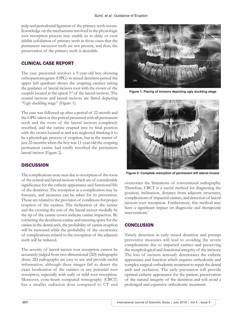
Sunil, et al.: Guidance of Eruption
257 International Journal of Scientific Study | July 2016 | Vol 4 | Issue 4
pulp and periodontal ligament of the primary teeth occurs. Knowledge on the mechanisms involved in the physiologic root resorption process may enable us to delay or even inhibit exfoliation of primary teeth in those cases that the permanent successor teeth are not present, and thus, the preservation of the primary teeth is desirable.
CLINICAL CASE REPORT
The case presented involves a 9-year-old boy showing orthopantomogram (OPG) in mixed dentition period the upper left quadrant shows the erupting canines taking the guidance of lateral incisors root with the crown of the cuspids located at the apical 3rd of the lateral incisors. The central incisors and lateral incisors are flared depicting “Ugly duckling stage” (Figure 1).
The case was followed up after a period of 22-month and the OPG taken at this period presented with all permanent teeth and the roots of the lateral incisors completely resorbed, and the canine erupted into its final position with the crown located at and was neglected thinking it to be a physiologic process of eruption, but in the matter of just 22-months when the boy was 11-year-old the erupting permanent canine had totally resorbed the permanent lateral incisor (Figure 2).
DISCUSSION
The complications were seen due to resorption of the roots of the central and lateral incisors which are of considerable significance for the esthetic appearance and functional life of the dentition. The resorption as a complication may be foreseen, and measures can be taken for its prevention. Those are related to the provision of conditions for proper eruption of the canines. The inclination of the canine and the crossing the axis of the lateral incisor medially by the tip of the canine crown indicate canine impaction. By extracting the deciduous canine and ensuring space for the canine in the dental arch, the probability of canine eruption will be increased while the probability of the occurrence of complications related to the resorption of the adjacent teeth will be reduced.
The severity of lateral incisor root resorption cannot be accurately judged from two-dimensional (2D) radiographs alone. 2D radiographs are easy to use and provide useful information, although these images fail to detect the exact localization of the canines or any potential root resorption, especially with early or mild root resorption. Moreover, cone-beam computed tomography (CBCT) has a smaller radiation dose compared to CT and
overcomes the limitations of conventional radiography. Therefore, CBCT is a useful method for diagnosing the position, inclination, distance from adjacent structures, complications of impacted canines, and detection of lateral incisors root resorption. Furthermore, this method may have a significant impact on diagnostic and therapeutic interventions.7
CONCLUSION
Timely detection in early mixed dentition and prompt preventive measures will lead to avoiding the severe complications due to impacted canines and preserving the morphological and functional integrity of the incisors. The loss of incisors seriously deteriorates the esthetic appearance and function which requires orthodontic and complex surgical-orthodontic treatment to repair the dental arch and occlusion. The early prevention will provide optimal esthetic appearance for the patient, preservation of the natural integrity of the dentition and will avoid a prolonged and expensive orthodontic treatment.
Figure 1: Flaring of incisors depicting ugly duckling stage
Figure 2: Complete resorption of permanent left lateral incisor

Sunil, et al.: Guidance of Eruption
258International Journal of Scientific Study | July 2016 | Vol 4 | Issue 4
REFERENCES
1. Ericson S, Kurol J. Incisor resorption caused by maxillary cuspids. A radiographic study. Angle Orthod 1987;57:332-46.
2. Ericson S, Kurol J. Incisor root resorptions due to ectopic maxillary canines imaged by computerized tomography: A comparative study in extracted teeth. Angle Orthod 2000;70:276-83.
3. Sasakura H, Yoshida T, Murayama S, Hanada K, Nakajima T. Root resorption of upper permanent incisor caused by impacted canine. An
analysis of 23 cases. Int J Oral Surg 1984;13:299-306.4. Ericson S, Kurol PJ. Resorption of incisors after ectopic eruption of
maxillary canines: A CT study. Angle Orthod 2000;70:415-23.5. Ericson S, Kurol J. Early treatment of palatally erupting maxillary canines
by extraction of the primary canines. Eur J Orthod 1988;10:283-95.6. Kim Y, Hyun HK, Jang KT. The position of maxillary canine impactions and
the influenced factors to adjacent root resorption in the Korean population. Eur J Orthod 2012;34:302-6.
7. Harokopakis-Hajishengallis E. Physiologic root resorption in primary teeth: Molecular and histological events. J Oral Sci 2007;49:1-12.
How to cite this article: Sunil T, Gowda NC, Amitha HA, Kiran YC, Kavyashree BS. Guidance of Eruption - Myth or Reality? A Case Report. Int J Sci Stud 2016;4(4):256-258.
Source of Support: Nil, Conflict of Interest: None declared.