Interatrial blocks. A separate entity from left atrial enlargement: a consensus report
7
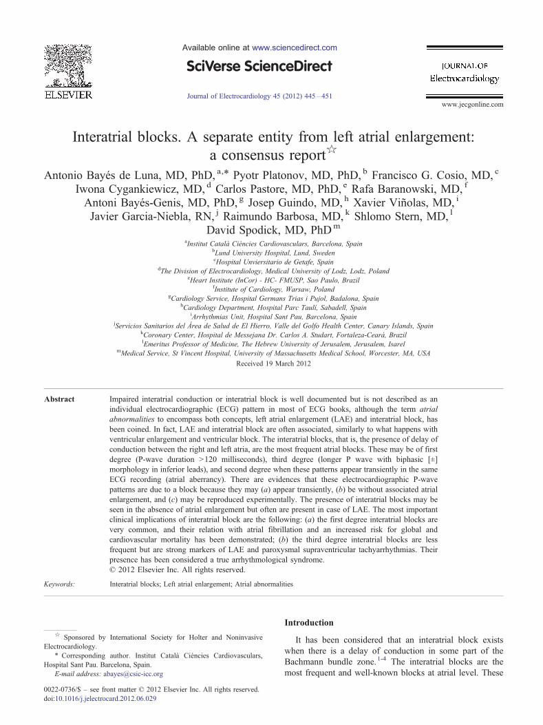
Interatrial blocks. A separate entity from left atrial enlargement: a consensus report ☆ Antonio Bayés de Luna, MD, PhD, a, ⁎ Pyotr Platonov, MD, PhD, b Francisco G. Cosio, MD, c Iwona Cygankiewicz, MD, d Carlos Pastore, MD, PhD, e Rafa Baranowski, MD, f Antoni Bayés-Genis, MD, PhD, g Josep Guindo, MD, h Xavier Viñolas, MD, i Javier Garcia-Niebla, RN, j Raimundo Barbosa, MD, k Shlomo Stern, MD, l David Spodick, MD, PhD m a Institut Català Ciències Cardiovasculars, Barcelona, Spain b Lund University Hospital, Lund, Sweden c Hospital Unviersitario de Getafe, Spain d The Division of Electrocardiology, Medical University of Lodz, Lodz, Poland e Heart Institute (InCor) - HC- FMUSP, Sao Paulo, Brazil f Institute of Cardiology, Warsaw, Poland g Cardiology Service, Hospital Germans Trias i Pujol, Badalona, Spain h Cardiology Department, Hospital Parc Taulí, Sabadell, Spain i Arrhythmias Unit, Hospital Sant Pau, Barcelona, Spain j Servicios Sanitarios del Área de Salud de El Hierro, Valle del Golfo Health Center, Canary Islands, Spain k Coronary Center, Hospital de Messejana Dr. Carlos A. Studart, Fortaleza-Ceará, Brazil l Emeritus Professor of Medicine, The Hebrew University of Jerusalem, Jerusalem, Isarel m Medical Service, St Vincent Hospital, University of Massachusetts Medical School, Worcester, MA, USA Received 19 March 2012 Abstract Impaired interatrial conduction or interatrial block is well documented but is not described as an individual electrocardiographic (ECG) pattern in most of ECG books, although the term atrial abnormalities to encompass both concepts, left atrial enlargement (LAE) and interatrial block, has been coined. In fact, LAE and interatrial block are often associated, similarly to what happens with ventricular enlargement and ventricular block. The interatrial blocks, that is, the presence of delay of conduction between the right and left atria, are the most frequent atrial blocks. These may be of first degree (P-wave duration N 120 milliseconds), third degree (longer P wave with biphasic [±] morphology in inferior leads), and second degree when these patterns appear transiently in the same ECG recording (atrial aberrancy). There are evidences that these electrocardiographic P-wave patterns are due to a block because they may (a) appear transiently, (b) be without associated atrial enlargement, and (c) may be reproduced experimentally. The presence of interatrial blocks may be seen in the absence of atrial enlargement but often are present in case of LAE. The most important clinical implications of interatrial block are the following: (a) the first degree interatrial blocks are very common, and their relation with atrial fibrillation and an increased risk for global and cardiovascular mortality has been demonstrated; (b) the third degree interatrial blocks are less frequent but are strong markers of LAE and paroxysmal supraventricular tachyarrhythmias. Their presence has been considered a true arrhythmological syndrome. © 2012 Elsevier Inc. All rights reserved. Keywords: Interatrial blocks; Left atrial enlargement; Atrial abnormalities Introduction It has been considered that an interatrial block exists when there is a delay of conduction in some part of the Bachmann bundle zone. 1-4 The interatrial blocks are the most frequent and well-known blocks at atrial level. These Available online at www.sciencedirect.com Journal of Electrocardiology 45 (2012) 445 – 451 www.jecgonline.com ☆ Sponsored by International Society for Holter and Noninvasive Electrocardiology. ⁎ Corresponding author. Institut Català Ciències Cardiovasculars, Hospital Sant Pau. Barcelona, Spain. E-mail address: [email protected] 0022-0736/$ – see front matter © 2012 Elsevier Inc. All rights reserved. doi:10.1016/j.jelectrocard.2012.06.029
-
Upload
independent -
Category
Documents
-
view
0 -
download
0
Transcript of Interatrial blocks. A separate entity from left atrial enlargement: a consensus report
Available online at www.sciencedirect.com
Journal of Electrocardiology 45 (2012) 445–451www.jecgonline.com
Interatrial blocks. A separate entity from left atrial enlargement:a consensus report☆
Antonio Bayés de Luna, MD, PhD, a,⁎ Pyotr Platonov, MD, PhD, b Francisco G. Cosio, MD, c
Iwona Cygankiewicz, MD, d Carlos Pastore, MD, PhD, e Rafa Baranowski, MD, f
Antoni Bayés-Genis, MD, PhD, g Josep Guindo, MD, h Xavier Viñolas, MD, i
Javier Garcia-Niebla, RN, j Raimundo Barbosa, MD, k Shlomo Stern, MD, l
David Spodick, MD, PhDm
aInstitut Català Ciències Cardiovasculars, Barcelona, SpainbLund University Hospital, Lund, SwedencHospital Unviersitario de Getafe, Spain
dThe Division of Electrocardiology, Medical University of Lodz, Lodz, PolandeHeart Institute (InCor) - HC- FMUSP, Sao Paulo, Brazil
fInstitute of Cardiology, Warsaw, PolandgCardiology Service, Hospital Germans Trias i Pujol, Badalona, Spain
hCardiology Department, Hospital Parc Taulí, Sabadell, SpainiArrhythmias Unit, Hospital Sant Pau, Barcelona, Spain
jServicios Sanitarios del Área de Salud de El Hierro, Valle del Golfo Health Center, Canary Islands, SpainkCoronary Center, Hospital de Messejana Dr. Carlos A. Studart, Fortaleza-Ceará, BrazillEmeritus Professor of Medicine, The Hebrew University of Jerusalem, Jerusalem, Isarel
mMedical Service, St Vincent Hospital, University of Massachusetts Medical School, Worcester, MA, USA
Received 19 March 2012
Abstract Impaired interatrial conduction or interatrial block is well documented but is not described as an
☆ Sponsored byElectrocardiology.
⁎ CorrespondingHospital Sant Pau. Ba
E-mail address: ab
0022-0736/$ – see frodoi:10.1016/j.jelectroc
individual electrocardiographic (ECG) pattern in most of ECG books, although the term atrialabnormalities to encompass both concepts, left atrial enlargement (LAE) and interatrial block, hasbeen coined. In fact, LAE and interatrial block are often associated, similarly to what happens withventricular enlargement and ventricular block. The interatrial blocks, that is, the presence of delay ofconduction between the right and left atria, are the most frequent atrial blocks. These may be of firstdegree (P-wave duration N120 milliseconds), third degree (longer P wave with biphasic [±]morphology in inferior leads), and second degree when these patterns appear transiently in the sameECG recording (atrial aberrancy). There are evidences that these electrocardiographic P-wavepatterns are due to a block because they may (a) appear transiently, (b) be without associated atrialenlargement, and (c) may be reproduced experimentally. The presence of interatrial blocks may beseen in the absence of atrial enlargement but often are present in case of LAE. The most importantclinical implications of interatrial block are the following: (a) the first degree interatrial blocks arevery common, and their relation with atrial fibrillation and an increased risk for global andcardiovascular mortality has been demonstrated; (b) the third degree interatrial blocks are lessfrequent but are strong markers of LAE and paroxysmal supraventricular tachyarrhythmias. Theirpresence has been considered a true arrhythmological syndrome.© 2012 Elsevier Inc. All rights reserved.
Keywords: Interatrial blocks; Left atrial enlargement; Atrial abnormalities
International Society for Holter and Noninvasive
author. Institut Català Ciències Cardiovasculars,rcelona, [email protected]
nt matter © 2012 Elsevier Inc. All rights reserved.ard.2012.06.029
Introduction
It has been considered that an interatrial block existswhen there is a delay of conduction in some part of theBachmann bundle zone.1-4 The interatrial blocks are themost frequent and well-known blocks at atrial level. These

446 A. Bayés de Luna et al. / Journal of Electrocardiology 45 (2012) 445–451
are expressed in the electrocardiogram (ECG) by thepresence of P-wave duration that equals or exceeds 120milliseconds and presents usually a bimodal morphology,especially in leads I, II, and VL and inferior leads.2-4 Sincethen, there are several reports describing the ECG charac-teristics of different types of interatrial blocks, and alsohave been described their clinical implications.5-16 Thesepublications considered that, in spite that the ECG patternsfound in left atrial enlargement (LAE) are often signifi-cantly influenced by the presence of impaired interatrialconduction, the same as that happens with ventricularenlargement and ventricular blocks, these 2 ECG patternsmay appear individually, in spite that the atrial blocks ingeneral and the ECG patterns of interatrial blocks inparticular are not described and even mentioned as an ECGpattern in most of books of ECG.17-22 On the contrary, theterm atrial abnormalities23-25 has been coined to encompassboth the terms LAE and interatrial block. This consensusreport emphasizes the concept that interatrial blocks andatrial enlargement are separate entities and that interatrialblocks, similarly to other types of blocks at sinoatrial, AVjunctional, and ventricular level, are a specific ECG patternthat may present first, second, and third degree types ofconduction block.
Where the interatrial blocks are located
During sinus rhythm, interatrial conduction occurs viaanatomically distinct connections composed of workingmyocardium fibers already described in the beginning ofXX century by Bachmann, Wenckebach, and Thorel. Theprimary left atrium (LA) breakthrough is in the Bachmannbundle area in two thirds of cases, and the rest occursaround the fossa ovalis and rarely in the coronary sinus.15
It has been demonstrated16,26 that, in most of cases, P-wave morphology can be used to correctly identify the leftatrial breakthrough site and the corresponding atrialimpulse propagation route. It has to be also noted thatthe relative proximity of site of origin in the sinus originwithin the sinus node to the Bachmann bundle or posteriorconnections may affect P-wave morphology. Deteriorationof conduction between the right atrium and LA—theinteratrial blocks—is the most frequent and well-studiedtype of atrial conduction disturbance that is discussedbelow in detail.
When an ECG pattern may be considered as beingcaused by an interatrial block
There are 3 criteria that define an ECG pattern as beingcaused by a block or deterioration of conduction26:
(a) the ECG pattern may appear transiently, and thepattern may change abruptly and progressively tomore advanced forms;
(b) the ECG pattern may appear without associated otherprocesses such as cardiac chamber enlargement or
ischemia, although in many cases, 1 or more of theseconditions may coexist; and
(c) similar ECG pattern may be reproduced experimentally.
P-wave ECG patterns that meet criteria forinteratrial block
The P-wave ECG pattern of interatrial block maybe transient
Transient deterioration of interatrial conduction, that is,interatrial block, of first or third degree may appear in thesame ECG recording on a beat-by-beat basis (Fig. 5).These cases may be considered as an illustration of atrialaberrancy, a concept coined by Chung27 in 1972, similarlyto ventricular aberrancy.
The P-wave ECG pattern of interatrial block may appearwithout associated atrial enlargement9,28
The prolonged P-wave duration (P-wave duration ≥120milliseconds) may be present in the elderly and also as aconsequence of an acute illness as pericarditis or acutemyocardial infarction without atrial enlargement. In fact, inthese cases, the duration of P wave may be as long as that inLAE, but the P loop does not move so clearly backwards asin a figure 8 shape, which results in a much smaller nega-tive P-wave component in lead V1. However, the associa-tion of LAE and the presence of advanced interatrial block(wide P-wave ≥120 milliseconds and ± in leads II, III, andVF) are common.
The P-wave ECG pattern may be reproduced experimentally
Experimental studies29 have demonstrated that cuttingthe Bachmann bundle at either right or left atrial side resultsin a typical ECG pattern with wide P wave with biphasic ±morphology in inferior leads. It was also demonstrated30 thatan attenuation of interatrial conduction without affectingatrioventricular conduction may occur after ablation of inte-ratrial conduction zones along the right atrial septum. Thisintervention produces partial interatrial conduction blockwith an increase of P-wave duration.
How do we measure interatrial conduction time
We refer to the increased duration of P wave in surfaceECG and in VCG recordings. Other techniques that may offermore exact and/or precise measurements are signal averagingP wave, 2-bidimensional echocardiography with timeDoppler imaging, and intracardiac ECG measurement.31,32
May the interatrial blocks be of first, second, andthird degree
The interatrial blocks, by analogy with other types ofblock, (sinoatrial, atrioventricular, and/or bundle-brunchblock at the ventricular level), may be of first (partial),second (transient interatrial block, atrial aberrancy), or thirddegree (advanced) (see before) (Fig. 1).

Fig. 1. Diagram of atrial conduction under normal circumstances (A), first-degree interatrial block (B), and third-degree interatrial block with left atriaretrograde activation.
447A. Bayés de Luna et al. / Journal of Electrocardiology 45 (2012) 445–451
Electrocardiographic pattern of first-degree (partial)interatrial block
The P wave has a normal electrical axis. The electricalimpulse is conducted from the right to the LA through thenormal propagation route but with a delay (Fig. 1B). Theendocardial recording confirms that the coronary sinus acti-vation time is delayed.
∎ The ECG shows that (a) a P wave of 120 milli-seconds or more, usually bimodal that is especiallyvisible in leads I, II, or III, and (b) the P-wavemorphology in V1 often presents a P-wave negativemode that is less evident than in cases of LAEbecause in the absence of LAE, the P loop is directedin a less backward direction.
∎ In the presence of P wave 120 milliseconds or more,LAE is common. In fact, it has been considered thatthe wide and bimodal P wave of LAE is explainedmore because of underlying interatrial block ratherthan the longer distance that the impulse has to goacross.28
∎ The presence of isolated cases of partial interatrialblock may explain that, in some series, the specificityof the 120-millisecond cutoff for diagnosis of LAEmay be low (around 50%). However, when weconsider the association of P-wave prolongation witha P-wave negative terminal mode more than 40 milli-seconds in lead V1, the specificity is much higher.26
∎ Sometimes, it is possible to record the progressiveappearance of the ECG pattern of first-degree and laterthird-degree interatrial block (Fig. 2).
∎ It is often found in the elderly9,10 in the absence ofassociated LAE. Recently, it has been shown to be amarker of atrial fibrillation and cardiovascular and all-cause mortality.33
l
Electrocardiographic pattern of third-degree (advanced)interatrial block7,8,34
• It is an infrequent (≈1% of patients with valve heartdisease)7 but a very easily recognizable ECG pattern(Figs. 1C, 3-5).
• The electrical impulse is blocked in the upper andmiddle part of the interatrial septum, in the Bachmannbundle zone, and/or in the upper part of LA so thatretrograde left atrial activation occurs via muscularconnections in the vicinity of coronary sinus.7,8,16 Inrare occasions, right atrium and LA can demonstratedissociated electrical activity.26
• The ECG shows that (a) P-wave duration 120milliseconds or more and (b) the morphology of Pwave are usually bimodal in lead I and VL and biphasic[±] in leads II, III, and VF because of caudocranialactivation of the LA and also often in V1 and V2.
• The electrophysiologic mechanism underlying thisECG pattern has been explained using deductive ECG-VCG data correlated with recordings obtained fromthe high and low right atrium, coronary sinus, rightpulmonary artery, and esophagus and also with elec-troanatomic mapping.25,34
• It has been also demonstrated that the block may belocated in the LA not affecting conduction through theproximal Bachmann bundle. However, this does notmodify the terminal inferosuperior activation of theLA that explains the typical morphology ± in II, III,and VF. In fact, the same ± morphology appears aftercutting the Bachmann bundle in dogs at the right andleft sides of the septum.29
• Bayés de Luna et al7 defined the ECG-VCG criteria(Figs. 1C, 3 and 5) and demonstrated that this type ofblock is a very specific (90%) but insensitive marker ofLAE. The duration of P wave is 120 milliseconds or

Fig. 2. A, A 65-year-old patient with previous anterior myocardial infarction and left superoanterior hemiblock plus long and bimodal P wave in many leadwith plus minus morphology in V1 suggestive of LAE plus first-degree interatrial lock. B, One year later (left), there is a left bundle-brunch block pattern, andthe P wave is negative in II, III, and VF. However, this is not AV junctional rhythm because (a) the heart rate changes clearly along the day and with exercise ais well seen (see I and II of B), (b) the first part of P wave in II, III, and VF is isodiphasic (see II), (c) after premature atrial beat (III), a pattern of first-degreeinteratrial block appears, followed again by the pattern of third-degree interatrial block and (d) the PR interval is the same as before (160 ms).
448 A. Bayés de Luna et al. / Journal of Electrocardiology 45 (2012) 445–451
more (120-170 milliseconds in this series of 83 cases).7
However, it is the morphology of the P wave (± ininferior leads) that pinpoints that there is a retrogradeactivation of the LA. The positive mode of P waves inleads II, III, and VF is at times not well seen probablybecause of fibrosis, and the diagnosis of junctional
s
s
rhythm due to an apparently negative P wave in II, III,and VF may be made.It was demonstrated that this type of block is veryfrequently accompanied by paroxysmal atrial arrhyth-mia, especially atypical atrial flutter8,34,35 in patientswith valvular heart diseases and cardiomyopathies.

Fig. 3. Above: P wave ± morphology in I, II, and III typical of third-degree interatrial block with retrograde conduction to the LA in a patient with double aorticand mitral valve disease. Observe how the AP and the angle between the direction of the activation in the first and second parts of the P wave are measured. To theright, high esophageal ECG (HE) and endocavitary registrations (high right atrium and low right atrium) demonstrate that the electrical stimulus moves firsdownwards (high right atrium to low right atrium) and then upwards (low right atrium to HE). Below: P loop morphology in the 3 planes with the inscription othe second part moving upwards. HRA indicates high right atrium; LRA, low right atrium.
449A. Bayés de Luna et al. / Journal of Electrocardiology 45 (2012) 445–451
Therefore, this association is considered an ECGclinical syndrome.34,36,37
• During long-term follow-up, especially in patientswith mitral valve disease or cardiomyopathy, thesuccessive appearance of first-degree and later third-degree interatrial block may be seen (Fig. 2).
Electrocardiographic pattern of second-degreeinteratrial block
∎ Similarly to the conduction block at the level of AVjunction, SA junction, or the ventricles, the interatrialblock may occur transiently on a beat-to-beat basis.
• In a typical case, the morphology of the P wave ischanging in the same recording from normal to
Fig. 4. Virtual anatomic rendering of the LA in the other patient with typical biphasic (±) P wave in leads II, III, and aVF suggestive of Bachmann bundle blockNote that early left atrial activation (white) occurs at the high septal wall, as expected for Bachmann bundle conduction. Activation does not progress through theleft atrial roof because of the presence of a large zone of low voltage (gray) that diverts activation toward the low septal (orange-yellow) then the low posterio(green) and finally the high posterior (violet) left atrial wall.
tf
interatrial block pattern or from first-degree interatrialblock to third-degree interatrial block pattern or viceversa, with or without relation to the precedingpremature beats, and may be considered as atrialaberrancy, similarly to ventricular aberrancy27 (Fig. 2).
Clinical implications of interatrial blocks
• Although the diagnosis of interatrial blocks isfrequently associated to LAE, there are cases,especially of first-degree interatrial blocks, withoutassociated LAE.
• The diagnosis of partial interatrial block by itself hasmany clinical implications that have been closely
.
r

Fig. 5. Typical ECG of third-degree interatrial block (P± in II, III, and VF and duration N120 milliseconds) in a patient with ischemic cardiomyopathy. Whenamplified, we can see the beginning of P in the 3 leads.
450 A. Bayés de Luna et al. / Journal of Electrocardiology 45 (2012) 445–451
studied by the Spodick group9,10,38 and others.15,33,34
It was demonstrated that the presence of first-degreeinteratrial block (prolonged interatrial conduction time[IACT]) is associated with abnormal left atrial function.Furthermore, IACT is related not only to anincreased P-wave duration but also to a prolongedPR interval.33,39 We know now that the prevalence offirst-degree interatrial block in the general population isvery high9,10 and that this condition has recently beenshown to be associated with cardiovascular and all-cause mortality.33
• The presence of third-degree interatrial block is veryfrequently associated with supraventricular arrhythmias.In fact, this type of interatrial block constitutes for someauthors a clear arrhythmological syndrome.34,36,37 Itspresence in patients with valvular heart disease orcardiomyopathy advices to study the risk of supraven-tricular arrhythmias (Holter ECG) to decide whether aprophylactic antiarrhythmic treatment and/or anticoagu-lation is needed. Bayés de Luna et al35 suggested thatantiarrhythmic treatment prevents recurrences of atrialtachycarrhythmia in these cases. D'Allonnes et al40
demonstrated the importance of synchronous biatrialpacing to prevent of recurrences of atrial fibrillation.
• It was also demonstrated that, in patients with sinusnode disease and intra-atrial conduction delay at theposterior triangle of Koch, low interatrial septumpacing was superior to right atrial appendage pacing inpreventing progression to persistent or permanentatrial fibrillation41 and is associated with the shortestatrial activation time compared with other atrial septalpacing sites in patients with prolonged P waves.42
• On the other hand, the presence of increased P-waveduration has been described as an important parameterwhen programming the optimal AV delay in patientswho require atrial pacing. In fact, it has been dem-
onstrated that the adverse effect of interatrial delay inbiventricular pacing may be avoided with interatrialseptum pacing.32
• In some studies, it has been shown that interatrialconduction time (IACT) measured by tissue Dopplerechocardiography was more closely related to the PRinterval than to the duration of the P wave.Furthermore, in patients with heart failure and wideQRS, IACT is frequently prolonged. Before cardiacresynchronization therapy (CRT), an IACT measuredby tissue Doppler imaging more than 100 millisecondscan affect LA remodeling and function after CRT atearly follow-up but does not influence the response inLV end-systolic volume or ejection fraction.43
Conclusions
• After all the arguments that we have presented, it isclear that interatrial blocks are an entity separate fromatrial enlargement. However, the 2 are often associatedespecially in the case of interatrial block of thirddegree, as often happens with ventricular enlargementand ventricular blocks.
• Interatrial blocks are located in the zone of the high/middle septum especially close to the Bachman bundlebut may also be located beyond the interatrial septumin the LA.
• In case of first-degree interatrial block, the ECG showsa P-wave duration 120 milliseconds or more. If theinteratrial block is of third degree, also, the P wave isprolonged, but the second part of the P wave in inferiorleads becomes negative because of the retrogradeactivation of the LA (P± in II, III, and VF).
• There are clear and important implications related withthe presence of interatrial blocks. They are frequently

451A. Bayés de Luna et al. / Journal of Electrocardiology 45 (2012) 445–451
associated to LAE that, in the case of third-degreeinteratrial blocks, have a specificity of 90% and tosupraventricular arrhythmias that are also, much morefrequent in third-degree interatrial blocks.
• The prevalence of first-degree interatrial block in thegeneral population is very high, whereas third-degreeinteratrial blocks are rarely seen.
References
1. Cohen J, Scherf D. Complete interatrial and intraatrial block. Am HeartJ 1965;70:23.
2. Brody DA, Arzbaecher R, Woosley M, et al. The normal atrial electro-gram:morphologic and quantitative variability in bipolar leads. AmHeart J 1967;74:4.
3. Zoneraich O, Zoneraich S. Intraatrial conduction disturbances: VCGpatterns. Am J Cardiol 1976;37:736.
4. Bachmann G. The significance of the splitting of the P wave in theelectrocardiogram. Ann Int Med 1941;14:1703.
5. Puech P. L'activité electrique auriculaire normale et pathologique.Paris: Masson Edit; 1956.
6. Castillo Fenoy A, Vernant P. Les troubles de la conduction inter-auriculaire pour bloc du faisceau du Bachmann. Arch Mal Coeur Vaiss1971;64:1490.
7. Bayés de Luna A, Fort de Ribot R, Trilla E, et al. Electrocardiographicand vectorcardiographic study of interatrial conduction disturbanceswith left atrial retrograde activation. J Electrocardiol 1985;18:1.
8. Bayés de Luna A, Cladellas M, Oter R, et al. Interatrial conductionblock and retrograde activation of the left atrium and paroxysmalsupraventricular tachyarrhythmias. Eur Heart J 1988;9:1112.
9. Spodick DH, Ariyarajah V. Interatrial block. The pandemic remainspoorly perceived. Pacing Clin Electrophysiol 2009;32:662.
10. Spodick DH, Ariyarajah V. Interatrial block: a prevalent, widelyneglected and portentous abnormality. J Electrocardiol 2008;41:61.
11. Ariyarajah V, Fisella ME, Spodick DH. Reevaluation of the criterion forinteratrial block. Am J Cardiol 2006;98:936.
12. Holmqvist F, Platonov P, Carlson J, et al. Abnormal P wave mor-phology is a predictor of atrial fibrillation in MADIT II patients. AnnNoninvasive Electrocardiol 2010;15:63.
13. Platonov P. Atrial conduction and atrial fibrillation. What can we learnform ECG? Cardiol J 2008;15:402.
14. Platonov PG, Mitrofanova L, Ivanov V, et al. Substrates for intra-atrialand interatrial conduction in the atrial septum: anatomical study on 84humans hearts. Heart Rhythm 2008;5:1189.
15. Tapanainen JM, Jurkko R, Holmqvist F, et al. Interatrial right-to-leftconduction in patients with paroxysmal atrial fibrillation. J Interv CardElectrophysiol 2009;L25:117.
16. Holmqvist F, Husser D, Tapanainen JM, et al. Interatrial conduction canbe accurately determined using standard 12-lead electrocardiography:validation of P-wave morphology using electroanatomic mapping inman. Heart Rhythm 2008;5:413.
17. Sodi-Pallares D. New basis of electrocardiography. Medical, St Louis:Mosby Co; 1956.
18. Coksey JD, Dunan M, Massie E. Clinical vectrocardiography andelectrocardiography. Chigago: Year Book Publishers; 1977.
19. Wagner GS. Marriott's practical electrocardiology. 10th ed. Philadel-phia: Lippincott Williams and Wilkins; 2001.
20. Gerstch M. The ECG: a two step approach for diagnosis. Endrich:Springer Verlag; 2004.
21. Surawicz B, Knilans TK. Chou's electrocardiography in clinicalpractice 6th ed. 2008. Philadelphia: Saunders Elsevier; 2008.
22. Macfarlane P, Van Oosterom A, Pahlm O, Kligfield P, Janse M, CammJ, editors. Comprehensive electrocardiology. London: Springer; 2011.
23. Lee K, Appleton C, Lester S, et al. Relation of ECG criteria for left atrialenlargement in two-dimensional echocardiographic left atrial measure-ments. Am J Cardiol 2007;99:113.
24. Tsao CW, JosephsonME, Hauser TH. Accuracy of electrocardiographiccriteria for atrial enlargement: validation with cardiovascular magneticresonance. J Cardiovasc Magn Reson 2008;10:7.
25. Cosio FG, Martín-Peñato A, Pastor A, et al. Atrial activation mappingin sinus rhythm in the clniical electrophysiology laboratory. Observa-tions in Bachmann's bundle block. J Cardiovasc Electorphysiol 2004;15:524.
26. Bayés de Luna A. Clinical electrocardiography: a textbook. Chichester,West Sussex, UK: Wiley-Blackwell; 2012.
27. Chung E. Aberrant atrial conduction. Aberrant atrial conduction 1972;34:341.
28. Josephson ME, Kastor JA, Morganroth J. Electrocardiographic leftatrial enlargement. Electrophysiologic, echocardiographic and hemo-dynamic correlates. Am J Cardiol 1977;39:967.
29. Waldo A, Hurry L, Bush J, et al. Effects on the canine P waves ofdiscrete lessions in the specialized atrial tracts. Circulation Res 1971;29:452.
30. Schwartzman D, Warman EN, Devine WA, et al. Attenuation ofinteratrial conduction using right atrial septal catheter ablation. J AmColl Cardiol 2001;38:892.
31. Daubert JC, Pavin D, Jauvert G, et al. Intra-and interatrial conductiondelay: implications for cardiac pacing. Pacing Clin Electrophysiol2004;27:507.
32. Porciani MC, Sabini A, Colella A, et al. Interatrial septum pacing avoidsthe adverse effect of interatrial delay in biventricular pacing: an echo-Doppler evaluation. Europace 2002;4:317.
33. Magnani JW, Gorodeski EZ, Johnson VM, et al. P wave duration isassociated with cardiovascular and all-cause mortality outcomes: theNational Health and Nutrition Examination Survey. Heart Rhythm2011;8:93.
34. Bayés de Luna A, Guindo J, Viñolas X, et al. Third-degree inter-atrialblock and supraventricular tachyarrhythmias. Europace 1999;1:43.
35. Bayés de Luna A, Cladellas M, Oter R, et al. Interatrial block withretrograde activation of the left atrium: influence of preventiveantiarrhythmic treatment. Int J Card 1989;22:147.
36. Braunwald E. Foreword. In: Bayés de Luna A, editor. ClinicalElectrocardiograhpy: a textbook. Chichester, West Sussex, UK:Wiley-Blackwell; 2012.
37. Daubert JC. Atrial flutter and interatrial conduction block. In: Waldo A,Touboul P, editors. Atrial Flutter. Armonk, NY: Futura Publishing;1996. p. 33.
38. Ariyarajah V, Apiyasawat S, Puri P, et al. Specific electrocardiographicmarkers of P-wave morphology in interatrial block. J Electrocardiol2006;39:380.
39. Chevalier S, Basta M, Leitch JW. The importance of the left atrio-ventricular interval during atrioventricular sequential pacing. PacingClin Electrophysiol 1997;20:2958.
40. D'Allonnes GR, Pavin D, Leclercq C, et al. Long-term effects ofbiatrial synchronous pacing to prevent drug-refractory atrial tachyar-rhythmia : a nine-year experience. J Cardiovasc Electrophysiol 2000;11:1081.
41. Verlato R, Botto GR, Massa R, et al. Efficacy of low interatrial septumpacing and right atrial appendage pacing for prevention of permanentatrial fibrillation in patients with sick sinus syndrome. Circ ArrhythmElectrophysiol 2011;4:844.
42. Huo Y, Holmqvist F, Carlson J, et al. Effects of baseline P-waveduration and choice of atrial septal pacing site on shortening atrialactivation time during pacing. Europace 2012 [Epub ahead of print].
43. Waggoner AD, Kalathiveetil S, Karen E, et al. Interatrial conductiontime and left atrial function in patients with left ventricular systolicdysfunction: effects of cardiac resynchronization therapy. J Am SocEchocardiogr 2009;22:472.