Interactions of Flavonoids with Iron and Copper Ions: A Mechanism for their Antioxidant Activity
10
Interactions of Flavonoids with Iron and Copper Ions: A Mechanism for their Antioxidant Activity LURDES MIRA a,b, *, M. TEREZA FERNANDEZ a , MARTA SANTOS a,b , RUI ROCHA a,b , M. HELENA FLORE ˆ NCIO a and KEITH R. JENNINGS c a Departamento de Quı ´mica e Bioquı ´mica, Faculdade de Cie ˆncias da Universidade de Lisboa, Edifı ´cio C8, Campo Grande, 1749-016 Lisboa, Portugal; b Centro de Metabolismo e Endocrinologia da Faculdade de Medicina de Lisboa, Laborato ´rio de Gene ´tica, Faculdade de Medicina de Lisboa, 1649-016 Lisboa, Portugal; c Department of Biological Sciences, Warwick University, Coventry CV4 7AL, UK Accepted by Professor B. Halliwell (Received 17 April 2002; In revised form 6 June 2002) The metal chelating properties of flavonoids suggest that they may play a role in metal-overload diseases and in all oxidative stress conditions involving a transition metal ion. A detailed study has been made of the ability of flavonoids to chelate iron (including Fe 3þ ) and copper ions and its dependence of structure and pH. The acid medium may be important in some pathological conditions. In addition, the ability of flavonoids to reduce iron and copper ions and their activity–structure relationships were also investigated. To fulfil these objectives, flavones (apigenin, luteolin, kaempferol, quer- cetin, myricetin and rutin), isoflavones (daidzein and genistein), flavanones (taxifolin, naringenin and naringin) and a flavanol (catechin) were investigated. All flavonoids studied show higher reducing capacity for copper ions than for iron ions. The flavonoids with better Fe 3þ reducing activity are those with a 2,3-double bond and possessing both the catechol group in the B-ring and the 3-hydroxyl group. The copper reducing activity seems to depend largely on the number of hydroxyl groups. The chelation studies were carried out by means of ultraviolet spectroscopy and electrospray ionisation mass spec- trometry. Only flavones and the flavanol catechin interact with metal ions. At pH 7.4 and pH 5.5 all flavones studied appear to chelate Cu 2þ at the same site, probably between the 5-hydroxyl and the 4-oxo groups. Myricetin and quercetin, however, at pH 7.4, appear to chelate Cu 2þ additionally at the ortho-catechol group, the chelating site for catechin with Cu 2þ at pH 7.4. Chelation studies of Fe 3þ to flavonoids were investigated only at pH 5.5. Only myricetin and quercetin interact strongly with Fe 3þ , complexation probably occurring again between the 5-hydroxyl and the 4-oxo groups. Their behaviour can be explained by their ability to reduce Fe 3þ at pH 5.5, suggesting that flavonoids reduce Fe 3þ to Fe 2þ before association. Keywords: Flavonoids; Metal chelation; Metal reduction; Anti- oxidant properties; Mass spectrometry INTRODUCTION Free radicals are continuously being formed in small amounts by normal processes of metabolism. Many of them serve useful physiological functions, [1,2] but they can damage the biomolecules when generated in excess, being implicated in the aetiology of several diseases and ageing. [3 – 5] In order to balance the physiological generation of free radicals, organisms have evolved a wide array of enzymatic and non- enzymatic endogenous antioxidant defenses. [6,7] Nevertheless, in situations of increased free radical generation the reinforcement of endogenous anti- oxidants with dietary antioxidants may be particu- larly important in diminishing the cumulative effects of oxidatively damaged molecules. Recent work highlights the potential health- promoting properties of flavonoids: [8 – 10] these are phenolic compounds widely distributed in fruits, vegetables, plant extracts as well as in plant-derived beverages such as tea and red wine. [11,12] Flavonoids have generated interest because of their broad ISSN 1071-5762 print/ISSN 1029-2470 online q 2002 Taylor & Francis Ltd DOI: 10.1080/1071576021000016463 *Corresponding author. Address: Departamento de Quı ´mica e Bioquı ´mica, Faculdade de Cie ˆncias da Universidade de Lisboa, Edifı ´cio C8, Campo Grande, 1749-016 Lisboa, Portugal. Tel.: þ351-217500935. Fax: þ 351-217500088. E-mail: [email protected] Free Radical Research, 2002 Vol. 36 (11), pp. 1199–1208
-
Upload
independent -
Category
Documents
-
view
0 -
download
0
Transcript of Interactions of Flavonoids with Iron and Copper Ions: A Mechanism for their Antioxidant Activity
Interactions of Flavonoids with Iron and Copper Ions: AMechanism for their Antioxidant Activity
LURDES MIRAa,b,*, M. TEREZA FERNANDEZa, MARTA SANTOSa,b, RUI ROCHAa,b, M. HELENA FLORENCIOa andKEITH R. JENNINGSc
aDepartamento de Quımica e Bioquımica, Faculdade de Ciencias da Universidade de Lisboa, Edifıcio C8, Campo Grande, 1749-016 Lisboa, Portugal;bCentro de Metabolismo e Endocrinologia da Faculdade de Medicina de Lisboa, Laboratorio de Genetica, Faculdade de Medicina de Lisboa, 1649-016Lisboa, Portugal; cDepartment of Biological Sciences, Warwick University, Coventry CV4 7AL, UK
Accepted by Professor B. Halliwell
(Received 17 April 2002; In revised form 6 June 2002)
The metal chelating properties of flavonoids suggest thatthey may play a role in metal-overload diseases and in alloxidative stress conditions involving a transition metalion. A detailed study has been made of the ability offlavonoids to chelate iron (including Fe3þ) and copperions and its dependence of structure and pH. The acidmedium may be important in some pathologicalconditions. In addition, the ability of flavonoids to reduceiron and copper ions and their activity– structurerelationships were also investigated. To fulfil theseobjectives, flavones (apigenin, luteolin, kaempferol, quer-cetin, myricetin and rutin), isoflavones (daidzein andgenistein), flavanones (taxifolin, naringenin and naringin)and a flavanol (catechin) were investigated. All flavonoidsstudied show higher reducing capacity for copper ionsthan for iron ions. The flavonoids with better Fe3þ
reducing activity are those with a 2,3-double bond andpossessing both the catechol group in the B-ring and the3-hydroxyl group. The copper reducing activity seems todepend largely on the number of hydroxyl groups. Thechelation studies were carried out by means of ultravioletspectroscopy and electrospray ionisation mass spec-trometry. Only flavones and the flavanol catechin interactwith metal ions. At pH 7.4 and pH 5.5 all flavones studiedappear to chelate Cu2þ at the same site, probably betweenthe 5-hydroxyl and the 4-oxo groups. Myricetin andquercetin, however, at pH 7.4, appear to chelate Cu2þ
additionally at the ortho-catechol group, the chelating sitefor catechin with Cu2þ at pH 7.4. Chelation studies of Fe3þ
to flavonoids were investigated only at pH 5.5. Onlymyricetin and quercetin interact strongly with Fe3þ,complexation probably occurring again between the5-hydroxyl and the 4-oxo groups. Their behaviour canbe explained by their ability to reduce Fe3þ at pH 5.5,
suggesting that flavonoids reduce Fe3þ to Fe2þ beforeassociation.
Keywords: Flavonoids; Metal chelation; Metal reduction; Anti-oxidant properties; Mass spectrometry
INTRODUCTION
Free radicals are continuously being formed in smallamounts by normal processes of metabolism. Manyof them serve useful physiological functions,[1,2] butthey can damage the biomolecules when generatedin excess, being implicated in the aetiology of severaldiseases and ageing.[3 – 5] In order to balance thephysiological generation of free radicals, organismshave evolved a wide array of enzymatic and non-enzymatic endogenous antioxidant defenses.[6,7]
Nevertheless, in situations of increased free radicalgeneration the reinforcement of endogenous anti-oxidants with dietary antioxidants may be particu-larly important in diminishing the cumulative effectsof oxidatively damaged molecules.
Recent work highlights the potential health-promoting properties of flavonoids:[8 – 10] these arephenolic compounds widely distributed in fruits,vegetables, plant extracts as well as in plant-derivedbeverages such as tea and red wine.[11,12] Flavonoidshave generated interest because of their broad
ISSN 1071-5762 print/ISSN 1029-2470 online q 2002 Taylor & Francis Ltd
DOI: 10.1080/1071576021000016463
*Corresponding author. Address: Departamento de Quımica e Bioquımica, Faculdade de Ciencias da Universidade de Lisboa, EdifıcioC8, Campo Grande, 1749-016 Lisboa, Portugal. Tel.: þ351-217500935. Fax: þ351-217500088. E-mail: [email protected]
Free Radical Research, 2002 Vol. 36 (11), pp. 1199–1208
pharmacological effects such as vasoprotective, anti-inflammatory, antiviral and antifungal actions(reviewed in Ref. [13]). Many of these effects arerelated to their antioxidant properties, which may bedue to their ability to scavenge free radicals[14 – 19]
and to synergistic effects with other antioxidants.[20]
Another antioxidant mechanism of flavonoids, notyet extensively studied, may result from theinteractions between flavonoid and metal ions(especially iron and copper) leading to chelatesformation that are only slightly active in the pro-motion of free-radical reactions.[21 – 27] Flavonoidshave also been reported to show prooxidant effects,which have been related with their iron and copperreducing activities. These reduced metals cancatalyse the production of hydroxyl radicals throughFenton reaction[28 – 31] and lipid radicals throughthe decomposition of preformed lipid hydro-peroxides.[32,33]
In order to be an effective catalyst in those radicalreactions, iron has to be present in an ionic or “free”form. Traces of iron salts are present in several bodyfluids, with the exception of blood plasma.[34]
Usually iron is safely sequestered in proteins thatnormally bind iron hindering or preventing its actionin catalysing radical reactions. Iron, however, can bereleased from those proteins at low pH[35] as a resultof protein damage produced by peroxides[36,37] or byreductive mobilization by O·2
2 .[38] Low pH values canbe generated locally during phagocytosis andinflammation[39] and during the ischemia/reper-fusion injury.[40] The nature of the small pool of iron,not bound to proteins, that exists in the cytosol of thecell for a variety of cellular functions and enzymes isunknown, but probably consists of both Fe2þ andFe3þ.[41] The presence of both ferric and ferrousforms of iron in the cytosol is an important pre-requisite for the initiation of lipid peroxidation.[42]
In addition, desferrioxamine, a chelator of Fe3þ, hasbeen shown to limit the amount of free radicalproduction in several different cells and species.[43]
Most copper is “tightly bound” to the plasmaprotein caeruloplasmin, but some is attached toalbumin and to aminoacids such as histidine that cancatalyse free radical reactions.[42,44]
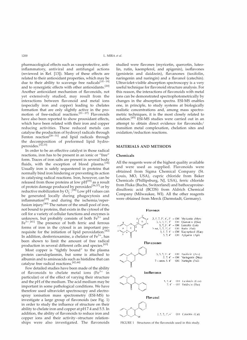
Few detailed studies have been made of the abilityof flavonoids to chelate metal ions (Fe3þ inparticular) or of the effect of varying their structureand the pH of the medium. The acid medium may beimportant in some pathological conditions. We havetherefore used ultraviolet spectroscopy and electro-spray ionisation mass spectrometry (ESI-MS) toinvestigate a large group of flavonoids (see Fig. 1)in order to study the influence of structure on theirability to chelate iron and copper at pH 7.4 and 5.5. Inaddition, the ability of flavonoids to reduce iron andcopper ions and their activity–structure relation-ships were also investigated. The flavonoids
studied were flavones (myricetin, quercetin, luteo-lin, rutin, kaempferol, and apigenin), isoflavones(genistein and daidzein), flavanones (taxifolin,naringenin and naringin) and a flavanol (catechin).Ultraviolet-visible absorption spectroscopy is a veryuseful technique for flavonoid structure analysis. Forthis reason, the interactions of flavonoids with metalions can be demonstrated spectrophotometrically bychanges in the absorption spectra. ESI-MS enablesone, in principle, to study systems at biologicallyrealistic concentrations and, among mass spectro-metric techniques, it is the most closely related tosolution.[45] ESI-MS studies were carried out in anattempt to obtain direct evidence for flavonoids/transition metal complexation, chelation sites andoxidation/reduction reactions.
MATERIALS AND METHODS
Chemicals
All the reagents were of the highest quality availableand were used as supplied. Flavonoids wereobtained from Sigma Chemical Company (St.Louis, MO, USA), cupric chloride from BakerChemicals (Phillipsburg, NJ, USA), ferric chloridefrom Fluka (Buchs, Switzerland) and bathocuproine-disulfonic acid (BCDS) from Aldrich ChemicalCompany (Milwaukee, WI). All the other reagentswere obtained from Merck (Darmstadt, Germany).
FIGURE 1 Structures of the flavonoids used in this study.
L. MIRA et al.1200
Metal Reduction Studies
Fe31 Reducing Activity
The ability of flavonoids to reduce Fe3þ was assayedby a modified ferrozine method.[31] Ferrozine is achromophoric chelator that strongly binds Fe2þ
forming a stable complex with a high extinctioncoefficient at 562 nm.[46] The reaction mixturecontained 50 mM Na-acetate buffer (pH 5.5), 1 mMferrozine, 25mM flavonoid and 100mM FeCl3. Thereaction was started by the addition of FeCl3 and theincrease of absorbance at 562 nm after 3 min wasrecorded using a control lacking ferrozine. The Fe2þ
concentration was determined by using an extinctioncoefficient for the FeðferrozineÞ2þ3 complex of27,900 M21 cm21.[46]
Cu21 Reducing Activity
The ability of flavonoids to reduce Cu2þ wasevaluated by measuring the formation of the complexbetween Cuþ and bathocuproinedisulfonic acid(BCDS).[47] The reaction mixture contained 20 mMKH2PO4/KOH buffer (pH 7.4), 200mM CuCl2,600mM BCDS and either 10mM ascorbate or 10mMflavonoid. The mixtures were incubated at 378C for120 min and then the absorbances were read at483 nm. The copper concentration was determined byusing an extinction coefficient for the (BCDS)2Cu(I)complex of 12,900 M21 cm21, which was evaluated bya calibration curve (ranging 5.0–125mM) obtained bystandard CuCl2 solutions and by using 200mMascorbate as reducing agent.
Chelation Studies
Spectrophotometric Analysis
The modifications of the absorption spectra of100mM flavonoid solutions when combined withequimolar concentrations of either Fe3þ or Cu2þ, in50 mM acetate buffer (pH 5.5) and 50 mM MOPSbuffer (pH 7.4) at room temperature, were spectro-photometrically analysed with a Pye UnicamUV2-100 spectrophotometer. All the spectra wererun against blanks containing the buffer and themetal ion.
Electrospray Ionisation Mass Spectrometry(ESI-MS) Analysis
The flavonoids studied by this technique were theflavones myricetin, quercetin, luteolin and kaemp-ferol and the flavanol catechin. All experiments wereperformed in a Quattro II QhQ tandem quadrupoleinstrument (Micromass Manchester) fitted with anelectrospray ionisation source operated in positiveion mode. The tip of the capillary was maintained at
the potential of 3 kV and the sampling cone wasmaintained at potentials within the range 230 to2120 V relative to ground, in order to optimise thespectra. The mobile phase consisted of methanol/water 1:1 and 0.1% of acetic acid and the optimumflow rate of the mobile phase was found to be 5ml/min. Solutions of cupric chloride or ferric chlorideand a ligand in a ratio 1:1 were prepared inconcentrations in the range 35–65mM in eachcomponent. The pH of these solutions was #5.5.Mass spectra were acquired by scanning the firstquadrupole mass analyser from m/z (mass to chargeratio) 2000 to 50–100 and ions were detected bymeans of a scintillator detector positioned afterthe first quadrupole. Collision induced dissociation(CID)[48] spectra were obtained by selecting aprecursor ion in the first quadrupole mass analyserand transferring it into the collision cell containingargon. Product ion spectra were obtained byscanning the second quadrupole mass analyser.The pressures and the voltages applied to thecollision cell were in the ranges (1–4) £ 1023 mbarand (20–60) eV, respectively, in order to optimise theconditions. All data were processed by means of theMassLynx data system (Micromass).
RESULTS
Reduction Studies
Fe31 Reduction Study
The reduction of Fe3þ ions by flavonoids was studiedat pH 5.5, due to the low solubility of iron atphysiological pH. In aqueous solution Fe3þ exists asthe hydrated ion FeðH2OÞ3þ6 which, except at low pH,hydrolyses and polymerises, precipitating hydratedferric oxides.[41] The reduction of iron was monitoredusing the chromophoric chelator ferrozine whichstrongly binds Fe2þ, forming a stable complex with ahigh extinction coefficient. Our results (Table I) showthat only flavones myricetin and quercetin reducedFe3þ effectively. Rutin, catechin and taxifolin weremoderately active and kaempferol and luteolin wererelatively poor reductants. All other flavonoidsstudied showed no activity.
Cu21 Reduction Study
The reduction of Cu2þ by flavonoids was measuredusing the bathocuproinedisulfonic acid (BCDS),which forms a complex with Cuþ. As can be inferredfrom the values represented in Table II, flavonoidsreduce copper ions with a high stoichiometry.Myricetin, with six hydroxyl groups, was the betterreductant followed by the flavonoids with fivehydroxyl groups quercetin (flavone), taxifolin (fla-vanone) and catechin (flavanol). The flavones
FLAVONOID–METAL INTERACTIONS 1201
luteolin (5,7,30,40 –OH) and kaempferol (3,5,7,40 –OH)show the same Cu2þ reducing activity and apigenin(5,7,40 –OH) has about half of their activities. Theflavone apigenin and the isoflavone genisteinpossessing the same hydroxyl groups (5,7,40 –OH)were less active than the flavonoids with fourhydroxyl groups; the flavanone naringenin (5,7,40 –OH), however, was twice more effective on reducingthe copper ions.
Chelation Studies
Spectrophotometric Study
The flavonoid spectra typically consist of twoabsorption maxima in the ranges 240– 285 nm(band II) and 300–550 nm (band I) depending onhydroxyl substitution. In our study, we examined alarge group of flavonoids from different structuralclasses, flavones, flavanones, isoflavones and aflavanol (Fig. 1), in order to test their ability tochelate Cu2þ and Fe3þ at both pH 7.4 and pH 5.5. Ourresults showed that spectral changes were observedfor flavones with Cu2þ ions at both pH values andwith Fe3þ at pH 5.5 only, due to the solubilityproblems of iron.[41] Small modifications in theabsorption spectrum of the flavanol catechin were
also observed upon addition of Cu2þ at pH 7.4. Thespectra of isoflavones (daidzein and genistein) andflavanones (taxifolin, naringenin and naringin) wereunchanged.
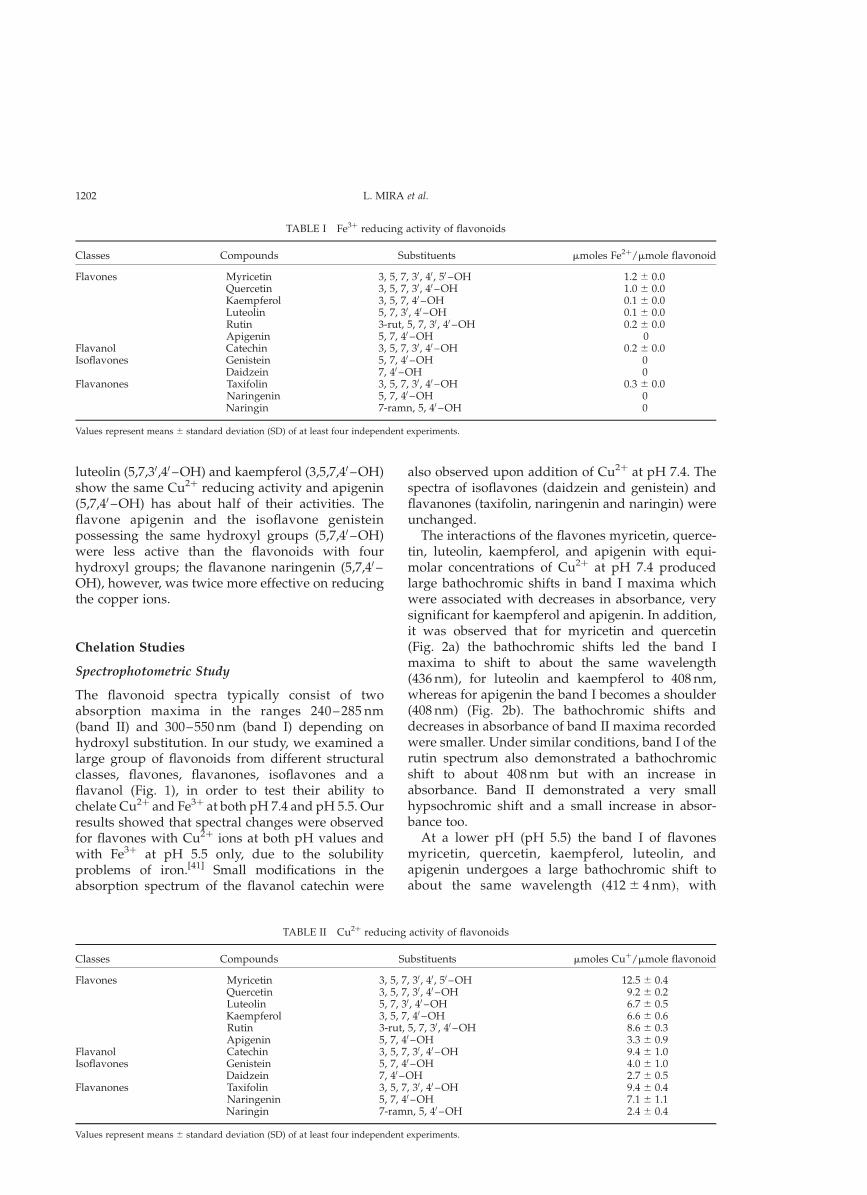
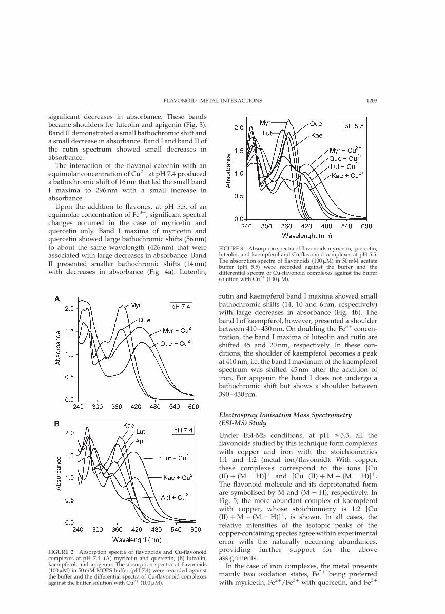
The interactions of the flavones myricetin, querce-tin, luteolin, kaempferol, and apigenin with equi-molar concentrations of Cu2þ at pH 7.4 producedlarge bathochromic shifts in band I maxima whichwere associated with decreases in absorbance, verysignificant for kaempferol and apigenin. In addition,it was observed that for myricetin and quercetin(Fig. 2a) the bathochromic shifts led the band Imaxima to shift to about the same wavelength(436 nm), for luteolin and kaempferol to 408 nm,whereas for apigenin the band I becomes a shoulder(408 nm) (Fig. 2b). The bathochromic shifts anddecreases in absorbance of band II maxima recordedwere smaller. Under similar conditions, band I of therutin spectrum also demonstrated a bathochromicshift to about 408 nm but with an increase inabsorbance. Band II demonstrated a very smallhypsochromic shift and a small increase in absor-bance too.
At a lower pH (pH 5.5) the band I of flavonesmyricetin, quercetin, kaempferol, luteolin, andapigenin undergoes a large bathochromic shift toabout the same wavelength ð412 ^ 4 nmÞ; with
TABLE II Cu2þ reducing activity of flavonoids
Classes Compounds Substituents mmoles Cuþ/mmole flavonoid
Flavones Myricetin 3, 5, 7, 30, 40, 50 –OH 12.5 ^ 0.4Quercetin 3, 5, 7, 30, 40 –OH 9.2 ^ 0.2Luteolin 5, 7, 30, 40 –OH 6.7 ^ 0.5Kaempferol 3, 5, 7, 40 –OH 6.6 ^ 0.6Rutin 3-rut, 5, 7, 30, 40 –OH 8.6 ^ 0.3Apigenin 5, 7, 40 –OH 3.3 ^ 0.9
Flavanol Catechin 3, 5, 7, 30, 40 –OH 9.4 ^ 1.0Isoflavones Genistein 5, 7, 40 –OH 4.0 ^ 1.0
Daidzein 7, 40 –OH 2.7 ^ 0.5Flavanones Taxifolin 3, 5, 7, 30, 40 –OH 9.4 ^ 0.4
Naringenin 5, 7, 40 –OH 7.1 ^ 1.1Naringin 7-ramn, 5, 40 –OH 2.4 ^ 0.4
Values represent means ^ standard deviation (SD) of at least four independent experiments.
TABLE I Fe3þ reducing activity of flavonoids
Classes Compounds Substituents mmoles Fe2þ/mmole flavonoid
Flavones Myricetin 3, 5, 7, 30, 40, 50 –OH 1.2 ^ 0.0Quercetin 3, 5, 7, 30, 40 –OH 1.0 ^ 0.0Kaempferol 3, 5, 7, 40 –OH 0.1 ^ 0.0Luteolin 5, 7, 30, 40 –OH 0.1 ^ 0.0Rutin 3-rut, 5, 7, 30, 40 –OH 0.2 ^ 0.0Apigenin 5, 7, 40 –OH 0
Flavanol Catechin 3, 5, 7, 30, 40 –OH 0.2 ^ 0.0Isoflavones Genistein 5, 7, 40 –OH 0
Daidzein 7, 40 –OH 0Flavanones Taxifolin 3, 5, 7, 30, 40 –OH 0.3 ^ 0.0
Naringenin 5, 7, 40 –OH 0Naringin 7-ramn, 5, 40 –OH 0
Values represent means ^ standard deviation (SD) of at least four independent experiments.
L. MIRA et al.1202
significant decreases in absorbance. These bandsbecame shoulders for luteolin and apigenin (Fig. 3).Band II demonstrated a small bathochromic shift anda small decrease in absorbance. Band I and band II ofthe rutin spectrum showed small decreases inabsorbance.
The interaction of the flavanol catechin with anequimolar concentration of Cu2þ at pH 7.4 produceda bathochromic shift of 16 nm that led the small bandI maxima to 296 nm with a small increase inabsorbance.
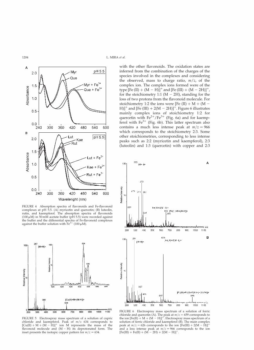
Upon the addition to flavones, at pH 5.5, of anequimolar concentration of Fe3þ, significant spectralchanges occurred in the case of myricetin andquercetin only. Band I maxima of myricetin andquercetin showed large bathochromic shifts (56 nm)to about the same wavelength (426 nm) that wereassociated with large decreases in absorbance. BandII presented smaller bathochromic shifts (14 nm)with decreases in absorbance (Fig. 4a). Luteolin,
rutin and kaempferol band I maxima showed smallbathochromic shifts (14, 10 and 6 nm, respectively)with large decreases in absorbance (Fig. 4b). Theband I of kaempferol, however, presented a shoulderbetween 410–430 nm. On doubling the Fe3þ concen-tration, the band I maxima of luteolin and rutin areshifted 45 and 20 nm, respectively. In these con-ditions, the shoulder of kaempferol becomes a peakat 410 nm, i.e. the band I maximum of the kaempferolspectrum was shifted 45 nm after the addition ofiron. For apigenin the band I does not undergo abathochromic shift but shows a shoulder between390–430 nm.
Electrospray Ionisation Mass Spectrometry(ESI-MS) Study
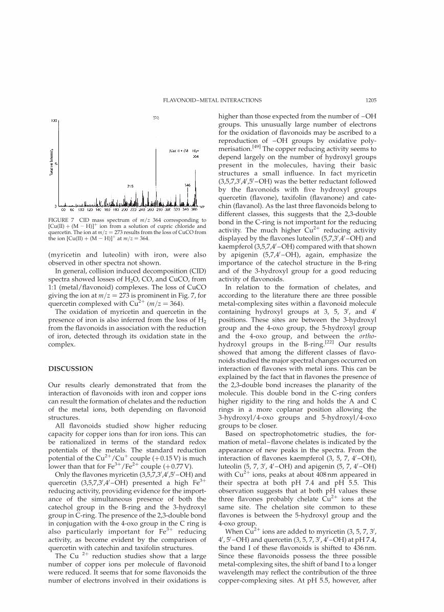
Under ESI-MS conditions, at pH #5.5, all theflavonoids studied by this technique form complexeswith copper and iron with the stoichiometries1:1 and 1:2 (metal ion/flavonoid). With copper,these complexes correspond to the ions [Cu(II) þ (M 2 H)]þ and [Cu (II) þ M þ (M 2 H)]þ.The flavonoid molecule and its deprotonated formare symbolised by M and (M 2 H), respectively. InFig. 5, the more abundant complex of kaempferolwith copper, whose stoichiometry is 1:2 [Cu(II) þ M þ (M 2 H)]þ, is shown. In all cases, therelative intensities of the isotopic peaks of thecopper-containing species agree within experimentalerror with the naturally occurring abundances,providing further support for the aboveassignments.
In the case of iron complexes, the metal presentsmainly two oxidation states, Fe2þ being preferredwith myricetin, Fe2þ/Fe3þ with quercetin, and Fe3þ
FIGURE 3 Absorption spectra of flavonoids myricetin, quercetin,luteolin, and kaempferol and Cu-flavonoid complexes at pH 5.5.The absorption spectra of flavonoids (100mM) in 50 mM acetatebuffer (pH 5.5) were recorded against the buffer and thedifferential spectra of Cu-flavonoid complexes against the buffersolution with Cu2þ (100mM).
FIGURE 2 Absorption spectra of flavonoids and Cu-flavonoidcomplexes at pH 7.4. (A) myricetin and quercetin; (B) luteolin,kaempferol, and apigenin. The absorption spectra of flavonoids(100mM) in 50 mM MOPS buffer (pH 7.4) were recorded againstthe buffer and the differential spectra of Cu-flavonoid complexesagainst the buffer solution with Cu2þ (100mM).
FLAVONOID–METAL INTERACTIONS 1203
with the other flavonoids. The oxidation states areinferred from the combination of the charges of thespecies involved in the complexes and consideringthe observed, mass to charge ratio, m/z, of thecomplex ion. The complex ions formed were of thetype [Fe (II) þ (M 2 H)]þ and [Fe (III) þ (M 2 2H)]þ,for the stoichiometry 1:1 (M 2 2H), standing for theloss of two protons from the flavonoid molecule. Forstoichiometry 1:2 the ions were [Fe (II) þ M þ (M 2
H)]þ and [Fe (III) þ 2(M 2 2H)]þ. Figure 6 illustratesmainly complex ions of stoichiometry 1:2 forquercetin with Fe2þ/Fe3þ (Fig. 6a) and for kaemp-ferol with Fe3þ (Fig. 6b). This latter spectrum alsocontains a much less intense peak at m=z ¼ 966which corresponds to the stoichiometry 2:3. Someother stoichiometries, corresponding to less intensepeaks such as 2:2 (myricetin and kaempferol), 2:3(luteolin) and 1:3 (quercetin) with copper and 2:3
FIGURE 6 Electrospray mass spectrum of a solution of ferricchloride and quercetin (A). The peak at m=z ¼ 659 corresponds tothe ion [Fe(II) þ M þ (M 2 H)]þ. Electrospray mass spectrum of asolution of ferric chloride and kaempferol (B). The main complexpeak at m=z ¼ 626 corresponds to the ion [Fe(III) þ 2(M 2 H)]þ
and a less intense peak at m=z ¼ 966 corresponds to the ion[Fe(III) þ Fe(II) þ (M 2 2H) þ 2(M 2 H)]þ.
FIGURE 5 Electrospray mass spectrum of a solution of cupricchloride and kaempferol. Peak of m/z 634 corresponds to[Cu(II) þ M þ (M 2 H)]þ ion M represents the mass of theflavonoid molecule and (M 2 H) its deprotonated form. Theinset presents the isotopic copper pattern for m=z ¼ 634:
FIGURE 4 Absorption spectra of flavonoids and Fe-flavonoidcomplexes at pH 5.5. (A) myricetin and quercetin; (B) luteolin,rutin, and kaempferol. The absorption spectra of flavonoids(100mM) in 50 mM acetate buffer (pH 5.5) were recorded againstthe buffer and the differential spectra of Fe-flavonoid complexesagainst the buffer solution with Fe3þ (100mM).
L. MIRA et al.1204
(myricetin and luteolin) with iron, were alsoobserved in other spectra not shown.
In general, collision induced decomposition (CID)spectra showed losses of H2O, CO, and CuCO, from1:1 (metal/flavonoid) complexes. The loss of CuCOgiving the ion at m=z ¼ 273 is prominent in Fig. 7, forquercetin complexed with Cu2þ ðm=z ¼ 364Þ:
The oxidation of myricetin and quercetin in thepresence of iron is also inferred from the loss of H2
from the flavonoids in association with the reductionof iron, detected through its oxidation state in thecomplex.
DISCUSSION
Our results clearly demonstrated that from theinteraction of flavonoids with iron and copper ionscan result the formation of chelates and the reductionof the metal ions, both depending on flavonoidstructures.
All flavonoids studied show higher reducingcapacity for copper ions than for iron ions. This canbe rationalized in terms of the standard redoxpotentials of the metals. The standard reductionpotential of the Cu2þ/Cuþ couple (þ0.15 V) is muchlower than that for Fe3þ/Fe2þ couple (þ0.77 V).
Only the flavones myricetin (3,5,7,30,40,50 –OH) andquercetin (3,5,7,30,40 –OH) presented a high Fe3þ
reducing activity, providing evidence for the import-ance of the simultaneous presence of both thecatechol group in the B-ring and the 3-hydroxylgroup in C-ring. The presence of the 2,3-double bondin conjugation with the 4-oxo group in the C ring isalso particularly important for Fe3þ reducingactivity, as become evident by the comparison ofquercetin with catechin and taxifolin structures.
The Cu 2þ reduction studies show that a largenumber of copper ions per molecule of flavonoidwere reduced. It seems that for some flavonoids thenumber of electrons involved in their oxidations is
higher than those expected from the number of –OHgroups. This unusually large number of electronsfor the oxidation of flavonoids may be ascribed to areproduction of –OH groups by oxidative poly-merisation.[49] The copper reducing activity seems todepend largely on the number of hydroxyl groupspresent in the molecules, having their basicstructures a small influence. In fact myricetin(3,5,7,30,40,50 –OH) was the better reductant followedby the flavonoids with five hydroxyl groupsquercetin (flavone), taxifolin (flavanone) and cate-chin (flavanol). As the last three flavonoids belong todifferent classes, this suggests that the 2,3-doublebond in the C-ring is not important for the reducingactivity. The much higher Cu2þ reducing activitydisplayed by the flavones luteolin (5,7,30,40 –OH) andkaempferol (3,5,7,40–OH) compared with that shownby apigenin (5,7,40 –OH), again, emphasize theimportance of the catechol structure in the B-ringand of the 3-hydroxyl group for a good reducingactivity of flavonoids.
In relation to the formation of chelates, andaccording to the literature there are three possiblemetal-complexing sites within a flavonoid moleculecontaining hydroxyl groups at 3, 5, 30, and 40
positions. These sites are between the 3-hydroxylgroup and the 4-oxo group, the 5-hydroxyl groupand the 4-oxo group, and between the ortho-hydroxyl groups in the B-ring.[22] Our resultsshowed that among the different classes of flavo-noids studied the major spectral changes occurred oninteraction of flavones with metal ions. This can beexplained by the fact that in flavones the presence ofthe 2,3-double bond increases the planarity of themolecule. This double bond in the C-ring confershigher rigidity to the ring and holds the A and Crings in a more coplanar position allowing the3-hydroxyl/4-oxo groups and 5-hydroxyl/4-oxogroups to be closer.
Based on spectrophotometric studies, the for-mation of metal–flavone chelates is indicated by theappearance of new peaks in the spectra. From theinteraction of flavones kaempferol (3, 5, 7, 40–OH),luteolin (5, 7, 30, 40 –OH) and apigenin (5, 7, 40–OH)with Cu2þ ions, peaks at about 408 nm appeared intheir spectra at both pH 7.4 and pH 5.5. Thisobservation suggests that at both pH values thesethree flavones probably chelate Cu2þ ions at thesame site. The chelation site common to theseflavones is between the 5-hydroxyl group and the4-oxo group.
When Cu2þ ions are added to myricetin (3, 5, 7, 30,40, 50–OH) and quercetin (3, 5, 7, 30, 40 –OH) at pH 7.4,the band I of these flavonoids is shifted to 436 nm.Since these flavonoids possess the three possiblemetal-complexing sites, the shift of band I to a longerwavelength may reflect the contribution of the threecopper-complexing sites. At pH 5.5, however, after
FIGURE 7 CID mass spectrum of m/z 364 corresponding to[Cu(II) þ (M 2 H)]þ ion from a solution of cupric chloride andquercetin. The ion at m=z ¼ 273 results from the loss of CuCO fromthe ion [Cu(II) þ (M 2 H)]þ at m=z ¼ 364:
FLAVONOID–METAL INTERACTIONS 1205
the addition of Cu2þ ions, band I of these flavonesundergoes a shift to about the same wavelength asthe shifts observed for kaempferol, luteolin andapigenin. Therefore, it seems that at pH 5.5 all thoseflavones interact with Cu2þ ions through the samesite, most probably between the 5-hydroxyl and the4-oxo groups. These arguments lead to the con-clusion that quercetin and myricetin should havemore chelation sites at pH 7.4 than at pH 5.5. In fact,the complexing ability of a catechol-type B ringincreases as the pH becomes more alkaline.[22] Insummary, our experimental data in solution supportthe idea that for chelation of copper the site betweenthe 5-hydroxyl group and the 4-oxo group is the mostimportant chelating site. Our results are in agree-ment with those of Thompson and Williams[50] whofound that the stability of the complex is higher incase of chelation via 5-OH, this probably beingassociated with the stability of the six-memberedring over that of the five-membered ring complexes.
The flavanol catechin spectrum also presentedmodifications after the addition of Cu2þ ions at pH7.4. Since catechin lacks the 4-oxo group we can inferthat catechin chelates Cu2þ through the ortho-catechol group. At pH 5.5, however, catechin doesnot chelate copper ions. This behaviour of catechin isin accordance with the fact that the complexingability of the catechol group increases as the pHincreases.
The ESI-MS studies provided a direct evidence forthe formation of complexes of various stoichio-metries, among which the 1:1 and 1:2 (metal/flavo-noid) had previously been considered as the morelikely.[22] Moreover, with the exception of catechin,all the flavonoids studied by ESI-MS lose CuCO asindicated by CID experiments. These losses occur inflavonoids that have in common the 4-oxo group andeither the 3 or 5-hydroxyl group as opposed tocatechin which does not possess the 4-oxo group.This reinforces our prediction of chelation at the4-oxo and 5-OH groups, based on spectrophoto-metric measurements, since luteolin, which does nothave the 3-OH group, also shows chelation of Cu2þ.The behaviour of catechin confirms that thepreferred chelation site must involve the 4-oxogroup and either one of the 5-OH or 3-OH groups.When these pairs are not available, chelation willtake place at the 30 and 40 –OH groups. On the otherhand, the CID mass spectrum of the complexcatechin/copper with stoichiometry 1:1 contains anintense peak due to ion Cuþ. This result suggests thatthe complex catechin/copper might be very labile inacid medium[51] and might explain why this complexat pH 5.5 was not observed in our spectrophoto-metric studies.
In the chelation studies of iron to flavonoids at pH5.5 we observed that only the flavones myricetin andquercetin at an equimolar concentration present
a large bathochromic shift. At this acid medium,the complexation site is probably between the5-hydroxyl group and the 4-oxo group. In fact, thechelating ability of the catechol group decreases asthe pH decreases.[22] This is probably because thechelate formation requires both 30, 40 –OH groups tobe dissociated and the extent of dissociation maydiffer. In addition, the dissociation pattern ofindividual hydroxyl groups occurs in the sequence7–OH . 40 –OH . 5–OH[52] predicting a morefavoured dissociation for the 5-OH than for 3-OHgroup. All the other flavones present a moderateinteraction with Fe3þ. This can be rationalized interms of the number of hydroxyl groups. In fact,luteolin, rutin and kaempferol possess fewer –OHgroups than quercetin resulting in a lower negativecharge density at the chelation site. Furthermore, weobserved that, at pH 5.5 and equimolar concen-trations, all the flavones studied chelate Fe2þ (datanot shown). This suggests that flavonoids chelateiron more effectively when the metal ion is in itsbivalent form, which would mean that the flavonoidneeds to reduce Fe3þ to Fe2þ, before association. Theresults of the Fe3þ reduction study by flavonoids, infact, demonstrated that only the flavones myricetinand quercetin reduce Fe3þ at pH 5.5 and this doesexplain why they were the more effective flavones iniron chelation.
The complexation of iron and the variousflavonoids at pH # 5.5 was also observed by meansof ESI-MS, the stoichiometries 1:1 and 1:2 (metalion/flavonoid) being the most favoured, leading tocomplexes of higher abundance. In this case,experiments at high cone voltages were performed,rather than CID experiments, for kaempferol andmyricetin, leading to the loss of FeCO. Thisobservation provided extra confirmation of thechelation site being located between the 4-oxo andeither the 3- or 5-hydroxyl groups.
Another important observation obtained by MSwas that the most abundant complexes of quercetinand myricetin with iron involve Fe2þ, while kaemp-ferol, luteolin and catechin complexes preferentiallyinvolve Fe3þ. These data confirm our assumption,based on the spectrophotometric studies, thatquercetin and myricetin chelate Fe2þ since they arethe only flavones able to reduce Fe3þ to Fe2þ.
When one compares the ability of flavones tochelate copper and iron, it is important to note thatthe degree of complexation follows the order:Cu2þ . Fe2þ . Fe3þ: This order is to be expected,based on the prediction of Crystal Field Theory of an“extra” stabilization energy, due to the splitting ofthe d orbitals on forming the complex.[53] This energyis dependent on D, the ligand field splitting. Sincethese ions form high-spin octahedral complexeshaving d 9 (Cu2þ), d 6 (Fe2þ), and d 5 (Fe3þ)configurations, the stabilization energies decrease,
L. MIRA et al.1206
respectively, in the following order 3/5Do, 2/5Do and 0(Do values are typically about 10,000–2000 cm21 fordivalent and trivalent ions).
In conclusion, when flavonoids reduce transitionmetal ions, it is assumed that these flavonoids canexert pro-oxidant effects on promoting Fenton orHaber–Weiss reactions. We demonstrated, however,that those flavonoids also show capacity to chelateiron and copper. The chelation of metal ions byflavonoids may render those ions inactive ingenerating radicals or, alternatively, the generatedradicals will be intercepted by the flavonoidsthemselves. Therefore, the metal chelating propertiesof flavonoids support the assumption that thesecompounds may play an important role in metal-overload diseases such as Wilson’s disease (copperoverload) and hemochromatosis (iron overload).[54]
It is noteworthy that in the plasma of iron-overloadpatients non-transferrin-bound iron is present,apparently as complexes with citrate and acetate.[55]
In addition, the chelating properties of flavonoidsmay also be important in all oxidative stressconditions where a transition metal ion is involved.One of such conditions is atherosclerosis where theoxidative modifications of LDL in vivo appear toinvolve reactive oxygen species and transition metalions such as copper.[56]
Acknowledgements
This work was supported in part by PRAXIS XXI(project PRAXIS/2/2.1/QUI/255/94). Two grantsare gratefully acknowledged, one from PRAXIS XXIby Rui Rocha and another one from CalousteGulbenkian Foundation by M. Tereza Fernandez.The authors are also grateful to Prof. Ester Barbosaand Prof. Soledade Santos for helpful discussions.
References
[1] Suzuki, Y.J., Forman, H.J. and Sevanian, A. (1997) “Oxidantsas stimulators of signal transduction”, Free Radic. Biol. Med.22, 269–285.
[2] Babior, M.B. (2000) “Phagocytes and oxidative stress”, Am.J. Med. 109, 33–44.
[3] Halliwell, B. and Gutteridge, J.M.C. (1999) Free Radicals inBiology and Medicine (Oxford University Press, Oxford).
[4] Harman, D. (1982) “The free radical theory of aging”, In:Pryor, W.A., ed, Free Radicals in Biology (Academic Press,New York) Vol. 5, pp. 255–275.
[5] Beckman, K.B. and Ames, B.N. (1998) “The free radical theoryof aging”, Physiol. Rev. 78, 547–581.
[6] Sies, H. (1993) “Strategies of antioxidant defense”, Eur.J. Biochem. 215, 213–219.
[7] Fridovich, I. (1997) “Superoxide anion radical O·22 ; super-
oxide dismutases and related matters”, J. Biol. Chem. 272,18515–18517.
[8] So, F., Guthrie, N., Chambers, A.F., Moussa, M. and Carrol,K.K. (1996) “Inhibition of human breast cancer cellproliferation and delay of mammary tumorigenesis byflavonoids and citrus juice”, Nutr. Cancer 26, 167–181.
[9] Keli, S.O., Hertog, M.G.L., Feskens, E.J.M. and Kromhouta-tan, D. (1996) “Dietary flavonoids, antioxidant vitamins and
incidence of stroke: the Zutphen elderly study”, Arch. Intern.Med. 154, 637–642.
[10] Hertog, M.G., Feskens, E.J.M. and Kromhoutatan, D. (1997)“Antioxidant flavanols and coronary heart disease”, Lancet349, 699.
[11] Hertog, M.G.L., Hollman, P.C.H. and Katan, M.B. (1992)“Content of potentially anticarcinogenic flavonoids in 28vegetables and 9 fruits commonly consumed in TheNetherlands”, J. Agric. Food Chem. 40, 2379–2383.
[12] Hertog, M.G.L., Hollman, P.C.H. and van de Putte, B. (1993)“Content of potentially anticarcinogenic flavonoids of teainfusions, wines, and fruit juices”, J. Agric. Food Chem. 41,1242–1246.
[13] Di Carlo, G., Mascolo, N., Izzo, A.A. and Capasso, F. (1999)“Flavonoids: old and new aspects of a class of naturaltherapeutic drugs”, Life Sci. 65, 337–353.
[14] Bors, W. and Saran, M. (1987) “Radical scavenging byflavonoid antioxidants”, Free Radic. Res. Comms. 2, 289–294.
[15] Mira, L., Silva, M. and Manso, C.F. (1994) “Scavenging ofreactive oxygen species by silibinin dihemisuccinate”,Biochem. Pharmacol. 48, 753–759.
[16] Cao, G., Sofic, E. and Prior, R.L. (1997) “Antioxidant andprooxidant behaviour of flavonoids: structure-activityrelationships”, Free Radic. Biol. Med. 22, 749–760.
[17] Jovanovic, S.V., Steenken, S., Simic, M.G. and Hara, Y. (1998)“Antioxidant properties of flavonoids: reduction potentialsand electron transfer reactions of flavonoids radicals”, In:Rice-Evans, C. and Packer, L., eds, Flavonoids in Health andDisease (Marcel Dekker, New York), pp 137–161.
[18] Arora, A., Nair, M.G. and Strasburg, G.M. (1998) “Structure–activity relationships for antioxidant activities of a series offlavonoids in a liposomal system”, Free Radic. Biol. Med. 24,1355–1363.
[19] Mira, L., Silva, M., Rocha, R. and Manso, C.F. (1999)“Measurement of relative antioxidant activity of compounds:a methodological note”, Redox Rep. 199, 69–74.
[20] Filipe, P., Lanca, V., Silva, J.N., Morliere, P., Santus, R. andFernandes, A. (2001) “Flavonoids and urate antioxidantinterplay in plasma oxidative stress”, Mol. Cell. Biochem. 221,79–87.
[21] Afanas’ev, I.B., Dorozhko, A.I., Brodskii, A.V., Kostyuk, V.A.and Potapovitch, A.I. (1989) “Chelating and free radicalscavenging mechanisms of inhibitory action of rutin andquercetin in lipid peroxidation”, Biochem. Pharmacol. 38,1763–1769.
[22] Morel, I., Cillard, P. and Cillard, J. (1998) “Flavonoid–metalinteractions in biological systems”, In: Rice-Evans, C. andPacker, L., eds, Flavonoids in Health and Disease (MarcelDekker, New York), pp 163–177.
[23] van Acker, S.A.B.E., van den Berg, D.J., Tromp, M.N.J.L.,Griffioen, D.H., van Bennekom, W.P., van der Vijgh, W.J.F.and Bast, A. (1996) “Structural aspects of antioxidant activityof flavonoids”, Free Radic. Biol. Med. 20, 331–342.
[24] Miller, N.J., Castellucio, C., Tijburg, L. and Rice-Evans, C.(1996) “The antioxidant properties of theaflavins and theirgallate esters—radical scavengers or metal chelators?”, FEBSLett. 392, 40–44.
[25] Moran, J.F., Klucas, R.V., Grayer, R.J., Abian, J. and Becana, M.(1997) “Complexes of iron with phenolic compounds fromsoybean nodules and other legume tissues: prooxidantand antioxidant properties”, Free Radic. Biol. Med. 22,861–870.
[26] Ferrali, M., Signorini, C., Caciotti, B., Sugherini, L., Ciccoli, L.,Giachetti, D. and Comporti, M. (1997) “Protection againstoxidative damage of erythrocyte membrane by the flavonoidquercetin and its relation to iron chelating activity”, FEBSLett. 416, 123–129.
[27] Brown, J.E., Khodr, H., Hider, R.C. and Rice-Evans, C.A.(1998) “Structural dependence of flavonoid interactions withCu2þ ions: implications for their antioxidant properties”,Biochem. J. 330, 1173–1178.
[28] McCord, J.M. and Day, Jr, E.D. (1978) “Superoxide-dependentproduction of hydroxyl radical catalysed by iron-EDTAcomplex”, FEBS Lett. 86, 139–142.
[29] Halliwell, B. (1978) “Superoxide-dependent formation ofhydroxyl radicals in the presence of iron chelates”, FEBS Lett.92, 321–326.
FLAVONOID–METAL INTERACTIONS 1207
[30] Rowley, D.A. and Halliwell, B. (1983) “Superoxide-dependentand ascorbate-dependent formation of hydroxyl radicals inthe presence of copper salts: a physiologically significantreaction?”, Arch. Biochem. Biophys. 225, 279–284.
[31] Koppenol, W.H. (1993) “The centennial of the Fentonreaction”, Free Radic. Res. 15, 645–651.
[32] Svingen, B.A., Buege, J.A., O’Neal, F.O. and Aust, S.D. (1979)“The mechanism of NADPH-dependent lipid peroxidation”,J. Biol. Chem. 254, 5892–5899.
[33] Tadolini, B., Cabrini, L., Menna, C., Pinna, G.G. and Hakim,G. (1997) “Iron (III) stimulation of lipid hydroperoxide-dependent lipid peroxidation”, Free Radic. Res. 27, 563–576.
[34] Gutteridge, J.M.C., Rowley, D.A. and Halliwell, B. (1982)“Superoxide-dependent formation of hydroxyl radicals andlipid peroxidation in the presence of iron salts”, Biochem. J.206, 605–609.
[35] Baker, M.S. and Gebick, J.M. (1986) “The effect of pH onyields of hydroxyl radicals produced from superoxide bypotential biological iron chelators”, Arch. Biochem. Biophys.246, 581–588.
[36] Gutteridge, J.M.C. (1986) “Iron promoters of the Fentonreaction and lipid peroxidation can be released fromhaemoglobin by peroxides”, FEBS Lett. 201, 291–295.
[37] Puppo, A. and Halliwell, B. (1988) “Formation of hydroxylradicals in biological systems. Does myoglobin stimulatehydroxyl radicals formation from hydrogen peroxide?”, FreeRadic. Res. Comms. 4, 415–422.
[38] Thomas, C.E., Morehouse, L.A. and Aust, S.D. (1985) “Ferritinand superoxide-dependent lipid peroxidation”, J. Biol. Chem.260, 3275–3280.
[39] Winrow, V.R., Winyard, P.G., Morris, C.J. and Blake, D.R.(1993) “Free radicals in inflammation: second messengers andmediators of tissue destruction”, Br. Med. Bull. 49, 506–522.
[40] Floyd, R.A. (1990) “Role of oxygen free radicals incarcinogenesis and brain ischemia”, FASEB J. 4, 2587–2597.
[41] Crichton, R.R. and Ward, R.J. (1992) “Structure and molecularbiology of iron binding proteins and the regulation of “free”iron pools”, In: Lauffer, R.B., ed, Iron and Human Disease(CRC Press, Boca Raton, FL), pp. 23–75.
[42] Minotti, G. and Aust, S.D. (1987) “The requirement for iron(III) in the initiation of lipid peroxidation by iron (II) andhydrogen peroxide”, J. Biol. Chem. 262, 1098–1104.
[43] Caraceni, P., Van Thiel, D.H. and Borle, A.B. (1995) “Dualeffect of desferrioxamine on free radical formation andreoxygenation injury in isolated hepatocytes”, Am. J. Physiol.269, G132–G137.
[44] Marx, G. and Chevion, M. (1985) “Site-specific modificationof albumin by free radicals”, Biochem. J. 236, 397–400.
[45] Fernandez, M.T., Silva, M.M., Mira, L., Florencio, M.H., Gill,A. and Jennings, K.R. (1998) “Iron and copper complexationby angiotensin-converting enzyme inhibitors. A study byultraviolet spectroscopy and electrospray mass spec-trometry”, J. Inorg. Biochem. 71, 93–98.
[46] Stookey, L.L. (1970) “Ferrozine—a new spectrophotometricreagent for iron”, Anal. Chem. 42, 779–781.
[47] Cecconi, I., Moroni, M., Vilardo, P.G., Dal Monte, M., Borella,P., Rastelli, G., Constantino, L., Garland, D., Carper, D.,Petrash, J.M., Del Corso, A. and Mura, U. (1998) “Oxidativemodification of aldose reductase induced by copper ion.Factors and conditions affecting the process”, Biochemistry 37,14167–14174.
[48] Jennings, K.R. (2000) “The changing impact of collision-induced decomposition of ions on mass spectrometry”, Int.J. Mass Spectrom. 200, 479–493.
[49] Hotta, H., Sakamoto, H., Nagano, S., Osakai, T. and Tsujino, Y.(2001) “Unusually large numbers of electrons for theoxidation of polyphenolic antioxidants”, Biochim. Biophys.Acta 1526, 159–167.
[50] Thompson, M. and Williams, C.R. (1976) “Stability offlavonoid complexes of copper (II) and flavonoid antioxidantactivity”, Anal. Chim. Acta 85, 375–381.
[51] Hollman, P.C.H., van Trijp, J.M.P. and Buysman, M.N.C.P.(1996) “Fluorescence detection of flavanols in HPLC bypostcolumn chelation with aluminium”, Anal. Chem. 68,3511–3515.
[52] Agrawal, P.K. and Schneider, H.J. (1983) “Deprotonationinduced 13C NMR shifts in phenol and flavonoids”,Tetrahedron Lett. 24, 177–180.
[53] Cotton, F.A. and Wilkinson, G. (1967) Advanced InorganicChemistry (Wiley, London).
[54] Afanas’ev, I.B., Ostrachovitch, E.A., Abramova, N.E. andKorkina, L.G. (1995) “Different antioxidant activities ofbioflavonoid rutin in normal and iron-overloading rats”,Biochem. Pharmacol. 50, 627–635.
[55] Grootveld, M., Bell, J.D., Halliwell, B., Aruoma, O.I.,Bomford, A. and Sadler, P.J. (1989) “Non-transferrin-boundiron in plasma or serum from patients with idiopathichemochromatosis”, J. Biol. Chem. 264, 4417–4422.
[56] Jurgens, G., Ashy, A. and Esterbauer, H. (1990) “Detection ofnew epitopes formed upon oxidation of low-densitylipoprotein, lipoprotein (a) and very-low-density lipopro-tein”, Biochem. J. 265, 605–608.
L. MIRA et al.1208