Leucine Supplementation Accelerates Connective Tissue Repair of Injured Tibialis Anterior Muscle
Ž .Biochimica et Biophysica Acta 1351 1997 137–149
Interaction between proteins containing homeodomains associated toleucine zippers from sunflower 1
Daniel H. Gonzalez ), Estela M. Valle, Gabriela Gago Raquel L. Chan( )Area Biologıa Molecular, Facultad de Ciencias Bioquımicas y Farmaceuticas UNR and Programa Multidisciplinario de Biologıa´ ´ ´ ´
( )Experimental PROMUBIE, CONICET , Suipacha 531, 2000 Rosario, Argentina
Received 2 July 1996; revised 27 August 1996; accepted 20 September 1996
Abstract
A strategy based on the use of PCR with one degenerate oligonucleotide deduced from conserved sequences and lambdagt10 primers was used to isolate homeobox containing sequences from sunflower stem and root cDNA libraries. Sixdifferent partial cDNAs coding for the first 48 amino acids of homeodomains and amino terminal sequences were analyzedand found to be members of the HD-Zip superfamily, which contain a homeobox linked to a leucine zipper coding region.A full-length cDNA clone, Hahb-10, was isolated and characterized. The leucine zipper portions of Hahb-10 and of thepreviously reported Hahb-1 have been utilized to construct fusions with the N-terminal domain of the lambda repressor.These fusions were tested for their ability to bind to lambda promoters in vivo. The expression of a protein containing anactive dimerization domain, but not capable of DNA binding, exerts a dominant negative effect on the ability ofrepressor-zipper fusions to bind to its target DNA. From these experiments, it was concluded that Hahb-1 and -10, whenco-expressed, form preferentially homodimers. Exchange of conserved threonines and leucines at positions a and d of1 1
both zippers reduces dimerization efficiency and allows the formation of heterodimers, suggesting that these residues are,among others, determinants of the specificity of interaction, most likely through changes in hydrophobic packinginteractions at the dimer interface. The results imply that a great number of interacting molecular entities compose thisprotein superfamily which is presumably involved in regulating plant developmental responses.
Ž .Keywords: Homeodomain protein; Leucine zipper; Protein–protein interaction; Sunflower
1. Introduction
The development of living organisms proceedsaccording to a detailed program which specifies aspatial pattern and the sequence of the different de-
) Corresponding author. Fax: q54 41390465. E-mail:[email protected]
1 The sequence data in this paper have been submitted to theEMBLrGenBank Databases under the accession numbers L22848through L22852, L48485 and L48486.
velopmental events. The understanding of this pro-cess at the molecular level implies the knowledge ofthe information contained in the genomic DNA andhow this information is converted to the three-dimen-sional structure that constitutes the body of a livingorganism. In insects like Drosophila, a number ofgenes, called homeotic genes, are responsible, amongothers, for the spatial and temporal control of devel-
w xopment 1 . Many of these genes have a characteristicDNA sequence, the homeobox, which codes for aconserved amino acid structure, the homeodomainw x1,2 . This domain is involved in DNA binding, and
0167-4781r97r$17.00 Copyright q 1997 Elsevier Science B.V. All rights reserved.Ž .PII S0167-4781 96 00186-8

( )D.H. Gonzalez et al.rBiochimica et Biophysica Acta 1351 1997 137–149138
the proteins that contain it are transcription factorswhich act as regulators of the expression of othergenes. The pattern of expression of homeobox genes
w xis, in turn, spatially and temporally regulated 3 .Genes containing homeoboxes have been found in
many eukaryotic organisms, including yeasts, mam-w xmals and plants 2 . Plant homeoboxes have been
isolated only recently. Hybridization studies indicatethat there may be about 35 to 70 differenthomeobox-containing genes in Arabidopsis thalianaw x4 . The first homeobox gene identified in the plantkingdom was knotted-1, which mutation alters leaf
w xdevelopment 5 . Additional genes with homology toknotted-1 have been afterwards isolated from maize
w xand other species 6–8 . Other plant homeodomainproteins include Zmhox1a, which binds to the maize
w xshrunken gene promoter 9 , HAT3.1, an Arabidop-sis homeodomain protein with a cysteine-rich motifw x10 , PRHP and PRHA, which interact with promoter
w xelements of a pathogen defense-related gene 11 ,glabra-2, involved in trichome development in Ara-
w xbidopsis 12 , and a gene responsible for the hoodedmutation, which alters flower development in barleyw x13 .
Using degenerate oligonucleotides made from con-served homeodomain sequences for the screening of
w xcDNA libraries, several groups 4,14–18 have iso-lated a set of plant homeobox genes which have adistinct feature: they code for proteins termed HD-Zip,because they contain a homeodomain associated witha leucine zipper, a coiled-coil structure involved in
w xdimerization of proteins 19 . Leucine zippers consistof heptad repeats which residues are named a to g;leucines are present at d positions, and hydrophobicamino acids frequently occupy a positions. The spa-tial relationship between the DNA binding elementand the zipper in HD-Zip proteins is similar to that ofthe previously characterized b-Zip class of transcrip-
w xtion factors 20 , which contain a basic motif in-volved in DNA binding. That the homeodomain andleucine zipper of HD-Zip proteins are functional hasbeen demonstrated for two Arabidopsis proteinsŽ .Athb-1 and -2 , which bind a 9-bp dyad symmetric
w xDNA sequence as homodimers 21 .We have previously characterized a cDNA coding
Ž .for a sunflower HD-Zip protein Hahb-1 expressedw xin stems 22 . In this paper, we report the further
isolation of one full-length and six partial clones
from sunflower stem and root cDNA libraries. Se-quence analysis indicates that the encoded proteinscan be divided into two subfamilies. Using fusions ofthe leucine zipper region to the lambda phage repres-
w xsor N-terminal domain 23 , we have investigated theability of one member of each subtype to interact
Ž .with one another. We conclude that, for this pair: 1homodimer formation is favored over heterodimer
Ž .formation; 2 the specificity of the interaction seemsto be determined in part by threonine and leucine at
Ž .positions a and d of the zipper; 3 some other1 1
features of the rest of the molecule, presumablyamino acids at other a positions as deduced fromsequence comparisons, also influence the interaction.
2. Materials and methods
2.1. Plant material and genomic Southern blot analy-sis
Ž .Sunflower Helianthus annuus L. plants weregrown in a controlled environment cabinet until flow-ering. Total genomic DNA was isolated from stems
w xas described 24 with the addition of 30 mM sodiumascorbate and 0.5% polyvinylpyrrolidone to the ex-traction buffer. A full-length insert of a clone encod-
Ž .ing Hahb-1 1.2 kbp was made radioactive by ran-dom priming and hybridized to a blot containing 20mg of digested genomic DNA using standard proce-
w xdures 24 . Dried filters were exposed to KodakX-AR films with intensifying screens.
2.2. cDNA library screening
cDNA libraries from sunflower stem or root mRNAconstructed in lambda gt10 were kind gifts of Dr. A.Steinmetz and colleagues, Institut de Biologie Mole-
Ž .culaire des Plantes CNRS , Strasbourg, France. Forthe isolation of clones containing homeobox se-quences, a PCR-based strategy that uses degenerateoligonucleotide HOM1: 5X-CTTGGATCCGCNC-Ž . Ž .Ž . Ž . Ž . XTG NC GT GA TT TC TG AG AACCA-3 , and
Ž Xlambda gt10 sequencing primers 5 -AGCAAGTT-CAGCCTGGTTAAG-3X or 5X-CTTATGAGTATT-
X.TCTTCCAGGGTA-3 was employed as describedw x25 . PCR products were purified through NACS
Ž .columns BRL according to the manufacturers in-
( )D.H. Gonzalez et al.rBiochimica et Biophysica Acta 1351 1997 137–149 139
structions and cloned into the BamHI and EcoRIsites of the pUC119 phagemid. Clones were analyzedby colony hybridization with oligonucleotide HOM1and restriction mapping. Selected clones were se-quenced.
Ž .The insert of a clone coding for Hahb-9 450 bpwas used to screen 3P105 plaques from the root
Ž 32 .cDNA library. DNA was labelled with a- P dATPŽ . w x3000 Cirmmol using random priming 24 . Phage
ŽDNA was transferred to Hybond-N Amersham.Corp. . After overnight hybridization, blots were
washed and exposed to X-ray films. Positive cloneswere purified through successive rounds of platingand hybridization. Purified clones were used to iso-
w xlate phage DNA 26 .
2.3. DNA sequencing and analysis
Phage inserts were cloned into the EcoRI site ofpUC119 and subclones were made by restrictionenzyme cutting at internal sites. DNA sequencingwas accomplished by the dideoxi chain termination
w x Ž .method 27 using either T7 Sequenase PharmaciaŽ .or Taq DNA polymerase Promega sequencing kits.
Sequencing reactions were performed on both strandsusing double-stranded DNA and plasmid or insertspecific primers.
2.4. Construction of fusion proteins
w xTo amplify the sequences of Hahb-1 22 andHahb-10 coding for the leucine zipper and C-terminalregions, synthetic primers Z1: 5X-AGCGTC-
Ž . Ž . Ž .Ž . XGACGAA AG ACNAA AG CA AG TC TNGA-3 ,X Ž .Ž .and Z2: 5 -AGCGTCGACAAA AG TC TNAA-
Ž . Ž . XAG CA AG ACNGA-3 , were constructed. Each ofthese oligonucleotides, together with a plasmid spe-cific primer, was used to amplify clones H101K andH109, containing a 1.04 kbp EcoRIrKpnI insertfrom Hahb-1 and a full-length insert from Hahb-10,respectively. Reaction conditions were essentially as
w xdescribed 25 ; 30 cycles of 1 min at 948C, 2 min at508C and 2 min at 728C were made. When oligo-
Žnucleotides without a perfect match were used i.e.,.Z1 with H109 or Z2 with H101K , the annealing
temperature of the first 10 cycles was reduced to378C. PCR products were cut with SalI and BamHIŽ . Ž .for Hahb-1 or SalI and EcoRI for Hahb-10 , puri-
Ž .fied from an agarose gel using Gene-Clean Bio101 ,and cloned into the corresponding sites of pUC119 orpBluescript SK -, respectively. Selected clonesŽ .checked by sequence analysis were named Z11 and
ŽZ12 derived from Hahb-1 amplified with Z1 and Z2,. Žrespectively , and Z91 and Z92 for Hahb-10 derived
.clones .To construct an in-frame fusion of the lambda
repressor with the Hahb-1 and -10 zippers, theSal IrBamHI fragments of Z11 to 92 were cloned
w xinto plasmid pJH391 23 , a kind gift of Dr. J.C. Hu,Texas A and M University, USA. These constructswere named cZ11, cZ12, cZ91 and cZ92. For co-ex-pression experiments, the EcoRIrBamHI fragmentsof cZ11 and cZ12 or the EcoRI fragments of cZ91and cZ92 were cloned into the corresponding sites ofplasmid pSU38, bearing a pACYC184 replicon and a
w xkanamycin resistance gene 28 . Clones in pSU38were named cSZ11 to cSZ92. GSH transferase-zipperfusion proteins were constructed by cloning into the
w xSmaI and EcoRI sites of pGEX-2T 29 the corre-sponding HincIIrEcoRI fragments of Z11 to Z92.Clones were named GZ11 to GZ92.
2.5. Electrophoretic mobility shift assay
The protein extracts were prepared as follows:flasks containing LB medium with the corresponding
Žantibiotics 100 mgrml ampicillin; 50 mgrml.kanamycin were inoculated with a 1r100 dilution of
an overnight culture and incubated with shaking at378C. After an O.D. of 0.8 was reached, 0.5 mM600
IPTG was added and incubation was continued for2.5 h. At this time, cells were collected by centrifuga-
Ž .tion, washed with 25 mM Tris-HCl pH 8.0 plus 1mM EDTA and resuspended in 1 ml of this buffercontaining 100 mM KCl, 2 mM DTT and 1 mMPMSF. Cells were sonicated using an MSE sonicdisruptor at 75% maximal setting for three rounds of7 s with intervals of 30 s each. Unbroken cells andcell debris were removed by centrifugation at 48C. Tothe supernatant, 25% glycerol was added and aliquotswere frozen and stored at y808C.
For electrophoretic mobility shift assays, aliquotsŽ .of extracts 1 mg of protein were incubated with a
136-bp Bgl IIrHindIII DNA fragment containing thelambda-P promoter purified from plasmid pRIT2TRw x Ž .30 . The fragment 1 ng; 20 000 cpm for each assay
( )D.H. Gonzalez et al.rBiochimica et Biophysica Acta 1351 1997 137–149140
Ž 32 .was previously labelled with a- P dATP using theKlenow fragment of E. coli DNA polymerase I tofill-in the sites generated by restriction enzyme diges-
Ž .tion. Reactions 15 ml , containing 16 mM Tris-HClŽ .pH 8.0 , 66 mM KCl, 0.7 mM EDTA, 1.3 mM DTT,16% glycerol and 2 mg of yeast tRNA or 100 ng ofpBluescript SK - as non-specific competitors, wereincubated for 15 min at room temperature, supple-mented with 2.5% Ficoll and immediately loaded
Žonto a running gel 5% acrylamide, 0.08% bis-acrylamide in 0.5=TBE plus 2.5% glycerol; 1=
w x .TBE is 90 mM Tris-borate pH 8.3 , 2 mM EDTA .The gel was run in 0.5=TBE at 20 mA for 2 h anddried prior to autoradiography using Kodak X-Omatfilms.
2.6. Miscellaneous
Protein was measured by the method of Sedmakw xand Grossberg 31 ; the protein profile of different
Escherichia coli extracts was checked using SDS-w xpolyacrylamide gels according to Laemmli 32 fol-
lowed by Coomassie brilliant blue staining.
3. Results
3.1. Isolation of clones encoding homeodomain pro-teins from sunflower
In order to characterize homeobox containing se-quences from sunflower, we have employed a PCRstrategy based on the amplification of DNA from acDNA library using a degenerate oligonucleotide de-duced from conserved homeobox regions and primerscomplementary to vector sequences adjacent to the
w xinsert cloning site 25 . Degenerate oligonucleotideHOM1 was deduced from the conserved sequenceWFQNRRA, present in helix three of homeodomains.Since HOM1 is complementary to the RNA of inter-est, fragments amplified in this way are expected toencode the first 48 amino acids of the homeodomainand amino terminal sequences.
Using this technique, followed by library screen-ing, we have isolated a full-length cDNA coding forHahb-1, a HD-Zip protein expressed in stems, which
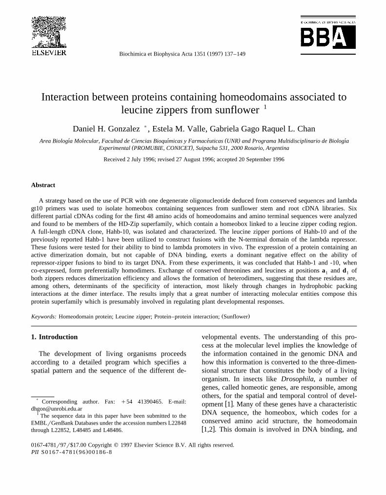
w xsequence was reported previously 22 . Sunflowergenomic DNA digested with various restriction en-
zymes, when probed with labelled Hahb-1 at 608C,showed several hybridizing bands of different inten-
Ž .sity Fig. 1 , even though under this condition only asubclass of homeodomain coding sequences are likelyto be identified. This result indicates the existence ofa large family of related sequences in sunflowergenomic DNA.
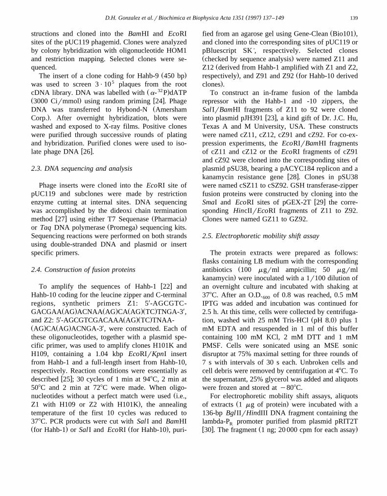
Additional screening of cloned PCR products al-lowed the isolation of eight new clones containingsequences coding for homeodomains, six of them
Ž .corresponding to distinct genes Fig. 2 . Sequencecomparisons indicate that they belong to the HD-Zipprotein superfamily. A phylogenetic tree based onamino acid sequence differences shows indeed thatthe homeodomains of these proteins are more related
Žto each other than to other plant homeodomains not.shown . This type of analysis also allows the identifi-
cation of two subfamilies among HD-Zip proteins,w xnamed HD-Zip I and II 16,17 . The number of
clones isolated allows the determination of a consen-Žsus sequence for each of these two subgroups Fig.
.2 . Clear differences exist at several positions of thehomeodomain.
Fig. 1. Southern blot analysis of sunflower genomic DNA probedwith Hahb-1. Ten micrograms of total genomic DNA were
Ž . Ž . Ž .digested with either BamHI A , EcoRI B or HindIII C . Theresulting fragments were separated on an agarose gel, transferredand probed as described in Section 2. Numbers indicate molecu-lar sizes in kbp.
( )D.H. Gonzalez et al.rBiochimica et Biophysica Acta 1351 1997 137–149 141
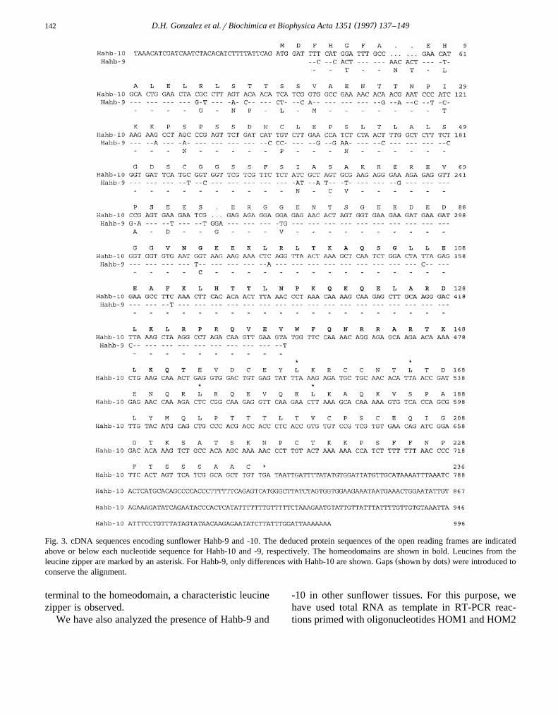
Ž .We have used one of the isolated clones Hahb-9to screen the root cDNA library. A clone containing a1.0 kbp insert was isolated, subcloned into pUC119
Ž .and sequenced Fig. 3 . This clone contains an openŽ .reading frame of 708 bp 236 amino acids . Surpris-
ingly, the nucleotide sequence was different from theone determined for the PCR-derived clone; this newclone was then named Hahb-10. The two sequencesare 85% identical at the amino acid level, and show acomplete conservation within the homeodomain. C-
Fig. 2. Sunflower homeodomains can be divided in two subfamilies. Sunflower homeodomains are compared with reported ArabidopsisHD-Zip proteins. Asterisks indicate identical amino acids; double-dots designate conservative substitutions. Between the two subclasses,identical and conserved amino acids of Athb-1 and HAT22, which belong to different subclasses, are shown. Below, the consensussequence for the HD-Zip I and II subgroups is presented. Uppercase letters indicate residues found in all members of each subfamily;lowercase letters show residues observed in most members; dots represent non-conserved positions; q and y stand for positively andnegatively charged residues; f represents a position occupied by hydrophobic amino acids.
( )D.H. Gonzalez et al.rBiochimica et Biophysica Acta 1351 1997 137–149142
Fig. 3. cDNA sequences encoding sunflower Hahb-9 and -10. The deduced protein sequences of the open reading frames are indicatedabove or below each nucleotide sequence for Hahb-10 and -9, respectively. The homeodomains are shown in bold. Leucines from the
Ž .leucine zipper are marked by an asterisk. For Hahb-9, only differences with Hahb-10 are shown. Gaps shown by dots were introduced toconserve the alignment.
terminal to the homeodomain, a characteristic leucinezipper is observed.
We have also analyzed the presence of Hahb-9 and
-10 in other sunflower tissues. For this purpose, wehave used total RNA as template in RT-PCR reac-tions primed with oligonucleotides HOM1 and HOM2
( )D.H. Gonzalez et al.rBiochimica et Biophysica Acta 1351 1997 137–149 143
Ž X X.5 -CGGTGGTTCGTCGTTCT-3 . This last oligo-nucleotide is based on Hahb-10 sequences, and hy-bridizes to Hahb-9 with two mismatches at its 5X end.A 290-bp amplified product was obtained with DNAfrom all tissues examined, including roots, hypocotyls,
Ž .stems and leaves not shown . To differentiate be-tween the two possible products, a restriction frag-ment length polymorphism was used. Digestion withthe restriction enzyme AÕaII originated bands of230-bp and 290-bp, corresponding to Hahb-9 and
Ž .-10, respectively not shown . This type of analysisshowed that Hahb-9 and -10 are both expressed instems and roots.
3.2. Interaction between HD-Zip proteins
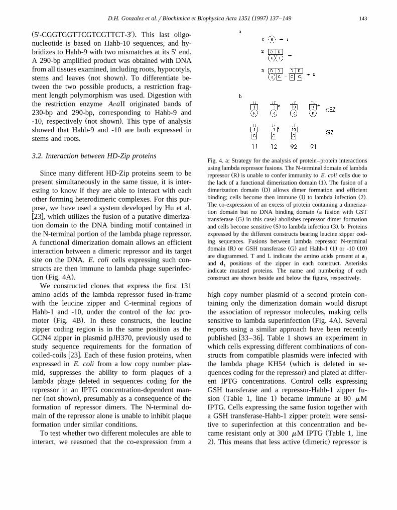
Since many different HD-Zip proteins seem to bepresent simultaneously in the same tissue, it is inter-esting to know if they are able to interact with eachother forming heterodimeric complexes. For this pur-pose, we have used a system developed by Hu et al.w x23 , which utilizes the fusion of a putative dimeriza-tion domain to the DNA binding motif contained inthe N-terminal portion of the lambda phage repressor.A functional dimerization domain allows an efficientinteraction between a dimeric repressor and its targetsite on the DNA. E. coli cells expressing such con-structs are then immune to lambda phage superinfec-
Ž .tion Fig. 4A .We constructed clones that express the first 131
amino acids of the lambda repressor fused in-framewith the leucine zipper and C-terminal regions ofHahb-1 and -10, under the control of the lac pro-
Ž .moter Fig. 4B . In these constructs, the leucinezipper coding region is in the same position as theGCN4 zipper in plasmid pJH370, previously used tostudy sequence requirements for the formation of
w xcoiled-coils 23 . Each of these fusion proteins, whenexpressed in E. coli from a low copy number plas-mid, suppresses the ability to form plaques of alambda phage deleted in sequences coding for therepressor in an IPTG concentration-dependent man-
Ž .ner not shown , presumably as a consequence of theformation of repressor dimers. The N-terminal do-main of the repressor alone is unable to inhibit plaqueformation under similar conditions.
To test whether two different molecules are able tointeract, we reasoned that the co-expression from a
Fig. 4. a: Strategy for the analysis of protein–protein interactionsusing lambda repressor fusions. The N-terminal domain of lambda
Ž .repressor R is unable to confer immunity to E. coli cells due toŽ .the lack of a functional dimerization domain 1 . The fusion of a
Ž .dimerization domain D allows dimer formation and efficientŽ . Ž .binding; cells become then immune I to lambda infection 2 .
The co-expression of an excess of protein containing a dimeriza-Žtion domain but no DNA binding domain a fusion with GST
Ž . .transferase G in this case abolishes repressor dimer formationŽ . Ž .and cells become sensitive S to lambda infection 3 . b: Proteins
expressed by the different constructs bearing leucine zipper cod-ing sequences. Fusions between lambda repressor N-terminal
Ž . Ž . Ž . Ž .domain R or GSH transferase G and Hahb-1 1 or -10 10are diagrammed. T and L indicate the amino acids present at a1
and d positions of the zipper in each construct. Asterisks1
indicate mutated proteins. The name and numbering of eachconstruct are shown beside and below the figure, respectively.
high copy number plasmid of a second protein con-taining only the dimerization domain would disruptthe association of repressor molecules, making cells
Ž .sensitive to lambda superinfection Fig. 4A . Severalreports using a similar approach have been recently
w xpublished 33–36 . Table 1 shows an experiment inwhich cells expressing different combinations of con-structs from compatible plasmids were infected with
Žthe lambda phage KH54 which is deleted in se-.quences coding for the repressor and plated at differ-
ent IPTG concentrations. Control cells expressingGSH transferase and a repressor-Hahb-1 zipper fu-
Ž .sion Table 1, line 1 became immune at 80 mMIPTG. Cells expressing the same fusion together witha GSH transferase-Hahb-1 zipper protein were sensi-tive to superinfection at this concentration and be-
Žcame resistant only at 300 mM IPTG Table 1, line. Ž .2 . This means that less active dimeric repressor is
( )D.H. Gonzalez et al.rBiochimica et Biophysica Acta 1351 1997 137–149144
Table 1Interaction between Hahb-1 and Hahb-10 leucine zippers
w x Ž .Plasmids Fusion to IPTG mM
Repressor GST 20 40 80 150 300
1. cSZ11rpGEX-2T cI-1 GST S S I I I2. cSZ11rGZ11 cI-1 GST-1 S S S S I3. cSZ11rGZ92 cI-1 GST-10 S S I I I4. cSZ92rpGEX-2T cI-10 GST S I I I I5. cSZ92rGZ92 cI-10 GST-10 S S I I I6. cSZ92rGZ11 cI-10 GST-1 S I I I I
Sensitivity to lambda superinfection of cells expressing different zipper-repressor fusion proteins was monitored by plating each cloneŽ . Ž .infected with lambda KH54 100 pfu with different IPTG concentrations included in the plating medium S: sensitive; I: immune .
Ž .Numbers 1 and 10 indicate proteins expressing Hahb-1 and Hahb-10 sequences, respectively, as fusions with lambda repressor cI orŽ .GSH transferase GST .
present in these cells at a given IPTG concentration,Ž .because of the formation of inactive repressorrGSH
transferase heterodimers through the Hahb-1 zipperportions of each fusion protein.
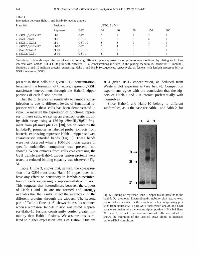
That the difference in sensitivity to lambda super-infection is due to different levels of functional re-pressor within these cells has been demonstrated invitro. To measure the expression of functional repres-sor in these cells, we set up an electrophoretic mobil-ity shift assay using a 136-bp HindIIIrBglII frag-
w xment from plasmid pRIT2T 30 , which contains thelambda-P promoter, as labelled probe. Extracts fromR
bacteria expressing repressor-Hahb-1 zipper showedŽ .characteristic retarded bands Fig. 5 . These bands
were not observed when a 100-fold molar excess ofŽspecific unlabelled competitor was present not
.shown . When extracts from cells co-expressing theGSH transferase-Hahb-1 zipper fusion proteins were
Žtested, a reduced binding capacity was observed Fig..5 .
Table 1, line 3, shows that, in turn, the co-expres-sion of a GSH transferase-Hahb-10 zipper does nothave any effect on sensitivity to lambda superinfec-tion of cells expressing a repressor-Hahb-1 fusion.This suggests that heterodimers between the zippersof Hahb-1 and -10 are not formed and stronglyindicates that the results reflect the interaction of thedifferent proteins through the zippers. The second
Ž .part of Table 1 lines 4–6 shows the results obtainedwhen a repressor-Hahb-10 fusion was tested. Repres-sor-Hahb-10 fusions consistently confer greater im-munity than Hahb-1 fusions. We assume this is re-lated to higher expression levels of Hahb-10 fusions
at a given IPTG concentration, as deduced fromŽ .Western blot experiments see below . Competition
experiments agree with the conclusion that the zip-pers of Hahb-1 and -10 interact preferentially withthemselves.
Since Hahb-1 and Hahb-10 belong to differentsubfamilies, as is the case for Athb-1 and Athb-2, for
Fig. 5. Binding of repressor-Hahb-1 zipper fusion proteins to thelambda-P promoter. Electrophoretic mobility shift assays wereR
performed as described with extracts of cells co-expressing pro-Ž .teins from clones cSZ11 plus GSH transferase lane 2 , or a GSH
Žtransferase fusion with the leucine zipper portion of Hahb-1 lane.3 . Lane 1, extract from non-transformed cells was added. F
shows the migration of the labelled DNA alone; B indicatesprotein-DNA complexes.
( )D.H. Gonzalez et al.rBiochimica et Biophysica Acta 1351 1997 137–149 145
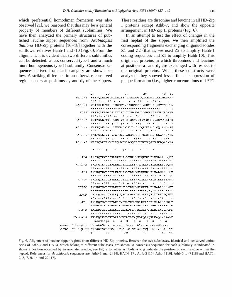
which preferential homodimer formation was alsow xobserved 21 , we reasoned that this may be a general
property of members of different subfamilies. Wehave then analyzed the primary structures of pub-lished leucine zipper sequences from Arabidopsis
w xthaliana HD-Zip proteins 16–18 together with theŽ .sunflower relatives Hahb-1 and -10 Fig. 6 . From the
alignment, it is evident that two different subfamiliescan be detected: a less-conserved type I and a muchmore homogeneous type II subfamily. Consensus se-quences derived from each category are shown be-low. A striking difference in an otherwise conservedregion occurs at positions a and d of the zippers.1 1
These residues are threonine and leucine in all HD-ZipI proteins except Athb-7, and show the opposite
Ž .arrangement in HD-Zip II proteins Fig. 6 .In an attempt to test the effect of changes in the
first heptad of the zipper, we then amplified thecorresponding fragments exchanging oligonucleotides
ŽZ1 and Z2 that is, we used Z2 to amplify Hahb-1.coding sequences and Z1 to amplify Hahb-10 . This
originates proteins in which threonines and leucinesat positions a and d are exchanged with respect to1 1
the original proteins. When these constructs wereanalyzed, they showed less efficient suppression of
Žplaque formation i.e., higher concentrations of IPTG
Fig. 6. Alignment of leucine zipper regions from different HD-Zip proteins. Between the two subclasses, identical and conserved aminoacids of Athb-7 and HAT4, which belong to different subclasses, are shown. A consensus sequence for each subfamily is indicated. Zshows a position occupied by an aromatic residue; see Fig. 2 for other symbols. a to g indicate the position of each residue within the
w x w x w x w x w xheptad. References for Arabidopsis sequences are: Athb-1 and -2 14 , HAT4 17 , Athb-3 15 , Athb-4 16 , Athb-5 to -7 18 and HAT1,w x2, 3, 7, 9, 14 and 22 17 .

( )D.H. Gonzalez et al.rBiochimica et Biophysica Acta 1351 1997 137–149146
Table 2Interaction with proteins modified in the first heptad of the zipper
w x Ž .Plasmids Fusion to IPTG mM
Repressor GST 20 40 80 150 300
Ž .a Hahb-11. cSZ11rpGEX-2T cI-1 GST S S I I I
)2. cSZ11rGZ12 cI-1 GST-1 S S S S I)3. cSZ12rpGEX-2T cI-1 GST S S S S I) )4. cSZ12rGZ12 cI-1 GST-1 S S S S S)5. cSZ12rGZ11 cI-1 GST-1 S S S S S
Ž .b Hahb-10)6. cSZ91rpGEX-2T cI-10 GST S S S I I)7. cSZ91rGZ92 cI-10 GST-10 S S S S S
Ž .c Hahb-1rHahb-10)8. cSZ12rpGEX-2T cI-1 GST S S S S I)9. cSZ12rGZ92 cI-1 GST-10 S S S S S
10. cSZ92rpGEX-2T cI-10 GST S I I I I)11. cSZ92rGZ12 cI-10 GST-1 S S I I I
)12. cSZ91rpGEX-2T cI-10 GST S S S I I)13. cSZ91rGZ11 cI-10 GST-1 S S S S S) )14. cSZ91rGZ12 cI-10 GST-1 S S S I I
Sensitivity to lambda superinfection of cells expressing different zipper-repressor fusion proteins was monitored as described in Table 1.Asterisks indicate proteins with modifications at positions a and d .1 1
were required to protect cells from lambda infection;Table 2, lines 1,3 for Hahb-1; lines 10,12 for Hahb-
.10 . The results can be explained by a relative de-crease in expression level of the mutated proteins orby a reduced dimerization efficiency. Western blotanalysis of extracts expressing GSH transferase fu-sions to mutated and non-mutated zippers using anti-bodies raised against GSH transferase showed nodifference in expression levels due to the exchange of
Ž .both amino acids not shown . We assume then that areduced dimerization efficiency is the most likelyexplanation.
Co-expression experiments as those described inTable 1 were then performed with different combina-tions of constructs. These experiments show that eachmutated protein is able to interact with its corre-
Žsponding non-mutated counterpart Table 2, lines 1–5.for Hahb-1; lines 6,7 for Hahb-10 , implying that
conserved residues at positions a and d are not1 1
essential for dimerization. In other words, that thoseproteins with a different arrangement at these posi-tions are able to interact, provided that no unfavor-able interactions arise from other parts of the zipper,as may be the case for the Hahb-1rHahb-10 pair.
Notably, the mutated proteins were also able tointeract with the non-mutated partner of a differentsubtype; that is, Hahb-1 dimerized with mutant Hahb-
Ž10, and Hahb-10 dimerized with mutant Hahb-1 Ta-.ble 2, lines 8–13 . This indicates that the mutants
have lost dimerization specificity, since each one ofthem interacts with both non-mutated proteins. Fi-nally, the two mutated proteins do not interact in a
Ž .noticeable manner Table 2, lines 12,14 . Taken to-gether, the results imply that there is no single residueor region that is responsible by itself for the observedspecificity of interaction.
4. Discussion
In this paper, we characterize cDNAs coding forHD-Zip proteins from sunflower. We have employeda PCR-based strategy that allowed the rapid screen-ing and characterization of different members of thissuperfamily. Our data extend previous observationson the existence of two subfamilies for which differ-ent consensus sequences can be derived. Sequenceconservation is higher within the HD-Zip II subfam-
( )D.H. Gonzalez et al.rBiochimica et Biophysica Acta 1351 1997 137–149 147
ily, and extends to regions outside the HD-Zip do-main. Sequence comparisons to other published HD-Zip proteins indicate that Hahb-10 is more similar to
w xHAT9 and HAT22 from Arabidopsis 17 . Stretchesof conserved amino acids are observed along theentire coding region, including the C-terminal se-quence KPXFArNPFTXXSAAC, where X means anon-conserved residue and Ar an aromatic aminoacid. An additional sequence adjacent to the leucine
Ž .zipper CPSCE is present in all published type IIHD-Zip proteins.
Southern blot hybridizations indicate the existenceof a large number of related sequences in sunflowergenomic DNA. That many of these are genes simulta-neously expressed in the same tissue is suggested bythe number of different clones we have isolated froma stem cDNA library. There, out of seven indepen-dent clones coding for HD-Zip proteins, six corre-sponded to different genes. This suggests that theactual number of genes represented in the cDNAlibrary should be much greater. In fact, we haveobtained evidence that clones coding for Hahb-9 and
Ž .-10 are also present in the stem library not shown .Since leucine zippers are involved in protein–pro-
tein interactions, we were interested in studying whichsequences within the zipper influence these interac-tions. For this purpose, we have employed a system
w xdeveloped by Hu et al. 23 , who utilized lambdaphage repressor-leucine zipper fusions to study theresidues of the yeast transcription factor GCN4 in-volved in dimerization. In a similar way, we havecloned the leucine zipper regions of Hahb-1 and -10in-frame with the N-terminal portion of the repressor.As shown under results, the co-expression of a sec-ond protein containing only the zipper has a negativeeffect on the ability of the repressor to inhibit plaqueformation. This effect is observed at certain IPTGconcentrations. Above these, the amount of repressordimers may be high enough to inhibit superinfection,as would be expected for a competitive interaction.Similar systems have recently been employed byseveral authors to study protein–protein interactionsw x33–36 .
Experiments in which different combinations ofrepressor-zipper and GSH transferase-zipper fusions
Ž .were co-expressed indicated that: 1 Hahb-1 andHahb-10, when co-expressed, form preferentially ho-
Ž .modimers; 2 when conserved leucine and threonine
at positions a and d are exchanged, specificity is1 1
lost and dimer formation with both unmodified formsis observed, indicating that conservation of aminoacids at these positions is necessary but not sufficientto preserve specificity. In other words, that there ismore than one region that determines the specificityof interaction. Repressor-zipper fusions with muta-tions in the first heptad showed less efficient repres-sion than their non-mutated counterparts. As men-tioned in Section 3, we think this effect is due to areduced dimerization efficiency. This may be relatedto changes in hydrophobic packing interactions at thedimer interface, mainly because residues at positiona , which are in the vicinity of residues at d , are2 1
Ždifferent in HD-Zip I and II proteins i.e., tyrosine.and cysteine, respectively . Mutant proteins, in turn,
compete efficiently, as GSH transferase fusions, withnon-mutated repressor-zipper fusions. This may indi-cate some preference for the formation of het-erodimers between mutated and non-mutated proteinsover mutant homodimers. An alternative explanationis that our system is not sensitive enough to detectthe difference in the interaction efficiency of thesecompetitor proteins.
w xRecently, Joung et al. 34 reported that a changeat position d of the coiled-coil dimerization domain1
of the E. coli cAMP receptor produces a mutant withaltered, but not lost, dimerization specificity. Indeed,the mutant protein is able to form homodimers, butnot heterodimers, with the non-mutated counterpart.This may be related to the fact that in this case thecoiled-coil structure is shorter than the leucine zip-pers of both Hahb-1 and -10, so that changes in oneheptad may affect more drastically the interaction.For Hahb-1 and -10, other parts of the zipper, whichremain unmodified, may be responsible for the for-mation of dimers with non-mutated proteins. Bothresults agree, however, in the fact that changes ofinterfacial residues of the first heptad provoke changesin the specificity of the interaction.
Previous studies showed that, in other zippers,charged residues at e and g positions are important
w xdeterminants of interaction specificity 37,38 . As anexample, the b-Zip transcription factor Fos is unableto homodimerize, but readily forms heterodimers with
w xanother b-Zip protein, Jun 39 . If two chargedŽresidues derived from either Jun or Fos those at
.positions g and e are introduced into the GCN41 2
( )D.H. Gonzalez et al.rBiochimica et Biophysica Acta 1351 1997 137–149148
zipper, they are able to confer this behavior to thew xengineered molecules 40 . As a rule, residues at g
positions in one heptad interact with those at e posi-tions in the previous heptad of the opposite strand, sothat similarly charged amino acids destabilize thedimer. Whether the contrary, that is, that oppositelycharged amino acids stabilize the interaction by salt-bridge formation, is also valid, has been recently
w xchallenged by results from Lumb and Kim 41 . Theanalysis of residues at e and g positions in Hahb-1and -10 suggests that heterodimer formation wouldnot be disfavored over homodimer formation. No-tably, eight attractive electrostatic interactions arepossible in Hahb-10 dimers, and six in Hahb-1 dimersand Hahb-1rHahb-10 heterodimers. No repulsive in-teraction is observed in any of these forms.
A difference among interfacial residues occurs at apositions. Apart from the above-mentioned change atposition a , residues at a , a and a are also differ-1 2 3 5
Ž .ent Fig. 6 . These positions are conserved insideŽeach subfamily tyrosine, an aromatic amino acid,
and asparagine, respectively, in HD-Zip I proteins,and cysteine, cysteine or valine, and an aliphatic
.amino acid in HD-Zip II proteins . We propose thatthese residues are good candidates as determinants ofthe specificity of interaction.
Since, according to our results, zippers from HD-Zip proteins that have the same amino acids at a and1
d positions are able to interact, this opens the possi-1
bility that most members of each subfamily should beable to form heterodimers with other members of thesame subfamily. This should be true, unless unfavor-able interactions arise in other parts of the molecule.
w x w xIn two proteins, Athb-3 15 and HAT7 17 , whichare highly homologous in sequence, histidines arepresent at positions g and e . This would originate4 5
repulsive interactions in homodimers and in het-erodimers with other proteins which have positively
w xcharged residues at e , such as Athb-1 14 . It would5
be interesting to know if Athb-3 and HAT7 are ableto form homodimers or are prone to interact withdifferent molecules. A similar concept would be ap-plicable to HAT1, a member of the HD-Zip II sub-family, which contains glutamic acid residues at posi-tions g and e .3 4
Although little is known about the roles of HD-Zipproteins, transformation experiments with constructsthat either increase or decrease the expression levels
of Arabidopsis HAT4 gave clear indications thatw xthey influence plant development 42 . A detailed
knowledge of the factors that regulate the interactionamong different HD-Zip proteins would allow the useof native or modified leucine zipper domains asdevelopmental regulators, since they should exert adominant negative effect upon binding to HD-Zip
w xproteins 43 .As a conclusion, our results indicate that, at least
for Hahb-1 and -10, specificity would be the result ofthe sum of interactions along different parts of thezipper, most likely at the dimer interface. Sequencecomparisons indicate that this may apply to othermembers of the family.
Acknowledgements
We are indebted to Dr. Andre Steinmetz and col-´Žleagues Institut de Biologie Moleculaire des Plantes,
.Strasbourg for the kind gift of the cDNA librariesŽused in this study. We also thank Dr. Jim Hu Texas
.A&M University for generously providing the clonesand strains used to express lambda repressor-zipperfusions. This work was supported in part by grants
Ž .from Fundacion Antorchas Argentina and Universi-´dad Nacional de Rosario. D.H.G., E.M.V. and R.L.C.
Ž .are members of CONICET Argentina .
References
w x Ž .1 Gehring, W.J. 1987 Science 236, 1245–1252.w x2 Gehring, W.J., Qian, Y.Q., Billeter, M., Furukubo-Tokunaga,
K., Schier, A.F., Resendez-Perez, D., Affolter, M., Otting,Ž .G. and Wuethrich, K. 1994 Cell 78, 211–223.
w x Ž .3 McGinnis, W. and Krumlauf, R. 1992 Cell 68, 283–302.w x Ž .4 Schena, M. and Davis, R.W. 1992 Proc. Natl. Acad. Sci.
USA 89, 3894–3898.w x Ž .5 Vollbrecht, E., Veit, B., Sinha, N. and Hake, S. 1991
Nature 350, 241–243.w x6 Matsuoka, M., Ichikawam H., Saito, A., Tada, Y., Fujimura,
Ž .T. and Kano-Murakami, Y. 1993 Plant Cell 5, 1039–1048.w x7 Kerstetter, R., Vollbrecht, E., Lowe, B., Veit, B., Yam-
Ž .aguchi, J. and Hake, S. 1994 Plant Cell 6, 1877–1887.w x8 Lincoln, C., Long, J., Yamaguchi, J., Serikawa, K. and
Ž .Hake, S.C. 1994 Plant Cell 6, 1859–1876.w x Ž .9 Bellmann, R. and Werr, W. 1992 EMBO J. 11, 3367–3374.
w x Ž .10 Schindler, U., Beckmann, H. and Cashmore, A.R. 1993Plant J. 4, 137–150.

( )D.H. Gonzalez et al.rBiochimica et Biophysica Acta 1351 1997 137–149 149
w x11 Korfhage, U., Trezzini, G.F., Meier, I., Hahlbrock, K. andŽ .Somssich, I.E. 1994 Plant Cell 6, 695–708.
w x Ž .12 Rerie, W.G., Feldmann, K.A. and Marks, M.D. 1994Genes Dev. 8, 1388–1399.
w x13 Muller, K.J., Romano, N., Gerstner, O., Garcia-Maroto, F.,¨Ž .Pozzi, C., Salamini, F. and Rohde, W. 1995 Nature 374,
727–730.w x Ž .14 Ruberti, I., Sessa, G., Lucchetti, S. and Morelli, G. 1991
EMBO J. 10, 1787–1791.w x15 Mattsson, J., Soderman, E., Svenson, M., Borkird, C. and¨
Ž .Engstrom, P. 1992 Plant Mol. Biol. 18, 1019–1022.¨w x16 Carabelli, M., Sessa, G., Baima, S., Morelli, G. and Ruberti,
Ž .I. 1993 Plant J. 4, 469–479.w x Ž .17 Schena, M. and Davis, R.W. 1994 Proc. Natl. Acad. Sci.
USA 91, 8393–8397.w x18 Soderman, E., Mattsson, J., Svenson, M., Borkird, C. and¨
Ž .Engstrom, P. 1994 Plant Mol. Biol. 26, 145–154.¨w x Ž .19 O’Shea, E.K., Klemm, J.D., Kim, P.S. and Alber, T. 1991
Science 254, 539–544.w x Ž .20 Vinson, C.R., Sigler, P.B. and McKnight, S.L. 1989 Sci-
ence 246, 911–916.w x Ž .21 Sessa, G., Morelli, G. and Ruberti, I. 1993 EMBO J. 12,
3507–3517.w x Ž .22 Chan, R.L. and Gonzalez, D.H. 1994 Plant Physiol. 106,
1687–1688.w x Ž .23 Hu, J.C., O’Shea, E.K., Kim, P.S. and Sauer, R.T. 1990
Science 250, 1400–1403.w x24 Ausubel, F.M., Brent, R., Kingston, R.E., Moore, D.D.,
Ž . Ž .Seidman, J.G., Smith, J.A. and Struhl, K. eds. 1987Current Protocols in Molecular Biology. Greene Publishingand Wiley-Interscience, New York.
w x Ž .25 Gonzalez, D.H. and Chan, R.L. 1993 Trends Genet. 9,231–232.
w x Ž .26 Kaslow, D.C. 1986 Nucleic Acids Res. 14, 6767.w x Ž .27 Sanger, F., Niklen, S. and Coulson, A.R. 1977 Proc. Natl.
Acad. Sci. USA 74, 5463–5467.w x28 Bartolome, B., Jubete, Y., Martınez, E. and de la Cruz, F.´ ´
Ž .1991 Gene 102, 75–78.w x Ž .29 Smith, D.B. and Johnson, K.S. 1988 Gene 67, 31–40.w x Ž .30 Nilsson, B., Abrahmsen, L. and Uhlen, M. 1985 EMBO J.
4, 1075–1080.w x Ž .31 Sedmak, J.J. and Grossberg, S.E. 1977 Anal. Biochem. 79,
544–552.w x Ž .32 Laemmli, U.K. 1970 Nature 227, 680–685.w x Ž .33 Bunker, C.A. and Kingston, R.E. 1995 Nucleic Acids Res.
23, 269–276.w x34 Joung, J.K., Chung, E.H., King, G., Yu, Ch., Hirsh, A.S.
Ž .and Hochschild, A. 1995 Genes Dev. 9, 2986–2996.w x35 Longo, F., Marchetti, M.A., Castagnoli, L., Battaglia, P.A.,
Ž .Gigliani, F. 1995 Biochem. Biophys. Res. Commun. 206,326–334.
w x36 Marchetti, A., Abril-Marti, M., Illi, B., Cesareni, G. andŽ .Nasi, S. 1995 J. Mol. Biol. 248, 541–550.
w x Ž .37 O’Shea, E.K., Rutkowski, R. and Kim, P.S. 1992 Cell 68,699–708.
w x Ž .38 Graddis, T.J., Myszka, D.G. and Chaiken, I.M. 1993 Bio-chemistry 32, 12664–12671.
w x Ž .39 Curran, T. and Franza, B.R. Jr. 1988 Cell 55, 395–397.w x40 John, M., Briand, J.P., Granger-Schnarr, M. and Schnarr, M.
Ž .1994 J. Biol. Chem. 269, 16247–16253.w x Ž .41 Lumb, K.J. and Kim, P.S. 1995 Science 268, 436–439.w x Ž .42 Schena, M., Lloyd, A.M. and Davis, R.W. 1993 Genes
Dev. 7, 367–379.w x Ž .43 Herskowitz, I. 1987 Nature 329, 219–222.
Copyright © 2022 FDOKUMEN