Integrated optical devices for lab-on-a-chip biosensing applications
25
citable using Digital Object Identifier – DOI) Early View publication on wileyonlinelibrary.com (issue and page numbers not yet assigned; Laser Photonics Rev., 1–25 (2011)/ DOI 10.1002/lpor.201100025 LASER & PHOTONICS REVIEWS Abstract The application of portable, easy-to-use and highly sensitive lab-on-a-chip biosensing devices for real-time diag- nosis could offer significant advantages over current analytical methods. Integrated optics-based biosensors have become the most suitable technology for lab-on-chip integration due to their ability for miniaturization, their extreme sensitivity, robustness, reliability, and their potential for multiplexing and mass produc- tion at low cost. This review provides an extended overview of the state-of-the-art in integrated photonic biosensors technology including interferometers, grating couplers, microring resonators, photonic crystals and other novel nanophotonic transducers. Particular emphasis has been placed on describing their real biosensing applications and wherever possible a comparison of the sensing performances between each type of device is included. The way towards achieving operative lab-on-a-chip platform incorporating the photonic biosensors is also reviewed. Concluding remarks regarding the future prospects and poten- tial impact of this technology are also provided. Integrated optical devices for lab-on-a-chip biosensing applications M.-Carmen Estevez, Mar Alvarez and Laura M. Lechuga * 1. Introduction Biosensors are devices able to detect a specific substance by converting the recognition from a biological entity (i.e. DNA, antibody, enzyme, . . . ) into an electrical signal that can be further processed and related to the concentration of the substance under analysis. Biosensors can provide se- lective, sensitive, fast, direct and cost-effective analyses. In addition, they can perform tests in real-time without using fluorescent labels or amplification steps and with a mini- mum volume of samples and reagents [1]. As compared to standard techniques that are usually time consuming, expen- sive and require labelling and trained personal, biosensing technology offers clear advantages. Lab-on-a-chip (LOC) are miniaturized devices in which all functionalities are integrated in the same platform, from sample preparation to signal delivery [2]. Ideally, a LOC device should contain enough hard-wired intelligence and robustness to be used by nonskilled personal and should deliver the results directly to a central monitoring station. It is clear that achieving a small, portable and easy-to-use lab-on-a-chip device for diagnostics could offer significant advantages over standard methods. Although significant progress has been accomplished at the LOC field during recent years, very few standalone devices have emerged [3]. Most current devices are simple planar microfluidic devices that do not incorporate the detection and after the reaction has taken place, the readout must be done with complex in- strumentation in laboratory settings. That is the main reason why incorporating “on-chip” detection by using biosensors is a new technology that shows great promise. The main application fields of this technology can be clinical diag- nostics, environmental monitoring, chemical and biological warfare surveillance, food industry and veterinary and indus- trial process control, among others. By using this advanced technology, diagnosis in developing countries could become an important achievement for the near future. Photonic biosensors are well-established technologies for the sensitive monitoring of molecular interactions. They could afford the requirements for the “on-chip” detection in lab-on-a-chip platforms due to their outstanding char- acteristics of sensitivity, label-free and real-time detection. The detection principle of most optical biosensors is based on the evanescent field detection. In the evanescent wave mechanism (see Fig. 1), a bioreceptor layer is immobilized onto the surface of a waveguide; the exposure to the partner analyte produces a biomolecular interaction affecting the guiding properties of the waveguide (specifically, a varia- tion of the refractive index) via the modification through the evanescent field. The variation of the refractive index can be evaluated by any of the waveguiding optical properties (intensity, phase, resonant momentum, polarization, . . . ) and this variation can be correlated with the concentration of the analyte and with the affinity constant of the interaction, resulting in a quantitative value of the interaction. As the evanescent wave decays exponentially while it penetrates into the outer medium, it only detects changes taking place on the surface of the waveguide, since the inten- sity of the evanescent field is much higher in this particular region. For most waveguide systems, this decay length is of Nanobiosensors and Bioanalytical Applications Group, Research Center on Nanoscience and Nanotechnology (CSIC) & CIBER-BBN. Campus UAB, 08193 Bellaterra, Barcelona, Spain * Corresponding author: e-mail: [email protected] © 2011 by WILEY-VCH Verlag GmbH & Co.KGaA, Weinheim
-
Upload
independent -
Category
Documents
-
view
3 -
download
0
Transcript of Integrated optical devices for lab-on-a-chip biosensing applications

citable using Digital Object Identifier – DOI)
Early View publication on wileyonlinelibrary.com(issue and page numbers not yet assigned;
Laser Photonics Rev., 1–25 (2011) / DOI 10.1002/lpor.201100025
LASER & PHOTONICSREVIEWS
Abstract The application of portable, easy-to-use and highlysensitive lab-on-a-chip biosensing devices for real-time diag-nosis could offer significant advantages over current analyticalmethods. Integrated optics-based biosensors have become themost suitable technology for lab-on-chip integration due to theirability for miniaturization, their extreme sensitivity, robustness,reliability, and their potential for multiplexing and mass produc-tion at low cost. This review provides an extended overview ofthe state-of-the-art in integrated photonic biosensors technologyincluding interferometers, grating couplers, microring resonators,photonic crystals and other novel nanophotonic transducers.Particular emphasis has been placed on describing their realbiosensing applications and wherever possible a comparisonof the sensing performances between each type of device isincluded. The way towards achieving operative lab-on-a-chip
platform incorporating the photonic biosensors is also reviewed.Concluding remarks regarding the future prospects and poten-tial impact of this technology are also provided.
Integrated optical devices for lab-on-a-chipbiosensing applicationsM.-Carmen Estevez, Mar Alvarez and Laura M. Lechuga*
1. Introduction
Biosensors are devices able to detect a specific substanceby converting the recognition from a biological entity (i.e.DNA, antibody, enzyme, . . . ) into an electrical signal thatcan be further processed and related to the concentrationof the substance under analysis. Biosensors can provide se-lective, sensitive, fast, direct and cost-effective analyses. Inaddition, they can perform tests in real-time without usingfluorescent labels or amplification steps and with a mini-mum volume of samples and reagents [1]. As compared tostandard techniques that are usually time consuming, expen-sive and require labelling and trained personal, biosensingtechnology offers clear advantages.
Lab-on-a-chip (LOC) are miniaturized devices in whichall functionalities are integrated in the same platform, fromsample preparation to signal delivery [2]. Ideally, a LOCdevice should contain enough hard-wired intelligence androbustness to be used by nonskilled personal and shoulddeliver the results directly to a central monitoring station.It is clear that achieving a small, portable and easy-to-uselab-on-a-chip device for diagnostics could offer significantadvantages over standard methods. Although significantprogress has been accomplished at the LOC field duringrecent years, very few standalone devices have emerged [3].Most current devices are simple planar microfluidic devicesthat do not incorporate the detection and after the reactionhas taken place, the readout must be done with complex in-strumentation in laboratory settings. That is the main reasonwhy incorporating “on-chip” detection by using biosensors
is a new technology that shows great promise. The mainapplication fields of this technology can be clinical diag-nostics, environmental monitoring, chemical and biologicalwarfare surveillance, food industry and veterinary and indus-trial process control, among others. By using this advancedtechnology, diagnosis in developing countries could becomean important achievement for the near future.
Photonic biosensors are well-established technologiesfor the sensitive monitoring of molecular interactions. Theycould afford the requirements for the “on-chip” detectionin lab-on-a-chip platforms due to their outstanding char-acteristics of sensitivity, label-free and real-time detection.The detection principle of most optical biosensors is basedon the evanescent field detection. In the evanescent wavemechanism (see Fig. 1), a bioreceptor layer is immobilizedonto the surface of a waveguide; the exposure to the partneranalyte produces a biomolecular interaction affecting theguiding properties of the waveguide (specifically, a varia-tion of the refractive index) via the modification through theevanescent field. The variation of the refractive index canbe evaluated by any of the waveguiding optical properties(intensity, phase, resonant momentum, polarization, . . . ) andthis variation can be correlated with the concentration ofthe analyte and with the affinity constant of the interaction,resulting in a quantitative value of the interaction.
As the evanescent wave decays exponentially while itpenetrates into the outer medium, it only detects changestaking place on the surface of the waveguide, since the inten-sity of the evanescent field is much higher in this particularregion. For most waveguide systems, this decay length is of
Nanobiosensors and Bioanalytical Applications Group, Research Center on Nanoscience and Nanotechnology (CSIC) & CIBER-BBN. Campus UAB,08193 Bellaterra, Barcelona, Spain* Corresponding author: e-mail: [email protected]
© 2011 by WILEY-VCH Verlag GmbH & Co. KGaA, Weinheim

2
LASER & PHOTONICSREVIEWS
M. C. Estevez, M. Alvarez, and L. M. Lechuga: Integrated optical biosensors
Figure 1 (online color at: www.lpr-journal.org)Scheme of the evanescent field sensing. Abiomolecular interaction taking place at the wave-guide surface within the evanescent region inducesa change in the effective refractive index of thetransmitted light mode. The evaluation of this op-tical change gives a measure of the number ofmolecules which has been detected.
the order of 0.1–1 μm. For that reason, it is not necessary tocarry out a prior separation of nonspecific components (asin conventional analyses) because any change in the bulk so-lution will hardly affect the sensor response. Therefore, themost significant advantages of evanescent-based mechanismare the highly sensitive and specific label-free detection oftarget molecules or biochemical reactions in real time, withreduced nonspecific binding, which makes this detectionmechanism one of the most useful for detection of targetsin complex real samples.
The most common optical evanescent wave biosensor isthe surface plasmon resonance (SPR) device [4] based onthe variation of the reflectivity on a metallic layer in closecontact with a dielectric media. The SPR biosensor has beenwidely developed and commercialized and hundreds of pub-lications have demonstrated its outstanding performance toevaluate complex biosensing interactions [4]. But, a SPRsensor has a relatively large size and its miniaturization inlab-on-chip platforms is complex. Moreover, the sensitivityis usually limited to the nanomolar range, which is extremelyuseful in diverse applications [5–7] but not enough for ap-plications requiring lower detection levels (pM-fM or evensingle molecule), which are usual in clinical practice.
Photonic sensors based on integrated optics (IO) couldsolve the aforementioned SPR problems, as they can beeasily miniaturized and they offer high potential for chipintegration; moreover, by using evanescent wave as the de-tection mechanism, sensitivities can be extremely high (pMin a label-free scheme). Moreover, integrated optics allowsa great flexibility in the materials and structures selectionand fabrication of arrays of sensors with the same char-acteristics within the same chip for multiplexing analysiscan be afforded. Materials employed in IO devices are Si,Si3N4, SiON, SiO2 or polymers, and techniques such as iondiffusion in glass, chemical vapor deposition, spin-coating,nanoimprinting, electron-beam lithography, etc. are com-monly employed for the fabrication. By using silicon pho-tonics technology, additional advantages such as robustness,reliability, low power consumption and potential for massproduction with consequent reduction of production costsare added. Other technologies such as III-V compounds orlithium niobate, often used in optical telecommunications,have shown less suitability for IO biosensors as they aremore complex and expensive.
Most of the refractive-index IO sensors rely on theinduced changes of the effective refractive index by thebiomolecular interaction in the evanescent area. In order toachieve the highest sensitivity, the waveguiding structuremust be optimized by looking for a maximum change of theeffective refractive index due to the sensing biolayer. Sensi-tivity is a complex parameter involving geometry, materialand working wavelength of the waveguide, and also otheraspects such as the chemical activation of the surface, thebiofunctionalization method and the resolution and noiseof the optical readout system. In addition, fluidics must betaken into account (i.e. flow-cell volume, means of sam-ple injection, diffusion, dispersion). Each of these aspectsmust be properly designed and optimized before deliveringa functional biosensor device. Integrated optical sensorssuch as grating-couplers, interferometers, photonic crystals,microring resonators, slot waveguides or silicon wires havebeen extensively studied in recent years [8–12], but so faronly a few of them are commercially available.
One of the main advantages of the IO technology is thepossibility to integrate all the functions (chemical, optical,microfludics and electronics) in one single platform offeringan ideal solution for the implementation of true lab-on-a-chip devices. This area is still in its infancy, but remarkableprogress has been achieved during recent years [3, 13]. Thisis reflected in the increasing number of publications address-ing new or optimized sensing configurations.
In this chapter, an overview of the main integrated op-tical sensors will be presented with special focus on themost relevant ones in terms of sensing performance andintegration capability, and whose feasibility for label-freebiosensing has been proven. For each of them, a brief de-scription of its operating principle, design, fabrication andreadout resolution will be presented, and, when reported, thebiosensing dynamic range and detection limit will be sum-marized. Application in clinical diagnostics, environmentalmonitoring, or in the food and agroalimentary industrieswill be included as well as a brief section describing thecommercial devices already on the market. One of the cru-cial aspects that will be discussed is the way towards a trulylab-on-a-chip integration, although few examples of com-pletely integrated platforms can be found in the literature.Finally, an outlook of the future prospects of this technologywill be given.
© 2011 by WILEY-VCH Verlag GmbH & Co. KGaA, Weinheim www.lpr-journal.org

Laser Photonics Rev. (2011)
REVIEWARTICLE
3
Figure 2 (online color at: www.lpr-journal.org) General scheme for the activation of the sensor surface and common strategies forbiofunctionalization: surface cleaning (1), chemical activation of plain surface (2), immobilization of the specific bioreceptors (3) and finaldetection of the target molecule (4). The bioassays depicted in (3) and (4) are the following: (a) Mixed self-assembled monolayer (SAM)with reactive and non-reactive silanes compounds with specific antibodies covalently immobilized for protein recognition; (b) hydrophilicand biocompatible reactive monolayer based on pegylated-silane or dextran compounds with proteins covalently immobilized forantibody recognition; (c) Affinity tags immobilized on the surface (i.e. protein A, or streptavidin onto biotinylated surface) to achieveappropriate orientation of specific antibodies for protein recognition; (d) covalent immobilization of DNA or RNA probes (together withnon-specific spacers) for hybridization with complementary DNA/RNA strands.
2. Biofunctionalization and immobilizationstrategies
The selection of an appropriate procedure to immobilizethe biological element on the sensor surface has become acritical step in the biosensor area, and enormous efforts arecontinuously invested in order to optimize novel strategiesaccording to the application. The immobilization processshould not only guarantee an efficient coverage of the trans-ducer surface with the biomolecules, while keeping intacttheir properties (functionality, structure, biological activ-ity, affinity, specificity . . . ), but it should also ensure theirstability for storage and regeneration. Moreover, most ofthe applications, such as clinical and medical diagnosis,agroalimentary analysis, or environmental field surveillance,require well-defined surfaces with biocompatible properties,minimizing nonspecific adsorption when analyzing com-plex real samples. The choice of the most effective strategyof immobilization that combines the above considerationsusually becomes the key factor that turns a sensing deviceinto a valid and applicable analytical tool with the requiredquality standards. Moreover, the bioreceptor layer directlyaffects the reproducibility, selectivity and resolution of anysensor device.
A wide variety of biomolecules can be used as biore-ceptors, i.e. antibodies, nucleic acid sequences, peptides,enzymes, cell receptors and many others. The selectedbiomolecule is dictated by the application and must be cho-sen to be highly specific for the target molecule and stable
enough to be immobilized without losing functionality. Sev-eral types of routes can be used to biofunctionalize thesensor surface: (i) physical adsorption by direct depositionof the biomolecule; (ii) covalent binding of the biomoleculeto the surface (using a crosslinker previously immobilizedon the surface or following more complex strategies [14]);(iii) noncovalent interactions to a previously deposited ac-tive layer, either by nonspecific electrostatic interactions orby noncovalent affinity binding (i.e. biotin-avidin systems,His-Tag system, Protein A/G for antibodies) (iv) physicalentrapment in a polymer layer. Figure 2 summarizes thegeneral immobilization strategies.
Physical adsorption is a simple strategy based onhydrophobic and electrostatic interactions between thebiomolecule and the surface, but it can lead to the easydesorption of the active receptors under flow conditionsand also when regeneration cocktails are applied to breakthe interaction event (which usually implies high or lowpH solutions, salt concentrations, organic solvents, etc.).Moreover, issues related to reproducibility together withundesired folding of the biomolecules onto the surface arecommon drawbacks of this strategy, which makes it notadvisable in most of the cases even despite its simplicity.
Covalent binding can be made through one of the chem-ical groups of the biomolecule. It is recommended to usea group whose blocking does not compromise the overallfunctionality of the biomolecule. Amino, carboxylic or thiolgroups are the preferred option to couple proteins. For nu-cleic acid immobilization, it is possible to take advantage
www.lpr-journal.org © 2011 by WILEY-VCH Verlag GmbH & Co. KGaA, Weinheim

4
LASER & PHOTONICSREVIEWS
M. C. Estevez, M. Alvarez, and L. M. Lechuga: Integrated optical biosensors
of the versatility of the DNA synthesis that allows the in-corporation of reactive groups at the end of the sequence.Specially difficult is the attachment of antibodies in an ori-ented way (leaving the affinity binding sites free), which canbe done by controlled binding through the carbohydratesgroups of the constant region or by using affinity proteins(such as A or G Protein) [15]. Another noncovalent affin-ity binding strategy is based on the biotin-avidin system,through the formation of a sandwich-like layer (biotin onthe surface/avidin/biotinylated biomolecule) [14–16].
Before the biofunctionalization step, a previous chemi-cal activation of the sensor surface is always needed. Silicon,silicon oxide and silicon nitride are conventional materi-als commonly used for integrated optical transducers thatcan be functionalized using the well-known silane chem-istry. Silicon-based surfaces require a prior activation stepto oxidize the surface and to expose the silanol groups forcrosslinking with the silane. Whereas silicon oxide is hy-drophilic, silicon nitride is an electronically neutral non-porous material, whose modification is usually not straight-forward. In the case of silicon nitride, methods are based onan initial etching to remove the native SiO2 layer to be fur-ther oxidized to form a new oxide layer [17]. Other strategiesinvolve the derivatization of the Si-H and Si-N groups of thesilicon nitride using more drastic conditions [18–21]. Theactivated surface is subsequently modified with organosi-lanes forming a silane layer with exposed reactive groupssusceptible to reaction with the bioreceptor. Hundreds of dif-ferent organosilanes with a wide variety of structures, length,functionality and chemical and physical properties are nowa-days commercially available, although the most commonlyemployed are those with short alkyl chain (i.e. propyl, butyl),ending in amino, thiol, epoxy or carboxylic groups.
As a first step, evaluation of the biosensor capabilityis done using purified or spiked samples in buffer. But inorder to assess the real performance of any device, com-plex samples that contain high concentration of nonspecificmolecules, such as blood, urine, serum, saliva, tears, cere-brospinal or medullar fluids should be tested. For prepa-ration of the receptor layer that must avoid matrix effectsand large background signals, less conventional reagentsthat provide a more hydrophilic, biocompatible, and an-tifouling surface are increasingly being used such as dex-tran polymer [22] or poly(ethylene) glycol (PEG) deriva-tives [23–28].
Overall, to achieve the full potential of the optical sens-ing devices, the choice of the surface modification, the bio-functionalization procedures, and the type and conditionsof the assay become as relevant and crucial as the develop-ment and optimization itself of the integrated optics-basedsensing structures.
3. Biosensors based on integrated optics
In this section, an overview of the main biosensing tech-nologies based on integrated optics will be presented. Themain driving force behind the development of integratedoptical biosensors is to push the sensitivity for label-free
detection of minimum amounts of substances, which are theconcentration normally found in human fluids at the start ofa disease (such as cancer) or in contaminated water or foodor in a biowarfare attack: the lower the limit of detection,the earlier the disease or the pollutant could be detected.The limit of detection (LOD) and sensitivity are the mostimportant parameters that define the sensor performance.The sensitivity is mostly referred to the strength of the light-matter interaction. Limit of detection can be defined as thesmallest amount of analyte that produces a quantifiable out-put signal and will depend on the resolution of the opticalreadout system and therefore it is strongly related to theexperimental noise. Contributions from laser or light-sourceintensity fluctuations, microfluidics, and thermal variations,among others, can have a strong affect on the overall noise.Thus, while the sensitivity of a device can be very high, theassociated noise can lead to moderate LOD. Moreover, forbiosensing a receptor must be previously immobilized, andthe biofunctionalization protocol also plays a crucial rolein achieving low detection limits. We have chosen the limitof detection as the most suitable parameter for comparingthe different IO technologies. Two main ways of specify-ing the limit of detection can be employed: (i) accordingto the bulk sensitivity (expressed as refractive index units(RIU) as all the evanescent wave sensors are sensitive to anychange in the bulk refractive index of the solution abovethe sensor surface; (ii) according to the surface sensitivity,as the evanescent sensors are sensitive to any accumulationof mass on the sensor surface, which is an evaluation ofthe real biosensing capabilities of a transducer, normallyexpressed as surface mass density (pg/mm2). The detectionlimit can also be expressed as analyte concentration (i.e. inng/mL or molarity), but this value is not directly comparableamong different sensors since it will depend on the targetmolecule and its affinity constant. The best resolutions forbulk refractive-index changes (bulk sensitivity) are withinthe range of 10�5 to 10�8 RIU, which depending on the an-alyte and transducer mechanism means that concentrationsdown to ng/mL or pg/mL can be determined.
Table 1 shows a comparison of the limits of detection(in RIU and pg/mm2) for the integrated optical sensors de-scribed in this review.
3.1. Interferometric waveguide sensors
Among the different integrated optical sensors, the interfero-metric ones are the most attractive for biosensing due to theirhigh sensitivity and broad dynamic range. Mach-Zehnder(MZI), Young (YI) and Hartman interferometers are theusual configurations employed for sensing (see Fig. 3).
In the integrated version of MZI, an input optical wave-guide is split into two arms that after a certain distanceare recombined again in an output optical waveguide (seeFig. 3A for general structure and Fig. 3B for a view of anintegrated array of MZI). A biomolecular interaction inthe sensor area within the evanescent field will produce avariation in the effective refractive index of the light prop-agating through this area, inducing a phase difference be-
© 2011 by WILEY-VCH Verlag GmbH & Co. KGaA, Weinheim www.lpr-journal.org

Laser Photonics Rev. (2011)
REVIEWARTICLE
5
Device Mass detection limit
(pg/mm2)
RI detection limit
(RIU)
References
SPRa 1–5(a,b) 10�5–10�7 [4]
Grating Couplers
Input and/or/output n.d.(c)� 2–5 �10�6 [64, 66]
0�3 � 10�6 [82]
Reflected mode 10 3 �10�6 [73, 74]
Interferometer
Mach Zehnder n. d. 7 �10�6 [41]
0�06 1 �10�7 [44]
0�01(d) 2 �10�8�d� [12]
n. d. 9�2 �10�7 [212]
Young n. d. 5 �10�6 [52]
0�75 9 �10�8 [47]
0�013(d) 9 �10�9 [48]
0�020(d) 8�5 �10�8 [54]
Hartman n. d. � 10�6 [60]
Bimodal waveguide n. d. 2�5 �10�7 [61]
Microring resonator n. d. � 10�5 [92]
3�4(d) n. d. [112]
15/1.5(d) 7�6 �10�7 [96, 97]
Photonic Crystals 0�42 3�4 �10�5 [124]
500 � 10�3 [142, 143]
2�1(d) 6 �10�4 [145]
7�5(d) 7 �10�5 [148, 150]
1000 n. d. [153]
Silicon Wires
Mach-Zehnder configuration 0�25(d) n. d. [161]
Resonator configuration n. d. � 2 �10�6 [162]
Slot-waveguides 16 n. d. [167]
n. d. 8�8 �10�6 [169]
0�9 5 �10�6 [168]
n. d. 4�2 �10�5 [95]
n. d. 7 �10�6 [171]
a SPR is shown for comparison of the platforms with a standard optical biosensing device.
b averaged c n. d. not determined d estimated
Table 1 Comparison of limit of detection(LOD) in integrated optical biosensors.
tween the light travelling in the sensor and the referencearms. The interferometric modulation at the device outputis described by:
I �I0
2
�E2
S �E2R �2ESER cosΔΦ
�� (1)
ΔΦ �2πL
λ�neff, S�neff, R� � (2)
where E is the electric field propagating along the wave-guide, neff is the effective refractive index, Φ is the phase, Lis the detection length, λ is the light wavelength and I0 andI are the light intensity at the input and output, respectively.The labels S and R stand for the sensing and the reference
arm, respectively. An attractive aspect of this device is thepossibility of using long interaction lengths, thereby increas-ing the sensitivity. The optimization of the signal-to-noiseratio implies maximizing the transfer functions and hencemaximizing all partial sensitivities (e.g. ∂ΔΦ/∂neff, etc.)and, at the same time, minimizing all the perturbing effects,e.g. temperature or wavelength drift. As can be deducedfrom Eq. (1), one main drawback of the interferometric sen-sors is that due to the cosine dependency, the sensitivitywill depend on the position of the interferometric curve,with higher sensitivity at the quadrature points and stronglydecreasing near the maximum or minimum of the curve. Amodulation system should be an option in order to track theresponse to the quadrature position.
www.lpr-journal.org © 2011 by WILEY-VCH Verlag GmbH & Co. KGaA, Weinheim

6
LASER & PHOTONICSREVIEWS
M. C. Estevez, M. Alvarez, and L. M. Lechuga: Integrated optical biosensors
Figure 3 (online color at: www.lpr-journal.org) Several interferometric transducers for sensing: (A) Mach-Zehnder interferometer;(B) Mach-Zehnder interferometer sensor array with reference channels, reprinted from [210] with permission from IEEE (© 2009, IEEE);(C) Four Channel Young Interferometer, reprinted with permission from [56]. Copyright 2007 American Chemical Society; (D) HartmanInterferometer; (E) Bimodal Waveguide Interferometer.
The main condition of an integrated interferometric de-vice for biosensing application is the single-mode behaviorof the waveguides. Each mode in a waveguide propagates ata different speed and its evanescent tail is different. Hence,if several modes, each one having a different sensitivity,simultaneously propagate in the waveguide, the informationcarried by them interferes, resulting in a decrease of thesignal. For a fixed working wavelength, the modal prop-erties of a guiding structure depend on the thickness ofthe waveguide core and on the refractive-index contrast be-
tween the core and the surrounding media (claddings). Toobtain single-mode waveguides, it is required to have a min-imum difference between the refractive indices (RI) and/ora decrease of the waveguide dimensions. As a rule, if thedifference between the RI of the core and the cladding ishigher than 10%, single mode can only be achieved withcore thicknesses of hundreds of nanometers. In addition, fordense packaging of devices or for defining complex devices(such as Mach-Zehnder interferometers), the waveguidesmust have 2D confinement, that is, light should remain con-
© 2011 by WILEY-VCH Verlag GmbH & Co. KGaA, Weinheim www.lpr-journal.org

Laser Photonics Rev. (2011)
REVIEWARTICLE
7
fined in the cross section. There are several possibilities toachieve this confinement, but the most commonly employedis to partially etch the waveguide core forming a rib that con-fines the light in the transversal direction. A careful designof the rib is required to avoid excitation of lateral modes.As an example, for high index contrast waveguides (as forexample Si3N4/SiO2), the rib should not exceed severalnanometers in depth to assure single-mode behavior.
Glass-based [29–32] and polymer-based MZ interfer-ometers using NOA81 [33] or SU-8 resist [34], have beenfabricated, and even assessed for biosensing purposes [35]with promising results. Recently, Crespi et al. [32] havereported the fabrication of a 3D MZI inscribed in a fused sil-ica chip with a previous fabricated microchannel, by usingfemtosecond laser writing. However, in spite of the compact-ness and the high integration reached, the extrapolated LODis only of 1.5 × 10�4 RIU, and no biosensing results havebeen reported. Most common MZ interferometers are basedon silicon nitride as core layer and silica as cladding. Initialstudies with silicon-based MZI were addressed to optimizethe structure and to improve the sensitivities for biosens-ing applications [36–40]. Two different configurations, onebased on total internal reflection (TIR-based MZI) [37, 41]and another one based on antiresonant reflecting opticalwaveguide (ARROW-based MZI) [42, 43] have been pro-posed.
For TIR waveguides using silicon-related materials,maximum surface sensitivity and single-mode behavior areachieved for high refractive index contrast and core thick-nesses of hundreds of nanometers. The reported sensor has acore waveguide layer of 250 nm (Si3N4, n � 2�00), a widthof 4 μm and a rib of 3 nm, over a SiO2 (n � 1�46) claddinglayer of 2 μm. The device is covered with a protective SiO2
layer except for the sensor area, to bring the waveguide intocontact with the environment [41]. Bulk sensitivity for adevice with a sensor window of 15 mm showed a limit ofdetection of refractive index of 8 × 10�6 RIU, which cor-responds to a surface sensitivity of around 2 × 10�4 nm�1.Preliminary biosensing measurements based on antigen-antibody interactions showed the feasibility of the device.Further biosensing real-time experiments of DNA hybridiza-tion reached detection levels of target complementary DNAof 10 pM with, high specificity as compared to control se-quences [44], which means an extremely high surface massLOD of 0.06 pg/mm2.
An ARROW-based MZI was also developed [43]. Inthis case, the optical confinement of light is based on an-tiresonant reflections rather than total internal reflections;these waveguides exhibit low losses and permit larger di-mensions (micrometers instead of nanometers) for achievingsingle mode behavior. An optimized structure with a corelayer (SiO2, � 2 μm thickness, ncore � 1�485), a secondcladding layer (SiO2, 2 μm, n � 1�46) and a first claddinglayer (Si3N4, 120 μm thickness, n � 2�00) was fabricated.The rib depth was 60% of the core thickness and the wave-guide width would have to be lower than 8 μm to obtainsingle-mode behavior [45]. An initial refractive-index de-tection limit of 2.5 × 10�5 RIU was obtained [45], furtherimproved to 2.5 × 10�6 RIU [46]. The device was also eval-
uated as immunosensor for the detection of an insecticide,carbaryl, by using a competitive immunoassay [46].
A Young interferometer (YI) is another well-exploredinterferometric device for biosensing. The YI is a wave-guide with an integrated Y-junction acting as a beam splitterand, contrary to the MZI, the two beams are not recom-bined again. The exiting light from both arms is projectedonto a CCD camera giving an interference pattern. When abiomolecular interaction takes place on the sensing arm, itinduces a variation in the effective refractive index in thisarm with respect to the reference one (neff, S�neff, R). Thephase difference of the two interfering rays is given by:
Φ �2πλ
�d � x
f� �neff, S �neff, R� �L
�� (3)
where d is the distance between the two branches, f is thedistance between the output sensor and the readout cameraand x denotes the position on the camera. As an output, thefringe pattern moves laterally. One disadvantage of the YIdevice is the distance required from the output to the detectorin order to get a maximum resolution. Advantages of the YIinclude the simplicity of the arrangement, the detection ofthe complete intensity distribution and the identical lengthof the arms that avoids side effects arising from temperatureand wavelength drifts.
Brandenburg and coworkers [47, 48] developed a free-space YI based on a 154-nm thick Ta2O5 waveguide withsingle-mode behavior. In this configuration, two separatebeams are coupled into the sensing chip via a grating cou-pler. After propagating through the channels, light from bothbeams is coupled out by a second grating and diffracted bya double slit. The interference pattern is monitored on aCCD camera. A detection limit based on adsorbed mole-cules on the surface reached values of 0.75 pg/mm2 and aresolvable variation of effective refractive index of 9 × 10�8
RIU [47]. Using this device, two different monoclonal an-tibodies have been immobilized on both channels and thespecificity has been monitored together with the influenceof samples diluted in complex matrices such as blood [49].A careful study of an immobilization procedure based onthe noncovalent deposition of bilayers of antibodies wasfollowed, which guaranteed the absence of adsorption ofinterfering substances when spiked blood samples wereadded. Although the sensitivity levels were not studied indepth, it was possible to detect a low molecular weight sub-stance as methotrexate in a direct way. By incorporatinga two-channel fluidic cell (3 μL volume) for the referenceand the sensing arm, monitoring of recombinant proteinproduction was attempted [50]. A methodology based onthe immobilization onto a streptavidin-modified surface ofbiotinylated antibodies, which recognize a tag introducedduring the protein expression process, was carried out show-ing a LOD around 50 ng/mL. This LOD slightly increasesup to 350 ng/mL when cell lysate was used instead of buffer(as this is the real media from which the produced pro-tein is extracted). Moreover, the viability of the device forclinical diagnostics was demonstrated by the detection oftuberculosis-specific antibodies in undiluted serum samples
www.lpr-journal.org © 2011 by WILEY-VCH Verlag GmbH & Co. KGaA, Weinheim

8
LASER & PHOTONICSREVIEWS
M. C. Estevez, M. Alvarez, and L. M. Lechuga: Integrated optical biosensors
from infected patients [51]. A YI based on a Ta2O5 planarwaveguide (150 nm thick) with integrated gratings for in-and outcoupling of the light, and with a PDMS flow cellmounted on top, showed a bulk detection limit of 5 × 10�6
RIU and DNA hybridization sensing of 50 nM [52].Ymeti et al. [53] have developed a four-channel inte-
grated Young interferometer that enables direct independentmeasurements in each sensing area, including one taken asa reference (Fig. 3C). The refractive-index resolution wasclose to 8.5 × 10�8 RIU (corresponding to an estimatedprotein mass coverage resolution of 20 fg/mm2) [54] whichis one of the most sensitive values so far reported. Withthis design it was possible to correct and reduce by tentimes the temperature or setup drift. Moreover, the integra-tion with microfluidics has reduced the sample volume andthe analysis response time, opening up the possibility tohave a miniaturized system. An immunoassay based on theimmobilization of specific antibodies for herpes simplexvirus type 1 (HSV-1) detection was implemented with thisdevice. Using one channel as reference and the other onewith an oriented antibody immobilization using protein A, alimit of detection as low as 850 particles/mL was achieved,worsening when serum samples were evaluated [55, 56].
In the Hartman interferometer (Fig. 3D) a linearly polar-ized light is incoupled through multiple sensing areas of anarray of interferometers by using a grating fabricated ontothe chip. The viability of the platform for biosensing [57]was demonstrated by detecting levels of hormones as lowas 0.5 ng/mL in undiluted serum and blood samples, usingan amplified sandwich immunoassay [58, 59]. An improvedand more robust prototype integrating the laser diode, thewaveguides, the flow cell and the CCD camera was devel-oped. The device showed a LOD of 10�6 RIU and wasrecently used for the detection of avian influenza [60] bydetermining the virus antigen (HA, hemagluttination anti-gen) with the specific antibodies covalently bound onto thesensing channel.
Contrary to previous designs, in which two arms (a sens-ing and a reference one) are separated, a novel configurationbased on interferometry but using a single waveguide hasbeen recently developed. The so-called bimodal waveguide(BMWG) [61] is a single waveguide, with two differentzones: a starting one of single-mode behavior and a second-one that supports two modes (zero- and first-order modes)that propagate at different velocities depending on the re-fractive index of the cladding layer (see Fig. 3E). The inter-ference pattern at the exit of the waveguide changes if therefractive index varies, for instance, as a consequence of abiointeraction event. The configuration is significantly lesscomplex than for conventional interferometers, while thesensitivity level achieved is comparable (detection limit of2.5 × 10�7 RIU [61]). The sensing chip was fabricated ontoa silicon substrate using standard microelectronics technol-ogy. A Si3N4 layer (n� 2�00, thickness = 350 nm) was de-posited onto SiO2 (n� 1�46, thickness = 2 μm) and a ridge-type waveguide (2 nm height) was formed on top by BHFetching. The single-mode region was reduced to 150 nmthickness. The device is covered with a SiO2 claddinglayer. The initial design includes chips with dimensions
of 30 × 10 mm2 and 26 independent interferometers. A pre-liminary immunosensing of human growth hormone hasshown an excellent sensitivity of 30 pg/mL (Lechuga et al.,unpublished results).
3.2. Grating-coupled waveguide sensors
Grating-coupled waveguide sensors are one of the first trans-ducers earlier developed [62, 63]. A grating coupler is asystem of periodic disturbances in a planar single-modewaveguide (commonly SiO2–TiO2 or Ta2O5). The gratingallows the excitation of a guided mode of the waveguide ata certain angle of incidence when the incoupling conditionis fulfilled:
neff � nair sinα� l�
λΛ
�� (4)
where neff is the effective refractive index of the waveguide,nair is the refractive index of air, α is the angle of incidenceof the light, l is the diffraction order, λ is the wavelength andΛ is the grating period. As can be deduced from Eq. (4), theincoupling angle is sensitive to any variation in the refrac-tive index at the surface of the waveguide (neff) within theevanescent field region. Therefore, induced changes in thecoupled angle can be used for sensing. Conversely, it is pos-sible to evaluate the outcoupling angle of the guided light.
In the input grating coupler device, s- and p-polarizedlaser beams are scanned at a variable angle to excite bothTE and TM modes. The grating sensor requires a precisemechanical movement of the rotation stage that includes thesensing platform, the fluidics and the photodetectors [64,65].On the other hand, output grating devices are based on themonitoring of the outcoupling angle, which does not requirea moving stage, simplifying the setup and decreasing theresponse time [66,67]. The in- and output grating configura-tions were initially developed by Nellen and Lukosz in theearly 1990s, showing a refractive index LOD around 10�6
RIU. Some initial biosensing evaluations using model sys-tems as IgG/antiIgG or biotin/streptavidin showed moderatesensitivities. Years later, more in-depth biosensing experi-ments were demonstrated, such as competitive immunoas-says [68, 69], online monitoring of monoclonal antibodiesproduction [70], the influence of surface biofunctionaliza-tion [71] or the determination of kinetic rate constants [72].A new setup configuration based on reflection-mode opera-tion avoiding any moving part was developed by Branden-burg et al. [73], which considerably simplified the previousone. The incident polarized light is focused on the gratingby a lens in such a way that all the coupling angles in the ex-pected range are included, and the minimum of the reflectedlight is followed by a CCD camera. A similar bulk LOD(3 × 10�6 RIU) was reached and some biosensing experi-ments based on competitive immunoassays were reported,such as pesticide detection [74], optimization of surfacebiofunctionalization [49] or DNA hybridization [75, 76].
More recently, Grego et al. [77] have used silicon oxyni-tride (SiOxNy) deposited by plasma-enhanced chemical va-por deposition (PECVD) to improve the performance of
© 2011 by WILEY-VCH Verlag GmbH & Co. KGaA, Weinheim www.lpr-journal.org

Laser Photonics Rev. (2011)
REVIEWARTICLE
9
Figure 4 (online color at: www.lpr-journal.org) (A) Wavelength InterrogatedOptical Sensor (WIOS) based on in andoutput grating couplers. Two different grat-ing configurations are shown: a gratingwith different periods for in and output anda single grating with different thicknesslayer. Photograph of a compact WIOS in-strument with (1) laser source, (2) beamexpander, (3) deflection mirror, (4) sensingchip with fluidic cell, and (5) array of plasticoptical fibers. Reprinted from [82] with per-mission from Elsevier. (B) Lab-on-a-chipbased on WIOS configuration adapted toa polymer-based self-contained microflu-idic cartridge. Adapted and reproducedfrom [85] with permission of the Royal So-ciety of Chemistry.
a grating coupled sensor. Two technologies, colloidal self-assembly and imprint lithography were used in order toincorporate the gratings on the waveguide. With the lastone, two-dimensional grating structures with optimum pitchin two orthogonal directions were designed and tested ininput grating coupler configurations by angular and wave-length interrogation [78]. This 2D structure showed a sen-sitivity enhancement (a factor of two) as compared witha conventional one-dimensional grating sensor [79]. Yuenand coworkers [80] have developed a self-referencing op-tical detection system, by using novel microfluidics whichefficiently divides the grating in two halves. The grating op-erates under reflection mode, and the CCD image obtainedcan be processed to analyze the separate responses.
Wiki et al. [81,82] developed the so-called WIOS (wave-length interrogated optical sensor), based on grating cou-plers (Fig. 4A). The approach is based on a single-modewaveguide structured with two grating couplers, to in- andoutcouple the light, respectively. The change of the refrac-tive index due to an interaction onto the surface is moni-tored by scanning the resonance peak at a fixed angle ofincidence by a tunable laser diode and adjusted with a mir-ror. The emitted light is collected by a multimode fiber anddetected by photodiodes. In order to have different in andout coupling angles, two different grating configurationswere designed, one with a grating with different periods forin- and output and the other one based on a single gratingwith different thickness layer (see Fig. 4A). Reference padsclose to sensing pads are also included for self-referencing,thus improving the signal-to-noise ratio. A compact deviceincluding four channels and the laser source, the mirror,
the flow cell and other components was also developed andtested for biosensing of low molecular weight analytes, suchas biotin, and large biomolecules, such as antibodies, show-ing mass detection limits of 0.3 pg/mm2. The device showeda label-free detection of molecules as small as 200 Da. Fur-ther development of this integrated device has improvedits multiplexed capabilities by using chips with up to 24different sensing areas (each one with the input and outputgratings). Adrian et al. [83] have applied the device witha flow cell of three 7.2-μL channels for the detection ofsulfonamides, a family of antibiotics widely used in vet-erinary medicine as feed additives. The test was based onindirect immunoassays where the antigen was covalentlyimmobilized onto the chip surface previously functionalizedwith a photopolymerizable dextran layer to avoid nonspe-cific interactions. A limit of detection in milk samples of0.5 μg/L was achieved but an additional amplification stepwith a secondary antibody was required. The multiplexedcapabilities of the device were tested for the simultaneousdetection of four different families of antibiotics [84]. Byfollowing a similar protocol as above, four different anti-gens were spotted in separate sensing areas. The multiassay,with capability to detect 34 antimicrobials, was done in amilk sample showing the same sensitivity with high lev-els of specificity, indicating the potential of the sensor asa screening analytical tool. The combination of the WIOSconfiguration with a polymer-based self-contained microflu-idic cartridge [85] has added more value to the sensor as alab-on-a-chip device to be employed at the agroalimentaryindustry (Fig. 4B). Semiquantitative assays for the simulta-neous detection of three different antibiotic families have
www.lpr-journal.org © 2011 by WILEY-VCH Verlag GmbH & Co. KGaA, Weinheim

10
LASER & PHOTONICSREVIEWS
M. C. Estevez, M. Alvarez, and L. M. Lechuga: Integrated optical biosensors
been developed. Pasche et al. [86] simultaneously detectedthree different cytokines using a sandwich immunoassayand the sensor was also tested in cell cultures for studyinginflammatory processes by the quantification of cytokinesreleased by the cells. In the field of genomics, it has beenemployed to study the kinetics of covalent immobilizationof double-stranded oligonucleotides in order to evaluate theoptimal hybridization efficiency after sequential denatura-tion [87].
3.3. Ring-resonator sensors
Ring-resonator transducers are increasingly being used forbiosensing due to their high sensitivity and their potential tobe produced in highly dense arrays for multiplexed analysis.In a ring-resonator configuration, light is coupled by aninput waveguide via the evanescent field into a circularwaveguide. Coupled light propagates through the loop inthe form of whispering-gallery modes in such a way thatconstructive interference is generated in the multiple round-trips over the ring circumference, considerably enhancingthe sensitivity. Detection in a ring resonator is based ona refractive-index change, which is related to the WGMspectral position through the resonant condition:
λ � 2πneffr�m� (5)
where m is an integer describing the WGM angular momen-tum, λ is the wavelength; r is the radius of the ring and neff
is the effective refractive index experienced by the WGM. Achange in the effective refractive index of the ring environ-ment due to biointeractions onto the ring surface shifts theresonance spectrum, which can be monitored by scanningthe wavelength or by measuring the intensity profile at afixed wavelength.
Contrary to straight waveguides, in ring resonators in-teraction is no longer determined by the length of the wave-guide but rather by the number of revolutions within thering, which is indicated by the resonator quality factor (Qfactor). The effective length (Leff) is related to the Q factorby:
Leff � Qλ�2πn� (6)
where λ is the wavelength and n the refractive index of thering resonator. High Q factors indicate low optical lossesand long photon lifetimes, which is translated into nar-row linewidths and high peak resolution (which means ahigh sensitivity). Relatively high Q factors of � 106 can beachieved in resonators of a few μm (typically around 50–200 μm), which is equivalent to having planar waveguides ofseveral cm. For example, a quality factor of 108 means thatthe molecule will be sampled more than 100 000 times [91].Therefore, despite the small size of the resonator, it couldachieve higher sensitivities than straight waveguides, whileusing orders of magnitude less surface area. That is the rea-son why these devices can be subjected to a high degreeof integration.
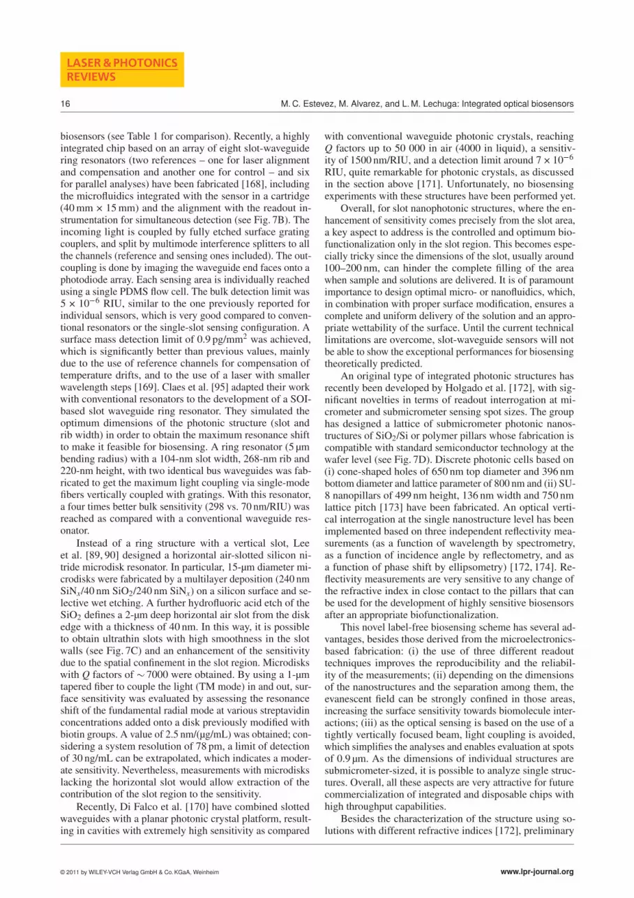
A few types of integrated optical-based resonating struc-tures have being explored for biosensing, such as the pla-nar microring resonators based on microdisks [88–91], mi-crorings [92–97], or microtoroids [98–102]. Compact ringresonators with optimal photonic properties and optimalsensitivity can be fabricated (see Fig. 5) including complexconfigurations incorporating multiple resonators. Differentmaterials such as glass-based [103, 104], SixNy–SiO2 [105]or polymer-based [106–109] ring resonators have beenreported and applied to DNA, proteins or bacteria detec-tion [104] (Fig. 5A). Recently, special attraction is gainingsilicon-on-insulator (SOI)-based resonators [110]. De Voset al. [92] have described SOI microring resonators (5 μmradius, Q factor of 20 000) with a bulk refractive indexsensitivity of 70 nm/RIU, which corresponds to a minimaldetectable refractive-index shift of 10�5 RIU. Biosensingevaluation of avidin interaction onto a biotinylated surfaceled to estimated detection limits around 10 ng/mL [92, 111],which is comparable to other label-free sensors. Recently,the same group has reported an array of SOI microring res-onators [112] based on three series of four rings connectedto a single-input waveguide, one for each series, and eachone with individual output waveguides (Fig. 5B). The incom-ing and outcoming light is coupled by grating couplers andthe collected signal of the twelve resonators is imaged onan infrared camera. The four resonators in each series havedifferent circumference ratios (with variations about 30 nm)showing independent and not overlapped resonance spectra.A PDMS flow cell with independent microfluidic channels(200 × 50 μm2) was mounted on top of the chip for sampledelivery. The multiplexed capabilities were evaluated byimmobilizing three different proteins and by successivelyflowing two of the three specific antibodies. Due to the opti-mized surface biofunctionalization, the device showed highspecificity and low nonspecific adsorption (which could bealso corrected by one resonator acting as reference) [111].With an estimated peak resolution around 0.4 pm and a Qfactor around 2 × 10⁴, a surface mass detection limit resultedof 3.4 pg/mm2.
Li et al. [94] proposed a two concentric ring structure inorder to increase the notch depth and the sensing area at thesame time. The sensor was fabricated over a SOI wafer withan outer radius of 21 μm, and an inner radius of 20 μm, andan air gap of 480 nm between them, coupled to a waveguideof 10 μm width tapered down to 480 nm. The estimated Qfactor of this resonator was around 5.1 × 10⁴ and the bulksensitivity was considerably high, � 683 nm/RIU. Iqbal etal. [96] have recently developed a biosensing platform basedon the integration of 32 ring resonators accessed via a buswaveguide, which are simultaneously interrogated (Fig. 5C).24 of them are used as sensing resonators, with an etchedwindow on the surface for sample interaction, whereas thereminding eight, left under the cladding layer, are used asreference for temperature-induced drift. Light is coupled inand out by grating couplers. The chip is covered with a flowcell that comprises the fluidic ports, channels, and reservoirsfor fluid delivery to the 32 sensors by using gaskets that de-fine the channels in each sensing area. The optical setup iscomplex but allows simultaneous reading of 24 interactions.
© 2011 by WILEY-VCH Verlag GmbH & Co. KGaA, Weinheim www.lpr-journal.org

Laser Photonics Rev. (2011)
REVIEWARTICLE
11
Figure 5 (online color at: www.lpr-journal.org) Several resonator sensing configurations: (A) Silicon chip with five rings verticallycoupled to input and output port waveguides. Reprinted from [104] with permission from Elsevier. (B) Array platform of SOI-based ringresonators packaged with PDMS microfluidics. Reprinted from [112] with permission from IEEE (© 2009 IEEE) (C) SEM micrograph ofa single ring with a window opened in the cladding polymer to expose the sensor surface. Microchip with 32 sensors and a six-channelPDMS microfluidic cell for multiplexed analyses. Reprinted from [96] and [116] with permission from IEEE (© 2010 IEEE) and fromAmerican Chemical Society, respectively. (D) Top: Scanning electron micrographs of microtoroids; Bottom: Microtoroids images showingthe tapered fiber for incoupling of the light. Reprinted from [98] with permission from AAAS and from [101].
A RI detection limit of 7.6 × 10�7 was achieved [96], withgood repeatability among the different resonators and anestimated surface LOD of 1.5 pg/mm2 [97]. AntiIgG/IgGand biotin/streptavidin interactions were used as proof-of-concept systems in order to assess the biosensing capabil-ities, and a preliminary real-time multiplexed analysis ofDNA hybridization showed the viability of the device [96].From the strict biosensing point of view, this platform hasbeen applied a few times for in-depth bioanalytical problems.For instance, by immobilizing specific antibodies [113], cy-tokines have been detected at low concentrations (below0.1 ng/mL in buffer) [114] but incorporating a secondary an-tibody for amplification of the signal. A sandwich assay wasapplied to the detection of cellular secretion of cytokines,showing the viability of measuring them in cell culturemedia, while retaining the sensitivity. Moreover, regenera-tion of the antibody layer was achieved, a step not alwayscontemplated in experiments with biosensors. The use of
several microrings (n � 15) diminishes the uncertainties,and increases the reliability of the tests. Similarly, a cancerbiomarker has been detected by immobilizing the specificantibodies for carcinoembryonic antigen (CEA) in a singlemicroring [115] achieving a detection limit around 25 ng/mLin a complex media like serum, which is close to relevantdiagnostic concentrations. The multiplexed capabilities ofthe arrayed resonators were evaluated by immobilizing sixdifferent antibodies (five specific antibodies and one nonspe-cific as control, four microrings for each one, see Fig. 5C)for the multiplexed determination of five relevant proteins.Prostate-specific antigen (PSA), α-fetoprotein (AFP), carci-noembryonic antigen (CEA), α-tumor necrosis factor (TNF-α), and interleukin-8 (IL-8) were simultaneously detected inan unknown protein cocktail solution [116]. A fast (10 min),sensitive (�150 fmol) and simultaneous detection of mul-tiple micro-RNAs (miRNAs) using single-strand DNA asimmobilized probes onto the microring array (four different
www.lpr-journal.org © 2011 by WILEY-VCH Verlag GmbH & Co. KGaA, Weinheim

12
LASER & PHOTONICSREVIEWS
M. C. Estevez, M. Alvarez, and L. M. Lechuga: Integrated optical biosensors
sequences) has also been recently published [117]. Single-nucleotide polymorphism could even be discriminated andexpression levels of miRNA in cells were also determined.
Toroidal-shaped-based microcavities offer resolutionlevels significantly higher than previous structures, sincethey exhibit ultrahigh Q factors (�108) [118, 119] whilepreserving the same advantages for mass production and in-tegration capabilities as planar ring resonators. Microtoroidscan be fabricated in arrays over a silicon wafer using stan-dard lithography techniques (see Fig. 5D) with dimensionsranging from 30 to 150 μm for the major diameter to 2.5–6 μm for the minor diameter. The structures are coupled toa low-loss tapered optical fiber and have been evaluatedfor biosensing by immobilizing specific antibodies usingprotein G for oriented attachment. With this configuration,cytokines were detected at extremely low levels, with a LODaround 5 aM (5 × 10�18 M), showing a wide working rangeof around 12 orders of magnitude in buffer conditions andwith the remarkable capability of resolving single-moleculedetection [98]. The capabilities of the sensor were also evalu-ated in 10-fold diluted serum. The same group has attemptedthe fabrication of low-loss polymer [99] or polymer-silicahybrid [120] toroids, which, even with a lower Q factors(105–107), are still significantly competitive with other ringresonators. Nevertheless, their biosensing capabilities havenot been shown as yet.
3.4. Photonic-crystal waveguide sensors
Photonic-crystal-based biosensors are a relatively noveltechnology that is gaining much attention and is increasinglyappearing in the literature [121, 122]. Photonic crystals arewell-defined nanostructures with periodically repeated vari-ations in the refractive index in one, two or three orthogonaldirections generating one-, two- or three-dimensional pho-tonic crystals (1D, 2D or 3D PhC). The lattice structure isgenerated on the length scale of the light wavelength, whichgenerates photonic bandgaps where light cannot propagatein the crystal. The width and position of the photonic gapis highly dependent on the refractive-index change betweenthe dielectric materials and on the periodicity of the struc-ture [123], which is exploited for sensing.
Integrated photonic crystals have been interestingly ap-plied in dielectric mirrors, lasers, narrowband optical filters,microresonators and in waveguide structures and due totheir high degree of integration they are ideal candidates forlab-on-chip biosensing. Device structures based on lineargratings and 2-dimensional gratings (i.e. arrays of holesarranged in checkerboard or hexagonal close-packed gridsalong the sensor surface) are the simplest ones in terms offabrication and sensing. The introduction of discrete or linedefects on the grating structure generates transducer cavi-ties with enhanced sensitivity. The strong confinement ofthe light in the periodic lattice makes these materials veryattractive not only because of the sensitivity but also due totheir small dimensions, limited to a few μm2, which allowsfor low sample consumption.
Cunningham’s group has pioneered the developmentof bulk photonic-crystal-based biosensors [124–126]. Theyhave implemented a one-dimensional subwavelength poly-mer grating coated with a layer of high refractive index(TiO2) [127]. White light is perpendicularly impinged onthe sensor surface and the reflected light is collected, whichshifts after a biomolecular interaction takes place. The fab-rication has been implemented into continuous sheets ofplastic films. The interrogation method can read individu-ally several areas of the surface, avoiding optical crosstalkbetween adjacent sensor regions. The overall technology of-fers the possibility to produce low-cost, disposable sensingsurfaces with high throughput capabilities [128]. In addition,the sensing surfaces have been adapted to be allocated at thebottom of microtiter plates (96, 384 or 1536 formats) [128]or microarray slides [128]. Moreover, they can be integratedwithin microfluidic networks by replica molding of thephotonic-crystal sensors and fluid channels in a simulta-neous process [129–131]. The latter configuration permitskinetic and high-resolution endpoint spatial measurements,improving the detection time and decreasing the detectionlimit as compared to standard in-well detection [132]. Re-cently, a microfluidic array chip with combinatorial mixingand on-chip sensing capabilities has also been demonstratedusing AtC (actuate to close) microfluidic networks [133].The sensors have been exploited for a wide range of bioas-says [134], including cell-based assays, characterization andstudy of cells attachment to surfaces [135–137], protein andvirus detection [132] . [138], study of small molecules aggre-gation for drug screening [139], or for protein-protein inter-actions [140], among others. The technology based on stan-dard microplate-based assays has been commercialized bySRU Biosystems (www.srubiosystems.com) (see Sect. 4).
Photonic crystals with a defined structure can offer in-teresting alternatives. Besides a few examples, such as apillar-based array of 2D photonic crystals [141] recently de-veloped but not evaluated for biosensing, most of the workis focused on hole-array PhC (see Fig. 6A). Planar photonicwaveguides, where a guided wave is generated by missingholes or defects in the bulk photonic-crystal structure havebeen fabricated on a SOI wafer. Unfortunately, and despitethe large effort invested, scarce biosensing examples can befound in the literature and such examples are only at theproof-of-concept level. Most of the work is mainly focusedon fabrication, simulation experiments and bulk refractive-index evaluation to establish the sensitivity of the structures.
Some examples are worthy of mention, such as the workby Dofner et al. [142, 143] who designed and fabricateda SOI photonic-crystal waveguide consisting of a hexago-nal lattice of holes with a single missing row line defectand point cavities in different arrangements (either a defectbased on three missing holes, a single hole or a shifted lineof holes arranged perpendicular to the waveguide orienta-tion) (see Fig. 6A, bottom). The optical properties of all thestructures were assessed by TE-polarized light coupled inand out of the ridge waveguide by a tapered fiber, allowingin-planar-geometry evaluation. By placing a flow cell ontop of the structure, refractive-index sensing in the mostoptimal case (Q factor � 3000) gave a bulk LOD of � 10�3
© 2011 by WILEY-VCH Verlag GmbH & Co. KGaA, Weinheim www.lpr-journal.org

Laser Photonics Rev. (2011)
REVIEWARTICLE
13
Figure 6 (online color at: www.lpr-journal.org) (A) SEM images of several photonic crystal-based waveguides sensors. Reprintedfrom [146] with permission from the Optical Society of America and from [143,153] with permission from Elsevier. (B) SEM imageof a nanoscale optofluidic array of photonic crystal microcavities with multiplexed capabilities (top) and a scheme of a PDMS fluidicchannel used for sample delivery (bottom). Reprinted from [148] with permission from the Optical Society of America. (C) Scheme ofthe evaluation set-up of a photonic crystal microcavity sensor, showing the optics (laser, fiber polarizer, input collimating and focusingoptics, output objective, photodetector and alignment camera) and integrated microfluidics. SEM image of the photonic crystal structureis also included. Reprinted from [154] with permission from Elsevier.
RIU [142]. Surface sensitivity was assessed by physisorp-tion of BSA or avidin as model proteins, giving a masssensitivity around 24.7 nm/pg, with a minimal detectablemass of around 4 fg and a detection limit of � 500 pg/mm2.
Skivesen et al. [144] have fabricated a waveguide withholes of 240 nm in diameter, a lattice constant of 370 nmand a length of 25 μm, using electron-beam lithography andinductively coupled plasma etching. A BSA adsorption wasdemonstrated under static conditions. A similar structurehas been used for protein and DNA sensing [145, 146]. Thephotonic crystal length was 20 μm with a lattice constantand hole radius of 390 and 111 nm, respectively. A 500-nmwide single-mode access waveguide was employed to ob-tain a sharp band edge (see Fig. 6A). A LOD of 6 × 10�4
RIU was achieved [145]. Tapered fibers were used for thein- and outcoupling of TE-polarized light and a flow cellwas mounted on top of the sensing surface. Hybridization ofDNA strands was monitored in the nM range [146]. For pro-teins, anti-BSA was detected over a BSA-functionalized sur-face, reaching a mass detection limit of 2.1 pg/mm2 (whichmeans a total mass detection limit of 0.7 fg).
1D photonic crystal microcavities fabricated by X-raylithography on an SOI wafer have also been described [147],
introducing defects in the structure by breaking the period-icity of the holes. The resonant wavelength can be tunedaccording to the defect cavity spacing. This type of structurehas been adapted by Erickson’s group for the developmentof the so-called nanoscale optofluidic sensor array (NOSA).This sensor has a microcavity structure (resonator) that isevanescently coupled to an adjacent single-mode siliconwaveguide. The design of the structure includes parallelresonator arrays with slightly different cavity spacing and,therefore, different resonant wavelength and with individ-ual Q factors between 1000 and 3000, all of them placedalong a single waveguide (see Fig. 6B). Nanotapers wereused for light coupling and a PDMS flow channel runningorthogonally to the array was mounted onto the chip, forsimultaneous and multiplexed detection [148]. A bulk re-fractive index LOD of 7 × 10�5 RIU was experimentallydetermined, which could be translated into a high sensitivityfor mass detection, when considering the extremely highconfinement in the inner holes. Detection of serotypes ofDengue virus was demonstrated by immobilizing four dif-ferent DNA probes in different resonators and subsequentlyadding the specific DNA target for one of them [149]. Theseexperiments only showed a proof-of-concept for nucleic
www.lpr-journal.org © 2011 by WILEY-VCH Verlag GmbH & Co. KGaA, Weinheim

14
LASER & PHOTONICSREVIEWS
M. C. Estevez, M. Alvarez, and L. M. Lechuga: Integrated optical biosensors
acid interaction. Similarly, immunochemical multiplexed de-tection of interleukins (IL) was also addressed [150]. Threedifferent specific antibodies for IL4, 6, and 8, respectively,were immobilized and a sandwich assay was performed byadding the target followed by a secondary specific antibodyin order to enhance the signal. It is worth noticing that thedevice has low sensitivity since it has moderate Q factors(3000), which may be enhanced by optimizing the crystalstructure (such as the gap and hole dimensions). Althoughthe sensitivity of this PhC sensor is not high, this is oneof the few examples showing biosensing capabilities whileincluding a multiplexed configuration.
Lee and Fauchet [151] have also made a strong contri-bution in this area by developing two-dimensional photoniccrystal-based structures. Biotin-streptavidin interaction andcovalent immobilization of BSA were evaluated and a min-imum mass coverage of around 2.5 fg was detected [151].SOI-based wafers were used to fabricate photonic-crystalwaveguides with a defect line in the structure to guide thelight. Recently, a structure where the defect line is generatedadjacent to the photonic microcavities has been proposed insuch a way that the device can operate as a multichannel sen-sor [152,153]. This structure shows a limited sensitivity withQ factors around 400 and bulk LOD in the range of 10�2
RIU. Biosensing evaluation was done under a static regime,with incubation steps and evaluation in dry environment.The surface was covalently modified with antibodies (anti-IgG) and target antibodies (IgG) were detected, observing adetection limit around 67 nM (10 μg/mL) that correspondsto a surface sensitivity of 2.3 × 10⁵ nm/M [153]. Consider-ing that the major contribution to the signal comes from themost sensitive area of the region (the defect in the structureand the surrounding holes) a surface density of 1 ng/mm2
and a minimum mass of 1.5 fg could be detected.
Zlatanovic et al. [154] designed a photonic crystal(Fig. 6C) structure with a ridge waveguide but with lowQ factors (around 400). Real-time biosensing was demon-strated by immobilizing biotin-BSA and detecting specificantibodies against biotin. An excellent limit of detection of20 pM of antibody was achieved, which in terms of massand considering the affinity of the pair antibody/antigenwas found to be around 4.5 fg. Moreover, the experimentsincluded the regeneration of the bioactive surface. Theseresults demonstrated that despite the low resolution of thedevice (due to the low Q factor), an adequate biofunctional-ization of the surface could lead to acceptable sensitivitiesfor biodetection.
Overall, photonic crystal structures still remain as apromising type of transducers with moderate sensitivities ascompared with other label-free photonic sensors. Better fea-tures could be achieved by optimizing the crystal and defectdimensions and also by positioning the bioreceptor only inthe holes (by controlled immobilization), where the confine-ment is maximum, therefore resulting in highest resonantshifts. In fact, a growing number of examples are appear-ing in the literature focused on single-particle detection bytrapping them at the holes; single virus or small pathogenscould be detected in this way [155–157].
3.5. Silicon wires, slot waveguides and othernanophotonic sensing configurations
In this section we include an overview of the new and emerg-ing trends in the design and implementation of nanopho-tonic structures for sensing, which we envisage could behighly competitive with the ones described in previous sec-tions, either due to their sensitivity or to their capabilitiestowards integration and miniaturization. Among them, slotwaveguide-based structures and silicon wires stand out fortheir potential enhanced sensitivity due to the confinementof the electromagnetic field and for their highly integrationcapabilities in compact dense arrays of individual sensorswith versatile geometries. Unfortunately, few biosensingexperiments have been demonstrated with these devices,and only proof-of-concept experiments with model systemshave been published so far.
Silicon photonic wires are submicrometer channel wave-guides fabricated by electron-beam lithography and reactiveion etching on SOI wafers. The high index contrast betweenthe silicon core (n� 3�5) and silica cladding (n� 1�5) yieldsa strong field confinement and allows for sharp waveguidebends radii of few micrometers with low losses. With thistechnology the fabrication of waveguides with higher com-pactness and higher intrinsic sensitivity (provided by theTM mode) than the lower-contrast index material counter-parts is possible. Initially developed for optical switches,directional couplers and other telecommunications applica-tions, silicon wires first showed up in the biosensing field bythe pioneer work of Janz’s group. This group first describeda Mach-Zehnder interferometer based on SOI photonic wirewaveguides [158] and a double spiral millimeter-long res-onator folded within a compact size of 100–150 μm (and a Qfactor of 17 600) (Fig. 7A). Both sensors were fabricated onSOI wafer with a Si core thickness of 0.26 μm and a buriedSiO2 cladding of 2 μm. Single-mode wire waveguides of450 nm width were selected and a SU-8 cover layer wasemployed to isolate the nonsensing regions. Bulk sensitivityand surface biomolecular interactions were evaluated un-der flow regime (using a PDMS microflow cell). Updatedversions incorporate improvements such as a reference armto suppress wavelength and temperature drift [159, 160],integration of dense arrays of six spiral MZI with an SU-8 fluidic channel for the delivery of liquids [161], or theuse of four resonators [162], which overall opens up thepossibility to perform multiplexed analyses (see Fig. 7A).The dimensions of the folded wires are suitable for a con-trolled surface functionalization using conventional spotters.The performance of the sensing devices was assessed byimmobilizing two different antibodies in individual sensorsand simultaneously evaluating them with the correspondinganti-IgG. For the MZI configuration, TM polarized light isdelivered by an optical fiber that splits the light to the sixsensors; parallel readout of the light exiting the six outputwaveguides is done by a near-infrared camera. Results haveshown the discrimination between the different biomodifiedsurfaces, demonstrating the high potential of silicon wires-based devices for multiplexed biosensing. However, resultsare not yet conclusive about the sensitivity of these sensors
© 2011 by WILEY-VCH Verlag GmbH & Co. KGaA, Weinheim www.lpr-journal.org

Laser Photonics Rev. (2011)
REVIEWARTICLE
15
Figure 7 (online color at: www.lpr-journal.org) (A) Silicon photonic wire MZI and resonator sensor arrays. Details of the structuresand the SU-8 microfluidic channels aligned to the sensor windows are shown. Reprinted from [159–161] with permission from theOptical Society of America. (B) Slot-waveguide ring resonator chip, consisting of 6 sensing channels plus 2 reference ones. Detailsof the structures and grating couplers of the resonator array (left) and the compact sensor cartridge for multiplexed sensing (right)are shown. Reprinted from [168] with permission of the Royal Society of Chemistry. (C) SEM images of fabricated air-slot disk with adiameter of 15 μm and an air slot depth of 2 μm. Reprinted from [89] with permission from the Optical Society of America. (D) SEMimages of SU-8 nanocolumns lattices. Reprinted from [175] with permission from Elsevier.
and only estimated values of resolvable surface coveragearound 0.25 pg/mm2 are theoretically predicted (correspond-ing to a detected molecular mass of 0.5 fg) [161]. In the caseof resonators, with radius between 20 and 28 μm (and im-proved Q factors of 25 000) and a reference included in thestructure, a bulk sensitivity of 135 nm/RIU (�2 × 10�6 RIU)has been achieved and a surface immunochemical detectiondown to 20 pM (resolvable mass of 40 ag) [162] has beenshown so far.
Slot waveguides [163–165] consist of two slabs of highrefractive index materials separated by a nanometer-scalelow refractive index slot region and surrounded by low re-fractive index cladding materials in such a way that light isstrongly confined in the slot region. A stronger light-analyteinteraction can be achieved within this region as comparedto conventional rib or planar waveguides, which results inan enhanced sensitivity. Moreover, slot waveguides can befabricated within structures such as resonators or interfer-
ometers by employing CMOS compatible materials andtechnology, including SOI technology, which enables minia-turization, and further integration within lab-on-a-chip plat-forms.
So far, slot-waveguide resonators have been reported byonly two research groups. Barrios et al. [166] pioneered theslot-waveguide configuration, using Si3O4–SiO2 for the fab-rication of a slot-waveguide ring resonator structure, with aradius of 70 μm and slots of 200 nm for both the waveguideand the resonator. A bulk sensitivity of 212 nm/RIU for aresonator with a Q factor of 1800 was reached, significantlybetter than conventional resonators, indicating a consider-able contribution from the slot region. Biosensing evaluationwas done off-flow by covalently immobilizing antibodiesonto the surface and detecting their specific target (in thiscase BSA was the model system) showing a surface limitof detection of 16 pg/mm2 [167], which indicates a poorto moderate sensitivity as compared to most of the optical
www.lpr-journal.org © 2011 by WILEY-VCH Verlag GmbH & Co. KGaA, Weinheim
16
LASER & PHOTONICSREVIEWS
M. C. Estevez, M. Alvarez, and L. M. Lechuga: Integrated optical biosensors
biosensors (see Table 1 for comparison). Recently, a highlyintegrated chip based on an array of eight slot-waveguidering resonators (two references – one for laser alignmentand compensation and another one for control – and sixfor parallel analyses) have been fabricated [168], includingthe microfluidics integrated with the sensor in a cartridge(40 mm × 15 mm) and the alignment with the readout in-strumentation for simultaneous detection (see Fig. 7B). Theincoming light is coupled by fully etched surface gratingcouplers, and split by multimode interference splitters to allthe channels (reference and sensing ones included). The out-coupling is done by imaging the waveguide end faces onto aphotodiode array. Each sensing area is individually reachedusing a single PDMS flow cell. The bulk detection limit was5 × 10�6 RIU, similar to the one previously reported forindividual sensors, which is very good compared to conven-tional resonators or the single-slot sensing configuration. Asurface mass detection limit of 0.9 pg/mm2 was achieved,which is significantly better than previous values, mainlydue to the use of reference channels for compensation oftemperature drifts, and to the use of a laser with smallerwavelength steps [169]. Claes et al. [95] adapted their workwith conventional resonators to the development of a SOI-based slot waveguide ring resonator. They simulated theoptimum dimensions of the photonic structure (slot andrib width) in order to obtain the maximum resonance shiftto make it feasible for biosensing. A ring resonator (5 μmbending radius) with a 104-nm slot width, 268-nm rib and220-nm height, with two identical bus waveguides was fab-ricated to get the maximum light coupling via single-modefibers vertically coupled with gratings. With this resonator,a four times better bulk sensitivity (298 vs. 70 nm/RIU) wasreached as compared with a conventional waveguide res-onator.
Instead of a ring structure with a vertical slot, Leeet al. [89, 90] designed a horizontal air-slotted silicon ni-tride microdisk resonator. In particular, 15-μm diameter mi-crodisks were fabricated by a multilayer deposition (240 nmSiNx/40 nm SiO2/240 nm SiNx) on a silicon surface and se-lective wet etching. A further hydrofluoric acid etch of theSiO2 defines a 2-μm deep horizontal air slot from the diskedge with a thickness of 40 nm. In this way, it is possibleto obtain ultrathin slots with high smoothness in the slotwalls (see Fig. 7C) and an enhancement of the sensitivitydue to the spatial confinement in the slot region. Microdiskswith Q factors of � 7000 were obtained. By using a 1-μmtapered fiber to couple the light (TM mode) in and out, sur-face sensitivity was evaluated by assessing the resonanceshift of the fundamental radial mode at various streptavidinconcentrations added onto a disk previously modified withbiotin groups. A value of 2.5 nm/(μg/mL) was obtained; con-sidering a system resolution of 78 pm, a limit of detectionof 30 ng/mL can be extrapolated, which indicates a moder-ate sensitivity. Nevertheless, measurements with microdiskslacking the horizontal slot would allow extraction of thecontribution of the slot region to the sensitivity.
Recently, Di Falco et al. [170] have combined slottedwaveguides with a planar photonic crystal platform, result-ing in cavities with extremely high sensitivity as compared
with conventional waveguide photonic crystals, reachingQ factors up to 50 000 in air (4000 in liquid), a sensitiv-ity of 1500 nm/RIU, and a detection limit around 7 × 10�6
RIU, quite remarkable for photonic crystals, as discussedin the section above [171]. Unfortunately, no biosensingexperiments with these structures have been performed yet.
Overall, for slot nanophotonic structures, where the en-hancement of sensitivity comes precisely from the slot area,a key aspect to address is the controlled and optimum bio-functionalization only in the slot region. This becomes espe-cially tricky since the dimensions of the slot, usually around100–200 nm, can hinder the complete filling of the areawhen sample and solutions are delivered. It is of paramountimportance to design optimal micro- or nanofluidics, which,in combination with proper surface modification, ensures acomplete and uniform delivery of the solution and an appro-priate wettability of the surface. Until the current technicallimitations are overcome, slot-waveguide sensors will notbe able to show the exceptional performances for biosensingtheoretically predicted.
An original type of integrated photonic structures hasrecently been developed by Holgado et al. [172], with sig-nificant novelties in terms of readout interrogation at mi-crometer and submicrometer sensing spot sizes. The grouphas designed a lattice of submicrometer photonic nanos-tructures of SiO2/Si or polymer pillars whose fabrication iscompatible with standard semiconductor technology at thewafer level (see Fig. 7D). Discrete photonic cells based on(i) cone-shaped holes of 650 nm top diameter and 396 nmbottom diameter and lattice parameter of 800 nm and (ii) SU-8 nanopillars of 499 nm height, 136 nm width and 750 nmlattice pitch [173] have been fabricated. An optical verti-cal interrogation at the single nanostructure level has beenimplemented based on three independent reflectivity mea-surements (as a function of wavelength by spectrometry,as a function of incidence angle by reflectometry, and asa function of phase shift by ellipsometry) [172, 174]. Re-flectivity measurements are very sensitive to any change ofthe refractive index in close contact to the pillars that canbe used for the development of highly sensitive biosensorsafter an appropriate biofunctionalization.
This novel label-free biosensing scheme has several ad-vantages, besides those derived from the microelectronics-based fabrication: (i) the use of three different readouttechniques improves the reproducibility and the reliabil-ity of the measurements; (ii) depending on the dimensionsof the nanostructures and the separation among them, theevanescent field can be strongly confined in those areas,increasing the surface sensitivity towards biomolecule inter-actions; (iii) as the optical sensing is based on the use of atightly vertically focused beam, light coupling is avoided,which simplifies the analyses and enables evaluation at spotsof 0.9 μm. As the dimensions of individual structures aresubmicrometer-sized, it is possible to analyze single struc-tures. Overall, all these aspects are very attractive for futurecommercialization of integrated and disposable chips withhigh throughput capabilities.
Besides the characterization of the structure using so-lutions with different refractive indices [172], preliminary
© 2011 by WILEY-VCH Verlag GmbH & Co. KGaA, Weinheim www.lpr-journal.org

Laser Photonics Rev. (2011)
REVIEWARTICLE
17
biosensing experiments have recently been performed by co-valent immobilization of BSA onto the nanopillars and thesubsequent biodetection of the interaction with the specificantibody, reaching sensitivities around 3 ng/mL [175] understatic conditions. Further implementation of an appropriatemicrofluidic flow cell and a flow delivery system will allowthe optimum infiltration of the solutions through the wholesensor lattice. This will be the next step for these promisingdevices that could offer multiplexing evaluation of at least1200 samples per hour.
3.6. Optonanomechanical sensors
Optomechanical (OM) sensing is a novel technology de-rived from the standard micro- and nanoelectromechanicalsystems widely employed during recent years in the biosens-ing field. Specifically, nanomechanical systems based onsilicon microcantilevers have demonstrated an extremelyhigh sensitivity in the measurement of frequency [176, 177]and deflection changes [178] due to mass accumulation orbiosensing interactions [179–183], and in the measurementof forces between single molecules [184] or even of forcesarising from single electron spins [185].
However, in order to achieve full working prototypesas lab-on-a-chip biosensors, the commonly used opticalreadout method for nanomechanical sensors has severe limi-tations related to the complex optical alignment of multiplecantilevers at the same time, and the diffraction constraintswhen the size of the cantilever is reduced below the wave-length. In an optonanomechanical sensor, the cantilever it-self is an optical waveguide the output intensity of which isa function of the bending induced by a biomolecular interac-tion. With this new configuration it is possible to overcomethe aforementioned limitations of standard nanomechani-cal sensors. These microscale optical waveguide cantileversare mainly addressed to sensing applications and severalworks have demonstrated their motion sensitivity. The work-ing principle of the optical microcantilevers do not rely onrefractive-index changes, but on the microcantilever bend-ing or resonance frequency shift, measured through the lightintensity collected at the cantilever free end. The limit ofdetection can be evaluated in terms of analyte concentra-tion (i.e. in ng/mL or molarity), according to the cantileverbending (in nm) or frequency change (Hz), or according tosurface mass density (pg/mm2).
The most extended configuration for biosensing is theend-coupled one, which consists of a microcantilever actingas a waveguide; the light propagates along the cantileveruntil reaching the free end. After crossing a small gap, lightis collected by an output waveguide (see Fig. 8A). Severalparameters can have an influence on the system sensitivity,such as the distance from the cantilever edge to the col-lecting waveguide, the coupling efficiency or the cantilevermaterial and rigidity. Different materials, cantilever shapes,and light coupling methods have been investigated. The de-vice fabricated by Zinoviev et al. [186] employs an arrayof 20 waveguide silicon dioxide microcantilevers, each oneof them connected with a silicon nitride multimode input
Figure 8 (online color at: www.lpr-journal.org) (A) Schematicrepresentation of the optical waveguide cantilever (OWC) work-ing principle. Input light is coupled to the cantilever through theevanescent field of an input waveguide, and output light is col-lected in a misaligned waveguide after crossing a small gap. (B)Picture and SEM image of an OWC chip array with 20 waveguidemicrocantilevers. Reproduced from [180] with permission of theRoyal Society of Chemistry.
and output waveguide; the light from the input waveguideis coupled to the cantilever through the overlapping of theevanescent field (Fig. 8B). With this configuration, the can-tilever displacement can be detected with a resolution of18 fm Hz�1/2, sensitivity comparable to the standard opticallever method, which indicates its suitability for biosens-ing applications.
A silicon microcantilever etched to form a single-modewaveguide rib, with a capture asymmetrical multimodewaveguide that terminates in a Y-branch, was proposed byNoh et al. [187]. The Y-branch forms a differential signalthat is monotonically dependent on the microcantilever de-flection, achieving a signal sensitivity of 1.4 × 10�4 nm�1.More recently, the same group has reported a sensitivity en-hancement by modifying the mode structure of double-steprib waveguides used to capture light, reaching a sensitivitycomparable to the optical lever method [188]. Besides theuse of silicon material, there is an increasing interest onpolymeric materials for waveguides fabrication. EmployingSU-8 [189–192] and PDMS [193] polymers several groupshave already developed, integrated optical waveguide can-tilevers. Indium phosphide has also been employed showingup as a suitable material for the fabrication of electrostati-cally actuated end-coupled optical waveguide MEMs [194].Other works suggest the use of silicon photonic crystals forguiding the light through the microcantilever [195–197], oron chip in-plane Fabry-Perot interferometer readout [198].Working with arrays of optical waveguide microcantileversis more challenging as a multiplexed input of the light ineach cantilever must be achieved. With this aim, different
www.lpr-journal.org © 2011 by WILEY-VCH Verlag GmbH & Co. KGaA, Weinheim

18
LASER & PHOTONICSREVIEWS
M. C. Estevez, M. Alvarez, and L. M. Lechuga: Integrated optical biosensors
strategies have been proposed, such as using diffractiongratings [199], a network of waveguide splitters [200] orintegrated silicon light sources and detectors self-alignedto silicon waveguides [201]. A higher integration and scalereduction of these nanophotonic platforms allow the directactuation of MEMs by the force exerted by the propagatedphotons. Li et al. [202, 203] suggested two different in-tegrated photonic platforms designs: an embedded beamresonator with an on-chip interferometer for the displace-ment sensing, and a pair end-to-end coupled waveguidecantilevers. Both platforms include two input/output gratingcouplers at the end of the waveguides for light coupling.A displacement sensitivity of 40 fm Hz�1/2 was achievedwith the end-to-end coupled cantilevers working under vac-uum conditions.
However, to our knowledge, the application of this tech-nology to the biosensor field is scarce. For example, Koevet al. [191] were able to detect homocysteine in liquid at alowest concentration of 10 μM with a minimum detectablecantilever bending of 5 nm. Nordin’s group [204] has re-cently demonstrated the integration of a 16 microcantileverarray using in-plane photonic readout with a 2-layer PDMS-based microfluidic chip that integrated valves to control thefluid flow through the microchannels. However, only thestudy of the transient response of the microcantilevers wasreported. A higher integration and scale reduction of thesemechanophotonic platforms can be easily achieved, pro-ducing devices with a very small mass and low dissipation,ideal for fundamental studies [205–208], although with nobiological applications reported so far.
4. Current commercial technologies basedon label-free photonic biosensors
Some of the technologies described in the previous sectionshave evolved into commercial platforms for label-free de-tection, which has exponentially expanded the number andvariety of applications in biotechnology, biology, and clini-cal analysis. In this section we will give an overview of themost relevant biosensor devices based on integrated optical
transducers that have been successfully transferred into themarket (see Table 2). As can be appreciated, none of themhas achieved the status of complete “lab-on-a-chip” portableplatform with compact and reduced size. Instead, most ofthem are bulky instruments mainly for laboratory use witha complex and challenging road to achieve point-of-carestatus. However, all of them have demonstrated the excel-lent performances of the integrated optics-based sensorsin terms of sensitivity and automation, and, in some cases,even in terms of multiplexing (allowing medium to highthroughput analysis).
Farfield
In 2000 the Farfield Group launched onto the market theAnaLight® interferometer based on dual polarization inter-ferometry (DPI). Whereas all the interferometric schemespreviously discussed employ only one polarization, DPIuses both TM and TE polarizations in a sensing transducerthat mimics a Young configuration. By simultaneously mea-suring both polarizations, determination of the thicknessand the refractive index of a film adsorbed on the sensorsurface can be done under real-time conditions, which in-creases the versatility of the device for advanced and com-plex measurements. Sensitivity of the instrument reachesrefractive-index variation of 10�7 RIU, and a mass surfacesensitivity of 0.1 pg/mm2. Two simultaneous evaluationsare possible by a dual-channel flow cell with small sample-volume capability (�50 μL). A bulkier platform, AnaLight®4D Workstation (analysis station: 90 × 48 × 35 cm and pumpsize 28 × 23 × 16 cm) is also commercialized for automaticsampling using vials, and 96- and 384-well formats. Theinstrument has been exploited for diverse biosensing evalua-tions, using surfaces already preactivated and functionalizedwith appropriate chemistry. Among others, the platformallows for thermodynamics, kinetics and affinity studies,such as protein interaction studies, protein conformationalchanges, specific biodetection and biomolecular interactions,surface density evaluation, lipid bilayer formation and char-acterization, biocompatibility studies and protein or lipid
Table 2 Current commercial technologies based on integrated optics biosensing(a).
Company Instrument Technology Format Throughput Webpage
Farfield AnaLight Young Interferometry Flow conditions Low www.farfield-group.com
SRU Biosensing BIND Biosensor Photonic Crystal Cartridge (16)
Microplate (96–1536)
Very High www.srubiosystems.com
Corning EPIC System Waveguide Resonant
Grating
Microplate (384) High www.corning.com
Axela DotLab Optical Grating Flow/static conditions Medium www.axelabiosensors.com
Microvacuum OWLS Optical Grating
Couplers
Flow conditions Low www.owls-sensors.com
Genalyte Maverick Ring resonators Consumable with 1 to
128 resonators
Medium www.genalyte.com
a Sensitivity levels are in the low ng/mL range in all the cases.
© 2011 by WILEY-VCH Verlag GmbH & Co. KGaA, Weinheim www.lpr-journal.org

Laser Photonics Rev. (2011)
REVIEWARTICLE
19
aggregation analyses. The main disadvantage is the limita-tion to only two samples due to the microfluidics systemand the readout electronics.
SRU Biosystems
SRU Biosystems, Inc. has developed the BIND™ Biosensor,based on the use of photonic crystals in a grating config-uration. The platform offers high multiplexed capabilitiesin a microplate format (up to 1536 wells). The instrumenthas a high level of resolution (in the pm level) and a broaddynamic range, which ensures the analysis of both smallmolecules and large entities such as cells or bacteria. Thesensing surfaces are modified following optimized proto-cols to enable both cell-based and biochemical assays withexcellent performance. Different readers are available bothfor single-point mode and imaging mode. The instrumenthas fast detection times (96-well in 30 s, 384-well in 1 min,1536-well in 3 min), or even faster with an advanced readoutmode (96-well in 8 s, 384-well in 15 s, 1536-well in 45 s).Among others, the platform has demonstrated its useful-ness in diverse applications such as for cell morphology andadhesion assays, ion-channel assays, small-molecule andfragment screening, and for the analysis of interactions oflarge genomic, protein, peptide or antibody libraries againsta wide range of biochemical targets.
Corning
A quite similar technology to BIND™ Biosensor has beenmarketed by Corning. Epic® System is a high through-put screening platform based on waveguide resonant grat-ings. This device reaches a detection limit around 5 pg/mm2
and has a broad dynamic range. Some functionalized sur-faces are available (three types: cell-based, biochemical orfibronectin coated, also for cell assays). The instrument canbe coupled to an automatic liquid handler in order to en-hance its throughput performance and has a rate analysisof 6 s/microplate requiring sample volumes of 15–80 μL.A main disadvantage is related to the dimensions of thewhole instrumentation (0.83 m × 1.15 m × 1.98 m) and itshigh price, which can limit its implementation in laborato-ries and other institutions. A benchtop version of the reader(EnSpire®Multimode Plate Reader, in collaboration withPerkin-Elmer) is also currently commercialized, with moreconvenient features, which allows for label free detection aswell as for fluorescence, absorbance, and luminescence mea-surements.
Axela
Axela group has launched the dotLab® System, an instru-ment based on diffractive optical gratings. The grating isfabricated on the lower area of a prism-shaped surface,which is integrated in a 10 μL flow channel disposable plas-tic chip. Each dot lab sensor contains eight assay spots
aligned with the linear flow channel for multiple assays.The fluidic controller can work under static, continuous-flow or constant-mixing mode. The samples and solutionsare delivered to the sensor by an automated sampling sys-tem (from reagent bottles or 96-well microplates), allowingreal-time evaluation. The dotLab® is provided with severalattachment chemistries (i.e. streptavidin-modified, ProteinG, Goat-Anti-Mouse-Fc or for covalent binding) which in-creases the flexibility for the coupling of bioreceptors tothe surface. Features of the device include label-free andquantitative evaluation within a broad dynamic range. Thedimensions are comparatively smaller than for SRU andCorning instruments (43.5 cm H × 57.9 cm W × 69.3 cm D)but the multiplexed capabilities are more limited, since theanalysis time is slower and only eight simultaneous testsare possible. The instrument can be used for protein-proteininteraction studies, epitope mapping, or for the detectionand quantification of proteins, among others.
Microvacuum
Microvacuum Ltd. manufactures the OWLS System basedon optical waveguide lightmode spectroscopy [63]. In thissensor, polarized light is coupled by an input grating andguided by total internal reflection to the end of the wave-guide where it is detected by the photodiodes. The lightmode spectrum can be obtained by varying the angle ofincidence, whereas the flow cell is fixed over the gratingcoupler waveguide sensor chip. From the mode spectra it ispossible to determine the physical parameters of the covermedium (thickness, refractive index, density, etc.). The chipis mounted on a precision goniometer that provides theadjustment of the angle of incidence. It operates under con-tinuous flow for real-time evaluation. The instrument hasa compact small-to-medium size and includes the optome-chanical system, the electronics, temperature control andthe sensor holder, together with the injection unit (with sev-eral volume sample loops), although the fluidic pump isexternal. The data acquisition and the software evaluate therefractive index and thickness of the adsorbed layer, andprovide qualitative kinetics information. The instrumentcan be applied for biomolecular interaction studies (protein-protein, nucleic acids, membrane protein-lipid bilayer . . . ),cell-based assays, surface coverage characterization, drugscreening or kinetic analysis. The sensor surface has generalfeatures that can be modified on request to improve perfor-mance (i.e. dimensions of the chip, dielectric coatings of thesurface, passivation or functionalization with silanes). Theinstrument can be combined with two additional units: anelectrochemical system and a fluorescent measuring system.Unfortunately, the instrument has very limited throughputcapabilities since it can only perform single measurementson relatively large chips (around 12 mm × 8 mm).
Genalyte
Relatively recently, Genalyte has started commercializingthe Maverick Detection System based on an array of micror-
www.lpr-journal.org © 2011 by WILEY-VCH Verlag GmbH & Co. KGaA, Weinheim

20
LASER & PHOTONICSREVIEWS
M. C. Estevez, M. Alvarez, and L. M. Lechuga: Integrated optical biosensors
Figure 9 (online color at: www.lpr-journal.org) Envisionedlab-on-a-chip platform with on-chip detection using integratedoptics-based transducers. In the figure, an array of Mach-Zehnderinterferometers are integrated with individual microfluidics chan-nels, grating couplers for the in and output of the light in eachsensing channel, read-out photodetectors and CMOS electronicsfor data processing.
ing resonator silicon sensors. The instrument currently inthe market is the third generation and has a compact design,which delivers the samples (typically less than 25 μL) from a96-well plate into the plastic chip consumable that containsthe sensing surface, consisting of an array of silicon micror-ings (from 1 up to 128 in each surface, including 8 controls).The system can perform simultaneous measurements in realtime, and results are available on a timescale between 5–45min, depending on the concentration to be detected. Someof the features of the instrument are the possibility to per-form kinetics measurements (using 2 flow channels whichdeliver samples to 16 sensors each) or end-point measure-ments using a 96-well footprint; it has a wide dynamic rangeup to eight orders of magnitude, with sensitivities close to0.1 ng/mL, a limit of detection around 0.1 fg and it is ableto measure targets of molecular weight of only 10 Da.
5. Integration in lab-on-a-chip platforms
The major challenge in the silicon photonic biosensor fieldis to achieve a fully operative lab-on-a-chip platform withon-chip detection. Further efforts are addressed to keepa high level of sensitivities and to develop complete SiCMOS-compatible integrated devices that incorporate thephotonic systems with microfluidics and the optoelectron-ics [46, 209, 210]. For the development of a complete pho-tonic lab-on-a-chip device, several units must be incorpo-rated on the same platform: (i) the photonic sensors, (ii) theflow cells and the flow delivery system, including miniatur-ized valves and pumps (iii) the light sources and photode-tectors array or miniaturized CCD cameras (iv) processingelectronics (i.e. CMOS) and, (v) final packaging with the re-quired firmware and software (see Fig. 9). Integration couldbe monolithic (all functionalities are incorporated in onesingle chip) or hybrid (functionalities are separated on sev-eral chips). In the monolithic approach photodiodes andlasers can be embedded in the waveguides. Due to eco-
nomic and technical reasons the hybrid approach is mostlypreferred, where the components are settled apart. Even ifthe individual components are well known, the subsysteminterfaces between them are difficult to optimize and theseare still the major barriers to be overcome. With the hybridarrangement, the optoelectronics and the readout system canbe reusable and only the cartridge containing the sensingchip with the microfludics for independent applications canbe interchanged.
Most of the work done so far mainly includes the integra-tion of the sensors and the fluidics (which has emerged as anew discipline called “optofluidics”) and the development ofdiscrete components for future integration [211]. Few exam-ples can be found in the literature of complete lab-on-a-chipdevelopments. For instance, Jokerst and coworkers [212]have developed an integrated evanescent-field multimodeMach-Zehnder interferometric sensor fully compatible withSi-CMOS process with a LOD of 2.5 × 10�6 RIU. In furtherrefinements a fully integrated version was put in place withinterferometric sensors of only 5 mm length that reached arefractive-index sensitivity of �9.2 × 10�7 RIU [212]. Un-fortunately, this device has not been tested yet for biosens-ing applications.
A monolithically integrated interferometric lab-on-a-chip biosensor is under development within the frameworkof a European project [213]. The principle of operation ofthe PYTHIA lab-on-a-chip is based on broadband Mach-Zehnder interferometer sensors and including lasers, pho-todetectors and microfluidics. Femtosecond laser microma-chining is nowadays an emerging tool for the fabrication ofmonolithic optofluidic devices, allowing the direct fabrica-tion of integrated optical devices and microfluidics in fusedsilica chips [32, 214].
Lab-on-a-chip with the on-chip detection is still verymuch a work in progress, but recent advances in integratedoptics-based biosensors and optofluidics indicate that theycould eventually be achieved in the near-medium future.Cutting-edge technological progress will give rise to a robustproduct that could run on an automated system minimizingoperator skills.
6. Future outlook
During recent years a myriad of different developments atthe biosensor field using many diverse technologies and de-vices has been appearing, such as nanomechanical or MEMSresonators sensors, semiconductor-wire-based transducers,highly integrated microelectrodes, carbon-nanotube-basedsensors, localized surface plasmon resonance or SERS inmetal nanostructures, but none of them has shown the excel-lent performances for label-free sensing achieved by pho-tonic and nanophotonic devices based on integrated optics,as can be deduced from their excellent limits of detectionin real biosensing (shown throughout this review). Somecommercial optical biosensors have been introduced intothe market but most of them are bulky and expensive andonly offer a general technology, with a limited number ofdedicated applications.
© 2011 by WILEY-VCH Verlag GmbH & Co. KGaA, Weinheim www.lpr-journal.org

Laser Photonics Rev. (2011)
REVIEWARTICLE
21
Integrated optics technology is opening the way towardstruly portable lab-on-a-chip platforms that could be em-ployed outside the laboratory. But still limitations in thetechnology, problems in the integration of all the compo-nents in one single microsystem and the connection withreal-world applications should be overcome. In the nearfuture, we will employ lab-on-a-chip biosensor devices forassessing the safety of our food or beverages, for fast envi-ronmental pollution identification and for the fast identifi-cation of illness in emergencies or by diagnostics people atthe doctor’s office in a more comfortable way than is donetoday. Due to the intensive research effort that is being donein public institutions and at small, medium and large privatecompanies there is no doubt that lab-on-chip hand-held de-vices will be a reality in our future society and will impactvery positively our lifestyle.
Acknowledgements. The authors acknowledge the financial sup-port from Fundacion M. Botın and SENA project (TSI-020301-2008-11) from the Spanish Ministry of Industry. M. A. acknowl-edges financial support from Juan de la Cierva Program (MICINN-JDC) from the Spanish Ministry of Science and Innovation.
Received: 25 April 2011, Revised: 26 July 2011, Accepted: 19August 2011
Published online: 27 September 2011
Key words: Photonic biosensors, evanescent wave detection,lab-on-a-chip, biofunctionalization, silicon photonics.
M. Carmen Estevez graduated with aBachelor of Chemistry in 1998 and ob-tained her M. Sc. and Ph. D. in Chemistryfrom the University of Barcelona in 1999and 2005, respectively. After her Ph. D.,she joined the University of Florida in2006 as a Postdoctoral Researcher un-til 2008 where she focused on the pro-duction and applications of fluorescentnanoparticles in bioanalytical applications.
In 2009 she joined the Nanobiosensors and Bioanalytical Ap-plications group at the Research Center on Nanoscience andNanotechnology (CIN2) where she is focusing on surface bio-functionalization and clinical diagnostics applications of label-free optical biosensors.
Mar Alvarez is a researcher at theNanobiosensors and Bioanalytical Appli-cations Group in the Research Center onNanoscience and Nanotechnology (CIN2)of the Spanish National Research Council(CSIC), with a special interest in biotech-nology and nanofabrication for biosensordevices. She is particularly involved in thedevelopment of nanomechanical-basedbiosensors and their integration in lab-on-
chip platforms for clinical diagnosis. She obtained her MScand Ph. D. degrees in Physics from the Autonoma University of
Madrid, Spain. After her Ph. D. she joined Monash University(Melbourne, Australia) for postdoctoral experience.
Laura M. Lechuga is a Full Profes-sor of the Spanish National ResearchCouncil (CSIC). She is the Head of theNanobiosensors and Bioanalytical Ap-plications Group at the Research Cen-ter on Nanoscience and Nanotechnology
(CIN2), CSIC. The principal focus of her research program isthe development of biosensor devices based on plasmonics,magnetoplasmonics, integrated photonics and nanomechanicsprinciples, including surface biofunctionalization, microfluidicsand lab-on-a-chip integration. The biosensing platforms are ap-plied to the environmental control of pollutants, early diagnosisof cancer and diseases and genomics and proteomics.
References
[1] L. M. Lechuga, in: Biosensors and Modern Biospecific An-
alytical Techniques, Vol. 44, edited by L. Gorton (Elsevier
Science BV, Amsterdam, Netherlands, 2005).[2] J. V. Jokerst, J. W. Jacobson, B. D. Bhagwandin, P. N. Flo-
riano, N. Christodoulides, and J. T. McDevitt, Anal. Chem.
82(5), 1571 (2010).[3] F. S. Ligler, Anal. Chem. 81(2), 519 (2008).[4] J. Homola, Chem. Rev. 108(2), 462 (2008).[5] E. Mauriz, A. Calle, J. J. Manclus, A. Montoya, A. Hilde-
brandt, D. Barcelo, and L. M. Lechuga, Biosens. Bioelec-
tron. 22(7), 1410 (2007).[6] J. Trevino, A. Calle, J. M. Rodrıguez-Frade, M. Mellado,
and L. M. Lechuga, Clin. Chim. Acta 403(1–2), 56 (2009).[7] L. Carrascosa, A. Calle, and L. Lechuga, Anal. Bioanal.
Chem. 393(4), 1173 (2009).[8] D. Erickson, S. Mandal, A. Yang, and B. Cordovez, Mi-
crofluid. Nanofluid. 4(1), 33 (2008).[9] A. L. Washburn and R. C. Bailey, Analyst 136(2), 227
(2011).[10] F. Vollmer and S. Arnold, Nature Meth. 5(7), 591 (2008).[11] X. D. Fan, I. M. White, S. I. Shopova, H. Zhu, J. D. Suter,
and Y. Sun, Anal. Chim. Acta 620, 8 (2008).[12] P. Lambeck, Meas. Sci. Technol. 17(8), R93 (2006).[13] C. Monat, P. Domachuk, and B. J. Eggleton, Nature Photon.
1(2), 106 (2007).[14] L. S. Wong, F. Khan, and J. Micklefield, Chem. Rev. 109(9),
4025 (2009).[15] B. Prieto-Simon, M. Campas, and J. L. Marty, Protein Pept.
Lett. 15(8), 757 (2008).[16] F. Rusmini, Z. Zhong, and J. Feijen, Biomacromolecules
8(6), 1775 (2007).[17] J. Diao, D. Ren, J. R. Engstrom, and K. H. Lee, Anal.
Biochem. 343(2), 322 (2005).[18] A. Arafat, M. Giesbers, M. Rosso, E. J. R. Sudholter,
K. Schroen, R. G. White, L. Yang, M. R. Linford, and
H. Zuilhof, Langmuir 23(11), 6233 (2007).[19] A. Arafat, K. Schroen, L. C. P. M. de Smet, E. J. R. Sudholter,
and H. Zuilhof, J. Am. Chem. Soc. 126(28), 8600 (2004).[20] M.-J. Banuls, V. Gonzalez-Pedro, C. A. Barrios,
R. Puchades, and A. Maquieira, Biosens. Bioelectron.
25(6), 1460 (2010).
www.lpr-journal.org © 2011 by WILEY-VCH Verlag GmbH & Co. KGaA, Weinheim

22
LASER & PHOTONICSREVIEWS
M. C. Estevez, M. Alvarez, and L. M. Lechuga: Integrated optical biosensors
[21] P. Wu, P. Hogrebe, and D. W. Grainger, Biosens. Bioelectron.
21(7), 1252 (2006).
[22] A. Akkoyun and U. Bilitewski, Biosens. Bioelectron. 17(8),
655 (2002).
[23] S. Sharma, R. W. Johnson, and T. A. Desai, Biosens. Bio-
electron. 20(2), 227 (2004).
[24] M. Cerruti, S. Fissolo, C. Carraro, C. Ricciardi, A. Majum-
dar, and R. Maboudian, Langmuir 24(19), 10646 (2008).
[25] N.-P. Huang, R. Michel, J. Voros, M. Textor, R. Hofer,
A. Rossi, D. L. Elbert, J. A. Hubbell, and N. D. Spencer,
Langmuir 17(2), 489 (2001).
[26] N.-P. Huang, J. Voros, S. M. De Paul, M. Textor, and
N. D. Spencer, Langmuir 18(1), 220 (2002).
[27] M. P. Schwartz, F. Cunin, R. W. Cheung, and M. J. Sailor,
Phys. Status Solidi A 202(8), 1380 (2005).
[28] R. Marie, A. B. Dahlin, J. O. Tegenfeldt, and F. Hook, Bioin-
terphases 2(1), 49 (2007).
[29] B. Drapp, J. Piehler, A. Brecht, G. Gauglitz, B. J. Luff,
J. S. Wilkinson, and J. Ingenhoff, Sens. Actuators B 39(1–
3), 277 (1997).
[30] B. J. Luff, J. S. Wilkinson, J. Piehler, U. Hollenbach, J. In-
genhoff, and N. Fabricius, J. Lightwave. Technol. 16(4),
583 (1998).
[31] Z.-M. Qi, N. Matsuda, K. Itoh, M. Murabayashi, and
C. R. Lavers, Sens. Actuators B 81(2–3), 254 (2002).
[32] A. Crespi, Y. Gu, B. Ngamsom, H. J. W. M. Hoekstra,
C. Dongre, M. Pollnau, R. Ramponi, H. H. van den Vlekkert,
P. Watts, G. Cerullo, and R. Osellame, Lab Chip 10(9), 1167
(2010).
[33] A. Der, S. Valkai, A. Mathesz, I. Ando, E. K. Wolff, and
P. Ormos, Sens. Actuators B 151(1), 26 (2010).
[34] B. Y. Shew, C. H. Kuo, Y. C. Huang, and Y. H. Tsai, Sens.
Actuators A 120(2), 383 (2005).
[35] B. Y. Shew, Y. C. Cheng, and Y. H. Tsai, Sens. Actuators A
141(2), 299 (2008).
[36] L. M. Lechuga, A. T. M. Lenferink, R. P. H. Kooyman, and
J. Greve, Sens. Actuators B 25(1–3), 762 (1995).
[37] E. F. Schipper, A. M. Brugman, C. Dominguez,
L. M. Lechuga, R. P. H. Kooyman, and J. Greve, Sens.
Actuators B 40(2–3), 147 (1997).
[38] R. G. Heideman and P. V. Lambeck, Sens. Actuators B 61(1–
3), 100 (1999).
[39] F. Brosinger, H. Freimuth, M. Lacher, W. Ehrfeld, E. Gedig,
A. Katerkamp, F. Spener, and K. Cammann, Sens. Actuators
B 44(1–3), 350 (1997).
[40] M. Weisser, G. Tovar, S. Mittler-Neher, W. Knoll,
F. Brosinger, H. Freimuth, M. Lacher, and W. Ehrfeld,
Biosens. Bioelectron. 14(4), 405 (1999).
[41] F. Prieto, B. Sepulveda, A. Calle, A. Llobera, C. Dominguez,
A. Abad, A. Montoya, and L. M. Lechuga, Nanotechnology
14(8), 907 (2003).
[42] F. Prieto, A. Llobera, D. Jimenez, C. Domenguez, A. Calle,
and L. M. Lechuga, J. Lightwave Technol. 18(7), 966
(2000).
[43] F. Prieto, L. M. Lechuga, A. Calle, A. Llobera, and
C. Dominguez, J. Lightwave Technol. 19(1), 75 (2001).
[44] K. Zinoviev, L. G. Carrascosa, J. Sanchez del Rıo, B. Sepul-
veda, C. Domınguez, and L. M. Lechuga, Adv. Opt. Tech.
(Article ID: 383927), 6 p (2008).
[45] F. Prieto, B. Sepulveda, A. Calle, A. Llobera, C. Domınguez,
and L. M. Lechuga, Sens. Actuators B 92(1–2), 151 (2003).
[46] L. M. Lechuga, E. Mauriz, B. Sepulveda, J. Sanchez del Rıo,
A. Calle, G. Armelles, and C. Dominguez, in: Proceedings
of the NATO Advanced Research Workshop on Frontiers in
Planar Lightwave Circuit Technology, Vol. 216, edited by
S. Janz, J. Ctyroki, and S. Tanev (Kluwer, Otawa, Canada,
2006), pp. 287.[47] A. Brandenburg, R. Krauter, C. Kunzel, M. Stefan, and
H. Schulte, Appl. Opt. 39(34), 6396 (2000).[48] K. Schmitt, B. Schirmer, C. Hoffmann, A. Brandenburg, and
P. Meyrueis, Biosens. Bioelectron. 22(11), 2591 (2007).[49] E. Brynda, M. Houska, A. Brandenburg, and A. Wikerstal,
Biosens. Bioelectron. 17(8), 665 (2002).[50] C. Hoffmann, K. Schmitt, A. Brandenburg, and S. Hart-
mann, Anal. Bioanal. Chem. 387(5), 1921 (2007).[51] T. Nagel, E. Ehrentreich-Forster, M. Singh, K. Schmitt,
A. Brandenburg, A. Berka, and F. F. Bier, Sens. Actuators B
129(2), 934 (2008).[52] D. Hradetzky, C. Mueller, and H. Reinecke, J. Opt. A: Pure
Appl. Opt. 8(7), S360 (2006).[53] A. Ymeti, J. S. Kanger, J. Greve, P. V. Lambeck, R. Wijn,
and R. G. Heideman, Appl. Opt. 42(28), 5649 (2003).[54] A. Ymeti, J. Greve, P. V. Lambeck, R. Wijn, R. G. Heideman,
and J. S. Kanger, Appl. Opt. 44(17), 3409 (2005).[55] A. Ymeti, J. S. Kanger, J. Greve, G. A. J. Besselink,
P. V. Lambeck, R. Wijn, and R. G. Heideman, Biosens. Bio-
electron. 20(7), 1417 (2005).[56] A. Ymeti, J. Greve, P. V. Lambeck, T. Wink,
S. W. F. M. van Hovell, T. A. M. Beumer, R. R. Wijn,
R. G. Heideman, V. Subramaniam, and J. S. Kanger, Nano
Lett. 7(2), 394 (2007).[57] B. H. Schneider, J. G. Edwards, and N. F. Hartman, Clin.
Chem. 43(9), 1757 (1997).[58] B. H. Schneider, E. L. Dickinson, M. D. Vach, J. V. Hoijer,
and L. V. Howard, Biosens. Bioelectron. 15(1–2), 13 (2000).[59] B. H. Schneider, E. L. Dickinson, M. D. Vach, J. V. Hoijer,
and L. V. Howard, Biosens. Bioelectron. 15(11–12), 597
(2000).[60] J. Xu, D. Suarez, and D. Gottfried, Anal. Bioanal. Chem.
389(4), 1193 (2007).[61] K. E. Zinoviev, A. B. Gonzalez-Guerrero, C. Domınguez,
and L. M. Lechuga, J. Lightwave Technol. 29(13), 1926
(2011).[62] K. Erdelyi, A. G. Frutos, J. J. Ramsden, I. Szendro, and
G. Voirin, Grating-Based Optical Biosensors. Handbook of
Biosensors and Biochips (John Wiley & Sons, Ltd, New
York, 2008).[63] J. Voros, J. J. Ramsden, G. Csucs, I. Szendro, S. M. De Paul,
M. Textor, and N. D. Spencer, Biomaterials 23(17), 3699
(2002).[64] P. M. Nellen, K. Tiefenthaler, and W. Lukosz, Sens. Actua-
tors 15(3), 285 (1988).[65] P. M. Nellen and W. Lukosz, Sens. Actuators B 1(1–6), 592
(1990).[66] W. Lukosz, D. Clerc, P. M. Nellen, C. Stamm, and P. Weiss,
Biosens. Bioelectron. 6(3), 227 (1991).[67] W. Lukosz, P. M. Nellen, C. Stamm, and P. Weiss, Sens.
Actuators B 1(1–6), 585 (1990).[68] F. F. Bier, R. Jockers, and R. D. Schmid, Analyst 119(3),
437 (1994).[69] F. F. Bier and R. D. Schmid, Biosens. Bioelectron. 9(2), 125
(1994).[70] R. Polzius, F. F. Bier, U. Bilitewski, V. Jager, and
R. D. Schmid, Biotechnol. Bioeng. 42(11), 1287 (1993).
© 2011 by WILEY-VCH Verlag GmbH & Co. KGaA, Weinheim www.lpr-journal.org

Laser Photonics Rev. (2011)
REVIEWARTICLE
23
[71] R. Polzius, T. Schneider, F. F. Biert, U. Bilitewski, and
W. Koschinski, Biosens. Bioelectron. 11(5), 503 (1996).[72] R. Polzius, E. Dießel, F. F. Bier, and U. Bilitewski, Anal.
Biochem. 248(2), 269 (1997).[73] A. Brandenburg, R. Polzius, F. Bier, U. Bilitewski, and
E. Wagner, Sens. Actuators B 30(1), 55 (1996).[74] J. Piehler, A. Brandenburg, A. Brecht, E. Wagner, and
G. Gauglitz, Appl. Opt. 36(25), 6554 (1997).[75] F. F. Bier and F. W. Scheller, Biosens. Bioelectron. 11(6–7),
669 (1996).[76] F. F. Bier, F. Kleinjung, and F. W. Scheller, Sens. Actuators
B 38(1–3), 78 (1997).[77] S. Grego, S. Naskar, A. M. Patel, A. Huffman, C. A. Bower,
and B. R. Stoner, Proc. SPIE 6123, 61230D (2006).[78] S. Grego, J. R. McDaniel, and B. R. Stoner, Sens. Actuators
B 131(2), 347 (2008).[79] S. Grego, Y. Cao, C. A. Bower, B. R. Stoner, and
T. J. Suleski, in: Integrated Optics: Devices, Materials, and
Technologies XI, Proceedings of SPIE , Vol. 6475, edited
by Y. Sidorin and C. A. Waechter, (Society of Photo Optical,
Bellingham, 2007), pp. 47504.[80] P. K. Yuen, N. H. Fontaine, M. A. Quesada, P. Mazumder,
R. Bergman, and E. J. Mozdy, Lab Chip 5(9), 959 (2005).[81] M. Wiki and R. E. Kunz, Opt. Lett. 25(7), 463 (2000).[82] K. Cottier, M. Wiki, G. Voirin, H. Gao, and R. E. Kunz,
Sens. Actuators B 91(1–3), 241 (2003).[83] J. Adrian, S. Pasche, J. M. Diserens, F. Sanchez-Baeza,
H. Gao, M. P. Marco, and G. Voirin, Biosens. Bioelectron.
24(11), 3340 (2009).[84] J. Adrian, S. Pasche, D. G. Pinacho, H. Font, J. M. Diserens,
F. Sanchez-Baeza, B. Granier, G. Voirin, and M. P. Marco,
TrAC, Trends Anal. Chem. 28(6), 769 (2009).[85] G. Suarez, Y. H. Jin, J. Auerswald, S. Berchtold,
H. F. Knapp, J. M. Diserens, Y. Leterrier, J. A. E. Manson,
and G. Voirin, Lab Chip 9(11), 1625 (2009).[86] S. Pasche, M. Giazzon, B. Wenger, G. Franc, R. Ischer,
G. J. Oostingh, and G. Voirin, Proced. Chem. 1(1), 738
(2009).[87] J. Razumovitch, K. de Franca, F. Kehl, M. Wiki, W. Meier,
and C. Vebert, J. Phys. Chem. B 113(24), 8383 (2009).[88] R. W. Boyd and J. E. Heebner, Appl. Opt. 40(31), 5742
(2001).[89] S. Lee, S. C. Eom, J. S. Chang, C. Huh, G. Y. Sung, and
J. H. Shin, Opt. Exp. 18(20), 20638 (2010).[90] S. Lee, S. C. Eom, J. S. Chang, C. Huh, G. Y. Sung, and
J. H. Shin, Opt. Exp. 18(11), 11209 (2010).[91] A. Schweinsberg, S. Hocde, N. N. Lepeshkin, R. W. Boyd,
C. Chase, and J. E. Fajardo, Sens. Actuators B 123(2), 727
(2007).[92] K. De Vos, I. Bartolozzi, E. Schacht, P. Bienstman, and
R. Baets, Opt. Exp. 15(12), 7610 (2007).[93] K. M. De Vos, I. Bartolozzi, P. Bienstman, R. Baets, and
E. Schacht, Proc. SPIE 6447, K4470 (2007).[94] X. Li, Z. Zhang, S. Qin, T. Wang, F. Liu, M. Qiu, and Y. Su,
Appl. Opt. 48(25), F90 (2009).[95] T. Claes, J. G. Molera, K. De Vos, E. Schacht, R. Baets, and
P. Bienstman, IEEE Photon. J. 1(3), 197 (2009).[96] M. Iqbal, M. A. Gleeson, B. Spaugh, F. Tybor, W. G. Gunn,
M. Hochberg, T. Baehr-Jones, R. C. Bailey, and L. C. Gunn,
IEEE J. Sel. Top. Quantum 16(3), 654 (2010).[97] M. S. Luchansky, A. L. Washburn, T. A. Martin, M. Iqbal,
L. C. Gunn, and R. C. Bailey, Biosens. Bioelectron. 26(4),
1283 (2010).
[98] A. M. Armani, R. P. Kulkarni, S. E. Fraser, R. C. Flagan, and
K. J. Vahala, Science 317(5839), 783 (2007).
[99] A. M. Armani, A. Srinivasan, and K. J. Vahala, Nano. Lett.
7(6), 1823 (2007).
[100] A. M. Armani and K. J. Vahala, Proc. SPIE 6376, U48–U57
(2006).
[101] H. K. Hunt, C. Soteropulos, and A. M. Armani, Sensors
10(10), 9317 (2010).
[102] X. M. Zhang, H. S. Choi, and A. M. Armani, Appl. Phys.
Lett. 96(15) (2010).
[103] A. Yalcin, K. C. Popat, J. C. Aldridge, T. A. Desai,
J. Hryniewicz, N. Chbouki, B. E. Little, K. Oliver, V. Van,
C. Sai, D. Gill, M. Anthes-Washburn, M. S. Unlu, and
B. B. Goldberg, IEEE J. Sel. Top. Quantum Electron. 12(1),
148 (2006).
[104] A. Ramachandran, S. Wang, J. Clarke, S. J. Ja, D. Goad,
L. Wald, E. M. Flood, E. Knobbe, J. V. Hryniewicz,
S. T. Chu, D. Gill, W. Chen, O. King, and B. E. Little,
Biosens. Bioelectron. 23(7), 939 (2008).
[105] A. Ksendzov and Y. Lin, Opt. Lett. 30(24), 3344 (2005).
[106] P. Rabiei and W. H. Steier, presented at the 14th Annual
Meeting of the IEEE Lasers and Electro-Optics Society
(LEOS 2001), San Diego, CA, USA, 2001, unpublished.
[107] M. Jazbinsek, P. Rabiei, C. Bosshard, and P. Gunter, XXX
AIP Conf Proc 709, 187 (2004).
[108] A. Chen, L. Dalton, T. Sherwood, A. K. Y. Jen, P. Rabiei,
W. Steier, Y. Huang, G. T. Paloczi, J. K. S. Poon, A. Scherer,
and A. Yariv, Proc. SPIE 5708, 187 (2005).
[109] C.-Y. Chao and L. J. Guo, Appl. Phys. Lett. 83(8), 1527
(2003).
[110] R. Dekker, N. Usechak, M. Forst, and A. Driessen, J. Phys.
D, Appl. Phys. 40(14), R249 (2007).
[111] K. De Vos, J. Girones, S. Popelka, E. Schacht, R. Baets, and
P. Bienstman, Biosens. Bioelectron. 24(8), 2528 (2009).
[112] K. De Vos, J. Girones, T. Claes, Y. De Koninck, S. Popelka,
E. Schacht, R. Baets, and P. Bienstman, IEEE Photon. J.
1(4), 225 (2009).
[113] J. Y. Byeon, F. T. Limpoco, and R. C. Bailey, Langmuir
26(19), 15430 (2010).
[114] M. S. Luchansky and R. C. Bailey, Anal. Chem. 82(5), 1975
(2010).
[115] A. L. Washburn, L. C. Gunn, and R. C. Bailey, Anal. Chem.
81(22), 9499 (2009).
[116] A. L. Washburn, M. S. Luchansky, A. L. Bowman, and
R. C. Bailey, Anal. Chem. 82(1), 69 (2010).
[117] A. J. Qavi and R. C. Bailey, Angew. Chem. Int. Ed. 49(27),
4608 (2010).
[118] D. K. Armani, T. J. Kippenberg, S. M. Spillane, and K. J. Va-
hala, Nature 421(6926), 925 (2003).
[119] A. M. Armani, D. K. Armani, B. Min, K. J. Vahala, and
S. M. Spillane, Appl. Phys. Lett. 87(15) (2005).
[120] H. S. Choi, X. M. Zhang, and A. M. Armani, Opt. Lett.
35(4), 459 (2010).
[121] R. V. Nair and R. Vijaya, Prog. Quantum Electron. 34(3),
89 (2010).
[122] J. D. Joannopoulos, S. G. Johnson, J. N. Winn, and
R. D. Meade, Photonic Crystals. Molding the Flow of Light
(Princeton University Press, Princeton, New Jersey, 2008).
[123] I. D. Block, N. Ganesh, M. Lu, and B. T. Cunningham, IEEE
Sens. J. 8(3–4), 274 (2008).
[124] B. Cunningham, P. Li, B. Lin, and J. Pepper, Sens. Actuators
B 81(2–3), 316 (2002).
www.lpr-journal.org © 2011 by WILEY-VCH Verlag GmbH & Co. KGaA, Weinheim

24
LASER & PHOTONICSREVIEWS
M. C. Estevez, M. Alvarez, and L. M. Lechuga: Integrated optical biosensors
[125] B. Cunningham, B. Lin, J. Qiu, P. Li, J. Pepper, and B. Hugh,
Sens. Actuators B 85(3), 219 (2002).
[126] B. Cunningham, J. Qiu, P. Li, and B. Lin, Sens. Actuators
B 87(2), 365 (2002).
[127] I. D. Block, L. L. Chan, and B. T. Cunningham, Sens. Actu-
ators B 120(1), 187 (2006).
[128] B. T. Cunningham, P. Li, S. Schulz, B. Lin, C. Baird, J. Ger-
stenmaier, C. Genick, F. Wang, E. Fine, and L. Laing, J.
Biomol. Screening 9(6), 481 (2004).
[129] C. J. Choi and B. T. Cunningham, Lab Chip 6(10), 1373
(2006).
[130] C. J. Choi and B. T. Cunningham, Lab Chip 7(5), 550
(2007).
[131] C. J. Choi, I. D. Block, B. Bole, D. Dralle, and B. T. Cun-
ningham, IEEE Sens. J. 9(12), 1697 (2009).
[132] C. J. Choi, A. R. Belobraydich, L. L. Chan, P. C. Mathias,
and B. T. Cunningham, Anal. Biochem. 405(1), 1 (2010).
[133] B. R. Schudel, C. J. Choi, B. T. Cunningham, and P. J. A. Ke-
nis, Lab Chip 9(12), 1676 (2009).
[134] B. T. Cunningham and L. Laing, Expert Rev. Proteom. 3(3),
271 (2006).
[135] B. Lin, P. Li, and B. T. Cunningham, Sens. Actuators B
114(2), 559 (2006).
[136] L. L. Chan, S. L. Gosangari, K. L. Watkin, and B. T. Cun-
ningham, Apoptosis 12(6), 1061 (2007).
[137] L. L. Chan, S. L. Gosangari, K. L. Watkin, and B. T. Cun-
ningham, Sens. Actuators B 132(2), 418 (2008).
[138] M. F. Pineda, L. L. Y. Chan, T. Kuhlenschmidt, C. J. Choi,
M. Kuhlenschmidt, and B. T. Cunningham, IEEE Sens. J.
9(4), 470 (2009).
[139] L. L. Chan, E. A. Lidstone, K. E. Finch, J. T. Heeres,
P. J. Hergenrother, and B. T. Cunningham, Jala – J. Assoc.
Lab. Aut. 14(6), 348 (2009).
[140] J. T. Heeres, S. H. Kim, B. J. Leslie, E. A. Lidstone,
B. T. Cunningham, and P. J. Hergenrother, J. Am. Chem.
Soc. 131(51), 18202 (2009).
[141] T. Xu, N. Zhu, M. Y. C. Xu, L. Wosinski, J. S. Aitchison,
and H. E. Ruda, Opt. Exp. 18(6), 5420 (2010).
[142] D. F. Dorfner, T. Hurlimann, T. Zabel, L. H. Frandsen,
G. Abstreiter, and J. J. Finley, Appl. Phys. Lett. 93(18),
181103 (2008).
[143] D. Dorfner, T. Zabel, T. Hurlimann, N. Hauke, L. Frandsen,
U. Rant, G. Abstreiter, and J. Finley, Biosens. Bioelectron.
24(12), 3688 (2009).
[144] N. Skivesen, A. Tetu, M. Kristensen, J. Kjems, L. H. Frand-
sen, and P. I. Borel, Opt. Exp. 15(6), 3169 (2007).
[145] J. Garcıa-Ruperez, V. Toccafondo, M. J. Banuls,
J. G. Castello, A. Griol, S. Peransi-Llopis, and A. Maquieira,
Opt. Exp. 18(23), 24276 (2010).
[146] V. Toccafondo, J. Garcıa-Ruperez, M. J. Banuls, A. Griol,
J. G. Castello, S. Peransi-Llopis, and A. Maquieira, Opt.
Lett. 35(21), 3673 (2010).
[147] J. S. Foresi, P. R. Villeneuve, J. Ferrera, E. R. Thoen,
G. Steinmeyer, S. Fan, J. D. Joannopoulos, L. C. Kimer-
ling, H. I. Smith, and E. P. Ippen, Nature 390(6656), 143
(1997).
[148] S. Mandal and D. Erickson, Opt. Exp. 16(3), 1623 (2008).
[149] S. Mandal, R. Akhmechet, L. Chen, S. Nugen, A. Baeumner,
and D. Erickson, Proc. SPIE 6645, 6451J (2007).
[150] S. Mandal, J. M. Goddard, and D. Erickson, Lab Chip 9(20),
2924 (2009).
[151] M. Lee and P. M. Fauchet, Opt. Exp. 15(8), 4530 (2007).
[152] E. Guillermain and P. M. Fauchet, Proc. SPIE 7167, 71670D
(2009).
[153] S. Pal, E. Guillermain, R. Sriram, B. L. Miller, and
P. M. Fauchet, Biosens. Bioelectron. 26(10), 4024 (2011).
[154] S. Zlatanovic, L. W. Mirkarimi, M. M. Sigalas,
M. A. Bynum, E. Chow, K. M. Robotti, G. W. Burr,
S. Esener, and A. Grot, Sens. Actuators B 141(1), 13
(2009).
[155] M. R. Lee and P. M. Fauchet, Opt. Lett. 32(22), 3284 (2007).
[156] S. Mandal, X. Serey, and D. Erickson, Nano. Lett. 10(1), 99
(2010).
[157] D. Erickson, X. Serey, Y.-F. Chen, and S. Mandal, Lab Chip
11(6), 995 (2011).
[158] A. Densmore, D. X. Xu, P. Waldron, S. Janz, P. Cheben,
J. Lapointe, A. Delage, B. Lamontagne, J. H. Schmid, and
E. Post, IEEE Photon. Technol. Lett. 18(21–24), 2520
(2006).
[159] A. Densmore, D. X. Xu, S. Janz, P. Waldron, T. Mischki,
G. Lopinski, A. Delage, J. Lapointe, P. Cheben, B. Lamon-
tagne, and J. H. Schmid, Opt. Lett. 33(6), 596 (2008).
[160] D. X. Xu, M. Vachon, A. Densmore, R. Ma, S. Janz, A. De-
lage, J. Lapointe, P. Cheben, J. H. Schmid, E. Post, S. Mes-
saoudene, and J. M. Fedeli, Opt. Exp. 18(22), 22867 (2010).
[161] A. Densmore, M. Vachon, D. X. Xu, S. Janz, R. Ma,
Y. H. Li, G. Lopinski, A. Delage, J. Lapointe, C. C. Luebbert,
Q. Y. Liu, P. Cheben, and J. H. Schmid, Opt. Lett. 34(23),
3598 (2009).
[162] D. X. Xu, M. Vachon, A. Densmore, R. Ma, A. Delage,
S. Janz, J. Lapointe, Y. Li, G. Lopinski, D. Zhang, Q. Y. Liu,
P. Cheben, and J. H. Schmid, Opt. Lett. 35(16), 2771 (2010).
[163] V. R. Almeida, Q. Xu, C. A. Barrios, and M. Lipson, Opt.
Lett. 29(11), 1209 (2004).
[164] F. Dell’Olio and V. M. N. Passaro, Opt. Exp. 15(8), 4977
(2007).
[165] C. A. Barrios, Sensors 9(6), 4751 (2009).
[166] C. A. Barrios, K. B. Gylfason, B. Sanchez, A. Griol,
H. Sohlstrom, M. Holgado, and R. Casquel, Opt. Lett.
32(21), 3080 (2007).
[167] C. A. Barrios, M. J. Banuls, V. Gonzalez-Pedro, K. B. Gyl-
fason, B. Sanchez, A. Griol, A. Maquieira, H. Sohlstrom,
M. Holgado, and R. Casquel, Opt. Lett. 33(7), 708 (2008).
[168] C. F. Carlborg, K. B. Gylfason, A. Kazmierczak, F. Dortu,
M. J. B. Polo, A. Maquiera Catala, G. M. Kresbach,
H. Sohlstrom, T. Moh, L. Vivien, J. Popplewell, G. Ro-
nan, C. A. Barrios, G. Stemme, and W. van der Wijngaart,
Lab Chip 10(3), 281 (2010).
[169] K. B. Gylfason, C. F. Carlborg, A. Kazmierczak,
F. Dortu, H. Sohlstrom, L. Vivien, C. A. Barrios,
W. van der Wijngaart, and G. Stemme, Opt. Exp. 18(4),
3226 (2010).
[170] A. Di Falco, L. O’Faolain, and T. F. Krauss, Appl. Phys.
Lett. 92(8), 083501 (2008).
[171] A. Di Falco, L. O’Faolain, and T. F. Krauss, Appl. Phys.
Lett. 94(6), 063503 (2009).
[172] M. Holgado, R. Casquel, B. Sanchez, C. Molpeceres,
M. Morales, and J. L. Ocana, Opt. Exp. 15(20), 13318
(2007).
[173] D. Lopez-Romero, C. A. Barrios, M. Holgado, M. F. Laguna,
and R. Casquel, Microelectron. Eng. 87(4), 663 (2010).
[174] M. Holgado, R. Casquel, C. Molpeceres, M. Morales, and
J. L. Ocana, Sensor Lett. 6(4), 564 (2008).
© 2011 by WILEY-VCH Verlag GmbH & Co. KGaA, Weinheim www.lpr-journal.org

Laser Photonics Rev. (2011)
REVIEWARTICLE
25
[175] M. Holgado, C. A. Barrios, F. J. Ortega, F. J. Sanza,
R. Casquel, M. F. Laguna, M. J. Banuls, D. Lopez-Romero,
R. Puchades, and A. Maquieira, Biosens. Bioelectron.
25(12), 2553 (2010).
[176] T. Braun, M. K. Ghatkesar, N. Backmann, W. Grange,
P. Boulanger, L. Letellier, H.-P. Lang, A. Bietsch, C. Gerber,
and M. Hegner, Nature Nanotechnol. 4(3), 179 (2009).
[177] A. Salehi-Khojin, S. Bashash, N. Jalili, M. Muller, and
R. Berger, J. Appl. Phys. 105(1), 013506 (2009).
[178] J. W. Ndieyira, M. Watari, A. D. Barrera, D. Zhou, M. Vogtli,
M. Batchelor, M. A. Cooper, T. Strunz, M. A. Horton,
C. Abell, T. Rayment, G. Aeppli, and R. A. McKendry,
Nature Nanotechnol. 3(11), 691 (2008).
[179] M. Alvarez, A. Calle, J. Tamayo, L. M. Lechuga, A. Abad,
and A. Montoya, Biosens. Bioelectron. 18(5–6), 649 (2003).
[180] M. Alvarez and L. M. Lechuga, Analyst 135(5), 827 (2010).
[181] A. K. Naik, M. S. Hanay, W. K. Hiebert, X. L. Feng, and
M. L. Roukes, Nature Nanotechnol. 4, 445 (2009).
[182] N. Privorotskaya, Y.-S. Liu, J. Lee, H. Zeng, J. A. Carlisle,
A. Radadia, L. Millet, R. Bashir, and W. P. King, Lab Chip
10(9), 1135 (2010).
[183] S. Singamaneni, M. C. LeMieux, H. P. Lang, C. Gerber,
Y. Lam, S. Zauscher, P. G. Datskos, N. V. Lavrik, H. Jiang,
R. R. Naik, T. J. Bunning, and V. V. Tsukruk, Adv. Mater.
20(4), 653 (2008).
[184] S. W. Stahl, E. M. Puchner, and H. E. Gaub, Rev. Sci. In-
strum. 80(7), 073702 (2009).
[185] D. Rugar, R. Budakian, H. J. Mamin, and B. W. Chui, Nature
430(6997), 329 (2004).
[186] K. Zinoviev, C. Dominguez, J. A. Plaza, V. J. Cadarso-Busto,
and L. M. Lechuga, J. Lightwave Technol. 24(5), 2132
(2006).
[187] J. W. Noh, R. R. Anderson, S. Kim, W. Hu, and G. P. Nordin,
Opt. Exp. 17(22), 20012 (2009).
[188] J. W. Noh, R. R. Anderson, S. Kim, W. Hu, and G. P. Nordin,
Nanotechnology 21(15), 155501 (2010).
[189] R. Panergo, C.-S. Huang, C.-S. Liu, P. G. Reinhall, and
W.-C. Wang, J. Lightwave Technol. 25(3), 850 (2007).
[190] T. Xu, M. Bachman, F.-G. Zeng, and G.-P. Li, Sens. Actua-
tors A 114(2–3), 176 (2004).
[191] S. T. Koev, R. Fernandes, W. E. Bentley, and R. Ghodssi,
IEEE T. Biomed. Circ. S. 3(6), 415 (2009).
[192] M. Nordstrom, D. A. Zauner, A. Boisen, and J. Hubner, J.
Lightwave Technol. 25(5), 1284 (2007).
[193] A. Llobera, V. J. Cadarso, K. Zinoviev, C. Dominguez,
S. Buttgenbach, J. Vila, J. A. Plaza, and S. Biittgenbach,
IEEE Photon. Technol. Lett. 21(2), 79 (2009).
[194] M. W. Pruessner, N. Siwak, K. Amarnath, S. Kanakaraju,
W.-H. Chuang, and R. Ghodssi, J. Micromech. Microeng.
16(4), 832 (2006).
[195] C. Lee, J. Thillaigovindan, C.-C. Chen, X. T. Chen, Y.-
T. Chao, S. Tao, W. Xiang, A. Yu, H. Feng, and G. Q. Lo, in
Appl. Phys. Lett. (2008).
[196] W. H. P. Pernice, M. Li, and H. X. Tang, Opt. Exp. 17(15),
12424 (2009).
[197] W. Xiang and C. Lee, IEEE J. Sel. Top. Quantum Electron.
15(5), 1323 (2009).
[198] M. W. Pruessner, T. H. Stievater, M. S. Ferraro, W. S. Rabi-
novich, J. L. Stepnowski, and R. A. McGill, Lab Chip 10(6),
762 (2010).
[199] K. Zinoviev, C. Dominguez, and A. Vila, Opt. Exp. 13(21),
8618 (2005).
[200] W. S. Hu, R. Anderson, Y. S. Qian, J. G. Song, J. W. Noh,
S. Kim, and G. P. Nordin, Rev. Sci. Instrum. 80(8) (2009).
[201] K. Misiakos, I. Raptis, A. Gerardino, H. Contopanagos, and
M. Kitsara, Lab Chip 9(9), 1261 (2009).
[202] M. Li, W. H. P. Pernice, and H. X. Tang, Nature Photon. 3(8),
464 (2009).
[203] M. Li, W. H. P. Pernice, C. Xiong, T. Baehr-Jones,
M. Hochberg, and H. X. Tang, Nature 456(7221), 480
(2008).
[204] R. R. Anderson, W. Hu, J. W. Noh, W. C. Dahlquist,
S. J. Ness, T. M. Gustafson, D. C. Richards, S. Kim,
B. A. Mazzeo, A. T. Woolley, and G. P. Nordin, Lab Chip
11(12), 2088 (2011).
[205] M. D. LaHaye, O. Buu, B. Camarota, and K. C. Schwab,
Science 304(5667), 74 (2004).
[206] K. C. Schwab and M. L. Roukes, Phys. Today 58(7), 36
(2005).
[207] J. D. Teufel, T. Donner, M. A. Castellanos-Beltran,
J. W. Harlow, and K. W. Lehnert, Nature Nanotechnol. 4(12),
820 (2009).
[208] J. D. Teufel, J. W. Harlow, C. A. Regal, and K. W. Lehnert,
Phys. Rev. Lett. 101(19), 197203 (2008).
[209] F. J. Blanco, M. Agirregabiria, J. Berganzo, K. Mayora,
J. Elizalde, A. Calle, C. Dominguez, and L. M. Lechuga, J.
Micromech. Microeng. 16(5), 1006 (2006).
[210] N. M. Jokerst, L. Luan, S. Palit, M. Royal, S. Dhar,
M. A. Brooke, and T. Tyler, IEEE T. Biomed. Circ. S. 3(4),
202 (2009).
[211] K. B. Mogensen and J. P. Kutter, Electrophoresis 30(S1),
S92 (2009).
[212] J. J. Lillie, M. A. Thomas, N. M. Jokerst, S. E. Ralph,
K. A. Dennis, and C. L. Henderson, J. Opt. Soc. Am. B
23(4), 642 (2006).
[213] in PHYTIA European Union project (www.pythia-
project.eu), last visited July 2011.
[214] R. Osellame, H. J. W. M. Hoekstra, G. Cerullo, and M. Poll-
nau, Laser Photon. Rev. 5(3), 442 (2011).
www.lpr-journal.org © 2011 by WILEY-VCH Verlag GmbH & Co. KGaA, Weinheim