Injection of FGF6 accelerates regeneration of the soleus muscle in adult mice
9
Injection of FGF6 accelerates regeneration of the soleus muscle in adult mice Anne-Sophie Armand a , Thierry Launay a,b , Claude Pariset a , Bruno Della Gaspera a , Fre ´de ´ric Charbonnier a,c , Christophe Chanoine a, * a Laboratoire de Biologie du De ´veloppement et de la Diffe ´renciation Neuromusculaire, LNRS UMR 7060, Centre Universitaire des Saints-Pe `res, Universite ´ Rene ´ Descartes, 45 rue des Saints-Pe `res, F-75720 Paris Cedex 06, France b UFR STAPS, Universite ´ Rene ´ Descartes, France c De ´partement STAPS, Universite ´ d’Evry, Evry, France Received 3 June 2003; received in revised form 21 July 2003; accepted 22 July 2003 Abstract FGF6, a member of the fibroblast growth factor (FGF) family, accumulated almost exclusively in the myogenic lineage, supporting the finding that FGF6 could specifically regulate myogenesis. Using FGF6 ( / ) mutant mice, important functions in muscle regeneration have been proposed for FGF6 but remain largely controversial. Here, we examined the effect of a single injection of recombinant FGF6 (rhFGF6) on the regeneration of mouse soleus subjected to cardiotoxin injection, specifically looking for molecular and morphological phenotypes. The injection of rhFGF6 has two effects. First, there is an up-regulation of cyclin D1 mRNA, accounting for the regulating role of a high FGF6 concentration on proliferation, and second, differentiation markers such as CdkIs and MHC I and Tn I increase and cellular differentiation is accelerated. We also show a down-regulation of endogenous FGF6, acceleration of FGFR1 receptor expression and deceleration of the FGFR4 receptor expression, possibly accounting for biphasic effects of exogenous FGF6 on muscle regeneration. D 2003 Elsevier B.V. All rights reserved. Keywords: FGF; FGF6; Growth factor; Muscle regeneration; Soleus 1. Introduction Skeletal muscle regeneration occurs following injury and is sustained by mononucleated muscle cells, called satellite cells [1]. Following injury, these quiescent mononucleated stem cells are activated. After proliferation, the descendants of the satellite cells called muscle precursor cells (mpc) leave the cell cycle and fuse, forming multinucleated myo- tubes which consequently replace the damaged muscle. The proliferation and differentiation of the satellite cells into muscle fibers are controlled by a network of growth factors, signaling molecules and transcription factors [2]. Fibroblast growth factors (FGFs) make up a large family of polypeptide growth factors that have diverse roles, during embryonic development, in regulating cell proliferation, migration and differentiation [3]. In the adult organism, FGFs are homeostatic factors and function in tissue repair and response to injury [3]. The FGFs transduce their signals to the cell through transmembrane tyrosine kinase receptors (FGFRs) for which four distinct genes have been discovered (FGFR1 – FGFR4) [4]. Among the FGF family members, FGF6 exhibits a restricted expression profile predominantly in the myogenic lineage in adult and developing skeletal muscle [5,6] that has suggested that it may be a component of signaling events associated with the somite [7] as well as the regeneration of the adult muscle [8]. FGF6 preferentially uses FGFR1 and FGFR4 for signal transduction [9,10]. Mutations in growth factor receptors expressed in skeletal muscle resulted either in early embryonic lethality (FGFR1) [11–13], thereby preventing analysis of their functions in muscle cells, or in no apparent phenotypic alterations of muscle development and its maintenance (FGFR4) [14]. In vitro studies indicated that FGF6 differentially regulated the expression of FGFR1 and FGFR4 in the C2 myoblast cells [8]. FGF6 stimulated the proliferation of C2 myoblasts and increased the expression of muscle cell differentiation markers or delayed differentiation into myotubes at low or 0167-4889/$ - see front matter D 2003 Elsevier B.V. All rights reserved. doi:10.1016/S0167-4889(03)00103-4 * Corresponding author. Fax: +33-1-42-86-21-19. E-mail address: [email protected] (C. Chanoine). www.bba-direct.com Biochimica et Biophysica Acta 1642 (2003) 97 – 105
-
Upload
univ-paris5 -
Category
Documents
-
view
1 -
download
0
Transcript of Injection of FGF6 accelerates regeneration of the soleus muscle in adult mice
www.bba-direct.com
Biochimica et Biophysica Acta 1642 (2003) 97–105
Injection of FGF6 accelerates regeneration of the soleus
muscle in adult mice
Anne-Sophie Armanda, Thierry Launaya,b, Claude Pariseta, Bruno Della Gasperaa,Frederic Charbonniera,c, Christophe Chanoinea,*
aLaboratoire de Biologie du Developpement et de la Differenciation Neuromusculaire, LNRS UMR 7060, Centre Universitaire des Saints-Peres,
Universite Rene Descartes, 45 rue des Saints-Peres, F-75720 Paris Cedex 06, FrancebUFR STAPS, Universite Rene Descartes, France
cDepartement STAPS, Universite d’Evry, Evry, France
Received 3 June 2003; received in revised form 21 July 2003; accepted 22 July 2003
Abstract
FGF6, a member of the fibroblast growth factor (FGF) family, accumulated almost exclusively in the myogenic lineage, supporting the
finding that FGF6 could specifically regulate myogenesis. Using FGF6 (� /� ) mutant mice, important functions in muscle regeneration
have been proposed for FGF6 but remain largely controversial. Here, we examined the effect of a single injection of recombinant FGF6
(rhFGF6) on the regeneration of mouse soleus subjected to cardiotoxin injection, specifically looking for molecular and morphological
phenotypes. The injection of rhFGF6 has two effects. First, there is an up-regulation of cyclin D1 mRNA, accounting for the regulating role
of a high FGF6 concentration on proliferation, and second, differentiation markers such as CdkIs and MHC I and Tn I increase and cellular
differentiation is accelerated. We also show a down-regulation of endogenous FGF6, acceleration of FGFR1 receptor expression and
deceleration of the FGFR4 receptor expression, possibly accounting for biphasic effects of exogenous FGF6 on muscle regeneration.
D 2003 Elsevier B.V. All rights reserved.
Keywords: FGF; FGF6; Growth factor; Muscle regeneration; Soleus
1. Introduction
Skeletal muscle regeneration occurs following injury and
is sustained by mononucleated muscle cells, called satellite
cells [1]. Following injury, these quiescent mononucleated
stem cells are activated. After proliferation, the descendants
of the satellite cells called muscle precursor cells (mpc)
leave the cell cycle and fuse, forming multinucleated myo-
tubes which consequently replace the damaged muscle. The
proliferation and differentiation of the satellite cells into
muscle fibers are controlled by a network of growth factors,
signaling molecules and transcription factors [2].
Fibroblast growth factors (FGFs) make up a large family
of polypeptide growth factors that have diverse roles, during
embryonic development, in regulating cell proliferation,
migration and differentiation [3]. In the adult organism,
FGFs are homeostatic factors and function in tissue repair
0167-4889/$ - see front matter D 2003 Elsevier B.V. All rights reserved.
doi:10.1016/S0167-4889(03)00103-4
* Corresponding author. Fax: +33-1-42-86-21-19.
E-mail address: [email protected] (C. Chanoine).
and response to injury [3]. The FGFs transduce their signals
to the cell through transmembrane tyrosine kinase receptors
(FGFRs) for which four distinct genes have been discovered
(FGFR1–FGFR4) [4]. Among the FGF family members,
FGF6 exhibits a restricted expression profile predominantly
in the myogenic lineage in adult and developing skeletal
muscle [5,6] that has suggested that it may be a component
of signaling events associated with the somite [7] as well as
the regeneration of the adult muscle [8]. FGF6 preferentially
uses FGFR1 and FGFR4 for signal transduction [9,10].
Mutations in growth factor receptors expressed in skeletal
muscle resulted either in early embryonic lethality (FGFR1)
[11–13], thereby preventing analysis of their functions in
muscle cells, or in no apparent phenotypic alterations of
muscle development and its maintenance (FGFR4) [14]. In
vitro studies indicated that FGF6 differentially regulated the
expression of FGFR1 and FGFR4 in the C2 myoblast cells
[8]. FGF6 stimulated the proliferation of C2 myoblasts and
increased the expression of muscle cell differentiation
markers or delayed differentiation into myotubes at low or
A.-S. Armand et al. / Biochimica et Biophysica Acta 1642 (2003) 97–10598
high concentrations, respectively [8]. However, the effect of
exogenously supplied FGF6 has never been studied during
myogenesis in vivo. To better understand the importance of
FGF6 for muscle regeneration, mice with a homozygous
disruption of the FGF6 gene have been generated in two
laboratories and skeletal muscle regeneration has been
studied in these FGF6 (� /� ) mice, but this has given rise
to contradictory results. In one case, a regeneration defect
accompanied by fibrosis was associated with the absence of
a functional FGF6 gene [15] whereas in the other case, the
kinetics of muscle regeneration was not affected [16].
In this work, to provide a better insight into the involve-
ment of FGF6 in muscle regeneration, we examined the
molecular and morphological effects of a single injection of
recombinant FGF6 on the regeneration of mouse soleus
subjected to cardiotoxin injection. Using semi-quantitative
RT-PCR assays, we monitored the expression of regulatory
genes involved in proliferation, cell cycle sorting and
differentiation in relation to morphological changes in
rhFGF6-injected regenerating soleus versus regenerating
control soleus.
2. Materials and methods
2.1. Animals and muscle injury
Studies were carried out on adult Mus musculus C3H
(about 30 g) originating from the breeding center R. Janvier
(Le Genest Saint-Isle, France). Animals were anesthetized
by intraperitoneal injection of 3.5% chloral hydrate (1.7 ml/
100 g). The skin was cut and 35 Al of cardiotoxin (10� 5 M
in 0.9% NaCl) from Naja mossambica nigricollis venom
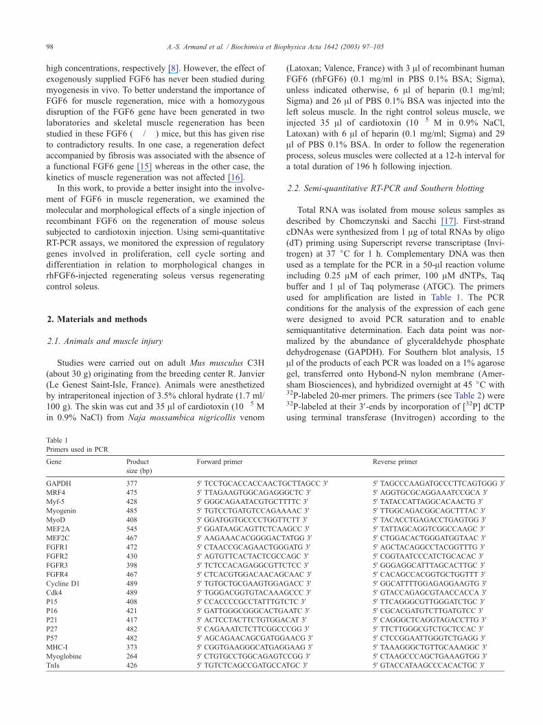
Table 1
Primers used in PCR
Gene Product
size (bp)
Forward primer
GAPDH 377 5VTCCTGCACCACCAACTGMRF4 475 5VTTAGAAGTGGCAGAGGMyf-5 428 5VGGGCAGAATACGTGCTTMyogenin 485 5VTGTCCTGATGTCCAGAAMyoD 408 5VGGATGGTGCCCCTGGTTMEF2A 545 5VGGATAAGCAGTTCTCAAMEF2C 467 5VAAGAAACACGGGGACTFGFR1 472 5VCTAACCGCAGAACTGGGFGFR2 430 5VAGTGTTCACTACTCGCCFGFR3 398 5VTCTCCACAGAGGCGTTCFGFR4 467 5VCTCACGTGGACAACAGCycline D1 489 5VTGTGCTGCGAAGTGGAGCdk4 489 5VTGGGACGGTGTACAAAGP15 408 5VCCACCCCGCCTATTTGTP16 421 5VGATTGGGCGGGCACTGAP21 417 5VACTCCTACTTCTGTGGAP27 482 5VCAGAAATCTCTTCGGCCP57 482 5VAGCAGAACAGCGATGGMHC-I 373 5VCGGTGAAGGGCATGAGMyoglobine 264 5VCTGTGCCTGGCAGAGTCTnIs 426 5VTGTCTCAGCCGATGCCA
(Latoxan; Valence, France) with 3 Al of recombinant human
FGF6 (rhFGF6) (0.1 mg/ml in PBS 0.1% BSA; Sigma),
unless indicated otherwise, 6 Al of heparin (0.1 mg/ml;
Sigma) and 26 Al of PBS 0.1% BSA was injected into the
left soleus muscle. In the right control soleus muscle, we
injected 35 Al of cardiotoxin (10� 5 M in 0.9% NaCl,
Latoxan) with 6 Al of heparin (0.1 mg/ml; Sigma) and 29
Al of PBS 0.1% BSA. In order to follow the regeneration
process, soleus muscles were collected at a 12-h interval for
a total duration of 196 h following injection.
2.2. Semi-quantitative RT-PCR and Southern blotting
Total RNA was isolated from mouse soleus samples as
described by Chomczynski and Sacchi [17]. First-strand
cDNAs were synthesized from 1 Ag of total RNAs by oligo
(dT) priming using Superscript reverse transcriptase (Invi-
trogen) at 37 jC for 1 h. Complementary DNA was then
used as a template for the PCR in a 50-Al reaction volume
including 0.25 AM of each primer, 100 AM dNTPs, Taq
buffer and 1 Al of Taq polymerase (ATGC). The primers
used for amplification are listed in Table 1. The PCR
conditions for the analysis of the expression of each gene
were designed to avoid PCR saturation and to enable
semiquantitative determination. Each data point was nor-
malized by the abundance of glyceraldehyde phosphate
dehydrogenase (GAPDH). For Southern blot analysis, 15
Al of the products of each PCR was loaded on a 1% agarose
gel, transferred onto Hybond-N nylon membrane (Amer-
sham Biosciences), and hybridized overnight at 45 jC with32P-labeled 20-mer primers. The primers (see Table 2) were32P-labeled at their 3V-ends by incorporation of [32P] dCTP
using terminal transferase (Invitrogen) according to the
Reverse primer
CTTAGCC 3V 5VTAGCCCAAGATGCCCTTCAGTGGG 3VGCTC 3V 5VAGGTGCGCAGGAAATCCGCA 3VTTC 3V 5VTATACCATTAGGCACAACTG 3VAAC 3V 5VTTGGCAGACGGCAGCTTTAC 3VCTT 3V 5VTACACCTGAGACCTGAGTGG 3VGCC 3V 5VTATTAGCAGGTCGGCCAAGC 3VATGG 3V 5VCTGGACACTGGGATGGTAAC 3VATG 3V 5VAGCTACAGGCCTACGGTTTG 3VAGC 3V 5VCGGTAATCCCATCTGCACAC 3VTCC 3V 5VGGGAGGCATTTAGCACTTGC 3VCAAC 3V 5VCACAGCCACGGTGCTGGTTT 3VACC 3V 5VGGCATTTTGGAGAGGAAGTG 3VCCC 3V 5VGTACCAGAGCGTAACCACCA 3V
CTC 3V 5VTTCAGGGCGTTGGGATCTGC 3VATC 3V 5VCGCACGATGTCTTGATGTCC 3VCAT 3V 5VCAGGGCTCAGGTAGACCTTG 3VCGG 3V 5VTTCTTGGGCGTCTGCTCCAC 3VAACG 3V 5VCTCCGGAATTGGGTCTGAGG 3VGAAG 3V 5VTAAAGGGCTGTTGCAAAGGC 3VCGG 3V 5VCTAAGCCCAGCTGAAAGTGG 3VTGC 3V 5VGTACCATAAGCCCACACTGC 3V
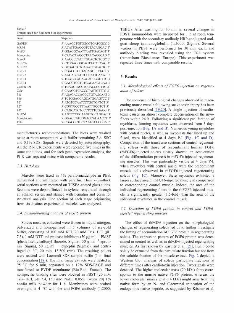
Table 2
Primers used for Southern blot experiments
Gene Sequence
GAPDH 5VAAAGCTGTGGCGTGATGGCC 3VMRF4 5VACATTGAGCGTCTACAGGAC 3VMyf-5 5VGGAGGCAATTAATTGACAGT 3VMyogenin 5VCACATAAGGCTAACACCCAG 3VMyoD 5VAAGGCCACTTGCACTCTGGC 3VMEF2A 5VCTGGAGGGCAGTTATCTCAG 3VMEF2C 5VGTGACTGTGAGATTGCACTG 3VFGFR1 5VCGACCTGCTACAGCTTCGCT 3VFGFR2 5VAGGAGCGCTGCCATTCAAGT 3VFGFR3 5VTGGTCCAGAGCAGCGAGTTG 3VFGFR4 5VGAGGTCCTCTGGCAAGTCAA 3VCycline D1 5VTGAACTACCTGGACCGCTTC 3VCdk4 5VCAAGGTCACCCTAGTGTTTG 3VP15 5VAGAGACCAGGCTGTAGCAAT 3VP16 5VTCTGGAGCAGCATGGAGTCC 3VP21 5VATGTCCAATCCTGGTGATGT 3VP27 5VCGGTGCCTTTAATTGGGTCT 3VP57 5VCAGGATGTGCCTCTTCGAGG 3VMHC-I 5VAGTTCCGCAAGGTGCAGCAC 3VMyoglobine 5VGGAGCATGGGAGCACAACCT 3VTnIs 5VCGACGCTGCTAAGTCCCCGA 3V
A.-S. Armand et al. / Biochimica et Biophysica Acta 1642 (2003) 97–105 99
manufacturer’s recommendations. The blots were washed
twice at room temperature with buffer containing 2� SSC
and 0.1% SDS. Signals were detected by autoradiography.
All the RT-PCR experiments were repeated five times in the
same conditions, and for each gene expression analysis, the
PCR was repeated twice with comparable results.
2.3. Histology
Muscles were fixed in 4% paraformaldehyde in PBS,
dehydrated and infiltrated with paraffin. Then 7-Am-thick
serial sections were mounted on TESPA-coated glass slides.
Sections were deparaffinized in xylene, rehydrated through
an ethanol series, and stained with hemalun and eosin for
structural analysis. One section of each stage originating
from six distinct experimental muscles was analyzed.
2.4. Immunoblotting analysis of FGF6 protein
Soleus muscles collected were frozen in liquid nitrogen,
pulverized and homogenized in 5 volumes of ice-cold
buffer, consisting of 100 mM KCl, 20 mM Tris–HCl (pH
7.5), 1 mM DTT and protease inhibitors (50 Ag ml� 1 PMSF
(phenylmethylsulfonyl fluoride, Sigma), 50 g ml� 1 aproti-
nin (Sigma), 50 Ag ml� 1 leupeptin (Sigma)), and centri-
fuged (4 jC, 20 min, 13,500 rpm). The resulting pellets
were reacted with Laemmli SDS sample buffer (1� final
concentration [18]). The final tissue extracts were heated at
95 jC for 5 min, separated on a 12% SDS-PAGE and
transferred to PVDF membrane (Bio-Rad, France). The
nonspecific binding sites were blocked in PBST (20 mM
Tris–HCl, pH 7.4, 150 mM NaCl, 0.05% Tween 20) 1%
nonfat milk powder for 1 h. Membranes were probed
overnight at 4 jC with the anti-FGF6 antibody (1:2000;
TEBU). After washing for 30 min in several changes in
PBST, immunoblots were incubated for 1 h at room tem-
perature with the secondary antibody HRP-conjugated anti-
goat sheep immunoglobulin (1:5000; Sigma). Several
washes in PBST were performed for 30 min each, and
antibody binding was revealed using the ECL system
(Amersham Biosciences Europe). This experiment was
repeated three times with comparable results.
3. Results
3.1. Morphological effects of FGF6 injection on regener-
ation of soleus
The sequence of histological changes observed in regen-
erating mouse muscle following snake toxin injury has been
previously described [19,20]. A single injection of cardio-
toxin causes an almost complete degeneration of the myo-
fibers within 24 h. Following a significant proliferation of
myoblasts, forming myotubes were observed from 3 days
post-injection (Fig. 1A and B). Numerous young myotubes
with central nuclei, as well as myoblasts that lined up and
fused, were identified at 4 days P-I (Fig. 1C and D).
Comparison of the transverse sections of control regenerat-
ing soleus with those of recombinant human FGF6
(rhFGF6)-injected soleus clearly showed an acceleration
of the differentiation process in rhFGF6-injected regenerat-
ing muscles. This was particularly visible at 4 days P-I,
when myotubes with central nuclei were the predominant
muscle cells observed in rhFGF6-injected regenerating
soleus (Fig. 1C). Moreover, these myotubes exhibited a
larger surface area in rhFGF6-injected muscle in comparison
to corresponding control muscle. Indeed, the area of the
individual regenerating fibers in the rhFGF6-injected mus-
cle is significantly greater (1.5-fold) than the area of the
individual myotubes in the control muscle.
3.2. Detection of FGF6 protein in control and FGF6-
injected regenerating muscles
The effect of rhFGF6 injection on the morphological
changes of regenerating soleus led us to further investigate
the timing of accumulation of FGF6 protein in regenerating
soleus. The expression pattern of FGF6 protein was deter-
mined in control as well as in rhFGF6-injected regenerating
muscles. As first shown by Kastner et al. [21], FGF6 could
solely be extracted from the particulate fraction but not from
the soluble fraction of the muscle extract. Fig. 2 depicts a
Western blot analysis of soleus particulate fractions at
different times after cardiotoxin injection. Two signals were
detected. The higher molecular mass (20 kDa) form corre-
sponds to the murine native FGF6 protein, whereas the
lower molecular mass signal (14 kDa) might arise from the
native form by an N- and C-terminal truncation of the
endogenous native peptide, as suggested by Kastner et al.
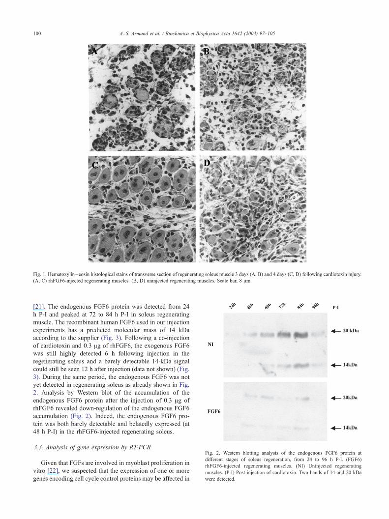
Fig. 2. Western blotting analysis of the endogenous FGF6 protein at
different stages of soleus regeneration, from 24 to 96 h P-I. (FGF6)
rhFGF6-injected regenerating muscles. (NI) Uninjected regenerating
muscles. (P-I) Post injection of cardiotoxin. Two bands of 14 and 20 kDa
were detected.
Fig. 1. Hematoxylin–eosin histological stains of transverse section of regenerating soleus muscle 3 days (A, B) and 4 days (C, D) following cardiotoxin injury.
(A, C) rhFGF6-injected regenerating muscles. (B, D) uninjected regenerating muscles. Scale bar, 8 Am.
A.-S. Armand et al. / Biochimica et Biophysica Acta 1642 (2003) 97–105100
[21]. The endogenous FGF6 protein was detected from 24
h P-I and peaked at 72 to 84 h P-I in soleus regenerating
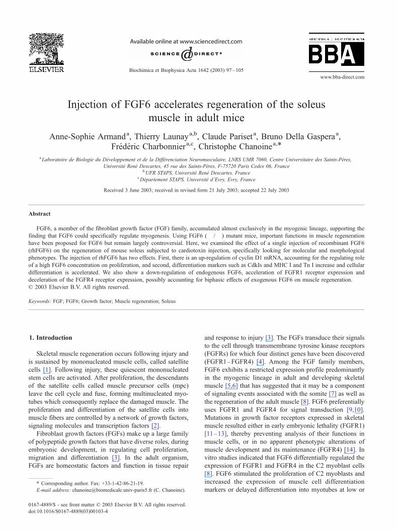
muscle. The recombinant human FGF6 used in our injection
experiments has a predicted molecular mass of 14 kDa
according to the supplier (Fig. 3). Following a co-injection
of cardiotoxin and 0.3 Ag of rhFGF6, the exogenous FGF6
was still highly detected 6 h following injection in the
regenerating soleus and a barely detectable 14-kDa signal
could still be seen 12 h after injection (data not shown) (Fig.
3). During the same period, the endogenous FGF6 was not
yet detected in regenerating soleus as already shown in Fig.
2. Analysis by Western blot of the accumulation of the
endogenous FGF6 protein after the injection of 0.3 Ag of
rhFGF6 revealed down-regulation of the endogenous FGF6
accumulation (Fig. 2). Indeed, the endogenous FGF6 pro-
tein was both barely detectable and belatedly expressed (at
48 h P-I) in the rhFGF6-injected regenerating soleus.
3.3. Analysis of gene expression by RT-PCR
Given that FGFs are involved in myoblast proliferation in
vitro [22], we suspected that the expression of one or more
genes encoding cell cycle control proteins may be affected in
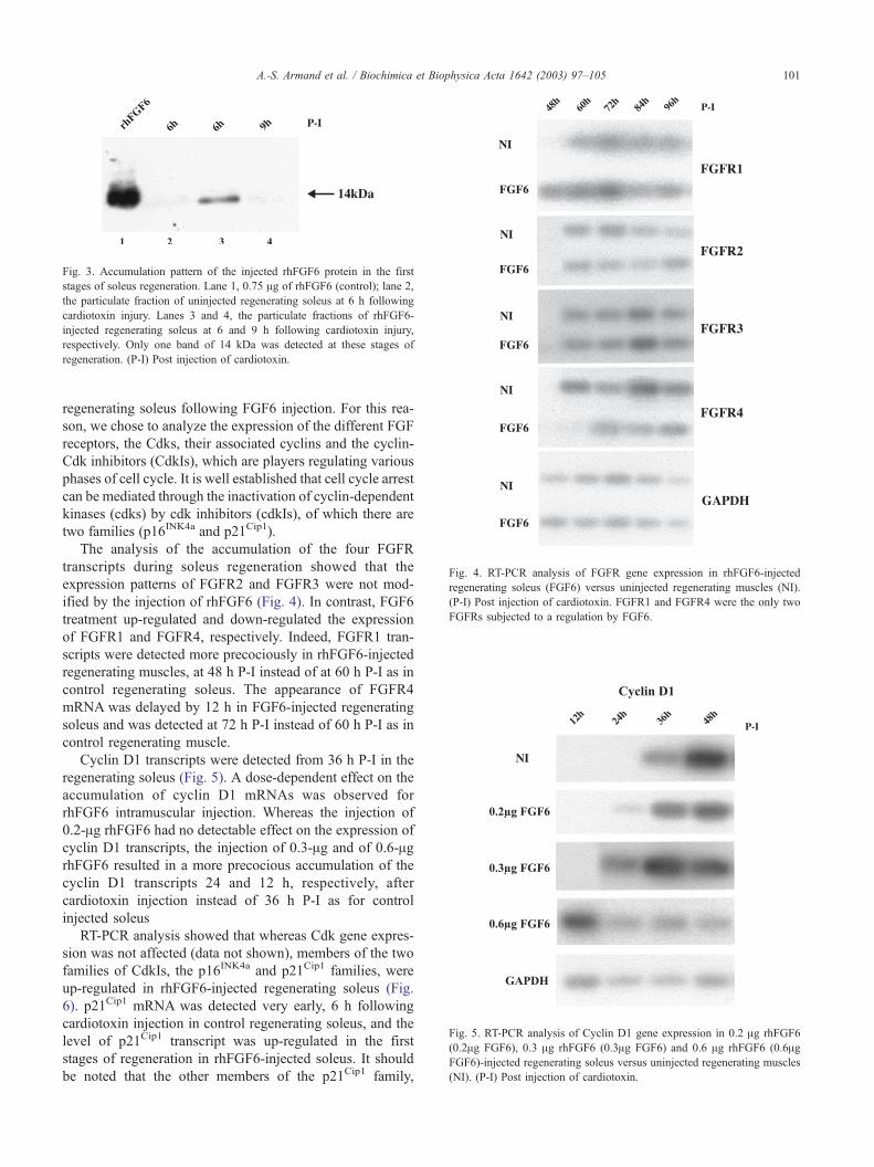
Fig. 4. RT-PCR analysis of FGFR gene expression in rhFGF6-injected
regenerating soleus (FGF6) versus uninjected regenerating muscles (NI).
(P-I) Post injection of cardiotoxin. FGFR1 and FGFR4 were the only two
FGFRs subjected to a regulation by FGF6.
Fig. 5. RT-PCR analysis of Cyclin D1 gene expression in 0.2 Ag rhFGF6
(0.2Ag FGF6), 0.3 Ag rhFGF6 (0.3Ag FGF6) and 0.6 Ag rhFGF6 (0.6AgFGF6)-injected regenerating soleus versus uninjected regenerating muscles
(NI). (P-I) Post injection of cardiotoxin.
Fig. 3. Accumulation pattern of the injected rhFGF6 protein in the first
stages of soleus regeneration. Lane 1, 0.75 Ag of rhFGF6 (control); lane 2,
the particulate fraction of uninjected regenerating soleus at 6 h following
cardiotoxin injury. Lanes 3 and 4, the particulate fractions of rhFGF6-
injected regenerating soleus at 6 and 9 h following cardiotoxin injury,
respectively. Only one band of 14 kDa was detected at these stages of
regeneration. (P-I) Post injection of cardiotoxin.
A.-S. Armand et al. / Biochimica et Biophysica Acta 1642 (2003) 97–105 101
regenerating soleus following FGF6 injection. For this rea-
son, we chose to analyze the expression of the different FGF
receptors, the Cdks, their associated cyclins and the cyclin-
Cdk inhibitors (CdkIs), which are players regulating various
phases of cell cycle. It is well established that cell cycle arrest
can be mediated through the inactivation of cyclin-dependent
kinases (cdks) by cdk inhibitors (cdkIs), of which there are
two families (p16INK4a and p21Cip1).
The analysis of the accumulation of the four FGFR
transcripts during soleus regeneration showed that the
expression patterns of FGFR2 and FGFR3 were not mod-
ified by the injection of rhFGF6 (Fig. 4). In contrast, FGF6
treatment up-regulated and down-regulated the expression
of FGFR1 and FGFR4, respectively. Indeed, FGFR1 tran-
scripts were detected more precociously in rhFGF6-injected
regenerating muscles, at 48 h P-I instead of at 60 h P-I as in
control regenerating soleus. The appearance of FGFR4
mRNA was delayed by 12 h in FGF6-injected regenerating
soleus and was detected at 72 h P-I instead of 60 h P-I as in
control regenerating muscle.
Cyclin D1 transcripts were detected from 36 h P-I in the
regenerating soleus (Fig. 5). A dose-dependent effect on the
accumulation of cyclin D1 mRNAs was observed for
rhFGF6 intramuscular injection. Whereas the injection of
0.2-Ag rhFGF6 had no detectable effect on the expression of
cyclin D1 transcripts, the injection of 0.3-Ag and of 0.6-AgrhFGF6 resulted in a more precocious accumulation of the
cyclin D1 transcripts 24 and 12 h, respectively, after
cardiotoxin injection instead of 36 h P-I as for control
injected soleus
RT-PCR analysis showed that whereas Cdk gene expres-
sion was not affected (data not shown), members of the two
families of CdkIs, the p16INK4a and p21Cip1 families, were
up-regulated in rhFGF6-injected regenerating soleus (Fig.
6). p21Cip1 mRNA was detected very early, 6 h following
cardiotoxin injection in control regenerating soleus, and the
level of p21Cip1 transcript was up-regulated in the first
stages of regeneration in rhFGF6-injected soleus. It should
be noted that the other members of the p21Cip1 family,
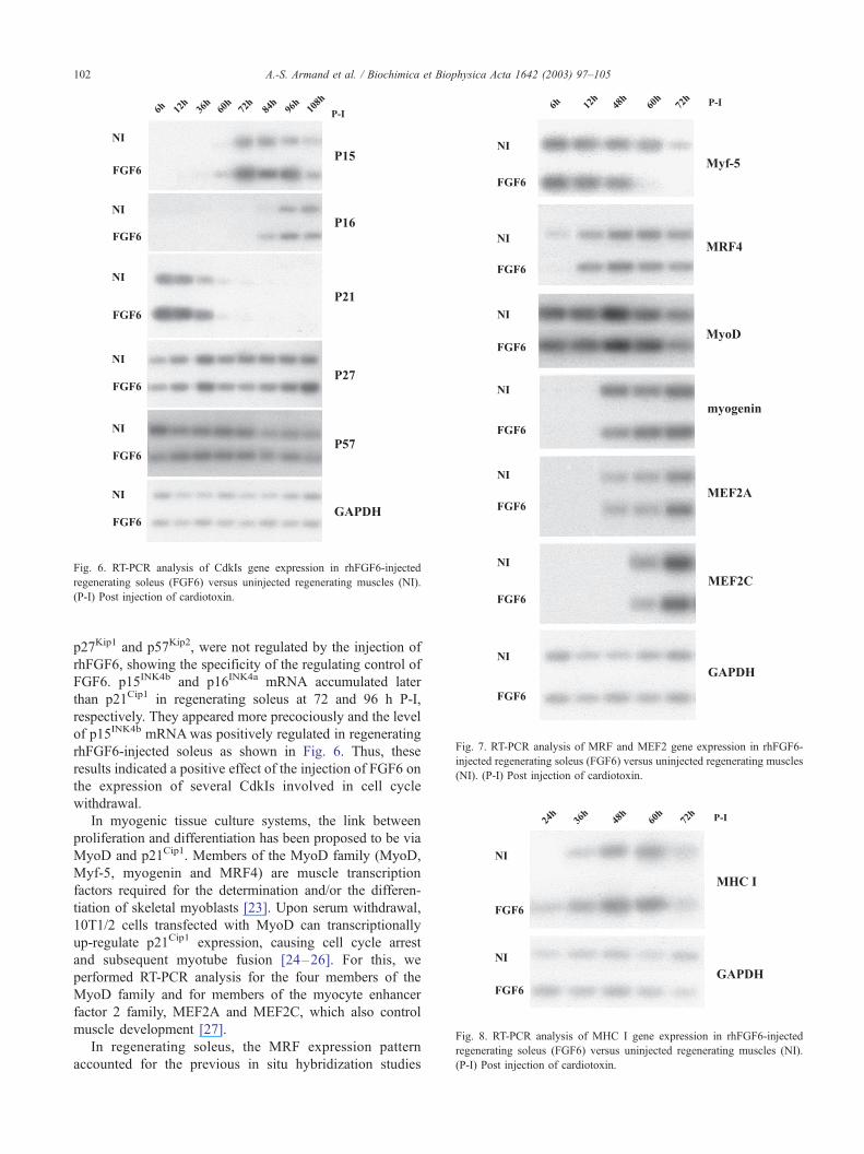
Fig. 7. RT-PCR analysis of MRF and MEF2 gene expression in rhFGF6-
injected regenerating soleus (FGF6) versus uninjected regenerating muscles
(NI). (P-I) Post injection of cardiotoxin.
Fig. 8. RT-PCR analysis of MHC I gene expression in rhFGF6-injected
regenerating soleus (FGF6) versus uninjected regenerating muscles (NI).
(P-I) Post injection of cardiotoxin.
Fig. 6. RT-PCR analysis of CdkIs gene expression in rhFGF6-injected
regenerating soleus (FGF6) versus uninjected regenerating muscles (NI).
(P-I) Post injection of cardiotoxin.
A.-S. Armand et al. / Biochimica et Biophysica Acta 1642 (2003) 97–105102
p27Kip1 and p57Kip2, were not regulated by the injection of
rhFGF6, showing the specificity of the regulating control of
FGF6. p15INK4b and p16INK4a mRNA accumulated later
than p21Cip1 in regenerating soleus at 72 and 96 h P-I,
respectively. They appeared more precociously and the level
of p15INK4b mRNAwas positively regulated in regenerating
rhFGF6-injected soleus as shown in Fig. 6. Thus, these
results indicated a positive effect of the injection of FGF6 on
the expression of several CdkIs involved in cell cycle
withdrawal.
In myogenic tissue culture systems, the link between
proliferation and differentiation has been proposed to be via
MyoD and p21Cip1. Members of the MyoD family (MyoD,
Myf-5, myogenin and MRF4) are muscle transcription
factors required for the determination and/or the differen-
tiation of skeletal myoblasts [23]. Upon serum withdrawal,
10T1/2 cells transfected with MyoD can transcriptionally
up-regulate p21Cip1 expression, causing cell cycle arrest
and subsequent myotube fusion [24–26]. For this, we
performed RT-PCR analysis for the four members of the
MyoD family and for members of the myocyte enhancer
factor 2 family, MEF2A and MEF2C, which also control
muscle development [27].
In regenerating soleus, the MRF expression pattern
accounted for the previous in situ hybridization studies
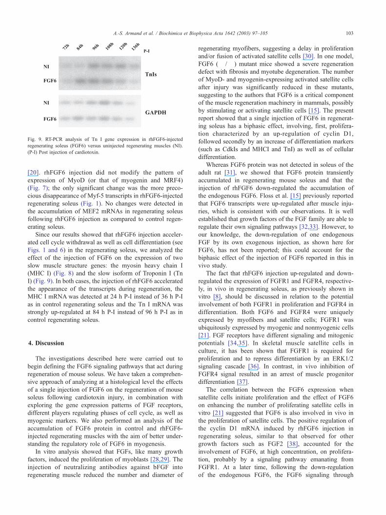
Fig. 9. RT-PCR analysis of Tn I gene expression in rhFGF6-injected
regenerating soleus (FGF6) versus uninjected regenerating muscles (NI).
(P-I) Post injection of cardiotoxin.
A.-S. Armand et al. / Biochimica et Biophysica Acta 1642 (2003) 97–105 103
[20]. rhFGF6 injection did not modify the pattern of
expression of MyoD (or that of myogenin and MRF4)
(Fig. 7); the only significant change was the more preco-
cious disappearance of Myf-5 transcripts in rhFGF6-injected
regenerating soleus (Fig. 1). No changes were detected in
the accumulation of MEF2 mRNAs in regenerating soleus
following rhFGF6 injection as compared to control regen-
erating soleus.
Since our results showed that rhFGF6 injection acceler-
ated cell cycle withdrawal as well as cell differentiation (see
Figs. 1 and 6) in the regenerating soleus, we analyzed the
effect of the injection of FGF6 on the expression of two
slow muscle structure genes: the myosin heavy chain I
(MHC I) (Fig. 8) and the slow isoform of Troponin I (Tn
I) (Fig. 9). In both cases, the injection of rhFGF6 accelerated
the appearance of the transcripts during regeneration, the
MHC I mRNA was detected at 24 h P-I instead of 36 h P-I
as in control regenerating soleus and the Tn I mRNA was
strongly up-regulated at 84 h P-I instead of 96 h P-I as in
control regenerating soleus.
4. Discussion
The investigations described here were carried out to
begin defining the FGF6 signaling pathways that act during
regeneration of mouse soleus. We have taken a comprehen-
sive approach of analyzing at a histological level the effects
of a single injection of FGF6 on the regeneration of mouse
soleus following cardiotoxin injury, in combination with
exploring the gene expression patterns of FGF receptors,
different players regulating phases of cell cycle, as well as
myogenic markers. We also performed an analysis of the
accumulation of FGF6 protein in control and rhFGF6-
injected regenerating muscles with the aim of better under-
standing the regulatory role of FGF6 in myogenesis.
In vitro analysis showed that FGFs, like many growth
factors, induced the proliferation of myoblasts [28,29]. The
injection of neutralizing antibodies against bFGF into
regenerating muscle reduced the number and diameter of
regenerating myofibers, suggesting a delay in proliferation
and/or fusion of activated satellite cells [30]. In one model,
FGF6 (� /� ) mutant mice showed a severe regeneration
defect with fibrosis and myotube degeneration. The number
of MyoD- and myogenin-expressing activated satellite cells
after injury was significantly reduced in these mutants,
suggesting to the authors that FGF6 is a critical component
of the muscle regeneration machinery in mammals, possibly
by stimulating or activating satellite cells [15]. The present
report showed that a single injection of FGF6 in regenerat-
ing soleus has a biphasic effect, involving, first, prolifera-
tion characterized by an up-regulation of cyclin D1,
followed secondly by an increase of differentiation markers
(such as CdkIs and MHCI and TnI) as well as of cellular
differentiation.
Whereas FGF6 protein was not detected in soleus of the
adult rat [31], we showed that FGF6 protein transiently
accumulated in regenerating mouse soleus and that the
injection of rhFGF6 down-regulated the accumulation of
the endogenous FGF6. Floss et al. [15] previously reported
that FGF6 transcripts were up-regulated after muscle inju-
ries, which is consistent with our observations. It is well
established that growth factors of the FGF family are able to
regulate their own signaling pathways [32,33]. However, to
our knowledge, the down-regulation of one endogenous
FGF by its own exogenous injection, as shown here for
FGF6, has not been reported; this could account for the
biphasic effect of the injection of FGF6 reported in this in
vivo study.
The fact that rhFGF6 injection up-regulated and down-
regulated the expression of FGFR1 and FGFR4, respective-
ly, in vivo in regenerating soleus, as previously shown in
vitro [8], should be discussed in relation to the potential
involvement of both FGFR1 in proliferation and FGFR4 in
differentiation. Both FGF6 and FGFR4 were uniquely
expressed by myofibers and satellite cells; FGFR1 was
ubiquitously expressed by myogenic and nonmyogenic cells
[21]. FGF receptors have different signaling and mitogenic
potentials [34,35]. In skeletal muscle satellite cells in
culture, it has been shown that FGFR1 is required for
proliferation and to repress differentiation by an ERK1/2
signaling cascade [36]. In contrast, in vivo inhibition of
FGFR4 signal resulted in an arrest of muscle progenitor
differentiation [37].
The correlation between the FGF6 expression when
satellite cells initiate proliferation and the effect of FGF6
on enhancing the number of proliferating satellite cells in
vitro [21] suggested that FGF6 is also involved in vivo in
the proliferation of satellite cells. The positive regulation of
the cyclin D1 mRNA induced by rhFGF6 injection in
regenerating soleus, similar to that observed for other
growth factors such as FGF2 [38], accounted for the
involvement of FGF6, at high concentration, on prolifera-
tion, probably by a signaling pathway emanating from
FGFR1. At a later time, following the down-regulation
of the endogenous FGF6, the FGF6 signaling through
A.-S. Armand et al. / Biochimica et Biophysica Acta 1642 (2003) 97–105104
FGFR4 could have a positive effect on muscle differenti-
ation, increasing CdkIs as well as myogenic markers such
as slow Tn I and MHC I. In accounting for the specific
myogenic expression of FGF6 and FGFR4 [21], we
suggest that, in regenerating soleus, FGFR1 may regulate
ongoing proliferation of myoblasts, acting in a similar
manner to its action in other cell systems whereas FGFR4
might be involved in a myogenic-specific pathway. The
up-regulation of p15INK4b, p16INK4a and p21Cip1 by
rhFGF6 injection would account for growth arrest and
the subsequent acceleration of myogenic differentiation
observed in regenerating soleus. The p16INK4a family
(p16INK4a, p15INK4b, p18INK4c and p19INK4d) specifically
inhibits cdk4 and cdk6, while the p21Cip1 family (p21Cip1,
p57 Kip2 and p27Kip1) inhibits all cdks involved in the G1/S
transition (reviewed by Sherr and Roberts [39]). Consid-
ering the role of MRFs and MEF2 proteins in muscle
differentiation [23,27], it appears surprising that neither
MRF nor MEF2 were positively regulated in regenerating
soleus following FGF6 injection. These results contrast
with those of Pizette et al. [8] that indicated that FGF6
added at low levels increased the expression of MyoD and
myogenin in culture of C2 myoblasts. This accounted for
the requirement of myogenin in the myoblast-to-myotube
transition, including myoblast fusion and terminal differ-
entiation [23]. Using a MyoD (� /� ) mutant, Megeney et
al. [40] showed that MyoD plays a crucial role in satellite
cell function, the transition from proliferation to differen-
tiation being delayed in satellite cells from mice lacking
MyoD [41]. The more precocious disappearance of Myf-5
transcripts in rhFGF6-injected versus non-injected regener-
ating soleus could reflect the acceleration of the myogenic
differentiation in rhFGF6-injected animals, since we have
previously shown that Myf-5 mRNA transiently accumu-
lated in the first stages of soleus regeneration followed by
a significant decrease in forming myotubes [20]. MRFs
and MEF2 proteins are subjected to posttranslational reg-
ulation and numerous studies point out the crucial role of
the phosphorylations on the functional activity of these
transcription factors [42–44]. For this reason, a better
understanding of the involvement of MRFs as well as
members of the MEF2 family in the FGF6 signaling
pathways should be considered in relation to their phos-
phorylation status.
Among the members of the different growth factor
families (TGFs, IGFs, FGFs) regulating muscle regenera-
tion, to our knowledge, IGF1 was the only growth factor
able to stimulate both mpc proliferation and muscle
hypertrophy (increase in protein content and size of
myofibers) during muscle regeneration [2]. This role was
supported by experiments in which direct infusion of IGF1
into the tibialis anterior muscles of adult rat led to
increased total muscle protein and DNA content, demon-
strating skeletal muscle hypertrophy concomitant with
satellite cell activation [45,46]. Here, using an in vivo
model, we strongly suggested that one member of the FGF
family, FGF6, also has a dual function in proliferation and
muscle differentiation. This study permitted us to identify
clearly a subset of genes regulated by FGF6 during muscle
regeneration. Future analysis of the expression pattern of
these FGF6-regulated genes in regenerating muscles of the
FGF6 (� /� ) mouse in combination with rescue experi-
ments by injection of FGF6 is likely to provide new
insights into the function of FGF6 in adult muscle.
Acknowledgements
Anne-Sophie Armand held a doctoral fellowship from
the Ministere de l’Education Nationale de la Recherche et de
la Technologie (MENRT). We thank R. Cassada for critical
reading of the manuscript.
References
[1] A. Mauro, Satellite cells of skeletal muscle fibers, J. Biophys. Bio-
chem. Cytol. 9 (1961) 493–498.
[2] P. Seale, M.A. Rudnicki, A new look at the origin, function, and
‘‘stem-cell’’ status of muscle satellite cells, Dev. Biol. 218 (2000)
115–124.
[3] D.M. Ornitz, N. Itoh, Fibroblast growth factors, Genome Biol. 2
(2001) 3005–3012.
[4] G. Szebenyi, J.F. Fallon, Fibroblast growth factors as multifunctional
signaling factors, Int. Rev. Cytol. 185 (1999) 45–106.
[5] O. deLapeyriere, V. Ollendorff, J. Planche, M.O. Ott, S. Pizette, F.
Coulier, D. Birnbaum, Expression of the Fgf6 gene is restricted to
developing skeletal muscle in the mouse embryo, Development 118
(1993) 601–611.
[6] J.K. Han, G.R. Martin, Embryonic expression of FGF6 is restricted to
the skeletal muscle lineage, Dev. Biol. 158 (1993) 549–554.
[7] S. Grass, H.H. Arnold, T. Braun, Alterations in somite patterning of
Myf-5-deficient mice: a possible role for FGF-4 and FGF6, Develop-
ment 122 (1996) 141–150.
[8] S. Pizette, F. Coulier, D. Birnbaum, O. deLapeyriere, FGF6 modulates
the expression of fibroblast growth factor receptors and myogenic
genes in muscle cells, Exp. Cell Res. 224 (1996) 143–151.
[9] S. Vainikka, J. Partanen, P. Bellosta, F. Coulier, D. Birnbaum, C.
Basilico, M. Jaye, K. Alitalo, Fibroblast growth factor receptor-4
shows novel features in genomic structure, ligand binding and signal
transduction, EMBO J. 11 (1992) 4273–4280.
[10] D.M. Ornitz, J. Xu, J.S. Colvin, D.G. McEwen, C.A. MacArthur, F.
Coulier, G. Gao, M. Goldfarb, Receptor specificity of the fibroblast
growth factor family, J. Biol. Chem. 271 (1996) 15292–15297.
[11] C.X. Deng, A. Wynshaw-Boris, M.M. Shen, C. Daugherty, D.M.
Ornitz, P. Leder, Murine FGFR-1 is required for early postimplantation
growth and axial organization, Genes Dev. 8 (1994) 3045–3057.
[12] C. Deng, M. Bedford, C. Li, X. Xu, X. Yang, J. Dunmore, P. Leder,
Fibroblast growth factor receptor-1 (FGFR-1) is essential for normal
neural tube and limb development, Dev. Biol. 185 (1997) 42–54.
[13] T.P. Yamaguchi, K. Harpal, M. Henkemeyer, J. Rossant, FGFR-1 is
required for embryonic growth and mesodermal patterning during
mouse gastrulation, Genes Dev. 8 (1994) 3032–3044.
[14] M. Weinstein, X. Xu, K. Ohyama, C.X. Deng, FGFR-3 and FGFR-4
function cooperatively to direct alveogenesis in the murine lung, De-
velopment 125 (1998) 3615–3623.
[15] T. Floss, H.H. Arnold, T. Braun, A role for FGF6 in skeletal muscle
regeneration, Genes Dev. 11 (1997) 2040–2051.
[16] F. Fiore, A. Sebille, D. Birnbaum, Skeletal muscle regeneration is not

A.-S. Armand et al. / Biochimica et Biophysica Acta 1642 (2003) 97–105 105
impaired in Fgf6� /� mutant mice, Biochem. Biophys. Res. Com-
mun. 272 (2000) 138–143.
[17] P. Chomczynski, N. Sacchi, Single-step method of RNA isolation by
acid guanidinium thiocyanate–phenol–chloroform extraction, Anal.
Biochem. 162 (1987) 156–159.
[18] U.K. Laemmli, Cleavage of structural proteins during the assembly of
the head of bacteriophage T4, Nature 227 (1970) 680–685.
[19] R. Couteaux, J.C. Mira, A. d’Albis, Regeneration of muscles after
cardiotoxin injury: I. Cytological aspects, Biol. Cell 62 (1988)
171–182.
[20] T. Launay, A.S. Armand, F. Charbonnier, J.C. Mira, E. Donsez, C.L.
Gallien, C. Chanoine, Expression and neural control of myogenic
regulatory factor genes during regeneration of mouse soleus, J. His-
tochem. Cytochem. 49 (2001) 887–899.
[21] S. Kastner, M.C. Elias, A.J. Rivera, Z. Yablonka-Reuveni, Gene ex-
pression patterns of the fibroblast growth factors and their receptors
during myogenesis of rat satellite cells, J. Histochem. Cytochem. 48
(2000) 1079–1096.
[22] I. Husmann, L. Soulet, J. Gautron, I. Martelly, D. Barritault, Growth
factors in skeletal muscle regeneration, Cytokine Growth Factor Rev.
7 (1996) 249–258.
[23] M.E. Buckingham, Which myogenic factors make muscle? Curr.
Biol. 4 (1994) 61–63.
[24] K. Guo, J. Wang, V. Andres, R.C. Smith, K. Walsh, MyoD-induced
expression of p21 inhibits cyclin-dependent kinase activity upon my-
ocyte terminal differentiation, Mol. Cell. Biol. 15 (1995) 3823–3829.
[25] O. Halevy, B.G. Novitch, D.B. Spicer, S.X. Skapek, J. Rhee, G.J.
Hannon, D. Beach, A.B. Lassar, Correlation of terminal cell cycle
arrest of skeletal muscle with induction of p21 by MyoD, Science
267 (1995) 1018–1021.
[26] S.B. Parker, G. Eichele, P. Zhang, A. Rawls, A.T. Sands, A. Bradley,
E.N. Olson, J.W. Harper, S.J. Elledge, p53-independent expression of
p21Cip1 in muscle and other terminally differentiating cells, Science
267 (1995) 1024–1027.
[27] B.L. Black, E.N. Olson, Transcriptional control of muscle develop-
ment by myocyte enhancer factor-2 (MEF2) proteins, Annu. Rev. Cell
Dev. Biol. 14 (1998) 167–196.
[28] S.M. Sheehan, R.E. Allen, Skeletal muscle satellite cell proliferation
in response to members of the fibroblast growth factor family and
hepatocyte growth factor, J. Cell. Physiol. 181 (1999) 499–506.
[29] Z. Yablonka-Reuveni, M.A. Rudnicki, A.J. Rivera, M. Primig, J.E.
Anderson, P. Natanson, The transition from proliferation to differen-
tiation is delayed in satellite cells from mice lacking MyoD, Dev. Biol.
210 (1999) 440–455.
[30] J.P. Lefaucheur, A. Sebille, Basic fibroblast growth factor promotes in
vivo muscle regeneration in murine muscular dystrophy, Neurosci.
Lett. 202 (1995) 121–124.
[31] K. Sakuma, K. Watanabe, M. Sano, I. Uramoto, T. Totsuka, Differ-
ential adaptation of growth and differentiation factor 8/myostatin,
fibroblast growth factor 6 and leukemia inhibitory factor in over-
loaded, regenerating and denervated rat muscles, Biochim. Biophys.
Acta 1497 (2000) 77–88.
[32] G. Minowada, L.A. Jarvis, C.L. Chi, A. Neubuser, X. Sun, N. Haco-
hen, M.A. Krasnow, G.R. Martin, Vertebrate Sprouty genes are in-
duced by FGF signaling and can cause chondrodysplasia when
overexpressed, Development 126 (1999) 4465–4475.
[33] A. Sasaki, T. Taketomi, T. Wakioka, R. Kato, A. Yoshimura, Identi-
fication of a dominant negative mutant of Sprouty that potentiates
fibroblast growth factor—but not epidermal growth factor—induced
ERK activation, J. Biol. Chem. 276 (2001) 36804–36808.
[34] Z.Q. Wang, M.R. Fung, D.P. Barlow, E.F. Wagner, Regulation of
embryonic growth and lysosomal targeting by the imprinted Igf2/
Mpr gene, Nature 372 (1994) 464–467.
[35] S. Vainikka, V. Joukov, S. Wennstrom, M. Bergman, P.G. Pelicci, K.
Alitalo, Signal transduction by fibroblast growth factor receptor-4
(FGFR-4). Comparison with FGFR-1, J. Biol. Chem. 269 (1994)
18320–18326.
[36] N.C. Jones, Y.V. Fedorov, R.S. Rosenthal, B.B. Olwin, ERK1/2 is
required for myoblast proliferation but is dispensable for muscle gene
expression and cell fusion, J. Cell. Physiol. 186 (2001) 104–115.
[37] I. Marics, F. Padilla, J.F. Guillemot, M. Scaal, C. Marcelle, FGFR4
signaling is a necessary step in limb muscle differentiation, Develop-
ment 129 (2002) 4559–4569.
[38] S.S. Rao, D.S. Kohtz, Positive and negative regulation of D-type
cyclin expression in skeletal myoblasts by basic fibroblast growth
factor and transforming growth factor beta. A role for cyclin D1 in
control of myoblast differentiation, J. Biol. Chem. 270 (1995)
4093–4100.
[39] C.J. Sherr, J.M. Roberts, CDK inhibitors: positive and negative reg-
ulators of G1-phase progression, Genes Dev. 13 (1999) 1501–1512.
[40] L.A. Megeney, B. Kablar, K. Garrett, J.E. Anderson, M.A. Rudnicki,
MyoD is required for myogenic stem cell function in adult skeletal
muscle, Genes Dev. 10 (1996) 1173–1183.
[41] L.A. Sabourin, A. Girgis-Gabardo, P. Seale, A. Asakura, M.A. Rud-
nicki, Reduced differentiation potential of primary MyoD� /� myo-
genic cells derived from adult skeletal muscle, J. Cell Biol. 144
(1999) 631–643.
[42] L. Li, R. Heller-Harrison, M. Czech, E.N. Olson, Cyclic AMP-de-
pendent protein kinase inhibits the activity of myogenic helix-loop-
helix proteins, Mol. Cell. Biol. 12 (1992) 4478–4485.
[43] P.L. Puri, V. Sartorelli, Regulation of muscle regulatory factors by
DNA-binding, interacting proteins, and posttranscriptional modifica-
tions, J. Cell. Physiol. 185 (2001) 155–173.
[44] H. Wu, B. Rothermel, S. Kanatous, P. Rosenberg, F.J. Naya, J.M.
Shelton, K.A. Hutcheson, J.M. DiMaio, E.N. Olson, R. Bassel-Duby,
R.S. Williams, Activation of MEF2 by muscle activity is mediated
through a calcineurin-dependent pathway, EMBO J. 20 (2001)
6414–6423.
[45] G.R. Adams, F. Haddad, The relationships among IGF-1, DNA con-
tent, and protein accumulation during skeletal muscle hypertrophy, J.
Appl. Physiol. 81 (1996) 2509–2516.
[46] G.R. Adams, S.A. McCue, Localized infusion of IGF-I results in
skeletal muscle hypertrophy in rats, J. Appl. Physiol. 84 (1998)
1716–1722.