Initial interaction of apoA-I with ABCA1 impacts in vivo metabolic fate of nascent HDL
12
Initial interaction of apoA-I with ABCA1 impacts in vivo metabolic fate of nascent HDL Anny Mulya, 1, * Ji-Young Lee, 2, * Abraham K. Gebre, * Elena Y. Boudyguina, * Soon-Kyu Chung, * Thomas L. Smith, † Perry L. Colvin, § Xian-Cheng Jiang, ** and John S. Parks 3, * Departments of Pathology* and Orthopaedic Surgery, † Wake Forest University Health Sciences, Winston-Salem, NC; Division of Gerontology, § University of Maryland School of Medicine, and Department of Veterans Affairs and Veterans Affairs Medical Center Baltimore, Geriatric Research, Education and Clinical Center, Baltimore, MD; and Department of Anatomy and Cell Biology,** State University of New York, Downstate Medical Center, Brooklyn, NY Abstract We investigated the in vivo metabolic fate of pre- b HDL particles in human apolipoprotein A-I transgenic (hA-I Tg ) mice. Pre-b HDL tracers were assembled by incuba- tion of [ 125 I]tyramine cellobiose-labeled apolipoprotein A-I (apoA-I) with HEK293 cells expressing ABCA1. Radiola- beled pre-b HDLs of increasing size (pre-b1, -2, -3, and -4 HDLs) were isolated by fast-protein liquid chromatography and injected into hA-I Tg -recipient mice, after which plasma decay, in vivo remodeling, and tissue uptake were moni- tored. Pre-b2, -3, and -4 had similar plasma die-away rates, whereas pre-b1 HDL was removed 7-fold more rapidly. Ra- diolabel recovered in liver and kidney 24 h after tracer injec- tion suggested increased (P , 0.001) liver and decreased kidney catabolism as pre-b HDL size increased. In plasma, pre-b1 and -2 were rapidly (,5 min) remodeled into larger HDLs, whereas pre-b3 and -4 were remodeled into smaller HDLs. Pre-b HDLs were similarly remodeled in vitro with control or LCAT-immunodepleted plasma, but not when incubated with phospholipid transfer protein knockout plasma. Our results suggest that initial interaction of apoA-I with ABCA1 imparts a unique conformation that par- tially determines the in vivo metabolic fate of apoA-I, result- ing in increased liver and decreased kidney catabolism as pre- b HDL particle size increases. —Mulya, A., J-Y. Lee, A. K. Gebre, E. Y. Boudyguina, S-K. Chung, T. L. Smith, P. L. Colvin, X-C. Jiang, and J. S. Parks. Initial interaction of apoA-I with ABCA1 impacts in vivo metabolic fate of nascent HDL. J. Lipid Res. 2008. 49: 2390–2401. Supplementary key words Apolipoprotein A-I • phospholipid transfer protein • lecithin:cholesterol acyltransferase • in vivo catabolism • high density lipoproteins • ABCA1 transporter HDLs are the smallest lipoprotein (7–12 nm in diam- eter) and the most dense (1.063–1.21 g/ml) of the plasma lipoprotein particles and consist of a surface monolayer of protein, phospholipid (PL), and free cholesterol sur- rounding a hydrophobic core of triglyceride (TG) and cholesteryl ester (CE) (1). Apolipoprotein A-I (apoA-I) is the predominant apolipoprotein on HDLs, representing 80–90% of the total protein (2). Interest in understanding HDL metabolism stems from the well-documented fact that plasma HDL cholesterol concentrations are inversely associated with coronary heart disease risk (3). This in- verse association is most likely due to the role of HDLs in reverse cholesterol transport (RCT), a process in which HDLs transport excess cholesterol from peripheral tissue to the liver for secretion into bile and, ultimately, for excre- tion in the feces (4). However, other functions of HDL may result in protection against development of coronary heart disease, including protecting plasma LDLs from oxi- dation (5), decreasing inflammation (6), and improving vascular function (7). HDLs are a heterogeneous mixture of discrete-sized par- ticles that can be separated by density (8), size (9), electro- phoretic mobility (10), and apolipoprotein content (11). Analysis of plasma HDL particles by agarose gel electro- phoresis has shown that most plasma HDLs migrate in the a position (90–95% of total HDL in normal human This work was supported by National Institutes of Health Grants HL-049373 (J.S.P.), HL-054176 (J.S.P.), and AT027820 (J.S.P.), and an American Heart Association Mid-Atlantic Affiliate Pre-doctoral Fellowship 0515420U (A.M.). Manuscript received 9 May 2008 and in revised form 11 June 2008. Published, JLR Papers in Press, June 25, 2008. DOI 10.1194/jlr.M800241-JLR200 Abbreviations: ABCA1, ATP binding cassette transporter A1; apoA-I, apolipoprotein A-I; hA-I Tg , human apolipoprotein A-I trans- genic; CE, cholesteryl ester; FCR, fractional catabolic rate; FPLC, fast-protein liquid chromatography; MPM, mouse peritoneal macro- phage; NDGGE, nondenaturing gradient gel electrophoresis; PL, phospholipid; PLTP, phospholipid transfer protein; RCT, reverse cho- lesterol transport; SR-BI, scavenger receptor class B type I; TC, tyra- mine cellobiose; TG, triglyceride. 1 Present address of A. Mulya: Department of Cell Biology, Lerner Research Institute, Cleveland Clinic Foundation, Cleveland, OH 44195. 2 Present address of J-Y. Lee: Department of Nutrition and Health Sciences, University of Nebraska, Lincoln, NE 68583. 3 To whom correspondence should be addressed. e-mail: [email protected] The online version of this article (available at http://www.jlr.org) contains supplementary data in the form of a figure. Copyright © 2008 by the American Society for Biochemistry and Molecular Biology, Inc. 2390 Journal of Lipid Research Volume 49, 2008 This article is available online at http://www.jlr.org by guest, on July 18, 2016 www.jlr.org Downloaded from
-
Upload
independent -
Category
Documents
-
view
1 -
download
0
Transcript of Initial interaction of apoA-I with ABCA1 impacts in vivo metabolic fate of nascent HDL
Initial interaction of apoA-I with ABCA1 impacts in vivometabolic fate of nascent HDL
Anny Mulya,1,* Ji-Young Lee,2,* Abraham K. Gebre,* Elena Y. Boudyguina,* Soon-Kyu Chung,*Thomas L. Smith,† Perry L. Colvin,§ Xian-Cheng Jiang,** and John S. Parks3,*
Departments of Pathology* and Orthopaedic Surgery,† Wake Forest University Health Sciences,Winston-Salem, NC; Division of Gerontology,§ University of Maryland School of Medicine, and Departmentof Veterans Affairs and Veterans Affairs Medical Center Baltimore, Geriatric Research, Education andClinical Center, Baltimore, MD; and Department of Anatomy and Cell Biology,** State University ofNew York, Downstate Medical Center, Brooklyn, NY
Abstract We investigated the in vivo metabolic fate of pre-b HDL particles in human apolipoprotein A-I transgenic(hA-I Tg) mice. Pre-b HDL tracers were assembled by incuba-tion of [125I]tyramine cellobiose-labeled apolipoprotein A-I(apoA-I) with HEK293 cells expressing ABCA1. Radiola-beled pre-b HDLs of increasing size (pre-b1, -2, -3, and -4HDLs) were isolated by fast-protein liquid chromatographyand injected into hA-I Tg-recipient mice, after which plasmadecay, in vivo remodeling, and tissue uptake were moni-tored. Pre-b2, -3, and -4 had similar plasma die-away rates,whereas pre-b1 HDL was removed 7-fold more rapidly. Ra-diolabel recovered in liver and kidney 24 h after tracer injec-tion suggested increased (P , 0.001) liver and decreasedkidney catabolism as pre-b HDL size increased. In plasma,pre-b1 and -2 were rapidly (,5 min) remodeled into largerHDLs, whereas pre-b3 and -4 were remodeled into smallerHDLs. Pre-b HDLs were similarly remodeled in vitro withcontrol or LCAT-immunodepleted plasma, but not whenincubated with phospholipid transfer protein knockoutplasma. Our results suggest that initial interaction ofapoA-I with ABCA1 imparts a unique conformation that par-tially determines the in vivo metabolic fate of apoA-I, result-ing in increased liver and decreased kidney catabolism aspre-b HDL particle size increases.—Mulya, A., J-Y. Lee,A. K. Gebre, E. Y. Boudyguina, S-K. Chung, T. L. Smith,P. L. Colvin, X-C. Jiang, and J. S. Parks. Initial interactionof apoA-I with ABCA1 impacts in vivo metabolic fate ofnascent HDL. J. Lipid Res. 2008. 49: 2390–2401.
Supplementary key words Apolipoprotein A-I • phospholipid transferprotein • lecithin:cholesterol acyltransferase • in vivo catabolism • highdensity lipoproteins • ABCA1 transporter
HDLs are the smallest lipoprotein (7–12 nm in diam-eter) and the most dense (1.063–1.21 g/ml) of the plasma
lipoprotein particles and consist of a surface monolayerof protein, phospholipid (PL), and free cholesterol sur-rounding a hydrophobic core of triglyceride (TG) andcholesteryl ester (CE) (1). Apolipoprotein A-I (apoA-I) isthe predominant apolipoprotein on HDLs, representing80–90% of the total protein (2). Interest in understandingHDL metabolism stems from the well-documented factthat plasma HDL cholesterol concentrations are inverselyassociated with coronary heart disease risk (3). This in-verse association is most likely due to the role of HDLsin reverse cholesterol transport (RCT), a process in whichHDLs transport excess cholesterol from peripheral tissueto the liver for secretion into bile and, ultimately, for excre-tion in the feces (4). However, other functions of HDLmay result in protection against development of coronaryheart disease, including protecting plasma LDLs from oxi-dation (5), decreasing inflammation (6), and improvingvascular function (7).
HDLs are a heterogeneous mixture of discrete-sized par-ticles that can be separated by density (8), size (9), electro-phoretic mobility (10), and apolipoprotein content (11).Analysis of plasma HDL particles by agarose gel electro-phoresis has shown that most plasma HDLs migrate inthe a position (90–95% of total HDL in normal human
This work was supported by National Institutes of Health Grants HL-049373( J.S.P.), HL-054176 ( J.S.P.), and AT027820 ( J.S.P.), and an American HeartAssociation Mid-Atlantic Affiliate Pre-doctoral Fellowship 0515420U (A.M.).
Manuscript received 9 May 2008 and in revised form 11 June 2008.
Published, JLR Papers in Press, June 25, 2008.DOI 10.1194/jlr.M800241-JLR200
Abbreviations: ABCA1, ATP binding cassette transporter A1;apoA-I, apolipoprotein A-I; hA-I Tg, human apolipoprotein A-I trans-genic; CE, cholesteryl ester; FCR, fractional catabolic rate; FPLC,fast-protein liquid chromatography; MPM, mouse peritoneal macro-phage; NDGGE, nondenaturing gradient gel electrophoresis; PL,phospholipid; PLTP, phospholipid transfer protein; RCT, reverse cho-lesterol transport; SR-BI, scavenger receptor class B type I; TC, tyra-mine cellobiose; TG, triglyceride.
1 Present address of A. Mulya: Department of Cell Biology, LernerResearch Institute, Cleveland Clinic Foundation, Cleveland, OH 44195.
2 Present address of J-Y. Lee: Department of Nutrition and HealthSciences, University of Nebraska, Lincoln, NE 68583.
3 To whom correspondence should be addressed.e-mail: [email protected] online version of this article (available at http://www.jlr.org)
contains supplementary data in the form of a figure.
Copyright © 2008 by the American Society for Biochemistry and Molecular Biology, Inc.
2390 Journal of Lipid Research Volume 49, 2008 This article is available online at http://www.jlr.org
by guest, on July 18, 2016w
ww
.jlr.orgD
ownloaded from
plasma), whereas only 5–10% migrate in the pre-b posi-tion (designated as pre-b HDL) (12). The term pre-bHDL has evolved to describe any lipoprotein that migratesin the pre-b position on agarose gel electrophoresis, in-cluding lipid-free apoA-I, lipid-poor HDL (containing onlya few molecules of lipid), and discoidal HDLs (containingPL, cholesterol, and apoA-I, but no core lipid) (13, 14).Lipid-poor and discoidal HDLs are referred to as nascentHDLs because they must undergo maturation processesthat ultimately result in their conversion into mature,spherical plasma HDLs containing a core of hydrophobiclipid (i.e., CE and TG). Although pre-b HDLs are minorconstituents of plasma HDL, there is an intense interest inhow these particles are formed and catabolized, inasmuchas several studies have suggested that pre-b HDLs are theinitial acceptors of peripheral tissue cholesterol in RCT(15, 16). Despite the potential importance of pre-b HDLsin RCT, little is known about their in vivo metabolism.
We previously isolated a pre-b HDL fraction from plasmaof human apoA-I transgenic (hA-I Tg) mice using a com-bination of anti-human apoA-I immunoaffinity and size-exclusion chromatography and investigated its in vivometabolism in hA-I Tg mice (17), whose plasma HDL sizeheterogeneity resembles that in human plasma (18, 19).The pre-b HDL tracer isolated from hA-I Tg mouse orhuman plasma, which was ,7.1 nm in diameter, exhib-ited two metabolic fates in vivo. About 60% of the pre-b HDL tracer was rapidly removed from plasma andcatabolized by the kidney, whereas the remainder wasrapidly transferred to medium-sized (8.6 nm-diameter)plasma HDL particles, resulting in a slower removal ratefrom plasma and a preferential uptake and catabolism bythe liver instead of the kidney. These results suggestedthat most pre-b HDLs circulating in plasma might notbe nascent particles that are undergoing maturation inplasma but rather are terminal particles ready to be catab-olized and incapable of mediating RCT. This conceptwas further supported by the absence of radiolabeledpre-b HDL in plasma after injection of small or largeplasma HDL tracers into nonhuman primates or hA-I Tg
mice (17, 20).The initial and obligatory step in HDL particle assembly
is the addition of lipid to apoA-I by ABCA1, a member ofthe ATP binding cassette transporter family (21). Thecritical nature of this step for nascent HDL biogenesisand for maintaining plasma HDL cholesterol levels isdemonstrated in Tangier disease subjects and ABCA1knockout mice, both characterized by a near absence ofplasma HDL (22, 23). In a recent study, we showed thatincubation of apoA-I with human embryonic kidney293 cells expressing ABCA1 (HEK293-ABCA1) is necessaryand sufficient to produce at least four discrete-sized pre-bHDLs that can be isolated to apparent homogeneity (14).These pre-b HDL particles bound poorly with ABCA1when added back to ABCA1-expressing cells, suggestingthat other non-ABCA1-mediated pathways must functionto add more lipids and complete the maturation process.The lack of other HDL-modifying proteins [i.e., ATPbinding cassette transporter G1 (ABCG1), phospholipid
transfer protein (PLTP), LCAT, apoM, or scavenger re-ceptor class B type I (SR-BI)] in the HEK293 cells or con-ditioned medium of these cells suggested that the pre-bHDLs were nascent, rudimentary HDLs poised for mat-uration in vivo.
The purpose of the present study was to determine thein vivo plasma decay, interconversion, and tissue sites ofcatabolism for the ABCA1-generated nascent pre-b HDLs.Our results suggest a novel finding that the initial inter-action of apoA-I with ABCA1 in vitro determines, in part,the plasma remodeling and tissue site of catabolism ofthese pre-b HDL particles in vivo.
METHODS
AnimalshA-I Tg mice (line 427) (24) were obtained from Charles River
Laboratories (Wilmington, MA). The mice were housed in theWake Forest University Health Sciences transgenic facility andmaintained on a chow diet. All protocols and procedures wereapproved by the Animal Care and Use Committee of Wake ForestUniversity Health Sciences.
Isolation and iodination of apoA-IHuman HDLs were isolated by sequential ultracentrifugation
of human plasma, and apoA-I was isolated from HDL by GndHCldenaturation (25, 26). The purity of the apoA-I and phosphoruscontent of lipid extract from 1 mg of purified apoA-I were con-firmed by SDS-PAGE and the method of Fiske and Subbarow(27), respectively. ApoA-I preparations contained less than onemolecule of PL per molecule of apoA-I.
ApoA-I was coupled to 125I-radiolabeled tyramine cellobiose(TC) (a generous gift from Dr. Steve Adelman, Wyeth-Ayerst)as previously described (17, 28–30). Briefly, 0.01 mmol TC/mgapoA-I protein was incubated for 10 min with 5 mCi of 125I(carrier-free) in a microreaction vessel coated with 20 mg Iodogen(1,3,4,6-tetrachloro-3a,6a-dephenylglycouril; Pierce ChemicalCo.). The reaction was stopped by transferring the 125I-radio-labeled TC to a second (iodogen-free) reaction vessel contain-ing 10 ml of 0.1 M NaHSO3 and 5 ml of 0.1 M NaI. ApoA-I wascoupled to the [125I]TC with cyanuric chloride (1:1 protein toTC molar ratio) by incubation at room temperature for 30 min.The [125I]TC-apoA-I was then passed over a desalting column(Bio-Rad) to remove free iodine and dialyzed overnight in0.15 M NaCl, 0.01% EDTA, pH 7.4. After removal from dialysis,the tracers were assayed for protein concentration using absor-bance at 280 nm (y 5 1.13 ml/mg), and an aliquot was takenfor radioactivity quantification.
Cell culture and ABCA1 expressionHuman embryonic kidney (HEK)-293 ABCA1 (HEK293-
ABCA1) (31) cells (gift from Dr. Michael Hayden, University ofBritish Columbia) were maintained in DMEM supplementedwith 10% FBS, 2 mM L-glutamine, 100 U/ml penicillin, 100 mg/ml streptomycin, and 50 mg/ml hygromycin B (Invitrogen).HEK293-FlpIn cells were used as negative controls and cul-tured in DMEM complete media in the presence of 50 mg/mlZeocin™. Cells from a rat hepatoma cell line (McA-RH7777) weremaintained in DMEM media supplemented with 10% FBS, 2 mML-glutamine, 100 U/ml penicillin, and 100 mg/ml streptomycin.
Mouse peritoneal macrophages (MPMs) were isolated fromthe peritoneal cavity of C57BL/6 mice 4 days after intraperito-
In vivo metabolism of nascent HDL assembled by ABCA1 2391
by guest, on July 18, 2016w
ww
.jlr.orgD
ownloaded from
neal injection of 1 ml of 10% thioglycolate. The cells obtainedwere washed with Media A (MEM 1 10 mM HEPES) (Cellgro;Mediatech, Inc.), spun at 100 g for 20 min, and plated into 6-wellplates at a density of 1 3 106 cells/well in MEM supplementedwith 10% FBS, 100 U/ml penicillin, 100 mg/ml streptomycin,1% MEM vitamin solution 1003 (Mediatech, Inc.), and 2 mML-glutamine. Cells were washed 2 h later and incubated overnightbefore use in experiments.
ABCA1 expression among the different cell types (HEK293,HEK293-ABCA1, McA-RH7777, MPM) was determined usingWestern blot analysis. Total cell protein was harvested using RIPAlysis buffer (50 mM Tris, 150 mM NaCl, 1% nonidet P-40, 10%sodium cholate, 1 mM EDTA) containing protease inhibitors(1 mM PMSF, 1 mg/ml pepstain, 1 mg/ml leupeptin, 1 mg/mlaprotinin). Cellular proteins (25 mg) were separated on 4–16%SDS-PAGE gels, transferred to nitrocellulose membranes (Schleicherand Schuell BioScience), and incubated for 2 h at room tem-perature with rabbit anti-human/mouse ABCA1 (1:1,000 dilu-tion) (32) or anti-GAPDH monoclonal antibody (1:2,000; SantaCruz 32233), which cross-reacts with human, rat, and mouseGAPDH. The blots were then incubated with HRP-linked anti-rabbit IgG or anti-mouse IgG (Amersham) (1:5,000 dilution) atroom temperature for 1 h. Immunoblots were visualized with achemiluminescent reagent (Pierce), and the chemiluminescencewas captured with an LAS-3000 imaging system (Fujifilm LifeScience) and quantified using Multi Gauge™ software.
Formation of pre-b HDLControl (HEK293-FlpIn) and HEK293-ABCA1 cells were plated
in 53 150 mm dishes, and McA-RH7777 cells were plated in 6-wellplates and grown until they reached 95% confluence. MPMs werecultured for 2 days after isolation before experiments were ini-tiated. All cells (HEK293, McA-RH7777, and MPMs) were washedthree times with serum-free medium and then incubated with10 mg/ml of [125I]TC-apoA-I (specific activity 5 5 3 104 cpm/mg)in serum-free medium for 24 h. The conditioned medium fromcontrol, ABCA1, McA-RH7777 cells, and MPMs was harvested andaliquots analyzed on 4–30% nondenaturing gradient gel electro-phoresis (NDGGE). The remainder of the conditioned media wasconcentrated using Amicon Ultra-10 concentrators and fraction-ated on three Superdex 200 HR fast-protein liquid chromatogra-phy (FPLC) columns (Amersham-Biosciences) connected in seriesand equilibrated with 0.15 M NaCl, 0.01% EDTA, pH 7.4(column buffer). The particles were eluted at a flow rate of0.3 ml/min. Individual fractions were analyzed for 125I radioactiv-ity and the 125I elution profile was plotted. An aliquot of each frac-tion of HEK293-ABCA1 cell conditioned media was analyzed byNDGGE to determine which fractions contained homogeneous-sized nascent HDL particles, after which the homogeneous-sizedfractions were pooled for subsequent analyses.
In vivo turnover studyIn vivo turnover studies were performed with [125I]TC-pre-b
HDL particles as previously described (17) with modifications.Briefly, 1.5–3 3 105 cpm of the radiolabeled tracer was injectedinto the jugular vein of anesthetized recipient hA-I Tg mice. Bloodsamples were obtained by retro-orbital bleeding at 5 min, 30 min,and 1, 2, 3, 5, and 24 h after dose injection to determine theplasma decay of radiolabeled doses. Radioactivity in a 15 mlsample of plasma was quantified using a g counter. Aliquots ofplasma from the various time points were fractionated onNDGGE for 1,100 V-h at 10°C to determine the fractional distri-bution of apoA-I radioactivity. After electrophoresis, gels wereexposed in a phosphorimager cassette and the images weredeveloped and quantified using a Typhoon 8600 (Molecular
Dynamics, Sunnyvale, CA) and ImageQuant software (version5.2). Twenty-four hours after tracer injection, animals were eu-thanized, tissues were harvested and digested with 1 N NaOHovernight at 60°C, and 125I radioactivity was quantified. The re-maining carcasses were digested with 10 g KOH in 150 ml of etha-nol for 2–3 days; the digested carcasses were boiled in a waterbath until the volume of ethanol reached about 30 ml. All re-maining ethanol was counted for 125I radioactivity.
Plasma volume was estimated as 3.5% of body weight, andthe total amount of radiolabel in plasma at each time pointwas determined by multiplying the 125I cpm/ml by plasmavolume. For the data presented in this study, percentage of in-jected dose remaining in plasma at each time point was deter-mined by dividing the amount of total radioactivity in plasmaby the dose injected 3 100%. Percentage of injected dosetrapped in the tissue was calculated by dividing the 125I radioac-tivity in a particular tissue by the dose injected 3 100%. Frac-tional catabolic rate (FCR) values for HDL tracer decay fromwhole plasma and uptake by tissues were performed using Sim-ulation, Analysis, and Modeling (SAAM) software, as describedpreviously (17).
In vitro incubation studyTo investigate the role of LCAT and PLTP in remodeling pre-
b HDL particles, we performed an in vitro incubation studyusing C57BL/6 mouse plasma that had been immunodepletedof LCAT or PLTP2/2 mouse plasma.
LCAT-immunodepleted plasma was prepared as follows. Pre-immune serum and rabbit anti-mouse LCAT antisera were incu-bated at 56°C for 20 min to inactivate LCAT. After inactivation,either preimmune serum or rabbit anti-mouse LCAT antiserum(0.6 of plasma volume) was added to C57Bl/6 mouse plasma andincubated overnight at 4°C with rotation.
LCAT bound to IgG was pelleted by adding Protein A beadsto the incubation mixture, followed by a 2 h incubation at 4°Cand low-speed centrifugation. The supernatant was recoveredand had 1% of the LCAT activity (33) measured in preimmuneserum-treated plasma.
Twenty microliters of LCAT immunodepleted, preimmune-treated plasma, or plasma from wild-type (PLTP1/1) or PLTP2/2
mice was incubated with [125I]TC radiolabeled pre-b1, -2, -3 and -4HDL particles (20,000 cpm) at 4°C for 1 h or 37°C for 5 or 60 min.Subsequently, incubated plasma samples were resolved on 4–30%NDGGE and gels were analyzed by phosphorimager analysis.
In vitro reactivity of pre-b HDL with LCATTo determine the reactivity of individual pre-b HDL particles
with LCAT, pre-b1, -2, -3 and -4 HDLs were prepared using thesame protocol as described above, except that cells were radio-labeled with [3H]cholesterol for 24 h before incubation withapoA-I. Aliquots of pre-b1, -2, -3, and -4 HDL (104 dpm of [3H]cholesterol) were then incubated with 50 ng of purified humanrecombinant LCAT in 0.5 ml of buffer (10 mM Tris, pH 7.4,140 mM NaCl, 0.01% EDTA, 0.07% NaN3, 0.6% BSA, 2 mM b-mercaptoethanol) at 37°C for 1 h as described previously (33,34). After incubation, the samples were lipid-extracted and freecholesterol and CE radiolabels were separated and quantified.
Statistical analysisDifferences among the genotypes of mice were analyzed using
one-way ANOVA, followed by Tukeyʼs multiple comparison test toidentify individual differences. All statistical analyses were per-formed using GraphPad Prism 4 (GraphPad Software, Inc., SanDiego, CA).
2392 Journal of Lipid Research Volume 49, 2008
by guest, on July 18, 2016w
ww
.jlr.orgD
ownloaded from
RESULTS
Nascent pre-b HDL formation by ABCA1 cellsWe previously reported that several heterogeneous-sized
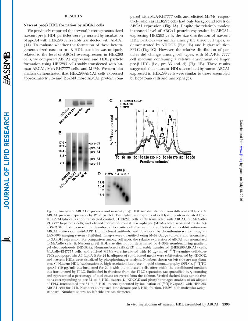
nascent pre-b HDL particles were generated by incubationof apoA-I with HEK293 cells stably transfected with ABCA1(14). To evaluate whether the formation of these hetero-geneous-sized nascent pre-b HDL particles was uniquelyrelated to the level of ABCA1 overexpression in HEK293cells, we compared ABCA1 expression and HDL particleformation using HEK293 cells stably transfected with hu-man ABCA1, McA-RH7777 cells, and MPMs. Western blotanalysis demonstrated that HEK293-ABCA1 cells expressedapproximately 1.5- and 2.5-fold more ABCA1 protein com-
pared with McA-RH7777 cells and elicited MPMs, respec-tively, whereas HEK293 cells had only background levels ofABCA1 expression (Fig. 1A). Despite the relatively modestincreased level of ABCA1 protein expression in ABCA1-expressing HEK293 cells, the size distribution of nascentHDL particles was similar among the three cell types, asdemonstrated by NDGGE (Fig. 1B) and high-resolutionFPLC (Fig. 1C). However, the relative distribution of par-ticles did change among cell types, with McA-RH 7777cell medium containing a relative enrichment of largerpre-b HDL (i.e., pre-b3 and -4) (Fig. 1B). These resultssuggested that nascent HDLs assembled by human ABCA1expressed in HEK293 cells were similar to those assembledby hepatoma cells and macrophages.
Fig. 1. Analysis of ABCA1 expression and nascent pre-b HDL size distribution from different cell types. A:ABCA1 protein expression by Western blot. Twenty-five micrograms of cell lysate protein isolated fromHEK293-FlpIn cells (non-transfected control), HEK293 cells stably transfected with ABCA1, rat McArdle-RH7777 hepatoma cells, and elicited mouse peritoneal macrophages (MPMs) were separated by 4–16%SDS-PAGE. Proteins were then transferred to a nitrocellulose membrane, blotted with rabbit anti-mouseABCA1 antisera or anti-GAPDH monoclonal antibody, and developed by chemiluminescence using anLAS-3000 imaging system (FujiFilm). Images were quantified using Multi Gauge software and normalizedto GAPDH expression. For comparison among cell types, the relative expression of ABCA1 was normalizedto McArdle cells. B: Nascent pre-b HDL size distribution determined by 4–30% nondenaturing gradientgel electrophoresis (NDGGE). Nontransfected (HEK293) and stably transfected (HEK293-ABCA1) cells,McArdle-RH7777 cells, and elicited MPMs were incubated with 10 mg/ml of [125I]tyramine cellobiose(TC)-apolipoprotein A-I (apoA-I) for 24 h. Aliquots of conditioned media were subfractionated by NDGGE,and nascent HDLs were visualized by phosphorimager analysis. Numbers shown on left side are nm diam-eter. C: Nascent HDL fractionation by high-resolution fast-protein liquid chromatography (FPLC). [125I]TC-apoA-I (10 mg/ml) was incubated for 24 h with the indicated cells, after which the conditioned mediumwas fractionated by FPLC. Radiolabel in fractions from the FPLC separation was quantified by g countingand represented a percentage of total count recovered from the column. Vertical dashed lines denote frac-tions corresponding to pre-b1 to -5 HDL tracers. D: NDGGE and phosphorimager analysis of an aliquotof FPLC-fractionated pre-b1 to -5 HDL tracers generated by incubation of [125I]TC-apoA-I with HEK293-ABCA1 cells for 24 h. Numbers above each lane denote pre-b HDL fraction. HMW, high-molecular-weightstandard. Numbers shown on left side are nm diameter.
In vivo metabolism of nascent HDL assembled by ABCA1 2393
by guest, on July 18, 2016w
ww
.jlr.orgD
ownloaded from
Preparation of nascent pre-b HDL tracerNascent pre-b HDL tracers for in vivo turnover studies
were generated by incubating [125I]tyramine cellobiose-apoA-I ([125I]TC-apoA-I) with HEK293-ABCA1 cells for24 h. ApoA-I was coupled with [125I]TC, a tissue-residualizingcompound, allowing us to determine tissue sites of catabo-lism of the injected doses. Following incubation of [125I]TC-apoA-I with HEK293-ABCA-expressing cells, the condi-tioned medium was fractionated by FPLC, and pre-b HDLelution from the column was monitored by g counting. Asshown in Fig. 1C (black circles), five distinct peaks wereeluted from post-fractionation of ABCA1 cell-conditionedmedium after FPLC. Fractions for each peak were pooled,as indicated by the vertical dashed lines in Fig. 1C, anddesignated as pre-b1, -2, -3, -4, and -5, from the smallestto the largest particle size. Aliquots of each pool weresubjected to NDGGE, and gels were developed using aphosphorimager (Fig. 1D). These results show that fivediscrete-sized pre-b HDLs of apparent homogeneity were
isolated by the FPLC procedure. Using two-dimensional gelelectrophoresis (14), we observed that all nascent HDLmigrated in the pre-b position (data not shown). Suffi-cient amounts of pre-b1 and -4 were available for in vivoturnover studies.
Plasma turnover and tissue uptake of nascent pre-b HDLsin hA-I Tg mice
Isolated homogeneous-sized pre-b HDLs (pre-b1, -2, -3,and -4) were injected into hA-I Tg-recipient mice to monitorthe in vivo metabolic fate of the particles. For all tracers,the injected apoA-I tracer mass was about 5 mg per animal(33 105 cpm per mouse/63 104 cpm/mg5 5 mg/mouse)and represented approximately 0.17% of the total plasmaapoA-I mass in hA-I Tg mice (plasma apoA-I pool size 5?3,000 mg) (35). Plasma was drawn from recipient miceat the indicated times to determine the kinetics of turn-over of the tracers in the circulation. Plasma die-awaycurves for each tracer are shown in Fig. 2A. The plasma
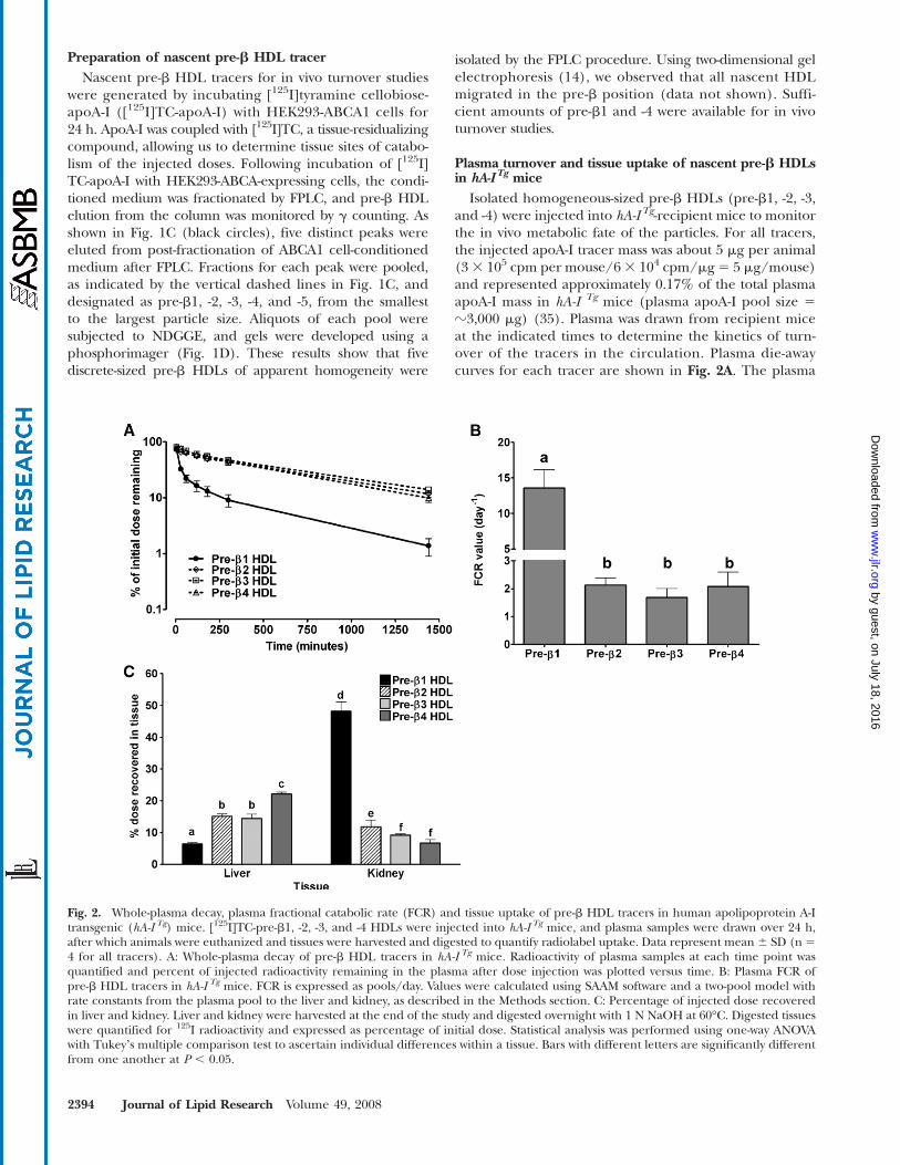
Fig. 2. Whole-plasma decay, plasma fractional catabolic rate (FCR) and tissue uptake of pre-b HDL tracers in human apolipoprotein A-Itransgenic (hA-I Tg) mice. [125I]TC-pre-b1, -2, -3, and -4 HDLs were injected into hA-I Tg mice, and plasma samples were drawn over 24 h,after which animals were euthanized and tissues were harvested and digested to quantify radiolabel uptake. Data represent mean 6 SD (n 54 for all tracers). A: Whole-plasma decay of pre-b HDL tracers in hA-I Tg mice. Radioactivity of plasma samples at each time point wasquantified and percent of injected radioactivity remaining in the plasma after dose injection was plotted versus time. B: Plasma FCR ofpre-b HDL tracers in hA-I Tg mice. FCR is expressed as pools/day. Values were calculated using SAAM software and a two-pool model withrate constants from the plasma pool to the liver and kidney, as described in the Methods section. C: Percentage of injected dose recoveredin liver and kidney. Liver and kidney were harvested at the end of the study and digested overnight with 1 N NaOH at 60°C. Digested tissueswere quantified for 125I radioactivity and expressed as percentage of initial dose. Statistical analysis was performed using one-way ANOVAwith Tukeyʼs multiple comparison test to ascertain individual differences within a tissue. Bars with different letters are significantly differentfrom one another at P , 0.05.
2394 Journal of Lipid Research Volume 49, 2008
by guest, on July 18, 2016w
ww
.jlr.orgD
ownloaded from
die-away of all tracers was similar except that of pre-b1HDL tracer, which was cleared from the plasma signifi-cantly faster than the others. Analysis of plasma kinetic datademonstrated a 7-fold greater FCR for pre-b1 (13.58 65.16 pools/day) compared with pre-b2 (2.13 6 0.53 pools/day), pre-b3 (1.68 6 0.66 pools/day), and pre-b4 (2.07 61.04 pools/day), which were not significantly differentfrom one another (Fig. 2B).
To identify the catabolic sites of the nascent pre-b HDLtracers in hA-ITg mice, tissues were removed from recipientmice 24 h after tracer injection and quantified for radio-label uptake as percentage of injected tracer. The liverand kidney were the only organs that were quantitativelysignificant in the uptake and catabolism of the radiola-beled tracers and together represented .90% of radiolabeluptake (see supplementary Fig. I). Figure 2C summarizesthe percentage of tissue uptake of injected tracers in theliver and kidney. In the liver, pre-b1 HDL tracer uptakewas significantly less than that of pre-b2 and -3, and uptakeof pre-b4 was significantly higher than the other tracers.Thus, there was a trend of increased liver tracer uptakewith increasing initial nascent pre-b HDL size. The oppo-site trend, however, was observed for kidney uptake. Pre-b1 tracer uptake was significantly higher than the othertracers and pre-b3 and -4 uptake was significantly lowerthan pre-b2. The data suggest that kidney uptake of pre-bHDL tracer was inversely related to initial pre-b HDL size.
Plasma turnover and tissue uptake of apoA-I andlipid-poor nascent pre-b1 HDLs in hA-I Tg mice
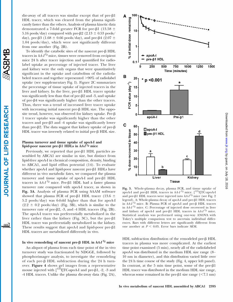
Previously, we reported that pre-b1 HDL particles as-sembled by ABCA1 are similar in size, but distinct fromlipid-free apoA-I in chemical composition, density, bindingto ABCA1, and lipid efflux potential (14). To evaluatewhether apoA-I and lipid-poor nascent pre-b1 HDLs havedifferent in vivo metabolic fates, we compared the plasmaturnover and tissue uptake of apoA-I and pre-b1 HDLtracers in hA-I Tg mice. Pre-b1 HDL had a faster plasmaturnover rate compared with apoA-I tracer, as shown inFig. 3A. Analysis of plasma FCR using SAAM softwareshowed that plasma FCR of pre-b1 HDL tracer (13.6 65.2 pools/day) was 6-fold higher than that for apoA-I(2.2 6 0.2 pools/day) (Fig. 3B), which is similar to theturnover rate of pre-b2, -3, and -4 HDL tracers (Fig. 2B).The apoA-I tracer was preferentially metabolized in theliver rather than the kidney (Fig. 3C), but the pre-b1HDL tracer was preferentially metabolized in the kidney.These results suggest that apoA-I and lipid-poor pre-b1HDL tracers are metabolized differently in vivo.
In vivo remodeling of nascent pre-b HDL in hA-I Tg miceAn aliquot of plasma from each time point of the in vivo
turnover study was fractionated by NDGGE, followed byphosphorimager analysis, to investigate the remodelingof each pre-b HDL subfraction during the 24 h turn-over. Figure 4 shows gel images from a representativemouse injected with [125I]TC-apoA-I and pre-b1, -2, -3 and-4 HDL tracers. Unlike the plasma die-away data (Fig. 2A),
HDL subfraction distribution of the remodeled pre-b HDLtracers in plasma was more complicated. At the earliesttime point examined (5 min), nearly all of the radiolabeledapoA-I was distributed in the medium HDL size range (8–10 nm in diameter), and this distribution varied little overthe 24 h time course of the study (Fig. 4, upper left panel).In contrast, at the 5 min time point, some of the pre-b1HDL tracer was distributed in the medium HDL size range,whereas some remained in the pre-b1 size range (,7.1 nm)
Fig. 3. Whole-plasma decay, plasma FCR, and tissue uptake ofapoA-I and pre-b1 HDL tracers in hA-I Tg mice. [125I]TC-apoA-Iand pre-b1 HDL tracers were injected into hA-I Tg mice (see Fig. 2legend). A: Whole-plasma decay of apoA-I and pre-b1 HDL tracersin hA-I Tg mice. B: Plasma FCR of apoA-I and pre-b HDL tracersin hA-I Tg mice. C: Percentage of injected dose recovered in liverand kidney of apoA-I and pre-b1 HDL tracers in hA-I Tg mice.Statistical analysis was performed using one-way ANOVA withTukeyʼs multiple comparison test to ascertain individual differ-ences. Bars with different letters are significantly different fromone another at P , 0.05. Error bars indicate SEM.
In vivo metabolism of nascent HDL assembled by ABCA1 2395
by guest, on July 18, 2016w
ww
.jlr.orgD
ownloaded from

(Fig. 4, upper middle panel). This pattern of in vivo remo-deling resembled that in a previous study using plasma-isolated pre-b HDL (17). The pre-b2 HDL tracer remodeledinto at least four distinct HDL size ranges between 7 and13 nm in diameter, three of which were larger than the in-jected tracer (Fig. 4, upper right panel). In contrast, the pre-b3 HDL tracer was distributed into three distinct size rangesbetween 7 and 10.4 nm in diameter, one of which was similarin size to the injected pre-b3 HDL (Fig. 4, lower left panel).Pre-b4 HDL tracer was distributed into three smaller HDLparticles, ,10 nm in diameter, all of which were smallerthan the original pre-b4 HDL tracer (Fig. 4, lower rightpanel). Thus, even though the whole-plasma decay forpre-b2, -3, and -4 HDL tracers was similar, the remodelingof these tracers in plasma was distinctly different, withapoA-I and pre-b1 and -2 remodeled into larger particlesand pre-b3 and -4 remodeled into smaller particles.
Using phosphorimager analysis of NDGGE data similarto those in Fig. 4, we quantified the amount of radiolabelassociated with particles,7.1 nm in diameter and particlesin the medium HDL size range (8–10 nm) for each timepoint of the apoA-I and pre-b1 HDL tracer turnover stud-ies (using ImageQuant Software), followed by analysiswith the SAAM II program. Five minutes after injection,62.6% of the pre-b1 tracer remained as pre-b1 (,7.1 nm),and was removed from the circulation at an extraordinarilyrapid rate (FCR 92.92 6 5.85 pools/day); the remaining22.8% of pre-b1 tracer was remodeled to medium-sizedHDLs, which were removed from circulation much moreslowly (FCR 7.76 6 4.75 pools/day). Additional analysessuggested that as much as 97.7% of radioactivity found in
kidney of animals injected with pre-b1 HDL tracers couldbe accounted for by the pre-b1 tracer, which was rapidlyremoved from circulation and not converted to largerparticles during the first 5 min after injection (data notshown). This suggests that pre-b1 HDL particles not imme-diately converted to or associated with medium-sized HDLwithin 5 min after injection are removed rapidly from cir-culation by the kidney. Kinetic analysis also suggested thatradioactivity recovered in the liver of mice injected withpre-b1 HDL tracer was derived almost exclusively frompre-b1 tracer that was remodeled into medium-sized HDL.Similar kinetic results were observed for plasma-isolatedpre-b HDL in our previous study (17).
In vitro remodeling of nascent pre-b HDL tracersTo determine whether HDL-remodeling factors such as
LCAT (36) and PLTP (37, 38) promote nascent pre-bHDLremodeling in vivo, we performed in vitro incubations ofmouse plasma with nascent pre-b HDL tracers. To investi-gate the role of LCAT, we performed an in vitro incubationof nascent pre-b HDL tracers with either control or LCAT-immunodepleted plasma. LCAT was immunodepletedfrom plasma using anti-mouse LCAT antibody, which re-duced LCAT activity by 99% compared with preimmuneserum-incubated plasma (control). The role of PLTP innascent pre-b HDL remodeling was also investigated byincubation of nascent pre-b HDL tracers in wild-type ver-sus PLTP2/2 mouse plasma. Because most of the in vivoremodeling occurred within the first hour after tracer in-jection, each tracer was incubated with plasma for 5 or60 min at 37°C, or 60 min at 4°C as a control. At the
Fig. 4. Size distribution of pre-b HDL tracers after intravenous injection into hA-ITg-recipient mice. Plasmasamples were harvested at the indicated times after [125I]TC-apoA-I and pre-b1, -2, -3, and -4 HDL tracerinjection. Plasma samples (15 ml aliquot) were fractionated on 4–30% NDGGE, and radiolabel distributionwas visualized using a phosphorimager. A phosphorimager result of one representative recipient mousefor each tracer is shown. HMW protein standard (lane 1) and dose (lane 2) are shown for reference. Lanes3–9 contain plasma samples taken at 5 min, 30 min, and 1, 2, 3, 5, and 24 h, respectively.
2396 Journal of Lipid Research Volume 49, 2008
by guest, on July 18, 2016w
ww
.jlr.orgD
ownloaded from
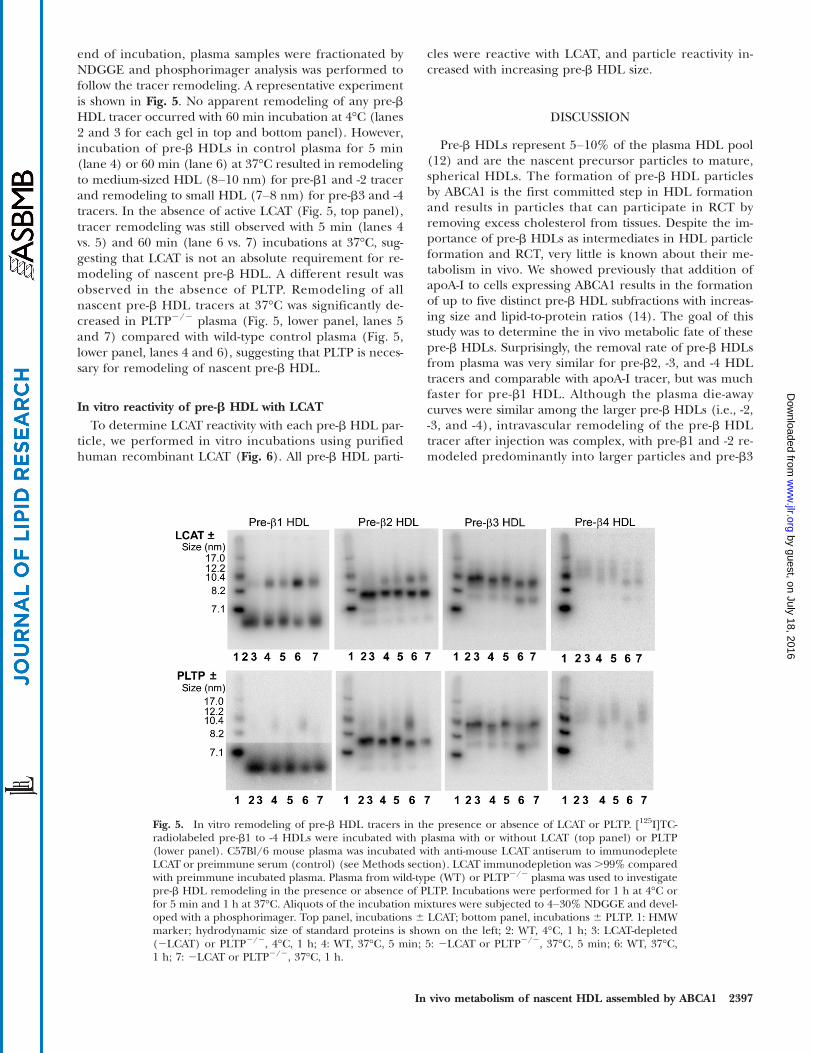
end of incubation, plasma samples were fractionated byNDGGE and phosphorimager analysis was performed tofollow the tracer remodeling. A representative experimentis shown in Fig. 5. No apparent remodeling of any pre-bHDL tracer occurred with 60 min incubation at 4°C (lanes2 and 3 for each gel in top and bottom panel). However,incubation of pre-b HDLs in control plasma for 5 min(lane 4) or 60 min (lane 6) at 37°C resulted in remodelingto medium-sized HDL (8–10 nm) for pre-b1 and -2 tracerand remodeling to small HDL (7–8 nm) for pre-b3 and -4tracers. In the absence of active LCAT (Fig. 5, top panel),tracer remodeling was still observed with 5 min (lanes 4vs. 5) and 60 min (lane 6 vs. 7) incubations at 37°C, sug-gesting that LCAT is not an absolute requirement for re-modeling of nascent pre-b HDL. A different result wasobserved in the absence of PLTP. Remodeling of allnascent pre-b HDL tracers at 37°C was significantly de-creased in PLTP2/2 plasma (Fig. 5, lower panel, lanes 5and 7) compared with wild-type control plasma (Fig. 5,lower panel, lanes 4 and 6), suggesting that PLTP is neces-sary for remodeling of nascent pre-b HDL.
In vitro reactivity of pre-b HDL with LCATTo determine LCAT reactivity with each pre-b HDL par-
ticle, we performed in vitro incubations using purifiedhuman recombinant LCAT (Fig. 6). All pre-b HDL parti-
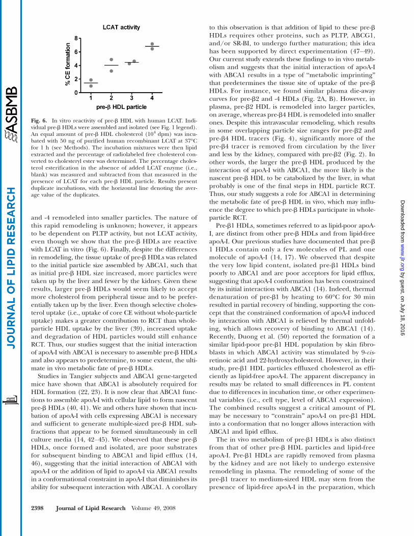
cles were reactive with LCAT, and particle reactivity in-creased with increasing pre-b HDL size.
DISCUSSION
Pre-b HDLs represent 5–10% of the plasma HDL pool(12) and are the nascent precursor particles to mature,spherical HDLs. The formation of pre-b HDL particlesby ABCA1 is the first committed step in HDL formationand results in particles that can participate in RCT byremoving excess cholesterol from tissues. Despite the im-portance of pre-b HDLs as intermediates in HDL particleformation and RCT, very little is known about their me-tabolism in vivo. We showed previously that addition ofapoA-I to cells expressing ABCA1 results in the formationof up to five distinct pre-b HDL subfractions with increas-ing size and lipid-to-protein ratios (14). The goal of thisstudy was to determine the in vivo metabolic fate of thesepre-b HDLs. Surprisingly, the removal rate of pre-b HDLsfrom plasma was very similar for pre-b2, -3, and -4 HDLtracers and comparable with apoA-I tracer, but was muchfaster for pre-b1 HDL. Although the plasma die-awaycurves were similar among the larger pre-b HDLs (i.e., -2,-3, and -4), intravascular remodeling of the pre-b HDLtracer after injection was complex, with pre-b1 and -2 re-modeled predominantly into larger particles and pre-b3
Fig. 5. In vitro remodeling of pre-b HDL tracers in the presence or absence of LCAT or PLTP. [125I]TC-radiolabeled pre-b1 to -4 HDLs were incubated with plasma with or without LCAT (top panel) or PLTP(lower panel). C57Bl/6 mouse plasma was incubated with anti-mouse LCAT antiserum to immunodepleteLCAT or preimmune serum (control) (see Methods section). LCAT immunodepletion was .99% comparedwith preimmune incubated plasma. Plasma from wild-type (WT) or PLTP2/2 plasma was used to investigatepre-b HDL remodeling in the presence or absence of PLTP. Incubations were performed for 1 h at 4°C orfor 5 min and 1 h at 37°C. Aliquots of the incubation mixtures were subjected to 4–30% NDGGE and devel-oped with a phosphorimager. Top panel, incubations 6 LCAT; bottom panel, incubations 6 PLTP. 1: HMWmarker; hydrodynamic size of standard proteins is shown on the left; 2: WT, 4°C, 1 h; 3: LCAT-depleted(2LCAT) or PLTP2/2, 4°C, 1 h; 4: WT, 37°C, 5 min; 5: 2LCAT or PLTP2/2, 37°C, 5 min; 6: WT, 37°C,1 h; 7: 2LCAT or PLTP2/2, 37°C, 1 h.
In vivo metabolism of nascent HDL assembled by ABCA1 2397
by guest, on July 18, 2016w
ww
.jlr.orgD
ownloaded from
and -4 remodeled into smaller particles. The nature ofthis rapid remodeling is unknown; however, it appearsto be dependent on PLTP activity, but not LCAT activity,even though we show that the pre-b HDLs are reactivewith LCAT in vitro (Fig. 6). Finally, despite the differencesin remodeling, the tissue uptake of pre-bHDLs was relatedto the initial particle size assembled by ABCA1, such thatas initial pre-b HDL size increased, more particles weretaken up by the liver and fewer by the kidney. Given theseresults, larger pre-b HDLs would seem likely to acceptmore cholesterol from peripheral tissue and to be prefer-entially taken up by the liver. Even though selective choles-terol uptake (i.e., uptake of core CE without whole-particleuptake) makes a greater contribution to RCT than whole-particle HDL uptake by the liver (39), increased uptakeand degradation of HDL particles would still enhanceRCT. Thus, our studies suggest that the initial interactionof apoA-I with ABCA1 is necessary to assemble pre-b HDLsand also appears to predetermine, to some extent, the ulti-mate in vivo metabolic fate of pre-b HDLs.
Studies in Tangier subjects and ABCA1 gene-targetedmice have shown that ABCA1 is absolutely required forHDL formation (22, 23). It is now clear that ABCA1 func-tions to assemble apoA-I with cellular lipid to form nascentpre-b HDLs (40, 41). We and others have shown that incu-bation of apoA-I with cells expressing ABCA1 is necessaryand sufficient to generate multiple-sized pre-b HDL sub-fractions that appear to be formed simultaneously in cellculture media (14, 42–45). We observed that these pre-bHDLs, once formed and isolated, are poor substratesfor subsequent binding to ABCA1 and lipid efflux (14,46), suggesting that the initial interaction of ABCA1 withapoA-I or the addition of lipid to apoA-I via ABCA1 resultsin a conformational constraint in apoA-I that diminishes itsability for subsequent interaction with ABCA1. A corollary
to this observation is that addition of lipid to these pre-bHDLs requires other proteins, such as PLTP, ABCG1,and/or SR-BI, to undergo further maturation; this ideahas been supported by direct experimentation (47–49).Our current study extends these findings to in vivo metab-olism and suggests that the initial interaction of apoA-Iwith ABCA1 results in a type of “metabolic imprinting”that predetermines the tissue site of uptake of the pre-bHDLs. For instance, we found similar plasma die-awaycurves for pre-b2 and -4 HDLs (Fig. 2A, B). However, inplasma, pre-b2 HDL is remodeled into larger particles,on average, whereas pre-b4 HDL is remodeled into smallerones. Despite this intravascular remodeling, which resultsin some overlapping particle size ranges for pre-b2 andpre-b4 HDL tracers (Fig. 4), significantly more of thepre-b4 tracer is removed from circulation by the liverand less by the kidney, compared with pre-b2 (Fig. 2). Inother words, the larger the pre-b HDL produced by theinteraction of apoA-I with ABCA1, the more likely is thenascent pre-b HDL to be catabolized by the liver, in whatprobably is one of the final steps in HDL particle RCT.Thus, our study suggests a role for ABCA1 in determiningthe metabolic fate of pre-b HDL in vivo, which may influ-ence the degree to which pre-b HDLs participate in whole-particle RCT.
Pre-b1 HDLs, sometimes referred to as lipid-poor apoA-I, are distinct from other pre-b HDLs and from lipid-freeapoA-I. Our previous studies have documented that pre-b1 HDLs contain only a few molecules of PL and onemolecule of apoA-I (14, 17). We observed that despitethe very low lipid content, isolated pre-b1 HDLs bindpoorly to ABCA1 and are poor acceptors for lipid efflux,suggesting that apoA-I conformation has been constrainedby its initial interaction with ABCA1 (14). Indeed, thermaldenaturation of pre-b1 by heating to 60°C for 30 minresulted in partial recovery of binding, supporting the con-cept that the constrained conformation of apoA-I inducedby interaction with ABCA1 is relieved by thermal unfold-ing, which allows recovery of binding to ABCA1 (14).Recently, Duong et al. (50) reported the formation of asimilar lipid-poor pre-b1 HDL population by skin fibro-blasts in which ABCA1 activity was stimulated by 9-cis-retinoic acid and 22-hydroxycholesterol. However, in theirstudy, pre-b1 HDL particles effluxed cholesterol as effi-ciently as lipid-free apoA-I. The apparent discrepancy inresults may be related to small differences in PL contentdue to differences in incubation time, or other experimen-tal variables (i.e., cell type, level of ABCA1 expression).The combined results suggest a critical amount of PLmay be necessary to “constrain” apoA-I on pre-b1 HDLinto a conformation that no longer allows interaction withABCA1 and lipid efflux.
The in vivo metabolism of pre-b1 HDLs is also distinctfrom that of other pre-b HDL particles and lipid-freeapoA-I. Pre-b1 HDLs are rapidly removed from plasmaby the kidney and are not likely to undergo extensiveremodeling in plasma. The remodeling of some of thepre-b1 tracer to medium-sized HDL may stem from thepresence of lipid-free apoA-I in the preparation, which
Fig. 6. In vitro reactivity of pre-b HDL with human LCAT. Indi-vidual pre-b HDLs were assembled and isolated (see Fig. 1 legend).An equal amount of pre-b HDL cholesterol (104 dpm) was incu-bated with 50 ng of purified human recombinant LCAT at 37°Cfor 1 h (see Methods). The incubation mixtures were then lipidextracted and the percentage of radiolabeled free cholesterol con-verted to cholesteryl ester was determined. The percentage choles-terol esterification in the absence of added LCAT enzyme (i.e.,blank) was measured and subtracted from that measured in thepresence of LCAT for each pre-b HDL particle. Results presentduplicate incubations, with the horizontal line denoting the aver-age value of the duplicates.
2398 Journal of Lipid Research Volume 49, 2008
by guest, on July 18, 2016w
ww
.jlr.orgD
ownloaded from
cannot be distinguished or separated from pre-b1 HDL byexisting experimental methodologies. In fact, the mostsensitive measurement of lipid-free apoA-I content in thepre-b1 HDL preparation appears to be the rapid remodel-ing of radiolabel to medium-sized HDLs, because we haveshown here (Fig. 4) and previously (17) that radiolabeledlipid-free apoA-I is transferred quantitatively to medium-sized HDLs within 5 min after injection into recipientmice. Unlike lipid-free apoA-I, which can readily andrapidly transfer to medium-sized HDLs (17, 51), pre-b1HDLs appear unable to do so. We speculate that a fewmolecules of PL in pre-b1 HDL are enough to stabilizethe structure of apoA-I, preventing its interaction withABCA1 and its association or transfer to preexisting plasmaHDL particles. Taken together, our results suggest thatpre-b1 HDLs in plasma are terminal particles that arenot sufficiently lipidated by ABCA1 to prevent their pre-mature removal from the circulation by the kidney. In-sufficient lipidation of apoA-I may occur due to limitedavailability of ABCA1 expression on the cell surface, lim-ited availability of cellular lipid, or both. If our hypoth-esis is correct, any perturbation that results in additionallipidation of apoA-I to produce relatively more pre-b2, -3,or -4 HDLs, rather than pre-b1, will result in a proportionalincrease in catabolism of pre-b HDLs by the liver and mayenhance HDL whole-particle RCT.
Another novel and surprising finding of our study wasthe remodeling of pre-b HDL tracers in plasma after injec-tion. Subsequent studies showed that tracer remodelingalso occurred with in vitro incubation in plasma, dem-onstrating that the remodeling factors were available inplasma. Although the molecular nature of this remodelingprocess is unknown, it requires active PLTP (Fig. 5), whichhas been shown to remodel plasma HDL particles intolarger and smaller HDL products (37, 38). Interestingly,we observed in our in vivo and in vitro studies that pre-b1and -2 HDLs were remodeled into larger particles, whereaspre-b3 and -4 HDLs were remodeled into smaller, average-sized HDLs. The very rapid nature of the remodeling(,5 min) suggested that it might involve pre-b HDLfusion with, or transfer to, preexisting plasma HDL parti-cles. However, the data do not support random exchangeof radiolabeled apoA-I, inasmuch as remodeling occurredin discrete size ranges of HDL (Figs. 4, 5), not through-out the entire range of particle sizes, and required PLTPand 37°C incubation. We also observed that pre-b HDLparticles were reactive with LCAT (Fig. 6); however, LCATactivity was not necessary for remodeling. A similar con-clusion was reached in our previous study, in which in vitroremodeling of plasma isolated pre-b HDLs, which weresimilar in size and composition to the pre-b1 HDLs in thisstudy, occurred unabated in the presence of an LCAT in-hibitor (35). These data do not contradict the essentialrole for LCAT in the maturation of nascent discoidal pre-b HDLs to spherical plasma HDLs through cholesterolesterification (52–55). Rather, they suggest that choles-terol esterification was not a necessary prerequisite forrapid remodeling in our study. Other plasma factors thathave been shown to remodel HDL particles, such as CE
transfer protein and hepatic lipase, were not investigatedin the present study (13). Further studies are required todefine the molecular nature of the rapid remodeling ofABCA1-generated pre-b HDLs in plasma.
To our knowledge, this is the first study to define thein vivo metabolic fate of pre-b HDL particles assembledby ABCA1, and the results fill some critical gaps in ourknowledge regarding HDLmetabolism. It is now clear fromour studies in tissue-specific ABCA1 knockout mice thatnearly all of the plasma HDL pool can be accounted forby liver and intestinal production (32, 57). It is also inter-esting that only these two tissues quantitatively produceapoA-I (56). There is also mounting evidence that assemblyof most HDL particles occurs at the cell surface (45, 58–64)or in a recycling endosomal compartment by ABCA1 (65–70) and not in the secretory pathway of the endoplasmicreticulum and Golgi apparatus (71). Because we haveshown that poorly lipidated apoA-I (i.e., pre-b1 HDL) israpidly catabolized by the kidney, whereas lipid-freeapoA-I rapidly and quantitatively transfers to plasma HDLs(17, 51), newly synthesized apoA-I from hepatocytes orintestinal epithelial cells has several metabolic fates. IfapoA-I escapes lipidation in the secretory pathway andat the cell surface by ABCA1, it is likely to associate withmature HDL particles after entering the circulation (17,51). If lipidation in the secretory pathway or at the cellsurface by ABCA1 is sufficient to form pre-b2, -3, or -4HDLs, these particles will undergo further lipidation bynon-ABCA1-mediated pathways and ultimately be con-verted to mature plasma HDL particles by LCAT in plasma.If, on the other hand, apoA-I is poorly lipidated, withonly a few molecules of PL in the secretory pathway orby ABCA1 at the cell surface, it will not contain enoughlipid to prevent its rapid clearance from plasma by thekidney. Thus, increasing hepatic or intestinal ABCA1 ex-pression in the absence of increased lipid availability orincreasing hepatic or intestinal apoA-I secretion in theabsence of increased ABCA1 expression may not result ina proportional increase in plasma HDL concentration.
The authors thank Dr. Michael Hayden from the University ofBritish Columbia, Vancouver, Canada for providing us withABCA1-expressing cells.
REFERENCES
1. Atkinson, D., and D. M. Small. 1986. Recombinant lipoproteins:implications for structure and assembly of native lipoproteins.Annu. Rev. Biophys. Biophys. Chem. 15: 403–456.
2. Eisenberg, S. 1984. High density lipoprotein metabolism. J. LipidRes. 25: 1017–1058.
3. Gordon, D. J., J. L. Probstfield, R. J. Garrison, J. D. Neaton, W. P.Castelli, J. D. Knoke, D. R. Jacobs, Jr., S. Bangdiwala, and H. A.Tyroler. 1989. High-density lipoprotein cholesterol and cardiovas-cular disease. Four prospective American studies. Circulation. 79:8–15.
4. Fielding, C. J., and P. E. Fielding. 1995. Molecular physiology ofreverse cholesterol transport. J. Lipid Res. 36: 211–228.
5. Nofer, J. R., B. Kehrel, M. Fobker, B. Levkau, G. Assmann, andA. von Eckardstein. 2002. HDL and arteriosclerosis: beyond reversecholesterol transport. Atherosclerosis. 161: 1–16.
In vivo metabolism of nascent HDL assembled by ABCA1 2399
by guest, on July 18, 2016w
ww
.jlr.orgD
ownloaded from
6. Xia, P., M. A. Vadas, K. A. Rye, P. J. Barter, and J. R. Gamble. 1999.High density lipoproteins (HDL) interrupt the sphingosine kinasesignaling pathway. A possible mechanism for protection againstatherosclerosis by HDL. J. Biol. Chem. 274: 33143–33147.
7. Yuhanna, I. S., Y. Zhu, B. E. Cox, L. D. Hahner, S. Osborne-Lawrence, P. Lu, Y. L. Marcel, R. G. Anderson, M. E. Mendelsohn,H. H. Hobbs, et al. 2001. High-density lipoprotein binding to scav-enger receptor-BI activates endothelial nitric oxide synthase. Nat.Med. 7: 853–857.
8. Anderson, D. W., A. V. Nichols, T. M. Forte, and F. T. Lindgren.1977. Particle distribution of human serum high density lipo-proteins. Biochim. Biophys. Acta. 493: 55–68.
9. Blanche, P. J., E. L. Gong, T. M. Forte, and A. V. Nichols. 1981.Characterization of human high-density lipoproteins by gradientgel electrophoresis. Biochim. Biophys. Acta. 665: 408–419.
10. Kunitake, S. T., K. J. La Sala, and J. P. Kane. 1985. ApolipoproteinA-I-containing lipoproteins with pre-beta electrophoretic mobility.J. Lipid Res. 26: 549–555.
11. Cheung, M. C., and J. J. Albers. 1984. Characterization of lipo-protein particles isolated by immunoaffinity chromatography. Par-ticles containing A-I and A-II and particles containing A-I but noA-II. J. Biol. Chem. 259: 12201–12209.
12. OʼConnor, P. M., B. R. Zysow, S. A. Schoenhaus, B. Y. Ishida, S. T.Kunitake, J. M. Naya-Vigne, P. N. Duchateau, R. F. Redberg, S. J.Spencer, S. Mark, et al. 1998. Prebeta-1 HDL in plasma of normo-lipidemic individuals: influences of plasma lipoproteins, age, andgender. J. Lipid Res. 39: 670–678.
13. Rye, K. A., and P. J. Barter. 2004. Formation and metabolism ofprebeta-migrating, lipid-poor apolipoprotein A-I. Arterioscler. Thromb.Vasc. Biol. 24: 421–428.
14. Mulya, A., J. Y. Lee, A. K. Gebre, M. J. Thomas, P. L. Colvin, and J. S.Parks. 2007. Minimal lipidation of pre-beta HDL by ABCA1 resultsin reduced ability to interact with ABCA1. Arterioscler. Thromb. Vasc.Biol. 27: 1828–1836.
15. Castro, G. R., and C. J. Fielding. 1988. Early incorporation ofcell-derived cholesterol into pre-beta-migrating high-density lipo-protein. Biochemistry. 27: 25–29.
16. Huang, Y., A. von Eckardstein, and G. Assmann. 1993. Cell-derivedunesterified cholesterol cycles between different HDLs and LDL forits effective esterification in plasma. Arterioscler. Thromb. 13: 445–458.
17. Lee, J. Y., L. Lanningham-Foster, E. Y. Boudyguina, T. L. Smith, E. R.Young, P. L. Colvin, M. J. Thomas, and J. S. Parks. 2004. Prebetahigh density lipoprotein has two metabolic fates in human apolipo-protein A-I transgenic mice. J. Lipid Res. 45: 716–728.
18. Chajek-Shaul, T., T. Hayek, A. Walsh, and J. L. Breslow. 1991. Ex-pression of the human apolipoprotein A-I gene in transgenic micealters high density lipoprotein (HDL) particle size distribution anddiminishes selective uptake of HDL cholesteryl esters. Proc. Natl.Acad. Sci. USA. 88: 6731–6735.
19. Rubin, E. M., B. Y. Ishida, S. M. Clift, and R. M. Krauss. 1991. Ex-pression of human apolipoprotein A-I in transgenic mice resultsin reduced plasma levels of murine apolipoprotein A-I and theappearance of two new high density lipoprotein size subclasses. Proc.Natl. Acad. Sci. USA. 88: 434–438.
20. Colvin, P. L., E. Moriguchi, P. H. R. Barrett, J. S. Parks, and L. L.Rudel. 1999. Small HDL particles containing two apoA-I moleculesare precursors in vivo to medium and large HDL particles con-taining three and four apoA-I molecules in nonhuman primates.J. Lipid Res. 40: 1782–1792.
21. Yokoyama, S. 2000. Release of cellular cholesterol: molecularmechanism for cholesterol homeostasis in cells and in the body.Biochim. Biophys. Acta. 1529: 231–244.
22. Fredrickson, D. S. 1964. The inheritance of high density lipoproteindeficiency (Tangier disease). J. Clin. Invest. 43: 228–236.
23. McNeish, J., R. J. Aiello, D. Guyot, T. Turi, C. Gabel, C. Aldinger,K. L. Hoppe, M. L. Roach, L. J. Royer, J. de Wet, et al. 2000. Highdensity lipoprotein deficiency and foam cell accumulation in micewith targeted disruption of ATP-binding cassette transporter-1. Proc.Natl. Acad. Sci. USA. 97: 4245–4250.
24. Walsh, A., Y. Ito, and J. L. Breslow. 1989. High levels of humanapolipoprotein A-I in transgenic mice result in increased plasmalevels of small high density lipoprotein (HDL) particles comparableto human HDL3. J. Biol. Chem. 264: 6488–6494.
25. Nichols, A. V., E. L. Gong, P. J. Blanche, T. M. Forte, andD. W. Anderson. 1976. Effects of guanidine hydrochloride onhuman plasma high density lipoproteins. Biochim. Biophys. Acta.446: 226–239.
26. Parks, J. S., and L. L. Rudel. 1979. Isolation and characterization ofhigh density lipoprotein apoproteins in the non-human primate(vervet). J. Biol. Chem. 254: 6716–6723.
27. Fiske, C. H., and Y. Subbarow. 1925. Colorimetric determination ofphosphorus. J. Biol. Chem. 66: 375–400.
28. Pittman, R. C., T. E. Carew, C. K. Glass, S. R. Green, C. A. Taylor,Jr., and A. D. Attie. 1983. A radioiodinated, intracellularly trappedligand for determining the sites of plasma protein degradationin vivo. Biochem. J. 212: 791–800.
29. Glass, C. K., R. C. Pittman, G. A. Keller, and D. Steinberg. 1983.Tissue sites of degradation of apoprotein A-I in the rat. J. Biol. Chem.258: 7161–7167.
30. Huggins, K. W., E. R. Burleson, J. K. Sawyer, K. Kelly, L. L. Rudel,and J. S. Parks. 2000. Determination of the tissue sites responsiblefor the catabolism of large high density lipoprotein in the Africangreen monkey. J. Lipid Res. 41: 384–394.
31. See, R. H., R. A. Caday-Malcolm, R. R. Singaraja, S. Zhou, A.Silverston, M. T. Huber, J. Moran, E. R. James, R. Janoo, J. M.Savill, et al. 2002. Protein kinase A site-specific phosphorylationregulates ATP-binding cassette A1 (ABCA1)-mediated phospholipidefflux. J. Biol. Chem. 277: 41835–41842.
32. Timmins, J. M., J. Y. Lee, E. Boudyguina, K. D. Kluckman, L. R.Brunham, A. Mulya, A. K. Gebre, J. M. Coutinho, P. L. Colvin,T. L. Smith, et al. 2005. Targeted inactivation of hepatic Abca1causes profound hypoalphalipoproteinemia and kidney hyper-catabolism of apoA-I. J. Clin. Invest. 115: 1333–1342.
33. Parks, J. S., A. K. Gebre, and J. W. Furbee. 1999. Lecithin-cholesterolacyltransferase. Assay of cholesterol esterification and phospho-lipase A2 activities. Methods Mol. Biol. 109: 123–131.
34. Chisholm, J. W., A. K. Gebre, and J. S. Parks. 1999. Characteriza-tion of C-terminal histidine-tagged human recombinant lecithin:cholesterol acyltransferase. J. Lipid Res. 40: 1512–1519.
35. Lee, J. Y., J. M. Timmins, A. Mulya, T. L. Smith, Y. Zhu, E. M. Rubin,J. W. Chisholm, P. L. Colvin, and J. S. Parks. 2005. HDLs in apoA-Itransgenic Abca1 knockout mice are remodeled normally in plasmabut are hypercatabolized by the kidney. J. Lipid Res. 46: 2233–2245.
36. Nakamura, Y., L. Kotite, Y. Gan, T. A. Spencer, C. J. Fielding, andP. E. Fielding. 2004. Molecular mechanism of reverse cholesteroltransport: reaction of pre-beta-migrating high-density lipoproteinwith plasma lecithin/cholesterol acyltransferase. Biochemistry. 43:14811–14820.
37. Jauhiainen, M., J. Metso, R. Pahlman, S. Blomqvist, A. van Tol, andC. Ehnholm. 1993. Human plasma phospholipid transfer pro-tein causes high density lipoprotein conversion. J. Biol. Chem. 268:4032–4036.
38. Settasatian, N., M. Duong, L. K. Curtiss, C. Ehnholm, M. Jauhiainen,J. Huuskonen, and K. A. Rye. 2001. The mechanism of the remod-eling of high density lipoproteins by phospholipid transfer protein.J. Biol. Chem. 276: 26898–26905.
39. Trigatti, B., A. Rigotti, and M. Krieger. 2000. The role of the high-density lipoprotein receptor SR-BI in cholesterol metabolism. Curr.Opin. Lipidol. 11: 123–131.
40. Francis, G. A., R. H. Knopp, and J. F. Oram. 1995. Defective removalof cellular cholesterol and phospholipids by apolipoprotein A-I inTangier Disease. J. Clin. Invest. 96: 78–87.
41. Remaley, A. T., U. K. Schumacher, J. A. Stonik, B. D. Farsi, H.Nazih, and H. B. Brewer, Jr. 1997. Decreased reverse cholesteroltransport from Tangier disease fibroblasts. Acceptor specificityand effect of brefeldin on lipid efflux. Arterioscler. Thromb. Vasc. Biol.17: 1813–1821.
42. Duong, P. T., H. L. Collins, M. Nickel, S. Lund-Katz, G. H. Rothblat,and M. C. Phillips. 2006. Characterization of nascent HDL particlesand microparticles formed by ABCA1-mediated efflux of cellularlipids to ApoA-I. J. Lipid Res. 47: 832–843.
43. Liu, L., A. E. Bortnick, M. Nickel, P. Dhanasekaran, P. V. Subbaiah,S. Lund-Katz, G. H. Rothblat, and M. C. Phillips. 2003. Effectsof apolipoprotein A-I on ATP-binding cassette transporter A1-mediated efflux of macrophage phospholipid and cholesterol: for-mation of nascent high density lipoprotein particles. J. Biol. Chem.278: 42976–42984.
44. Krimbou, L., H. H. Hajj, S. Blain, S. Rashid, M. Denis, M.Marcil, and J. Genest. 2005. Biogenesis and speciation of nascentapoA-I-containing particles in various cell lines. J. Lipid Res. 46:1668–1677.
45. Maric, J., R. S. Kiss, V. Franklin, and Y. L. Marcel. 2005. Intracellu-lar lipidation of newly synthesized apolipoprotein A-I in primarymurine hepatocytes. J. Biol. Chem. 280: 39942–39949.
2400 Journal of Lipid Research Volume 49, 2008
by guest, on July 18, 2016w
ww
.jlr.orgD
ownloaded from
46. Denis, M., B. Haidar, M. Marcil, M. Bouvier, L. Krimbou, andJ. Genest, Jr. 2004. Molecular and cellular physiology of apolipo-protein A-I lipidation by the ATP-binding cassette transporter A1(ABCA1). J. Biol. Chem. 279: 7384–7394.
47. Gelissen, I. C., M. Harris, K. A. Rye, C. Quinn, A. J. Brown, M.Kockx, S. Cartland, M. Packianathan, L. Kritharides, and W. Jessup.2006. ABCA1 and ABCG1 synergize to mediate cholesterol exportto apoA-I. Arterioscler. Thromb. Vasc. Biol. 26: 534–540.
48. Vaughan, A. M., and J. F. Oram. 2006. ABCA1 and ABCG1 orABCG4 act sequentially to remove cellular cholesterol and generatecholesterol-rich HDL. J. Lipid Res. 47: 2433–2443.
49. Oram, J. F., G. Wolfbauer, A. M. Vaughan, C. Tang, and J. J. Albers.2003. Phospholipid transfer protein interacts with and stabilizesATP-binding cassette transporter A1 and enhances cholesterol ef-flux from cells. J. Biol. Chem. 278: 52379–52385.
50. Duong, P. T., G. L. Weibel, S. Lund-Katz, G. H. Rothblat, and M. C.Phillips. 2008. Characterization and properties of pre{beta}-HDLparticles formed by ABCA1-mediated cellular lipid efflux to apoA-I. J. Lipid Res. 49: 1006–1014.
51. Kee, P., K. A. Rye, J. L. Taylor, P. H. Barrett, and P. J. Barter. 2002.Metabolism of apoA-I as lipid-free protein or as component ofdiscoidal and spherical reconstituted HDLs: studies in wild-typeand hepatic lipase transgenic rabbits. Arterioscler. Thromb. Vasc. Biol.22: 1912–1917.
52. Ishida, B. Y., D. Albee, and B. Paigen. 1990. Interconversion ofprebeta-migrating lipoproteins containing apolipoprotein A-I andHDL. J. Lipid Res. 31: 227–236.
53. Miida, T., M. Kawano, C. J. Fielding, and P. E. Fielding. 1992.Regulation of the concentration of pre beta high-density lipoproteinin normal plasma by cell membranes and lecithin-cholesterol acyl-transferase activity. Biochemistry. 31: 11112–11117.
54. Kunitake, S. T., C. M. Mendel, and L. K. Hennessy. 1992. Inter-conversion between apolipoprotein A-I-containing lipoproteinsof pre-beta and alpha electrophoretic mobilities. J. Lipid Res. 33:1807–1816.
55. Babiak, J., H. Tamachi, F. L. Johnson, J. S. Parks, and L. L. Rudel.1986. Lecithin:cholesterol acyltransferase-induced modifications ofliver perfusate discoidal high density lipoproteins from Africangreen monkeys. J. Lipid Res. 27: 1304–1317.
56. Brunham, L. R., J. K. Kruit, J. Iqbal, C. Fievet, J. M. Timmins,T. D. Pape, B. A. Coburn, N. Bissada, B. Staels, A. K. Groen, et al.2006. Intestinal ABCA1 directly contributes to HDL biogenesisin vivo. J. Clin. Invest. 116: 1052–1062.
57. Dixon, J. L., and H. N. Ginsberg. 1992. Hepatic synthesis of lipo-proteins and apolipoproteins. Semin. Liver Dis. 12: 364–372.
58. McCall, M. R., T. M. Forte, and V. G. Shore. 1988. Heterogeneity ofnascent high density lipoproteins secreted by the hepatoma-derivedcell line, Hep G2. J. Lipid Res. 29: 1127–1137.
59. Chisholm, J. W., E. R. Burleson, G. S. Shelness, and J. S. Parks. 2002.ApoA-I secretion from HepG2 cells: evidence for the secretion of
both lipid-poor apoA-I and intracellularly assembled nascentHDL. J. Lipid Res. 43: 36–44.
60. Tsujita, M., C. A. Wu, S. Abe-Dohmae, S. Usui, M. Okazaki, andS. Yokoyama. 2005. On the hepatic mechanism of HDL assemblyby the ABCA1/apoA-I pathway. J. Lipid Res. 46: 154–162.
61. Kiss, R. S., D. C. McManus, V. Franklin, W. L. Tan, A. McKenzie,G. Chimini, and Y. L. Marcel. 2003. The lipidation by hepatocytesof human apolipoprotein A-I occurs by both ABCA1-dependentand -independent pathways. J. Biol. Chem. 278: 10119–10127.
62. Denis, M., Y. D. Landry, and X. Zha. 2008. ATP-binding cassette A1-mediated lipidation of apolipoprotein A-I occurs at the plasmamembrane and not in the endocytic compartments. J. Biol. Chem.283: 16178–16186.
63. Faulkner, L. E., S. E. Panagotopulos, J. D. Johnson, L. A. Woollett,D. Y. Hui, S. R. Witting, J. N. Maiorano, and W. S. Davidson. 2008.An analysis of the role of a retroendocytosis pathway in ABCA1-mediated cholesterol efflux from macrophages. J. Lipid Res. 49:1322–1332.
64. Vedhachalam, C., A. B. Ghering, W. S. Davidson, S. Lund-Katz, G. H.Rothblat, and M. C. Phillips. 2007. ABCA1-induced cell surface bind-ing sites for apoA-I. Arterioscler. Thromb. Vasc. Biol. 27: 1603–1609.
65. Schmitz, G., H. Robenek, U. Lohmann, and G. Assmann. 1985.Interaction of high density lipoproteins with cholesteryl ester-ladenmacrophages: biochemical and morphological characterization ofcell surface receptor binding, endocytosis and resecretion of highdensity lipoproteins by macrophages. EMBO J. 4: 613–622.
66. Neufeld, E. B., J. A. Stonik, S. J. Demosky, Jr., C. L. Knapper, C. A.Combs, A. Cooney, M. Comly, N. Dwyer, J. Blanchette-Mackie, A. T.Remaley, et al. 2004. The ABCA1 transporter modulates late endo-cytic trafficking: insights from the correction of the genetic defectin Tangier disease. J. Biol. Chem. 279: 15571–15578.
67. Smith, J. D., C. Waelde, A. Horwitz, and P. Zheng. 2002. Evaluationof the role of phosphatidylserine translocase activity in ABCA1-mediated lipid efflux. J. Biol. Chem. 277: 17797–17803.
68. Takahashi, Y., and J. D. Smith. 1999. Cholesterol efflux to apo-lipoprotein AI involves endocytosis and resecretion in a calcium-dependent pathway. Proc. Natl. Acad. Sci. USA. 96: 11358–11363.
69. Hassan, H. H., D. Bailey, D. Y. D. Lee, I. Iatan, A. Hafiane, I. Ruel, L.Krimbou, and J. Genest. 2008. Quantitative analysis of ABCA1-dependent compartmentalization and trafficking of apolipoproteinA-I: Implications for determining cellular kinetics of nascent highdensity lipoprotein biogenesis. J. Biol. Chem. 283: 11164–11175.
70. Lorenzi, I., A. von Eckardstein, C. Cavelier, S. Radosavljevic,and L. Rohrer. 2008. Apolipoprotein A-I but not high-density lipo-proteins are internalised by RAW macrophages: roles of ATP-binding cassette transporter A1 and scavenger receptor BI. J. Mol.Med. 86: 171–183.
71. Hamilton, R. L., A. Moorehouse, and R. J. Havel. 1991. Isolationand properties of nascent lipoproteins from highly purified rathepatocytic Golgi fractions. J. Lipid Res. 32: 529–543.
In vivo metabolism of nascent HDL assembled by ABCA1 2401
by guest, on July 18, 2016w
ww
.jlr.orgD
ownloaded from