Effects of FSH on testicular mRNA transcript levels in the hypogonadal mouse
Upload
independentCategory
view
4download
0
A
pvomcScAadhp(dhmdt©
K
P
0
Small Ruminant Research 78 (2008) 87–95
Available online at www.sciencedirect.com
Influence of different concentrations of LH and FSH on in vitrocaprine primordial ovarian follicle development
M.V.A. Saraiva a,∗, J.J.H. Celestino a, R.N. Chaves a, F.S. Martins a, J.B. Bruno a,I.B. Lima Verde a, M.H.T. Matos a, G.M. Silva a, E.P. Porfirio b, S.N. Báo b,
C.C. Campello a, J.R.V. Silva a, J.R. Figueiredo a
a Faculty of Veterinary Medicine, LAMOFOPA, PPGCV, State University of Ceara, Fortaleza, CE, Brazilb Laboratory of Electron Microscopy, Department of Cell Biology, University of Brasilia, Brasilia, DF, Brazil
Received 19 June 2007; received in revised form 26 May 2008; accepted 27 May 2008Available online 11 July 2008
bstract
The roles of gonadotrophins in the regulation of primordial ovarian follicular development remains unclear. The aims of theresent study were to investigate the effects of LH alone or in combination with FSH on the survival, activation and growth of initro caprine primordial follicles, using histology and transmission electron microscopy (TEM). To this end, samples of the caprinevarian cortex were cultured for 1 or 7 days (39 ◦C), in an atmosphere of 5% CO2, in minimum essential medium (MEM—controledium) supplemented with different concentrations of LH (0, 1, 5, 10, 50 or 100 ng/ml—Experiment 1). In Experiment 2, the
ontrol medium was supplemented with FSH (50 ng/ml) and the different concentrations of LH (0, 1, 5, 10 or 50 ng/ml also used).mall samples of non-cultured ovarian tissue, as well as those cultured for 1 or 7 days in a specific medium were processed forlassical histological evaluation and TEM to evaluate the follicular integrity and to calculate the percentage of normal follicles.dditionally, the effects of FSH on the oocyte and follicle diameters of the cultured follicles were evaluated. Results showed that
fter 7 days of culture, the presence of LH (10, 50 or 100 ng/ml) in the culture media significantly increases the percentage ofeveloping follicles (P < 0.05). In addition, fragments cultured in media supplemented with FSH alone or FSH plus LH had aigher percentage of developing follicles after 1 and 7 days of culture, compared to the control (P < 0.05). At the end of cultureeriod, 1 and 5 ng/ml LH increased the follicular diameter, while the addition of 1, 5 or 10 ng/ml LH increased the oocyte diameter
P < 0.05). A combination of FSH with 1 and 5 ng/ml LH increased the follicular diameter. With regard to follicle survival, lowoses of LH (1 ng/ml) maintained follicle viability—similar to day 0 and in the control medium after 7 days of culture. However,igher concentrations of LH (5, 10, 50 and 100 ng/ml) induced atresia in the goat follicles. Culture of the ovarian cortex for 7 days in edium supplemented with FSH alone or FSH plus 1 ng/ml LH keptay 0. In conclusion, it can be said that, the addition of low concentratihe goat follicular ultrastructural integrity, but LH in doses higher tha
2008 Elsevier B.V. All rights reserved.
eywords: Caprine; Primordial follicles; In vitro; LH; FSH
∗ Corresponding author at: Programa de Pós-Graduacão em Ciências Veteriré-Antrais (LAMOFOPA), Universidade Estadual do Ceará (UECE), Av. Pa
Brazil. Tel.: +55 85 3101 9852; fax: +55 85 3101 9840.E-mail address: [email protected] (M.V.A. Saraiva).
921-4488/$ – see front matter © 2008 Elsevier B.V. All rights reserved.doi:10.1016/j.smallrumres.2008.05.008
the ultrastructural characteristics of follicles similar to that ofons of LH (1 ng/ml) combined with or without FSH maintainedn 5 ng/ml induced atresia in in vitro goat preantral follicles.
nárias (PPGCV), Laboratório de Manipulacão de Oócitos e Folículosranjana, 1700, Campus do Itaperi, Fortaleza, CE, CEP 60740-000,

umina
88 M.V.A. Saraiva et al. / Small R1. Introduction
It is well established that gonadotrophins are essen-tial for antral follicular development, but not for thedevelopment of preantral follicles (primordial, primaryand secondary follicles). However, large quantitativechanges in the population of preantral follicles afterhypophysectomy in mice and ewes have provided evi-dence that gonadotrophins do affect the development offollicles at the preantral stages (Dufour et al., 1979). Theeffects of FSH on the preantral follicle development inhumans and domestic species have also been monitored.When Wright et al. (1999) added FSH to cortical sam-ples of human ovaries, the percentage of atretic follicleswas decreased and follicle diameter increased during a15-day culture period. In contrast, the addition of FSHat a rate of 5 ng/ml (Derrar et al., 2000) or graded dosesof FSH (Fortune et al., 1998) had no effect on follicularpopulations during longer culture (7–14 days) periodsof bovine cortical tissue. However, a recent study ofcaprine follicles has shown FSH to be able to promotethe activation of primordial follicles and further growthof activated follicles at a level of 50 ng/ml (Matos et al.,2007). Receptors for FSH are expressed in the granulosacells of the primary follicle (Ulloa-Aguirre et al., 1995;O’Shaughnessy et al., 1997) and more recent reportshave indicated its presence in the oocytes (Méduri etal., 2002).
Regarding LH, much less is known regarding itspotential effects on the growth of the preantral folli-cles. Wu et al. (2000) indicated that in mice LH isneeded for the in vitro development of the preantralfollicles to the antral stage. Large preantral follicles(150–160 �m) developed to antral follicles in the pres-ence of FSH and mice serum. However, the additionof LH together with serum and FSH, was importantin promoting the development of the small preantralfollicles (85–110 �m) to the antral stage. Liu et al.(2002) developed a culture system that allowed thematuration of the preantral follicles in vitro with recom-binant gonadotrophins. Receptors for LH have a morewidespread distribution and are also found in non-gonadal tissues (Reshef et al., 1990; Reiter et al., 1995).In the ovary, LH receptors are found primarily in theinterstitial and thecal cells (McFarland et al., 1989)and only in the mid-follicular phase, do the granu-losa cells acquire LH receptors (Zeleznick and Hillier,1984; Yamoto et al., 1992), together with the preovu-
latory follicle and the corpus luteum (McFarland et al.,1989). The acquisition of LH receptors by the granu-losa cells depends on the actions of FSH and estradioltogether—where FSH induces LH receptors in the gran-nt Research 78 (2008) 87–95
ulosa cells by increasing transcription of the LH receptorgene (Segaloff, 1996).
Although the importance of FSH and LH for antralfollicle development in vivo is well know, the effects ofboth hormones on the primordial follicular growth of thecaprine ovarian cortex during culture has not yet beenevaluated. Thus, the aims of the present study were toinvestigate the effects of LH alone or in combination withFSH on the survival, activation and growth of caprineprimordial follicles cultured in vitro, using histology andtransmission electron microscopy (TEM).
2. Materials and methods
2.1. Source of ovaries
Ovaries (n = 8 for Experiment 1, n = 8 for Experiment 2)from 8 adult non-pregnant mixed-breed goats were collectedat a local slaughterhouse. All animals were cyclic and in goodbody condition. The ovaries were removed, washed and trans-ported in minimum essential medium (MEM), to the laboratorywithin 30 min in thermo flasks containing water at 33 ◦C.
2.2. Culture medium
The control medium used was minimum essentialmedium (Cultilab, Rio de Janeiro, Brazil), supplementedwith ITS (insulin 6.25 �g/ml, transferrin 6.25 �g/ml, andSe 6.25 ng/ml), 0.23 mM pyruvate; 2 mM glutamine; 2 mMhypoxanthine; 1.25 mg/ml BSA, 100 �g/ml penicillin and100 �g/ml streptomycin (Vetec, Rio de Janeiro, Brazil).This control medium (MEM) was supplemented with pFSH(50 ng/ml) and/or porcine LH (1, 5, 10, 50 or 100 ng/ml) (bothprovided by Dr. J.F. Beckers, Liège, Belgium). All chemicalsused in the present study were purchased from Sigma ChemicalCo. (St. Louis, MO, USA), unless otherwise indicated.
2.3. Experimental protocol
In Experiment 1, goat ovaries (n = 8) were collected andstripped of all fatty tissue and ligaments, and cut in half,whereafter the medulla, large antral follicles and corporalutea were removed. Following this, the ovarian cortexwas dissected into 11 tissue samples of approximately3 mm × 3 mm (1 mm thick). One sample was immediatelyfixed for classic histological studies (non-cultured controls)(Matos et al., 2007), while a smaller sample (1 mm3) wasrandomly collected and subsequently fixed for ultrastructural(TEM) examination. The other samples of the ovarian cortexwere individually cultured in vitro in 1 ml of culture mediumsupplemented with different concentrations of porcine LH (1,
5, 10, 50 or 100 ng/ml). To evaluate the interaction betweenFSH and LH (Experiment 2), eight ovaries were collected andthe obtained tissue samples of the ovarian cortex individuallycultured in vitro in 1 ml of culture medium supplemented withpFSH (50 ng/ml) alone or in combination with LH (1, 5, 10
umina
o7dmo
2f
ootiFo(e
lTngealmr
pwcawf
2
ew0wrSccbaaee
2
s
cle development is shown in Table 2. After 1 and 7 daysof culture respectively, all treatments showed a signifi-cant reduction in the percentage of primordial follicles.This was followed by a significant increase in developing
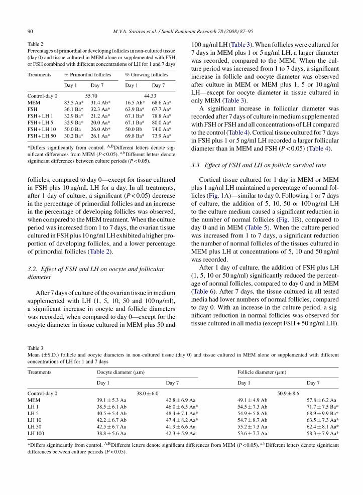
Table 1Percentage of primordial or developing follicles in non-cultured tis-sue (day 0) and tissue cultured in MEM alone or supplemented withdifferent concentrations of LH for 1 and 7 days
Treatments % Primordial follicles % Growing follicles
Day 1 Day 7 Day 1 Day 7
Control-day 0 44.21 55.26MEM 34.2 Aa 30.9 Aa 65.8 Aa 69.1 AaLH 1 34.4 Aa 43.6 Aa 65.6 Aa 56.4 AaLH 5 38.3 Aa 28.6 Aa 61.7 Aa 71.4 AaLH 10 25.5 Aa* 17.4 Ba* 74.5 Aa* 82.6 Ba*LH 50 37.0 Aa 11.8 Bb* 62.9 Ab 88.2 Ba*
M.V.A. Saraiva et al. / Small R
r 50 ng/ml). The culture was performed for a period of 1 ordays (39 ◦C), with 5% CO2 in air, using a 24-well culture
ish. Every 2 days, the culture medium was replaced by freshedium. Each treatment was repeated four times, using the
varies of four different animals for each experiment.
.4. Histological analyses and assessment of in vitroollicular growth
To evaluate the morphology of the caprine follicles after 1r 7 days of culture in both experiments, a small part (1 mm3)f each fragment was randomly removed for transmission elec-ron microscopy (TEM) studies, while the remainder was fixedn Carnoy for 12 h for histological studies (Matos et al., 2007).or each sample of ovarian cortex, 7 �m sections were mountedn slides, stained with periodic acid Schiff and hematoxylinPAS staining system, Sigma, Inc., St. Louis, MO, USA), andxamined by light microscopy (Zeiss, Germany).
The follicles were classified as primordial or developing fol-icles (intermediate, primary or secondary) (Silva et al., 2004).hese follicles were classified individually as histologicallyormal where an intact oocyte was present, surrounded byranulosa cells, well organized in one or more layers. Degen-rated follicles were defined as those with a retracted oocyte,pyknotic nucleus, and/or surrounded by disorganized granu-
osa cells, detached from the basement membrane. From eachedium and culture period, approximately 120 follicles were
andomly selected and evaluated.To evaluate the follicular activation and growth, only intact
rimordial and growing follicles, with a visible oocyte nucleus,ere recorded on day 0 (controls) and after 1 or 7 days of
ulture. Oocyte and follicle diameters were measured with theid of an ocular micrometer (Matos et al., 2007). These valuesere used to assess the effect of the hormonal treatment on
ollicular growth.
.5. Ultrastructural analyses
Ultrastructural analysis was performed according to Matost al. (2007). Briefly, small samples of ovarian cortex tissueere fixed in 2% paraformaldehyde, 2.5% glutaraldehyde, and.1 M sodium cacodylate buffer (pH 7.2). Thereafter, samplesere post-fixed in 1% osmium tetroxide, 0.8% potassium fer-
icyanide and 5 mM CaCl2 in 0.1 M sodium cacodylate buffer.amples were then dehydrated in a graded series of acetoneoncentrations and embedded in Spurr’s epoxy resin. The folli-les, classified as histologically normal in the semi-thin toluidinlue stained sections (3 �m), were subjected to ultrastructuralnalyses. For this purpose, ultra-thin sections (70 nm) were cutnd then contrasted with uranyl acetate and lead citrate, andxamined using a Jeol 1011 (Jeol, Tokyo, Japan) transmissionlectron microscope.
.6. Statistical analysis
Data of follicle and oocyte diameters were analyzedtatistically using the Kolmogorov-Smirnov and Bartlett’s
nt Research 78 (2008) 87–95 89
tests—applied to confirm a normal distribution and homogene-ity of variance, respectively. Analysis of variance was then doneusing the GLM procedure of SAS (1999) and the Dunnett’stest implemented to compare the control groups against eachtreatment (Steel et al., 1997). The Student’s t-test was used tocompare means between 1 and 7 days of culture. Differencesbetween groups were considered to be significant at a confi-dence level of P < 0.05 (results expressed as means ± S.D.).The chi-square test was used to compare the percentage ofviable follicles between the days.
3. Results
3.1. Effects of FSH and LH on primordial follicleactivation
The percentage of primordial and developing folli-cles in the goat ovarian tissue before and after culture inthe media supplemented with LH is set out in Table 1.After 1 day of culture in medium containing 10 ng/mlLH, a significant reduction in the number of primor-dial follicles and a concomitant increase of developingfollicles was observed—when compared to day 0 (non-cultured control). At day 7 of culture, the addition of 10,50 or 100 ng/ml LH significantly (P < 0.05) increased thepercentage of developing follicles in relation to day 0,and follicles cultured in MEM. By increasing the cultureperiod from 1 to 7 days, a higher percentage (P < 0.05) ofdeveloping follicles was recorded in the ovarian tissuecultured in media supplemented with 50 or 100 ng/mlLH.
The effects of both FSH and LH on primordial folli-
LH 100 30.6 Aa 5.0 Bb* 69.4 Ab 95.0 Ba*
*Differs significantly from control. A,BDifferent letters denote sig-nificant differences from MEM (P < 0.05). a,bDifferent letters denotesignificant differences between culture periods (P < 0.05).
90 M.V.A. Saraiva et al. / Small Rumina
Table 2Percentages of primordial or developing follicles in non-cultured tissue(day 0) and tissue cultured in MEM alone or supplemented with FSHor FSH combined with different concentrations of LH for 1 and 7 days
Treatments % Primordial follicles % Growing follicles
Day 1 Day 7 Day 1 Day 7
Control-day 0 55.70 44.33MEM 83.5 Aa* 31.4 Ab* 16.5 Ab* 68.6 Aa*FSH 36.1 Ba* 32.3 Aa* 63.9 Ba* 67.7 Aa*FSH + LH 1 32.9 Ba* 21.2 Aa* 67.1 Ba* 78.8 Aa*FSH + LH 5 32.9 Ba* 20.0 Aa* 67.1 Ba* 80.0 Aa*FSH + LH 10 50.0 Ba 26.0 Ab* 50.0 Bb 74.0 Aa*FSH + LH 50 30.2 Ba* 26.1 Aa* 69.8 Ba* 73.9 Aa*
*Differs significantly from control. A,BDifferent letters denote sig-nificant differences from MEM (P < 0.05). a,bDifferent letters denotesignificant differences between culture periods (P < 0.05).
follicles, compared to day 0—except for tissue culturedin FSH plus 10 ng/mL LH for a day. In all treatments,after 1 day of culture, a significant (P < 0.05) decreasein the percentage of primordial follicles and an increasein the percentage of developing follicles was observed,when compared to the MEM treatment. When the cultureperiod was increased from 1 to 7 days, the ovarian tissuecultured in FSH plus 10 ng/ml LH exhibited a higher pro-portion of developing follicles, and a lower percentageof primordial follicles (Table 2).
3.2. Effect of FSH and LH on oocyte and folliculardiameter
After 7 days of culture of the ovarian tissue in medium
supplemented with LH (1, 5, 10, 50 and 100 ng/ml),a significant increase in oocyte and follicle diameterswas recorded, when compared to day 0—except for theoocyte diameter in tissue cultured in MEM plus 50 andTable 3Mean (±S.D.) follicle and oocyte diameters in non-cultured tissue (day 0)concentrations of LH for 1 and 7 days
Treatments Oocyte diameter (�m)
Day 1 Day 7
Control-day 0 38.0 ± 6.0MEM 39.1 ± 5.3 Aa 42.8 ± 6.9 ALH 1 38.5 ± 6.1 Ab 46.0 ± 6.5 ALH 5 40.5 ± 5.4 Ab 48.4 ± 7.1 ALH 10 42.2 ± 6.7 Ab 47.4 ± 8.2 ALH 50 42.5 ± 6.7 Aa 41.9 ± 6.6 ALH 100 38.8 ± 5.6 Aa 42.3 ± 5.9 A
*Differs significantly from control. A,BDifferent letters denote significant difdifferences between culture periods (P < 0.05).
nt Research 78 (2008) 87–95
100 ng/ml LH (Table 3). When follicles were cultured for7 days in MEM plus 1 or 5 ng/ml LH, a larger diameterwas recorded, compared to the MEM. When the cul-ture period was increased from 1 to 7 days, a significantincrease in follicle and oocyte diameter was observedafter culture in MEM or MEM plus 1, 5 or 10 ng/mlLH—except for oocyte diameter in tissue cultured inonly MEM (Table 3).
A significant increase in follicular diameter wasrecorded after 7 days of culture in medium supplementedwith FSH or FSH and all concentrations of LH comparedto the control (Table 4). Cortical tissue cultured for 7 daysin FSH plus 1 or 5 ng/ml LH recorded a larger folliculardiameter than in MEM and FSH (P < 0.05) (Table 4).
3.3. Effect of FSH and LH on follicle survival rate
Cortical tissue cultured for 1 day in MEM or MEMplus 1 ng/ml LH maintained a percentage of normal fol-licles (Fig. 1A)—similar to day 0. Following 1 or 7 daysof culture, the addition of 5, 10, 50 or 100 ng/ml LHto the culture medium caused a significant reduction inthe number of normal follicles (Fig. 1B), compared today 0 and in MEM (Table 5). When the culture periodwas increased from 1 to 7 days, a significant reductionthe number of normal follicles of the tissues cultured inMEM plus LH at concentrations of 5, 10 and 50 ng/mlwas recorded.
After 1 day of culture, the addition of FSH plus LH(1, 5, 10 or 50 ng/ml) significantly reduced the percent-age of normal follicles, compared to day 0 and in MEM(Table 6). After 7 days, the tissue cultured in all tested
media had lower numbers of normal follicles, comparedto day 0. With an increase in the culture period, a sig-nificant reduction in normal follicles was observed fortissue cultured in all media (except FSH + 50 ng/ml LH).and tissue cultured in MEM alone or supplemented with different
Follicle diameter (�m)
Day 1 Day 7
50.9 ± 8.6a 49.1 ± 4.9 Ab 57.8 ± 6.2 Aaa* 54.5 ± 7.3 Ab 71.7 ± 7.5 Ba*a* 54.9 ± 5.8 Ab 68.9 ± 9.9 Ba*a* 54.7 ± 8.7 Ab 63.5 ± 7.3 Aa*a 55.2 ± 7.3 Aa 62.4 ± 8.1 Aa*a 53.6 ± 7.7 Aa 58.3 ± 7.9 Aa*
ferences from MEM (P < 0.05). a,bDifferent letters denote significant

M.V.A. Saraiva et al. / Small Ruminant Research 78 (2008) 87–95 91
Table 4Mean (±S.D.) follicle and oocyte diameters in non-cultured tissue (day 0) and tissue cultured in MEM alone or supplemented with FSH and FSHwith different concentrations of LH for 1 and 7 days
Treatments Oocyte diameter (�m) Follicle diameter (�m)
Day 1 Day 7 Day 1 Day 7
Control-day 0 40.6 ± 6.9 53.5 ± 9.3MEM 41.3 ± 6.0 Aa 44.0 ± 7.2 Aa 55.5 ± 6.8 Aa 59.6 ± 7.8 AaFSH 40.5 ± 5.3 Aa 43.1 ± 6.4 Aa 56.6 ± 7.3 Aa 61.3 ± 10.1 Aa�*FSH + LH 1 39.4 ± 6.6 Ab 45.9 ± 6.8 Aa 56.4 ± 8.3 Ab 70.5 ± 7.7 Ba�*FSH + LH5 42.0 ± 5.9 Ab 46.4 ± 4.7 Aa 56.7 ± 7.0 Ab 69.4 ± 9.6 Ba�*FSH + LH 10 42.2 ± 6.7 Ab 45.7 ± 5.9 Aa 54.7 ± 8.7 Ab 64.6 ± 7.9 Aa�*FSH + LH 50 43.3 ± 7.5 Aa 42.2 ± 6.4 Aa 58.4 ± 9.18 Aa 62.6 ± 8.2 Aa�*
*Differs significantly from control. A,BDifferent letters denote significant differences from MEM (P < 0.05). a,bDifferent letters denote significantdifferences between culture periods (P < 0.05). α,βDifferent letters denote significant differences from FSH (P < 0.05).
Fn
TMsd
%
T
CMLLCLLL
*ns
ig. 1. Histologically normal (A) and (B) degenerated follicles after in vitro cucleus; GC: granulosa cell.
able 5ean (±S.D.) morphologically normal follicles in non-cultured tis-
ue (day 0) and tissue cultured in MEM alone or supplemented withifferent concentrations of LH for 1 and 7 days
Normal follicles
reatments Day 1 Day 7
ontrol-day 0 65.9 ± 4.2EM 59.9 ± 6.7 Aa 33.3 ± 6.6 Ab*
H1 56.6 ± 5.8 Aa 30.6 ± 9.9 Aa*H5 39.9 ± 8.2 Ba* 14.0 ± 6.5 Bb*ontrol-day 0 65.9 ± 4.2H10 38.9 ± 5.1 Ba* 17.0 ± 5.5 Bb*H50 37.8 ± 6.9 Ba* 12.8 ± 7.4 Bb*H100 29.4 ± 10.9 Ba* 15.4 ± 6.8 Ba*
Differs significantly from control. A,BDifferent letters denote sig-ificant differences from MEM (P < 0.05). a,bDifferent letters denoteignificant differences between culture periods (P < 0.05).
ulture, stained with periodic acid Schiff-hematoxylin. O: oocyte; Nu:
Table 6Percentages of morphologically normal follicles in non-cultured tissue(day 0) and tissue cultured in MEM alone or supplemented with FSHand different concentration of LH for 1 and 7 days
% Normal follicles
Treatments Day 1 Day 7
Control-day 0 77.6MEM 78.4 Aa 57.9 Ab*FSH 68.6 Aa 54.8 Ab*FSH + LH1 59.8 Ba* 54.1 Ab*FSH + LH5 64.2 Ba* 50.0 Ab*FSH + LH10 56.2 Ba* 40.9 Bb*FSH + LH50 37.4 Ba* 40.0 Ba*
*Differs significantly from control. A,BDifferent letters denote sig-nificant differences from MEM (P < 0.05). a,bDifferent letters denotesignificant differences between culture periods (P < 0.05).

92 M.V.A. Saraiva et al. / Small Ruminant Research 78 (2008) 87–95
culturee with Fucleu; G
Fig. 2. Electron micrography of follicles before and after 7 days offollicle after culture in medium containing 1 ng/ml LH; (C) after culturin medium supplemented with FSH and 5 ng/ml LH. O: oocyte; Nu: n
3.4. Ultrastructural characteristics of culturedfollicles
Follicles cultured for 7 days in MEM plus 1 ng/mlLH and in MEM plus FSH and LH (1 ng/ml) had ultra-structural characteristics similar to day 0 (non-culturedcontrol) (Fig. 2A). In contrast, follicles cultured in MEMalone or MEM plus FSH and LH (5 ng/ml) showedsigns of degeneration. The normal follicles had intactoocytes and nuclear membranes, uncondensed nuclearchromatin, and cytoplasm rich in organelles. The cyto-plasm contained numerous rounded mitochondria withperipheral cristae and continuous mitochondrial mem-branes (Fig 2A–C). In all the oocytes, a large number
of vesicles were spread throughout the cytoplasm, butGolgi complexes were rarely observed. Both the smoothand rough endoplasmic reticulum was present, either asisolated aggregations or as complexes with mitochon-. (A) Normal follicle from day 0 (non-cultured control); (B) normalSH plus 1 ng/ml LH and (D) degenerated follicles after 7 days cultureC: granulosa cell; M: mitochondria; ER: endoplasmic reticulum.
dria. The granulosa cells had irregularly shaped nuclei,and the cytoplasm contained a great number of elongatedmitochondria with lamellar cristae and well-developedrough endoplasmic reticulum (Fig. 2C). Degeneratedfollicles showed a low cytoplasm density in the oocyteand granulosa cells, as well as broken oocyte and nuclearmembranes (Fig. 2D).
4. Discussion
This study for the first time demonstrated the effectof different concentrations of LH, alone or in associationwith FSH, on primordial follicular development andviability after in vitro culture of goat ovarian cortical
tissue. A higher rate of primordial follicle activationwas recorded in the ovarian cortex cultured for 7 days inmedium supplemented with high concentrations of LH(10, 50 or 100 ng/ml). Flaws et al. (1997) demonstrated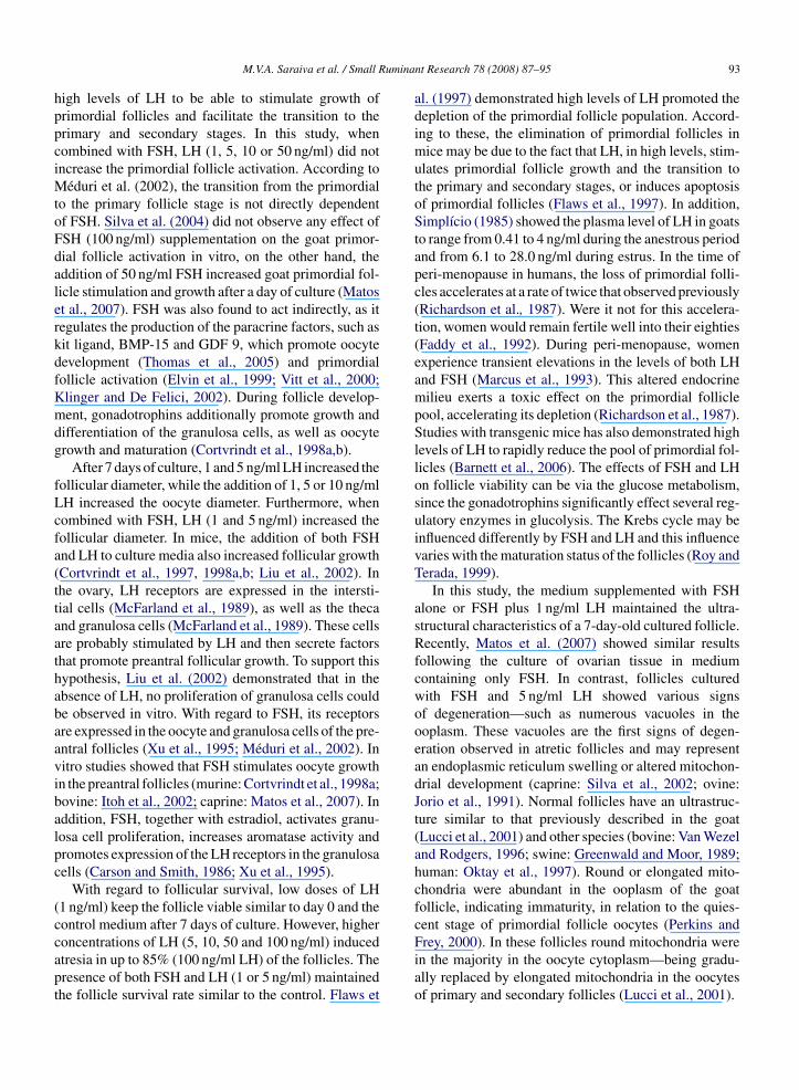
umina
hppciMtoFdalerkdfKmdg
fLcfa(ttaathabaavibalpc
(ccapt
M.V.A. Saraiva et al. / Small R
igh levels of LH to be able to stimulate growth ofrimordial follicles and facilitate the transition to therimary and secondary stages. In this study, whenombined with FSH, LH (1, 5, 10 or 50 ng/ml) did notncrease the primordial follicle activation. According to
éduri et al. (2002), the transition from the primordialo the primary follicle stage is not directly dependentf FSH. Silva et al. (2004) did not observe any effect ofSH (100 ng/ml) supplementation on the goat primor-ial follicle activation in vitro, on the other hand, theddition of 50 ng/ml FSH increased goat primordial fol-icle stimulation and growth after a day of culture (Matost al., 2007). FSH was also found to act indirectly, as itegulates the production of the paracrine factors, such asit ligand, BMP-15 and GDF 9, which promote oocyteevelopment (Thomas et al., 2005) and primordialollicle activation (Elvin et al., 1999; Vitt et al., 2000;linger and De Felici, 2002). During follicle develop-ent, gonadotrophins additionally promote growth and
ifferentiation of the granulosa cells, as well as oocyterowth and maturation (Cortvrindt et al., 1998a,b).
After 7 days of culture, 1 and 5 ng/ml LH increased theollicular diameter, while the addition of 1, 5 or 10 ng/mlH increased the oocyte diameter. Furthermore, whenombined with FSH, LH (1 and 5 ng/ml) increased theollicular diameter. In mice, the addition of both FSHnd LH to culture media also increased follicular growthCortvrindt et al., 1997, 1998a,b; Liu et al., 2002). Inhe ovary, LH receptors are expressed in the intersti-ial cells (McFarland et al., 1989), as well as the thecand granulosa cells (McFarland et al., 1989). These cellsre probably stimulated by LH and then secrete factorshat promote preantral follicular growth. To support thisypothesis, Liu et al. (2002) demonstrated that in thebsence of LH, no proliferation of granulosa cells coulde observed in vitro. With regard to FSH, its receptorsre expressed in the oocyte and granulosa cells of the pre-ntral follicles (Xu et al., 1995; Méduri et al., 2002). Initro studies showed that FSH stimulates oocyte growthn the preantral follicles (murine: Cortvrindt et al., 1998a;ovine: Itoh et al., 2002; caprine: Matos et al., 2007). Inddition, FSH, together with estradiol, activates granu-osa cell proliferation, increases aromatase activity andromotes expression of the LH receptors in the granulosaells (Carson and Smith, 1986; Xu et al., 1995).
With regard to follicular survival, low doses of LH1 ng/ml) keep the follicle viable similar to day 0 and theontrol medium after 7 days of culture. However, higher
oncentrations of LH (5, 10, 50 and 100 ng/ml) inducedtresia in up to 85% (100 ng/ml LH) of the follicles. Theresence of both FSH and LH (1 or 5 ng/ml) maintainedhe follicle survival rate similar to the control. Flaws etnt Research 78 (2008) 87–95 93
al. (1997) demonstrated high levels of LH promoted thedepletion of the primordial follicle population. Accord-ing to these, the elimination of primordial follicles inmice may be due to the fact that LH, in high levels, stim-ulates primordial follicle growth and the transition tothe primary and secondary stages, or induces apoptosisof primordial follicles (Flaws et al., 1997). In addition,Simplício (1985) showed the plasma level of LH in goatsto range from 0.41 to 4 ng/ml during the anestrous periodand from 6.1 to 28.0 ng/ml during estrus. In the time ofperi-menopause in humans, the loss of primordial folli-cles accelerates at a rate of twice that observed previously(Richardson et al., 1987). Were it not for this accelera-tion, women would remain fertile well into their eighties(Faddy et al., 1992). During peri-menopause, womenexperience transient elevations in the levels of both LHand FSH (Marcus et al., 1993). This altered endocrinemilieu exerts a toxic effect on the primordial folliclepool, accelerating its depletion (Richardson et al., 1987).Studies with transgenic mice has also demonstrated highlevels of LH to rapidly reduce the pool of primordial fol-licles (Barnett et al., 2006). The effects of FSH and LHon follicle viability can be via the glucose metabolism,since the gonadotrophins significantly effect several reg-ulatory enzymes in glucolysis. The Krebs cycle may beinfluenced differently by FSH and LH and this influencevaries with the maturation status of the follicles (Roy andTerada, 1999).
In this study, the medium supplemented with FSHalone or FSH plus 1 ng/ml LH maintained the ultra-structural characteristics of a 7-day-old cultured follicle.Recently, Matos et al. (2007) showed similar resultsfollowing the culture of ovarian tissue in mediumcontaining only FSH. In contrast, follicles culturedwith FSH and 5 ng/ml LH showed various signsof degeneration—such as numerous vacuoles in theooplasm. These vacuoles are the first signs of degen-eration observed in atretic follicles and may representan endoplasmic reticulum swelling or altered mitochon-drial development (caprine: Silva et al., 2002; ovine:Jorio et al., 1991). Normal follicles have an ultrastruc-ture similar to that previously described in the goat(Lucci et al., 2001) and other species (bovine: Van Wezeland Rodgers, 1996; swine: Greenwald and Moor, 1989;human: Oktay et al., 1997). Round or elongated mito-chondria were abundant in the ooplasm of the goatfollicle, indicating immaturity, in relation to the quies-cent stage of primordial follicle oocytes (Perkins and
Frey, 2000). In these follicles round mitochondria werein the majority in the oocyte cytoplasm—being gradu-ally replaced by elongated mitochondria in the oocytesof primary and secondary follicles (Lucci et al., 2001).
umina
94 M.V.A. Saraiva et al. / Small RIn conclusion, it can be said that the addition of FSH(50 ng/ml) combined or not with low concentrations ofLH (1 ng/ml) maintains the goat preantral follicles ultra-structural integrity after in vitro culture. Further studieson the effects of LH in the modulation of paracrinegrowth factors are needed to understand the early fol-liculogenesis in goats.
Acknowledgements
This work was supported by CAPES. Márcia VivianeAlves Saraiva is a recipient of a grant from CAPES(Brazil). The donation of FSH and LH by Dr. Jean-Francois Beckers of the University of Liège, Belgium,is acknowledged.
References
Barnett, K.R., Schilling, C., Greenfeld, C.R., Tomic, D., Flaws, J.A.,2006. Ovarian follicle development and transgenic mouse models.Hum. Reprod. Update 12, 537–555.
Carson, R., Smith, J., 1986. Development and steroidogenic activityof preantral follicles in the neonatal rat ovary. J. Endocrinol. 110,87–92.
Cortvrindt, R.G., Hu, Y., Liu, J., Smitz, J.E., 1998a. Timed analysis ofthe nuclear maturation of oocytes in early preantral mouse folli-cle culture supplemented with recombinant gonadotrophin. Fertil.Steril. 70, 1114–1125.
Cortvrindt, R., Hu, Y., Smitz, J., 1998b. Recombinant luteiniz-ing hormone as a survival and differentiation factor increasesoocyte maturation in recombinant follicle stimulating hormone-supplemented mouse preantral follicle culture. Hum. Reprod. 13,1292–1302.
Cortvrindt, R., Smitz, J., Van Steirteghem, A.C., 1997. Assessment ofthe need for follicle stimulating hormone in early preantral mousefollicle culture in vitro. Hum. Reprod. 12, 759–768.
Derrar, N., Price, C.A., Sirard, M.A., 2000. Effect of growth factorsand co-culture with ovarian medulla on the activation of primordialfollicles in explants of bovine ovarian cortex. Theriogenology 54,587–598.
Dufour, J., Cahill, L.P., Mauleon, P., 1979. Short and long-term effectsof hypophysectomy and unilateral ovariectomy on populations fol-licular in sheep. J. Reprod. Fertil. 7, 301–309.
Elvin, J.A., Yan, C.N., Wang, A., Nishimori, K., Matzuk, M.M.,1999. Molecular characterisation of the follicle defects in thegrowth differentiation factor 9-deficient ovary. Mol. Endocrinol.13, 1018–1034.
Faddy, M.J., Gosden, R.G., Gougeon, A., Richardson, S.J., Nelson,J.F., 1992. Accelerated disappearance of ovarian follicle sinmid-life: implications for fore casting menopause. Hum. Reprod. 7,1342–1346.
Flaws, J.A., Abbud, R., Mann, R.J., Nilson, J.H., Hirshfield, A.N.,1997. Chronically elevated luteinizing hormone depletes pri-
mordial follicles in the mouse ovary. Biol. Reprod. 57, 1233–1237.Fortune, J.E., Kito, S., Wandji, S.-A., Srsen, V., 1998. Activation ofbovine and baboon primordial follicles in vitro. Theriogenology49, 441–449.
nt Research 78 (2008) 87–95
Greenwald, G.S., Moor, R.M., 1989. Isolation and preliminary charac-terization of pig primordial follicles. J. Reprod. Fertil. 87, 561–571.
Itoh, T., Kacchi, M., Abe, H., Sendai, Y., Hoshi, H., 2002. Growth,antrum formation, and estradiol production of bovine preantralfollicles cultured in a serum-free medium. Biol. Reprod. 67,1099–1105.
Jorio, A., Mariana, J.C., Lahlou-kassi, A., 1991. Development of thepopulation of ovarian follicles during the prepubertal period inD’man and Timahdit shee. J. Reprod. Fertil. 26, 239–250.
Klinger, F.G., De Felici, M., 2002. In vitro development of growingoocytes from fetal mouse oocytes: stage-specific regulation by stemcell factor and granulosa cells. Dev. Biol. 244, 85–95.
Liu, H.C., Zhiying, H., Rosenwaks, Z., 2002. In vitro culture andin vitro maturation of mouse preantral follicles with recombinantgonadotrophins. Fertil. Steril. 77, 373–383.
Lucci, C.M., Silva, R., Carvalho, C.A., Figueiredo, R., Báo, S.N.,2001. Light microscopical and ultrastructural characterization ofgoat preantral follicles. Small Rum. Res. 41, 61–69.
Marcus, M., Grunfeld, L., Berkowitz, G., Kaplanp Godbold, J., 1993.Urinary follicle-stimulating hormone as a biological marker ofovarian toxicity. Fertil. Steril. 59, 931–933.
Matos, M.H.T., Lima-Verde, I.B., Luque, M.C.A., Maia Jr., J.E., Silva,J.R., Celestino, J.J.H., Martins, F.S., Báo, S.N., Lucci, A.M.,Figueiredo, J.R., 2007. Essential role of follicle stimulating hor-mone in the maintenance of caprine preantral follicle viability invitro. Zygote 15, 173–182.
McFarland, K.C., Sprengel, R., Phillips, H.S., Kohler, M., Rosem-blit, N., Nikolics, K., Segaloff, D.L., Seeburg, H., 1989. Lutropin-choriogonadotropin receptor: an usual member of the G protein-coupled receptor family. Science 245, 494–499.
Méduri, G., Charnaux, N., Driancourt, M.A., Combettes, L., Granet,A., Vannier, B., Loosfelt, H., Migrom, E., 2002. Follicle-stimulating hormone receptors in oocytes? J. Clin. Endocrinol.Metabol. 87, 2266–2276.
Oktay, K., Briggs, D., Gosde, R.G., 1997. Ontogeny of follicle-stimulating hormone receptor gene expression in isolated humanovarian follicles. J. Clin. Endocrinol. Metabol. 82, 3748–3751.
O’Shaughnessy, J., McLelland, D., McBride, M.W., 1997. Regulationof luteinizing hormone receptor and follicle-stimulating hormone-receptor messenger ribonucleic acid levels during development inthe neonatal mouse ovarian. Biol. Reprod. 57, 602–608.
Perkins, G.A., Frey, T.G., 2000. Recent structural insight into mito-chondria gained by microscope. Micron 31, 97–111.
Reiter, E., McNamara, M., Closset, J., Hennen, G., 1995. Expressionand functionality of luteinizing hormone/chorionic gonadotrophinreceptor in the rat prostate. Endocrinology 136, 917–923.
Reshef, E., Lei, Z.M., Rao, C., Pridham, D.D., Chegini, N., Luborsky,J.L., 1990. The presence of gonadotrophin receptors in non-pregnant human uterus, human placenta, fetal membranes anddeciduas. J. Clin. Endocrinol. Metabol. 70, 421–430.
Richardson, S.J., Senikas, A., Nelson, J.E., 1987. Follicular depletionduring themenopausal transition: evidence for accelerated loss andultimate exhaustion. J. Clin. Endocrinol. Metabol. 65, 1231–1237.
Roy, S.K., Terada, D.M., 1999. Actives of glucose metabolic enzymesin human preantral follicles: In vitro modulation by follicle-stimulating hormone, luteinizing hormone, epidermal growthfactor, insulin-like growth factor 1, and transforming growth factor
�1. Biol. Reprod. 60, 763–768.Segaloff, D.L., 1996. Regulation of the LH receptor in the ovary duringfollicular development. In: Filicore, M., Flamigni, C. (Eds.), TheOvary: Regulation, Dysfunction and Treatment. Elsevier, Amster-dam, pp. 23–28.

umina
S
S
S
S
T
U
M.V.A. Saraiva et al. / Small R
ilva, J.R.V., Ferreira, M.A.L., Costa, S.H.F., Santos, R.R., Carvalho,F.C.A., Rodrigues, A.R., Lucci, C.M., Báo, S.N., Figueiredo, J.R.,2002. Degeneration rate of preantral follicles in the ovaries of goats.Small Rum. Res. 43, 203–209.
ilva, J.R., Van Den Hurk, R., Matos, M.H.T., Santos, R.R., Pes-soa, C., Moraed, M.O., Figueiredo, J.R., 2004. Influences ofFSH and EGF on primordial follicles during in vitro cultureof caprine ovarian cortical tissue. Theriogenology 61, 1691–1704.
implício, A.A., 1985. Reproduction in three native genotypes of goatsunder two feeding management systems in northeast Brazil, andprogesterone and luteinizing hormone profiles during the estrouscycle and seasonal anestrus in Spanish goats in the United States.Dissertation of Doctor Philosophy, Utah State University, Logan-Utah, XII.
teel, R.G.D., Torrie, J.H., Dickey, D., 1997. Principles and proceduresof statistics: a biometrical approach, 3rd ed. McGraw-Hill, NewYork, NY, 666 pp.
homas, F.H., Ethier, J.-F., Shimasaki, S., Vanderhyden, B.C., 2005.Follicle-stimulating hormone regulates oocyte growth by mod-ulation of expression of oocyte and granulosa cell factors.
Endocrinology 146, 941–949.lloa-Aguirre, A., Midgley, A.R., Beitins, J.R., Padmanab-han, I.Z., 1995. Follicle-stimulating isohormones: characteriza-tion and physiological relevance. Endocrinol. Rev. 16, 765–787.
nt Research 78 (2008) 87–95 95
Van Wezel, I., Rodgers, R.J., 1996. Morphological characterization ofprimordial follicles and their environment in vivo. Biol. Reprod.55, 1003–1010.
Vitt, U.A., Hayashi, M., Klein, C., Hsueh, A.J., 2000. Growth dif-ferentiation factor-9 stimulates proliferation but suppresses thefollicle-stimulating hormone-induced differentiation of culturedgranulosa cells from small antral and preovulatory rat follicles.Biol. Reprod. 62, 370–377.
Wright, C.S., Hovatta, O., Margara, R., Trew, G., Winston, R.M.L.,Franks, S., Hardy, K., 1999. Effects of follicle-stimulating hormoneand serum substitution on the in-vitro growth of human ovarianfollicles. Hum. Reprod. 14, 1555–1562.
Wu, J., Nayudu, L., Kiesel, S., Michelmann, H.W., 2000. Luteinizinghormone has a stage-limited effect on preantral follicle develop-ment in vitro. Biol. Reprod. 63, 320–327.
Xu, Z., Garverick, A., Smith, G.W., Smith, M.F., Amilton, S.A.,Younquist, R., 1995. Expression of follicle-stimulating hormoneand luteinizing hormone receptor messenger ribonucleic acids inbovine follicles during the first follicular wave. Biol. Reprod. 53,951–957.
Yamoto, M., Shima, K., Nakano, R., 1992. Gonadotrophins recep-
tors in human ovarian follicles and corporea lutea throughout themenstrual cycle. Horm. Res. 37, 11–25.Zeleznick, A.J., Hillier, S.G., 1984. The role of goadotrophins in theselection of the preovulatory follicle. Clin. Obst. Ginecol. 27,927–940.
Copyright © 2022 FDOKUMEN