Infection and Reactive Changes
51
Bacterial and rickettsial infection Peripheral blood In adults, the usual haematological response to an acute bacterial infection is neutrophil leucocytosis with a left shift (an increase of band forms and pos- sibly the appearance of neutrophil precursors in the peripheral blood) (Fig. 3.1). The neutrophils usually show toxic granulation and may show Döhle bodies and cytoplasmic vacuolation. When there is very severe bacterial infection and in neonates, alco- holics and patients with reduced bone marrow reserve, neutrophilia does not occur but there is a left shift with the above ‘toxic’ changes in neu- trophils. Certain bacterial infections, specifically typhoid, paratyphoid and tularaemia, are character- ized by neutropenia rather than neutrophilia. In THREE INFECTION AND REACTIVE CHANGES Infection The response of the bone marrow to infection is very variable, depending on the nature and chronicity of the infection, the age of the subject and the presence of any associated diseases. The response differs according to whether the infection is bacterial, rickettsial, viral or fungal. The periph- eral blood and bone marrow responses to infec- tion are non-specific and similar changes occur in many other conditions, including trauma and other tissue damage, administration of growth factors, carcinoma, Hodgkin’s disease, non-Hodgkin’s lymphoma and auto-immune disorders such as systemic lupus erythematosus. Only a minority of patients with an infection show peripheral blood or bone marrow changes suggestive of a particular micro-organism. 90 Fig. 3.1 PB, bacterial infection, left shift and toxic granulation. MGG ×940.
-
Upload
khangminh22 -
Category
Documents
-
view
3 -
download
0
Transcript of Infection and Reactive Changes

Bacterial and rickettsial infection
Peripheral blood
In adults, the usual haematological response to anacute bacterial infection is neutrophil leucocytosiswith a left shift (an increase of band forms and pos-sibly the appearance of neutrophil precursors in theperipheral blood) (Fig. 3.1). The neutrophils usuallyshow toxic granulation and may show Döhle bodiesand cytoplasmic vacuolation. When there is verysevere bacterial infection and in neonates, alco-holics and patients with reduced bone marrowreserve, neutrophilia does not occur but there is aleft shift with the above ‘toxic’ changes in neu-trophils. Certain bacterial infections, specificallytyphoid, paratyphoid and tularaemia, are character-ized by neutropenia rather than neutrophilia. In
THREE
INFECTION AND REACTIVECHANGES
Infection
The response of the bone marrow to infection is very variable, depending on the nature andchronicity of the infection, the age of the subjectand the presence of any associated diseases. Theresponse differs according to whether the infectionis bacterial, rickettsial, viral or fungal. The periph-eral blood and bone marrow responses to infec-tion are non-specific and similar changes occur inmany other conditions, including trauma and othertissue damage, administration of growth factors,carcinoma, Hodgkin’s disease, non-Hodgkin’s lymphoma and auto-immune disorders such as systemic lupus erythematosus. Only a minority ofpatients with an infection show peripheral blood orbone marrow changes suggestive of a particularmicro-organism.
90
Fig. 3.1 PB, bacterial infection, left shift and toxic granulation.MGG ×940.

INFECTION AND REACTIVE CHANGES 91
severe infection, particularly if there is shock orhypoxia, nucleated red blood cells may appear inthe blood, the presence of both granulocyte precur-sors and nucleated red cells being referred to asleuco-erythroblastosis. The lymphocyte count isreduced but a few atypical lymphocytes, includingplasmacytoid lymphocytes, may be present; plasmacells are sometimes seen. The eosinophil count is reduced during acute infection but eosinophiliacan occur during recovery. Children may respondto bacterial infection with lymphocytosis ratherthan neutrophilia, and certain bacterial infections,particularly whooping cough and sometimes bru-cellosis, are characterized by lymphocytosis. In bac-terial infection, the platelet count is often reducedbut sometimes increased.
Certain bacterial infections can be complicated byhaemolytic anaemia. Infection by E. coli or Shigellaspecies can be followed by a micro-angiopathichaemolytic anaemia as part of a haemolytic uraemicsyndrome. Sepsis due to Clostridium welchii can becomplicated by acute haemolysis with spherocyticred cells. Mycoplasma infection is commonly asso-ciated with the production of cold auto-antibodiesso that red cell agglutinates are present in bloodfilms made at room temperature and haemolyticanaemia sometimes occurs.
Rarely, neutrophils contain phagocytosed bacte-ria. The presence of bacteria, either extracellularlyor within neutrophils, is usually seen only in over-
whelming infections, particularly when there isassociated hyposplenism. In relapsing fever, how-ever, the characteristic spiral organisms of Borreliaspecies appear episodically in the bloodstream andare seen lying free between red cells. In ehrlichialinfections, organisms are often detectable withinmonocytes or granulocytes. They are detected in monocytes in human monocytic ehrlichiosis(caused by Ehrlichia chaffeensis) and in neutrophilsin human granulocytic ehrlichiosis and humaninfection by Ehrlichia ewingii (the agent responsiblefor canine granulocytic ehrlichiosis) [1]. The detec-tion of organisms within leucocytes is facilitated byexamination of buffy coat films.
In more chronic infections, there may beanaemia, increased rouleaux formation, increasedbackground staining and monocytosis (Fig. 3.2).Anaemia is initially normocytic and normochromicbut, as the infection becomes increasingly chronic,the anaemia develops the characteristics of theanaemia of chronic disease with the cells producedbeing hypochromic and microcytic.
Pulmonary tuberculosis characteristically causesanaemia, increased rouleaux formation and, whensevere, neutrophilia. Monocytosis occurs in aminority of patients. Monocytopenia is actuallymore common than monocytosis. In miliary tuber-culosis, leucocytosis is uncommon and leucopeniais common. The lymphocyte count is reduced. Aminority of patients have monocytosis. Pancytopenia
Fig. 3.2 PB, bacterial infection,monocytosis and neutrophilia.MGG ×940.

92 CHAPTER THREE
may occur, sometimes as a consequence ofhaemophagocytosis and, less often, in associationwith bone marrow necrosis.
Rickettsial infections cause varied haematologicaleffects which may include neutrophilia, neutro-penia, lymphocytosis, the presence of atypical lymphocytes and thrombocytopenia which is some-times severe.
Occasionally, severe infections are associatedwith a haematological picture which simulatesleukaemia, designated a leukaemoid reaction (seebelow).
Bone marrow cytology
In severe bacterial infection, the bone marrow fea-tures reflect those of the peripheral blood. There isgranulocytic hyperplasia with associated toxicchanges (Fig. 3.3). In overwhelming infection, themarrow sometimes shows an increase of granulo-cyte precursors but with few maturing cells.Erythropoiesis is depressed and erythroblasts showreduced siderotic granulation. When there isthrombocytosis, megakaryocytes may be increased.Macrophages are increased and, in a minority ofpatients with severe infection, prominent haemo-phagocytosis occurs (see page 119). When infectionis chronic, an increase of iron stores is apparent.
Microscopy or bone marrow culture occasionally
provides evidence of a specific infection. InWhipple’s disease, the causative organisms may beseen within bone marrow macrophages [2]. Inhuman monocytic ehrlichiosis, organisms can bedetected in bone marrow monocytes, whereas inhuman granulocytic ehrlichiosis, caused by arelated organism, they are present in granulocytes[3]. Bacteria visible within macrophages have alsobeen reported in bacterial endocarditis. In typhoidfever, the bone marrow aspirate shows only non-specific features but bone marrow culture can be useful since it increases the detection rate by50% in comparison with culture of the peripheral blood [4]. In brucellosis, the bone marrow is usuallyhypercellular with prominent haemophagocytosisand increased eosinophils and plasma cells [5].Bone marrow cultures are occasionally positivewhen blood cultures are negative but, as the diag-nosis can be made serologically, bone marrow aspiration is not indicated for this purpose.
In tuberculosis, the bone marrow aspirate showsnon-specific features such as increased iron storesand an increase in macrophages, often with hae-mophagocytosis. Mycobacteria may be detected in a minority of cases using specific stains (Ziehl–Neelsen or auramine). Cultures should always beperformed when this diagnosis is suspected. In lep-romatous leprosy, bacilli may be apparent as nega-tive images within bone marrow macrophages [6].
Fig. 3.3 BM aspirate film from a patient with severe infectionshowing heavy toxic granulationand vacuolation of neutrophilprecursors. MGG ×940.

INFECTION AND REACTIVE CHANGES 93
Bone marrow histology
Severe bacterial infection leads to an increase inmarrow cellularity due to granulocytic hyperplasia.There is often left shift of the granulocytic series, i.e.an increase in the numbers of immature precursors(myelocytes and promyelocytes) in relation tomature polymorphonuclear neutrophils (Fig. 3.4).However, the normal topographical arrangement ofgranulopoiesis is retained, with the more immaturecells (mainly promyelocytes) found predominantlyin the paratrabecular region [7]. Megakaryocytesare often increased in number [8]; they are mor-phologically normal, but there may be an increase in‘bare’ megakaryocyte nuclei and increased emperi-polesis. Erythropoiesis is often reduced althoughmorphologically normal.
In more chronic bacterial infections, changes inother lineages become apparent. Bone marrowplasmacytosis is a common but non-specific re-sponse [9]. Very rarely, there may be up to 50%plasma cells in the marrow in reactive conditionsincluding infections. The differential diagnosis ofincreased numbers of plasma cells in the marrow isdiscussed on page 166. In infections, plasma cellsare distributed through the marrow in an interstitialmanner, often with focal pericapillary accentua-tion. This may be accompanied by plasma cell satel-litosis, in which a central macrophage is surrounded
by three or more plasma cells. The plasma cells havemature nuclear and cytoplasmic characteristicsalthough there are often occasional binucleateforms and cells containing Russell bodies. Chronicinfection is also associated with an increased fre-quency of reactive lymphoid aggregates (see page144). Macrophages may be increased and com-monly contain ingested granulocytes (Fig. 3.5). Awide variety of infectious agents, including bacteria,can cause a secondary haemophagocytic syndrome(see page 119). Stromal changes related to infectioninclude prominent sinusoids, focal reticulin fibrosisand rarely, in very severe chronic infections, gelati-nous change (see page 128). A decrease in marrowcellularity and loss of fat spaces usually accompanygelatinous transformation [10].
Mycobacterial infections can cause the same reactive changes as infection by other bacteria.However, haemophagocytosis and granuloma for-mation are particularly common. Whenever granu-lomas are present it is necessary to perform specialstains such as a Ziehl–Neelsen stain or an auraminestain, in the latter instance mycobacteria beingdetected by fluorescence microscopy. In one study,immunohistochemistry with a polyclonal antibodyto mycobacteria was found to be more sensitivethan an acid-fast stain [11]. Occasional patientswith mycobacterial infection have extensive necro-sis which is indicative of poor prognosis. In those
Fig. 3.4 BM trephine biopsysection, bacterial infection withleukaemoid reaction: there isincreased cellularity andgranulocytic hyperplasia with left shift; note emperipolesis of a mature neutrophil by amegakaryocyte. Paraffin-embedded, H&E ×195.

94 CHAPTER THREE
with impaired immunity, there may be diffusemacrophage hyperplasia rather than granulomaformation. Macrophages may appear foamy.Mycobacterium leprae infection may cause granulomaformation and, in lepromatous leprosy, prolifera-tion of foamy macrophages. Patients have also beenreported in whom interstitial lepra bacilli werefound in bone marrow sections, stained with a Fitestain, in the absence of any macrophage prolifera-tion or granulomas [12].
In ehrlichiosis, the bone marrow is often hyper-cellular with both granulocytic and megakaryocytichyperplasia. Granulomas are often present. Ehrlichialinclusions can be identified in macrophages [13](Fig. 3.6). Ehrlichiae can be detected immunocyto-chemically [13].
Granuloma formation can also occur in rickettsialinfections such as Q fever (infection by Coxiella bur-netti) and Rocky Mountain spotted fever (infectionby Rickettsia rickettsii).
Problems and pitfalls
Mycobacteria are not always detectable with specialstains in patients with haemophagocytosis or gran-ulomas caused by miliary tuberculosis. Cultures aremore sensitive than microscopy.
Acute myeloid leukaemia (AML) can be simu-lated by overwhelming infection with associated
cytopenia. Leukaemoid reactions can also simulatechronic myelomonocytic leukaemia (CMML). In aleukaemoid reaction, anaemia and thrombocytope-nia are common. The white cell count is some-times low with neutropenia and sometimes high;the blood film may show granulocyte precursors or be leuco-erythroblastic. Leukaemoid reactionsare most often seen in association with very severebacterial infection, particularly when there is co-existing megaloblastic anaemia. The bone marrowresponse to miliary tuberculosis can also simulateleukaemia. Toxic changes such as toxic granulationand neutrophil vacuolation are useful clues to thecorrect diagnosis. If necessary, a bone marrow exam-ination makes the distinction. In severe infectionthere may be ‘maturation arrest’ with increasedpromyelocytes but there is no significant increase inblasts and the promyelocytes differ from those ofacute hypergranular promyelocytic leukaemia inhaving prominent Golgi zones and no Auer rods.
The blood and bone marrow features of severechronic infection may be difficult to distinguishfrom those of CMML since ‘toxic’ changes may be lacking. Consideration of the clinical featuresfacilitates the distinction. It is imprudent to make adiagnosis of CMML in a patient with a signifi-cant infection unless there are dysplastic features of a type showing a strong association with themyelodysplastic syndromes (see Chapter 4).
Fig. 3.5 Section of BM trephinespecimen showing neutrophilshadows within macrophages in a patient with Hodgkin’s disease;similar features are seen in bacterialinfection. Paraffin-embedded, H&E ×940.

INFECTION AND REACTIVE CHANGES 95
Viral infection
Peripheral blood
Viral infections usually provoke lymphocytosis.Often the cells that are produced are morpholog-ically fairly normal but sometimes they have atypical features. Infectious mononucleosis con-sequent on infection by the Epstein–Barr virus(EBV) is characterized by the production of largenumbers of atypical lymphocytes, often referred toas atypical mononuclear cells (Fig. 3.7). These arepleomorphic, usually large, and often have abun-
dant basophilic cytoplasm and nuclei with a diffusechromatin pattern and nucleoli. The presence of large numbers of atypical mononuclear cells isnot specific for EBV infection; this may also be a feature of primary infection by other viruses(cytomegalovirus (CMV), human immunodefici-ency virus (HIV), hepatitis A and adenovirus) and oftoxoplasmosis, as well as being seen in hypersensi-tivity reactions to drugs. Smaller numbers of similaratypical lymphocytes are seen in a wider variety of infective (and non-infective) conditions. Viralinfections which may be associated with markedlymphocytosis without many atypical features
Fig. 3.6 Intracellular ehrlichiae in a BM trephine biopsy section: (a) PAS stain with haematoxylincounterstain; (b) immunoperoxidasestain with anti-ehrlichial IgGantibody. (By courtesy of Dr CDPaddock, Dr RL Kerschmann and Dr BG Herndier, San Francisco,reprinted by permission of the NewEngland Journal of Medicine, 329,1165, 1993.)
(a)
(b)

96 CHAPTER THREE
include those due to Coxsackie virus, various adenoviruses and HIV; other haematological abnor-malities produced by HIV are discussed below (seepage 124). In immunosuppressed hosts, e.g. follow-ing bone marrow transplantation, CMV infectioncan cause neutropenia, thrombocytopenia and pan-cytopenia. CMV infection in such patients may be associated with very large virus-infected cells,which are probably endothelial in origin, in thefeathered edge of the film [14]. Viral infections, par-ticularly those due to herpesviruses, may be associ-ated with a haemophagocytic syndrome (see page119) with the peripheral blood showing resultantpancytopenia. In a few individuals, viral hepatitis,particularly non-A non-B non-C hepatitis, is followedin a period of a few weeks or months by pancyto-penia caused by aplastic anaemia. In certain sub-jects who are unable to mount a normal immuneresponse to EBV, infection by this virus may also befollowed by chronic pancytopenia due to bone mar-row aplasia. Parvovirus commonly causes transientpure red cell aplasia but, unless red cell survival isreduced, this may go unnoticed; less often, it is acause of neutropenia or thrombocytopenia.
Viral infections may be complicated by cytopeniasconsequent on either damage to cells by immunecomplexes or auto-antibody production. Rubellaand, less often, other viral infections may be fol-lowed by transient thrombocytopenia caused bydamage to platelets by immune complexes. Infecti-ous mononucleosis may be complicated by eitherauto-immune thrombocytopenia or auto-immune
haemolytic anaemia due to a cold antibody withanti-i specificity; in these cases, there are red cellagglutinates and occasional spherocytes. Rarely,viral infections, particularly measles, are followedby acute haemolysis due to an auto-antibody withanti-P specificity (‘paroxysmal’ cold haemoglobin-uria); in these cases, the blood film usually showsonly occasional spherocytes and subsequently poly-chromasia. Contrary to what might be anticipatedfrom the name, there is only a single episode ofhaemolysis.
Chronic hepatitis C infection can be a cause ofthrombocytopenia. Some patients develop mixedcryoglobulinaemia with associated haematologicalfeatures (see page 355). A single case has also beenreported of pure red cell aplasia associated withhepatitis C infection [15].
In patients with impaired immunity, EBV infec-tion may be associated with a lymphoprolifera-tive disorder which may evolve to lymphoma (seepage 319).
If fresh tissue is available, molecular techniquescan be used for the detection of viral infection. Forexample, in suspected EBV infection, reverse tran-scriptase polymerase chain reaction (RT-PCR) canbe used to detect messenger RNA (mRNA) forEBNA-1 and -2, LMP-1, -2A and -2B, BZLF1(ZEBRA ‘protein’) and BCRF1 (viral IL10).
Bone marrow cytology
In viral infection, the bone marrow shows an increase
Fig. 3.7 PB, infectiousmononucleosis, atypicallymphocytes. MGG ×940.

INFECTION AND REACTIVE CHANGES 97
of lymphocytes, either typical or atypical. In someinfections, particularly by herpesviruses, haemo-phagocytosis is prominent (see page 119). In im-munosuppressed hosts, CMV infection can also causemarked bone marrow hypocellularity with suppres-sion of all lineages [16]. When haemolytic anaemiaoccurs, e.g. in infectious mononucleosis, erythroidhyperplasia will be apparent. In pure red cell aplasiaconsequent on parvovirus B19 infection there areprominent, very large proerythroblasts with a strik-ing lack of more mature cells. In one patient withparvovirus-induced pancytopenia there were alsolarge atypical cells of granulocyte lineage which wereshown to contain viral antigens [17]. Parvovirusinfection can be confirmed by immunofluorescencewith monoclonal antibodies to either a capsid anti-gen [17] or a nuclear antigen, or by in situ DNAhybridization. When viral infections are compli-cated by thrombocytopenia due to increased plateletdestruction, megakaryocytes are present in normalor increased numbers. Occasionally, platelets arevisible within bone marrow macrophages (Fig. 3.8).
Bone marrow histology
Viral infections may lead to an increase of bonemarrow lymphocytes, plasma cells and macro-phages, with or without haemophagocytosis.Atypical lymphoid cells, morphologically identicalto those in the peripheral blood, can be seen in themarrow in EBV and CMV infection; in CMV infec-tion, eosinophilic intranuclear inclusions may be
seen, albeit rarely, in endothelial cells and in cells,probably macrophages, within granulomas or inter-spersed among haemopoietic cells [18]. Atypicallymphoid cells with intranuclear viral inclusionshave also been detected in human herpesvirus 6 (HHV6) infection [19]. In the post-transplant setting, CMV [16] and HHV6 [20] infection cancause severe bone marrow hypoplasia. The pres-ence of CMV (Fig. 3.9) and EBV can be confirmedby immunohistochemistry using monoclonal anti-bodies (Table 3.1). However, it should be notedthat, even in patients with generalized CMV infec-tion, it is unusual for virus-infected cells to bedetectable in the bone marrow. Pure red cell aplasiais characteristically seen in infection by parvovirusB19 and is readily detected in trephine biopsy sec-tions (see page 376). Very occasionally, it has beenassociated with erythroid hyperplasia, sometimeswith marked dyserythropoiesis [25]. ParvovirusB19 capsid antigens may be detectable by immuno-histochemistry although this technique is less sen-sitive than PCR [26]; curiously, in one study, thepositive cells were among the few late erythroidcells while the giant proerythroblasts were nega-tive [26]. Viral infections, particularly herpesvirusinfections, can lead to formation of small non-caseating granulomas (see Table 3.3). Chronic hepatitis B and hepatitis C infection may be asso-ciated with the presence of reactive lymphoid nodules [27]. Hepatitis C infection can also be com-plicated by the development of low grade, B-celllymphoma, often supervening in a patient with
Fig. 3.8 BM aspirate. Plateletswithin a macrophage in a patientwith severe thrombocytopeniaduring acute CMV infection. MGG ×960.

98 CHAPTER THREE
Fig. 3.9 BM trephine biopsysection from a renal transplantpatient with CMV infectionshowing one positive cell (top left). Paraffin-embedded,immunoperoxidase with Dako-CMV McAb ×940.
Table 3.1 Techniques applicable to trephine biopsy specimens for confirmation of certain viral infections.
Virus
Parvovirus B19
Cytomegalovirus(CMV)
Epstein–Barr virus (EBV)
Herpes simplex
* LMP McAb is a cocktail of clones (CS1-4) raised against a fusion protein containing 189 amino acids of the terminus of LMP. Cross-reaction with normal early myeloid and erythroid precursors, leukaemic myeloblasts and leukaemiclymphoblasts has been reported [23] but this has not been our experience when using formalin fixation and EDTAdecalcification; we have observed no false-positive staining in normal bone marrow or in AML, and in ALL we haveobserved weak nuclear staining only, whereas the specific product would be cytoplasmic.
Monoclonal antibody or other specific reagent
R92F6 (Novocastra) directed at capsid proteinsVP1 and VP2
Digoxigenin-labelled parvovirus B19 DNAprobe [21]
AAC10 (Dako) directed at CMV lower matrixprotein pp65; QB1/06 (Novocastra) directedat CMV late antigens
CMV probe ISH kit (Novocastra) or CMVBioprobe (ENZO Diagnostics) [22]
CS1-4 directed at LMP-1 (Dakopatts orNovocastra); PE2 directed at EBNA-2 (Dakoand Novocastra)
G3-E31 (Novocastra) directed at 50–52 kDdiffuse early antigen; W1-F2 (Novocastra)directed at 85 kD restricted early antigen
Clone βZ.1 directed at βZLF1 (Dako)
EBER1 PNA probe/FITC kit (Dako) or EBER-probe ISH kit (Novocastra)
Rabbit polyclonal antiserum (Dako)
Technique
Immunohistochemistry with detection of anuclear antigen or a viral capsid antigen
In situ hybridization for detection of viral DNA
Immunohistochemistry with detection of nuclearand cytoplasmic antigen
In situ hybridization for detection of viral DNA
Immunohistochemistry for detection of latentmembrane protein-1 (LMP-1)* or Epstein–Barrnuclear antigen-2 (EBNA-2) in latent infection
EBV early lytic antigens
Immunohistochemistry for detection of BZLF (‘ZEBRA’ protein) in lytic EBV infection
In situ hybridization for detection of nuclearEpstein–Barr early RNA (EBER)
Southern blot analysis to detect EBV clonality(dependent on the number of terminal repeats) [24]
Immunohistochemistry for detection of variousshared and type-specific antigens

INFECTION AND REACTIVE CHANGES 99
mixed type II cryoglobulinaemia; the lymphoidinfiltrate is then monoclonal and more extensive.
As already noted, immunohistochemistry is useful in the diagnosis of viral infections. The spe-cial techniques applicable to trephine biopsies forconfirmation of infection by specific viruses aresummarized in Table 3.1.
Problems and pitfalls
Lymphocytosis with atypical lymphocytes can beconfused with a lymphoproliferative disorder, par-ticularly with mantle cell lymphoma and the mixedcell type of chronic lymphocytic leukaemia inwhich neoplastic cells are pleomorphic. When thereis diagnostic difficulty, immunophenotyping is indicated to demonstrate or exclude a clonal pro-liferation of B lymphocytes.
Virus-induced haemophagocytic syndrome canbe confused with malignant histiocytosis. The lat-ter is a very rare condition and the diagnosis should be made with caution. It is characterized byproliferation of very immature cells of monocyte–macrophage lineage which, in contrast to the cells of virus-induced haemophagocytic syndrome, usually show little phagocytic activity. Markedhaemophagocytosis therefore suggests that macro-phage activation is reactive. Virus-induced haemo-phagocytosis can also be confused with other reactive haemophagocytic syndromes, includingthose associated with some T-cell and NK-cell lymphomas.
Fungal infection
Peripheral blood
Fungal infections have no specific haematologicalfeatures. Some fungal diseases, for example actino-mycosis and coccidioidomycosis, are associatedwith neutrophilia. Aspergillus and other fungi mayevoke an allergic response and be associated witheosinophilia. When systemic fungal infectionsoccur in immunocompromised hosts, fungi such ascandida or histoplasma are occasionally seen in theperipheral blood. Candida may also be seen in bloodfilms when there is infection originating from a colonized indwelling venous line.
Bone marrow cytology
Fungi are rarely seen in the bone marrow aspiratein immunologically normal subjects. Histoplasma isan exception, organisms sometimes being detectedwithin bone marrow macrophages. Fungi are muchmore often detected in bone marrow aspirates fromseverely immunocompromised patients, such asthose with HIV infection or following bone marrowtransplantation, when Candida albicans, Aspergillusfumigatus, Histoplasma capsulatum (Fig. 3.10),Cryptococcus neoformans (Fig. 3.11) and Penicilliummarneffei (Fig. 3.12) [28] may be seen. Organismsmay be within macrophages or lying free. Althoughfungi may be seen in bone marrow aspirate films, atrephine biopsy is usually more sensitive in the
Fig. 3.10 BM aspirate.Histoplasmosis in a patient withAIDS, showing numerousorganisms within a macrophage.MGG ×940.

Fig. 3.12 BM aspirate. Penicilliummarneffei in an HIV-positive man.MGG ×960. (By courtesy of Dr KF Wong, Hong Kong.)
Fig. 3.11 (a) BM aspirate. Buddingcryptococcus in an HIV-positiveman. MGG ×960. (b) BM aspirate.Budding cryptococcus in anotherHIV-positive man. GMS ×960. (Bycourtesy of Dr Christine Costello,London.)
(a)
(b)
100 CHAPTER THREE
INFECTION AND REACTIVE CHANGES 101
detection of fungal infection. Fungi may also be cul-tured from the bone marrow, sometimes whenperipheral blood culture is negative.
Bone marrow histology
In systemic fungal infections, particularly inimmunocompromised patients such as those withacquired immune deficiency syndrome (AIDS) (see
page 123), organisms can sometimes be identifiedin the marrow (Figs 3.13–3.15). Usually fungi arewithin macrophages, including the altered macro-phages comprising granulomas, or are associatedwith necrotic tissue. Rarely they are detectable onlywithin megakaryocytes [29].
The histological features of the important fungithat can be seen in histological sections of bonemarrow are summarized in Table 3.2. Cases of
Fig. 3.13 BM trephine biopsysection from a patient with AIDSshowing Histoplasma capsulatum.Paraffin-embedded, PAS-diastase×960.
Table 3.2 Differential diagnosis of fungal and protozoal pathogens.
Species
FungiCandida albicans
Cryptococcus neoformans
Histoplasma capsulatumPenicillium marneffei
ProtozoaToxoplasma gondii
Leishmania donovani
PAS, periodic acid–Schiff; GMS, Grocott’s methenamine silver stain; IHC, immunohistochemistry.* Pre-treatment with diastase to remove glycogen considerably reduces positive staining of neutrophils andmegakaryocytes and facilitates the detection of micro-organisms.† Histoplasma capsulatum var duboisii, which is the cause of African histoplasmosis, is a larger organism, 12–15 µm indiameter [30].‡ Tachyzoites PAS-negative, cysts and bradyzoites usually PAS-positive [31].
Special stains
PAS* and GMS
PAS*, GMS and mucicarmine
PAS* and GMSPAS* and GMS
Giemsa, PAS‡, IHC
Giemsa
Tissue forms
Non-branching pseudohyphae and small (2–4 µm)budding yeast forms
Yeast forms (5–10 µm), thick capsule, narrow-basedunequal buds
Small (2–5 µm) yeast forms†Small (2–6 µm) round or oval to sausage shape containing
reddish-purple dot-like structures and sometimes septae
Tachyzoites (ovoid 3 × 6 µm, tiny nucleus, ‘single-dot’appearance); occasionally cysts with numerous smallbradyzoites
Small (3 µm) intracellular amastigote, nucleus andparanuclear kinetoplast give ‘double-dot’ appearance
102 CHAPTER THREE
disseminated infection caused by unusual fungianot previously known to be pathogenicaare alsobeing recognized in patients with AIDS [32].
Problems and pitfalls
Diagnosis of fungal infection does not present anydifficulty when organisms are detectable in thebone marrow. However, when there are granulo-mas without detectable micro-organisms, or if thereare only reactive changes, diagnosis can be difficult.Bone marrow culture is more sensitive than bonemarrow microscopy for the detection of fungi and
can be employed when bone marrow examinationis done for investigation of a fever, particularly inimmunosuppressed patients. For example, bonemarrow cultures are positive in 90% of patientswith disseminated histoplasmosis whereas the detec-tion rate with microscopy of bone marrow aspiratesis considerably lower and the proportion of patientswith granulomas is lower still [33,34]. However, itshould be noted that peripheral blood cultures forhistoplasma have almost as high a success rate asbone marrow cultures [34]. Supplementary tests,such as testing urine or serum for cryptococcal orhistoplasma antigens, can also be useful. The test for
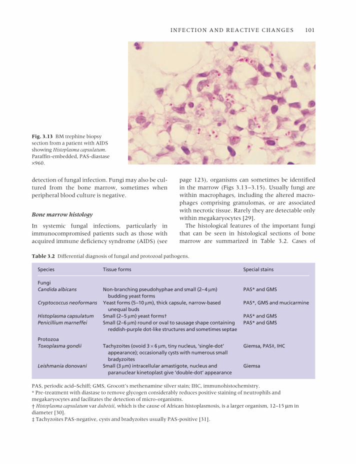
Fig. 3.14 BM trephine biopsysection, Cryptococcus neoformans ina patient with AIDS. Paraffin-embedded: (a) H&E ×960;(b) alcian blue ×376.
(a)
(b)

INFECTION AND REACTIVE CHANGES 103
histoplasma antigen in the urine is highly sensitiveand in endemic areas this test can avoid the need for a bone marrow examination [35]. PCR for thedetection of aspergillus DNA can also be useful.
Parasitic diseases
Peripheral blood
The presence of parasites within the bowel may leadto blood loss with consequent iron deficiencyanaemia. Eosinophilia is also common in pati-ents with helminth infections. In malaria and
babesiosis, parasites are seen within red cells. Malariais associated with haemolytic anaemia and some-times thrombocytopenia. Atypical lymphocytes are present both in malaria and in some patientswith hyper-reactive malarial splenomegaly. Toxo-plasmosis can be associated with lymphocytosis and the presence of considerable numbers of atypical lymphocytes. Leishmaniasis can cause leucopenia and phagocytosed parasites are occa-sionally detectable within peripheral blood mono-cytes or neutrophils. Other parasites detectable free in the peripheral blood are trypanosomes andmicrofilariae.
Fig. 3.15 Section of BM trephinebiopsy specimen in a patient withAIDS with Penicillium marneffeiinfection. Paraffin-embedded: (a) H&E ×376; (b) PAS ×376.
(a)
(b)

104 CHAPTER THREE
Fig. 3.17 BM aspirate,leishmaniasis, showing amacrophage containing numerousorganisms which, in addition to anucleus, have a small paranuclearkinetoplast giving them acharacteristic ‘double-dot’appearance. MGG ×940.
Bone marrow cytology
Malaria parasites are sometimes detected, in redcells or neutrophils, in a bone marrow aspirate (Fig. 3.16), although bone marrow aspiration is nota recommended diagnostic method if malaria is suspected. The bone marrow in malaria also showshypercellularity due to erythroid and granulocytichyperplasia, dyserythropoiesis, giant metamyelo-cytes and increased eosinophils, lymphocytes,plasma cells and macrophages [36,37]. There maybe haemophagocytosis and macrophages can also
contain malarial pigment. In hyper-reactive malar-ial splenomegaly there may be a marked increase inbone marrow lymphocytes [38]. A bone marrowaspirate is very useful in the diagnosis of leish-maniasis (Fig. 3.17) and is a recommended diag-nostic method when this diagnosis is suspected.Trypanosomes are sometimes detected in the bonemarrow, but less often than leishmania. Detection ismore common in immunosuppressed patients [39].Microfilaria are occasionally observed in a bonemarrow aspirate in immunologically normal hosts(Fig. 3.18). Other parasites are rarely detected in the
Fig. 3.16 BM aspirate from apatient with falciparum malariashowing gametocytes. MGG ×960.(By courtesy of Dr D Swirsky,Leeds.)

INFECTION AND REACTIVE CHANGES 105
Fig. 3.18 BM aspirate from apatient with filariasis showing amicrofilaria. MGG ×470. (Bycourtesy of Mrs Seema M Zainal,Bahrein.)
Fig. 3.19 BM aspirate showingToxoplasma gondii in a patient who had had a renal transplant.MGG ×940. (By courtesy of Dr R Cobcroft, Brisbane.)
bone marrow, except in severely immunosup-pressed patients in whom pneumocystis [40] andtoxoplasma (Fig. 3.19) have sometimes been found.Features useful in identifying various protozoanparasites are shown in Table 3.2.
An increase of bone marrow eosinophils andtheir precursors is often apparent when there ishelminth infection.
Bone marrow histology
A bone marrow biopsy in malaria usually shows
increased cellularity and increased macrophageactivity, often with haemophagocytosis. Duringacute attacks of malaria, sinusoids may be packedwith parasitized red cells [37]. In patients who have suffered recurrent attacks of malaria, the bonemarrow may be slate grey or black due to deposi-tion of malaria pigment. It is important to distin-guish malaria pigment (haemozoin) from formalinpigment.
In visceral leishmaniasis, there may be granu-loma formation. In more severe cases there is a dif-fuse increase of macrophages. The organisms are

106 CHAPTER THREE
often seen within macrophages (Fig. 3.20); theirsmall size (3 µm) sometimes leads to their beingconfused with the fungus Histoplasma capsulatum.However, leishmania fail to stain with periodicacid–Schiff (PAS) or silver stains and a Giemsa stainwill demonstrate a small paranuclear basophilicbody, known as the kinetoplast, giving the organ-ism a characteristic ‘double-dot’ appearance.
Granulomas are also seen in toxoplasmosis;rarely, in immunosuppressed individuals, organ-isms are seen in the marrow (Fig. 3.21). These usu-
ally take the form of tachyzoites, which are 3–6 µmin diameter and have a tiny single nucleus. Occa-sionally, cysts containing numerous bradyzoites arepresent. These are of a similar size to tachyzoitesand also have a single nucleus; the lack of a kineto-plast helps to distinguish them from leishmania.Toxoplasma can be identified with a polyclonalantibody.
Microsporidiosis rarely involves the bone marrowbut involvement has been reported at autopsy in apatient with AIDS [41].
Fig. 3.20 BM trephine biopsysection from an HIV-positive manshowing Leishmania donovaniwithin macrophages. H&E ×960.
Fig. 3.21 BM trephine biopsysection, toxoplasmosis, showing a small granuloma in aparatrabecular position containingseveral small organisms with asingle nucleus consistent with thetachyzoites of Toxoplasma gondii;fungal stains were negative and the patient had raised serum IgMantibodies to Toxoplasma. Plastic-embedded, H&E ×390.

INFECTION AND REACTIVE CHANGES 107
Extrapulmonary infection with Pneumocystiscarinii is rare; it is invariably secondary to pul-monary disease, and appears to be more common inthose patients who have received aerosolized pen-tamidine therapy. Approximately a third of patientswith extrapulmonary infection have marrowinvolvement [42]. There are areas of ‘frothy’ exu-date which is pink on an H&E stain; Grocott’smethenamine silver stain demonstrates the cysts,which are 4–6 µm in diameter and often have acrumpled or cup-shaped appearance. Diagnosis isaided by immunohistochemistry using monoclonalantibody of the 3F6 clone (available commerciallyfrom Dako and Novocastra).
Bone marrow granulomas
A granuloma is a compact aggregate of macro-phages. These may include a major component ofepithelioid macrophages which have large amountsof pale pink cytoplasm and ovoid or elongatednuclei with a dispersed chromatin pattern. Oftenseveral epithelioid cells fuse to form a giant cell.Two major types of giant cell are recognized in gran-ulomas: (i) Langhans’ type, which has numerousnuclei arranged around the periphery of the cell;and (ii) foreign body type with nuclei scatteredthroughout the cell. Other cell types, including lym-phocytes, plasma cells, neutrophils, eosinophils andfibroblasts, may be found within granulomas, butthese are not a constant feature.
A wide range of aetiological agents are associatedwith marrow granulomas (Table 3.3). It should benoted that patients with immunodeficiency, such asAIDS, may fail to produce granulomas in response toinfection with organisms that stimulate granulomaformation in normal individuals; this is probablyconsequent on the lack of important T-cell functionsthat facilitate formation of some types of granuloma.
Peripheral blood
There are no specific peripheral blood findings asso-ciated with the presence of bone marrow granulo-mas. The blood film may show features associatedwith the primary disease or, if bone marrow diseaseis extensive, there may be anaemia or pancytopeniawith a leuco-erythroblastic blood film. Lympho-penia is also common [43].
Table 3.3 Bone marrow granulomas [43–46].
InfectionTuberculosisAtypical mycobacterial infectionDisseminated bacillus Calmette–Guérin (BCG)
infection [47]BrucellosisLeprosySyphilisTyphoid feverLegionnaire’s diseaseTularaemia [43]Ehrlichiosis [44]Q feverRocky Mountain spotted fever [43]LeishmaniasisToxoplasmosisHistoplasmosisCryptococcosisSaccharomyces infection [43]BlastomycosisCoccidioidomycosis [48]ParacoccidioidomycosisHerpesvirus infection (EBV, CMV, herpes zoster)Hantaan virus infection (Korean haemorrhagic fever)
[49]Cat scratch disease [49]
Sarcoidosis
Malignant diseaseHodgkin’s disease*Multiple myeloma [50]Non-Hodgkin’s lymphoma*Chronic NK-cell lymphocytosis [51]Mycosis fungoides [43]Acute lymphoblastic leukaemia [43,46,49] Myelodysplastic syndrome [46]Metastatic carcinoma (breast or colon) [52,53]
Drug hypersensitivityPhenytoinProcainamidePhenylbutazone [43]ChlorpropamideSulfasalazine [54]Ibuprofen [43]Indometacin [43]AllopurinolCarbamazepine [55]
Associated with eosinophilic interstitial nephritis
Reaction to foreign substancesAnthracosis and silicosis [46,49,56]Talc [57]Berylliosis [58]
* With or without bone marrow infiltration.

108 CHAPTER THREE
Fig. 3.23 BM trephine biopsysection, miliary tuberculosis,showing an epithelioid granulomacontaining a Langhans’ giant cell;there are numerous lymphocytes at the periphery of the granuloma.Paraffin-embedded, H&E ×390.
Bone marrow cytology
There are no specific features in the bone marrowaspirate in patients with bone marrow granulomas.Occasionally, it is possible to recognize epithelioidcells.
Bone marrow histology
Lipid granulomas
Lipid granulomas (Fig. 3.22) were previously re-
ported to be the most common type of granulomaseen in the marrow, being present in up to 9% ofbiopsies [59]. In our experience, they are consider-ably less common than this. They are of no clinicalimportance and must be distinguished from epithe-lioid granulomas which they can sometimes resem-ble. Similar lesions may be seen in the liver, spleenand lymph nodes and some of these cases have beenreported to be associated with the ingestion of min-eral oil [60]. In the marrow they are usually locatedclose to sinusoids or lymphoid nodules and measurefrom 0.2 to 0.8 mm in diameter. They contain fat
Fig. 3.22 BM trephine biopsysection, lipid granuloma. Paraffin-embedded, H&E ×390.

INFECTION AND REACTIVE CHANGES 109
vacuoles which vary in size but are usually smallerthan the vacuoles in marrow fat cells; these arefound both within macrophages and extracellu-larly. Lipid granulomas usually have plasma cells,eosinophils and lymphocytes within them, andapproximately 5% contain giant cells. Occasionally,the fat vacuoles may be small and easily overlooked,giving the granulomas a sarcoid-like appearance.
Other granulomas
Unless a specific organism can be demonstratedwithin a granuloma, there are usually no histolog-ical features that allow a definitive diagnosis to be made [61]. Because of this, it is important for the pathologist to be aware of all relevant clinicaldetails, in order to be able to suggest an appropriatedifferential diagnosis. All biopsies with granulomasshould have special stains for acid-fast bacilli andfungi performed. Ideally, in those cases in whichmarrow granulomas with an infective aetiology arepossible, for example in patients with a pyrexia ofunknown origin, this should be anticipated and partof the marrow aspirate should be cultured formycobacteria and fungi.
Granulomas are found on marrow biopsy in 15–40% of patients with miliary tuberculosis (Fig. 3.23).Tuberculous granulomas usually contain Langhans’-type giant cells and caseation is present in approx-
imately half the cases with marrow involvement[61]. Acid-fast bacilli cannot be demonstrated inmost cases and, when seen, they are usually scanty(Fig. 3.24). Approximately 50% of patients with dis-seminated Mycobacterium avium intracellulare infec-tion have marrow granulomas (Figs 3.25 and 3.26),ranging from small, ill-defined lymphohistiocyticaggregates to larger, more solid lymphohistiocyticlesions, and small, well-formed epithelioid granulo-mas [62]. Giant cells are only present in a minorityof lesions and necrosis is not usually seen. Whenorganisms are very numerous, the macrophagescontaining them may appear foamy (Fig. 3.26a) or may even resemble Gaucher’s cells. Occasion-ally, organisms are recognizable on a Giemsa stain(Fig. 3.26b). Atypical mycobacteria may be demon-strated by special stains, sometimes in large num-bers; they tend to be longer, more curved and morecoarsely beaded than tubercle bacilli. They are PAS-positive, whereas M. tuberculosis is PAS-negative.Marrow granulomas containing foamy macrophagesare occasionally seen in patients with leprosy; a Fitestain will demonstrate the acid-fast bacilli of M. lep-rae [6]. Foamy macrophages may also be a featureof granulomas due to typhoid. Small, poorly formedepithelioid granulomas are found in the bone marrow in most cases of brucellosis. Distinctive‘doughnut-type’ granulomas may be seen in themarrow in Q fever [63], although they do not
Fig. 3.24 BM trephine biopsysection, miliary tuberculosis,showing a granuloma containingan acid-fast bacillus. Paraffin-embedded, Ziehl–Neelsen stain×970.

110 CHAPTER THREE
appear to be specific for this disease [18]. They havealso been reported, for example, in CMV infectionin immunodeficient subjects [64] and in Hodgkin’sdisease, infectious mononucleosis and typhoid fever.This type of granuloma often has a central emptyspace, surrounded by neutrophils, lymphocytes,histiocytes and concentrically arranged, laminatedfibrinoid material; more haphazardly arrangedlesions without a central space also occur, as dosmall areas of fibrinoid necrosis [63].
Disseminated infection by the fungus H. capsula-tum usually involves the bone marrow; in normalhosts there are numerous granulomas, often with
Langhans’ giant cells and necrosis. Discrete granu-lomas are present in only a minority of immu-nodeficient hosts; more commonly such patientshave ill-defined lymphohistiocytic aggregates orsheets of macrophages infiltrating between haemo-poietic cells [65] (see Fig. 3.13). Tiny yeast forms,2–5 µm in diameter, some of which show unequalbudding, are present within macrophages. Theorganisms may be seen with an H&E stain, but arebest visualized using Gomori’s methenamine silver(GMS) stain or a PAS stain. Cryptococcus neoformansinfection may cause granulomas. The organisms seenin tissue sections are yeasts (5–10 µm in diameter)
Fig. 3.25 BM trephine biopsysection from a patient with AIDSand disseminated M. aviumintracellulare infection. (a) Poorlyformed granuloma made up ofepithelioid macrophages, many ofwhich have vacuolated cytoplasm.Paraffin-embedded, H&E ×390.(b) Large numbers of acid-fastbacilli; note the coarse beading ofthe organisms. Paraffin-embedded,Ziehl–Neelsen stain ×970.
(a)
(b)
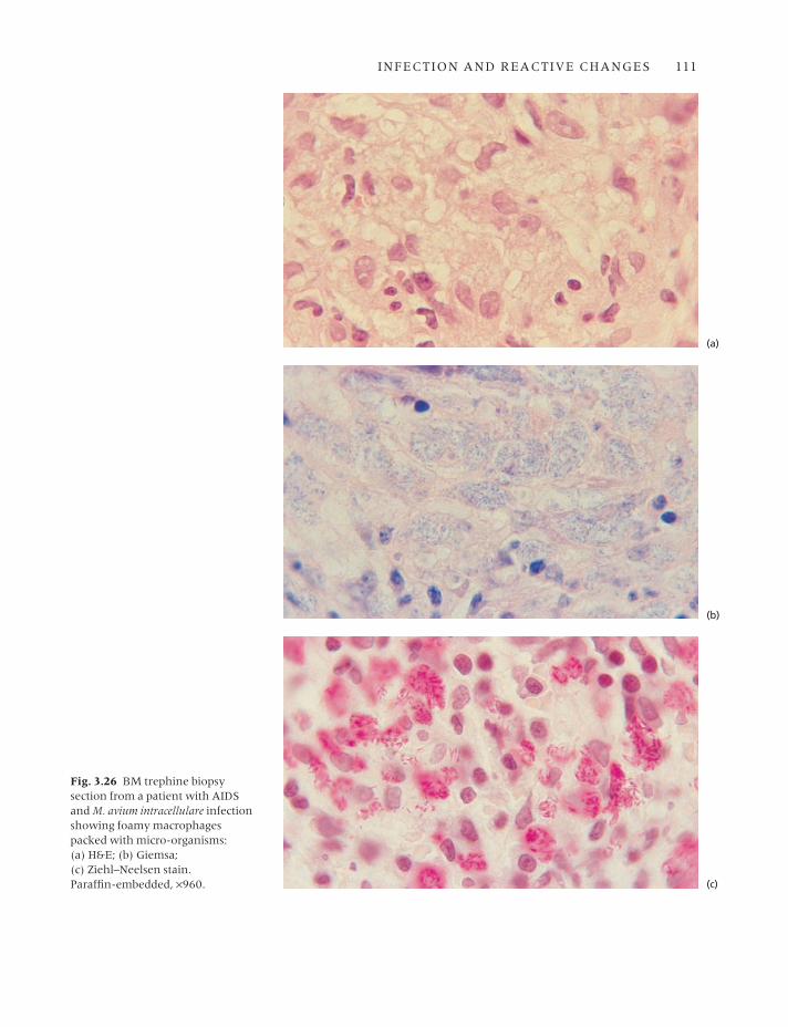
Fig. 3.26 BM trephine biopsysection from a patient with AIDSand M. avium intracellulare infectionshowing foamy macrophagespacked with micro-organisms: (a) H&E; (b) Giemsa; (c) Ziehl–Neelsen stain. Paraffin-embedded, ×960.
(a)
(b)
(c)
INFECTION AND REACTIVE CHANGES 111

112 CHAPTER THREE
with a wide capsular halo and narrow-based,unequal budding (Fig. 3.27); with a mucicarminestain the capsule of the yeast is red. The capsule isalso well stained with alcian blue (see Fig. 3.14b)and with PAS (Fig. 3.27a). Infection with the proto-zoans Leishmania donovani and Toxoplasma gondiimay involve the marrow and provoke granulomaformation (see Figs 3.20 and 3.21).
In up to 60% of patients with infectious mono-nucleosis, small epithelioid granulomas are seen in histological sections of bone marrow biopsy;Langhans’ giant cells and necrosis are not seen.
Marrow granulomas are seen less commonly inother viral infections.
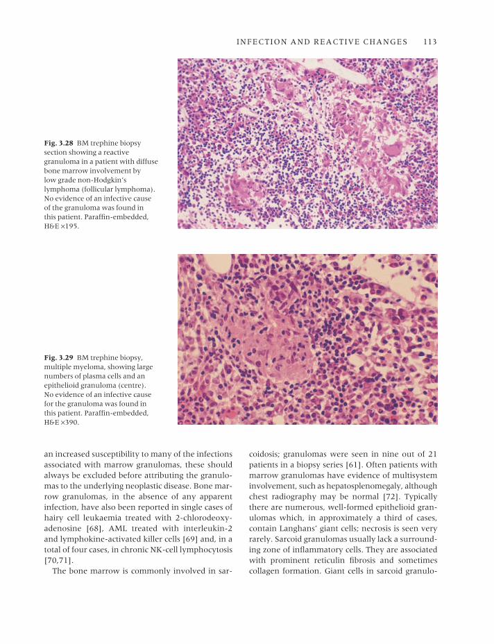
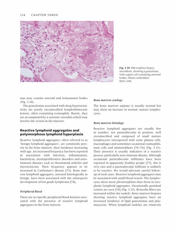
Granulomas may be seen in Hodgkin’s disease[66], non-Hodgkin’s lymphoma (Fig. 3.28) andmultiple myeloma [50] (Fig. 3.29) in associationwith neoplastic infiltration of the marrow. In bothHodgkin’s disease and non-Hodgkin’s lymphoma,granulomas also occur in the absence of marrowinvolvement; these are usually small, well-formedepithelioid granulomas, although larger, poorlyformed lymphohistiocytic lesions have also beenreported [67]. Since patients with lymphoma have
Fig. 3.27 BM trephine biopsysections from a patient with C.neoformans infection. Paraffin-embedded. (a) A small epithelioidgranuloma containing large yeastforms with narrow-based unequalbudding. PAS ×195. (b) Buddingyeast forms. GMS ×390.
(a)
(b)
INFECTION AND REACTIVE CHANGES 113
an increased susceptibility to many of the infectionsassociated with marrow granulomas, these shouldalways be excluded before attributing the granulo-mas to the underlying neoplastic disease. Bone mar-row granulomas, in the absence of any apparentinfection, have also been reported in single cases ofhairy cell leukaemia treated with 2-chlorodeoxy-adenosine [68], AML treated with interleukin-2and lymphokine-activated killer cells [69] and, in atotal of four cases, in chronic NK-cell lymphocytosis[70,71].
The bone marrow is commonly involved in sar-
coidosis; granulomas were seen in nine out of 21patients in a biopsy series [61]. Often patients withmarrow granulomas have evidence of multisysteminvolvement, such as hepatosplenomegaly, althoughchest radiography may be normal [72]. Typicallythere are numerous, well-formed epithelioid gran-ulomas which, in approximately a third of cases,contain Langhans’ giant cells; necrosis is seen veryrarely. Sarcoid granulomas usually lack a surround-ing zone of inflammatory cells. They are associatedwith prominent reticulin fibrosis and sometimescollagen formation. Giant cells in sarcoid granulo-
Fig. 3.28 BM trephine biopsysection showing a reactivegranuloma in a patient with diffusebone marrow involvement by low grade non-Hodgkin’slymphoma (follicular lymphoma).No evidence of an infective cause of the granuloma was found in this patient. Paraffin-embedded,H&E ×195.
Fig. 3.29 BM trephine biopsy,multiple myeloma, showing largenumbers of plasma cells and anepithelioid granuloma (centre). No evidence of an infective causefor the granuloma was found in this patient. Paraffin-embedded,H&E ×390.
114 CHAPTER THREE
mas may contain asteroid and Schaumann bodies(Fig. 3.30).
The granulomas associated with drug hypersensi-tivity are poorly circumscribed lymphohistiocyticlesions, often containing eosinophils. Rarely, theyare accompanied by a systemic vasculitis which mayinvolve the vessels in the marrow.
Reactive lymphoid aggregates andpolymorphous lymphoid hyperplasia
Reactive lymphoid aggregates, often referred to as‘benign lymphoid aggregates’, are commonly pres-ent in the bone marrow, their incidence increasingwith age. An increased frequency has been reportedin association with infection, inflammation,haemolysis, myeloproliferative disorders and auto-immune diseases such as rheumatoid arthritis andthyrotoxicosis. Their frequency appears to beincreased in Castleman’s disease [73]. Bone mar-row lymphoid aggregates, assessed histologically asbenign, have been associated with the subsequentdevelopment of low grade lymphoma [74].
Peripheral blood
There are no specific peripheral blood features asso-ciated with the presence of reactive lymphoidaggregates in the bone marrow.
Bone marrow cytology
The bone marrow aspirate is usually normal butmay show an increase in normal, mature lympho-cytes.
Bone marrow histology
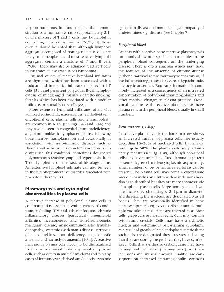
Reactive lymphoid aggregates are usually few in number, not paratrabecular in position, well circumscribed and composed of small mature lymphocytes interspersed with some plasma cells,macrophages and sometimes occasional eosinophils,mast cells and immunoblasts [74–76] (Fig. 3.31).Their presence is usually indicative of a reactiveprocess, particularly auto-immune disease. Althoughoccasional paratrabecular infiltrates have beenreported in apparently healthy people [77], this isvery rare and a paratrabecular infiltrate is unlikelyto be reactive. We would advocate careful follow-up of such cases. Reactive lymphoid aggregates maybe associated with small blood vessels. The lympho-cytes show more pleomorphism than those in neo-plastic lymphoid aggregates. Occasionally germinalcentres are seen [78] (Fig. 3.32). Reticulin fibres areincreased within the nodule. Bone marrow biopsiesshowing reactive lymphoid aggregates have anincreased incidence of lipid granulomas and plas-macytosis. When lymphoid nodules are relatively
Fig. 3.30 BM trephine biopsy,sarcoidosis, showing a granulomawith a giant cell containing asteroidbodies. Plastic-embedded, H&E ×390.
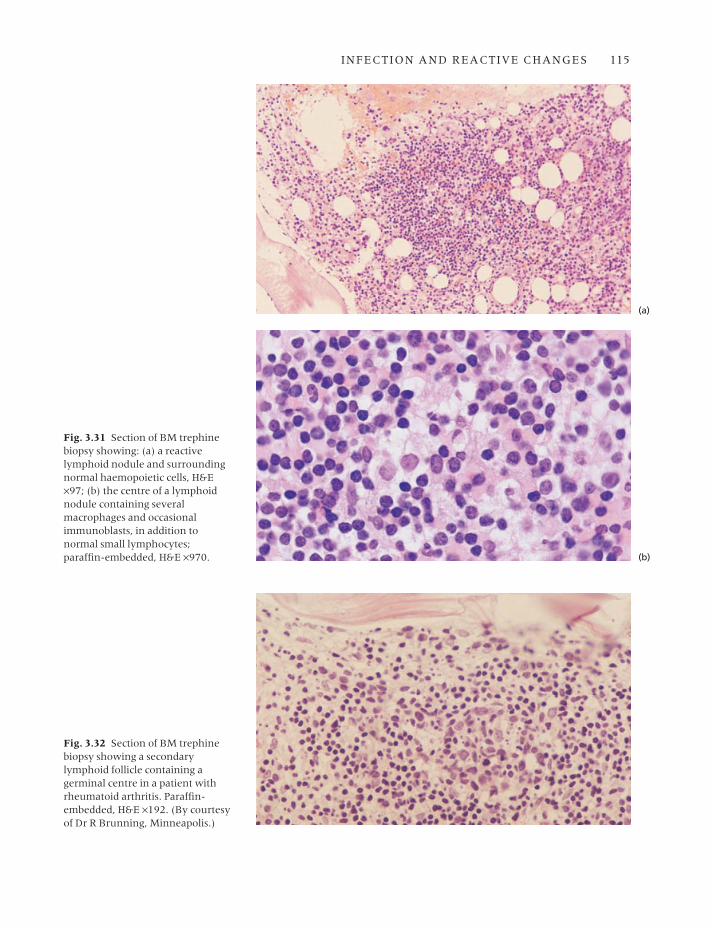
Fig. 3.32 Section of BM trephinebiopsy showing a secondarylymphoid follicle containing agerminal centre in a patient withrheumatoid arthritis. Paraffin-embedded, H&E ×192. (By courtesyof Dr R Brunning, Minneapolis.)
Fig. 3.31 Section of BM trephinebiopsy showing: (a) a reactivelymphoid nodule and surroundingnormal haemopoietic cells, H&E×97; (b) the centre of a lymphoidnodule containing severalmacrophages and occasionalimmunoblasts, in addition tonormal small lymphocytes;paraffin-embedded, H&E ×970.
(a)
(b)
INFECTION AND REACTIVE CHANGES 115
large or numerous, immunohistochemical demon-stration of a normal κ:λ ratio (approximately 2:1) or of a mixture of T and B cells may be helpful inconfirming their reactive nature [76,79,80]. How-ever, it should be noted that, although lymphoidaggregates composed of homogeneous B cells arelikely to be neoplastic and most reactive lymphoidaggregates contain a mixture of T and B cells[79,80], there may also be admixed reactive T cellsin infiltrates of low grade B-cell lymphoma.
Unusual causes of reactive lymphoid infiltratesare thymoma, which has been associated with anodular and interstitial infiltrate of polyclonal Tcells [81], and persistent polyclonal B-cell lympho-cytosis of middle-aged, mainly cigarette-smoking,females which has been associated with a nodularinfiltrate, presumably of B cells [82].
More extensive lymphoid infiltrates, often withadmixed eosinophils, macrophages, epithelioid cells,endothelial cells, plasma cells and immunoblasts,are common in AIDS (see Figs 3.43 and 3.44) andmay also be seen in congenital immunodeficiency,angioimmunoblastic lymphadenopathy, followingbone marrow transplantation and occasionally inassociation with auto-immune diseases such asrheumatoid arthritis. It is sometimes not possible todistinguish this condition, sometimes designatedpolymorphous reactive lymphoid hyperplasia, fromT-cell lymphoma on the basis of histology alone. An extensive lymphoid infiltrate can also be seen in the lymphoproliferative disorder associated withphenytoin therapy [83].
Plasmacytosis and cytologicalabnormalities in plasma cells
A reactive increase of polyclonal plasma cells iscommon and is associated with a variety of condi-tions including HIV and other infections, chronicinflammatory diseases (particularly rheumatoidarthritis), haemopoietic and non-haemopoieticmalignant disease, angio-immunoblastic lympha-denopathy, systemic Castleman’s disease, cirrhosis,diabetes mellitus, iron deficiency, megaloblasticanaemia and haemolytic anaemia [9,84]. A reactiveincrease in plasma cells needs to be distinguishedfrom bone marrow infiltration by neoplastic plasmacells, such as occurs in multiple myeloma and in manycases of immunocyte-derived amyloidosis, systemic
light chain disease and monoclonal gammopathy ofundetermined significance (see Chapter 7).
Peripheral blood
Patients with reactive bone marrow plasmacytosiscommonly show non-specific abnormalities in theperipheral blood consequent on the underlying disease. There is often anaemia which may have the features of the anaemia of chronic disease(either a normochromic, normocytic anaemia or, ifthe inflammatory process is severe, a hypochromic,microcytic anaemia). Rouleaux formation is com-monly increased as a consequence of an increasedconcentration of polyclonal immunoglobulins andother reactive changes in plasma proteins. Occa-sional patients with reactive plasmacytosis haveplasma cells in the peripheral blood, usually in smallnumbers.
Bone marrow cytology
In reactive plasmacytosis the bone marrow showsan increased number of plasma cells, not usuallyexceeding 10–20% of nucleated cells, but in rarecases up to 50%. The plasma cells are predomi-nately mature (see Fig. 1.48) although occasionalcells may have nucleoli, a diffuse chromatin patternor some degree of nucleocytoplasmic asynchrony.Small numbers of bi- or trinucleated forms can bepresent. The plasma cells may contain cytoplasmicvacuoles or inclusions. Intranuclear inclusions havealso been described but they are more characteristicof neoplastic plasma cells. Large homogeneous hya-line inclusions, often single, 2–3 µm in diameterand displacing the nucleus, are designated Russellbodies. They are occasionally identified in bonemarrow aspirates (Fig. 3.33). Cells containing mul-tiple vacuoles or inclusions are referred to as Mottcells, grape cells or morular cells. Cells may containcytoplasmic crystals. Cells may have a pyknoticnucleus and voluminous pale-staining cytoplasm,as a result of greatly dilated endoplasmic reticulum;such cells are designated thesaurocytes indicatingthat they are storing the products they have synthe-sized. Cells that synthesize carbohydrate may haveflaming pink cytoplasm (‘flaming cells’). All theseinclusions and unusual tinctorial qualities are con-sequent on increased immunoglobulin synthesis
116 CHAPTER THREE

INFECTION AND REACTIVE CHANGES 117
within the rough endoplasmic reticulum. Nuclearinclusions, known as Dutcher bodies, are con-sequent on invagination from the cytoplasm. Cellsof these various types are characteristic of condi-tions with immune stimulation but neoplasticplasma cells often show similar features. Plasma-cytic satellitism (a central macrophage surroundedby plasma cells) and increased mast cells, eosin-ophils and megakaryocytes favour reactive ratherthan neoplastic plasmacytosis [9].
Occasionally, plasma cells contain haemosiderininclusions which are irregular in shape, relativelylarge and stain greenish-black with a May–Grünwald–Giemsa (MGG) stain (see Fig. 2.4). Theirpresence is associated with iron overload (e.g.haemochromatosis and transfusion siderosis) andwith chronic alcoholism [85].
Bone marrow histology
A trephine biopsy section in reactive plasmacytosisshows an interstitial infiltrate of plasma cells, partic-ularly adjacent to capillaries (see Fig. 1.49). Plasmacells are sometimes clustered around macrophages.A minority of cases have small clusters of plasmacells but large homogeneous nodules, which are afeature of multiple myeloma, are not seen. The vari-ety of inclusions described above may also be appar-ent in histological sections (Fig. 3.34) and somedegree of cellular immaturity may be noted. Russell
bodies and Dutcher bodies stain pink with an H&Estain and show variable PAS staining. In reactiveplasmacytosis immunocytochemistry shows that κ- and λ-expressing plasma cells are present in aratio of approximately 2:1. About half the plasmacells express γ heavy chain, about a third α and theremainder µ [86].
Plasmacytosis may be associated with other reactive changes such as granulocytic hyperplasia,lymphoid aggregates and increased numbers ofmacrophages. The macrophages may have en-hanced haemophagocytic activity and an increasediron content.
Mast cells
Small numbers of mast cells are present in normalbone marrow aspirates. However, it should be notedthat the normal number of mast cells in the trephinebiopsy of healthy subjects has not been defined.Mast cells are increased as a reactive change in asso-ciation with a variety of pathological processes.Increased numbers have been noted in infection,inflammation, renal failure, lymphoproliferative dis-orders and reactive lymphocytosis, aplastic anaemia,paroxysmal nocturnal haemoglobinuria, myelopro-liferative disorders and the myelodysplastic syn-dromes [87–89]. Mast cells accumulate in areas ofconnective tissue proliferation, for example in frac-ture callus and in zones of osteitis fibrosa in patients
Fig. 3.33 BM aspirate film showinga Russell body in a plasma cell in apatient with reactive plasmacytosis.MGG ×940.

Fig. 3.34 BM trephine biopsysection, myelodysplastic syndrome,showing prominent plasma cellsincluding one with multiplecytoplasmic vacuoles (Mott cell).Plastic-embedded, H&E ×940.
118 CHAPTER THREE
with renal failure. Increased numbers of mast cellsare present also in systemic mastocytosis but theseneoplastic mast cells are usually morphologicallyabnormal (see page 221).
Peripheral blood
There are no specific peripheral blood features asso-ciated with a reactive increase of bone marrow mastcells.
Bone marrow cytology
The cytological features of bone marrow mast cellshave been described on page 25. Lymphocytosisand plasmacytosis may co-exist with a reactiveincrease in mast cells.
Bone marrow histology
The characteristics of mast cells in histological sec-tions have been described on page 26 and the fea-tures of systemic mastocytosis will be described onpage 223. In histological sections, mast cells may beconfused with fibroblasts or macrophages becauseof their elongated shape, oval nuclei and the poorstaining of their granules with H&E. The lesionsassociated with drug hypersensitivity, previouslydesignated ‘eosinophilic fibrohistiocytic lesions’[90], are now known to represent proliferation of
eosinophils and mast cells, mainly in patients withsystemic mastocytosis.
When mast cells are increased, there may be anassociated increase in plasma cells and lymphocytes.
Histiocytosis
An increase in macrophages (histiocytes) is com-mon in a variety of infective and inflammatory conditions and whenever there is bone marrowhyperplasia, ineffective haemopoiesis or increasedbreakdown of blood cells. All of the conditions cap-able of causing granuloma formation and haemo-phagocytic syndromes can also cause an increase of macrophages without granuloma formation orprominent haemophagocytosis. Macrophages pres-ent may range from relatively immature cells withlimited phagocytic capacity to mature cells whichmay be foamy or contain cellular debris, haemo-siderin or a few haemopoietic cells, erythrocytes,neutrophils or platelets.
Increased bone marrow macrophages may be seenafter granulocyte–macrophage colony-stimulatingfactor (GM-CSF) therapy when they may consti-tute as many as 90% of bone marrow cells [91].They have been prominent in patients with failureto engraft after bone marrow transplantation par-ticularly, but not only, when GM-CSF has beengiven [92]. A marked increase in macrophages hasbeen reported at presentation of acute lympho-
INFECTION AND REACTIVE CHANGES 119
blastic leukaemia in a patient without any apparentinfection [93].
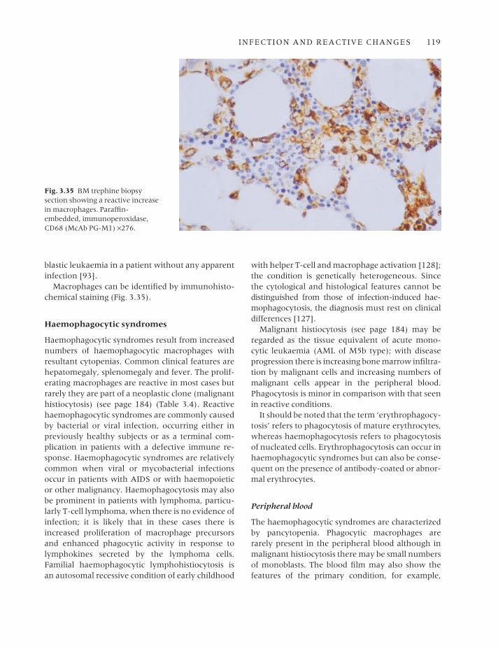
Macrophages can be identified by immunohisto-chemical staining (Fig. 3.35).
Haemophagocytic syndromes
Haemophagocytic syndromes result from increasednumbers of haemophagocytic macrophages withresultant cytopenias. Common clinical features arehepatomegaly, splenomegaly and fever. The prolif-erating macrophages are reactive in most cases butrarely they are part of a neoplastic clone (malignanthistiocytosis) (see page 184) (Table 3.4). Reactivehaemophagocytic syndromes are commonly causedby bacterial or viral infection, occurring either inpreviously healthy subjects or as a terminal com-plication in patients with a defective immune re-sponse. Haemophagocytic syndromes are relativelycommon when viral or mycobacterial infectionsoccur in patients with AIDS or with haemopoieticor other malignancy. Haemophagocytosis may alsobe prominent in patients with lymphoma, particu-larly T-cell lymphoma, when there is no evidence ofinfection; it is likely that in these cases there isincreased proliferation of macrophage precursorsand enhanced phagocytic activity in response tolymphokines secreted by the lymphoma cells.Familial haemophagocytic lymphohistiocytosis isan autosomal recessive condition of early childhood
with helper T-cell and macrophage activation [128];the condition is genetically heterogeneous. Sincethe cytological and histological features cannot bedistinguished from those of infection-induced hae-mophagocytosis, the diagnosis must rest on clinicaldifferences [127].
Malignant histiocytosis (see page 184) may beregarded as the tissue equivalent of acute mono-cytic leukaemia (AML of M5b type); with diseaseprogression there is increasing bone marrow infiltra-tion by malignant cells and increasing numbers ofmalignant cells appear in the peripheral blood.Phagocytosis is minor in comparison with that seenin reactive conditions.
It should be noted that the term ‘erythrophagocy-tosis’ refers to phagocytosis of mature erythrocytes,whereas haemophagocytosis refers to phagocytosisof nucleated cells. Erythrophagocytosis can occur inhaemophagocytic syndromes but can also be conse-quent on the presence of antibody-coated or abnor-mal erythrocytes.
Peripheral blood
The haemophagocytic syndromes are characterizedby pancytopenia. Phagocytic macrophages arerarely present in the peripheral blood although inmalignant histiocytosis there may be small numbersof monoblasts. The blood film may also show thefeatures of the primary condition, for example,
Fig. 3.35 BM trephine biopsysection showing a reactive increasein macrophages. Paraffin-embedded, immunoperoxidase,CD68 (McAb PG-M1) ×276.

120 CHAPTER THREE
atypical lymphocytes in patients with familial lymphohistiocytosis (Fig. 3.36) or EBV or otherviral infection.
Bone marrow cytology
In the secondary haemophagocytic syndromesthere are increased numbers of macrophages andhaemophagocytosis is usually prominent, withmany macrophages having ingested numerous cellsof various haemopoietic lineages (Figs 3.37 and
3.38). The macrophages are mainly mature and lackatypical features. The bone marrow aspirate mayalso show other abnormalities due to the primarydisease. For example, in viral infections there isusually an increase of lymphocytes, which may beimmature or atypical, and in bacterial infection thereis granulocytic hyperplasia with toxic changes inthe neutrophil lineage. In familial haemophagocyticlymphohistiocytosis, the bone marrow findings (Fig. 3.39) are identical to those of infection-inducedhaemophagocytosis. Repeated bone marrow exam-
Table 3.4 Conditions associated with a haemophagocytic syndrome [45,94–127].
Malignant histiocytosis
Reactive haemophagocytic syndromes
Induced by viral infectionHerpesviruses
EB virus (including fatal infectious mononucleosis in X-linked lymphoproliferative syndrome)
Herpes simplexHerpes zosterHuman herpesvirus 6 [94]Human herpesvirus 7 [95]Cytomegalovirus
Other virusesAdenovirusMeasles (vaccine virus)Influenza A [96]Para-influenzaVacciniaRubella (congenital)Parvovirus B19 [97]Respiratory syncytial virus [98]Kyasanur forest diseaseDengue [99]Hepatitis A virus [100]
Induced by bacterial infectionSalmonella typhiBrucellosisStaphylococcal, streptococcal, E. coli, H. influenzae,
Acinetobacter species, Bacteroides fragilis,Pseudomonas species and Klebsiella speciesinfections [99,100–104]
Legionnaire’s disease (Legionella pneumophila)Mycobacterium tuberculosisAtypical mycobacterial infectionPsittacosis [99]Mycoplasma pneumoniae infection [105]Human granulocytic and monocytic ehrlichiosis
[106,107]
Induced by rickettsiaeRocky Mountain spotted feverQ fever [108]Rickettsia tsutsugamushi infection [99]
Induced by protozoan and other parasitesToxoplasmosisLeishmaniasisMalaria [109]Babesiosis [110]
Induced by fungiHistoplasmosisCandidiasis [103]Trichosporonosis [111]
Associated with certain lymphomas, particularly T-celllymphomas [112–114], including T-cell granularlymphocytic lymphoma [115] but occasionally naturalkiller (NK) lymphoma [116], B-cell lymphoma [117]and Hodgkin’s disease [118]
Kawasaki’s diseaseKikuchi’s disease [119]Anticonvulsant lymphadenopathySarcoidosis [114]Systemic lupus erythematosus [99] and other auto-
immune disease [120]Graft-versus-host disease (GVHD) [121]Following GM-CSF [122] or IL-3 [123] administrationCytophagic histiocytic panniculitis [124]Lysinuric protein intolerance [125]Griscelli syndrome [126]As a terminal complication in patients with various
immunodeficiencies and malignant conditions (ALL, CLL, HD, NHL, hairy cell leukaemia, AML,carcinoma, Chediak–Higashi syndrome)—probably asa complication of infection
Familial haemophagocytic lymphohistiocytosis [127]

INFECTION AND REACTIVE CHANGES 121
Fig. 3.36 PB film from a child with familial lymphohistiocytosisshowing atypical lymphoid cells.MGG ×940.
Fig. 3.37 BM aspirate, reactivehaemophagocytosis in a patientwith AIDS and miliary tuberculosis.MGG ×940.
Fig. 3.38 BM aspirate, reactivehaemophagocytosis, showing an iron-laden macrophage that has ingested neutrophils andneutrophil precursors. Perls’ stain ×940.

ination may be needed to establish the diagnosis,since abnormalities are detected at presentation inless than a third of patients [127]. When haemo-phagocytosis is secondary to a T-cell lymphoma themarrow may show closely intermingled lymphomacells and macrophages but cases have also beenreported in which the bone marrow shows onlyhaemophagocytosis with the lymphomatous infiltratebeing confined to other tissues [113].
In malignant histiocytosis, the bone marrowinfiltrate may initially be very scanty. The abnormalpopulation includes a much larger proportion ofmonoblasts and promonocytes while mature histio-cytes are relatively less common. Phagocytic activityis not prominent.
Bone marrow histology
In early cases of reactive haemophagocytic syn-drome (e.g. virus-induced) there is a hypercellularbone marrow with few macrophages [103]. Laterthere are more macrophages with hypoplasia oferythroid and myeloid lines; megakaryocyte num-bers are either normal or increased. The degree ofmacrophage infiltration is variable (Fig. 3.40); insome cases it is inconspicuous, while in others thereis diffuse replacement of the marrow by maturemacrophages with a low nucleocytoplasmic ratio,dispersed chromatin, inconspicuous nucleoli and
abundant cytoplasm which is often vacuolated.Haemophagocytosis is often less apparent than inmarrow films [103,129]; however, in some cases it is striking (Fig. 3.41). Features of the underlyingdisease may be present. Atypical lymphoid cells areseen in some cases of EBV infection. Lymphomacells may be detected (see Fig. 6.61) but it should benoted that there may be no detectable lymphomacells in the bone marrow in cases of lymphoma-associated haemophagocytic syndrome. Granulomasmay be present in tuberculosis [130] and in a varietyof other infections associated with a haemophago-cytic syndrome (see Table 3.4).
If haemophagocytic lymphohistiocytosis is sus-pected, a trephine biopsy is recommended becauseclusters or sheets of macrophages may be detectedin cases in which the bone marrow aspirate showsno significant abnormality [131].
In malignant histiocytosis, the marrow biopsy isoften normal in the early stages of the disease.Later, infiltration may occur; infiltration is initiallyfocal then diffuse.
Problems and pitfalls
The major problems in assessing haemophagocyticsyndromes are: (i) distinguishing malignant his-tiocytosis from reactive conditions; and (ii), in reac-tive haemophagocytosis, determining whether the
Fig. 3.39 BM aspirate, familiallymphohistiocytosis, showinghaemophagocytosis and atypicallymphoid cells. MGG ×940.
122 CHAPTER THREE

INFECTION AND REACTIVE CHANGES 123
Fig. 3.40 BM trephine biopsysection, AIDS, showing increasedcellularity, dyserythropoiesis andnumerous macrophages, some of which contain erythrocytes and apoptotic normoblasts(haemophagocytosis).Paraffin-embedded, H&E ×390.
Fig. 3.41 BM section from anautopsy on a child with terminalhaemophagocytic syndrome as acomplication of Chediak–Higashisyndrome showing macrophagesstuffed with red cells. Paraffin-embedded, H&E ×916. (By courtesyof Dr Wendy Erber and Dr L Matz,Perth, Western Australia.)
underlying condition is an infection or, less often, alymphoid neoplasm. Malignant histiocytosis is rarewhereas reactive haemophagocytosis is relativelycommon. Demonstration of rearrangement ofimmunoglobulin heavy chain (IGH) or T-cell recep-tor (TCR) genes provides presumptive evidence ofan underlying neoplasm but does not give definiteevidence of the lineage involved. Molecular evi-dence of clonality is lacking when the associatedneoplasm is of NK lineage. In malignant histiocyto-sis, cytogenetic analysis occasionally shows an
abnormality characteristic of neoplasms of mono-cyte lineage, thus confirming the diagnosis. In gen-eral, marked haemophagocytosis suggests a reactiverather than a neoplastic condition. Vigorous effortsshould be made to identify an infective cause. In situhybridization techniques (see Table 3.1) are usefulfor demonstrating underlying CMV or EBV infec-tion [132], and stains and cultures for mycobacteriamay be positive. Most childhood cases of haemo-phagocytic lymphohistiocytosis, both sporadic andfamilial, are infection-related. However, some

sporadic cases in children have atypical large gran-ular lymphocytes; since T cells in such cases aresometimes shown to be clonal, it is possible thatthere is an underlying T-cell neoplasm [133].
HIV infection and the acquired immunedeficiency syndrome
In HIV infection, the main indications for exam-ination of the bone marrow are pyrexia of un-known origin and various cytopenias. It may also be required as a staging procedure in patients with lymphoma. A biopsy is very often useful, par-ticularly in patients with a hypocellular aspirate.Part of any aspirate from a febrile patient should be submitted for microbiological culture. In addi-tion to routine stains, biopsied tissues should bestained, when appropriate, for acid-fast bacilli andfungi.
Peripheral blood
The earliest haematological manifestations of HIVinfection occur at the time of primary infectionwhen atypical lymphocytes appear in the peripheralblood, often in association with a febrile illnesswhich clinically can resemble infectious mono-nucleosis. Primary HIV infection also occasion-ally causes transient pancytopenia. Patients withestablished infection may have lymphocytosis, consequent on an increase in CD8-positive lym-phocytes, or isolated thrombocytopenia resultingfrom peripheral destruction of platelets. Throm-bocytopenia may also be a feature of a syndromeresembling thrombotic thrombocytopenic purpuraand, in such cases, red cell fragments are present[134]. Late in the course of the disease, there is usu-ally pancytopenia with very marked lymphopenia.Red cells may show anisocytosis and poikilocytosis.The reticulocyte count is reduced. Neutrophils maybe dysplastic. Because of the frequency of oppor-tunistic infections, the peripheral blood may alsoshow non-specific reactive changes such asincreased rouleaux formation, left shift and toxicchanges in neutrophils, and the presence of im-mature monocytes and reactive lymphocytes. Thehaematological effects of the HIV infection itself,and the associated opportunistic infections, may be
compounded by the effects of therapy; patients taking zidovudine, particularly in the higher dosesused in single-agent anti-retroviral therapy, usuallyhave marked macrocytosis and there may bemarked dysplastic changes in blood cells.
Bone marrow cytology
Early in the course of HIV infection the bone mar-row is of normal cellularity or, during the course ofintercurrent infection, shows granulocytic hyper-plasia. When there is immune thrombocytopenia,megakaryocyte numbers are normal or increased.Common non-specific changes are infiltration bylymphocytes and plasma cells and an increase ofmacrophages or eosinophils. Erythropoiesis mayshow mild dysplastic features such as nuclear irregularity and fragmentation. In patients takingzidovudine, erythropoiesis is megaloblastic and dys-erythropoiesis is more marked; dysplastic changessuch as nuclear fragmentation are also noted in the granulocytic series. Giant metamyelocytes arequite common, their presence correlating with theoccurrence of detached nuclear fragments in granu-locytes [135]. Apoptosis is increased. With diseaseprogression, the bone marrow becomes progres-sively more hypocellular and aspiration becomesdifficult due to increased reticulin deposition.Gelatinous transformation is very common, beinggreatest in cachectic patients. Bone marrow necro-sis is sometimes seen. In patients with advanced disease, the bone marrow aspirate may provide evidence of miliary tuberculosis, atypical mycobac-terial infection or disseminated fungal or parasiticinfection. Because of the deficient host response,mycobacterial infection may be associated with thepresence of numerous bacteria within macro-phages, which may be foamy or may morphologic-ally resemble Gaucher’s cells. Haemophagocyticsyndromes (see page 119) secondary to tuberculosisor other infections are relatively common inpatients with AIDS (see Fig. 3.37). Bone marrowinvolvement is common when lymphoma, particu-larly Burkitt’s lymphoma or Hodgkin’s disease,complicates AIDS. The former is often detected in a bone marrow aspirate but in Hodgkin’s disease the aspirate is often negative, even when the bonemarrow is infiltrated.
124 CHAPTER THREE
INFECTION AND REACTIVE CHANGES 125
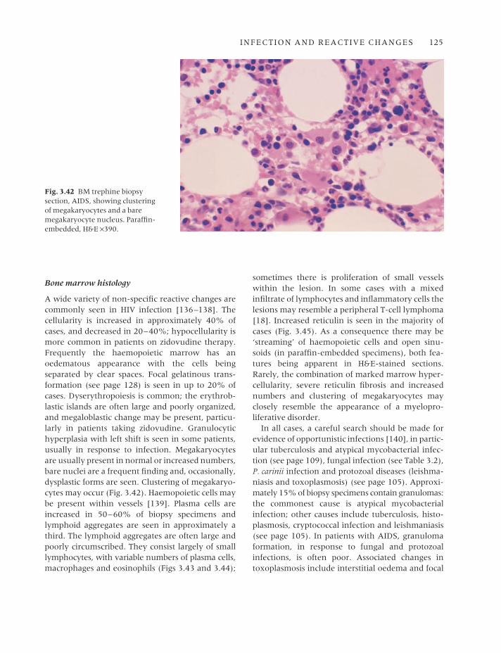
Bone marrow histology
A wide variety of non-specific reactive changes arecommonly seen in HIV infection [136–138]. Thecellularity is increased in approximately 40% ofcases, and decreased in 20–40%; hypocellularity ismore common in patients on zidovudine therapy.Frequently the haemopoietic marrow has an oedematous appearance with the cells being separated by clear spaces. Focal gelatinous trans-formation (see page 128) is seen in up to 20% ofcases. Dyserythropoiesis is common; the erythrob-lastic islands are often large and poorly organized,and megaloblastic change may be present, particu-larly in patients taking zidovudine. Granulocytichyperplasia with left shift is seen in some patients,usually in response to infection. Megakaryocytesare usually present in normal or increased numbers,bare nuclei are a frequent finding and, occasionally,dysplastic forms are seen. Clustering of megakaryo-cytes may occur (Fig. 3.42). Haemopoietic cells maybe present within vessels [139]. Plasma cells areincreased in 50–60% of biopsy specimens and lymphoid aggregates are seen in approximately athird. The lymphoid aggregates are often large andpoorly circumscribed. They consist largely of smalllymphocytes, with variable numbers of plasma cells,macrophages and eosinophils (Figs 3.43 and 3.44);
sometimes there is proliferation of small vesselswithin the lesion. In some cases with a mixedinfiltrate of lymphocytes and inflammatory cells thelesions may resemble a peripheral T-cell lymphoma[18]. Increased reticulin is seen in the majority ofcases (Fig. 3.45). As a consequence there may be‘streaming’ of haemopoietic cells and open sinu-soids (in paraffin-embedded specimens), both fea-tures being apparent in H&E-stained sections.Rarely, the combination of marked marrow hyper-cellularity, severe reticulin fibrosis and increasednumbers and clustering of megakaryocytes mayclosely resemble the appearance of a myelopro-liferative disorder.
In all cases, a careful search should be made forevidence of opportunistic infections [140], in partic-ular tuberculosis and atypical mycobacterial infec-tion (see page 109), fungal infection (see Table 3.2),P. carinii infection and protozoal diseases (leishma-niasis and toxoplasmosis) (see page 105). Approxi-mately 15% of biopsy specimens contain granulomas:the commonest cause is atypical mycobacterialinfection; other causes include tuberculosis, histo-plasmosis, cryptococcal infection and leishmaniasis(see page 105). In patients with AIDS, granulomaformation, in response to fungal and protozoalinfections, is often poor. Associated changes in toxoplasmosis include interstitial oedema and focal
Fig. 3.42 BM trephine biopsysection, AIDS, showing clusteringof megakaryocytes and a baremegakaryocyte nucleus. Paraffin-embedded, H&E ×390.
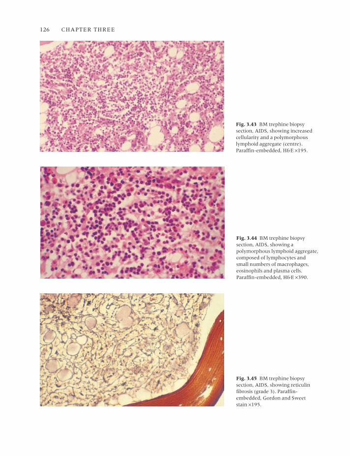
126 CHAPTER THREE
Fig. 3.43 BM trephine biopsysection, AIDS, showing increasedcellularity and a polymorphouslymphoid aggregate (centre).Paraffin-embedded, H&E ×195.
Fig. 3.44 BM trephine biopsysection, AIDS, showing apolymorphous lymphoid aggregate,composed of lymphocytes andsmall numbers of macrophages,eosinophils and plasma cells.Paraffin-embedded, H&E ×390.
Fig. 3.45 BM trephine biopsysection, AIDS, showing reticulinfibrosis (grade 3). Paraffin-embedded, Gordon and Sweet stain ×195.
INFECTION AND REACTIVE CHANGES 127
necrosis with both free forms and pseudocysts being found within granulocytes, macrophages andmegakaryocytes [31]. Organisms can be identifiedusing a polyclonal antibody [31]. Atypical mycobac-teria can be cultured from the marrow in up to 20%of cases of AIDS. Granulomas are present in approx-imately 50% of culture-positive cases and acid-fastbacilli can be seen with special stains in 60%. Atyp-ical mycobacteria may be seen in biopsies in theabsence of granulomas, usually within macrophagesscattered throughout the marrow; occasionally cells which morphologically resemble Gaucher’scells (see page 109) are seen. Culture of the bone marrow and examination of sections for acid-fastbacilli has been found to be less sensitive thanperipheral blood culture in the diagnosis of atypicalmycobacterial infection in AIDS but, when bacillican be found in a stained film, diagnosis is muchmore rapid than when it is dependent on culture[141]. Focal areas of epithelioid angiomatosis havebeen observed in the marrow of HIV-positive pati-ents with bacillary angiomatosis or disseminated cat scratch disease, infective conditions consequenton infection by organisms of the Rochalimaea genusor the related Afipia felis [142,143]; a Warthin–Starry stain may demonstrate the causative organ-isms [143].
Bone marrow involvement by multicentricCastleman’s disease has been reported in severalHIV-positive patients who were also positive forHHV8 [144].
The incidence of non-Hodgkin’s lymphoma isgreatly increased in HIV infection [145]. Marrowinvolvement is common in non-Hodgkin’s lym-phoma associated with AIDS. These are almostalways high grade lymphomas, either Burkitt’s orBurkitt-like lymphoma, or diffuse large B-cell lymphoma (immunoblastic or anaplastic large cellsubtypes). Bone marrow infiltration is much morecommon in Burkitt’s lymphoma than in diffuselarge B-cell lymphomas. Infiltration is usually dif-fuse (see Fig. 6.54).
Hodgkin’s disease is now also recognized as anAIDS-related lymphoma. There is a propensity toinvolve the bone marrow early in the course of theillness so that a trephine biopsy may provide thefirst evidence of the disease [146].
Plasma cell tumours also occur as AIDS-relatedneoplasms. Although initial presentation is often at
extramedullary sites, the bone marrow may also beinfiltrated [147].
Kaposi’s sarcoma has a greatly increased inci-dence in AIDS and, when spread to the bone mar-row occurs, may be detected by trephine biopsy.
Bone marrow necrosis
The term ‘bone marrow necrosis’ is conventionallyused to describe necrosis of haemopoietic cells ornecrosis of neoplastic cells that have replaced nor-mal bone marrow cells. Bone marrow stromal cellsmay also be necrotic. There may or may not be associated death of bone cells and of bone. Bonemarrow necrosis results from impairment of the bloodsupply, often in association with a hypercellularmarrow. A high concentration of tumour necrosisfactor in the blood may act as a mediator of necrosis[148]. Anti-phospholipid antibodies may also cause,or contribute to, bone marrow necrosis [149,150].Causes are multiple (Table 3.5). Necrosis is followedeither by haemopoietic regeneration, with or with-out small fibrotic scars, or occasionally by extensivefibrosis. Bone marrow necrosis commonly occurs atmultiple sites. Clinical features include bone painand fever. Fatal fat embolism can occur [159].
Peripheral blood
If bone marrow necrosis is extensive, pancytopeniaoccurs. The blood film shows leuco-erythroblastic,and sometimes micro-angiopathic [160], features.Recovery is associated with a rise in the reticulocytecount and recovery of the haemoglobin concentra-tion and platelet and white cell counts.
Bone marrow cytology
A bone marrow aspirate is often macroscopicallyabnormal. It is sometimes opaque and whitish andsometimes reddish-purple. The stained film showsamorphous pink-staining material in which the faintoutlines of necrotic cells can be seen (Fig. 3.46).Necrotic nuclei appear as darker-staining smudges.Some intact cells may also be present.
Bone marrow necrosis is more often noted inbone marrow trephine sections than in aspirates.This may be partly because a larger volume is sam-pled and partly because necrotic cells mixed with
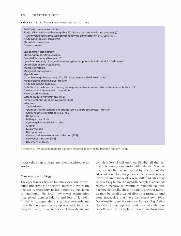
128 CHAPTER THREE
intact cells in an aspirate are often dismissed as anartefact.
Bone marrow histology
The appearances depend to some extent on the con-dition underlying the necrosis. In cases in which thenecrosis is secondary to infiltration by leukaemia or lymphoma (Fig. 3.47), low power examinationmay reveal hypercellularity and loss of fat cells. In the early stages there is nuclear pyknosis and the cells have granular cytoplasm with indistinctmargins. Later, there is nuclear karyorrhexis and
complete loss of cell outlines. Finally, all that re-mains is amorphous eosinophilic debris. Marrownecrosis is often accompanied by necrosis of theadjacent bone. In some patients, the necrosis is veryextensive and biopsy of several different sites maybe necessary before a diagnostic sample is obtained.Necrotic marrow is eventually repopulated withhaemopoietic cells. The only signs of previous necro-sis may be small areas of fibrous scarring aroundbony trabeculae that have lost osteocytes [161].Occasionally there is extensive fibrosis (Fig. 3.48).Necrosis of haemopoietic and stromal cells may be followed by metaplastic new bone formation
Table 3.5 Causes of bone marrow necrosis [45,151–156].
Relatively common associationsSickle cell anaemia and haemoglobin S/C disease (particularly during pregnancy)Acute myeloid leukaemia (sometimes following administration of G-CSF [157])Acute lymphoblastic leukaemiaMetastatic carcinomaCaisson disease
Less common associationsChronic granulocytic leukaemiaEssential thrombocythaemia [151]Lymphoma (mainly high grade non-Hodgkin’s lymphoma but also Hodgkin’s disease)*Chronic lymphocytic leukaemiaMultiple myelomaMalignant histiocytosisMyelofibrosisOther haemoglobinopathies (S/D, S/β thalassaemia and sickle cell trait)Megaloblastic anaemia plus infectionAcute haemolytic anaemiaEmbolism of the bone marrow, e.g. by vegetations from cardiac valves or tumour embolism [152]Disseminated intravascular coagulationHyperparathyroidismSystemic lupus erythematosus [153]Primary anti-phospholipid syndrome [149]Infections
Typhoid feverGram-positive infections, e.g. streptococcal and staphylococcal infectionGram-negative infections, e.g. E. coliDiphtheriaMiliary tuberculosisCytomegalovirus infection [154]Q feverMucormycosisHistoplasmosisFusobacterium necrophorum infection [155]Parvovirus infection [156]HIV infection (AIDS)
* Necrosis of low grade lymphoma has been observed following fludarabine therapy [158].
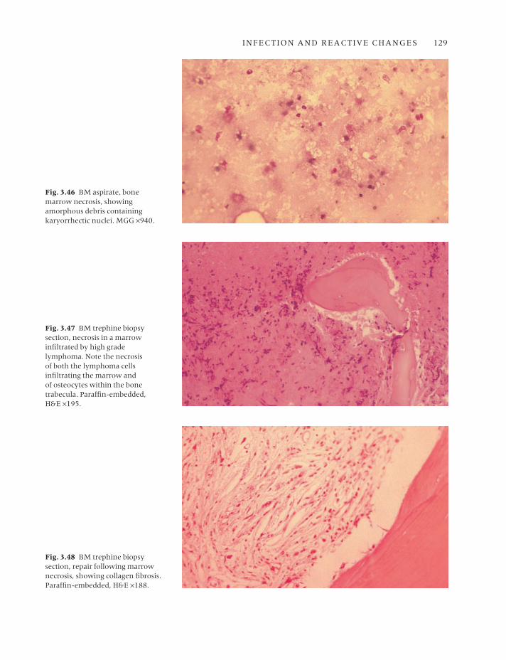
INFECTION AND REACTIVE CHANGES 129
Fig. 3.46 BM aspirate, bonemarrow necrosis, showingamorphous debris containingkaryorrhectic nuclei. MGG ×940.
Fig. 3.47 BM trephine biopsysection, necrosis in a marrowinfiltrated by high gradelymphoma. Note the necrosis of both the lymphoma cellsinfiltrating the marrow and of osteocytes within the bonetrabecula. Paraffin-embedded, H&E ×195.
Fig. 3.48 BM trephine biopsysection, repair following marrownecrosis, showing collagen fibrosis.Paraffin-embedded, H&E ×188.

130 CHAPTER THREE
(Fig. 3.49), initially woven bone which is sub-sequently remodelled into structurally normalbone.
Problems and pitfalls
Immunohistochemistry may be very misleading ifused in an attempt to identify neoplastic cells withinnecrotic areas, as a consequence both of the ten-dency of antibodies to adhere in a non-specific wayto necrotic tissue and of loss of some antigens bynecrotic cells.
Gelatinous transformation
Gelatinous transformation, also known as serousdegeneration or serous atrophy, is a condition inwhich there is loss of fat cells and haemopoietic cellsfrom the bone marrow with replacement by anincreased amount of ground substance. Commoncauses include anorexia nervosa and cachexia dueto chronic debilitating illnesses such as carcinomaand tuberculosis or other chronic infection. Thecondition is also observed in AIDS, which is nowthe commonest cause of this abnormality. It has alsobeen reported in association with renal failure,coeliac disease [162] and severe hypothyroidism[163]. It occurs at sites exposed to high-dose X-irradiation. Gelatinous transformation can alsodevelop rapidly; it is seen in acute illnesses with
multiple organ failure [10] and in severe acuteinfections.
Peripheral blood
The peripheral blood shows variable cytopenia,often pancytopenia. In patients with anorexia ner-vosa, acanthocytes are seen but their presence hasnot been noted in other patients with gelatinoustransformation.
Bone marrow cytology
The aspirate may not spread normally when a filmis prepared. It contains amorphous material, some-times fibrillar or finely granular, which is composedof acid mucopolysaccharide with a high content ofhyaluronic acid. With Romanowsky stains it is pinkor pinkish-purple (Fig. 3.50). This abnormal groundsubstance is positive with a PAS stain, the reactionbeing diastase resistant. Positive staining also occurswith alcian blue; this reaction is stronger at pH 2.5than pH 1.0 [10]. A toluidine blue stain is weaklypositive.
Bone marrow histology
The changes are usually focal; less commonly thewhole of a biopsy section is affected. There is atro-phy of fat cells, which are both reduced in number
Fig. 3.49 BM trephine biopsysection showing new boneformation in an area of previousbone marrow necrosis. Paraffin-embedded, Giemsa ×188.

INFECTION AND REACTIVE CHANGES 131
and are of variable size. In the affected areas there isa mild to marked hypoplasia of haemopoietic cells.Both fat and haemopoietic cells are replaced byamorphous material which, with H&E staining, hasa light blue to pale pink colour and a finely granularappearance (Fig. 3.51). Its other staining character-istics are identical to those seen in marrow films(see above).
Bone marrow fibrosis
Bone marrow fibrosis indicates an increase of reti-
culin or of reticulin and collagen in the bone marrow.Such fibrosis may be focal or generalized. Reticulinand collagen deposition are graded 0–4 as shown in Table 2.2. Some patients with grade 4 fibrosis also have osteosclerosis. Increased reticulin forma-tion and even collagen deposition can revert to normal if the causative condition is amenable totreatment.
An increase in bone marrow reticulin is commonand is a useful non-specific indication that the mar-row is abnormal. It is also of some use in differentialdiagnosis; for example, it may be increased in
Fig. 3.50 BM aspirate, gelatinoustransformation, showingamorphous purplish-pink material.MGG ×377.
Fig. 3.51 BM trephine biopsy,gelatinous transformation, showingreplacement of haemopoieticmarrow and fat cells by amorphouspink material. H&E ×390.

132 CHAPTER THREE
Table 3.6 Causes of grade 4 bone marrow fibrosis [45,164].
Generalized myelofibrosis
Malignant diseasePrimary (idiopathic) myelofibrosis* (myelofibrosis with myeloid metaplasia)Myelofibrosis secondary to essential thrombocythaemia or polycythaemia rubra vera*Chronic granulocytic leukaemia*Transitional myeloproliferative syndromeAcute megakaryoblastic leukaemia*Other acute myeloid leukaemiasAcute lymphoblastic leukaemia [165]Systemic mastocytosis*The myelodysplastic syndromes (particularly secondary MDS)Paroxysmal nocturnal haemoglobinuriaHodgkin’s diseaseNon-Hodgkin’s lymphomaMultiple myelomaWaldenström’s macroglobulinaemiaSecondary carcinoma*
Bone and connective tissue diseasesMarble bone disease—osteopetrosisPrimary and secondary hyperparathyroidismNutritional and renal rickets (vitamin D deficiency)OsteomalaciaPrimary hypertrophic osteoarthropathy [166]
MiscellaneousTuberculosisOther granulomatous diseasesGrey platelet syndromeSystemic lupus erythematosusSystemic sclerosisPrior thorium dioxide administration
Focal or localized
OsteomyelitisPaget’s diseaseFollowing bone marrow necrosisFollowing irradiation of the bone marrowAdult T-cell leukaemia/lymphomaHealing fractureSite of previous trephine biopsy
* Osteosclerosis may also occur.
hypoplastic AML and hypoplastic myelodysplasticsyndromes but is not increased in aplastic anaemia.Conditions characteristically associated with in-creased reticulin deposition, but with little if anycollagen formation, include hairy cell leukaemiaand HIV infection. Reticulin deposition may also bea feature of myeloproliferative disorders, such as
polycythaemia rubra vera and essential thrombo-cythaemia, and occurs in some patients with acuteleukaemia (lymphoblastic as well as myeloid),myelodysplastic syndromes and multiple myeloma.The increase in reticulin in uncomplicated poly-cythaemia rubra vera and essential thrombo-cythaemia is not marked.

INFECTION AND REACTIVE CHANGES 133
A focal increase in reticulin may have a differentsignificance from a general increase, often indicat-ing the presence of a focal infiltrate.
Collagen deposition is uncommon and of greaterdiagnostic significance than an increase in reticulin.It is therefore useful to distinguish between thesetwo degrees of abnormality, either by grading thefibrosis or by using the terms ‘reticulin fibrosis’ for agrade 3 abnormality and ‘fibrosis’ or ‘myelofibrosis’for a grade 4 abnormality. Causes of bone marrowfibrosis are shown in Table 3.6.
Peripheral blood
Bone marrow fibrosis is commonly associated witha leuco-erythroblastic anaemia; red cells show aniso-cytosis and poikilocytosis with teardrop poikilocytesoften being prominent. When fibrosis is extensive,there may also be thrombocytopenia and leuco-penia. The blood may also show abnormalitiesrelated to the primary disease that has caused the fibrosis. When bone marrow fibrosis has developed acutely, as in acute megakaryoblasticleukaemia, there may be little anisocytosis andpoikilocytosis and the blood film is not necessarilyleuco-erythroblastic.
Bone marrow cytology
Bone marrow fibrosis often leads to failure to aspir-ate marrow or to aspiration of peripheral blood or a
diluted marrow specimen. Otherwise the aspiratemay show carcinoma cells or the specific features of the primary disease that has led to the fibrosis. If there is associated osteosclerosis, the aspirate may contain increased numbers of osteoblasts andosteoclasts.
Bone marrow histology
The appearances of primary myelofibrosis (myelo-fibrosis with myeloid metaplasia) are described onpage 204. In secondary fibrosis, findings range froma mild increase of fibroblasts and the presence ofscattered collagen fibres to a dense fibrosis whichobliterates normal haemopoietic tissue. An increaseof reticulin can be suspected from an H&E stain bythe distortion and angularity of fat cells and by the‘streaming’ of haemopoietic cells held between par-allel reticulin fibres. Fibrosis may also be suspectedwhen sinusoids, which are normally collapsed inparaffin-embedded specimens, are held open.Increased reticulin deposition is confirmed by a sil-ver impregnation technique such as the Gomoristain. Collagen fibres are detected as eosinophilicfibres which may be in bundles. The oval nuclei offibroblasts are apparent in relation to the collagenfibres. It is important not to confuse either endothe-lial cells of collapsed capillaries or mast cells withfibroblasts. The presence of collagen is confirmed by a trichrome stain such as a Martius Scarlet Blue stain (Fig. 3.52). The distribution of reticulin
Fig. 3.52 BM trephine biopsysection showing marrow infiltratedby breast cancer with associatedstromal fibrosis. Collagen is evidentas blue strands when demonstratedby Martius Scarlet Blue trichromestain. MSB ×94.

134 CHAPTER THREE
and collagen within the marrow depends on thecausative condition. In renal osteodystrophy andprimary hyperparathyroidism, fibrosis is usuallyparatrabecular. In Paget’s disease, it is often prefer-entially paratrabecular. When associated with amyeloproliferative disorder or with the grey plate-let syndrome, fibrosis may be spatially related tomegakaryocytes.
Problems and pitfalls
When the bone marrow is very fibrotic, any bonemarrow aspirate is likely to be very unrepresenta-tive. No attempt should be made to assess cellular-ity. However, it is always worth looking for tumourcells or dysplastic haemopoietic cells, regardless ofthe quality of the aspirate. If no aspirate can beobtained, a trephine biopsy imprint can yield usefulinformation as to the nature of any abnormal cellspresent.
Due weight must be given to the presence of focal reticulin deposition and the correspondingarea in the H&E-stained sections should be care-fully examined. A generalized increase in reticulinis an indication of an abnormal bone marrow and an explanation should be sought by review-ing of clinical, haematological and histological features.
When fibrosis is very pronounced, it may bedifficult to distinguish residual haemopoietic cells,particularly megakaryocytes, from tumour cells.Immunohistochemistry is useful in this situation.
References1 Goodman JL (1999) Ehrlichiosisaticks, dogs and
doxycycline. N Engl J Med, 341, 195–197.2 Rausing A (1973) Bone marrow biopsy in the
diagnosis of Whipple’s disease. Acta Med Scand, 193,5–8.
3 Barenfanger J, Patel PG, Dumler JS and Walker DH(1996) Clinical and pathology rounds: identifyinghuman ehrlichiosis. Lab Med, 27, 372–374.
4 Farooqui BJ, Khurshid M, Ashfaq MK and Khan MA (1991) Comparative yield of Salmonella typhifrom blood and bone marrow cultures in patientswith fever of unknown origin. J Clin Pathol, 44,258–259.
5 Al-Eissa YA, Assuhaimi SA, Al-Fawaz IM, Higgy KE, Al-Nasser MN and Al-Mobaireek KF (1993)
Pancytopenia in children with brucellosis: clinicalmanifestations and bone marrow findings. ActaHaematol, 89, 132–136.
6 Lawrence C and Schreiber AJ (1979) Leprosy’s foot-prints in bone marrow macrophages. N Engl J Med,300, 834–835.
7 Schmid C, Frisch B, Beham A, Jager K and Kettner G(1990) Comparison of bone marrow histology inearly chronic granulocytic leukaemia and in leukae-moid reaction. Eur J Haematol, 44, 154–158.
8 Thiele J, Holgado S, Choritz H and Georgii A (1983)Density distribution and size of megakaryocytes ininflammatory reaction of the bone marrow (myelitis)and chronic myeloproliferative disorders. Scand J Hae-matol, 31, 329–341.
9 Hyun BH, Kwa D, Gabaldon H and Ashton JK (1976)Reactive plasmacytic lesions of the bone marrow. AmJ Clin Pathol, 65, 921–928.
10 Amos RJ, Deane M, Ferguson C, Jeffries G, Hinds CJand Amess JAL (1990) Observations on the haemo-poietic response to critical illness. J Clin Pathol, 43,850–856.
11 Wiley EL, Perry A, Nightingale SD and Lawrence J(1994) Detection of Mycobacterium avium-intracellularecomplex in bone marrow specimens of patients withacquired immunodeficiency syndrome. Am J ClinPathol, 101, 446–451.
12 Suster S, Cabello-Inchausti B and Robinson MJ(1990) Nongranulomatous involvement of the bonemarrow in lepromatous leprosy. Am J Clin Pathol, 92,797–801.
13 Paddock CD, Suchard DP, Grumbach KL, Hadley WK,Kerschmann RL, Abbey NWBG et al. (1993) Briefreport: fatal seronegative ehrlichiosis in a patientwith HIV infection. N Engl J Med, 329, 1164–1167.
14 Pooley RJ Jr, Peterson L, Finn WG and Kroft SH(1999) Cytomegalovirus-infected cells in routinelyprepared peripheral blood films of immunosup-pressed patients. Am J Clin Pathol, 112, 108–112.
15 Davidovitz Y, Halpern Z, Vardi J, Ballin A and MeytesD (1998) Pure red cell aplasia responsive to inter-feron-α in a patient with hepatitis C infection. ActaHaematol, 100, 213–215.
16 Bilgrami S, Almeida GD, Quinn JJ, Tuck D,Bergstrom S, Dainiak NJL et al. (1994) Pancytopeniain allogeneic marrow transplant recipients: role ofcytomegalovirus. Br J Haematol, 87, 357–362.
17 Kobayashi S, Maruta A, Yamamoto T, Katayama N,Higuchi R, Sakano YA et al. (1998) Human parvovirusB19 capsid antigen in granulocytes in parvovirus-B19-induced pancytopenia after bone marrow trans-plantation. Acta Haematol, 100, 195–199.
18 Brunning RD. Bone marrow. In: Rosai J (ed)Ackerman’s Surgical Pathology, 7th edn, vol 2. Mosby,St Louis, 1989.

INFECTION AND REACTIVE CHANGES 135
19 Prezioso PJ, Cangiarella J, Lee M, Nuovo GJ,Borkowsky W, Orlow SJ and Greco MA (1992) Fataldisseminated infection with human herpes virus-6. JPediatr, 120, 921–923.
20 Johnston RE, Geretti A-M, Prentice HG, Clark AD,Wheeler AC, Potter M and Griffiths PD (1999) HHV-6-related secondary graft failure following allogeneicbone marrow transplantation. Br J Haematol, 105,1041–1043.
21 Liu W, Ittman M, Liu J, Schoentag R, Tierno P, GrecoMA et al. (1997) Human parvovirus in bone marrowsfrom adults with acquired immunodeficiency syn-drome: a comparative study using in situ hybridiza-tion and immunohistochemistry. Hum Pathol, 28,760–766.
22 Rimsza LM, Vela EE, Frutiger YM, Rangel CS, SolanoM, Richter LC, Grogan TM and Bellamy LC (1996)Rapid automated combined in situ hybridization andimmunohistochemistry for sensitive detection ofvirus in paraffin-embedded tissue biopsies. Am J ClinPathol, 106, 544–548.
23 Hamer RD, Scott M, Shahab I, Carey TT, Cousar JBand Macon WR (1996) Latent membrane proteinantibody reacts with normal hematopoietic precur-sor cells and leukemic blasts in tissues lackingEpstein–Barr virus genome by polymerase chainreaction. Am J Clin Pathol, 106, 469–474.
24 Cleary ML, Nalesnik MA, Shearer WT and Sklar J(1988) Clonal analysis of transplant-associated lym-phoproliferations based on the structure of genetictermini of the Epstein–Barr virus. Blood, 72, 349–352.
25 Rinn R, Chow WS and Pinkerton PH (1995)Transient acquired myelodysplasia associated withparvovirus B19 infection in a patient with congenitalspherocytosis. Am J Hematol, 50, 71–72.
26 Dorvault C, Raikow R and Contis L (1998) Evalua-tion of a monoclonal antibody to human parvovirusin B5-fixed bone marrow biopsy specimens. Am J ClinPathol, 110, 540–541.
27 Thiele J, Zirbes TK, Kvasnicka HM and Fischer R(1999) Focal lymphoid aggregates (nodules) in bonemarrow biopsies: differentiation between benignhyperplasia and malignant lymphomaaa practicalguide. J Clin Pathol, 52, 294–300.
28 Wong KF, Tsang DNC and Chan JKC (1994) Bonemarrow diagnosis of penicilliosis. N Engl J Med, 330,717–718.
29 Ferry JA, Pettit CJ, Rosenberg AE and Harris NL(1991) Fungi in megakaryocytes. An unusual mani-festation of fungal infection of the bone marrow. AmJ Clin Pathol, 96, 577–581.
30 Strauchen JA. Diagnostic Histopathology of the BoneMarrow. Oxford University Press, Oxford, 1996.
31 Brouland JP, Audouin J, Hofman P, Le Tourneau A,Basset D, Rio B et al. (1996) Bone marrow involve-
ment by disseminated toxoplasmosis in acquiredimmunodeficiency syndrome: the value of bone mar-row trephine biopsy and immunohistochemistry forthe diagnosis. Hum Pathol, 27, 302–306.
32 Piehl MR, Kaplan RL and Haber MH (1988) Dis-seminated penicilliosis in a patient with acquiredimmunodeficiency syndrome. Arch Pathol Lab Med,112, 1262–1264.
33 Wheat LJ, Connolly-Stringfield PA, Baker RL,Curfman MF, Eads ME, Israel KS et al. (1990)Disseminated histoplasmosis in the acquired immunedeficiency syndrome: clinical findings, diagnosis andtreatment, and review of the literature. Medicine, 69,361–374.
34 McKinsey DS, Gupta MR, Riddler SA, Driks MR,Smith DL and Kurtin PJ (1989) Long-term ampho-tericin B therapy for disseminated histoplasmosis inpatients with acquired immunodeficiency syndrome(AIDS). Ann Intern Med, 111, 655–659.
35 Wheat LJ, Connolly-Stringfield P, Kohler RB, FramePT and Gupta MR (1989) Histoplasma capsulatumpolysaccharide antigen detection in diagnosis andmanagement of disseminated histoplasmosis inpatients with acquired immunodeficiency syndrome.Am J Med, 87, 396–400.
36 Abdalla SH (1991) Hematopoiesis in human malaria.Blood Cells, 16, 401–416.
37 Phillips RE and Pavsol G (1992) Anaemia of Plasmodiumfalciparum malaria. Baillière’s Clin Haematol, 5, 315–330.
38 Bates I, Bedu-Addo G, Bevan DH and Rutherford TR (1991) Use of immunoglobulin gene rearrange-ments to show clonal lymphoproliferation in hyper-reactive malarial splenomegaly. Lancet, 337, 505–507.
39 Kirchhoff LV (1993) American trypanosomiasis(Chagas’ disease)aa tropical disease now in theUnited States. N Engl J Med, 329, 639–644.
40 Grimes MM, La Pook JD, Bar MH, Wasserman HS andDwork A (1987) Disseminated Pneumocystis cariniiinfection in a patient with acquired immunodefici-ency syndrome. Hum Pathol, 18, 307–308.
41 Yachnis AT, Berg J, Martinez-Salazar A, Bender BS,Diaz L, Rojiani AM et al. (1996) Disseminatedmicrosporidiosis specially infecting the brain, heart,and kidneys. Report of a newly recognized pan-sporoblastic species in two symptomatic AIDSpatients. Am J Clin Pathol, 106, 535–543.
42 Telsak EE, Cote RJ, Gold JMW, Campbell SW andArmstrong D (1990) Extrapulmonary Pneumocystiscarinii infections. Rev Infect Dis, 12, 380–386.
43 Bodem CR, Hamory BH, Taylor HM and Kleopfer L(1983) Granulomatous bone marrow disease. A re-view of the literature and clinicopathological analysisof 58 cases. Medicine (Baltimore), 62, 372–383.

44 Dumler JS, Dawson JE and Walker DH (1993)Human ehrlichiosis: hematopathology and immu-nohistologic detection of Ehrlichia chaffeensis. HumPathol, 24, 391–396.
45 Bain BJ and Wickramasinghe SNW. Pathology of the bone marrow: general considerations. In: Wick-ramasinghe SNW (ed) and Symmers W St C (seriesed) Systemic Pathology, Volume 2: Blood and BoneMarrow, 3rd edn. Churchill Livingstone, Edinburgh,1986.
46 Vilalta-Castel E, Váldes-Sanchez MD, Guerra-ValesJM, Teno-Esteban C, Garzón A, Lopéz JI et al. (1988)Significance of granulomas in bone marrow: a studyof 40 cases. Eur J Haematol, 41, 12–14.
47 Epstein HD and Kruskall MS (1988) Spuriousleukopenia due to in vitro granulocyte aggregation.Am J Clin Pathol, 89, 652–655.
48 Ampel NM, Ryan KJ, Carry PJ, Wieden MA andSchifman RB (1986) Fungemia due to Coccidioidesimmitis. An analysis of 16 episodes in 15 patients anda review of the literature. Medicine (Baltimore), 65,312–321.
49 Hyun BH. Colour Atlas of Clinical Hematology. Igaku-Shoin Med US, New York, 1986.
50 Falini B, Tabilio A, Velardi A, Cernetti C, Aversa Fand Martelli MF (1982) Multiple myeloma with a sarcoidosis-like reaction. Scand J Haematol, 29,211–216.
51 Rabbani GR, Phyliky RL and Tefferi A (1999) A long-term study of patients with chronic natural killer celllymphocytosis. Br J Haematol, 106, 960–966.
52 Bhargava V and Farhi DC (1988) Bone marrow granulomas: clinicopathologic findings in 72 casesand review of the literature. Hematol Pathol, 2, 43–50.
53 Kettle P and Allen DC (1997) Bone marrow granulo-mas in infiltrating lobular breast cancer. J Clin Pathol,50, 166–168.
54 Poland GA and Love KR (1986) Marked atypical lym-phocytosis, hepatitis, and skin rash in sulfasalazinedrug allergy. Am J Med, 81, 707–708.
55 Case Records of the Massachusetts General Hospital(1996) A seven-year-old boy with fever, lym-phadenopathy, hepatosplenomegaly, and prominenteosinophilia. N Engl J Med, 335, 577–586.
56 Pelstring RJ, Kim K, Lower EE and Swerdlow SH(1988) Marrow granulomas in coal workers’ pneu-moconiosis. Am J Clin Pathol, 89, 553–556.
57 Lewis JH, Sundeen JT, Simon GL, Schulof RS, WandGS, Gelfand RL et al. (1985) Disseminated talc granu-lomatosis: acquired immunodeficiency syndrome andfatal cytomegalovirus infection. Arch Pathol Lab Med,109, 147–150.
58 Davis S and Trubowitz S. Pathologic reactions involv-ing the bone marrow. In: Trubowitz S and Davis S
(eds) The Human Bone Marrow: Anatomy, Physiology andPathophysiology, vol. 2. CRC Press, Boca Raton, 1982.
59 Rywlin AM and Ortega R (1971) Lipid granulomas ofthe bone marrow. Am J Clin Pathol, 57, 457–462.
60 Hudson P and Robertson GM (1966) Demonstrationof mineral oil in splenic lipid granulomas. Lab Invest,15, 1134–1135.
61 Pease GL (1956) Granulomatous lesions in bone mar-row. Blood, 11, 720–734.
62 Farhi DC, Mason UG and Horsburgh CR (1984) The bone marrow in disseminated Mycobacteriumavium intracellulare infection. Am J Clin Pathol, 83,463–468.
63 Okun DB, Sun NCJ and Tanaka KR (1979) Bonemarrow granulomas in Q-fever. Am J Clin Pathol, 71,117–121.
64 Young JF and Goulian M (1993) Bone marrow fibrinring granulomas and cytomegalovirus infection. Am JClin Pathol, 99, 65–68.
65 Kurtin PJ, McKinsey DS, Gupta MR and Driks M(1989) Histoplasmosis in patients with acquiredimmunodeficiency syndrome. Hematologic and bonemarrow manifestation. Am J Clin Pathol, 93, 367–372.
66 Kadin ME, Donaldson SS and Dorfman RF (1970)Isolated granulomas in Hodgkin’s disease. N Engl JMed, 283, 859–861.
67 Yu NC and Rywlin AM (1982) Granulomatouslesions of the bone marrow in non-Hodgkin’s lym-phoma. Hum Pathol, 13, 905–910.
68 Franco V, Florena AM, Quintini G and Musso M(1994) Bone marrow granulomas in hairy cell leu-kaemia following 2-chlorodeoxyadenosine therapy.Histopathology, 24, 271–273.
69 O’Brien DV, Boon AP and Boughton BJ (1992) Bonemarrow granulomas in acute myeloid leukaemia fol-lowing interleukin 2 and lymphokine-activated killercells. Histopathology, 20, 271–272.
70 Amodie LA and Tefferi A (1996) Bone marrow gran-ulomas and fevers of unknown origin associated withchronic natural killer cell lymphocytosis. Br JHaematol, 93 (Suppl. 2), 108.
71 Tefferi A and Li CY (1997) Bone marrow granulomasassociated with chronic natural killer cell lymphocy-tosis. Am J Hematol, 54, 258–262.
72 Browne PM, Sharma OP and Salkin D (1978) Bonemarrow sarcoidosis. JAMA, 240, 2654–2655.
73 Frizzera G, Banks PM, Massarelli G and Rosai J(1983) A systemic lymphoproliferative disorder withmorphologic features of Castleman’s disease. Path-ological findings in 15 patients. Am J Surg Pathol, 7,211–231.
74 Faulkner-Jones BE, Howie AJ, Boughton BJ andFranklin IM (1988) Lymphoid aggregates in bonemarrow: study of eventual outcome. J Clin Pathol, 41,768–775.
136 CHAPTER THREE

INFECTION AND REACTIVE CHANGES 137
75 Rywlin AM, Ortega RS and Dominguez CJ (1974)Lymphoid nodules of bone marrow. Blood, 43,389–400.
76 Sangster G, Crocker J, Nar P and Leylan MJ (1986)Benign and malignant (B cell) focal lymphoid aggre-gates in bone marrow trephines shown by means ofan immunogold-silver technique. J Clin Pathol, 39,453–457.
77 Harris NL and Ferry JA. Follicular lymphoma andrelated disorders. In: Knowles DW (ed) NeoplasticHematopathology. Williams & Wilkins, Baltimore,1992.
78 Farhi DC (1989) Germinal centres in the bone mar-row. Hematol Pathol, 3, 133–136.
79 Horny HD, Wehrmann M, Griessner H, Tiemann M,Bultman B and Kaiserling E (1993) Investigation ofbone marrow lymphocyte subsets in normal, reactiveand neoplastic states using paraffin-embedded biopsyspecimens. Am J Clin Pathol, 99, 142–149.
80 Bluth RF, Casey TT and McCurley TL (1993)Differentiation of reactive from neoplastic small-celllymphoid aggregates in paraffin-embedded marrowparticle preparations using L26 (CD20) and UCHL-1(CD45 RO) monoclonal antibodies. Am J Clin Pathol,99, 150–156.
81 Smith GP, Perkins SL, Segal GH and Kjeldsberg CR(1994) T-cell lymphocytosis associated with invasivethymomas. Am J Clin Pathol, 102, 447–453.
82 Agrawal S, Matutes E, Voke J, Dyer MJS, Khokhar Tand Catovsky D (1994) Persistent polyclonal B-celllymphocytosis. Leuk Res, 18, 791–795.
83 Gatter K and Brown D. An Illustrated Guide to BoneMarrow Diagnosis. Blackwell Science, Oxford, 1997.
84 Klein H and Block M (1953) Bone marrow plasmacy-tosis. Blood, 8, 1034–1041.
85 Wulfhekel U and Dullmann J (1999) Storage of ironin bone marrow plasma cells. Ultrastructural charac-terization, mobilization, and diagnostic significance.Acta Haematol, 101, 7–15.
86 Crocker J and Curran RC (1981) Quantitative studyof the immunoglobulin-containing cells in treph-ine samples of bone marrow. J Clin Pathol, 34,1080–1082.
87 Peart KM and Ellis HA (1972) Quantitative observa-tions on iliac crest bone marrow mast cells in chronicrenal failure. J Clin Pathol, 28, 947–955.
88 Yoo D, Lessin LS and Jensen WN (1978) Bone mar-row mast cells in lymphoproliferative disorders. AnnIntern Med, 88, 753–757.
89 Prokimer M and Polliack A (1980) Increased bonemarrow mast cells in preleukemic syndromes, acuteleukaemia, and lymphoproliferative disorders. Am JClin Pathol, 75, 34–38.
90 Rywlin AM, Hoffman EP and Ortega RS (1972)Eosinophilic fibrohistiocytic lesions of bone marrow:
a new morphologic finding probably related to drughypersensitivity. Blood, 40, 464–472.
91 Wilson PA, Ayscue LH, Jones GR and Bentley SA(1993) Bone marrow histiocytic proliferation in asso-ciation with colony-stimulating factor therapy. Am JClin Pathol, 99, 311–313.
92 Rosenthal NS and Fahri DC (1994) Failure to engraftafter bone marrow transplantation, bone marrowmorphologic findings. Am J Clin Pathol, 102, 821–824.
93 Hirayama Y, Yoshioka K and Shiozaki N (1995)Histiocytosis and two classes of M-proteinemia in pre-treated Ph positive ALL. Am J Hematol, 49,358–359.
94 Huang LM, Lee CY, Lin KH, Chuu WM, Lee PI, Chen RL et al. (1990) Human herpesvirus-6 associ-ated with fatal haemophagocytic syndrome. Lancet,336, 60–61.
95 Kawa-Ha K, Tanaka K, Inoue M, Sakata N, Okada S,Kurata T et al. (1993) Isolation of human herpesvirus7 from a child with symptoms mimicking chronicEpstein–Barr virus infection. Br J Haematol, 84, 545–548.
96 Potter MN, Foot ABM and Oakhill A (1991)Influenza A and virus-associated haemophagocyticsyndrome: cluster of three cases in children withacute leukaemia. J Clin Pathol, 44, 297–299.
97 Komp DM, Buckley PJ, McNamara J and van Hoff J(1989) Soluble interleukin-2 receptors in hemo-phagocytic histiocytosis. Pediatr Hematol Oncol, 6,253–264.
98 Lelong F (1993) Syndrome d’activation macro-phagique. Les Mercredis Cytologiques de Necker, No 5.
99 Wong K-F and Chan JKC (1992) Reactive haemo-phagocytic syndromeaa clinico-pathologic study of40 patients in an Oriental population. Am J Med, 93,177–180.
100 Tsuda H and Fujisao S (1999) Th1/Th2 milieu in adult hemophagocytic syndrome. Acta Haematol, 101,157–160.
101 Woda BA and Sullivan J (1993) Reactive histiocyticdisorders. Am J Clin Pathol, 99, 460–463.
102 Manoharan A and Catovsky D. Histiocytic medullaryreticulosis revisited. In: Schmalzl F, Huhn D andSchaefer HE (eds) Haematology and Blood Transfusion.Volume 27: Disorders of the Monocyte Macrophage System.Springer-Verlag, Berlin, 1981.
103 Risdall RJ, Brunning RD, Hernandez JI and GordonDH (1984) Bacteria associated hemophagocytic syn-drome. Cancer, 54, 2968–2972.
104 Bourquelot P, Bouscary D, Paul G, Picard F, Varet B,Dreyfus F and Tulliez M (1996) Hemophagocytic syn-drome associated with Bacteroides fragilis infectionin a patient with acute monoblastic leukemia. LeukLymphoma, 22, 177–179.

105 Gill K and Marrie TJ (1987) Hemophagocytosis sec-ondary to Mycoplasma pneumoniae infection. Am JMed, 82, 668–670.
106 Abbott KC, Vukelja ST, Smith CE, McAllister CK,Konkol KA, O’Rourke TJ et al. (1991) Hemophago-cytic syndrome: a cause of pancytopenia in humanehrlichiosis. Am J Hematol, 38, 230–234.
107 Walker DH and Dumler JS (1997) Human monocyticand granulocytic ehrlichioses. Discovery and diagno-sis of emerging tick-borne infections and the criticalrole of the pathologist. Arch Pathol Lab Med, 121,785–791.
108 Estrov Z, Bruck R, Shtalrid M, Berrebi A andResnitzky P (1983) Histiocytic hemophagocytosis inQ-fever. Arch Pathol Lab Med, 108, 7.
109 Shome DK, Rakheja D and Sonkar S (1997)Haemophagocytosis in bone marrow aspirates. ActaHaematol, 98, 173–174.
110 Auerbach M, Haubenstock A and Soloman G (1980)Systemic babebiosis. Another cause of the hemo-phagocytic syndrome. Am J Med, 80, 301–303.
111 Higgins EM, Layton DM, Arya R, Salisbury J and duVivier AWP (1994) Disseminated Trichosporon beigeliiinfection in an immunosuppressed child. J Roy SocMed, 87, 292–293.
112 Jaffe ES, Costa J and Fauci AS (1981) Erythro-phagocytic T lymphoma. N Engl J Med, 305, 103–104.
113 Chan E, Pi D, Chan GT, Todd D and Ho FC (1989)Peripheral T-cell lymphoma presenting as hemophago-cytic syndrome. Hematol Oncol, 7, 275–285.
114 Sonneveld P, van Lom K, Kappers-Klunne M, PrinsMER and Abels J (1990) Clinico-pathological diag-nosis and treatment of malignant histiocytosis. Br JHaematol, 75, 511–516.
115 Noguchi M, Kawano Y, Sato N and Oshimi K (1997)T-cell lymphoma of CD3+CD4+CD56+ granular lymphocytes with hemophagocytic syndrome. LeukLymphoma, 26, 349–358.
116 Okuda T, Sakamoto S, Deguchi T, Misawa S, KashimiK, Yoshihara T et al. (1991) Hemophagocytic syn-drome associated with natural killer cell leukemia.Am J Hematol, 38, 321–323.
117 Takeshita M, Kikuchi M, Ohshima K, Nibu K,Suzumiya J, Hisano S et al. (1993) Bone marrowfindings in malignant histiocytosis and/or malignantlymphoma with concurrent hemophagocytic syn-drome. Leuk Lymphoma, 12, 79–89.
118 Chim CS and Hui PK (1997) Reactive hemophago-cytic syndrome and Hodgkin’s disease. Am J Hematol,55, 49–50.
119 Mahadeva U, Allport T, Bain B and Chan WK (2000)Haemophagocytic syndrome with histiocytic necro-tizing lymphadenitis (Kikuchi’s disease). J Clin Pathol,53, 636–638.
120 Kumakura S, Ishikura H, Umegae N, Yamagata S and Kobayashi S (1997) Autoimmune–associatedhemophagocytic syndrome. Am J Med, 102, 113–115.
121 Janka GE (1989) Familial hemophagocytic lympho-histiocytosis: diagnostic problems and differentialdiagnosis. Pediatr Hematol Oncol, 6, 219–226.
122 Urabe AU (1994) Colony-stimulating factor andmacrophage proliferation. Am J Clin Pathol, 101, 116.
123 Hurwitz N, Probst A, Zufferey G, Tichelli A, Pless M,Kappos L et al. (1996) Fatal vascular leak syndromewith extensive hemorrhage, peripheral neuropathyand reactive erythrophagocytosis: an unusual com-plication of recombinant IL-3 therapy. LeukLymphoma, 20, 337–340.
124 Cheah PL, Looi LM, Tan PE, Bosco J and Kuperan P(1992) Cytophagic histiocytic panniculitis: a diag-nostic dilemma. Hematol Oncol, 10, 331–337.
125 Van den Bos C, de Konig TJ, Bierings MB, Poll-TheBT, Zegers BJM, Rijkers GT and Révész T (1998)Hemopoietic and immunological abnormalities intwo patients with lysinuric protein intolerance. Br JHaematol, 102, 314.
126 Çetin M, Hiçsönmez G and Gögüs S (1998)Myelodysplastic syndrome associated with Griscellisyndrome. Leuk Res, 22, 859–862.
127 Aricó M, Caselli D and Burgio GR (1989) Familialhemophagocytic lymphohistiocytosis: clinical fea-tures. Pediatr Hematol Oncol, 6, 247–251.
128 Ohadi M, Lalloz MR, Sham P, Zhao J, Dearlove AM,Shiach C et al. (1999) Localization of a gene for fam-ilial hemophagocytic lymphohistiocytosis at chro-mosome 9q21.3–22 by homozygosity mapping. Am JHum Genet, 64, 165–171.
129 Risdall RJ, McKenna RW, Nesbitt ME, Krivit W,Balfour HH, Simmons RL and Brunning RD (1979)Virus-associated hemophagocytic syndrome: a ben-ign histiocytic proliferation distinct from malignanthistiocytosis. Cancer, 44, 993–1002.
130 Campo E, Condom E, Miro M-J, Cid M-C andRomagosa V (1986) Tuberculosis associated haemo-phagocytic syndrome. Cancer, 58, 2640–2645.
131 Hirst WJR, Layton DM, Singh S, Mieli-Vergani G,Chessells JM, Strobel S and Pritchard J (1994)Haemophagocytic lymphohistiocytosis, experience intwo U.K. centres. Br J Haematol, 88, 731–739.
132 Han K, Kim Y, Kahng J, Lee J, Moon Y, Kang C and Shim S (1996) In situ hybridization studies ofcytomegalovirus and Epstein–Barr virus in reactivehistiocytic hyperplasia with hemophagocytosis. ActaHaematol, 96, 140–145.
133 Imashuku S, Hibi S, Morinaga S, Takagi K, Chen J, Mugishima H et al. (1997) Haemophagocytic lymphohistiocytosis in association with granularlymphocyte proliferative disorders in early child-
138 CHAPTER THREE

INFECTION AND REACTIVE CHANGES 139
hood: characteristic bone marrow morphology. Br JHaematol, 96, 708–714.
134 Nair JMG, Bellevue R, Bertoni M and Dosik H (1988) Thrombotic thrombocytopenia in patientswith the acquired immunodeficiency syndrome(AIDS)-related complex: a report of two cases. AnnIntern Med, 109, 242–243.
135 Bain BJ (1997) The haematological features of HIVinfection. Br J Haematol, 99, 1–8.
136 Oshorne BM, Guarda LA and Butler JJ (1984) Bonemarrow biopsies in the acquired immunodeficiencysyndrome. Hum Pathol, 15, 1048–1053.
137 Scheider DR and Picker LJ (1985) Myelodysplasia inthe acquired immunodeficiency syndrome. Am J ClinPathol, 84, 144–152.
138 Castella A, Croxson TS, Mildvan D, Witt DH andZalusky R (1985) The bone marrow in AIDS. Am JClin Pathol, 84, 425–432.
139 Mehta K, Gascoyne P and Robboy S (1992) Thegelatinous bone marrow (serous atrophy) in patientswith acquired immunodeficiency syndrome. ArchPathol Lab Med, 116, 504–508.
140 Bain BJ (1998) The haematologist and the infec-tive complications of HIV infection. Haematology, 3,153–164.
141 Hussong J, Peterson LR, Warren JR and Peterson L-AC (1998) Detecting disseminated Mycobacteriumavium complex infections in HIV-positive patients.Am J Clin Pathol, 110, 806–809.
142 Relman DA, Loutit JS, Schmidt TM, Falkow S andTompkins LS (1990) The agent of bacillary angio-matosisaan approach to the identification of uncul-tured pathogens. N Engl J Med, 323, 1573–1580.
143 Milam MW, Balerdi MJ, Toney JF, Foulis PR, MilamCP and Behnke RH (1990) Epithelioid angiomatosissecondary to disseminated cat scratch disease involv-ing the bone marrow and skin in a patient withacquired immune deficiency syndrome: a case report.Am J Med, 88, 180–183.
144 Zietz C, Bogner JR, Goebel F-D and Lohrs U (1999)An unusual cluster of Castleman’s disease duringhighly active antiretroviral therapy for AIDS. N Engl JMed, 340, 1923–1924.
145 Bain BJ (1998) Lymphomas and reactive lymphoidlesions in HIV infection. Blood Rev, 12, 154–162.
146 Karcher DS (1993) Clinically unsuspected Hodgkindisease presenting initially in the bone marrow ofpatients infected with the human immunodeficiencyvirus. Cancer, 71, 1235–1238.
147 Kumar S, Kumar D, Schnadig VJ, Selvanayagam Pand Slaughter DP (1994) Plasma cell myeloma inpatients who are HIV-positive. Am J Clin Pathol, 102,633–639.
148 Knupp C, Pekela PH and Cornelius P (1988)Extensive bone marrow necrosis in patients with
cancer and tumor necrosis factor activity in plasma.Am J Hematol, 29, 215–221.
149 Paydas S, Kocak R, Zorludemir S and Baslamisli F(1997) Bone marrow necrosis in antiphospholipidsyndrome. J Clin Pathol, 50, 261–262.
150 Murphy PT, Sivakumaran M, Casey MC, Liddicoat Aand Wood JK (1998) Lymphoma associated bonemarrow necrosis with raised anticardiolipin anti-body. J Clin Pathol, 51, 407–409.
151 Majumdar G, Phillips JK and Pearson TC (1994)Massive bone marrow necrosis and post-necroticmyelofibrosis in a patient with primary thrombo-cythaemia. J Clin Pathol, 47, 674–676.
152 Laso F-J, González-Diaz M, Paz J-I and De Castro S(1983) Bone marrow necrosis associated with tumoremboli and disseminated intravascular coagulation.Arch Intern Med, 143, 2220.
153 Upchurch KS (1988) Case records of theMassachusetts General Hospital. Case 38–1988. NEngl J Med, 319, 768–781.
154 Rustgi VK, Sacher RA, O’Brien P and Garagusi VF(1983) Fatal disseminated cytomegalovirus infectionin an apparently normal adult. Arch Intern Med, 143,372–373.
155 Epstein M, Pearson ADJ, Hudson SJ, Bray R, JaylorM and Beesley J (1992) Necrobacillosis with pancy-topenia. Arch Dis Child, 67, 958–959.
156 Petrella T, Bailly F, Mugneret F, Caillot D, ChavanatP, Guy H et al. (1992) Bone marrow necrosis andhuman parvovirus associated infection preceding aPh1+ acute lymphoblastic leukemia. Leuk Lymphoma,8, 415–419.
157 Katayama Y, Deguchi S, Shinagawa K, Teshima T,Notohara K, Taguchi KM et al. (1998) Bone mar-row necrosis in a patient with acute myeloblasticleukemia during administration of G-CSF and rapidhematologic recovery after allotransplantation ofperipheral blood stem cells. Am J Hematol, 57, 238–240.
158 Aboulafia DM and Demirer T (1995) Fatal bone mar-row necrosis following fludarabine administration ina patient with indolent lymphoma. Leuk Lymphoma,19, 181–184.
159 Case Records of the Massachusetts General Hospital(1998) Tachycardia, changed mental status, and pan-cytopenia in an elderly man with treated lymphoma.N Engl J Med, 339, 254–261.
160 Bermejo A, Gonzalez FA, Villegas A and Alarcon C(1995) Bone marrow necrosis. Am J Hematol, 50,65–66.
161 Kiraly JF and Wheby MS (1976) Bone marrownecrosis. Am J Med, 60, 361–368.
162 Clarke BE, Brown DJ and Xipell JM (1983)Gelatinous transformation of the bone marrow.Pathology, 15, 85–88.

lymphoblastic leukaemia of childhood. J Clin Pathol,31, 313–315.
166 Fontenay-Roupie M, Dupuy E, Berrou E, Tobelem Gand Bryckaert M (1995) Increased proliferation ofbone-marrow derived fibroblasts in primitive hyper-trophic osteoarthropathy with severe myelofibrosis.Blood, 85, 3229–3238.
140 CHAPTER THREE
163 Savage RA and Sipple C (1987) Marrow myxedema:gelatinous transformation of marrow ground sub-stance in patient with severe hypothyroidism. ArchPathol Lab Med, 111, 375–377.
164 McCarthy DM (1985) Fibrosis of the bone marrow:content and causes. Br J Haematol, 59, 1–7.
165 Hann IM, Evans DIK, Marsden HB, Jones PM andPalmer MK (1978) Bone marrow fibrosis in acute