Incorporation of a Formalized Emergency Radiology Curriculum to Facilitate Population of a...
12
Incorporation of a Formalized Emergency Radiology Curriculum to Facilitate Population of a MIRC-based Digital Teaching File Frank J. Welte, 1 Sunah C. Kim, 1 Devang J. Doshi, 1 Stephen C. O’Connor, 1 and Bret F. Coughlin 1,2 Teaching files are integral to radiological training. Digital Imaging and Communication in Medicine compatible digital radiological data and technological advances have made digital teaching files a desirable way to preserve and share representative and/or unusual cases for training purposes. The Medical Imaging Resource Community (MIRC) system developed by the Radiological Society of North America (RSNA) is a robust multi-platform digital teaching file implementation that is freely available. An emergency radiology training curriculum developed by the American Society of Emergency Radiology (ASER) was incorporated to determine if such an approach might facilitate the entry, maintenance, and cataloguing of interesting cases. The RSNA MIRC software was obtained from the main MIRC website and installed. A coding system was developed based on the outline form of the ASER curriculum. Weekly reports were generated tallying the number of cases in each category of the curriculum. Resident participation in the entry and maintenance of cases markedly increased after incorporation of the ASER curriculum. The coding schema facilitated progress assessment. Ultimately, 454 total cases were entered into the MIRC database, representing at least 42% of the subcategories within the ASER curriculum (161 out of 376). The incorporation of the ASER emergency radiology curriculum greatly facilitated the location, cataloguing, tracking, and maintenance of representative cases and served as an effective means by which to unify the efforts of the department to develop a comprehensive teaching resource within this subspecialty. This approach and format will be extended to other educational curricula in other radiological subspecialties. KEY WORDS: Medical Imaging Resource Center (MIRC), electronic teaching file, emergency radiology, experiential, Extensible Markup Language (XML), web technology, computers in medicine, computer communication networks, data mining, database management systems, digital libraries, education, medical, image libraries, radiology teaching files, teaching BACKGROUND R adiologists have always collected interesting studies for teaching purposes and these collected teaching files continue to be integral to radiological training. Case conferences and ap- prenticeship have traditionally been used to guide self-directed learning and review of numerous cases present within a departmental teaching file can facilitate this learning process. A physical teaching file based upon categorized hard copies has inherent limitations, including space require- ments, degradation and physical damage of films over time, the cumulative loss of loaned films, and difficulty in cataloguing and tracking cases contained within the repository. Furthermore, as senior physicians retire, a wealth of interesting 1 From the Department of Radiology, Baystate Medical Center, Tufts University School of Medicine, 759 Chestnut Street, Springfield, MA, USA. 2 From the Department of Radiology, Hartford Hospital, 85 Seymour Street, Suite 200, Hartford, CT, USA. Electronic supplementary material The online version of this article (doi:10.1007/s10278-009-9178-8) contains supplemen- tary material, which is available to authorized users. Correspondence to: Frank J. Welte, Department of Radiol- ogy, Baystate Medical Center, Tufts University School of Medicine, 759 Chestnut Street, Springfield, MA, USA; e-mail: [email protected] Copyright * 2009 by Society for Imaging Informatics in Medicine Online publication 13 February 2009 226 Journal of Digital Imaging, Vol 23, No 2 (April), 2010: pp 226Y237
-
Upload
independent -
Category
Documents
-
view
2 -
download
0
Transcript of Incorporation of a Formalized Emergency Radiology Curriculum to Facilitate Population of a...
Incorporation of a Formalized Emergency RadiologyCurriculum to Facilitate Population of a MIRC-based Digital
Teaching File
Frank J. Welte,1 Sunah C. Kim,1 Devang J. Doshi,1 Stephen C. O’Connor,1 and Bret F. Coughlin1,2
Teaching files are integral to radiological training. DigitalImaging and Communication in Medicine compatibledigital radiological data and technological advances havemade digital teaching files a desirable way to preserveand share representative and/or unusual cases for trainingpurposes. The Medical Imaging Resource Community(MIRC) system developed by the Radiological Society ofNorth America (RSNA) is a robust multi-platform digitalteaching file implementation that is freely available. Anemergency radiology training curriculum developed by theAmerican Society of Emergency Radiology (ASER) wasincorporated to determine if such an approach mightfacilitate the entry, maintenance, and cataloguing ofinteresting cases. The RSNAMIRC software was obtainedfrom the main MIRC website and installed. A codingsystem was developed based on the outline form of theASER curriculum. Weekly reports were generated tallyingthe number of cases in each category of the curriculum.Resident participation in the entry and maintenance ofcases markedly increased after incorporation of the ASERcurriculum. The coding schema facilitated progressassessment. Ultimately, 454 total cases were entered intothe MIRC database, representing at least 42% of thesubcategories within the ASER curriculum (161 out of376). The incorporation of the ASER emergency radiologycurriculum greatly facilitated the location, cataloguing,tracking, and maintenance of representative cases andserved as an effective means by which to unify the effortsof the department to develop a comprehensive teachingresource within this subspecialty. This approach andformat will be extended to other educational curricula inother radiological subspecialties.
KEY WORDS: Medical Imaging Resource Center (MIRC),electronic teaching file, emergency radiology, experiential,Extensible Markup Language (XML), web technology,computers in medicine, computer communicationnetworks, data mining, database management systems,digital libraries, education, medical, image libraries,radiology teaching files, teaching
BACKGROUND
R adiologists have always collected interestingstudies for teaching purposes and these
collected teaching files continue to be integral toradiological training. Case conferences and ap-prenticeship have traditionally been used to guideself-directed learning and review of numerouscases present within a departmental teaching filecan facilitate this learning process. A physicalteaching file based upon categorized hard copieshas inherent limitations, including space require-ments, degradation and physical damage of filmsover time, the cumulative loss of loaned films, anddifficulty in cataloguing and tracking casescontained within the repository. Furthermore, assenior physicians retire, a wealth of interesting
1From the Department of Radiology, Baystate MedicalCenter, Tufts University School of Medicine, 759 ChestnutStreet, Springfield, MA, USA.
2From the Department of Radiology, Hartford Hospital, 85Seymour Street, Suite 200, Hartford, CT, USA.Electronic supplementary material The online version of thisarticle (doi:10.1007/s10278-009-9178-8) contains supplemen-tary material, which is available to authorized users.
Correspondence to: Frank J. Welte, Department of Radiol-ogy, Baystate Medical Center, Tufts University School ofMedicine, 759 Chestnut Street, Springfield, MA, USA; e-mail:[email protected]
Copyright * 2009 by Society for Imaging Informatics inMedicine
Online publication 13 February 2009
226 Journal of Digital Imaging, Vol 23, No 2 (April), 2010: pp 226Y237
cases is often lost or poorly transferred to the nextcurator of their individual collection. Teachingfiles in any form are not static and require constantmaintenance and updating due to improvements intechnology and development of new imagingmodalities and methods as well as new insightsinto disease diagnosis and pathophysiology.DICOM (Digital Imaging and Communication
in Medicine) compatible digital radiological dataand technological advances have made digitalteaching files a more desirable way to preserveand share representative or unusual cases fortraining purposes. The widespread availability offilm scanning technology allows traditional hardcopy studies to be preserved and incorporated intoa more robust digital framework.The typical approach by which an institutional
teaching file is accumulated is simply the additionof cases as they are encountered, with littlecommunication or coordination between radiolog-ists. There are differing opinions regarding wheth-er a shared institutional teaching file or a personalcollection of cases is preferred. Anecdotal experi-ence has shown that an uncoordinated approachcan lead to a poorly representative collection ofcases within the repository and a lack of overallknowledge of the categories of cases presentwithin the department or even within one’s owncollection. A useful teaching file would ideallycontain a representative collection of both commonand uncommon cases within all subspecialties ofinterest, these cases being locatable, well docu-mented, and preserved.Faculty and residents in the Baystate Medical
Center radiology department desired an easilyadaptable teaching file system and a means ofpopulating it efficiently. It was hypothesized thatthe use of a formalized emergency radiologytraining curriculum would facilitate the populationof a departmental teaching file. The MedicalImaging Resource Community (MIRC) systemdeveloped by the Radiological Society of NorthAmerica (RSNA) is a robust multi-platform digitalteaching file implementation that is actively devel-oped and freely available. A MIRC system waslocally implemented and an emergency radiologytraining curriculum developed by the AmericanSociety of Emergency Radiology (ASER) wasutilized to determine if this approach mightfacilitate the entry, maintenance, and cataloguingof interesting cases within this subspecialty.
METHODS
MIRC Site Installation
The RSNA MIRC software has been previouslydescribed1–7 and detailed MIRC documentation isavailable online8,9. The RSNA MIRC software(v. T27h) was installed in an off-site location on aMac G4 500 MHz dual processor desktop com-puter running OS X (v. 10.4.10; Apple Computers,Cupertino, CA, USA). Individual user accountswere created for each resident and faculty member.A pre-packaged version of the Jakarta Tomcat(v. 4.1.24) Java-based web server software (TheApache Software Foundation, Forest Hill, MD,USA)10 referred to as “CompleteTomcat”11 wasalso installed. The Tomcat installation was laterupgraded to v. 5.5.12. Source code for JakartaTomcat can be found at the main Apache site12.The free ClamAV (SourceFire, Columbia, MD,USA) antivirus software with the ClamXav (MarkAllan, Edinburgh, Scotland) Graphical User Inter-face (GUI) was installed.As is the case at many institutions attempting to
develop digital teaching files, no long-term insti-tutional Information Technology support could beallocated for this project. For this reason, a MIRCserver was installed off-site, using our institutionalguidelines for Health Insurance Portability andAccountability Act (HIPAA) compliance. Anupgraded residential cable modem Internet con-nection and Internet Service Provider (ISP; Com-cast, Philadelphia, PA, USA) were employed fordata communication with a server peak downloadspeed of 16 Mbps (Megabits/second) and peakupload speed of 768 Kbps (Kilobits/second).Typical residential high speed Internet connectionsare subject to Internet Protocol (IP) addresschanges at varying frequencies—a configurationknown as a dynamic IP address. Because the IPaddress of the host computer is used by clientcomputers to locate the MIRC site via a DomainName System (DNS) server, either a static IPaddress (and domain name) must be purchased atconsiderable cost or some means of regularlycommunicating the host computer’s current IPaddress to the world must be instituted.The free version of the No-IP.com DNS routing
service was implemented on the host computer.The No-IP.com parent server received the currentIP address of the local host computer from a small
INCORPORATION OF A FORMALIZED EMERGENCY RADIOLOGY CURRICULUM TO… 227
application running on the host computer at user-definable intervals and associated this IP addresswith a fixed domain name (bmcmirc.no-ip.org inthis case), which successfully overcame the dy-namic IP limitation13. The No-IP.com serviceessentially mapped the dynamic IP address of theMIRC site onto one of the fixed No-IP.com IPaddresses, which was transmitted to DNS serversglobally. When requests for this address arrived atNo-IP.com, the service relayed the request to theappropriate dynamic IP address. No MIRC data-base information or Protected Health Information(PHI) was transmitted through the No-IP.comservice. Compliance with HIPAA is built into theMIRC site installation configuration, includinglogging of individual record accesses and theability to generate audit reports.Image annotation within the installed version of
the MIRC software relies upon the Java AdvancedImaging (JAI; Sun Microsystems, Santa Clara,CA, USA) addition to the Java runtime environ-ment and is built into the MIRC installation8,which was downloaded and installed separately.Backup of the entire MIRC system and data to
an external Firewire hard drive (Fantom TitaniumII, Fantom Drives, Torrance, CA, USA) wasperformed nightly using the commercial versionof the SuperDuper (v. 2.1.4) program (Shirt PocketSoftware, Weston, MA, USA). Additional periodicbackups to optical disks were performed. Al-though cases were anonymized and no PHI wasincluded in the MIRC database, the backup harddrive and backup optical disks were all stored in alocked, secure location. New cases were reviewedin detail on a weekly basis to ensure that no PHIwas inadvertently entered or retained in thedatabase.Using readily available hardware, technologies,
and software, installation and maintenance of astand-alone, off-site MIRC digital teaching filesystem was straightforward. The overall system asconfigured was quite stable, typically running formonths at a time without requiring restart orsignificant intervention. The automated backupprocess was transparent and reliable.The time to perform the initial installation of the
MIRC system was approximately 4 h. A total of 25user accounts were initially created (15 residentsand ten faculty). Review of the ASER curriculumand devising a coding schema required approxi-mately 2 h. Creating user accounts for each of the
faculty and residents required 1 h. Daily mainte-nance of the MIRC system required only 10–20 min at most and involved verifying that back-ups had been performed, that the system wasrunning correctly, and that cases were being storedin the system correctly. Generation and distributionof reports required approximately 1 h per week.Due to technical difficulties with implementa-
tion, neither the HTTPS encrypted protocol nor aVirtual Private Network (VPN) was employed.Stripping cases of PHI allowed the use of a non-encrypted network connection; however, this hadthe downside of making case tracking and latermodification somewhat problematic.
CASE CODING AND TRACKING
To facilitate tracking and sorting of cases, acoding schema based upon the ASER curricu-lum14,15 was developed. Similar coding systemshave previously been employed for this purpose(see, for example, Ref. 16).ASER curriculum categories and subcategories
with imaging correlates were assigned uniquecodes based on a collapsed outline rule. Forexample, the first portion of the ASER outline isas follows:
A. Central Nervous System
1. Skull fractures2. Extra-axial hemorrhages
a. subdural hematomab. epidural hematoma
•••
An example of a skull fracture would be codedas “ED:A1,” whereas an example of an epiduralhematoma would be “ED:A2b.” Relevant codesfor each case were manually entered into the“Keywords” field. Cases with multiple diagnoseswere coded with all applicable codes in the“Keywords” field separated by commas. Propercoding of cases was verified on a weekly basis.The full coding schema can be seen in the samplereport included in the Electronic supplementarymaterial.
228 F. WELTE ET AL.
CASE IDENTIFICATION AND ENTRY
Individual residents and attending physiciansreviewed weekly reports containing information onthe types of cases still needed within the MIRCdatabase and used this information to guide caseidentification. Each resident in the department wasalso required to identify and enter into the teachingfile at least two cases per month, although thisrequirement was not strictly enforced. The data-base administrator manually verified the number ofcases entered by each resident by searching forcases authored by each individual on a weeklybasis. No limitations on case entry were imposed,i.e., multiple examples of any disease categorywere allowed if the physician identifying the casefelt that they should be included.The threshold for determining whether a case
should be included in the database was variableand left up to each individual. The highest prioritywas typically given to cases not yet represented inthe database. As better examples of each diseaseentity were identified, these cases were queued fordatabase entry. Cases deemed no better examplesof a given coding subcategory were entered at asomewhat lower priority, but not altogether dis-missed, as additional examples of each subcatego-ry were felt to be helpful for training purposes.Inclusion of variant manifestations of diseaseentities was particularly encouraged.Non-emergency cases were also entered to a
lesser degree, but not tabulated in the weeklyreports, unless they were in some way related to acoded ASER subcategory, in which case they weregiven a subcategory specific “Other” code. Forexample, a difficult to categorize case presentingin the emergency department might be simplycoded as “ED:Other,” whereas a male genitouri-nary (GU) emergency case not falling within theASER-specified curriculum subcategories wouldbe coded as “ED:H,” since all male GU emergencysubcategories begin with “ED:H.”Cases were entered using either the built-in
web-based MIRC case entry tool or the standaloneRadPix program (v. 3.15b-146; Weadock Soft-ware, LLC, Ann Arbor, MI, USA)5,17. Each caseentered using RadPix was additionally coded withthe standardized anatomic and pathologic catego-ries according to the Radiologic Lexicon (RadLex)schema18–21. Anatomic sites selected from drop-down menus within RadPix and RadLex categories
were automatically added to the Anatomy field ofthe Indexed Content section for these cases in ahierarchical format, for example:
NervousSystemnHeadnbaseofskullNervousSystemnHypothalamusnInfundibulumNervousSystemnHypothalamusnNeurohypophysis
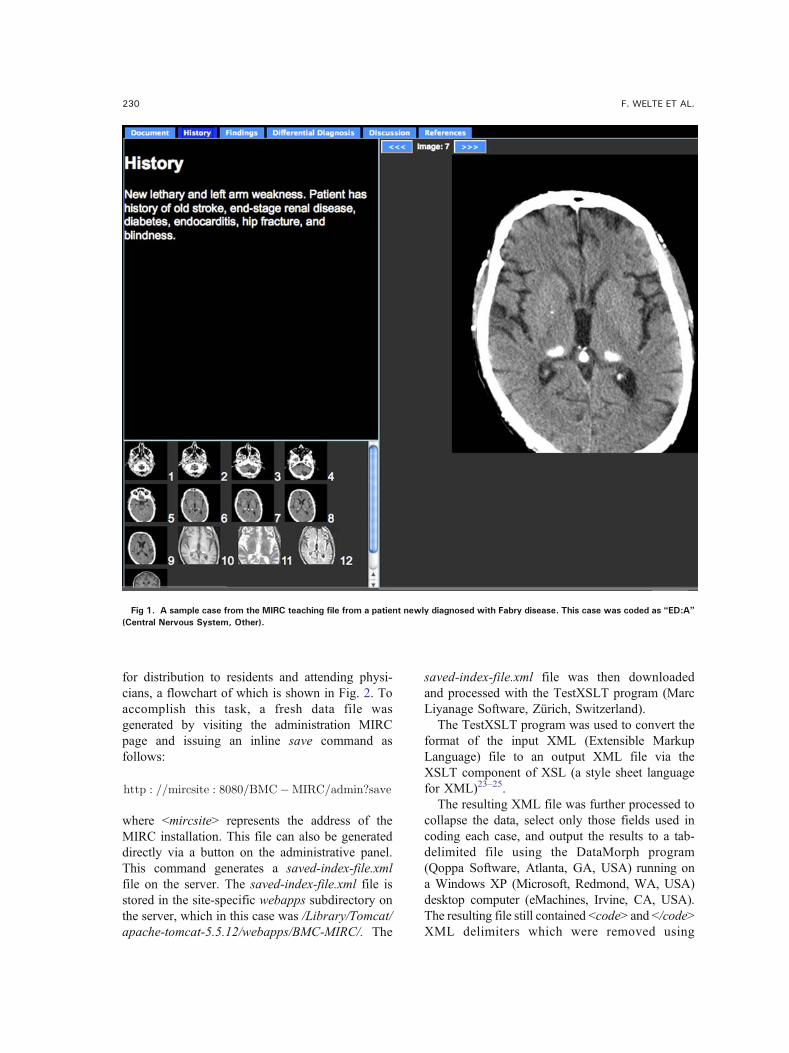
The choice of whether to use RadPix or theMIRC case entry tool was left to each individual.Access to RadPix was limited due to a limitednumber of licenses and inability to install thesoftware on our institutional computers. Themajority of cases were entered using the built-in MIRC case entry tool. To ease the learningcurve involved with entering cases using thebuilt-in MIRC case authoring tool, a detailedCase Entry Guide was distributed electronicallyvia e-mail and in print form to all users. Thisguide is included in the Electronic supplementarymaterial. A page from a sample case is shown inFig. 1.Study images were exported from Synapse
(FujiFilm Medical Systems, Chicago, IL, USA)PACS (Picture Archiving and CommunicationSystem) stations to bitmap (BMP) format andexternally converted to JPG using FastStone ImageViewer v. 2.5 (FastStone Software, Calgary, AB,Canada)22. A minority of cases included imageannotations, in part due to the difficulty of enteringannotations using the built-in MIRC annotationtool for the average user, and due to the need toinstall the JAI software on each computer whereannotations were to be entered. Prior to enteringimages into the database, all identifiable patientinformation was stripped from both images andtext. Final authored cases were to be reviewed byresident authors in conjunction with a facultymember to verify accuracy and completeness,although this review was not universal, largelydue to resident and faculty time constraints. Caseswithin the MIRC database were not made availableoutside our institution via the Publish function inMIRC due to the lack of a reliable internal peerreview system.
REPORT GENERATION AND DISTRIBUTION
A manual procedure was used to generateregular tallies of the content of the MIRC database
INCORPORATION OF A FORMALIZED EMERGENCY RADIOLOGY CURRICULUM TO… 229
for distribution to residents and attending physi-cians, a flowchart of which is shown in Fig. 2. Toaccomplish this task, a fresh data file wasgenerated by visiting the administration MIRCpage and issuing an inline save command asfollows:
http : ==mircsite : 8080=BMC�MIRC=admin?save
where Gmircsite9 represents the address of theMIRC installation. This file can also be generateddirectly via a button on the administrative panel.This command generates a saved-index-file.xmlfile on the server. The saved-index-file.xml file isstored in the site-specific webapps subdirectory onthe server, which in this case was /Library/Tomcat/apache-tomcat-5.5.12/webapps/BMC-MIRC/. The
saved-index-file.xml file was then downloadedand processed with the TestXSLT program (MarcLiyanage Software, Zürich, Switzerland).The TestXSLT program was used to convert the
format of the input XML (Extensible MarkupLanguage) file to an output XML file via theXSLT component of XSL (a style sheet languagefor XML)23–25.The resulting XML file was further processed to
collapse the data, select only those fields used incoding each case, and output the results to a tab-delimited file using the DataMorph program(Qoppa Software, Atlanta, GA, USA) running ona Windows XP (Microsoft, Redmond, WA, USA)desktop computer (eMachines, Irvine, CA, USA).The resulting file still contained Gcode9 and G/code9XML delimiters which were removed using
Fig 1. A sample case from the MIRC teaching file from a patient newly diagnosed with Fabry disease. This case was coded as “ED:A”(Central Nervous System, Other).
230 F. WELTE ET AL.
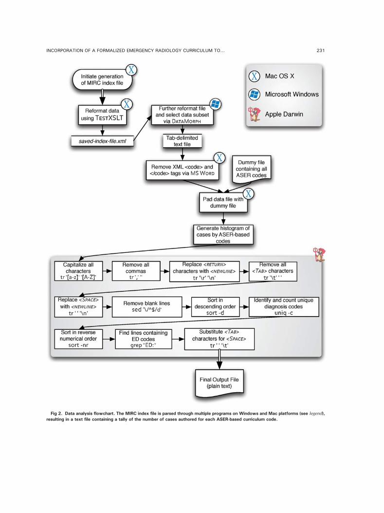
Fig 2. Data analysis flowchart. The MIRC index file is parsed through multiple programs on Windows and Mac platforms (see legend),resulting in a text file containing a tally of the number of cases authored for each ASER-based curriculum code.
INCORPORATION OF A FORMALIZED EMERGENCY RADIOLOGY CURRICULUM TO… 231
Microsoft Word (Microsoft) and the result saved as asimple text file.Next, a static file containing a list of all possible
case codes and their corresponding diagnoses wasappended to the processed data file for the purposeof filling in the gaps where case types were missing;total case counts for each category were later
decremented by one to account for this. Without thisstep, categories for which no cases had been enteredwould be excluded from the report.A diagnosis code frequency table was then
generated using a string of piped command linetext utilities in Mac OS X Darwin (Apple Com-puters), as follows:
cat DataMorph output file:txt tr 0 a�z½ �0 0 A�Z½ �0��
�� tr 0;0 0 0 tr 0nr0 0nn0j
j tr 0nt0 0 0 tr0 0 0nn0j j sed 0= ^$=d0 sort �dj j uniq �c sort �nrj j grep00ED :00 tr0 0 0nt0j > output:txt
Breaking down this “word histogram” func-tion26, the output of the DataMorph manipulationis sent through the tr command (translate; trans-lates the first argument to the second argumentusing regular expressions) to capitalize all charac-ters in the file. Next, all commas are removed andGreturn9 characters are replaced with Gnew line9characters. Tabs are replaced with spaces andGnew line9 characters are added to the appropriateplaces in the file. The sed (stream editor) commandline program is used to remove blank lines.Finally, unique words (diagnosis codes) are iden-tified and counted via the uniq command, theresults sorted via sort, and the lines containingvalid diagnostic codes identified and displayedusing grep. Tabs are then added and the streamoutput directed to a data file. The “|” vertical barpipe command sends the output of the precedingcommand into the following one. A flow chart ofthis process is shown in Fig. 2. Additional manip-ulations and formatting of this output file wereperformed after import into Microsoft Excel (Micro-soft), including aggregation of multiple diseasecodes into appropriate departmental subdivisions.Reports were then distributed to resident and
attending physicians in the department in PortableDocument Format (PDF; Adobe Systems, SanJose, CA, USA) via e-mail. Subsets of this reportwere printed and posted in the reading areas foreach subsection of the department for additionalexposure and accessibility. Paper copies were alsodistributed directly to subsection Chiefs whereparticularly large percentages of categories wereunderrepresented in the database. A completesample report is included in the Electronicsupplementary material.
RESULTS
Prior to the adoption of an organized, focusedcurriculum to guide case identification, case entryby residents was at best approximately five to tencases per month, with only three residents regu-larly participating in any given month (out of atotal of 15 radiology residents) and two facultymembers (of a total of 25). The distribution ofcases within the MIRC database was initiallyskewed toward common cases such as acuteappendicitis and pulmonary embolism. Less com-mon diseases were only haphazardly represented.Most residents and faculty had private collectionsof interesting cases, which were often onlyidentified by patient and were not organized intocoherent teaching file cases.Within approximately 4 months, 454 total cases
had been entered into the MIRC database, occu-pying 737 Megabytes (MB) of hard drive space, anaverage of 1.7 MB per case. Typical Joint Photo-graphic Experts Group (JPG; JPEG) images were512×512 pixels (100 Kilobytes [KB]) for CTimages, but could be up to 3,000×3,000 pixels(380 KB) for scanned radiographs. Overall, 42%of the subcategories within the modified ASERcurriculum were represented (161 out of 376),although this is an underestimation because “Oth-er” subcategories are included in the denominatorof this calculation.The amount of textual information included in
each case was variable, ranging from no text toextensive topic reviews comparable to casereports. The majority of cases contained sufficienttextual information to allow the cases to be usedfor teaching purposes, i.e., history, diagnosis, and
232 F. WELTE ET AL.
a brief discussion of the topic. Residents oftenentered case “skeletons” and returned later to fill indetails on the topic. The time required to author acase was quite variable; depending on howfamiliar the author was with the MIRC case entrysystem, the complexity of the case, and the extentof the case write-up. Cases were frequently enteredin multiple stages, beginning with entry of only acase skeleton, often performed by an assistant,followed by later entry of more detailed informa-tion. The overall time to author a case ranged fromapproximately 15 min to 1 h. One of the biggestfactors increasing the time to author a case was theneed to retrieve and scan hardcopy films. Thelargest factors in decreasing case authoring timewere the availability of an assistant and havingcase images available in digital format on PACS.With the introduction of the focused ASER
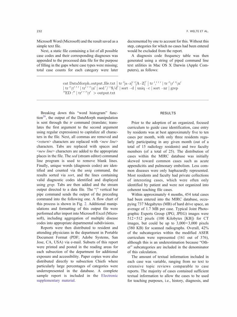
curriculum and distribution of regular reports onthe contents of the teaching file, physicians weremuch more likely to contribute cases. Withinseveral months of establishing the describedsystem, 100% of residents and approximately40% of faculty (ten total) had contributed cases,with the number of participating residents andfaculty steadily increasing over time. Case entryincreased from approximately 3.3 cases per monthper resident (ten cases/month/three residents) to7.5 (454 cases/4 months/15 residents), so that boththe number of residents participating and theirindividual productivity increased over this period(Fig. 3). The number of cases per month perfaculty member increased from approximately 5per month (ten cases/month/two faculty) up to 11.4per month (454 cases/4 months/ten faculty).Residents stated anecdotally that, while they
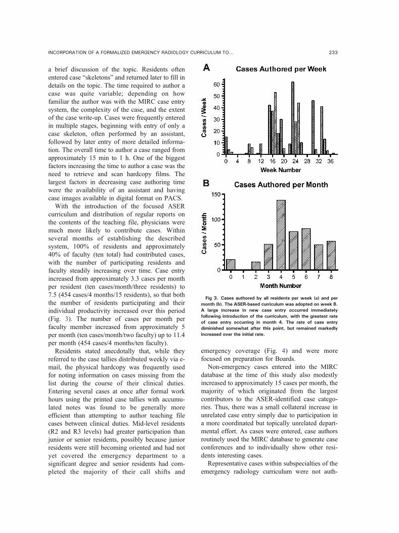
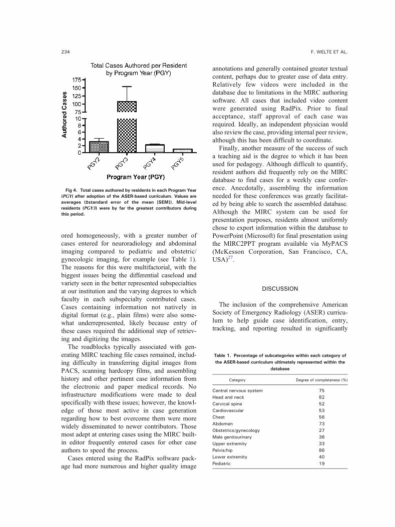
referred to the case tallies distributed weekly via e-mail, the physical hardcopy was frequently usedfor noting information on cases missing from thelist during the course of their clinical duties.Entering several cases at once after formal workhours using the printed case tallies with accumu-lated notes was found to be generally moreefficient than attempting to author teaching filecases between clinical duties. Mid-level residents(R2 and R3 levels) had greater participation thanjunior or senior residents, possibly because juniorresidents were still becoming oriented and had notyet covered the emergency department to asignificant degree and senior residents had com-pleted the majority of their call shifts and
emergency coverage (Fig. 4) and were morefocused on preparation for Boards.Non-emergency cases entered into the MIRC
database at the time of this study also modestlyincreased to approximately 15 cases per month, themajority of which originated from the largestcontributors to the ASER-identified case catego-ries. Thus, there was a small collateral increase inunrelated case entry simply due to participation ina more coordinated but topically unrelated depart-mental effort. As cases were entered, case authorsroutinely used the MIRC database to generate caseconferences and to individually show other resi-dents interesting cases.Representative cases within subspecialties of the
emergency radiology curriculum were not auth-
Fig 3. Cases authored by all residents per week (a) and permonth (b). The ASER-based curriculum was adopted on week 8.A large increase in new case entry occurred immediatelyfollowing introduction of the curriculum, with the greatest rateof case entry occurring in month 4. The rate of case entrydiminished somewhat after this point, but remained markedlyincreased over the initial rate.
INCORPORATION OF A FORMALIZED EMERGENCY RADIOLOGY CURRICULUM TO… 233
ored homogeneously, with a greater number ofcases entered for neuroradiology and abdominalimaging compared to pediatric and obstetric/gynecologic imaging, for example (see Table 1).The reasons for this were multifactorial, with thebiggest issues being the differential caseload andvariety seen in the better represented subspecialtiesat our institution and the varying degrees to whichfaculty in each subspecialty contributed cases.Cases containing information not natively indigital format (e.g., plain films) were also some-what underrepresented, likely because entry ofthese cases required the additional step of retriev-ing and digitizing the images.The roadblocks typically associated with gen-
erating MIRC teaching file cases remained, includ-ing difficulty in transferring digital images fromPACS, scanning hardcopy films, and assemblinghistory and other pertinent case information fromthe electronic and paper medical records. Noinfrastructure modifications were made to dealspecifically with these issues; however, the knowl-edge of those most active in case generationregarding how to best overcome them were morewidely disseminated to newer contributors. Thosemost adept at entering cases using the MIRC built-in editor frequently entered cases for other caseauthors to speed the process.Cases entered using the RadPix software pack-
age had more numerous and higher quality image
annotations and generally contained greater textualcontent, perhaps due to greater ease of data entry.Relatively few videos were included in thedatabase due to limitations in the MIRC authoringsoftware. All cases that included video contentwere generated using RadPix. Prior to finalacceptance, staff approval of each case wasrequired. Ideally, an independent physician wouldalso review the case, providing internal peer review,although this has been difficult to coordinate.Finally, another measure of the success of such
a teaching aid is the degree to which it has beenused for pedagogy. Although difficult to quantify,resident authors did frequently rely on the MIRCdatabase to find cases for a weekly case confer-ence. Anecdotally, assembling the informationneeded for these conferences was greatly facilitat-ed by being able to search the assembled database.Although the MIRC system can be used forpresentation purposes, residents almost uniformlychose to export information within the database toPowerPoint (Microsoft) for final presentation usingthe MIRC2PPT program available via MyPACS(McKesson Corporation, San Francisco, CA,USA)27.
DISCUSSION
The inclusion of the comprehensive AmericanSociety of Emergency Radiology (ASER) curricu-lum to help guide case identification, entry,tracking, and reporting resulted in significantly
Fig 4. Total cases authored by residents in each Program Year(PGY) after adoption of the ASER-based curriculum. Values areaverages (±standard error of the mean [SEM]). Mid-levelresidents (PGY3) were by far the greatest contributors duringthis period.
Table 1. Percentage of subcategories within each category ofthe ASER-based curriculum ultimately represented within the
database
Category Degree of completeness (%)
Central nervous system 75Head and neck 82Cervical spine 52Cardiovascular 53Chest 56Abdomen 73Obstetrics/gynecology 27Male genitourinary 36Upper extremity 33Pelvis/hip 86Lower extremity 40Pediatric 19
234 F. WELTE ET AL.
increased participation by radiology residents andfaculty and a broader collection of cases within thedatabase. Within a month of initiating this focusedapproach, nearly all radiology residents wereparticipating to varying degrees. Interest in addingmissing cases to the repository increased substan-tially at the time case tallies were distributed toresidents and section Chiefs. Interaction with thesection Chiefs during the distribution of theseperiodic reports had the added effect of reinforcingtheir participation in the building of the overallteaching file.Upon review of the list of needed cases, individual
attending physicians were often able to recall casesfrom their own hardcopy or digital teaching files forinclusion in the central MIRC system, accomplishingthe dual goals of digitizing and cataloguing theirdispersed teaching files and filling in missing caseswithin the ASER curriculum. Residents who weremost active in the project also became contacts forfaculty, who often passed on their own interestingcases to these residents for entry into MIRC,providing additional educational opportunities.Rather than slowing down case entry by adding
an additional layer to an already cumbersomeprocess, the adoption of a standardized frameworkto use as a catalogue for identifying cases for entryinto the teaching file actually facilitated the overallprocess. A component of this success is likely thatrelatively few technically adept individuals in thedepartment handled the brunt of the behind-the-scenes requirements to install and maintain theteaching file and generate weekly reports. Anotherfactor was the availability of an assistant to enterbasic case skeletons upon which case authorscould later build. Aside from the use of thecurriculum framework, the presence of the studyitself and the resources allocated to it likely hadsome effect in terms of galvanizing faculty effortsto recruit more participants for the general teachingfile program. Without these additional resources,the progress in participation ascribed to the ASER-based curriculum would likely have been less.The small increase in the entry of non-ASER
cases may be related to individuals’ subjectivelyincreased awareness of the presence of the MIRCteaching file among both residents and faculty,presence of a more coordinated means by which todisseminate instructions on how to access thedatabase, and a “critical mass” effect wherebyenough people were involved in a unified effort
that other physicians felt compelled to contributetheir own cases, whether they were specificallyrelated to the ASER curriculum or not.One limitation frequently encountered was the
difficulty of entering cases into the MIRC system.While the web-based case entry system built intoMIRC is quite functional, many users had signif-icant difficulty successfully entering cases. Thedepartmental PACS was not directly attached tothe MIRC server, necessitating the manual capture,conversion, and transfer of images. The learningcurve for MIRC case assembly and entry wassignificant enough that very few attending physi-cians ultimately felt comfortable entering cases ontheir own without resident assistance.These limitations led to most cases being entered
by residents, whether they were the ones to identifythe case or not. An unintended consequence of thisarrangement was that residents who were moreadept at entering cases saw many more of theseinteresting cases as a result, as these were filteredthrough them for processing. The use of RadPix as aclient-side stand-alone case entry tool helpedovercome this limitation to some degree; however,its limited availability curtailed its impact.One of the greatest challenges to the adoption of
a MIRC-based digital teaching file system oncecase entry difficulties are overcome is the mainte-nance over time. Residents and faculty havelimited time and expertise to continually verifythe integrity of the database, test backups, andmonitor security issues; a dedicated staff personmay ultimately better accomplish these tasks. Thetime needed to collect information and author cases,peer review authored cases, and ensure currency ofthe database represents an additional burden.Another limitation encountered in the context of
this project was in the generation of reports fromthe content of the MIRC database. While thedatabase was well organized internally, there wasno straightforward way to mine the data containedtherein. The information held on the MIRC hostcould be accessed and tabulated, but the procedurewas not straightforward and required the use ofmultiple programs and relatively sophisticatedtechniques on two computing platforms. A simplerway of accessing and analyzing the informationstored in MIRC sites would be beneficial forresearch and teaching purposes.With the availability of information on a global
scale, radiologists have the opportunity to train by
INCORPORATION OF A FORMALIZED EMERGENCY RADIOLOGY CURRICULUM TO… 235
reviewing cases from many previously unavailableresources. This capability allows cases involvinggeographically localized diseases to be disseminat-ed widely, improving medical education. A digitalteaching file system with the built-in capability totransfer information on the Internet, such as theRSNA MIRC system, is a natural tool to aid indistribution of this specialized information.
CONCLUSIONS
The inclusion of a comprehensive, tabulatedcurriculum for a specific subfield of radiology didenhance the orderly population of a MIRC-baseddigital teaching file. Resident and faculty interestand participation increased substantially relative tothe initial situation where individuals simplyidentified interesting cases haphazardly and loggedthem without communication or guidance intotheir own non-standard disconnected teaching filesystems. Such an approach may prove beneficialfor other programs as teaching files are imple-mented or augmented.Some hindrances were encountered in using the
MIRC system, primarily involving the entry ofcases and the analysis of the contents of thedatabase. These limitations were ultimately over-come, but may be points to consider for radiologydepartments implementing such a system. Inparticular, an external tool such as RadPix shouldbe considered to ease case entry. The degree oftechnical facility required to maintain such asystem likely necessitates strong InformationTechnology support for sustainability.Incorporation of other established subspecialty
radiology training curricula is needed to determineif these findings are valid for other subspecialtiesand what curriculum format might be mosteffective in driving physician participation anddigital teaching file case entry. Additional investi-gation is also needed to determine how participa-tion and case identification and entry rates varywith the number of subspecialties and casecategories presented to radiologists and if asaturation point exists where efficiency mightplateau or decrease.
ACKNOWLEDGEMENTS
The authors wish to acknowledge Dr. J. Robert Kirkwood,MD, FACR for his support of this project. We also thank the
many residents and faculty members who contributed cases tothe database and Ms. Lindsay Coughlin for data input and casemanagement. We thank the MIRC community on the RSNAMIRC users’ forums and John Perry and Drs. William Weadockand Trongtum Tongdee in particular for useful conversationsand suggestions. We also thank Ms. Sharon Wayne forproofreading the manuscript.
Competing interests The authors state that they have nocompeting interests.
Authors' contributions FJW developed the project,installed the necessary software, analyzed the data,entered cases into the database, and wrote and reviewedthe manuscript.
SK entered cases into the database, participated incoordination of the project, and reviewed and edited thefinal manuscript.
DD entered cases into the database, participated incoordination of the project, and reviewed and edited thefinal manuscript.
SOC contributed to the conceptual development of theproject, case entry, coordination of the project, faculty andresident recruitment, and reviewed and edited the finalmanuscript.
BC contributed to the conceptual development of theproject, case identification, faculty and residentrecruitment, and reviewed and edited the final manuscript.
REFERENCES
1. Siegel E, Channin D, Perry J, Carr C, Reiner B: MedicalImage Resource Center 2002: an update on the RSNA’sMedical Image Resource Center. J Digit Imaging 15:2–4, 2002
2. Siegel E, Reiner B: The Radiological Society of NorthAmerica’s Medical Image Resource Center: an update. J DigitImaging 14:77–79, 2001
3. Yang GL, Aziz A, Narayanaswami B, Anand A, Lim CC,Nowinski WL: Informatics in radiology (infoRAD): multimediaextension of medical imaging resource center teaching files.Radiographics 25:1699–1708, 2005
4. Siegel E, Reiner B: Electronic teaching files: seven-yearexperience using a commercial picture archiving and commu-nication system. J Digit Imaging 14:125–127, 2001
5. Roth CJ, Weadock WJ, Dipietro MA: A novel applicationof the MIRC repository in medical education. J Digit Imaging18:85–90, 2005
6. Gentili A, Chung CB, Hughes T: Informatics in radiology:use of the MIRC DICOM service for clinical trials toautomatically create teaching file cases from PACS. Radio-graphics 27:269–275, 2007
7. Yang GL, Lim CC: Singapore National Medical ImageResource Centre (SN.MIRC): a world wide web resource forradiology education. Ann AcadMed Singapore 35:558–563, 2006
8. Main Page—MircWiki. Available at http://mircwiki.rsna.org/index.php?title=Main_Page Accessed 18 November 2007
236 F. WELTE ET AL.
9. MIRC: Medical Imaging Resource Center. Available athttp://www.rsna.org/mirc/ Accessed 18 November 2007
10. Jakarta Tomcat. Available at http://tomcat.apache.org/Accessed 8 November 2007
11. Complete Tomcat. Available at http://www.serverlogistics.com/tomcat.php Accessed 10 November 2007
12. The Jakarta Site—Jakarta Downloads. Available at http://jakarta.apache.org/site/downloads/ Accessed 10 November 2007
13. No-IP—Dynamic DNS, Static DNS for Your Dynamic IP.Available at http://www.no-ip.com/ Accessed 10 November 2007
14. Novelline RA: Core Curriculum in Emergency Radiol-ogy: American Society of Emergency Radiology, 2006
15. Emergency Radiology Educational Resources. Availableat http://www.erad.org/newweb/education.htm Accessed 11November 2007
16. Minati L, et al: Bio-image warehouse system: conceptand implementation of a diagnosis-based data warehouse foradvanced imaging modalities in neuroradiology. J Digit Imag-ing 20:32–41, 2007
17. RadPix—Radiology Presentation Software and MIRCAuthoring Tool. Available at http://www.radpix.com/ Accessed11 November 2007
18. Langlotz CP: RadLex: a new method for indexing onlineeducational materials. Radiographics 26:1595–1597, 200619. RadLex: A Lexicon for Uniform Indexing and Retrieval
of Radiology Information Resources. Available at http://www.rsna.org/Radlex/index.cfm Accessed 25 May 200820. RadLex Term Browser. Available at http://radlex.org/
viewer Accessed 25 May 200821. Radlex Wiki. Available at http://radlexwiki.rsna.org/
index.php?title=Main_Page Accessed 25 May 200822. FastStone Image Viewer. Available at http://www.
faststone.org/23. XSL Transformations (XSLT). Available at http://www.
w3.org/TR/xslt Accessed 1 January 200824. TestXSLT. Available at http://www.entropy.ch/software/
macosx/#testxslt Accessed 1 January 200825. Extensible Markup Language (XML). Available at http://
www.w3.org/XML/ Accessed 1 January 200826. Word Histogram. Available at http://ipucu.enderunix.
org/view.php?id=1234&lang=en Accessed 1 January 200827. MIRC2PPT. Available at http://www.mypacs.net/repos/
mpv3_repo/static/m/Solutions/mirc2ppt.htm Accessed 10/20/2008 2008
INCORPORATION OF A FORMALIZED EMERGENCY RADIOLOGY CURRICULUM TO… 237