THURSDAY, FEBRUARY 28 - European Society of Radiology
40
THURSDAY, FEBRUARY 28 ECR TODAY
-
Upload
khangminh22 -
Category
Documents
-
view
1 -
download
0
Transcript of THURSDAY, FEBRUARY 28 - European Society of Radiology

THURSDAY, FEBRUARY 28
ECR TODAY

WF 1 – LEADING BY EXAMPLE Thursday, February 28, 12:30–14:00, The Church
CHAIRPERSON H. Hricak; New York, NY/US
SPEAKERS & PANELLISTS D. Akata; Ankara/TR, L.E. Derchi; Genoa/IT, C. Estrampes; Paris/FR, M.H. Fuchsjäger; Graz/AT,
H. Hricak; New York, NY/US, L. Leong; Hong Kong/CN, C. Palmer; London/UK
WF 2 – CHANGING THE IMAGE OF WOMEN IN LEADERSHIP: GENERATIONAL DIFFERENCES
AND SIMILARITIESThursday, February 28, 15:45–17:00, The Church
MODERATOR R.A. Kubik-Huch; Baden/CH
SPEAKERS & PANELLISTSU.I. Attenberger; Mannheim/DE, C. Hess; San Francisco, CA/US, R.A. Kubik-Huch; Baden/CH,
E.A. Morris; New York, NY/US, M.F. Reiser; Munich/DE, V. Vilgrain; Paris/FR
WF 3 – WOMEN IN CHALLENGING ENVIRONMENTSFriday, March 1, 14:00–15:15, The Church
MODERATORR.G.H. Beets-Tan; Amsterdam/NL
SPEAKERS & PANELLISTS Ş. Bahar Özvarış; Ankara/TR, E. Balogun; Lagos/NG, R.G.H. Beets-Tan; Amsterdam/NL,
D. Husseiny Salama; Cairo/EG, S.F. Khan; Islamabad/PK, L. O’Riordan; Ipswich/UK
WF 4 – LEADERSHIP AND MENTORSHIPFriday, March 1, 16:00–17:25, The Church
MODERATORJ.E. Husband; London/UK
SPEAKERS & PANELLISTSM. Abdel-Wahab; Vienna/AT, C. Beardmore; Guildford/UK, H. Hricak; New York, NY/US,
J.E. Husband; London/UK, V.P. Jackson; Tucson, AZ/US, G.P. Krestin; Rotterdam/NL, G. McGinty; New York, NY/US
All sessions will have dedicated rooms for remote viewing and will be streamed live online. More info at myESR.org/wif.
WOMEN IN FOCUS – THE BIGGER PICTURE

ECR TODAY 2019EUROPEAN CONGRESS OF RADIOLOGY
DAILY NEWS FROM EUROPE’S LEADING IMAGING MEETING | THURSDAY, FEBRUARY 28, 2019
myESR.org #ECR2019
BY FRANCES RYLANDS-MONK
Knowing your differentials will unlock migration-related diagnosesMigration has created new challenges for clinical radiology, largely due to the number of imported diseases that it generates.
This trend necessitates the use of novel imaging strategies and differ-ent ways of approaching interpre-tation. At today’s session, ‘Radiology and migrations’, ECR 2019 delegates will hear about the latest think-ing from specialists in the field and learn how to tackle migration-re-lated cases.
The total number of asylum seek-ers in countries across the Euro-pean Union has increased from around 15,000 to almost 1.4 million annually in the last two decades, stated Prof. Okan Akhan, who will moderate the session. These people are fleeing from violence, torture, persecution, and political or eth-nic oppression, and are unable to return to their country of origin. In addition, the number of economic migrants seeking a better life has also increased, he noted.
Whatever the reason for migra-tion, the health of these individ-uals is important for their social inclusion and integration in society, which in turn has an impact on the sociopolitical and economic out-come for this population, accord-ing to Akhan, professor of radi-ology and chief of interventional radiology and abdominal imaging at Hacettepe University Hospital in Ankara, Turkey. Besides gener-ating discussion about the reasons for migration and its humanitarian consequences, he hopes speakers will provide analysis of the impor-tance of migration-related diseases and how to recognise them clini-cally and radiologically.
He added that it was important to keep discussing the phenome-non of migration-related diseases,
and crucially to raise awareness of them among radiologists and other health professionals.
“The health issues of immigrants are manifold and in many situa-tions they are very challenging to physicians. Awareness of these con-ditions is mandatory to ensure good clinical practice for these patient populations that carry a huge bur-den in chronic, infectious, mental, and neurological diseases,” Akhan said.
Radiologists need to be aware of the types of infectious diseases immigrants suffer from, such as tuberculosis, HIV, viral hepatitis, malaria, schistosomiasis, echinococ-cosis, neurocysticercosis or similar diseases that reflect epidemiology in the country of origin. Addition-ally, immigrants may present with psychological problems, chronic diseases resulting in polymorbid-ity, cancer, and neurological dis-eases and acute or chronic malnu-trition. These conditions may result in anaemia, growth disorders, men-tal and physical development dis-orders, immunosuppression, neu-ropathy, bone disorders and other organ dysfunction, and it is easy to overlook these aspects during daily clinical work, he explained.
Radiological examination of these patients is crucial because imaging plays an important role in the diagnosis and differential diag-nosis of most of these diseases, but many radiologists in western Euro-pean countries are not familiar with them.
For instance, the incidence of cystic echinococcosis (CE), other-wise known as hydatid cysts, is
spreading across Europe, mainly due to migration. CE has a preva-lence of 1% in rural Turkey, and it is also endemic in southern Europe, including the Balkan countries, southern Italy, France, Spain, and Portugal. Case numbers are also growing in other parts of Europe due to immigration and travel to and from endemic regions. This means that diagnostic imaging and interventional procedures for CE treatment is becoming an impor-tant topic for western and northern European radiologists, who increas-ingly will encounter this disease in
their daily practice, and they must remember that what they think is a simple cyst may in reality be more complex and dangerous for the patient, Akhan suggested.
After ultrasound diagnosis of CE and classification, radiologists should follow up with MRI and MR cholangiopancreatography (MRCP) when communication between the cystic cavity and the biliary system is suspected. Whenever possible, the disease should be treated with different percutaneous techniques which, compared to surgery, are associated with fewer major com-
plications, lower mortality rates, shorter hospital stays, and lower recurrence rates, he said.
Dr. Bernadette Abela-Ridder, team leader on neglected zoonotic diseases and neglected tropical dis-eases (NTDs) in the Department for the Control of NTDs of the World Health Organization (WHO) in Geneva, Switzerland, will pro-vide her insight into how migra-tion is impacting incidence and outcomes.
This photo shows part of a search-and-rescue exercise that simulated migrants arriving by boat during a Summer School on Refugee and Migrant Health, held in July 2017 in Syracuse, Italy. If migrants are in a serious medical condition and treatment cannot wait until arrival in port, the Italian Coast Guard launches a medical evacuation operation by helicopter to immedi-ately transfer them to a medical fa-cility onshore. (Provided by WHO/Paolo di Pietro)
Every migrant that arrives on board undergoes security screening, has their identification checked and photo taken, and undergoes initial medical screen-ing by the Italian Relief Corps of the Order of Malta (CISOM). Any potentially urgent or infectious cases are reported to representatives of the Maritime, Air and border Health Office (USMAF) of the Italian Ministry of Health for treatment isolation or emergency evacuation as required. (Provided by WHO/Paolo di Pietro)
TECHNOLOGY & RESEARCHNew MRI scanners and patient-centric
accessories target productivity and better clinical outcomes
COMMUNITY NEWSA success story with new challenges:
European Radiology, the ESR’s flagship journal continues to grow
17 25
CLINICAL CORNERCan you do more to improve
communication of critical information to patients?
HIGHLIGHTSAfrican radiologists call for
more cooperation with the ESR in radiation protection
93
continued on page 3

Q U ESTION THUR
SDAY
-FEBRUARY 28 NO 1We dare you to solve a very difficult EDiR question.
This is your chance to sit the examination and attend
the ECR 2020 for freeSolve the question
posted at the EBR blog
before 13:30h.
The question
right answer and
the winner will
be announced at
the EBR blog at
14:00h today.
The European Board of Radiology will raffle amongst the winners a free examination fee for the examination that will take place within the ECR 2020 frame. ECR 2020 free registration will be also included!
Go to the EBR blog at blog.myebr.org to find the EDiR Question of the Day, and further interesting resources to prepare for the examination.
B L O G . M Y E B R . O R G
EUROPEAN DIPLOMA IN RADIOLOGY – QUESTION OF THE DAY

continued from page 1
HIGHLIGHTS 3ECR TODAY | THURSDAY, FEBRUARY 28, 2019
#ECR2019myESR.org
With increasing levels of con-flict, civil unrest, poverty, and per-secution, there are an estimated 68.5 million people currently for-cibly displaced worldwide. Mal-nourished people with poor hous-ing and sanitation and restricted access to healthcare results in populations at a high risk of dis-ease, especially NTDs, stated Abela-Ridder.
Global migration is changing the epidemiology of many of these diseases with the emergence and re-emergence of NTDs in non- or low-endemic countries. The lack of awareness, diagnostics and treatment in host countries poses a challenge to appropriately diag-nosing and treating affected indi-viduals, and to establishing the endemic foci of disease, she said.
Outbreaks of dengue, Leishmani-asis and schistosomiasis have been recorded in low prevalence areas where intermediate hosts could allow for local transmission. The prevalence of Chagas disease (also known as American trypanosomi-asis), strongyloidiasis and schisto-somiasis in migrant populations ranges from 4.2% to 48.5%, 11% to 56.1%, and 5.8% to 44% respectively.
Picking up on the issue of hyatid cysts, Abela-Ridder noted that cases of CE and neurocysticercosis have also been detected in Europe, with 53% of neurocysticercosis cases attributed to immigrants.
Systematically screening migrant populations on arrival to a host country to reduce disease progres-sion and severity should be consid-ered, but she cautioned that effort was needed to ensure that individ-uals are not discriminated against
because of health problems. Better data on disease prevalence and bur-den in migrant populations remain key to increased awareness in host countries, and timely access to appropriate care for migrants, she continued.
Providing radiology tips dur-ing the session, Dr. Tim Weber, sen-ior physician at Heidelberg Univer-sity Hospital in Germany, will shed light on the patterns to look for and how to evaluate unusual radi-ological conditions, after combin-ing patients’ radiological and epide-miological features for differential diagnosis.
Finally, the panel discussion will focus not only on raising aware-ness among health professionals but also on how future education programmes might adopt migra-tion-related disease imaging into the curriculum.
BY MÉLISANDE ROUGER
African radiologists call for more cooperation with the ESR in radiation protection
African radiologists will share their knowledge and experience of medical imaging practice in their respective countries, today during the ‘ESR meets Africa’ session. Examples of cooperation with the European Society of Radiology will be presented, notably in radiation protection, a field where efforts between Europe and Africa are starting to pay off.
Africa is big, diverse and full of possibilities. Its myriad of coun-tries (54), languages, cultures and economic scenarios offers unmatched potential, but it can also complicate the organisation of radiology.
Equipment and workforce tend to vary considerably from one country to another. For exam-ple, the radiologists’ ratio ranges from 1 to 80 per million population, depending on the country.
“Our main challenges are in training and the implementation of national legislation that can help organise the field, especially regarding radiation protection,” said Prof. Hassen Gharbi from Tunis, Tunisia, who will co-chair the session with ESR President Lorenzo Derchi from Genoa, Italy.
Technological advances have opened new horizons for the appli-cation of ionising radiation in healthcare all around the world
and this has led to an increase in medical imaging procedures using radiation, also in Africa.
Unlike most of the continent, Tunisia, Algeria and Morocco have dedicated guidelines on ionis-ing radiation use. But even there, rules must be implemented to help healthcare professionals prescribe examinations adequately.
Prof. Dina Husseiny Salama and Prof. Hassen Gharbi, together with ESR President Prof. Lorenzo E. Derchi and Prof. Guy Frija, Chair of EuroSafe Imaging, surrounded by delegates at the 5th African Society of Radiology conference in January 2019, in Cairo, Egypt.
Professional Challenges Session
Thursday, February 28, 08:30–10:00, Studio 2019PC 5 Radiology and migrations
» Chairperson’s introductionO. Akhan; Ankara/TR
» Impact of migration on health: WHO perspectiveS. Severoni; Copenhagen/DK
» Importance of migration for neglected tropical diseases: WHO perspectiveB. Abela-Ridder; Geneva/CH
» Clinical importance of the diseases related to migrationT. Junghanss; Heidelberg/DE
» Radiology of the diseases related to migrationT. Weber; Heidelberg/DE
» Panel discussion: How do we raise awareness among health professionals to recognise diseases related to migration? How can we adapt migration and migration related diseases into our education?
ESR meets Session
Thursday, February 28, 10:30–12:00, RoomBESR meets Africa EM 1 Radiology in Africa: facing challenges and opportunities
Presiding: L.E. Derchi; Genoa/IT H.A. Gharbi; Tunis/TN
» Chairperson’s introductionL.E. Derchi; Genoa/ITH.A. Gharbi; Tunis/TN
» Structured strategies to combat radiation protection challenges in Africa. What can the ESR do?D. Husseiny Salama; Cairo/EG
» Is imaging underused in Africa? East Africa as an example. Solutions: what can the ESR do?S. Vinayak; Nairobi/KE
» Interlude 1: Enjoy the difference between African and European musicH.A. Gharbi; Tunis/TN
» How to promote radiation protection in West Africa. Needs and expected role of the ESRE.H. Niang; Dakar Fann/SN
» Most important challenges for imaging in North AfricaL. Rezgui Marhoul; Tunis/TN » » Interlude 2: Enjoy the difference between Arabic and European musicH.A. Gharbi; Tunis/TN
» The WHO’s programme in Africa: the past, the present and the futureM.D.R. Perez; Geneva/CH
» Panel discussion: Radiology in Africa, reality and dreamsG. Frija; Paris/FRL.E. Derchi; Genoa/ITH.A. Gharbi; Tunis/TND. Husseiny Salama; Cairo/EGS. Vinayak; Nairobi/KEE.H. Niang; Dakar Fann/SNL. Rezgui Marhoul; Tunis/TNM.D.R. Perez; Geneva/CHB. Mansouri; Algiers/DZK.M. Naidu; Cape Town/ZA
» ConclusionL.E. Derchi; Genoa/ITH.A. Gharbi; Tunis/TN
This session is part of the EuroSafe Imaging campaign.
continued on page 4

When: Friday, March 1st, 18:00 CETWhere: DO & CO Hotel Vienna
Leaders in breast cancer detection will share their clinical experience with ProFound AI™
for digital breast tomosynthesis and clinical research on risk assessment.
New Frontier: Predicting Breast Cancer through Artifi cial Intelligence
Space is limited, advanced RSVP required.Please visit X1, booth AI-17 to register and be entered to win an Apple Watch.
Networking. Cocktails. Food.
continued from page 3
HIGHLIGHTS4 ECR TODAY | THURSDAY, FEBRUARY 28, 2019
#ECR2019 myESR.org
“There is a growing need for structured strategies and a holistic approach towards the full integra-tion of radiation safety and clinical imaging guidelines in Africa,” said Prof. Dina Husseiny Salama from Cairo, Egypt.
Prof. Husseiny Salama, who will talk about radiation protection during the session, will highlight the role of strategic planning for more cooperation between the ESR and African radiology societies.
“There have been proactive actions in Africa to improve the situation and enhance the imple-mentation of radiation protection in several countries, however fur-ther actions and joint activities are needed to enhance the process. Local initiatives work, but we need to accelerate things through coop-eration with the ESR. Africa needs free, evidence-based tools, which a global player like the ESR can pro-vide,” she said.
The ESR launched the EuroSafe Imaging campaign three years ago to promote the safe use of ionis-ing radiation in medicine, and is now developing a EuroSafe Imag-ing Star assessment scheme for low and middle-income coun-tries (LMICs), to recognise imag-ing departments that embody best practice in radiation protection.
Justification of examinations is another key aspect of radiation protection and the ESR is support-ing the use and uptake of imaging referral guidelines, especially in LMICs. The ESR iGuide web por-tal is free to use for ESR radiologist members, and a model with unre-stricted access for LMICs is cur-rently under development.
“The ESR iGuide is an excellent online tool to help clinicians jus-tify examinations, and it’s already showing results in Egypt. Coop-eration with the ESR works on our side, so why not in the rest of Africa?” Prof. Husseiny Salama said.
The process for a stepwise imple-mentation of the ESR iGuide was started last year at four centres of excellence in Cairo. A recently performed audit showed that the number of inappropriate referrals declined significantly, especially in the emergency setting. “So far, we’ve had 15% fewer inappropri-ate referrals, which is quite impor-tant for patient radiation protec-tion and the financial budget of the radiology department,” Prof. Hus-seiny Salama said.
The ESR is also supporting Afri-can radiologists by offering the opportunity for non-European radiological national societies to become Associate Institutional Members. The ESR has ten Asso-
ciate Institutional Members from Africa, including Algeria, Egypt, Ivory Coast, Mauritania, Morocco, Nigeria, South Africa, Tanzania, Tunisia and Uganda.
Professionals in the field out-side of Europe can also become cor-responding members of the ESR. Corresponding members are enti-tled to a wide range of benefits, including reduced registration fees for ECR 20201, free access to all con-tents of the ESR e-learning plat-form Education on Demand, the option to participate in the Euro-pean Diploma in Radiology (EDiR) and all the activities of the Euro-pean School of Radiology (ESOR), and many more.
Both memberships are com-pletely free of charge.
A 92% increase in abstracts from Africa was registered for ECR 2019, as a result of inviting Africa to the ‘ESR meets’ programme. The num-ber of African ESR members also rose from 3% to 5% in just over a year. The benefits of joining the ESR must be made even clearer south of the Mediterranean, so that African radiologists can enjoy all the support the ESR can offer, according to ESR President Prof. Lorenzo Derchi.
“We hope this is just the start. It is only a matter of making us visi-ble and making the advantages of ESR membership known. We hope
that ‘ESR meets Africa’ will be the right place and moment to get to know each other better,” he said.
African radiologists have high hopes regarding their cooperation with Europeans, according to Prof. Gharbi.
“Medical imaging is advanc-ing rapidly in Africa but we want things to develop in the utmost safety conditions and in respect with our guidelines. Africa hopes the ESR can help with radiation protection, and also to help pro-mote, advance and homogenise training,” he said.
Ultrasound can play a major role in promoting radiation protection, as it provides easy access to sophis-ticated diagnostic methods for the poorest patients. “Two thirds of the global population have no access to medical imaging. Ultrasound must play an important role in radiation protection and in tropical diseases, for example hydatid disease, a sce-nario we are commonly faced with in Africa,” Gharbi concluded.
1 Provided that ESR 2019 membership is activated and
approved by August 31, 2019.
Prof. Dina Husseiny Salama from Cairo, Egypt will highlight the role of strategic planning for more cooperation between the ESR and African radiology societies in today’s ‘ESR meets Africa’ session.
Prof. Hassen Gharbi from Tunis, Tunisia, will co-chair the ‘ESR meets Africa’ session with ESR President Lorenzo Derchi.

Dive into interventional radiology at the Cube 2.0Open 8:30-17:30February 27 - March 2 at the DC Tower, ECR City
For more information visit www.myESR.org/cube
The Cube is located in the cuboid annex
of the DC Tower.
HIGHLIGHTS 5ECR TODAY | THURSDAY, FEBRUARY 28, 2019
#ECR2019myESR.org
BY MÉLISANDE ROUGER
A new beginning in cancer imaging has just begun, says Beets-TanProfessor Regina Beets-Tan is chair of the department of radiology at The Netherlands Cancer Institute in Amsterdam, full professor of radiology at the University of Maastricht and adjunct professor of abdominal and oncological radiology at the University of Southern Denmark. She will present the Wilhelm Conrad Röntgen Honorary Lecture, entitled ‘Oncologic imaging: a new beginning has just begun’ at ECR 2019.
She shared a few thoughts with us on the future of her specialty in an interview ahead of the congress.
ECR Today: You have chosen quite an iconic and broad topic for this honorary lecture. What points will you cover exactly?
Regina Beets-Tan: The audience will get a glimpse of the future of cancer care and the role of imaging.
The world of cancer medicine is changing rapidly. Major steps for-ward have been taken. Advanced imaging and computing technol-ogy, screening programmes; these all will result in the early detection of more tumours. Minimally inva-sive treatment, including inter-ventional therapy, will have an increasingly important role. Tar-geted therapy, which specifically hits the cancer genes, and immu-notherapy, which uses the patient’s own immune system to kill can-cer cells, will result in prolonged survival of patients who are in the final stage of metastatic dis-ease. It will be ‘precision medicine’; we do not want to give the wrong treatment to the wrong patient. As advocated by Prof René Ber-nards, a respected leader in can-cer research at the Netherlands Cancer Institute: ‘Within 15 years, cancer will become a chronic dis-ease’. And I believe this is true. This transformation will change the way we will practice oncologic imaging. This will require us to rec-reate our discipline. With this lec-ture, I would like to take my young
colleagues on a 20-minute journey towards their future.
ECRT: Your field of research is abdominal and oncologic imaging, especially MRI of rectal cancer. What are the latest developments in rectal cancer medicine?
Regina Beets-Tan: Colorectal cancer screening programmes and modern technology in imaging and endoscopy have resulted in the detection of early and smaller rec-tal tumours. Multimodality treat-ment of advanced rectal cancer that combines radiotherapy with che-motherapy, or even with immuno-therapy, has resulted in more com-plete responses, which brings the need for surgery in these cases into question. The focus will be on qual-ity of life. It is going to be more min-imal invasive treatment and local tumour excision rather than rectal amputation. We are going for active surveillance (Watch and Wait) for complete responders after treat-ment. It will be paramount to per-form accurate selection and fol-low-up of patients, and accurately predict who will truly benefit from organ preservation. Modern imag-
ing technology (functional MRI, molecular imaging) together with endoscopy brings us very far. Yet some problems remain unsolved, like the accurate assessment of nodal disease. There is still a lot of work to do and investment in research investigating the role of modern imaging and computing imaging in colorectal cancer man-agement is much needed.
ECRT: What are the challenges facing radiologists today and tomorrow?
Regina Beets-Tan: Radiology surely has challenges lying ahead, but challenges create new oppor-tunities. Imaging technology and computational imaging are used not only in our own discipline but also in the clinical specialties around us. We should not be pro-tective. Fear should not dictate our
acts. As much as we learn from cli-nicians, we need to be willing to teach back. We cannot monopo-lise knowledge. We can invent and investigate, but after that we need to share. By sharing our knowl-edge we gain respect. It is with-out doubt that imaging technology and diagnosis remain the main-stay of radiology. These are skills that are truly ours, for which our clinical colleagues really need us and, above all, respect us. I am con-vinced that by combining the tech-nological and digital progress with a thorough understanding of the disease and treatment options, we will strengthen our role in the mul-tidisciplinary team.
There could not be a more excit-ing time for the oncological radiol-ogist, because a new beginning in cancer imaging has just begun.
Prof. Regina Beets-Tan from Amster-dam will talk about future aspects of oncologic imaging in today’s honorary lecture.
Wilhelm Conrad Röntgen Honorary Lecture
Thursday, February 28, 12:15–12:45, Room AOncologic imaging: a new beginning has just begun
Regina G.H. Beets-Tan; Amsterdam/NL
ON C OLO GIC

Cardiovascular and Interventional Radiological Society of Europe C RSE
E U R O P E A N C O N F E R E N C E O N E M B O L O T H E R A P Y
ET2019
EMBOLOTHERAPYThe latest addition to the CIRSE conference family focusing exclusively on a key area of interventional radiology: embolisation in all its applications.
EARLY BIRD FEES UNTIL MARCH 21!
June 26-29 | Valencia | SpainM A S T E R I N G E M B O L I S A T I O N
Some highlights from the ET 2019 programme
Special Topic Sessions Examining the current evidence on new or controversial developments in embolotherapy.
Technical Focus Sessions Highlighting the latest trends in specific embolic materials, delivery systems and advanced guiding modalities.
Case Remedy Sessions Featuring case discussions including therapy options, technical aspects, outcome and follow-up.
Morbidity and Mortality Conferences Looking at the “bad days” as well as the “good days” in the angiosuite.
www.ETconference.org

HIGHLIGHTS 7ECR TODAY | THURSDAY, FEBRUARY 28, 2019
#ECR2019myESR.org
BY VIVIENNE RAPER
3D printing makes steady transition from laboratory bench to patient bedsideThree-dimensional printing has spread from craniomaxilliofacial surgery to a wide range of medical disciplines, and can help demonstrate the value of radiologists in a future with artificial intelligence and machine learning. That’s the view of Dr. Philipp Brantner, joint head of the 3D Printing Lab at the University Hospital of Basel, Switzerland.
“We don’t know what the future will look like, but I think we will be faced with a very different way of working,” he said. “3D print-ing might be a useful extension to add value to existing imaging techniques.”
His talk today will cover estab-lished and emerging surgical appli-cations of 3D printing. These include creating a 3D model of a fractured bone or kidney to help plan mini-mally invasive procedures.
“3D prints help surgeons get a closer understanding of the ana-tomical situation they’re about to see,” Brantner noted. “In the oper-ating theatre they don’t have the field of view like radiologists do with cross-sectional imaging, so a 3D representation can give a better roadmap to a tumour, for example.”
The future role of radiology might be to expand existing imag-ing services to include the creation of 3D models, and radiology is per-fectly positioned to incorporate this technology into clinical rou-
tines because it lies at the cross-roads of many disciplines and takes the technological lead within hos-pitals. Surgeons need radiologists to advise them on the limits of 3D printing and to choose the correct imaging protocols for 3D printing because the accuracy of the under-lying image controls the fidelity of the resulting anatomical rep-resentation, he commented.
Advising surgeons on the appli-cations of 3D printing is part of Dr. Francesco Moscato’s work at the Medical University of Vienna. Mos-cato, who is an associate professor at the Center for Medical Physics and Biomedical Engineering, coop-erates with a wide variety of sur-geons and specialist radiologists to apply 3D printing to medical problems.
“Usually the clinician comes with a very diverse set of wishes and thinks the technology can solve everything, and we have to spend a couple of meetings to understand what they want and what we can offer,” he explained.
Moscato, who is primarily a uni-versity researcher, usually deals with difficult cases where a sur-geon has a problem with a specific procedure or a desire to improve it. He has obtained a research grant to study patients at risk of thrombo-embolic complications by using CT scans to create transparent silicon casts that can be used to visualise and study intracardiac flow.
Among his cases was a patient with a hard-to-find paravalvu-lar leak around the mitral valve. The hole was hard to find and the patient had an unsuccessful pro-cedure to close it. “We printed a model and it turned out there were three possible holes, the largest of which was not a straight hole from the atrium to ventricle,” Moscato said. “The interventional cardiolo-gists could then plan the procedure and finally successfully treat the patient.”
He is also helping a colleague who works in hybrid multimodal-ity imaging, by creating anatomical
phantoms to help them find and quantify distortions and artefacts in CT and PET. However, one of the biggest challenges to implementing a 3D printing lab is securing fund-ing, he continues.
“We rely on research grants to have staff, we don’t have the hos-pital pay somebody to do the 3D printing process, because 3D print-ing in our centre is not institu-tionalised yet,” he explained, add-ing that he is trying to establish 3D printing as part of the clinical routine.
The challenge of ongoing fund-ing also affects Dr. Karen Eley, a clinical lecturer in radiology and lead on the project to establish a centralised 3D printing lab located in the media studio at Addenbro-oke’s Hospital, Cambridge, U.K.
“We were funded entirely by charitable money, which included the initial salary costs of our dedi-cated 3D printing technician. Ongo-ing salary costs are now having to be supplemented by him taking on additional roles in the media stu-dio,” she explained.
At today’s session, Eley will dis-cuss the benefits of setting up a centralised 3D printing lab. Avoid-ing the high cost of duplicate equip-ment and consumables is the big-gest benefit, especially in the U.K.’s National Health Service. Another advantage of a centralised facility is having a dedicated technician who specialises in 3D printing.
“The 3D printing technician is the most valuable person in the lab,” she said. “If we lost him, the service would be lost, because no one has any spare capacity to take on these additional tasks.”
The technician saves radiologists time doing routine image segmen-tations, allowing them to focus on the more specialist and complex parts of their job. He also ensures the equipment is maintained to a high standard and keeps abreast of U.K. regulations.
Other benefits include informa-tion sharing and easy commission-ing of services. The lab is a hub in
the hospital where clinicians from multiple specialties can discuss ideas and get feedback on projects. Because the lab is integrated into the hospital PACS, surgeons can easily commission 3D print jobs for work – or for training purposes – via a centralised form.
Yet, despite the benefits, Eley said some people in the hospi-tal are still unaware of the 3D lab and its services. In addition, some clinicians feel it would be faster to implement their own cheap printer rather than using the cen-tralised lab.
In general, the benefits of 3D printing still need to be proven in some specialities, according to Brantner. He is running tri-als to see whether the technology brings quantifiable benefits to sur-gery – an important step to secur-ing acceptance and funding. He is also looking at whether, for exam-ple, a 3D model can reduce the time taken for orbital fracture surgery. Early results look promising. Print-ing an orbital mesh before surgery can reduce by 30% the time spent in the operating room.
B
Special Focus Session
Thursday, February 28, 16:00–17:30, Room GSF 8f The 3D printing lab from bench to bedside
» Chairperson’s introductionF. Kainberger; Vienna/AT
» Creating a 3D printing lab in radiologyF. Moscato; Vienna/AT
» Cardiovascular applications of 3D printingM. Tam; Southend/UK
» Supporting the surgeon with 3D printingP. Brantner; Basle/CH
» Challenges of centralised 3D printingK.A. Eley; Cambridge/UK
» Panel discussion: What are real advantages of 3D printing?
3D printed models in the media studio at Addenbrooke’s Hospital in Cambridge. (Provided by Dr. Karen Eley)
A photo of the main 3D printer used at Addenbrooke’s Hospital. (Provided by Dr. Karen Eley)
INF O R M ATIC
S P H YSIC S
CLINICAL TRIALS IN RADIOLOGYSTRAIGHT FROM THE RESEARCH CENTRE …… TO THE WORLD’S MOST INNOVATIVE IMAGING MEETING
Watch and listen to the results at the SKY HIGH STAGE
Wednesday to Friday 10:30 – 12:00
… TO THE WORLD’S MOST INNOVATIVE IMAGING MEETING
Watch and listen to the results at the SKY HIGH STAGE
Wednesday to Friday 10:30 – 12:00
Vienna

The Artificial Intelligence Exhibition (AIX)
Making its grand debut at ECR 2019, the AIX brings the hottest topic in radiology to the heart of the technical exhibition.
Meet the innovators applying machine learning, deep learning and big data to medical imaging, and take in illuminating sessions at the AIX Theatre. You can even start your own deep learning adventure thanks to self-paced training provided by Nvidia’s Deep Learning Institute in partnership with the ESR.
Whether you’re exploring AI for the first time, researching it, or just want to chat about the future over a free juice at Algorithms Bar, the AIX is a must visit at ECR 2019!
The AIX is located in the X1 hall. For more details, and the full AIX Theatre programme, visit
www.myESR.org/ai

myESR.org
ECR TODAY | THURSDAY, FEBRUARY 28, 2019
CLINICAL CORNER 9
#ECR2019
Is 7 Tesla MRI ready yet to go clinical? And how can it make a difference?
Discussion centres on imaging’s role in cases of pregnancy-associated breast cancer
BY FRANCES RYLANDS-MONK
Can you do more to improve communication of critical information to patients?The radiological community has done a great deal to quash the idea of ‘the invisible radiologist’ who has little or no contact with patients, but some uncomfortable truths remain. Anecdotally at least, some patients perceive radiologist presence to lag well behind that of other specialists, and many medical universities still don’t incorporate communication training into their curricula.
Nobody knows better about the importance of effective communi-cation when receiving critical infor-mation than patients themselves. Austrian-born Caroline Justich was diagnosed with two tumours, a plexus papilloma and a pituitary adenoma, in 2004 at the age of 27, and she underwent drastic surgery. She resumed her job in institutional banking until she started a family in 2009, returning to work after each maternity leave.
Her back problems began dur-ing her third pregnancy, and when the baby was seven months old in the summer of 2016, Justich broke a lumbar vertebra while water-ski-ing. Doctors had put the recurrent pain and fragility down to repeated pregnancies, but two months later she could no longer move and she was diagnosed in the October with breast cancer, which had metasta-sised to the liver, L2 to L4 vertebrae, lymph nodes, and femur. Further-more, there were two types of can-cer present.
She believes that without positive and clear messages from her radiol-ogist and other healthcare profes-sionals, she would have been far less optimistic and more fearful when starting her therapy.
Speaking to delegates at today’s session, Justich, who recently became a member of the ESR’s Patient Advisory Group (ESR-PAG), will emphasise that radiologists need to be mindful of their nonver-bal communication.
“As a patient, you always try to read the radiologist’s face, or study how they talk to colleagues while your examination takes place. These nonverbal clues lead to uncertainty and misinterpretation that result in distress for the patient. This needs to stop,” she told ECR Today.
She believes that some doc-tors are overwhelmed by the fate of their patients and may react strangely because they are trying to protect themselves or simply do not know what to say. As a case in point, she revealed how the profes-sional performing her lumbar CT had been friendly before the exam,
but during the process, she allowed what she saw on the screen to show in her face. She looked deeply wor-ried, stopped talking to Justich, and didn’t even come back later to say goodbye.
With the increasing focus on early detection, demand for radi-ologists’ communication skills will also grow, she suggested. Therefore, certain techniques that don’t take too much time will be useful. She pointed to the ‘BATHE’ technique, developed in 1999 by U.S. psychia-trists Dr. Joseph A. Lieberman and Dr. Marian Stuart. This psychother-apeutic method involves asking about patients’ current situation (Background), how they feel (Affect), their anxieties (Troubles), and their coping strategies (Handling), while being Empathetic.
Her own studies in industrial psy-chology in the U.S., and her subse-quent career in sales and corporate finance, as well as her direct expe-rience as a patient, has taught Jus-tich that a person can say the same thing in many different ways, with varying impact.
“How information is transmitted plays a major role in a patient’s med-ical pathway. My situation was very bad, but I had a smart oncologist. He told me that they were already able to completely cure 3% of patients at the same clinical stage as myself, and that in many cases they could transform the cancer into a chronic disease with patients living well for five to 15 years. There and then I decided I would be a part of the 3%,” she noted.
She pointed to the flipside of how the scenario could have played out.
“Imagine if he had told me that 97% of diagnosed patients die within the first five years? My willpower would not have been so strong. I might not have started therapy at all but rather spent the remaining time with my loved ones,” Justich said.
Another problem patients face is the delay between diagnostic findings until finally getting an appointment with the specialist. Patients should not be sent home
with incomprehensible examina-tion results, or receive a letter, which they might open at a time when they can’t contact anyone who could explain its contents, she rec-ommended. Instead, wherever pos-sible a radiologist needs to be pres-ent to discuss results and to prepare patients positively for further steps, and they should give hope, show empathy, and deal with this first stage of shock.
Justich advocates establishing one radiologist in each institute to provide patients with clarity and support immediately after results, though she admits that cost implica-tions needed to be weighed against the increase in patient well-being.
“In my case, it was my two radi-ologists who gave me the hope and certainty that I could make it. In my mind I thank them both every day for their empathy during my first stage of shock and the way they stopped me feeling so powerless,” she said.
Justich believes that for trainee radiologists, education programmes should focus on communication, and in hospitals, there should be a routine review of communication techniques every other year. Cru-cially, when radiologists deliver crit-ical information, patients shouldn’t be sent away or left to deal with it alone.
No doubt many of Justich’s comments will resonate with the panel at today’s session. The con-tinuous transformation of radiol-
ogists towards their role as clini-cally involved doctors has changed patients’ expectations – and those of other specialists, according to Prof. Christian Loewe, co-chair of the session, along with Prof. Michael Fuchsjäger.
Besides the traditional need for precise written language, radiolo-gists nowadays should be skilled in direct communication with other clinical colleagues and, even more importantly, with patients, said Loewe, who is head of the Cardi-ovascular & Interventional Radi-ology Department at the Medical University of Vienna.
Increasingly radiologists are the first doctors to inform patients about a possibly unfavourable find-ing, particularly during ultrasound,
or image-guided intervention and therapy.
“The shortage of clinicians in many countries is leading to even heavier schedules in usual outpatient services, and increas-ingly higher numbers of oncology patients asking for direct commu-nication with the radiologist right after staging CT. Consequently, radiologists need to find the right empathetic words,” Loewe said.
In his introduction today, he aims to remind delegates about the need to communicate directly with patients as well as highlight the possible problems, namely the potential gap that may arise in some cases between what is said by radiologists and what is heard by patients.
Special Focus Session
Thursday, February 28, 16:00–17:30, Room OSF 8c The art to transmit and to receive: how to communicate critical information to our patients
» Chairpersons’ introductionM.H. Fuchsjäger; Graz/AT
» Chairpersons’ introduction (Part 2)C. Loewe; Vienna/AT
» How patients feel about communicationC. Justich; Vienna/AT
» Fundamentals of physician/patient communicationL. Fallowfield; Brighton/UK
» How can we improve communication with our patients?F.J. Gilbert; Cambridge/UK
» Panel discussion: How to convey that we care
Fierce debate continues over artificial intelligence’s future impact on radiology10 12 13
Radiologists helped Caroline Justich find the courage to choose to take the plunge into therapy rather than stand hesitant and fearful on the sidelines. (Provided by Diego Artioli www.escapista.net)

CLINICAL CORNER10 ECR TODAY | THURSDAY, FEBRUARY 28, 2019
#ECR2019 myESR.org
BY VIVIENNE RAPER
Fierce debate continues over artificial intelligence’s future impact on radiologyWhether artificial intelligence (AI) will replace radiologists or make their jobs easier continues to be a source of intense discussion, but is the speed of technological development keeping pace with the hype surrounding AI, and what does the future hold? Today’s Special Focus session will examine the evidence.
In late 2016, Geoffrey Hinton was famously quoted as saying it was obvious ‘we should stop training radiologists’ because they’ll soon be replaced by AI. His words spurred fierce debate among radiologists and academics, which shows no sign of disappearing today. That’s according to Prof. Bram van Gin-neken, a professor from the Depart-ment of Radiology, Radboud Uni-versity, the Netherlands, who will explain the basics of deep and machine learning in today’s session.
AI promises to automate simple, repetitive tasks. Some work, such as screening routine mammograms, won’t need radiologists, unless an abnormality is found – which hap-pens in 1–2% of cases, noted Van Ginneken. Thirona, a company he co-founded in 2014, already has a product on the market that anal-yses CT scans to check if patients are eligible for certain treatments for chronic obstructive pulmonary disease (COPD).
“We have hundreds of hospitals worldwide sending us CT scans,” he explained. “Our customers are pul-monary physicians, and they used to ask a radiologist to look at certain things, but now they send the scans to an external company and the radiologist is out of the loop.”
Algorithms might help radiolo-gists make predictions about the likely prognosis of a disease or the effect of a treatment by exploiting information beyond known mark-ers, according to Dr. Georg Langs, an associate professor heading the Computational Imaging Research Lab at the Department of Biomed-ical Imaging and Image-guided Therapy, Medical University of Vienna. The data generated by unsupervised learning might then be used to create hypotheses for medical research, or to enable the delivery of personalised medicine.
“The role of radiologists will change and I’ll speculate that they’ll
become more of a comprehensive collector, integrator and interpreter of information – and not just imag-ing information,” he told ECR Today. “AI is helpful because this role wouldn’t be possible without it.”
Another participant at today’s session is Prof. Polina Golland, the Henry Ellis Warren (1894) professor of electrical engineering and com-puter science at the Massachusetts Institute of Technology in Cam-bridge, U.S.
“Machine learning will free radi-ologists from tedious work, so they can do more cerebral tasks,” she said. “You could ask the question ‘how would a PC change how people do accounting in business?’ Looking back, the answer is people stopped doing arithmetic by hand or on a calculator – everything is done automatically by a spreadsheet.”
Although specific tasks can be done with software, AI has not delivered on all its early promise, according to some observers.
“You can see Google Health, etc. struggling in medicine, whereas they’ve been successful in other fields,” says Dr. Bram Stieltjes, head of research at University Hospital Basel, Switzerland.
Some technologies, such as deep learning, haven’t made much pro-gress after initial breakthroughs, says van Ginneken, and hospital IT departments are often slow to install the software that is avail-able. He anticipates that in 5–10 years, radiologists will be able to buy 10–30 software packages that, if he’s optimistic, will replace 10–15% of their most tedious tasks. The hype around AI is being driven by companies trying to secure money from investors, he argues.
“They promise fantastic things and make strong promises, but you can already see they’re not deliver-ing,” van Ginneken said.
The relatively slow pace of devel-opment can be attributed to the
differences between general image recognition and medical imaging. Langs pointed out that radiologists and machine learning researchers must work closely together to over-come these problems and create truly novel approaches.
“When an algorithm for general visual recognition is applied to med-ical imaging as it is, it struggles to deliver clinically meaningful results since the dominant variability it faces is just natural variability – everyone’s liver and lung are differ-ent – and most of the variability isn’t linked to disease,” he explained.
Another challenge is what Stieltjes describes as the ‘dirtiness’ of medical data. Data to feed into an algorithm may be of low quality compared to other non-medical fields, and are often mixed with free text that needs interpretation. This is an issue that hospitals and technological partners can face quite often.
The technology to develop algo-rithms also faces challenges. In his talk, Stieltjes will discuss his two-year study to develop an algorithm to classify tumours. The study annotated 4,000 lesions for super-vised learning, where an algorithm is trained using images where the lesion is manually labelled.
“I think this is one of the largest studies ever done, but it’s – by far – not good enough to do the job, basi-cally,” he commented.
Even with 4,000 labelled images, the group’s dataset wasn’t big enough to train the algorithm. Mean-while, unsupervised learning, where the algorithm learns about tumours from random images, wasn’t good enough to carry out tasks as com-plex as staging tumours.
To increase the images available to train an algorithm, Stieltjes and his colleagues are currently working to include annotation for AI as part of the radiological workflow. Another take-home message from his talk will be that projects to develop
supervised learning algorithms need to use images from multiple sites. Langs, on the other hand, will discuss weakly supervised learning, which has more images available to train an algorithm because it uses imaging data and linked informa-tion generated during clinical rou-tines to train AI software.
Given these challenges for AI, van Ginneken expects radiology to sur-vive at least another 50 years. Long-term predictions, however, are always hard to make, he said. The invention of deep learning was a surprise in his field, and nobody can ever be sure when the next unex-pected breakthrough might be.
Shown here is an ‘image feature’ assay that identifies clusters in large-scale medical imaging data of the lung. The clusters carry very specific clinical properties, suggesting that large-scale data can be mined for disease phenotypes and corresponding markers. It is an illustration of how machine learning can be used to discover new patterns and make them useful for diagnosis and prognosis. (Provided by Johannes Hofmanninger, Computational Imaging Research Lab, Medical University of Vienna)
Examples of medical image analysis results that support interventions and enable clinical research. (Provided by Dr. Polina Golland and colleagues)
AI
Special Focus Session
Thursday, February 28, 16:00–17:30, Room BSF 8a Artificial intelligence (AI): our future cannot be predicted, but we have to be prepared
» Chairpersons’ introductionG. Langs; Vienna/AT
» Basics of machine learning and deep learningB. Van Ginneken; Nijmegen/NL
» AI for lesion detection and characterisationB. Stieltjes; Basle/CH
» Machine learning in medical imaging going forwardP. Golland; Cambridge, MA/US
» Which impact does AI have on medicine?S.O. Schönberg; Mannheim/DE
» Panel discussion: What is the future role of a radiologist in the diagnostic process?

CLINICAL CORNER 11ECR TODAY | THURSDAY, FEBRUARY 28, 2019
#ECR2019myESR.org
BY MÉLISANDE ROUGER
Deep learning and radiomics: could future lie in combining best of both?In an exciting multidisciplinary session today, a panel of experts will highlight the powerful potential of deep learning in medical imaging and introduce promising solutions, such as distributed learning, which uses radiomics.
Artificial intelligence (AI) will pro-foundly change medicine within the next ten years, and radiologists and medical physicists have a role to play in this process, according to Prof. Marco Brambilla from Novara, Italy and Prof. John Damilakis from Iraklion, Greece, both medical physi-cists who will co-chair the session at the ECR.
Algorithms used in AI are based and trained on huge amounts of data to be able to distinguish whether a tumour is malignant or benign. This is especially true with deep learning (DL), which uses more horsepower than other methods.
Whatever the AI solution, data robustness must be checked. This is, to a large extent, part of the med-ical physicists’ duties, Brambilla explained. “When it comes to data, we have the old saying of ‘garbage in, garbage out’. We medical physicists have to check the consistency of the data that is provided. We are respon-sible for the acceptance of all radiol-ogy machines and software,” he said.
Radiology departments are cur-rently flooded with information. Just for an image acquisition proto-col, the medical team has to decide how the examination should be per-formed and determine parameters on tube potential, tube current, time, etc. These decisions generate a lot of numbers. “For each examination, we can have 100 or more values,” Dami-lakis said.
All these numbers can be collected and machines can learn from this information, using DL for a large number of tasks, from image pro-duction to image reconstruction, dose optimisation, image processing and more.
Dose optimisation is paramount to minimising radiation and maintain-ing the highest possible quality. “At the moment this task is done manu-ally, but AI could help adjust dose for every patient according to their size, weight, sensitivity and other parame-ters,” Damilakis suggested.
Another area is justification, espe-cially in examinations that use ion-ising radiation. Justification is cur-rently based on information in books, but AI could very well help retrieve information from the literature or refine tools that already exist, such as the ESR iGuide, the clinical decision support system for European imag-ing referral guidelines developed by the ESR.
DL could also support research in medical imaging to develop non-in-vasive imaging-based biomarkers for radiomics, a field that involves the extraction and mining of a large amount of data and quantitative fea-tures from medical images.
Radiomics is a major advance for healthcare, as the information deliv-ered by radiology is still based on a qualitative and semi-subjective assessment, according to Philippe Lambin, a radiation oncologist from
Maastricht, the Netherlands, who now advises the university spinoff Oncoradiomics
“There are not that many quanti-tative data in the radiology report. Occasionally you have something on maximum tumour diameter. Healthcare, in general, is a little late. Machines using AI screen your lug-gage and perform facial recognition at the airport, but healthcare lags behind the rest of the world,” he said.
With radiomics, five categories of quantitative and measurable infor-mation can be extracted: intensity; tumour shape; tumour texture; wave-length; and complex semantic fea-tures that can represent friction, such as contact between the tumour and the bone.
Radiomics can generate 15,000 to 20,000 quantitative image charac-teristics, which may indicate gene mutations, and enable the differenti-ation of aggressive from non-aggres-sive tumours, or whether a tumour responds to immunotherapy or radi-otherapy or not. All these tasks can be done without using a single drop of contrast and one can determine the histological type of the tumour by mining the biological information from a CT scan.
DL covers pretty much the same applications as radiomics – auto-mated tumour segmentation, patient classification, etc. – but it uses a radi-cally different method. While in radi-omics one selects image character-istics with an already known image signature, such as heterogeneity, this process is not transparent with DL, which also requires huge horsepower. “We don’t know what the image char-acteristics are that can differentiate between a malignant and a benign tumour with DL. DL is a super com-puter. It is powerful but needs at least 10,000 images to work. But it is a black box; we do not really know how this works and this is annoying because doctors like to understand the pro-cess,” Lambin said.
DL demands a lot of data to create learning databases and sometimes this means using synthetic data. “If we can have a million data, I would bet that DL would be better than traditional radiomics, but we would still have the issue of an uninter-pretable algorithm,” he said.
Current research combines the best of DL – particularly automated segmentation, which is very useful in clinical trials – with radiomics, by enforcing robust image characteris-tics on DL. “This looks like a promis-ing approach for the future,” he said.
Many conditions must be met for this alliance to work. Data is required for both approaches, so AI modules would need to be constantly updated, as new machines and treatments emerge continuously.
Collecting millions of images from different countries remains an issue, since data confidentiality legislation can affect data sharing. To overcome that challenge, Lambin and his team propose to distribute learning from federated databases, i.e. to send the learning modules to each hospital database instead of having the hospi-tal send their data into a centralised system.
“The benefit is that hospitals keep their databanks within their systems, which are protected by firewalls, rather than centralising the multiple databases from different countries,” he said.
The researchers already tested the solution in various hospitals around the world, with results just as good as when data is centralised.
Implementing a radiomics solu-tion into clinical practice is a long and winding road. Quality research must be used and evaluated. A quality score for assessing studies is available on the radiomics.world website, which makes sure there are enough patients involved and no external validation.
The TRIPOD classification is another resource for assessing the quality of the biomarkers that are used in a trial. Radiomics signatures must meet the TRIPOD level 4 crite-ria to be deemed worthy. Prospective studies are sometimes also required,
with a predetermined signature that needs to be validated in the study.
Software must then receive a CE mark or FDA approval and must be integrated into workflow. Very often, this means integration within an already chaotic hospital IT system. Reimbursement can prove difficult even when added value has been shown. “Using our radiomics signa-ture, we can reduce the number of useless interventions in kidney cysts, for example, but in some countries hospital managers argue that they are paid per intervention. Financial incentives may go in the wrong direc-tion,” Lambin said.
Today radiomics is in the valley of death, a compelling metaphor that is often used to describe the gap between scientific validation and application in clinical routine. But within five to ten years, every hos-pital will use a radiomics solution because extracting information from an existing image is cheaper and more attractive than a genomic signature, which requires a tissue sample to be taken and sent to a lab, or a biopsy that may not repre-sent a tumour’s evolution over time, Lambin predicted. “All these issues disappear with radiomics, but it needs to be rigorously validated and fulfil an unmet clinical need to pay off,” he concluded.
EFOMP Workshops (European Federation of Organisations for Medical Physics)
Thursday, February 28, 08:30–10:00, Room GEF 1 Big data and the big picture: deep learning in optimisation of medical imaging (part A)
Moderator: J. Damilakis; Iraklion/GR
» Chairperson’s introductionJ. Damilakis; Iraklion/GR
» Imaging and dose biobanksE. Neri; Pisa/IT
» Statistical methods for analysis of multidimensional imaging dataK. Van Leemput; Copenhagen/DK
» The use of radiomics in medical imagingP. Lambin; Maastricht/NL
Thursday, February 28, 10:30–12:00, Room GEF 2 Big data and the big picture: deep learning in optimisation of medical imaging (part B)
Moderator: M. Brambilla; Novara/IT
» Chairperson’s introductionM. Brambilla; Novara/IT
» Computer analysis in chest imaging: from rule-based to machine learning to deep learningB. Van Ginneken; Nijmegen/NL
» Deep learning in CT optimisationM. Kachelrieß; Heidelberg/DE
» From image quality to care outcomeM. Kortesniemi; Helsinki/FI
These sessions are part of the EuroSafe Imaging campaign.
Image from Radiomics: extracting more information from medical images using advanced feature analysis. Lambin P, Rios-Velazquez E, Leijenaar R, Carvalho S, van Stiphout RG, Granton P, Zegers CM, Gillies R, Boellard R, Dekker A, Aerts HJ. Eur J Cancer. 2012 Mar;48(4):441-6. doi: 10.1016/j.ejca.2011.11.036. Epub 2012 Jan 16.
AIP H YSIC S

CLINICAL CORNER12 ECR TODAY | THURSDAY, FEBRUARY 28, 2019
#ECR2019 myESR.org
BY FRANCES RYLANDS-MONK
Is 7 Tesla MRI ready yet to go clinical? And how can it make a difference?Clinical 7 Tesla (7T) MRI is no longer a distant pipedream for researchers and is gaining ground in routine scenarios, advocates of the technique state. Its growing pertinence for diseases such as multiple sclerosis (MS), epilepsy, and dementia means that radiologists need to know about the latest 7T practice and its potential, but some practical issues still remain.
The possibility of 7T revealing increased anatomical detail and achieving better contrast-to-noise ratio (CNR) of brain structures and brain pathology can help to increase the confidence in several diagnoses, including microinfarcts, pituitary microadenomas and cor-tical dysplasia in epilepsy patients, according to the technique’s supporters.
“At the end of the day, the CNR of 7T is more important than the increased signal-to-noise ratio (SNR), compared to 3T and 1.5T MRI. This means that 7T can bet-ter detect pathologies, including small lesions,” noted Prof. Jeroen Hendrikse, chair of the Radiol-ogy Department at the University Medical Center (UMC) in Utrecht, the Netherlands. “Furthermore, methods to improve CNR at 7T can also be exploited for better diag-nosis at 3T and 1.5T, as has been shown for the detection of cortical microinfarcts.”
Specifically, he plans to discuss how recent work shows that 7T detects small pituitary adenomas in patients with Cushing’s syn-drome, when no pituitary lesion was detected at lower MRI field-strengths. Furthermore, 7T vessel wall imaging can depict the bur-den of intracranial atherosclero-sis, including small intracranial plaques. However, he sounded a note of caution.
“Detailed imaging at 7T will result in the visibility of more vas-cular and parenchymal lesions and care should be taken to use the
modality for diagnostic imaging only in patients with a high risk of disease to avoid false positive diag-nosis,” Hendrikse told ECR Today ahead of the congress.
Nevertheless, it’s an exciting time for 7T application, with potential for its use in the assessment of the total burden of disease and in clinical neurodegenerative disease research, allowing the quantifi-cation of flow in the small perfo-rating arteries, which is crucial in the development of small vessel disease (SVD) such as white mat-ter lesions of presumed vascular origin.
“In clinical research, 7T not only allows these perforating arteries to become visible with detailed time-of-flight MR angiography (MRA) methods, but also quantifies the flow velocity profiles of these per-forating arteries in the deep grey matter and the deep white matter with phase-contrast MRA meth-ods. Until now only the paren-chyma correlates, or white matter lesions, could be seen with MRI; the underlying vascular disease was presumed, but could not be quanti-fied or investigated,” he noted.
So just how soon will 7T be seen in the routine clinical arena? The jury is still out, but it may be coming fairly soon, according to Hendrikse. In fact, it already is here for specific indications, particularly patients with a high prior risk of a pituitary adenoma with undetected lesions at lower MRI field-strengths, and patients for whom an important clinical decision has to be made,
such as for neurosurgery, he added.Aiming to enlighten ECR dele-
gates about the power and plurality of 7T for cardiovascular and abdom-inal imaging during today’s session, Prof. Jeanette Schulz-Menger, direc-tor of the Cardiovascular MR Work-ing Group at Charité University Hospital in Berlin, will highlight 7T MRI’s potential as a multidiscipli-nary modality.
“I hope to convince radiologists at the session that clinical 7T is not crazy or undo-able, but also that it is not always the solution,” she told ECR Today.
She said that at the Erwin L. Hahn Institute for Magnetic Reso-nance Imaging in Essen, 7T MRI is already guiding therapeutic deci-sion-making ahead of brain surgery for differentiation of dissection, plaque formation and stenosis.
Translating the power of 7T to cardiology brings hitherto unseen benefits, according to cardiologist Schulz-Menger: 7T can identify small structures, potentially high-lighting which patients will go into heart failure, and while it is not ready for deployment tomorrow, nor is this application too far off.
Clinicians need to be aware of 7T’s dual capacity to show tissue contrast and movement, she noted. Furthermore, in the abdomen, 7T can detect renal artery steno-sis without the need for contrast agents, which has advantages for patients. Despite the challenges of abdominal imaging, 7T offers a means to quantify pathophysiol-ogy, detect perfusion, and poten-
tially measure deoxygenation of the kidneys.
“Without doubt, 7T is a motor that is enhancing understanding, guiding therapy decisions, and cre-ating new possibilities for depict-ing static anatomy and pathophys-iology without the use of contrast. The question is: do radiologists want to follow it or drive it?” she noted.
While work across the 7T cardiac community used to be focussed on developing a stable gating device to reduce 7T ‘black holes’ and motion artefacts, now the biggest challenge is the capacity to generate fast homogeneous images regardless of different coil structures, Schulz-Menger said. She also stressed that deployment of 7T in clinical routine must be vendor-driven and for that, there needs to be approval by the regulatory authorities.
Several centres are already using 7T for imaging MS. In Ber-lin, group collaborations are also working on how to deploy 7T to measure sodium and potas-sium in the heart as a means to explain arterial hypertension, using dedicated coils for quantifi-cation, while others are exploring non-invasive haemodynamics. With such clinical information, doctors can edge nearer to pro-viding personalised medicine to their patients, according to Schulz-Menger.
She added that it is important for radiologists and cardiologists to work together with other users of 7T to reduce turf friction and put patients first.
“I personally train both radiol-ogists and cardiologists in CMR. Those who do it best should take it forward,” she concluded.
In clinical imaging, 7 Tesla MRI can make a difference in the detection and rule-out of pituitary lesions. Compared to low-field MRI, 7T MRI in 16 patients resulted in three newly detected pituitary lesions (shown by the white arrow), which had direct implications for neurosurgical planning (see De Rotte, Hendrikse et al. Europ. Radiology 2016). (Provided by Prof. Jeroen Hendrikse)
7T MRI can detect the total burden of larger and smaller infarcts including small microinfarcts in the deep grey matter (shown here inside red frames). (Provided by Prof. Jeroen Hendrikse)
C A R DIACN EU R O M S KP H YSIC S
1.5T 7T
Special Focus Session
Thursday, February 28, 08:30–10:00, Room YSF 5a 7 Tesla MR goes clinical
» Chairpersons’ introductionS. Trattnig; Vienna/AT
» Why is there a benefit to higher static magnetic field in MR?P. Jezzard; Oxford/UK
» Neuroimaging at 7 T: where does it make the difference?J. Hendrikse; Utrecht/NL
» MSK imaging at 7 T: what are the additional clinical benefitsS. Trattnig; Vienna/AT
» Cardiovascular and abdomen MR at 7 T – still a challenge?J. Schulz-Menger; Berlin/DE
» Panel discussion: Will 7 Tesla become clinical routine?

CLINICAL CORNER 13ECR TODAY | THURSDAY, FEBRUARY 28, 2019
#ECR2019myESR.org
BY EDNA ASTBURY-WARD
Discussion centres on imaging’s role in cases of pregnancy-associated breast cancer
The physiological changes that occur within the breast during pregnancy result in increased breast volume and associated pal-pable nodularity, which makes the clinical and radiological evaluation of the breast difficult. Attendees at today’s Special Focus session on breast imaging in pre-menopausal women can learn about the pathol-ogy and techniques used during pregnancy and breastfeeding and up to one year postpartum, as well as other topics.
Ultrasound is the first-line modality when a pregnant woman presents at the radiology depart-ment with a palpable breast mass. It allows good differentiation between a solid tumour lesion and a simple fluid-filled cyst. For further local staging of tumour, ultrasound of the breast and axilla should be performed, according to Prof. Chantal Van Ongeval, a radiologist at Leuven University Hospital in Belgium.
However, a concern is that many radiologists are unsure what may be the next step for a radiological examination of a patient in whom malignancy is strongly suspected during pregnancy or breastfeeding, and she advises delegates that once a suspicious mass is found on ultra-sound, then it is best to go straight to core needle biopsy. If histology confirms malignancy, additional two-view mammography of each breast should be performed. Van Ongeval recommends mammog-raphy in such cases because local staging includes evaluation for microcalcifications, reflecting the presence of in situ cancer, and two-view mammography is acceptable
because the dose to the foetus is negligible.
Performing radiological exami-nations on a pregnant patient can pose particular challenges, espe-cially as gestation increases and the physiological changes associ-ated with pregnancy make opti-mal positioning difficult. Should a tumour be diagnosed, then she recommends that a whole-body diffusion-weighted MRI (DWI) is performed for tumour staging and systemic metastases. She advises that MRI examinations of pregnant patients should not use gadolini-um-based contrast agents.
“The rules of admission of inter-mediate and lowest risk gadolinium contrast media are described in the contrast-medium safety protocol of the European Society of Urogeni-tal Radiology,” she noted. “An alter-native for gadolinium is the use of diffusion-weighted breast imaging. For systemic staging, whole-body-DWI MRI seems very promising.”
The expertise of multidiscipli-nary teams can be important in complex cases. Should mammogra-phy be required, Van Ongeval said it can be useful to seek the advice of a medical physicist or radiation safety expert, who is an invaluable part of the team when important decisions about radiological proce-dures need to be made.
Breast imaging in pre-menopau-sal women can present a range of difficulties, according to fellow speaker Dr. Fleur Kilburn-Toppin, consultant radiologist at Adden-brooke’s Hospital, Cambridge, U.K. Many studies have confirmed the relationship between high mam-mographic density and reduced sensitivity in younger women with dense breasts, and high mam-mographic density in premenopau-
sal may also increase the risk of radiation-induced malignancy. Fur-thermore, hormonal fluctuations during the menstrual cycle and subsequent increased fluid compo-nent of the breast tissue may also influence image quality on ultra-sound, she added.
Menstrual changes cause more background parenchymal contrast enhancement in breast tissue dur-ing MRI, making examinations tricky to interpret due to false-pos-itive findings, Kilburn-Toppin commented. Additionally, the risk of radiation-induced malignancy is higher in the young glandular breast, and on ultrasound, a higher proportion of hyperechoic glandu-lar tissue versus hypoechoic fatty tissue can provide problems for the interpreting clinician.
“A limitation of breast MRI in pre-menopausal women is that hor-monal fluctuations during the men-strual cycle affect gadolinium uptake in normal breast tissue, which can make MRI examinations challenging to interpret due to false-positive find-ings, or risk masking malignancy,” she stated. “Studies have suggested performing scans during the folli-cular phase (day 3–14) to minimise tissue enhancement, and for this reason scheduling of the MRI exam-ination in pre-menopausal women is recommended during the second week of the menstrual cycle.”
Particular patterns and degree of non-mass enhancement may be associated with benign background parenchymal enhancement, but repeat short interval imaging can be necessary in certain circumstances if there is diagnostic uncertainty.
Kilburn-Toppin noted that the role of mammography in women under the age of 40 is controversial and guidelines are often conflicting.
Digital mammography generally performs better than conventional mammography, but is less sensi-tive in women with denser breasts, and there is significant evidence to support the use of ultrasound as the initial imaging technique in younger women because it tends to have greater sensitivity than mam-mography, she continued. Combin-ing mammography and ultrasound in symptomatic women under the age of 55 years improves sensitivity
while only slightly decreasing spec-ificity. The high sensitivity and neg-ative predictive value of ultrasound also make it a suitable imaging technique for younger women with focal breast concerns.
Overall, her main take-home mes-sage is to bear in mind that dense breast parenchyma and variable enhancement of normal breast tis-sue secondary to hormonal changes make imaging a challenge in pre-menopausal women.
Special Focus Session
Thursday, February 28, 16:00–17:30, Room E1SF 8e Breast imaging in pre-menopausal women
» Chairperson’s introductionI. Thomassin-Naggara; Paris/FR
» Physiological changes and benign breast diseases in young womenF. Kilburn-Toppin; Cambridge/UK
» Breast cancer screening in pre-menopausal womenP. Clauser; Vienna/AT
» Pregnancy-associated breast cancer (PABC)C. Van Ongeval; Leuven/BE
» Panel discussion: At what age should we start to screen women for breast cancer?
B RE A ST
Whole body MRI (b1000 image) shows a mass in the left breast. (Provided by Prof. Chantal van Ongeval)
Ultrasound of the palpable mass (2A) and a suspicious lymph node (2B) shows an irregular hyporeflective lesion, with a second lesion more lateral. A suspicious cortical thickening of a suspicious lymph node can be seen in the axilla. (Provided by Prof. Chantal van Ongeval)
2A
2B
Mammography of the left breast of a 30-week pregnant woman who presented with a palpable mass. Left breast craniocaudal view (1A) and oblique view (1B) show spiculated mass with distortion of the parenchyma in the upper outer quadrant of the left breast. In the dense part of the tumour and caudal to the tumour, suspicious microcalcifications are present. Pathology showed an oestrogen receptor-positive, progesterone receptor-positive, human epidermal growth factor receptor moderately differentiated invasive ductal carcinoma with ductal carcinoma in situ. (Provided by Prof. Chantal Van Ongeval)
1B1A

CLINICAL CORNER14 ECR TODAY | THURSDAY, FEBRUARY 28, 2019
#ECR2019 myESR.org
BY KATHARINA MIEDZINSKA
Experts to provide insights into percutaneous biopsy proceduresImage-guided percutaneous biopsy (PB) plays a crucial role in the management of cancer patients.
Percutaneous biopsy procedures, which include both fine needle aspiration biopsy (FNA) and core needle biopsy (CNB), are often necessary to get confirmation of a diagnosis of malignancy, without which a treatment plan cannot be established, as well as to charac-terise locoregional spread, possible lymph node involvement, and dis-tant metastases.
In oncological patients who are undergoing treatment, repeated biopsies can be necessary to eval-uate the response to therapy, to characterise residual disease or to confirm recurrent cancer. In the past few years, different techniques have been developed that allow appropriate tissue samples to be obtained from different parts of the body, to establish a firm pathologi-cal diagnosis, as well as various nee-dles and devices for the respective applications.
PB can be performed using a variety of imaging modalities for guidance, including US, CT, MR, and fluoro-CT. Improvements in needle designs, the development of new biopsy techniques, and continuous advances within the field of image-guided technology have contrib-uted to improving the procedure. It is generally a safe method with high performance that can be used for cancer diagnosis and treatment; the efficacy and safety results of percu-taneous needle biopsy depend on the technique, choice of indication, and patient monitoring.
“Despite some concerns related to its safety and yield, PB allows the detailed analysis of heteroge-neous tumours, which paves the way to a personalised therapeu-tic approach. This is particularly relevant, for example, in cases in which post-therapeutic imaging, such as PET/CT, shows a non-uni-form pattern of response, making it necessary to gain more informa-tion about the different parts of a tumour,” said Prof. José Bilbao, from the department of radiology, Clínica Universidad de Navarra, Pamplona, Spain, who will chair this special focus session today.
He also pointed out the impor-tance of a close interdisciplinary collaboration and the important role of radiologists within an inter-
disciplinary team: “The radiologist performing PB needs to be a part of the multidisciplinary tumour board (MDTB) deciding on the individual-ised management of each patient, which is essential to facilitate decision-making, accelerate and improve processes, extend treat-ment options offering cross-speci-ality knowledge, and to sequence therapies after the identification of all available options. Within the MDTB, radiologists should progress from skilful technicians performing procedures to clinical consultants who treat patients and participate actively in continuity of care and the follow-up process,” he said.
The latter will also be the key topic of the presentation by Prof. Max Seidensticker, from the depart-ment of radiology at the Univer-sity Hospital in Munich, Germany, although his talk will refer in par-ticular to follow-up of patients in the immediate post-procedure. “To know the specific risks of biopsies and to be aware of the patient’s
individual risk is fundamentally important when checking patients in the immediate post-procedure and making decisions regarding the appropriate post-procedure fol-low-up,” he said. “Regardless of the specific imaging after a biopsy, mon-itoring of vital signs periprocedural and immediately after a procedure is mandatory, especially after biop-sies of parenchymal and well vascu-larised structures,” he added.
Seidensticker: “It is important to always bear in mind that early identified complications are easier to handle than late ones, as in cases of late complications important time might be lost and you usually have to handle an unstable patient in addition to the treatment of the complication.”
During his presentation, Seiden-sticker plans not only to discuss how to check patients in the imme-diate post-procedure and how to handle the follow-up process, but also to provide an insight into the management of post-procedure
complications. “Not all of them are avoidable,” he said, suggesting that when it comes to the handling of complications, a part of the answer lies in “being optimally prepared for everything” and not forgetting the “obvious”. “Making sure, prior
to the procedure, that all material needed to handle specific compli-cations is available in the room, and knowing which number to call in case of major complications is just as important as being aware of standard procedures covering complication management. Last but not least, knowledge about potential complications and proper patient monitoring are the keys to identifying complications early and to reacting accordingly, as you can only ever realise what you know and expect,” he concluded.
However, Seidensticker is not the only person providing guidance during the course. Other radiolo-gists will share their suggestions as well, including Dr. Peter Popovic, from the clinical radiology institute at the University Medical Center Ljubljana, Slovenia, who will famil-iarise ECR delegates with indica-tions for percutaneous biopsy and pre-treatment evaluation.
Furthermore, Dr. Laura Croc-etti, from the division of diagnos-tic imaging and intervention, Pisa University School of Medicine, Italy, will discuss how to guide pro-cedures and the pros and cons of different imaging modalities for guidance, while Dr. Paulo Manuel Vilares Morgado from the Hospi-tal S. João Alameda Prof. H. Mon-teiro in Porto, Portugal, will pursue the question of how to carry out a procedure, focusing especially on needles and the handling of biopsy specimens.
Patient with left renal mass suspicious for renal cell cancer (A, planning CT w/o contrast, mass indicated by red arrow); medically inoperable. Coagulopathy due to liver cirrhosis. Biopsy in prone position, 18G needle, coaxial system (B). Small haematoma posterior to the left kidney followed by rotation of the patient in supine position in order to improve compression (C). CT control five minutes later (C) showed an increase of the haematoma, vitals normal, minor pain (D), leading to decision to perform CT with contrast agent (E) which shows active bleeding (red arrow). Immediate angiography with coiling of the needle track (not shown) controlled the bleeding; vitals normal, minor pain. (F) shows regular follow-up CT w/o contrast four hours after angio displaying just a small haematoma, illustrating the beneficial effect of rapid complication management. Patient was discharged the next day. (Provided by Prof. Max Seidensticker, University Hospital Munich, Germany)
Special Focus Session
Thursday, February 28, 08:30–10:00, Room F2SF 5b Basic interventional radiology for non-interventionalists: let’s start with biopsy!
» Chairperson’s introductionJ.I. Bilbao; Pamplona/ES
» How to decide indications and pre-treatment evaluationP. Popovic; Ljubljana/SI
» How to guide your procedureL. Crocetti; Pisa/IT
» How to do your procedureP. Vilares Morgado; Porto/PT
» How to follow-up on your patientsM. Seidensticker; Munich/DE
» Panel discussion: Deciding to biopsy within a multidisciplinary board
ROMEITALY
www.esgar.org
ESGAR 2019June 5-8Annual Meeting and Postgraduate Course
ON C OLO GIC
INTERVE N TIO
NAL

CLINICAL CORNER 15ECR TODAY | THURSDAY, FEBRUARY 28, 2019
#ECR2019myESR.org
BY CHARLOTTE BEARDMORE AND JONATHAN MCNULTY
Radiographers: at the heart of cancer treatment and research A joint EFRS/EORTC session
The European Federation of Radi-ographer Societies (EFRS) is pleased to be collaborating with the Euro-pean Organisation for Research and Treatment of Cancer (EORTC) for a joint session at ECR 2019 (today, 16:00–17:30). This session focuses upon the important and pivotal role that radiographers have in being able to support and lead research with the goal of improving out-comes for cancer patients. The ses-sion will be chaired by the EFRS Vice President, Charlotte Beardmore, and Professor Marion Smits (Erasmus MC, Rotterdam, the Netherlands), who will represent the EORTC in her capacity as Chair of the EORTC Imaging Research Group.
Radiographers undertake imag-ing either as part of screening programmes, routine investiga-tive imaging, or imaging as part of surveillance scanning for cancer patients following the completion of treatment. In addition, radiogra-phers specialising in cancer treat-ments are responsible for the daily care of patients across the radio-
therapy pathway and for planning and delivery of the cancer patients’ treatment as part of a multi-profes-sional team. Radiographers acquire the data which contributes to local, national, and multicentre interna-tional research collaborations and clinical trials.
The European Qualifications Framework (EQF) Level 6 Bench-marking Document for Radiog-raphers, published by the EFRS, clearly describes the capabilities required of the profession in order for a radiographer to fulfil the above diagnostic and radiotherapy roles, and also to competently con-tribute to the research agenda and so develop the evidence base in rela-tion to their area of practice1.
During their initial training (bach-elor’s degree), radiographers may lead local research, and through their core skills, embedded at qual-ification, contribute to research undertaken as part of wider collab-orations through multi-stakeholder studies within imaging and radio-therapy, such as EORTC trials, and as
active team members in the research process. During their professional career development, many radiog-raphers develop their research skills at higher levels through formal master’s and doctoral level educa-tion and training, enabling them to be active researchers, and principal investigators, within their profes-sional fields of practice and so sup-port the goal of improving outcomes for cancer patients through growth of the evidence base2.
Today’s session has three key objectives:1. To explore the role of radiogra-
phers in supporting research across cancer imaging and treatment;
2. To understand the impact of research led by radiographers;
3. To appreciate the necessary requirements to foster a research culture within clinical imaging and radiotherapy services.Three leading radiographer
researchers from across Europe will be contributing to this session, together with a leading academic radiologist with a special interest in neuro-oncological imaging. Asso-ciate Professor Jonathan McNulty, EFRS President, will provide an overview of the roles of radiogra-phers in research, which will build on a presentation delivered as part of the ESR Research Committee ses-sion at ECR 2018. His presentation will provide an insight into under-standing how professional develop-ment is supporting the development of radiographer research roles; will consider the opportunities for radi-ographer research; and will discuss the impact of radiographer research in improving patient outcomes.
The next speaker, the session co-chair, Prof. Marion Smits, will consider the importance of estab-lishing the scientific and clinical value of advanced imaging. She will discuss the importance of imaging in supporting oncology research, the range of trials evaluating imaging techniques, and the importance of team working in enabling effective research.
The session will then consider the importance of a research cul-ture; ‘Fostering excellence in radi-ation oncology research through a clinical research culture’ will be pre-sented by Dr. Helen McNair who is Lead Research Radiographer from the Royal Marsden NHS Trust, Lon-don, UK. Dr. McNair will discuss the barriers and enablers, which impact upon delivering effective research and the importance of patient and professional input into trial design. This presentation will also show-case how clinical departments can utilise the resources already avail-able with the goal of developing a more confident and competent cli-nician-researcher culture.
Dr. Marjolein van Os, from Rot-terdam, the Netherlands, and repre-senting the EORTC as Chair of their Radiation Oncology Group Radiation Therapy Technologists Section, will then conclude the presentations and discuss the importance of the radio-therapy radiographer’s (RTs) role in radiation therapy quality assurance (RTQA) in clinical trials, presenting best practice options for multicentre RTQA within clinical trials.
To conclude what promises to be a very interesting joint session, there will be a short panel discussion, moderated by the session chairs and
involving all speakers, on factors they recognise as important in pre-paring departments for high quality clinical trials.
If you are interested in under-standing more about the radiog-rapher’s skills and roles within oncological research, supporting improvements in cancer care, in oncologic imaging, or professional issues, then please do join our excit-ing session at ECR 2019.
Charlotte Beardmore is Director of Professional Policy at the Society and College of Radiographers, London, UK, and Vice President of the European Federation of Radiographer Societies.
Associate Professor Jonathan McNulty is Associate Dean, School of Medicine, University College Dublin, Ireland, and President of the European Federation of Radiographer Societies.
References
1 European Federation of Radiographer Societies. European
Qualifications Framework (EQF) Level 6 Benchmarking
Document: Radiographers. 2nd Edition. Utrecht: Europe-
an Federation of Radiographer Societies; 2018.
2 European Federation of Radiographer Societies. European
Qualifications Framework (EQF) Level 7 Benchmarking
Document: Radiographers. Utrecht: European Federation
of Radiographer Societies; 2017.
Dr. Helen McNair, Lead Research Radiographer from the Royal Marsden NHS Trust, second from left, pictured with radiotherapy radiographer colleagues beside the UK’s first MR Linac at the Royal Marsden Institute of Cancer Research (ICR).
RA
DIO G R A PHERS O
N C OLO GIC
Joint Session of the EFRS and EORTC (European Organisation for Research and Treatment of Cancer)
Thursday, February 28, 16:00–17:30, Room YRadiographers: at the heart of cancer treatment and research
Moderators: C. Beardmore; London/UK M. Smits; Rotterdam/NL
» An overview of the roles of radiographers in researchJ. McNulty; Dublin/IE
» Establishing the scientific and clinical value of advanced imagingM. Smits; Rotterdam/NL
» Fostering excellence in radiation oncology research through a clinical research cultureH. McNair; London/UK
» Radiation therapy quality assurance (RTQA) in clinical trials: the role of the radiographer/radiation therapy technologistM. van Os; Rotterdam/NL
» Panel discussion: Preparing our departments for high quality clinical trials
BY ALEXIS RICOEUR
Image-guided percutaneous techniques in the treatment of aggressive vertebral haemangiomas
Vertebral haemangiomas (VHs) are the most common benign ver-tebral tumours, accounting for 2–3% of all spine tumours. Their incidence is estimated between 10% and 12% in the general population. They are actually slow flow vas-cular malformations, without any mitotic activity. The large majority
of VHs are quiescent and asympto-matic, usually discovered inciden-tally on diagnostic imaging. Only 1% of all VHs are symptomatic, responsible for vertebral pain and may cause neurological symptoms due to medullary or nerve root compression secondary to vertebral fracture or extraosseous extension.
VHs become aggressive when they show specific features on imaging studies including significant bone expansion, rupture of the cortical bone, extension into posterior ele-ments, extraosseous extension or compressive fractures. The ratio of fatty and vascular components on MRI directly correlates with the
imaging features of evolution and aggressiveness. Aggressive VHs are usually symptomatic.
In the past, symptomatic aggres-sive VHs (SAVHs) used to be treated with ‘en-bloc’ surgery and radio-therapy. Recently, other therapeu-tic options, such as percutaneous vertebroplasty (PVP), percutaneous
ethanol instillation (sclerotherapy), arterial embolisation and less inva-sive surgical techniques, alone or in combination, have allowed phy-sicians to avoid any radiation or extensive blood loss complications.
continued on page 16
INT ERVE N TIO
NAL

CLINICAL CORNER16 ECR TODAY | THURSDAY, FEBRUARY 28, 2019
#ECR2019 myESR.org
However, mainly due to their rarity, the management of SAVHs remains controversial and there is no consensus regarding the opti-mal treatment of this pathology.
The purpose of our study was to evaluate the safety and the clini-cal efficacy, regarding symptom resolution, of image-guided scle-rotherapy followed by additional percutaneous PVP, or PVP alone, in the treatment of patients with SAVHs. A treatment algorithm was then suggested for these patients.
Standard PVP techniques were used, consisting of intravertebral injection of poly-methyl-meth-acrylate (PMMA) which both induces thrombosis of the VH and consolidates the vertebrae. In addition, PMMA’s polymerisation produces heat resulting in neu-rolysis and therefore in pain alle-viation. Of the 38 patients treated with PVP alone for SAVH limited to the vertebral body, 35 patients (92.1%) achieved complete pain relief at last follow-up without any complications.
Nonetheless, PVP is not appro-priate for treating SAVHs extending
to the epidural space, mainly due to the risk of epidural cement leak-age that could worsen spinal cord compression. Such cases require prior sclerotherapy or post-pro-cedure laminectomy. Intraver-tebral sclerotherapy consists of direct percutaneous intraverte-bral instillation of alcohol-based agents. Aside from obliterating VH’s vascular component, sclero-sants induce a fibrous retraction that shrinks the VH’s epidural extension and can reduce the spi-nal cord compression. Therefore, it can achieve resolution of neu-rological symptoms and avoid epi-dural cement leakage while addi-tional PVP is performed.
Our study reveals that PVP alone or in combination with prior sclerotherapy is safe and efficient for the treatment of symptomatic, aggressive VHs. Local recurrences are rare and occur several years after percutaneous treatment. In cases of severe motor neu-rological compromise, a combi-nation of these percutaneous treatments with surgery is advo-cated to achieve adequate clinical improvements.
Dr. Alexis Ricoeur is an Interventional Radiology Fellow at the Interventional Radiology Department, Centre Léon Bérard, in Lyon, France.
BY TILMAN EMRICH
Prognostic role of feature tracking in suspected MINOCA: sensitive or senseless?
Assume you are being faced with a patient showing symptoms of acute myocardial infarction arriving in the emergency department. Typ-ically, these patients are transferred to a cardiac catheter laboratory for invasive coronary angiography. But how should you proceed if the angiography shows non-obstructed coronary arteries? Recently, the term ‘Myocardial Infarction with Non-Obstructed Coronary Arteries’ (MINOCA) has been established for this situation. Cardiac MR (CMR) represents the preferable option for this clinical problem as it can eas-ily differentiate between ischaemic and non-ischaemic aetiologies of the myocardial injury (as myocar-ditis, Tako Tsubo cardiomyopathy or ‘true’ myocardial infarction). Pre-vious studies have shown that nine out of ten patients with acute chest pain are correctly diagnosed by means of CMR.
Besides powerful diagnostic information, CMR can be used to determine relevant prognos-tic features about the patient’s future. The aim of the work we are presenting in today’s session was to investigate the prognostic role of strain parameters by CMR feature tracking in comparison to established CMR risk param-eters like end-diastolic volume (EDV) in a cohort of suspected MINOCA patients. Strain param-eters evolved in recent years from feature tracking CMR and present many benefits for diagnosing cer-
tain cardiac diseases. In general, strains inspect myocardial defor-mation and therefore invest dis-turbances of different myocardial muscle layers. Broadly speaking, there are longitudinal, radial and circumferential strain parameters representing the respective car-diac deformation during the heart cycle.
First studies investigating left ventricular deformation have shown an independent prognostic value of impaired strain parameters in ischaemic and non-ischaemic cardiomyopathies (e.g. Tako Tsubo
cardiomyopathy). They found that patients with cardiovascular events after infarction had significantly decreased strain parameters. Global longitudinal strain was highlighted as the strongest determinant of future cardiovascular events within 12 months after infarction.
Our latest study, which aimed to determine the prognostic role feature tracking might have in suspected MINOCA, included CMR of 145 patients in compari-son to a cohort of 62 healthy vol-unteers. Patients in the trial group showed symptoms of acute chest pain, elevated cardiac enzymes and unobstructed coronary arter-ies in coronary angiography. All patients were followed over four years. The primary endpoints were defined as MACE (death, stroke, congestive heart failure, recurrent hospitalisation or the need for an interventional or sur-gical cardiac procedure). A dedi-cated feature tracking software programme enabled tracking of the movement of endocardial and
epicardial contours of the left and right ventricle and calculation of strain values (Figure 1). As a major result, significantly higher rates of primary endpoints were observed for patients with reduced strain parameters, independent of the type of strain (Figure 2).
After adjusting to known prog-nostic parameters like end-dias-tolic volume, only global left ven-tricular radial strain and global right ventricular longitudinal strain remained as independent predictors of future cardiovascu-lar events in suspected MINOCA.
If you want to learn more about the independent prognostic impli-cations of strain parameters in suspected MINOCA and discuss the latest advances in CMR feature tracking, visit the session ‘Cardiac MR myocardial feature tracking’ today at 10:30 in Room M 20.
Tilman Emrich is Junior Consultant at the Department of Radiology, University Medical Center Mainz, Germany.
My Thesis in 3 Minutes: Interventional
Thursday, February 28, 16:00–17:30, Sky High StageMY 8 Interventional Radiology
Moderators: V. Bérczi; Budapest/HU P. Haage; Wuppertal/DE
Image-guided percutaneous techniques in the treatment of aggres-sive vertebral haemangiomas: clinical outcomes and therapeutic strategy
A. Ricoeur1, J. Caudrelier2, X. Buy3, J. Garnon2, G. Koch2, R. Cazzato2, A. Gan-gi2; 1Lyon/FR, 2Strasbourg/FR, 3Bordeaux/FR
Scientific Session: Cardiac
Thursday, February 28, 10:30–12:00, Room M 1SS 603 Cardiac MR myocardial feature tracking
Moderators: R. Heiß; Erlangen/DE R. Salgado; Antwerp/BE
Prognostic role of feature tracking in suspected Myocardial Infarc-tion with Non-Obstructed Coronary Arteries (MINOCA): sensitive or senseless?
T. Emrich, T. Leckebusch, P. Wenzel, F. Jungmann, M.C. Halfmann, R.M. Brumberg, C. Düber, K.-F. Kreitner; Mainz/DE
MR, fluoroscopy and CT images in a 59-year-old female patient suffering from localised back pain. (A) Axial T1 FS gadolinium MR image showing intense enhancement limited to T10 vertebral body related to an aggressive VH (type III). (B) Sagittal fluoroscopy image showing PMMA cement injected through 10-Gauge vertebroplasty needle positioned within the T10 vertebral body. (C) Axial CT image centred on T10 (performed immediately post-procedure) demonstrates the good cement filling of the haemangioma.
Figure 1: Reduced radial strain in apical segments due to Tako Tsubo cardiomyopathy.
Figure 2: Impaired longitudinal strain leads to a significant increase in MACE (compared to patients with preserved strain) in suspected MINOCA.
A
B
C
continued from page 15
C A R DIAC

17
myESR.org
ECR TODAY | THURSDAY, FEBRUARY 28, 2019
TECHNOLOGY & RESEARCH
#ECR2019
ESR iGuide – clinical decision support for the ESR’s referral guidelines
EIBIR helps clinical studies stay on track and manage their data
EUCLID project carries out study to establish clinical DRLs for Europe
BY WAYNE FORREST
New MRI scanners and patient-centric accessories target productivity and better clinical outcomesIt’s more than 40 years since the introduction of clinical MRI, but judged on the product enhancements on display in the technical exhibition at ECR 2019, the pace of change shows no sign of slowing down. The modality continues to evolve with sharper image quality, less time-consuming scans, and expanding value for a variety of applications. The emphasis is on improving diagnoses, patient outcomes and comfort, while simultaneously providing greater productivity for imaging facilities.
With the addition of the 3.0 Tesla Lumina and 1.5 Tesla Altea to the Magnetom range, Siemens Health-ineers is seeking to create more affordable price points for scanners with 70cm magnet bores. Both sys-tems also follow the company’s Bio-Matrix concept involving the use of sensors and tuners to automatically adapt protocols to patient anatomy. Sensors embedded in the table, for example, track a patient’s breath-ing, which helps clinicians select an optimal, personalised scan protocol.
The two systems also feature the Turbo Suite MR acceleration package to maximise workflow by reducing unwanted variation in scans between patients, and to enhance patient comfort, there is a new in-bore patient entertainment system, Innovision, so clinicians can show customised video content to patients.
“With rising patient volumes in the face of falling reimbursement, there is a clear need for greater efficiency and productivity. At the same time, the quality of exam results should not diminish,” said Jan Chudzik, head of global prod-uct marketing for MRI at Siemens Healthineers, noting that there was a 20% reimbursement cut in Swit-zerland for imaging from January 1, 2018, and a 30% drop in reimburse-ment for neuro MRI over the past five years in the U.S., for example.
At its ECR booth, the vendor also has details of the BioMatrix version of its Magnetom Amira, a 60cm bore MRI scanner available in some international markets. The com-pany also has a blanket Contour coil available with 24 or 48 elements, as well as two new BioMatrix sen-sors for its Magnetom Vida and Sola MRI systems. The Kinetic sensor is integrated into the magnet bore for motion correction; the Beat sensor is designed to capture cardiac sig-nals. The Beat sensor is available as an option on the Lumina and Altea scanners.
Hitachi Medical Systems Europe is highlighting the 1.5 Tesla Ech-elon Smart Plus and the 1.2 Tesla Oasis at ECR 2019. The Echelon Smart Plus MRI offers a variety
of features, including SmartQual-ity for enhanced clinical images. Other advanced applications include SmartSpeed technology, 1.5 Tesla superconducting system for reduced examination time; Smart-Comfort for a quieter patient expe-rience; SmartEco to reduce elec-tricity consumption and decrease operating costs; and SmartSpace to provide the smallest possible instal-lation footprint. The scanner’s 50cm field-of-view is suited for a number of clinical applications that include musculoskeletal, brain C-spine, L-spine, whole spine, and abdomi-nal imaging.
Hitachi is promoting its Oasis MRI scanner. It is fitted with a 1.2 Tesla vertical-field superconduc-tive magnet for high signal-to-noise ratio. The device’s open architec-ture has a two-pillar asymmetric design with full panoramic view for patients who deal with claus-trophobia and anxiety. The Oasis’ fully motorised, extra-wide, 82cm patient table with lateral table top movement can accommodate larger patients. Meanwhile, SoftS-ound technology helps to silence gradients and scanner noise, while fast imaging with RAPID parallel imaging and Zenith coils support scanning for every patient.
Philips Healthcare’s spotlight shines on its 1.5 Tesla Ingenia Ambition MRI scanner, which the company launched in late 2018. Most notably, Ingenia Ambition
features a fully sealed magnet for helium-free operations. BlueSeal is designed to greatly reduce the amount of liquid helium needed to cool the magnet to less than 0.5% of conventional scanner require-ments. The fully sealed magnet does not require a vent pipe and is approximately 900 kg lighter than its predecessor, which in turn sig-nificantly reduces the siting chal-lenges of conventional magnets and lowers construction costs, the company noted in a statement.
The Easy Switch feature allows for quick deactivation of the Blue-Seal magnetic field, if something should be stuck in the bore. Once the problem is resolved, an in-house or Philips technician can begin an automated start to bring the mag-net back online. In addition, the Ingenia Ambition’s SmartExam ana-lytics allows for automatic planning, scanning, and processing, which is designed a single operator to manage the full MRI scan from the patient’s side with the touch of one button.
GE Healthcare continues to refine and develop the adaptive imaging receive (AIR) coil technol-ogy. The flexible, lightweight com-ponents can be incorporated into blankets that conveniently wrap more closely around regions of interest to enhance image quality, and the coils are much less of a bur-
den to frail patients and neonates, according to the vendor.
GE now offers a 30-channel AIR anterior array (AA) coil, a 21-channel multipurpose (MP) large coil, and a 20-channel MP medium coil. The AIR AA coils are designed to con-form to the human body, while the AIR MP coils allow for better posi-tioning for musculoskeletal MRI scans of broken joints, long bones, extremities in casts, or other anat-omies that do not match the con-figurations of standard coils. The coils will be available on the firm’s 1.5 Tesla and 3 Tesla MRI machines.
Esaote is promoting the O-scan premium MRI scanner for extrem-ity imaging. Like the company’s other MRI products, the 0.31 Tesla O-scan has an open, compact-mag-net design and an ergonomic patient chair to help avoid issues with claustrophobia. The set-up also allows for easier access to all patient joints and extremities. No radiofrequency (RF) shielding is required, and the device can operate from a standard wall plug, stated the manufacturer.
Among its available features are advanced applications, such as eXP metallic artefact reduc-tion (MAR) and high-resolution 3D image acquisition, along with dedicated dual-phased array coils. Dynamic MRI can detect abnormalities that are difficult or impossible to find on standard MRI scanners. The application can be particularly beneficial in sports imaging.
Finally, Canon Medical Systems is again highlighting the Vantage Orian, a wide-bore, 1.5 Tesla scan-ner that the company first intro-duced at ECR 2018. To develop Orian, the vendor redesigned its MRI digital architecture by mov-ing the electronics in the scanner gantry, enabling the use of an independent analogue-to-digital converter for each of the system’s 128 channels. The machine also fea-tures a dockable, removable table, while supporting Pianissimo noise reduction technology and MR The-atre patient entertainment system found on other Canon magnets, according to the company.
By conforming to a patient’s body, AIR technology coils can be placed closer to specific regions of interest and allow for greater detail of the chest, spine, lungs, and other body parts. Left: Coronal T2-weighted PROPELLER MR image. Right: Sagittal proton density, fast spin-echo image of humerus (1024 x 768, 2.5mm slices, 3.28 minutes). (Provided by GE Healthcare)
Ingenia Ambition MRI with 3D NerveView can help visualise the brachial and lumbar plexus with a high-resolution T2-weighted, turbo spin echo acquisition with reduced remaining intra-lumen signal of the veins. (Provided by Philips Healthcare)
Abdominal MR scan taken on the 1.5 Tesla Altea system using Caipirinha, a parallel imaging acquisition technique. (Provided by Siemens Healthineers)
19 21 23
Technical Exhibition Opening HoursThursday, February 28 to Saturday, March 2 10:00–17:00Sunday, March 3 10:00–14:00

TECHNOLOGY & RESEARCH18 ECR TODAY | THURSDAY, FEBRUARY 28, 2019
#ECR2019 myESR.org
BY SIMON HARRIS
Quantitative imaging to drive uptake of AI in MRIAs the use of artificial intelligence (AI) in medical imaging gains pace, specific use-cases are emerging where AI-enabled image analysis tools are delivering clear clinical and/or commercial benefits. For x-ray and CT imaging, these can be broadly categorised as productivity tools. For example, algorithms that detect and classify lung nodules or breast lesions, and triage solutions that route incoming studies to the most appropriate specialist and prioritise urgent cases in the reading list based on the initial AI findings.
However, for MR imaging, the value of AI will be to accelerate the use of quantitative imaging in daily clinical practice, by automating the time-consuming and often impre-cise task of manually taking meas-urements of imaging features. This extends from automated size meas-urements, such as the long and short measurements of tumours, to more advanced quantitative char-acterisation of tumour phenotypes, including texture, position in the organ and heterogeneity. The fac-tors driving the move to quantita-tive imaging and image-based phe-notyping include: • Enables more informed decision
making and improved diagnostic accuracy
• Ability to compare changes and disease progression between prior and current imaging studies
• More personalised treatment planning
• Ability to predict a patient’s response to treatmentBy 2023, the world market for
AI-enabled MRI analysis software
is forecast to exceed $500 million, with brain imaging expected to be the largest sub-sector. While neu-rological imaging biomarkers have been used in research projects and clinical trials for many years, they have only recently started to gain acceptance in daily clinical prac-tice. There is a growing interest in the use of imaging biomarkers for the diagnosis, treatment planning and management of a variety of neurological disorders, including Alzheimer’s disease, multiple sclero-sis and traumatic brain injury (TBI).
Beyond neurology, quantitative imaging will gradually be applied across all MRI subspecialties, with prostate imaging, MSK and cardi-ovascular (e.g. non-invasive diag-nosis of coronary artery disease) some of the other key use-cases. For prostate imaging, the trend to quantitative imaging will be driven by growing interest in the use of multiparametric MRI as a first-line investigation for patients with sus-pected prostate cancer.
Multiparametric MRI exams can be difficult and time consuming to
interpret and suffer from high inter-reader variability. As such, machine learning-based image analysis will play an increasingly important role, both in terms of helping radiologists cope with increasing scan volumes as multiparametric MRI becomes more widely used to detect prostate can-cer and to generate the quantitative imaging data to improve diagnostic and treatment decision making.Barriers to market adoption
The trend to quantitative imaging is at an early stage and several mar-ket barriers need to be addressed. None of these are considered road-blocks and they are expected to be gradually overcome in the following years. • Market awareness and education
on the benefits of quantitative imaging
• Lack of reproducibility between the results obtained from different vendors’ quantitative imaging software
• Integration into radiologists’ workflows; quantitative imaging results need to be presented at the time of the primary read,
rather than post-processing • Most PACS systems do not allow
for the searching and mining of quantitative imaging data
Boosting the value of radiologistsThe rapid rise of AI in medical
imaging has been met with a mix-ture of fear, scepticism and enthu-siasm by radiologists, with the ini-tial anxiety over job security now giving way to a general acceptance that AI will augment rather than replace radiologists. This holds particularly true for quantitative imaging, as enriching radiology reports with quantitative imag-ing data adds value for referring specialists, strengthening the role of radiology. In the coming years, referrals for medical imaging exams will increasingly request quantita-tive results rather than purely text-based reports.
Moreover, radiologists who are skilled in quantitative imaging will strengthen their role in multidis-ciplinary decision-making, par-ticularly as the move to precision medicine gains pace. For private radiology practices, offering quan-titative imaging will differentiate their services from their compet-itors, helping to retain customers and win new ones from practices who are slow to react. As radiology continues to evolve in today’s era of value-based care and AI-enabled care delivery, quantitative imag-ing will help radiologists to remain both relevant and valued.
Simon Harris is Principal Analyst & Managing Director at Signify Research, a UK-based independent supplier of market intelligence and consultancy to the global healthcare technology industry.
Neurological Prostate Musculoskeletal Cardiovascular Rest of Body Liver Breast
$ 239.6
$ 80 $ 76.1 $ 72.1 $ 69.8
$ 25.7 $ 21.3
World market for AI-enabled MRI analysis software in 2023 ($ millions)
INTELLIGENCE. INNOVATION. IMAGING.THE PERFECT VISION OF AI
APRIL 5-6, BARCELONA/SPAIN
mAIesr.orgAN ESR PREMIUM EVENTBROUGHT TO YOU BY ESOR
Staff Box Editorial Board
ESR Executive Council
Bernd Hamm, Berlin/DEChair of the ESR Board of Directors
Lorenzo E. Derchi, Genoa/ITPresident
Boris Brkljačić, Zagreb/HR1st Vice-President
Michael Fuchsjäger, Graz/AT2nd Vice-President
Paul M. Parizel, Antwerp/BEImmediate Past-President
Nikoleta Traykova, Plovdiv/BGCommunication and External Affairs Committee Chair
Lorenzo E. Derchi, Genoa/ITECR 2019 Programme Planning Committee Chair
Boris Brkljačić, Zagreb/HRECR 2020 Programme Planning Committee Chair
Carlo Catalano, Rome/ITEducation Committee Chair
Deniz Akata, Ankara/TRFinance and Internal Affairs Commit-tee Chair
Christoph D. Becker, Geneva/CHNational Societies Committee Chair
Regina Beets-Tan, Amsterdam/NLPublications Committee Chair
Adrian P. Brady, Cork/IEQuality, Safety and Standards Committee Chair
Olivier Clément, Paris/FRResearch Committee Chair
Afshin Gangi, Strasbourg/FRSubspecialties and Allied Sciences Committee Chair
Valérie Vilgrain, Clichy/FRESOR Scientific/Educational Director
Peter Baierl, Vienna/ATExecutive Director
EditorJulia Patuzzi, Vienna/AT
Consulting EditorPhilip Ward, Chester/UK
Associate EditorSimon Lee, Vienna/AT
Contributing WritersEdna Astbury-Ward, Chester/UKStefanie Bolldorf, Vienna/ATBrian Casey, San Francisco, CA/USIrene Christoffel, Vienna/ATJonathan Clark, Vienna/ATFlorian Demuth, Vienna/ATWayne Forrest, San Francisco, CA/USPeter Gordebeke, Vienna/ATCarina Kern, Vienna/ATAbraham Kim, San Francisco, CA/USKatharina Krischak, Vienna/ATBecky McCall, London/UKKate Madden Yee, San Francisco, CA/USKatharina Miedzinska, Vienna/ATVivienne Raper, London/UKErik L. Ridley, San Francisco, CA/USMélisande Rouger, Madrid/ESFrances Rylands-Monk, St. Meen Le Grand/FRPamela Zolda, Vienna/AT
Design & LayoutLukáš Pajerchin, Theresa Röder, Niko Schmidt, Vienna/AT
Marketing & AdvertisementsDenise Cosulich, Konrad Friedrich E-Mail: [email protected]
Contact the Editorial OfficeESR Office Am Gestade 1 1010 Vienna, Austria Phone: (+43-1) 533 40 64-0 E-Mail: [email protected]
ECR Today is published 5x during ECR 2019.Circulation: 15,000Printed by agensketterl Druckerei GmbH, Vienna 2019
myESR.org

TECHNOLOGY & RESEARCH 19ECR TODAY | THURSDAY, FEBRUARY 28, 2019
#ECR2019myESR.org
BY JONATHAN CLARK
Gathering perspectives on shifting paradigms: value as a basis for practice in radiology
The ESR’s position on val-ue-based trends in healthcare in general, and radiology in par-ticular, was set out in a concept paper on value-based radiology published by the ESR in Insights into Imaging in August 2017. Within radiology, the issue of volume-based vs. value-based practice had been debated for a number of years. In recognition of the importance of this emerg-ing paradigm, the ESR’s working group on value-based imaging formally became the ESR’s Val-ue-Based Radiology Subcommit-tee in 2018. The new subcommit-tee continues with the working group’s task of leading the ESR’s response to trends within health-care that increasingly emphasise value-based aspects in relation to quality of care, patient safety and reimbursement systems. The subcommittee reports to the ESR Board of Directors.
The ESR sees value-based radi-ology as a paradigm that comple-ments and enhances its existing approach to quality and safety, while focusing on what actually matters most to professionals, patients and payers: value. How-ever, coming to a clear definition of what ‘value’ actually means to different groups is not as straight forward as it may seem. Thus, influ-enced by the survey conducted by the University of Utah, ‘The State of Value in US Healthcare’, the Val-ue-Based Radiology Subcommittee has been working with the ESR’s Patient Advisory Group to under-take a survey of patients across Europe in order to come to a better understanding of what this key term means to the key stakeholder in radiology.
Additionally, over the last year, the Value-Based Radiology Sub-committee has been collaborat-ing with representatives from the
American College of Radiology (ACR), Canadian Association of Radiologists (CAR), International Society for Strategic Studies in Radiology (IS3R), Royal Austral-ian and New Zealand College of Radiologists (RANZCR), and the Radiological Society of North America (RSNA) to produce a joint paper aiming to both locate radi-ology firmly within the wider val-ue-based healthcare concept, and to set out ways in which radiolo-gists can seek to improve the value that they contribute to outcomes.
Various aspects of implementing value-based radiology in clinical practice will be discussed during the ‘Value in radiology in theory and practice’ Coffee & Talk session on Friday morning from 08:30–09:30 in room C&T 3. This session, led by an ‘all-star’ panel of international experts representing the ACR, CAR, IS3R, RANZCR and RSNA, is one you definitely shouldn’t miss!
BY PETER GORDEBEKE
EIBIR helps clinical studies stay on track and manage their dataSetting up and running multicentre clinical studies is time-consuming and comes with many administrative tasks. The European Institute for Biomedical Imaging Research (EIBIR) provides support services to clinical studies investigating or evaluating imaging methods.
This includes extensive support for the design/preparation phase, as well as the execution phase. EIBIR currently provides its support to two industry-funded multicentre clinical studies: • The MIPA study, led by the
University of Milan and funded by Bayer, is investigating pre-op-erative breast MRI for breast cancer and recently finished its patient enrolment.
• The SPECIFIC study is inves-tigating myocardial perfusion imaging using perfusion CT and is funded by Siemens and Bayer, with Erasmus Medical Centre and University Hospital Tübingen serving as the scien-tific leads.
MIPAThe MIPA study is led by EIBIR
and Prof. Francesco Sardanelli (Uni-versity of Milan, Italy and past-pres-ident of EUSOBI).
The study conducts a systematic evaluation of pre-operative breast MRI, examining individual patient data in a multicentre setting with the aim of clarifying matters regarding the ongoing uncertainty in the application of pre-operative MRI in breast cancer patients.
MIPA collects data on recent first-time breast cancer diagnoses and compares surgical outcomes for those who undergo pre-operative MRI with those who do not. Data is being collected from 34 centres in Europe and beyond. The results will be vital for a better understanding of the effect pre-operative breast MRI has on clinical decision-making.
In 2018, MIPA completed its recruitment of the target sample size of 7,000 patients. Prof. Sardanelli will present results of the MIPA study at ECR 2019 in the RC 502 session ‘MRI for early detection, staging and man-agement of breast cancer’ today at 08:30–10:00 in room E1.
EIBIR acts as the contracting partner for all participating cen-tres, provides management and administrative support, handles all
finances, and leads dissemination efforts.SPECIFIC
The SPECIFIC study is an inter-national clinical study investigat-ing myocardial perfusion imaging, which is funded by Siemens and Bayer with Erasmus University Medical Centre (Rotterdam, the Netherlands) as the study sponsor, and the University Hospital Tübin-gen as co-sponsor.
Cardiac CT provides accurate assessment of the coronary arteries and detects significant coronary ste-nosis with high diagnostic accuracy. This information is highly relevant, but ignores the haemodynamic relevance of lesions detected this way, which is essential for clinical decision-making. The recent devel-opment of third-generation, dual-source CT allows for the assess-ment of myocardial perfusion, and may determine the haemodynamic relevance of coronary lesions.
The objective of the SPECIFIC study is to determine the diag-nostic accuracy of CT myocardial perfusion imaging for the detec-
tion of haemodynamically rele-vant coronary stenosis, as deter-mined by invasive fractional flow reserve as a reference standard in patients with suspected or known coronary artery disease who have been clinically referred for invasive angiography.
SPECIFIC is investigating the feasibility of this approach through an international multicentre study with recruitment in the Nether-lands, Germany, Poland, Switzer-land, Japan and the United States. At the start of the year, 90 of the 120 planned patients (75%) have been included, and the study aims to fin-ish recruitment in mid-2019.
Within SPECIFIC, EIBIR provides management and administrative support, handles financial matters between the study and participat-ing sites, as well as dissemination of the study results through well-es-tablished channels. Additionally, EIBIR monitors the electronic case report forms for the study.Electronic data capture platform
EIBIR launched its Electronic Data Capture platform in 2018. This
platform can be used to collect and manage almost any type of digital data which is part of a clinical study, including DICOM images. The plat-form uses a secure web application for building and managing study databases with great flexibility and ease-of-use. EIBIR can also assist in setting up the data collection forms for your study.
EIBIR’s Electronic Data Capture Platform allows researchers to col-lect a wide range of data for their studies in an easy-to-use system. Among others, the platform is currently in use by the EUCLID project, a European Commission Tender project on clinical diagnos-tic reference levels for x-ray med-ical imaging led by the European Society of Radiology, and a large multicentre study on a machine learning CT-derived FFR applica-tion under the lead of Erasmus Medical Centre.
If you’d like to learn more about EIBIR’s support to clinical studies, stop by the EIBIR Lounge on the entrance level, or visit the website at www.eibir.org.
Coffee & Talk Session
Friday, March 1, 08:30–09:30, Coffee & Talk 3C 13 Value in radiology in theory and practice
» Chairperson’s introductionM.H. Fuchsjäger; Graz/AT
» Artificial intelligence and value-based healthcareP.R. Algra; Alkmaar/NL
» What do patients value?J. Birch; Poole, Dorset/UK
» Value-based pay models for radiologyV.M. Rao; Philadelphia, PA/US
» Quantity vs quality? Different aspects of the value equationL. Lawler; Wellington/NZ
» Value of radiology: the Canadian perspectiveE. Lee; Langley, BC/CA
» VBR concepts around the globe: similarities and contrastsJ.A. Brink; Boston, MA/US
» Open forum discussion: International perspectives on value in radiology

TECHNOLOGY & RESEARCH20 ECR TODAY | THURSDAY, FEBRUARY 28, 2019
#ECR2019 myESR.org
BY CHARBEL SAADE
Peristaltic contrast media injector provides improvements for liver CT
The rapid increase in techno-logical advancement in computed tomography has provided radiol-ogists with submillimetre images with improved quality of the liver parenchyma and vascular system, while being cost effective and acces-sible. However, the chief focus of CT liver protocols has been overlooked when it comes to contrast media enhancement performance of the liver parenchyma with different types of injection systems. There are currently two types of delivery systems (Figure 1).
The first is direct drive, which is made of a piston plunger that has a ram, which is moved by a drive motor, an elastic head, a stretcher rod, and a syringe. The second is peristaltic drive, which employs compression and relaxation of the tube, drawing the contents into the delivery tube, which creates a seal between the suction and the dis-charge side of the pump, eliminat-ing product slip and reducing deliv-ery pressure of the contrast media.
The magnitude of liver paren-chymal opacification depends on patient-related factors and injec-tion-related factors such as body weight, cardiac output, contrast media volume, contrast media concentration, injection rate and
saline chaser. Optimal liver opaci-fication during CT is affected by many contrast media techniques such as a single bolus compared to split bolus injection techniques that can be with or without a saline chaser. In addition, the delivered contrast media volumes range from 80 to 150 mL, with the overall liver parenchymal opacification of 112.4 ± 14.5 HU when compared to dif-ferent iodine concentrations dur-ing split bolus injection protocol. Also, studies have demonstrated that different injection protocols result in variable liver parenchymal enhancement. Saline has a pivotal role in the enhancement of the liver parenchyma, with the lowest recorded enhancement of the liver of 71.5 ± 19.6 HU when a saline flush was injected at a rate of 2 mL/s post-contrast administration, and the highest of 75.1 ± 27.5 HU when the saline flush was injected at 8 mL/s. When no saline flush was administered after contrast injec-tion, the liver enhancement was 74.7 ± 23.1 HU. Therefore, optimal liver parenchymal opacification varies significantly in literature, with lim-itations in the clinical setting due to contrast media injection param-eters, patient selection and scanner parameters during liver CT. In our
study we demonstrated that there was no significant difference in total liver, functional or segmental parenchymal opacification between each contrast media injection sys-tem with mean opacification from 93–96 HU (which was higher than reported in literature for all liver segments). For the total number of patients being scanned, group A and group B were the same popu-lation but underwent two injection techniques with a time duration of approximately one year apart. The study showed that peristaltic drive had increased quantitative image quality, pathology detection, cost saving with lower radiation doses, and contrast volume compared to direct drive injection systems.
Finally, the overall workflow
and cost analysis demonstrated a significant saving of $5252.87 (58% saving) for the 182 patients who underwent contrast injection from the peristaltic injector. This cost saving increased revenue for the institution without sacrificing image quality, radiation dose or pathology detection. Employing a peristaltic pump can provide radi-ology departments a value-based approach to imaging without sac-rificing patient outcomes.
Charbel Saade, PhD is Program Coordinator and Assistant Professor of Medical Imaging Sciences at the Faculty of Health Sciences at the American University of Beirut Medical Center in Beirut, Lebanon.
Made possible.Oncology solutions
Thursday, 28 February
Meet us in Expo X3
9:30 – 10:30 | ULHigh Frequency Imaging of Peripheral Nerves in Neck and Upper ExtremitiesProf. H. Gruber, Innsbruck, Austria
11:00 – 12:00 | ULHigh Frequency Imaging Peripheral Nerves Groin and Lower ExtremitiesDr. E. Skalla, Innsbruck, Austria
12:30 – 13:30 | ULClinical Application of New Techniques in Breast UltrasoundProf. I. Thomassin Naggara, Paris, France
14:00 – 15:00 | ULUltrasound Multi-Parametric Approach in Oncology Prof. T. Fischer, Berlin, Germany
9:30 – 10:30 | CTAdded Value of Ultra-Low Dose CT, Dose Equivalent to Chest XR, for Diagnosing Chest Pathology Dr. I. Hernandez, Leiden, The Netherlands
11:30 – 12:30 | HIIThe New Age of Advanced Visualization - Using Global Illumination for MSK and Forensic Imaging Prof. A. Blum, Nancy, France
13:30 – 14:30 | CTAiCE: World’s First Deep-Learning CT ReconstructionDr. Z. Yu, Chicago, USA
15:30 – 16:30 | HIIImproving Your Confidence: CT-Subtraction in Head and Neck Studies Dr. S. M. Niehues, Berlin, Germany
Room 0.16 (Entrance Level)Room 0.14 (Entrance Level)
WORKSHOPS
Missed our Lunch Symposium? Watch the presentations on our YouTube channel: www.youtube.com/CanonMedicalEU For a complete overview of our educational program, please visit https://eu.medical.canon/ecr2019
A
B D O MIN AL
Figure 1: Left: direct drive, which is made of a piston plunger that has a ram, which is moved by a drive motor, an elastic head, a stretcher rod, and a syringe. Right: peristaltic drive contrast injector, which employs compression and relaxation of the tube, drawing the contents into the delivery tube which creates a seal between the suction and the discharge side of the pump, eliminating product slip and reducing delivery pressure of the contrast media.
Scientific Session
Thursday, February 28, 14:00–15:30, Room M 2SS 701 CT technical frontiers in liver imaging Moderators: A. Blachar; Tel Aviv/IL C. Sofia; Messina/IT
Multidetector liver CT: improved image quality, decreased radiation and contrast media dose with peristaltic contrast media injection systemC. Saade, S. Khalifeh, L. Karout, L. Naffaa; Beirut/LB

TECHNOLOGY & RESEARCH 21ECR TODAY | THURSDAY, FEBRUARY 28, 2019
#ECR2019myESR.org
BY FLORIAN DEMUTH
ESR iGuide – clinical decision support for the ESR’s referral guidelinesSupporting referral workflows for more appropriate imaging
The appropriate utilisation of medical imaging is a hot topic in most European healthcare sys-tems as well as at the EU level. As one of the most innovative medical societies in the world, the European Society of Radiol-ogy (ESR) is proud to be able to offer a state-of-the-art solution to support referral workflows to ensure better utilisation of radiol-
ogy resources and provide better value for patients.
ESR iGuide is a clinical decision support (CDS) system utilising imag-ing referral guidelines developed by the ESR, based on the American Col-lege of Radiology Appropriateness Criteria (ACR AC). The ESR advo-cates the use of CDS integrated into existing imaging referral workflows to support referring doctors with
evidence-based guidance in selecting the most appropriate procedure for their patients the first time. Deliv-ered by the ESR’s Quality and Safety in Imaging (QSI) GmbH, ESR iGuide enhances healthcare professionals’ interaction with order entry systems by providing actionable, tailored information at the right steps in the workflow. The combined expertise of the ACR and the ESR ensures that referrers and patients can be con-fident of receiving evidence-based, expert-developed guidance.
An integration-ready solution, ESR iGuide is fully configurable to deliver optimal results, tightly embedded in existing electronic referral workflows. Depending on healthcare organisations’ needs, simpler options for adopting elec-tronic decision support are avail-able through different integration options, or even by using the user-friendly, stand-alone ESR iGuide web-portal, for which licensing options are available for all user types, from individual physicians to country-wide licences, for example in the context of compliance with the EURATOM Basic Safety Stand-ards Directive (2013/59).
ESR iGuide enhances referral workflows for better medical imaging utilisation.
Discover ESR iGuide at ECR 2019
Enabling radiologists and other healthcare professionals to deliver appropriate medical imaging services is central to the ESR’s mission, and the European Congress of Radiology is the ideal venue to showcase how ESR iGuide can help achieve this mission in clinical practice.For a demonstration of ESR iGuide, we invite congress delegates to visit the ESR iGuide booth, located within the ESR EuroSafe Imaging Lounge on the first floor of the congress venue, near Foyer N. The ESR iGuide team is available to explain workflow and integration options, and to discuss options for imple-menting ESR iGuide in different settings.The ESR has also organised two Coffee & Talk sessions on ESR iGuide (see right) which focus on imple-mentation experiences in Europe as well as the Middle East and Africa.
ESR iGuide is the European Society of Radiology’s clinical decision support solution to deliver evidence-based guidance for imaging referrals at the point of care.
We would like to meet you personally at ECR 2019 for a demonstration of ESR iGuide!
EuroSafe Imaging Lounge, 1st floor - Foyer N
myESR.org/ESRiGuide
Clinical Decision Support
Coffee & Talk Sessions
Thursday, February 28, 12:30–13:30, Coffee & Talk 2C 9 Dissemination of ESR iGuide in Europe: use cases
Moderators: L. Donoso; Barcelona/ES G. Frija; Paris/FR
» ESR iGuide adoption in CroatiaB. Brkljačić; Zagreb/HR
» ESR iGuide adoption in SwedenH. Stahlbrandt; Eksjö/SE
» ESR iGuide adoption in BelgiumG.M. Villeirs; Ghent/BE
» Open forum discussion: National and regional approaches to ESR iGuide implementation
Thursday, February 28, 14:00–15:00, Coffee & Talk 2C 11 Dissemination of ESR iGuide in Africa and the Middle East: use cases Moderators: G. Frija; Paris/FR B. Mansouri; Algiers/DZ
» Chairperson’s introductionB. Mansouri; Algiers/DZ
» ESR iGuide adoption in EgyptD. Husseiny Salama; Cairo/EG
» ESR iGuide adoption in UgandaM.G. Kawooya; Kampala/UG
» ESR iGuide adoption in TunisiaL. Rezgui Marhoul; Tunis/TN
» ESR iGuide adoption in IranM. Fatehi; Tehran/IR
» Open forum discussion: Use cases in Africa and the Middle East – challenges and solutions
These sessions are part of the EuroSafe Imaging campaign.

TECHNOLOGY & RESEARCH22 ECR TODAY | THURSDAY, FEBRUARY 28, 2019
#ECR2019 myESR.org
BY JENIA VASSILEVA AND OLA HOLMBERG, INTERNATIONAL ATOMIC ENERGY AGENCY, VIENNA, AUSTRIA
IAEA’s actions to make radiology safer for patients around the worldThe International Atomic Energy Agency (IAEA) leads the development of international standards for radiation protection and safety and the actions on implementation of these standards in its 170 Member States. The International Action Plan for the Radiological Protection of Patients, approved soon after the first strategic International Conference in 2001 in Malaga, Spain, initiated an international collaboration to strengthen radiation protection of patients in medical uses of ionising radiation.
The next important step was the issuing of the Bonn Call for Action – a joint statement by IAEA and WHO, agreed at the International Con-ference on Radiation Protection in Medicine – Setting the Scene for the Next Decade, held in 2012 in Bonn, Germany. It highlights ten actions and related sub-actions for strength-ening radiation protection in med-icine. The Bonn Call for Action is the guiding document for the IAEA activities in the medical field, which include developing guiding docu-ments, information and training
resources for health professionals, e-learning, webinars, posters and leaflets in many languages, all freely available from the specialised Radi-ation Protection of Patients website (http://rpop.iaea.org). The IAEA also offers training and knowledge exchange for health profession-als, with a focus on less developed countries.
In response to the Bonn Call for Action, many international organi-sations launched their own specific actions, including the EuroSafe Imaging Call for Action by the Euro-pean Society of Radiology (ESR).
Developments around the world since the Bonn conference were reviewed in the International Con-ference – Achieving Change in Prac-tice, held in 2017 in Vienna. Repre-sentatives of 97 countries and 16 international organisations agreed that intensified work be conducted in response to the Bonn Call for Action, highlighting the impor-tance of partnerships between national governments, civil society, international agencies, researchers, educators, institutions and profes-sional associations.
An outcome from the 2017 con-ference was the decision to pro-duce an ‘Implementation toolkit’ for the Bonn Call for Action that is under development by the IAEA. This will be a specialised webpage organised around the 10 actions and will provide links to useful tools and text-material developed by different organisations, training material, and many other resources that will help in implementing each specific call. This will be a liv-ing document, updated with the latest available approaches. Inter-
national and professional societies have an important role to play in the development and promotion of the Bonn Call for Action Imple-mentation Toolkit.
A Coffee & Talk session on March 1, 13:30–14:30, Room Coffee & Talk 2, will discuss the importance of taking a team approach and devel-oping a safety culture in the imag-ing department. The session will involve three radiology teams from Croatia, Bulgaria and Latvia, who will present their good practices in building effective teams for achiev-ing optimum results in medical imaging. The team from Croatia will focus on justification based on shared decision-making by a refer-ring medical practitioner and a radiologist, who use referral guide-lines for imaging to select the most appropriate exam for the patient. Patients also need to be informed about the expected benefits, risks and limitations of the proposed radiological procedure, as well as the consequences of not under-going the procedure. Once the procedure has been deemed nec-essary, the next step is to perform it with the lowest possible dose to obtain the required diagnostic information.
Optimisation includes selection of equipment and associated soft-ware, setting a robust maintenance and quality assurance programme, and performing regular patient dose assessment using diagnostic reference levels (DRLs). Imaging protocols need to be optimised: the team from Bulgaria will describe how the radiologists, radiographers and medical physicists work as a team, each making a unique contri-
bution towards optimised imaging. Finally, the paediatric hospital team from Latvia will explain how they make use of the departmental inci-dent reporting system for learning from errors and improving safety culture and patient care.
The IAEA supports the develop-ment of team culture by providing training to teams in the framework of the IAEA Technical cooperation programme, with the aim of mak-ing justification and optimisation an integral part of daily medical practice.
The cooperation between IAEA, WHO and the professional socie-ties is a good model to be followed at national level. By joining efforts and combining their competence, different stakeholders can improve quality and safety in medical imag-ing for the benefit of patients.
Jenia Vassileva, PhD, is a diagnostic medical physicist working as a radiation protection specialist in the Radiation Protection of Patients Unit of the IAEA. She is the technical officer for the IAEA TC projects in the field of medical radiation protection in Europe.
Ola Holmberg, PhD, is the Head of Radiation Protection of Patients Unit of the IAEA. He was the scientific secretary for the International Conferences on Radiation Protection in Medicine in 2012 in Bonn and in 2017 in Vienna.
GE Healthcare and ESR
Partnering on Artificial Intelligence
E L E V A T I N G R A D I O L O G Y
AI & ANALYTICS THEATER – Booth 303 - X3
10:15 am Pneumothorax detection and localization in X-Ray images: why annotation matters
11:00 am AI presentation delivered by Intel
12:15 pm Lunch and learn preview: annotation & AI in imaging
01:00 pm AI presentation delivered by Amazon Web Services
01:30 pm Ins and outs of doing annotations so AI works
02:30 pm AI automated plan, scan, and diagnose to provide exam consistency in MR
03:30 pm Preventative for no-shows enabled by AI
Plus more topics! To get a complete list of today’s talks in the theater, please go to gehealthcare.com/ai-theaterThis theater is made possible thanks to collaborations with our partners
Today’s topics: February 28th 2019
Coffee & Talk Session
Friday, March 1, 13:30–14:30, Coffee & Talk 2C 17 Team approach and safety culture in the imaging department
» Keynote Lecture:K.-O. Lövblad; Geneva/CH
» Chairperson’s introductionJ.N. Vassileva; Vienna/AT
» Performing the appropriate procedure: whose role is it?B. Brkljačić; Zagreb/HR
» Performing procedure appropriately: whose role is it?D. Kostova-Lefterova; Sofia/BG
» Improving patient safety through learning from errorsL. Sembele; Riga/LV
» Open forum discussion: How to build an effective team to achieve best results?
This session is part of the EuroSafe Imaging campaign.
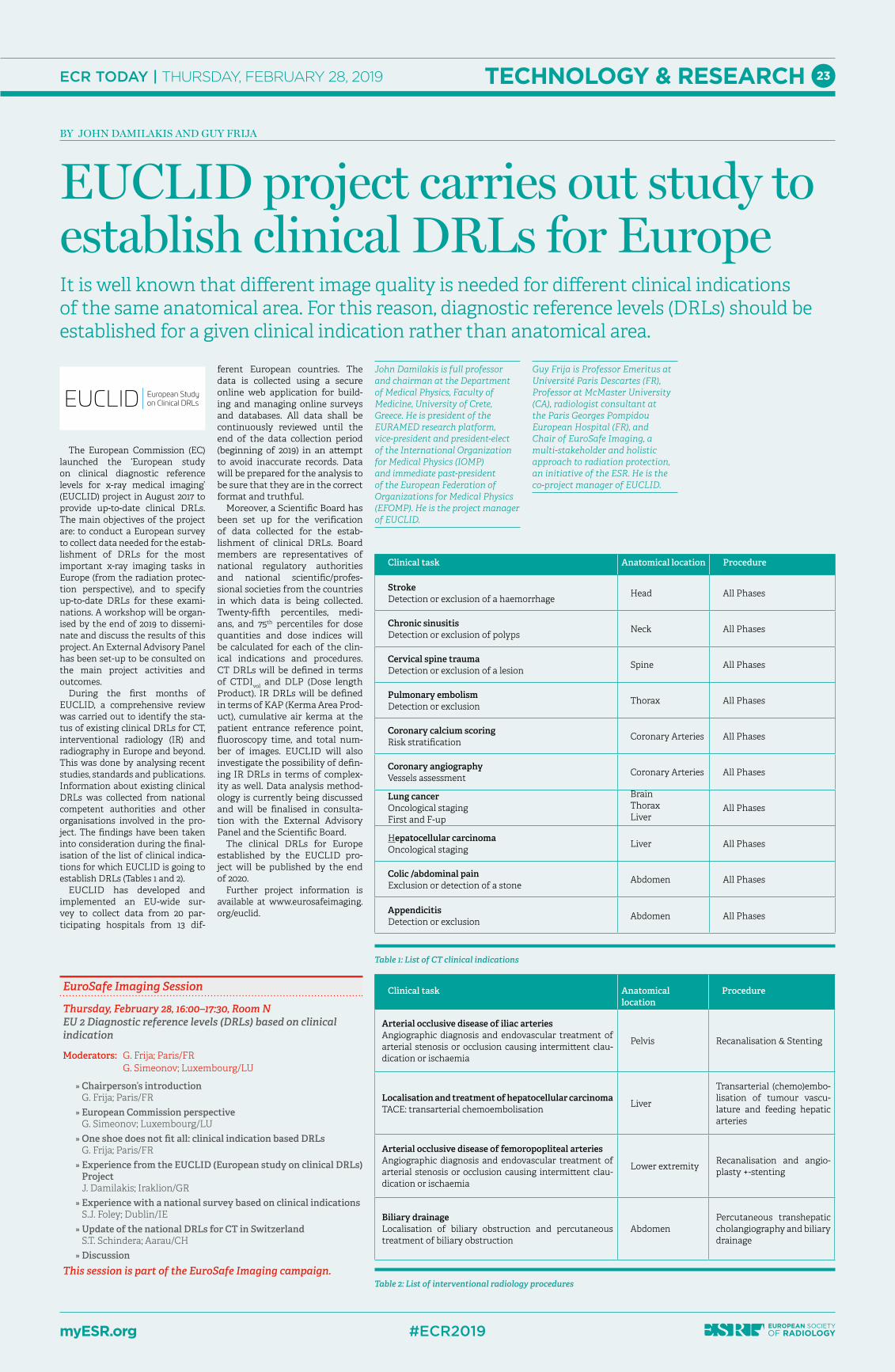
TECHNOLOGY & RESEARCH 23ECR TODAY | THURSDAY, FEBRUARY 28, 2019
#ECR2019myESR.org
BY JOHN DAMILAKIS AND GUY FRIJA
EUCLID project carries out study to establish clinical DRLs for EuropeIt is well known that different image quality is needed for different clinical indications of the same anatomical area. For this reason, diagnostic reference levels (DRLs) should be established for a given clinical indication rather than anatomical area.
The European Commission (EC) launched the ‘European study on clinical diagnostic reference levels for x-ray medical imaging’ (EUCLID) project in August 2017 to provide up-to-date clinical DRLs. The main objectives of the project are: to conduct a European survey to collect data needed for the estab-lishment of DRLs for the most important x-ray imaging tasks in Europe (from the radiation protec-tion perspective), and to specify up-to-date DRLs for these exami-nations. A workshop will be organ-ised by the end of 2019 to dissemi-nate and discuss the results of this project. An External Advisory Panel has been set-up to be consulted on the main project activities and outcomes.
During the first months of EUCLID, a comprehensive review was carried out to identify the sta-tus of existing clinical DRLs for CT, interventional radiology (IR) and radiography in Europe and beyond. This was done by analysing recent studies, standards and publications. Information about existing clinical DRLs was collected from national competent authorities and other organisations involved in the pro-ject. The findings have been taken into consideration during the final-isation of the list of clinical indica-tions for which EUCLID is going to establish DRLs (Tables 1 and 2).
EUCLID has developed and implemented an EU-wide sur-vey to collect data from 20 par-ticipating hospitals from 13 dif-
ferent European countries. The data is collected using a secure online web application for build-ing and managing online surveys and databases. All data shall be continuously reviewed until the end of the data collection period (beginning of 2019) in an attempt to avoid inaccurate records. Data will be prepared for the analysis to be sure that they are in the correct format and truthful.
Moreover, a Scientific Board has been set up for the verification of data collected for the estab-lishment of clinical DRLs. Board members are representatives of national regulatory authorities and national scientific/profes-sional societies from the countries in which data is being collected. Twenty-fifth percentiles, medi-ans, and 75th percentiles for dose quantities and dose indices will be calculated for each of the clin-ical indications and procedures. CT DRLs will be defined in terms of CTDIvol and DLP (Dose length Product). IR DRLs will be defined in terms of KAP (Kerma Area Prod-uct), cumulative air kerma at the patient entrance reference point, fluoroscopy time, and total num-ber of images. EUCLID will also investigate the possibility of defin-ing IR DRLs in terms of complex-ity as well. Data analysis method-ology is currently being discussed and will be finalised in consulta-tion with the External Advisory Panel and the Scientific Board.
The clinical DRLs for Europe established by the EUCLID pro-ject will be published by the end of 2020.
Further project information is available at www.eurosafeimaging.org/euclid.
John Damilakis is full professor and chairman at the Department of Medical Physics, Faculty of Medicine, University of Crete, Greece. He is president of the EURAMED research platform, vice-president and president-elect of the International Organization for Medical Physics (IOMP) and immediate past-president of the European Federation of Organizations for Medical Physics (EFOMP). He is the project manager of EUCLID.
Guy Frija is Professor Emeritus at Université Paris Descartes (FR), Professor at McMaster University (CA), radiologist consultant at the Paris Georges Pompidou European Hospital (FR), and Chair of EuroSafe Imaging, a multi-stakeholder and holistic approach to radiation protection, an initiative of the ESR. He is the co-project manager of EUCLID.
EuroSafe Imaging Session
Thursday, February 28, 16:00–17:30, Room NEU 2 Diagnostic reference levels (DRLs) based on clinical indication
Moderators: G. Frija; Paris/FR G. Simeonov; Luxembourg/LU
» Chairperson’s introductionG. Frija; Paris/FR
» European Commission perspectiveG. Simeonov; Luxembourg/LU
» One shoe does not fit all: clinical indication based DRLsG. Frija; Paris/FR
» Experience from the EUCLID (European study on clinical DRLs) ProjectJ. Damilakis; Iraklion/GR
» Experience with a national survey based on clinical indicationsS.J. Foley; Dublin/IE
» Update of the national DRLs for CT in SwitzerlandS.T. Schindera; Aarau/CH
» Discussion
This session is part of the EuroSafe Imaging campaign.
Clinical task Anatomical location
Procedure
Arterial occlusive disease of iliac arteriesAngiographic diagnosis and endovascular treatment of arterial stenosis or occlusion causing intermittent clau-dication or ischaemia
Pelvis Recanalisation & Stenting
Localisation and treatment of hepatocellular carcinomaTACE: transarterial chemoembolisation
Liver
Transarterial (chemo)embo-lisation of tumour vascu-lature and feeding hepatic arteries
Arterial occlusive disease of femoropopliteal arteriesAngiographic diagnosis and endovascular treatment of arterial stenosis or occlusion causing intermittent clau-dication or ischaemia
Lower extremityRecanalisation and angio-plasty +-stenting
Biliary drainage Localisation of biliary obstruction and percutaneous treatment of biliary obstruction
AbdomenPercutaneous transhepatic cholangiography and biliary drainage
Table 2: List of interventional radiology procedures
Clinical task Anatomical location Procedure
StrokeDetection or exclusion of a haemorrhage
Head All Phases
Chronic sinusitisDetection or exclusion of polyps
Neck All Phases
Cervical spine traumaDetection or exclusion of a lesion
Spine All Phases
Pulmonary embolismDetection or exclusion
Thorax All Phases
Coronary calcium scoring Risk stratification
Coronary Arteries All Phases
Coronary angiographyVessels assessment
Coronary Arteries All Phases
Lung cancerOncological stagingFirst and F-up
BrainThoraxLiver
All Phases
Hepatocellular carcinomaOncological staging
Liver All Phases
Colic /abdominal painExclusion or detection of a stone
Abdomen All Phases
AppendicitisDetection or exclusion
Abdomen All Phases
Table 1: List of CT clinical indications

TECHNOLOGY & RESEARCH24 ECR TODAY | THURSDAY, FEBRUARY 28, 2019
#ECR2019 myESR.org
BY NATHALIE LASSAU
Five simultaneous data challenges bring competitive buzz to JFR 2018Global interest in artificial intelligence (AI) is growing rapidly, due to the availability of large and scalable datasets in every industry, advances in computing power, and the never-ending release of new algorithms.
The development of new stand-ards of machine learning, such as deep learning, has a tremendous impact on radiological activities. This major transformation seems to enable radiologists to leverage their value, efficiency and accuracy. The strategic positioning of all actors (patients, radiologists, AI expert, IS/IT department) is crucial for a suc-cessful transition. The Canadian Association of Radiologists (CAR) and the French Society of Radiol-ogy (Société Française de Radiolo-gie – SFR) published a white paper focused on artificial intelligence in 2018, reflecting both the importance of this topic and probably the impact on the community of radiologists in the near future.
Furthermore, this year, the Euro-pean Union (EU) is reforming data protection legislation on the regu-lar basis of the GDPR (general data protection regulation) with new legislation implemented since May 25, 2018.
In this context, for the first time, the French Society of Radiol-ogy organised five data challenges including three modalities during the JFR 2018 (Journées Franco-phones de Radiologie) in order to create a structured dynamic on this topic, based on different steps:1. To teach radiologists the new
rules of GDPR while building a large multicentric prospective database of US, CT and MRI images of patients
2. To build an ecosystem from May to September 2018 including public and private radiologists, researchers, and start-ups, but also large companies and students from engineering schools
3. To provide all French stakehold-ers able to work together during the competition with a secure framework, offering a realis-tic picture of the benefits and concerns in October 2018
Five clinical questions were selected by the SFR in May. Fifty private and public hospitals included more than 3,500 patients prospectively in the four months up to September: • Renal cortex segmentation
with CT scan (1,574 images from kidney)
• Detection of meniscus fissure (1,823 images from meniscus)
• Detection and characterisation of hepatic lesions with ultrasonog-raphy (545 images from liver)
• Tumour invasion of thyroid cartilage with CT scan (511 images from cartilage)
• Detection of breast tumours with MRI (504 image from breast)Twenty-six teams were created,
with a total of 180 people, includ-ing radiologists, student engineers, researchers and start-ups or large companies.
The competition started on September 15: the teams had full access to one third of the data and
labels on the website of the data challenge. From this time, they were able to train their algorithms and implement some data engi-neering, with the doctors working with them, from anywhere at any time.
A month later, on October 12, a second third of the data and labels was released as a validation set. This was meant to trigger them to come to the JFR in order to stimulate competition between them. That way, they were also able to meet each other, to grow relationships between research-ers and engineers, but most of all between clinicians and engineers. Thus, after reaching some results, they were offered the chance to use this validation set to compare their AUC values thanks to a leader board. Only the ones who had sub-mitted their results were allowed to refer back to it.
At 14:00 on Sunday, October 14, the last remaining part of the ini-
tial dataset was released, contain-ing only the data but not the labels. The participants then had one hour to run their model on it and out-put a file containing their predic-tions. The results were announced on Monday, October 15 at midday. Five winners, one per challenge, should have been awarded a prize, but as in one challenge two teams had results that were too close, there were six winners, with a prize of €3,000 offered by SFR for each winner:
Okwinautes was the winner of two challenges: liver and breast; LyPhTe was the winner of two challenges: meniscus and kidney; SynovIA was the winner for carti-lage thyroid; and Radioadvisor for meniscus.
Professor Nathalie Lassau is Professor of Radiology and Co-Head of imaging Laboratory IR4M. UMR8081 at Gustave Roussy Institut, University Paris Sud.
the next level in human-centred design Getting curious? Discover how our technological innovations put people first. Meet us at booth X2/225 to see our new arrivals unveiled on Thursday, February 28, 15.30. Afterwards, why not help us celebrate with a traditional Japanese Kagamiwari ceremony?

ECR TODAY | THURSDAY, FEBRUARY 28, 2019
COMMUNITY NEWS 25
myESR.org #ECR2019
ACI is present and future of CME in radiology
ESOR increases commitment to radiological education
European Society of Thoracic Imaging (ESTI) on lung cancer screening: facing the challenge
BY STEFANIE BOLLDORF AND IRENE CHRISTOFFEL
A success story with new challenges: European Radiology, the ESR’s flagship journal, continues to growAfter his first full year in office, we asked Editor-in-Chief Prof. Yves Menu from Paris, France, about challenges and opportunities arising from the increasing submissions and success of European Radiology, the flagship of the ESR’s journal family.
ECR Today: You are about to conclude your first year in office, which has seen a significant increase in submissions. What do you think contributed to this rise and how did it influence your first year as Editor-in-Chief?
Yves Menu: Managing a scien-tific journal is a matter of time, quantity, and quality. Everybody understands the difficult com-promise between these words. I can confirm that in 2018 we have reached the highest number of submissions ever, with an increase of more than 30%.
There is no single explanation. It is true that scientific publication is an increasingly recognised metric for academic activity and recog-nition. More teams are developing research and this probably broad-ens the base of potential authors. I am certain that most journals that are referenced with a high impact factor have observed the same phenomenon.
Having said that, European Radiology is not only witnessing this phenomenon, but is probably among the leading titles in terms of the increase in submission num-bers. I would like to think that the improved management of manu-scripts created a favourable atmos-phere – we were able to bring down the time to decision to 25 days on average, which compares very well with other journals. Of course, this is a mean delay and not all man-uscripts receive a decision within this period, as some require more attention, need additional expert opinion or create a debate. This is for time and quantity.
In terms of quality, authors not only expect a quick answer, they also need it to be detailed and rele-vant, because it speeds up the revi-sion. We have tried hard to increase the content of our answers.
Even a decision of ‘reject’ rarely comes without detailed expla-nation. Unfortunately, most of
the manuscripts we receive will be rejected. Fortunately, many contain useful science and will be published elsewhere. It is my pleasure to think that the review-ing process by European Radiology greatly helps these manuscripts to improve, and to become accept-able submissions for different journals. We, like other journals, certainly have a role in the overall improvement of scientific publica-tion in imaging, beyond the pages of European Radiology.
The role of the reviewers is instrumental and I would like to tell them how much I am impressed with the quality of most reviews. Of course, some comments are controversial, as some reviewers fail to understand the authors’ messages. However, even these ‘fails’ are important, since reviewers, in their diversity, illustrate the spectrum of opinions that the final article will induce within our readership.
Finally, I would like to think that this improved management has probably attracted a signif-icant proportion of additional submissions.
ECRT: When looking at the submissions European Radiology has received throughout 2018, are there specific trends or hot topics that stand out from the crowd?
YM: Papers dealing with artificial intelligence are now on-trend. However, my feeling is that we are only at the beginning of the phenomenon. Nobody is able to predict correctly its exact role in the future, and this allows all fantasies, predicting the dis-appearance of radiology or, con-versely, a mighty development. As usual, the truth is probably not the middle-of-the-road, that would finally mean ‘no-change’,
Prof. Yves Menu is the past Chair of Radiology at the Saint Antoine Hospital and University Pierre & Marie Curie, Paris, and Editor-in-Chief of European Radiology since January 2018
27 2928
Bringing the art of imaging to you
www.european-radiology.org
Bringing the art of imaging to you
continued on page 26
26 ECR TODAY | THURSDAY, FEBRUARY 28, 2019
#ECR2019 myESR.org
COMMUNITY NEWS
ADS-02530-EUR-EN Rev.001 (01/19) Hologic Inc. ©2019 All rights reserved. Hologic, The Science of Sure and associated logos are trademarks and/or registered trademarks of Hologic, Inc., and/or its subsidiaries in the United States and/or other countries. This information is intended for medical professionals in the U.S. and other markets and is not intended as a product solicitation or promotion where such activities are prohibited. Because Hologic materials are distributed through websites, eBroadcasts and tradeshows, it is not always possible to control where such materials appear. For speci�c information on what products are available for sale in a particular country, please contact your local Hologic representative.
Visit our booth X2-211 during ECR2019 to learn more
Separating hype from reality in breast health
but a deep transformation of our profession.
However, all those who, like me, have seen the outbreak of US, CT, MRI, hybrid imaging, or interven-tional radiology know how our profession is flexible and prone to adaptation. All editors are fac-ing many papers on AI and are still looking for reliable and sim-ple criteria for the evaluation of the scientific robustness. In the developmental period, we have to accept that not all studies fulfil all requirements, as we should not curb the intense intellectual fer-ment. However, we cannot accept all studies with obvious flaws like very small samples, biased recruit-ment, minimal validation that pre-vent any attempt to generalise.
Other very fertile grounds are hybrid imaging, parametric MRI, dual energy CT, and dosimetry. I also observe a growing interest in some specific fields like communi-cation with the patient and quality assurance.
Overall, we wish to maintain European Radiology as the journal for the whole community, and keep a balance between all topics. Also, I greatly insist on the readability of articles. I should say that there is
a lot of work ahead. My opinion is that there are no such complicated topics that could not be explained clearly and be understood by the majority of our readers.
ECRT: The Editorial Board of European Radiology has grown and includes representatives from all over the world. Have you been able to identify a trend in certain regions or countries where the specialty of radiology has recently flourished and seen substantial progress?
YM: European Radiology is now-adays a worldwide communication pathway. We have observed a sig-nificant increase in submissions from Asia, and notably from China, joining Korea and Japan, as well as ‘traditional’ western countries in the leading group in terms of sub-mission numbers. Therefore, it was more than fitting that some col-leagues from these countries would participate in the daily life of the journal through a position on the Editorial Board. Including people from different countries has a huge benefit, not only for representation purposes, but also – and this may be more important – for the dialogue between people who have differ-ent cultures and, therefore, bring mutual intellectual enrichment.
However, beyond the numbers, I
would like to highlight two differ-ent feelings.
One is that not only numbers increased, but quality of research has improved as well. I now very seldom reject papers for their very poor presentation, absence of clin-ical relevance and questions about the reality of the results. This makes the Editor’s life more interesting, as we receive a huge quantity of rea-sonable studies, and more difficult, as the difference between selection and rejection is more and more nar-row and the competition is hot.
Another feeling is the satisfaction of seeing that there are countries that seem to have increased their representation after their presence in scientific journals was minimal for different reasons. These coun-tries are from the Middle East, South America, and the Indian subcontinent, for instance. In this world as it is, it is a great pleasure to see that the scientific debate is a powerful factor of peaceful com-munication, mutual esteem, debate, and, finally, friendship.
ECRT: Your lecture at this year’s Coffee & Talk Session on ethical traps in publication is called ‘Ethical issues during peer review’. Have you experienced any major issues in this regard during your editorship? What do
you think authors are most wor-ried about and how do you deal with it as the Editor-in-Chief?
YM: The main potential prob-lems would be plagiarism, dupli-cate publication, and doubtful methods. I should say that we did not encounter any major eth-ical problems. Most authors very transparently disclose potential problems. We have had no cases of duplicate publication and no real cases of plagiarism.
However, as language is still a problem for international authors, some of them are tempted to use significant sentences picked up in already published papers. We use software to detect similarities. Although it is only a ‘syntactic borrowing’ without any scientific plagiarism, I imagine that the orig-inal authors could be upset at see-ing verbatim copies of entire sen-tences or even paragraphs.
I encourage the authors to limit the ‘imitation’ to simple expres-sion, and never more than six to seven words in a row. I would not accept longer copies.
ECRT: In addition to the renowned Albert L. Baert Edi-torial Fellowship, you have introduced the European Radi-ology Reviewer Fellowships, and rumours are spreading
that you will be introducing the role of a ‘Junior Editor’. In your own words, what can we expect and what are your plans for the future in your role as Editor-in-Chief?
YM: It is of utmost importance to associate our young colleagues to the journal. They represent the future, and many of them, already the present. What they bring to European Radiology is fresh air, new knowledge and enthusiasm. Conversely, what we could do for them is to teach the editorial pro-cess, how to review, how to write, how to communicate. None of these are innate. This is why I promote the idea of a ‘School of European Radiology’, gathering several initiatives dedicated to our younger colleagues. Is this not the best way to prepare them for the future?
I would like to conclude with one single wish that seems obvi-ous, but nevertheless summarises my goal: it is simply to promote the idea that articles are published in the journal to be read, not only to be written. It seems obvious, but behind this oversimplified state-ment, there is an entire revolution in the authors’, reviewers’ and edi-tors’ daily life. More details will fol-low in the near future!
continued from page 25

27ECR TODAY | THURSDAY, FEBRUARY 28, 2019
#ECR2019myESR.org
COMMUNITY NEWS
BY VALÉRIE VILGRAIN
ESOR increases commitment to radiological educationThe European School of Radiology (ESOR) has completed thirteen years in action; thirteen years of adding value to radiology. From the beginning, Prof. Nicholas Gourtsoyiannis was the Scientific/Educational Director of ESOR and his talents have made ESOR a success story. I took over the position at ECR 2018 and my will is indeed to pursue the investment in radiological education in Europe and beyond.
The three main goals of ESOR, set at its inception in 2005, are still to assist in harmonising radiological education throughout Europe, by supporting the adoption and uti-lisation of the European Training Curricula (ETC); to build a genuine and firm interest in subspecialisa-tion in radiology; and to raise the scientific profile in radiological edu-cation in Europe and around the globe.
During the past thirteen years ESOR has gradually evolved into a major international provider of complementary education in radiology, offering a wide range of modular activities, including foundation and advanced courses, teach-the-teachers and visiting professorship programmes, visiting schools, seminars, tutorials, pre-paratory courses for the European Diploma in Radiology (EDiR), schol-arships, exchange programmes for fellowships and full one-year fel-
lowships. With its expanded course programme and the intercontinen-tal scholarships and exchange pro-grammes for fellowships, ESOR is able to respond adequately to the actual needs of radiologists around the globe, following agreements between the ESR and correspond-ing member societies. So far, ESOR has delivered structured continu-ing education to more than 21,500 residents and board-certified radi-ologists worldwide.
The importance of ESOR’s con-tributions can also be appreci-ated if one considers that almost 80% of ESOR participants every year come from the radiologically evolving world, while more than 50% of scholarships and exchange programmes for fellowships are granted to applicants from Eastern Europe, CIS Countries, Latin Amer-ica, China, the Middle East and Africa. In order to foster the ESOR community even further, ESOR is
proud to announce the implemen-tation of an ESOR Alumni Club for past scholars and fellows. Through this initiative, ESOR aims to main-tain the link with successful schol-ars and fellows through active participation in education. Addi-tionally, there is a dedicated ESOR lounge at ECR 2019, on the 2nd level in Foyer B, for exchange and network-ing, as well as Coffee & Talk sessions focused on education.
In 2018, ESOR started a new fel-lowship programme, the ‘Bracco Fellowships’, which offers 90 grants to residents and young board-cer-tified radiologists over a period of three years (2018–2020). This new initiative was created on the occasion of the 90th anniversary of Bracco and has been very well received by the ESOR community. Moreover, ESOR has successfully completed its first ever activity in south-east Asia, with an ASKLE-PIOS course in Bangkok, Thailand.
This course, attended by 140 partic-ipants from the region, was organ-ised in partnership with the Royal College of Radiologists of Thailand (RCRT) and supported by Guerbet. In addition to this course, ESOR also returned to China for two one-day courses in partnership with the Chinese Society of Radiology (CSR) and Guerbet. Due to this great suc-cess, ESOR will come back to south-east Asia in 2019, expanding to the Philippines.
Additionally, ESOR is enriching its portfolio in 2019, by offering 52 courses and sessions, as well as 120 scholarships and fellowships. One of the highlights will be the first ESOR course in India, organised jointly with the Indian Radiological and Imaging Association (IRIA). As ESOR is not only concerned with education in radiology but also aims to prepare the new generation with methods that could impact multiple facets of radiology, ESOR is proud to organise the ESR’s first two-day course on Artificial Intelligence, in Barcelona on April 5–6, 2019.
ESOR is instrumental in mobi-lising resources available for radi-ological education in Europe and
the world, and for creating a long-term educational commitment and a structured network. All of this hugely appreciated use of teaching resources has been implemented with the unlimited support of highly esteemed and renowned lecturers, tutors, mentors, volun-teer reference training centres, local organisers, subspecialty and national societies, academic and research institutions, and valued industrial partners. ESOR is greatly indebted to them all.
I am extremely grateful to the ESR for entrusting me with the leadership of ESOR. I am more than happy to work closely with the ESOR Steering and Programme Planning Committee and the ESOR community as a whole and I am confident that ESOR will continue to deliver education successfully, connecting the world of radiology. I encourage all of you to find out what ESOR can offer you.
Prof. Valérie Vilgrain is the Scientific/Educational Director of the European School of Radiology (ESOR) and chairperson of the ESR’s ESOR Committee.
The ESOR ASKLEPIOS course in Bangkok, Thailand, was organised in partnership with the Royal College of Radiologists of Thailand (RCRT) and supported by Guerbet. It attracted 140 participants from south-east Asia, making the first ever ESOR activity in the region a huge success (© Guerbet Asia Pacific Ltd.).
ESOR Session
Thursday, February 28, 14:00–15:30, Room M 3Artificial intelligence: presented with new lecture formats
Moderators: B. Brkljačić; Zagreb/HR V. Vilgrain; Clichy/FR
» IntroductionB. Brkljačić; Zagreb/HR
» ESOR in action 2019V. Vilgrain; Clichy/FR
» Artificial intelligence (AI) and machine learning (ML) in medical imaging (in Pecha Kucha format)W.J. Niessen; Rotterdam/NL
» Deep learning (DL) in medical imaging (in TED format)A. Alberich-Bayarri; Valencia/ES
» My experience: one-year fellowshipY. Purcell; Dublin/IE
» Awards
ESOR GALEN Courses 2019
The GALEN Courses have been designed to familiarise young radiologists with the established approaches and most recent achievements in diagnostic imaging, related to topics across the modalities. The courses are aimed at residents and board-certified radiologists from all over Europe.
GALEN Foundation Course
NeuroradiologyMay 23-25, Lisbon/Portugal
GALEN Advanced Courses
Women’s ImagingJune 13-14, Malmö/Sweden
Cardio-Thoracic Cross-Sectional ImagingJuly 4-5, Tübingen/Germany
Oncologic Imaging from Head to ToeSeptember 5-6, Paris/France
For further information on the detailed programmes and registration, please visit esor.org

28 ECR TODAY | THURSDAY, FEBRUARY 28, 2019
#ECR2019 myESR.org
COMMUNITY NEWS
BY MILOS A. LUCIC
Accreditation Council in Imaging succeeds in maximising synergiesThe Accreditation Council in Imaging (ACI), created under the EBR umbrella, works in close collaboration with the European Union of Medical Specialists (UEMS) and the European Accreditation Council for Continuing Medical Education (EACCME).
Since its establishment, the ACI teams – comprised of EBR and UEMS members – have been suc-cessfully led by Prof. Dragoş Negru and Prof. Paolo Ricci. Thanks to their efforts and those of Prof. Christian Loewe, Chair of the ACI Review Committee, the ACI Review and Policy committees have suc-ceeded in maximising synergy by uniting their substantial resources. This collaboration has provided added value in the accreditation of imaging events and has led to more events being accredited in radiol-ogy than in any other specialty. This achievement reflects the ded-ication of education providers in the field of radiology and directly benefits attendees of the live edu-cational events offered, and users of the electronic learning material, by providing a stable source of CME points.
To better understand the cur-rent status of continuing medical education (CME) and continuing professional development (CPD) in European countries, and to learn more about the wishes and expec-tations of the European radiological community in this regard, the ACI has conducted three surveys over the last two years. The results of
the first surveys indicate marked differences in the accreditation systems between countries and show that the European radio-logical community has important views on this. Most respondents would support implementation of an equal or unified CME/CPD system in all European countries, and would be in favour of recog-nising the European CME credits (ECMEC) as the universal European CME unit. In acknowledgement of these views as the voice of Euro-pean radiologists, which should not be overlooked in future CME/CPD planning, the ACI will continue to direct strong efforts toward achiev-ing further harmonisation of radio-logical standards. One of the ACI’s top priorities should be to provide systematic and detailed informa-tion on the CME/CPD systems in Europe and their equivalence to the UEMS accreditation system at the European level, a task that has been strongly supported by Prof. Ricci.
In light of the emerging gener-ation shift, a feature of which is a generation-dependent level of read-iness to use available and future technologies in both radiology and education, another ACI survey was designed to predict where the
future CME/CPD learning concepts would lead the radiology profes-sion. Although most radiologists still value networking with their colleagues during live educational events, certain discrete tendencies are emerging, which could be early indicators of the shift towards enhanced electronic media use in the learning process. This trend, together with heavy workloads and financial restraints, may have an impact on future CME acqui-sition in radiology, manifested as an increasing interest in webinars. Being web-based, webinars are eas-ily accessible and far less demand-ing in terms of time and cost for participants. They also provide an opportunity for the apparently still very important element of human interaction, as any participant can communicate with the speaker or moderator. Unfortunately, webinars are still considered to be live educa-tional events by EACCME 2.0.
Furthermore, very few webi-nars in radiology are accredited at this time. Based on the ACI survey data and feedback from educa-tion providers collected by Prof. Loewe, Prof. Ricci voiced the need to facilitate the process of webinar accreditation during the UEMS and
ACI meeting at the 4th UEMS con-ference on the future of CME/CPD in Brussels last November. Because of the expected future importance and impact of this educational plat-form, the ACI will continue to work jointly with the UEMS to define a simple, quick accreditation path for webinars that could become a val-uable source of CME/CPD learning for radiology and many other fields.
One major challenge is to antic-ipate and learn more about the wishes of upcoming generations, including the opinions of the mil-lennial generation (generation Z), who are just entering the radiology profession. This view should par-ticularly take into account future technological advances, including the impact of artificial intelligence and raw data analysis-based radiol-ogy. The ACI will continue to search for relevant information important to radiology professionals by asking for their personal opinions on dif-ferent topics, as every single voice matters in this regard.
Based on an already robust and vigorous organisational foundation established by the ACI team, an effective and highly competent EBR Office, outstandingly led by Ms. Vio-leta Iranzo, and the firm and trusted
partnership with the UEMS, the ACI will have a proactive role in the effort to achieve full harmonisation of future radiological standards, and will face the upcoming chal-lenges with firm confidence.
Prof. Milos A. Lucic is a Full Professor of Radiology and Neuroradiology at the Faculty of Medicine, University of Novi Sad, and also teaches at the Faculty of Medicine, University of Belgrade, and Faculty of Medicine, University of Banja Luka. He is Chairman of the MRI School of the MRI Society of Serbia, and is the current vice president of the Serbian Society of Neuroradiology, past president of the Serbian Society of Radiology, and general secretary of the Balkan Society of Radiology (BSR). Prof. Lucic developed a Diagnostic Imaging Centre in Sremska Kamenica, Novi Sad, that is a reference centre of regional importance for MRI and hybrid imaging. He is currently Medical Director of Affidea in Belgrade, Serbia. After ECR 2019 and for the following two years, Milos Lucic will be the new ACI Scientific Director.
BY PAOLO RICCI
ACI is present and future of CME in radiology
On March 7, 2015, an agreement to create the Accreditation Council in Imaging (ACI) was signed between the European Society of Radiology (ESR) and the Union Européenne des Médecins Spécialistes (UEMS).
From that date onwards, a fruit-ful relationship began between these two entities. During the first years and based on the work of Prof. Dragoş Negru, former Sci-entific Director, and Profs. Loewe and Lucic, Chairs of the Reviewing Committee and Policy Committee, respectively, a solid foundation for live events and e-learning mate-rial in the world of radiology and imaging was laid down and is now accredited by the Accreditation Council in Imaging.
Today, radiology – together with oncology and cardiology – is one of the medical subspecialties with the largest number of applicants for event accreditation. More than 450 applications have been submitted and accredited since 2016, thanks to the excellent work of the Review-ing Committee, led by my colleague Christian Loewe. A key to this suc-cess lies in the work done by mem-
bers of the Policy and Reviewing Committees, and by the staff, who are in charge of the administrative tasks.
One of the most important aspects of this collaboration is that the incoming ACI Scientific Direc-tor will be appointed by the EBR Shareholders’ Board at the Euro-pean Congress of Radiology (ECR) for a fixed period of two years. Candidates from the two entities (the EBR and UEMS Radiology Sec-tion) will hold the position of ACI Scientific Director on an alternat-ing basis. My predecessor, Dragoş Negru, carried out a fantastic job setting down the basis and the most important aspects to be con-sidered for effective performance, which have now been established.
In March 2017, when the new European Accreditation Council for Continuing Medical Education (EACCME) 2.0 criteria were imple-mented, I started my journey as Scientific Director. It was at that point that the EACCME developed and presented several criteria to provide better service, according to the current needs of Continuing
Medical Education (CME) and Con-tinuing Professional Development (CPD). It is thanks to the surveys devised by Milos Lucic and by the members of the Policy Committee that the ACI came to vital con-clusions in this respect, to offer better service to both providers and physicians who require CME/CPD to accomplish their tasks as professionals.
Through our surveys, we wanted to anticipate future requirements and define the areas that require additional work to offer the ser-vice that radiology and imaging deserve. We are all very keen on live events, but e-learning and webi-nars are the future. Therefore, the ACI is working hand-in-hand with the UEMS and leading a project to create a new accreditation scheme for webinars, which are currently considered live events. The name of the game is that presently, in order to accredit a one-hour webi-nar, the same effort for accrediting a one, two, or three-day live event like the ECR is required. Thus, we would like to offer this not only to radiology and imaging, but also to
other medical subspecialties that have shown an interest in this new approach: a new way of accrediting webinars organised by national societies and subspecialties socie-ties and offered to their members.
As I was told at the last confer-ence about CME/CPD in Brussels, the excellent and fruitful relation-ship between the UEMS and the ESR/EBR is certainly an example that should be emulated by other subspecialties.
After ECR 2019 and for the fol-lowing two years, Milos Lucic will be the new Scientific Director. I am sure that thanks to him and the team he will be leading, we will have e-learning material and webi-nars that are well-placed among the applications processed.
I am proud to have played a role in CME/CPD standardisation in the field of radiology. The Accred-itation Council in Imaging (ACI) is definitely a one-step guide to CME accreditation, which supports the UEMS European Accreditation Council for CME (EACCME) in delivering and harmonising the highest level of CME in imaging.
Paolo Ricci is Associate Professor of Radiology at the Department of Radiological Sciences, La Sapienza University in Rome, Italy. He is President of the UEMS Section of Radiology, Chairman of the UEMS Grouping 3 ‘Multidisciplinary’, and a Member of the UEMS Enlarged Executive Committee. He is the Scientific Director of the Accreditation Council in Imaging (March 2017 – March 2019).

29ECR TODAY | THURSDAY, FEBRUARY 28, 2019
#ECR2019myESR.org
COMMUNITY NEWS
BY MARIE-PIERRE REVEL
European Society of Thoracic Imaging (ESTI) on lung cancer screening: facing the challengeLung cancer is the number one cancer killer worldwide, and along with efforts to end the tobacco epidemic, every effort should be made to detect lung cancer at a curable stage in high-risk individuals.
This is the aim of lung cancer screening (LCS) based on low-dose CT, which started two decades ago. After the Early Lung Cancer Action Project (ELCAP) demon-strated in 1999 that low-dose CT detected early stage lung cancers, in 2011, the randomised National Lung cancer Screening Trial
(NLST) reported a 20% reduction in lung cancer-related mortality in individuals being screened, at the cost of a 24% false positive rate. In September 2018, the NELSON trial, using volumetry-based lung nodule management, reported a 26% mortality reduction with only 2.2% positive screens. With this
recent communication, LCS can be expected to move from trial to practice in most European coun-tries. In expectation of this, ESTI considered it a priority to propose dedicated training so that the radiologists involved will be able to reproduce the results of the NELSON trial, with no more than 2% positive screens, and 39% of those confirmed to be lung cancer. In view of the high prevalence of lung nodules, it is essential that only trained certified radiologists are involved in its delivery. If large scale LCS is to be performed, there are not enough specialised thoracic radiologists available to deliver it, so a dedicated train-ing programme is needed to help bridge the gap in expertise.
In this regard, an online meet-ing with the European Society of Thoracic Imaging (ESTI) executive and strategic committee members was held on January 9, 2019. Par-ticipants were asked the following three questions: should ESTI offer dedicated LCS training, should it be distinct from the ESTI diploma, and should a certification test be organised?
All members unanimously answered yes to these three questions.
The decision was taken to organ-ise working groups during 2019 to define the content and delivery of dedicated LCS training. This will
probably include registration for a series of webinars/e-learning ses-sions on lung nodule management and the technical requirements for screening, attendance at volu-metry software workshops during ESTI and ECR meetings, and train-ing on LCS cases.
Establishing a common, struc-tured report is one of the manda-tory tasks for ESTI. The detected indeterminate solid nodules should be reported in terms of vol-ume, to follow the NELSON man-agement guidelines. The coronary artery calcification score should also be assessed. ESTI working groups will also have to decide whether screening still requires double reading or single reading with CAD as second reader.
Building large datasets to develop algorithms based on artificial intelligence (AI) will be another objective of our society and an intense research field.
Some AI-based programmes claim that their results approach radiologists’ performance levels for lung cancer prediction, but they have not yet been sufficiently validated.
However, AI is a rapidly evolving field and it is difficult to predict whether human expertise will still be needed for LCS in the future.
While waiting for AI to poten-tially replace the need for a radi-ologist reading, we have an obliga-
tion to provide our colleagues with the means to train themselves in screening.
Besides LCS training, ESTI has already organised a Diploma in Thoracic Imaging, endorsed by the ESR, for which the very first exam-ination will take place in Paris during the ESTI annual meeting in May this year. Thoracic imag-ing covers a large variety of top-ics: emergencies affecting large vessels, malignancies of the lungs, mediastinum and pleura, chronic diseases such as COPD, infections, interstitial lung diseases and vas-culitis. We thought it was impor-tant to organise a diploma for radi-ologists interested in formalising their knowledge in thoracic imag-ing, as most European countries do not offer formal certification in subspecialties. The diploma better defines what thoracic imaging is about and what essential knowl-edge is expected from a chest radi-ologist. Depending on the number of candidates, the diploma exam will also be offered during future ECRs.Visit our website at www.myESTI.org for more information.
Marie-Pierre Revel is full professor of radiology at Cochin Hospital Paris Descartes University in Paris, France, and the current president of ESTI.
C HE ST
Volumetry of part-solid lung nodule, with doubling time measurement. Doubling time of the solid component is 121 days, reflecting its aggressiveness, whereas the whole nodule doubling time is 529 days and that of the nonsolid portion is more than 2 years.
BY USTUN AYDINGOZ
Education and research are at the forefront of the ESSR agendaEmbedded in the origin of the word ‘education’ is the Proto-Indo-European root ‘deuk-’ meaning ‘to lead’. Education leads out from the darkness of ignorance and, as the great polymath of the 18th century, Benjamin Franklin, put it, an investment in knowledge pays the best interest. Education and research, both among the noblest human endeavours, are top priorities for the European Society of Musculoskeletal Radiology (ESSR).
Founded in 1993 and evolving thereafter into one of the largest radiology subspecialty organisa-tions in Europe, the ESSR currently has more than 1,800 members from 66 countries in five continents. The 25th annual meeting of the society, held in Amsterdam during four days last June, drew almost 1,000
attendees, 18% of whom were radi-ology residents. Almost half of the congress was devoted to meeting the increasing demand for mus-culoskeletal ultrasound in the form of a course for residents and less-experienced radiologists and a master-class workshop on nerves for the experienced. This year’s
annual meeting will be in Lisbon on June 26─29, with a focus on hip and advanced imaging, and will again feature ultrasound courses as well.
One of the many facets of the ESSR’s educational activities are the live webinars, each lasting about 60 minutes and featuring a case presentation, followed by a lec-
ture given by an expert, and capped with a discussion, during which attendees can ask questions. Reg-istration for the webinars is free of charge. To give an example, the last of four webinars held in 2018, with a lecture by Professor Filip Vanhoe-nacker from Antwerp titled ‘Lumps and bumps of the soft tissue – how
to differentiate benign from malig-nant lesions’, was joined by 230 attendees. As of December 2018, four webinars have been planned for 2019 on the subjects of MRI of the paediatric knee, musculoskeletal
continued on page 30
M S K
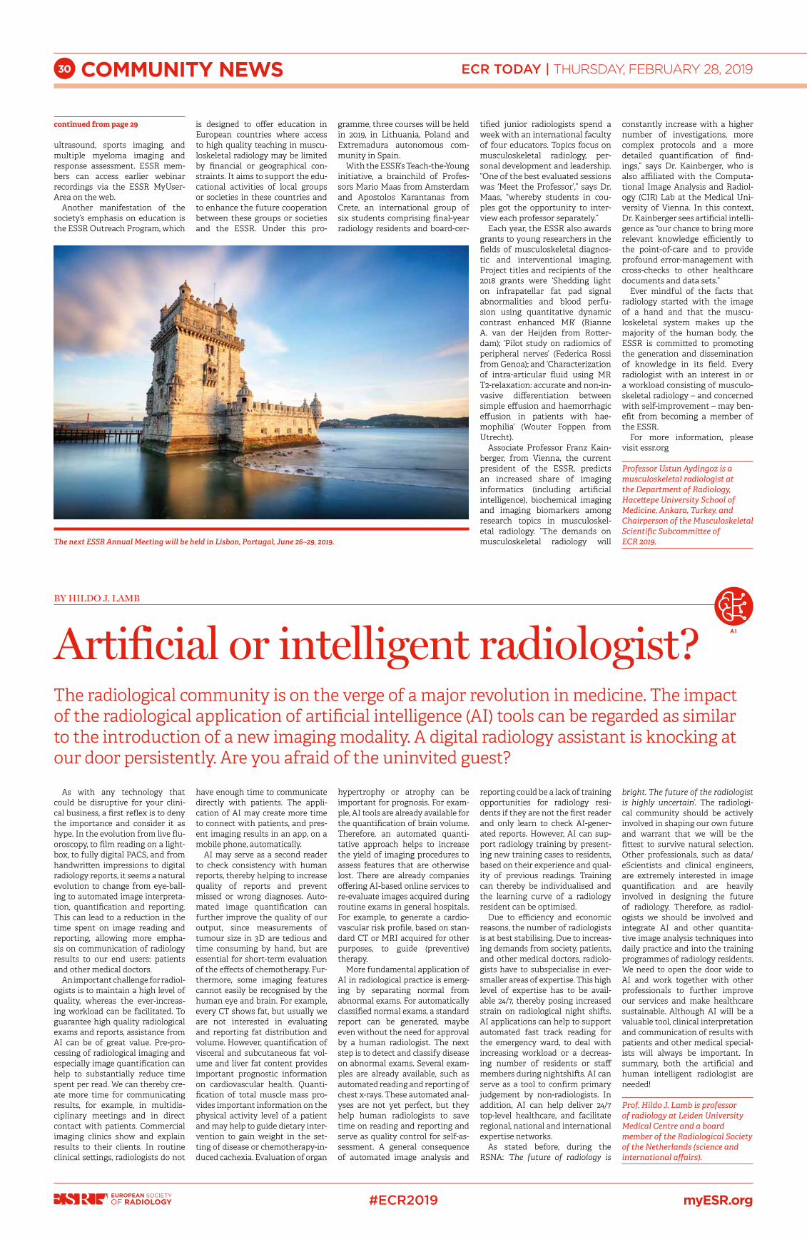
30 ECR TODAY | THURSDAY, FEBRUARY 28, 2019
#ECR2019 myESR.org
COMMUNITY NEWS
BY HILDO J. LAMB
Artificial or intelligent radiologist?The radiological community is on the verge of a major revolution in medicine. The impact of the radiological application of artificial intelligence (AI) tools can be regarded as similar to the introduction of a new imaging modality. A digital radiology assistant is knocking at our door persistently. Are you afraid of the uninvited guest?
As with any technology that could be disruptive for your clini-cal business, a first reflex is to deny the importance and consider it as hype. In the evolution from live flu-oroscopy, to film reading on a light-box, to fully digital PACS, and from handwritten impressions to digital radiology reports, it seems a natural evolution to change from eye-ball-ing to automated image interpreta-tion, quantification and reporting. This can lead to a reduction in the time spent on image reading and reporting, allowing more empha-sis on communication of radiology results to our end users: patients and other medical doctors.
An important challenge for radiol-ogists is to maintain a high level of quality, whereas the ever-increas-ing workload can be facilitated. To guarantee high quality radiological exams and reports, assistance from AI can be of great value. Pre-pro-cessing of radiological imaging and especially image quantification can help to substantially reduce time spent per read. We can thereby cre-ate more time for communicating results, for example, in multidis-ciplinary meetings and in direct contact with patients. Commercial imaging clinics show and explain results to their clients. In routine clinical settings, radiologists do not
have enough time to communicate directly with patients. The appli-cation of AI may create more time to connect with patients, and pres-ent imaging results in an app, on a mobile phone, automatically.
AI may serve as a second reader to check consistency with human reports, thereby helping to increase quality of reports and prevent missed or wrong diagnoses. Auto-mated image quantification can further improve the quality of our output, since measurements of tumour size in 3D are tedious and time consuming by hand, but are essential for short-term evaluation of the effects of chemotherapy. Fur-thermore, some imaging features cannot easily be recognised by the human eye and brain. For example, every CT shows fat, but usually we are not interested in evaluating and reporting fat distribution and volume. However, quantification of visceral and subcutaneous fat vol-ume and liver fat content provides important prognostic information on cardiovascular health. Quanti-fication of total muscle mass pro-vides important information on the physical activity level of a patient and may help to guide dietary inter-vention to gain weight in the set-ting of disease or chemotherapy-in-duced cachexia. Evaluation of organ
hypertrophy or atrophy can be important for prognosis. For exam-ple, AI tools are already available for the quantification of brain volume. Therefore, an automated quanti-tative approach helps to increase the yield of imaging procedures to assess features that are otherwise lost. There are already companies offering AI-based online services to re-evaluate images acquired during routine exams in general hospitals. For example, to generate a cardio-vascular risk profile, based on stan-dard CT or MRI acquired for other purposes, to guide (preventive) therapy.
More fundamental application of AI in radiological practice is emerg-ing by separating normal from abnormal exams. For automatically classified normal exams, a standard report can be generated, maybe even without the need for approval by a human radiologist. The next step is to detect and classify disease on abnormal exams. Several exam-ples are already available, such as automated reading and reporting of chest x-rays. These automated anal-yses are not yet perfect, but they help human radiologists to save time on reading and reporting and serve as quality control for self-as-sessment. A general consequence of automated image analysis and
reporting could be a lack of training opportunities for radiology resi-dents if they are not the first reader and only learn to check AI-gener-ated reports. However, AI can sup-port radiology training by present-ing new training cases to residents, based on their experience and qual-ity of previous readings. Training can thereby be individualised and the learning curve of a radiology resident can be optimised.
Due to efficiency and economic reasons, the number of radiologists is at best stabilising. Due to increas-ing demands from society, patients, and other medical doctors, radiolo-gists have to subspecialise in ever-smaller areas of expertise. This high level of expertise has to be avail-able 24/7, thereby posing increased strain on radiological night shifts. AI applications can help to support automated fast track reading for the emergency ward, to deal with increasing workload or a decreas-ing number of residents or staff members during nightshifts. AI can serve as a tool to confirm primary judgement by non-radiologists. In addition, AI can help deliver 24/7 top-level healthcare, and facilitate regional, national and international expertise networks.
As stated before, during the RSNA: ‘The future of radiology is
bright. The future of the radiologist is highly uncertain’. The radiologi-cal community should be actively involved in shaping our own future and warrant that we will be the fittest to survive natural selection. Other professionals, such as data/eScientists and clinical engineers, are extremely interested in image quantification and are heavily involved in designing the future of radiology. Therefore, as radiol-ogists we should be involved and integrate AI and other quantita-tive image analysis techniques into daily practice and into the training programmes of radiology residents. We need to open the door wide to AI and work together with other professionals to further improve our services and make healthcare sustainable. Although AI will be a valuable tool, clinical interpretation and communication of results with patients and other medical special-ists will always be important. In summary, both the artificial and human intelligent radiologist are needed!
Prof. Hildo J. Lamb is professor of radiology at Leiden University Medical Centre and a board member of the Radiological Society of the Netherlands (science and international affairs).
The next ESSR Annual Meeting will be held in Lisbon, Portugal, June 26–29, 2019.
ultrasound, sports imaging, and multiple myeloma imaging and response assessment. ESSR mem-bers can access earlier webinar recordings via the ESSR MyUser-Area on the web.
Another manifestation of the society’s emphasis on education is the ESSR Outreach Program, which
is designed to offer education in European countries where access to high quality teaching in muscu-loskeletal radiology may be limited by financial or geographical con-straints. It aims to support the edu-cational activities of local groups or societies in these countries and to enhance the future cooperation between these groups or societies and the ESSR. Under this pro-
gramme, three courses will be held in 2019, in Lithuania, Poland and Extremadura autonomous com-munity in Spain.
With the ESSR’s Teach-the-Young initiative, a brainchild of Profes-sors Mario Maas from Amsterdam and Apostolos Karantanas from Crete, an international group of six students comprising final-year radiology residents and board-cer-
tified junior radiologists spend a week with an international faculty of four educators. Topics focus on musculoskeletal radiology, per-sonal development and leadership. “One of the best evaluated sessions was ‘Meet the Professor’,” says Dr. Maas, “whereby students in cou-ples got the opportunity to inter-view each professor separately.”
Each year, the ESSR also awards grants to young researchers in the fields of musculoskeletal diagnos-tic and interventional imaging. Project titles and recipients of the 2018 grants were ‘Shedding light on infrapatellar fat pad signal abnormalities and blood perfu-sion using quantitative dynamic contrast enhanced MR’ (Rianne A. van der Heijden from Rotter-dam); ‘Pilot study on radiomics of peripheral nerves’ (Federica Rossi from Genoa); and ‘Characterization of intra-articular fluid using MR T2-relaxation: accurate and non-in-vasive differentiation between simple effusion and haemorrhagic effusion in patients with hae-mophilia’ (Wouter Foppen from Utrecht).
Associate Professor Franz Kain-berger, from Vienna, the current president of the ESSR, predicts an increased share of imaging informatics (including artificial intelligence), biochemical imaging and imaging biomarkers among research topics in musculoskel-etal radiology. “The demands on musculoskeletal radiology will
constantly increase with a higher number of investigations, more complex protocols and a more detailed quantification of find-ings,” says Dr. Kainberger, who is also affiliated with the Computa-tional Image Analysis and Radiol-ogy (CIR) Lab at the Medical Uni-versity of Vienna. In this context, Dr. Kainberger sees artificial intelli-gence as “our chance to bring more relevant knowledge efficiently to the point-of-care and to provide profound error-management with cross-checks to other healthcare documents and data sets.”
Ever mindful of the facts that radiology started with the image of a hand and that the muscu-loskeletal system makes up the majority of the human body, the ESSR is committed to promoting the generation and dissemination of knowledge in its field. Every radiologist with an interest in or a workload consisting of musculo-skeletal radiology – and concerned with self-improvement – may ben-efit from becoming a member of the ESSR.
For more information, please visit essr.org
Professor Ustun Aydingoz is a musculoskeletal radiologist at the Department of Radiology, Hacettepe University School of Medicine, Ankara, Turkey, and Chairperson of the Musculoskeletal Scientific Subcommittee of ECR 2019.
continued from page 29
AI

Special Exhibition: Into the great Outdoors
From Waldmüller to Schindler
LEOPOLD MUSEUM1070 Vienna, Museumsplatz 1
www.leopoldmuseum.org
Rudolf Ribarz, Apple Tree, 1875 © Leopold Museum Vienna

WHAT’S ON TODAY IN VIENNA?THURSDAY, FEBRUARY 28, 2019
Theatre & Dance Die Stühle by Eugène Ionesco
AKADEMIETHEATER | 19:301010 Vienna, Lisztstraße 1 Phone: +43 1 51444 4145 | www.burgtheater.at
Hiob based on the novel by Joseph Roth
BURGTHEATER | 19:301010 Vienna, Universitätsring 2 Phone: +43 1 51444 4145 | www.burgtheater.at
ExorzistBy Dominic Oley
BRONSKI & GRÜNBERG | 19:301090 Vienna, Müllnergasse 2 Phone: +43 681 20 67 45 40 | www.bronski-gruenberg.at
Josef und Maria by Peter Turrini
KAMMERSPIELE DER JOSEFSTADT | 19:301010 Vienna, Rotenturmstraße 20 Phone: +43 1 42 700 300 | www.josefstadt.org
Knuckles become cloudsby Anna Prokopová (Czech Republic), Costas Kekis (Gre-ece), Andrea Gunnlaugsdóttir (Iceland)
TANZQUARTIER WIEN | 18:00 | TQW STUDIOS
Originsby Oleg Soulimenko (Russia)
TANZQUARTIER WIEN | 19:30 | TQW HALLE G1070 Vienna, Museumsplatz 1 Phone: +43 1 581 35 91 | www.tqw.at
Der Besuch der alten Dame by Friedrich Dürrenmatt
THEATER IN DER JOSEFSTADT | 19:301080 Vienna, Josefstädter Straße 26 Phone: +43 1 42 700 300 | www.josefstadt.org
Tuesdays with Morrie by Jeffrey Hatcher and Mitch Albom
VIENNA’S ENGLISH THEATRE | 19:301080 Vienna, Josefsgasse 12 Phone: +43 1 402 12 60 0 | www.englishtheatre.at
Rojava by Ibrahim Amir
VOLKSTHEATER | 19:301070 Vienna, Neustiftgasse 1 Phone: +43 1 52111 400 | www.volkstheater.at
Concerts & Sounds Wiener SymphonikerConductor Alain Altinoglu Nora Gubisch, mezzo-soprano Denis Matsuev, pianoF. Liszt: concerto for piano and orchestra no. 2 a major; S. Prokofjew: Cantata for mezzo-soprano, choir and orchestra, op. 78
MUSIKVEREIN | 19:30 | GROSSER SAAL1010 Vienna, Bösendorferstraße 12 www.musikverein.at
Lisa Bassenge ‘Borrowed And Blues’ (Germany)
PORGY & BESS | 20:301010 Vienna, Riemergasse 11 www.porgy.at
Coquette JazzbandTraditional Jazz, Swing
JAZZLAND | 21:001010 Vienna, Franz-Josefs-Kai 29 www.jazzland.at
Modeselektor (Germany) + Catnapp (Argentina)
ARENA | 19:001030 Vienna, Baumgasse 80 www.arena.wien
The Lemonheads (USA)
CHELSEA1080 Vienna, Lerchenfelder Gürtel, U-Bahnbögen 29–30 www.chelsea.co.at
Marsimoto (Germany)
GASOMETER | 20:00BA-CA Halle Gasometer 1110 Vienna, Guglgasse 8 | www.planet.tt
Opera & Musical Theatre
Der Mann von La ManchaMusical by Mitch Leigh
Conductor: Lorenz C. Aichner
VOLKSOPER | 19:301090 Vienna, Währingerstraße 78 www.volksoper.at
Vienna Opera Ball
STAATSOPER1010 Vienna, Opernring 2 www.wiener-staatsoper.at
I Am From AustriaMusical with songs by Rainhard Fendrich
RAIMUNDTHEATER | 19:301060 Vienna, Wallgasse 18–20 www.musicalvienna.at
Bodyguard by Lawrence Kasdan & Alexander Dinelaris
RONACHER | 19:301010 Vienna, Seilerstätte 9 www.musicalvienna.at
Cinema & Film The Four Horsemen of the Apocalypse (USA 1921) by Rex Ingram
FILMMUSEUM | 18:30
Torneranno i prati (Greenery will bloom again, Italy 2014)by Ermanno Olmi
FILMMUSEUM | 21:001010 Vienna, Augustinerstraße 1 Phone: +43 1 533 70 54 | www.filmmuseum.at
Buena Vista Social Club (Germany/USA/UK/France/Cuba, 1999) by Wim Wenders
METRO KINOKULTURHAUS | 18:45
Pina (Germany/UK/France, 2011) by Wim Wenders
METRO KINOKULTURHAUS | 21:001010 Vienna, Johnannesgasse 4 Phone: +43 1 512 18 03 | www.filmarchiv.at
Please note that all performances, except at Vienna’s English Theatre, are in German
Ulli Maier, Johannes Silberschneider in Josef und Maria by Turrini © Herwig Prammer
Marsimoto © dachstock.ch
Lisa Bassenge © Gilles Soubeyrand
Patricia Nessy, Christian Graf, Robert Meyer, Ensemble of Volksoper Wien in Der Mann von La Mancha by Leigh © Barbara Pálffy / Volksoper Wien
Pina by Wenders, 2011 © IFC

ECR TODAY EUROPEAN CONGRESS OF RADIOLOGY THURSDAY, FEBRUARY 28, 2019
myESR.org

PEOPLE & PLACESTHURSDAY, FEBRUARY 28, 2019
ECR FACES
By Aidan Boyd-Thorpe and Mélisande RougerHow can one balance work and personal life? What is the key to academic success? What can experien-ced radiologists teach their younger peers? Leading experts will address questions that many individuals will have to face throughout their careers, during the Women in Focus programme, a brand new series of ECR sessions that will shine the spotlight on women in healthcare. Prof. Hedvig Hricak, Chair of the Department of Radiology at Memorial Sloan Kette-ring Cancer Center (MSK), New York, US, explains why she accepted the invitation to chair this pro-gramme, how it came to light, and why both women and men should attend the sessions.ECR Today: What can you tell us about the Women in Focus programme and why should people attend this event?Hedvig Hricak: The Women in Focus programme will address many career questions that are of par-ticular concern for women, but that affect everyone, regardless of gender or career stage.While the roles and opportunities for women in aca-demia and leadership have changed over the years, women still have a way to go, and the key is to help each other and create networks, understanding that the networks should be diverse and not gender spe-cific, since the concerns that affect women are not only about gender but are broadly cultural.Each session of the Women in Focus programme features speaker presentations followed by a panel discussion that will incorporate audience participa-tion. Therefore, I believe the programme will pro-vide an excellent forum in which to learn from each other’s perspectives and discuss differences of opi-nion. It should be helpful for everyone who attends, as even though it is called Women in Focus and does address challenges women confront in academia and in leadership positions, many of the points cove-red are relevant to both genders. The overarching goal is to help people move forward, to succeed and be happy with their lives – and not to whine! The speakers, panellists and moderators are women and men who have led successful careers in academia and industry and have much wisdom to share.ECRT: One of the sessions will focus on mentoring: how important is it to have a mentor?HH: Mentoring is very important. Mentorship is based on trust – relationships cannot be legislated or formalised. Some people are outstanding mentors, and some are not but may have outstanding skills in
other areas. Also, the mentor and the mentee need to be appropriately matched and have similar goals and values. That is more important than whether the mentor is the same gender as the mentee or whether the mentor and mentee are even in the same speci-alty or the same profession. However, it is certainly important for women to have some females who serve as role models and help them see that it is pos-sible to ‘make it’ as a woman.ECRT: Why did you take up the challenge of organi-sing ‘Women in Focus’ and what have you learned during your time as chair of the programme?HH: Through my career as both a physician-scien-tist and a leader, I have mentored many individuals – both women and men – and have had many valua-ble opportunities to observe women, as well as men, wrestling with career decisions in academia and in various leadership positions. From that, and from my own life journey, from my successes and my mista-kes, I believe I have learned many helpful points that I wish someone had shared with me at the beginning of my career. I took on the challenge of organising this programme because I wanted not only to share my own experience, but also to reach out to other experienced leaders so that we and our fellow ECR participants could learn from others’ perspectives.I am deeply grateful to the ESR President, Professor Lorenzo Derchi, and to the ESR Executive Director, Mr. Peter Baierl, for whole-heartedly supporting it. I was so fortunate to be able to attract a diverse and talented group of moderators, speakers and panellists whose ideas and insights helped shape the programme, and I believe we are in for a memorable event.I am especially grateful to Prof. Rahel Kubik, who not only embraced the concept but also unselfishly offered her input and contributed to the content and planning of the entire programme. I am also very grateful to Dame Janet Husband for her wise contri-butions and lifelong mentorship, and to Prof. Regina Beets-Tan for ensuring a global perspective and balance of opinions. In addition, my heartfelt thanks go to Aidan Boyd-Thorpe from the ESR Office for skilfully and efficiently overseeing the organisation of the programme. It was such a pleasure to work with the entire group.
Read part 2 of this interview in tomorrow’s issue of ECR Today.
Professor Hedvig Hricak from New York is the force behind the ‘Women in Focus’ initiative taking place today and tomorrow at the Church, featuring a series of lectures dedicated to women working in healthcare.
Photography ©
Sebastian K
reuzberger, Elisabeth G
atterburg
The Cube
Tech Gate
New forum will connect generations, highlighting women’s contribution to medicine
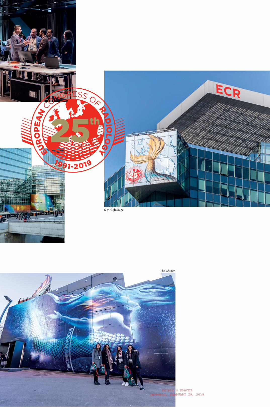
PEOPLE & PLACESTHURSDAY, FEBRUARY 28, 2019
The Cube
Sky High Stage
Tech Gate
The Church
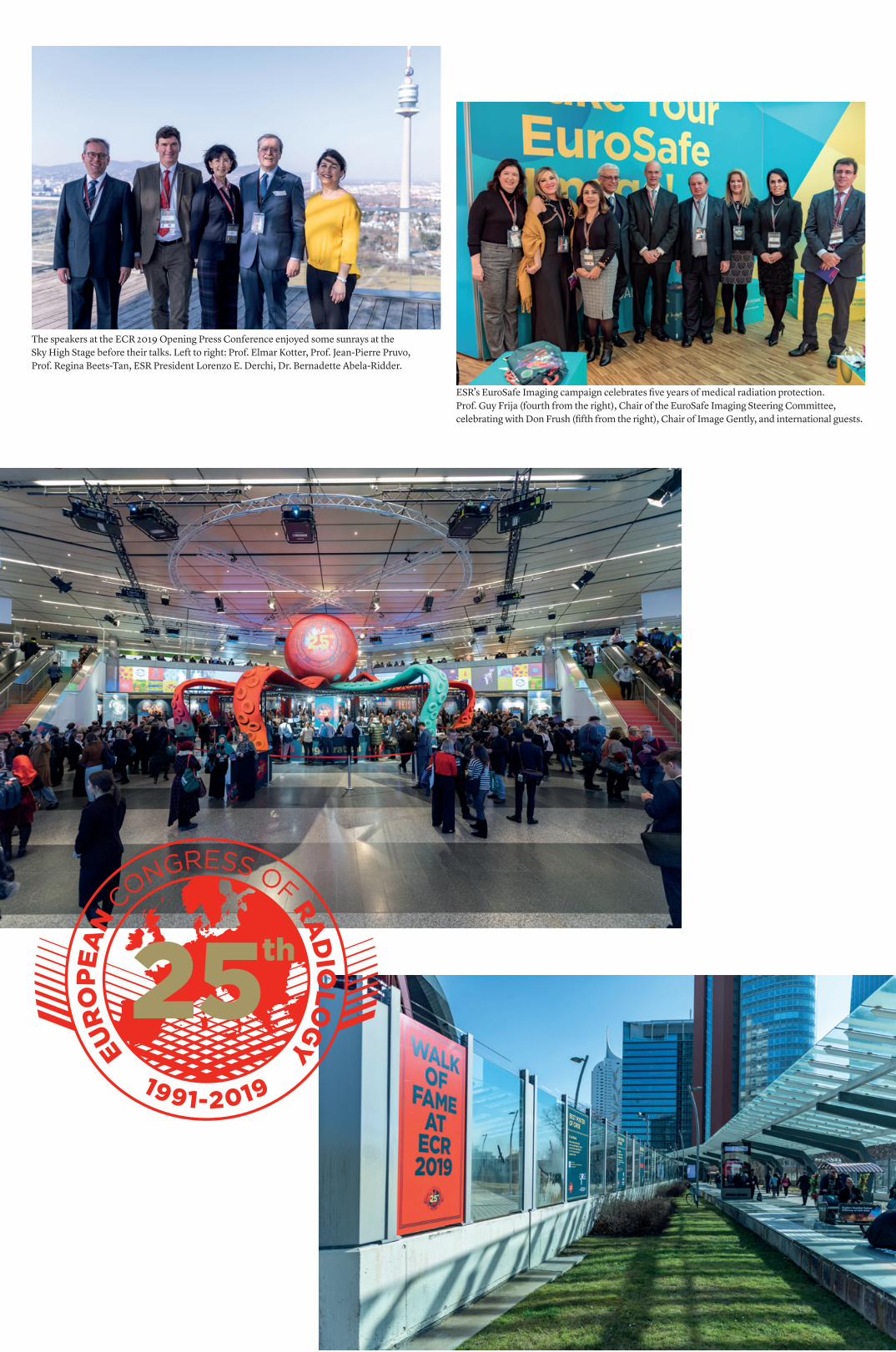
The speakers at the ECR 2019 Opening Press Conference enjoyed some sunrays at the Sky High Stage before their talks. Left to right: Prof. Elmar Kotter, Prof. Jean-Pierre Pruvo, Prof. Regina Beets-Tan, ESR President Lorenzo E. Derchi, Dr. Bernadette Abela-Ridder.
ESR’s EuroSafe Imaging campaign celebrates five years of medical radiation protection. Prof. Guy Frija (fourth from the right), Chair of the EuroSafe Imaging Steering Committee, celebrating with Don Frush (fifth from the right), Chair of Image Gently, and international guests.


THURSDAY, FEBRUARY 28 & FRIDAY, MARCH 1
A SERIES OF SPECIAL, NON-SCIENTIFIC SESSIONS
DEDICATED TO WOMEN WORKING IN HEALTHCARE
ORGANISED BY PROF. LORENZO E. DERCHI AND PROF. HEDVIG HRICAK
The Church
Women in Focus
Be Inspired