BARNARD INSTITUTE OF RADIOLOGY MADRAS MEDICAL ...
105
THREE-DIMENSIONAL COLOR DOPPLER SONOGRAPHIC ASSESSMENT OF CHANGES IN VOLUME AND VASCULARITY OF FIBROIDS - BEFORE AND AFTER UTERINE ARTERY EMBOLIZATION Dissertation Submitted for M.D. DEGREE EXAMINATION In Radio – Diagnosis BRANCH – VIII BARNARD INSTITUTE OF RADIOLOGY MADRAS MEDICAL COLLEGE & RESEARCH INSTITUTE THE TAMILNADU DR. M.G.R. MEDICAL UNIVERSITY CHENNAI – TAMILNADU APRIL 2012
-
Upload
khangminh22 -
Category
Documents
-
view
1 -
download
0
Transcript of BARNARD INSTITUTE OF RADIOLOGY MADRAS MEDICAL ...

THREE-DIMENSIONAL COLOR DOPPLER
SONOGRAPHIC ASSESSMENT OF CHANGES IN
VOLUME AND VASCULARITY OF FIBROIDS -
BEFORE AND AFTER UTERINE ARTERY
EMBOLIZATION
Dissertation Submitted for
M.D. DEGREE EXAMINATION
In Radio – Diagnosis
BRANCH – VIII
BARNARD INSTITUTE OF RADIOLOGY
MADRAS MEDICAL COLLEGE &
RESEARCH INSTITUTE
THE TAMILNADU DR. M.G.R. MEDICAL UNIVERSITY
CHENNAI – TAMILNADU
APRIL 2012
CERTIFICATE
This is to certify that the dissertation entitled “THREE
DIMENSIONAL COLOR DOPPLER SONOGRAPHIC ASSESSMENT
OF CHANGES IN VOLUME AND VASCULARITY OF FIBROIDS -
BEFORE AND AFTER UTERINE ARTERY EMBOLIZATION”
presented here is the bonafide original work done by Dr.S.Anbumalar, in the
Barnard Institute of Radiology and Madras Medical College, Chennai 600003,
in partial fulfilment of the requirements for the M.D Radiodiagnosis, Branch -
VIII Examination of the Tamil Nadu Dr.MGR Medical University to be held in
April 2012.
Dr.S.KALPANA, Dr.S.SUNDARESWARAN,
Associate Prof. and Guide, Chief Civil Surgeon and Co-Guide,
Barnard Institute of Radiology, Barnard Institute of Radiology,
Madras Medical College, Madras Medical College,
Chennai – 600 003. Chennai – 600 003.
Prof.N.KAILASANATHAN, Prof.VANITHA .K,
Professor of Radiology, Director and Professor,
Barnard Institute of Radiology, Barnard Institute of Radiology,
Madras Medical College, Madras Medical College,
Chennai – 600 003. Chennai – 600 003.
DEAN Rajiv Gandhi Government General Hospital and
Madras Medical College, Chennai - 600 003.
DECLARATION
I, Dr.S.Anbumalar, solemnly declare that this dissertation entitled,
“THREE DIMENSIONAL COLOR DOPPLER SONOGRAPHIC
ASSESSMENT OF CHANGES IN VOLUME AND VASCULARITY OF
FIBROIDS - BEFORE AND AFTER UTERINE ARTERY
EMBOLIZATION” is a bonafide work done by me for the degree of M.D.
during the period of June 2009 to May 2012 under the guidance and
supervision of Prof.Vanitha .K, M.D., D.M.R.D., D.R.M., Director and
Professor, Barnard Institute of Radiology, Madras Medical College, Chennai –
600 003. This dissertation is submitted to The Tamil Nadu Dr.M.G.R. Medical
University, towards partial fullfillment of requirement for the award of M.D.
Degree in Radiodiagnosis, (Branch- VIII).
Place: Chennai Signature of the Candidate
Date: (Dr.S.Anbumalar)
Dr.S.KALPANA, Dr.S.SUNDARESWARAN,
Associate Prof. and Guide, Chief Civil Surgeon and Co-Guide,
Barnard Institute of Radiology, Barnard Institute of Radiology,
Madras Medical College, Madras Medical College,
Chennai – 600 003. Chennai – 600 003.
ACKNOWLEDGEMENT
I would like to thank Prof.V.Kanagasabai, M.D., Dean, Madras
Medical College and Research Institute for giving me permission to conduct
the study in this institution.
With extreme gratefulness, I express my indebtedness to
Prof.VANITHA .K, M.D., D.M.R.D., D.R.M., Director and Professor,
Barnard Institute of Radiology, for having encouraged me to take up this study.
But for her guiding spirit, perseverance and wisdom, this study would not have
been possible.
I express my sincere thanks and gratitude to Prof.M.PRABAKARAN,
MD, DMRD., Former Director, Barnard Institute of Radiology, for having
encouraged me to take up this study.
I express my sincere thanks and gratitude to
Prof.T.S.SWAMINATHAN, M.D., D.M.R.D., F.I.C.R., Former Director,
Barnard Institute of Radiology for his immense kindness, constant support and
consistent encouragement in conducting this study.
I wish to thank Prof. N. KAILASANATHAN, M.D.,D.M.R.D.,
Prof.K.MALATHY, M.D.,D.M.R.D., Prof.A.P.ANNADURAI,

M.D.,D.M.R.D., and Prof.K.THAYALAN for their support, valuable
criticisms and encouragement.
I wish to thank my Associate Professors Dr.S.KALPANA,
M.D.,D.M.R.D, Dr.S.BABU PETER, M.D.DNB, Dr.D.RAMESH, M.D.,
and Chief Civil Surgeon Dr.S.SUNDARESWARAN, D.M.R.D., for their
support, valuable criticisms and encouragement.
I am greatly indebted to my Assistant Professors Dr.J.DEVIMEENAL,
M.D., D.M.R.D., DNB, F.R.C.R., Dr.E.MANIMEKALA, M.D., DNB.,
Dr.J.CHEZHIAN, M.D., Dr.K. GEETHA, M.D., and fellow postgraduates
for their untiring help.
Last but not the least, I thank all my patients for their cooperation,
without whom this study would not have been possible.

CONTENTS
Sl.No. Title Page No.
1. INTRODUCTION 1
2. AIM OF THE STUDY 4
3. REVIEW OF LITERATURE 5
4. MATERIALS AND METHODS 26
5. ANALYSIS AND RESULTS 45
6. DISCUSSION 56
7. CONCLUSION 64
PROFORMA
ABBREVIATIONS
BIBLIOGRAPHY
MASTER CHART
INTRODUCTION
Menorrhagia is defined as bleeding that originates from the uterus . In
developing countries, the majority of cases are due to fibroid uterus. Massive
menorrhagia is a major clinical and surgical problem with a mortality of 80%,
which is most often related to hemodynamic instability.3,4
Uterine fibroids are the most frequent tumors of the female genital tract,
occurring in 20–50% of women who are older than 40 years. Uterine fibroid,
the most common cause of nonacute abnormal uterine bleeding, is also the
most common solid uterine neoplasm occurring in 20–40% of all women
during their reproductive period10
.
Uterine artery embolization (UAE) was introduced in the 1970s to treat
postpartum hemorrhage14.
In the 1990s, this technique was successfully used
preoperatively 3–10 days before myomectomy to reduce bleeding during the
surgical phase14
. In 1995, Ravina et al.15
proposed embolization of uterine
arteries as an alternative to surgical treatment of uterine leiomyoma.
MASSIVE MENORRHAGIA :
A) DEFINITION
Menorrhagia is defined as heavy or prolonged uterine bleeding that
occurs at regular intervals. Some sources define menorrhagia further as the loss
of ≥ 80 mL blood per cycle or bleeding > 7 days.
B) CLINICAL CONSIDERATIONS
Conservative management of massive menorrhagia carries a mortality
rate of 50%–100%5
and the mortality is up to 35% even in patients undergoing
operation6. Surgery remains the procedure of choice in the treatment of massive
menorrhagia caused by specific conditions, such as dysfunctional uterine
bleeding, hypertension, and endocrine etiology, that is resistant to other
therapies12
.
Embolization has become a first-line treatment for symptomatic uterine
fibroid8 .
Therapeutic uterine artery embolization is a good treatment adjunct to
control uterine bleeding and reduces the need for high-risk hysterectomy7.
UAE may help to avoid surgery in patients who are not good surgical
candidates. Should menorrhagia recur in these patients, repeat embolization can
be performed safely10
. Even in surgical candidates, UAE is effective in
preparing the patient for elective rather than high-risk surgery7.
The goal of uterine fibroid embolisation is to stop blood flow to the
uterus through the uterine arteries, thus depriving myomas of their blood
supply9
to produce ischemic infarction .
Various non uterine systemic arteries, as well as ovarian arteries, may
also contribute to menorrhagia, and their implication is dependent on the
underlying location of fibroid13,14,15,16,17,18
. Non Uterine systemic collateral
vessels must be particularly suspected when there is evidence of larger size of
fibroid11,19
. Recognition and occlusion of Non Uterine systemic collaterals
providing blood to hypervascular fibroid lesions is essential for successful
percutaneous embolotherapy of fibroid 2,17,18,19,20
. Prior to embolization, the
interventional radiologist needs to be aware of the dominant side of the uterine
artery bleeding, and the most likely source of bleeding has to be identified to
determine which vessel is to be occluded. Since the uterine circulation is the
most frequent source of menorrhagia, embolization of uterine arteries is usually
the favored therapeutic option to stop the bleeding11,21
.
Recent important technologic advances in three dimensional color
doppler sonography, have introduced a comprehensive, noninvasive method
of evaluating the entire uterus, allowing detailed assessment of the vascularity
and volume of fibroid1,2
. Three dimensional color doppler sonography can also
help in the planning of a focused and efficient non uterine systemic artery
embolization. It provides a precise road map for the interventional radiologist
in performing an endovascular treatment for menorrhagia. Uterine artery
embolization leads to good technical success and fibroid volume reduction10
.
The aim of this study was to evaluate the effectiveness of three
dimensional color Doppler sonography in assessment of volume and
vascularity reduction of fibroids treated by uterine artery embolisation.
AIM OF THE STUDY
The purpose of the present study is to prospectively evaluate the
accuracy of three-dimensional color doppler sonography in depicting changes
in fibroid volume and vascularity of pre and post uterine artery embolisation in
patients undergoing treatment of fibroid.
REVIEW OF LITERATURE
RELEVANT ANATOMY
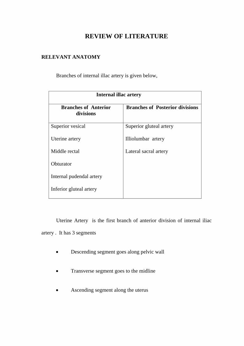
Branches of internal illac artery is given below,
Internal illac artery
Branches of Anterior
divisions
Branches of Posterior divisions
Superior vesical
Uterine artery
Middle rectal
Obturator
Internal pudendal artery
Inferior gluteal artery
Superior gluteal artery
Illiolumbar artery
Lateral sacral artery
Uterine Artery is the first branch of anterior division of internal iliac
artery . It has 3 segments
Descending segment goes along pelvic wall
Transverse segment goes to the midline
Ascending segment along the uterus
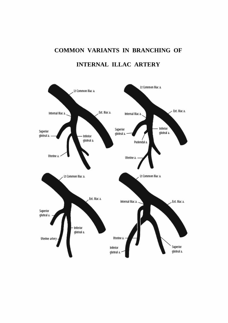
COMMON VARIANTS IN BRANCHING OF
INTERNAL ILLAC ARTERY
The uterus is supplied by a single arterial vascular system composed of
the uterine artery, which account for 99% of the arterial blood supply to the
uterus. Uterine artery has a characteristic U shape with descending segment
that parallels lateral pelvic wall , a transverse segment that crosses the distal
ureter at the level of cervix and a ascending segment that courses along the
uterine margin at the edge of the broad ligament
The uterine vasculature feeding the uterus is situated close to the ovarian
arteries and the two systems are connected by anastomoses.
NORMAL AND VARIANT ARTERIAL SUPPLY TO THE UTERUS
Normal arterial anatomy and variants are the key to the uterine artery
embolisation.
Normal variants : abdominal aorta origin , <1% congenital absence of
botn uterine arteries. 5- 10% can have retrograde utero – ovarian anastomosis
of size less than 500micrometer
Possible variation in uterine artery;
Cervico vaginal artery
Perforating arteries, terminal branches to the fallopian tubes and
ovaries
Inferior vesicle artery has common origin
Can have completely or partially absent uterine artery which can
be bilateral.
Ovarian artery : anteromedial origin from abdominal aorta shows
characteristic cork screw appearance in angiogram . 5- 10 % of ovarian artery
may supply uterine fibroid.
Other sources of fibroid supply is from :
Round ligament artery and lumbar artery which necessitates flush
aortography after embolisation that identifies specific feeding artery.
Identification of the normal arterial anatomy and main variations in
uterine fibroid tumor vascularization are key to the safety and success of the
procedure22
Ovarian supply to the leiomyoma should be excluded by placing
the tip of the catheter at L1 level during initial flush aortogram. If discovered,
ovarian artery can also be safely embolized55
.
Special mention :
Pedunculated subserosal fibroid may have arterial pedicle from adjacent
illac, renal or aortic branches. After embolisation it may separate from uterus
and drift into peritoneal cavity55
.Bicornuate uterus may be supplied by one
uterine artery only .

Differential diagnosis
1. Endometrial polyp with a single feeding vessel from an
intracavitary submucosal fibroid.
2. Adenomyosis is distinguished by rain drop appearance, multiple
scattered vessels or intratumoral vascularity, but fibroid has
feeding from peripheral vessels.
3. EMBOLOTHERAPHY:
Embolization is defined as the "therapeutic introduction of various
substances into the circulation to occlude selective vessels, either to arrest or
prevent haemorrhage, to devitalize a structure, tumour or organ.
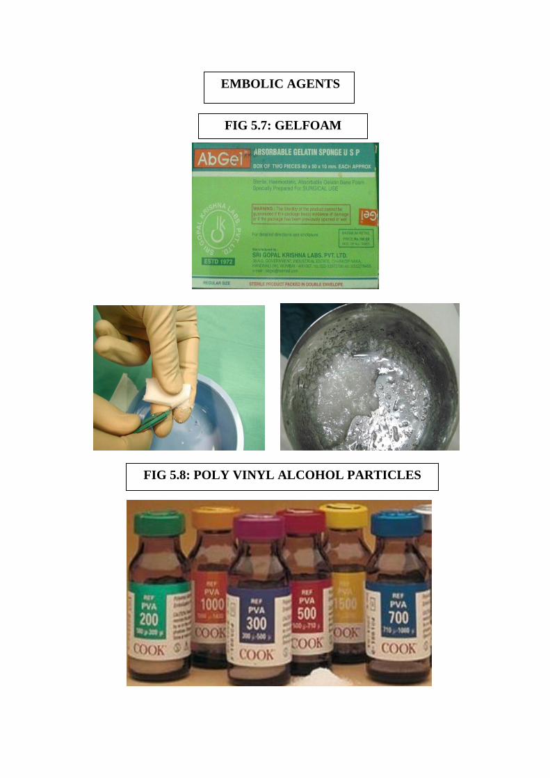
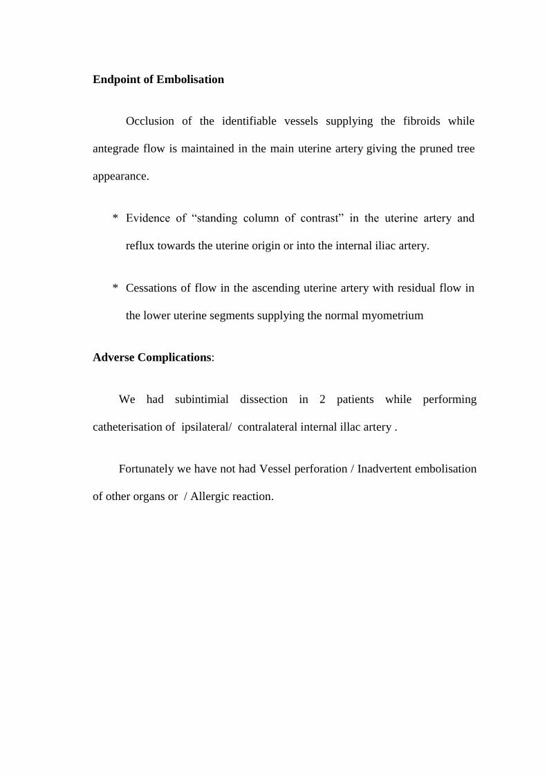
UAE- EMBOLIC AGENTS:
There are two different embolic agents used commonly
1. Permanent embolic agent is polyvinyl alcohol particles (PVA).
2. Temporary embolic agent is gelfoam.
Polyvinyl alcohol particles (PVA):
PVA is the most often used embolic agents in patients with
uterine fibroids.
PVA is permanent embolic agent is injected to occlude
predominantly peri - fibroidal arterial plexus seen surrounding
the fibroid .
Varying Particle sizes of 355-500 µm and 500-710 µm are
available commercially. Commonly used particle size is of 355-
500 µm.
GELFOAM (GELATIN SPONGE) :
Following gelfoam is injected to occlude the main uterine artery .
Gelfoam (gelatin sponge) is the most commonly used material. It
is a readily available, slowly resorbable material that can be used
as individual pledgets, torpedoes, or part of a slurry. For initial
distal occlusion, 0.5- to 2.0-mm cubes can be used followed by 3-
to 4-mm pledgets or torpedoes for more proximal occlusion.
Gelfoam pledgets are mixed with dilute contrast within a1 - or 3-
ml syringe. Since the Gelfoam pledgets float within the contrast
saline solution, the tip of the syringe should be pointed upward. A
theoretical disadvantage of Gelfoam particles is that their
resorption may lead to more rapid recanalization and recurrent
bleeding.
Three-Dimensional Color Doppler Sonography in pre and post Uterine
Artery Embolisation of Fibroids :
Arthur C. Fleischer, Edwin F. Donnelly, et al. Three-Dimensional
Color Doppler Sonography (3D CDS) before and after fibroid embolization
20009. Quantitated 3D CDS provides global depiction of the small arteries and
veins within the fibroid and provides an estimate of completeness of
embolization. 3D CDS can be used to for predict which patients will be
responders or nonresponders. 12 of 31 fibroids were hypervascular, 10 of 31
were isovascular, and 9 of 31 were hypovascular. Of the 12 hypervascular
fibroids, 10 (83%) became reduced in volume by 50%, whereas only 4 of 10
(40%) isovascular fibroids and 2 of 9 (22%) hypovascular fibroids decreased
in volume by 50%.
Sangeet Ghai, MD, Dheeraj K. Rajan, et al . Uterine artery embolization
for leiomyomas: Pre and post procedural evaluation with ultrasound (US),
200546
. US is a readily available first-line imaging modality and a well-
accepted method for both pre- and postprocedural evaluation of patients who
undergo UAE. Follow-up imaging is performed at 3, 6, and 12 months after
UAE to quantify volume reduction in the uterus and leiomyoma. Volume
reduction of the dominant fibroid is greater than that of the uterus, and follow-
up US has shown a reduction in uterine size of up to 40%, with the dominant
fibroid decreasing in size by up to 70% (9,39,40). The majority of fibroid
shrinkage occurs within a 6-month period following embolization, with further
reduction in size occurring between 6 and 12 months.
C. Joseph Muniz, MD, Arthur C. et al6
states that the Three-dimensional
Color Doppler Sonography and Uterine Artery Arteriography of Fibroids
Assessment of Changes in Vascularity Before and After Embolization, 2002 .
In 13 (87%) of 15 patients there was agreement; in 2 (13%) of 15 there was
disagreement. In both cases of disagreement, three-dimensional color Doppler
sonography showed collateral flow not depicted by uterine artery arteriography.
The mean reduction in quantitated vascularity after uterine artery embolization
was 44% (range, 19%–78%). Three-dimensional color Doppler sonography
accurately depicts fibroid vascularity and in some cases can reveal collateral
flow not depicted by uterine artery arteriography.
Jean-Pierre Pelage, Julien Cazejust, et al
200523
. Uterine Fibroid
Vascularization and Clinical Relevance to Uterine Fibroid Embolization,
Identification of the normal arterial anatomy and main variations in uterine
fibroid tumor vascularization are key to the safety and success of the
procedure.
Kitamura, Susan M Ascher, et al. Imaging Manifestations of
Complications Associated with Uterine Artery Embolization, 200528
Fibroids
in contact with the endometrial surface, including submucosal fibroids or
intramural fibroids with a submucosal component, pose an increased risk for
fibroid passage. Women over 45 years of age are at increased risk for ovarian
dysfunction after UAE, because they have a higher prevalence of uterine-
ovarian arterial anastomoses (43% of cases) compared with women under
45 years of age (5%).
Suhny Abbara, Boris Nikolic et al 200754
. Frequency and extent of
Uterine Perfusion via Ovarian Arteries observed during Uterine Artery
Embolization for Leiomyomas, Of the visualized ovarian arteries (n = 88), 52%
(46/88) were smaller than, 25% (22/88) were equal to, and 23% (20/88) were
larger than the diameter of a 5-French catheter. The aortogram revealed that
61% (54/88) of the ovarian arteries extended into the pelvis, whereas 38%
(33/88) did not. Selective injections were performed in 54 ovarian arteries. Of
these, 69% (37/54) of the ovarian arteries had residual fibroid perfusion from
the ovarian arteries after UAE (10 left-sided, 15 right-sided, six bilateral = 37
ovarian arteries).
Jean-Pierre Pelage et al, 200523
Regular PVA particles do not
completely occlude the lumen of the occluded arteries because of their irregular
shape and heterogeneous calibration and occlusion is completed by thrombus
formation. For uterine fibroid, the recommended diameter is 700–900µm
(compared to 500–700µm tris-acryl microspheres) for PVA microspheres and
900 µm for hydrogel polyzene-F microspheres.
Kenneth Murphy, et al 27
has given that non target embolization of
should be avoided to minimize the risk of postembolization sexual dysfunction.
the aim of embolization is not to occlude the UA, but to occlude vessels that
supply the fibroid(s) while sparing perfusion to the normal uterus proper.
Bruce McLucas, Rita Perrella, et al 20024, Role of Uterine Artery
Doppler Flow in Fibroid Embolization, Initial peak systolic velocity was
positively correlated with the size and shrinkage of myomas and uterine
volume. Peak systolic velocity was positively correlated with the size and load
of embolization particles and was significantly lower (mean, 33.2 cm/s) in
patients with adenomyosis than those without adenomyosis (mean, 39.3 cm/s).
High peak systolic velocity (>64 cm/s) was a significant predictor of failure.
Postembolization peak systolic velocity (mean, 21.85 cm/s) was significantly
lower than preembolization peak systolic velocity (mean, 40.33 cm/s) and was
not correlated with uterine fibroid embolization variables.
TP Jain, DN Srivastava, RP Sahu, et al, (2007)23
clearly demonstrated
Uterine artery embolization for symptomatic fibroids with imaging follow up,
All 32 patients had successful procedures. Overall, 25 patients responded,
giving a clinical success rate of 78.12%. Mean reduction in volume of uterus
and fibroid was 33 and 59.7% and 48.9 and 75.5% on US at 3 and 12 months
respectively, and 33.3 and 58.6% on MRI at 3 months. Volume reduction on
US and MRI at 3 months was highly correlative.
E. Aitken, A. Khaund, et al 2006, 10
the normal human myometrium has
a vascular spatial gradient absent in small fibroids, A quantitative gradient
within the myometrial vascular system, which is absent in fibroids, has been
demonstrated. These structural differences between diseased and healthy
tissues are probably because of differing expression of angiogenic growth
factors. Regarding UAE common indications are Symptomatic Fibroids and
Post Partum Haemorrhage
Uterine ischemia causes pain so limited embolisation is essential, Julien
Cazejust, et al 22
Drs.Lussenhop and Spence
first occluded a cerebral
arteriovenous malformation under radiographic and catheter guidance in 1960.
Charles Dotter is acknowledged as founder of the specialty from his landmark
work on angioplasty, first published in circulation in 1960. Another who
contributed early was Dr. Baum, who developed techniques for controlling G-I
bleeding using Vasopressin.
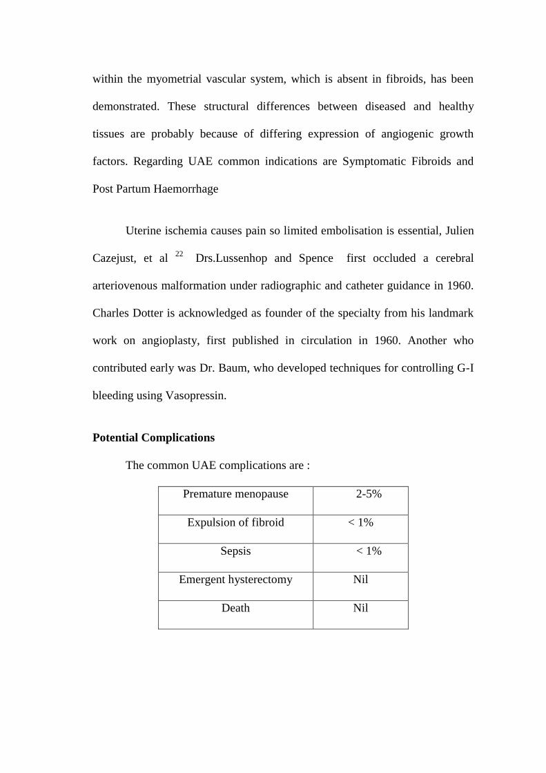
Potential Complications
The common UAE complications are :
Premature menopause 2-5%
Expulsion of fibroid < 1%
Sepsis < 1%
Emergent hysterectomy Nil
Death Nil
Following Drs.Lussenhops and Doppman in the USA, Japanese
physicians developed selective catheter techniques for treating spinal cord
AVMs. In 1972, Dr. Dotter and Dr. Rosch35
again developed techniques for
controlling bleeding due to ulcer disease by embolizing autologous clots.
Dr.White et.al first used this technique successfully in 1974 to actually control
ulcer bleeding with the survival of patient survived. Early in the 1970s, many
radiologists began to use embolotherapy for lifesaving haemorrhages.
Since 1960's, embolization of uterine artery has been used to treat post
partum haemorrhage. Uterine artery embolisation was used to control heavy
bleeding from cervical carcinoma. A recent case report from Ottawa hospital,
Canada says that uterine artery embolisation was used to control bleeding from
cervical pregnancy. The embolic agents used are polyvinyl alcohol, acryilic
spheres, gelfoam, steel coils and these are approved by the U.S. Food and
Drug Administration (FDA) for this purpose.
Uterine Artery Embolization for fibroid uterus was first used as a
technique to limit blood loss during surgical removal of fibroids, and
performed well before the surgery. However, it was found that after
embolization and while awaiting surgery many patients no longer had
symptoms and frequently the operation itself proved to be unnecessary. UAE
was introduced as a treatment for fibroids in 1992 in Paris by Dr. Jacques-
Henri Ravina, a gynaecologist. The treatment was first performed in the U.S. at
the University of California at Los Angeles (UCLA). There are now many such
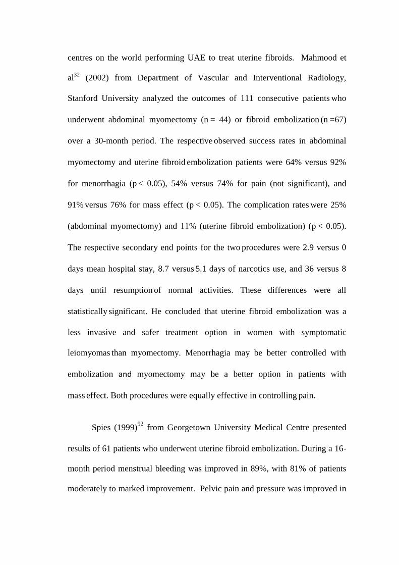
centres on the world performing UAE to treat uterine fibroids. Mahmood et
al32
(2002) from Department of Vascular and Interventional Radiology,
Stanford University analyzed the outcomes of 111 consecutive patients who
underwent abdominal myomectomy (n = 44) or fibroid embolization (n =67)
over a 30-month period. The respective observed success rates in abdominal
myomectomy and uterine fibroid embolization patients were 64% versus 92%
for menorrhagia (p < 0.05), 54% versus 74% for pain (not significant), and
91% versus 76% for mass effect (p < 0.05). The complication rates
were 25%
(abdominal myomectomy) and 11% (uterine fibroid embolization) (p < 0.05).
The respective secondary end points for the two procedures were 2.9 versus 0
days mean hospital stay, 8.7 versus 5.1 days of narcotics use, and 36 versus 8
days until resumption of normal activities. These differences were all
statistically significant. He concluded that uterine fibroid embolization was a
less invasive and safer treatment option in women with symptomatic
leiomyomas than myomectomy. Menorrhagia may be better controlled with
embolization and myomectomy may be a better option in patients with
mass effect. Both procedures were equally effective in controlling
pain.
Spies (1999)52
from Georgetown University Medical Centre presented
results of 61 patients who underwent uterine fibroid embolization. During a 16-
month period menstrual bleeding was improved in 89%, with 81% of patients
moderately to marked improvement. Pelvic pain and pressure was improved in
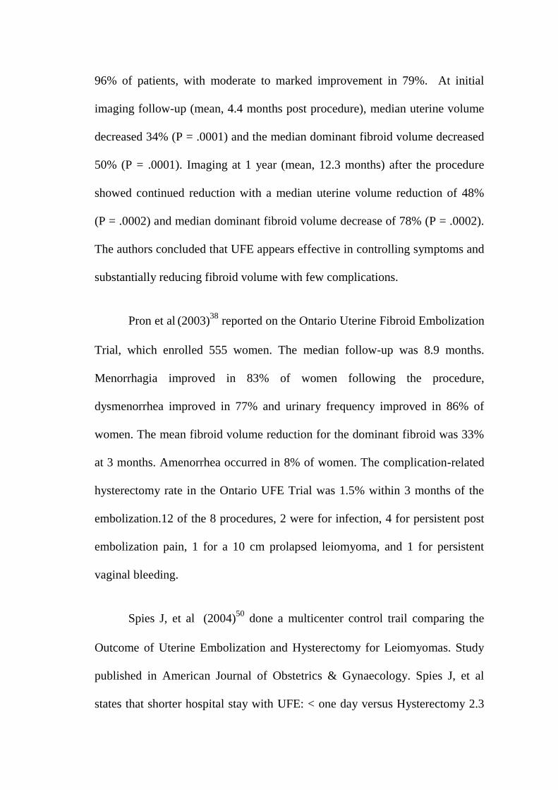
96% of patients, with moderate to marked improvement in 79%. At initial
imaging follow-up (mean, 4.4 months post procedure), median uterine volume
decreased 34% (P = .0001) and the median dominant fibroid volume decreased
50% (P = .0001). Imaging at 1 year (mean, 12.3 months) after the procedure
showed continued reduction with a median uterine volume reduction of 48%
(P = .0002) and median dominant fibroid volume decrease of 78% (P = .0002).
The authors concluded that UFE appears effective in controlling symptoms and
substantially reducing fibroid volume with few complications.
Pron et al (2003)
38 reported on the Ontario Uterine Fibroid Embolization
Trial, which enrolled 555 women. The median follow-up was 8.9 months.
Menorrhagia improved in 83% of women following the procedure,
dysmenorrhea improved in 77% and urinary frequency improved in 86% of
women. The mean fibroid volume reduction for the dominant fibroid was 33%
at 3 months. Amenorrhea occurred in 8% of women. The complication-related
hysterectomy rate in the Ontario UFE Trial was 1.5% within 3 months of the
embolization.12 of the 8 procedures, 2 were for infection, 4 for persistent post
embolization pain, 1 for a 10 cm prolapsed leiomyoma, and 1 for persistent
vaginal bleeding.
Spies J, et al (2004)
50 done a multicenter control trail comparing the
Outcome of Uterine Embolization and Hysterectomy for Leiomyomas. Study
published in American Journal of Obstetrics & Gynaecology. Spies J, et al
states that shorter hospital stay with UFE: < one day versus Hysterectomy 2.3
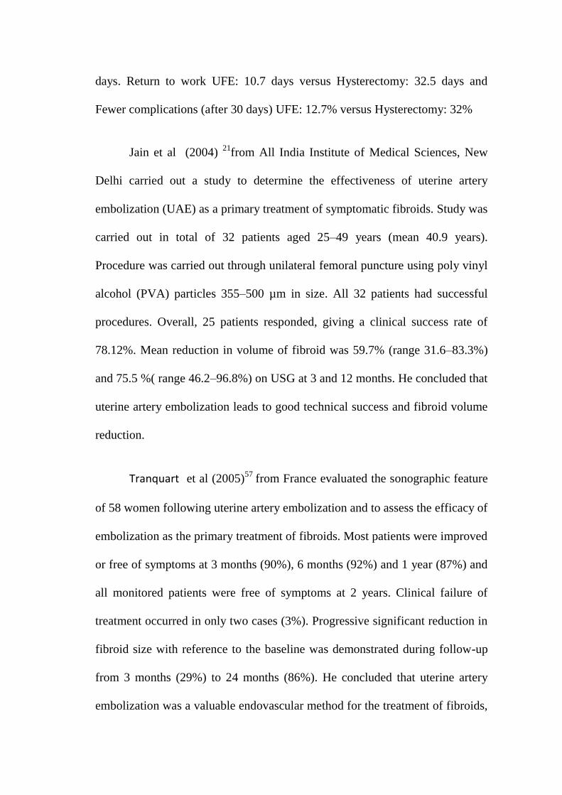
days. Return to work UFE: 10.7 days versus Hysterectomy: 32.5 days and
Fewer complications (after 30 days) UFE: 12.7% versus Hysterectomy: 32%
Jain et al (2004) 21
from All India Institute of Medical Sciences, New
Delhi carried out a study to determine the effectiveness of uterine artery
embolization (UAE) as a primary treatment of symptomatic fibroids. Study was
carried out in total of 32 patients aged 25–49 years (mean 40.9 years).
Procedure was carried out through unilateral femoral puncture using poly vinyl
alcohol (PVA) particles 355–500 µm in size. All 32 patients had successful
procedures. Overall, 25 patients responded, giving a clinical success rate of
78.12%. Mean reduction in volume of fibroid was 59.7% (range 31.6–83.3%)
and 75.5 %( range 46.2–96.8%) on USG at 3 and 12 months. He concluded that
uterine artery embolization leads to good technical success and fibroid volume
reduction.
Tranquart et al (2005)57
from France evaluated the sonographic feature
of 58 women following uterine artery embolization and to assess the efficacy of
embolization as the primary treatment of fibroids. Most patients were improved
or free of symptoms at 3 months (90%), 6 months (92%) and 1 year (87%) and
all monitored patients were free of symptoms at 2 years. Clinical failure of
treatment occurred in only two cases (3%). Progressive significant reduction in
fibroid size with reference to the baseline was demonstrated during follow-up
from 3 months (29%) to 24 months (86%). He concluded that uterine artery
embolization was a valuable endovascular method for the treatment of fibroids,
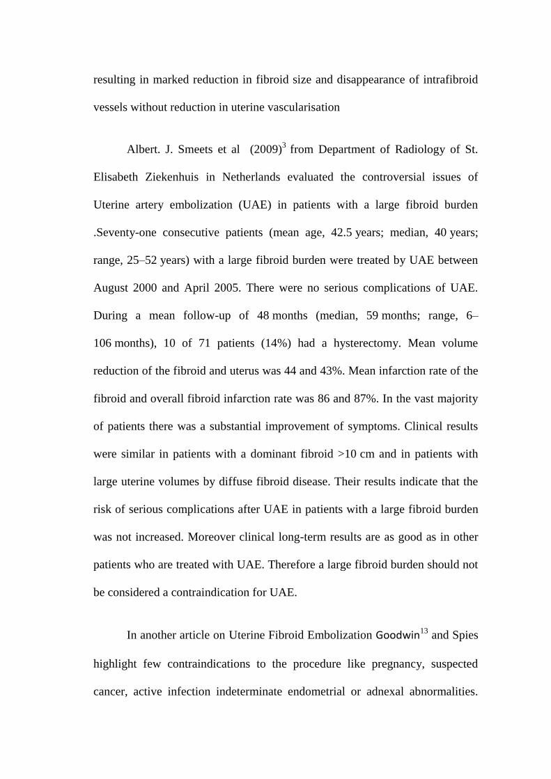
resulting in marked reduction in fibroid size and disappearance of intrafibroid
vessels without reduction in uterine vascularisation
Albert. J. Smeets et al (2009)3
from Department of Radiology of St.
Elisabeth Ziekenhuis in Netherlands evaluated the controversial issues of
Uterine artery embolization (UAE) in patients with a large fibroid burden
.Seventy-one consecutive patients (mean age, 42.5 years; median, 40 years;
range, 25–52 years) with a large fibroid burden were treated by UAE between
August 2000 and April 2005. There were no serious complications of UAE.
During a mean follow-up of 48 months (median, 59 months; range, 6–
106 months), 10 of 71 patients (14%) had a hysterectomy. Mean volume
reduction of the fibroid and uterus was 44 and 43%. Mean infarction rate of the
fibroid and overall fibroid infarction rate was 86 and 87%. In the vast majority
of patients there was a substantial improvement of symptoms. Clinical results
were similar in patients with a dominant fibroid >10 cm and in patients with
large uterine volumes by diffuse fibroid disease. Their results indicate that the
risk of serious complications after UAE in patients with a large fibroid burden
was not increased. Moreover clinical long-term results are as good as in other
patients who are treated with UAE. Therefore a large fibroid burden should not
be considered a contraindication for UAE.
In another article on Uterine Fibroid Embolization Goodwin13 and Spies
highlight few contraindications to the procedure like pregnancy, suspected
cancer, active infection indeterminate endometrial or adnexal abnormalities.
They also cite the U.K. Hysterectomy or Percutaneous Embolisation for
Uterine Leiomyomata (HOPEFUL)19
study, which showed 2.6% incidence of
septicaemia after uterine fibroid embolization, with 1.1% of the women
requiring emergency hysterectomy. Severe infection, often necessitating urgent
hysterectomy, was a rare but well-established complication of uterine fibroid
embolization.
The Fibroid registry26
(Fibroid Registry for Outcomes Data) of 3160
patients showed an emergency hysterectomy rate of only 0.09% at 30 days.
Emergency hysterectomy for bleeding has been described 4 months after
uterine fibroid embolization.
In the Randomized Clinical Embolization versus Hysterectomy (EMMY)
27 trial, researchers in The Netherlands found those six weeks after treatment,
women who underwent UFE reported higher satisfaction scores than those
who had hysterectomies. Two years later, 90 percent of the women in both
categories said they were satisfied with their therapies, the researchers
reported in the March 2008 edition of Radiology.
Ninety percent of women who underwent UFE avoided a hysterectomy,
said Scott Goodwin, M.D, 48
the lead author and a professor and chair of the
Department of Radiological Sciences in the School of Medicine at the
University of California, Irvine. Study results were published in the January
2008 issue of Obstetrics & Gynaecology.
Research at Georgetown University till 1999, 20.000 to 25,000 patients
has had this procedure world-wide. Their initial results, along with those that
have been published or presented at scientific meetings, says that symptomatic
improvement in 85-90% of patients with the large majority of patients
markedly improved. The improvement rate was similar for heavy menstrual
bleeding and for pressure and pain symptoms. Most patients have rated this
procedure as very tolerable and in almost all cases hospitalization is necessary
for only one night. In some centres, the patients are treated and discharged the
same day. The expected average reduction in the volume of the fibroids is 40-
50% in three months, with reduction in the overall uterine volume of about 30-
40%. Over time, the fibroids continue to shrink. With several years follow-up
now available, it appear that fibroids which are successfully treated does not re
grow.
Giovanna Tropeano et al11
from Department of Obstetrics and
Gynaecology, University Catholic del Sacro Cuore, Italy reports 50–60%
reduction in fibroid size and 85–95% relief of symptoms following UAE. UAE
offered shorter hospital stays (1–2 days for UAE versus 5–5.8 days for
hysterectomy) and recovery times (9.5–28 days for UAE versus 36.2–63 days
for surgery) and major complication rates (2–15% for UAE versus 2.7–20% for
surgery, in 3 Randomised control trials. Four studies analysing cost-
effectiveness found UAE more cost-effective than surgery. There is insufficient
evidence regarding fertility and pregnancy outcome after UAE. They
concluded that good quality evidence supports the safety and effectiveness of
UAE for women with symptomatic fibroids. The current available data are
insufficient to routinely offer UAE to women who wish to preserve or enhance
their fertility.
Hayden Homer16
Department of Obstetrics and Gynaecology, Institute
for Women’s Health, University College London has said that data on
pregnancy following uterine artery embolisation (UAE)
are scarce, with just
over 200 pregnancies reported. Two small prospective trials
of UAE versus
surgical intervention suggest increased levels of adverse pregnancy outcomes
following fibroid embolisation. This study suggests that in the absence of more
robust evidence, caution should be exercised
in recommending UAE to women
who wants to retain their reproductive ambitions.
Joao and Marisa (2010)
24studied on pregnancy out come after uterine
fibroid embolisation. This study shows comparable fertility rates between the
two primary uterus-sparing treatments uterine fibroid embolization (UFE) and
surgical myomectomy, which is considered the gold standard for symptomatic
fibroids in women who wish to conceive. Of the 743 patients who received
UFE treatment, 74 wanted to conceive and had been unable. Most women
opted for UFE as a fertility treatment after failure of myomectomy or in vitro
fertilization or because hysterectomy was the only suggested option. Of the
74 women who wanted to become pregnant, 44 of them became pregnant
(59.5%). There are five (11.3%) ongoing pregnancies and 39 (88.7%) finished
pregnancies, with 33 successful live births (84.6%), four spontaneous
abortions (10.3%), one induced abortion and one stillbirth. There were 22
caesarean deliveries (66.6%), two preterm deliveries at 36 weeks (6.1%) and
five low birth weights. This study concluded that in the future, UFE will
probably be a first-line treatment option even for women who wish to
conceive and are unable due to the presence of uterine fibroids.
Rashid. S. et al 40
studied the effects of Uterine Artery Embolisation and
Surgical Treatment on Ovarian Function in Women with Uterine Fibroids.
Some studies have shown that patients treated with UAE develop amenorrhea
(with rates varying from 1%–8% and as high as 14%) and have increased
follicle-stimulating hormone (FSH) levels (within the menopausal range)
suggesting ovarian dysfunction may occur as a consequence of UAE. This
study compares the effects of UAE and surgery on ovarian function and
menstrual cycle characteristics in women with uterine fibroids. The impact of
age on ovarian function was also studied. All surgical patients had ovarian
conservation at the time of surgery. Ovarian function assessed by the levels of
serum FSH were measured on day 3 of the menstrual cycle before treatment,
and at 6 and 12 months post-treatment and menstrual cycle characteristics. No
significant difference was found between the two treatment groups in the rate
of ovarian failure at 12 months (UAE: 11% versus surgery: 18%; P = 0.44.
These findings provide no evidence that UAE accelerates deterioration in
ovarian function at 1 year, in comparison with surgery.
Van der Kooij SM et al (2010) 58
from Department of Radiology, Academic
Medical Center, Amsterdam compared the clinical outcome and health related quality
of life (HRQOL), 5 years after uterine artery embolization (UAE) or hysterectomy in
the treatment of menorrhagia caused by uterine fibroids. Patients with symptomatic
uterine fibroids who were eligible for hysterectomy were assigned randomly 1:1 to
hysterectomy or UAE. Endpoints after 5 years were re-intervention rates, menorrhagia
and HRQOL measures that were assessed by validated questionnaires. UAE had a
positive effect both on urinary and defecation function.
MATERIALS AND METHOD
30 patients (mean age, 31.67 years, age range 26-35years) referred to
our institution in a six-month period for endovascular treatment of menorrhagia
underwent three dimensional color Doppler as part of pre and post therapeutic
evaluation.
Study Design: Prospective interventional trial
Study Place: Barnard Institute of Radiology, Madras Medical College.
Collaborating Unit: Institute of Obstetrics and Gynaecology,
Madras Medical College, Chennai.
Study Population: Women with symptomatic fibroid
Sample Size: 30 Women with symptomatic fibroid.
METHODOLOGY :
Patient Selection Criteria
* Inclusion criteria :
1. Women with symptomatic fibroid (menstrual disturbances or
pressure symptoms due to size or pain) who want to retain their
uterus and avoid surgery.
2. Solitary or multiple intramural fibroids of less than 7 cm.
3. Parous women who completed family and age less than 40 yrs.
4. Ultrasound findings without any degeneration of fibroid
5. Women who gave consent to undergo uterine artery embolisation
and participate in study.
* Exclusion criteria :
1. Women with asymptomatic fibroid.
2. Infertile women or parous women who want to conserve uterus
for future pregnancy.
3. Fibroids of more than 7 cm or with degenerative changes or
hypovascular in Doppler study.
4. Sub mucous or pedunculated Sub serosal fibroid.
5. Associated pelvic / adnexal and endometrial pathologies .
6. Previous history of anaphylaxis to contrast.
7. Renal function test abnormality.
8. Systemic illness like HIV, HbsAg +ve, abnormal renal profile,
Coagulation disorders.
Preoperative work up
History, pelvic exam .
Basic blood investigations done including Hb%, CBC, Blood
Urea, Serum creatinine, HIV, HbsAg, Coagulation profile (BT,
CT,PT,PTT).
FSH (day 3 ) to assess ovarian reserve
USG of Pelvis and abdomen done to know the location, size,
number and volume of fibroid. Those who had degeneration
(calcification or cystic degeneration) are excluded from the study.
Doppler USG done to know the vascularity of fibroid. Hypo
vascular fibroids are excluded from study.
Fractional curettage performed to rule out any associated
malignant changes in endometrium.
Pap smear to rule out cervical carcinoma.
Informed consent
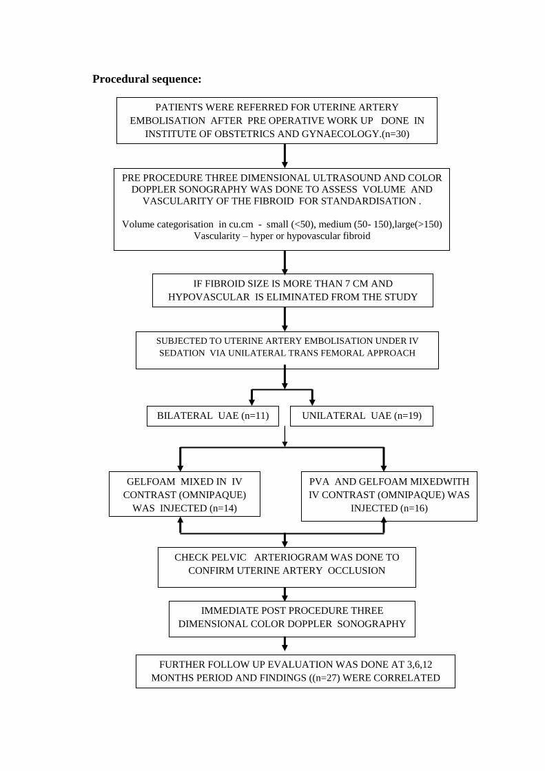
Procedural sequence:
PATIENTS WERE REFERRED FOR UTERINE ARTERY
EMBOLISATION AFTER PRE OPERATIVE WORK UP DONE IN
INSTITUTE OF OBSTETRICS AND GYNAECOLOGY.(n=30)
PRE PROCEDURE THREE DIMENSIONAL ULTRASOUND AND COLOR
DOPPLER SONOGRAPHY WAS DONE TO ASSESS VOLUME AND
VASCULARITY OF THE FIBROID FOR STANDARDISATION .
Volume categorisation in cu.cm - small (<50), medium (50- 150),large(>150)
Vascularity – hyper or hypovascular fibroid
SUBJECTED TO UTERINE ARTERY EMBOLISATION UNDER IV
SEDATION VIA UNILATERAL TRANS FEMORAL APPROACH
IMMEDIATE POST PROCEDURE THREE
DIMENSIONAL COLOR DOPPLER SONOGRAPHY
.
FURTHER FOLLOW UP EVALUATION WAS DONE AT 3,6,12
MONTHS PERIOD AND FINDINGS ((n=27) WERE CORRELATED
IF FIBROID SIZE IS MORE THAN 7 CM AND
HYPOVASCULAR IS ELIMINATED FROM THE STUDY
UNILATERAL UAE (n=19)
BILATERAL UAE (n=11)
GELFOAM MIXED IN IV
CONTRAST (OMNIPAQUE)
WAS INJECTED (n=14)
PVA AND GELFOAM MIXEDWITH
IV CONTRAST (OMNIPAQUE) WAS
INJECTED (n=16)
CHECK PELVIC ARTERIOGRAM WAS DONE TO
CONFIRM UTERINE ARTERY OCCLUSION
Clinical symptoms :
In this prospective study 30 patients (n=30) with fibroid uterus having at
least one of the following symptoms were selected.
Symptoms related to leiomyomas were classified into three categories:
abnormal bleeding (menorrhagia, metrorrhagia),
Pressure symptoms like (Increased frequency of urination, lower
abdominal heaviness, Constipation, uni-or bilateral hydronephrosis),
Pelvic pain.
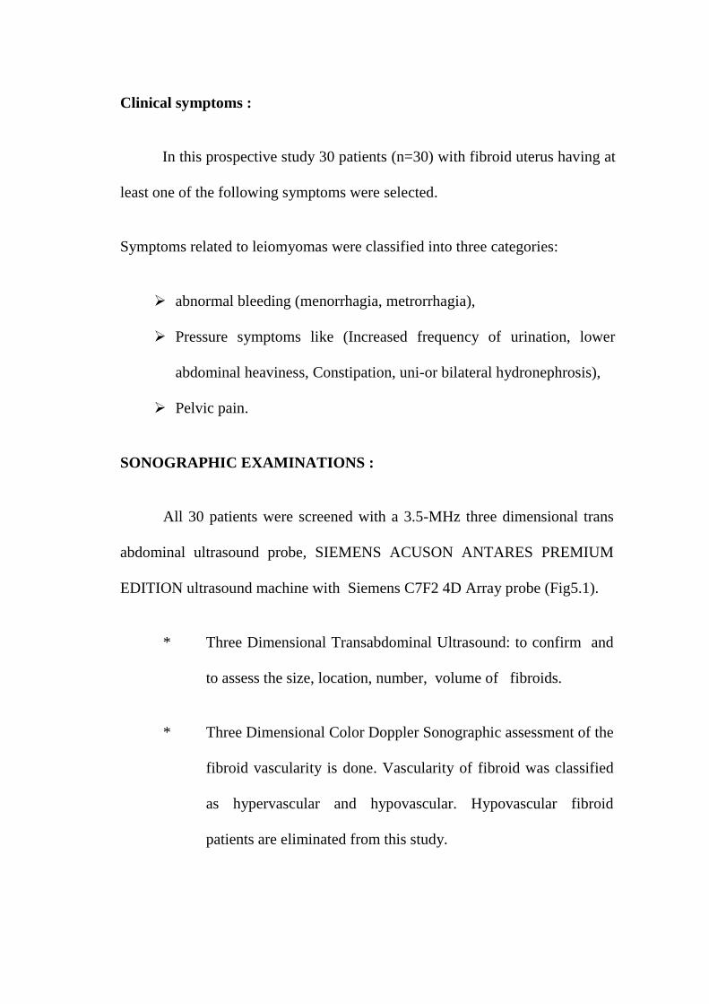
SONOGRAPHIC EXAMINATIONS :
All 30 patients were screened with a 3.5-MHz three dimensional trans
abdominal ultrasound probe, SIEMENS ACUSON ANTARES PREMIUM
EDITION ultrasound machine with Siemens C7F2 4D Array probe (Fig5.1).
* Three Dimensional Transabdominal Ultrasound: to confirm and
to assess the size, location, number, volume of fibroids.
* Three Dimensional Color Doppler Sonographic assessment of the
fibroid vascularity is done. Vascularity of fibroid was classified
as hypervascular and hypovascular. Hypovascular fibroid
patients are eliminated from this study.
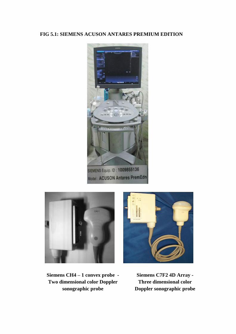
FIG 5.1: SIEMENS ACUSON ANTARES PREMIUM EDITION
Siemens C7F2 4D Array -
Three dimensional color
Doppler sonographic probe
Siemens CH4 – 1 convex probe -
Two dimensional color Doppler
sonographic probe

UTERINE ARTERY EMBOLISATION :
Patients selected according to the inclusion criteria were taken up for
uterine artery embolisation (UAE) after adequate counselling. Eligible patients
were advised to use contraception before the procedure and LMP confirmed.
PATIENT PREPARATION:
History of any medications ?
Any allergies, especially to contrast materials.
Any recent illnesses or other medical conditions.
If there is any possibility for pregnancy.
Nil oral previous night prior to the procedure.
1. All patients were explained about the procedure and its possible
complications.
2. They were also informed about alternative treatment options
available.
3. Written consent was obtained from all patients.
Informed consent obtained from all the patients before the procedure.

Patient has to be shaved in both groins & axillary region if femoral pulse
is absent; clotting time, bleeding time, blood urea, serum creatinine levels
should be within normal limits .
PROFORMA : See annexure
PRE PROCEDURE :
This examination is usually done as an inpatient procedure in our
hospital.
PROCEDURE:
Patient position : Supine
Pre Anaesthetics
Inj Dexamethasone 8mg I.V Stat
Inj. Pheniramine Maleate I amp I.M Stat
Inj. Atropine Iamp I.V.Stat
Anti biotic prophylaxis:
* Inj Ampicillin 1gm I.V (ATD)
Anesthesia/ Sedation :
Inj Tramadol 1 amp IM
A nurse or technologist will insert an intravenous (IV) line into a small
vein premedication is given (1cc of atropine &1cc of phenramine maleate).
Small dose of sedative is given through the IV to lessen the anxiety of
the patient during the procedure.
Approach : Percutaneous right or left trans femoral approach.
PROCEDURE :
Uterine artery embolisation technique done in our hospital
Angiography and Embolisation was performed using an SCHIMADZU,
JAPAN 800mA conventional angiographic unit with digital dicom
fluoroscopy (FIG 5.2 )
(A). With aseptic precautions under local anaesthesia femoral puncture
was made. A line is drawn from the anterior superior illac spine to the
pubic symphysis & 1 cm below the midpoint of the line, a point is
marked. Local anaesthetic given by keeping the middle finger &index
finger the pulse is felt. A small incision in the skin is made & the
subcutaneous tissue is separated with the artery forceps. By using the
Seldinger’s technique, Seldinger single puncture needle (FIG 5.3), is
used to puncture the artery. When the spurt of blood comes out of the
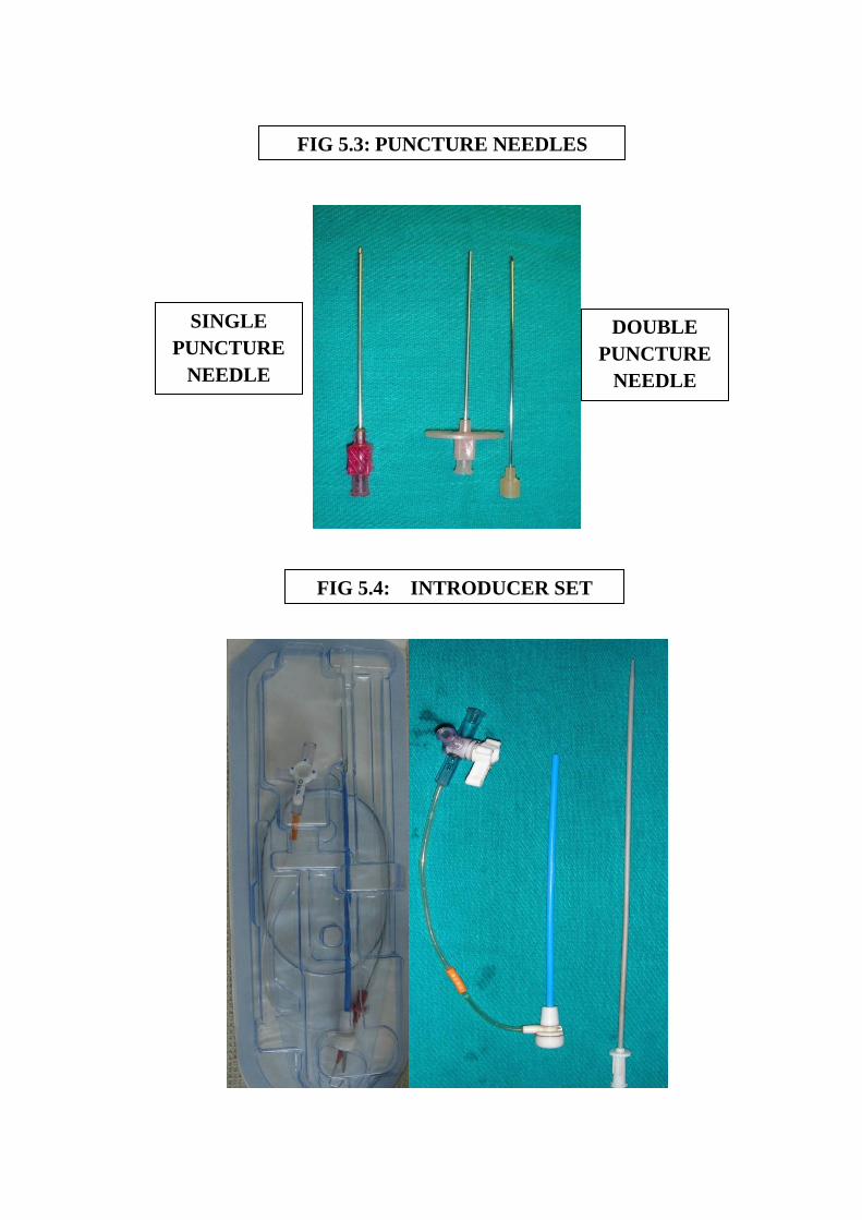
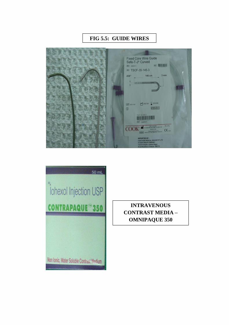
needle, the guide wire (FIG 5.5) is inserted through the needle with help
of the introducer. Once the guide wire is inserted upto the abdominal
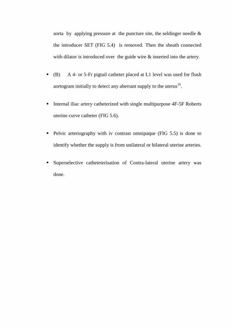
aorta by applying pressure at the puncture site, the seldinger needle &
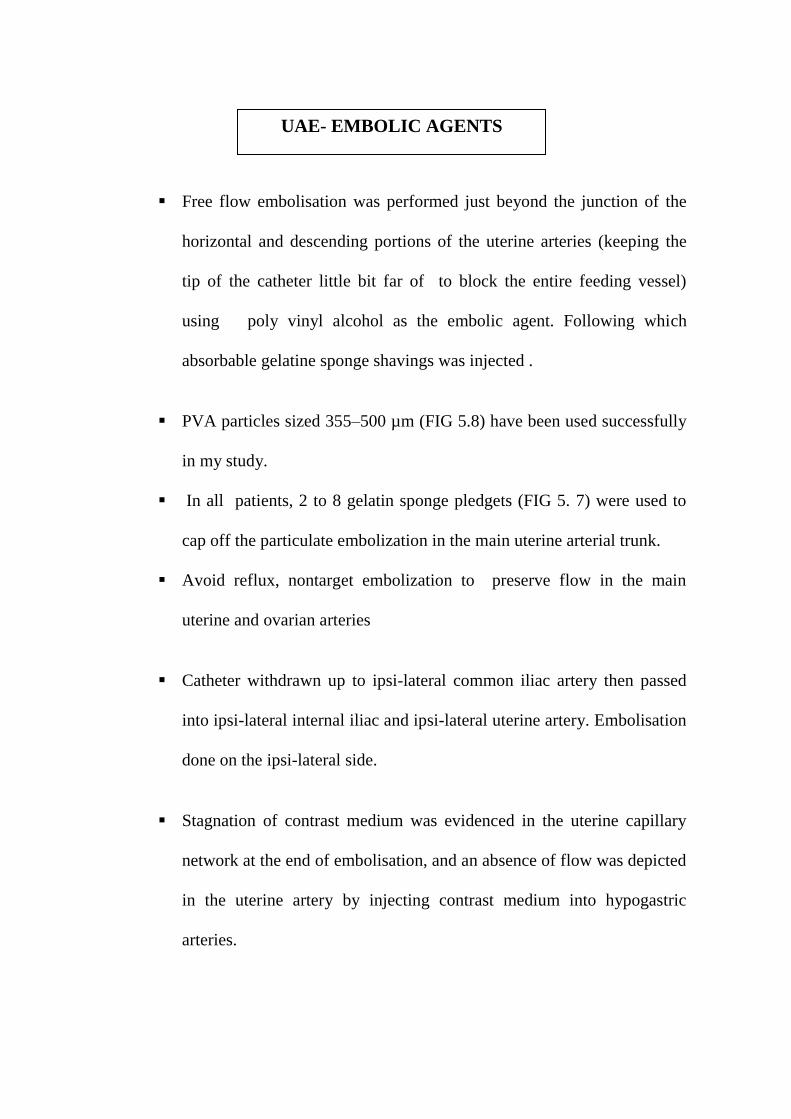
the introducer SET (FIG 5.4) is removed. Then the sheath connected
with dilator is introduced over the guide wire & inserted into the artery.
(B) A 4- or 5-Fr pigtail catheter placed at L1 level was used for flush
aortogram initially to detect any aberrant supply to the uterus10
.
Internal iliac artery catheterized with single multipurpose 4F-5F Roberts
uterine curve catheter (FIG 5.6).
Pelvic arteriography with iv contrast omnipaque (FIG 5.5) is done to
identify whether the supply is from unilateral or bilateral uterine arteries.
Superselective catheteterisation of Contra-lateral uterine artery was
done.
Free flow embolisation was performed just beyond the junction of the
horizontal and descending portions of the uterine arteries (keeping the
tip of the catheter little bit far of to block the entire feeding vessel)
using poly vinyl alcohol as the embolic agent. Following which
absorbable gelatine sponge shavings was injected .
PVA particles sized 355–500 µm (FIG 5.8) have been used successfully
in my study.
In all patients, 2 to 8 gelatin sponge pledgets (FIG 5. 7) were used to
cap off the particulate embolization in the main uterine arterial trunk.
Avoid reflux, nontarget embolization to preserve flow in the main
uterine and ovarian arteries
Catheter withdrawn up to ipsi-lateral common iliac artery then passed
into ipsi-lateral internal iliac and ipsi-lateral uterine artery. Embolisation
done on the ipsi-lateral side.
Stagnation of contrast medium was evidenced in the uterine capillary
network at the end of embolisation, and an absence of flow was depicted
in the uterine artery by injecting contrast medium into hypogastric
arteries.
UAE- EMBOLIC AGENTS

FIG.5.2 SCHIMADZU 800mA DIGITAL DICOM
FLUOROSCOPY
DOUBLE
PUNCTURE
NEEDLE
FIG 5.4: INTRODUCER SET
FIG 5.3: PUNCTURE NEEDLES
SINGLE
PUNCTURE
NEEDLE
INTRAVENOUS
CONTRAST MEDIA –
OMNIPAQUE 350
FIG 5.5: GUIDE WIRES
CATHETERS

FIG 5.6: CATHETERS
COBRA CATHETER
RENAL DOUBLE CURVE
(RDC) CATHETER
ROBERTS UTERINE
CURVE (RUC)
CATHETER
PIGTAIL CATHETER
EMBOLIC AGENTS
FIG 5.7: GELFOAM
FIG 5.8: POLY VINYL ALCOHOL PARTICLES
Endpoint of Embolisation
Occlusion of the identifiable vessels supplying the fibroids while
antegrade flow is maintained in the main uterine artery giving the pruned tree
appearance.
* Evidence of “standing column of contrast” in the uterine artery and
reflux towards the uterine origin or into the internal iliac artery.
* Cessations of flow in the ascending uterine artery with residual flow in
the lower uterine segments supplying the normal myometrium
Adverse Complications:
We had subintimial dissection in 2 patients while performing
catheterisation of ipsilateral/ contralateral internal illac artery .
Fortunately we have not had Vessel perforation / Inadvertent embolisation
of other organs or / Allergic reaction.

Post procedure events:
Post procedure :
All 30 patients were subjected to immediate (within an hour) of post
embolisation ultrasonography and colour Doppler study .
Three Dimensional Transabdominal Ultrasound to assess the size and
volume of fibroid, and the presence of embolic agents within the fibroid.
Three Dimensional Color Doppler Sonographic assessment of the fibroid
vascularity was done. Hypovascularity in the embolised fibroid patients
was noted.
Post procedure monitoring :
Patients were observed in high dependence ward for 6 hrs then at least 48
hrs in the routine wards. All the patients were carefully observed for any
complications like groin haematoma /retroperitoneal haematoma. Vitals were
checked. Patients were kept nil per oral for 4 hours. They were advised bed rest
for 6 hours and then ambulated. Patients were discharged after 48-72 hrs.
On discharge:
Patients were advised to report if appearance of new symptoms like
Any unbearable lower abdominal pain
Fever or Purulent vaginal discharge

Swelling in lower limbs
Breathing difficulty
All of them were advised to maintain menstrual calendar in detail
regarding the duration, number of pads soiled, clots passed and associated pain.
Patients were asked to report any hysterectomy or subsequent surgery.
Treatment considered failed if any symptom persisted / worsened after
embolisation .
Follow up:
At the end of 3rd
month all patients were called back and enquired about
their menstruation, dysmenorrhoea and pressure symptom.
Three-Dimensional Trans Abdominal USG Pelvis and Three-Dimensional
Color Doppler Sonography performed to know the size , volume and the
vascularity of fibroid. The greatest decrease in vascularity occurred one day
after the procedure. whereas the greatest volume reduction was found at the
end of 3 month follow up.
Similarly at 6th
and 12th
month patients were again reviewed enquired
about symptoms with sonographic examination to know the size, volume and
the vascularity of fibroid .
ANALYSIS AND RESULTS
32 patients with uterine fibroid selected according to the inclusion
criteria were taken up for the study.
Evaluation of clinical symptoms was classified as increased, unchanged,
improved, or absent (i.e., symptom-free).
Sonographic examinations were always performed using a 3.5-MHz
three dimensional trans abdominal probe with color Doppler sonography
(SIEMENS ACUSON ANTARES MACHINE). Findings was analyzed by
measuring the size, volume and vascularity of the fibroid.
Successful embolization was done in 30 patients. For one patient
catheterisation could not be done due to vasospasm on both sides and another
patient had subintimal dissection during catheterisation, so procedure
abandoned.
We therefore analysed the data for 30 patients but 3 patients was lost to
follow up at 12 months so, for statistical calculation we have taken 27 samples.
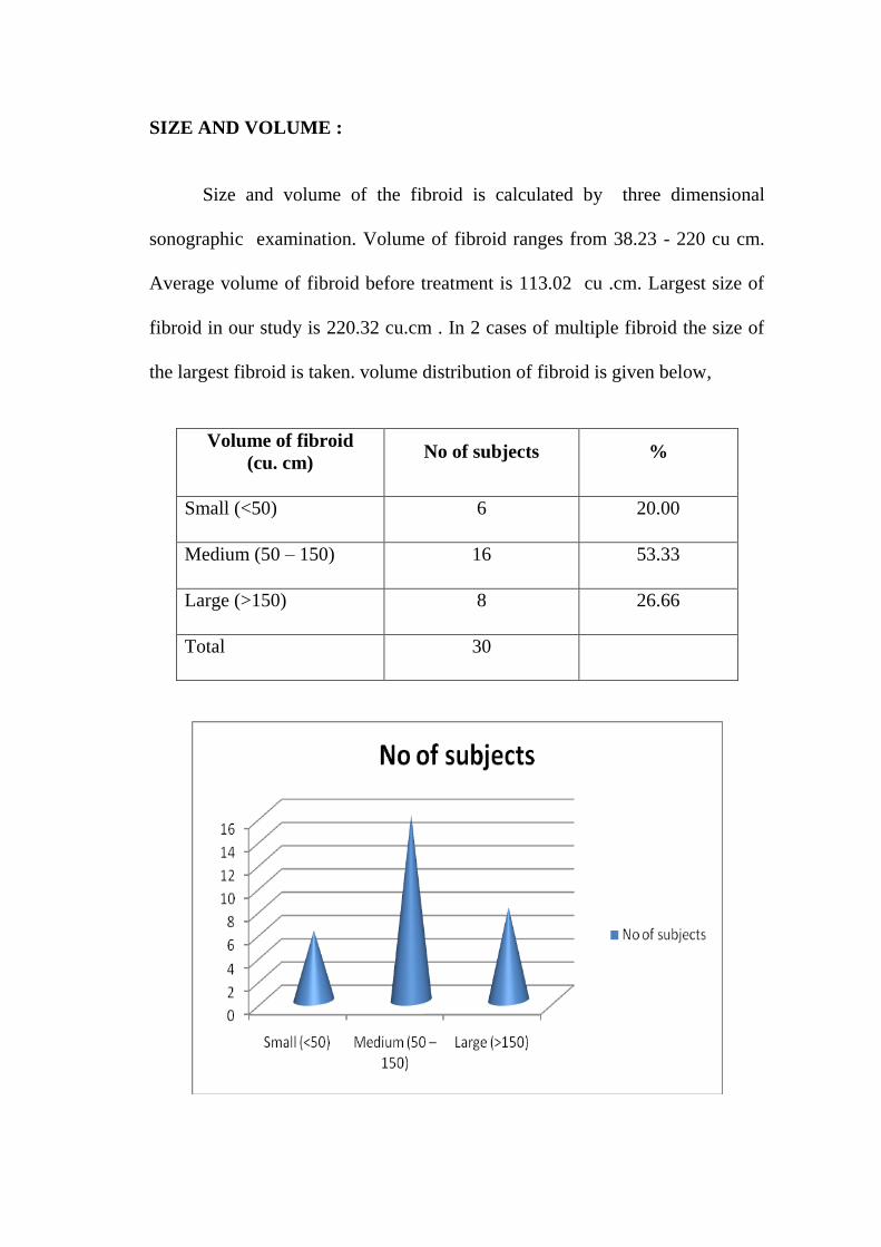
SIZE AND VOLUME :
Size and volume of the fibroid is calculated by three dimensional
sonographic examination. Volume of fibroid ranges from 38.23 - 220 cu cm.
Average volume of fibroid before treatment is 113.02 cu .cm. Largest size of
fibroid in our study is 220.32 cu.cm . In 2 cases of multiple fibroid the size of
the largest fibroid is taken. volume distribution of fibroid is given below,
Volume of fibroid
(cu. cm) No of subjects %
Small (<50) 6 20.00
Medium (50 – 150) 16 53.33
Large (>150) 8 26.66
Total 30

CATHETERISATION :
Unilateral or bilateral selective uterine artery catheterization and
embolization were carried out by unilateral transfemoral puncture,, successfully
in all 30 patients.
No of subjects %
Unilateral embolisation 19 70.37
Bilateral embolisation 8 29.62
Un successful
catheterisation
2 6.25
Total 32
COMPLICATIONS:
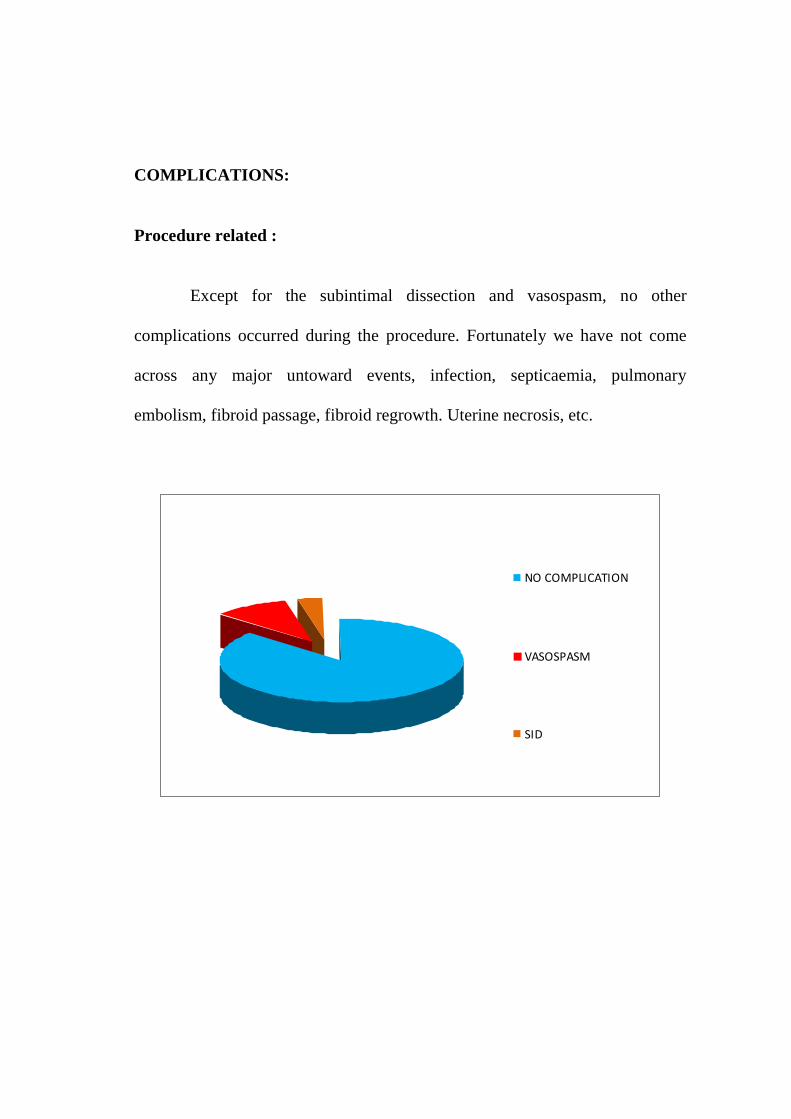
Procedure related :
Except for the subintimal dissection and vasospasm, no other
complications occurred during the procedure. Fortunately we have not come
across any major untoward events, infection, septicaemia, pulmonary
embolism, fibroid passage, fibroid regrowth. Uterine necrosis, etc.
NO COMPLICATION
VASOSPASM
SID
FOLLOW UP
Clinical Follow Up :
All the 30 patients who had good clinical outcome, resumed normal
menstrual cycle at 1–3 months.
One patient presented with fever and vaginal discharge 1 week
after procedure.
Uterine ischemia causing pain found in all 30 patients ,treated
with analgesics.
Post embolization syndrome55
in the form of abdominal
discomfort, pain and fever was noted in 2 patients. These
symptoms were transient and did not require any treatment except
mild analgesics and antipyretics.
ULTRASOUND FOLLOW UP:
VOLUME CHANGES :
Using prolate ellipsoid formula10
, fibroid volume is calculated in our
study.
Volume was measured using the formula :
length x width x depth x 0.5233
When computed only 27 patients came for 12 months follow up , so we
have selected n= 27 for comparison statistics, the mean reduction in fibroid
volume were statistically significant. By using highly correlative friedman’s
test, volume change after 3 months, at 6 months and at 12 months was
significant (P value <0.001) with associated marked reduction in vascularity of
all hypervascular fibroid.
In larger volume (>150 cu.cm) fibroids : volume reduction was 42.24
% (range 32.61 - 50.25 %) at 3 months, at 6 months 76.22% (61.47 - 89.38 %)
and at 12 months was 88.02 % (range 81.12 - 93.63 %).
For medium volume (50 -150 cu.cm) ) fibroids : volume reduction
was 47.90 % (range 24.81- 67.04 %) at 3 months, at 6 months 72.48% (44.36
- 89.12 %) and at 12 months was 83.86 % (range 61.85 - 93.24 %)
For smaller volume (< 50 cu.cm) ) fibroids : volume reduction was
42.24 % (range 20.00- 59.60%) at 3 months, at 6 months 76.22% (35.36 -
86.80 %) and at 12 months was 88.02 % (range 44.96- 91.92 %).
On pre - UAE US, all patient had fibroids that were mixed /hypo echoic
and were hypoechoic on post- UAE US scans.
In this study moderate to marked reduction in larger fibroid volume was
seen in 72.48% 83.86 % at 6 months, and 88% at 12 months respectively while
using PVA with gelfoam combination.
VASCULARITY ASSESSMENT
In our study only hypervascular fibroid were selected and we have
assessed reduced vascularity in them .

OVERALL RESULTS
A. Three dimensional color Doppler sonographic examination results
of volume and vascularity assessment :
Median uterine fibroid size was reduced by 75 – 88%, better
reduction is achieved in
1. Bilateral approach – 88.32%
2. Embolic agents (PVA +Gelfoam) – 88.32%
3. Large volume fibroid - 88.02%
Dominant fibroid size was reduced by 42%
B. Clinical improvement :
83% reduction in menorrhagia
77% reduction in dysmenorrhea
86% reduction in urinary frequency (Pressure symptoms).
DESCRIPTIVES STATISTICS
REDUCTION PERCENTAGE IN VOLUME OF FIBROIDS OVER
TIME ASSESSMENT:
TABLE :1
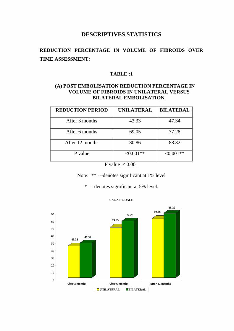
(A) POST EMBOLISATION REDUCTION PERCENTAGE IN
VOLUME OF FIBROIDS IN UNILATERAL VERSUS
BILATERAL EMBOLISATION.
REDUCTION PERIOD UNILATERAL BILATERAL
After 3 months 43.33 47.34
After 6 months 69.05 77.28
After 12 months 80.86 88.32
P value <0.001** <0.001**
P value < 0.001
Note: ** ---denotes significant at 1% level
* --denotes significant at 5% level.
43.3347.34
69.05
77.2880.86
88.32
0
10
20
30
40
50
60
70
80
90
After 3 months After 6 months After 12 months
UAE APPROACH
UNILATERAL BILATERAL

TABLE : 2
(B) POST EMBOLISATION REDUCTION PERCENTAGE IN
VOLUME OF FIBROIDS WITH DIFFERING EMBOLIC
AGENTS.
REDUCTION PERIOD GELFOAM PVA + GELFOAM
After 3 months 37.61 50.04
After 6 months 65.30 76.44
After 12 months 77.76 87.31
P value <0.001** <0.001**
P value < 0.001
Note: ** ---denotes significant at 1% level
* --denotes significant at 5% level.
MATERIALS USED
37.61
65.3
77.76
50.04
76.44
87.31
0
10
20
30
40
50
60
70
80
90
100
After 3 months After 6 months After 12 months
GELFOAM PVA + GELFOAM
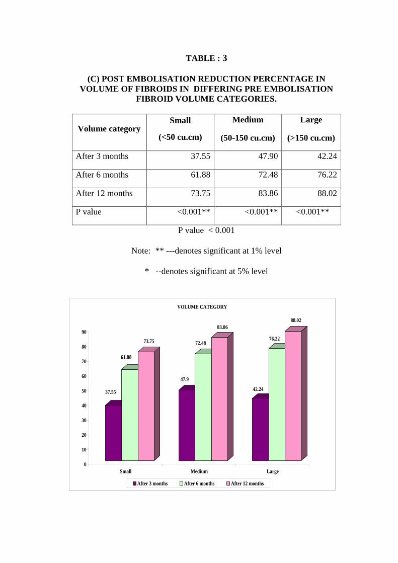
TABLE : 3
(C) POST EMBOLISATION REDUCTION PERCENTAGE IN
VOLUME OF FIBROIDS IN DIFFERING PRE EMBOLISATION
FIBROID VOLUME CATEGORIES.
Volume category Small
(<50 cu.cm)
Medium
(50-150 cu.cm)
Large
(>150 cu.cm)
After 3 months 37.55 47.90 42.24
After 6 months 61.88 72.48 76.22
After 12 months 73.75 83.86 88.02
P value <0.001** <0.001** <0.001**
P value < 0.001
Note: ** ---denotes significant at 1% level
* --denotes significant at 5% level
37.55
61.88
73.75
47.9
72.48
83.86
42.24
76.22
88.02
0
10
20
30
40
50
60
70
80
90
Small Medium Large
VOLUME CATEGORY
After 3 months After 6 months After 12 months
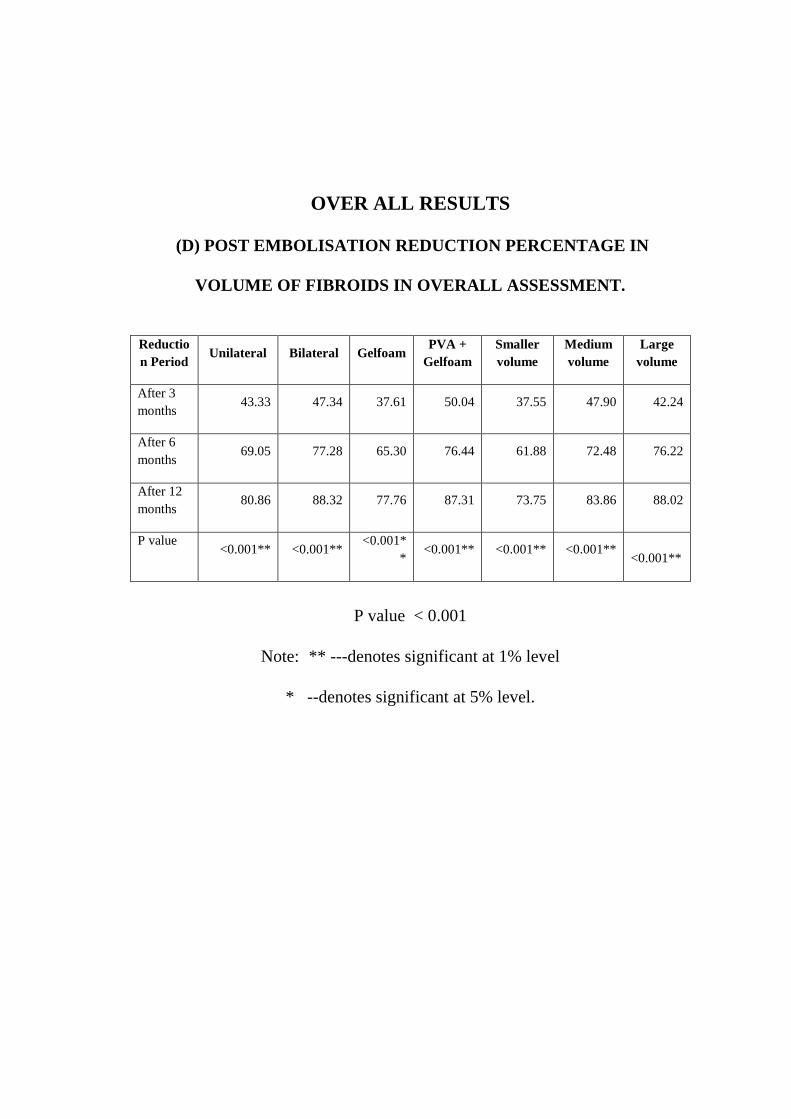
OVER ALL RESULTS
(D) POST EMBOLISATION REDUCTION PERCENTAGE IN
VOLUME OF FIBROIDS IN OVERALL ASSESSMENT.
Reductio
n Period Unilateral Bilateral Gelfoam
PVA +
Gelfoam
Smaller
volume
Medium
volume
Large
volume
After 3
months 43.33 47.34 37.61 50.04 37.55 47.90 42.24
After 6
months 69.05 77.28 65.30 76.44 61.88 72.48 76.22
After 12
months 80.86 88.32 77.76 87.31 73.75 83.86 88.02
P value <0.001** <0.001**
<0.001*
* <0.001** <0.001** <0.001**
<0.001**
P value < 0.001
Note: ** ---denotes significant at 1% level
* --denotes significant at 5% level.
DISCUSSION
Fibroid uterus causing menorrhagia is one of the common morbidity
encountered in the women of child bearing age group. Hence many young
women opt to undergo hysterectomy at an early age. Uterine artery
embolisation is minimally invasive alternative primary treatment of fibroids
with preservation of uterus.
This study was done to assess the efficacy of uterine artery embolisation
with three dimensional color doppler sonography. Pre embolization and
postembolization three-dimensional
color Doppler sonography was
prospectively compared in 30 patients who underwent uterine
artery
embolization as primary treatment of symptomatic fibroids.
3D CDS has recently become available to quantify vascularity that gives
good correlation to vessels greater than 100 micrometer. Three-dimensional
color Doppler sonography was performed by using a scanner with color power
angiographic imaging capability.
For purposes of comparison,
fibroids were classified as either
hypervascular or hypovascular relative to myometrial vascularity before and
minutes to several hours after uterine artery embolization. Changes in fibroid
vascularity (i.e., from hypervascular to hypovascular) as depicted by three-
dimensional color Doppler sonography are stated in Edwin F 9
Med 2005.

Totally 32 samples were selected for the study, according to the
inclusion and exclusion criteria. Of which for 2 patients embolisation could
not be done because of subintimal dissection and vasospasm happened during
the procedure. Uterine artery embolisation was performed by using a standard
selective
catheter and standard embolization technique. Successful
embolisation was done for 30 patients. Hence we have followed 30 patients
upto 12 months to assess the changes in vascularity .
In this study we have done both unilateral and bilateral UAE, where
bilateral embolisation proved statistically significant (P value < 0.001**). In
contrast, Edwin F, Arthur C. Fleischer, et al9, in their 2005
study, did
unilateral UAE for all patients. Also TP Jain, DN Srivastava etal55
2007,
demonstrated that Ipsilateral catheterization of uterine artery was safe, cost-
effective and with lesser incidence of complications such as haematoma and
dissection of arteries.
Uterine artery embolization is an effective and established treatment
method to control intractable bleeding, C. Fleischer, et al 2005
9 due to various
other obstetric and gynaecological disorders. Uterine fibroid embolisation acts
mainly by occluding the blood supply to the tumour thus it arrests the growth
and cause ischemia and necrosis. This causes shrinkage of the tumour and stops
appearance of new fibroids as per long term follow up studies 55
.
Duration of the procedure varied from 45 minutes to 75 minutes. The
average duration of the procedure was 65 minutes. This is comparable with
results of previous researches.
Radiation dose : 9-20 cgy in 1–2 hours procedure.
The mean total procedure time as reported by other investigators has
been 78.4 min for bilateral approach and 44.29 and 61min for unilateral
approach. In this study intermittent fluoroscopy was used to reduce the
exposure. Fluoroscopic exposure time was 45 min, which is more compared
with Boris nikolic MD, james b, et al 65
2001.
In this study, average volume of fibroid before embolisation is 113.02
cu.cm. (ranges 38.23 - 220 cu. cm). Largest fibroid volume in the study
population was 220.32 cu.cm. In our series we have not encountered any
additional complications in larger fibroids. This correlates with the recent study
by Albert J. Smeets Robbert,et al3, 2009, which depicts that the complications
were not increased and the clinical response is also good.
Small size fibroid (less than 7 cm) show better response in size
reduction and symptomatically feel better. Less than 1 percent of patients
undergo hysterectomy due to embolisation failure, Jean-Pierre Pelage et al 23
2005.
The greatest decrease in vascularity occurred 1 day after the procedure,
whereas the greatest volume change was found at 3 month follow-up
examination agreeing with the results depicted by Arthur C. Fleischer, F.
Donnelly., et al9 in 2000. This statement correlates well in our study, selected
hypervascular fibroids showed marked reduction in vascularity.
In another study, hyper vascular fibroids (12 of 30) tended to decrease
in size after treatment more than isovascular (10 of 30) or hypovascular (8 of
30). In our study most fibroids were hypervascular before UAE and became
hypovascular after UAE as depicted by Arthur C. Fleischer, MD, Edwin F
Med 20059.
Jean-Pierre Pelage et al 23
2005 states that the main reason for not
embolizing the pedunculated fibroids is the potential risk of postprocedural
torsion and is also stated in Laurent Brunereau Denis Herbreteau et al30
,
2002, who reported sudden and massive necrobiosis changes in the embolised
fibroids .
Bruce McLucas, Rita Perrella, et al,9
states that largest myoma diameter
of greater than 8 cm, PSV values of greater than 64cm/s during screening was
found associated with UFE failure. In contrast, our study could not find any
significant potential in accurately predicting the UAE failure .
Ultrasound is faster, widely available and cheaper, Three dimensional
color doppler are more accurate and reproducible .It is better in selecting
patients for UFE and also best response assessment, Arthur C. Fleischer,
MD, Edwin F Med, 2000, 2005
9.
Previous study shows MR imaging is sub optimal in assessment of
fibroid volume, but reliably detect adenomyosis as given by Edwin F, Arthur
C. Fleischer, et al 9
2005. In our study we have not done MR imaging
correlation for all patients as it is not accurately depicting quantification of
vascularity.
Uterus always has the capacity to develop new leiomyomas after
endovascular embolization with 150- to 250 micrometer particles Sangeet
Ghai, et al,2
2005. Several Studies shows that PVA injection followed by
gelfoam shows better results than with gelfoam used alone. Gelfoam Vs Poly
Vinyl Alcohol is an ideal embolic agent in UAE .
Among 30 patients in the study 14 of them were embolised with gel
foam and remaining 16 was embolised with poly vinyl alcohol particles of 350-
500 µm size followed by gelfoam. On analysing the volume reduction, both
was equally effective with comparative higher statistical significant (p value
<0.001) results with PVA followed by gelfoam. This result is comparable with
Katz et al26
1998 who studied the effectiveness of gelatin sponge pledgets
versus polyvinyl alcohol for embolization. They concluded that materials are
equally effective. This result is in contrary to the statement by Jean-Pierre
Pelage et al 23
2005, for targeted embolisation of the perifibroid arterial plexus,
injection of PVA particles with diameter larger than 500 micrometer is
recommended.
Studies by Derdelyn and Pelage et al64
, 2001 have shown that Tris-acryl
microspheres are more uniform in size and the particle sizes do not changed in
liquids. They have little tendency to clump after injection and animal studies
indicate that they have less tissue reaction than is typically seen with PVA .
As in C. Joseph Muniz, MD, Arthur C. et al6 , 2002 our study analysis
showed that almost all of the patients were clinically improved at 3 months, 6
months, and 1 year, and all were symptom-free at 1 years. Sonography was an
ideal method to show and follow the progressive reduction in the size of the
main leiomyoma procedure as proved by Laurent Brunereau Denis
Herbreteau et al ,30
in 2000.
Remarkable average Volume Reduction at the 3rd
month especially in
larger volume (>150 cu.cm) of fibroid was 42.24 % (range 32.61 - 50.25 %)
at 3 months, at 6 months 76.22% (61.47 - 89.38 %) and at 12 months was
88.02 % (range 81.12 - 93.63 %) (p<0.0001 highly significant). It is
comparable with that seen in the study by Spies JB, Scialli AR, et al 53
in 1999
who has reported 50% reduction at 6th
month and 78% reduction at 1st year.
All patients should be monitored with clinical and sonographic
examinations for more than 2 years after the endovascular procedure,
Embolization cannot therefore be considered a radical treatment compared with
hysterectomy, depicted by Laurent Brunereau Denis Herbreteau et al,30
in
2000.
Jean-Pierre Pelage et al23
2005; Vaginal discharge and fibroid expulsion
are more common with intramural fibroid.
Overall, the information regarding fibroid vascularity ideally obtained
on 3D CDS may be useful in determining which patients are best suited for
UAE therapy9. 3D CDS is less expensive and more extensively available and
can depict actual vessels within and surrounding fibroids . Good results of
UAE were seen in a larger volume hypervascular fibroids approached
bilaterally with PVA followed by gelfoam embolic agents.
LIMITATIONS
1. For one patient catheterisation could not be done due to vasospasm on
both sides and another patient had subintimal dissection during
catheterisation, so procedure abandoned.
2. Since all 30 patients, responded to fibroid embolization, it is not
possible from data acquired in this small series to come to any
conclusions regarding the use of 3D CDS for predicting which patients
will be responders or nonresponders .
3. Few patients have lost their long term follow up at 12 months or later
of post embolisation.
CONCLUSION
* From this study we have found that Uterine Artery Embolisation for the
patients having symptomatic uterine fibroid is an effective and safe
alternate treatment with significant reduction in volume and vascularity
of fibroid particularly in less than 7 cm fibroids.
* Three dimensional color Doppler sonography assists in assessing the
vascularity within the fibroid before and after embolisation.
* Single trans femoral approach with bilateral uterine arteries
embolisation technique was used successfully in most of our patients.
Combined PVA - Gelfoam is one of the ideal embolic agents effective in
causing volume and vascularity reduction along with relief of the
symptoms in patients with fibroid. Hypervascular fibroid respond well
to embolisation . It has less failure rates in long term follow up. This
procedure has good patient’s tolerance, short recovery time, quick and
sustained symptomatic improvement. This procedure may reduce the
need for invasive surgery in many patients.
* From this study we conclude that Three Dimensional Color Doppler
Imaging can be a tool for pre and post UAE evaluation in assessing
reduction in fibroid vascularity and volume. It is less expensive, more
extensively available and provides an estimate of completeness of
embolisation.
BIBLIOGRAPHY
1. Agdi, M. and Tulandi, T. “Endoscopic management of uterine
fibroids.” Best Practice & Research Clinical Obstetrics & Gynecology,
online publication 4 Mar 2008.
2. Akinola, OI; Fabamwo, AO; Ottun, AT; Akinniyi, OA (2005). "Uterine
artery ligation for management of uterine fibroids". International journal
of gynaecology and obstetrics: the official organ of the International
Federation of Gynaecology and Obstetrics 91 (2): 137–40.
3. Albert J. Smeets, Robbert J. Nijenhuis, et al Uterine Artery
Embolization in Patients with a Large Fibroid Burden: Long-Term
Clinical and MR Follow-up. The Netherlands 15-12-2009.
4. Bruce McLucas, Rita Perrella, et al, Role of Uterine Artery Doppler
Flow in Fibroid Embolization © 2002 by the American Institute of
Ultrasound in Medicine J Ultrasound Med 21:113–120, 2002.
5. Brunereau L, Herbreteau D, Gallas S et al. Uterine artery embolization
in the primary treatment of uterine leiomyomas. AJR 2000; 175: 1267–
72.
6. C. Joseph Muniz, MD, Arthur C. et al. Three-dimensional Color
Doppler Sonography and Uterine Artery Arteriography of Fibroids
Assessment of Changes in Vascularity Before and After Embolization
(C) 2002 by the American Institute of Ultrasound in Medicine J
Ultrasound Med 21:129–133, 2002
7. Carbonell Esteve JL, Acosta R, Heredia B, Pérez Y, Castañeda MC,
Hernández AV: Mifepristone for the treatment of uterine leiomyomas.
Obstet Gynecol. 2008, 112:1029–36.PubMed
8. Day Baird D,Dunson DB ,Hill MC,et al. Incidence of lieomyoma:
ultrasound evidence. Am J Obstet gynecol 2003:188:100-107
9. Donnelly, MD, et al. Three-Dimensional Color Doppler Sonography
before and after fibroid embolization by the American Institute of
Ultrasound in Medicine J Ultrasound Med 19:701–705, 2000, 2005 .
10. E.Aitken, A.Khaund, S.A.Hamid,et al, The normal human myometrium
has a vascular spatial gradient absent in small fibroids, (C), Human
Reproduction Vol.21, No10 pp. 2669–2678, 2006
11. Giovanna Tropeano, Sonia Amoroso and Giovanni Scambia.
Nonsurgical management of uterine fibroids. Department of Obstetrics
and Gynaecology, University Catholic del Sacro Cuore, Italy.
12. Goldfarb HA. Myoma coagulation (myolysis). Obstet Gynecol Clin
North Am. 2000;27:421–30.
13. Goodwin SC, McLucas B et al. Preliminary experience with Uterine
artery embolisation for the treatment of uterine fibroids – J Vasc. Interv
Radiol, 1997; 8 : 517 – 26.
14. Goodwin SC, McLucas B, Lee M et al., Uterine artery embolisation for
the treatment of urterine leiomyoma – midterm results. J Vasc. Interv.
Radiol, 1999; 10 : 1159 - 65
15. Hallberg L, Nilsson L. Determination of menstrual blood loss. Scand J
Clin Lab Invest 1964; 16: 244–248.
16. Hayden Homer, PhD MRCOG. Pregnancy outcomes after uterine artery
embolisation for fibroids. Department of Obstetrics and Gynaecology,
Institute for Women’s Health, London.
17. Hehenkamp WJ, Volkers NA, Birnie E, Reekers JA, Ankum WM:
Symptomatic uterine fibroids: Embolisation versus Hysterectomy
(EMMY) Trial. Radiology. 2008, 246:823–32.PubMed.
18. Higham, J. M., O'brien, P. M. S. and Shaw, R. W. (1990), Assessment
of Menstrual Blood Loss Using a Pictorial Chart. Bog: An International
Journal of Obstetrics & Gynaecology, 97: 734–739.
19. Hirst A, Dutton S, Wu O: A multi-centre retrospective cohort study
comparing the efficacy, safety and cost-effectiveness of hysterectomy
and uterine artery embolisation for the treatment of symptomatic uterine
fibroids. The HOPEFUL study. Health Technol Assess. 2008, 12:1–248.
20. Ho SS, Cowan NC. Uterine artery embolisation for uterine fibroids
using a 4F Rosch inferior mesenteric catheter. Eur Radiol 2005; 15:
1168–72.
21. Howkins and Bourne Shaws textbook of gynecology. 14th
edition:p-320
22. Jean-Pierre Pelage et, al A review of Current embolic agents Friday,
Aug 2009
23. Jean-Pierre Pelage, Julien Cazejust, et al. Uterine Fibroid
Vascularization and Clinical Relevance to Uterine Fibroid Embolization
RadioGraphics 2005; 25: S99–S117
24. João M. Pisco Ph.D.,Marisa Duarte M.D., et al. Pregnancy after uterine
fibroid embolization : Uterine fibroid embolization shows fertility rates
comparable to myomectomy. Department of Radiology, St. Louis
Hospital, Portugal. March 15, 2010.
25. Kailasam,C; Cahill, D (2008). "Review of the safety, efficacy and
patient acceptability of the levonorgestrel-releasing intrauterine system".
Patient preference and adherence 2: 293–302.
26. Katz R, Mitty H, Stancato-Pasik A, et al: Comparison of uterine artery
embolization for fibroids using gelatin sponge pledgets and polyvinyl
alcohol. J Vasc Interv Radiol 9(suppl 1):184, 1998.
27. Kenneth Murphy, et al. Uterine fibroid embolization: state of the art
from Medscape Radiology
28. Kitamura, Susan M Ascher, et al. Imaging Manifestations of
Complications Associated with Uterine Artery Embolization, Radio
Graphics , 2005: 25 : S119–S132
29. L. Honey, A et al. Department of Obstetrics and Gynaecology, Division
of Reproductive Medicine, Ottawa Hospital, Ottawa, Ontario, Canada
30. Laurent Brunereau Denis Herbreteau et al, Uterine Artery
Embolization in the Primary Treatment of Uterine Leiomyomas:
Technical Features and Prospective Follow-Up with Clinical and
Sonographic Examinations in 58 Patients AJR 2000 ;175 : 1267–1272
31. Liu, WM; Ng, HT; Wu, YC; Yen, YK; Yuan, CC (2001). "Laparoscopic
bipolar coagulation of uterine vessels: a new method for treating
symptomatic fibroids". Fertility and sterility 75 (2): 417–22.
32. Mahmood K. Razavi, Gloria Hwang, et al. Abdominal Myomectomy
versus Uterine Fibroid Embolization in the Treatment of Symptomatic
Uterine Leiomyomas. Department of Vascular and Interventional
Radiology & Department of Gynaecology and Obstetrics, Stanford
University Hospital, Stanford, October 30, 2002.
33. Marshall LM ,Spiegeiman D,Barbieri et al. Variation in the incidence of
uterine leiomyoma among premenopausal woman by age and race .
Obstet gynecol 1997;94:967-973
34. Matchar DB, Myers ER, Barber MW, et al. Management of uterine
fibroids. AHRQ Publication No. 01-E052. Rockville, MD: Agency for
Healthcare Research and Quality. July 2001.
35. Myers ER et al. the fibroid registry: symptom and quality of life status.
obstet Gynecol. 2005:106(6):1309-18
36. Nick Raine-Fenning and Arthur C Fleischer Clarifying the role of three-
dimensional transvaginal sonography in reproductive medicine: an
evidenced-based appraisal Journal of Experimental & Clinical Assisted
Reproduction 2005, 2:10
37. Pelage JP, Soyer P, Dref OL et al. Uterine arteries: bilateral
catheterization with a single femoral approach and a single 5F catheter –
Technical Note. Radiology 1999; 210: 573–75.
38. Pron G, Bennett J, Common A, Wall J, Asch M, Sniderman K, for the
Ontario Uterine Fibroid Embolization Collaborative Group. Uterine
fibroids reduction and symptom relief after uterine artery embolization
for fibroids. Fertile Sterile 2003; 79:120–127.
39. PronG, Bennett J, Common A, Wall J, Asch M, Sniderman K. The
Ontario Uterine Fibroid Embolization Trial. II. Uterine fibroid reduction
and symptom relief after uterine artery embolization for fibroids. Fertil
Steril 2003; 79: 120–127.
40. Rashid. S, Khaund. A, Murray. L. S, et al The Effects of Uterine Artery
Embolisation and Surgical Treatment on Ovarian Function in Women
with Uterine Fibroids.
41. Ravina JH, Bouret JH, Cirary-Vigneron N, et al: Application of
particulate arterial embolization in the treatment of uterine
fibromyomata. Bull Fr Nat Acad Med 181(2):1-10, 1997.
42. Ravina JH, Herbretean D, Ciraru-Vigneror N et al. Arterial embolisation
to treat uterine myomata. Lancet 1995; 346: 671–72.
43. Ringold S. FDA approves ultrasound fibroid therapy. JAMA
2004;292:2826
44. Robert I. White, MD, "Interventional Radiology: Reflections and
Expectations", Radiology, 1987; 162: 593-600.

45. Roth AR, Spies JB, Walsh SM et al (2000) Pain after uterine artery
embolization for leiomyomata: Can its severity be predicted and does
severity predict outcome? J Vasc Interv Radiol.11(8):1047–1052
46. Sangeet Ghai, MD, Dheeraj K. Rajan, et al. uterine artery embolization
for leiomyomas: Pre and post procedural evaluation with us . Radio
Graphics 2005; 25:1159–1176
47. Sankaran, S.; Manyonda, I. (2008). "Medical management of
fibroids". Best Practice & Research Clinical Obstetrics & Gynaecology.
48. Scott Goodwin, M.D. Long-Term Efficacy of Uterine Fibroid
Embolization Confirmed. Department of Radiological Sciences in the
School of Medicine at the University of California, Irvine.
49. Soriano, D. et al. "Pregnancy outcome after laparoscopic and
laparoconverted myomectomy." European Journal of Obstetrics &
Gynecology and Reproductive Biology 108 (2003): 194–198.
50. Spies J, et al a multicenter control trail comparing the Outcome of
Uterine Embolization and Hysterectomy for Leiomyomas. Study
published in American Journal of Obstetrics & Gynaecology – 2004.
51. Spies JB, Spector A, Roth AR, Baker CM, Mauro L, Murphy-Skrynarz
K. Complications after uterine artery embolization for leiomyomas.
Obstet.Gynecol 2002;100: 873–880.
52. Spies JB, Scialli AR, Jha RC, Imaoka I, Ascher SM, Fraga VM, Barth
KH. Initial results from uterine fibroid embolization for symptomatic
leiomyoma. Department of Radiology, Georgetown University Medical
Centre, Washington, DC 2007-2197, USA J Vasc Interv Radiol. 1999
Oct;10(9):1145-7.
53. Stencato-Pasik A, Mitty HA, Richard HM, et al: Obstetric
embolotherapy: Effects on menses and pregnancy. Radiology
204(3):791-793, 1996.
54. Suhny Abbara, Boris Nikolic et al. Frequency and extent of Uterine
Perfusion via Ovarian Arteries observed during Uterine Artery
Embolization for Leiomyomas, AJR 2007; 188:1558–1563
55. TP Jain, DN Srivastava, RP Sahu ,et al. Uterine artery embolization
for symptomatic fibroids with imaging follow up, (2007).
56. TP Jain, DN Srivastava, et al from Departments of Radiology and
Obstetrics and Gynaecology, All India Institute of Medical Sciences,
New Delhi, India. Uterine artery embolizations for symptomatic fibroids
with imaging follow up. Australasian Radiology (2007) 51, 246–252
57. Tranquart F, Brunereau L, Cottier JP, et al Prospective sonographic
assessment of uterine artery embolization for the treatment of fibroids.
Ultrasound Obstet Gynecol. 2002 Jan;19(1):81-7.
58. Van der Kooij SM, Hehenkamp WJ, et al. Department of Radiology,
Amsterdam .Am J Obstet Gynecol. 2010 Aug; 203(2):e1-6.
59. Vashisht A, Studd JW, Corey AH et al. Fibroid embolisation: a
technique not without significant complications. Br J Obstet
Gynaecol.2000; 107: 1166–70.
60. Walker WJ, Pelage JP. Uterine artery embolisation for symptomatic
fibroids: clinical results in 400 women with imaging follow up. Br J
Obstet Gynaecol 2002; 109:1262–72.
61. Wewers M.E. & Lowe N.K. (1990) A critical review of visual analogue
scales in the measurement of clinical phenomena. Research in Nursing
and Health 13, 227±236.
62. Wilkens J, Chwalisz K, Han C, et al. Effects of the selective
progesterone receptor modulator asoprisnil on uterine artery blood flow,
ovarian activity, and clinical symptoms in patients with uterine
leiomyoma scheduled for hysterectomy. J Clin Endocrinol Metab. 2008,
93:4664–71.
63. Yuri Kitamura, MD, From the Department of Radiology, Georgetown
University Hospital, Washington. Presented as a education exhibit at the
2004 RSNA Scientific Assembly. May 2005.
64. Particles Choice of for uterine artery embolization for leiomyomata ,
Derdelyn and Pelage et al , September of 2001.
65. Boris nikolic MD, james b, et al @scvir 2001 , uterine artery
embolisation : reduced radiation with refined technique.

PROFORMA
Name:
Indication for UFE: opted / anaemia / previous surgeries / obesity / others
Age (yrs): 25-30 31-35 35-40
History:
Menorrhagia - Assessed by Pictoral Blood Loss Assessment Chart
Dysmenorrhoea – Assessed by Visual Analogue Scale
Pressure Symptoms: Yes/no
Increased frequency and urgency: Yes/no
Lower abdominal heaviness: Yes/no
Constipation: Yes/no
Marital status: married/unmarried
Obstetric history: No of children Nil / 1 / 2 / >2
History of Allergy: Yes/no
History of previous surgeries: Yes / No
Clinical Examination:
General Examination:
Height : Weight: BMI:
Anaemia:
Peripheral edema:
Miscellaneous:
CVS:
RS:
Abdomen:
Gynaecological Examination:
Speculum examination:
P/V:
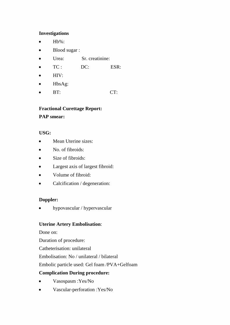
Investigations
Hb%:
Blood sugar :
Urea: Sr. creatinine:
TC : DC: ESR:
HIV:
HbsAg:
BT: CT:
Fractional Curettage Report:
PAP smear:
USG:
Mean Uterine sizes:
No. of fibroids:
Size of fibroids:
Largest axis of largest fibroid:
Volume of fibroid:
Calcification / degeneration:
Doppler:
hypovascular / hypervascular
Uterine Artery Embolisation:
Done on:
Duration of procedure:
Catheterisation: unilateral
Embolisation: No / unilateral / bilateral
Embolic particle used: Gel foam /PVA+Gelfoam
Complication During procedure:
Vasospasm :Yes/No
Vascular-perforation :Yes/No
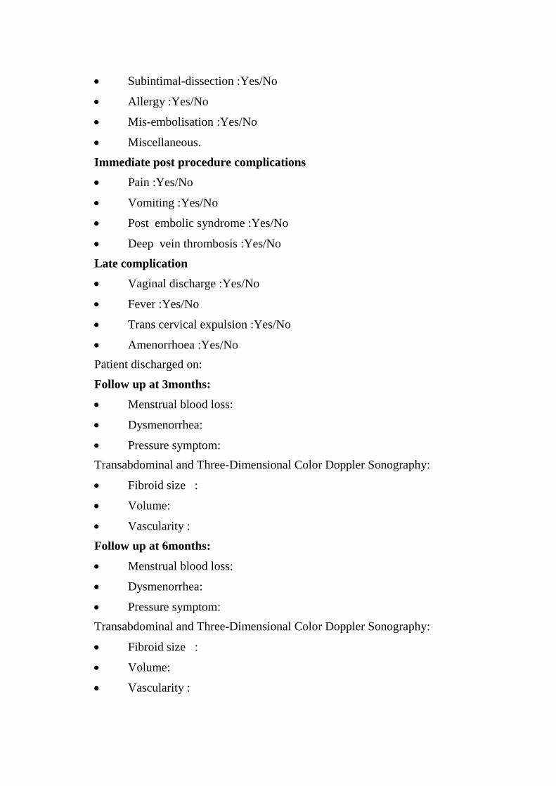
Subintimal-dissection :Yes/No
Allergy :Yes/No
Mis-embolisation :Yes/No
Miscellaneous.
Immediate post procedure complications
Pain :Yes/No
Vomiting :Yes/No
Post embolic syndrome :Yes/No
Deep vein thrombosis :Yes/No
Late complication
Vaginal discharge :Yes/No
Fever :Yes/No
Trans cervical expulsion :Yes/No
Amenorrhoea :Yes/No
Patient discharged on:
Follow up at 3months:
Menstrual blood loss:
Dysmenorrhea:
Pressure symptom:
Transabdominal and Three-Dimensional Color Doppler Sonography:
Fibroid size :
Volume:
Vascularity :
Follow up at 6months:
Menstrual blood loss:
Dysmenorrhea:
Pressure symptom:
Transabdominal and Three-Dimensional Color Doppler Sonography:
Fibroid size :
Volume:
Vascularity :

Follow up at 12 months:
Menstrual blood loss:
Dysmenorrhea:
Pressure symptom:
Transabdominal and Three-Dimensional Color Doppler Sonography:
Fibroid size :
Volume:
Vascularity :
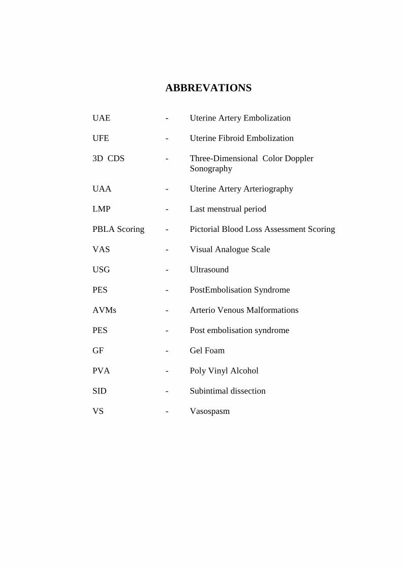
ABBREVATIONS
UAE - Uterine Artery Embolization
UFE - Uterine Fibroid Embolization
3D CDS - Three-Dimensional Color Doppler
Sonography
UAA - Uterine Artery Arteriography
LMP - Last menstrual period
PBLA Scoring - Pictorial Blood Loss Assessment Scoring
VAS - Visual Analogue Scale
USG - Ultrasound
PES - PostEmbolisation Syndrome
AVMs - Arterio Venous Malformations
PES - Post embolisation syndrome
GF - Gel Foam
PVA - Poly Vinyl Alcohol
SID - Subintimal dissection
VS - Vasospasm
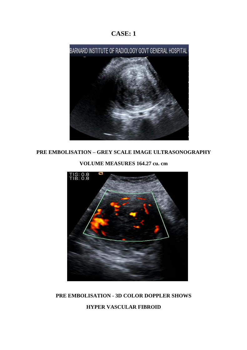
CASE: 1
PRE EMBOLISATION – GREY SCALE IMAGE ULTRASONOGRAPHY
VOLUME MEASURES 164.27 cu. cm
PRE EMBOLISATION - 3D COLOR DOPPLER SHOWS
HYPER VASCULAR FIBROID
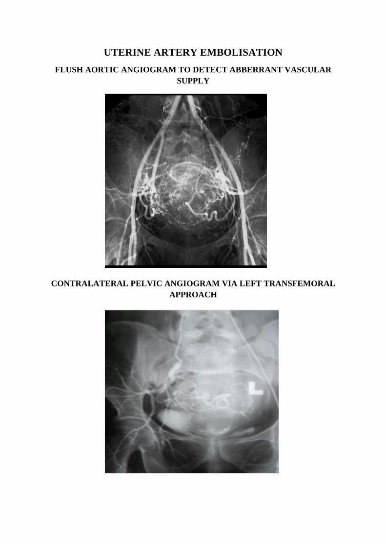
UTERINE ARTERY EMBOLISATION
FLUSH AORTIC ANGIOGRAM TO DETECT ABBERRANT VASCULAR
SUPPLY
CONTRALATERAL PELVIC ANGIOGRAM VIA LEFT TRANSFEMORAL
APPROACH

IPSILATERAL PELVIC SUPER SELECTIVE ANGIOGRAM
ANGIOGRAM OF UTERINE ARTERIES
ARTERIAL PHASE OF CONTRAST VENOUS PHASE OF CONTRAST
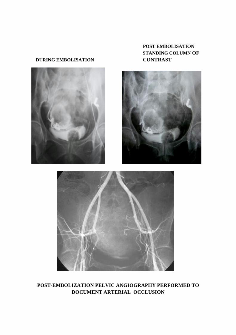
POST EMBOLISATION
STANDING COLUMN OF
DURING EMBOLISATION CONTRAST
POST-EMBOLIZATION PELVIC ANGIOGRAPHY PERFORMED TO
DOCUMENT ARTERIAL OCCLUSION
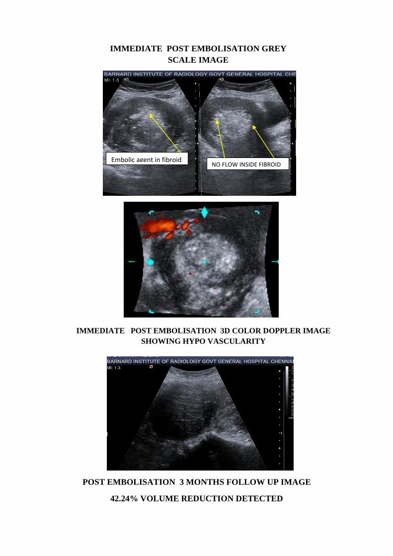
POST EMBOLISATION 3 MONTHS FOLLOW UP IMAGE
42.24% VOLUME REDUCTION DETECTED
Embolic agent in fibroid
NO FLOW INSIDE FIBROID
IMMEDIATE POST EMBOLISATION GREY
SCALE IMAGE
IMMEDIATE POST EMBOLISATION 3D COLOR DOPPLER IMAGE
SHOWING HYPO VASCULARITY
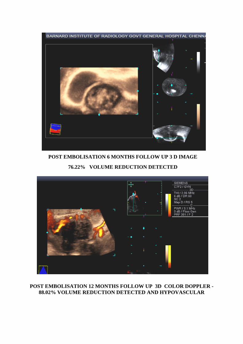
POST EMBOLISATION 6 MONTHS FOLLOW UP 3 D IMAGE
76.22% VOLUME REDUCTION DETECTED
POST EMBOLISATION 12 MONTHS FOLLOW UP 3D COLOR DOPPLER -
88.02% VOLUME REDUCTION DETECTED AND HYPOVASCULAR
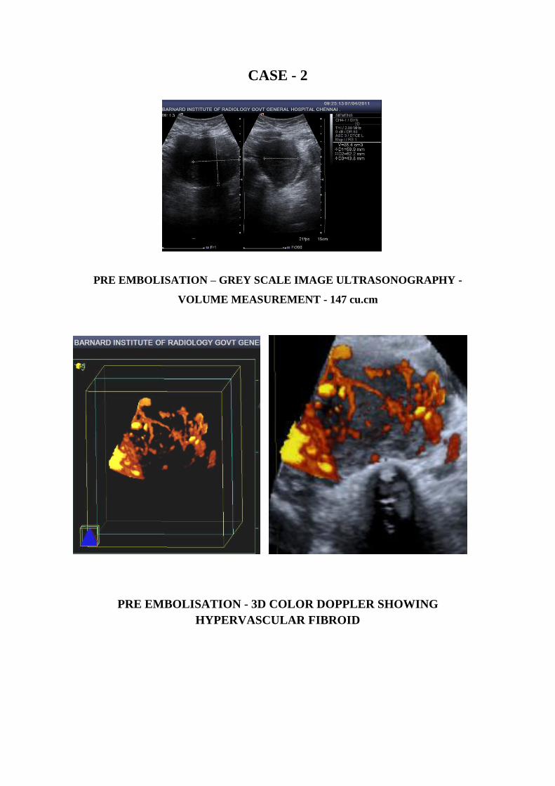
CASE - 2
PRE EMBOLISATION – GREY SCALE IMAGE ULTRASONOGRAPHY -
VOLUME MEASUREMENT - 147 cu.cm
PRE EMBOLISATION - 3D COLOR DOPPLER SHOWING
HYPERVASCULAR FIBROID
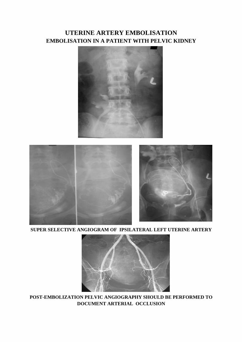
UTERINE ARTERY EMBOLISATION
EMBOLISATION IN A PATIENT WITH PELVIC KIDNEY
SUPER SELECTIVE ANGIOGRAM OF IPSILATERAL LEFT UTERINE ARTERY
POST-EMBOLIZATION PELVIC ANGIOGRAPHY SHOULD BE PERFORMED TO
DOCUMENT ARTERIAL OCCLUSION
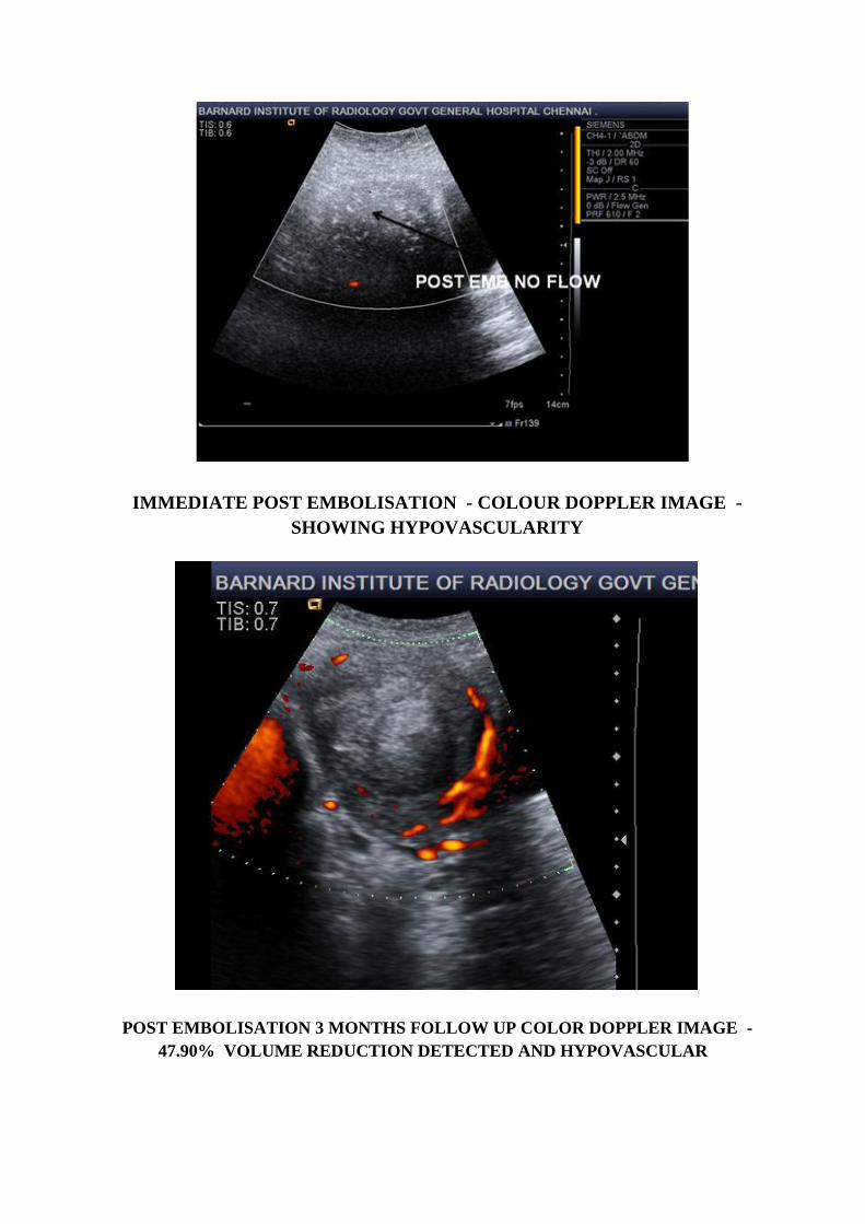
IMMEDIATE POST EMBOLISATION - COLOUR DOPPLER IMAGE -
SHOWING HYPOVASCULARITY
POST EMBOLISATION 3 MONTHS FOLLOW UP COLOR DOPPLER IMAGE -
47.90% VOLUME REDUCTION DETECTED AND HYPOVASCULAR

POST EMBOLISATION 6 MONTHS FOLLOW UP IMAGE –
72.48% VOLUME REDUCTION DETECTED AND HYPOVASCULAR
POST EMBOLISATION 12 MONTHS FOLLOW UP IMAGE -
83.86 % VOLUME REDUCTION DETECTED AND HYPOVASCULAR
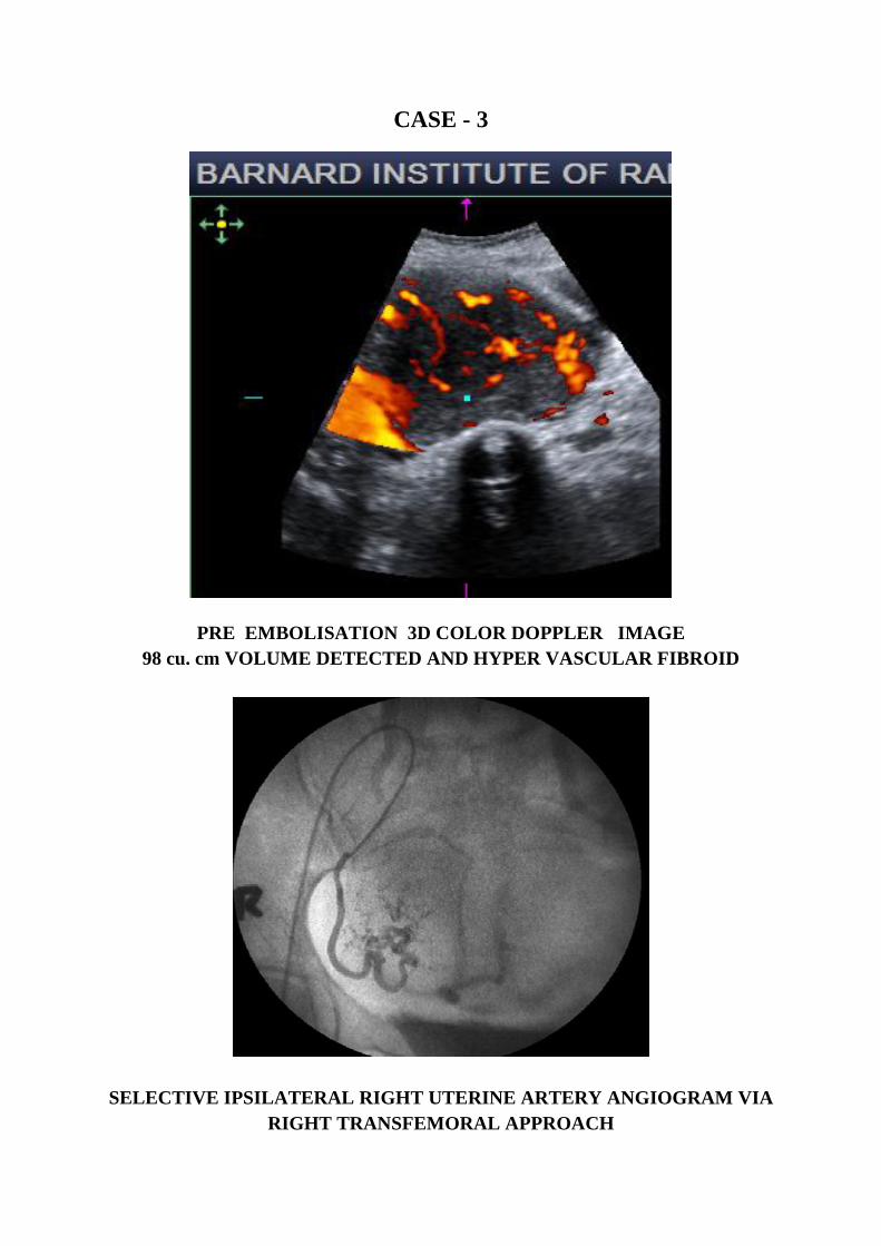
CASE - 3
PRE EMBOLISATION 3D COLOR DOPPLER IMAGE
98 cu. cm VOLUME DETECTED AND HYPER VASCULAR FIBROID
SELECTIVE IPSILATERAL RIGHT UTERINE ARTERY ANGIOGRAM VIA
RIGHT TRANSFEMORAL APPROACH
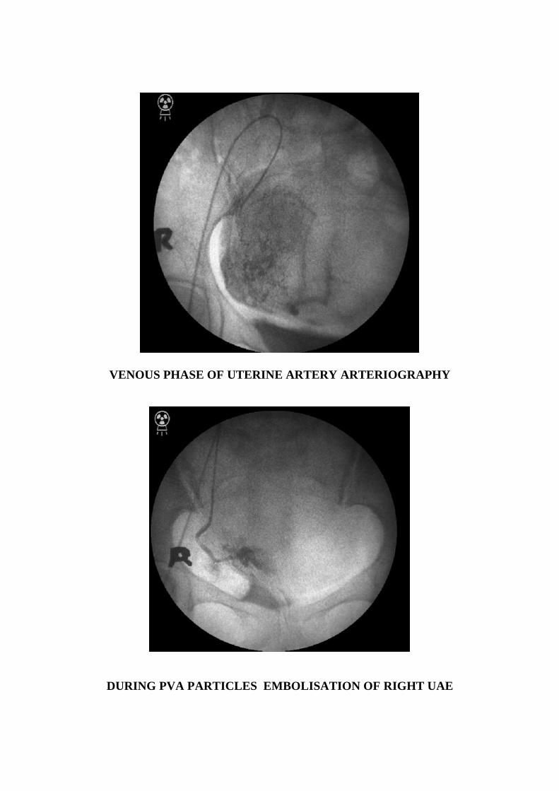
VENOUS PHASE OF UTERINE ARTERY ARTERIOGRAPHY
DURING PVA PARTICLES EMBOLISATION OF RIGHT UAE

DURING GEL FOAM INJECTION OF RIGHT MAIN UTERINE ARTERY
POST EMBOLISATION STANDING COLUMN OF CONTRAST
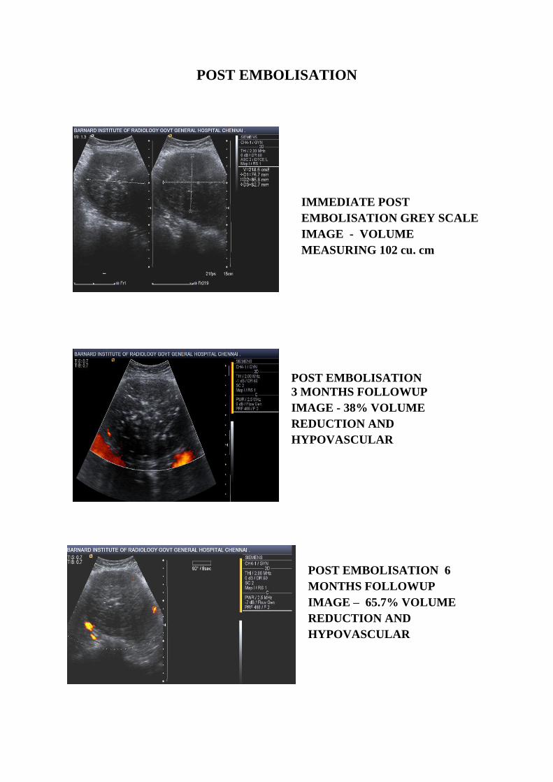
POST EMBOLISATION
IMMEDIATE POST
EMBOLISATION GREY SCALE
IMAGE - VOLUME
MEASURING 102 cu. cm
POST EMBOLISATION
3 MONTHS FOLLOWUP
IMAGE - 38% VOLUME
REDUCTION AND
HYPOVASCULAR
POST EMBOLISATION 6
MONTHS FOLLOWUP
IMAGE – 65.7% VOLUME
REDUCTION AND
HYPOVASCULAR

POST EMBOLISATION 12 MONTHS 3D CDS SONOGRAPHIC IMAGE –
80 %VOLUME REDUCTION AND HYPOVASCULAR
POST EMBOLISATION 12 MONTHS 3D COLOR DOPPLER IMAGE
SHOWING MARKED REDUCTION IN VASCULARITY
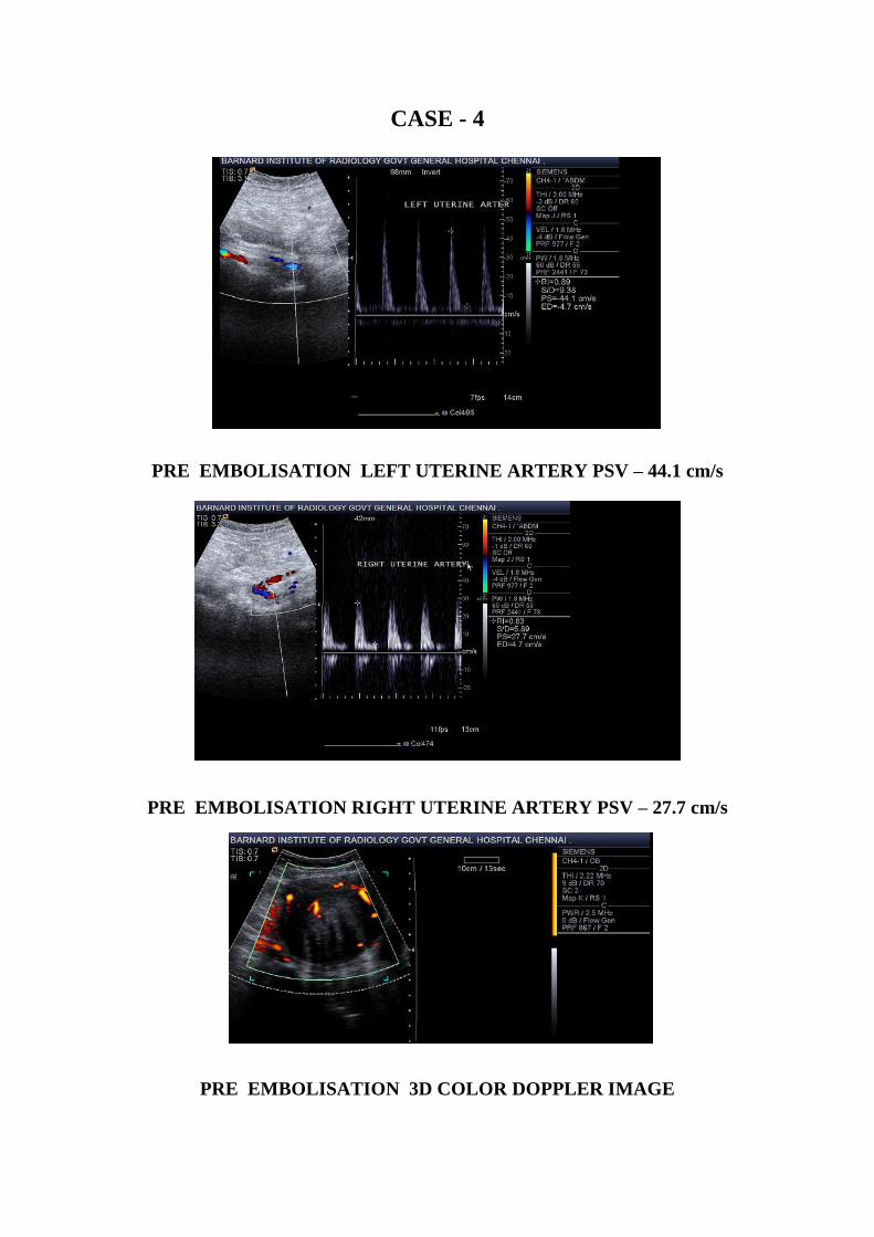
CASE - 4
PRE EMBOLISATION LEFT UTERINE ARTERY PSV – 44.1 cm/s
PRE EMBOLISATION RIGHT UTERINE ARTERY PSV – 27.7 cm/s
PRE EMBOLISATION 3D COLOR DOPPLER IMAGE
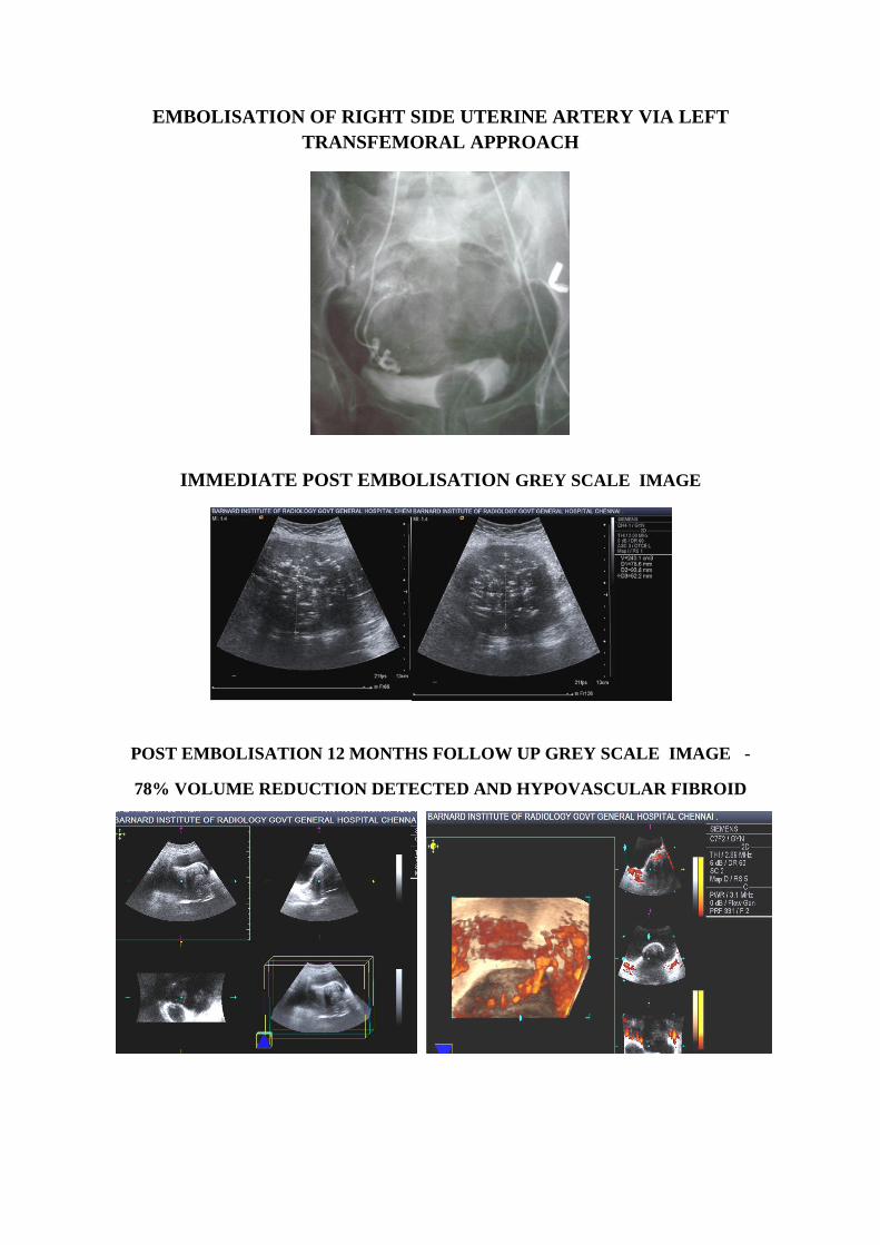
EMBOLISATION OF RIGHT SIDE UTERINE ARTERY VIA LEFT
TRANSFEMORAL APPROACH
IMMEDIATE POST EMBOLISATION GREY SCALE IMAGE
POST EMBOLISATION 12 MONTHS FOLLOW UP GREY SCALE IMAGE -
78% VOLUME REDUCTION DETECTED AND HYPOVASCULAR FIBROID
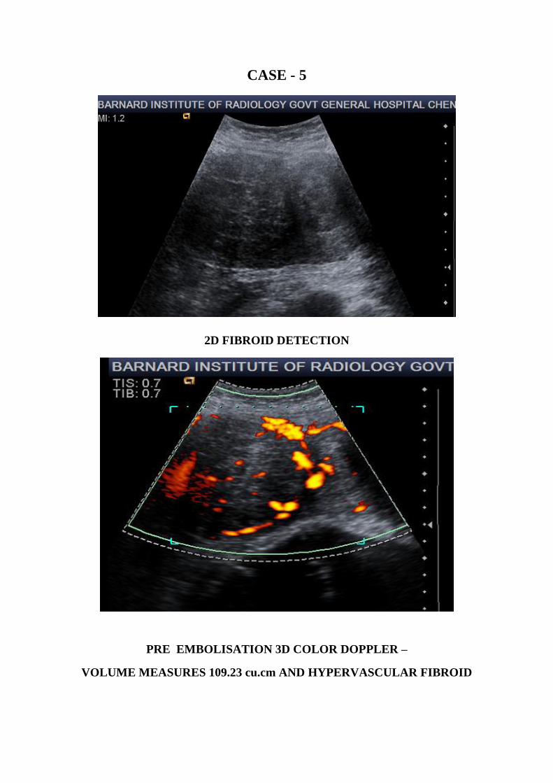
CASE - 5
2D FIBROID DETECTION
PRE EMBOLISATION 3D COLOR DOPPLER –
VOLUME MEASURES 109.23 cu.cm AND HYPERVASCULAR FIBROID
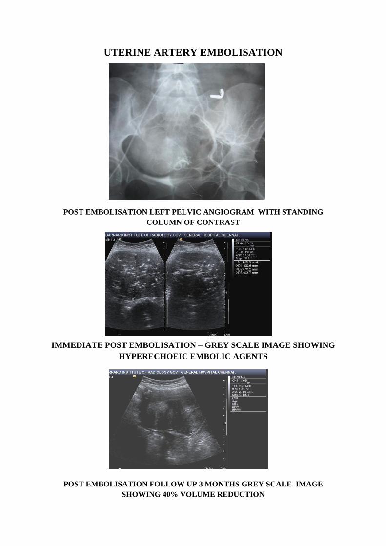
UTERINE ARTERY EMBOLISATION
POST EMBOLISATION LEFT PELVIC ANGIOGRAM WITH STANDING
COLUMN OF CONTRAST
IMMEDIATE POST EMBOLISATION – GREY SCALE IMAGE SHOWING
HYPERECHOEIC EMBOLIC AGENTS
POST EMBOLISATION FOLLOW UP 3 MONTHS GREY SCALE IMAGE
SHOWING 40% VOLUME REDUCTION

POST EMBOLISATION 6 MONTHS 3D COLOR DOPPLER FOLLOWUP
63% VOLUME REDUCTION AND HYPO VASCULAR FIBROID
POST EMBOLISATION 12 MONTHS FOLLOW UP
81% VOLUME REDUCTION AND HYPO VASCULAR FIBROID
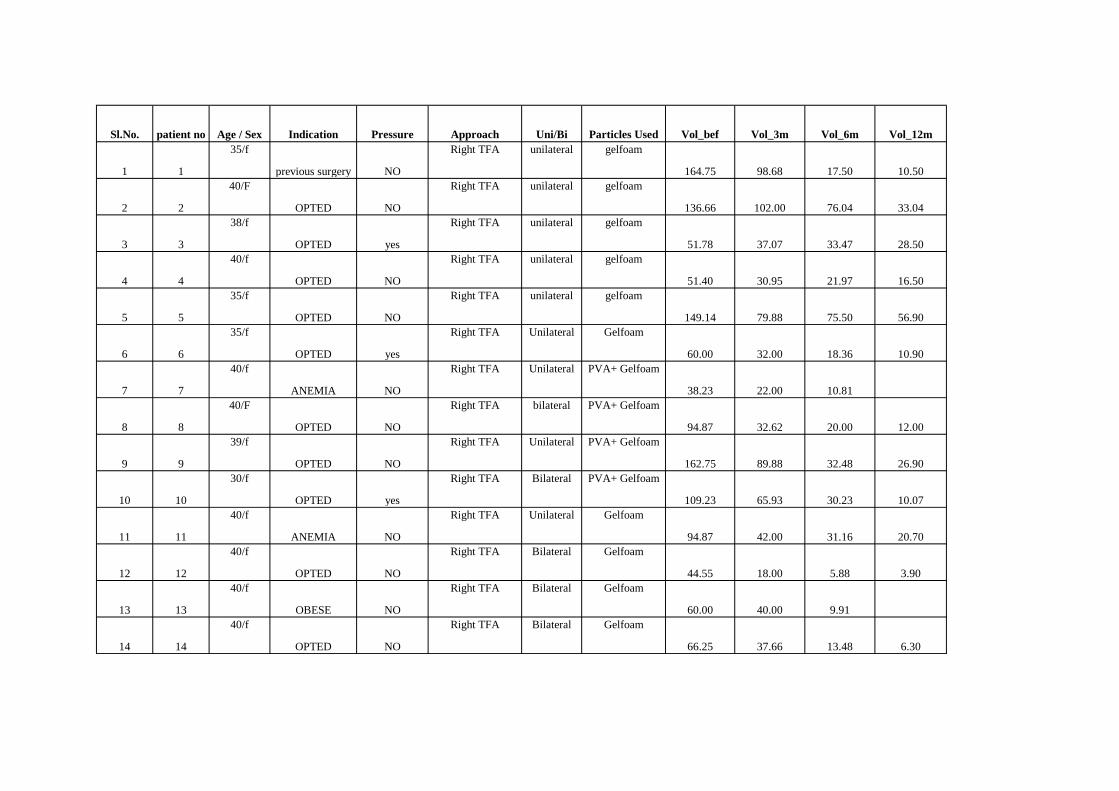
Sl.No. patient no Age / Sex Indication Pressure Approach Uni/Bi Particles Used Vol_bef Vol_3m Vol_6m Vol_12m
1 1
35/f
previous surgery NO
Right TFA unilateral gelfoam
164.75 98.68 17.50 10.50
2 2
40/F
OPTED NO
Right TFA unilateral gelfoam
136.66 102.00 76.04 33.04
3 3
38/f
OPTED yes
Right TFA unilateral gelfoam
51.78 37.07 33.47 28.50
4 4
40/f
OPTED NO
Right TFA unilateral gelfoam
51.40 30.95 21.97 16.50
5 5
35/f
OPTED NO
Right TFA unilateral gelfoam
149.14 79.88 75.50 56.90
6 6
35/f
OPTED yes
Right TFA Unilateral Gelfoam
60.00 32.00 18.36 10.90
7 7
40/f
ANEMIA NO
Right TFA Unilateral PVA+ Gelfoam
38.23 22.00 10.81
8 8
40/F
OPTED NO
Right TFA bilateral PVA+ Gelfoam
94.87 32.62 20.00 12.00
9 9
39/f
OPTED NO
Right TFA Unilateral PVA+ Gelfoam
162.75 89.88 32.48 26.90
10 10
30/f
OPTED yes
Right TFA Bilateral PVA+ Gelfoam
109.23 65.93 30.23 10.07
11 11
40/f
ANEMIA NO
Right TFA Unilateral Gelfoam
94.87 42.00 31.16 20.70
12 12
40/f
OPTED NO
Right TFA Bilateral Gelfoam
44.55 18.00 5.88 3.90
13 13
40/f
OBESE NO
Right TFA Bilateral Gelfoam
60.00 40.00 9.91
14 14
40/f
OPTED NO
Right TFA Bilateral Gelfoam
66.25 37.66 13.48 6.30
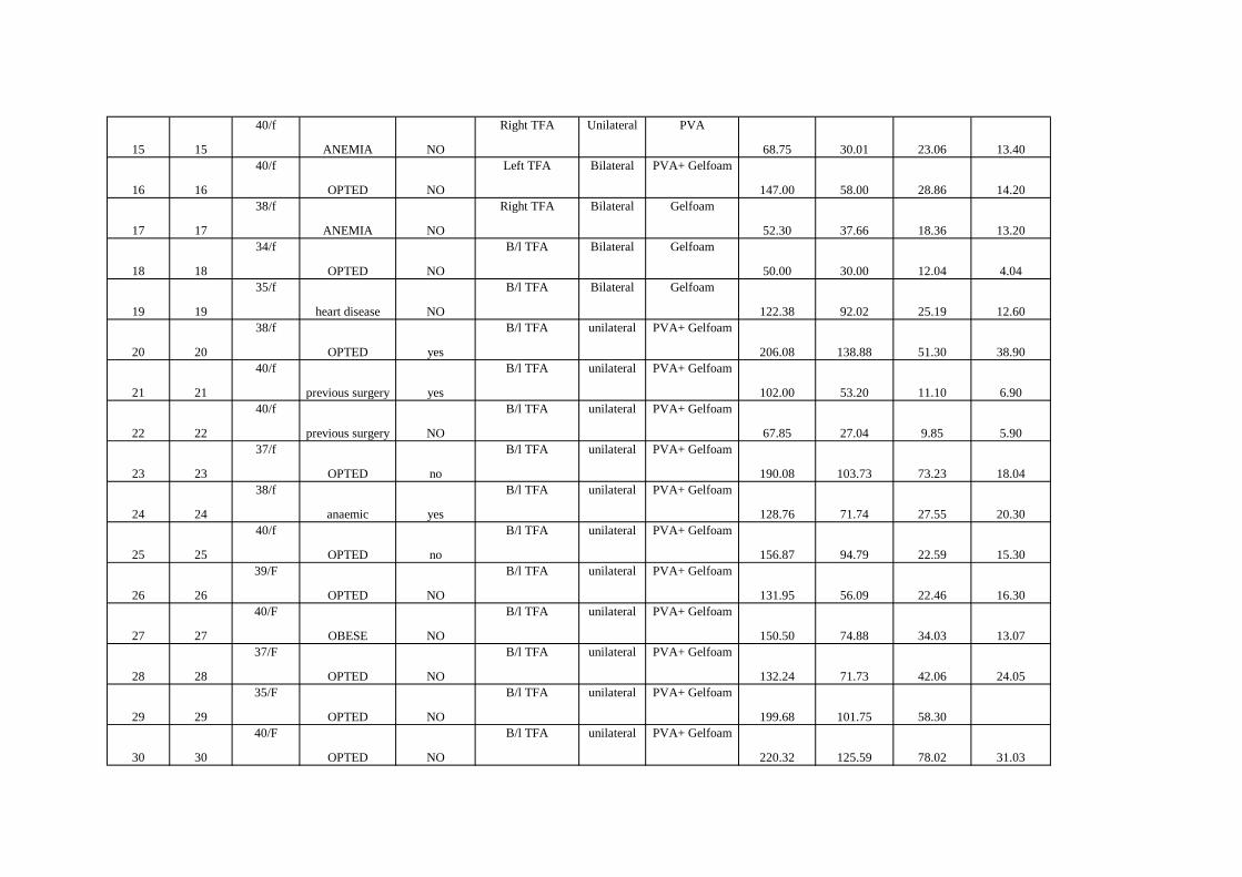
15 15
40/f
ANEMIA NO
Right TFA Unilateral PVA
68.75 30.01 23.06 13.40
16 16
40/f
OPTED NO
Left TFA Bilateral PVA+ Gelfoam
147.00 58.00 28.86 14.20
17 17
38/f
ANEMIA NO
Right TFA Bilateral Gelfoam
52.30 37.66 18.36 13.20
18 18
34/f
OPTED NO
B/l TFA Bilateral Gelfoam
50.00 30.00 12.04 4.04
19 19
35/f
heart disease NO
B/l TFA Bilateral Gelfoam
122.38 92.02 25.19 12.60
20 20
38/f
OPTED yes
B/l TFA unilateral PVA+ Gelfoam
206.08 138.88 51.30 38.90
21 21
40/f
previous surgery yes
B/l TFA unilateral PVA+ Gelfoam
102.00 53.20 11.10 6.90
22 22
40/f
previous surgery NO
B/l TFA unilateral PVA+ Gelfoam
67.85 27.04 9.85 5.90
23 23
37/f
OPTED no
B/l TFA unilateral PVA+ Gelfoam
190.08 103.73 73.23 18.04
24 24
38/f
anaemic yes
B/l TFA unilateral PVA+ Gelfoam
128.76 71.74 27.55 20.30
25 25
40/f
OPTED no
B/l TFA unilateral PVA+ Gelfoam
156.87 94.79 22.59 15.30
26 26
39/F
OPTED NO
B/l TFA unilateral PVA+ Gelfoam
131.95 56.09 22.46 16.30
27 27
40/F
OBESE NO
B/l TFA unilateral PVA+ Gelfoam
150.50 74.88 34.03 13.07
28 28
37/F
OPTED NO
B/l TFA unilateral PVA+ Gelfoam
132.24 71.73 42.06 24.05
29 29
35/F
OPTED NO
B/l TFA unilateral PVA+ Gelfoam
199.68 101.75 58.30
30 30
40/F
OPTED NO
B/l TFA unilateral PVA+ Gelfoam
220.32 125.59 78.02 31.03