Emergency Radiology Cases
386
EMERGENCY RADIOLOGY CASES
-
Upload
khangminh22 -
Category
Documents
-
view
1 -
download
0
Transcript of Emergency Radiology Cases

EMERGENCY RADIOLOGY CASES


Emergency Radiology Cases

Published and Forthcoming books in the Cases in Radiology series:
Body MRI Cases , William Brant and Eduard de Lange Breast Imaging Cases , Catherine Appleton and Kimberly Wiele Cardiac Imaging Cases , Charles White and Joseph Jen-Sho Chen Chest Imaging Cases , Sanjeev Bhalla, Cylen Javidan-Nejad, Kristopher W. Cummings, and Andrew Bierhals Emergency Radiology Cases , Hani Abujudeh Gastrointestinal Imaging Cases , Angela Levy, Koenraad Mortele, and Benjamin Yeh Genitourinary Imaging Cases, Mark Lockhart and Rupan Sanyal Musculoskeletal Imaging Cases , Mark Anderson and Stacy Smith Neuroradiology Cases , Cliff ord Eskey, Cliff ord Belden, David Pastel, Arastoo Vossough, and Albert Yoo Nuclear Medicine Cases , Chun Kim Pediatric Imaging Cases , Ellen Chung Ultrasound Cases, Leslie Scoutt, Ulrike Hamper, and Teresita Angtuaco

1
Hani H. Abujudeh, MD, MBA, FSIR Associate Professor of Radiology
Harvard Medical School
Massachusetts General Hospital
Boston, Massachusetts
Emergency Radiology Cases

1 Oxford University Press is a department of the University of Oxford. It furthers the University’s objective of excellence in research, scholarship, and education by publishing worldwide.
Oxford New York Auckland Cape Town Dar es Salaam Hong Kong Karachi Kuala Lumpur Madrid Melbourne Mexico City Nairobi New Delhi Shanghai Taipei Toronto
With offi ces in Argentina Austria Brazil Chile Czech Republic France Greece Guatemala Hungary Italy Japan Poland Portugal Singapore South Korea Switzerland Th ailand Turkey Ukraine Vietnam
Oxford is a registered trademark of Oxford University Press in the UK and certain other countries.
Published in the United States of America by Oxford University Press 198 Madison Avenue, New York, NY 10016
© Oxford University Press 2014
All rights reserved. No part of this publication may be reproduced, stored in a retrieval system, or transmitted, in any form or by any means, without the prior permission in writing of Oxford University Press, or as expressly permitted by law, by license, or under terms agreed with the appropriate reproduction rights organization. Inquiries concerning reproduction outside the scope of the above should be sent to the Rights Department, Oxford University Press, at the address above.
You must not circulate this work in any other form and you must impose this same condition on any acquirer.
Library of Congress Cataloging-in-Publication DataAbujudeh, Hani H., author.Emergency radiology cases / Hani Abujudeh. p. ; cm.—(Cases in radiology)Includes bibliographical references and index.ISBN 978–0–19–994117–9 (alk. paper)I. Title. II. Series: Cases in radiology. [DNLM: 1. Diagnostic Imaging—Case Reports. 2. Emergency Medicine—Case Reports. 3. Radiology—Case Reports. WN 180]RC78.7.D53616.07′54—dc23 2013022349
Th is material is not intended to be, and should not be considered, a substitute for medical or other professional advice. Treatment for the conditions described in this material is highly dependent on the individual circumstances. And, while this material is designed to off er accurate information with respect to the subject matter covered and to be current as of the time it was written, research and knowledge about medical and health issues is constantly evolving and dose schedules for medications are being revised continually, with new side eff ects recognized and accounted for regularly. Readers must therefore always check the product information and clinical procedures with the most up-to-date published product information and data sheets provided by the manufacturers and the most recent codes of conduct and safety regulation. Th e publisher and the authors make no representations or warranties to readers, express or implied, as to the accuracy or completeness of this material. Without limiting the foregoing, the publisher and the authors make no representations or warranties as to the accuracy or effi cacy of the drug dosages mentioned in the material. Th e authors and the publisher do not accept, and expressly disclaim, any responsibility for any liability, loss or risk that may be claimed or incurred as a consequence of the use and/or application of any of the contents of this material.
9 8 7 6 5 4 3 2 1 Printed in the United States of America on acid-free paper

To my mentors, Stephen Baker, James Thrall, Robert Novelline.
To my aunts, Wissal Arnout and Badeea Arnout.
To my wife Shima.
Thank you so much for your support.


vii
Acknowledgments
Th e Publisher thanks the following for their time and advice:
Mark Anderson, University of Virginia Sanjeev Bhalla, Mallinckrodt Institute of Radiology, Washington University Michael Bruno, Penn State Hershey Medical Center Melissa Rosado de Christenson, St. Luke's Hospital of Kansas City Rihan Khan, University of Arizona Angela Levy, Georgetown University Alexander Mamourian, University of Pennsylvania Stacy Smith, Brigham and Women’s Hospital


ix
Preface
Th is book provides a concise, high-yield, imaging overview of the spectrum Emergency and Trauma conditions. Th e cases are presented in an easy-to-read for-mat, including the most recent information. Although the book is not intended to be comprehensive it includes the most important presentations in the acute set-tings. Th e images are of high quality and include the most recent technologies, such as three-dimensional imaging. Th e book is divided into Trauma and Nontrauma Emergencies, and it is further subdivided by body regions. Th ere is an additional section on Pediatric Emergency Cases. We hope this book will serve as a quick reference, and assist you in mastering Emergency Radiology.
Hani H. Abujudeh, MD, MBA, FSIR


xi
Contents
Contributors xiii
Part I. Trauma
Section 1. Brain 3
Section 2. Spine 37
Section 3. Chest 63
Section 4. Abdomen 87
Section 5. Upper Extremity 121
Section 6. Lower Extremity 151
Part II. Nontrauma
Section 1. Brain 189
Section 2. Head, Neck, and Spine 219
Section 3. Chest 245
Section 4. Abdomen 269
Part III. Pediatric
Index of Cases 355
Index 359


xiii
Essmaeel Abdel-Dayem , MD South Shore Radiology Associates Weymouth, Massachusetts First Author: Cases 132, 136, 142, 146
Hani H. Abujudeh , MD, MBA Associate Professor of Radiology Harvard Medical School Massachusetts General Hospital Boston, Massachusetts Book Editor: Emergency Radiology Cases Senior Author: Cases 18, 21, 22, 24, 25 27,
28, 30, 36, 38, 45, 46, 47, 49, 53, 54, 58, 59, 60, 64, 66, 67, 69, 76, 78, 83, 95, 101, 111, 115, 121, 124, 123, 125, 126, 127, 128, 129, 130, 131, 132, 133, 134, 136, 137, 142, 146, 149, 150, 151, 153, 155, 163, 164
Tarik K. Alkasab , MD Instructor in Radiology Department of Radiology Massachusetts General Hospital Boston, Massachusetts Section Editor: Trauma - Abdomen First Author: Cases 17, 20, 147, 148
Shima Aran, MD Research FellowDepartment of RadiologyMassachusetts General Hospital Boston, MassachusettsFirst Author: Case 54
Laura L. Avery , MD Assistant Professor of Radiology Massachusetts General Hospital Boston, Massachusetts Part Editor: Nontrauma First Author: Cases 13, 14, 15, 16
Yolanda Bryce , MD Clinical Fellow in Radiology (EXT) Department of Radiology Mount Auburn Hospital Cambridge, Massachusetts First Author: Cases 25, 47, 76, 125, 127, 129,
130, 133, 137, 153, 163, 164
Judah G. Burns , MD Assistant Professor of Radiology Division of Neuroradiology Albert Einstein College of Medicine Montefi ore Medical Center Bronx, New York First Author: Cases 1, 2, 5
Scott Cameron , MD Department of Diagnostic Imaging Newport Hospital Newport, Rhode Island First author: Case 53, 149
Carson Campe , MD Clinical Fellow in Radiology (EXT) Department of Radiology Massachusetts General Hospital Boston, Massachusetts Section Editor: Trauma - Upper Extremity First Author: Cases 18, 22, 28, 58, 59, 60, 67 Senior Author: Cases 61, 65
Enzo Cento , MD Advanced Radiology Services Grand Rapids, Michigan First Author: Cases 41, 44, 48, 96, 135, 138,
154, 161
Contributors

xiv Contributors
Robert Chen , MD Instructor in Radiology Department of Radiology Massachusetts General Hospital Boston, Massachusetts First Author: Cases 68, 88, 97, 162
Garry Choy , MD Instructor in Radiology Department of Radiology Massachusetts General Hospital Boston, Massachusetts Section Editor: Trauma - Chest First Author: Cases 33, 34, 35, 37, 113, 114,
118, 122, 123
Ryan M. Christie , MD Assistant Professor of Radiology Division of Emergency Radiology Emory University School of Medicine Atlanta, Georgia First Author: Cases 29, 31, 32
Laleh Daft ari Besheli Research FellowDepartment of RadiologyMassachusetts General Hospital Boston MassachusettsFirst Author: Case 150
Dameon Duncan , MD Assistant Professor of Radiology Department of Radiology Albert Einstein College of Medicine Montefi ore Medical Center Bronx, New York First Author: Case 116
R. Joshua Dym , MD Assistant Professor of Radiology Department of Radiology Albert Einstein College of Medicine Montefi ore Medical Center Bronx, New York First Author: Cases 119, 120, 139, 141, 160
Daniel T. Ginat , MD Instructor in Radiology Department of Radiology Massachusetts General Hospital Boston, Massachusetts First Author: 10, 100, 107, 108
Andrew J. Gunn, MD Clinical Fellow in Radiology (EXT) Department of Radiology Mount Auburn Hospital Boston, Massachusetts First Author: Cases 30, 36, 95, 115, 121, 128,
131, 134
Harlan B. Harvey , MD Clinical Fellow in Radiology (EXT) Department of Radiology Mount Auburn Hospital Boston, Massachusetts First Author: Cases 21, 24, 49, 101, 151
Rania Hitto MD Clinical FellowDivision of Neuro-RadiologyMassachusetts General Hospital Boston, MassachusettsFirst Author: Case 112
Luke F. M. Hoagland , MD Clinical Fellow in Radiology (EXT) Department of Radiology Mount Auburn Hospital Boston, Massachusetts First Author: Cases 46, 124, 155
Jamlik O. Johnson , MD Assistant Professor of Radiology Division Director for Emergency Radiology Emory University School of Medicine Atlanta, Georgia Section Editor: Nontrauma - Abdomen First Author: Cases 42, 43, 117, 140
Jason M. Johnson , MD Instructor in Radiology Department of Radiology Massachusetts General Hospital Boston, Massachusetts Section Editor: Nontrauma - Head, Neck,
and Spine First Author: Cases 103, 104, 105, 106, 109, 110
Rathachai Kaewlai , MD Department of Radiology Bumrungrad International Hospital Bangkok, Th ailand Part Editor: Trauma First Author: Cases 92, 94

xvContributors
Christine Kassis , MD Clinical Fellow in Radiology (EXT) Department of Radiology Mount Auburn Hospital Boston, Massachusetts First Author: Case 126
Taj Kattapuram , MD Clinical Fellow in Radiology (EXT) Department of Radiology Mount Auburn Hospital Boston, Massachusetts First Author: Cases 27, 38, 66, 78, 111
Faisal Khosa , MD Assistant Professor of Radiology Division of Emergency Radiology Emory University School of Medicine Atlanta, Georgia Senior Author: Cases 39, 40, 55, 145
Mykol Larvie MDInstructor in Radiology Department of Radiology Massachusetts General Hospital Boston, Massachusetts Section Editor: Trauma - Brain First Author: Cases 3, 4, 7, 8, 9, 11, 87, 90,
99, 102
Peter MacMahon , MD Department of Radiology Mater Misericordiae University Hospital Dublin, Ireland Section Editor: Trauma - Spine First Author: Cases 56, 61, 62, 63, 65, 77, 79,
80, 81, 82, 86
Louis Marone , MD Clinical Fellow in Radiology (EXT) Department of Radiology Massachusetts General Hospital Boston, Massachusetts Section Editor: Nontrauma - Chest First Author: Cases 19, 26
Timothy Meehan , MD Clinical Fellow in Radiology (EXT) Department of Radiology Mount Auburn Hospital Boston, Massachusetts First Author: Cases 45, 64, 69
Parul Penkar , MD Instructor in Radiology Department of Radiology Massachusetts General Hospital Boston, Massachusetts First Author: Cases 70, 71, 72, 73, 74, 75,
84, 85
Otto Rapalino, MD Instructor in Radiology Department of Radiology Massachusetts General Hospital Boston, Massachusetts First Author: Cases 89, 91, 98
Marianne Reed , MD Diagnostic Radiology Yale-New Haven Hospital New Haven, Connecticut Senior Author: Cases 103, 105, 106, 109, 110
Javier M. Romero , MD Assistant Professor of Radiology Department of Radiology Massachusetts General Hospital Boston, Massachusetts First Author: Cases 6, 12, 93
Pamela W. Schaefer , MD Director, MR Imaging Associate Director, Neuroradiology Massachusetts General Hospital Boston, Massachusetts Section Editor: Nontrauma - Brain
Meir H. Scheinfeld, MD, PhD Assistant Professor, Department of Radiology Albert Einstein College of Medicine Director, Division of Emergency Radiology Montefi ore Medical Center Bronx, New York First Author: Case 57
J. Gabriel Schneider , MD Clinical Fellow in Radiology (EXT) Department of Radiology Mount Auburn Hospital Boston, Massachusetts Section Editor: Trauma–Lower Extremity First Author: Case 50
xvi Contributors
Randheer Shailam , MD Instructor in Radiology Department of Radiology Massachusetts General Hospital Boston, Massachusetts Part Editor: Pediatric First Author: Cases 152, 156, 157, 158, 159
Michael Spektor Assistant Professor in Radiology Department of Radiology Albert Einstein College of Medicine Montefi ore Medical Center Bronx, New York First Author: Case 144
Freddie Swain , MD Assistant Professor of Radiology Division of Emergency Radiology Emory University School of Medicine Atlanta, Georgia First Author: Cases 39, 40, 55, 145
Adam Ulano , MD Resident in Radiology Mount Auburn Hospital Cambridge, Massachusetts First Author: Case 83
Jason Weiden , MD Assistant Professor of Radiology Division of Emergency Radiology Emory University School of Medicine Atlanta, Georgia First Author: Cases 23, 51, 52, 143

TraumaPart I


BrainSection 1


5
Case 1
History ▶ None
6
Findings ▶ Imaging checklists
■ Fractures classifi ed into three types: longitudinal, transverse, and mixed/oblique ■ Middle ear ossicles (most common ossicular injuries involve the incus and its articulations) ■ Otic capsule (involvement increases risk of SNHL, facial nerve injury, CSF leak) ■ Carotid canal (involvement should prompt evaluation for ICA dissection or occlusion)
▶ On MRI, T1W hyperintensity can be used to assess for middle ear or labyrinthine hemorrhage
Diff erential Diagnosis ▶ Pseudofracture: Multiple sutures, fi ssures, and aqueducts course through the temporal bone
■ Typically bilateral, symmetric, and corticated margins
Teaching Points ▶ Fracture through temporal bone, oft en with associated facial nerve injury or ossicular involvement ▶ Th ree types of fractures
■ Longitudinal: Parallels long axis of petrous bone; higher risk of ossicular dislocation ■ Transverse: Perpendicular to long axis of petrous bone; higher risk of facial nerve injury ■ Mixed/oblique type
▶ Communication between middle ear and membranous labyrinth caused by oval/round window rupture is called perilymphatic fi stula
Management ▶ Conservative management is usual fi rst-line therapy. Many CSF leaks spontaneously resolve. Carefully
monitor for possible meningitis.
Further Readings Dahiya R , Keller JD , Litofsky NS , Bankey PE , Bonassar LJ , Megerian CA . Temporal bone fractures: otic capsule sparing versus
otic capsule violating clinical and radiographic considerations . J Trauma . 1999 ; 47 ( 6 ): 1079–1083 . Saraiya PV , Aygun N . Temporal bone fractures . Emerg Radiol . 2009; 16 ( 4 ): 255–265 .
Case 1 Temporal Bone Fracture (Longitudinal)
7
Case 2
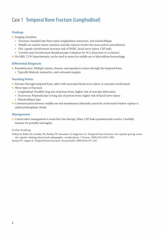
History ▶ Fall at nursing home.

8
Findings CT
▶ Crescentic, hyperdense collection within the extra-axial space that can cross suture lines but limited by dural attachments
▶ Pitfalls on CT ■ Acute SDH may be heterogeneous or low in density ■ Mixed-density subdural can be seen with clot retraction or arachnoid tear ■ Isodense subdural may be present with anemia or subacute hemorrhage
MRI
▶ Variable signal intensity on T1W/T2W imaging; hyperintense on FLAIR ▶ Displaced bridging veins oft en visible with contrast
Diff erential Diagnosis ▶ Epidural hematoma: Lenticular (biconvex) extra-axial hemorrhage, limited by suture lines (may cross dural
attachments); associated skull fracture oft en seen on CT ▶ Hygroma: simple CSF collection in subdural space ▶ Empyema: Peripherally enhancing, infected collection of pus; restricted diff usion on DWI
Teaching Points ▶ Acute collection of blood products between the inner layer of the dura and arachnoid membranes ▶ Acute hemorrhage is usually as a result of severe head trauma, high-velocity acceleration, or deceleration head
injury. Chronic SHD is usually caused by more trivial trauma in patients with risk factors (chronic alcoholism, epilepsy, coagulopathy).
▶ In children, neonatal hematomas may be related to delivery, and usually resolve. In infants and toddlers, nonaccidental trauma must be considered.
▶ Typically overlies convexity, although posterior fossa hemorrhages can occur ▶ SDHs may be symptomatic even when small, especially in young patients ▶ Density characteristics are not an absolute indicator of relative timing of hemorrhage
Management ▶ Careful neurologic monitoring with expectant surgical management
Further Readings Freeman WD , Aguilar MI . Intracranial hemorrhage: diagnosis and management . Neurol Clin . 2012 ; 30 ( 1 ): 211–240 . Barnes PD . Imaging of nonaccidental injury and the mimics: issues and controversies in the era of evidence-based medicine .
Radiol Clin North Am . 2011 ; 49 ( 1 ): 205–229 .
Case 2 Acute Subdural Hematoma

9
History ▶ 46-year-old male who fell down stairs.
Case 3

10
Findings CT
▶ A large right parietal epidural hematoma involving rupture of the right middle meningeal artery causes severe mass eff ect, including left ward midline shift
▶ Heterogeneous density within the hematoma refl ects recent and possibly active extravasation (arrow) ▶ Th ere is a nondisplaced fracture in the right parietal bone (arrowhead) ▶ Th e anteroinferior margin of the right parietal epidural hematoma is bounded by the right temporoparietal suture ▶ Th ere is a smaller left frontotemporal subdural hematoma
CTA (lower right image)
▶ Dural and superfi cial cortical vessels are displaced away from the calvarium by the epidural hematoma
Clinical Presentation ▶ Most commonly associated with major head trauma ▶ Epidural hematomas may develop over time, resulting in a lucid interval during which the patient is less
symptomatic followed by more profound impairment
Pathophysiology ▶ Intracranially, the dura is the periosteum and epidural hemorrhage requires the dissection of the dura away
from its calvarial attachment ▶ Epidural hematomas are most commonly related to arterial rupture and are frequently seen in the setting of
calvarial fractures, with increased frequency related to displaced fractures ▶ Epidural hematomas may also arise from venous disruption ▶ Middle meningeal artery branches in the temporal and parietal regions are vulnerable to injury, and most
epidural hematomas occur in these regions
Teaching Points ▶ Major head trauma, calvarial fracture, and a lucent interval followed by more profound impairment are
features concerning for epidural hematoma ▶ Epidural hematoma does not typically cross sutures unless there is severe fracture at the suture line ▶ Large epidural hematomas are typically lentiform in confi guration, although small epidural hematomas may
conform to local boundaries ▶ Postcontrast images may show active extravasation
Management ▶ Patients with even small epidural hematomas must be carefully monitored, because progressive bleeding may
rapidly become life threatening ▶ Medical therapy should be directed toward maintaining cerebral perfusion pressure, and may include volume
resuscitation, osmotic diuretics, and hyperventilation ▶ Surgical drainage may be achieved with burr holes or craniectomy
Case 3 Epidural Hematoma
11
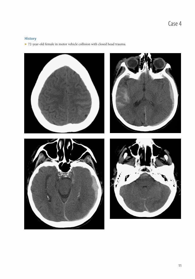
History ▶ 72-year-old female in motor vehicle collision with closed head trauma.
Case 4

12
Findings ▶ Hyperdensity consistent with subarachnoid hemorrhage (SAH) outlining the left precentral gyrus (upper left ) ▶ SAH outlining the right sylvian fi ssure and infi ltrating sulci in the right temporal lobe (upper right) ▶ Trace intraventricular hemorrhage layering in the occipital horn of the right lateral ventricle (lower left ) ▶ SAH in the prepontine cistern (lower right) ▶ Subdural hematoma overlying the left temporal, parietal, and occipital lobes and extending along the falx and
left tentorial leafl et (multiple images)
Clinical Presentation ▶ Common symptoms include headache, nausea and vomiting, and decreased consciousness
Pathophysiology ▶ Th e arachnoid mater overlies the pia mater, which is the deepest layer of the meninges covering the brain and
spinal cord, and SAH expands the space between these coverings ▶ Th e pia mater is extensively innervated with nerve fi bers that transmit pain and are irritated by blood,
resulting in severe headache, such as a thunderclap headache ▶ Intraventricular hemorrhage is a subtype of SAH
Teaching Points ▶ Head CT is the most appropriate fi rst examination to evaluate for SAH ▶ Lumbar puncture is oft en more sensitive than CT for SAH, and may reveal evidence of chronic SAH, such as
xanthochromia ▶ MRI is relatively less sensitive for early SAH, although very sensitive for chronic SAH, which produces a
strong susceptibility signal ▶ Traumatic SAH is strongly associated with other forms of traumatic brain injury, including contusion and
diff use axonal image
Management ▶ Th e diagnosis of traumatic SAH requires exclusion of nontraumatic SAH, which may precipitate subsequent
trauma (e.g., a fall or motor vehicle collision) ▶ When there is any consideration that nontraumatic SAH is present, vascular imaging with CT angiography is
indicated to evaluate for intracranial aneurysm, the leading cause of nontraumatic SAH ■ Complications of SAH that warrant close observation ■ SAH may impair CSF resorption and lead to increased intracranial pressure and hydrocephalus ■ SAH can cause vasospasm, typically within 4–10 days, that may result in territorial ischemia ■ Hunt & Hess classifi cation grades the clinical presentation from 1 (mildest) to 5 (most severe) ■ Fischer grade classifi es the quantity and location of SAH on CT from 1 (none evident) to 4 (diff use or
intraventricular or intraparenchymal extension)
Case 4 Subarachnoid Hemorrhage

13
History ▶ Motor vehicle accident.
Case 5
14
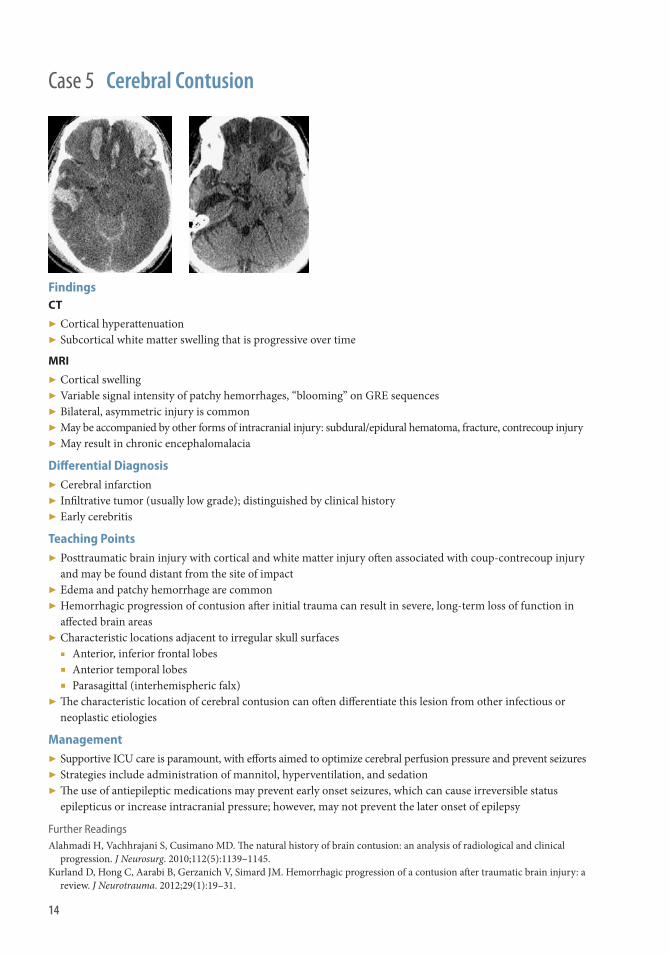
Findings CT
▶ Cortical hyperattenuation ▶ Subcortical white matter swelling that is progressive over time
MRI
▶ Cortical swelling ▶ Variable signal intensity of patchy hemorrhages, “blooming” on GRE sequences ▶ Bilateral, asymmetric injury is common ▶ May be accompanied by other forms of intracranial injury: subdural/epidural hematoma, fracture, contrecoup injury ▶ May result in chronic encephalomalacia
Diff erential Diagnosis ▶ Cerebral infarction ▶ Infi ltrative tumor (usually low grade); distinguished by clinical history ▶ Early cerebritis
Teaching Points ▶ Posttraumatic brain injury with cortical and white matter injury oft en associated with coup-contrecoup injury
and may be found distant from the site of impact ▶ Edema and patchy hemorrhage are common ▶ Hemorrhagic progression of contusion aft er initial trauma can result in severe, long-term loss of function in
aff ected brain areas ▶ Characteristic locations adjacent to irregular skull surfaces
■ Anterior, inferior frontal lobes ■ Anterior temporal lobes ■ Parasagittal (interhemispheric falx)
▶ Th e characteristic location of cerebral contusion can oft en diff erentiate this lesion from other infectious or neoplastic etiologies
Management ▶ Supportive ICU care is paramount, with eff orts aimed to optimize cerebral perfusion pressure and prevent seizures ▶ Strategies include administration of mannitol, hyperventilation, and sedation ▶ Th e use of antiepileptic medications may prevent early onset seizures, which can cause irreversible status
epilepticus or increase intracranial pressure; however, may not prevent the later onset of epilepsy
Further Readings Alahmadi H , Vachhrajani S , Cusimano MD . Th e natural history of brain contusion: an analysis of radiological and clinical
progression . J Neurosurg . 2010 ; 112 ( 5 ): 1139–1145. Kurland D , Hong C , Aarabi B , Gerzanich V , Simard JM . Hemorrhagic progression of a contusion aft er traumatic brain injury: a
review . J Neurotrauma . 2012 ; 29 ( 1 ): 19–31 .
Case 5 Cerebral Contusion

15
History ▶ None
Case 6

16
Findings CT
▶ Multiple hyperattenuated foci measuring 1–15 mm, typically in the cortical-subcortical junction, corpus callosum, and brainstem.
▶ Th ese lesions may present a hypodense halo that likely represents peripheral edema. ▶ Sulci eff acement may be present, with blurring of the gray and white matter interphase representing
brain edema.
MRI
▶ Multiple foci of blooming in GRE and SW images. ▶ Restricted diff usion in the cortical-subcortical junction, corpus callosum and brainstem. ▶ High T2/FLAIR signal in the areas of injury. ▶ Th e splenium is the segment most frequently involved of the corpus callosum. ▶ Brainstem involvement has a very poor clinical prognosis.
Teaching Points ▶ Patients usually lose conscience and likely persist with altered mental status when they suff er DAI. ▶ Th is lesion is the result of traumatic acceleration/deceleration or rotational injuries. ▶ Th e degree of DAI severity is associated with the location of the injury. In ascending order of severity: cortical
subcortical junction, corpus callosum, and brainstem. ▶ Brainstem DAI results in high mortality. ▶ Facial or skull fractures are not always associated with this type of trauma.
Management ▶ Supportive ICU care is paramount, with eff orts aimed to optimize cerebral perfusion pressure and prevent
seizures. ▶ Strategies including administration of mannitol, hyperventilation, and sedation are important for the control
of brain edema. ▶ Th e use of antiepileptic medications may prevent early onset seizures.
Case 6 Diff use Axonal Injury

17
History ▶ 52-year-old male with thrombocytopenia who fell from a bar stool.
Case 7

18
Findings CT
▶ A large left cerebral subdural hematoma causes severe mass eff ect and brain herniation. ▶ Th e medial aspect of the left temporal lobe (the uncus) is displaced rightward across the tentorium, resulting
in left uncal herniation. ▶ Portions of the left cerebral hemisphere, principally the left cingulate gyrus and the corpus callosum, are
displaced to the right beneath the falx cerebri, resulting in subfalcine (or cingulate) herniation. CTA
▶ Left uncal herniation results in compression of the posterior cerebral artery (PCA) and posterior communicating artery (axial image, arrow). Th is may result in PCA territory infarction.
▶ Subfalcine herniation results in compression of the anterior cerebral arteries (ACAs). Normal right ACAs are present, whereas the left the ACAs are highly attenuated (coronal image, arrowhead). Th is may result in ACA territory infarction.
▶ Additional types of brain herniation (not depicted) include ■ Upward or downward transtentorial herniation of the thalami, brainstem, and medial temporal lobes
(central herniation) ■ Cerebellar tonsil herniation through the foramen magnum ■ Transcalvarial herniation, in which a portion of the brain protrudes through a defect in the calvarium that
may be congenital, traumatic, or postsurgical Teaching Points ▶ Acute brain herniation requires emergent treatment ▶ May be caused by
■ Intrinsic processes: intra-axial hemorrhage, edema or tumor ■ Extrinsic processes: extra-axial hemorrhage, tumor, trauma ■ Hydrocephalus or ventricular entrapment ■ Compression of cerebral arteries may cause infarction ■ Subfalcine herniation: ACA territory infarction ■ Uncal herniation: PCA territory infarction ■ Uncal herniation may impinge cranial nerves, particularly the third cranial nerves
Management ▶ Intracranial pressure monitoring is indicated when there are signs, symptoms, or circumstances concerning
for elevated intracranial pressure ▶ Medical: hypertonic saline, mannitol ▶ Surgical: hemicraniectomy Further Readings Andrews BT . Th e recognition and management of cerebral herniation syndromes. In: Loft us CM, ed. Neurosurgical
Emergencies . 2nd ed. New York: Th ieme; 2008: 34–44 . Ropper AH. Hyperosmolar therapy for raised intracranial pressure . N Engl J Med. 2012;367:746–752.
Case 7 Intracranial Herniation

19
History ▶ 51-year-old found down with ethanol intoxication.
Case 8

20
Findings ▶ Subtle anterolisthesis of the C5 and C6 vertebrae ▶ Disruption of the anterior longitudinal ligament, posterior longitudinal ligament, and supraspinous ligament
(Figure 8.1; long, medium, and short arrows, respectively) ▶ Prevertebral soft tissue swelling from C6 through T3 ▶ Extensive T2 hyperintensity consistent with edema in the posterior paraspinal muscles (Figure 8.2a;
arrowheads ) ▶ Extensive edema in the posterior paraspinal soft tissues extending from the occiput superiorly through T2
inferiorly (Figure 8.2) ▶ T2 hyperintensity consistent with edema in the spinal cord at C6 through C7 refl ecting spinal cord injury
(see Case 9) ▶ Signal hyperintensity between spinous processes from C4 through T1 indicates injury to the interspinous
ligamentous
Clinical Presentation ▶ Spine ligamentous injury may occur with relatively mild trauma, such as fall from standing height and low-
speed motor vehicle collisions ▶ Point tenderness may relate to spine ligamentous injury, although this is not a sensitive or specifi c fi nding
for such
Spectrum of Imaging Findings ▶ Alignment abnormality
■ Anterior, posterior, and lateral spondylolisthesis ■ Widening of spinous processes
▶ Intervertebral disk disruption ▶ Frank disruption of ligaments ▶ Edema in paraspinal soft tissues ▶ Epidural hematoma, particularly in relation to disruption of the posterior longitudinal ligament ▶ Craniocervical junction injuries
■ Apical ligament ■ Alar ligaments ■ Cruciate ligaments ■ Tectorial membrane ■ Anterior and posterior atlantooccipital membranes ■ Posterior atlantoaxial membrane
Teaching Points ▶ Spine ligamentous injury is more apparent when imaged early, such as within 72 hours of injury, before edema
begins to resolve ▶ In the cervical (C3-C7), thoracic and lumbar spine, two of three columns intact (anterior, posterior, and
middle) is generally regarded as mechanically stable
Management ▶ Immobilization of the entire spinal column is essential until spine is cleared ▶ Immobilization with braces is the mainstay of therapy for spine ligamentous injury without accompanying
bone or spinal cord injury ▶ Nonsteroidal anti-infl ammatory drugs are useful for pain control ▶ Surgery reserved to restore mechanical instability
Case 8 Spine Ligamentous Injury

21
History ▶ 36-year-old male who fell two stories.
Case 9

22
Findings CT
▶ Comminuted fractures of the T11 and T12 vertebral bodies resulting in retropulsion of bone fragments into the spinal canal and loss of vertebral body height
MRI
▶ Vertebral body fractures with bone marrow edema ▶ Abnormal expansion and edema in the inferior spinal cord consistent with acute contusion, prominently
involving the conus medullaris (Figure 9.1a; arrow) ▶ Edema in the central spinal cord and posterior columns (Figure 9.2a; arrowhead)
Clinical Presentation ▶ Symptoms are proportional to the severity of injury and level of spinal cord involvement ▶ High cervical SCI may cause coma and death because of brainstem injury ▶ Spinal cord injury without radiographic abnormality (SCIWORA): SCI occurring in the absence of
abnormality detectable on plain radiographs or CT imaging ▶ SCIWORA most commonly occurs in children and frequently results in delayed presentation of even severe
symptoms, such as paralysis
Pathophysiology ▶ Acute SCI most commonly arises from trauma and involves intramedullary edema and oft en hemorrhage ▶ Nonacute SCI may arise from chronic trauma, most frequently in the setting of degenerative disk changes,
resulting in spondylomyelopathy
Teaching Points ▶ Th e degree of SCI may be disproportionate to spinal canal narrowing, because cord injury may result from
transient deformations, as with SCIWORA ▶ Both acute and nonacute SCI may be present, especially in patients with signifi cant degenerative changes ▶ Spinal cord edema may increase substantially in SCI, whereas hemorrhage generally does not
Management ▶ Immobilization of the entire spinal column is essential until spine is cleared ▶ Prompt glucocorticoid administration reduces injury ▶ Loss of motor function is an indication for urgent surgical decompression ▶ Spine MRI is indicated in patients with neurologic defi cits and for evaluation of obtunded patients
Further Readings Chandra J , Sheerin F , Lopez de Heredia L , Meagher T , King D , Belci M , Hughes RJ . MRI in acute and subacute post-traumatic
spinal cord injury: pictorial review . Spinal Cord. 2012; 50:2 –7 . Chittiboina P , Cuellar-Saenz H , Notarianni C , Cardenas R , Guthikonda B . Head and spinal cord injury: diagnosis and
management . Neurol Clin. 2012; 30: 241–276–ix .
Case 9 Spinal Cord Injury

23
History ▶ None
Case 10
24
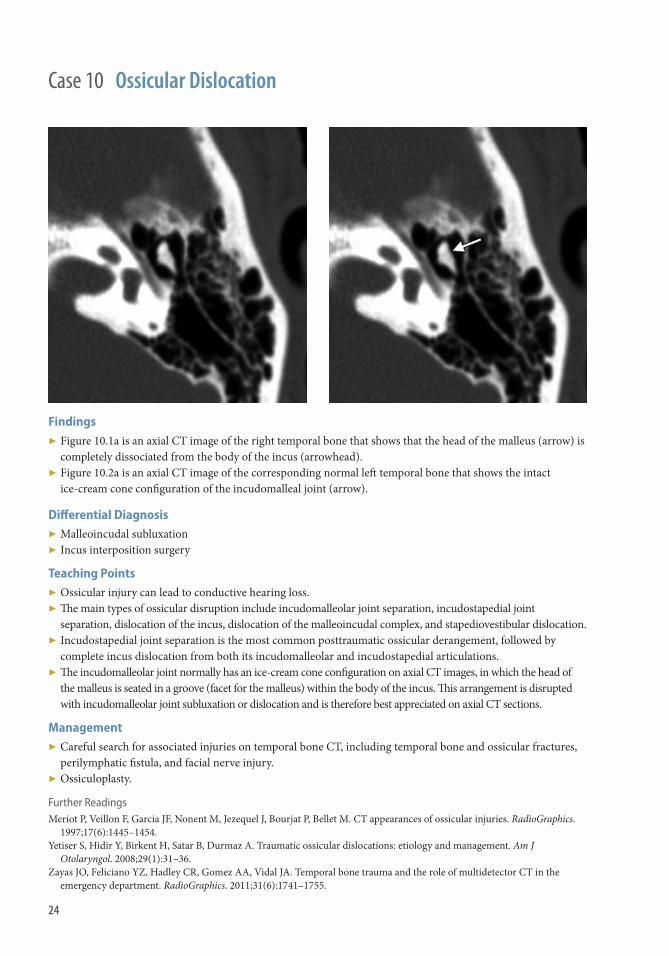
Findings ▶ Figure 10.1a is an axial CT image of the right temporal bone that shows that the head of the malleus (arrow) is
completely dissociated from the body of the incus (arrowhead). ▶ Figure 10.2a is an axial CT image of the corresponding normal left temporal bone that shows the intact
ice-cream cone confi guration of the incudomalleal joint (arrow).
Diff erential Diagnosis ▶ Malleoincudal subluxation ▶ Incus interposition surgery
Teaching Points ▶ Ossicular injury can lead to conductive hearing loss. ▶ Th e main types of ossicular disruption include incudomalleolar joint separation, incudostapedial joint
separation, dislocation of the incus, dislocation of the malleoincudal complex, and stapediovestibular dislocation. ▶ Incudostapedial joint separation is the most common posttraumatic ossicular derangement, followed by
complete incus dislocation from both its incudomalleolar and incudostapedial articulations. ▶ Th e incudomalleolar joint normally has an ice-cream cone confi guration on axial CT images, in which the head of
the malleus is seated in a groove (facet for the malleus) within the body of the incus. Th is arrangement is disrupted with incudomalleolar joint subluxation or dislocation and is therefore best appreciated on axial CT sections.
Management ▶ Careful search for associated injuries on temporal bone CT, including temporal bone and ossicular fractures,
perilymphatic fi stula, and facial nerve injury. ▶ Ossiculoplasty.
Further Readings Meriot P , Veillon F , Garcia JF , Nonent M , Jezequel J , Bourjat P , Bellet M . CT appearances of ossicular injuries . RadioGraphics .
1997 ; 17 ( 6 ): 1445–1454 . Yetiser S , Hidir Y , Birkent H , Satar B , Durmaz A . Traumatic ossicular dislocations: etiology and management . Am J
Otolaryngol . 2008 ; 29 ( 1 ): 31–36 . Zayas JO , Feliciano YZ , Hadley CR , Gomez AA , Vidal JA . Temporal bone trauma and the role of multidetector CT in the
emergency department . RadioGraphics . 2011 ; 31 ( 6 ): 1741–1755 .
Case 10 Ossicular Dislocation

25
History ▶ 86-year-old female who fell and injured her left face.
Case 11

26
Findings CT
▶ Preseptal, intraorbital density representing hematoma extending along the lateral orbital wall, with a convex margin projecting intraorbitally
▶ Mildly displaced fracture of the lateral orbital wall ▶ Marked mass eff ect contributing to mild proptosis ▶ Mild stretching of the optic nerve caused by proptosis ▶ Extensive preseptal periorbital soft tissue swelling CTA
▶ Punctate focus of contrast within the hematoma concerning for pseudoaneurysm or possibly active extravasation (Figure 11.1a )
▶ Intraorbital displacement of the lateral rectus muscle (Figure 11.2a ) Clinical Presentation ▶ Commonly associated with trauma, especially blunt injury ▶ Clinical signs may include proptosis and decreased range of motion of the ipsilateral globe ▶ Clinical symptoms may include pain, decreased visual acuity, and diplopia resulting from decreased range of motion ▶ An aff erent pupillary defect refl ects nerve injury ▶ Surgical procedures that may cause orbital hematoma include endoscopic sinus surgery, blepharoplasty, and
orbital reconstruction Pathophysiology ▶ In trauma, hemorrhage is most frequently subperiosteal related to disruption of small vessels in the periosteum ▶ Orbital hematomas may arise from extension of infection, particularly sinusitis, into the subperiosteal intraorbital space ▶ Less common causes of orbital hematoma include ruptured vascular malformation and hemorrhagic neoplasm ▶ Postseptal hemorrhage (posterior to the orbital septum) is more likely to cause injury to the globe, optic nerve,
and other orbital structures than preseptal hematoma (anterior to the orbital septum) ▶ May cause an acute orbital compartment syndrome, which may lead to vision-threatening compressive optic
neuropathy, which requires emergent management Teaching Points ▶ Traumatic and nontraumatic orbital hematomas most commonly occur in the subperiosteal space ▶ Active extravasation on postcontrast imaging is concerning for rapid expansion of the hematoma Management ▶ Conservative therapy may include glucocorticoids for anti-infl ammatory eff ect, especially with delayed presentations ▶ Surgical treatment may involve hematoma evacuation and orbit reconstruction ▶ Hematoma evacuation may also be performed with needle aspiration Further Reading Ramakrishnan VR , Palmer JN . Prevention and management of orbital hematoma . Otolaryngol Clin North Am .
2010 ; 43 : 789–800 .
Case 11 Orbital Hematoma

27
History ▶ None
Case 12

28
CT ▶ Th ere is bulging of the cavernous sinus ▶ Proptosis is noted ipsilateral to the fi stula ▶ Th ere is enlargement of the superior ophthalmic vein ▶ Th ere is enlargement of the periorbital muscles
CTA ▶ Enlargement and early contrast fi lling of the cavernous sinus ▶ Noticeable enlargement of the superior ophthalmic vein
DSA ▶ Is the diagnostic gold standard ▶ Early fi lling of the petrosal sinus and ophthalmic vein is noted when the intracavernous carotid artery is injected ▶ Early contrast in the cavernous sinus
Teaching points ▶ Patients may have tinnitus. At auscultation of the head, patients may have a bruit. ▶ Th e patient may experience diplopia and ophthalmoplegia (oft en from CN VI palsy). ▶ Th ere could be visual loss caused by edema and blood shunting. ▶ CCF are divided in direct and indirect fi stulas.
Management High-fl ow CCSF requires surgical repair or endovascular surgery. Current microcatheter techniques permit access to the cavernous sinus by several routes. Low-fl ow dural sinus CCSF that occurs spontaneously is very likely to resolve spontaneously.
Surgical repair is considered in cases in which there is increased risk of vision loss (from glaucoma, corneal exposure, or posterior segment ischemia); new visual symptoms; or the development of headache.
Case 12 Carotid Cavernous Fistula

29
History ▶ None
Case 13

30
Findings ▶ Figure 13.1. Axial image demonstrates comminuted fracture involving the inferior orbital rim with fracture
through the base of the frontal process of maxilla (arrows). Th ere is posterior displacement of the left nasolacrimal duct with extension of fracture to the duct with normal right-sided duct (arrowheads).
▶ Figure 13.2. Coronal image demonstrates a fracture through the nasal-frontal suture superiorly and fracture of the inferior orbital rim inferiorly. Th e orbital rim involvement is mildly comminuted.
▶ Figures 13.3, 13.4. Th ree-dimensional reformations give summary representation of nasal displacement and fracture of the orbital rim. Note well-demonstrated medial orbital fractures on the oblique image (black arrows).
Diff erential diagnosis ▶ Nasal bone fractures.
Teaching Points ▶ Th e naso-orbito-ethmoid (NOE) complex includes skeletal structures that are shared by both the nasal and
orbital regions including the nasal bones, ethmoid bones and inferior orbital rims. ▶ Fractures commonly result in posterior displacement of the anterior nasal structures into the medial orbital
rim and ethmoid sinuses, so-called telescoping. ▶ NOE fractures are clinically classifi ed according to the degree of comminution of the central bone fragment of
the orbital rim onto which the medial canthal tendon inserts. Th is tendon and related structures gives stability to the medial orbit and in cases of injury telecanthus can occur.
Management ▶ Management of NOE fractures is typically with open reduction and internal fi xation. ▶ Th e main goals are to restore the patient’s appearance and to restore the anatomic position of the medial
canthal tendon and the bony segment(s) to which it is attached. ▶ Attention may also be directed at repair or obliteration of the nasolacrimal duct if needed.
Further Reading Mehta N, Butala P, Bernstein MP . Th e imaging of maxillofacial trauma and its pertinence to surgical intervention. Radiol Clin
North Am. 2012 ; 50 ( 1 ): 43–57.
Case 13 Left-sided Nasal Orbital Ethmoid Fracture

31
History ▶ None
Case 14

32
Case 14 Right-Sided Orbital Floor Blow-Out Fracture
Findings ▶ Axial image (left ) at the level of the maxillary sinus shows opacifi cation of the right maxillary sinus. A bone
fragmentation of the orbital fl oor consistent with a fallen fragment (arrow) is identifi ed with adjacent herniated intraorbital fat and layering blood.
▶ Sagittal image (center) demonstrates the large defect in the posterior orbital fl oor fracture with herniation of orbital contents.
▶ Coronal image (right) in soft tissue window demonstrates orbital fl oor fracture with “trapdoor” fragment medially and herniation of orbital fat into the defect. Inferior rectus is identifi ed extending into the defect. Hyperintense blood products are identifi ed in the maxillary sinus.
Diff erential Diagnosis None
Teaching Points ▶ Orbital blow-out fractures result from direct blows to the orbit (fi st, ball), which increase intraorbital pressure
and result in fracture of the thin bony orbital lining (i.e., the fl oor or medial wall). ▶ Th e strong orbital rim remains intact with blow-out fractures. ▶ Th e fracture fragments and orbital soft tissue contents collapse outward into the air-fi lled sinus. When the
extraocular muscles are involved and become herniated into the sinuses the globe may become entrapped. Signifi cant increase in orbital volume and herniation of orbital fat may lead to enophthalmos and visual changes.
Management ▶ Clinically signifi cant indications for repair include entrapment and enophthalmos.
Further Reading Mehta N, Butala P, Bernstein MP . Th e imaging of maxillofacial trauma and its pertinence to surgical intervention. Radiol Clin
North Am . 2012 ; 50 ( 1 ): 43–57 .

33
History ▶ None
Case 15

34
Case 15 Zygoma Complex Fracture
Findings ▶ Axial image (left ) demonstrates fracture of the anterior and posterior/lateral wall of the maxillary sinus at the
zygomaticomaxillary sutures (arrowheads). Additionally, a fracture of the zygomatic arch with fracture of the zygomaticotemporal suture is seen (arrow).
▶ A superior axial image (center) through the level of the orbit demonstrates a fracture of the lateral orbital wall at the level of the zygomaticosphenoid suture (arrow). A more anterior fracture is seen as diastasis of the zygomaticofrontal suture (arrowhead). Frequently this fracture is best seen on coronal images.
▶ Slightly oblique three-dimensional image (right) demonstrates all four sutural attachments of the zygoma. Th e zygomaticotemporal suture (long arrow), the anterior zygomaticomaxillary suture (open arrow), the zygomaticofrontal suture (arrowhead), and the zygomaticosphenoid suture (short arrow ).
Diff erential Diagnosis ▶ Le Fort fractures
Teaching Points ▶ A direct blow to the lateral midface (the zygomatic region) can result in disruption of the zygoma from its
anatomic connections to the temporal, sphenoid, frontal, and maxillary bones. ▶ Th e zygomaticomaxillary involvement propagates through the infraorbital rim and orbital fl oor. Th is may
result in globe entrapment. ▶ Depression of the zygomatic arch may result in compression of the underlying temporalis tendon at its
insertion point on the coronoid process of the mandible, manifesting as trismus (limited mouth opening).
Management ▶ Th e zygoma complex fracture may be reduced and fi xated with malleable plates. Zygoma complex fractures
are classifi ed according to the direction and magnitude of displacement, and bony integrity of the zygoma, as originally described by Knight and North using plain fi lms.
Further Reading Mehta N, Butala P, Bernstein MP . Th e imaging of maxillofacial trauma and its pertinence to surgical intervention. Radiol Clin
North Am. 2012 ; 50 ( 1 ): 43–57 .

35
Case 16
History ▶ None

36
Case 16 Le Fort Maxillary Fracture
Findings Le Fort II fracture
▶ Figure 16.6: Axial images of Le Fort II fracture demonstrates intact lateral orbital walls (arrows). Th e lateral orbital wall would be involved in a Le Fort III fracture.
▶ Figure 16.7: Inferior axial image demonstrates the orbital rims are fractured bilaterally (arrowheads), a fi nding in all Le Fort II fractures.
▶ Figure 16.8: Axial image more inferiorly demonstrates fractures of the pterygoid plates bilaterally (arrows). ▶ Figures 16.9 and 16.10: Th ree-dimensional images demonstrate fracture through the inferior and medial
orbital rims (black arrows). A horizontal fracture is seen just superior to the nasofrontal suture (arrowhead). On sagittal view, note the propagation of the fracture through the posterior maxilla to involve the pterygoid plates (small black arrows).
Diff erential Diagnosis ▶ Although naso-orbital-ethmoid fractures do include the inferior and medial orbital rims, they do not extend
across the nasofrontal suture superiorly and do not involve the pterygoid plates. ▶ Th e Le Fort III fractures are a form of total cranial-facial disjunction and involve the zygoma and lateral
orbital walls.
Teaching Points ▶ All Le Fort maxillary fractures demonstrate fractures of the pterygoid plates. ▶ Le Fort I level extends horizontally at or just superior to the alveolar process of the maxilla with the fracture
involving the anterior and posterior/lateral walls of the maxillary sinus. Th e nasal septum is also fractured. Clinically this fracture presents with a distinctly mobile palate relative to the remainder of the midface.
▶ Le Fort II fracture extends from the pterygoid plates to involve the inferior and medial orbital rims. Midline a fracture of the nasal bones or a diathesis at the nasofrontal suture occurs. Th e line of fracture includes the posterior/lateral and anterior walls of the maxillary sinus, the orbital fl oor, and the medial orbital wall. Clinically, the maxillary and nasal regions are mobile.
▶ Le Fort III, “craniofacial disjunction,” the fracture line propagates from the pterygoid plates to involve the connection between the upper face and the skull base. Specifi cally, the fracture involves the pterygoid plates, zygomatic arches, lateral and medial orbital rims, and nasal bones or nasofrontal suture.

SpineSection 2


39
History ▶ Motor vehicle accident.
Case 17

40
Findings ▶ Fracture lines extend through the occipital condyles as they touch the articular facets of C1, usually best seen
in the coronal plane. ▶ Some fractures may be comminuted, with multiple small fragments. ▶ May see medial displacement of a smaller fracture fragment. ▶ May be associated with more complex skull base fractures or other cervical spine injuries. ▶ MRI may demonstrate disruption of craniocervical ligamentous structures.
Diff erential Diagnosis ▶ Unfused apophyses/ossifi cation centers ▶ Occipital condyle fracture
Teaching Points ▶ Most frequently occurs in the context of other craniocervical injuries. ▶ Anderson and Montesano classifi cation system
■ Type I: Comminuted occipital condyle fracture, usually resulting from loading injury (similar to Jeff erson fracture). Stable injury, with contralateral alar ligament intact.
■ Type II: Extension of skull base fracture through the occipital condyle. Also considered stable, because ligamentous structures remain intact.
■ Type III (most common): Avulsion fracture at the insertion of the alar ligament from the dens to the occipital condyle. Because of stress on the contralateral alar ligament, this is possibly an unstable injury.
▶ Tuli criteria for radiologic instability ■ >8 degrees of occipitoatloid rotation ■ >1 mm of occipitoatloid translation ■ >7 mm of overhand of C2 on C1 (total both sides) ■ >4-mm interval between anterior aspect of odontoid and posterior aspect of the anterior arch of C1 ■ <13-mm interval between posterior aspect of the odontoid and anterior aspect of the posterior arch of C1.
▶ CTA may be used to assess for vascular injury to the vertebral artery as it passes through the transverse groove of the atlas just posterior to the occipitoatloid articulations.
▶ MRI when a patient is stable is used to further assess the ligamentous structures, and assess for associated cord injury.
▶ Patients may experience nerve palsies of cranial nerves IX through XII because of involvement of these structures as they pass through the adjacent hypoglossal canal and jugular foramen.
▶ Management and clinical course is typically dictated by the associated injuries rather than the occipital condylar fractures as such.
Management ▶ Typically dictated by associated injuries. ▶ Treatment options for unstable injuries may include surgical fi xation or halo traction.
Case 17 Occipital Condyle Fracture

41
History ▶ None
Case 18

42
Findings ▶ Burst fracture of the C1 vertebral body
Radiograph
▶ Odontoid view: Off set between margins of C1 lateral masses and margins of subjacent C2 articular facets ■ Combined off set >7 mm indicates rupture of transverse ligament ■ May be normal variant in pediatric population ■ Axial rotation can mimic off set
▶ Lateral view: Increased atlantodental interval on lateral ■ Interval >6 mm indicates rupture of transverse ligament
▶ Flexion/Extension views ■ Maybe required to evaluate integrity of transverse ligament and thus stability of fracture
▶ Beware of normal variants in pediatric population
CT: Low threshold for CT
▶ Off set on odontoid view indication for CT ▶ Mechanism of trauma or symptoms is indication
Diff erential Diagnosis ▶ Congenital variations in C1 arch fusion ▶ Pseudosubluxation of C1 on C2 without fracture
Teaching Points Mechanism
▶ Axial loading: C1 vertebral body compressed between occipital condyles and C2 vertebral body resulting in forces that drive fracture fragments outward from central canal. Result is increased canal volume; spinal cord injury is rare.
▶ Hyperextension: Posterior arch fractures
Fracture Subtypes
▶ Bilateral anterior or posterior arch fractures (single arch) ▶ Anterior and posterior arch fractures (includes classic Jeff erson 4 part fracture)
Lateral mass fracture ▶ Exclude congenital failure of fusion: three primary ossifi cation sites that may not fuse
Management Further Imaging
▶ CTA if suspicion of vertebral artery or PICA injury ■ Extension of fracture into foramen transversarium ■ Wallenberg syndrome (PICA)
Transverse ligament integrity determines stability of fracture
▶ Transverse ligament disruption results in unstable fracture ▶ Imaging fi ndings (above) determine suspicion for ligament rupture
Treatment
▶ Stable fracture (intact transverse ligament): Cervical collar ▶ Unstable fracture (ruptured transverse ligament)
■ Cervical traction and halo ■ Surgery considered with increased displacement of C1/C2
Case 18 Jeff erson Fracture

43
History ▶ 35-year-old female in high-velocity motor vehicle accident.
Case 19

44
Findings Radiograph
▶ Wedging of anterior vertebral body, focal kyphosis, widening of spinous processes. ▶ On AP radiograph, may see increased luceny of involved vertebral body, indicating a displaced spinous
process (empty body sign).
CT
▶ Vertebral body fracture with anterior wedging causing focal kyphosis (arrowhead). ▶ Transversely oriented fracture through posterior elements with increased interspinous distance (arrow). ▶ Uncovering of the articular facets (naked facet sign) secondary to distraction of the posterior elements. ▶ On axial images, may see gradual loss of pedicle defi nition (dissolving pedicle sign). ▶ May see retropulsion of fracture fragment into spinal canal, suggesting a burst component.
MRI
▶ Hypointense fracture line on all sequences, with surrounding band of edema (sandwich sign). ▶ Disruption of interspinous and supraspinous ligaments. ▶ Spinal cord contusion, if present.
Diff erential Diagnosis ▶ Shear injury ▶ Distraction injury ▶ Burst fracture ▶ Compression fracture ▶ Pathologic fracture
Teaching Points Mechanism
▶ Flexion/distraction injury with compression of anterior column and distraction of middle and posterior columns, associated with use of the lap belt portion of a seat belt, which acts as a fulcrum around which the spine rotates during a motor vehicle accident.
▶ Most commonly occurs at T11-L3, but may occur in the midthoracic spine. ▶ Usually a mix of bony and soft tissue injury, with purely ligamentous Chance-type fractures rare.
Associations
▶ Approximately 40% of patients with Chance-type fractures have associated intraabdominal injuries, of which bowel and mesentery injuries are most common.
▶ Focal neurologic defi cits may or may not be present. ▶ Spinal cord injury especially if retropulsion of fragment into spinal canal.
Management ▶ Oft en conservative with bracing despite initial instability of injury. ▶ Surgical fi xation is indicated with increasing degrees of instability and ligamentous injury or if reduction
cannot be maintained with bracing.
Case 19 Chance Fracture

45
Case 20
History ▶ 67-year-old woman fell off horse, cannot turn head.

46
Findings ▶ C1 is displaced with respect to C2, and rotated such that the C1 articular facet is anterior to the superior
articular facet of C2. ▶ May also see fracture of the facets/articular processes associated with the impaction. ▶ Depending on relative displacement of C1 with respect to C2, may have marked narrowing of the canal. ▶ CTA may demonstrate cutoff or dissection of a vertebral artery. ▶ MRI may demonstrate associated ligamentous abnormality/disruption and elevated T2 signal in the cord.
Diff erential Diagnosis ▶ Chronic atlantoaxial rotatory fi xation ▶ Acute atlantoaxial rotatory fi xation
Teaching Points ▶ Hawkins and Fielding initially classifi ed fi xation by the degree of dissociation between the anterior arch of C1
and the odontoid ■ Type I: Normal distance between anterior arch and odontoid suggests lesser ligamentous injury. ■ Type II: 3–5 mm of anterior displacement of the anterior arch suggests transverse ligamentous injury. ■ Type III: >5 mm of anterior displacement suggests transverse and alar ligamentous injury. ■ Type IV: Posterior displacement of C1 with respect to C2.
▶ More recently, Pang has developed a classifi cation system for chronic atlantoaxial rotatory fi xation in pediatric patients that relies on a set of CT images of the atlantoaxial joint taken in at least three positions. Th is allows distinction between C1-C2 fi xation and muscular torticollis.
▶ Acute, traumatic atlantoaxial rotatory fi xation in adults is much rarer and clinically similar to facet dislocation/fracture at other levels.
▶ CTA should be considered to evaluate for a vascular injury of the vertebral arteries.
Management ▶ Longitudinal traction and halo fi xation can be used in acute cases. ▶ May require subsequent fusion of C1/C2 for stabilization.
Further Readings Rojas CA , Hayes A , Bertozzi JC , Guidi C , Martinez CR . Evaluation of the C1-C2 articulation on MDCT in healthy children
and young adults . AJR Am J Roentgenol . 2009 ; 193 ( 5 ): 1388–1392 . Booth TN . Cervical spine evaluation in pediatric trauma . AJR Am J Roentgenol . 2012 ; 198 ( 5 ): W417–W425 . Pang D . Atlantoaxial rotatory fi xation . Neurosurgery . 2010 ; 66 (suppl 3): 161–183 .
Case 20 Atlantoaxial Rotatory Fixation

47
Case 21
History ▶ None

48
Case 21 Hangman’s Fracture
Findings Radiography
▶ Anterior subluxation of C2 on C3 ▶ Lucency through the posterior elements of C2 compatible with fracture ▶ Prevertebral soft tissue thickening
CT
▶ Fractures of bilateral pars interarticulares of C2 ▶ Involvement of the transverse foramen warrants CTA to rule out vertebral artery injury
Diff erential Diagnosis ▶ Physiologic displacement of C2 on C3 in infants and young children ▶ Dens-arch synchondroses in children ▶ Primary spondylolyses in children
Teaching Points ▶ Bilateral pars interarticularis fractures of C2 (axis) ▶ Hyperextension cervical spine injury ▶ Named hangman’s fracture because during judicial hangings the executioner would place knot of the noose
under the chin of the person being hung resulting in this injury pattern ■ Nowadays, most injuries caused by face or chin hitting dashboard in a motor vehicle collision causing
hyperextension and distraction ▶ Levine classifi cation: (does not apply to children)
■ Type I: <3 mm translation, no angulation ■ Type II (most common): >3 mm translation, and >10 degrees of angulation ■ Type III: all characteristics of type II + bilateral interfacetal dislocation
▶ Presentation ■ Cervical spine point tenderness ■ Absence of neurologic injury is common, because the fracture tends to expand the spinal canal preventing
cord compression ■ In very severe cases, the C3 body is subluxed posteriorly causing cord compression with devastating
neurologic injury
Management ▶ Halovest traction/immobilization for 12 weeks ▶ Surgical fusion for nonunion (rarely necessary)
Further Reading Li XF , Dai LY , Lu H , Chen XD . A systematic review of the management of hangman’s fractures . Eur Spine J.
2006 ; 15 ( 3 ): 257–269 .

49
History ▶ None
Case 22

50
Findings Anterior dislocation of inferior articular process relative to superior articular process of the caudal vertebral level Diagnosis ▶ Radiographs: anteroposterior, lateral, and oblique ▶ CT with sagittal reconstructions ▶ Anterolisthesis of 25%–50%
■ If anterolisthesis >50% must suspect bilateral facet dislocation ▶ Assess patency of foramen transversarium
■ Risk of vertebral artery injury increased in hyperfl exion injuries Associated Injuries ▶ Fractures (ipsilateral or contralateral)
■ Lateral mass fracture ■ Articular process fractures ■ Transverse process fracture ■ Lamina fracture
▶ Contralateral facet injury ■ Fracture ■ Subluxation
▶ Posterior ligamentous injury (MRI) ■ Ligamentum fl avum ■ Interspinous and superspinous ligaments ■ Posterior longitudinal ligament usually intact or at most partially torn, preventing further anterolisthesis
▶ Radiculopathy ■ Superior articular process rest in neural foramen aft er dislocation
▶ Cord injury RARE with unilateral facet dislocation ■ Stable injury when not accompanied by destabilizing fractures
Diff erential Diagnosis ▶ Bilateral facet dislocation ▶ Hyperfl exion fracture Teaching Points Mechanism
▶ Hyperfl exion-rotation ■ Most common in mid or lower cervical spine
▶ Axial rotation with fi xed pivot point on one facet resulting in contralateral anterior dislocation Management ▶ Closed reduction with cervical traction if neurologically intact ▶ Prereduction MRI if abnormal neurologic examination or altered mental state ▶ Surgery if
■ Failure of closed reduction ■ Middle column injury (PLL, posterior annulus fi brosis) ■ Associated fractures result in instability ■ Flexion-extension views demonstrate instability aft er 12 weeks
Case 22 Unilateral Facet Dislocation of the Cervical spine

51
Case 23
History ▶ Trauma

52
Findings Radiography
▶ Increased interspinous distance on anteroposterior radiograph = increased space between spinous processes of aff ected level.
▶ Marked anterior subluxation and prevertebral soft tissue swelling on lateral radiograph.
CT
▶ Naked facet or “empty hamburger” sign on axial images with uncovered articulating processes. ▶ Marked anterior subluxation of superior vertebral body (in this case C6 on C7), greater than 50%
anterolisthesis. ▶ Bilateral jumped or locked facets (inferior articular process lies anterior to the superior articular process on
both sides of the spine). ▶ Associated with fractures of the superior facet, inferior facet, and fl oating lateral mass.
MRI
▶ Marked anterior subluxation; spinal cord injury including cord edema, hemorrhage, or transection; traumatic disk herniation; and associated ligamentous and paraspinal injury.
Diff erential Diagnosis ▶ Unilateral facet dislocation ▶ Facet subluxation ▶ Perched facets
Teaching Points ▶ Severe unstable hyperfl exion distraction injury causing facet joints to jump over each other and become
locked. ▶ Disruption of all three spinal columns including all major spinal ligaments, intervertebral disks, and facet joint
capsules at the aff ected level. ▶ Greater than 50% anterior subluxation. ▶ Facets may not dislocate completely and may become perched atop the subjacent facets. ▶ Commonly present with neurologic defi cits.
Management ▶ Operative stabilization and fusion required aft er reduction because of extensive ligamentous disruption.
Further Readings Goldberg AL, Kershah SM. Advances in imaging of vertebral and spinal cord injury . J Spinal Cord Med . 2010 l; 33 ( 2 ): 105–116 . Mhuircheartaigh NN, Kerr JM, Murray JG. MR imaging of traumatic spinal injuries . Semin Musculoskelet Radiol .
2006 ; 10 ( 4 ): 293–307 .
Case 23 Bilateral Facet Dislocation

53
History ▶ None
Case 24

54
Types Figure 1. C2-3 Hyperfl exion Injury. (A) Sagittal image from a non-contrast enhanced cervical spine CT demonstrates a chip fracture of the anteroinferior corner of the C2 vertebral body (thin solid arrow) and fat stranding in the posterior soft tissues suggestive of edema and/or hemorrhage (thick solid arrow). (B) Sagittal STIR image from a non-contrast enhanced cervical spine MRI demonstrates abnormal increased STIR hyperintensity in the anterior (dashed arrow) and posterior (open arrow) soft tissues consistent with soft tissue injury.
Figure 2. C6-7 Hyperfl exion Injury. (A) Sagittal image from a non-contrast enhanced cervical spine CT demonstrates subtle fi ndings including narrowing of the anterior disc space at the C6-7 level (thin solid arrow), minimal C7 vertebral body height loss and widening of the posterior interspinous space (thick solid arrow). (B) Sagittal STIR image from a non-contrast enhanced cervical spine MRI demonstrates abnormal increased STIR hyperintensity in the posterior soft tissues from C1-C7 particularly in the C6-7 interspinous space (open arrow) and apparent focal rupture of the ligamentum fl avum at the C6-7 level (dashed arrow).
Flexion-type injuries of the cervical spine represent a spectrum of injuries bound by a common injury mechanism. Commonly recognized types (in increasing severity) include ▶ Hyperfl exion ligamentous sprain or partial tear ▶ Clay-shoveler’s fracture ▶ Stable and unstable wedge fracture ▶ Unilateral facet dislocation ▶ Bilateral facet dislocation ▶ Flexion teardrop fracture Hyperfl exion sprain injuries encompass soft tissue injuries of the cervical spine with or without fracture.
Findings Radiograph and CT
▶ Anterior disk space narrowing and posterior interspinous space widening. ▶ Translation at the level of ligamentous injury. ▶ Prominence of the prevertebral soft tissues. MRI
▶ Increased T2-signal involving the spinal ligaments and adjacent soft tissue, most commonly the interspinous ligaments and posterior soft tissues, representing edema and/or hemorrhage.
▶ Increased T2 or STIR signal involving the anterior portion of the vertebral bodies of the involved level, representing marrow edema secondary to bony contusion.
Teaching Points Etiology
▶ Hyperfl exion is the most common injury mechanism of the cervical spine, accounting for almost half of all cervical spine injuries.
▶ Eliciting the mechanism of injury and vertebral levels of point tenderness, if available, are of critical importance in identifying subtle injuries.
Management Optimal management of unstable injuries or injuries with associated neurologic defi cits requires early consultation with a spine surgeon or neurosurgeon. May require early surgical intervention or decompression in the case of spinal cord impingement. Studies evaluating early corticosteroid treatment for neurologic impairment have shown mixed results.
Case 24 Hyperfl exion Sprain Injury

55
Case 25
History ▶ 59-year-old female status post fall down stairs.

56
Findings ▶ Figure 25.1. Sagittal view demonstrates anterior greater than posterior compression deformity of the C6
vertebral body. ▶ Figure 25.2. Sagittal view, off midline demonstrates displaced fracture of the facet resulting in facet
malalignment. ▶ Figure 25.3. Vertebral body fracture and displaced fracture of the right facet. ▶ Figure 25.4. Arrow denotes the teardrop fracture. ▶ Figure 25.5. Arrow denotes facet fracture.
Diff erential Diagnosis Burst fracture
Teaching Points ▶ Mechanism: Forceful fl exion and axial compression of the cervical spine that occurs when the neck is fl exed
and the head strikes a solid object, such as in diving into shallow pool of water or hitting head on dashboard in a motor vehicle collision.
▶ Neurologic impact includes anterior cord syndrome: quadraplegia with loss of anterior column senses of pain, temperature, and touch sensations; and preservation of posterior column senses of position, motion, and vibration.
▶ CT and radiographic fi ndings ■ Anteroinferior margin of cervical vertebral body is fractured. ■ Posterior ligaments are disrupted with portion of the vertebral displaced backward into the spinal canal. ■ Intervertebral disk between fractured vertebral body and vertebral body below may be disrupted. ■ Reciprocal distractive force may results in disruption of the posterior structures including the interspinous
ligamentous fracture, facet misalignment, and laminar fractures
Management Neurosurgical intervention is likely necessary.
Further Reading Kim K , Chen H , Russell E , Rogers L . Flexion teardrop fracture of the cervical spine: radiographic characteristics . Am J Radiol.
1989; 152 : 319–326.
Case 25 Cervical Flexion Teardrop Fracture

57
Case 26
History ▶ 48-year-old male construction worker who fell from scaff olding.

58
Findings Radiograph
▶ Classically demonstrates increased interpedicular distance compared with vertebral body above and below. CT
▶ Compression of L2 vertebral body. ▶ Loss of height of posterior cortex with extension of fracture line through it (arrow). ▶ Retropulsion of bone fragment with narrowing of spinal canal (arrowhead).
Diff erential Diagnosis ▶ Wedge compression fracture ▶ Split compression fracture ▶ Chance fracture ▶ Pathologic fracture
Teaching Points Etiology
▶ Fall from height with landing on feet is a common mechanism. ▶ Axial loading of vertebral body with compressive failure of anterior and posterior cortex of vertebral body. ▶ Failure of both anterior and middle columns. ▶ With rapid axial load, fl uid in nucleus pulposus becomes pressurized and expands in all directions, unable to
escape through normal pores and fi ssures, resulting in bursting of vertebral body as one proposed mechanism. Diff erentiation
▶ Wedge compression and split fractures result from an axial load with a fl exion component and failure of the anterior column. Th e latter is associated with coronal fracture lines through the vertebral body. Th e posterior cortex is intact with no retropulsion in these types of fractures.
▶ A Chance fracture is a fl exion/distraction injury, with anterior wedge compression of the vertebral body and a transverse fracture extending through the posterior elements. Th is results in compression of the anterior column and distraction of the middle and posterior columns.
Associations
▶ Lower extremity fractures, pelvic fractures, other spinal fractures, dural laceration, epidural hematoma.
Management ▶ Conservative if neurologically intact. ▶ Fixation with laminectomy if neurologic defi cit, kyphosis >20 degrees, >50% compression of vertebral body,
subluxation of facet joints.
Further Readings Rutherford EE . Lumbar spine fusion and stabilization: hardware, techniques, and imaging appearances . Radiographics .
2007 ; 27 : 1737–1749 . Heary RF . Decision-making in burst fractures of the thoracolumbar and lumbar spine . Indian J Orthop . 2007 ; 41 ( 4 ): 268–276 .
Case 26 Lumbar Burst Fracture

59
Case 27
History ▶ None

60
Findings ▶ Ultrasound: intimal fl ap, double lumen each with diff erent signal on Doppler fl ow, dissecting aneurysm ▶ CT: narrowed lumen, double lumen, dissection fl ap, hematoma ▶ MRA: vessel narrowing +/- aneurismal dilation of dissected artery. Susceptibility MR may show blooming
artifact of blood products. ▶ Angiography: gold standard; intimal fl ap and double lumen; long segment of arterial narrowing “string sign”
Diff erential Diagnosis ▶ Intramural thrombus ▶ Atheromatous plaque ▶ Fibromuscular dysplasia ▶ Pseudoaneurysm ▶ Glomus vagale paraganglioma ▶ Carotid space schwannoma
Teaching Points ▶ Most frequent presentation is headache. May also experience pain in face or neck, Horner syndrome, brain
ischemia, dizziness. ▶ ICA dissection in young patients, usually at the base of the skull. ▶ ICA dissection in older patients, usually at the carotid bifurcation. ▶ Trauma is most common cause.
Management ▶ Anticoagulation to prevent thrombosis and embolism in extracranial dissections. Anticoagulation
contraindicated in cases of intracranial dissecting aneurysms with subarachnoid hemorrhage. ▶ Consider endovascular or surgical intervention if persistent symptoms caused by thromboembolic events and/
or dissecting aneurysm.
Further Readings Rodallec MH , Marteau V , Gerber S , et al. Craniocervical arterial dissection: spectrum of imaging fi ndings and diff erential
diagnosis . Radiographics . 2008 ; 28: 1711–1728 . Shin JH , Suh DC , Choi CG , et al. Vertebral artery dissection: spectrum of imaging fi ndings with emphasis on angiography and
correlation with clinical presentation . Radiographics . 2000 ;20: 1687–1696 .
Case 27 Vertebral / Carotid Artery Dissection

61
Case 28
History ▶ 77-year-old woman with facial and forehead trauma during fall while ambulating.

62
Case 28 Odontoid Fracture
Findings ▶ Fracture of odontoid process (dens) of the C2 vertebral body.
■ Transverse fracture of dens (type II) with posterior angulation and displacement of proximal fragment. Spinal canal stenosis with cord edema.
▶ Subtypes: (Anderson and D’Alonzo classifi cation). ■ Type I: avulsion fracture through the superolateral extent of odontoid process at attachment of alar
ligament (rare). ■ Type II: fracture through junction of odontoid process and body.
■ Increased risk of nonunion (look for these!) ■ Comminution at base of odontoid fracture fragment ■ >5 mm initial translocation
■ Posterior displacement greater risk than anterior ■ >10 degrees angulation ■ Elderly ■ Delayed diagnosis
■ Type III: fracture involving the odontoid process and body of C2.
Diff erential Diagnosis ▶ Os odontoideum ▶ C1/C2 subluxation from ligamentous laxity
■ Rheumatoid arthritis ■ Trisomy 21
▶ Condylus tertius ■ Congenital variant third occipital condyle extending from clivus
▶ Nonfusion of apical odontoid epiphysis (ossiculum terminale)
Teaching Points ▶ Etiology
■ Hyperextension or less likely hyperfl exion of upper cervical spine ▶ Cord injury rare because of capacious central canal at C1 and C2 levels.
■ Cord injury may occur with severe hyperextension and posterior displacement of coronoid fragment. ▶ Risk of vertebral artery injury if fracture extends through foramen transversarium
Management Type I
▶ Most are stable fractures ■ High rate of successful healing with nonoperative treatment ■ Treat with semirigid collar for symptoms
▶ Unstable if fl exion/extension reveals subluxation of C1 ■ Needs surgical fi xation
▶ Unstable if associated with occipitoatlantal dislocation ■ Needs surgical fi xation
Type II
▶ Unstable; risk of nonunion 30%–50% ▶ Initial management with halo vest ▶ If nonsurgical management fails: surgical fi xation Type III
▶ Unstable fracture (occiput, C1, and proximal C2 fragment move as unit increasing motion at fracture site) ▶ High rate of successful healing with nonoperative treatment

ChestSection 3


65
History ▶ None
Case 29

66
Findings ▶ Hyperlucent left upper quadrant ▶ Widening and deepening of the left costophrenic angle (solid arrow) ▶ Triangular lucency medial left cardiophrenic sulcus (open arrow)
Diff erential Diagnosis ▶ Normal ▶ Skin fold ▶ External artifact: sheets, clothing, hair braids ▶ Bullous disease ▶ Pneumoperitoneum
Teaching Points ▶ Identifi cation of a pneumothorax on plain radiography relies primarily on visualization of the pleural line
between the aerated lung and the pathologically air-fi lled potential space of the pleura. Th us, if the pleural edge is not tangential to the X-ray beam, a pleural line is not necessarily seen. Th is is particularly true in the supine radiograph.
▶ Most common locations: anteromedial, subpulmonic, apicolateral, posteromedial ▶ Deep sulcus sign: larger/wider lateral costophrenic recess than contralateral side ▶ Hyperlucent upper abdominal quadrant ▶ Outline of the medial diaphragm beneath the cardiac silhouette ▶ Sharply defi ned diaphragmatic contour despite dense lung parenchymal air space disease
Management ▶ Cross-sectional imaging may be needed for confi rmation ▶ Pneumothoraces normally resorb at ~1% per day ▶ Th oracotomy tube may be necessary if clinically symptomatic
Further Readings Tocino I . Pneumothorax in the supine patient: radiographic anatomy. RadioGraphics . 1985; 5( 4) : 557–586 . McLoud TC , Boiselle PM . Th oracic Radiology: Th e Requisites . 2nd ed. Philadelphia : Mosby Elsevier; 2010: 422 .
Case 29 Occult Pneumothorax

67
Case 30
History ▶ 55-year-old female with the acute onset of chest pain.
68
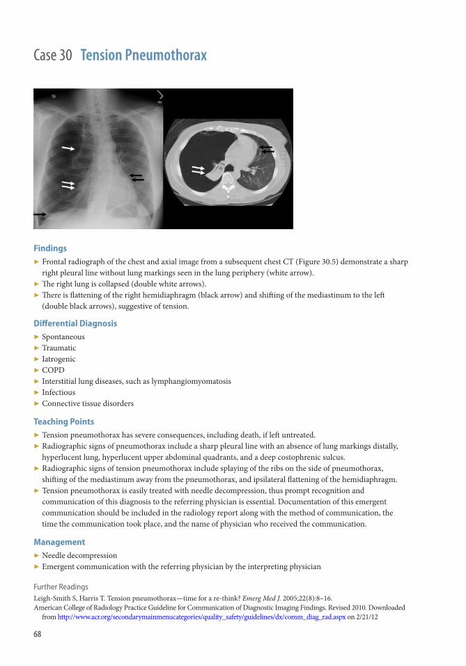
Findings ▶ Frontal radiograph of the chest and axial image from a subsequent chest CT (Figure 30.5) demonstrate a sharp
right pleural line without lung markings seen in the lung periphery (white arrow). ▶ Th e right lung is collapsed (double white arrows). ▶ Th ere is fl attening of the right hemidiaphragm (black arrow) and shift ing of the mediastinum to the left
(double black arrows), suggestive of tension.
Diff erential Diagnosis ▶ Spontaneous ▶ Traumatic ▶ Iatrogenic ▶ COPD ▶ Interstitial lung diseases, such as lymphangiomyomatosis ▶ Infectious ▶ Connective tissue disorders
Teaching Points ▶ Tension pneumothorax has severe consequences, including death, if left untreated. ▶ Radiographic signs of pneumothorax include a sharp pleural line with an absence of lung markings distally,
hyperlucent lung, hyperlucent upper abdominal quadrants, and a deep costophrenic sulcus. ▶ Radiographic signs of tension pneumothorax include splaying of the ribs on the side of pneumothorax,
shift ing of the mediastinum away from the pneumothorax, and ipsilateral fl attening of the hemidiaphragm. ▶ Tension pneumothorax is easily treated with needle decompression, thus prompt recognition and
communication of this diagnosis to the referring physician is essential. Documentation of this emergent communication should be included in the radiology report along with the method of communication, the time the communication took place, and the name of physician who received the communication.
Management ▶ Needle decompression ▶ Emergent communication with the referring physician by the interpreting physician
Further Readings Leigh-Smith S , Harris T . Tension pneumothorax—time for a re-think? Emerg Med J. 2005 ; 22 ( 8 ): 8–16 . American College of Radiology Practice Guideline for Communication of Diagnostic Imaging Findings . Revised 2010. Downloaded
from http://www.acr.org/secondarymainmenucategories/quality_safety/guidelines/dx/comm_diag_rad.aspx on 2/21/12
Case 30 Tension Pneumothorax
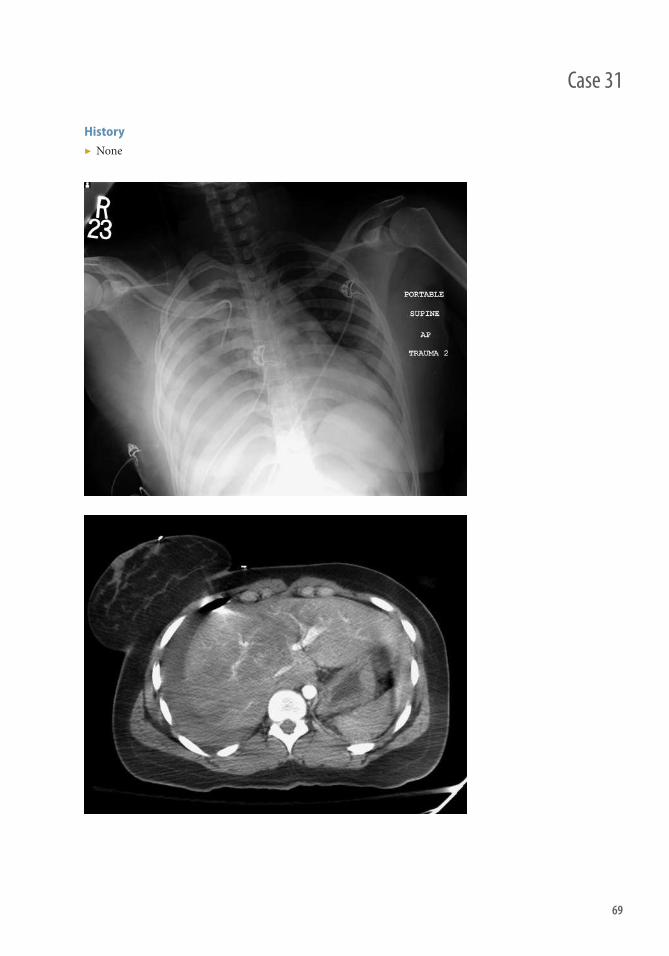
69
History ▶ None
Case 31

70
Findings Plain Film
▶ Right-sided pleural eff usion and air space opacity ▶ Multiple right-sided rib fractures
Computed Tomography
▶ Hyperdense fl uid (blood) contained within the pleural space ▶ Regions of interest demonstrate increasing density with dependency
Diff erential Diagnosis ▶ Pleural eff usion ▶ Chylothorax ▶ Pleural-based mass/scarring
Teaching Points ▶ Although most commonly associated with chest wall trauma, it can be seen with thoracic aortic injury,
anticoagulation, metastatic malignancy, tuberculosis, and pulmonary infarct. ▶ A single hemithorax can hold 4 L of blood, allowing for exsanguination. ▶ Blood tends to clot quickly followed by a decrease in density with evolution and lysis. ▶ Empyema can develop if it becomes superinfected. ▶ Fibrothorax can develop from fi brous adhesions. Th is can yield scarring and even entrapment of the underlying lung.
Management ▶ Generally treated with single or double thoracotomy tubes depending on size (proper positioning should be
inferior and posterior for fl uid, compared with apical and anterior for air) ▶ Surgical intervention may be required if there is immediate removal of >1 L of acute hemorrhage or if there is
continued bleeding >200 cc/h over 2–4 hours ▶ If large clot persists (i.e., retained hemothorax >500 cc), chemical lysis with fi brinolytic agents may be used to
decrease the risk of empyema or fi brothoroax development
Further Readings Chest trauma hemothorax . Trauma.org . http://www.trauma.org/archive/thoracic/CHESTh aemo.html . McLoud TC , Boiselle P M . Th oracic Radiology: Th e Requisites . 2nd ed. Philadelphia : Mosby Elsevier; 2010: 422. Parry GW , Morgan WE , Salama FD . Management of haemothorax . Ann R Coll Surg Engl. 1996 ; 78 : 325–326.
Case 31 Hemothorax

71
Case 32
History ▶ None

72
Findings ▶ Chest radiograph: Vague opacity right mid lung ▶ CT: Patchy alveolar and ground-glass opacities throughout the right lung ▶ Additional images from diff erent patients demonstrate similar fi ndings in the right upper lobe (Figures 32.3,
32.4), and patchy RML contusions and more dense posterior contusions in a separate patient (Figure 32.5)
Diff erential Diagnosis ▶ Other air space processes: Pneumonia, aspiration, edema, ARDS
Teaching Points ▶ Pulmonary contusion is one of the most common fi ndings in blunt chest trauma
■ Th ey are the most common chest injury in children ▶ Etiology is from traumatic hemorrhage into the lung parenchyma and air spaces ▶ Although up to one-third are inapparent on initial radiography, they generally become apparent within 6
hours of initiating trauma, and almost always by 24 hours ▶ Clinical symptoms range from clinically silent to hemoptysis and impaired gas exchange ▶ Radiographically appears the same as any other air space process
■ Alveolar fi lling can yield ground-glass or “fl uff y” infi ltrates to dense consolidation ■ May or may not demonstrate air bronchograms secondary to bronchial fi lling with hemorrhage ■ Can be contralateral to side of injury (contrecoup)
▶ Look for associated blunt force injury (i.e., rib fractures, fl ail chest, scapular fractures, soft tissue contusions)
Management ▶ If small, can resolve rapidly (oft en within 24–48 hours), but most resolve with 3–5 days. ▶ More advanced disease can lead to impaired oxygenation, increased pulmonary vascular resistance, and
decreased lung compliance. ▶ 40% of patients require mechanical ventilation, 50%–60% of severe contusions develop bilateral acute
respiratory distress syndrome (ARDS). ▶ Clinical management involves fl uid restriction; however, this must be weighted with the risk of hypoperfusion,
which is associated with development of ARDS in addition to multiorgan consequences. ▶ Most frequent complication is pneumonia.
Further Readings Brohi K . Chest trauma pulmonary contusion. Trauma.org . http://www.trauma.org/archive/thoracic/CHESTcontusion . Wagner RB . Classifi cation of parenchymal injuries of the lung. Radiology . 1988; 167 : 77–82 .
Case 32 Pulmonary Contusion

73
History ▶ 23-year-old female status post high-impact motor vehicle accident with left -sided chest pain.
Case 33

74
Findings ▶ Axial CT image demonstrates a cavitary rounded lesion with air-fl uid level consistent with lung laceration. Of
note, there is adjacent lung contusion and small pneumothorax.
Diff erential Diagnosis ▶ Pneumatocele ▶ Lung laceration otherwise has a fairly pathognomic appearance. If not provided history of acute injury, CT or
radiographic fi ndings could also be similar to those of ■ Cavitary malignant nodule ■ Cavitary infection/nodule
Teaching Points ▶ Pulmonary lacerations result from both blunt and penetrating trauma ▶ Can be a marker for high-energy mechanism of injury ▶ Can be associated with lung contusion, pneumothorax, or hemothorax ▶ Complications not common but can include infection, abscess, pneumothorax, or bronchopleural fi stula
formation ▶ CT is more sensitive than chest radiography ▶ Imaging signs of lung laceration include round or ovoid structures that can be fi lled with either air,
hemorrhage, or other fl uid; air-fl uid levels may be present; multiple lacerations may be present
Management ▶ Most commonly, expectant management unless there are complications ▶ If complications or if severe laceration, surgical treatment including suturing, stapling, and wedge resection
can be considered
Further Readings Kaewlai R , Avery LL , Asrani AV, et al. Multidetector CT of blunt thoracic trauma . RadioGraphics . 2008 ; 28 ( 6 ): 1555–1570 . Hollister M , Stern EJ , Steinberg KP . Type 2 pulmonary laceration: a marker of blunt high-energy injury to the lung . AJR Am J
Roentgenol . 1995 ; 165 ( 5 ): 1126 .
Case 33 Lung Laceration
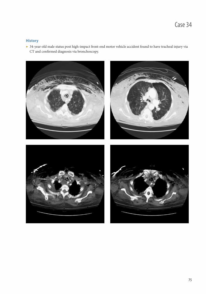
75
Case 34
History ▶ 34-year-old male status post high-impact front-end motor vehicle accident found to have tracheal injury via CT and confi rmed diagnosis via bronchoscopy.

76
Findings ▶ Axial CT images in lung windows (Figure 34.1) demonstrate extensive subcutaneous emphysema and
concurrent pneumothorax indicating high-impact mechanism of injury (Figure 34.2). ▶ Axial CT image (Figure 34.3) in mediastinal windows demonstrates posterior wall irregularity and
caliber increase of trachea with small foci of extraluminal mediastinal air adjacent to the right of trachea (Figure 34.4).
Diff erential Diagnosis of Tracheal Rupture ▶ Tracheal diverticulum ▶ Tracheoesophageal fi stula ▶ Pneumomediastinum
Teaching Points ▶ Radiographic fi ndings: Tracheal dilatation, pneumomediastinum, or subcutaneous emphysema. ▶ CT fi ndings: Abrupt dilatation and increase in caliber of diameter of trachea, adjacent pneumomediastinum,
mediastinal hematoma, or tracheal wall irregularity or outpouching. ▶ Associated imaging fi ndings: Extensive subcutaneous emphysema, pneumothorax, or other associated chest
wall trauma. ▶ Chest radiographs are not sensitive when compared with CT in detecting tracheal injury.
Management ▶ Dependent on location and severity of the injury and if patient is stable. ▶ Goal of treatment is to ensure a patent airway to prevent respiratory compromise. ▶ Intubation and mechanical ventilation is initial fi rst-line treatment. ▶ Consideration of laryngoscopy and bronchoscopy for further evaluation. ▶ Although less severe cases can be managed without surgery, surgical intervention is considered the gold
standard for treatment of any tracheal or bronchial tear.
Further Readings Kaewlai R , Avery LL , Asrani AV, et al. Multidetector CT of blunt thoracic trauma . RadioGraphics . 2008 ; 28 ( 6 ): 1555–1570 . Chen JD , Shanmuganathan K , Mirvis SE , Killeen KL , Dutton RP . Using CT to diagnose tracheal rupture . AJR Am J Roentgenol .
2001 ; 176 ( 5 ): 1273–1280 .
Case 34 Tracheobronchial Injury

77
History ▶ 35-year-old male status post front-end motor vehicle accident
Case 35

78
Findings ▶ Frontal radiograph of the chest (Figure 35.1) demonstrates multiple left -sided rib fractures. ▶ CT image (Figures 35.2a, 35.2b, 35.2c, 35.2d) shows multiple ribs (>3) with fractures in two or more locations. ▶ Th ree-dimensional volume rendering (Figures 35.3a, 35.3b, 35.3c) illustrates multiple ribs (>3) with fractures
in two or more locations.
Diff erential Diagnosis ▶ If meeting defi nition of three or more ribs fractured in two or more locations, fl ail chest has a fairly
characteristic appearance.
Teaching Points ▶ Flail chest is usually associated with high-impact mechanisms of blunt trauma. ▶ Flail chest is essentially segment of thoracic ribs being separated from remainder of chest wall. ▶ Can be life-threatening given high association with other injuries. ▶ If a patient presents with fl ail chest, there are usually additional associated injuries, such as pneumothorax,
hemothorax, lung laceration, or lung contusions. ▶ Clinical examination reveals a paradoxical movement of the fl ail segment during breathing. ▶ Radiographic fi ndings: Flail chest occurs when three or more ribs are fractured in two or more locations. ▶ Chest radiographs are not as sensitive as CT in clearly defi ning fl ail segments.
Management ▶ Critical care including mechanical ventilation and close monitoring ▶ Surgical treatment is required in severe cases and can result in faster ventilator wean time, and shorter
ICU stay
Further Readings Pettiford BL , Luketich JD , Landreneau RJ . Th e management of fl ail chest . Th orac Surg Clin . 2007 ; 17 ( 1 ): 25–33 . Collins J . Chest wall trauma . J Th orac Imaging . 2000 ; 15 ( 2 ): 112–119 .
Case 35 Flail Chest

79
Case 36
History ▶ 65-year-old man who was the unrestrained driver in a motor vehicle collision

80
Findings ▶ Frontal (Figure 36.2a) and lateral (Figure 36.2b) radiographs of the chest demonstrate a fracture of the
mid-sternum (arrow) with some anterior displacement of the distal fracture fragment. Th is fi nding is confi rmed on axial (Figure 36.2c) and sagittal reformatted (Figure 36.2d) images from contrast-enhanced chest CT in bone windows (arrow).
Diff erential Diagnosis ▶ Pectus excavatum ▶ Pathologic fracture ▶ Osteomyelitis ▶ Ossifi cation center
Teaching Points ▶ Sternal fractures are most commonly the result of motor vehicle collisions with deceleration injury to
the chest. ▶ CT is the modality of choice to evaluate for sternal fractures, which are best demonstrated on sagittally
reformatted images. ▶ Sternal fractures can be subtle and are frequently nondisplaced. Oft en, the only clue to the presence of a
sternal fracture is a mediastinal hematoma ▶ Mediastinal hematomas from sternal fracture can be distinguished from those associated with aortic injury by
the presence of a fat plane between the hematoma and the aorta ▶ Sternal fractures are associated with high-energy trauma; thus, careful evaluation for injury to the heart and
aorta is warranted.
Management ▶ Careful evaluation for other injuries in the chest, especially to the heart and aorta ▶ Adequate analgesia with possible surgical intervention
Case 36 Sternal Fracture

81
History ▶ 41-year-old male status post front-end motor vehicle accident with air-bag deployment presenting with chest pain.
Case 37

82
Case 37 Hemopericardium in Blunt Cardiac Trauma (Myocardial Injury)
Findings ▶ Frontal radiograph of the chest (Figure 37.1) demonstrates an enlarged cardiac silhouette. ▶ Axial (Figure 37.2a) and sagittal (Figure 37.2b) CT images demonstrate a hyperdense pericardial
eff usion consistent with hemopericardium. Of note, no aortic dissection is present raising concern for hemopericardium from underlying myocardial injury.
Diff erential Diagnosis ▶ Infl ammatory pericardial eff usion ▶ Infected pericardial eff usion ▶ Aortic dissection resulting in hemopericardium
Teaching Points ▶ Hemopericardium can occur secondary to occult myocardial muscle injury, such as from infarct or direct
tissue injury. However, although not as common, hemopericardium can also occur in setting of myocardial rupture.
▶ Hemopericardium can also occur in setting of ascending aortic dissection. Hence, it is important to evaluate and exclude associated traumatic aortic injury in trauma patients.
▶ Radiographic fi ndings: Enlarged cardiac silhouette suggestive of a pericardial eff usion. ▶ CT fi ndings: Hemopericardium appears as a hyperdense pericardial eff usion. ▶ Cardiac tamponade is a life-threatening condition that can result from hemopericardium; cardiac tamponade
is a clinical diagnosis but imaging may provide early warning. ▶ Can be associated with constrictive pericarditis and may be delayed in nature because of evolution of
myocardial muscle contusion and/or infarction. ▶ Close interval and continuous monitoring aft er presentation is required to evaluate for cardiac tamponade.
Management ▶ Close monitoring for worsening of hemopericardium and to detect cardiac tamponade. ▶ If necessary, surgical treatment and drainage of pericardial eff usion.
Further Readings Krejci CS , Blackmore CC , Nathens A . Hemopericardium: an emergent fi nding in a case of blunt cardiac injury . AJR Am J
Roentgenol . 2000 Jul; 175 ( 1 ): 250 . Pretre R , Chilcott M . Blunt trauma to the heart and great vessels . N Engl J Med. 1997 ; 336 ( 9) : 626–632. Goldstein L , Mirvis SE , Kostrubiak IS , Turney SZ . CT diagnosis of acute pericardial tamponade aft er blunt chest trauma . AJR
Am J Roentgenol . 1989 Apr;152(4):739–41.

83
Case 38
History ▶ None

84
Findings Plain Film
▶ Elevated hemidiaphragm ▶ Hemithorax opacity ▶ Mediastinal shift to nonaff ected side ▶ Presence of traumatic thoracic injuries (rib fractures, pneumothorax, and so forth).
CT
▶ Diaphragmatic defect with herniation of abdominal contents into the thorax ▶ Focal constriction of the herniated contents (collar sign) ▶ Dependent location of herniated abdominal contents (dependent viscera sign)
Diff erential Diagnosis ▶ Bochdalek or Morgagni hernia ▶ Hiatal hernia ▶ Eventration of the hemidiaphragm ▶ Paralysis of the hemidiaphragm ▶ Pleural lesions
Teaching Points ▶ CT is best modality for evaluation. ▶ Most common cause is penetrating trauma from knife/gunshot wounds or blunt trauma from motor vehicle
accidents and falls. ▶ Rupture from blunt trauma may present years aft er inciting event.
Management ▶ Early diagnosis and surgical repair for best prognosis.
Further Readings Desir A , Ghaye B . CT of blunt diaphragmatic rupture . Radiographics . 2012 Mar-Apr; 32 ( 2 ): 477–98 . McLoud TC , Boiselle PM .“Th oracic trauma.” In: Th oracic Radiology: Th e Requisites , 2e (Requisites in Radiology) Philadelphia,
PA : Mosby; 2010 . pp. 160–180 .
Case 38 Diaphragmatic Rupture

85
Case 39
History ▶ None

86
Case 39 Aortic injury
Findings ▶ Pseudoaneurysm with extensive mediastinal hematoma (Figure 39.1) ▶ Pseudoaneurysm with extensive mediastinal hematoma (Figure 39.2) ▶ Small intimal fl ap with mediastinal hematoma and left hemothorax and pulmonary contusion (Figure 39.3)
Teaching Points ▶ Most acute traumatic aortic injuries (ATAI) result from rapid deceleration, most commonly as a result of a
motor vehicle collision, especially head-on and side-impact collisions. Proposed mechanisms contributing to ATAI include shearing forces, rapid deceleration, hydrostatic forces, and the osseous pinch.
▶ Immediately lethal in up to 90% of cases with mortality rapidly rising within the fi rst 24 hours if they are left untreated, which underscores the importance of rapid diagnosis and defi nitive therapy.
▶ Th oracic aortic rupture (TAR) is recognized as a cause of death in victims of blunt trauma. ▶ Immediate mortality is 85% but in the group who survive to reach hospital there is a reasonable chance of
successful surgical repair. ▶ Contrast-enhanced CT has been established as the imaging modality of choice for stable patients with a
high-risk mechanism of injury and clinical features or chest radiologic abnormalities suggestive of TAR.
Typical CT fi ndings ▶ Intramural hematoma with or without intimal tear ▶ Abrupt change in aortic caliber ▶ Diminished caliber of the descending aorta (pseudocoarctation) ▶ Pseudoaneurysm ▶ Extravasation of contrast material
Th e extent and morphology of aortic injuries vary widely, ranging from intimal hemorrhage to complete transection.
Most injuries occur at the junction of the arch and proximal descending thoracic aorta. Partial lacerations usually involve only the inner two vessel wall layers, resulting in a contained rupture. Adventitial injuries are almost universally fatal because of rapid exsanguination. Temporary tamponade may
be achieved by surrounding mediastinal soft tissues.
Management ▶ Within the past decade, endovascular therapy has gained increased acceptance, primarily because of a
signifi cant decrease in mortality and morbidity compared with those of surgery. ▶ Complications aft er endovascular repair include endoleak, endograft collapse, stroke, upper extremity
ischemia, paraplegia, graft infection, endograft structural failure, missed injury or stent migration, and access site complications.
▶ Complications aft er surgical repair, paraplegia and ischemia to other organs, graft dehiscence, graft infection, and graft stenosis may occur.

AbdomenSection 4


89
History ▶ Trauma
Case 40

90
Case 40 Liver Injury
Findings ▶ Laceration: Irregular linear/branching hypodensities on contrast-enhanced CT indicates tearing of hepatic
parenchyma. ▶ Subcapsular hematoma: Oval hypodensity that indents the hepatic contour (as opposed to perihepatic fl uid,
which does not). ▶ Intraparenchymal hematoma/contusion: Focal, hypodense areas on contrast-enhanced CT, usually with poorly
defi ned margins. ▶ Active hemorrhage: Manifests as extravasation of contrast, either into an intraparenchymal hematoma or into
the peritoneal cavity. ▶ Laceration extending into the major hepatic veins or IVC is a particularly concerning feature and usually
represents an indication for surgery. ▶ Low-density fl uid collections representing bilomas may be seen adjacent to traumatized liver.
Teaching Points ▶ Th e liver is the second most frequently injured (20%) intra-abdominal organ, in part because of its relatively
fi xed position and large size. ▶ Liver laceration is half as frequent as splenic laceration but results in greater morbidity. ▶ AAST grading system
■ Grade I ■ Subcapsular hematoma, <10% surface area ■ Laceration extending <1 cm into parenchyma
■ Grade II ■ Subcapsular hematoma, 10–50% of surface area ■ Intraparenchymal hematoma <10 cm diameter ■ Laceration extending 1–3 cm into parenchyma
■ Grade III ■ Subcapsular hematoma >50% surface area or expanding or ruptured ■ Intraparenchymal hematoma ≥ 10 cm diameter or expanding or ruptured ■ Laceration >3 cm depth
■ Grade IV ■ Ruptured intraparenchymal hematoma with active bleeding ■ Laceration involving 25–75% of a hepatic lobe or one to three segments of a lobe
■ Grade V ■ Laceration involving >75% of a hepatic lobe of >3 segments of a lobe ■ Involvement of major venous structures (retrohepatic IVC, major hepatic veins)
■ Grade VI ■ Hepatic avulsion
▶ Extension of lacerations into the major hepatic venous structures is correlated with severe injuries, including arterial bleeding, and increased risk for delayed complications.
▶ Periportal low attenuation could represent blood tracking adjacent to portal branches or dilated periportal lymphatics related to elevated central venous pressure.
▶ Intrahepatic or subcapsular gas maybe detected, usually 1–3 days aft er injury, and may be secondary to either necrosis rather than infection.
Management ▶ Approximately 80% of traumatic liver injuries are treated nonoperatively. ▶ Active hemorrhage detected with CT may be treated with angiography and embolization. ▶ Biliary endostent placement and percutaneous catheter drainage may be useful in nonsurgical management of
complications.

91
History ▶ 14-year-old male with abdominal pain and vomiting aft er a motor vehicle accident.
Case 41
92
Case 41 Duodenal Hematoma
Findings CT
▶ Eccentric or concentric high density within the duodenal wall (arrows) ▶ Narrowing of the duodenal lumen with upstream gastric dilation depending on the size and extent of
hematoma ▶ Free air, wall discontinuity, and/or extraluminal contrast seen with perforation
Fluoroscopy—Upper GI Series
▶ Narrowing of the duodenal lumen with “coil spring” appearance ▶ Delayed transit of contrast material ▶ Extravasation with perforation
Diff erential Diagnosis ▶ Perforated duodenal ulcer ▶ Extensive blood clot within the duodenal lumen from other GI source ▶ Small bowel mass ▶ Crohn disease
Teaching Points ▶ Mechanism of injury
■ Blunt trauma causing compression of the duodenum against the spinal column (e.g., seat belt injuries, deceleration trauma, sports injuries, and handlebar compression)
■ Nonaccidental trauma should be considered in children <4 years old ▶ Second and third segments are most commonly aff ected ▶ Commonly associated with injury to the pancreas (coexist in >50% of cases) ▶ Other causes
■ Iatrogenic (e.g., endoscopy, pH probe placement) ■ Bleeding disorders ■ Henoch-Schönlein purpura
▶ AAST grading ■ I: Hematoma of a single segment or partial-thickness laceration ■ II: Hematoma involving multiple segments or laceration with disruption of <50% circumference ■ III: Laceration with disruption of 50%–75% of the circumference of second segment; disruption of
50%–100% of the circumference of any other segment ■ IV: Laceration/disruption of >75% of the circumference of second segment or involvement of the ampulla
or common bile duct ■ V: Laceration/massive disruption of the duodenopancreatic complex or duodenal devascularization ■ For multiple injuries, the grade is advanced by one
Management ▶ Supportive/nonoperative for low-grade, isolated hematomas without perforation ▶ Surgical management for duodenal perforation or signifi cant associated pancreatic injury

93
History ▶ Male stabbed with an ice pick and repeated blows to the abdomen. Physical evaluation demonstrated superfi cial lacerations and ecchymosis along the left fl ank.
Case 42
History
94
Case 42 Small bowel/Mesenteric Injury
Findings ▶ Figures 42.1 and 42.2: Contrast-enhanced CT of the abdomen demonstrates hyperenhancing bowel in the left
midabdomen with discontinuous mucosa surrounded by intermediate-density fl uid (arrow). Intraluminal focal blush of contrast represents active bleeding that layers dependently in the peritoneum (arrowheads).
▶ Figures 42.3 and 42.4: Coronal and sagittal reformatted CT images demonstrate a focal hyperattenuating blush in the bowel lumen. Th ickened bowel and peritoneal fl uid are noted.
Diff erential Diagnosis ▶ Blunt versus penetrating small bowel injury with perforation ▶ Focal ileus ▶ Shock bowel ▶ Ingested material
Teaching Points ▶ Abdominopelvic CT is the preferred diagnostic examination for the evaluation of penetrating and blunt
abdominal trauma in hemodynamically stable patients. ▶ Unstable patients should undergo exploratory laparotomy. ▶ Although the solid organs of the abdomen are more commonly injured, bowel injury represents approximately
5% of blunt abdominal injuries. ▶ Meticulous and systematic interrogation of the bowel and the use of coronal and sagittal reformatting improve
detection rates of bowel injury; however, CT may not be able to pinpoint the exact site of injury. ▶ CT’s accuracy of detecting small bowel injury is 82%, with a sensitivity of 64% and specifi city of 97%. ▶ Findings that suggest bowel injury include focal bowel wall thickening, bowel hematoma, extravasation of
contrast into the bowel lumen, focal abnormal wall enhancement, bowel perforation, and intermediate fl uid in the dependent peritoneum or retroperitoneum.
Management ▶ Small bowel injury represents a small portion of the injuries associated with blunt abdominal trauma. ▶ Small bowel injury is oft en noted in association with solid organ injury. In cases of perforation, surgical repair
is warranted. ▶ Less severe cases of bowel hematoma or nonspecifi c fl uid may warrant close monitoring in the context of the
patient’s overall injury profi le.
Further Readings LeBedis CA , Anderson SW , Soto JA . CT imaging of blunt traumatic bowel and mesenteric injuries . Radiol Clin North Am .
2012 ; 50 ( 1 ): 123–136 Killeen KL , Shanmuganathan K , Poletti PA , et al. Helical computed tomography of bowel and mesenteric injuries . J Trauma .
2001 ; 51 ( 1 ): 26–36 .
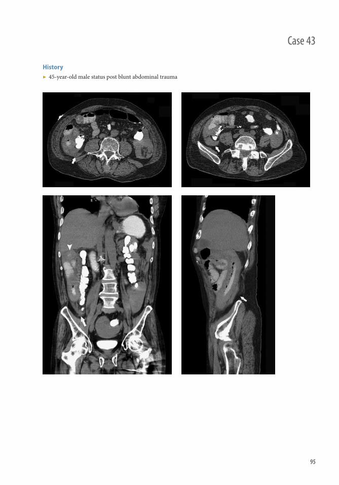
95
History ▶ 45-year-old male status post blunt abdominal trauma
Case 43
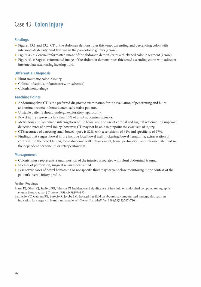
96
Case 43 Colon Injury
Findings ▶ Figures 43.1 and 43.2: CT of the abdomen demonstrates thickened ascending and descending colon with
intermediate density fl uid layering in the paracolonic gutters (arrow). ▶ Figure 43.3: Coronal reformatted image of the abdomen demonstrates a thickened colonic segment (arrow). ▶ Figure 43.4: Sagittal reformatted image of the abdomen demonstrates thickened ascending colon with adjacent
intermediate attenuating layering fl uid.
Diff erential Diagnosis ▶ Blunt traumatic colonic injury ▶ Colitis (infectious, infl ammatory, or ischemic) ▶ Colonic hemorrhage
Teaching Points ▶ Abdominopelvic CT is the preferred diagnostic examination for the evaluation of penetrating and blunt
abdominal trauma in hemodynamically stable patients. ▶ Unstable patients should undergo exploratory laparotomy. ▶ Bowel injury represents less than 10% of blunt abdominal injuries. ▶ Meticulous and systematic interrogation of the bowel and the use of coronal and sagittal reformatting improve
detection rates of bowel injury; however, CT may not be able to pinpoint the exact site of injury. ▶ CT’s accuracy of detecting small bowel injury is 82%, with a sensitivity of 64% and specifi city of 97%. ▶ Findings that suggest bowel injury include focal bowel wall thickening, bowel hematoma, extravasation of
contrast into the bowel lumen, focal abnormal wall enhancement, bowel perforation, and intermediate fl uid in the dependent peritoneum or retroperitoneum.
Management ▶ Colonic injury represents a small portion of the injuries associated with blunt abdominal trauma. ▶ In cases of perforation, surgical repair is warranted. ▶ Less severe cases of bowel hematoma or nonspecifi c fl uid may warrant close monitoring in the context of the
patient’s overall injury profi le.
Further Readings Brasel KJ , Olson CJ , Staff ord RE , Johnson TJ . Incidence and signifi cance of free fl uid on abdominal computed tomographic
scan in blunt trauma . J Trauma . 1998; 44 ( 5 ): 889–892 . Eanniello VC , Gabram SG , Eusebio R , Jacobs LM . Isolated free fl uid on abdominal computerized tomographic scan: an
indication for surgery in blunt trauma patients? Connecticut Medicine . 1994; 58 ( 12 ): 707–710 .

97
Case 44
History ▶ 25-year-old male with abdominal pain and nausea following an MVA.

98
Findings CT
▶ May be normal within the fi rst 12 hours aft er injury ▶ Pancreatic enlargement, peripancreatic edema or hematoma (black arrows), with or without fl uid between
pancreas and splenic vein ▶ Diff use or localized hypoattenuating area with preserved enhancement suggests contusion ▶ Hematoma appears as heterogeneous or hyperattenuating lesion ▶ Lacerations demonstrate linear low-attenuation lesions that can be diffi cult to see (white arrows) ▶ Ductal disruption may be identifi ed
MRI
▶ T1WI: Variable signal intensity ▶ T2WI: Peripancreatic fl uid or pseudocyst ▶ Contrast-enhanced T1WI: Nonenhancing or hypoenhancing areas caused by contusion, laceration, fl uid
collection, or necrosis. ▶ MRCP may demonstrate duct compromise
Diff erential Diagnosis ▶ Pancreatitis ▶ Duodenal injury or rupture ▶ “Shock” pancreas
Teaching Points ▶ CT is the preferred initial examination
■ MRCP useful for diagnosing duct injury, which is important for guiding management ▶ Commonly associated with other organ injuries (80%) or the duodenum ▶ More common with penetrating injury than blunt trauma ▶ Grading
■ I: Minor hematoma or laceration without duct injury ■ II: Major contusion or laceration without duct injury ■ III: Distal transection or parenchymal injury with duct injury ■ IV: Proximal transection or parenchymal injury involving the ampulla or bile duct ■ V: Massive disruption of the pancreatic head
▶ Complications are common, occurring in 30%–60% of cases ■ Postraumatic pancreatitis ■ Pseudocysts ■ Pancreatic duct fi stulas ■ Abscess
Management ▶ Conservative management for low -grade injuries with intact duct (Grade I/II) ▶ Higher-grade injuries with ductal disruption usually require surgical management
Case 44 Pancreatic Injury
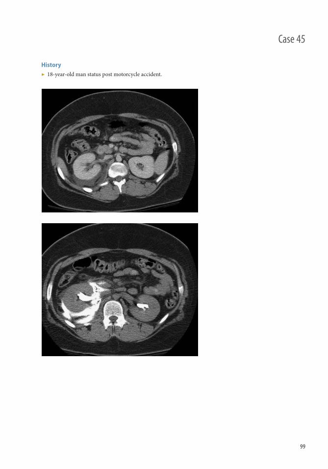
99
Case 45
History ▶ 18-year-old man status post motorcycle accident.

100
Findings CT
▶ Noncontrast images show simple fl uid density collection. ▶ Delayed images show increasing density as excreted contrast gathers in the collection. ▶ Located adjacent to aff ected kidney and/or along the course of urinary tract in the retroperitoneum and pelvis.
Ultrasound
▶ Fluid collection in the perirenal space, pelvis, or retroperitoneum along the course of urinary tract. ▶ Intermittent signal on color Doppler from turbulence in the urinoma caused by urine fl ow.
Diff erential Diagnosis ▶ Lymphocele ▶ Abscess ▶ Hematoma
Teaching Points ▶ Encapsulated collection of urine caused by injury to the intrarenal or extrarenal collecting system. Most
commonly located in the perirenal space. ▶ Etiology
■ Blunt or penetrating trauma results in collecting system, ureteral, or urinary bladder injury. ■ Iatrogenic causes. ■ For perirenal urinomas, trauma is a more common cause. Periureteral urinomas are more commonly
iatrogenic. ▶ Presentation: Pain, fever, hydronephrosis, and electrolyte imbalances if found late. ▶ Urinoma in the context of renal parenchymal injury is highly suggestive of collecting system injury (grade IV
renal injury).
Management ▶ Small urinomas are usually managed conservatively and usually resolve spontaneously. ▶ Percutaneous drainage may be considered for larger or infected urinomas. ▶ Nephrostomy catheters, ureteral stents, or nephroureteral catheters may also be placed to divert urine fl ow.
Further Readings Titton RL , Gervais DA , Hahn PF , Harisinghani MG , Arellano RS , Mueller PR . Urine leaks and urinomas: diagnosis and
imaging–guided intervention . RadioGraphics . 2003 ; 23 ( 5 ): 1133–1147 . Testa AC, Gaurilcikas A, Licameli A, Di Stasi C, Lorusso D, Scambia G, et al. Sonographic imaging of urinoma . Ultrasound
Obstet Gynecol . 2009 ; 33 ( 4 ): 490–491 .
Case 45 Urinoma

101
History ▶ 45-year-old male status post motor vehicle collision.
Case 46

102
Case 46 Adrenal Injury
Findings CT
▶ Hyperdense nonenhancing hematoma (50-80 HU); chronically, this will gradually decrease in size and density, and may eventually form calcifi cations
▶ Asymmetric round or distorted enlargement of the adrenal gland ▶ Periadrenal fat stranding or frank hemorrhage, possibly with retroperitoneal extension ▶ Other traumatic fi ndings (i.e., fractures, pneumothorax, laceration/contusion of other solid organs)
Ultrasound
▶ Avascular hyperechoic or hypoechoic mass; over time this will decrease in size and echogenicity, chronically becoming anechoic, possibly with shadowing calcifi cations
Diff erential Diagnosis ▶ Adrenal hemorrhage from nontraumatic causes: Hemorrhagic adrenal mass, adrenal vein ligation/thrombosis
during liver transplantation ■ Bilateral adrenal hemorrhages more likely to occur from systemic etiologies, such as in the setting of
anticoagulation or in situations of high stress (i.e., sepsis [Waterhouse-Friderichsen syndrome], burns, or hypotension)
▶ Adrenal hyperplasia ▶ Adrenal adenoma or myelolipoma ▶ Pheochromocytoma, adrenal carcinoma, metastasis, or lymphoma
Teaching Points ▶ Blunt force trauma is the most common etiology ▶ Typically unilateral, right much more common than left ▶ When bilateral, it can result in a catastrophic crisis of adrenal insuffi ciency
■ In the setting of minimal trauma with bilateral adrenal hemorrhage, a search of underlying coagulopathy is warranted
▶ If the mass does not decrease in size or resolve on follow-up CT, it may represent a disorder other than trauma and should be further evaluated as deemed clinically appropriate
Management ▶ Th erapeutic interventions for associated injuries that commonly accompany adrenal trauma ▶ Replacement steroid therapy in the setting of bilateral adrenal injury and subsequent crisis of adrenal insuffi ciency ▶ Adrenalectomy is typically not necessary, except in the setting where an underlying adrenal tumor is present
Further Readings Blake MA, Cronin CG, Boland GW. Adrenal imaging . AJR Am J Roentgenol . 2010 Jun; 194 ( 6 ): 1450–60 . Mayo-Smith WW , Boland GW , Noto RB , Lee MJ . State-of-the-art adrenal imaging . RadioGraphics . 2001 ; 21 ( 4 ): 995–1012 .

103
Case 47
History ▶ 78-year-old male status post bladder transurethral resection of the prostate.

104
Findings ▶ CT cystography demonstrates contrast extravasation into perivesical region (Figure 47.1). ▶ May see defect in the bladder wall (Figure 47.2). ▶ Complex extraperitoneal bladder rupture will also demonstrate extravasation into other perineal spaces and
along the fascial planes of the abdominal wall and the thighs (Figure 47.3).
Diff erential Diagnosis ▶ Intraperitoneal bladder rupture or combined extraperitoneal and intraperitoneal bladder rupture. ▶ Interstitial bladder injury where a tearing of the luminal layers of the bladder wall with an intact serosa can
lead to contrast with the wall.
Teaching Points ▶ Extraperitoneal rupture is the most common type of bladder injury. ▶ Mechanisms of injury
■ Penetrating trauma ■ Blunt trauma associated with pelvic fractures ■ Iatrogenic (biopsy or TURP)
▶ CT cystography is absolutely indicated in the context of both gross hematuria and pelvic fractures. Less strongly indicated in patients with either gross hematuria or pelvic fracture.
▶ Grading system ■ Type 1: Bladder contusion ■ Type 2: Intraperitoneal rupture ■ Type 3: Interstitial injury ■ Type 4: Extraperitoneal rupture ■ 4A: Simple extraperitoneal rupture (perivesical space) ■ 4B: Complex extraperitoneal rupture (extending to scrotum, perineum, thigh)
▶ Type 5: Combined intraperitoneal and extraperitoneal rupture ■ Cannot adequately assess an underdistended bladder: 300 mL of contrast fi lling is needed to exclude rupture.
Management ▶ Typically, conservative treatment for extraperitoneal rupture with large-bore transurethral or suprapubic
catheter.
Further Readings Vaccaro JP , Brody JM . CT cystography in the evaluation of major bladder trauma . RadioGraphics . 2000 ; 20 ( 5 ): 1373–1381 . Corriere JN , Sandler CM . Diagnosis and management of bladder injuries. Urol Clin North Am . 2006 ; 33 ( 1 ): 67–71–vi .
Case 47 Extraperitoneal Bladder Rupture

105
History ▶ 21-year-old male with abdominal pain and hematuria following an MVA.
Case 48

106
Findings Ultrasound
▶ Free intraperitoneal fl uid ▶ May bladder wall irregularity/discontinuity (black arrow) or intraluminal hemorrhage (white arrow).
Conventional Cystography
▶ Contrast extravasation into the peritoneal cavity
CT
▶ Low-attenuation fl uid in the peritoneal cavity (HU measurement)
CT Cystography
Diff erential Diagnosis ▶ Diff erentials for intraperitoneal fl uid associated with trauma
■ Simple fl uid (<20 HU) ■ Ascites ■ Bile ■ Urine ■ Intermediate density (20-40 HU) ■ Bowel contents (small bowel injury) ■ Old blood ■ High density (>40 HU) ■ Blood ■ Opacifi ed bowel contents or urine
Teaching Points ▶ Etiology
■ Blunt trauma (most common): Seat belt or steering wheel injury ■ Penetrating injury: Gunshot or knife wounds ■ Iatrogenic injury
▶ Location of extravasation depends on site of injury. Superior rupture causes extravasation into intraperitoneal space. Th is is usually a result of direct pressure applied against a distended bladder.
▶ CT cystography: Bladder must be adequately distended with minimum of 300 mL diluted contrast for adults. For children, estimated fi lling for cystography is based on the formula: bladder capacity = 60 mL + (30 mL × age in years)
▶ Grading system ■ Type 1: Bladder contusion ■ Type 2: Intraperitoneal rupture ■ Type 3: Interstitial injury ■ Type 4: Extraperitoneal rupture (4A simple, and 4B complex) ■ Type 5: Combined intraperitoneal and extraperitoneal rupture
Management ▶ Intraperitoneal and combined intraperitoneal and extraperitoneal ruptures usually require primary
surgical repair.
Case 48 Intraperitoneal Bladder Rupture

107
History ▶ None
Case 49

108
Case 49 Urethral Injury
Findings Retrograde Urethrography (imaging test of choice)
▶ Discontinuity or irregularity of the urethra ▶ Extravasation of contrast from the urethra
■ Partial rupture: Contrast is seen within the urethra proximal to defect ■ Complete rupture: No contrast is seen within urethra proximal to defect
▶ In posterior urethral injury ■ If the urogenital diaphragm remains intact, contrast is only be seen in the pelvis ■ If the urogenital diaphragm is disrupted, contrast is seen in the pelvis and the perineum
Intravenous Urethrography
▶ Extravasation of contrast in voiding phase ■ Adjacent to bulbous urethra: Anterior urethral injury ■ Around the bladder base/prostate: Posterior urethral injury
CT
▶ Perineal or periurethral stranding in a patient with pelvic trauma ▶ Ischiocavernous hematoma
Teaching Points ▶ Urethral injury is seen in as many as 25% of men with pelvic fractures ▶ Location
■ Two-thirds involve the posterior urethra ■ Associated with pelvic fracture related to MVC or fall from height ■ Typically involves the membranous urethra
■ One-third involve the anterior urethra ■ Straddling-type injury ■ Typically involve the immobile bulbous urethra, which is crushed against an inferior pubic ramus
■ Female urethral injury is rare given short urethral length ▶ Classifi cation
■ Goldman System (most widely accepted): Based on the anatomic location of the injury ■ Type 1: Urethra stretch without rupture ■ Type 2: Partial or complete rupture of the membranous urethra above the level of the urogenital
diaphragm ■ Type 3: Partial or complete rupture of the membranous urethra with disruption of the urogenital
diaphragm ■ Type 4: Bladder neck injury with extension into the urethra ■ Type 5: Isolated anterior urethral injury
▶ Stricture is a common late complication of anterior urethral injury, seen in as many at two-thirds of patients
Management ▶ Partial rupture: Placement of a urethral catheter for 2–3 weeks to allow urethral healing; follow-up
urethography to confi rm healing ▶ Complete rupture: Surgical repair
■ Surgical repair 3–6 months to allow pelvic hematoma to resolve ■ Suprapubic catheter is placed to divert urine

109
History ▶ Motorcycle accident.
Case 50

110
Findings ▶ Testicular contour is ill-defi ned on ultrasound (Figures 50.1 and 50.2). ▶ Heterogeneous echotexture of the testicular parenchyma (Figures 50.1 and 50.2). ▶ Interruption of the tunica may be directly visualized (Figure 50.3). ▶ May also see abnormal or absent vascularity within a portion of the testis. ▶ Linear hypoechoic band without vascularity can indicate testicular fracture, a rarer injury. ▶ Hematoma may be seen within the testis; within the tunica vaginalis (hematocele); within the scrotal wall; or
within the spermatic cord.
Teaching Points ▶ Testicular rupture is protrusion of the seminiferous tubules through a tear in the tunica albuginea (arrow).
■ Most occur from blunt trauma ■ Blunt trauma usually requires higher specifi city of clinical and imaging fi ndings for surgical exploration
than penetrating trauma ■ Ultrasound evaluation is particularly useful in the setting of blunt trauma
▶ Various testicular injuries have overlapping sonographic features, and a heterogeneous appearance can refl ect testicular hematoma and/or contusion (injured, devitalized tissue), with or without the presence of rupture. ■ Rupture should, therefore, not be diagnosed based on heterogeneity alone ■ Hypoechoic avascular linear tissue defects refl ect shear injury (testicular fracture) and may be seen
concurrently with other injuries ▶ Th e combined fi ndings of heterogeneous echotexture and an ill-defi ned contour of the testicle have high
sensitivity and specifi city for rupture. ■ Adjacent hematoma can simulate a testicular contour abnormality, and so the contour abnormality is
considered an indirect sign of rupture ■ Heterogeneity concurrent with tissue interruption at surface of the testicle (direct observation of tunica
albuginea tear) is highly specifi c for rupture, but the latter fi nding is oft en diffi cult to make ▶ Testicular hematoma requires follow-up imaging to resolution.
■ Partly, this is to evaluate for the development of necrosis and infection of the hematoma. ■ In addition, testicular tumors can present as pain related to minor trauma and simulate the appearance of
hematoma on ultrasound. ▶ Traumatic testicular torsion has a similar ultrasound appearance to nontraumatic torsion.
Management ▶ Most testicular ruptures can be surgically repaired when detected promptly. ▶ Extensive rupture requires orchiectomy. ▶ Intratesticular hematomas require imaging follow-up.
Case 50 Scrotal/Testicular Injury

111
Case 51
History ▶ Trauma

112
Findings US
▶ Hypoechoic free fl uid in the peritoneal cavity ▶ May see some complexity in this fl uid depending on age ▶ Must diff erentiate bowel contents and physiologic fl uid
CT
▶ Hyperdense fl uid within the peritoneal cavity typically measuring between 30 and 60 HU ▶ May see “sentinel clot,” where highest-density material gathers around an injured organ ▶ May see focal area with density similar to adjacent vessel, suggestive of active extravasation ▶ Triangular collections of blood in mesenteric refl ections usually indicate mesenteric source of bleeding
Diff erential Diagnosis ▶ Intraperitoneal free fl uid
■ Simple ascites ■ Bile, secondary to biliary injury ■ Urine, secondary to urinary tract injury ■ Chyle, secondary to lymphatic injury ■ Intestinal contents, related to bowel injury
▶ High-attenuation ascites on CT ■ Infection especially tuberculosis ■ Malignancy, such as ovarian/appendiceal tumor (pseudomyxoma) ■ Ascitic fl uid on a background of severe fatty liver
▶ Mimics of hemoperitoneum on US ■ Fluid-fi lled bowel loops ■ Perinephric fat
Teaching Points ▶ Etiologies of hemoperitoneum
■ Trauma ■ Gynecologic sources: Ruptured ovarian cyst, ruptured ectopic pregnancy, ovarian torsion ■ Spontaneous hemorrhage associated with anticoagulation therapy ■ Iatrogenic, following surgery or invasive procedure ■ Highly vascular neoplasms
▶ Ruptured aortic or visceral artery aneurysms ▶ Attenuation values at CT help diff erentiate blood products from other sources of intraperitoneal fl uid
■ Clotted blood measures 45-70 HU ■ Unclotted intraperitoneal blood usually measures 30-45 HU ■ May measure <30 HU in patients with decreased serum hematocrit level ■ Exact attenuation of blood depends on age, extent, and location of hemorrhage
▶ Highest attenuation hematoma or “sentinel clot” is closest to site of bleeding and may be used to identify source of bleeding
▶ Spleen and liver injuries are the most common solid organ injuries to result in hemoperitoneum ▶ Duodenum and proximal jejunum are most common sites of bowel injury giving rise to hemoperitoneum
Management ▶ Close monitoring of vital signs for evidence of hemodynamic instability ▶ Active extravasation and/or hemodynamic instability may indicate need for emergent surgical or endovascular
treatment ▶ Defi nitive treatment through correction of underlying pathology (e.g., pseudoaneurysm coiling, splenectomy)
Case 51 Hemoperitoneum

113
Case 52
History ▶ Trauma

114
Findings ▶ Focal area of hyperattenuation within a hematoma on initial postcontrast images ▶ On delayed images (typically at 5 minutes), the area of hyperattenuation enlarges and changes in shape and
attenuation ▶ Can be found associated with solid organ injuries, muscular injuries, or direct vascular injuries
Diff erential Diagnosis ▶ Pseudoaneurysm and arteriovenous fi stula ▶ Opacifi ed urine from injured renal collecting system ▶ Radiopaque foreign body or bone fragment ▶ Enteric contrast leakage from bowel injury
Teaching Points ▶ Important indicator of morbidity and mortality in trauma patients ▶ Typically associated with active hemorrhage ▶ Localization of source of bleeding important for determining appropriate course of management ▶ Can be seen from various abdominal structures including liver, adrenal glands, kidneys, pancreas, spleen,
mesentery, and soft tissues ▶ Dual-phase CT protocol with 5-minute delayed images key for diff erentiation from pseudoaneurysm
■ Active extravasation enlarges, changes in shape, and remains hyperdense ■ Pseudoaneurysm does not change in shape or size and washes out with the vascular pool
▶ Adjacent vascular structures should be evaluated for dissection, pseudoaneurysm, and lack of enhancement (caused by spasm and/or occlusion)
Management ▶ Expectant/supportive management may be used for minor injuries, especially intramuscular or subcutaneous
soft tissue injuries. ▶ Signifi cant injuries, such as in patients with multiple injuries, or solid organ injuries, such as the spleen, may
require endovascular or open surgical management. ▶ Interventions may range from intravenous fl uid and transfusions of blood products to endovascular
embolization, surgical ligation, or resection of injured organ.
Further Readings Murakami AM , Anderson SW , Soto JA , Kertesz JL , Ozonoff A , Rhea JT . Active extravasation of the abdomen and pelvis in
trauma using 64MDCT . Emerg Radiol . 2009 ; 16 ( 5 ): 375–382 . Hamilton JD , Kumaravel M , Censullo ML , Cohen AM , Kievlan DS , West OC . Multidetector CT evaluation of active
extravasation in blunt abdominal and pelvic trauma patients . RadioGraphics . 2008 ; 28 ( 6 ): 1603–1616 .
Case 52 Active Extravasation

115
Case 53
History ▶ 34-year-old man posttrauma, worsening abdominal pain.

116
Case 53 Posttraumatic bile duct leak
Findings ▶ Abdominal CT with IV contrast: Jagged curvilinear hypodensities (arrow) through the left and right lobes of
the liver compatible with laceration. Low-attenuation fl uid around the liver and spleen and fl uid in contiguity with the left lobe laceration.
▶ ERCP: Occlusion cholangiogram demonstrates contrast leakage from left sided biliary radicals
Diff erential Diagnosis ▶ Simple ascites ▶ Hemoperitoneum from laceration of portal veins, hepatic arteries, or hepatic veins ▶ Peritoneal carcinomatosis and infi ltrative liver metastases
Teaching Points ▶ Presence of free fl uid adjacent to the liver in a posttraumatic patient should raise concern for parenchymal
laceration and underlying vascular or biliary injury. ▶ Biliary complications as a result of bile leakage caused by injury to the bile duct system include biloma, biliary
fi stula, bilemia, and bile peritonitis. ▶ At CT, progressive enlargement of a well-circumscribed, hypodense intraparenchymal or perihepatic
collection aft er trauma strongly suggests the diagnosis of a biloma. ▶ Further evaluation with ERCP may be helpful to exclude biliary injury. ▶ Sulfur-colloid scintigraphy, or MRI with Eovist (intravenous contrast agent excreted by both the kidneys and
biliary system), with delayed imaging can serve as noninvasive ways of identifying a bile duct leak.
Management ▶ Percutaneous drainage, in combination with endoscopic sphinchterotomy and/or biliary stenting, can be a
good initial treatment in posttraumatic bile duct injury. ▶ Surgical management may be needed in more complex cases.
Further Readings Yoon W , Jeong YY , Kim JK , Seo JJ , Lim HS , Shin SS , et al. CT in blunt liver trauma . RadioGraphics . 2005; 25 : 87–104 . Haney PJ , Whitley NO , Brotman S , Cunat JS , Whitley J. Liver injury and complications in the postoperative trauma patient: CT
evaluation . AJR Am J Roentgenol. 1982; 139 ( 2 ): 271–275 .

117
Case 54
History ▶ MVA

118
Case 54 Splenic Injury/Laceration
Findings CT
▶ Coronal images with IV contrast demonstrating a splenic laceration with active extravasation (arrow)
Arterial Angiogram
▶ Pre-embolization and postembolization demonstrating an active bleeding/pseudoaneurysm in the spleen
Teaching Points ▶ Th e spleen is the most commonly injured organ aft er blunt trauma to the abdomen. ▶ Th e American Association for the Surgery of Trauma (AAST) splenic injury grading system:
■ Grade I ■ Subcapsular hematoma <10% surface area ■ Capsular laceration <1 cm depth
■ Grade II ■ Subcapsular hematoma 10%–50% surface area ■ Intraparenchymal hematoma <5 cm ■ Laceration 1–3 cm depth not involving trabecular vessels
■ Grade III ■ Subcapsular hematoma >50% of surface area or expanding ■ Intraparenchymal hematoma >5 cm/expanding ■ Laceration >3 cm depth or involving trabecular vessels ■ Ruptured subcapsular or parenchymal hematoma
■ Grade IV ■ Laceration involving segmental/hilar vessels with major devascularization (>25% of spleen)
■ Grade V ■ Shattered spleen ■ Hilar vascular injury with devascularized spleen
▶ Th e focused assessment with sonography in trauma (FAST) examination is more useful in hemodynamically unstable patients; however, a negative examination does not exclude splenic injury.
▶ Th e diagnostic peritoneal lavage (DPL) is largely replaced by the FAST in most major trauma centers. ▶ Pitfalls in diagnosis of splenic injury
■ Normal lobulation/cleft mimics tear ■ Adjacent nonopacifi ed bowel loop (jejunum) may look like laceration ■ Too early acquisition of scan aft er bolus spleen can appear inhomogeneous (diff erential enhancement of red
and white pulp) ■ Previous infarct ■ Perisplenic fl uid from ascites or lavage
Management ▶ Hemodynamically unstable: patient with a positive FAST scan or DPA/DPL requires emergent abdominal
exploration. ▶ Hemodynamically stable: patients with low-grade (I–III) blunt or penetrating splenic injuries without any
evidence for other intra-abdominal injuries, active contrast extravasation, may be initially observed safely. ▶ Th e use of nonsurgical management as the fi rst-line therapeutic step in adults has been advocated by Th e
Eastern Association for the Surgery of Trauma Practice Management Guidelines Working Group. ▶ Surgery is usually performed in patients who have traumatic injuries to the spleen and unstable hemodynamics. ▶ Th e splenic arterial embolization is mostly used with evidence of arterial injury on CT scans. With
nonoperative management, splenic function is preserved, and the lifelong risk of postsplenectomy sepsis, surgery complications, hospitalization periods, and costs are lower.

119
Case 55
History ▶ Trauma

120
Case 55 Renal Laceration with Hematoma, Vascular Extravasation, and Urine Extravasation
Findings ▶ Axial CT with contrast demonstrating right kidney injury with contrast extravasation (arrow). Th ere is an
extensive hematoma surrounding the kidney. ▶ Axial CT, with delayed imaging demonstrating urine leak (double arrows).
Teaching Points ▶ Arterial-phase CT is useful to help delineate renal arterial injury, but nephrographic and urinary phase
imaging may also be necessary to fully delineate renal injuries. ▶ Delayed CT images are important to check for urine leak, which is typically treated with placement of ureteral
stent. Delayed CT is also useful to separate extravasated urine (which accumulates) from extravasated arterial contrast material, which dilutes aft er the bolus is stopped.
▶ Lacerations connecting the cortical surfaces through the hilum are termed fractures. Multiple separated renal fragments, which may or may not be perfused, are termed shattered.
▶ AAST grading system (1 ) ■ Grade I
■ Contusion; or ■ Nonexpanding subcapsular hematoma
■ Grade II ■ Nonexpanding perirenal hematoma; or ■ Laceration <1 cm deep into the renal cortex
■ Grade III ■ Laceration >1 cm but not extending to the collecting system
■ Grade IV ■ Laceration extending to the collecting system; or ■ Main renal artery/vein injury
■ Grade V ■ Shattered kidney; or ■ Renal hilum avulsion
▶ Chronic subcapsular hematoma can lead to compression necrosis of parenchyma (the Page kidney phenomenon), a rare cause of secondary hypertension and renal failure.
Management ▶ Patients with hematuria of <35 red blood cells per high-power fi eld, with no history of hypotension, and with
no pelvic fracture can usually forego imaging of the urinary tract. ▶ Parenchymal contusions and minor superfi cial lacerations do not necessitate surgery in 95% of cases (2). ▶ Major renal lacerations with urine leak in stable patients may be treated nonsurgically, with observation, serial
imaging, and placement of a stent in the ureter (3). ▶ Injuries with persistent large urine leak, ureteropelvic junction avulsion, enlarging central and subcapsular
hematoma, or extensively devitalized parenchyma (particularly in an immunocompromised patient) need surgical intervention.

Upper ExtremitySection 5

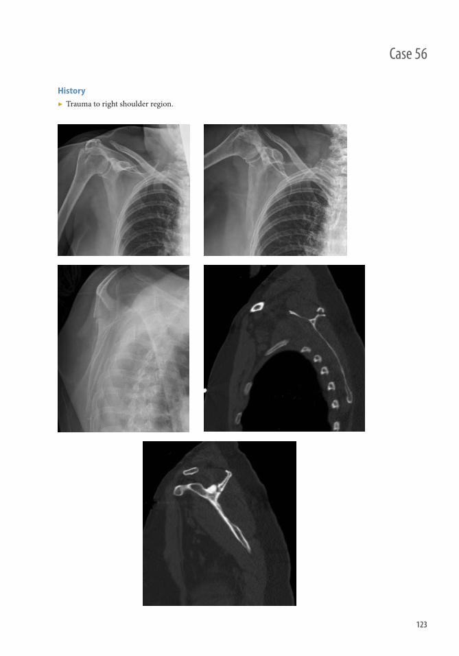
123
History ▶ Trauma to right shoulder region.
Case 56

124
Findings Radiography
▶ On the AP radiograph view of right shoulder it is diffi cult to appreciate the cortical discontinuity involving the spine of the scapula. Additional scapular views demonstrate this fracture to better advantage.
▶ Th is is also evident on the scapular Y-view. ▶ Th ere is normal glenohumeral alignment and no additional fracture.
CT
▶ Th e fracture of the spine of the scapula is confi rmed on CT. It extended into the suprascapular notch but not to the glenoid articular surface.
Teaching Points ▶ Fractures of the scapula are relatively rare injuries; they represent 3%–5% of all fractures involving the
shoulder girdle and 1% of fractures overall. ▶ Direct trauma to the lateral or posterosuperior aspect of the forequarter is the most common mechanism
of injury. Ninety percent of patients who sustain scapular fractures have other associated injuries. Because attention is oft en directed initially toward these other injuries, fractures of the scapula are oft en initially overlooked.
▶ Fractures that involve the glenoid neck and scapular body are the most common ▶ Th ere is a high incidence of pneumothorax, occurring in half of patients. Most are delayed in onset, occurring
1–3 days aft er the injury. Some authors advocate a follow-up chest radiograph.
Management ▶ If nonoperative treatment is chosen, the shoulder is immobilized for comfort. Passive exercises are initiated
early and active-assisted range of motion exercises are begun once there is evidence of union. ▶ Th e decision to proceed to surgical treatment is primarily based on the degree of displacement. ▶ If surgery is considered, it is usually performed through a posterior approach. Screw fi xation alone is not
usually suffi cient because of the thin quality of the scapular bone. Plate fi xation with horizontally or obliquely oriented plates with multiple screws is therefore usually necessary to obtain stability.
Further Readings Lapner PC , Uhthoff HK , Papp S . Scapula fractures. Orthop Clin North Am . 2008 ; 39 ( 4 ): 459–474 , vi. PMID: 18803976 Cole PA , Gauger EM , Schroder LK . Management of scapular fractures. J Am Acad Orthop Surg . 2012 ; 20 ( 3 ): 130–141 . PMID: 2238228
Case 56 Scapular Fracture
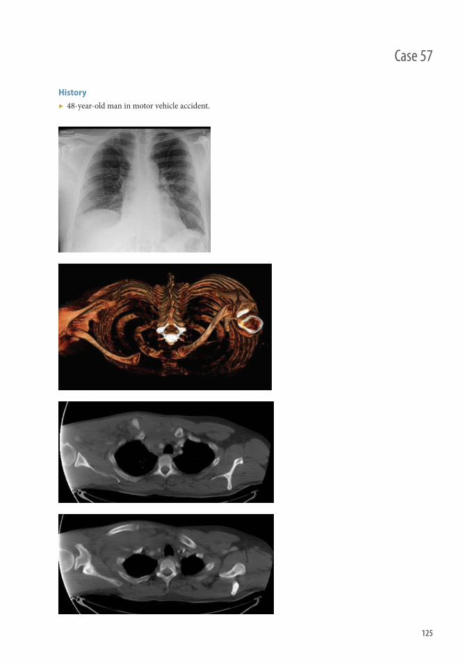
125
History ▶ 48-year-old man in motor vehicle accident.
Case 57
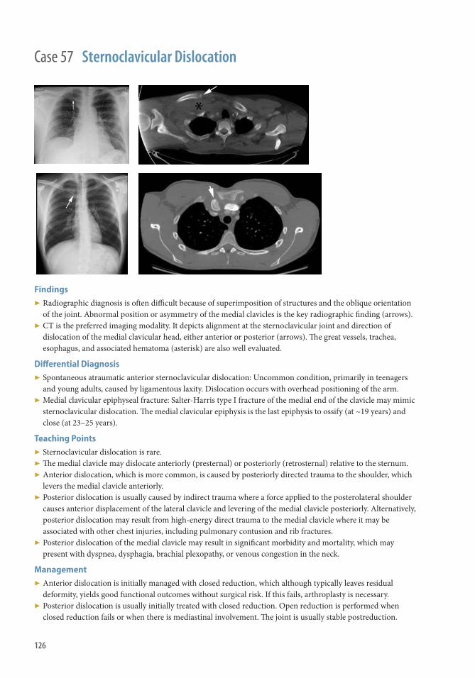
126
Case 57 Sternoclavicular Dislocation
Findings ▶ Radiographic diagnosis is oft en diffi cult because of superimposition of structures and the oblique orientation
of the joint. Abnormal position or asymmetry of the medial clavicles is the key radiographic fi nding (arrows). ▶ CT is the preferred imaging modality. It depicts alignment at the sternoclavicular joint and direction of
dislocation of the medial clavicular head, either anterior or posterior (arrows). Th e great vessels, trachea, esophagus, and associated hematoma (asterisk) are also well evaluated .
Diff erential Diagnosis ▶ Spontaneous atraumatic anterior sternoclavicular dislocation: Uncommon condition, primarily in teenagers
and young adults, caused by ligamentous laxity. Dislocation occurs with overhead positioning of the arm. ▶ Medial clavicular epiphyseal fracture: Salter-Harris type I fracture of the medial end of the clavicle may mimic
sternoclavicular dislocation. Th e medial clavicular epiphysis is the last epiphysis to ossify (at ~19 years) and close (at 23–25 years).
Teaching Points ▶ Sternoclavicular dislocation is rare. ▶ Th e medial clavicle may dislocate anteriorly (presternal) or posteriorly (retrosternal) relative to the sternum. ▶ Anterior dislocation, which is more common, is caused by posteriorly directed trauma to the shoulder, which
levers the medial clavicle anteriorly. ▶ Posterior dislocation is usually caused by indirect trauma where a force applied to the posterolateral shoulder
causes anterior displacement of the lateral clavicle and levering of the medial clavicle posteriorly. Alternatively, posterior dislocation may result from high-energy direct trauma to the medial clavicle where it may be associated with other chest injuries, including pulmonary contusion and rib fractures.
▶ Posterior dislocation of the medial clavicle may result in signifi cant morbidity and mortality, which may present with dyspnea, dysphagia, brachial plexopathy, or venous congestion in the neck.
Management ▶ Anterior dislocation is initially managed with closed reduction, which although typically leaves residual
deformity, yields good functional outcomes without surgical risk. If this fails, arthroplasty is necessary. ▶ Posterior dislocation is usually initially treated with closed reduction. Open reduction is performed when
closed reduction fails or when there is mediastinal involvement. Th e joint is usually stable postreduction.

127
Case 58
History ▶ None

128
Findings ▶ Injury to acromioclavicular joint supporting structures (including coracoclavicular ligaments)
Subtypes (Rockwood classifi cation)
▶ Type I: Acromioclavicular ligament sprain without ligamentous tear ■ No imaging abnormality, no joint malalignment
▶ Type II: Acromioclavicular ligament tear ■ Increased acromioclavicular distance >5–6 mm on AP view
■ Normal anatomic variation: no >3 mm diff erence from contralateral ■ May be partial tear of ligaments and require stress views with weights to diagnose
▶ Type III: Complete tear of acromioclavicular and coracoclavicular ligaments ■ Superior displacement of clavicle
■ Increased coracoclavicular distance >11–13 mm on AP view ■ Normal anatomic variation: no >5 mm diff erence from contralateral
▶ Type IV: Complete tear of acromioclavicular and coracoclavicular ligaments with posterior displacement of clavicle ■ Because superior displacement may be absent, AP view may appear normal; need axillary view to detect
posterior displacement ▶ Type V: Complete tear of acromioclavicular and coracoclavicular ligaments and torn muscular attachments to
clavicle (deltoid and trapezius) with SUPERIOR displacement of clavicle ▶ Type VI: Complete tear of acromioclavicular and coracoclavicular ligaments with INFERIOR displacement of
clavicle (rare)
Diff erential Diagnosis ▶ Normal anatomic variation ▶ Clavicle fracture
Teaching Points ▶ Mechanism
■ Direct blow to superior aspect of shoulder, usually from fall (most common) ■ Fall on outstretched hand (less common)
Management ▶ Treatment
■ Type I: Conservative (rest, pain control, activity modifi cation) ■ Type II: Conservative (rest, pain control, activity modifi cation) ■ Type III: Mostly conservative; some opt for surgical management if vocation/avocation requires heavy lift ing ■ Type IV–VI: Surgical fi xation
Case 58 Acromioclavicular (AC) Joint Separation (Type III)

129
Case 59
History ▶ None
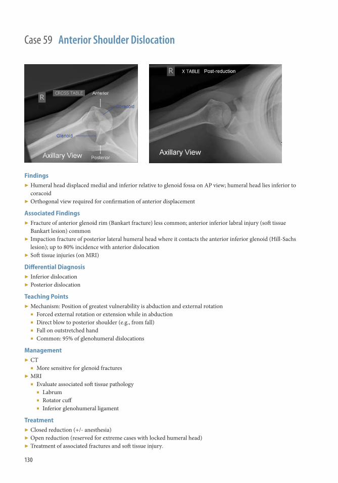
130
Case 59 Anterior Shoulder Dislocation
Findings ▶ Humeral head displaced medial and inferior relative to glenoid fossa on AP view; humeral head lies inferior to
coracoid ▶ Orthogonal view required for confi rmation of anterior displacement
Associated Findings ▶ Fracture of anterior glenoid rim (Bankart fracture) less common; anterior inferior labral injury (soft tissue
Bankart lesion) common ▶ Impaction fracture of posterior lateral humeral head where it contacts the anterior inferior glenoid (Hill-Sachs
lesion); up to 80% incidence with anterior dislocation ▶ Soft tissue injuries (on MRI)
Diff erential Diagnosis ▶ Inferior dislocation ▶ Posterior dislocation
Teaching Points ▶ Mechanism: Position of greatest vulnerability is abduction and external rotation
■ Forced external rotation or extension while in abduction ■ Direct blow to posterior shoulder (e.g., from fall) ■ Fall on outstretched hand ■ Common: 95% of glenohumeral dislocations
Management ▶ CT
■ More sensitive for glenoid fractures ▶ MRI
■ Evaluate associated soft tissue pathology ■ Labrum ■ Rotator cuff ■ Inferior glenohumeral ligament
Treatment ▶ Closed reduction (+/- anesthesia) ▶ Open reduction (reserved for extreme cases with locked humeral head) ▶ Treatment of associated fractures and soft tissue injury .

131
History ▶ None
Case 60
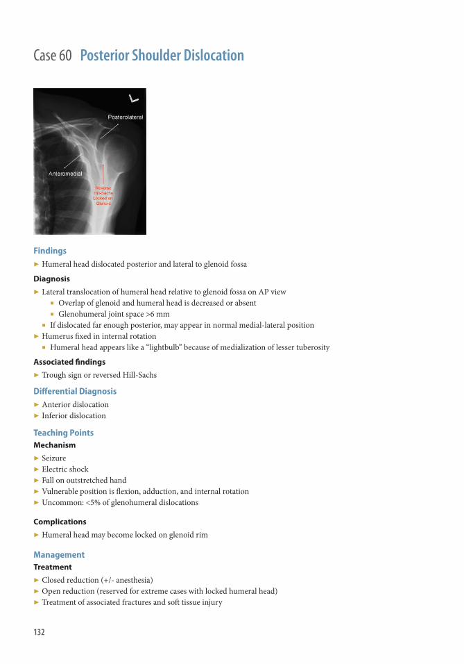
132
Findings ▶ Humeral head dislocated posterior and lateral to glenoid fossa
Diagnosis
▶ Lateral translocation of humeral head relative to glenoid fossa on AP view ■ Overlap of glenoid and humeral head is decreased or absent ■ Glenohumeral joint space >6 mm
■ If dislocated far enough posterior, may appear in normal medial-lateral position ▶ Humerus fi xed in internal rotation
■ Humeral head appears like a “lightbulb” because of medialization of lesser tuberosity
Associated fi ndings
▶ Trough sign or reversed Hill-Sachs
Diff erential Diagnosis ▶ Anterior dislocation ▶ Inferior dislocation
Teaching Points Mechanism
▶ Seizure ▶ Electric shock ▶ Fall on outstretched hand ▶ Vulnerable position is fl exion, adduction, and internal rotation ▶ Uncommon: <5% of glenohumeral dislocations
Complications
▶ Humeral head may become locked on glenoid rim
Management Treatment
▶ Closed reduction (+/- anesthesia) ▶ Open reduction (reserved for extreme cases with locked humeral head) ▶ Treatment of associated fractures and soft tissue injury
Case 60 Posterior Shoulder Dislocation

133
History ▶ Trauma to right shoulder region.
Case 61

134
Findings ▶ Th ere is a mildly displaced fracture of the proximal humerus at the metadiaphysis. ▶ No intra-articular extension identifi ed; no dislocation
Diff erential Diagnosis ▶ None
Teaching Points ▶ Proximal humeral fractures are common injuries accounting for 4%–5% of all fractures and are the third most
common fracture in patients older than 65 years of age. ▶ Surgical neck fractures are extracapsular, usually have an adequate blood supply, and relatively low incidence
of AVN; in contrast, anatomic neck fractures have a much higher incidence of AVN. ▶ In older patients they follow low-energy trauma, such as a fall on an outstretched hand from a standing height.
Th is can lead to marked comminution caused by osteoporosis. In younger patients they are higher-energy injuries and a fracture-dislocation can occur as the capsuloligamentous structures fail before the bone.
▶ Anatomic neck fractures occur just below the articular surface. Th ey are rare and have a poor prognosis because of loss of the blood supply to the head fragment.
▶ Surgical neck fractures occur between the level of the tuberosities and the insertion of pectoralis major. Th ey are extracapsular and the blood supply to the head is preserved.
Management ▶ Nonoperative treatment for stable proximal humeral fractures (stability is present when the patient can
actively move extremity without pain, and limb can be moved passively with little pain and no abnormal motion between fragments).
▶ Operative management for unstable fractures, using percutaneous screws, plate, and screw constructs, or hemiarthroplasty depending on severity.
Further Readings Harrison JW , Howcroft DW , Warner JG , Hodgson SP . Internal fi xation of proximal humeral fractures. Acta Orthop Belg .
2007 ; 73 ( 1 ): 1–11 . PMID: 17441651 Robinson CM , Amin AK , Godley KC , Murray IR , White TO . Modern perspectives of open reduction and plate fi xation of
proximal humerus fractures. J Orthop Trauma . 2011 ; 25 ( 10 ): 618–629 . PMID: 21904170
Case 61 Humerus Fracture, Proximal

135
Case 62
History ▶ Trauma to right elbow region

136
Case 62 Elbow Dislocation
Findings Radiography
▶ Th ere is posterior, lateral, and proximal displacement of the radius and ulna relative to the distal humerus. ▶ No displaced fracture identifi ed. Th ere is some associated soft tissue swelling.
Diff erential Diagnosis ▶ None
Teaching Points ▶ Th e elbow is the second most commonly dislocated joint in the body behind the shoulder in the adult
population. ▶ Simple dislocations represent dissociation of the ulnohumeral joint without concomitant fracture. Complex
instability occurs when a fracture is associated with dislocation. Bony injuries associated with dislocation include radial head and neck fractures, coronoid fractures, and avulsion of the medial and/or lateral epicondyles.
▶ Simple elbow dislocations are classifi ed by the direction of displacement as posterior, anterior, or divergent based on the relationship of the ulna and radius to the humerus. Th e most common direction of dislocation is posterior with the forearm positioned either medial or lateral to the humerus.
Management ▶ Once a stable reduction is obtained, most simple dislocations can be managed nonoperatively with splinting
or bracing, guided by the degree of instability determined during postreduction examination. ▶ Surgical management of simple elbow dislocation is indicated in elbows that remain unstable, even when
placed in fl exion and pronation. Th ese elbows typically have extensive soft tissue injury involving the lateral and ulnar collateral ligaments, the common extensor and fl exor-pronator muscle attachments, and the anterior capsule.
Further Readings Rosas HG , Lee KS . Imaging acute trauma of the elbow. Semin Musculoskelet Radiol . 2010 ; 14 ( 4 ): 394–411 . PMID: 20827621 Parsons BO , Ramsey ML . Acute elbow dislocations in athletes. Clin Sports Med . 2010 ; 29 ( 4 ): 599–609 . PMID: 20883899 Morrey BF. Current concepts in the management of complex elbow trauma. Surgeon . 2009 ; 7 ( 3 ): 151–161 . PMID: 19580179

137
Case 63
History ▶ Trauma to right elbow region.

138
Findings Radiography
▶ Th ere is an impacted comminuted fracture of the lateral articular surface of the radial head (straight arrow ). ▶ Th e lateral radiograph demonstrates a large elbow joint eff usion with a “sail-sign” anteriorly (curved arrow)
that represents the displaced anterior fat pad. In addition the posterior fat pad is visible consistent with a large eff usion.
Teaching Points ▶ Radial head fractures are the most common type of elbow fractures. Fractures are classifi ed based on the
degree of displacement, size of articular fragment, and degree of comminution. ▶ Th ey are usually caused by a fall on the outstretched hand, with the elbow fl exed. Axial and valgus loading
shears the radial head against the capitellum. ▶ Comminuted head fractures oft en signify a high-energy injury, although in osteoporotic bone they can result
from simple falls. Comminuted radial head fractures are the marker of a signifi cant and extensive elbow injury that only rarely, if ever, occurs in isolation. Comminuted radial head fractures have been associated with ligamentous injuries, proximal ulnar and capitellar fractures, and forearm interosseous membrane disruption.
▶ Radiographs of both wrists should be obtained when the clinical examination reveals forearm or ulnar-sided wrist pain that is suggestive of injury to the interosseous ligament.
▶ CT with sagittal, coronal, and three-dimensional reconstructions may assist with preoperative planning and can help the surgeon predict whether a displaced radial head fracture can be treated with ORIF or whether an arthroplasty is needed.
Management ▶ Nonoperative management is favored for undisplaced fractures. ▶ Options for the treatment of displaced fractures include nonoperative management, fragment excision, whole
head excision, open reduction and internal fi xation, and radial head arthroplasty.
Further Readings Rosenblatt Y , Athwal GS , Faber KJ . Current recommendations for the treatment of radial head fractures. Orthop Clin North
Am . 2008 ; 39 ( 2 ): 173–185. PMID: 1837480 Ring D . Radial head fracture: open reduction-internal fi xation or prosthetic replacement. J Shoulder Elbow Surg . 2011 ; 20 (Suppl
2): S107–S112 . PMID: 21281915
Case 63 Radial Head Fracture

139
Case 64
History ▶ 40-year-old man status post fall on an outstretched hand.

140
Findings ▶ Fracture of the radial shaft with dislocation of the distal radioulnar joint (DRUJ) ▶ Radial shaft fracture usually involves the middle to distal third ▶ DRUJ dislocation is usually dorsal because of shortening and rotation by the brachioradialis and rotation of
the distal fragment toward the ulna by the pronator quadratus
Diff erential Diagnosis ▶ Monteggia fracture-dislocation ▶ Essex-Lopresti ▶ Isolated radial shaft fracture
Teaching Points ▶ Etiology
■ Fall on an outstretched hand with a fl exed elbow ■ High-energy direct blow with forearm pronation
▶ Epidemiology: Uncommon, 3%–7% of all forearm fractures; more common in males ▶ Two types
■ Type I: Fracture within 7.5 cm of midarticular surface of the distal radius. Greater chance of DRUJ instability, potentially requiring K wire fi xation.
■ Type II: Fracture >7.5 cm from the joint surface. ■ Th e closer the radial shaft fracture to the DRUJ, the more likely the joint becomes unstable.
▶ CT is the best method to evaluate the DRUJ in uncertain cases
Management ▶ Most adults require open reduction internal fi xation.
Further Readings Giannoulis FS , Sotereanos DG . Galeazzi fractures and dislocations . Hand Clin . 2007 ; 23 : 153–163 . Rettig ME , Raskin KB . Galeazzi fracture-dislocation: a new treatment-oriented classifi cation . J Hand Surg Am . 2001 ; 26 ( 2 )
228–235 .
Case 64 Galeazzi Fracture-Dislocation

141
History ▶ Trauma to wrist region.
Case 65

142
Case 65 Colles Fracture
Findings Th ere is an impacted comminuted intra-articular fracture of the distal radius with dorsal displacement and angulation of the distal fragment. In addition there is a fracture of the distal ulnar styloid.
Diff erential Diagnosis ▶ Smith fracture ▶ Barton fracture ▶ Die-punch fracture
Teaching Points ▶ Fractures of the distal radius are very common, with an incidence ranging between 10% and 25% of all
fractures. ▶ Th e Colles fracture was fi rst described by Abraham Colles, an Irish surgeon, in 1814 before the advent of
radiography. Typically the distal radial fragment is dorsally displaced and angulated with resultant “dinner fork” deformity.
▶ Th e usual victim is a middle-aged or elderly woman who falls on the outstretched palm of her hand, force being translated upward to the glenohumeral articulation of the shoulder.
▶ Smith fractures occur in younger patients and are the result of high-energy trauma on the volar fl exed wrist. Volar comminution and intra-articular extension are more common. A Barton fracture only involves the rim of the radius and can be volar or dorsal. Dislocation of the radiocarpal joint is the hallmark of Barton fractures.
▶ A die-punch fracture is a depression fracture of the lunate fossa of the distal radius. It is the result of a transverse load through the lunate.
Management ▶ Anatomic reduction with stable fi xation is the treatment of choice for displaced, unstable fractures of the distal
part of the radius. Th e goals of treatment are to restore the articular surface congruency and to restore the radial height, radial inclination, and palmar tilt.
▶ CT is a useful radiographic adjunct and is recommended in the assessment of intraarticular distal radius fractures when the fracture pattern, the extent of comminution, or the magnitude and direction of the displacement of the fracture fragments cannot be determined with certainty on plain radiograph.
▶ Untreated fractures of the base of the ulnar styloid result in high rates of nonunion and have been associated with distal radioulnar joint instability. Th e role of internal fi xation of the ulnar styloid is controversial.

143
History ▶ None
Case 66

144
Findings ▶ Plain fi lm: Disruption of cortex or trabecula most commonly along the middle third (waist); normal if occult
fracture ▶ CT: Disruption of cortex or trabecula; better defi nes fracture displacement and angulation ▶ MRI: T1 hypointense fracture line; T2 hyperintense bone marrow edema; postcontrast images help determine
fracture fragment viability; best to evaluate occult fractures and ligamentous injury ▶ Chronic complications
■ Malunion: “humpback” deformity ■ Delayed union: Incomplete union aft er 4 months ■ Nonunion: Failure to heal aft er 6 months (persistent fracture line and sclerotic margins of bone) ■ Osteonecrosis: Fragmentation and collapse; T1 and T2 dark bone
Diff erential Diagnosis ▶ Intraosseous vascular vessel ▶ Nonunion from prior trauma ▶ Partial coalition (extremely rare)
Teaching Points ▶ Most common carpal bone fracture and most commonly fractured at the waist. ▶ Blood supply from radial artery enters at the waist and supplies the proximal bone in a retrograde fashion.
Th is makes the proximal fracture fragment vulnerable to osteonecrosis.
Management ▶ Cast immobilization for stable and nondisplaced ▶ Closed reduction with percutaneous pin or screw fi xation or ORIF for displaced or unstable fractures.
Further Readings Kaewlai R , Avery LL , Asrani AV , et al. Multidetector CT of carpal injuries: anatomy, fractures, and fracture-dislocations .
RadioGraphics . 2008; 28: 1771–1784 . Cassidy C, Leonard R . Fractures and dislocations of the carpus. In: Skeletal Trauma: Basic Science, Management, and
Reconstruction . Philadelphia, PA: Saunders/Elsevier, Vol. 1 . 2009: 1343–1360 . Breitenseher MJ , Metz VM , Gilula LA , et al. Radiographically occult scaphoid fractures: value of MR imaging in detection .
Radiology. 1997 ; 203: 245–250 .
Case 66 Scaphoid Fracture

145
Case 67
History ▶ None

146
Findings ▶ Avulsion fracture of the dorsal aspect of the triquetrum from traction at the insertion of the dorsal intercarpal
(trapezoidal-triquetral) and radiotriquetral ligaments (arrows )
Diagnosis ▶ Lateral radiograph of wrist
■ Ossifi c fragment dorsal to triquetrum ▶ May also be seen on oblique view ▶ Rarely identifi able on AP view ▶ Associated fi ndings
■ Triangular fi brocartilage injury ■ Lunotriquetral ligament injury
Diff erential Diagnosis ▶ Acute versus chronic fracture ▶ Carpal fracture-dislocation
Teaching Points ▶ Second most common carpal bone fracture aft er scaphoid fracture ▶ Th ree types
■ Dorsal avulsion (most common; >90% of triquetral fractures) ■ Oft en occurs in isolation
■ Volar avulsion ■ Triquetral body fracture (rare)
■ Usually direct blow ■ Associated with perilunate dislocation
▶ Mechanism: Fall on outstretched hand; hyperextension or hyperfl exion of wrist with axial loading
Management ▶ Further imaging: MRI if suspicion of associated soft tissue ▶ Treatment: Immobilization; most fractures progress to asymptomatic fi brous union
Further Reading Goldfarb CA , Yin Y , Gilula LA , et al. Wrist fractures: what the clinician wants to know . Radiology . 2001 ; 219: 11–28.
Case 67 Triquetral Fracture: Dorsal Avulsion

147
Case 68
History ▶ None

148
Case 68 First Metacarpal Fracture
Findings Bennett
▶ Oblique two-part fracture at base of fi rst metacarpal extending into the carpometacarpal joint. ■ Causes dorsolateral dislocation of the main metacarpal shaft with a smaller triangular volar lip fragment
remaining attached to the trapezium by the anterior oblique ligament
Rolando
▶ Comminuted fracture at base of fi rst metacarpal extending into the carpometacarpal joint.
Diff erential Diagnosis ▶ Bennett or Rolando fracture.
Teaching Points Etiology
▶ Both Bennett and Rolando fractures caused by axial force directed onto a partially fl exed metacarpal.
Prognosis
▶ Bennett fractures with displacement of 1 mm or less typically associated with excellent outcomes. ▶ Rolando fracture uncommon but associated with decreased long-term grip strength and mobility. Also more
diffi cult to treat than Bennett fracture.
Management Bennett
▶ Operative management preferred, because conservative measures associated with worse clinical outcomes. General indication for conservative management includes an articular step-off of 2 mm or less. Surgery includes open or percutaneous techniques.
Rolando
▶ High degree of comminution precludes operative treatment; if two large fracture fragments exist, without signifi cant comminution, operative management is preferred.
Further Readings Zhang X , Shao Zhang , et al. Treatment of a Bennett fracture using tension band wiring . J Hand Surg. 2012 ; 37 : 427–433. Kjaer-Peterson K , Langhoff L , Anderson K . Bennett’s fracture . J Hand Surg Br. 1990 ; 15 : 58–61. Carlsen BT, Moran SL . Th umb trauma: Bennett fractures, Rolando fractures, and ulnar collateral ligament injuries . J Hand
Surg. 2009 ; 34 : 945–952.

149
History 20-year-old male status post fall from height.
Case 69

150
Case 69 Monteggia Fracture-Dislocation
Findings ▶ Fracture of the ulna with radial head dislocation from the capitellum ▶ Ulnar fracture usually involves the proximal one-third of the ulna ▶ Dislocation of the radial head is most frequently anterior
Diff erential Diagnosis ▶ Isolated fracture of the ulna ▶ Galeazzi fracture-dislocation ▶ Anterior elbow dislocation
Teaching Points ▶ Etiology
■ Direct blow to the forearm ■ Fall on an outstretched hand with forced pronation
▶ Epidemiology: Uncommon, approximately 1%–2% of all forearm fractures. Most common in adult males. ▶ Separate radiographs of the elbow and forearm are helpful in uncertain cases particularly when radial head
dislocation is not obvious ▶ Bado classifi cation based primarily on direction of radial head dislocation. Ulnar fracture angulation
corresponds to direction of radial head dislocation.
Management ▶ Usually with open reduction internal fi xation
Further Readings Beutel BG . Monteggia fractures in pediatric and adult populations . Orthopedics . 2012 ; 35 ( 2 ): 138–144. Eathiraju S , Mudgal CS , Jupiter JB . Monteggia fracture-dislocations . Hand Clin . 2007 ; 23 ( 2 ): 165–177.

Lower ExtremitySection 6


153
History ▶ None
Case 70

154
Findings ▶ Vertical sacral fracture with sacroiliac joint diastasis ▶ Fractures of the bilateral superior and inferior pubic rami ▶ Diastasis of the pubic symphysis ▶ Th ree-dimensional CT image demonstrates a similar pattern of fi ndings, with fractures of the acetabula and
transverse processes
Teaching Points ▶ Injury occurs due to anterior or posterior forces, usually from MVA. ▶ Classifi ed into three types
■ Type I: Vertical fracture of the superior and inferior pubic rami on one or both sides, with mild widening of the pubic symphysis and/or the sacroiliac joint.
■ Type II: Diastasis of the pubic symphysis and unilateral or bilateral sacroiliac joints anteriorly, with disruption of the sacrospinous, sacrotuberous, and anterior sacroiliac ligaments. Intact posterior sacroiliac ligaments. Produces an open-book type of fracture when both the iliac bones are rotated externally.
■ Type III: Complete disruption of the anterior and posterior sacroiliac joints causing lateral displacement and complete separation of the iliac wing from the sacrum.
▶ Type II and III fractures are unstable. Concomitant posterior acetabular wall fracture or posterior dislocation is common, but sacral fractures are rare.
▶ Variants include bucket-handle fracture, characterized by ipsilateral fractures of the rami, with contralateral sacroiliac joint diastasis or fracture; and straddle fracture, caused by direct impact on the pubic symphysis with resultant bilateral superior and inferior pubic rami or obturator ring fractures.
Management ▶ Treatment of associated arterial and bladder/urethral injuries. ▶ If stable, managed conservatively. Unstable fractures require pelvic stabilization and open reduction.
Further Readings Mirvis ES , Shanmuganathan K . Imaging in Trauma and Critical Care . 2nd ed. Philadelphia, Pa : Saunders , 2003 . Harris JH, Harris WH . Th e Radiology of Emergency Medicine. 4th ed. Philadelphia: Lippincott, Williams & Wilkins; 1999. Manaster BJ , May GA , Disler DG . Musculoskeletal imaging: the requisites , 3rd ed. St. Louis, MO : Mosby , 2006 .
Case 70 Pelvic Fracture, Anteroposterior Compression

155
History ▶ None
Case 71

156
Case 71 Vertical shear fracture
Findings ▶ Superior hemipelvis displacement (right-sided on radiograph image). ▶ Sacral fractures (right-sided shown). ▶ Vertical pubic rami fractures (right-sided, arrow). ▶ Evidence of bladder and urethral injuries can be seen by contrast extravasation within the space of Retzius
from extraperitoneal bladder rupture (axial CT image), with or without intraperitoneal rupture.
Teaching Points ▶ Results from a fall from height onto the lower extremities. ▶ Superior-inferior forces cause unstable injury.
■ Manifests as a vertically oriented fracture through the pubic rami and either the sacrum or sacroiliac complex. Alternatively, diastasis of the sacroiliac joint and the pubic symphysis with superior displacement of the ipsilateral hemipelvis, caused by ligamentous disruption, may be seen. Also termed a Malgaigne fracture.
■ Disruption of the sacrospinous and sacrotuberous ligaments. ▶ Higher incidence of arterial injury and urethral injury in males.
Management ▶ Control of life-threatening hemorrhage. ▶ Pelvic stabilization with external fi xators followed by intraoperative fi xation of the fractures. ▶ Retrograde cystogram and urethrogram to evaluate for bladder or urethral injuries.
Further Reading Yoon W , Kim JK , Jeong YY , et al. Pelvic arterial hemorrhage in patients with pelvic fractures: detection with contrast-enhanced
CT . RadioGraphics . 2004 ; 24 : 1591–1605 .

157
Case 72
History ▶ None

158
Findings ▶ Horizontal pubic rami fractures (arrows). ▶ Sacral impaction fracture (arrowhead) and sacroiliac joint diastalsis (asterisk). ▶ Iliac fractures with or without rotation of hemipelvic segments (see below).
Teaching Points ▶ Most common type of fracture from lateral impaction forces. ▶ Classifi ed into three types.
■ Type I: sacral impaction fracture at the site of the impact and horizontal fractures of unilateral or bilateral pubic rami.
■ Type II: horizontal fractures of the pubic rami accompanied by either widening of the posterior sacroiliac joint and internal rotation of the anterior hemipelvic segment or fracture of the iliac wing or sacrum.
■ Type III: internal rotation of the ipsilateral pelvis on the side of impact and external rotation of the contralateral hemipelvis, with disruption of the contralateral ligaments. Usually associated with ipsilateral lateral compression type I or II fracture and contralateral anteroposterior compression fracture. An unstable fracture with high risk of arterial injury.
Management ▶ Pelvic reduction by external fi xators or open reduction and internal fi xation.
Further Readings Mirvis SE , Shanmuganathan K (eds.): Imaging in trauma and critical care , 2nd ed. Philadelphia, PA , W.B. Saunders , 2003. Harris JHJ , Harris WH . Th e radiology of emergency medicine , 4th ed. Philadelphia : Lippincott Williams & Wilkins , 1999 . Manaster BJ , May DA , Disler DG . Musculoskeletal imaging: the requisites . 3 rd ed. St Louis, Mo : Mosby , 2006.
Case 72 Lateral Compression Fracture

159
History ▶ None
Case 73

160
Case 73 Acetabular Fracture
Findings ▶ Radiograph and axial CT images demonstrate a complex, comminuted, both column left acetabular fracture
extending into the left iliac wing (black and white arrows), and protrusion acetabula (arrow heads). Minimally displaced fractures of the left superior and inferior pubic rami and old fractures of the right superior and inferior pubic rami are also seen. Th e patient has undergone posterior lumbar spine fi xation.
Teaching Points ▶ Letournel’s system classifi es acetabular fractures into 10 major fracture patterns, of which fi ve are simple and
fi ve are complex. ▶ Simple fracture patterns include posterior wall; posterior column (obturator ring and ilioischial line are
interrupted); anterior wall; anterior column (obturator ring and iliopectineal line are interrupted with fracture of the iliac wing); and transverse acetabular fracture, which divides the acetabulum into superior and inferior halves (disrupting the ilioischial and iliopectineal lines).
▶ Complex fracture patterns include posterior column with posterior wall; transverse with posterior wall; T-type (transverse and inferior vertical fracture components); anterior column with posterior hemitransverse fracture; and both column fractures.
Management ▶ Fractures with no or minimal displacement, or sparing of the superior acetabular dome, can be maintained
conservatively with closed reduction and traction. ORIF is indicated in most displaced fractures.
Further Readings Brandser E, Marsh JL . Acetabular fractures: easier classifi cation with a systematic approach . AJR Am J Roentgenol .
1998 ; 171 : 1217–1228. Manaster BJ , May DA , Disler DG . Musculoskeletal imaging: the requisites . 3 rd ed. St Louis, Mo : Mosby , 2006 .

161
History ▶ None
Case 74

162
Findings ▶ Subtle increased sclerosis involving the medial cortex of the left femoral neck (arrow). Corresponding coronal
T1 and T2 FSE MRI images demonstrate abnormal decreased and increased signal, respectively, involving the medial cortex of the subcapital femoral neck (arrowheads).
Diff erential Diagnosis ▶ Overlying marginal osteophytes could be mistaken for subcapital fracture.
Teaching Points ▶ Two methods for classifi cation. Divided into three types based on the site of fracture
■ Subcapital: inferior to the femoral head. ■ Transcervical: between the subcapital region and intertrochanteric crest. ■ Basicervical: base of the femoral neck.
▶ Garden method divides the fracture into four types based on the degree of displacement ■ Stage 1: incomplete fracture ■ Stage 2: complete fracture without displacement ■ Stage 3: complete fracture with minimal displacement ■ Stage 4: complete fracture with displacement
▶ Complications, including nonunion and avascular necrosis, increase with degree of displacement.
Management ▶ Nondisplaced fractures are treated with Knowles pin or screw fi xation. Displaced fractures or patients with
preexisting severe degenerative changes are treated with hip arthroplasty.
Further Readings Harris JHJ , Harris WH . Th e radiology of emergency medicine , 4th ed. Philadelphia : Lippincott Williams & Wilkins , 1999 . Manaster BJ , May DA , Disler DG, Musculoskeletal imaging: the requisites . 3rd ed. St Louis, Mo : Mosby , 2006 .
Case 74 Femoral Neck Fracture

163
History ▶ None
Case 75
164
Findings ▶ Intertrochanteric fracture with proximal and distal fragments and fracture of both greater and lesser
trochanters.
Teaching Points ▶ Classifi ed based on the number of fracture fragments as
■ Two-part: Proximal and distal fragments. ■ Th ree-part: Proximal and distal fragments and either the lesser or the greater trochanter. ■ Four-part: Proximal and distal fragments, greater and lesser trochanters.
▶ Isolated greater and lesser trochanter fractures are associated with avulsion of the gluteus medius/minimus and iliopsoas muscles, respectively.
Management ▶ ORIF with cortical femoral plate and dynamic hip screw.
Further Readings Harris JH, Harris WH . Th e Radiology of Emergency Medicine . 4th ed. Philadelphia : Lippincott, Williams & Wilkins ; 1999 . Manaster BJ , May GA , Disler DG . Musculoskeletal imaging: the requisites , 3rd ed. St. Louis, MO : Mosby , 2006 .
Case 75 Intertrochanteric Fracture

165
History ▶ 55-year-old man status post motor vehicle collision with direct trauma to left knee.
Case 76
166
Findings Patellar fractures may be displaced or nondisplaced; involve the mid, lower, or upper pole; and may be comminuted, transverse, or vertical in orientation. Fractures may also be osteochondral in nature.
Diff erential Diagnosis ▶ Bipartite patella ▶ Tendon rupture ▶ Patellar dislocation
Teaching Points ▶ Mechanisms of injury include direct, indirect, and combined trauma
■ Direct: Typically results from a fall or from impact against the dashboard in a motor vehicle collision. Th e patella is predisposed to fracture by a paucity of anterior prepatellar soft tissue and a rigid femur, posteriorly. Direct fractures are usually comminuted.
■ Indirect: Occurs during jumping or unexpected rapid fl exion of the knee against a fully contracted quadriceps muscle. It results from a three-point tension between the quadriceps tendon superiorly, the infrapatellar tendon inferiorly, and the femur posteriorly. Indirect fractures are usually transverse and displaced.
▶ Orientation of fracture lines includes transverse, vertical, and stellate. Vertical fracture is rare. ▶ AO Classifi cation
■ A: extra-articular—extensor mechanism avulsion ■ B: partial articular—extensor mechanism intact ■ C: Complete articular—disrupted extensor mechanism
▶ Patellar fracture can be mistaken with bipartite patella. However, bipartite patella is located in the upper outer quadrant, and has corticated margins.
▶ On physical examination there may be a palpable abnormality and possibly excessive extension.
Management Surgical intervention may be necessary.
Further Reading Tuong B , White J , Louis L , Cairns R , Andrews G , Forster B . Get a kick out of this: the spectrum of knee extensor mechanism
injuries . Br J Sports Med . 2011; 45 : 140–146.
Case 76 Patellar fracture

167
History ▶ None
Case 77

168
Case 77 Knee Dislocation, Posterior
Findings ▶ Th e tibia is posteriorly displaced relative to the distal femur (no associated fracture in this case). ▶ Direct angiography demonstrates injury to the popliteal artery with abrupt cut-off .
Diff erential Diagnosis None
Teaching Points ▶ Occurs with high-velocity injuries, such as motor vehicle crashes, and low-velocity mechanisms, such as
martial arts kicks and water skiing. ▶ Usually results in rupture of three of the four major stabilizing ligaments of the knee, although occasionally
only two of the ligaments are torn. ▶ Knowledge of mechanism and physical examination fi ndings, in addition to imaging fi ndings, is important
because dislocated knees may reduce spontaneously and may lead to missed or delayed diagnosis. ▶ CT arteriography to determine if there has been concomitant popliteal artery injury should be performed,
because the incidence has been reported to be between 21% and 32% in cases of posterior knee dislocation.
Management ▶ Most patients with multiligament knee injuries undergo surgical management; however, some patients may
be best served by a nonoperative approach, with the goal of achieving good range of motion and strength and normal gait mechanics.
▶ Open vascular repair, such as direct repair with or without arteriorrhaphy, interposition replacement, and bypass graft , remains the standard of care in managing vascular injury associated with extremity trauma. Although surgical technique aff ects outcome, results are primarily dependent on early detection of vascular injury followed by immediate treatment.
Further Readings Skendzel JG , Sekiya JK , Wojtys EM . Diagnosis and management of the multiligament-injured knee . J Orthop Sports Phys Th er .
2012 ; 42 ( 3 ): 234–42 . PMID: 22383035 Halvorson JJ , Anz A , Langfi tt M , et al. Vascular injury associated with extremity trauma: initial diagnosis and management . J
Am Acad Orthop Surg . 2011 ; 19 ( 8 ): 495–504 . PMID: 21807917 Merritt AL , Wahl C . Initial assessment of the acute and chronic multiple-ligament injured (dislocated) knee . Sports Med
Arthrosc . 2011 ; 19 ( 2 ): 93–103 . PMID: 21540706 Rajeswaran G , Williams A , Mitchell AW . Radiology and management of multiligament injuries of the knee . Semin
Musculoskelet Radiol . 2011 ; 15 ( 1 ): 42–58 . PMID: 21332019

169
Case 78
History ▶ None

170
Findings ▶ Fat-fl uid level in the suprapatellar bursa (lipohemarthrosis) on plain radiography, best seen on cross-table
lateral views. ▶ Tibial plateau fracture may be subtle on routine two-view radiography. Oblique radiography and/or CT should
be considered for further evaluation in case of high suspicion. ▶ Younger patients: vertical split fracture. ▶ Older patients: depressed plateau may appear as a sclerotic line below the level of the cortex.
Diff erential Diagnosis ▶ ACL tear and associated Segond fracture ▶ MCL/LCL avulsion fracture
Teaching Points ▶ CT is better for detecting occult fractures and characterizes fracture fragments (size, depression, position,
comminution), which can infl uence surgical management. ▶ Lateral plateau fracture more common than medial plateau. ▶ 25% are caused by motor vehicle accident; “bumper” or “fender.” ▶ Schatzker and OTA/AO classifi cation used by orthopedic surgeons to determine surgical management.
Management ▶ Reduction and early ambulation. ▶ Immediate surgery if open fracture, neurovascular injury or compartment syndrome.
Further Readings Markhardt BK , Gross JM , Monu JU . Schatzker classifi cation of tibial plateau fractures: use of CT and MR imaging improves
assessment . RadioGraphics . 2009 ; 29 : 585–597 . Computed tomography of tibial plateau fractures . AJR Am J Roentgenol . 1984 ; 142 ( 6 ): 1181–1186 .
Case 78 Tibial Plateau Fracture

171
Case 79
History ▶ None

172
Findings Radiography
▶ Horizontally orientated fracture through the base of the medial malleolus (white arrow) ▶ Widening of the medial clear space of the ankle mortise (arrowhead) ▶ Spiral fracture of the proximal fi bula (black arrow)
Diff erential Diagnosis None
Teaching Points ▶ Spiral fracture of the upper third of the fi bula with disruption of the distal tibiofi bular syndesmosis and
associated injuries (e.g., fracture of the medial malleolus, fracture of the posterior malleolus, and rupture of the deltoid ligament)
▶ Fibular fracture occurs secondary to external rotation at the ankle (with the foot in either eversion or inversion) ■ An eversion external rotation type of fi bular fracture is associated with a nondisplaced fracture of the
posterior tubercle of the tibia and always with rupture of the syndesmosis and anterior capsule of the ankle joint
▶ If an injury of the medial ankle joint is noted, namely widening of the medial clear space or an isolated medial malleolar fracture, then radiographs of the rest of the tibia and fi bula are indicated
Management ▶ Goal is to maintain a normal ankle mortise ▶ Reduction of the fi bula is important, because shortening results in lateral talar displacement, which
predisposes the patient to painful ankle arthrosis ▶ Th e tibiofi bular syndesmosis is usually treated with a syndesmotic screw
Further Readings Hutchinson AJ, Frampton AE, Bhattacharya R. Operative fi xation for complex tibial fractures. Ann R Coll Surg Engl.
2012;94(1):34–38. PMID: 22524923 Mashru RP, Herman MJ, Pizzutillo PD. Tibial shaft fractures in children and adolescents. J Am Acad Orthop Surg.
2005;13(5):345–352. PMID: 16148360
Case 79 Maisonneuve Fracture

173
Case 80
History ▶ Hindfoot trauma
174
Case 80 Calcaneal Fracture
Findings ▶ Comminuted calcaneal fracture demonstrating fracture lines extending caudally and posteriorly from the
angle of Gissane, and a secondary fracture line extending superiorly and posteriorly toward the superior calcaneal border
▶ Boehler angle is <20 degrees ▶ Impactions and angulation of the posterior facet is seen on CT
Teaching Points ▶ Calcaneal fractures account for 60% of all tarsal injuries and are mostly caused by motor vehicle collisions and
falls (high force compression mechanism) ▶ Th e angle of Boehler is an important angle obtained from a lateral radiograph
■ Formed by the intersection of a line from the highest point of the posterior calcaneal tuberosity to the highest point of the posterior facet, and a second line from the latter point to the highest point of the anterior process
■ An angle of Boehler less than 20 degrees indicates posterior facet collapse from an underlying calcaneal fracture
▶ Sanders system of classifi cation is the most commonly used for intra-articular fractures (75% calcaneal fractures) ■ Based on fracture lines through the posterior facet of the subtalar joint ■ Type I: Nondisplaced (displacement <2 mm), regardless of number ■ Type II: Single fracture line with two articular fragments
■ IIA: lateral fracture line ■ IIB: central fracture line ■ IIC: medial fracture line
■ Type III: Two fracture lines with three articular fragments ■ Type IV: Comminuted (>3 fragments)
▶ Extra-articular fractures (25%) include all fractures that do not involve the posterior facet of the subtalar joint ■ Type A: Anterior process of calcneus ■ Type B: Body of calcaneus ■ Type C: Posterior calcaneus (including posterior tuberosity and medial tubercle)
▶ Approximately 10% of aff ected individuals also present with compression injuries of the spine, most commonly in the thoracolumbar region between the T12 and L2 vertebrae
Management ▶ In general, nonsurgical treatment is indicated for nondisplaced or minimally displaced closed extra-articular
fractures ▶ Open reduction with internal fi xation within 3 weeks aft er displaced intra-articular and open fractures

175
History ▶ Trauma to lateral foot.
Case 81

176
Case 81 Metatarsal Fracture
Findings ▶ Transversely orientated proximal fi ft h metatarsal fracture extends into the cuboid-metatarsal articulation (arrows).
Diff erential Diagnosis ▶ Jones fracture ▶ Stress fracture
Teaching Points ▶ Th ree anatomic subgroups to proximal fi ft h metatarsal fractures
■ Tuberosity avulsion fractures—can extend into the cuboid-metatarsal articulation) ■ Fractures at the metaphyseal/diaphyseal junction, extending into but no further than the fourth/fi ft h
intermetatarsal articulation (Jones fracture) ■ Proximal diaphyseal stress fractures
▶ Avulsion fractures occur at the insertion of the peroneus brevis and lateral cord of the plantar fascia. ▶ Distinction between Jones and proximal diaphyseal fractures is oft en difficult but not usually necessary
because the clinical outcomes are not diff erent between the two fracture locations. ▶ Fractures of the base distal to the tuberosity can also be classifi ed based on radiologic appearances
■ Torg Type I: narrow fracture line, absent intramedullary sclerosis ■ Torg type II: widening of the fracture line and evidence of intramedullary sclerosis ■ Torg type III: complete obliteration of the medullary canal by sclerotic bone
Management ▶ Nondisplaced tuberosity avulsion fractures can be treated conservatively, but surgical treatment is indicated if
the fracture is displaced more than 2 mm or when there is more than 30% of the cubometatarsal joint involved. ▶ Th e Jones fracture is known for prolonged healing time and even nonunion. ▶ Nondisplaced or minimally displaced shaft fractures can be treated conservatively; however, screw or
percutaneous K-wire fi xation is indicated when the dislocation is >3–4 mm or the angulation is >10 degrees. ▶ Indication for operative treatment also depends on Torg classification
■ Type I fractures are managed nonoperative ■ Type II fractures can be managed nonoperative or operatively, depending on activity level ■ Type III fractures have more complications and should be operated on
Further Readings Zwitser EW , Breederveld RS . Fractures of the fi ft h metatarsal: diagnosis and treatment . Injury . 2010 ; 41 ( 6 ): 555–562 . PMID: 1957053 Rammelt S , Heineck J , Zwipp H . Metatarsal fractures . Injury . 2004 ; 35 (suppl 2 ): SB77–SB86 . PMID: 1531588

177
Case 82
History ▶ None

178
Case 82 Lisfranc Injury
Findings ▶ Malalignment of the medial border of the second metatarsal with the medial border of the middle cuneiform (arrow). ▶ Mildly displaced intra-articular fracture of the medial base of the second metatarsal (arrowheads) occurs at
the insertion of the Lisfranc ligament.
Diff erential Diagnosis None
Teaching Points ▶ Fracture dislocation of the tarsometatarsal (Lisfranc) joint is an uncommon foot injury, and up to one-third is
missed on initial presentation. ▶ Th ree types of injury
■ Type A: Total incongruity of the tarsometatarsal joint. Displacement is in one plane, which can be sagittal, coronal, or combined.
■ Type B: Partial incongruity of the joint, which may be medial or lateral. ■ Medial displacement aff ects the first metatarsal, either isolated or combined with the second, third, or
fourth metatarsal ■ With lateral displacement the first metatarsal is unaff ected.
■ Type C (divergent-type): Partial or total incongruity may be present. Th e first metatarsal is displaced medially and any of the other metatarsals could be displaced laterally.
▶ On AP view, diastasis of >2 mm between the base of the first and second metatarsal suggests a Lisfranc injury. ▶ On lateral view, the superior border of the base of the first metatarsal should align with the superior border of
the medial cuneiform. ▶ On oblique view, the medial border of the fourth metatarsal should align with the medial border of the cuboid. ▶ Th e “fleck” sign on AP view is pathognomonic for a Lisfranc injury and represents an avulsion fracture of
the base of the second metatarsal or medial cuneiform resulting from traction of the Lisfranc ligament. It is radiologically evident in 90% of the patients with a Lisfranc fracture.
Management ▶ Mild sprains to the Lisfranc joint that are anatomically stable and nondisplaced are treated with immobilization. ▶ Displaced or unstable Lisfranc injuries usually undergo reduction (closed or open) and operative fixation.
Further Reading Kalia V , Fishman EK , Carrino JA , Fayad LM . Epidemiology, imaging, and treatment of Lisfranc fracture-dislocations revisited .
Skeletal Radiol . 2012 ; 41 ( 2 ): 129–136 . PMID: 21431438

179
Case 83
History ▶ 14-year-old boy developed right knee pain while playing basketball.

180
Case 83 Tibial Tubercle Avulsion Fracture
Findings Avulsion fracture of the tibial tubercle (arrow) is seen with patella alta and intra-articular extent through the epiphysis (arrowhead).
Diff erential Diagnosis ▶ Osgood-Schlatter disease (chronic, irregular ossifi cation of the tibial tubercle). ▶ Anatomic variant of the tibial tubercle.
Teaching Points ▶ Uncommon fracture, teenage males more than females. ▶ Secondary to quadricpes contraction against a fi xed lower leg or anterior blow to lower leg during quadriceps
contraction. ▶ Th ree types
■ Type I (most common) ■ IA: Incomplete separation of fragment from metaphysis ■ IB: Complete separation
■ Type II: Tubercle epiphysis lift ed anteriorly and proximally ■ IIA: Single fracture ■ IIB: Comminuted
■ Type III: Fracture extends through the epiphysis into the joint space ■ IIIA: Single fracture ■ IIIB: Comminuted, displaced fragments
Management ▶ Type I: Typically ORIF if >5 mm of asymmetric displacement of patella compared with unaff ected limb on
extension lateral radiograph ▶ Type II and III: Usually requires ORIF ▶ Type III: High incidence of compartment syndrome; consider prophylactic fasciotomy
Further Readings Dupuis CS, et al. Injuries and conditions of the extensor mechanism of the pediatric knee . RadioGraphics . 2009 ; 29 : 877–886. Chow SP , Lam JJ , Leong JCY . Fracture of the tibial tubercle in the adolescent . J Bone Joint Surg . 1990 ; 72 ( 2 ): 231–234. Ogden JA, Tross RB, Murphy MJ. Fractures of the tibial tuberosity in adolescents . J Bone Joint Surg . 1980 ; 62 ( 2 ): 205–215.
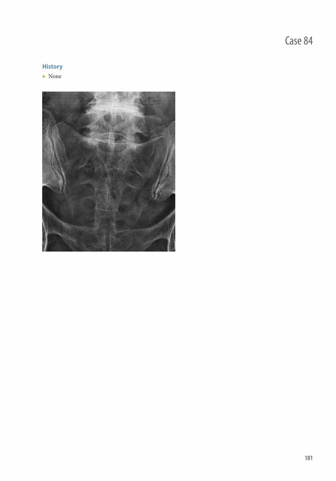
181
Case 84
History ▶ None

182
Case 84 Sacral Fracture
Findings ▶ Fracture lucency (arrows) and disruption of the sacral arcuate lines (arrowhead) indicate the presence of fracture. ▶ Hypointense T1 and hyperintense T2 linear abnormalities are MRI fi ndings consistent with insuffi ciency fractures.
Diagnosis Sacral fracture
Diff erential Diagnosis Pelvic fractures.
Teaching Points ▶ Denis classifi cation of sacral fractures
■ Zone 1: Fracture occurs lateral to the sacral foramina and may cause injury to the L5 nerve root in 6% of cases. ■ Zone 2: Fracture involves one or more of the sacral foramina and may lead to unilateral lumbar or sacral
neuropathies. Th e fracture may extend to Zone 1. ■ Zone 3: Fracture involves the central sacral canal and may extend to involve the other two zones. Bilateral
neurologic defi cits can occur, as well as bowel and bladder incontinence. ▶ Sacral insuffi ciency fracture
■ Seen with osteoporosis or aft er radiation therapy. Th ese fractures are oft en radiographically occult. ■ Vertical, mixed sclerotic, and lucent lines along the sacral wings if the fracture is unilateral. If bilateral, a
horizontal line at the level of the S2 or S3 sacral foramina produces the classic Honda sign. ■ Best diagnosed on CT, MRI, or radionuclide bone scans.
Management ▶ Usually conservative. ORIF is reserved for sacral fractures with pelvic instability and neurological defi cits.
Further Readings Diel J , Ortiz O , Losada RA . Th e sacrum: pathologic spectrum, multimodality imaging, and subspecialty approach .
RadioGraphics . 2001;21:83–104. Manaster BJ , May DA , Disler DG . Musculoskeletal imaging: the requisites . 3 rd ed. St Louis, Mo : Mosby , 2006 .

183
Case 85
History ▶ None

184
Case 85 Hip Dislocation
Findings ▶ Posterior hip dislocation (white arrow) with associated acetabular fracture. ▶ Inferior (obturator) type anterior hip dislocation (black arrow).
Diagnosis Hip dislocation
Teaching Points ▶ Posterior hip dislocation is more common than anterior dislocation.
■ Clinically, the limb is shortened, adducted, extended, and internally rotated ■ Femoral head is posteriorly and superiorly positioned with respect to the acetabulum, and the femur is
internally rotated with greater trochanter in profi le and lesser trochanter being obscured ■ Associated with fracture of the posterior acetabular wall
▶ Anterior dislocation is uncommon and occurs with forced abduction and external rotation. Divided in to two types. ■ Inferior (obturator) dislocation occurs with hip fl exion, with the femoral head positioned anteriorly and
medially over the obturator foramen ■ Superior dislocation occurs with extension of the femur with the femoral head positioned superiorly and
either medial or lateral to the acetabulum ■ Can be confused with posterior dislocation; diff erentiate by external rotation (lesser trochanter in profi le)
with anterior dislocation and internal rotation (lesser trochanter obscured) with posterior dislocation ■ Important to distinguish anterior from posterior dislocation because failure of reduction may result if
misdiagnosed ■ Associated with fractures of the acetabulum, femoral head, anterior superior iliac spine, and greater trochanter
▶ Complications of hip dislocation include avascular necrosis, if not reduced in a timely manner, and osteoarthritis.
Management ▶ Closed reduction. Open reduction should be performed if closed reduction is unsuccessful, if there are
associated complex fractures, if bony fragments remain in the joint space, or the joint remains unstable.
Further Readings Erb RE, Steele JR, Nance EP Jr, et al. Traumatic anterior dislocation of the hip: spectrum of plain fi lm and CT fi ndings . AJR Am
J Roentgenol. 1995 ; 65 ( 5 ): 1215–1219 . Manaster BJ , May DA , Disler DG . Musculoskeletal imaging: the requisites . 3 rd ed. St Louis, Mo : Mosby , 2006 .

185
Case 86
History ▶ None

186
Case 86 Ankle fracture
Findings ▶ Oblique, vertically oriented medial malleolus fracture (white arrow). ▶ Horizontal lateral malleolus fracture (black arrow) at the level of the tibial plafond. ▶ Soft tissue swelling with tibiotalar joint eff usion (arrowheads). ▶ Mortise symmetry preserved.
Diagnosis Bimalleolar ankle fracture. Likely inversion-type caused by fracture morphology.
Diff erential diagnosis None
Teaching Points ▶ Two common classifi cation schemes
■ Danis-Weber classifi cation: type A, fi bular fracture below the syndesmosis; type B, fi bular fracture at the level of the syndesmosis; type C, fi bular fracture above the syndesmosis.
■ Lauge-Hansen classifi cation: 1. Supination-external rotation (SE) caused by external rotation of an inverted foot; 2. Pronation-external rotation caused by external rotation of an everted foot; 3. Supination-adduction (pure inversion); 4. Pronation-abduction (pure eversion).
▶ Lauge-Hansen SE injuries account for nearly two-thirds of all ankle fractures, and are disproportionately associated with low-energy trauma, older individuals, and females.
▶ SE injury has four stages ■ First: Anterior tibiofi bular ligament rupture ■ Second: Spiral or oblique lateral malleolus fracture ■ Th ird: Posterior tibiofi bular ligament rupture or avulsion (posterior malleolar fragment) ■ Fourth: Deltoid ligament rupture or avulsion (medial malleolar fragment)
Management ▶ Indications for operative treatment: Unstable Weber B fracture; >5 mm of medial clear space on static or stress
radiograph. ▶ Usually open reduction and internal fi xation with plate and screw constructs along the distal fi bula, and
syndesmotic screws.
Further Readings Okanobo H , Khurana B , Sheehan S , Duran-Mendicuti A , Arianjam A , Ledbetter S . Simplifi ed diagnostic algorithm for
Lauge-Hansen classifi cation of ankle injuries . RadioGraphics . 2012 ; 32 ( 2 ): E71–E84 . PMID: 22411951 Mandi DM. Ankle fractures . Clin Podiatr Med Surg . 2012 ; 29 ( 2 ): 155–186 , vii. PMID: 22424483

NontraumaPart II


BrainSection 1


191
History ▶ 44-year-old male who had an upper respiratory tract infection for weeks before developing a headache and fever to 104.8 °F .
Case 87

192
Case 87 Meningitis
Findings ▶ Diff use pachymeningeal enhancement (long arrow) and scattered leptomeningeal enhancement (short arrow) ▶ Diff use sulcal hyperintensity on FLAIR images ▶ Diff use cerebral cortical T2 hyperintensity (FLAIR and DWI images) ▶ Mild disproportionate prominence of the ventricles relative to the sulci concerning for early hydrocephalus ▶ Layering DWI-hyperintensity, representing pus, in the lateral ventricles (arrowhead)
Diff erential Diagnosis ▶ Bacterial infection: Streptococcus , Escherichia coli , Neisseria , Mycobacterium tuberculosis , Borrelia ,
Haemophilus ▶ Viral infection: Herpes simplex virus, varicella zoster virus, West Nile virus, enterovirus ▶ Neoplastic meningitis: Breast cancer, lung cancer, melanoma ▶ Infl ammatory: Idiopathic hypertrophic cranial pachymeningitis, rheumatoid, sarcoid ▶ Toxic: NSAIDs, antibiotics
Pathophysiology ▶ Meningitis results from irritation of the pachymeninges and/or the leptomeninges ▶ Th e spectrum of infection diff ers markedly between children and adults ▶ Widespread use of vaccines against Haemophilus , Streptococcus , and Neisseria has altered the epidemiology of
meningitis ▶ Infection may spread from an adjacent space, such as sinuses, or be introduced with penetrating trauma,
including surgery ▶ Chronic sequelae of meningitis may include cognitive defi cits, epilepsy, and cranial nerve injury
Clinical Presentation ▶ Can vary widely, even with a specifi c pathogen ▶ Headache, neck stiff ness, and fever may be early symptoms of bacterial meningitis, followed by photophobia,
confusion, and decreased consciousness ▶ Kernig and Brudzinski signs may be variably present in bacterial meningitis, and are not highly sensitive
Teaching Points ▶ Imaging fi ndings of meningitis are nonspecifi c, and may evoke a broad diff erential ▶ Th e diff erential may be substantially narrowed by consideration of the clinical presentation (e.g., signs of
infection and meningismus) and patient demographics (e.g., age and risk factors, such as known infections, exposures, or malignancies)
Management ▶ Lumbar puncture for CSF analysis can provide evidence of bacterial meningitis (elevated protein, decreased
glucose, leukocytosis) and provide a culture specimen to identify the specifi c pathogen ▶ Head imaging to evaluate for elevated intracranial pressure or cerebellar tonsillar herniation is not typically
warranted before lumbar puncture in the absence of clinical signs concerning for elevated intracranial pressure, such as decreased consciousness; pupil, gaze, or respiratory abnormalities; seizure; or papilledema

193
History ▶ 55-year-old woman with fever, headache, and irritability.
Case 88

194
Findings ▶ Typically a peripherally enhancing extra-axial fl uid collection, demonstrating T1 dark signal, T2/FLAIR bright
signal, and restricted diff usion ▶ Similar to any subdural collections, subdural empyemas does not cross midline and is crescentic in shape
Diff erential Diagnosis ▶ Sterile subdural collection (eff usion), chronic subdural hematoma
Teaching Points ▶ Etiology: Meningitis, hematogenous spread, direct extension from brain or extracranial source (such as from
paranasal sinuses or mastoiditis), and complications aft er surgery ▶ Presentation: Fever, headache, irritability, vomiting, seizures, altered mental status, photophobia ▶ Complications: Cerebritis, cerebral abscess, and venous or arterial infarction ▶ Other pearls
■ In postoperative patients, DWI does not demonstrate restricted diff usion in approximately 30% of patients (high false-negative rate); therefore, must have high clinical suspicion for subdural infection in postoperative setting even if DWI is negative
■ Peripheral enhancement usually more marked in empyemas than in chronic subdural hematomas ■ Blood degradation products may demonstrate restricted diff usion in postoperative patients, confounding
the picture
Management ▶ Surgical drainage and antibiotics
Further Readings Farrell CJ , Hoh BL , Pisculli ML, et al. Limitations of diff usion-weighted imaging in the diagnosis of postoperative infections .
Neurosurgery. 2008 ; 62 : 577–583. Grossman RI , Yousem DM . Th e Requisites: Neuroradiology . Philadelphia : Elsevier; 2003. Sinclair AG , Scoffi ngs DJ . Imaging the post-operative cranium . RadioGraphics. 2010 ; 30 : 461–482. Wong AM , Zimmerman RA , Simon EM, et al. Diff usion-weighted MR imaging of subdural empyemas in children . Am J
Neuroradiol. 2004 ; 25 : 1016–1021.
Case 88 Subdural Empyema

195
History ▶ 60-year-old male patient with fever and altered mental status.
Case 89

196
Findings CT
▶ Complete ring enhancing lesion with surrounding areas of low attenuation (representing vasogenic edema).
MRI
▶ Brain abscesses typically present as complete ring enhancing lesions (blue arrow) with surrounding T2 hyperintense areas of edema (light green arrow). Th e ring enhancing lesion has a smooth wall that is oft en thinner along its medial margin and it may have smaller “satellite” lesions.
▶ Diff usion weighted images demonstrate central restricted diff usion secondary to purulent material (yellow arrow). Lack of restricted diff usion is unusual for a pyogenic abscess but can be seen, particularly aft er partial treatment with antibiotics. Fungal and tuberculous abscesses may also lack internal restricted diff usion.
Diff erential Diagnosis ▶ Primary or secondary cerebral neoplasm with central necrosis ▶ Cystic parasitic diseases ▶ Resolving hematoma
Teaching Points ▶ A cerebral abscess is a focal infection of the brain parenchyma produced by bacteria, fungi, or mycobacteria,
resulting in the development of a collection of purulent material surrounded by a discrete capsule. A focal region of cerebritis oft en precedes formation of a brain abscess.
▶ Cerebral abscesses typically present as intracerebral complete ring enhancing lesions with central restricted diff usion. Lack of internal restricted diff usion does not exclude a cerebral abscess.
Management ▶ Antimicrobial and medical treatment for small abscesses. Larger abscesses, particularly with local mass eff ect,
usually require surgical evacuation.
Further Readings Hughes DC , Raghavan A , Mordekar SR , Griffi ths PD , Connolly DJA . Role of imaging in the diagnosis of acute bacterial
meningitis and its complications . Postgrad Med J. 2010; 86 ( 1018 ): 478–485 . Kastrup O , Wanke I , Maschke M . Neuroimaging of infections of the central nervous system . Semin Neurol .
2008; 28 ( 4 ): 511–522 .
Case 89 Cerebral Abscess

197
Case 90
History ▶ 48-year-old male with HIV (viral load of 900,000 copies/ml) presents with confusion, aphasia, and right-sided weakness.

198
Findings ▶ Findings of HIV encephalitis ▶ Abnormal T2 hyperintensity in the cerebral hemispheres, more pronounced within the cortex and
asymmetrically much greater in the left cerebral hemisphere than the right ▶ No signifi cant enhancement on postcontrast images ▶ Patchy restricted diff usion in the right cerebral hemisphere, including involvement of the left
perirolandic cortex ▶ T1 and T2 hyperintense subdural collections overlying the cerebral hemispheres, also more pronounced on
the left than the right ▶ Follow-up head MRI 2.5 years later (Figure 85.6) demonstrates severe atrophy (arrow) and confl uent
leukoencephalopathy (arrowhead) asymmetrically involving the left cerebral hemisphere
Clinical Presentation ▶ Focal neurologic defi cits may be present corresponding to regions of greatest brain parenchymal abnormality
(e.g., right-sided weakness related to involvement of the left perirolandic region in this patient) ▶ Global neurologic defi cits may be present, such as decreased consciousness, coma, and seizure ▶ Signs and symptoms of infection, such as fever, nausea, and malaise, are common ▶ Onset may be insidious or acute
Pathophysiology ▶ Infl ammatory process in the brain parenchyma that may be caused by viral, bacterial, autoimmune, or other
infl ammatory etiologies ▶ Viral: Herpes simplex virus, HIV, West Nile virus, VZV, CMV, measles, and others ▶ Bacterial and fungal (rare): Mycoplasma streptococcus; cryptococcus ▶ Autoantibody: Anti-NMDA, Hashimoto encephalitis, paraneoplastic syndromes ▶ Infl ammatory: Acute disseminated encephalomyelitis, progressive multifocal leukoencephalopathy
Teaching Points ▶ Clinical presentations of encephalitis frequently include idiosyncratic neurologic symptoms that oft en defy
simple syndromic classifi cation ▶ Imaging fi ndings may be nonspecifi c and atypical ▶ Correlation with patient demographic data and known risk factors may be helpful to produce a focused diff erential ▶ Electroencephalography abnormalities may correlate with areas of encephalitis-aff ected brain tissue
Management ▶ Supportive care: Directed toward maintenance of physiologic integrity in the brain. Th erapies may include
antiseizure medications; sedatives, including barbiturates, to prevent excitotoxic neuronal injury; and nonsteroidal anti-infl ammatory drugs
▶ Directed therapy targeted to a demonstrated etiology, such as antiviral and other antibiotic therapies; intravenous immunoglobulin; plasmapheresis; and glucocorticoids
Case 90 Encephalitis

199
Case 91
History ▶ 8-year-old male patient with cough, fever, and weakness.

200
Findings MRI
▶ Large asymmetrical white matter lesions (blue arrows) with variable enhancement (yellow arrows) and local mass eff ect, oft en involving gray matter structures, brainstem, and optic nerves.
▶ Abnormal T2 hyperintense signal and enhancement in the spinal cord.
Diff erential Diagnosis ▶ Multiple sclerosis ▶ Infectious encephalomyelitis ▶ Mitochondrial disorders ▶ Neoplastic disorders, including lymphoma/leukemia and gliomas
Teaching Points ▶ Clinical presentation: Acute disseminated encephalomyelitis is typically described as a monophasic
demyelinating disorder aff ecting the brain and spinal cord oft en preceded by a viral infection or vaccination. Clinically, there is oft en evidence of CSF pleocytosis and encephalopathy.
▶ Clinical evolution: Follow-up imaging demonstrates marked or complete resolution of the lesions. Cases of multiphasic or recurrent acute disseminated encephalomyelitis have also been described but are considered unusual.
Management ▶ Treatment typically consists of IV and/or oral steroids. Steroid-resistant cases are oft en treated with IVIg or
plasmapheresis.
Further Readings Lim T . Neuroimaging in postinfectious demyelination and nutritional disorders of the central nervous system. Neuroimaging
Clinics N Am. 21( 4) : 843–858 . Parrish JB , Yeh AEA . Acuted disseminated encephalomyelitis . Adv Exp Med Biol . 724 : 1–14 . Wender M . Acute disseminated encephalomyelitis (ADEM). J Neuroimmunol. 231 (1): 92–99 . Zettl UK , Stüve O , Patejdl R . Immune-mediated CNS diseases: a review on nosological classifi cation and clinical features .
Autoimmun Rev . 11 ( 3 ): 167–173 .
Case 91 Acute Disseminated Encephalomyelitis

201
Case 92
History ▶ A 51-year-old woman with acute onset of severe headache.

202
Case 92 Aneurysm hemorrhage
Findings CT/CTA
▶ Hyperattenuation, consistent with hemorrhage, in the basal subarachnoid spaces not limited to the perimesencephalic cisterns. Aneurysms can also bleed into the brain parenchyma, subdural space, and ventricles.
▶ An aneurysm can be seen on an unenhanced CT scan as a round or oval hyperattenuating lesion arising from a vessel.
▶ Avid enhancement of the aneurysm in the arterial phase aft er contrast administration.
MR/MRA
▶ Acute subarachnoid hemorrhage: isointense signal on T1WI, and high signal intensity on T2WI and FLAIR weighted images; there may be little to no susceptibility on T2* images.
▶ Flow void of aneurysm contiguous with the parent vessel most apparent on T2WI and proton density-weighted images.
▶ Partially thrombosed aneurysms have a lamellated appearance on T2WI. ▶ Flow-related enhancement in aneurysm on MRA. ▶ Aneurysm wall well appreciated on this technique.
Catheter Angiography
▶ Focal, saccular, dilatation of the artery with opacifi cation on the arterial phase. ▶ Morphology of aneurysm, relation of aneurysm neck to sac, and arterial origin equally appreciated on CTA
and DSA.
Diff erential Diagnosis ▶ Subarachnoid hemorrhage
■ Trauma ■ Perimesencephalic venous hemorrhage (nonaneurysmal subarachnoid hemorrhage) ■ Parenchymal hematoma that dissects into subarachnoid space ■ AVM or AVF ■ Bleeding tumor ■ Coagulopathy ■ Vasculitis ■ Amyloid angiopathy
▶ Intracranial aneurysm ■ Saccular aneurysms ■ Fusiform aneurysms ■ Mycotic aneurysms ■ Oncotic aneurysms ■ Aneurysms associated with AVM
Teaching Points ▶ Quantity and pattern of blood clot on noncontrast CT may be helpful for locating a ruptured aneurysm. ▶ Aneurysms that rupture most commonly are anterior communicating, posterior communicating, middle
cerebral artery bifurcation, and basilar tip aneurysms. ▶ Annual risk of rupture of aneurysms <10 mm and >10 mm is 0.7% and 4%, respectively. Th e risk is also higher
among women, symptomatic aneurysms, aneurysms in the posterior circulation, aneurysms with a height to neck ratio of >1.6, and aneurysms that are lobulated.
Management ▶ Endovascular coiling ▶ Surgical clipping

203
History ▶ None
Case 93

204
Findings MRI
▶ Depending on the acuteness of the hemorrhage, the signal on T1WI and T2WI diff ers. ▶ Hyperacute lesions show low signal on T1 and high signal on T2. ▶ Subacute lesions show high signal on T1 and low signal on T2. ▶ Chronic lesions show low signal on T1WI and T2WI.
NCCT
▶ Morphology ■ Parenchymal hyperdense lesion, with HU between 40 and 90. Th ese lesions commonly produce mass aff ect
and peripheral hypoattenuation representing vasogenic edema. ▶ Common locations
■ Putamen ■ Th alamus ■ Cerebellar and brainstem ■ Lobar
Teaching Points ▶ Th alamic hemorrhages usually drain into the ventricles. ▶ Ventricular hemorrhage is associated with poor prognosis. ▶ Mortality increases proportionally to the size of the hemorrhage. ▶ “Spot sign” predicts hematoma expansion and mortality.
Management ▶ Intensive care has increased survival of patients with ICH. ▶ Blood pressure control. ▶ Correction of procoagulant factors. ▶ Surgical evacuation in small and superfi cial hemorrhage has demonstrated improved clinical outcome.
Further Readings Qureshi AI , Mendelow AD , Hanley DF . Intracerebral haemorrhage . Lancet . 2009 9; 373 ( 9675 ): 1632–1644 . Chen ST , Chen SD , Hsu CY , Hogan EL . Progression of hypertensive intracerebral hemorrhage . Neurology .
1989 ; 39 ( 11 ): 1509–1514 . Broderick JP , Brott TG , Duldner JE , Tomsick T , Huster G . Volume of intracerebral hemorrhage . A powerful and easy-to-use
predictor of 30-day mortality. Stroke . 1993 ; 24 ( 7 ): 987–993 . Delgado Almandoz JE , Yoo AJ , Stone MJ , et al. Th e spot sign score in primary intracerebral hemorrhage identifi es patients at
highest risk of in-hospital mortality and poor outcome among survivors . Stroke. 2010 Jan;41(1):54–60. Goldstein JN , Fazen LE , Snider R , et al. Contrast extravasation on CT angiography predicts hematoma expansion in
intracerebral hemorrhage . Neurology . 2007 ; 68 ( 12 ): 889–894 .
Case 93 Hypertensive Intracerebral Hemorrhage

205
History ▶ 31-year-old woman with acute right-sided weakness. ▶ Angiographic image courtesy of Drs. Chai Kobkitsuksakul and Pakorn Jiarakongmun
Case 94

206
Findings CT/CTA
▶ Hyperdense tangle of vessels (“nidus”) with/without calcifi cation ▶ Enlarged feeding arteries and draining cerebral veins ▶ Lack of mass eff ect unless secondary to hemorrhage ▶ Bleeding may occur in the brain parenchyma, subarachnoid space, or ventricles ▶ Localized brain atrophy
MR/MRA
▶ Nidus of fl ow voids best appreciated on T2W and proton density-weighted images ▶ T2 hyperintense adjacent brain tissue caused by chronic ischemia and gliosis (steal phenomenon) ▶ Flow-related enhancement in feeding arteries, nidus, and draining veins
Catheter Angiography
▶ Tangle of vessels between enlarged arterial feeders and draining veins ▶ Aneurysms of the arterial feeders, within the nidus and anywhere in the circle of Willis ▶ Venous stenoses in the draining veins ▶ An acute hematoma can obscure an AVM
Diff erential Diagnosis for Hemorrhage with Abnormal Vessels ▶ Hemorrhagic tumor ▶ Dural arteriovenous fi stula ▶ Aneurysm with parenchymal hemorrhage
Teaching Points ▶ Brain AVM typically comes to attention in young adults before the age of 40 with slight male predominance. ▶ Most frequent clinical presentation is intracranial hemorrhage (50% of cases). AVM may bleed into brain
parenchyma, subarachnoid space, or ventricles. ▶ Spetzler-Martin grading system categorizes AVM on the basis of
■ Size ■ 1: small, maximum diameter <3 cm ■ 2: medium, diameter 3–6 cm ■ 3: large, diameter >6 cm
■ Pattern of venous drainage ■ 0: superfi cial only ■ 1: any component of deep venous drainage
■ Neurologic eloquence of adjacent brain ■ 0: noneloquent areas ■ 1: eloquent areas—sensorimotor, language, and visual cortex; hypothalamus and thalamus; internal
capsule; brainstem; cerebellar peduncles; and deep cerebellar nuclei ■ Patients are subjected to lifelong risk of repeated hemorrhages. ■ Multiple AVMs occur in hereditary hemorrhagic telangiectasia and Wyburn-Mason syndromes
Management ▶ Surgery for AVM removal ▶ Radiation therapy ▶ Partial endovascular occlusion followed by surgery or radiation therapy
Case 94 Bleeding Brain Arteriovenous Malformation

207
History ▶ 66-year-old female presents with acute onset of global aphasia and right hemiparesis.
Case 95

208
Findings ▶ Noncontrast head CT in brain windows demonstrates a hyperdense left middle cerebral artery (white arrow),
indicative of acute thrombus within the vessel. ▶ Noncontrast head CT in narrower windows demonstrates subtle hypodensity and loss of the gray-white
matter diff erentiation in the left lentiform nucleus, caudate nucleus, and upper internal capsule (black arrow). ▶ CTA MIP demonstrates a left M1 occlusion. ▶ Axial FLAIR image demonstrates slightly increased signal within the same region (double white arrows). ▶ DWI image demonstrates restricted diff usion within the same region (double black arrows). ▶ Μean transit time map identifi es prolonged transit time in a region much larger than that identifi ed by
restricted diff usion.
Diff erential Diagnosis for the Noncontrast Head CT Findings ▶ Neoplasm ▶ Cerebritis
Teaching Points ▶ Th e noncontrast head CT is used to identify hemorrhage. ▶ Little to no FLAIR MRI signal abnormality suggests that a stroke is <6 hours old. ▶ Diff usion-weighted MRI demonstrates increased signal within 30 minutes of symptom onset, providing the
gold standard for detecting acute ischemia. A DWI abnormality larger than 70 cc is a contraindication for intra-arterial recanalization procedures at some institutions.
▶ Perfusion maps used to detect ischemic but viable tissue at risk of infarction (normal on DWI but abnormal on perfusion) are typically mean transit time, time-to-peak, time to maximum of the deconvolved residue function, and/or cerebral blood fl ow maps. Patients without at perfusion-diff usion mismatch are less likely to benefi t from reperfusion therapy.
Management ▶ Emergent head CT to exclude hemorrhage and hypodensity in more than one-third of the middle cerebral
artery territory, ▶ Intravenous thrombolytic therapy for appropriate patients presenting within 4.5 hours of symptom onset. ▶ Intra-arterial recanalization procedures for appropriate patients with proximal vessel occlusions presenting
within 6 hours of symptom onset. ▶ Supportive therapy including heparinization strict blood pressure control for patients who cannot receive
other therapies.
Case 95 Acute Stroke

209
Case 96
History ▶ 23-year-old postpartum female with headache and visual disturbance.

210
Case 96 Posterior Reversible Encephalopathy Syndrome
Findings Distribution
▶ Most common: Posterior circulation predilection involving the posterior temporal, parietal, and occipital lobes ▶ Less common: Frontal lobes (frequently in a border zone distribution), basal ganglia, and brainstem
CT
▶ Patchy bilateral hypodense foci involving gray and subcortical white matter ▶ Hemorrhage in 20% ▶ +/- patchy enhancement
MRI
▶ T1: Hypointense cortical/subcortical lesions ▶ T2 and FLAIR: Hyperintense cortical/subcortical lesions ▶ T1C+: Variable patchy enhancement ▶ T2*: Blooming if hemorrhage present ▶ DWI: Usually elevated (consistent with vasogenic edema) but can occasionally show restricted diff usion
Diff erential Diagnosis ▶ Hypoglycemia ▶ Acute ischemia ▶ Status epilepticus ▶ Venous sinus thrombosis ▶ Infectious and infl ammatory leukoencephalidites
Teaching Points ▶ Neurologic disorder characterized by particular radiographic fi ndings combined with clinical manifestations
of headache, mental status change, seizure, and/or visual disturbances ▶ Associated with diverse conditions including
■ Preeclampsia ■ Acute/subacute hypertension ■ Drug toxicity (chemotherapy agents) ■ Autoimmune disorders ■ Sepsis
▶ Pathogenesis ■ Hypertension oft en common component ■ Vasogenic edema (two leading theories)
1. Hypertension exceeds the limits of autoregulation, leading to breakthrough brain edema 2. Hypertension leads to cerebral autoregulatory vasoconstriction, ischemia, and subsequent brain edema
■ Posterior circulation more susceptible to disturbances in autoregulation because of sparse sympathetic innervation
■ Overlap with reversible cerebral vasoconstrictive syndrome in some patients
Management ▶ Usually reversible if treated promptly ▶ Correct underlying conditions (e.g., treat hypertension, deliver child in preeclampsia)

211
Case 97
History ▶ 30-year-old female with protein S defi ciency presents with headaches.

212
Case 97 Acute Venus Sinus Thrombosis
Findings ▶ Noncontrast CT: in 20% of cases, direct visualization of hyperdense clotted venous sinus or cerebral vein;
hypodensity and gyral swelling in nonarterial distribution; bilateral cerebral manifestations in cases of midline venous clot (e.g., bilateral thalamic involvement with deep venous clot or bilateral parasagittal involvement with superior sagittal sinus thrombus).
▶ CT venography: fi lling defect within the intracranial venous system; 95% sensitive. ▶ MRI: two-dimensional time-of-fl ight MR venography demonstrates absence of fl ow-related enhancement in
clotted segment; susceptibility-weighted images also sensitive for cerebral venous clot.
Diff erential Diagnosis ▶ Normal venous variants; pacchionian arachnoid granulations, insuffi cient CT scan delay resulting in poor
opacifi cation of venous system (recommend delay of 45 seconds aft er 3 cc/s bolus injection for CTV), polycythemia.
Teaching Points ▶ Etiology: intracranial and systemic infections, immobilization, hypercoagulable disorders, malignancy,
dehydration, pregnancy, oral contraceptives. ▶ Presentation: Unlike arterial infarcts, typically slower and more progressive onset of symptoms, which include
headache, nausea/vomiting, papilledema, diffi culty with vision, focal neurologic defi cits, and seizures. ▶ Complications: edema, infarction, superimposed hemorrhage, hydrocephalus. ▶ Other pearls:
■ Th ink of venous thrombosis in younger or middle age patients with severe headache or patients with stroke-like symptoms in absence of typical risk factors.
■ Poor prognostic indicators: hemorrhage, involvement of deep venous system, and superimposed infection. ■ Location in order of decreasing frequency: superior sagittal sinus, transverse sinus, straight sinus, cortical
veins, deep venous system.
Management ▶ Systemic anticoagulation with occasional direct transcatheter thrombolysis. Angioplasty with venous stenting
performed anecdotally in cases of severe intracranial hypertension without clear long-term benefi t.

213
Case 98
History ▶ 30-year-old male patient with persistent postural headache.

214
Findings MRI
▶ Diff use pachymeningeal enhancement (blue arrow) and prominence of dural venous sinuses (green arrow). ▶ Engorgement and prominence of the pituitary gland (yellow arrow). ▶ Bilateral subdural collections, cerebellar tonsillar descent, or herniation (light green arrow). ▶ Spinal imaging oft en shows prominence of the spinal epidural venous plexus and subdural/epidural
collections.
Diff erential Diagnosis ▶ Chiari 1 malformation ▶ Chronic meningitis
Teaching Points ▶ Clinical presentation: Clinical syndrome characterized by a persistent postural headache, oft en with nausea
and vomiting but also associated with a variety of focal neurologic fi ndings. ▶ Etiology: Intracranial hypotension can be primary (spontaneous) or secondary to prior surgical procedures or
trauma.
Management ▶ Symptomatic management of headache initially. For persistent symptoms, identifi cation of a CSF leak is
important for a targeted epidural patch.
Further Readings Chiapparini L , Ciceri E , Nappini S , et al. Headache and intracranial hypotension: neuroradiological fi ndings . Neurol Sci .
25 ( 3 ): s138–s141 . Forghani R , Farb RI. Diagnosis and temporal evolution of signs of intracranial hypotension on MRI of the brain.
Neuroradiology. 50( 12) : 1025–1034 . Rahman M , Sharatchandra S, Bidari R , Quisling G , Friedman WA . Spontaneous intracranial hypotension: dilemmas in
diagnosis. Neurosurgery. 69( 1) : 4–14 – discussion 14.
Case 98 Intracranial Hypotension

215
History ▶ 37-year-old female with history of asthma.
Case 99
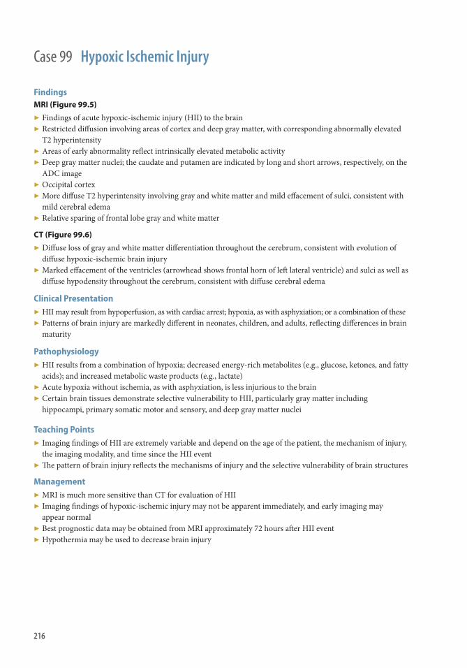
216
Case 99 Hypoxic Ischemic Injury
Findings MRI (Figure 99.5)
▶ Findings of acute hypoxic-ischemic injury (HII) to the brain ▶ Restricted diff usion involving areas of cortex and deep gray matter, with corresponding abnormally elevated
T2 hyperintensity ▶ Areas of early abnormality refl ect intrinsically elevated metabolic activity ▶ Deep gray matter nuclei; the caudate and putamen are indicated by long and short arrows, respectively, on the
ADC image ▶ Occipital cortex ▶ More diff use T2 hyperintensity involving gray and white matter and mild eff acement of sulci, consistent with
mild cerebral edema ▶ Relative sparing of frontal lobe gray and white matter
CT (Figure 99.6)
▶ Diff use loss of gray and white matter diff erentiation throughout the cerebrum, consistent with evolution of diff use hypoxic-ischemic brain injury
▶ Marked eff acement of the ventricles (arrowhead shows frontal horn of left lateral ventricle) and sulci as well as diff use hypodensity throughout the cerebrum, consistent with diff use cerebral edema
Clinical Presentation ▶ HII may result from hypoperfusion, as with cardiac arrest; hypoxia, as with asphyxiation; or a combination of these ▶ Patterns of brain injury are markedly diff erent in neonates, children, and adults, refl ecting diff erences in brain
maturity
Pathophysiology ▶ HII results from a combination of hypoxia; decreased energy-rich metabolites (e.g., glucose, ketones, and fatty
acids); and increased metabolic waste products (e.g., lactate) ▶ Acute hypoxia without ischemia, as with asphyxiation, is less injurious to the brain ▶ Certain brain tissues demonstrate selective vulnerability to HII, particularly gray matter including
hippocampi, primary somatic motor and sensory, and deep gray matter nuclei
Teaching Points ▶ Imaging fi ndings of HII are extremely variable and depend on the age of the patient, the mechanism of injury,
the imaging modality, and time since the HII event ▶ Th e pattern of brain injury refl ects the mechanisms of injury and the selective vulnerability of brain structures
Management ▶ MRI is much more sensitive than CT for evaluation of HII ▶ Imaging fi ndings of hypoxic-ischemic injury may not be apparent immediately, and early imaging may
appear normal ▶ Best prognostic data may be obtained from MRI approximately 72 hours aft er HII event ▶ Hypothermia may be used to decrease brain injury

217
Case 100
History ▶ None

218
Findings ▶ Axial (Figure 100.1) and coronal (Figure 100.2) contrast-enhanced CT images show an extraconal lenticular-
shaped fl uid collection (arrows) with rim-enhancement along the medial aspect of the left orbit resulting in proptosis. Stranding of the medial left orbital fat and eyelid are also present, indicative of orbital cellulitis. In addition, there is opacifi cation of the ethmoid sinuses, left greater than right, consistent with sinusitis.
▶ Axial T1 (Figure 100.3), axial (Figure 100.4), and coronal (Figure 100.5) postcontrast fat-suppressed T1, and fat-suppressed coronal T2 (Figure 100.6) MRIs also show the rim-enhancing superiosteal abscess in the medial left orbit (arrows). Th e preseptal and postseptal cellulitis are more conspicuous on MRI than the corresponding CT. Th e linear low signal intensity stricture surrounding the fl uid collection noted on the coronal T2 image represents the displaced periosteum. Th e diff usion-weighted image (Figure 100.7) and corresponding ADC map (Figure 100.8) show restricted diff usion within the abscess (arrows).
Diff erential Diagnosis ▶ Subperiostealphlegmon ▶ Orbital abscess/cellulitis ▶ Subperiosteal hematoma ▶ Sinonasal or nasolacrimal duct mucocele ▶ Epidermoid ▶ Rarely, neoplasms, such as metastasis
Teaching Points ▶ Orbital subperiosteal abscess most commonly results from ethmoid sinusitis. ▶ An elongated extraconal collection adjacent to lamina papyracea with peripheral enhancement is the typical
appearance of subperiosteal abscess on CT and MRI. Restricted diff usion can be observed in the collection. ▶ Complications include orbital cellulitis, optic neuritis, retinal ischemia, superior ophthalmic vein and
cavernous sinus thrombosis, meningitis, and intracranial abscess.
Management ▶ CT with contrast is considered to be the fi rst-line imaging modality for evaluating acute orbital infections. ▶ MRI with contrast is more sensitive than contrast-enhanced CT, particularly for delineating intracranial
complications. Dedicated orbital protocol T2 and postcontrast T1 sequences are particularly helpful. In addition, postcontrast T1 sequences of the entire brain should also be performed.
▶ Intravenous antibiotics may be adequate treatment for small uncomplicated abscesses, especially in patients <10 years of age.
▶ Medial subperiosteal abscesses that fail medical therapy are usually drained endoscopically, whereas more extensive abscesses may require external drainage.
Case 100 Orbital Subperiosteal Abscess

Head, Neck, and SpineSection 2

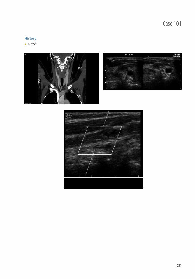
221
History ▶ None
Case 101

222
Findings Ultrasound
▶ Noncompressible vessel with mixed echogenicity intraluminal thrombus ▶ Absent fl ow on pulsed-wave Doppler
CTA/MRA
▶ Intraluminal fi lling defect without enhancement ▶ Increased vessel diameter (acute thrombosis)
■ In chronic thrombosis, the vessel may be smaller or unidentifi able ▶ ± edema or stranding of the perivascular fat (acute) ▶ Regional venous collaterals
■ More common in chronic thrombosis
Diff erential Diagnosis ▶ Tumor thrombus (e.g., sarcoma)
■ Intraluminal fi lling may enhance
Teaching Points ▶ Etiology
■ Primary (10%): idiopathic thrombosis and thrombosis secondary to thoracic outlet syndrome ■ Secondary (90%): central venous catheters; cancer (most commonly lung cancer and lymphoma/leukemia);
coagulation abnormalities (antiphospholipid antibody, factor V Leiden mutation, prothrombin mutation); cervical infections (Lemierre syndrome); trauma; postsurgical complication
▶ Presentation: pain and induration over the aff ected side, cervical edema, arm edema, superfi cial venous collaterals
▶ Complications ■ Pulmonary embolism: up to 10% of patients ■ Postthrombotic syndrome: up to 15% of patients ■ Intracranial propagation of the thrombus ■ Rare: elevated intracranial pressures have been reported with bilateral acute IJVT
Management ▶ Depends on the clinical setting and patient risk factors ▶ Treatment options include
■ Systemic anticoagulation ■ Line removal if catheter-associated thrombosis ■ Superior vena cava fi lter placement ■ Percutaneous interventions, such as mechanical thromebectomy or thromboaspiration (rarely necessary)
▶ Monitoring with Serial duplex ultrasound
Further Reading Gbaguidi X , Janvresse A , Benichou J , Cailleux N , Levesque H , Marie I . Internal jugular vein thrombosis: outcome and risk
factors . QJM . 2011 ; 104 ( 3 ): 209–219 .
Case 101 Internal Jugular Venous Thrombosis (IJVT)

223
History ▶ 21-year-old female who had a sore throat and respiratory distress.
Case 102
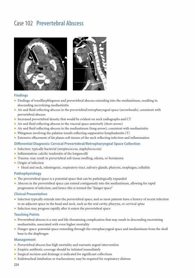
224
Findings ▶ Findings of tonsillarphlegmon and prevertebral abscess extending into the mediastinum, resulting in
descending necrotizing mediastinitis ▶ Air and fl uid refl ecting abscess in the prevertebral/retropharyngeal space (arrowheads), consistent with
prevertebral abscess ▶ Increased prevertebral density that would be evident on neck radiographs and CT ▶ Air and fl uid refl ecting abscess in the visceral space anteriorly (short arrow) ▶ Air and fl uid refl ecting abscess in the mediastinum (long arrow), consistent with mediastinitis ▶ Phlegmon involving the palatine tonsils refl ecting suppurative lymphadenitis (T) ▶ Extensive eff acement of fat planes soft tissues of the neck refl ecting infection and infl ammation
Diff erential Diagnosis: Cervical Prevertebral/Retropharyngeal Space Collection ▶ Infection: typically bacterial (streptococcus, staphylococcus) ▶ Infl ammation: calcifi c tendonitis of the longuscolli ▶ Trauma: may result in prevertebral soft tissue swelling, edema, or hematoma ▶ Origin of infection
■ Head and neck, odontogenic, respiratory tract, salivary glands, pharynx, esophagus, cellulitis
Pathophysiology ▶ Th e prevertebral space is a potential space that can be pathologically expanded ▶ Abscess in the prevertebral space can extend contiguously into the mediastinum, allowing for rapid
progression of infection, and hence this is termed the “danger space”
Clinical Presentation ▶ Infection typically extends into the prevertebral space, and so most patients have a history of recent infection
in an adjacent space in the head and neck, such as the oral cavity, pharynx, or cervical spine ▶ Infection may progress rapidly aft er it enters the prevertebral space
Teaching Points ▶ Prevertebral abscess is a rare and life-threatening complication that may result in descending necrotizing
mediastinitis, associated with even higher mortality ▶ Danger space: potential space extending through the retropharyngeal space and mediastinum from the skull
base to the diaphragm
Management ▶ Prevertebral abscess has high mortality and warrants urgent intervention ▶ Empiric antibiotic coverage should be initiated immediately ▶ Surgical incision and drainage is indicated for signifi cant collections ▶ Endotracheal intubation or tracheostomy may be required for respiratory distress
Case 102 Prevertebral Abscess

225
Case 103
History ▶ Pain, disability aft er cervical trauma ± radiculopathy, myelopathy.

226
Case 103 Acute Disk Herniation
Findings ▶ Disk herniation with evidence of spinal trauma on imaging or history ▶ Morphology: Same appearance as degenerative disk herniation ▶ Radiography is insensitive for disk pathology
■ May see focal disk space narrowing ■ Focal kyphosis, traumatic spondylolisthesis, dislocated facet joints ■ Fracture or compression deformity
▶ CT fi ndings ■ Soft tissue density in anterior or anterolateral epidural space; narrowing of disk space is a variable fi nding ■ Spinal fracture, facet subluxation
▶ MRI fi ndings (best seen on T2WI) ■ Disk material eff acing ventral CSF ± cord or nerve root compression ■ Cord edema may be present with cord compression ■ Possible disruption of posterior longitudinal ligament ■ Paraspinal eff usion or hematoma
Diff erential Diagnosis ▶ Nontraumatic disk herniation ▶ Epidural abscess, phlegmon ▶ Epidural tumor ▶ Epidural extension of vertebral metastases, aggressive hemangioma, lymphoma
Teaching Points ▶ MRI is the modality of choice to evaluate intervertebral disks and the soft tissue contents of spinal canal ▶ Cervical region most common, followed by thoracic; lumbar region least common ▶ MRI of spine should be performed before reduction of facet dislocation
Management ▶ Cord compression from traumatic herniation is a reported complication of reduction of cervical facet
dislocation without discectomy ▶ Conservative management with surgery for those with persistent symptoms including weakness, sensory loss,
and radiculopathy ▶ Percutaneous nucleoplasty can be used in patients who fail conservative management and are unwilling to
undergo a more invasive technique, such as spinal surgery
Further Readings Dai L , Jia L . Central cord injury complicating acute cervical disc herniation in trauma . Spine . 2000 1; 25 ( 3 ): 331–335,
discussion 336. Khashaba A . Acute intraosseous disc herniation . Injury . 2000 ; 31 ( 4 ): 271–272 . Kreichati GE , Kassab FN , Kharrat KE . Herniated intervertebral disc associated with a lumbar spine dislocation as a cause of
cauda equina syndrome: a case report . Eur Spine J . 2006 ; 15 ( 6 ): 1015–1018 . Reddy AS , Loh S , Cutts J , Rachlin J , Hirsch JA . New approach to the management of acute disc herniation . Pain Physician .
2005 ; 8 ( 4 ): 385–390 . Rizzolo SJ , Piazza MR , Cotler JM , Balderston RA , Schaefer D , Flanders A . Intervertebral disc injury complicating cervical spine
trauma . Spine . 1991 ; 16 ( 6 suppl): S187–S189 .

227
History ▶ Fever and acute or subacute spinal pain and tenderness
Case 104

228
Findings ▶ CT Findings: Increased epidural soft tissue; may be diffi cult to distinguish from disk in ventral epidural space ▶ CECT: Enhancing epidural mass/collection narrowing the spinal canal ▶ MRI Findings
■ T1WI: Isointense to hypointense to cord ■ T2WI: Hyperintense
■ Signal alteration in spinal cord secondary to compression, ischemia, or direct infection ■ DWI: Typically restrict diff usion similar to abscess in brain. ■ CE T1WI
■ Homogeneously or heterogeneously enhancing phlegmon ■ Peripherally enhancing necrotic abscess ■ Diff use dural enhancement in extensive abscess ■ Enhancing prominent anterior epidural veins or basivertebral venous plexus adjacent to abscess ■ Signal alteration in spinal cord secondary to compression, ischemia, or direct infection
Diff erential Diagnosis ▶ Extradural metastasis ▶ Spinal epidural lymphoma ▶ Epidural hematoma ▶ Extruded/migrated disk ▶ Epidural lipomatosis
Teaching Points ▶ Best imaging tool: sagittal and axial T2WI and T1WI MRI with gadolinium ▶ Epidural soft tissue with homogeneous or peripheral enhancement and adjacent spondylodiskitis
characteristic of spinal epidural abscess ▶ Carefully assess cord for signs of signal changes
■ Best seen on STIR and confi rmed on axial T2WI ■ DWI imaging useful to confi rm cord extension of abscess
Management ▶ Early empiric antibiotics with broad-spectrum coverage until causative pathogen isolated ▶ Emergent surgical decompression with abscess drainage advocated by some, even in absence of initial
neurologic compromise ■ Th is is because of the possibility of rapid progression of neurologic defi cits despite appropriate medical
therapy ▶ Medical therapy alone if substantial operative risks, extensive cranial-caudal involvement of spinal canal,
or paralysis >48–72 hours ▶ Location (dorsal versus ventral) spinal epidural abscess plays role in determining treatment plan
Further Readings Tompkins M , Panuncialman I , Lucas P , Palumbo M . Spinal epidural abscess . J Emerg Med . 2010 ; 39 ( 3 ): 384–390 . Zimmerer SME , Conen A , Müller AA , Sailer M , Taub E , Flückiger U , et al. Spinal epidural abscess: aetiology, predisponent
factors and clinical outcomes in a 4-year prospective study . Eur Spine J . 2011 ; 20 ( 12 ): 2228–2234 .
Case 104 Epidural Abscess

229
History ▶ Back pain and tenderness ± fever.
Case 105

230
Findings ▶ Paravertebral enhancing phlegmon or peripherally enhancing liquifi ed collection ▶ May extend into the psoas, iliacus, or posterior paraspinous muscles (erector spinae) ▶ Can be solitary, multiple, and/or multiloculated ▶ Extend along subligamentous and muscles planes ▶ Obliterated soft tissue fascial planes ▶ Intraspinal epidural extension with cord compression ▶ Involvement of lateral epidural space with neuroforaminal narrowing ▶ Radiography: Nonspecifi c fi ndings
■ Osseous abnormalities present in delayed fashion (see diskitis/osteomyelitis fi ndings) ■ May identify an enlarged psoas shadow or a paraspinal soft tissue density
▶ CT fi nding: Poorly defi ned soft tissue density with low-density intramuscular collection ▶ CECT
■ Diff use or peripheral enhancement ■ Enhancing disk space
▶ MRI fi ndings ■ T1WI
■ Isointense to hypointense ■ May be diffi cult to distinguish from normal musculature ■ Obscuration of musculofascial planes
■ T2WI ■ Hyperintense fl uid collection and surrounding muscle ■ Hypointense rim
■ DWI ■ Evidence of restricted diff usion increases conspicuity of epidural, vertebral, and paraspinal abscesses
■ T1WI C+ ■ Phlegmon: Diff use enhancement ■ Abscess: Peripherally enhancing fl uid collections
▶ Best imaging tool: Sagittal and axial T1WI C+ and T2WI MRI
Diff erential Diagnosis ▶ Neoplasm, primary or metastatic ▶ Desmoid, calcifi cations with low signal intensity ▶ Retroperitoneal hematoma ▶ Extramedullary hematopoiesis
Teaching Points ▶ Evaluate adjacent vertebral bodies and intervertebral disk spaces for spondylodiskitis ▶ Well-defi ned paraspinal collections with thin and smooth abscess wall along with presence of paraspinal or
intraspinal abscess and thoracic spine involvement are more suggestive of tuberculous versus pyogenic spondylitis
Management ▶ Intravenous antibiotics ▶ Analgesic medications ▶ Percutaneous catheter drainage ▶ Surgical debridement ▶ Spinal stabilization in some cases
Case 105 Paraspinal Abscess

231
Case 106
History ▶ Severe, localized back pain ± radiculopathy.
232
Findings ▶ Best diagnostic clue: fusiform, extradural mass-like process with signal characteristics of hemorrhage on MRI ▶ May be located at any level of spine
■ Most commonly seen in the dorsal epidural space extending over multiple levels ▶ CT: Hyperdense, extradural collection ▶ MRI fi ndings
■ T1WI ■ Epidural fl uid collection, signal depends on age of hematoma ■ Acute hemorrhage can be similar in signal to cord or CSF on T1WI ■ Subacute (methemoglobin) is hyperintense to CSF and cord
■ T2WI: Epidural fl uid collection, signal depends on age of hematoma ▶ STIR: Hyperintense relative to cord and epidural fat ▶ T2* GRE: Accentuates susceptibility artifact from blood products ▶ T1WI C+: Mild peripheral enhancement may be seen with subacute hematoma
Diff erential Diagnosis ▶ Epidural abscess (or phlegmon) ▶ Epidural lipomatosis ▶ Epidural tumor ▶ Extramedullary hematopoiesis ▶ Ossifi cation of posterior longitudinal ligament ▶ Sequestered disk fragment
Teaching Points ▶ When spontaneous, considered a neurosurgical emergency ▶ MRI study of choice; include contrast injection and fat saturation ▶ Active extravasation may enhance ▶ CT may help identify hemorrhage when MRI confusing
Management ▶ Surgical evacuation/decompression may be necessary to alleviate compression of spinal cord or cauda equina
syndrome ▶ If no signifi cant neurologic compromise, may be managed conservatively ▶ Vitamin K injection or fresh frozen plasma to reverse coagulopathies
Further Readings Al-Mutair A , Bednar D . Spinal epidural hematoma . J Am Acad Orthop Surg . 2010 ; 18 : 494–502 . Sklar EML , Donovan Post JM , Falcone S . MRI of acute spinal epidural hematomas . J Comput Assist Tomogr . 1999 ; 23 ( 2 ): 238 . Zhong W , Chen H , You C , Li J , Liu Y , Huang S . Spontaneous spinal epidural hematoma . J Clin Neurosci .
2011 ; 18 ( 11 ): 1490–1494 .
Case 106 Epidural Hematoma
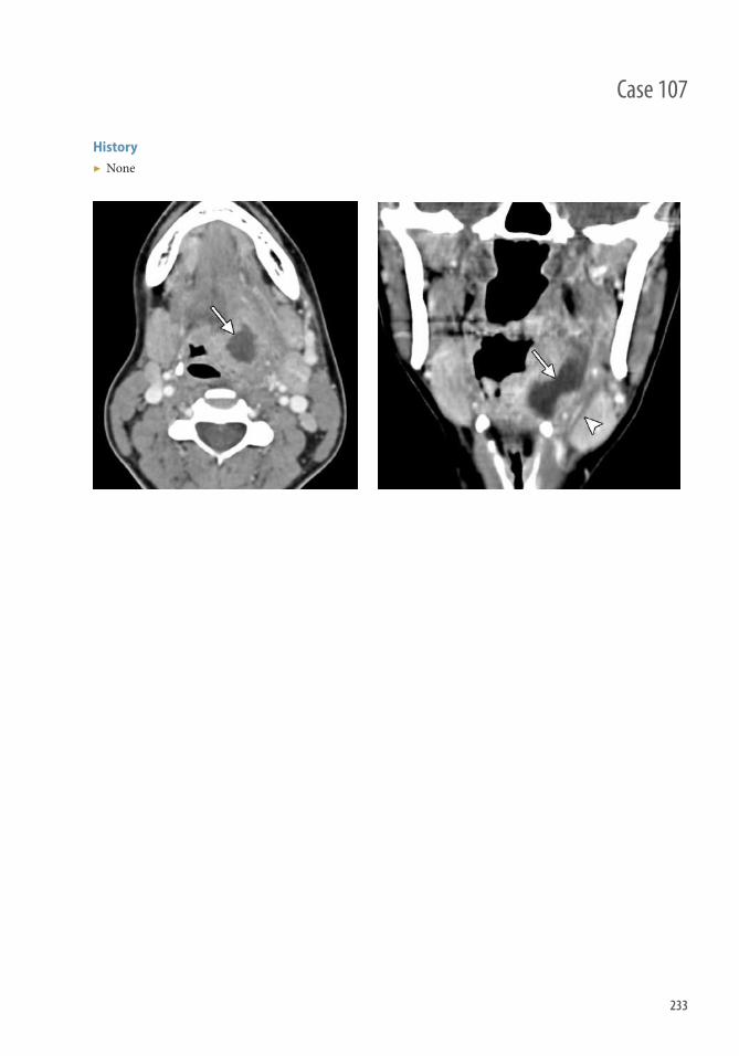
233
Case 107
History ▶ None
234
Findings ▶ Axial (Figure 107.3) and coronal (Figure 107.4) CT images show a low-attenuation collection with
surrounding enhancement located in the left lingual tonsil and extending into the parapharyngeal space. Th e linear low-attenuation structure that traverses the left submandibular gland likely represents a thrombosed glandular vein (arrowhead).
Diff erential Diagnosis ▶ Phlegmon ▶ Suppurative lymphadenopathy ▶ Squamous cell carcinoma ▶ Benign mixed tumor ▶ Tonsillar hypertrophy
Teaching Points ▶ Peritonsillar abscess can result as a complication of tonsillitis and consists of a collection of pus situated
external to the tonsillar capsule, tonsillar fossa, and superior constrictor muscle, extending into the parapharyngeal space and beyond.
▶ Findings on CT can include enlargement and enhancement of the tonsil and a fl uid collection with rim enhancement.
▶ Patients with peritonsillar abscess may present with sore throat, dysphagia, fever, and trismus. ▶ Extension of peritonsillar abscess posterolaterally into the carotid sheath can lead to jugular vein thrombosis
and carotid artery pseudoaneurysm. ▶ Hounsfi eld unit measurement has not been found to be reliable in distinguishing abscess from phlegmon.
Management ▶ Peritonsillar abscess is generally diagnosed clinically and treated with antibiotics and drainage. ▶ CT and MRI have high sensitivity for detecting abscess formation and its precise location and extension of
disease. ▶ Needle aspiration or intraoral ultrasound can also be performed to confi rm the diagnosis. ▶ Associated complications should be sought on imaging, especially in cases that do not respond to initial
treatment.
Further Readings Gonzalez-Beicos A , Nunez D. Imaging of acute head and neck infections . Radiol Clin North Am . 2012 ; 50 ( 1 ): 73–83 .
doi: 10.1016/j.rcl.2011.08.004. Smith JL 2nd, Hsu JM , Chang J. Predicting deep neck space abscess using computed tomography . Am J Otolaryngol .
2006 ; 27 ( 4 ): 244–247 . Johnson RF , Stewart MG. Th e contemporary approach to diagnosis and management of peritonsillar abscess . Curr Opin
Otolaryngol Head Neck Surg . 2005 ; 13 : 157–160 .
Case 107 Peritonsillar Abscess
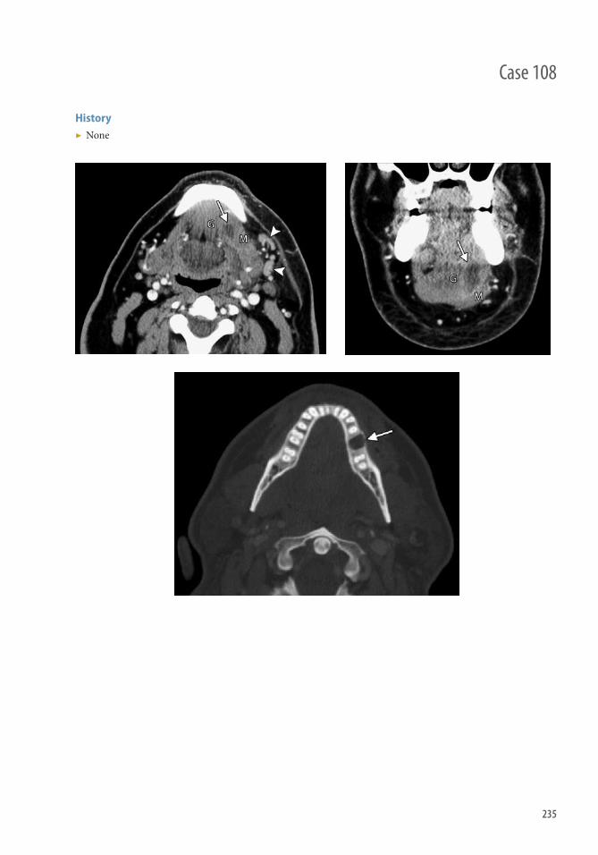
235
Case 108
History ▶ None
236
Findings ▶ Axial (Figure 108.1) and coronal (Figure 108.2) contrast-enhanced CT images show a fl uid collection
with ill-defi ned peripheral enhancement (arrows) in the left sublingual space, splaying apart the adjacent genioglossus-geniohyoid complex (G) and mylohyoid (M) muscles. Asymmetrically enlarged reactive left level IB lymph nodes are present (arrowheads). Th ere is also thickening of the left platysma and stranding of the surrounding subcutaneous fat.
▶ Axial CT image (Figure 108.3) in the bone window shows a recent tooth extraction cavity (arrow).
Diff erential Diagnosis ▶ Phlegmon/cellulitis without abscess ▶ Neoplasm: squamous cell carcinoma, salivary gland tumor ▶ Ranula ▶ Dermoid/epidermoid
Teaching Points ▶ Th e sublingual space lies within the fl oor of the mouth; contains the sublingual gland, submandibular duct,
and lingual neurovascular bundle; and is bounded by the mylohyoid inferolaterally, the genioglossus and geniohyoid medially, and intrinsic tongue muscles superiorly.
▶ Sublingual abscess most commonly results from dental disease and submandibular or sublingual duct stenosis. ▶ Th ickening of the ipsilateral plastysma, stranding of the subcutaneous fat, and regional lymphadenopathy may
be present as secondary signs of infection on contrast-enhanced CT. ▶ Sublingual abscess may not display peripheral enhancement in the very early stages. ▶ Ludwig angina is a rapidly progressive cellulitis of the bilateral sublingual spaces that can extend into
the submandibular space and as far as the mediastinum. Th is condition can result in dangerous airway compromise.
Management ▶ Antibiotic and surgical drainage. ▶ Emergent intubation if there is associated Ludwig angina. ▶ Th e role of imaging is to confi rm the diagnosis, delineate the extent of disease, and identify associated
complications.
Further Readings La’porte SJ , Juttla JK , Lingam RK . Imaging the fl oor of the mouth and the sublingual space . RadioGraphics .
2011 ; 31 ( 5 ): 1215–1230 . Jason A. McKellop , Suresh K , Mukherji MD , Emergency Head a nd Neck Radiology: Neck Infections . Appl Radiol .
Volume 39 , Number 7 , July-August 2010; http://appliedradiology.com/Issues/2010/07/Articles/AR_07-08-10_Mukherji/Emergency-Head-and-Neck-Radiology-Neck-Infections.aspx
Case 108 Sublingual Abscess

237
Case 109
History ▶ Fever and severe back pain

238
Case 109 Pyogenic Diskitis Osteomyelitis
Findings ▶ Disk space narrowing, end plate destruction, bone marrow edema ▶ Most common in lumbar spine; almost always in vertebral body (95%) with sparing of posterior elements ▶ Soft tissue changes around spine ▶ Radiographic fi ndings normal in early disease and/or nonspecifi c, (i.e., focal end plate destruction) ▶ Disk space “air” related to vacuum phenomenon, excludes infection ▶ CT better displays focal end plate destructive changes, specifi city added with fl uid in disk space and stranding/
fl uid in paraspinal soft tissues ■ ± paraspinal abscess
▶ MRI fi ndings ■ Abnormally enhancing fl uid in the disk space with associated vertebral end plate signal changes and bone
marrow edema ■ Bone marrow edema can oft en be best seen on T1W images ■ STIR or T2 with FS can also increase sensitivity
■ Paraspinal edema and phlegmon helps to distinguish from neoplasm ■ May see enhancement in psoas muscle, paraspinal soft tissues, or epidural space ■ Enhancement within disk space may be subtle, T1 FS and multiplanar imaging postcontrast helpful
Diff erential Diagnosis ▶ Amyloid deposition disease ▶ Mechanical disk disease/neuropathic spine (may enhance) ▶ Neoplasms extending across disk spaces (multiple myeloma, lymphoma, metastatic disease) ▶ Infl ammatory arthritis and crystal deposition
Teaching Points ▶ Radiographs may be nonspecifi c or appear normal in early disease ▶ MRI is most sensitive and specifi c
Management ▶ Antimicrobial therapy is mainstay of treatment ▶ Surgical debridement typically not indicated ▶ Surgical stabilization if signifi cant instability ▶ Rarely percutaneous drainage of psoas abscess
Further Readings DeSanto J, Ross JS . Spine infection/infl ammation . Radiol Clin North Am. 2011 ; 49 ( 1 ): 105–127 . Dunbar JAT , Sandoe JAT , Rao AS , Crimmins DW , Baig W , Rankine JJ . Th e MRI appearances of early vertebral osteomyelitis
and discitis . Clin Radiol . 2010 ; 65 ( 12 ): 974–981 . Tali ET . Spinal infections . Eur J Radiol . 2004 May; 50 ( 2 ): 120–33 .
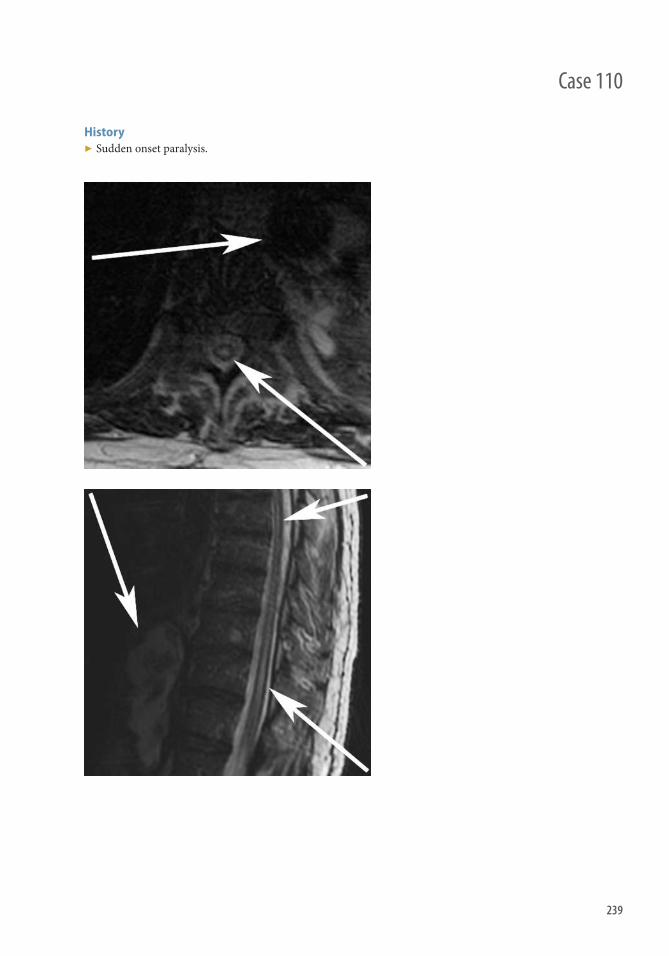
239
Case 110
History ▶ Sudden onset paralysis.

240
Findings General Features
▶ Best diagnostic clue: Central “owl’s eye” pattern; hyperintensity on T2WI within cord ▶ Location: Distal half of thoracic cord → arterial border zone ▶ Size: Usually more than one vertebral body segment ▶ Morphology: Central hyperintensity on T2WI, which involves central gray matter, more variable involvement
of cord periphery
CT Findings
▶ NECT: Noncontributory ▶ CTA
■ Useful to defi ne underlying aortic disease causing infarct, or to discover other etiologies mimicking cord infarct (arteriovenous dural fi stula)
■ Not directly useful for identifying ASA because lack of visualization of ASA does not prove diagnosis
MRI Findings
▶ T1WI ■ Slight cord expansion in acute phase ■ Cord atrophy as chronic fi nding ■ Hemorrhage conversion → hyperintense (rare)
▶ T2WI ■ Hyperintense central gray matter or entire cross-sectional area
▶ DWI ■ Hyperintense, as in brain infarcts
▶ MRA ■ Dynamic contrast enhanced
▶ Assess other etiologies that may mimic infarct, such as dural arteriovenous fi stula ▶ Not directly useful for identifying ASA because lack of visualization of ASA does not prove diagnosis
Imaging Recommendations
▶ Best imaging tool: MRI with contrast and diff usion DWI
Diff erential Diagnosis ▶ Multiple sclerosis ▶ ADEM/viral myelitis ▶ Neuromyelitis optica ▶ Spinal arteriovenous dural fi stula ▶ Radiation myelopathy ▶ Cord infection
Teaching Points ▶ Look for associated vertebral body infarctions as confi rmatory evidence of cord ischemia in aortic disease ▶ Classic imaging appearance: T2 hyperintensity involving anterior horn cells
Management ▶ Anticoagulation with heparin and aspirin ▶ Steroids ▶ Supportive care and rehabilitative physical therapy
Case 110 Spinal Cord Infarction

241
History ▶ None
Case 111
242
Findings Acute
▶ Ipsilateral enlargement of the submandibular gland, oft en painful ▶ Dilation of the submandibular duct (Wharton duct) secondary to ductal stenosis or obstructing calculus ▶ Oft en associated cellulitis and myositis ▶ Most commonly Staphylococcus aureus infection
Chronic
▶ Ipsilateral atrophy of the submandibular gland, oft en painless ▶ Dilation of submandibular duct secondary to chronic calculi and salivary stasis
Secondary Sialedenitis
▶ Typically a result of ductal obstruction secondary to squamous cell carcinoma in the fl oor of the mouth
Diff erential Diagnosis ▶ Dental infection ▶ Squamous cell carcinoma nodal metastases ▶ Benign mixed tumor ▶ Submandibular gland carcinoma
Teaching Points ▶ Best imaging tool is contrast-enhanced CT ▶ If no calculi visualized on study, consider other etiology, such as Sjögren syndrome; Kuttner tumor; or
sialadenosis secondary to diabetes, hypothyroidism, cirrhosis
Management ▶ Hydration, compression and massage, and antibiotics for infected gland ▶ Consider gland excision in recurrent cases ▶ Incision and drainage of abscess if present
Further Readings Avrahami E, et al. CT of submandibular gland sialolithiasis . Neuroradiology . 1996 ; 38 ( 3 ): 287–290 . Capps EF, et al. Emergency Imaging Assessment of Acute, Nontraumatic Conditions of the Head and Neck . Radiographics .
2010 ; 30 : 1335–1352 .
Case 111 Submandibular Sialadenitis

243
Case 112
History ▶ Patient with a neoplastic process with motor weakness and sensory complaints.

244
Findings MRI
▶ Axial and sagittal images of the cervical spine, demonstrating an enhancing mass indenting the spinal cord ▶ Cord compression can be graded by anteroposterior cord diameter: if anteroposterior cord diameter is greater
than two-thirds the cord diameter at the C1 spinal level it is defi ned as mild, if is less than two-thirds the cord diameter at the C1 level it is defi ned as severe.
Diff erential Diagnosis ▶ Myelitis (infl ammatory, infectious) ▶ Cord infarction
Teaching Points ▶ Cord compression from neoplasm can occur in multiple areas. More than one-third of patients have tumor at
multiple sites, thus the whole spine should be imaged. ▶ MRI is the best examination to delineate the spinal cord. ▶ CT can assist in surgical planning. Allows characterization of osseous disease and osseous anatomy. Does not
allow full characterization of the spinal cord. ▶ Myelography: Contrast allows the outer contour of the cord to be visualized, cord swelling can be seen. Can be
used in patients with contraindications to MRI.
Management ▶ Corticosteroids, surgical decompression, radiotherapy
Further Readings Cook AM , Lu TN , Tomlinson MJ , Vaidya M , Wakeley CJ , Goddard P . Magnetic resonance imaging of the whole spine
in suspected malignant spinal cord compression: impact on management . Clin Oncol. 1998; 10 : 39–43 (doi: 10.1016/S0936-6555(98)80111-8).
Husband DJ , Grant KA , Romaniuk CS . MRI in the diagnosis and treatment of suspected malignant spinal cord compression . Br J Radiol. 2001; 74 : 51–23 (doi:10.1148/rg.304095706).
Case 112 Cord Compression

ChestSection 3


247
History ▶ 41-year-old female on recently initiated chemotherapy in respiratory distress admitted to ICU.
Case 113
248
Findings ▶ CXR shows hazy opacity greatest in the mid to lower lung zones ▶ Axial CT images demonstrate scattered and diff use areas of ground-glass opacity
Diff erential Diagnosis ▶ Multifocal pneumonia ▶ Pulmonary edema ▶ Pulmonary hemorrhage ▶ Toxic inhalation ▶ ARDS
Teaching points ▶ Nonspecifi c appearance of lungs with scattered and/or diff use areas of ground-glass opacity ▶ Initially is scattered pattern of opacity but becomes more confl uent and diff use; chronic diff use alveolar
damage results in pulmonary fi brosis ▶ Fibrosis can start to develop in weeks but more easily detected on CT
Management ▶ Most commonly, expectant management and removal of insult (i.e., stop off ending drug)
Further Readings Rossi SE , Erasmus JJ , McAdams HP , Sporn TA , Goodman PC . Pulmonary drug toxicity: radiologic and pathologic
manifestations . RadioGraphics . 2000 ; 20 ( 5 ): 1245–1259 . Bydash J , Kasmani R , Naraharisetty K . Metal fume-induced diff use alveolar damage . J Th orac Imaging . 2010
May;25(2):W27–W29.
Case 113 Diff use Alveolar Damage

249
History ▶ 28-year-old patient with no history of cardiac disease presenting with shortness of breath aft er aggressive fl uid resuscitation.
Case 114
250
Findings ▶ Nonspecifi c hazy opacity greatest on chest radiograph throughout lungs ▶ Axial CT images demonstrate diff use areas of ground-glass opacity
Diff erential Diagnosis ▶ Pulmonary edema ▶ Pulmonary hemorrhage ▶ Toxic inhalation ▶ Diff use alveolar damage ▶ ARDS ▶ Multifocal pneumonia
Teaching points ▶ Unlike cardiogenic pulmonary edema, in noncardiogenic edema, there is typically no evidence of
cardiomegaly, apical vascular redistribution, Kerley lines ▶ Alveolar opacities and diff use ground-glass opacities on CT ▶ Noncardiogenic pulmonary edema is caused by changes in permeability of pulmonary small vessels ▶ Multiple causes of noncardiogenic pulmonary edema include drowning, fl uid overload, inhalational injury,
neurogenic pulmonary edema, allergic reaction, ARDS
Management ▶ Most commonly, expectant management and removal of insult ▶ ICU admission if severe respiratory distress with mechanical ventilation
Further Readings Gluecker T , Capasso P , Schnyder P , et al. Clinical and radiologic features of pulmonary edema . RadioGraphics .
1999 ; 19 : 1507–1531 . Milne EN , Pistolesi M , Miniati M , et al. Th e radiologic distinction of cardiogenic and noncardiogenic edema . AJR Am J
Roentgenol . 1985 ; 144 ( 5 ): 879–894 .
Case 114 Pulmonary Edema, Noncardiogenic
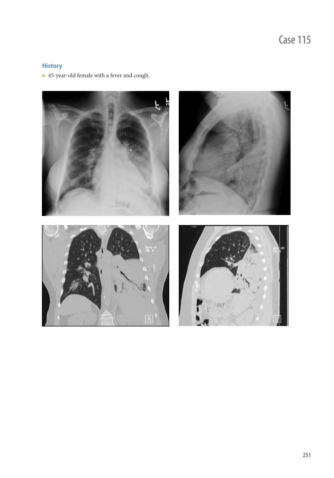
251
History ▶ 45-year-old female with a fever and cough.
Case 115

252
Findings ▶ Frontal (Figure 115.1) and lateral (Figure 115.2) radiographs of the chest demonstrate dense opacity
occupying the left lower lobe. Note the well-demarcated left major fi ssure . ▶ Th e left heart border is well demonstrated but there is obscuration of the left hemidiaphragm, consistent with
a lower lobar process. Th is is called the silhouette sign. ▶ Th ere is increased opacity over the lower vertebral bodies compared with the upper vertebral bodies. Th is is
called the spine sign. ▶ Th ere are air bronchograms . ▶ Th ese fi ndings are consistent with lobar pneumonia, which was confi rmed by chest CT. Coronal (Figure 115.3)
and sagittal (Figure 115.4) reformatted images are shown. Th ere are additional foci of infection seen in the right lung on CT.
Diff erential Diagnosis ▶ Edema ▶ Hemorrhage ▶ Atelectasis or lobar collapse ▶ Aspiration ▶ Bronchoalveolar carcinoma ▶ Cryptogenic organizing pneumonia/eosinophilic pneumonia
Teaching Points ▶ Pneumonia is a common diagnosis, which is classifi ed based on its anatomic location within the lung. ▶ Lobar pneumonia occurs when infectious organisms reach the subpleural zone of the lung. Th is results in
alveolar damage, hemorrhage, and rapid proliferation of the organism, which can spread to the adjacent lung through the pores of Kohn. Lobar pneumonia is much less common because of improved antibiotic treatment.
▶ Th e most common cause of community-acquired pneumonia is S. pneumoniae. Other causes of lobar pneumonia include Klebsiella , Legionella pneumophila, and Mycoplasma pneumoniae .
▶ Radiographic signs of pneumonia include ■ Air bronchograms: branching linear or tubular lucencies representing bronchi or bronchioles passing
through opacifi ed parenchyma (Figure 115.3 and 115.4) ■ Silhouette sign: obliteration of the borders of the heart, mediastinum, or diaphragms secondary to opacifi ed
adjacent lung parenchyma ■ Spine sign: increasing opacity over the vertebral bodies of the lower thorax is indicative of a lower lobar
process (Figure 115.2). ■ Bulging fi ssure sign: the lobe appears expanded because of voluminous edema. Th is is classically seen with
Klebsiella .
Management ▶ Antibiotic treatment
Case 115 Pneumonia

253
Case 116
History ▶ None

254
Findings ▶ Subcutaneous emphysema: Air within the neck and thoracic soft tissues. ▶ Continuous diaphragm sign: A continuous band of air seen underneath the heart on a frontal view of
the chest. ▶ Spinnaker sail sign: Elevation of the thymus by mediastinal air; seen in infants. ▶ Ring around the artery sign: Lucent air is seen surrounding the pulmonary artery or aorta. Th is fi nding is best
appreciated on the lateral view.
Diff erential Diagnosis ▶ Medial pneumothorax ▶ Pneumomediastinum ▶ Pneumopericardium
Teaching Points ▶ Pneumomediastinum can be caused by intrathoracic or extrathoracic sources. Intrathoracic sources include
alveolar rupture, esophageal rupture, or tracheal rupture Extrathoracic sources include dissecting air from the peritoneal cavity or the retroperitoneum. Th e most common source for mediastinal air with children and adults is alveolar rupture secondary to increased intrapulmonary pressure.
▶ Presentation of patients with pneumomediastinum is highly variable and may range from completely asymptomatic to excruciating chest pain. Classically on physical examination patients may present with a Hamman sign, which is a “crunching” sound heard in the precordium, which is synchronous with the heartbeat; this is usually present in about 50% of patients.
Management ▶ Oft en no treatment is necessary because the body reabsorbs the mediastinal air. If pneumomediastinum is
complicated by pneumothorax then a chest tube is placed.
Further Readings Zylak CM , Standen JR , Barnes GR , Zylak CJ . Pneumomediastinum revisited . RadioGraphics. 2000 ; 20 : 1043–1057 . Bejvan SM , Godwin JD . Pneumomediastinum: old signs and new signs . AJR Am J Roentgenol. 1996 ; 166 : 1041–1048 .
Case 116 Pneumomediastinum

255
History ▶ 35-year-old female with dyspnea and leg pain.
Case 117

256
Findings ▶ Figures 117.1: Ultrasonographic interrogation of the left femoral vein demonstrates echogenic fi lling defect
devoid of color fl ow or venous wave form. ▶ Figures 117.2: Contrast-enhanced CT of chest demonstrates multiple fi lling defects in the distal main
pulmonary arteries (arrows). ▶ Figures 117.5, 117.3, 117.4, and 117.7: Coronal reformatted images of the same patient show multiple emboli
(arrowhead) including saddle embolus in the upper and lower left pulmonary artery (arrows).
Diff erential Diagnosis ▶ Acute pulmonary embolus ▶ Chronic pulmonary embolus
Teaching Points ▶ Pulmonary embolism is occlusion of the pulmonary arterial tree secondary to emboli. ▶ Pulmonary embolism commonly results from deep vein thrombosis; however, it can also occur in the setting
of embolization of air, fat, amniotic fl uid, talc, or other foreign bodies. ▶ Th e emboli obstruct normal blood fl ow through the lungs and can increase the pressure in the pulmonary
arterial tree and right ventricle. ▶ Extremity venous ultrasound fi ndings include echogenic fi lling defects, lack of compression or augmentation,
and absence of color fl ow on Doppler. ▶ Most chest radiographs are normal. ▶ Rarely, a peripheral, wedge-shaped opacity (Hampton hump) is noted. Th is fi nding represents a pulmonary
infarct. ▶ Contrast-enhanced chest CT demonstrates fi lling defects in the pulmonary arterial tree.
Management ▶ Anticoagulation ▶ Th rombolysis ▶ Th rombectomy
Further Readings Stein PD , Fowler SE , Goodman LR , et al. Multidetector computed tomography for acute pulmonary embolism . N Engl J Med .
2006; 354 ( 22 ): 2317–2327 . Le Gal G , Righini M , Parent F , van Strijen M , Couturaud F . Diagnosis and management of subsegmental pulmonary embolism .
J Th romb Haemost . 2006; 4 ( 4 ): 724–731 . Schaefer-Prokop C , Prokop M . MDCT for the diagnosis of acute pulmonary embolism . Eur Radiol . 2006; 15 (suppl 4 ): D37–D41 .
Case 117 Pulmonary Embolism/Deep Vein Thrombosis

257
Case 118
History ▶ 49 year old with high fevers, right-sided chest pain, and dyspnea.

258
Findings ▶ Axial CT demonstrates a right-sided pleural eff usion with rim enhancement representing the split pleura sign
and underlying right lung pneumonia
Diff erential Diagnosis ▶ Pleural eff usion (exudative) ▶ Lung abscess ▶ Evolving hemothorax if in setting of trauma
Teaching Points ▶ Clinical symptoms may be nonspecifi c including fever, elevated WBC, chest pain, shortness of breath ▶ CT typically demonstrates fl uid density collection with possible locules of gas, septa, and a split pleura sign on
contrast-enhanced studies given that the parietal and visceral layers are divided by the empyema ▶ Chest radiograph shows pleural eff usion and potential loculated appearance of eff usion ▶ Ultrasound can reveal nonsimple fl uid in the pleural space, septations, or debris
Management ▶ Prompt evacuation of the empyema with antibiotics ▶ Consider surgical (VATS) and/or chest tube placement ▶ Fibrinolytics can also be placed into pleural cavity to break up loculations and septations
Further Readings Desai H , Agrawal A . Pulmonary emergencies: pneumonia, acute respiratory distress syndrome, lung abscess, and empyema .
Med Clin North Am . 2012 ; 96 ( 6 ): 1127–1148 . Weldon E , Williams J . Pleural disease in the emergency department . Emerg Med Clin North Am . 2012 ; 30 ( 2 ): 475–499 .
Case 118 Empyema
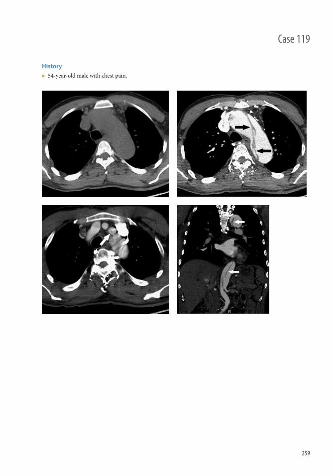
259
History ▶ 54-year-old male with chest pain.
Case 119

260
Findings Chest Radiograph
▶ May be normal in up to 12.4% of patients with dissection ▶ Th e most common fi nding is a widened mediastinum (61.6%)
CT
▶ Noncontrast images may show displaced intimal calcifi cation. High attenuation within the aortic wall indicates the presence of intramural hematoma, which may clinically mimic or coexist with acute aortic dissection
▶ Anintimal fl ap separates the false and true lumens ( black and white arrows in axial and coronal images); the false lumen is usually larger than the true lumen
▶ Th in bands of low attenuation representing residual media may be seen in the false lumen; this is known as the cobweb sign.
▶ Th e beak sign refers to a wedge-shaped area of hematoma at the edge of the false lumen that creates a plane into which the false lumen can expand
MRI
▶ An intimal fl ap is best seen on spin-echo “black blood” sequences ▶ A GRE cine sequence can be used to diff erentiate slow fl ow from thrombus within the false lumen, and can
evaluate for aortic regurgitation if the dissection involves the aortic root
Diff erential Diagnosis ▶ Intramural hematoma ▶ Tortuous aorta ▶ Th rombosed aortic aneurysm ▶ Traumatic aortic injury
Teaching Points ▶ Stanford Type A dissection
■ Intimal tear and/or associated intramural hematoma involves the ascending aorta, proximal to the origin of the left subclavian artery (60%–70% of cases, including the above case)
■ Dissection fl ap may involve coronary arteries, and may also extend into the great vessels (arrow) ■ Stanford Type A dissection may extend to involve the descending aorta (arrow) ■ Mortality may result from rupture into the pericardial space resulting in cardiac tamponade
▶ Stanford Type B dissection ■ Intimal tear involves the descending aorta, distal to the origin of the left subclavian artery and extending
caudally ■ Dissection fl ap may involve celiac, mesenteric, and renal arteries
Management ▶ Type A dissection requires urgent surgical repair ▶ Type B dissection is managed medically unless the patient is unstable or there is evidence of vascular
compromise to an organ
Case 119 Aortic Dissection (Stanford Type A)
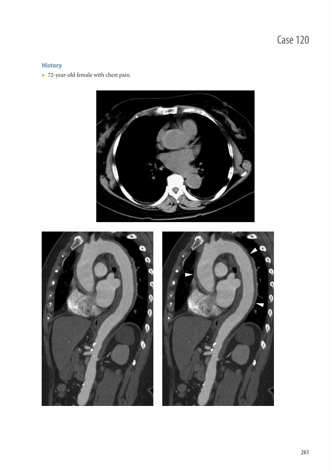
261
History ▶ 72-year-old female with chest pain.
Case 120

262
Case 120 Aortic Intramural Hematoma
Findings CT ▶ Noncontrast images show high-density hematoma within the wall of the thoracic and/or abdominal aorta
(arrows) and may also show displaced intimal calcifi cation ▶ Contrast-enhanced images make diagnosis diffi cult because the nonenhancing intramural hematoma may
appear similar to luminal thrombus (arrowheads) MRI
▶ Can be used to help diagnose or determine age of intramural hematoma ▶ Acute intramural hematoma is hyperintense on “white-blood” images and isointense on “black-blood” images ▶ Cine phase-contrast or GRE sequences can be used to diff erentiate from communicating aortic dissection Diff erential Diagnosis ▶ Classic or thrombosed aortic dissection ▶ Hematoma associated with penetrating atherosclerotic ulcer ▶ Atheroma or intraluminal thrombus ▶ Aortitis ▶ Retroperitoneal fi brosis or lymphoma Teaching Points ▶ Classic intramural hematoma results from spontaneous hemorrhage of the vasa vasorum within the media,
usually caused by hypertension ▶ CT, including noncontrast imaging, is the modality of choice ▶ Classifi ed in same manner as aortic dissection
■ Stanford Type A intramural hematoma involves the ascending aorta, beginning proximal to the origin of the left subclavian artery, and may extend caudally
■ Stanford Type B intramural hematoma involves the descending aorta, beginning distal to the origin of the left subclavian artery
■ Associated conditions and complications ■ May coexist with penetrating ulcer or classic dissection ■ May lead to dissection or aneurysm caused by weakening of the aortic wall ■ Associated penetrating atherosclerotic ulcer or ulcerlike projection indicates increased risk of future complications ■ Ulcerating lesions should be distinguished from focal intramural contrast enhancement or intramural
blood pools, which do not clearly communicate with the true lumen and are of unlikely clinical signifi cance ■ Patients not treated surgically should receive imaging follow-up
Management ▶ Usually treated in the same fashion as aortic dissection
■ Type A requires urgent surgical repair ■ Type B is managed medically unless the patient is unstable or there is evidence of vascular compromise to
an organ

263
History ▶ 75-year-old female with acute shortness of breath.
Case 121

264
Findings ▶ Frontal chest radiograph (Figure 121.1) and coronal reformat from a subsequent chest CT (Figure 121.2)
demonstrate veil-like opacity in the left middle and upper lung zones ▶ Th ere is crescentic lucency adjacent to the aortic arch ▶ Th ere is elevation of the left hilum, indicative of volume loss in the left upper lobe ▶ Th ere is a speculated mass in the left hilum and a left pleural eff usion
Diff erential Diagnosis ▶ Airway obstruction secondary to bronchogenic carcinoma, endobronchial secretions, or endobronchial
tumors, such as carcinoid ▶ Malpositioned endotracheal tube ▶ Airway compression secondary to fl uid, mass, or lymphadenopathy ▶ Scarring
Teaching Points ▶ Radiographic fi ndings of lobar collapse include veil-like opacities; opacities with sharp borders; and shift ing of
the mediastinal contents, hilum, and fi ssures toward the region of suspected volume loss. ▶ Luft sichel (“air sickle”) sign, as seen in this case, is most commonly a manifestation of left upper lobe collapse
with wedging of the aerated superior segment of the left lower lobe between the collapsed lung and the aorta creating a periaortic lucency.
▶ In left upper lobe collapse, the upper lobe collapses anteriorly and superiorly against the chest wall with resultant hyperexpansion of the left lower lobe, which oft en tents the left hemidiaphragm.
▶ Another notable radiographic sign of lobar collapse is the “reverse S sign of Golden” in which the right minor fi ssure is displaced superiorly and assumes a convex inferior border medially and a concave inferior border laterally secondary to collapse of the right upper lobe.
Management ▶ Cross-sectional imaging with CT should be performed to assess for obstructing lesions ▶ Bronchoscopy may be performed to remove the off ending lesion
Further Readings Blankenbaker DG . Th e luft sichel sign . Radiology. 1998 ; 208 ( 2 ): 319–320 . Gupta P . Th e Golden S sign . Radiology. 2004 ; 233 ( 3 ): 790–791 . Lubert M, Krause GR . Patterns of lobar collapse as observed radiographically. Radiology. 1951 ; 56 ( 2 ): 165–182 .
Case 121 Lobar Collapse

265
Case 122
History ▶ 25-year-old patient recently rescued by fi refi ghters from inhalation of smoke from burning house.

266
Findings ▶ Axial CT images demonstrate diff use ground-glass opacity
Diff erential Diagnosis ▶ Multifocal pneumonia ▶ Pulmonary edema ▶ Pulmonary hemorrhage ▶ Diff use alveolar damage ▶ ARDS ▶ PCP if signs of infection
Teaching Points ▶ Nonspecifi c appearance of lungs, diff use and scattered areas of ground-glass opacity ▶ Tree-in-bud opacities may also be present given infl ammatory process also involving small airways
Management ▶ Most commonly, expectant management and oxygen ▶ Laryngoscopy and bronchoscopy may be necessary to assess complications of airway ▶ Treat other concurrent comorbidities, such as burns, depending on inhalation injury
Further Readings Rossi SE , Erasmus JJ , McAdams HP , Sporn TA , Goodman PC . Pulmonary drug toxicity: radiologic and pathologic
manifestations . RadioGraphics . 2000 ; 20 ( 5 ): 1245–1259 . Bydash J , Kasmani R , Naraharisetty K . Metal fume-induced diff use alveolar damage . J Th orac Imaging . 2010
May; 25 ( 2 ): W27–W29 .
Case 122 Toxic Inhalation

267
Case 123
History ▶ Swelling of right upper extremity.

268
Case 123 SVC Obstruction
Findings ▶ CT coronal reformat images demonstrating obstruction of the SVC (arrow) ▶ Venous angiography demonstrates fi lling of a dilated azygous vein through collaterals ▶ Th ree-dimensional reformat demonstrating fi lling of a dilated azygous vein through collaterals
Diff erential Diagnosis ▶ Mediastinal mass ▶ Bronchogenic neoplasm ▶ Intravascular SVC thrombosis
Teaching Points ▶ Oft en, history and physical examination yield diagnosis but imaging can confi rm a mass obstructing the SVC ▶ Large mass can be seen on radiograph or CT in the region of the SVC ▶ Collateral circulation on contrast-enhanced CT can also be oft en visualized ▶ Obliteration or narrowing of SVC can be visualized on CT ▶ Obstruction caused by neoplastic invasion of the SVC and hemodynamically signifi cant narrowing ▶ Intravascular thrombus may also be found in association ▶ Collateral circulation involving the azygous vein, hemiazygous vein, intercostal vein, and internal mammary
veins can form ▶ Symptoms include dyspnea, arm swelling, chest pain, dysphagia, headache, nasal stuffi ness, light headedness,
and dramatic facial swelling in complete or near complete obstruction ▶ Early in clinical course, patient may be asymptomatic
Management ▶ CT is more accurate than chest radiograph in providing accurate detailed anatomic evaluation for location
and extent of neoplastic pathology obstructing SVC ▶ Stenting of SVC could be considered ▶ Chemotherapy or radiotherapy to treat primary tumor and lesion obstructing SVC ▶ Surgical management
Further Readings McCurdy MT , Shanholtz CB . Oncologic emergencies . Crit Care Med . 2012 ; 40 ( 7 ): 2212–2222 . Lepper PM , Ott SR , Hoppe H , et al. Superior vena cava syndrome in thoracic malignancies . Respir Care . 2011 ; 56 ( 5 ): 653–666 .

AbdomenSection 4


271
History ▶ 38-year-old male with fever, malaise, and jaundice.
Case 124

272
Findings Acute
▶ Diff use hepatic edema, resulting in diff use decrease in echogenicity or density of the liver; this may be particularly pronounced around the portal triads
▶ Hepatomegaly, splenomegaly, porta hepatis lymphadenopathy, and ascites ▶ Gallbladder wall thickening; oft en greater than with cholecystitis, and with a negative sonographic
Murphy sign ▶ Heterogeneous enhancement on CT ▶ US
■ “Starry-sky” appearance of liver: caused by relative increased echogenicity of the portal triads against the diff usely hypoechoic liver parenchyma
■ Relative increased echogenicity of ligamentum venosum and falciform ligament ■ Hypoechoic liver echotexture becomes more similar to that of the spleen and renal cortex
Chronic
▶ Diff use heterogeneity and coarsening of liver echotexture or density; this is caused by a combination of regenerating nodules, steatosis, fi brosis, and necrosis
▶ Hepatic atrophy, with a retracted irregular capsule; especially of the right lobe, possibly with compensatory hypertrophy of the left and caudate lobes
▶ Loss of defi nition or silhouetting of the portal veins on ultrasound
Diff erential Diagnosis ▶ Hepatic steatosis ▶ Hepatic congestion ▶ Diff use lymphoma, hepatocellular carcinoma, or metastatic disease
Teaching Points ▶ Viral hepatitis is the most common cause; other causes include alcohol, autoimmune, drug induced, radiation
induced, and nonalcoholic steatohepatitis ▶ In severe fulminant cases, can be acutely fatal without liver transplant ▶ Chronic hepatitis increases the risk of developing hepatocellular carcinoma, especially if caused by hepatitis
B virus
Management ▶ Antiviral therapy ▶ Alcohol cessation ▶ Hepatitis B vaccine is available for prophylaxis ▶ Hepatitis B immune globulin for empiric treatment in cases of suspected acute exposure
Further Readings Dienstag JL . Hepatitis B virus infection . N Engl J Med . 2008 ; 359 : 1486–1500 . Mortelé KJ , Segatto E , Ros PR . Th e infected liver: radiologic-pathologic correlation . RadioGraphics . 2004 ; 24 ( 4 ): 937–955 . Abu-Judeh HH. Th e “starry sky” liver with right-sided heart failure. AJR Am J Roentgenol. 2002 Jan;178(1):78.
Case 124 Hepatitis

273
Case 125
History ▶ Right upper quadrant pain, nausea, and vomiting. Hemodynamically unstable with leukocytosis and high alkaline phosphatase.

274
Findings ▶ Ultrasound image demonstrates a large gallstone at the gallbladder neck (Figure 125.1) ▶ Ultrasound image demonstrates gallbladder wall thickening (Figure 125.2) ▶ Ultrasound images demonstrate sludge within a gallbladder with thickened and edematous wall
(Figures 125.3)
Diff erential Diagnosis ▶ Gallbladder wall thickening from portal hypertension, ascites, and hypoalbuminemia
Teaching Points ▶ Usual presentation is right upper quadrant pain, nausea, and/or vomiting. ▶ Occurs in about one-third of patients with gallstones. ▶ 95% caused by calculous obstruction in the gallbladder neck or cystic duct resulting in increased intraluminal
pressure and distention and at times superimposed infection. ▶ Acalculous cholecystitis seen in patients with trauma, mechanical ventilation, hyperalimentation,
postoperative state, diabetes mellitus, vascular insuffi ciency, prolonged fasting, burns, and postpartum state. ▶ Complications include gangrenous cholecystitis, emphysematous cholecystitis, gallbladder perforation,
cholecystoenteric fi stula, porcelain gallbladder resulting from chronic or recurrent cholecystitis, which in turn may result in gallbladder carcinoma.
▶ Ultrasound is modality of choice for evaluation of cholecystitis, both sensitive and specifi c. ▶ Ultrasound most sensitive fi ndings: cholelithiasis and sonographic Murphy sign (maximal tenderness over the
gallbladder as seen by ultrasound). ▶ Additional ultrasound fi ndings: Gallbladder wall thickness >3 mm, gallbladder wall edema,
pericholecystic fl uid.
Management ▶ Cholecystectomy, laparoscopic favored ▶ Percutaneous cholecystostomy catheter for the unstable patient
Further Reading Hanbidge A , Buckler P , O’Malley M , Wilson S . From the RSNA Refresher Courses: imaging evaluation for the acute pain in the
right upper quadrant . RadioGraphics . 2004; 24 : 1117–1135.
Case 125 Acute Cholecystitis

275
History ▶ 89-year-old female with acute onset right upper abdominal pain.
Case 126

276
Case 126 Acute Cholangitis
Findings ▶ Coronal image from a contrast-enhanced CT of the abdomen demonstrates irregular dilatation of the distal
common bile duct, which measured up to 10 mm in maximum dimension. ▶ Axial image from a contrast-enhanced CT of the abdomen demonstrates the “target sign”: high-density
material within the gallbladder (likely sludge) surrounded by low-density bile.
Imaging Findings Ultrasound
▶ Dilatation, narrowing, or wall thickening of biliary ducts ▶ Echogenic material within the duct (pus) ▶ Stones (gallbladder or bile duct)
CT
▶ Biliary ductal dilatation ▶ High-density material within the duct (pus) ▶ “Bull’s eye”: dense stone surrounding by low-density bile ▶ Look for hepatic abscesses (potential complication)
MRCP
▶ Stones = low signal fi lling defects ▶ Bile = increased signal ▶ Structures, prestenotic biliary dilatation
Fluoroscopy
▶ Irregular dilatation of bile ducts ▶ Stone = fi lling defect ▶ May see communication with hepatic abscesses
Diff erential Diagnosis ▶ Primary sclerosing cholangitis (UC) ▶ AIDS-related recurrent cholangitis ▶ Chemotherapy cholangitis
Teaching Points ▶ Ascending cholangitis: bacterial infection of intrahepatic and extrahepatic biliary system secondary to biliary
obstruction ▶ Classic clinical presentation: Charcot triad (fever, right upper quadrant pain, jaundice)
■ Elevated WBC, bilirubin, alkaline phosphatase, positive blood cultures ■ Age 20–50 years
▶ Choledocholithiasis (number one cause); also, iatrogenic (post-ERCP)
Management ▶ Antibiotics (for gram-negative bacteria) ▶ Intervention to remove stone or relieve stricture; 100% mortality if not decompressed!
Further Readings
Statdx.com Hanau LH , Steigbigel NH . Acute cholangitis . Infect Dis Clin North Am . 2000 ; 14 ( 3 ): 521–546 .
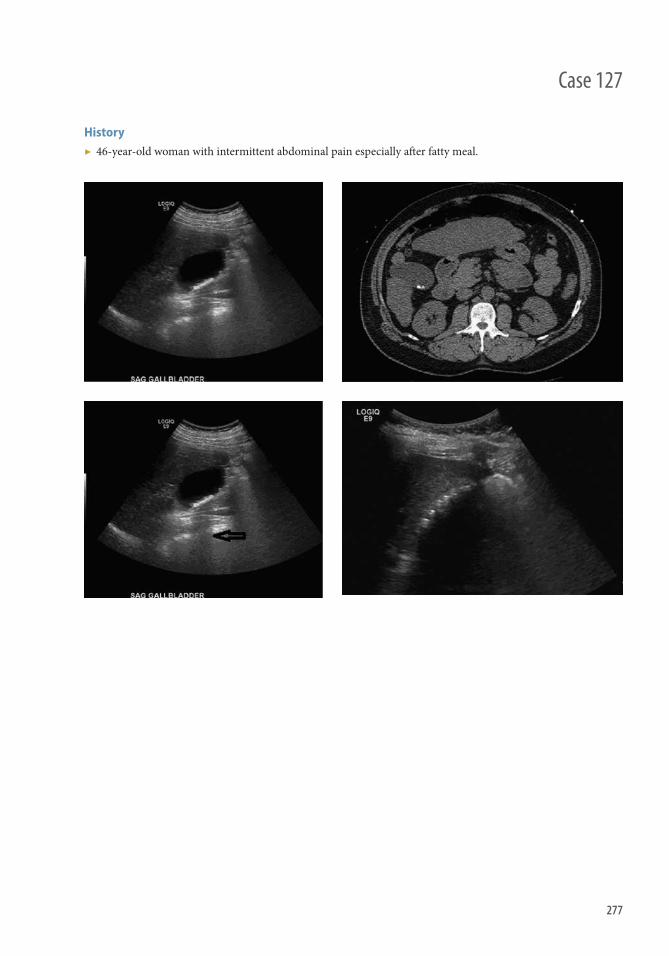
277
History ▶ 46-year-old woman with intermittent abdominal pain especially aft er fatty meal.
Case 127

278
Findings ▶ Ultrasound image of the gallbladder demonstrating shadowing gallstones ▶ CT demonstrating hyperdense foci consistent with gallstones ▶ Black open arrow denotes posterior acoustic shadowing ▶ Wall echo shadow sign, resulting from multiple gallstones in a contracted gallbladder
Diff erential Diagnosis ▶ Tumefactive sludge ▶ Cholesterol polyps ▶ Adenomyomatosis
Teaching Points ▶ Approximately 25 million adults in the United States have cholelithisis, women more oft en than men. ▶ Prevalence increases with age. ▶ Because of supersaturation of bile constituents, usually cholesterol. ▶ Aggravated by high-fat diet, sedentary lifestyle, and genetic predisposition. ▶ Association with obesity, diabetes, oral contraceptives, ileal disease, total parenteral nutrition, cirrhosis, and
spinal cord injury. ▶ Ultrasound characteristics of gallstones include hyperechoic structures within the gallbladder that are usually
mobile with patient position. Gallstones usually demonstrate posterior acoustic shadowing. ▶ Wall echo shadow sign (Figure 127.4): anterior wall of the gallbladder is echogenic, below which is a thin,
dark line of bile, and a highly echogenic line of superfi cial stones with associated posterior shadowing. Deeper stones are the posterior aspect of the gallbladder is not visible.
▶ CT fi ndings include hyperdense structures within the gallbladder. Th ey may have nitrogen gas inside the gallstones that may form the classic “Mercedes-Benz” sign.
▶ Complications include biliary colic, choledocholithiasis, acute cholecystitis, gallbladder perforation, pancreatitis, biliary fi stula, Mirizzi syndrome, porcelain gallbladder, increasing risk of gallbladder carcinoma
Management ▶ Cholecystectomy if symptomatic
Further Reading Bortoff G , Chen M , Ott D , Wolfman N , Routh W . Gallbladder stones: imaging and intervention . RadioGraphics.
2000; 20 : 751–766.
Case 127 Gallstones/Cholelithiasis

279
History ▶ Vomiting and epigastric pain.
Case 128
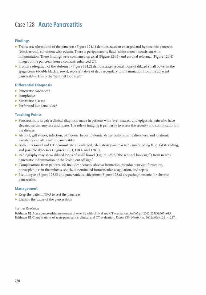
280
Findings ▶ Transverse ultrasound of the pancreas (Figure 124.1) demonstrates an enlarged and hypoechoic pancreas
(black arrow), consistent with edema. Th ere is peripancreatic fl uid (white arrow), consistent with infl ammation. Th ese fi ndings were confi rmed on axial (Figure 124.3) and coronal reformat (Figure 124.4) images of the pancreas from a contrast-enhanced CT.
▶ Frontal radiograph of the abdomen (Figure 124.2) demonstrates several loops of dilated small bowel in the epigastrum (double black arrows), representative of ileus secondary to infl ammation from the adjacent pancreatitis. Th is is the “sentinel loop sign.”
Diff erential Diagnosis ▶ Pancreatic carcinoma ▶ Lymphoma ▶ Metastatic disease ▶ Perforated duodenal ulcer
Teaching Points ▶ Pancreatitis is largely a clinical diagnosis made in patients with fever, nausea, and epigastric pain who have
elevated serum amylase and lipase. Th e role of imaging is primarily to assess the severity and complications of the disease.
▶ Alcohol, gall stones, infection, iatrogenia, hyperlipidemia, drugs, autoimmune disorders, and anatomic variability can all result in pancreatitis.
▶ Both ultrasound and CT demonstrate an enlarged, edematous pancreas with surrounding fl uid, fat stranding, and possible abscesses (Figures 128.3, 128.4, and 128.5).
▶ Radiography may show dilated loops of small bowel (Figure 128.2, “the sentinel loop sign”) from nearby pancreatic infl ammation or the “colon cut off sign.”
▶ Complications from pancreatitis include: necrosis, abscess formation, pseudoaneurysm formation, portosplenic vein thrombosis, shock, disseminated intravascular coagulation, and sepsis.
▶ Pseudocysts (Figure 128.5) and pancreatic calcifi cations (Figure 128.6) are pathognomonic for chronic pancreatitis.
Management ▶ Keep the patient NPO to rest the pancreas ▶ Identify the cause of the pancreatitis
Further Readings Balthazar EJ . Acute pancreatitis: assessment of severity with clinical and CT evaluation . Radiology. 2002 ; 223 ( 3 ): 603–613 . Balthazar EJ . Complications of acute pancreatitis: clinical and CT evaluation . Radiol Clin North Am. 2002 ; 40 ( 6 ): 1211–1227 .
Case 128 Acute Pancreatitis

281
History ▶ 78-year-old woman on chemotherapy with abdominal pain.
Case 129

282
Findings ▶ Linear and round foci of air within the intestinal wall ▶ Pneumatosis intestinalis is emphasized on lung windows ▶ Branching foci of air within the periphery of the liver denotes portal venous air in another patient
Diff erential Diagnosis None.
Teaching Points Pathogenesis
▶ Gas dissects into the intestinal wall from the intestinal lumen or the lungs by the mediastinum because of increases in pressure, such as bowel obstruction and emphysema.
▶ Gas-forming bacteria enter the submucosa through mucosal tears or increased mucosal permeability and produce gas within the intestinal wall.
Benign and Life-threatening Causes
▶ Benign causes ■ Pulmonary: COPD, asthma, pulmonary fi brosis, increased PEEP, cystic fi brosis ■ Systemic disease: Scleroderma, SLE, AIDS ■ Intestinal causes: Pyloric stenosis, enteritis, colitis, peptic ulcers, bowel obstruction ■ Iatrogenic: Barium enema, endoscopy, bowel surgery ■ Medications: Corticosteroids, lactulose, sorbitol, chemotherapy ■ Organ transplantation and graft -versus-host disease
▶ Life-threatening causes ■ Vascular: Mesenteric vascular disease, vasculitis ■ Intestinal: Obstruction/strangulation, enteritis, colitis, corrosive agent ingestion, toxic megacolon ■ Trauma ■ Organ transplantation most specifi cally bone marrow transplant ■ Collagen vascular disease
CT Findings
▶ Linear or bubbly gas in the bowel wall ▶ Benign fi ndings: pneumatosis only, without peritoneal signs on physical examination ▶ Life-threatening fi nding: pneumatosis with bowel wall thickening, dilated bowel, arterial or venous occlusion,
ascites, and/or hepatic portal or portomesenteric venous gas
Portal Venous Gas Versus Pneumobilia
▶ Portal venous gas: tubular branching lucencies extending to the periphery of the liver ▶ Pneumobilia: lucencies remain central
Management Treatment of underlying cause.
Further Reading Ho L , Paulson E , Th ompson W . Pneumatosis intestinalis in the adult: benign to Life-threatening causes . Am J Radiol .
2007 ; 188 : 1604–1613
Case 129 Pneumatosis Intestinalis

283
Case 130
History ▶ 66-year-old male with nausea and vomiting.

284
Findings ▶ Supine AP view of the abdomen demonstrates multiple abnormally dilated loops of small bowel. ▶ Upright PA view of the abdomen demonstrates multiple air-fl uid levels. ▶ Axial CT with intravenous and oral contrast demonstrates multiple loops of bowel with stranding in the
mesentery in the right abdomen. Th e colon is normal in caliber. ▶ Coronal CT showing fecalization of a loop of small bowel. ▶ Axial CT showing fecalization of a loop of small bowel.
Teaching Points Key Radiographic Findings
▶ Small bowel distention, with maximal dilated loops averaging 36 mm in diameter and exceeding 50% of the caliber of the largest visible colon loop.
▶ Presence of more than two air-fl uid levels, air-fl uid levels wider than 2.5 cm, and air-fl uid levels diff ering more than 2 cm in height from one another within the same small bowel loop
Key CT Findings
▶ Presence of dilated small bowel loops, with diameter >25 mm from outer wall to outer wall, proximal to normal caliber or collapse loops distally.
▶ Presence of small bowel feces sign (Figure 130.4) ▶ Sometimes obvious cause of small bowel obstruction, such as hernia or mass, is found; if none found, cause is
likely adhesions with a history of surgery ▶ CT signs of bowel at risk
■ Th ickened and enhancing bowel wall (caused by venous and lymphatic obstruction) ■ Portal venous gas (implies ischemic bowel) ■ Free air (implies perforated bowel)
▶ Strangulated bowel: bowel obstructed at two points
Causes of Small Bowel Obstruction Extrinsic Causes
▶ Adhesions, most common cause ▶ Internal and external hernias, second most common cause ▶ Endometriosis
Intrinsic Causes
▶ Infl ammatory, such as Crohn disease and eosinophilic gastroenteritis ▶ Neoplasm such as GIST, adenocarcinoma, and metastasis ▶ Hematoma from trauma and anticoagulation therapy ▶ Intussusception
Intraluminal Causes
▶ Gallstones ▶ Bezoars ▶ Foreign bodies
Management ▶ Bowel rest with gastric decompression with nasogastric tube. ▶ If obstruction does not resolve with conservative management, surgical intervention is warranted
Case 130 Small Bowel Obstruction

285
History ▶ Fever, nausea, and right lower quadrant pain.
Case 131

286
Findings ▶ Frontal abdominal radiograph (Figure 131.1) and right lower quadrant ultrasound (Figure 131.2) demonstrate
an appendicolith (black arrow) in the right lower quadrant with an enlarged appendix (white arrow). ▶ Axial (Figure 131.3) and coronal reformat (Figure 131.4) images from a contrast-enhanced CT scan of the
abdomen show a thickened appendix with hyperemic walls (white arrow). Th ere is also periappendiceal fat-stranding and free fl uid (double black arrows).
Diff erential Diagnosis ▶ Mesenteric infarct ▶ Epiploic appendicitis ▶ Mesenteric adenitis ▶ Cecal diverticulitis ▶ Crohn disease ▶ Terminal ileitis ▶ Typhlitis ▶ Appendiceal mucocele
Teaching Points ▶ Appendicitis is the most common surgical emergency in childhood ▶ Ultrasound has variable accuracy in its detection of appendicitis because of technical factors but is a
reasonable fi rst examination in children, pregnancy, and thin patients ▶ Ultrasonographic fi ndings in appendicitis include appendiceal diameter >6–7 mm, appendicolith, free fl uid,
and bowel wall thickening ▶ CT is both highly sensitive and specifi c for the detection of appendicitis. CT fi ndings of appendicitis include
appendiceal diameter >6–7 mm, hyperemia, bowel wall thickening including the cecal bar sign (Figure 131.5) and cecal arrowhead sign (Figure 131.6), fat stranding, lack of intraluminal air and contrast, and free fl uid
▶ MRI has been shown to be of value in the diagnosis of appendicitis in pregnancy without exposing the fetus to ionizing radiation
▶ Most complications of appendicitis are secondary to perforation (Figure 131.7) and include abscess formation, peritonitis, sepsis, bowel obstruction, infertility, and death
Management ▶ Surgical evaluation for appendectomy or drainage and antibiotics, if perforated
Further Readings Leite NP, et al. CT evaluation of appendicitis and its complications: imaging techniques and key diagnostic fi ndings . AJR Am J
Roentgenol. 2005 ; 185 ( 2 ): 406–417 . Sivit CJ, et al. When appendicitis is suspected in children . RadioGraphics. 2001 ; 21 ( 1 ): 247–262 . Cobben LP , et al. MRI for clinically suspected appendicitis during pregnancy . AJR Am J Roentgenol . 2004 ; 183 ( 3 ): 671–675 .
Case 131 Appendicitis

287
History ▶ None
Case 132

288
Findings Radiograph
▶ Prominent focal loop of air-distended large bowel with its axis extending from the right lower quadrant to the left upper quadrant.
▶ Small bowel dilatation and air-fl uid levels may be present depending on acuity.
CT
▶ Severe colonic dilation with decompressed colon distally. ▶ Dilation and fecalization of small bowel. ▶ Transition point in the colon. ▶ “Whirl sign” toward the root of the mesentery where it twists on itself. ▶ “Beak-like” tapering of the colon at the level of the volvulus. ▶ “Split wall” sign shows apparent separation of cecal wall by adjacent mesenteric fat.
Diff erential Diagnosis ▶ Sigmoid volvulus ▶ Ogilvie syndrome (pseudo-obstruction) ▶ Toxic megacolon ▶ Colonic obstruction
Teaching Points ▶ Usually presents with colicky abdominal pain, abdominal distention, and vomiting. ▶ Diff erent from “cecal bascule,” which refers to an abnormal location of the cecum in the mid-abdomen
resulting from the cecum folding up on itself without associated rotation along its axis. Th is can occur when the cecum is loosely attached to the mesentery and is excessively mobile.
Management ▶ Reduction with enema may be attempted. ▶ Treatment is usually surgical.
Further Readings Peterson CM , Anderson JS , Hara AK , Carenza JW , Menias CO . Volvulus of the gastrointestinal tract: appearances at
multimodality imaging . RadioGraphics . 2009 ; 29 ( 5 ): 1281–1293 . Rosenblat JM , Rozenblit AM , Wolf EL , DuBrow RA , Den EI , Levsky JM . Findings of cecal volvulus at CT . Radiology .
2010 ;2 56 ( 1 ): 169–175 . PubMed PMID: 20574094.
Case 132 Cecal Volvulus

289
Case 133
History ▶ 61-year-old male with left lower quadrant pain.

290
Findings ▶ A segment of large bowel with wall thickening and associated fat stranding. ▶ Diverticula are seen in the vicinity.
Diff erential Diagnosis Segmental colitis from infectious, infl ammatory, or ischemic etiology
Teaching Points ▶ Common condition in Western society because of diet poor in fi ber. ▶ Aff ects 5%–10% of people >45 years old and 80% of people >85 years of age. ▶ Manifests as outpouchings in the intestinal wall. ▶ Most common location is in the sigmoid. ▶ Caused by diverticulum occluded by stool, infl ammation, or food debris leading to microperforation of the
diverticulum and surrounding infl ammation. ▶ CT fi ndings of diverticulitis include segmental wall thickening, enhancement, adjacent fat stranding, and
presence of diverticula. ▶ Complications include abscess, colovesicular and entero fi stulas, perforation, and stricture formation.
Management ▶ Intravenous or oral antibiotics ▶ Percutaneous drainage of abscess ▶ Surgical management of fi stulas, perforation, and strictures.
Further Reading Horton K , Corl F , Fishman E . CT evaluation of the colon: infl ammatory disease . RadioGraphics . 2000; 20 : 399–418.
Case 133 Diverticulitis

291
History ▶ 32-year-old female presents with crampy abdominal pain.
Case 134

292
Findings ▶ Transverse ultrasound image of the right abdomen (Figure 134.1) demonstrates a “target sign” with an
isoechoic layer of outer bowel (the intussuscepiens, white arrow); a hyperechoic layer of mesenteric fat (black arrow); and a somewhat hypoechoic layer of inner bowel (the intussusceptum, double black arrows). Th ese fi ndings were confi rmed with a contrast-enhanced CT (Figure 134.3).
▶ Frontal radiograph of the abdomen (Figure 134.2) shows a soft tissue mass (the intussusceptum, double white arrows) in the mid-abdomen. Th is fi nding was also confi rmed with coronal reformatted images from a contrast-enhanced CT (Figure 134.4).
Diff erential Diagnosis ▶ Idiopathic ▶ Primary bowel tumors (both benign and malignant) ▶ Enteritis ▶ Lymphoma ▶ Meckel diverticulum ▶ Metastatic disease
Teaching Points ▶ Intussusception is the peristaltic telescoping of a loop of bowel with its mesenteric fat (the intussusceptum)
into the lumen of a contiguous loop of bowel (the intussuscepiens). Most intussusceptions occur in children, commonly between the ages of 6 months and 2 years.
▶ Intussusception is most commonly idiopathic in children, whereas most intussusceptions in adults are secondary to “lead points,” such as primary bowel tumors, lymphoma, or metastatic disease.
▶ Ultrasound is the initial imaging modality of choice for the diagnosis of intussusception, which can demonstrate a “target sign” (Figure 134.1) or a “pseudokidney sign” where there are multiple layers of hyperechogenicity and hypoechogenicity within a reniform mass.
▶ Radiographic signs of intussusception include a soft tissue mass (Figure 134.2), signs of obstruction, and a “crescent sign” where there is lucent air surrounding a soft tissue mass in the abdomen.
▶ CT can also demonstrate the “target sign” (Figure 134.3) and aid in the search of possible “lead points.”
Management ▶ Image-guided barium or air enema to reduce the intussusception with a surgeon on stand-by in case of bowel
perforation.
Further Readings del-Pozo G, Albillos JC, Tejedor D, et al. Intussusception in children: current concepts in diagnosis and enema reduction .
RadioGraphics. 1999 ; 19 ( 2 ): 299–319 . Kim YH , Blake MA , Harisinghani MG , et al. Adult intestinal intussusception: CT appearances and identifi cation of a causative
lead point . RadioGraphics. 2006 ; 26 ( 3 ): 733–744 .
Case 134 Intussusception

293
Case 135
History ▶ Abdominal pain and distention.

294
Findings Radiographs
▶ Bowel dilation with air-fl uid levels ▶ Pneumatosis intestinalis ▶ Portal venous gas
Contrast-enhanced Computed Tomography
▶ SMA, SMV, or other mesenteric vessel occlusion (most specifi c sign) ▶ Mesenteric or portal venous gas ▶ Decreased bowel wall enhancement caused by decreased arterial fl ow ▶ Bowel wall thickening (>3mm) caused by edema, hemorrhage, or superimposed infection ▶ Bowel dilation with air-fl uid levels ▶ Mesenteric fat infi ltration caused by edema (more common with venous occlusion) ▶ Pneumatosis
Diff erential Diagnosis ▶ Infectious enteritis ▶ Shock bowel ▶ Infl ammatory bowel disease (e.g., Crohn disease) ▶ Fibrosis mesenteritis
Teaching Points ▶ Presentation
■ Acute ischemia: sudden onset of abdominal pain, vomiting, diarrhea ■ Chronic ischemia: chronic, intermittent abdominal pain (intestinal angina)
▶ Etiology ■ Vascular occlusion (atherosclerosis, thromboembolic) ■ Bowel herniation or closed loop obstruction ■ Hypercoagulable states ■ Infl ammatory (e.g., vasculitis) ■ Hypoperfusion (e.g., sepsis, hypovolemia) ■ Drugs (e.g., cocaine)
▶ Occlusion of the SMA or SMV is the most specifi c sign ■ Arterial occlusion may be complete or partial
Management ▶ Surgical
■ Exploratory laparotomy with resection of nonviable bowel +/- vascular bypass ▶ Endovascular recanalization of vascular occlusion ▶ Anticoagulation for venous thrombosis
Further Readings Furukawa A , Kanasaki S , Kono N , et al. CT diagnosis of acute mesenteric ischemia from various causes . AJR Am J Roentgenol.
2009 Feb; 192 ( 2 ): 408–416 . Wiesner W , Khurana B , Ji H , et al. CT of acute bowel ischemia . Radiology. 2003; 226 : 635.
Case 135 Small Bowel Ischemia
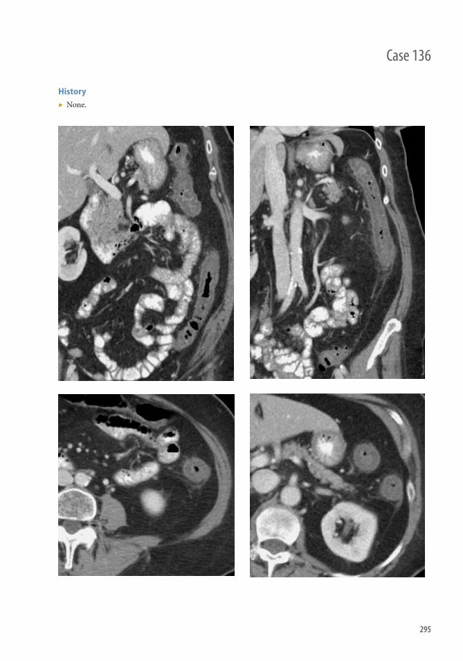
295
History ▶ None.
Case 136

296
Findings Radiograph
▶ “Th umbprinting” secondary to bowel wall edema ▶ Dilated loops of bowel secondary to ileus ▶ Pneumatosis or signs of portal venous air in the liver ▶ Free air if perforated
CT
▶ Wall thickening within a vascular territory ▶ Low-density bowel wall resulting in a “target sign” ▶ Pneumatosis or portal venous air ▶ Signs of vascular occlusion involving the SMA or SMV.
Diff erential Diagnosis ▶ Infectious colitis (pseudomembranous colitis “pancolitis”) ▶ Infl ammatory colitis ▶ Neoplastic (lymphoma can have associated lymphadenopathy) ▶ Radiation colitis (geographic involvement in the area of the radiation port) ▶ In immunocompromised individuals consider graft -versus-host disease or infections, such as cytomegalovirus
Teaching Points ▶ Symptoms include abdominal pain, bloody diarrhea. In severe cases with perforation, it may present as an
acute abdomen. ▶ Causes include arterial occlusion (atherosclerosis or embolic disease); venous thrombosis (hypercoagulable
state); and hypotension/shock. ▶ Demonstrates a vascular distribution when caused by arterial occlusion.
■ Superior mesenteric artery: from the cecum to the splenic fl exure. ■ Inferior mesenteric artery: splenic fl exure to rectum.
▶ When caused by low-fl ow states fi ndings may localize to watershed areas, such as the splenic fl exure (Griffi th point) or rectosigmoid junction (point of Sudeck).
Management ▶ Anticoagulation and thrombolysis ▶ Surgical resection may be required in sever cases with perforation or peritonitis
Further Readings Balthazar EJ , Yen BC , Gordon RB . Ischemic colitis: CT evaluation of 54 cases . Radiology . 1999 ; 211 ( 2 ): 381–388 . PubMed
PMID: 10228517. Dahnert W . Radiology Review Manual , 6th edn. Lippincott, Williams & Wilkins , Philadelphia, PA , 2007 .
Case 136 Ischemic Colitis

297
Case 137
History ▶ 21-year-old male with bloody diarrhea aft er course of amoxicillin for ear infection.

298
Findings ▶ Axial and coronal CT with intravenous and oral contrast demonstrating thickening of the entire colonic wall
consistent with pancolitis.
Diff erential Diagnosis ▶ Pancolitis from infl ammatory, infectious, or ischemic cause.
Teaching Points ▶ Causal agent: Clostridium diffi cile , producing toxins A and B. ▶ Proliferation of C. diffi cile caused by insult to the normal fl ora in the intestinal tract from antibiotics or
chemotherapy. ▶ Pseudomembranes are the result of fi brin, infl ammatory cells, and cellular debris adhering to intestinal walls. ▶ Results in watery diarrhea, abdominal pain, fever, leukocytosis, sepsis, toxic megacolon colonic perforation,
and even death. ▶ CT fi ndings: Diff use colonic thickening (pancolitis); pericolonic stranding and edema; ascites; and the
“accordion” sign (Figure 137.4), which is the appearance of oral contrast material between thickened intestinal folds.
Management ▶ Cessation of antibiotics, oral or intravenous metronidazole, oral vancomycin, attempts to replenish normal
intestinal fl ora.
Further Reading Kilpatrick I , Greenberg H . Gastrointestinal imaging: evaluating the CT diagnosis of Clostridium diffi cile colitis: should CT
guide therapy? Am J Radiol. 2001 ; 176 : 635–639 .
Case 137 Pseudomembranous Colitis

299
History ▶ 68-year-old male with IBD presenting with fever, abdominal pain, and bloody diarrhea.
Case 138

300
Findings Radiograph
▶ Marked colonic dilation (mean diameter 8–9 cm) ▶ Serial radiographs with progressive dilation ▶ Transverse colon most common ▶ Loss of normal haustral folds ▶ “Pseudopolyps” and thumbprinting caused by mucosal edema ▶ Pneumatosis ± pneumoperitoneum
CT
▶ Colonic dilation fi lled with air and fl uid ▶ Loss of normal mucosal pattern with irregular nodular contour ▶ Pneumatosis ± pneumoperitoneum ▶ Ascites ± mesenteric abscess
Diff erential Diagnosis ▶ Distal colon obstruction ▶ Adynamic ileus (Ogilvie syndrome) ▶ Sigmoid volvulus
Teaching Points ▶ Severe life-threatening complication of infl ammatory bowel disease or infectious colitis ▶ Ulcerative colitis most common ▶ Transverse colon most commonly aff ected ▶ Progressive dilation on serial radiographs should raise suspicion ▶ Dilated colon (8–9 cm) compared with normal diameter of 5–6 cm
Management ▶ Colectomy
Further Readings Imbriaco M , Balthazar EJ. Toxic megacolon: role of CT in evaluation and detection of complications . Clin Imaging.
2001; 25 : 349 . Th oeni RF , Cello JP. CT imaging of colitis . Radiology. 2006; 240 : 623 .
Case 138 Toxic Megacolon

301
History ▶ 40-year-old male with acute myelocytic leukemia presenting with abdominal pain status postchemotherapy.
Case 139
302
Case 139 Typhlitis
Findings Radiography
▶ Nonspecifi c and may be normal ▶ Possible fi ndings include cecal thumbprinting, distention of cecum and/or small bowel loops, soft tissue mass
in the right lower quadrant with paucity of right colonic gas, pneumatosis or free air
CT
▶ Circumferential low-density cecal wall thickening and/or distention with adjacent fat stranding (arrows) ▶ High-density hemorrhage may be present in cecal wall ▶ Infl ammation may extend to the appendix, ascending colon, and/or ileum ▶ Possible complications include hemorrhage, pneumoperitoneum, abscess, pneumatosis, portal venous gas, and
necrosis
US
▶ Th ickened bowel wall with hyperechogenic mucosa, decreased peristalsis, and hyperemia on color imaging (arrowheads)
Diff erential Diagnosis ▶ Appendicitis ▶ Infectious colitis, including pseudomembranous colitis ▶ Ischemic colitis ▶ Infl ammatory bowel disease ▶ Toxic megacolon ▶ Ogilvie syndrome ▶ Mesenteric adenitis
Teaching Points ▶ Etiology
■ Moderate neutropenia is invariably present, leading to compromised host defense ■ Other etiologic factors include mucosal injury from cytotoxic drugs; cecal distention causing impaired
perfusion; antibiotics and steroids, which alter normal gut fl ora; and bacterial or fungal invasion of the bowel wall
▶ Epidemiology ■ Neutropenia leading to typhlitis is most commonly a result of cytotoxic chemotherapeutic agents ■ Other causes of neutropenia include myelodysplastic syndromes, multiple myeloma, aplastic anemia,
lymphoma, AIDS, and immunosuppression aft er solid organ or bone marrow transplantation ▶ Presentation
■ Onset of symptoms is typically within 10–14 days of chemotherapy ■ Symptoms include right lower quadrant abdominal pain, fever, watery or bloody diarrhea, nausea,
vomiting, and abdominal distention
Management ▶ Conservative management includes bowel rest and IV broad-spectrum antibiotics ▶ Surgery is indicated with free perforation, abscess formation, refractory hemorrhage, or failure to respond to
medical management; options include cecostomy, hemicolectomy or total colectomy, and/or loop ileostomy
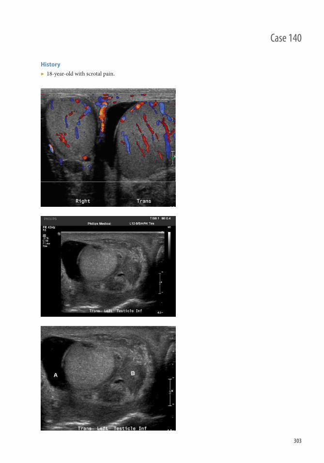
303
Case 140
History ▶ 18-year-old with scrotal pain.

304
Case 140 Epididymitis/Orchitis
Findings ▶ Ultrasound of the scrotum demonstrates increased color fl ow in the left testicle when compared with the right.
Note the reactive hydrocele (Figure 140.1). ▶ Ultrasound of the left hemiscrotum demonstrates a reactive hydrocele (Figure 140.2) and a heterogeneous
echogenic focus (Figure 140.3) at the base of the testicle, which represents an enlarged, infl amed epididymis.
Diff erential Diagnosis ▶ Epididymo-orchitis ▶ Epididymitis ▶ Orchitis ▶ Testicular trauma
Teaching Points ▶ Epididymitis is the most common cause of nontraumatic acute scrotal pain in adult men. ▶ Acute epididymitis is a clinical syndrome of pain, swelling, and infl ammation of the epididymis. Fever,
dysuria, or urethral discharge may accompany signs of epididymitis. ▶ It is usually caused by bacterial spread from the urethra, prostate, or bladder. It is commonly linked to
Chlamydia trachomatis or Neisseria gonorrhea in sexually active men; however, urinary tract or Escherichia coli infections can cause epididymitis in other settings.
▶ Symptoms of orchitis can mimic testicular torsion and include pain, swelling, and hematuria. Orchitis associated with sexually transmitted bacterial infection can involve the epididymis.
▶ Orchitis can be seen among adolescent boys during active mumps.
Management ▶ Antibiotic therapy in bacterial cases. Antibiotics are not suggested in viral causes. ▶ Anti-infl ammatory drugs.
Further Readings Aso C , Enríquez G , Fité M, et al. Gray-scale and color Doppler sonography of scrotal disorders in children: an update .
Radiographics. 2005 Sep-Oct;25(5):1197–1214. Blaivas M , Sierzenski P , Lambert M . Emergency evaluation of patients presenting with acute scrotum using bedside
ultrasonography . Acad Emerg Med . 2001 ; 8 ( 1 ): 90–93. Trojian TH , Lishnak TS , Heiman D . Epididymitis and orchitis: an overview . Am Fam Physician . 2009 ; 79 ( 7 ): 583–587 .

305
Case 141
History ▶ 42-year-old female with fever, altered mental status, and abdominal pain.

306
Findings CT
▶ Striated nephrogram: wedge-shaped areas of alternating hyperenhancement and hypoenhancement (Figures 141.3 and 141.4, white arrows); best seen during excretory phase
▶ Enlarged, edematous kidney with loss of corticomedullary diff erentiation ▶ Perinephric fat stranding and thickening of Gerota facia ▶ Hydronephrosis if obstructed ▶ Emphysematous pyelonephritis: bubbles or streaky foci of intraparenchymal gas ▶ Pyelitis: urothelial thickening/enhancement of the collecting system with normal renal parenchyma ▶ Emphysematous pyelitis: pyelitis with foci of gas within the collecting system
US
▶ Frequently normal ▶ Peripheral wedge-shaped hyperechoic or hypoechoic foci ▶ Loss of corticomedullary diff erentiation ▶ Focal area of hypoperfusion on power Doppler (Figure 141.5, black arrow) ▶ Focal bacterial nephritis may appear mass-like ▶ Gas may be seen as echogenic foci with posterior “dirty shadowing”
Nuclear Medicine
▶ Tc-99m DMSA scintigraphy shows focal or diff use photopenia ▶ Highly sensitive and preferred in pediatric patients; nonspecifi c in adults ▶ Cannot distinguish between pyelonephritis, abscess, cyst, infarct, or tumor
Diff erential Diagnosis ▶ Renal infarct ▶ Ureteral obstruction ▶ Trauma ▶ Renal vein thrombosis ▶ Hypotension ▶ Intratubular obstruction ▶ Lymphoma
Teaching Points ▶ Etiology
■ Bacterial invasion of the renal parenchyma and pelvis with tubular obstruction, interstitial edema, and vasospasm
■ Usually gram-negative organisms from ascending infection ▶ Complications
■ Complications are more common with diabetes, chronic renal disease, sickle cell disease, renal transplant, and AIDS and other immunocompromised states
■ Emphysematous pyelonephritis is a surgical emergency that usually occurs in patients with poorly controlled diabetes and represents necrotizing infection involving renal parenchyma
■ Other complications include abscess formation, acute renal failure, sepsis, and scarring
Management ▶ Antibiotics for noncomplicated pyelonephritis ▶ Nephrectomy for emphysematous pyelonephritis ▶ Emergent percutaneous drainage if obstructed
Case 141 Pyelonephritis

307
History ▶ None
Case 142

308
Case 142 Renal Abscess
Findings CT
▶ Low-attenuation mass with a thick, enhancing, irregular wall ▶ Surrounding fat stranding ▶ May contain foci of air internally ▶ Lack of central enhancement ▶ Possible hypoenhancement of the surrounding renal parenchyma ▶ May demonstrate perinephric extension
US
▶ Well-defi ned heterogeneous area of hypoenhancement ▶ No internal fl ow ▶ May demonstrate “dirty shadowing” if there are internal foci of air
MRI
▶ T1 hypointense ▶ T2 hyperintense ▶ May demonstrate wall enhancement
Diff erential Diagnosis ▶ Renal cell carcinoma ▶ Metastasis ▶ Lymphoma ▶ Hemorrhagic or infected renal cyst
Teaching Points ▶ Presents with fever, chills, dysuria, and fl ank pain ▶ May rupture into the collecting system or into the perinephric space
Management ▶ Antibiotic therapy and drainage ▶ Medical antibiotic therapy alone may be appropriate if the abscess is small
Further Readings Craig WD , Wagner BJ , Travis MD . Pyelonephritis: radiologic-pathologic review . RadioGraphics . 2008 ; 28 ( 1 ): 255–277 ; quiz
327–328 . PubMed PMID: 18203942. Kawashima A , Sandler CM , Goldman SM , Raval BK , Fishman EK . CT of renal infl ammatory disease . RadioGraphics .
1997 ; 17 ( 4 ): 851–866 ; discussion 867–8 . PubMed PMID: 9225387.

309
Case 143
History ▶ Left fl ank pain.
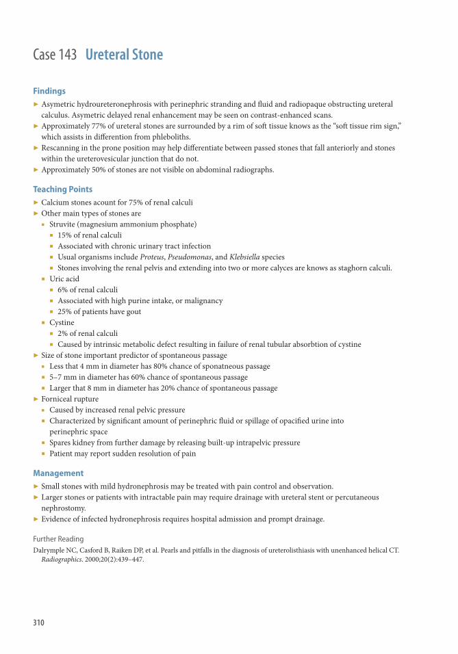
310
Case 143 Ureteral Stone
Findings ▶ Asymetric hydroureteronephrosis with perinephric stranding and fl uid and radiopaque obstructing ureteral
calculus. Asymetric delayed renal enhancement may be seen on contrast-enhanced scans. ▶ Approximately 77% of ureteral stones are surrounded by a rim of soft tissue knows as the “soft tissue rim sign,”
which assists in diff erention from phleboliths. ▶ Rescanning in the prone position may help diff erentiate between passed stones that fall anteriorly and stones
within the ureterovesicular junction that do not. ▶ Approximately 50% of stones are not visible on abdominal radiographs.
Teaching Points ▶ Calcium stones acount for 75% of renal calculi ▶ Other main types of stones are
■ Struvite (magnesium ammonium phosphate) ■ 15% of renal calculi ■ Associated with chronic urinary tract infection ■ Usual organisms include Proteus , Pseudomonas , and Klebsiella species ■ Stones involving the renal pelvis and extending into two or more calyces are knows as staghorn calculi.
■ Uric acid ■ 6% of renal calculi ■ Associated with high purine intake, or malignancy ■ 25% of patients have gout
■ Cystine ■ 2% of renal calculi ■ Caused by intrinsic metabolic defect resulting in failure of renal tubular absorbtion of cystine
▶ Size of stone important predictor of spontaneous passage ■ Less that 4 mm in diameter has 80% chance of sponatneous passage ■ 5–7 mm in diameter has 60% chance of spontaneous passage ■ Larger that 8 mm in diameter has 20% chance of spontaneous passage
▶ Forniceal rupture ■ Caused by increased renal pelvic pressure ■ Characterized by signifi cant amount of perinephric fl uid or spillage of opacifi ed urine into
perinephric space ■ Spares kidney from further damage by releasing built-up intrapelvic pressure ■ Patient may report sudden resolution of pain
Management ▶ Small stones with mild hydronephrosis may be treated with pain control and observation. ▶ Larger stones or patients with intractable pain may require drainage with ureteral stent or percutaneous
nephrostomy. ▶ Evidence of infected hydronephrosis requires hospital admission and prompt drainage.
Further Reading Dalrymple NC , Casford B , Raiken DP , et al. Pearls and pitfalls in the diagnosis of ureterolisthiasis with unenhanced helical CT.
Radiographics . 2000 ; 20 ( 2 ): 439–447.

311
History ▶ 40 year old with generalized pelvic pain.
Case 144

312
Findings CT
▶ Distended thick-walled fallopian tube(s) ▶ Enlarged edematous ovaries ▶ Tubo-ovarian/pelvic abscess (solid arrows) ▶ Infl ammatory stranding surrounding pelvic structures (dashed arrow) ▶ Free pelvic fl uid (simple or complex) US
▶ Increased echogenicity of pelvic fat ▶ Distended serpiginous fallopian tube(s) with fl uid and/or debris ▶ Enlarged adjacent ovary ▶ Multilocular, complex, thick-walled, cystic adnexal mass ▶ Surrounding hyperemia with low resistive fl ow Diff erential Diagnosis ▶ Hemorrhagic ovarian cyst (± rupture) ▶ Endometriosis ▶ Ovarian neoplasm ▶ Pelvic abscess of diff erent etiology (e.g., diverticulitis, appendicitis, Crohn disease) Teaching Points ▶ Presentation
■ Fever, abdominal/pelvic pain, vaginal discharge, cervical/adnexal tenderness ▶ Risk factors
■ Multiple sexual partners, lower socioeconomic class, intrauterine contraceptive device, history of STD ▶ Pathology
■ Most commonly Neisseria gonorrhea or Chlamydia trachomatis , but 40% are polymicrobial ■ Results from untreated ascending vaginal infection that progresses to endometritis, salpingitis, and
tubo-ovarian abscess Management ▶ Intrauterine contraceptive device removed if present, antibiotic therapy ± abscess drainage ▶ Can lead to infertility and ectopic pregnancies if not diagnosed and treated early Further Readings Potter A, Chandrasekhar CA. et al. US and CT evaluation of acute pelvic pain of gynecologic origin in nonpregnant
premenopausal patients . RadioGraphics. 2008; 28 : 1645–1659 . Sam JW, Jacobs JE, Birnbaum BA. et al. Spectrum of CT fi ndings in acute pyogenic pelvic infl ammatory disease . RadioGraphics.
2002; 22 : 1327–1334 .
Case 144 Tubo-Ovarian Abscess/Pelvic Infl ammatory Disease

313
Case 145
History ▶ None

314
Findings ▶ Abdominal aortic aneurysm rupture with active extravasation ▶ Contained abdominal aortic aneurysm rupture: Coronal ▶ Contained abdominal aortic aneurysm: Sagittal
Teaching Points ▶ Characterization of an aortic aneurysm
■ Location ■ Ascending ■ Arch ■ Descending
■ Shape ■ Fusiform ■ Saccular
■ Etiology ■ Atherosclerotic ■ Mycotic ■ Traumatic ■ Infl ammatory ■ Iatrogenic ■ Congenital
▶ Th e wall stress is uniformly distributed in the nonaneurysmal aorta, whereas within an aortic aneurysm, regions of high and low stress distribution are present.
▶ CT allows precise measurement of aneurysm size and evaluation of disease extent. ▶ On average, an abdominal aortic aneurysm expands at a rate of 2–4 mm per year for aneurysms <4 cm,
2–5 mm for aneurysms 4–5 cm, and 3–7 mm for those >5 cm. ▶ Th e risk of rupture is size dependent with the risk being 2%, 10%, and 22% at 4 years, respectively. ▶ Most abdominal aortic aneurysms rupture into the retroperitoneum. ▶ Th oracic aortic aneurysm rupture may result in pericardial and/or pleural eff usions and mediastinal hematoma. ▶ Aortic aneurysms may be complicated by dissection and distal embolization, which can cause bowel or
extremity ischemia or infarction. ▶ CT is the modality of choice when evaluating patients for aneurysm leak. Signs of impending rupture
■ Increased aneurysm size ■ Low thrombus-to-lumen ration ■ Hemorrhage into a mural thrombus ■ Peripheral crescent-shaped area of high attenuation within the aneurysm ■ Draping of the aneurysmal aorta over the vertebrae ■ Noncontrast CT shows high attenuation surrounding the aorta with loss of the fat interface between the
aortic wall and hematoma. ▶ Focal discontinuity in circumferential wall calcifi cations is more frequently observed in unstable or ruptured
aneurysms. ▶ A true aneurysm involves all three wall layers: intima, media, and adventitia. ▶ A false aneurysm or pseudoaneurysm involves one or two wall layers.
Management ▶ Endovascular aneurysm repair versus conventional open surgical repair ▶ Initial length of hospital stay is shorter for the patients undergoing endovascular aneurysm: however, these patients
oft en have more frequent readmission for the treatment of procedure-related complications, chiefl y endoleak.
Case 145 Abdominal Aortic Aneurysm (AAA Rupture)
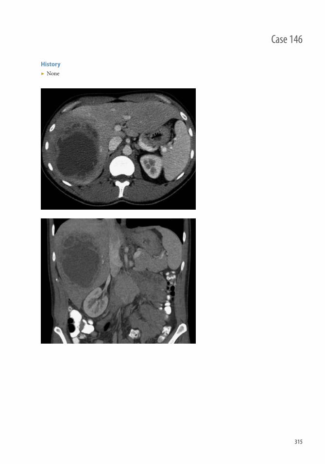
315
History ▶ None
Case 146

316
Findings CT
▶ Peripherally enhancing lesions with central low density ▶ Lacy appearance with satellite lesions ▶ Oft en found in the dependent portion of right lobe ▶ May contain foci of gas
US
▶ Poorly defi ned ▶ Variable internal echogenicity but oft en heterogeneous ▶ No central fl ow
MRI
▶ Usually heterogeneous and T1 hypointense centrally ▶ Usually T2 hyperintense centrally ▶ Oft en demonstrates capsular enhancement
Diff erential Diagnosis ▶ Metastatic disease ▶ Hepatocellular carcinoma ▶ Biliary cystadenoma/carcinoma ▶ Echinococcal cyst
Teaching Points ▶ Presents with fever, chills, and upper abdominal pain. ▶ Rare in healthy individuals and usually seen in the setting of an immunocompromised patient. ▶ Look for diverticulitis, appendicitis, or other source of infection as a potential cause. ▶ In developing countries, parasitic abscesses are more common.
Management ▶ Surgical consult and IR drainage. ▶ Medical antibiotic therapy alone may be appropriate if the abscess is small.
Further Readings Mortelé KJ , Segatto E , Ros PR . Th e infected liver: radiologic-pathologic correlation . RadioGraphics . 2004 ; 24 ( 4 ): 937–955 .
PubMed PMID: 15256619. Mortelé KJ , Ros PR . Cystic focal liver lesions in the adult: diff erential CT and MR imaging features . RadioGraphics .
2001 ; 21 ( 4 ): 895–910 . PubMed PMID: 11452064.
Case 146 Liver Abscess
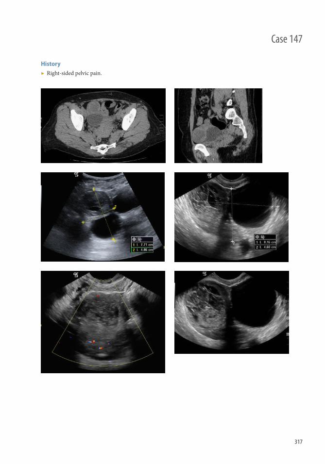
317
Case 147
History ▶ Right-sided pelvic pain.

318
Findings ▶ Enlarged (>5 cm), bulky-appearing ovary ▶ Frequently has an associated adnexal cyst or mass leading to increased size ▶ May see peritoneal fl uid ▶ Ultrasound
■ Doppler fl ow may be absent or systolic-only ■ May see hypoechoic (edematous) central ovary with hemorrhagic areas; follicles may be displaced to the
periphery ■ “Whirlpool” sign of a twisted vessel may be seen
▶ CT ■ Fallopian tube thickening ■ May see adnexal mass with twisted pedicle ■ May see deviation of the uterus to the torsed adnexa
▶ MR ■ Fallopian tube thickening ■ Heterogeneous ovarian parenchyma with hemorrhagic areas ■ May be able to narrow diagnosis of underlying ovarian lesion ■ Better delineation of blood products in lesion or peritoneum
Diff erential Diagnosis ▶ Hemorrhagic ovarian cyst ▶ Adnexal mass (benign) ▶ Ovarian hyperstimulation syndrome in the context of infertility treatment ▶ Ectopic pregnancy
Teaching Points ▶ Commonly occurs in reproductive-age females, and even more commonly during pregnancy. ▶ Most common underlying ovarian lesion is the mature cystic teratoma (dermoid cyst). ▶ Ovarian torsion is primarily a clinical diagnosis. Imaging features can suggest the diagnosis, but cannot rule it
out and are rarely specifi c enough for defi nitive diagnosis. ▶ Presence of Doppler fl ow within the ovary is not suffi cient to exclude torsion. Contrariwise, absence of
measurable fl ow is suggestive, but not diagnostic.
Management ▶ Classically, managed with salpingo-oophrectomy, usually laparoscopic out of concern for development of
thromboembolism. ▶ More conservative ovary-sparing treatment with simple laparoscopic detorsion may be used in
younger women.
Further Readings Rha SE , Byun JY , Jung SE , et al. CT and MR imaging features of adnexal torsion . RadioGraphics. 2002; 22( 2): 283–294 . Chang HC , Bhatt S , Dogra VS . Pearls and pitfalls in diagnosis of ovarian torsion . RadioGraphics. 2008; 28( 5): 1355–1368 ;
doi:10.1148/rg.285075130.
Case 147 Adnexal Torsion

319
History ▶ Pelvic pain, history of infertility.
Case 148

320
Findings Ultrasound
▶ Hypoechoic tubular structure in the adnexa adjacent to the ovary ▶ Debris within the structure ▶ May see thickened longitudinal folds ▶ No fl ow seen within the structure
MRI
▶ Dilated tubular structure, defi nitely separate from the ovary ▶ Clearer delineation of the tubular nature of the structure and its relationship to the ovary and uterus ▶ May see fl uid-fl uid level, suggestive of blood products ▶ Th ickness of the tubal wall can be important distinguishing factor between infl ammatory and
noninfl ammatory etiologies
Diff erential Diagnosis ▶ Hematosalpix versus hematosalpinx versus pyosalpinx ▶ Tubal ectopic pregnancy ▶ Tubo-ovarian abscess ▶ Peritoneal cyst ▶ Adnexal cystic lesion
Teaching Points ▶ Etiology of hydrosalpinx is typically related to obstruction caused by adhesions or endometriosis. ▶ Echogenic debris within the tubular structure suggests the possibility of pyosalpinx. Th ickened, enhancing
tube wall is also suggestive of an infl ammatory process. ▶ Th ickened longitudinal folds are highly suggestive of hydrosalpinx associated with chronic infl ammation. ▶ Carefully evaluate for a solid mass within the fl uid-fi lled structure, which could represent an ectopic
pregnancy or a fallopian tube adenocarcinoma. ▶ Can be associated with tubal factors for infertility, such as endometriosis or pelvic infl ammatory disease.
Management ▶ Management depends on underlying etiology: treatment of pelvic infl ammatory disease, of endometriosis, of
ectopic pregnancy, and so forth. ▶ In infertile patients, in vitro fertilization techniques may be used.
Further Readings Kim MK , Rha SE , Oh SN , et al. MR imaging fi ndings of hydrosalpinx: a comprehensive review . RadioGraphics.
2009; 29( 2): 495–507. doi:10.1148/rg.292085070. Moyle PL , Kataoka MY , Nakai A , Takahata A , Reinhold C , Sala E . Nonovarian cystic lesions of the pelvis . RadioGraphics.
2010; 30( 4): 921–938. doi:10.1148/rg.304095706. Rezvani M , Shaaban AM . Fallopian tube disease in the nonpregnant patient . RadioGraphics. 2011; 31( 2): 527–548. doi:10.1148/
rg.312105090.
Case 148 Hydrosalpinx

321
Case 149
History ▶ Abdominal pain.

322
Findings ▶ CT: Axial and coronal images, demonstrating dilated common bile duct (CBD) and dilated intrahepatic bile
ducts. A round hyperdense lesion in the lumen of the distal CBD (arrow). ▶ US: A round echogenic structure with posterior acoustic shadowing in a dilated CBD. ▶ ERCP: demonstrating a dilated CBD, with a large fi lling defect.
Diff erential Diagnosis ▶ CT fi ndings: CBD stricture from benign or malignant cause (ampullary carcinoma, pancreatic
adenocarcinoma), obstructed CBD from gallbladder sludge.
Teaching Points ▶ Gallstones can migrate from the gallbladder lumen into the bile duct and be associated with biliary colic,
jaundice, CBD obstruction, and hyperbilirubinemia and elevated alkaline phosphatase. ▶ Th e entire length of the CBD may not be visualized by ultrasound, making MRI/MRCP or ERCP useful
adjunct modalities in further evaluation for choledocholithiasis. ▶ CBD stones may be occult on CT, depending on their size and composition. ▶ CBD stones are present in 10%–15% of patients with gallstones.
Management ▶ If US demonstrates a stone, the patient should be referred for ERCP and defi nitive management with
papillotomy, stone removal, and potential subsequent cholecystectomy. ▶ If US fails to demonstrate a stone, the patient may be referred for MRI/MRCP, or thin-slice CT with and
without contrast as an alternative if an MRI cannot be obtained.
Further Readings Hanbidge AE, Buckler PM, O’Malley ME, et al. Imaging evaluation for acute pain in the right upper quadrant . RadioGraphics.
2004;24:1117 –1135 . Bortoff GA, Chen MYM, Ott DJ, et al. Gallbladder stones: imaging and intervention . RadioGraphics. 2000;20:751 –766 .
Case 149 Choledocholithiasis (CBD Stone)

323
History ▶ 20-year-old man presenting with acute scrotal pain.
Case 150

324
Case 150 Testicular Torsion
Findings ▶ Duplex ultrasonography is the main imaging modality used for the evaluation of acute scrotum. ▶ Th e most important fi nding is decreased or absent testicular blood fl ow and identifi cation of the torsion knot
in the spermatic cord located in the inguinoscrotal region above the testis and epididymis. ▶ Th e identifi cation of a twisted cord with intratesticular blood fl ow is a sign that the testis is viable and may be
salvaged aft er reduction and orchidopexy. ▶ Immediately aft er torsion (up to 3 h), testis volume and echogenicity may be normal. ▶ Later, an increase in testicular volume and hypoechogenicity or heterogeneous echogenicity, or both, may be
detected. ▶ Cystic areas may be seen in the late phase.
Diff erential Diagnosis ▶ Torsion of the appendix testis ▶ Epididymitis and epididymoorchitis ▶ Testicular trauma ▶ Idiopathic scrotal edema ▶ Inguinoscrotal hernia ▶ Testicular subtorsion ▶ Tunica vaginalis infl ammation ▶ Abnormal processus vaginalis ▶ Hydrocele ▶ Varicocele/thrombosed varicocele ▶ Vasculitis (e.g., Schönlein-Henoch purpura)
Presentation ▶ Acute scrotal pain ± nausea and vomiting ▶ Swollen/erythematous hemiscrotum/testicle ▶ Retracted testis ▶ Decreased or absent cremasteric refl ex ▶ Th e testis is painful to palpation and may be transversely oriented inside the scrotal sac
Management ▶ Surgical detorsion and orchiopexy of both testes ▶ Orchidectomy if the testes are necrotic and nonviable
Teaching Points ▶ It has two peak incidences: a small one in the neonatal period and a large one during puberty, but it can occur
at any age. ▶ Th is is a true surgical emergency, delay in its diagnosis may result in loss of a testis because of irreversible
ischemia and the viability of a torsed testicle is dependent on the duration and completeness of torsion. ▶ Detorsion within 4–8 hours has generally been accepted as the optimal interval in which to salvage the
aff ected testis. ▶ Surgery never should be delayed on the assumption of nonviability based on a clinical estimate of duration of
torsion. ▶ If based on the history and examination the potential risk of testicular torsion remains, surgical exploration
may be appropriate despite apparently normal imaging studies.

PediatricPart III


327
History ▶ None
Case 151

328
Findings Cranial US
▶ Regions of increased echogenicity (signifying hemorrhage) in the region of the caudothalamic groove ■ Located between the caudate head and the thalamus ■ Best seen in coronal and sagittal planes ■ Common pitfall: normal echogenic choroid plexus, which remains posterior to the caudothalamic groove
▶ ± uniform echogenic material in the lateral ventricles, most commonly layering in the occipital horns ▶ ± hydrocephalus ▶ ± ill-defi ned echogenicity in the periventricular white matter
■ May represent intraparenchymal extension of hemorrhage or periventricular leukomalacia ▶ Can be unilateral or bilateral
Diff erential Diagnosis ▶ Periventricular leukomalacia
Teaching Points ▶ Grading of GMH
■ Grade I: Hemorrhage involves the subependymal matrix only ■ Grade II: Intraventricular extension without ventricular enlargement ■ Grade III: Intraventricular extension with ventricular enlargement ■ Grade IV: Periventricular intraparenchymal extension
▶ Presentation ■ Typically preterm infants (<32 weeks gestation and <1,500 g)
■ Overall prevalence of 10%–15% in preterm infants <35 weeks ■ Most GMHs occur within the fi rst 3 days of life ■ Oft en asymptomatic
▶ Complications ■ Hydrocephalus: communicating or obstructive ■ Venous infarction in the periventricular parenchyma
■ May result in periventricular cyst formation and rarely a large porencephalic cyst ▶ Prognosis
■ Grade I, II, and III: typically have a good prognosis ■ Grade IV: >25% mortality rate, long-term defi cits in >50%
Management ▶ Routine screening for GMH in infants <30 weeks or <1,250 g at birth. Typical screening regimen
■ First screening examination at 4–5 days of age, or sooner if clinical concern ■ Second screening head ultrasound at ~36 weeks or at time of discharge
▶ Imaging follow-up of positive ultrasounds is dictated by clinical circumstances. ▶ Treatment options
■ Antenatal corticosteroids may reduce the risk of GMH ■ Treatment typically limited to supportive care ■ Hydrocephalus treated with shunting
Case 151 Germinal Matrix Hemorrhage

329
Case 152
History ▶ None

330
Findings ▶ Chest radiograph: Multiple rib fractures at various stages of healing (white arrows) ▶ Head CT: Subdural hemorrhage (thin white arrow), intraventricular hemorrhage (arrowheads), subarachnoid
hemorrhage (thick white arrow), scalp hematoma (asterisk), skull fracture (black arrow) ▶ Tibia radiograph: Metaphyseal fracture/metaphyseal corner fracture/classic metaphyseal lesion (thin arrow),
periosteal reaction (thick arrow)
Diff erential Diagnosis ▶ Osteogenesis imprefecta ▶ Leukemia ▶ Spondylometaphyseal dysplasia
Teaching Points ▶ Metaphyseal corner fractures are highly specifi c for nonaccidental trauma ▶ Other fractures highly specifi c for abuse include rib, sternum, scapula, and spinous processes
Management ▶ Notify referring physician and/or child protection team about fi ndings
Further Readings Kleinman PK ; Diagnostic Imaging of Child Abuse . 2nd edition. St. Louis, MO : CV Mosby, Inc ; 1998 . Slovis TL , Coley BD , Bulas DI , Faerber EN . Caff ey's Pediatric Diagnostic Imaging . 11th edn. Elsevier , Philadelphia ,
December, 2007 .
Case 152 Nonaccidental Trauma, Child Abuse

331
Case 153
History ▶ 5-year-old boy with cough and fever.

332
Findings ▶ Rounded opacity located in the superior segment of the right lower lobe
Diff erential Diagnosis ▶ Metastatic disease ▶ Primary malignancy
Teaching Points ▶ Common presentation of bacterial pneumonia in the pediatric population. ▶ Patient will present with common signs of pneumonia: cough, fever, and elevated WBC.
Management ▶ Antibiotics ▶ Follow-up radiograph aft er treatment to ensure resolution
Further Readings Brant WE , Helms CA , eds. Fundamentals of Diagnostic Radiology . 3rd ed. Philadelphia : Lippincott Williams & Wilkins , 2006 .
p. 1252 . Pappas M , Yamato L , Anene O . Pediatric radiology review . New Jersey. Humana Press , 2007 . p. 124 .
Case 153 Round Pneumonia

333
History ▶ 1.5-year-old boy with diffi culty stridor.
Case 154
334
Case 154 Croup
Findings Frontal Radiograph
▶ “Steeple” or inverted “V” sign: Symmetric subglottic tracheal narrowing with loss of normal shouldering caused by subglottic edema
Lateral Radiograph
▶ Inspiratory fi lms: Hypopharyngeal distention (more common) ▶ Expiratory fi lms: Hypopharyngeal collapse with overdistention of the lower cervical trachea ▶ Narrowing of the subglottic trachea with loss of defi nition ▶ Normal epiglottis and aryepiglottic folds.
Diff erential Diagnosis ▶ Epiglottitis ▶ Foreign body aspiration ▶ Exudative tracheitis ▶ Subglottic hemangioma
Teaching Points ▶ Radiography performed to exclude airway compromise from more serious causes of stridor ▶ Demographics
■ 6 months–3 years (peak at 1 year) ■ Typically younger than patients with epiglottitis (mean 3 years) or exudative tracheitis (6–10 years)
▶ Etiology ■ Viral (parainfl uenza virus most common)
▶ Epidemiology ■ Most common cause of upper airway obstruction in young children ■ Seasonal prevalence: Fall and Winter
Management ▶ Usually benign, self-limited viral illness ▶ Oral or inhaled corticosteroids ▶ Inhaled epinephrine or intubation for severe cases
Further Readings Knutson D , Aring A. Viral croup . Am Fam Physician. 2004 Feb 1;69(3):535–40. John SD , Swischuk LE : Stridor and upper airway obstruction in infants and children . Radiographics . 1992 Jul;12(4):625–43.

335
History ▶ 18-year-old male presenting with high fever, sore throat, stridor, and drooling.
Case 155

336
Findings ▶ “Th umb sign”: Marked enlargement of epiglottis on lateral radiograph (white arrow) ▶ Marked thickening and upward convexity of aryepiglottic folds (black arrow) ▶ May demonstrate hypopharyngeal widening and subglottic narrowing on AP view ▶ CT: Typically not necessary, but can assist with diff erential diagnosis and evaluation for emphysematous
epiglottitis or an associated abscess ■ Edematous low-attenuation swelling of the epiglottis and aryepiglottic folds, occasionally with an associated
phlegmon
Diff erential Diagnosis ▶ “Omega” epiglottis: Normal epiglottis imaged obliquely ▶ Croup ▶ Retropharyngeal abscess ▶ Exudative tracheitis
Teaching Points ▶ Typically a lateral radiograph is the only imaging needed for diagnosis; the patient should be maintained in
upright comfortable position for imaging ▶ Onset of total airway occlusion can be sudden, and is associated with agitation of the patient or airway;
children are especially at risk ▶ Classically associated with Haemophilus infl uenza type b (Hib), but this etiology is much less common aft er
introduction of Hib vaccine in 1985; now more likely to present from a wide range of pathogens ▶ Classically occurs in young children, but introduction of the Hib vaccine has resulted in a relative increase in
adult presentation
Management ▶ Steroids and wide-spectrum antibiotics ▶ Do not agitate the patient or airway, because this may precipitate sudden airway occlusion ▶ May require emergent intubation, ideally performed quickly by an experienced clinician
Further Readings Capps EF, Kinsella JJ, Gupta M, et al. Emergency imaging assessment of acute, nontraumatic conditions of the head and neck .
RadioGraphics . 2010 ; 30 ( 5 ): 1335–1352 . Shah RK, Roberson DW, Jones DT. Epiglottitis in the Hemophilus infl uenzae type B vaccine era: changing trends . Laryngoscope .
2004 ; 114 ( 3 ): 557–560 .
Case 155 Epiglottitis

337
History ▶ 4-year-old boy with intermittent fever, nausea, vomiting, and neck pain.
Case 156
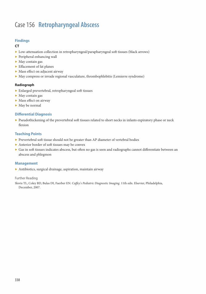
338
Findings CT
▶ Low-attenuation collection in retropharyngeal/parapharyngeal soft tissues (black arrows) ▶ Peripheral enhancing wall ▶ May contain gas ▶ Eff acement of fat planes ▶ Mass eff ect on adjacent airway ▶ May compress or invade regional vasculature, thrombophlebitis (Lemierre syndrome)
Radiograph
▶ Enlarged prevertebral, retropharyngeal soft tissues ▶ May contain gas ▶ Mass eff ect on airway ▶ May be normal
Diff erential Diagnosis ▶ Pseudothickening of the prevertebral soft tissues related to short necks in infants expiratory phase or neck
fl exion
Teaching Points ▶ Prevertebral soft tissue should not be greater than AP diameter of vertebral bodies ▶ Anterior border of soft tissues may be convex ▶ Gas in soft tissues indicates abscess, but oft en no gas is seen and radiographs cannot diff erentiate between an
abscess and phlegmon
Management ▶ Antibiotics, surgical drainage, aspiration, maintain airway
Further Reading Slovis TL , Coley BD , Bulas DI , Faerber EN . Caff ey's Pediatric Diagnostic Imaging . 11th edn. Elsevier , Philadelphia ,
December, 2007 .
Case 156 Retropharyngeal Abscess

339
History ▶ Cough wheezing
Case 157

340
Findings Chest Radiograph
▶ Asymmetric lung volumes (left greater than right) ▶ Hyperlucency (left lung) ▶ Oligemia (left lung) ▶ Mediastinal shift ▶ Static lung volumes on inspiration/expiration and decubitus images
Diff erential Diagnosis ▶ Viral pneumonia, asthma, endobronchial mass
Teaching Points ▶ Oft en diffi cult to obtain inspiration and expiration views because of young age and inability to cooperate. In
these cases obtain lateral decubitus views ▶ Lateral decubitus view, the dependent lung should collapse, consider chest fl uoroscopy ▶ Most foreign bodies are organic (peanut most common) and are not radiopaque ▶ Chest radiographs may be normal, show air trapping, atelectasis, and mediastinal shift
Management ▶ Foreign body removal
Further Reading Donnelly LF , Jones BV , O’Hara SM , et al. Diagnostic imaging : Pediatrics (2nd Ed.) Springer; Amysis , Salt Lake City (Utah) 2011 Slovis TL , Coley BD , Bulas DI , Faerber EN . Caff ey's Pediatric Diagnostic Imaging . 11th edn. Elsevier ,
Philadelphia ,December, 2007 .
Case 157 Bronchial Foreign Body

341
History ▶ 10 day old, born at 30 weeks gestation, feeding intolerance
Case 158

342
Findings ▶ Pneumatosis intestinalis ▶ Distended loops of bowel ▶ Portal venous gas ▶ Diff erential diagnosis ▶ Ileus
Teaching Points ▶ Pneumatosis may be linear or bubbly and is diagnostic of NEC ▶ First sign is oft en dilated loop or loops of bowel, most common in right lower quadrant, loops may be stacked ▶ Fixed loop of dilated bowel not changing over time is worrisome for NEC ▶ If pneumoperitoneum is suspected, do left later decubitus to better detect free air adjacent to liver
Management ▶ Serial KUBs for follow-up, NPO, surgical consultation
Further Readings Donnelly LF , Jones BV , O’Hara SM , et al. Diagnostic imaging : Pediatrics (2nd Ed.) Springer ; Amysis, Salt Lake City
(Utah) 2011 . Slovis TL , Coley BD , Bulas DI , Faerber EN . Caff ey's Pediatric Diagnostic Imaging . 11th edn. Elsevier ,
Philadelphia ,December, 2007 .
Case 158 Necrotizing Enterocolitis
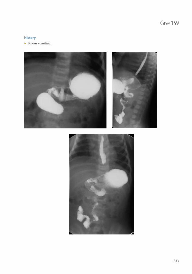
343
History ▶ Bilious vomiting.
Case 159

344
Findings ▶ Abnormal position of the duodenal-jejunal junction ▶ Spiraling of descending duodenum is diagnostic of volvulus ▶ Distended duodenal bulb, distended proximal duodenum Diff erential Diagnosis ▶ Pseudomalrotation, duodenal-jejunal junction (DJJ) displaced may be gastric or bowel distention, if suspected
repeat UGI when contrast has cleared ▶ If spiraling not seen, duodenal atresia or duodenal web
Teaching Points ▶ Malrotation is diagnosed on an upper GI when the DJJ does not meet the normal criteria ▶ Normal DJJ is to the left of the spine at the level of the duodenal bulb
Management ▶ Notify referring physician and/or pediatric surgery about fi ndings. ▶ If malrotation is present this is a true surgical emergency
Further Readings Swischuk LE (Ed.), Imaging of the Newborn, Infant and Young Child (4th edn), William and Wilkins , Baltimore, MD ( 1997 ). Kleinman PK ; Diagnostic Imaging of Child Abuse . 2nd edition. St. Louis, MO : CV Mosby, Inc ; 1998 . Slovis TL , Coley BD , Bulas DI , Faerber EN . Caff ey's Pediatric Diagnostic Imaging . 11th edn. Elsevier , Philadelphia ,
December, 2007 .
Case 159 Malrotation and Midgut Volvulus
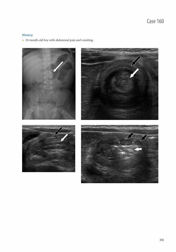
345
History ▶ 10-month-old boy with abdominal pain and vomiting.
Case 160

346
Findings ▶ Abdominal radiograph: rounded fi lling defect, soft tissue mass (white arrow) within the colonic gas,
nonvisualization of air-fi lled cecum on right side up lateral decubitus radiograph ▶ Sonography: bowel within bowel or target sign, concentric layers representing bowel (intussusceptum; white
arrows) surrounded by bowel (intussuscipiens; black arrows), pseudokidney appearance on longitudinal images, fat (short white arrow) and lymph nodes (short black arrows) between the two bowel loops
Diff erential Diagnosis ▶ Appendicitis ▶ Gastroenteritis ▶ Meckel diverticulum ▶ Ovarian pathology
Teaching Points ▶ Defi ned by the invagination of a part of the intestine into itself where a proximal segment of bowel (the
intussusceptum) telescopes into a distal segment (the intussuscipiens), dragging associated mesentery with it ▶ Consequent development of venous and lymphatic congestion results in intestinal edema, which can
ultimately lead to ischemia, perforation, and peritonitis ▶ Occurs most oft en near the ileocecal junction without an identifi able lead point ▶ A common cause of acute abdomen, particularly in children younger than 2 years of age
Management ▶ Retrograde pressure can be exerted using air or water-soluble medium in an enema reduction to reduce the
intussusception ▶ Surgical intervention may be required if symptoms are protracted, there are signs of peritonitis, or enema
reduction is unsuccessful
Further Readings Byrne AT , Geoghegan T , Govender P , et al. Th e imaging of intussusception . Clin Radiol. 2005 ; 60 : 39–46 . Daneman A , Navarro O . Intussusception Part 1: review of diagnostic approaches . Pediatr Radiol. 2003 ; 33 : 79–85 .
Case 160 Intussusception

347
History ▶ 12-year-old female with traumatic ankle pain and swelling.
Case 161

348
Findings Radiographs
▶ Type I: Occult or physeal widening ▶ Type II: Fracture extending from the metaphysis to the physis ▶ Type III: Fracture extending from the epiphysis to the physis ▶ Type IV: Fracture extending through the epiphysis, physis, and metaphysis ▶ Type V: Narrowing of the physis
CT
▶ Used to evaluate extent of fracture and degree of displacement
MRI
▶ Increased T2 signal at physis ▶ Hypointense fracture line on T1 and T2 weight sequences with surrounding bone marrow edema
Diff erential Diagnosis ▶ Ankle sprain ▶ Stress injury ▶ Triplane fracture
Teaching Points ▶ Type I fractures may be radiographically occult or show physeal widening
■ Consider comparison with contralateral limb ▶ Type II fracture most common (>70%)
■ Except distal humerus where most are type IV ■ Good prognosis
▶ Type III and Type IV more prone to chronic disability ▶ Type V associated with growth disturbances ▶ Complications
■ Premature epiphyseal closure with limb shortening ■ Joint incongruity ■ Trapped periosteum ■ More common with knee and ankle fractures
▶ Juvenile tillaux fracture (current case) ■ Exclusive type of fracture occurring in adolescence before complete fusion of the tibial growth plate ■ Th e tibial growth plate fuses from medial to lateral ■ Common age of incidence is 11–15 years (male mean age of 14 years; female mean age of 12 years)
Management ▶ Casting for low-grade Salter-Harris categories ▶ Open reduction and internal fi xation for high-grade Salter-Harris categories
Further Readings Barmada A , Gaynor T , Mubarak SJ, et al . Premature physeal closure following distal tibia physeal fractures: a new radiographic
predictor . J Pediatr Orthop . 2003 Nov-Dec;23(6):733–739. Resnik CS. Diagnostic imaging of pediatric skeletal trauma . Radiol Clin North Am . 1989 Sep;27(5):1013–1022.
Case 161 Salter-Harris Fracture Type III (Juvenile Tillaux Fracture)

349
History ▶ None
Case 162

350
Findings ▶ Spiral nondisplaced fracture of the mid tibial shaft
Diff erential Diagnosis ▶ Nonaccidental trauma (typically midshaft or upper one-third of tibia), whereas toddler fractures are typically
midshaft or lower one-third of tibia ▶ Osteomyelitis (in cases where fracture is occult and there are clinical signs of infection)
Teaching Points ▶ Etiology: Typically a toddler (1–2 years old, usually under 3 years old) undergoes a rotational twisting force as
he or she stumbles, falls, or gets foot stuck against an object ▶ Presentation: Refusal to bear weight or walk ▶ Clinical examination: Gently twisting foot with knee in fi xed position elicits pain ▶ Further studies: Need two views—the fracture may only be seen on one view; internal oblique view may be the
most sensitive. Tc MDP scan is sensitive but nonspecifi c and exposes to additional radiation ▶ Other
■ Can occur in the calcaneus and cuboid ■ Frequently occult (up to 43% of time), so low threshold for long leg cast
Management ▶ Long leg cast for 2–3 weeks
Further Readings Halsey MF , Finzel KC , Carrion WV, et al. Toddler’s fracture: presumptive diagnosis and treatment . J Pediatr Orthop.
2001 ; 21 : 152–156. Lonergan GJ , Baker AM , Morey MK, et al. From the Archives of the AFIP: Child abuse: radiologic-pathologic correlation .
RadioGraphics. 2003 ; 23 : 811–845.
Case 162 Toddler’s Fracture

351
History ▶ 10-year-old girl status post fall.
Case 163

352
Case 163 Torus/Buckle Fractures
Findings ▶ Oblique view demonstrating cortical irregularity of the distal radius and ulna (Figure 163.1) ▶ Lateral view demonstrating cortical irregularity of the distal radius and ulna with volar angulation of the distal
radius fracture (Figure 163.2) ▶ Oblique view with white arrows denoting fracture sites (Figure 163.3) ▶ AP view with white arrows demonstrating cortical irregularity of the distal radius and ulna (Figure 163.4) ▶ Lateral view with open black arrow demonstrating “dinner fork deformity” (Figure 163.4)
Diff erential diagnosis None
Teaching Points ▶ Most common location is in the distal radius ▶ Usually caused by fall on outstretched hand ▶ More common with low-energy injuries compared with displaced fractures, which usually result from higher
energy mechanisms ▶ Caused by highly cellular and porous pediatric bone with a large amount of collagen and cartilage, and thicker
and stronger periosteum compared with adults ▶ Patient may have a “dinner fork” deformity seen on lateral view (see Figure 163.4)
Management ▶ Buckle fractures are usually treated with short arm casts
Further Reading Carson S , Woolridge D , Colleti J , Kilgore K . Pediatric upper extremity injuries . Pediatr Clin North Am. 2006 Feb;53(1):41–67, v.

353
History ▶ 4-year-old boy with wheeze.
Case 164

354
Findings ▶ PA and lateral chest radiographs, respectively, demonstrate bilateral hilar opacities
Diff erential Diagnosis ▶ Viral pneumonia ▶ Reactive airway disease ▶ Bacterial pneumonia
Teaching Points ▶ Viruses are common pathogen in pediatric population, especially the fi rst 2 years of life, including RSV,
infl uenza, and adenovirus ▶ Radiographic fi ndings: bilateral hilar opacities, peribronchiole cuffi ng, atelectasis and/or hyperinfl ation
Management ▶ Outpatient therapy is adequate if the child does not have respiratory or feeding diffi culties ▶ Bronchodilators may be used especially with bronchiolitis-like symptoms
Further Reading Pappas , Michael , Yamato , Loren , Anene , Okechukwu . Pediatric radiology review . New Jersey . Humana Press , 2007 . p. 123.
Case 164 Viral pneumonia
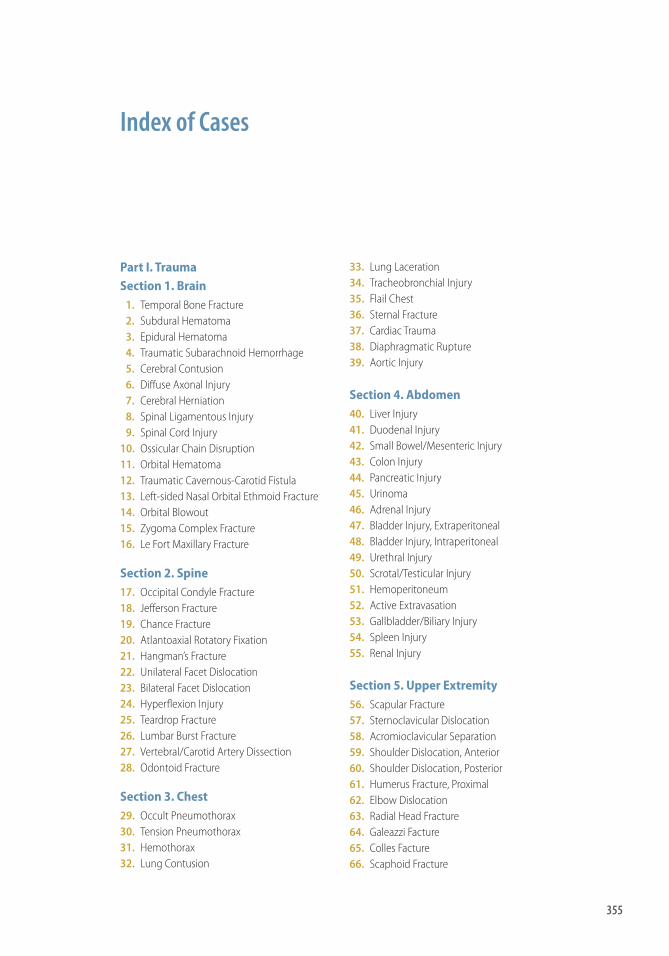
355
Index of Cases
Part I. TraumaSection 1. Brain 1. Temporal Bone Fracture 2. Subdural Hematoma 3. Epidural Hematoma 4. Traumatic Subarachnoid Hemorrhage 5. Cerebral Contusion 6. Diff use Axonal Injury 7. Cerebral Herniation 8. Spinal Ligamentous Injury 9. Spinal Cord Injury 10. Ossicular Chain Disruption 11. Orbital Hematoma 12. Traumatic Cavernous-Carotid Fistula 13. Left-sided Nasal Orbital Ethmoid Fracture 14. Orbital Blowout 15. Zygoma Complex Fracture 16. Le Fort Maxillary Fracture
Section 2. Spine 17. Occipital Condyle Fracture 18. Jeff erson Fracture 19. Chance Fracture 20. Atlantoaxial Rotatory Fixation 21. Hangman’s Fracture 22. Unilateral Facet Dislocation 23. Bilateral Facet Dislocation 24. Hyperfl exion Injury 25. Teardrop Fracture 26. Lumbar Burst Fracture 27. Vertebral/Carotid Artery Dissection 28. Odontoid Fracture
Section 3. Chest 29. Occult Pneumothorax 30. Tension Pneumothorax 31. Hemothorax 32. Lung Contusion
33. Lung Laceration 34. Tracheobronchial Injury 35. Flail Chest 36. Sternal Fracture 37. Cardiac Trauma 38. Diaphragmatic Rupture 39. Aortic Injury
Section 4. Abdomen 40. Liver Injury 41. Duodenal Injury 42. Small Bowel/Mesenteric Injury 43. Colon Injury 44. Pancreatic Injury 45. Urinoma 46. Adrenal Injury 47. Bladder Injury, Extraperitoneal 48. Bladder Injury, Intraperitoneal 49. Urethral Injury 50. Scrotal/Testicular Injury 51. Hemoperitoneum 52. Active Extravasation 53. Gallbladder/Biliary Injury 54. Spleen Injury 55. Renal Injury
Section 5. Upper Extremity 56. Scapular Fracture 57. Sternoclavicular Dislocation 58. Acromioclavicular Separation 59. Shoulder Dislocation, Anterior 60. Shoulder Dislocation, Posterior 61. Humerus Fracture, Proximal 62. Elbow Dislocation 63. Radial Head Fracture 64. Galeazzi Facture 65. Colles Facture 66. Scaphoid Fracture

356 Index of Cases
67. Triquetral Fracture 68. First Metacarpal Fracture 69. Monteggia Fracture
Section 6. Lower Extremity 70. Pelvic Fracture, Anteroposterior
Compression 71. Pelvic Fracture, Vertical Shear 72. Pelvic Fracture, Lateral Compression 73. Acetabular Fracture 74. Hip Fracture, Femoral Neck 75. Hip Fracture, Intertrochanteric 76. Patellar Fracture 77. Knee Dislocation, Posterior 78. Tibial Plateau Fracture 79. Maisonneuve Fracture 80. Calcaneal Fracture 81. Metatarsal Fracture 82. Lisfranc Injury 83. Tibia Tubercle Avulsion Fracture 84. Sacral Fracture 85. Hip Dislocation 86. Ankle Fracture
Part II. NontraumaSection 1. Brain 87. Meningitis 88. Empyema 89. Abscess 90. Encephalitis 91. Acute Disseminated Encephalomyelitis 92. Aneurysm Hemorrhage 93. Hypertensive Hemorrhage 94. Arteriovenous Malformation 95. Acute Ischemic Stroke 96. Posterior Reversible Encephalopathy
Syndrome 97. Acute Venous Sinus Thrombosis 98. Intracranial Hypotension 99. Hypoxic Ischemic Injury 100. Orbital Subperiosteal Abscess
Section 2. Head, Neck, and Spine 101. Jugular Vein Thrombosis 102. Prevertebral Abscess 103. Acute Disk Herniation 104. Epidural Abscess 105. Paraspinal Abscess 106. Epidural Hematoma 107. Peritonsillar Abscess 108. Sublingual Abscess
109. Pyogenic Diskitis Osteomyelitis 110. Spinal Cord Infarction 111. Submandibular Sialandentis 112. Cord Compression
Section 3. Chest 113. Diff use Alveolar Damage 114. Pulmonary Edema, Noncardiogenic 115. Pneumonia 116. Pneumomediastinum 117. Pulmonary Embolism/Deep Vein
Thrombosis 118. Empyema 119. Aortic Dissection 120. Aortic Intramural Hematoma 121. Lobar Collapse 122. Toxic Inhalation 123. SVC Obstruction
Section 4. Abdomen 124. Hepatitis 125. Acute Cholecystitis 126. Acute Cholangitis 127. Gallstone 128. Pancreatitis 129. Pneumatosis Intestinalis 130. Small Bowel Obstruction 131. Appendicitis 132. Cecal Volvulus 133. Diverticulitis 134. Intussusception 135. Small Bowel Ischemia 136. Colitis, Ischemic 137. Colitis, Pseudomembranous 138. Toxic Megacolon 139. Typhlitis 140. Epididymitis/Orchitis 141. Pylenonephritis 142. Renal Abscess 143. Ureteric Stone 144. Tubo-ovarian Abscess 145. Abdominal Aortic Aneurysm (AAA
Rupture) 146. Liver Abscess 147. Adnexal Torsion 148. Hydrosalpinx 149. CBD Stone 150. Testicular Torsion
Part III. Pediatric 151. Germinal Matric Hemorrhage

357Index of Cases
152. Nonaccidental Trauma, Child Abuse 153. Round Pneumonia 154. Croup 155. Epiglottitis 156. Retropharyngeal Abscess 157. Bronchial Foreign Body 158. Necrotizing Enterocolitis
159. Malrotation and Volvulus 160. Intussusception 161. Salter-Harris Fracture Type III (Juvenile
Tillaux Fracture) 162. Toddler’s Fracture 163. Torus Fracture 164. Viral Pneumonia


359
Index
AAA (abdominal aortic aneurysm) rupture, 313 – 314
AAST (American Association for the Surgery of Trauma)
grading system for renal laceration, 120 grading system for splenic injury, 118
Abdomen, 269 – 324 Abdominal aortic aneurysm (AAA) rupture,
313 – 314 Abdominal distention, 293 – 294 Abdominal pain, 281 – 282 , 293 – 294 ,
299 – 300 , 305 – 306 , 321 – 322 crampy, 291 – 292 intermittent, 277 – 278 left lower quadrant, 289 – 290 aft er motor vehicle accident, 91 – 92 ,
97 – 98 , 105 – 106 pediatric, 345 – 346 postchemotherapy, 301 – 302 posttrauma, 115 – 116 right lower quadrant, 285 – 286 right upper abdominal, 275 – 276 right upper quadrant, 273 – 274
Abdominal trauma, 87 – 120 blunt, 93 – 94 , 95 – 96 , 100 , 110 penetrating, 93 – 94 , 100
Abscesses cerebral, 195 – 196 epidural, 227 – 228 liver, 315 – 316 orbital subperiosteal, 217 – 218 paraspinal, 229 – 230 peritonsillar, 233 – 234 prevertebral, 223 – 224 renal, 307 – 308 retropharyngeal, 337 – 338 sublingual, 235 – 236 tubo-ovarian, 311 – 312
Abuse, child, 329 – 330
Acetabular fracture, 159 – 160 Acromioclavicular (AC) joint separation
type III, 127 – 128 Acute disk herniation, 225 – 226 Acute disseminated encephalomyelitis,
199 – 200 Acute myelocytic leukemia, 301 – 302 Acute subdural hematoma, 7 – 8 Acute traumatic aortic injuries (ATAI), 86 Acute venus sinus thrombosis, 211 – 212 Adnexal torsion, 317 – 318 Adrenal hemorrhage, bilateral, 102 Adrenal injury, 101 – 102 Air-bag deployment, 81 – 82 Air sickle (Luft sichel) sign, 264 Alveolar damage, diff use, 247 – 248 American Association for the Surgery of
Trauma (AAST) grading system for renal laceration, 120 grading system for splenic injury, 118
Anderson and D’Alonzo classifi cation, 62 Anderson and Montesano classifi cation, 40 Aneurysm hemorrhage, 201 – 202 Aneurysms
abdominal aortic (AAA): rupture, 313 – 314
annual risk of, 202 intracranial, 202
Angina, Ludwig, 236 Angle of Boehler, 174 Ankle fracture, 185 – 186
Danis-Weber classifi cation, 186 Lauge-Hansen classifi cation, 186 Salter-Harris Type I, 348 Salter-Harris Type II, 348 Salter-Harris Type III (juvenile tillaux),
347 – 348 Salter-Harris Type IV, 348 Salter-Harris Type V, 348

360 Index
Ankle pain, pediatric, 347 – 348 Anteroposterior compression fracture,
153 – 154 AO classifi cation
of patellar fractures, 166 of tibial plateau fracture, 170
Aortic dissection Stanford Type A, 259 – 260 Stanford Type B, 260
Aortic injury, 85 – 86 Aortic intramural hematoma, 261 – 262 Aortic rupture, thoracic, 86 Appendicitis, 285 – 286 Arteriovenous malformation
bleeding, 205 – 206 Spetzler-Martin grading system, 206
ATAI (acute traumatic aortic injuries), 86 Atlantoaxial rotatory fi xation, 45 – 46 Avulsion fractures
dorsal triquetral, 145 – 146 of tibial tubercle, 179 – 180 tuberosity, 176
Back pain, 229 – 230 , 231 – 232 , 237 – 238
Bado classifi cation, 150 Barton fracture, 142 Bennett fracture, 148 Bile duct leak, posttraumatic, 115 – 116 Bladder injury, 104 Bladder rupture
combined intraperitoneal and extraperitoneal, 104 , 106
extraperitoneal, 103 – 104 intraperitoneal, 105 – 106
Bladder transurethral resection of the prostate, 103 – 104
Blunt abdominal trauma, 93 – 94 , 95 – 96 , 100 , 102 , 110 , 118
Blunt cardiac trauma, 81 – 82 Blunt chest trauma, 84 Boehler angle, 174 Bowel hematoma, 96 Bowel injury, 94 , 96
fi ndings that suggest, 96 small bowel/mesenteric injury, 93 – 94
Brain, 189 – 218 Brain arteriovenous malformation, bleeding,
205 – 206 Brain injury
closed head trauma, 11 – 12
posttraumatic, 14 traumatic, 3 – 36
Bronchial foreign body, 339 – 340 Bucket-handle fracture, 154 Buckle fractures, 351 – 352 Bull’s eye, 276
Calcaneal fracture, 173 – 174 Calcium stones, 310 Calvarial fractures, 10 Cardiac trauma, blunt, 81 – 82 Carotid artery dissection, 59 – 60 Carotid cavernous fi stula, 27 – 28 Carpal bone fractures, 144 , 146 CBD (common bile duct) stone, 321 – 322 CCF (carotid cavernous fi stula), 27 – 28 Cecal bascule, 288 Cecal volvulus, 287 – 288 Central “owl’s eye” pattern, 240 Cerebral abscess, 195 – 196 Cerebral contusion, 13 – 14 Cervical fl exion teardrop fracture, 55 – 56 Cervical trauma
fl exion-type injury, 54 hyperfl exion injury, 53 , 54 pain, disability aft er, 225 – 226 unilateral facet dislocation, 49 – 50
Chance fracture, 43 – 44 , 58 Chest, 245 – 268
fl ail, 77 – 78 Chest pain, 81 – 82 , 259 – 260 , 261 – 262
acute onset, 67 – 68 left -sided, 73 – 74 right-sided, 257 – 258
Chest trauma, 63 – 86 Child abuse, 329 – 330 Cholangitis
acute, 275 – 276 ascending, 276
Cholecystitis acalculous, 274 acute, 273 – 274
Choledocholithiasis, 321 – 322 Cholelithiasis, 277 – 278 Clavicle. see Sternoclavicular dislocation Closed head trauma, 11 – 12 Colitis
ischemic, 295 – 296 pseudomembranous, 297 – 298
Collecting system injury, 100 Colles, Abraham, 142

361Index
Colles fracture, 141 – 142 Colon injury, 95 – 96 Common bile duct (CBD) stone,
321 – 322 Contusion
cerebral, 13 – 14 pulmonary, 71 – 72 testicular, 110
Cord compression, 243 – 244 Coup-contrecoup injury, 14 Craniofacial disjunction, 36 Crescent sign, 292 Croup, 333 – 334
DAI (diff use axonal injury), 15 – 16 Danis-Weber classifi cation, 186 Deep sulcus sign, 66 Deep vein thrombosis, 255 – 256 Denis classifi cation, 182 Diaphragmatic rupture, 83 – 84 Die-punch fracture, 142 Diff use axonal injury, 15 – 16 Dinner fork deformity, 351 – 352 Disability aft er cervical trauma, 225 – 226 Disk herniation, acute, 225 – 226 Diskitis osteomyelitis, pyogenic, 237 – 238 Dislocation
elbow, 135 – 136 facet, 49 – 50 , 51 – 52 Galeazzi fracture-dislocation, 139 – 140 hip, 183 – 184 incudomalleolar joint, 24 incus, 24 knee, 167 – 168 Monteggia fracture-dislocation, 149 – 150 ossicular, 23 – 24 shoulder, 129 – 130 , 131 – 132 sternoclavicular, 125 – 126 tarsometatarsal (Lisfranc) joint, 178
Distal radius fracture, 141 – 142 Diverticulitis, 289 – 290 Duodenal hematoma, 91 – 92 Duodenal-jejunal junction (DJJ),
normal, 344
Elbow dislocation, 135 – 136 Elbow trauma, 135 – 136 , 137 – 138 Embolism, pulmonary, 255 – 256 Empyema, 257 – 258
subdural, 193 – 194 Encephalitis, 197 – 198
Encephalomyelitis, acute disseminated, 199 – 200
Encephalopathy, posterior reversible syndrome, 209 – 210
Enterocolitis, necrotizing, 341 – 342 Eovist, 116 Epididymitis, 303 – 304 Epidural abscess, 227 – 228 Epidural hematoma, 9 – 10 , 231 – 232 Epigastric pain, 279 – 280 Epiglottitis, 335 – 336 Ethanol intoxication, 19 Extraperitoneal bladder rupture,
103 – 104 , 106 Extravasation
active, 113 – 114 urine, 119 – 120 vascular, 119 – 120
Facet dislocation bilateral, 51 – 52 unilateral cervical, 49 – 50
Facial trauma, 61 , 62 Falls
brain trauma in, 7 – 8 , 9 – 10 , 17 – 18 , 21 – 22 , 25 – 26
lower extremity trauma in, 156 on outstretched hand, 139 – 140 ,
149 – 150 , 352 pediatric, 351 – 352 spine trauma in, 45 – 46 , 55 – 56 ,
57 – 58 , 61 – 62 upper extremity trauma in, 139 – 140 ,
149 – 150 Femoral neck fracture, 161 – 162 Fibular fracture, 172 Fift h metatarsal fracture, 176 First metacarpal fracture, 147 – 148 Flail chest, 77 – 78 Fleck sign, 178 Flexion-type injury
cervical fl exion teardrop fracture, 55 – 56 of cervical spine, 54
Foot trauma lateral foot trauma, 175 – 176 Lisfranc injury, 177 – 178
Forehead trauma, 61 , 62 Foreign bodies, bronchial, 339 – 340 Fractures
acetabular, 159 – 160 ankle, 185 – 186 , 347 – 348

362 Index
Fractures (Cont.) Barton, 142 Bennett, 148 bucket-handle, 154 buckle, 351 – 352 calcaneal, 173 – 174 calvarial, 10 carpal bone, 144 cervical fl exion teardrop fracture, 55 – 56 Chance fracture, 43 – 44 , 58 Colles, 141 – 142 diaphyseal, 176 die-punch, 142 dorsal avulsion triquetral, 145 – 146 femoral neck, 161 – 162 fi ft h metatarsal, 176 fi rst metacarpal, 147 – 148 Galeazzi fracture-dislocation, 139 – 140 hangman's fracture, 47 – 48 humerus, 133 – 134 iliac, 158 intertrochanteric, 163 – 164 Jeff erson fracture, 41 – 42 Jones, 176 juvenile tillaux, 347 – 348 lateral compression, 157 – 158 Le Fort, 35 – 36 Le Fort I, 36 Le Fort II, 35 , 36 Le Fort III, 36 lumbar burst, 57 – 58 Maisonneuve, 171 – 172 medial malleolus, 172 , 186 metacarpal, 147 – 148 metatarsal, 175 – 176 Monteggia fracture-dislocation, 149 – 150 nasal orbital ethmoid, 29 – 30 occipital condyle, 39 – 40 odontoid, 61 – 62 orbital blow-out, 31 – 32 patellar, 165 – 166 pelvic, 153 – 154 , 155 – 156 , 157 – 158 proximal diaphyseal, 176 proximal humerus, 133 – 134 pubic rami, 156 , 158 radial head, 137 – 138 Rolando, 148 sacral, 156 , 181 – 182 sacral impaction, 158 sacral insuffi ciency, 182 Salter-Harris Type I, 348
Salter-Harris Type II, 348 Salter-Harris Type III, 347 – 348 Salter-Harris Type IV, 348 Salter-Harris Type V, 348 scaphoid, 143 – 144 scapular, 123 – 124 Smith, 142 spiral fi bular, 172 sternal, 79 – 80 straddle, 154 tarsometatarsal (Lisfranc), 178 temporal bone (longitudinal), 5 – 6 tibial plateau, 169 – 170 tibial tubercle avulsion, 179 – 180 toddler's, 349 – 350 Torg type I, 176 Torg type II, 176 Torg type III, 176 torus/buckle, 351 – 352 triquetral, 145 – 146 triquetral body, 146 tuberosity avulsion, 176 vertical shear, 155 – 156 zygoma complex, 33 – 34
Galeazzi fracture-dislocation, 139 – 140 Gallstones/cholelithiasis, 277 – 278 Garden classifi cation method, 162 Gas, portal venous, 282 Germinal matrix hemorrhage, 327 – 328 Glenoid neck fractures, 124 Golden: reverse S sign of, 264 Goldman classifi cation system, 108
Hamman sign, 254 Hangman's fracture, 47 – 48 Head, neck, and spine, 219 – 244 Head trauma
closed, 11 – 12 see also Brain injury
Hematoma aortic intramural, 261 – 262 bowel, 96 duodenal, 91 – 92 epidural, 9 – 10 , 231 – 232 intratesticular, 110 mediastinal, 80 orbital, 25 – 26 renal laceration with, 119 – 120 subdural, acute, 7 – 8 testicular, 110

363Index
Hematuria, 105 – 106 Hemopericardium, 81 – 82 Hemoperitoneum, 111 – 112 Hemorrhage
adrenal, 102 aneurysm, 201 – 202 germinal matrix, 327 – 328 intracerebral, 203 – 204 subarachnoid, 11 – 12 , 202 thalamic, 204 ventricular, 204
Hemothorax, 69 – 70 Hepatitis, 271 – 272 Herniation, intracranial, 17 – 18 Hindfoot trauma, 173 – 174 Hip dislocation, 183 – 184 HIV infection, 197 – 198 Humerus fractures, proximal, 133 – 134 Humpback deformity, 144 Hydrosalpinx, 319 – 320 Hyperfl exion injury
of cervical spine, 53 , 54 of spine, 53 – 54
Hypertensive intracerebral hemorrhage, 203 – 204
Hypotension, intracranial, 213 – 214 Hypoxic ischemic injury, 215 – 216
IJVT (internal jugular venous thrombosis), 221 – 222
Ileus, adynamic, 300 Iliac fractures, 158 Incudomalleolar joint subluxation or
dislocation, 24 Incudostapedial joint separation, 24 Incus dislocation, 24 Infarctions, spinal cord, 239 – 240 Infl ammatory bowel disease, 299 – 300 Inhalation
smoke, 265 – 266 toxic, 265 – 266
Internal carotid artery dissection, 60 Internal jugular venous thrombosis (IJVT),
221 – 222 Intertrochanteric fracture, 163 – 164 Intoxication, ethanol, 19 Intracerebral hemorrhage, hypertensive,
203 – 204 Intracranial aneurysm, 202 Intracranial herniation, 17 – 18 Intracranial hypotension, 213 – 214
Intramural hematoma, aortic, 261 – 262 Intraperitoneal bladder rupture, 104 ,
105 – 106 Intratesticular hematoma, 110 Intussusception, 291 – 292
pediatric, 345 – 346 Ischemic colitis, 295 – 296 Ischemic injury, hypoxic, 215 – 216
Jeff erson fracture, 41 – 42 Jones fracture, 176 Juvenile tillaux fracture (Salter-Harris
fracture Type III), 347 – 348
Kidney. see under Renal Kidney stones, 309 – 310 Knee dislocation, posterior, 167 – 168 Knee pain, 179 – 180 Knee trauma, 165 – 166
Laceration liver, 90 lung, 73 – 74 renal, 119 – 120 splenic, 117 – 118
Lateral compression fracture, 157 – 158 Lateral malleolus fracture, 186 Lauge-Hansen classifi cation, 186 Le Fort maxillary fracture, 35 – 36
Le Fort I, 36 Le Fort II, 35 , 36 Le Fort III, 36
Left fl ank pain, 309 – 310 Left knee trauma, 165 – 166 Left lower quadrant pain, 289 – 290 Left -sided chest pain, 73 – 74 Left -sided nasal orbital ethmoid
fracture, 29 – 30 Left vertebral artery dissection, 59 , 60 Leg pain, 255 – 256 Lemierre syndrome, 222 Letournel’s classifi cation system, 160 Leukemia, acute myelocytic, 301 – 302 Lipohemarthrosis, 170 Lisfranc injury, 177 – 178 Lisfranc (tarsometatarsal) joint fracture
dislocation, 178 Liver abscess, 315 – 316 Liver injury, traumatic, 89 – 90 Lobar collapse, 263 – 264 Lobar pneumonia, 251 , 252

364 Index
Lower abdominal pain left lower quadrant, 289 – 290 right lower quadrant, 285 – 286
Lower extremity trauma, 151 – 186 Ludwig angina, 236 Luft sichel (air sickle) sign, 264 Lumbar burst fracture, 57 – 58 Lung laceration, 73 – 74
Maisonneuve fracture, 171 – 172 Malleolus fracture
lateral, 186 medial, 172 , 186
Malrotation, 343 – 344 Medial malleolus fracture, 186
through base, 172 Mediastinal hematoma, 80 Megacolon, toxic, 299 – 300 Meningitis, 191 – 192 Mesenteric injury, 93 – 94 Metacarpal fractures, fi rst, 147 – 148 Metatarsal fractures, 175 – 176
fi ft h, 176 Lisfranc injury, 177 – 178 Torg type I, 176 Torg type II, 176 Torg type III, 176
Midgut volvulus, 343 – 344 Monteggia fracture-dislocation, 149 – 150 Motor vehicle accidents
abdominal trauma in, 91 – 92 , 97 – 98 , 99 – 100 , 101 – 102 , 105 – 106 , 109 – 110 , 117 – 118
brain trauma in, 13 – 14 bumper or fender accidents, 170 chest trauma in, 73 – 74 , 75 – 76 , 77 – 78 ,
79 – 80 , 81 – 82 closed head trauma in, 11 – 12 front-end, 77 – 78 , 81 – 82 lower extremity trauma in, 165 – 166 ,
169 – 170 spine trauma in, 39 – 40 , 43 – 44 trauma to upper extremity in, 125 – 126
Myelocytic leukemia, acute, 301 – 302 Myocardial injury, 81 – 82
Nasal orbital ethmoid fractures, 30 left -sided, 29 – 30
Naso-orbito-ethmoid (NOE) complex, 30 Neck, 219 – 244 Neck pain, pediatric, 337 – 338
Necrotizing enterocolitis, 341 – 342 Nursing home falls, 7 – 8
Occipital condyle fracture, 39 – 40 Occult pneumothorax, 65 – 66 Odontoid fracture, 61 – 62 Ogilvie syndrome, 300 Omega epiglottitis, 336 Orbital blow-out fractures, 32
right-sided, 31 – 32 Orbital hematoma, 25 – 26 Orbital subperiosteal abscess, 217 – 218 Orchitis, 303 – 304 Osgood-Schaltter disease, 180 Ossicular dislocation, 23 – 24 Ossicular disruption, 24 Osteomyelitis, pyogenic diskitis, 237 – 238 OTA/AO classifi cation, 170 Ovarian torsion, 318
Page kidney phenomenon, 120 Pain
abdominal. see Abdominal pain ankle, 347 – 348 back, 229 – 230 , 231 – 232 , 237 – 238 aft er cervical trauma, 225 – 226 chest. see Chest pain epigastric, 279 – 280 knee, 179 – 180 left fl ank, 309 – 310 leg, 255 – 256 neck, 337 – 338 pelvic, 311 – 312 , 317 – 318 , 319 – 320 right upper quadrant, 273 – 274 scrotal, 303 – 304 , 323 – 324 spinal, 227 – 228
Pancreatic injury, 92 , 97 – 98 Pancreatitis, acute, 279 – 280 Paraspinal abscess, 229 – 230 Patella, bipartite, 166 Patellar fracture, 165 – 166 Pediatrics, 325 – 354 Pediatric trauma, nonaccidental, 329 – 330 Pelvic fracture
anteroposterior compression, 153 – 154 lateral compression, 157 – 158 vertical shear, 155 – 156
Pelvic infl ammatory disease, 311 – 312 Pelvic pain, 311 – 312 , 319 – 320
right-sided, 317 – 318 Penetrating abdominal trauma, 93 – 94 , 100
365Index
Peritonsillar abscess, 233 – 234 Pneumatosis intestinalis, 281 – 282 Pneumobilia, 282 Pneumomediastinum, 253 – 254 Pneumonia, 251 – 252
round, 331 – 332 viral, 353 – 354
Pneumothorax, 66 deep sulcus sign of, 66 identifi cation of, 66 most common locations, 66 occult, 65 – 66 radiographic signs of, 68 tension, 67 – 68
Portal venous gas, 282 Posterior reversible encephalopathy
syndrome, 209 – 210 Posttraumatic brain injury, 14 Prevertebral abscess, 223 – 224 Prostate: transurethral resection of, 103 – 104 Protein S defi ciency, 211 – 212 Proximal diaphyseal fractures, 176 Pseudokidney sign, 292 Pseudomembranous colitis, 297 – 298 Pubic rami fractures
horizontal, 158 vertical, 156
Pulmonary contusion, 71 – 72 Pulmonary edema, noncardiogenic, 249 – 250 Pulmonary embolism/deep vein thrombosis,
255 – 256 Pulmonary fi brosis, 248 Pulmonary laceration, 73 – 74 Pyelonephritis, 305 – 306 Pyogenic diskitis osteomyelitis, 237 – 238
Radial head fracture, 137 – 138 Radial shaft fracture, 139 – 140 Renal abscess, 307 – 308 Renal injury, grade IV, 100 Renal laceration
grading system, 120 with hematoma, vascular extravasation,
and urine extravasation, 119 – 120 Renal parenchymal injury, 100 Retropharyngeal abscess, 337 – 338 Reverse S sign of Golden, 264 Right lower quadrant pain, 285 – 286 Right-sided chest pain, 257 – 258 Right-sided orbital blow-out
fractures, 31 – 32
Right-sided pelvic pain, 317 – 318 Right upper abdominal pain, 275 – 276 Right upper extremity swelling,
267 – 268 Right upper quadrant pain, 273 – 274 Rolando fracture, 148 Round pneumonia, 331 – 332
Sacral fractures, 156 , 181 – 182 Sacral impaction fracture, 158 Sacral insuffi ciency fracture, 182 Sacroiliac joint diastalsis, 158 SAH (subarachnoid hemorrhage), 11 – 12 Salter-Harris fractures
Type I, 348 Type II, 348 Type III (juvenile tillaux fracture),
347 – 348 Type IV, 348 Type V, 348
Scaphoid fracture, 143 – 144 Scapular fracture, 123 – 124 Schatzker classifi cation, 170 SCI (spinal cord injury), 21 – 22 Scrotal pain, 303 – 304 , 323 – 324 Scrotal/testicular injury, 109 – 110 Sentinel clots, 112 SHD (subdural hematoma)
acute, 7 – 8 chronic, 8
Shoulder dislocation anterior, 129 – 130 posterior, 131 – 132
Shoulder trauma, 123 – 124 , 133 – 134 Sialadenitis
secondary, 242 submandibular, 241 – 242
Sinus thrombosis, acute venus, 211 – 212 Small bowel injury, 93 – 94 Small bowel ischemia, 293 – 294 Small bowel obstruction, 283 – 284 Smith fractures, 142 Smoke inhalation, 265 – 266 Spetzler-Martin grading system, 206 Spinal cord compression, 243 – 244 Spinal cord infarction, 239 – 240 Spinal cord injury, 21 – 22 Spinal pain, acute or subacute, 227 – 228 Spine, 219 – 244 Spine ligamentous injury, 19 – 20 Spine trauma, 37 – 62

366 Index
Splenic injury, 117 – 118 AAST grading system, 118 diagnosis of, 118
Spot sign, 204 Sprain injury, hyperfl exion, 53 – 54 Sternal fracture, 79 – 80 Sternoclavicular dislocation, 125 – 126 Stones
common bile duct (CBD), 321 – 322 gallstones/cholelithiasis, 277 – 278 ureteral, 309 – 310
Straddle fracture, 154 Stroke, acute, 207 – 208 Subarachnoid hemorrhage
acute, 202 diff erential diagnosis, 202 traumatic, 11 – 12
Subdural empyema, 193 – 194 Subdural hematoma
acute, 7 – 8 chronic, 8
Sublingual abscess, 235 – 236 Subluxation, incudomalleolar joint, 24 Submandibular duct (Wharton duct)
dilation, 241 – 242 Submandibular sialadenitis, 241 – 242 Subperiosteal abscess, orbital, 217 – 218 Superior vena cava (SVC) obstruction,
267 – 268 SVC (superior vena cava) obstruction, 267 – 268 Swelling
noncardiogenic pulmonary edema, 249 – 250 right upper extremity, 267 – 268
TAR (thoracic aortic rupture), 86 Target signs, 292 Tarsometatarsal (Lisfranc) joint fracture
dislocation, 178 Teardrop fracture, cervical fl exion, 55 – 56 Temporal bone fracture, longitudinal, 5 – 6 Tension pneumothorax, 67 – 68 Testicular contusion, 110 Testicular hematoma, 110 Testicular injury, 109 – 110 Testicular rupture, 110 Testicular torsion, 323 – 324
traumatic, 110 Th alamic hemorrhage, 204 Th oracic aortic rupture (TAR), 86 Th rombocytopenia, 17 Th rombosis
acute venus sinus thrombosis, 211 – 212 deep vein, 255 – 256 internal jugular venous thrombosis
(IJVT), 221 – 222 Tibial plateau fracture, 169 – 170 Tibial tubercle avulsion fracture, 179 – 180 Tillaux fracture, juvenile, 347 – 348 Toddler's fracture, 349 – 350 Tonsils: peritonsillar abscess, 233 – 234 Torg type I fracture, 176 Torg type II fracture, 176 Torg type III fracture, 176 Torus/buckle fractures, 351 – 352 Toxic inhalation, 265 – 266 Toxic megacolon, 299 – 300 Tracheal injury, 75 – 76 Tracheobronchial injury, 75 – 76 Transurethral resection of the prostate,
103 – 104 Trauma, 1 – 186
to abdomen, 87 – 120 to brain, 3 – 36 to chest, 63 – 86 to lower extremity, 151 – 186 nonaccidental, 329 – 330 shoulder region, 123 – 124 , 133 – 134 to spine, 37 – 62 to upper extremity, 121 – 150
Traumatic aortic injuries, acute, 86 Traumatic testicular torsion, 110 Triquetral fractures
body fracture, 146 dorsal avulsion, 145 – 146 types of, 146
T-type fracture, acetabular, 160 Tuberosity avulsion fractures, 176 Tubo-ovarian abscess/pelvic infl ammatory
disease, 311 – 312 Typhlitis, 301 – 302
Upper abdominal pain right upper abdominal, 275 – 276 right upper quadrant, 273 – 274
Upper extremity swelling, 267 – 268 Upper extremity trauma, 121 – 150 Ureteral injury, 100 Ureteral stones, 309 – 310 Urethral injury, 107 – 108 Urinary bladder injury, 100 Urine extravasation, 119 – 120 Urinoma, 99 – 100 V

367Index
Vascular extravasation, 119 – 120 Ventricular hemorrhage, 204 Venus sinus thrombosis, acute, 211 – 212 Vertebral / carotid artery dissection, 59 – 60 Vertical shear fracture, 155 – 156 Viral pneumonia, 353 – 354 Volvulus
cecal, 287 – 288 midgut, 343 – 344
Wallenberg syndrome, 42 Weber B fracture, 186 Wharton duct (submandibular duct)
dilation, 241 – 242 Whirlpool sign, 318 Wrist trauma, 141 – 142
Zygoma complex fracture, 33 – 34