Inactivation performance and mechanism of Escherichia coli in aqueous system exposed to iron oxide...
48
Accepted Manuscript Title: Inactivation performance and mechanism of Escherichia coli in aqueous system exposed to iron oxide loaded graphene nanocomposites Author: Can-Hui Deng Ji-Lai Gong Guang-Ming Zeng Cheng-Gang Niu Qiu-Ya Niu Wei Zhang Hong-Yu Liu PII: S0304-3894(14)00346-X DOI: http://dx.doi.org/doi:10.1016/j.jhazmat.2014.05.011 Reference: HAZMAT 15918 To appear in: Journal of Hazardous Materials Received date: 2-2-2014 Revised date: 4-5-2014 Accepted date: 5-5-2014 Please cite this article as: <doi>http://dx.doi.org/10.1016/j.jhazmat.2014.05.011</doi> This is a PDF file of an unedited manuscript that has been accepted for publication. As a service to our customers we are providing this early version of the manuscript. The manuscript will undergo copyediting, typesetting, and review of the resulting proof before it is published in its final form. Please note that during the production process errors may be discovered which could affect the content, and all legal disclaimers that apply to the journal pertain.
-
Upload
independent -
Category
Documents
-
view
5 -
download
0
Transcript of Inactivation performance and mechanism of Escherichia coli in aqueous system exposed to iron oxide...
Accepted Manuscript
Title: Inactivation performance and mechanism of Escherichiacoli in aqueous system exposed to iron oxide loaded graphenenanocomposites
Author: Can-Hui Deng Ji-Lai Gong Guang-Ming ZengCheng-Gang Niu Qiu-Ya Niu Wei Zhang Hong-Yu Liu
PII: S0304-3894(14)00346-XDOI: http://dx.doi.org/doi:10.1016/j.jhazmat.2014.05.011Reference: HAZMAT 15918
To appear in: Journal of Hazardous Materials
Received date: 2-2-2014Revised date: 4-5-2014Accepted date: 5-5-2014
Please cite this article as: <doi>http://dx.doi.org/10.1016/j.jhazmat.2014.05.011</doi>
This is a PDF file of an unedited manuscript that has been accepted for publication.As a service to our customers we are providing this early version of the manuscript.The manuscript will undergo copyediting, typesetting, and review of the resulting proofbefore it is published in its final form. Please note that during the production processerrors may be discovered which could affect the content, and all legal disclaimers thatapply to the journal pertain.
Page 1 of 47
Accep
ted
Man
uscr
ipt
1
Highlights
Magnetic-graphene oxide (M-GO) with excellent antibacterial activity
is prepared.
The antibacterial activity of M-GO relies on concentration and mass
ratio of M/GO.
Synergetic antibacterial effect of M-GO is observed with M/GO mass
ratio of 9.09.
TEM images illustrate that M-GO has penetrated into the cytoplasm.
Synergetic mechanism accounts for the antibacterial activity of
M-GO.
Page 2 of 47
Accep
ted
Man
uscr
ipt
2
Inactivation performance and mechanism of Escherichia coli in aqueous system
exposed to iron oxide loaded graphene nanocomposites
Can-Hui Deng, Ji-Lai Gong*, Guang-Ming Zeng, Cheng-Gang Niu, Qiu-Ya Niu, Wei
Zhang and Hong-Yu Liu
College of Environmental Science and Engineering, Hunan University, Changsha,
410082, PR China
Key Laboratory of Environmental Biology and Pollution Control, Ministry of
Education, Hunan University, Changsha 410082, PR China
*Corresponding author. Tel: +86 731 88822829; Fax: +86 731 88822829
E-mail: [email protected] (Ji-Lai Gong)
Abstract
The challenge to achieve efficient disinfection and microbial control without
harmful disinfection byproducts calls for developing new technologies.
Magnetic-graphene oxide (M-GO) with magnetic iron oxide nanoparticles well
dispersed on graphene oxide (GO) nanosheets exerted excellent antibacterial activity
against Escherichia coli. The antibacterial performance of M-GO was dependent on
Page 3 of 47
Accep
ted
Man
uscr
ipt
3
the concentration and the component mass ratio of M/GO. The synergetic
antibacterial effect of M-GO was observed with M/GO mass ratio of 9.09. TEM
images illustrated the interaction between Escherichia coli cells and M-GO
nanocomposites. M-GO nanomaterials were possible to deposit on or penetrate into
cells leading to leakage of intercellular contents and loss of cell integrity. The
inactivation mechanism of E. coli by M-GO was supposed to result from both the
membrane stress and oxidation stress during the incubation period. M-GO with
excellent antibacterial efficiency against E. coli and separation-convenient property
from water could be potent bactericidal nanomaterials for water disinfection.
Keywords: graphene oxide; magnetic iron oxide; antibacterial; oxidative stress;
water disinfection
1. Introduction
One of the most ubiquitous and crucial event for people throughout the world is
to provide adequate safe potable water affordably from disinfecting water without
causing more problems during the disinfecting process itself. There have been a
number of conventional chemical disinfectants widely used for potable water
disinfection, including free chlorine [1], chloramines [2] and ozone [3], which can
efficiently inhibit some microbial pathogens. Embarrassingly, most of them can form
harmful disinfection byproducts (DBPs) when interacting with various components of
natural water, many of which are carcinogens [4, 5].
In recent years, alternative disinfection technologies using nanomaterials have
Page 4 of 47
Accep
ted
Man
uscr
ipt
4
attracted significant attention. Several nanomaterials have been used as antibacterial
agents including inorganic nanomaterials (such as silver nanoparticles (nAg) [6, 7],
zeolite-supported silver [8], silicalite-supported silver and gold [9], photocatalytic
TiO2 [10] and ZnO [11]), natural organic antimicrobial peptides [12], chitosan [13]
and natural organic lysozyme-layered double hydroxides nanocomposites
(LYZ-LDHs) [14]. In addition, carbon-based nanomaterials, such as fullerol [15],
aqueous fullerence (nC60) [16], and carbon nanotubes (CNTs) [17] have displayed
fascinating antibacterial activities.
Graphene oxide (GO), as a one-atom-thick sheet of sp2-bonded carbon atoms that
are tightly packed into a two-dimensional crystal [18], has attracted many scientists
attention, since the experimental discovery of Geim et al. [19]. GO nanosheets are a
chemically modified graphene with epoxide and phenol hydroxyl groups on their
basal planes and carboxyl groups at their edges [20]. Recently, it has been reported
that GO exerted antibacterial properties toward Escherichia coli through damaging
the cell membrane, leading to the efflux of intracellular contents [18, 20]. Moreover,
GO nanosheets have been used as support to disperse gold [21] or silver [22]
nanoparticles for catalytic and antibacterial applications.
Magnetic iron oxide nanoparticles have been used in magnetic resonance [23],
target-drug delivery [24] and magnetic separation of biological components [25, 26],
because of their unique magnetic properties [27] and biocompatibility [28].
Noticeably, concerns have been made in terms of their potential antibacterial property
[29, 30]. Auffan et al. [31] reported the toxic effects of iron-based nanoparticles (i. e.
Page 5 of 47
Accep
ted
Man
uscr
ipt
5
Fe3O4, γFe2O3 and Fe°) toward the Gram-negative bacterium Escherichia coli. They
proposed that the cytotoxic effects of iron oxide appeared to be associated with
different redox states. Taylor et al. [32] also investigated the antibacterial activity of
magnetic nanoparticles and found the numbers of Staphylococcus epidermidis
decreased when treated with magnetic iron oxide nanoparticles at the dosages equals
to or greater than100 μg/mL. Tran et al. [29] found that polyvinyl alcohol (PVA)
mediated iron oxide (IO) nanoparticles (referred as IO/PVA nanoparticles) inhibited
Staphylococcus aureus growth, and the bactericidal activity of IO/PVA was mainly
contributed to the concentration of the nanoparticles.
In this work, the introduction of iron oxide magnetic (M) nanoparticles into
graphene oxide was proposed to constitute a novel antibacterial nanomaterial, which
will combine the antibacterial properties of graphene oxide and the separation
convenience of magnetic nanoparticles. Magnetic-graphene oxide (M-GO) was
synthesized by depositing magnetic iron oxide nanoparticles on the surface of GO
nanosheets. Escherichia coli, a typical Gram-negative bacterium, was employed as a
model due to its well-known pathogen commonly involved in water contamination
and widely used in reference tests to measure bactericidal properties [33, 34]. The
interaction between E. coli cells and M-GO and antibacterial mechanism of M-GO
were also investigated.
2. Materials and methods
2.1 Strain and chemicals
Page 6 of 47
Accep
ted
Man
uscr
ipt
6
The bacteria strain E. coli ATCC 25922 was purchased from the China Center for
Type Culture Collection (Beijing, China). Stock cultures were maintained on LB agar
slants at 4 ℃. Graphite powder was obtained from Shanghai Jin-Shan-Ting new
chemical factory (Shanghai, China). Multi-wall carbon nanotubes (MWNTs) with
outer diameter 40-60 nm and length 5-15 μm were obtained from Shenzhen Nanoport
Company (Shenzhen, China). Glutathione detection kit was obtained from Nanjing
Jiancheng Bioengineering Institute (Nanjing, China). Sodium hydroxide (NaOH),
potassium permanganate (KMnO4), sodium nitrate (NaNO3), sulfuric acid (H2SO4),
hydrogen peroxide (H2O2), glutaraldehyde, glutathione (GSH), ferrous ammonium
sulfate [(NH4)2SO4·FeSO4·6H2O] and ammonium ferric sulfate [NH4Fe
(SO4)2·12H2O] were all purchased from Sinopharm chemical reagent Co., (Shanghai,
China). All the chemicals used in this study were of analytical reagent grade.
2.2 Preparation of GO
GO was prepared from graphite powder according to the method of Hummers
and Offeman [35] with some modification. Briefly, 1g graphite flakes, 23 mL H2SO4
and 0.5 g NaNO3 were added into a conical flask, and then mixed with 3 g KMnO4
under ice bath condition and magnetic stirring. Subsequently, the reaction was
controlled at a constant temperature of 35 ℃ for 1 h, followed by dilution with warm
de-ionized (DI) water. The reaction was continued at the temperature of 98 ℃ for 15
min and H2O2 was added to reduce the residual permanganate and manganese dioxide.
The resulting yellow suspension was filtered, centrifuged, and washed with DI water,
then freeze dried for 24 h to obtain graphite oxide powders. Finally, GO suspension
Page 7 of 47
Accep
ted
Man
uscr
ipt
7
was gained through ultrasonic exfoliation of the graphite oxide dispersed in DI water
for 45 min.
2.3 Synthesis of M-GO dispersions
The synthesis of M-GO was prepared on the basis of our previous report [36].
Firstly, 0.25 g graphite oxide was dispersed in 50 mL DI water with ultrasonication to
form suspension. Secondly, the iron oxide magnetic nanoparticles were prepared by
mixing ferric and ferrous solutions (molar ratio of 1.5 : 1 for Fe3+ and Fe2+,
respectively) with vigorous stirring under N2 atmosphere, with subsequent addition of
25 % aqueous ammonia to adjust pH at around 10. Then, a black precipitate was
allowed to age for 30 min at 85 ℃ to obtain M nanoparticles. Finally, GO
suspension was added dropwise to a certain amount of M dispersion at room
temperature with mild stirring for 45 min to obtain two kinds of M-GO with different
M/GO mass ratios of 5.56 for M1-GO and 9.09 for M2-GO (the mass ratio of M/GO
in M-GO nanocomposites was calculated by measuring weight percent of GO using
thermal gravimetric analysis (TGA)). Then, the M-GO nanocomposites were
separated using a magnet and thoroughly washed to neutral with DI water.
2.4 Cell preparation
Before each microbiological experiment, all samples and glassware were
sterilized at 121 ℃ for 15 min with autoclave. The bacterial strain (E. coli ATCC
25922) was grown in Luria-Bertani (LB) medium (tryptone 10 g, yeast extract 5 g,
and NaCl 5 g in 1L of DI water at pH of 7.0) at 37 ℃ for 24 h, on a rotary shaker at
approximately 120 rpm shaking speed. The cultures were harvested by centrifugation,
Page 8 of 47
Accep
ted
Man
uscr
ipt
8
washed three times to remove all traces of LB, and finally re-suspended in sterile DI
water. Bacterial cell suspensions were diluted to contain cells 106-107 CFU/mL.
2.5 Cell viability test
E. coli cells were incubated with various samples including GO, M, M1-GO and
M2-GO suspensions in DI water at 37 ℃ under 150 rpm shaking speed for 2 h at a
final cell concentration of 106-107 CFU/ml. For magnetic nanomaterials (i. e., M,
M1-GO and M2-GO), the mixture was first magnet separation. Then, the supernatants
were diluted to a series of 10-fold concentration gradient, and then 100 μL cell
dilutions were spread onto three LB plates per gradient solution, left to grow
overnight at 37 ℃. The ratio of the colony-forming units (CFU) between final
activated cells and the beginning cells of experiments was evaluated. The cells
suspension incubated without nanomaterials was used as control. All treatments were
prepared in triplicate.
2.6 TEM observation of E. coli cells
The cells treated and untreated with M-GO dispersion for 2 h were fixed with 3
% glutaraldehyde. The cells were washed three times by PBS, and then postfixed with
1 % osmium tetroxide for 2 h and washed again twice with PBS. The cells were then
dehydrated with 50, 70, 90 and 100 % ethanol for 10 min and embedded in Spurr’s
resin (polymerization at 60 ℃ overnight). The thin sections containing cells were
stained with 1 % uranyl acetate and Reynold’s lead citrate, air-dried, and then
examined under TEM.
2.7 Thiol oxidation and quantification
Page 9 of 47
Accep
ted
Man
uscr
ipt
9
The measurements of GSH oxidation by GO, M, M1-GO and M2-GO
nanomaterials were performed according to previous report with some modification
[37]. The concentration of thiols in GSH was quantified using assay kit.
Nanocomposites dispersion (250 μL) in 50 mM bicarbonate buffer (pH 8.6) was
added into 250 μL GSH (at the concentration of 0.4 mM in bicarbonate buffer) in
tubes to initiate the oxidation reaction. Then, the tubes described above were covered
with foil to prevent any illumination, placed on a shaker with a speed of 150 rpm at
room temperature (~25 ℃) for 2 h. After incubation, the nanomaterials were
separated by centrifugation with high speed for GO and by a magnet for M, M1-GO
and M2-GO. Then 100 μL aliquot of supernatant was withdrawn to place in a 96-well
plate, and then mixed with 100 μL buffer solution and 25 μL chromogenic agent
5,5’-dithio-bis-(2-nitroenzoic acid) (DTNB) to yield a yellow compound. Their
absorbance at 405 nm was measured on a microplate reader (Multiskan, USA). GSH
with bicarbonate buffer was used as a negative control and GSH with H2O2 (10 mM)
was used as a positive control. The loss of GSH was calculated using the method of
previous studies [20], where loss of GSH % = absorbance of (negative control –
sample) / absorbance of negative control × 100 %.
2.8 Characterization
The morphologies of the graphene-based nanomaterials and E. coli cells were
characterized using a field emission scanning election microscopy (FESEM)
(JSM-6700F LV microscope, Japan) and a transmission electron microscopy (TEM)
(JEM-3010). The structure phases of the synthesized antibacterial materials were
Page 10 of 47
Accep
ted
Man
uscr
ipt
10
analyzed by X-ray diffraction (XRD) (D/max 2550 X-ray diffractometer, Rigaku,
Japan). Infrared absorption spectra were measured on a Fourier transform infrared
(FTIR) spectroscope (IRAffinity-1, Shimadzu, Japan) at room temperature. And the
magnetization curve was recorded on vibrating sample magnetometer (Lake Shore
7410). ZRY-2P thermal analyzer was employed for TGA at temperature of 20-800 ℃
and heating rate of 20 ℃/min. The BET surface area was determined by Tristar 3020
volumetric analyzer (Micromeritics Instrument Corporation, USA). Raman spectra
were acquired on LabRAM-010 Laser Raman spectrometer (HORIBA Jobin Yvon,
France). Small-angle X-ray scattering experiments were performed with Anton Paar
SAXSess mc2. SAXS data were processed with SAXSquant program, where the
angular parameter (q) is defined as q = 4π sinθ/λ, whereθ and λ are the X-ray
scattering angle and wavelength, respectively. The obtained data were modified to
follow the Porod law, where the scattering intensity I (q) is proportional to q-2 for
moderate q values and to q-4 for large q values. The fractal dimension of the scattering
objects was calculated from the slop of the curve log I (q) vs. log (q).
3. Results and Discussion
3.1 Characterization of antibacterial materials
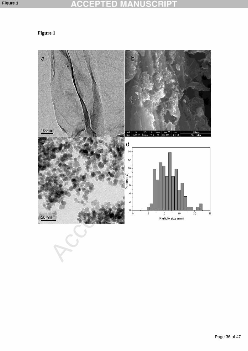
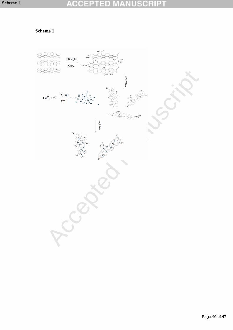
The preparation of M-GO nanocomposites was schematically illustrated in
Scheme 1. The typical morphology of GO were displayed in Fig. 1a and the images of
M-GO were observed using FESEM and TEM (shown in Fig. 1b, c, respectively). As
can be seen, the free-standing two dimensional GO sheets displayed flake-like shapes
Page 11 of 47
Accep
ted
Man
uscr
ipt
11
with high transparency and some wrinkles. Spherical magnetic particles with almost
uniform size were depicted in the SEM image (see Fig. 1b). As shown in Fig. 1c, M
nanoparticles with the average particle size of 11.64 nm were well-dispersed on the
GO matrix in M-GO nanocomposites. The histogram of the particle size distribution
was presented in Fig. 1d. The reason that small amount of nanoparticles tended to
anchor on the surface of GO with a high density can be explained by the magnetic
dipolar interaction among the M nanoparticles. Similar results were also observed in
previous report [38-40].
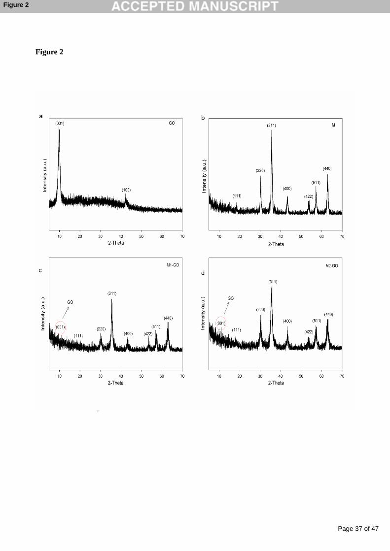
The X-ray diffraction (XRD) patterns of GO, M, M1-GO and M2-GO were
displayed in Fig. 2. It was observed that the two main diffraction peaks at 2θ = 10.0°
(001) and 42.3°(100) were attributed to the structure of GO nanosheets [41-43] in
the XRD pattern shown in Fig. 2a. The disappearance of the characteristic peak at 2θ
= 26.4° (002) in pristine graphite [44] was due to the introduction of
oxygen-containing groups on the surface of GO during oxidation process [45]. The
behavior of M nanoparticles was similar to M-GO in XRD pattern (Fig.2b, c, d). The
four main diffraction peaks at 2θ = 30.2°(220), 35.5°(311), 43.3°(400) and 57.2°
(511) that can be ascribed to maghemite or magnetite [46]. The two magnetic
composites have the similar crystal structure and it is difficult to distinguish according
to XRD patterns. The other two peaks at 2θ = 53.6° and 62.9°were assigned to the
(422) and (440) planes of hematite [47]. The relatively weak peak at 2θ = 18.3°(111)
was also observed due to the presence of goethite [36]. Therefore, iron oxides
nanoparticles in our work included magnetic magnetite (Fe3O4) and maghemite
Page 12 of 47
Accep
ted
Man
uscr
ipt
12
(γ-Fe2O3), and non-magnetic hematite (α-Fe2O3) and goethite (FeOOH). Compared to
M1-GO, M2-GO possessing a higher amount of M component produced more intense
M XRD peaks. It was noted that the characteristic peak of GO at 2θ = 10.0°(001)
was obviously reduced, and the GO peaks at 2θ = 42.3°(100) totally disappeared in
the XRD patterns of M-GO, which could be caused by the reasons as follows: The
weak peaks of carbon in M-GO resulted from the aggregation reduction of graphene
sheets and the increase of monolayer graphene in the presence of magnetite; the
strong peaks of the M nanoparticles overwhelming the weak carbon peaks [48]. The
results in our work were consistent with the previous studies [43].
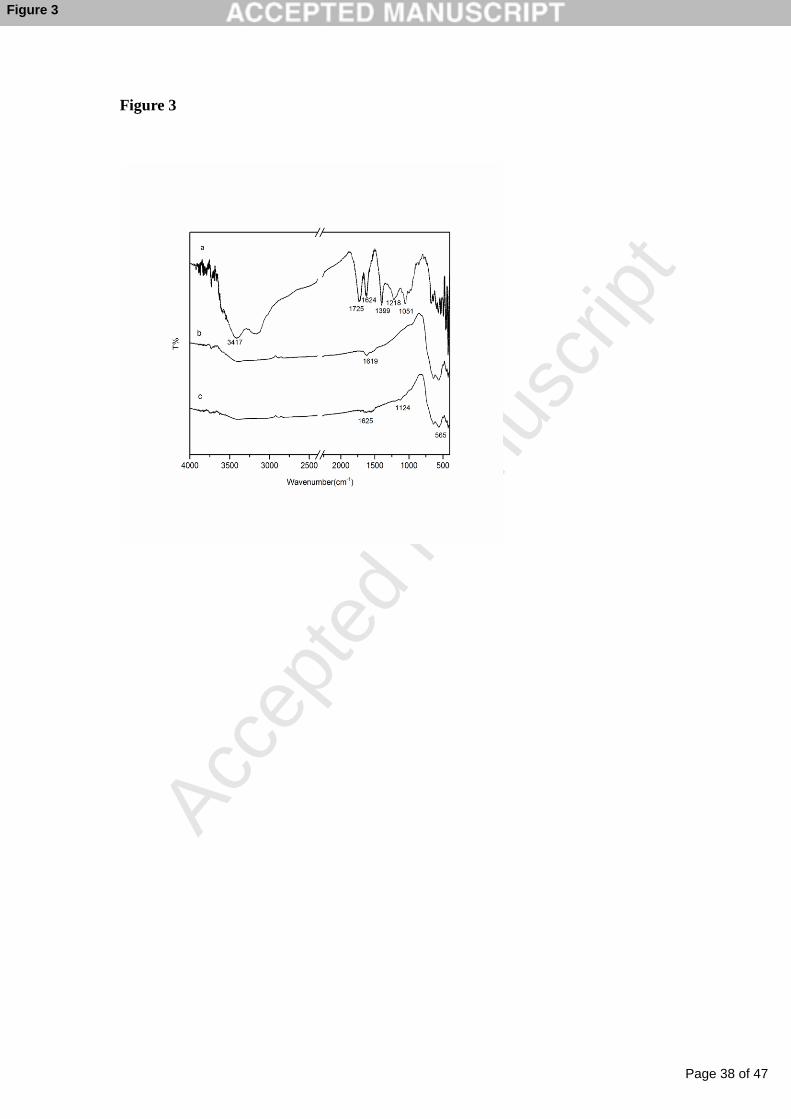
The functional groups of GO, M1-GO and M2-GO nanocomposites were
investigated by FTIR spectra shown in Fig. 3. For GO, the absorption peak at 3417
cm-1 was ascribed to the stretching of O-H [49]. The peaks at 1725 cm-1, 1624 cm-1
and 1399 cm-1 corresponded to carbonyl C=O stretching vibrations [43], aromatic
C=C stretching, and carboxyl O=C-O stretching mode of sp2 carbon skeletal network,
respectively, while the bands at 1218 cm-1 and 1051 cm-1 were associated with
stretching of C-O of epoxy and alkoxy groups, respectively [50]. For M1-GO, the
peaks at 1625 and 1124 cm-1 were assigned to the aromatic C=C stretch and C-O
stretch, respectively. The stretching vibration of C=C in M2-GO appeared at 1624
cm-1. Moreover, the transmittance band around at 565 cm-1 in M-GO was mainly
assigned to the stretching vibration of Fe-O [51, 52].
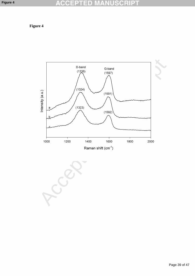
Raman spectroscopy is one of the most sensitive and nondestructive techniques
to probe the ordered and disordered crystal structures of carbon materials. As shown
Page 13 of 47
Accep
ted
Man
uscr
ipt
13
in Fig. 4, Raman spectrum of GO displayed two prominent peaks at 1338 and 1597
cm-1, corresponding to the well-documented D band and G band, respectively. The
Raman D bands shifted from 1338 to 1324 cm-1 and to 1323 cm-1 for M1-GO and
M2-GO, respectively. In addition, the Raman G bands shifted from 1597 to 1591 cm-1
and to 1592 cm-1 for M1-GO and M2-GO, respectively (see Fig. 4 b, c). For M-GO,
the Raman G and D bands shifted to lower frequency in comparison with that of GO,
indicating that GO was reduced [53, 54].
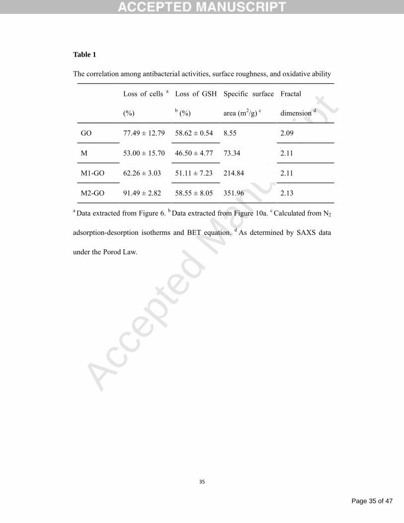
Different surface roughness of materials could significantly influence the
attachment of bacteria on the material surface at the period of interaction between
materials and bacteria [55, 56]. The surface properties of four types of nanomaterials
including GO, M, M1-GO and M2-GO were investigated. The specific surface areas
calculated from N2 adsorption-desorption isotherms and BET equation, and the
fractal dimension values of antibacterial nanomaterials as determined by SAXS data
were presented in Table 1. The specific surface areas of antibacterial materials were
8.55, 73.34, 214.84 and 351.96 m2/g for GO, M, M1-GO and M2-GO, respectively.
The specific surface of M1-GO and M2-GO were higher than that of GO and M,
which was consistent with the fact that M nanoparticles were well dispersed on the
surface of GO nanosheets. Moreover, the fractal dimension of GO, M, M1-GO and
M2-GO was assigned to 2.09, 2.11, 2.11 and 2.13, respectively. It was well known
that the value of fractal dimension was determined by the degree of surface
roughness of materials [57]. The fractal dimension value of GO close to 2.09 was
inclined to be smoothed, which was in agreement with the two-dimensional structure
Page 14 of 47
Accep
ted
Man
uscr
ipt
14
of GO. However, the fractal dimension of M-GO (2.11 for M1-GO, and 2.13 for
M2-GO) was higher than that of GO indicating that M nanoparticles were deposited
on the surface of GO nanosheets resulting more irregularity or roughness.
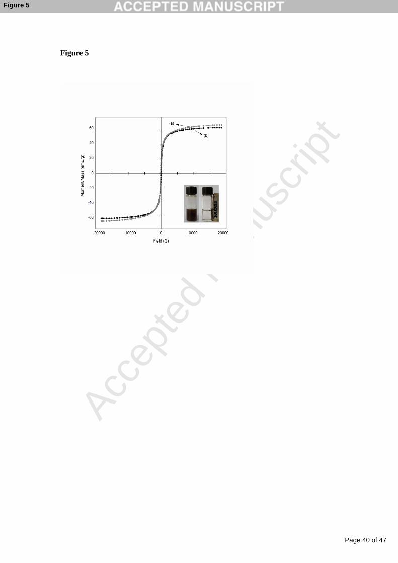
The magnetic properties of the synthesized M-GO nanocomposites were
recorded at room temperature (300 K) by VSM, as shown in Fig. 5. For the M1-GO,
the reduction in the value of saturation magnetization (60.80 emu/g) as compared with
that of M2-GO (68.71 emu/g) could be attributed to the relatively lower amount of M
nanoparticles loaded on GO sheets. Both M1-GO and M2-GO dispersions could be
separated from aqueous solution by a magnet. It was reported that saturation
magnetization of 16.30 emu/g was sufficient for magnetic separation [58]. The
performance of magnetic separation for M-GO was shown in the insert of Fig. 5.
3.2 Antibacterial activity of GO, M and M-GO dispersions
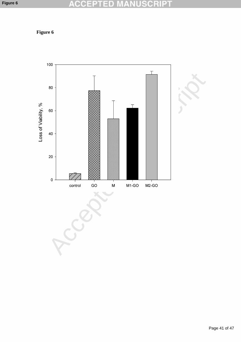
Antibacterial activity of four types of nanomaterials obtained in this work was
evaluated using a model bacterium E. coli. The aqueous suspensions of GO, M,
M1-GO, and M2-GO with the same concentration (100 μg/mL) were incubated with
E. coli cell suspensions (106 to 107 CFU/mL) for 2 h at 37 ℃ and 150 rpm shaking
speed. A series of 100 μL 10-fold cell dilutions were spread onto LB agar plates and
grown in biochemical incubator at 37 ℃ for 24 h. The loss percent of viability was
calculated to quantify the antibacterial ability of nanomaterials.
the loss of viability, % = (1 – ) × 100%
where N is the colony number of activated cells (at the range of 106-107 CFU/mL)
Page 15 of 47
Accep
ted
Man
uscr
ipt
15
before each experiment. is the colony-forming units of the activated cells after
incubation with antibacterial nanomaterials for 2 h (CFU/mL).
Fig. 6 shows the antibacterial properties of various nanomaterials including GO,
M, M1-GO, and M2-GO. GO dispersion exhibited an apparent antibacterial activity
with the cell inactivation percentage at 77.49 ± 12.79 %. M dispersion displayed a
little bit weaker antibacterial activity, with the inactivation percentage at 53.00 ±
15.70 % compared to GO. But for M1-GO, the loss of E. coli viability reached to
62.26 ± 3.03 %, which was a little higher than that of M nanoparticles dispersion, but
a little lower than that of GO dispersion. However, M2-GO possessed the strongest
bacterial inactivation among the four kinds of nanomaterials, with the inactivation
percentage up to 91.49 ± 2.82 %.
The disinfection activity of carbon-based nanocomposites including M1-GO,
M2-GO and magnetic multi-walled carbon nanotubes (M-MWNTs) were also
investigated. The synthesis of M-MWNTs was prepared according to our previous
literature [46]. Results showed that M-MWNTs with the same concentration of
100μg/mL exerted much weaker inactivation ability (34.19 ± 5.06 %) against E. coli
than that of M1-GO (62.26 ± 3.03 %) and M2-GO (91.49 ± 2.82 %).
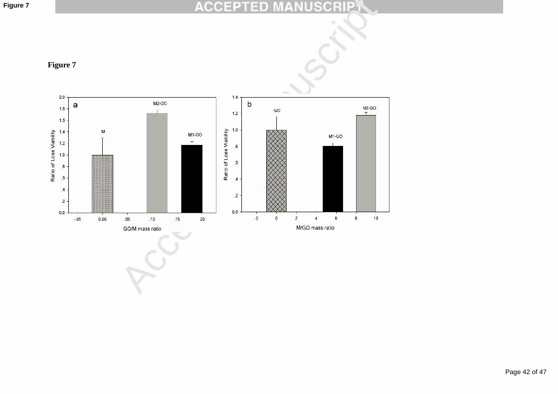
The effect of mass ratio of GO to M on antibacterial properties of nanomaterials
including GO, M, M1-GO, and M2-GO has been analyzed (shown in Fig. 7). The
total mass of four antibacterial nanomaterials was fixed. The ratios of GO/M were 0,
0.18 and 0.11 for M, M1-GO, and M2-GO, respectively. Noticeably, the bactericidal
ability of magnetic nanomaterials was enhanced in the presence of GO, which might
Page 16 of 47
Accep
ted
Man
uscr
ipt
16
be attributed to the moderate antibacterial properties of GO itself. However, the
antibacterial ability of GO-based nanomaterials was not proportional to GO mass
percentage in M-GO nanomaterials. The percent of cell viability loss increased for
M2-GO at the mass ratio of GO/M 0.11, and decreased for M1-GO at 0.18 (displayed
in Fig. 7a). It was concluded that the antibacterial activity of M-GO nanocomposites
was not only caused by GO component but also by M component. Additionally, the
bactericidal ability of magnetic nanomaterials was dependent on the ratio of M/GO
when addition of magnetite component into GO nanomaterials. The ratios of M/GO
were 0, 5.56 and 9.09 for M, M1-GO, and M2-GO, respectively. Cell viability loss
percent decreased for M1-GO at the mass ratio of M/GO 5.56 and increased for
M2-GO at 9.09. It was observed M1-GO with the M/GO mass ratio of 5.56 was not
beneficial to the cell viability loss compared with GO itself. On the contrary, M2-GO
exerted a higher antibacterial property with M/GO mass ratio of 9.09 when compared
to GO or M itself, illustrating that a synergistic antibacterial effect occurred between
GO and M (shown in Fig. 7b). Ma et al. [22] and Zhang et al. [59] reported that
silver-modified graphene materials displayed an excellent antibacterial activity
towards E. coli due to the synergistic effect of Ag nanoparticles and graphene oxide
(GO) or graphene nanosheets (GNS). Sreeprasad et al. [60] prepared a serial of
multifunctional graphene oxide/reduced graphene oxide (GO/RGO) based composites
by anchoring of native lactoferrin (NLf), chitosan (Ch) and Au clusters into GO/RGO,
such as RGO/GO-NLf-Ch and RGO/GO-Au@NLf-Ch. The composites exhibited
several folds higher antibacterial activity than GO/RGO itself, which was accounted
Page 17 of 47
Accep
ted
Man
uscr
ipt
17
for the synergetic effect of the combination of materials. Nangmenyi G et al. [61] also
reported a synergistic disinfection action between Fe2O3 and Ag on fiberglass when
compared to either Fe2O3 or Ag alone. Noticeably, synergistic antibacterial effect
between GO and M toward E. coli in our work was dependent on the mass ratio of
M/GO in M-GO nanomaterials. The optimal mass ratio of M to GO and the
mechanism of synergistic effect between GO and M will be investigated in our further
studies.
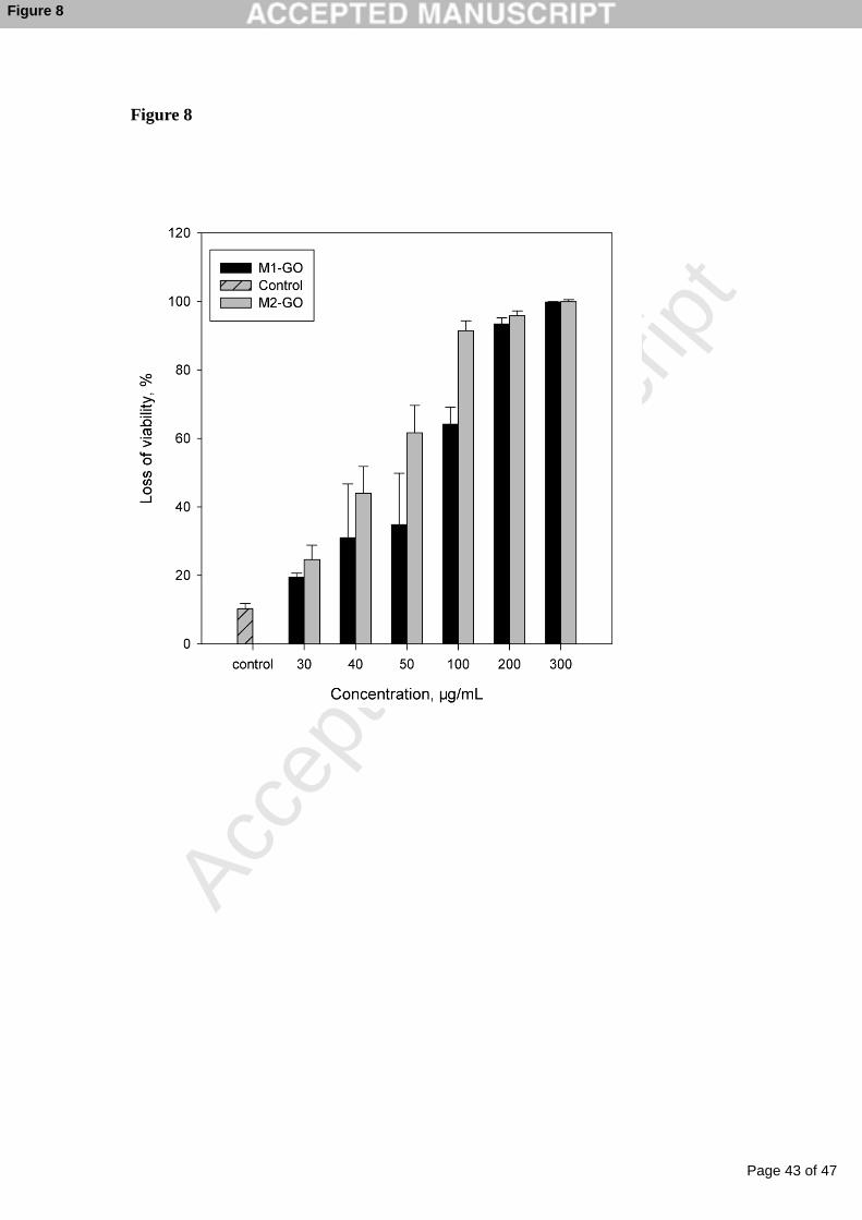
The concentration effect on the E. coli inactivation activity by M-GO was
presented in Fig. 8. The antibacterial property of M-GO (including M1-GO and
M2-GO) dispersions with diverse concentrations was investigated. The percent of cell
viability loss gradually went up with the increased concentration of M-GO. For
M1-GO, the loss of E. coli viability increased from 19.47 ± 1.20 % at the M1-GO
concentration of 30 μg/mL to 31.02 ± 15.70, 34.82 ± 15.00, 64.14 ± 5.00, 93.45 ±
1.76, and 99.84 ± 0.16 % after incubating with 40, 50, 100, 200, and 300 μg/mL
M1-GO suspensions, respectively. When the concentration of M1-GO achieved to 200
μg/mL, there were almost no living cells left. Compared with M1-GO, M2-GO
exhibited stronger antibacterial ability at the same concentration gradient. The
inactivation percent of E. coli by M2-GO increased from 24.65 ± 4.21 % at the
concentration of 30 μg/mL to 43.986 ± 7.83, 61.69 ± 7.89 % at the concentration of
40, and 50 μg/mL. There were 91.49 ± 2.82 % of E. coli cells were killed, when the
concentration of M2-GO reached to 100 μg/mL.
3.3 Interaction between E. coli cells and M-GO dispersion
Page 18 of 47
Accep
ted
Man
uscr
ipt
18
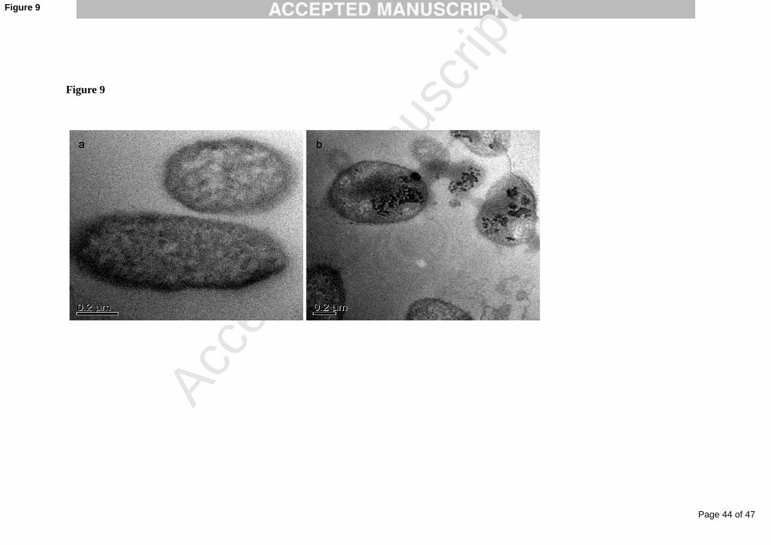
The interaction between E. coli cells and M-GO nanocomposites was illustrated
by TEM. Results revealed the cells treated with M-GO dispersion contained dark
granules around the outside cell wall and penetrated into the cytoplasm. These specks
were likely assigned to M-GO or M nanomaterials passed into cells via direct
interaction with E. coli during incubation. Similar phenomena were also observed by
Lee et al. [62] and Hu et al. [63]. Besides, most of E. coli cells lost their cellular
integrity, with significant destruction of the cell membrane and subsequent leakage of
cellular contents (Fig. 9b) after exposure to M-GO. This was similar to GO or rGO
[20], which induced membrane stress on E. coli cells, resulting in destruction of cell
structures. Such irreversible damage of cells induced by M-GO may play a significant
role in their antibacterial activity.
3.4 Oxidation ability of antibacterial materials
The mechanism of oxidation damage was the most accepted explanation for the
antibacterial activity of graphene-based nanomaterials. Meanwhile, previous
researches reported that the M nanoparticles generated reactive oxygen species (ROS)
via Fenton reactions when interacting with bacteria, leading to protein oxidation and
DNA damage, and finally resulting in cells death [29, 51]. In view of this, the
oxidation ability of four antibacterial nanomaterials in this work was evaluated by
measuring the loss percentage of GSH to confirm the oxidative damage toward
bacteria. GSH is a small thiol containing tripeptide antioxidant in most Gram-negative
bacteria cells at levels of 0.1-10 mM [64]. The thiol groups (-SH) in GSH were
oxidized to disulfide bond (-S-S) sensitively when exposed to ROS or other oxidants
Page 19 of 47
Accep
ted
Man
uscr
ipt
19
[16]. Therefore, in this work, the oxidation degree of GSH was measured in vitro to
reflect indirectly the cellular oxidative destruction induced by four types of
nanomaterials. GSH (0.4 mM) was explored to incubate with GO, M, M1-GO and
M2-GO (at the same concentration of 100 μg/mL) for 2 h. The loss of GSH was
quantified using a thiol quantization kit and the details of experiments were
represented in the section of Materials and Methods.
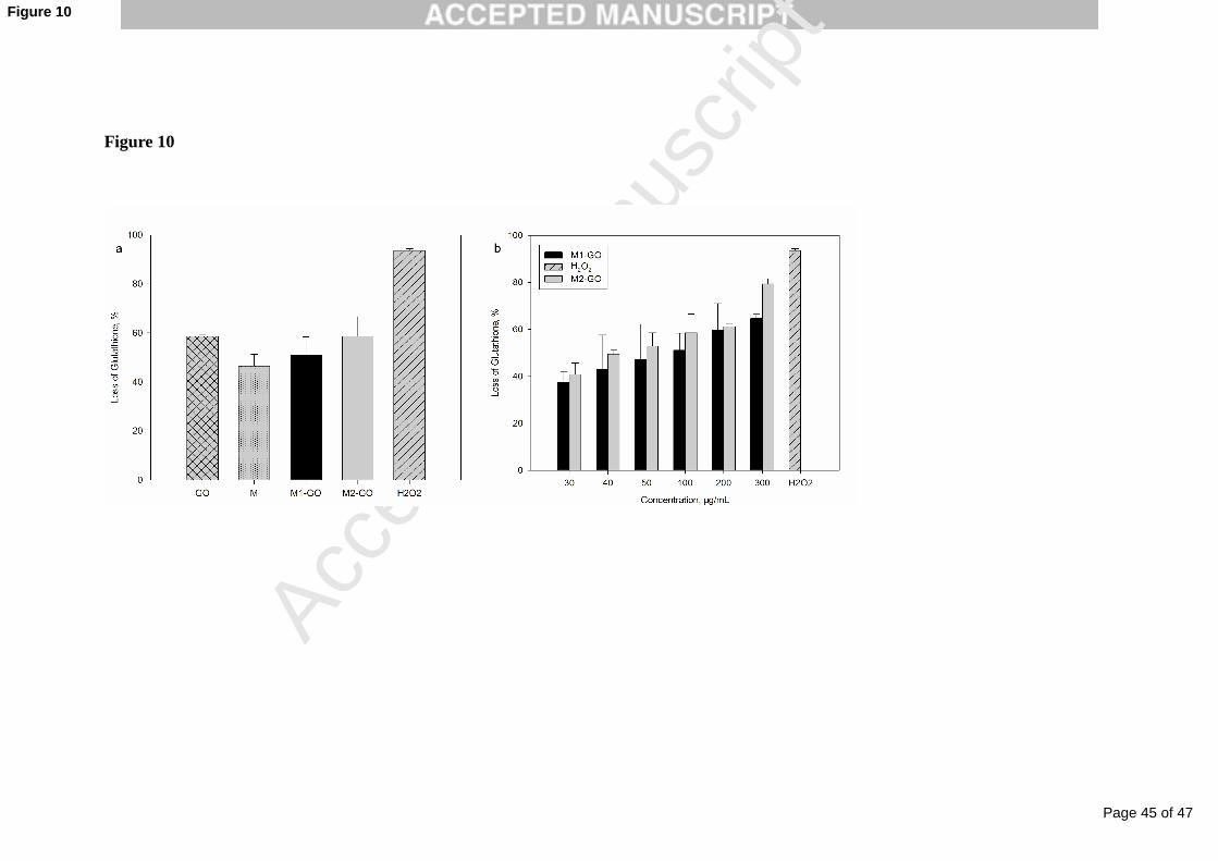
Fig. 10a shows that 58.62 ± 0.54 % and 46.50 ± 4.77 % of GSH were oxidized
by GO and M after 2 h incubation, respectively. Compared to M (GSH oxidation
percentage 46.50 ± 4.77 %), the loss percent of GSH rose up to 51.11 ± 7.23 % for
M1-GO, and 58.55 ± 8.05 % for M2-GO, respectively. The oxidation tendency of
GSH by GO, M, M1-GO and M2-GO was similar to the tendency of E. coli
inactivation induced by the four nanomaterails. Therefore, it was concluded that the
oxidative ability of the nanomaterials had a significant influence on their antibacterial
properties.
Considering the concentration-dependent antibacterial activities of M-GO (see
Fig. 8), we speculated the oxidative ability of M-GO toward GSH would be
concentration-dependent as well. M-GO with different concentrations (30-300 μg/mL)
were incubated with 0.4 mM GSH for 2 h. Fig. 10b shows the fraction of GSH
oxidized by M1-GO or M2-GO was concentration dependent. GSH oxidation by
M-GO was37.60 ± 3.69 % and 64.64 ± 1.53 % for M1-GO and 40.86 ± 5.91 % and
79.34 ± 2.82 % for M2-GO at the M-GO concentration of 30 and 300 μg/mL,
respectively. It was obvious that M2-GO has relatively higher oxidation reactivity
Page 20 of 47
Accep
ted
Man
uscr
ipt
20
than M1-GO at the same concentration. Furthermore, GSH oxidation increased with
increasing concentration of M1-GO or M2-GO, which was consistent with the trend
that antibacterial activity increased with increasing concentration of M-GO
nanomaterials.
3.5 Antibacterial mechanism of M-GO
The correlation among antibacterial activities, surface roughness and GSH
oxidation was summarized in Table 1. On the one hand, comparing GO and M2-GO,
they possessed similar capacities in oxidizing GSH (GO at 58.62 ± 0.54 % vs M2-GO
at 58.55 ± 8.05 %); however, M2-GO dispersion exerted much higher bactericidal
activity (91.49 ± 2.82 %) than GO dispersion (77.49 ± 12.79 %). Their difference was
that GO was two-dimension nanosheets with surface area of 8.55 m2/g, while M2-GO
possessed higher surface roughness with fractal dimension of 2.13 and surface area of
351.96 m2/g. Their distinct antibacterial activities indicated that surface property of
antibacterial materials played an important role in the antibacterial mechanism. On the
other hand, comparing GO and M, GO (fractal dimension of 2.09 and surface area of
8.55 m2/g) was obviously smoother than M (fractal dimension of 2.11 and surface
area of 73.34 m2/g). However, the antibacterial activity of GO (77.49 ± 12.79 %) was
much higher than that of M (53.00 ± 15.70 %). This was obviously correlated with
their different GSH oxidation capacities. In addition, among the four types of
antibacterial materials, M2-GO with the highest surface roughness and oxidation
ability also exerted the highest antibacterial activity. Therefore, results shown in Table
1 suggested that the antibacterial activity of materials were ascribed to their surface
Page 21 of 47
Accep
ted
Man
uscr
ipt
21
property and oxidization ability.
Liu et al. [20] proposed a three-step antibacterial mechanism for graphene-based
materials including initial bacteria cells deposited on graphene-based materials during
incubation period, membrane stress induced by direct interaction between sharp
nanosheets and bacteria and the following superoxide anion-independent oxidation
toward intercellular components of cells. They suggested that the antimicrobial
mechanism of graphene-based materials were contributed to the synergy of membrane
and oxidation stress.
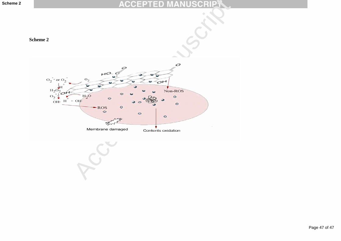
Here, the synergetic mechanism was supposed to account for observations in our
paper. The possible inactivation mechanism of M-GO toward bacteria in this work
was expressed clearly on Scheme 2. E. coli cells may first anchor on the surfaces of
M-GO during incubation in aqueous system. The shaking speed of 150 rpm used in
antibacterial assays had facilitated the suspension of M-GO in the aqueous solution.
Under the shaking condition, M-GO dispersion had more chances to interact with E.
coli for cell deposition.
After cells adhering to M-GO surfaces, the sharp edge of GO nanosheets may
destroy the integrity of cell membrane, then resulting in the leakage of intracellular
materials and finally cell death, as previously reported [18, 63]. It was likely that the
small size of M nanoparticles with average size of 11.64 nm maybe have
opportunities to penetrate into E. coli membranes. Lee et al [62] found that the reason
of zero-valent iron nanoparticles with sizes ranging from 10-80 nm had strong
bactericidal activity could be contributed to the small size nanoparticles penetration
Page 22 of 47
Accep
ted
Man
uscr
ipt
22
into cells. GSH oxidation assays (Fig. 9) demonstrated that the oxidation ability of
M-GO may play a vital role in bacteria inactivation when M-GO direct contacting
with cells. Liu et al. [20] illustrated that graphene-based materials were capable of
inducing ROS-independent oxidative stress toward E. coli cells. Tran et al. [29]
confirmed metal oxide Fe3O4 inhibited the growth of S. aureus via oxidative stress
generated by ROS. Therefore, it was possible that M-GO nanocomposites could also
oxidize bacterial components through mediating the oxidation ability of GO and M.
The strong oxidation activity of M-GO toward GSH in our work supported that
M-GO was efficient to oxidize thiols or other intercellular contents.
4. Conclusions
Four types of suspension (GO, M, M1-GO and M2-GO) were prepared to reveal
different antibacterial properties, specially the antibacterial activity of M-GO
nanocomposites. M-GO was synthesized by depositing magnetic iron oxide
nanoparticles on the surface of GO nanosheets, M nanoparticles could be supported
and stabilized on the GO surface resulting in excellent dispersion, and the
nanoparticles have an average size of 11.64 nm. The saturation magnetization was
60.80 emu/g for M1-GO and 68.71 emu/g for M2-GO, respectively. Therefore, M-GO
nanocomposites could be rapidly separated from aqueous solution using an external
magnetic field. Results showed that both bare and GO-coated magnetic iron oxide
nanocomposites were efficient in inhibiting the growth of E. coli. The various mass
ratios of M/GO in M-GO influenced the inactivation properties significantly. GO and
Page 23 of 47
Accep
ted
Man
uscr
ipt
23
M emerged synergistic effect on loss of cell viability, when M/GO mass ratio in
M-GO nanocomposites was adjusted to 9.09. And the antibacterial ability of M-GO
was concentration-dependent. The mechanisms of bacterial cytotoxicity caused by
M-GO may be relying on both physical membrane puncture and chemical cellular
matters oxidation, which were similar to the three-step antibacterial mechanisms of
CNTs. In views of these interesting properties of both strong magnetic property and
outstanding antibacterial ability, M-GO nanomaterials have the potential applications
for environmental drinking water treatments.
Acknowledgments
The authors are grateful for the financial supports from National Natural Science
Foundation of China (51039001, 50978088, 50808070, 21275044 and 51108166),
Interdisciplinary Research Funds for Hunan University, the Natural Science
Foundation of Hunan Province, China (Grant no. 12JJB003) and the Scientific
Research Foundation for the Returned Overseas Chinese Scholars, State Education
Ministry.
References:
[1] G.P. Winward, L.M. Avery, T. Stephenson, B. Jefferson, Chlorine disinfection of
grey water for reuse: Effect of organics and particles, Water Res. 42 (2008) 483-491.
[2] W. Lee, P. Westerhoff, Formation of organic chloramines during water disinfection
- chlorination versus chloramination, Water Res. 43 (2009) 2233-2239.
[3] P. Xu, M.L. Janex, P. Savoye, A. Cockx, V. Lazarova, Wastewater disinfection by
Page 24 of 47
Accep
ted
Man
uscr
ipt
24
ozone: main parameters for process design, Water Res. 36 (2002) 1043-1055.
[4] M.A. Shannon, P.W. Bohn, M. Elimelech, J.G. Georgiadis, B.J. Marinas, A.M.
Mayes, Science and technology for water purification in the coming decades, Nature
452 (2008) 301-310.
[5] S.D. Richardson, M.J. Plewa, E.D. Wagner, R. Schoeny, D.M. DeMarini,
Occurrence, genotoxicity, and carcinogenicity of regulated and emerging disinfection
by-products in drinking water: A review and roadmap for research, Mutat. Res-Rev
Mutat. 636 (2007) 178-242.
[6] V.K. Sharma, R.A. Yngard, Y. Lin, Silver nanoparticles: Green synthesis and their
antimicrobial activities, Adv. Colloid Interface Sci. 145 (2009) 83-96.
[7] Q. Bao, D. Zhang, P. Qi, Synthesis and characterization of silver nanoparticle and
graphene oxide nanosheet composites as a bactericidal agent for water disinfection, J.
Colloid Interface Sci. 360 (2011) 463-470.
[8] R. Guerra, E. Lima, M. Viniegra, A. Guzman, V. Lara, Growth of Escherichia coli
and Salmonella typhi inhibited by fractal silver nanoparticles supported on zeolites,
Microporous Mesoporous Mater. 147 (2012) 267-273.
[9] R. Guerra, E. Lima, A. Guzman, Antimicrobial supported nanoparticles: Gold
versus silver for the cases of Escherichia coli and Salmonella typhi, Microporous
Mesoporous Mater. 170 (2013) 62-66.
[10] O. Akhavan, E. Ghaderi, Photocatalytic Reduction of Graphene Oxide
Nanosheets on TiO2 Thin Film for Photoinactivation of Bacteria in Solar Light
Irradiation, J. Phys. Chem. C 113 (2009) 20214-20220.
Page 25 of 47
Accep
ted
Man
uscr
ipt
25
[11] M.A. Gondal, M.A. Dastageer, A. Khalil, K. Hayat, Z.H. Yamani, Nanostructured
ZnO synthesis and its application for effective disinfection of Escherichia coli micro
organism in water, J. Nanopart Res. 13 (2011) 3423-3430.
[12] E. Gazit, Self-assembled peptide nanostructures: the design of molecular building
blocks and their technological utilization, Chem. Soc. Rev. 36 (2007) 1263-1269.
[13] M.M. Lou, B. Zhu, I. Muhammad, B. Li, G.L. Xie, Y.L. Wang, H.Y. Li, G.C. Sun,
Antibacterial activity and mechanism of action of chitosan solutions against apricot
fruit rot pathogen Burkholderia seminalis, Carbohyd. Res. 346 (2011) 1294-1301.
[14] Q.Z. Yang, Y.Y. Chang, H.Z. Zhao, Preparation and antibacterial activity of
lysozyme and layered double hydroxide nanocomposites, Water Res. 47 (2013)
6712-6718.
[15] A.R. Badireddy, E.M. Hotze, S. Chellam, P. Alvarez, M.R. Wiesner, Inactivation
of Bacteriophages via photosensitization of fullerol nanoparticles, Environ. Sci.
Technol. 41 (2007) 6627-6632.
[16] D.Y. Lyon, P.J.J. Alvarez, Fullerene Water Suspension (nC60) Exerts Antibacterial
Effects via ROS-Independent Protein Oxidation, Environ. Sci. Technol. 42 (2008)
8127-8132.
[17] S. Kang, M. Herzberg, D.F. Rodrigues, M. Elimelech, Antibacterial effects of
carbon nanotubes: Size does matter, Langmuir 24 (2008) 6409-6413.
[18] O. Akhavan, E. Ghaderi, Toxicity of Graphene and Graphene Oxide Nanowalls
Against Bacteria, ACS Nano 4 (2010) 5731-5736.
[19] K.S. Novoselov, A.K. Geim, S.V. Morozov, D. Jiang, Y. Zhang, S.V. Dubonos,
Page 26 of 47
Accep
ted
Man
uscr
ipt
26
I.V. Grigorieva, A.A. Firsov, Electric field effect in atomically thin carbon films,
Science 306 (2004) 666-669.
[20] S.B. Liu, T.H. Zeng, M. Hofmann, E. Burcombe, J. Wei, R.R. Jiang, J. Kong, Y.
Chen, Antibacterial Activity of Graphite, Graphite Oxide, Graphene Oxide, and
Reduced Graphene Oxide: Membrane and Oxidative Stress, ACS Nano 5 (2011)
6971-6980.
[21] P.V. Kamat, Graphene-Based Nanoarchitectures. Anchoring Semiconductor and
Metal Nanoparticles on a Two-Dimensional Carbon Support, J. Phys. Chem. Lett. 1
(2009) 520-527.
[22] J.Z. Ma, J.T. Zhang, Z.G. Xiong, Y. Yong, X.S. Zhao, Preparation,
characterization and antibacterial properties of silver-modified graphene oxide, J.
Mater. Chem. 21 (2011) 3350-3352.
[23] C. Sun, J.S.H. Lee, M.Q. Zhang, Magnetic nanoparticles in MR imaging and
drug delivery, Adv. Drug Deliver. Rev. 60 (2008) 1252-1265.
[24] O. Veiseh, J.W. Gunn, M.Q. Zhang, Design and fabrication of magnetic
nanoparticles for targeted drug delivery and imaging, Adv. Drug Deliver. Rev. 62
(2010) 284-304.
[25] Q.A. Pankhurst, J. Connolly, S.K. Jones, J. Dobson, Applications of magnetic
nanoparticles in biomedicine, J. Phys. D: Appl. Phys. 36 (2003) R167-R181.
[26] D. Horak, M. Babic, H. Mackova, M.J. Benes, Preparation and properties of
magnetic nano- and microsized particles for biological and environmental separations,
J. Sep. Sci. 30 (2007) 1751-1772.
Page 27 of 47
Accep
ted
Man
uscr
ipt
27
[27] T.A. Liu, P. Spincemaille, L. de Rochefort, R. Wong, M. Prince, Y. Wang,
Unambiguous identification of superparamagnetic iron oxide particles through
quantitative susceptibility mapping of the nonlinear response to magnetic fields,
Magn. Reson. Imaging 28 (2010) 1383-1389.
[28] T.K. Jain, M.K. Reddy, M.A. Morales, D.L. Leslie-Pelecky, V. Labhasetwar,
Biodistribution, clearance, and biocompatibility of iron oxide magnetic nanoparticles
in rats, Mol. Pharm. 5 (2008) 316-327.
[29] N. Tran, A. Mir, D. Mallik, A. Sinha, S. Nayar, T.J. Webster, Bactericidal effect
of iron oxide nanoparticles on Staphylococcus aureus, Int. J. Nanomed. 5 (2010)
277-283.
[30] S. Chatterjee, A. Bandyopadhyay, K. Sarkar, Effect of iron oxide and gold
nanoparticles on bacterial growth leading towards biological application, J.
Nanobiotechnol. 9 (2011).
[31] M. Auffan, W. Achouak, J. Rose, M.A. Roncato, C. Chaneac, D.T. Waite, A.
Masion, J.C. Woicik, M.R. Wiesner, J.Y. Bottero, Relation between the redox state of
iron-based nanoparticles and their cytotoxicity toward Escherichia coli, Environ. Sci.
Technol. 42 (2008) 6730-6735.
[32] E.N. Taylor, T.J. Webster, The use of superparamagnetic nanoparticles for
prosthetic biofilm prevention, Int. J. Nanomed. 4 (2009) 145-152.
[33] O. Akhavan, E. Ghaderi, Escherichia coil bacteria reduce graphene oxide to
bactericidal graphene in a self-limiting manner, Carbon 50 (2012) 1853-1860.
[34] P.S.M. Dunlop, J.A. Byrne, N. Manga, B.R. Eggins, The photocatalytic removal
Page 28 of 47
Accep
ted
Man
uscr
ipt
28
of bacterial pollutants from drinking water, J. Photoch. Photobio. A: Chem 148 (2002)
355-363.
[35] W.S Hummers Jr., R.E. Offeman, Preparation of Graphitic Oxide, J. Am. Chem.
Soc. 80 (1958) 1339.
[36] J.H. Deng, X.R. Zhang, G.M. Zeng, J.L. Gong, Q.Y. Niu, J. Liang, Simultaneous
removal of Cd(II) and ionic dyes from aqueous solution using magnetic graphene
oxide nanocomposite as an adsorbent, Chem. Eng. J. 226 (2013) 189-200.
[37] C.D. Vecitis, K.R. Zodrow, S. Kang, M. Elimelech,
Electronic-Structure-Dependent Bacterial Cytotoxicity of Single-Walled Carbon
Nanotubes, ACS Nano 4 (2010) 5471-5479.
[38] M.J. Hu, Y. Lu, S. Zhang, S.R. Guo, B. Lin, M. Zhang, S.H. Yu, High yield
synthesis of bracelet-like hydrophilic Ni-Co magnetic alloy flux-closure nanorings, J.
Am. Chem. Soc. 130 (2008) 11606-11607.
[39] S.L. Pan, Z.G. An, J.J. Zhang, G.Z. Song, Synthesis and hierarchical assembly of
CoNi flowery particles, Mater. Chem. Phys. 124 (2010) 342-346.
[40] S. Bai, X.P. Shen, G.X. Zhu, M.Z. Li, H.T. Xi, K.M. Chen, In situ Growth of
NixCo100-x Nanoparticles on Reduced Graphene Oxide Nanosheets and Their
Magnetic and Catalytic Properties, ACS Appl. Mater. Interfaces 4 (2012) 2378-2386.
[41] J.J. Shi, J.J. Zhu, Sonoelectrochemical fabrication of Pd-graphene nanocomposite
and its application in the determination of chlorophenols, Electrochim. Acta 56 (2011)
6008-6013.
[42] Q.Y. Wang, X.Q. Cui, W.M. Guan, W.T. Zheng, J.L. Chen, X.L. Zheng, X.M.
Page 29 of 47
Accep
ted
Man
uscr
ipt
29
Zhang, C. Liu, T.Y. Xue, H.T. Wang, Z. Jin, H. Teng, Synthesis of flower-shape
palladium nanostructures on graphene oxide for electrocatalytic applications, J. Phys.
Chem. Solids 74 (2013) 1470-1474.
[43] Y.J. Yao, S.D. Miao, S.Z. Liu, L.P. Ma, H.Q. Sun, S.B. Wang, Synthesis,
characterization, and adsorption properties of magnetic Fe3O4@graphene
nanocomposite, Chem. Eng. J. 184 (2012) 326-332.
[44] G.X. Zhao, J.X. Li, X.M. Ren, C.L. Chen, X.K. Wang, Few-Layered Graphene
Oxide Nanosheets As Superior Sorbents for Heavy Metal Ion Pollution Management,
Environ. Sci. Technol. 45 (2011) 10454-10462.
[45] M.J. McAllister, J.L. Li, D.H. Adamson, H.C. Schniepp, A.A. Abdala, J. Liu, M.
Herrera-Alonso, D.L. Milius, R. Car, R.K. Prud'homme, I.A. Aksay, Single sheet
functionalized graphene by oxidation and thermal expansion of graphite, Chem. Mater.
19 (2007) 4396-4404.
[46] J.L. Gong, B. Wang, G.M. Zeng, C.P. Yang, C.G. Niu, Q.Y. Niu, W.J. Zhou, Y.
Liang, Removal of cationic dyes from aqueous solution using magnetic multi-wall
carbon nanotube nanocomposite as adsorbent, J. Hazard. Mater. 164 (2009)
1517-1522.
[47] M.A. Legodi, D. de Waal, The preparation of magnetite, goethite, hematite and
maghemite of pigment quality from mill scale iron waste, Dyes Pigments. 74 (2007)
161-168.
[48] X. Yang, C.L. Chen, J.X. Li, G.X. Zhao, X.M. Ren, X.K. Wang, Graphene
oxide-iron oxide and reduced graphene oxide-iron oxide hybrid materials for the
Page 30 of 47
Accep
ted
Man
uscr
ipt
30
removal of organic and inorganic pollutants, RSC Adv. 2 (2012) 8821-8826.
[49] J.F. Shen, Y.Z. Hu, M. Shi, N. Li, H.W. Ma, M.X. Ye, One Step Synthesis of
Graphene Oxide-Magnetic Nanoparticle Composite, J. Phys. Chem. C 114 (2010)
1498-1503.
[50] V. Chandra, J. Park, Y. Chun, J.W. Lee, I.C. Hwang, K.S. Kim, Water-Dispersible
Magnetite-Reduced Graphene Oxide Composites for Arsenic Removal, ACS Nano 4
(2010) 3979-3986.
[51] B.S. Inbaraj, T.Y. Tsai, B.H. Chen, Synthesis, characterization and antibacterial
activity of superparamagnetic nanoparticles modified with glycol chitosan, Sci.
Technol. Adv. Mat. 13 (2012).
[52] B.S. Inbaraj, T.H. Kao, T.Y. Tsai, C.P. Chiu, R. Kumar, B.H. Chen, The synthesis
and characterization of poly(gamma-glutamic acid)-coated magnetite nanoparticles
and their effects on antibacterial activity and cytotoxicity, Nanotechnology 22 (2011).
[53] Y.S. Fu, P. Xiong, H.Q. Chen, X.Q. Sun, X. Wang, High Photocatalytic Activity
of Magnetically Separable Manganese Ferrite-Graphene Heteroarchitectures, Ind. Eng.
Chem. Res. 51 (2012) 725-731.
[54] Y.S. Fu, H.Q. Chen, X.Q. Sun, X. Wang, Combination of cobalt ferrite and
graphene: High-performance and recyclable visible-light photocatalysis, Appl. Catal.
B: Environ. 111 (2012) 280-287.
[55] K. Shellenberger, B.E. Logan, Effect of molecular scale roughness of glass beads
on colloidal and bacterial deposition, Environ. Sci. Technol. 36 (2002) 184-189.
[56] C. Vasilescu, P. Drob, E. Vasilescu, I. Demetrescu, D. Ionita, M. Prodana, S.I.
Page 31 of 47
Accep
ted
Man
uscr
ipt
31
Drob, Characterisation and corrosion resistance of the electrodeposited
hydroxyapatite and bovine serum albumin/hydroxyapatite films on Ti-6Al-4V-1Zr
alloy surface, Corros. Sci. 53 (2011) 992-999.
[57] E. Lima, A. Guzmán-Vargas, J. Méndez-Vivar, H. Pfeiffer, J. Fraissard,
Fe-ZSM-5 Catalysts: Preparation in Organic Media, Fe-particle Morphology and NOx
Reduction Activity, Catal. Lett. 120 (2008) 244-251.
[58] J.L. Gong, X.Y. Wang, G.M. Zeng, L. Chen, J.H. Deng, X.R. Zhang, Q.Y. Niu,
Copper (II) removal by pectin-iron oxide magnetic nanocomposite adsorbent, Chem.
Eng. J. 185 (2012) 100-107.
[59] Z. Zhang, J. Zhang, B.L. Zhang, J.L. Tang, Mussel-inspired functionalization of
graphene for synthesizing Ag-polydopamine-graphene nanosheets as antibacterial
materials, Nanoscale 5 (2013) 118-123.
[60] T.S. Sreeprasad, M.S. Maliyekkal, K. Deepti, K. Chaudhari, P.L. Xavier, T.
Pradeep, Transparent, Luminescent, Antibacterial and Patternable Film Forming
Composites of Graphene Oxide/Reduced Graphene Oxide, ACS Appl. Mater.
Interfaces 3 (2011) 2643-2654.
[61] G. Nangmenyi, X.A. Li, S. Mehrabi, E. Mintz, J. Economy, Silver-modified iron
oxide nanoparticle impregnated fiberglass for disinfection of bacteria and viruses in
water, Mater. Lett. 65 (2011) 1191-1193.
[62] C. Lee, J.Y. Kim, W.I. Lee, K.L. Nelson, J. Yoon, D.L. Sedlak, Bactericidal effect
of zero-valent iron nanoparticles on Escherichia coli, Environ. Sci. Technol. 42 (2008)
4927-4933.
Page 32 of 47
Accep
ted
Man
uscr
ipt
32
[63] W.B. Hu, C. Peng, W.J. Luo, M. Lv, X.M. Li, D. Li, Q. Huang, C.H. Fan,
Graphene-Based Antibacterial Paper, ACS Nano 4 (2010) 4317-4323.
[64] R.C. Fahey, W.C. Brown, W.B. Adams, M.B. Worsham, Occurrence of
glutathione in bacteria, J. Bacteriol. 133 (1978) 1126-1129.
Page 33 of 47
Accep
ted
Man
uscr
ipt
33
Figure Captions
Figure 1 TEM image of GO (a), SEM image of M-GO (b), TEM image of M-GO (c),
and the size distribution of iron oxide nanoparticles (d).
Figure 2 XRD patterns of GO (a), M (b), M1-GO (c), and M2-GO (d).
Figure 3 FTIR spectra of GO (a), M2-GO (b), and M1-GO (c).
Figure 4 Raman spectra of GO (a), M1-GO (b) and M2-GO (c).
Figure 5 Magnetization curves of M2-GO (a) and M1-GO (b) at 300 K. The insert
shows the M-GO nanocomposites dispersion and magnetic separation.
Figure 6 Inactivation of E. coli by GO, M, M1-GO and M2-GO, at the same
concentration of 100 μg/mL for 2 h at 37 ℃.
Figure 7 Comparison of antibacterial activity of M-GO nanomatrials with various
GO/M mass ratios (a) and M/GO mass ratios (b).
Figure 8 Antibacterial activity of M-GO with various concentrations. 5 mL of M-GO
(at 30, 40, 50, 100, 200, 300 μg/mL) was incubated with 5 mL E. coli (106-107
CFU/mL) for 2 h at 37 ℃.
Figure 9 TEM images of E. coli cells treated with DI water (a) and M-GO (b) at 37
℃ for 2 h.
Figure 10 (a) Oxidation of GSH (0.4 mM) by GO, M, M1-GO and M2-GO, at the
same concentration of 100 μg/mL. (b) GSH (0.4 mM) was incubated with various
concentrations of M-GO for 2 h. GSH oxidized by H2O2 (10 mM) was used as a
positive control and GSH with bicarbonate buffer (50 mM) was used as a negative
control.
Page 34 of 47
Accep
ted
Man
uscr
ipt
34
Scheme 1 Schematic diagram of M-GO synthesis.
Scheme 2 Antibacterial mechanism of M-GO toward E. coli.
Page 35 of 47
Accep
ted
Man
uscr
ipt
35
Table 1
The correlation among antibacterial activities, surface roughness, and oxidative ability
Loss of cells a
(%)
Loss of GSH
b (%)
Specific surface
area (m2/g) c
Fractal
dimension d
GO 77.49 ± 12.79 58.62 ± 0.54 8.55 2.09
M 53.00 ± 15.70 46.50 ± 4.77 73.34 2.11
M1-GO 62.26 ± 3.03 51.11 ± 7.23 214.84 2.11
M2-GO 91.49 ± 2.82 58.55 ± 8.05 351.96 2.13
a Data extracted from Figure 6. b Data extracted from Figure 10a. c Calculated from N2
adsorption-desorption isotherms and BET equation. d As determined by SAXS data
under the Porod Law.
Page 36 of 47
Accep
ted
Man
uscr
ipt
Figure 1
Figure 1
Page 37 of 47
Accep
ted
Man
uscr
ipt
Figure 2
Figure 2
Page 38 of 47
Accep
ted
Man
uscr
ipt
Figure 3
Figure 3
Page 39 of 47
Accep
ted
Man
uscr
ipt
Figure 4
Figure 4
Page 40 of 47
Accep
ted
Man
uscr
ipt
Figure 5
Figure 5
Page 41 of 47
Accep
ted
Man
uscr
ipt
Figure 6
Figure 6
Page 42 of 47
Accep
ted
Man
uscr
ipt
Figure 7
Figure 7
Page 43 of 47
Accep
ted
Man
uscr
ipt
Figure 8
Figure 8
Page 44 of 47
Accep
ted
Man
uscr
ipt
Figure 9
Figure 9
Page 45 of 47
Accep
ted
Man
uscr
ipt
Figure 10
Figure 10
Page 46 of 47
Accep
ted
Man
uscr
ipt
Scheme 1
Scheme 1
Page 47 of 47
Accep
ted
Man
uscr
ipt
Scheme 2
Scheme 2