In Vivo Association of Ku with Mammalian Origins of DNA Replication
16
Molecular Biology of the Cell Vol. 12, 3386 –3401, November 2001 In Vivo Association of Ku with Mammalian Origins of DNA Replication Olivia Novac,* † Diamanto Matheos,* † Felipe D. Araujo,* † Gerald B. Price,* and Maria Zannis-Hadjopoulos* †‡ *McGill Cancer Center and † Department of Biochemistry, McGill University, Montreal, Quebec, Canada H3G 1Y6 Submitted February 13, 2001; Revised June 12, 2001; Accepted August 21, 2001 Monitoring Editor: Mark J. Solomon Ku is a heterodimeric (Ku70/86-kDa) nuclear protein with known functions in DNA repair, V(D)J recombination, and DNA replication. Here, the in vivo association of Ku with mammalian origins of DNA replication was analyzed by studying its association with ors8 and ors12, as assayed by formaldehyde cross-linking, followed by immunoprecipitation and quantitative polymerase chain reaction analysis. The association of Ku with ors8 and ors12 was also analyzed as a function of the cell cycle. This association was found to be approximately fivefold higher in cells synchronized at the G1/S border, in comparison with cells at G0, and it decreased by approximately twofold upon entry of the cells into S phase, and to near background levels in cells at G2/M phase. In addition, in vitro DNA replication experiments were performed with the use of extracts from Ku80 / and Ku80 / mouse embryonic fibroblasts. A decrease of 70% in in vitro DNA replication was observed when the Ku80 / extracts were used, compared with the Ku80 / extracts. The results indicate a novel function for Ku as an origin binding-protein, which acts at the initiation step of DNA replication and dissociates after origin firing. INTRODUCTION According to the replicon model (Jacob et al., 1963) origins are defined by specific DNA sequences (replicators) and an initia- tor protein or complex of proteins, that bind to this sequence (reviewed in Berezney et al., 2000). Once the origin is activated, the initiator protein unwinds the DNA duplex, allowing the entry of the replication machinery and synthesis of the first primers for chain elongation (reviewed in Ritzi and Knippers, 2000). Considerable progress has been made in Saccharomyces cer- evisiae toward our understanding of the regulation of initiation of DNA replication in relation to the cell cycle (reviewed by Quintana and Dutta, 1999). The origin recognition complex (ORC) was first described in yeast and led to the subsequent identification of ORC homologs in humans, invertebrates (Cae- norabditis elegans), plants (Arabidopsis thaliana), fission yeast, and flies (Drosophila melanogaster) (Gavin et al., 1995; Gossen et al., 1995). In budding yeast, ORC is bound to the replication ori- gins (or ARS elements) throughout the cell cycle (Diffley et al., 1994; Aparicio et al., 1997; Liang and Stillman, 1997). A prerep- lication complex (preRC) assembles during G1 phase of the cell cycle in preparation for initiation of DNA replication at the origin. In S. cerevisiae, this complex consists of ORC proteins, Cdc6p, and the family of MCM proteins, licensing factors. Afterward, activation of cell cycle-regulated protein kinases guides the “licensed” origin into S phase. The preRC gradually dissociates by releasing Cdc6p and MCM proteins; this pos- treplication complex persists until the next G1 phase when another round of replication can occur. ORC was recently shown to play a critical role in replication initiation by posi- tioning nucleosomes adjacent to yeast origins of replication, which influences the preRC assembly (Lipford and Bell, 2001), reinforcing the hypothesis that chromosomal context can sig- nificantly affect origin function (Newlon et al., 1993; Friedman et al., 1996). Chromosomal proteins often interact with DNA to control maintenance, propagation, and expression of the genome. Identification and isolation of proteins interacting with origins of replication are essential for understanding the mechanism of initiation of DNA replication. In S. cerevisiae, the Ku-like pro- tein (OBF2) was shown to be required for the assembly of a stable multiprotein complex at essential sequences within the eukaryotic origin of replication (Shakibai et al., 1996). Ku is an abundant heterodimeric nuclear protein, composed of 70- and 86-kDa subunits, originally identified as an autoantigen recognized by sera from patients with autoimmune diseases (Mimori et al., 1981). Furthermore, Ku is the regulatory subunit of the DNA-dependent serine/threonine protein kinase (DNA- PK) (Carter et al., 1990), and acts as the component of DNA-PK that confers binding to DNA (Dvir et al., 1992). Ku is present in all eukaryotes, suggesting conservation of function. This mul- * Corresponding author. E-mail address: [email protected] 3386 © 2001 by The American Society for Cell Biology
-
Upload
independent -
Category
Documents
-
view
1 -
download
0
Transcript of In Vivo Association of Ku with Mammalian Origins of DNA Replication

Molecular Biology of the CellVol. 12, 3386–3401, November 2001
In Vivo Association of Ku with Mammalian Origins ofDNA ReplicationOlivia Novac,*† Diamanto Matheos,*† Felipe D. Araujo,*† Gerald B. Price,*and Maria Zannis-Hadjopoulos*†‡
*McGill Cancer Center and †Department of Biochemistry, McGill University, Montreal, Quebec,Canada H3G 1Y6
Submitted February 13, 2001; Revised June 12, 2001; Accepted August 21, 2001Monitoring Editor: Mark J. Solomon
Ku is a heterodimeric (Ku70/86-kDa) nuclear protein with known functions in DNA repair, V(D)Jrecombination, and DNA replication. Here, the in vivo association of Ku with mammalian origins ofDNA replication was analyzed by studying its association with ors8 and ors12, as assayed byformaldehyde cross-linking, followed by immunoprecipitation and quantitative polymerase chainreaction analysis. The association of Ku with ors8 and ors12 was also analyzed as a function of the cellcycle. This association was found to be approximately fivefold higher in cells synchronized at the G1/Sborder, in comparison with cells at G0, and it decreased by approximately twofold upon entry of thecells into S phase, and to near background levels in cells at G2/M phase. In addition, in vitro DNAreplication experiments were performed with the use of extracts from Ku80�/� and Ku80�/� mouseembryonic fibroblasts. A decrease of �70% in in vitro DNA replication was observed when theKu80�/� extracts were used, compared with the Ku80�/� extracts. The results indicate a novelfunction for Ku as an origin binding-protein, which acts at the initiation step of DNA replication anddissociates after origin firing.
INTRODUCTION
According to the replicon model (Jacob et al., 1963) origins aredefined by specific DNA sequences (replicators) and an initia-tor protein or complex of proteins, that bind to this sequence(reviewed in Berezney et al., 2000). Once the origin is activated,the initiator protein unwinds the DNA duplex, allowing theentry of the replication machinery and synthesis of the firstprimers for chain elongation (reviewed in Ritzi and Knippers,2000).
Considerable progress has been made in Saccharomyces cer-evisiae toward our understanding of the regulation of initiationof DNA replication in relation to the cell cycle (reviewed byQuintana and Dutta, 1999). The origin recognition complex(ORC) was first described in yeast and led to the subsequentidentification of ORC homologs in humans, invertebrates (Cae-norabditis elegans), plants (Arabidopsis thaliana), fission yeast, andflies (Drosophila melanogaster) (Gavin et al., 1995; Gossen et al.,1995). In budding yeast, ORC is bound to the replication ori-gins (or ARS elements) throughout the cell cycle (Diffley et al.,1994; Aparicio et al., 1997; Liang and Stillman, 1997). A prerep-lication complex (preRC) assembles during G1 phase of the cellcycle in preparation for initiation of DNA replication at theorigin. In S. cerevisiae, this complex consists of ORC proteins,Cdc6p, and the family of MCM proteins, licensing factors.
Afterward, activation of cell cycle-regulated protein kinasesguides the “licensed” origin into S phase. The preRC graduallydissociates by releasing Cdc6p and MCM proteins; this pos-treplication complex persists until the next G1 phase whenanother round of replication can occur. ORC was recentlyshown to play a critical role in replication initiation by posi-tioning nucleosomes adjacent to yeast origins of replication,which influences the preRC assembly (Lipford and Bell, 2001),reinforcing the hypothesis that chromosomal context can sig-nificantly affect origin function (Newlon et al., 1993; Friedmanet al., 1996).
Chromosomal proteins often interact with DNA to controlmaintenance, propagation, and expression of the genome.Identification and isolation of proteins interacting with originsof replication are essential for understanding the mechanism ofinitiation of DNA replication. In S. cerevisiae, the Ku-like pro-tein (OBF2) was shown to be required for the assembly of astable multiprotein complex at essential sequences within theeukaryotic origin of replication (Shakibai et al., 1996). Ku is anabundant heterodimeric nuclear protein, composed of �70-and �86-kDa subunits, originally identified as an autoantigenrecognized by sera from patients with autoimmune diseases(Mimori et al., 1981). Furthermore, Ku is the regulatory subunitof the DNA-dependent serine/threonine protein kinase (DNA-PK) (Carter et al., 1990), and acts as the component of DNA-PKthat confers binding to DNA (Dvir et al., 1992). Ku is present inall eukaryotes, suggesting conservation of function. This mul-* Corresponding author. E-mail address: [email protected]
3386 © 2001 by The American Society for Cell Biology

tifunctional protein has been implicated in many cellular met-abolic processes, such as nonhomologous DNA double-strandbreak repair, site-specific V(D)J recombination of immuno-globulins and T-cell receptor genes, transcriptional regulation,telomeric maintenance, replicative senescence, cell cycle regu-lation, and DNA replication (Ruiz et al., 1999, and referencestherein; reviewed in Tuteja et al., 2000). Maintenance of thegenome’s integrity has been suggested to be accomplished bythe Ku80 caretaker gene, through suppression of chromosomalrearrangements (Difilippantonio et al., 2000). Most recently,Pucci et al. (2001) proposed a differential DNA-binding activityof Ku in human neoplastic tissues that might be associatedwith tumor progression. Ku is not only a double-strandedDNA end-binding protein but also has sequence-specific DNAbinding (Griffith et al., 1992; Ruiz et al., 1999), ATPase (Ochemet al., 1997), and helicase activities (Tuteja et al., 1990, 1993,1994). The role of Ku in cell cycle regulation has been largelyinvestigated in the past decade. Both Ku70 and Ku80 (or Ku86)subunits are coexpressed in human cell lines throughout thecell cycle. The catalytic subunit of DNA-PK (DNA-PKcs) is alsopresent in the nucleus in interphase cells, but unlike Ku, noneof DNA-PKcs was localized at the periphery of condensedchromosomes during mitosis (Koike et al., 1999). These dataalong with knockout data of Ku70, Ku86 and DNA-PKcs (Gaoet al., 1998) suggest that there is an important function of Ku ingrowth control, which is separate from the DNA-PK activity.Furthermore, a role for Ku in tumor suppression, has beensuggested (Nussenzweig et al., 1997; Li et al., 1998), becauseKu70 and Ku80 deficiencies facilitated neoplastic growth.
Evidence involving Ku in DNA replication is accumulating.Ku has been shown to associate with several origins of repli-cation, such as the adenovirus type 2 origin (de Vries et al.,1989), B48 human DNA, lamin B2 region (Toth et al., 1993),A3/4 sequence present in the minimal origin of the monkeyors8 and ors12 (Ruiz et al., 1999; our unpublished results), theChinese hamster dihydrofolate reductase origin ori� (Ruiz etal., 1999), and the human dnmt1 (DNA-methlytransferase) ori-gin (Araujo et al., 1999). Recently, Ku was found to bind tomatrix attachment regions, which are implicated in the loopdomain organization of chromatin (Galande and Kohwi-Shige-matsu, 2000). Matrix attachment regions have been shown tocolocalize with origins of replication (Largarkova et al., 1998).
Our laboratory has purified an origin binding activity (OBA)(Ruiz et al., 1995) through its ability to interact specifically withors8, a mammalian (monkey) origin of replication. OBA bindsspecifically to A3/4 (Ruiz et al., 1999), a 36-bp mammalianreplication origin sequence that is capable of supporting au-tonomous replication in vivo and in vitro (our unpublishedresults). Furthermore, OBA has helicase activity and associateswith proteins involved in DNA replication (our unpublishedresults), such as PCNA, DNA polymerases � and �, topoisom-erase II, RF-C, and Oct-1. Microsequencing analysis of theDNA binding activity of OBA revealed that it was identical tothe 86-kDa subunit of Ku antigen (Ruiz et al., 1999). In addition,the affinity-purified OBA fraction contained the 70-kDa sub-unit of Ku and DNA-PKcs. Furthermore, our laboratory haspreviously isolated origin-enriched sequences, ors, from early-replicating CV-1 monkey cells (Kaufmann et al., 1985; reviewedin Zannis-Hadjopoulos and Price, 1998,1999), which are capa-ble of conferring autonomous replication to plasmids in vivo(Frappier and Zannis-Hadjopoulos, 1987; Landry and Zannis-Hadjopoulos, 1991) and in vitro (Pearson et al., 1991). In addi-
tion, in vivo mapping of ors12 by competitive PCR demon-strated that it acts as a chromosomal origin of DNA replication(Pelletier et al., 1999). Among the ors, ors8 and ors12 have beencharacterized in detail. They both contain an internal minimalorigin fragment, 186 bp for ors8 (Todd et al., 1995) and 215 bpfor ors12 (Pelletier et al., 1997), AT-rich regions, inverted re-peats, bent DNA, the ARS consensus sequence of yeast, theconsensus for scaffold attachment regions of Drosophila, andvarious eukaryotic transcriptional regulatory elements (Rao etal., 1990). These sequences and structural features have beenassociated with origins of replication (reviewed in Zannis-Hadjopoulos and Price, 1998, 1999).
In the present study, we quantitated throughout the cellcycle, the in vivo binding of Ku to replication origin-containingsequences (ors8 and ors12), with the use of the formaldehydecross-linking technique (Strahl-Bolsinger et al., 1997). Immuno-precipitation of Ku-DNA cross-links was performed with an-tibodies against the 70- and 86-kDa subunits of Ku antigen andagainst the Ku70/86 heterodimer. Conventional, competitive,and real-time PCR were then performed with the use of theimmunoprecipitated material as template. Ku was found toassociate specifically with ors8 and ors12, because DNA frag-ments from these regions were enriched in the immunopre-cipitate compared with other portions of the genome not con-taining replication origins. Furthermore, higher binding of Kuto ors8 and ors12 was found at the G1/S border, in comparisonwith other stages of the cell cycle. The data suggest an involve-ment of Ku in mammalian DNA replication as an origin-binding protein.
Experimental Procedures
Cell Culture and SynchronizationCV-1 cells (monolayers) were cultured in minimal essential medium� (Invitrogen, Carlsbad, CA) supplemented with 10% fetal bovineserum (Invitrogen) (termed regular medium) at 37°C, as previouslydescribed (Mah et al., 1993). For synchronization to the G0/G1phase, 80% confluent CV-1 cells were placed in serum-free mediumfor 48 h. For synchronization to G1/S, S (Stephens et al., 1977), andM (Paulson and Taylor, 1982) phases, the procedure was modifiedas follows: 40% confluent CV-1 cells were treated with 2 mM thy-midine (Sigma, St. Louis, MO) for 12 h, released for 9 h in regularmedium without thymidine, and subsequently incubated for 12 hwith 2 mM thymidine and 400 �M mimosine (Sigma). For S phasesynchronization the cells were released, from the thymidine/mi-mosine block, for 2 h in regular medium. For synchronization to Mphase the cells were released from the thymidine/mimosine blockin regular medium supplemented with 1 �g/ml nocodazole (Sig-ma), for 14 h. Cell synchronization was monitored by flow cytom-etry. Mouse embryonic fibroblasts (MEFs) Ku80�/� and Ku80�/�
cells (kindly provided by Dr. A. Nussenzweig), were cultured inDMEM (Invitrogen) supplemented with 10% fetal bovine serum(Invitrogen) at 37°C, as described in Nussenzweig et al. (1996).
In Vivo Cross-linkingIn vivo cross-linking was performed as described in Ritzi et al.(1998) with some modifications. In brief, CV-1 cells, Ku80�/� andKu80�/� MEFs, grown as described above, were washed twice withphosphate-buffered saline to remove all traces of serum and thenformaldehyde (1%) in warm minimal essential medium � withoutserum was added for 10 min. Cells were then lysed (at 4°C) in lysisbuffer (50 mM HEPES/KOH pH 7.5, 140 mM NaCl, 1% Triton X-100,1 mM EDTA, 1 mM phenylmethylsulfonyl fluoride, one capsule ofprotease inhibitors; Roche Molecular Biochemicals) and drawn into
Ku in Mammalian DNA Replication
Vol. 12, November 2001 3387
and out of a 21-gauge hypodermic needle three times to effect celllysis and dispersion of nuclei. Cell lysates were then layered over 4ml of 12.5% glycerol in lysis buffer and nuclei were pelleted byspinning at 750 � g for 5 min in a benchtop centrifuge. The nuclearpellet was resuspended in 1 ml of lysis buffer.
Chromatin FragmentationCross-linked or noncross-linked cell nuclei were sonicated 10 timesfor 30 s each time, and the chromatin size was monitored by elec-trophoresis (Hecht and Grunstein, 1999). This treatment generatedfragments of �20 kb. To further reduce the chromatin size intosmaller fragments of 1.5 to 3.5 kb, DNA was then digested withSphI, HindIII, PstI, and EcoRI restriction endonucleases in NEB2buffer (100 U of each; New England Biolabs, Beverly, MA) at 37°Cfor 6 h.
Immunoprecipitation and DNA IsolationSheared chromatin lysed extracts were incubated with 50 �l of proteinG-agarose (Roche Molecular Biochemicals), to reduce backgroundcaused by nonspecific adsorption of irrelevant cellular proteins/DNAto protein G-agarose beads (as described in the protein G-agaroseprotocol). These cleared chromatin lysates were incubated, at 4°C for6 h on a rocker platform, with either 50 �l of preimmune goat serum(Santa Cruz Biotechnology, Santa Cruz, CA), or 20 �g of anti-Ku70(M-19) or anti-Ku86 (C-20) goat polyclonal antibodies (Santa CruzBiotechnology), or anti-Ku70/86 heterodimer (clone162) mouse mono-clonal antibody (NeoMarker), or anti-NF-�B p65 (C-20) goat polyclonalantibody (Santa Cruz Biotechnology) directed against the transcriptionfactor nuclear factor-�B (NF-�B) p65, or anti-SC35 (Sigma) rabbitmonoclonal antibody against the splicing factor SC-35. Protein G-agarose (50 �l) was then added and the incubation was continued for12 h. The precipitates were successively washed two times for 5 minwith 1 ml of each buffer: lysis buffer, WB1 (50 mM Tris-HCl pH 7.5, 500mM NaCl, 0.1% Nonidet P-40, 0.05% sodium deoxycholate), WB2 (asWB1 with no NaCl), and 1 ml of TE (20 mM Tris-HCl pH 8.0, 1 mmEDTA). The precipitates were finally resuspended in 200 �l of extrac-tion buffer (1% SDS/TE). Half of the sample was then incubated at65°C overnight to reverse the protein/DNA cross-links, followed by2-h incubation at 37°C with 100 �g of proteinase K (Roche MolecularBiochemicals). The other half (nonreversed cross-link) was incubated at50°C for 1 h with 100 �g of proteinase K. Finally, the samples wereprocessed for DNA purification by passing them through QIAquickPCR purification columns (QIAGEN, Valencia, CA).
Blocking of Anti-Ku Antibodies with Ku70 andKu86 Blocking PeptidesThe anti-Ku70 and anti-Ku86 antibodies were neutralized with a sev-enfold (by weight) excess of the Ku70 (sc-1486 P; Santa Cruz Biotech-nology) or the Ku86 (sc-1484 P; Santa Cruz Biotechnology) blockingpeptides, as previously described (Ruiz et al., 1999). The incubationswere carried out overnight at 4°C, and the neutralized antibodies werethen incubated with extracts of cross-linked CV-1 cells, as describedabove.
Western BlottingImmunoprecipitates were resuspended in electrophoresis samplebuffer (50 mM Tris-HCl pH 6.8, 100 mM dithiothreitol, 2% SDS, 0.1%bromophenol blue, 10% glycerol) and resolved on 8% SDS-poly-acrylamide gels, transferred to Immobilon-P membranes (Millipore,Bedford, MA) and probed with anti-Ku70, anti-Ku86, anti-NF-�Bp65, or anti-SC35 antibodies. Protein–antibody complexes were vi-sualized by enhanced chemiluminescence with the use of the Am-ersham Pharmacia Biotech ECL system (Arlington Heights, IL), withthe appropriate horseradish peroxidase-labeled conjugated antibod-ies (Santa Cruz Biotechnology).
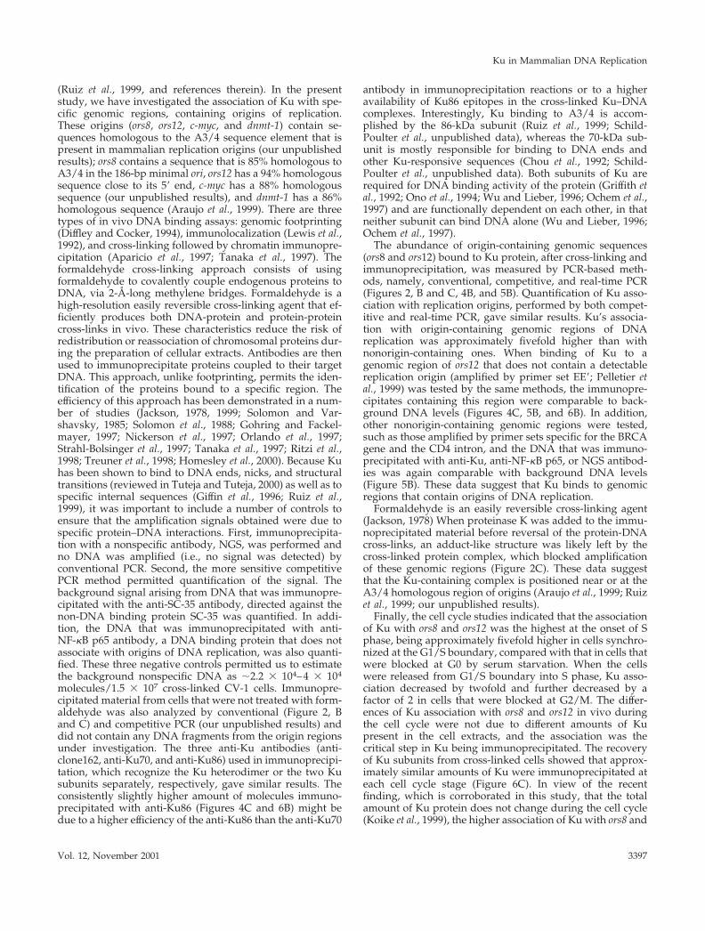
PCR Analysis of Immunoprecipitated DNAConventional PCR reactions were carried out in 25 �l with 1/200thof the immunoprecipitated material with the use of the Ready-To-Go PCR Beads from Amersham Pharmacia Biotech. The PCRreaction contained 1 �M of each primer (for primers sets AF, AC,DF, BE, and ADA A), which were designed as 20- or 24-mers with�50% GC content. Sequence for the CV-1 ors8 (accession no.M26221) and mouse genomic adenosine deaminase (ADA; acces-sion no. L20424) amplicon primers used:
Genomic CV-1, Ku80�/�, or Ku80�/� DNA (10 ng), used for thecontrol reactions, was obtained from total cell lysates of noncross-linked cells. Typically, an initial denaturation for 2 min at 94°C wasfollowed by 30 cycles with denaturation for 30 s at 94°C, annealingfor 30 s at 55 or 50°C, polymerization for 30 s at 72°C, and a finalextension for 5 min at 72°C. PCR products were separated on 2%agarose gels, visualized with ethidium bromide, and photographedwith an Eagle Eye apparatus (Speed Light/BT Sciencetech-LT1000).
Competitive PCR Analysis of ImmunoprecipitatedDNAThe primers, competitors and PCR conditions used are describedbelow.
Primer SequenceTANNEALING
(°C)
ors8 A 5�GCTGAAGCATTTGCACTTCA3� 55ors8 B 5�TTGCACTTCACTGAGCAGTCAT3�ors8 C 5�CATCTCCACTATAGCCATAT3� 55ors8 D 5�CTACCATGCCTAATGCAAAA3�ors8 E 5�GACCCATAAAGGCAAAAGTACC3� 55ors8 F 5�GCTTTCAGAGACGCCCCTGAAA3�ADA AF 5�CTGAGACTATCCTCCAGGTCTTCT3� 50ADA AR 5�CATGGCTGCCTATGACCAACAGAA3�
Primer Sequence Product SizeTANNEALING
(°C)
ors8 11 5�TTGCACTTCACTGAGCAGTCAT3� Genomic: 320 bp 55ors8 330 5�GACCCATAAAGGCAAAAGTACC3� Competitor: 267 bpB 5�CCCTTGATTAATGGTTGCTT3� Genomic: 463 bp 60B� 5�GCTGGTGGGGAATGTTAATG3� Competitor: 400 bpE 5�GGAATTCTGTCTTAGGCAAT3� Genomic: 250 bp 55E� 5�TGATATTGCCAATCAGGATC3� Competitor: 195 bp
O. Novac et al.
Molecular Biology of the Cell3388

PCR reactions were performed with Ready-To-Go PCR Beads(Amersham Pharmacia Biotech), and primer concentrations were asfor conventional PCR (see above). The difference lies in the additionof the appropriate competitor molecules to the reaction mixture.Each competitor was generated with the use of a third primer, asdescribed by Forster (1994). Ors8 competitor (ors8c) was generatedwith primers ors8 330 and 8C (5�TGAGCAGTCATGAAGAAAC-CTAACTGAGATG). BB� and EE� competitors were generated asdescribed in Pelletier et al. (1999).
Real-time PCR Quantification Analysis ofImmunoprecipitated DNAPCR reactions were carried out in 20 �l with 1/200th of the immu-noprecipitated material with the use of LightCycler capillaries(Roche Molecular Biochemicals) and the LightCycler-FastStart DNAMaster SYBR Green I (Roche Molecular Biochemicals). The PCRreaction contained 3 mM Mg2� and 1 �M of each primer of theappropriate primers set used; ors8 150, ors12 JJ�, ors12 MM�, EE�,BRCA, or CD4 intron. Because the optimal conditions for real-timePCR sometimes requires specific primer sets that differ from thosewith the use of conventional PCR, primer set ors8 150 was used toamplify a 150-bp genomic fragment of ors8 (Figure 3A). Primer setors12 JJ� and ors12 MM� were used to amplify a 360- or a 303-bpcorresponding genomic fragment of ors12 (Figure 3B). Primer setEE� was used to amplify a 250-bp genomic fragment, which wasmapped �5 kbp downstream of the origin of replication ors12(Figure 3B). A control set of primers from the African Green Mon-key BRCA2 gene (accession no. Z75666; Bignell, Micklem, Stratton,Ashworth, and Wooster, unpublished data; Pelletier et al., 1999) andthe CV-1 CD4 gene (accession no. AB052204; Matsunaga et al., 2000)were also used. Primer sets BRCA and CD4 intron amplify a frag-ment of 459 and 258 bp, respectively, from genomic CV-1 DNA.Primers were designed as 20–22 mers with �50% GC content.Sequence for the primers used was as follows:
Genomic CV-1 DNA (9.3, 18.6, 27.9, 37.2, and 55.8 ng), used forthe standard curve reactions (necessary for quantification of thePCR products) (Figure 5A), was obtained from total cell lysates ofnoncross-linked logarithmic 80% confluent cells. The quantificationof the PCR products was assessed by the LightCycler (Roche Mo-lecular Biochemicals), with the use of SYBR Green I dye as detectionformat (Pfitzner et al., 2000). The quantification program used asingle fluorescence reading at the end of each elongation step.Arithmetic background subtraction was used and the fluorescencechannel was set to F1. Typically, an initial denaturation for 10 minat 95°C was followed by 35 cycles with denaturation for 15 s at 95°C,
annealing for 10 s at 45°C (primer set ors8 150) or 50°C (primer setsors12 JJ�, ors12 MM�, EE�, BRCA, or CD4 intron), and polymerizationfor 15 s at 72°C. The specificity of the amplified PCR products wasassessed by performing a melting curve analysis cycle with a firstsegment set at 95°C for 0 s and a temperature transition of 20°C/s,a second segment set at 45°C or 50°C (depending on the annealingtemperature of primer set used) with a temperature transition rateof 20°C/s, and a third segment set at 95°C with a temperaturetransition rate set at 0.2°C/s. PCR products were also separated on2% agarose gels, visualized with ethidium bromide, and photo-graphed with an Eagle Eye apparatus (Speed Light/BT Sciencetech-LT1000) (our unpublished results).
In Vitro Mammalian DNA Replication AssayKu80�/� and Ku80�/� MEFs nuclear and cytosolic extracts wereprepared as previously described (Pearson et al., 1991), from loga-rithmically growing cell monolayers. The protein concentrations ofthe nuclear and cytoplasmic extracts were 3.0 and 3.5 mg/ml,respectively. In vitro replication was performed as previously de-scribed (Matheos et al., 1998), with slight modifications. Standardreactions included cytoplasmic (52.5 �g) and nuclear (21.0 �g) ex-tracts from either Ku80�/� or Ku80�/� cells, 2 mM ATP, 100 mMeach CTP, GTP, UTP, dATP, and dGTP, 10 �Ci each of [�-32P]dCTPand [�-32P]dTTP, 2 U of pyruvate kinase, and 200 ng of input p186plasmid (Todd et al., 1995). A control reaction with pBR322, aplasmid lacking a mammalian origin of DNA replication, was alsoincluded to show origin-dependent DNA replication of the p186plasmid. The reactions were performed at 30°C for 1 h. The reactionproducts were purified with the use of the QIAquick PCR purifica-tion kit (QIAGEN). Samples were digested with 0.8 U of DpnI (NewEngland Biolabs) for 45 min at 37°C in the presence of 1� NEB 4buffer and 100 mM NaCl. The samples were separated on 1%agarose gel in 1� TAE buffer (16–20 h, 50–55 V).
Quantification was performed on DpnI-digested products withthe use of a Fuji BAS2000 phosphorimager analyzer. These resultswere typically corrected for the amount of DNA recovered from thein vitro replication assay by quantitative analysis of the ethidiumbromide picture of the gel (not shown). This method of quantifica-tion for DNA recovery was also verified by quantifying and cor-recting for the amount of radionucleotide incorporated in unmeth-ylated pBluescript DNA, included in each reaction (not shown). Theamount of radioactive precursor incorporated into the DNA wasexpressed as a percentage of the wild-type reaction with Ku80�/�
cell extracts.
RESULTS
Immunoprecipitation of Ku70, Ku86, SC-35, and NF-�B p65 Proteins from Lysed Cell ExtractsThe Ku heterodimer as well as its Ku70- and Ku86-kDa sub-units were separately immunoprecipitated, with anti-clone162,anti-Ku70, or anti-Ku86 antibodies, respectively, from extractsof monkey (CV-1) cells that had been previously treated or notwith formaldehyde, to cross-link proteins bound to DNA invivo. As negative control, antibodies against the spliceosome-specific protein, SC-35, a nuclear protein that does not bind toDNA (Fu and Maniatis, 1990), or the transcription factor NF-�Bp65, a nuclear protein that binds DNA but does not associatewith origins of DNA replication (Meyer et al. 1991), were used.Western blot analyses showed that CV-1 whole-cell-extracts(CV-1 WCE), prepared from either cross-linked or noncross-linked cells, contained all three proteins, Ku, SC-35, and NF-�Bp65 proteins (Figure 1, A–E, lanes 1 and 2). In contrast, whennormal goat serum (NGS) was used, neither Ku, NF-�B p65,nor SC-35 was immunoprecipitated in either the cross-linked
Primer SequenceTANNEALING
(°C)
ors8 150F 5�-GACCCATAAAGGCAAAAGTACC-3� 45ors8 150R 5�-GGAAGATATTAAGATAGATGG-3�ors12 J 5�-CAGACATCAGCAAGTGACGG-3� 50ors12 J� 5�-TAGCCAATCTGCCCAATGTA-3�ors12 M� 5�-CATTCGTTCATCCATGTCTCC-3� 50ors12 M� 5�-GTGAATGAGGCAGTTTGAGGA-3�E 5�-GGAATTCTGTCTTAGGCAAT3� 50E� 5�-TGATATTGCCAATCAGGATC3�BRCA F 5�-GATCACAACTGCCCCAAAGT-3� 50BRCA R 5�-TGTTGTTTTTCGGAGGGATG-3�CD4 intron F 5�-AGCTCTGTTCTGTATCTTTG-3� 50CD4 intron R 5�-CCACAGGCACTTTTATCTTC-3�
Ku in Mammalian DNA Replication
Vol. 12, November 2001 3389

or untreated cells (Figure 1, A–E, lanes 5 and 6). Furthermore,Western blot analyses with the use of anti-Ku70 and anti-Ku86antibodies verified that the immunoprecipitated material fromeither the cross-linked or the untreated cells did contain Kuprotein (Figure 1, A–C, lanes 3 and 4). Western blot analysesperformed with anti-SC-35 antibody showed that the materialimmunoprecipitated from cross-linked cells contained �10times less SC-35 protein than the untreated ones (Figure 1D,lanes 3 and 4), indicating some nonspecific precipitation of thisprotein, whereas similar analyses performed with the anti-NF-�B p65 antibody showed that the material immunoprecipi-tated from either the cross-linked or untreated cells (Figure 1E,lanes 3 and 4) contained equivalent amount of NF-�B p65protein. The specificity of the anti-Ku70 and anti-Ku86 antibod-ies used was assayed by blocking with the corresponding Ku70and Ku86 peptides. Western blot analyses showed that neitherthe Ku70 nor the Ku86 subunits of Ku protein were immuno-precipitated from cross-linked CV-1 cells, when the Ku anti-bodies were treated with the respective blocking peptide be-fore immunoprecipitation (Figure 1, A and B, lane 7).
Ku70 and Ku86 Association with ors 8 and ors 12Detected by Formaldehyde Cross-linkingThe abundance of ors8- and ors12-containing genomic se-quences bound to Ku protein, after formaldehyde cross-linking and immunoprecipitation, was measured by PCR.Four sets of primers, AC, DF, BE, and AF, were used toamplify four respective regions in ors8 (Figure 2A). Whenthe immunoprecipitated protein-DNA cross-links werereversed, by incubating at 65°C overnight, all four regionsof ors8 were amplified, giving the expected 197-, 212-,320-, and 480-bp products, respectively (Figure 2B, lanes 1and 3). In contrast, the immunoprecipitated material fromthe noncross-linked cells did not result in PCR amplifica-tion of any of the four ors8 fragments (Figure 2B, lanes 2and 4), indicating first that cross-linking was requiredbefore immunoprecipitation with anti-Ku antibodies, andsecond that the material that was immunoprecipitatedwith these antibodies from the noncross-linked cells didnot contain detectable amounts of contaminating DNA,with the use of either the same amount of template asfrom the cross-linked cells, or 10 times more (our unpub-lished results) for the PCR reaction. The genomic DNA ofnoncross-linked cells gave the expected amplificationproducts with the corresponding primer sets (Figure 2, Band C, lane 5). A similar procedure was used with themonkey ors12 (Kaufmann et al., 1985), human c-myc(Vassilev and Johnson, 1990) and human dnmt1 (Araujo etal., 1999) origins of replication and specific origin-contain-ing fragments from each locus were similarly amplified(our unpublished results).
PCR Amplification across Ku Binding Site in ors8 IsBlocked If Cross-link Is not Reversed before PCRWhen proteinase K was added without reversing the pro-tein-DNA formaldehyde cross-link, the 480- and 320-bpors8 fragments were not amplified, whereas the 197- and212-bp fragments were (Figure 2C, lanes 1– 4). The regioncontaining the OBA/Ku binding site of ors8 (Ruiz et al.,1999), located within the 186-bp minimal ori, was not partof the amplified fragments when primer sets AC (197-bp
Figure 1. Immunoprecipitation assay showing that Ku, SC-35, andNF-�B p65 are present in both formaldehyde cross-linked or untreatedcells. Western blots (as described in EXPERIMENTAL PROCEDURES)were probed with 1/100th dilution of anti-Ku 86 (A), 1/400th dilution ofanti-Ku 70 (B), 1/100th dilution of anti-Ku 86 plus 1/400th dilution ofanti-Ku 70 (C), 1/1000th dilution of anti-SC-35 (D), and 1/100th dilution ofanti-NF-�B p65 (E). Lanes 1 and 2, 50 �g of cross-linked or not CV-1 WCE;lanes 3 and 4, 1/20th of immunoprecipitated Ku86, Ku70, SC-35, or NF-�Bp65 material from cross-linked or untreated cells; and lanes 5 and 6, 1/20thof immunoprecipitated normal goat serum material from cross-linked oruntreated cells. Lane 7, 1/20th of immunoprecipitated Ku86 or Ku70material from cross-linked cells that was obtained after the pretreatment ofthe anti-Ku70 and anti-Ku86 antibodies with the corresponding blockingpeptide.
O. Novac et al.
Molecular Biology of the Cell3390

product) and DF (212-bp) were used (Figure 2A). Mostlikely, as a result of the cross-linking, an adduct-likestructure may have been left within the Ku binding regionthat inhibited the amplification of the 480- and 320-bpfragments. Amplification of these fragments containingKu binding sites was possible with total genomic DNAfrom noncross-linked cells (Figure 2, B and C).
Competitive PCR with DNA Immunoprecipitatedwith Anti-Ku70, Anti-Ku86, Anti-SC-35, and NGS
To analyze whether the DNA that was immunoprecipitatedwith the anti-Ku antibodies after cross-linking with formal-dehyde was enriched in origin-containing sequences, and toquantify this association, competitive PCR was performed,
Figure 2. Ku association with ors8 and ors12 origins shown by PCR amplification. (A) Map of ors8, including location of primers A–F andtheir amplification products. The box represents the 186-bp minimal origin of ors8 and the hatched portion of the box represents the 59-bpKu binding site that contains the A3/4 sequence homologous sequence. (B) PCR amplification products 197, 212, 320, or 480 bp of ors8 withthe use of primer sets AC, DF, BE, or AF, respectively. Template DNA used the following: lanes 1 and 2, reversed cross-linked or notcross-linked Ku86 immunoprecipitate; lanes 3 and 4, reversed cross-linked or not cross-linked Ku70 immunoprecipitate; lane 5, CV-1 totalgenomic DNA from untreated cells; and lane 6, negative control to verify primer contamination; no template DNA added to PCR reaction.(C) As for B, but cross-links were not reversed.
Ku in Mammalian DNA Replication
Vol. 12, November 2001 3391

with the use of specific primers of ors8 and ors12. This wascompared with DNA obtained by immunoprecipitationwith an anti-SC-35 antibody or NGS, both used as negativecontrols (see competitive PCR raw data, Figure 4B). Com-petitive PCR was also used to standardize the differencesamong primers and competitors with respect to their ampli-fication efficiencies. CV-1 genomic DNA, obtained from dif-ferent regions that are either containing replication originsor not, was used to normalize the reaction products (Figure3, A and B). The linearity of each competitor was verified byplotting the ratio of competitor DNA product to target DNAproduct (ordinate) versus the number of competitor mole-cules used (abscissa) (Figure 4A). In logarithmically growingCV-1 cells, the immunoprecipitated DNA obtained with ei-ther anti-Ku86 or anti-Ku70 antibodies was enriched in ors8sequence by approximately fivefold, in comparison withanti-SC-35–immunoprecipitated DNA (Figure 4C, ors8c).Similar results were obtained with ors12 sequence, whereDNA that was immunoprecipitated with either anti-Ku86 oranti-Ku70 antibodies was enriched in origin sequence byapproximately six- and fivefold, respectively, in comparisonwith anti-SC-35–immunoprecipitated DNA (Figure 4C, ors12BB�). When NGS was used, ors8 and ors12 sequences wereamplified by approximately eight- and sixfold less, respec-tively, than when DNA was immunoprecipitated with theuse of anti-Ku86 and anti-Ku70 (Figure 4C). In contrast, asequence situated �5 kb downstream of ors12 was amplifiedby primer set EE� by approximately fourfold less than thesequence amplified by primer set BB�, which contains ors12when anti-Ku70 or anti-Ku80 antibodies were used for theimmunoprecipitation (Figure 4C, ors12 EE�). In addition, theDNA abundance in the region amplified by primer set EE�,corresponded to �3.0 � 104–4.5 � 104 molecules, when the
immunoprecipitation was performed with either Ku anti-bodies, anti-SC-35 antibody, or NGS (Figure 4C, ors12 EE�).
Real-time PCR with DNA Immunoprecipitated withAnti-Clone162, anti-NF-�B p65, and NGSThe association of Ku heterodimer (immunoprecipitatedwith anti-clone162 antibody), NF-�B p65, and NGS withorigin-containing sequences ors8 and ors12 and nonorigin-containing sequences EE�, BRCA, and CD4 intron was as-sayed by the real-time PCR quantification method with theuse of the LightCycler (Roche Molecular Biochemicals).Genomic CV-1 DNA was used to build the standard curvesnecessary for the quantification of the immunoprecipitatedDNA in different genomic regions (Figure 5A). In agreementwith the results obtained with the use of the competitivePCR quantification methodology (see above), the associationof Ku with ors8 and ors12 in logarithmically growing CV-1cross-linked cells was approximately 3- and 4-fold higher,respectively, than that of NF-�B p65, and 3.5-fold and 5-foldhigher, respectively, than NGS (Figure 5B, ors8 150, ors12 JJ�,ors12 MM). In comparison, the association of Ku with threegenomic regions that do not contain an origin of DNAreplication, EE�, BRCA, and CD4 was lower: the regionamplified by primer sets EE� was �3.5-fold lower and thoseamplified by primer sets BRCA and CD4 intron were �5-fold lower, respectively, than with the ors8 and ors12 origin-containing regions (Figure 5B). Finally, the amount of DNAimmunoprecipitated with anti-NF-�B p65 and NGS in ori-gin-containing regions was similar to that in nonorigin-containing regions (Figure 5B).
Cell Cycle-dependent Association of Ku withOrigins of ReplicationCompetitive PCR was also used to quantitatively assesswhether Ku associated with replication origins as a functionof the cell cycle. CV-1 cells were synchronized to G0, G1/S,S, and M phase (see EXPERIMENTAL PROCEDURES) andsynchronization was monitored by fluorescence-activatedcell sorting analysis (Figure 6A).
The association of Ku with ors8 and ors12 was thehighest at the G1/S boundary, decreased by approxi-mately twofold at the start of S phase, remained low atG2/M, by decreasing approximately another twofold, andreached background levels in serum-starved G0 cells (Fig-ure 6B). Background was considered to be the DNA thatwas brought down nonspecifically by anti-SC35 antibody(estimated as �2.2 � 104– 4 � 104 molecules/1.5 � 107
cross-linked CV-1 cells), presumably as a result of thecross-linking with SC-35, a protein that does not bind toDNA (Figure 1D). If the association of Ku with ors8 andors12 was set at 100% (Figure 4C), the background wasdetermined to be 15% (Figure 6B).
The amount of Ku present in the different phases of thecell cycle was also analyzed, by Western blotting analysesand no significant differences were found (our unpub-lished results), in agreement with previous observationsby Koike et al. (1999). Similarly, Western blot analysesshowed that approximately similar amounts of Ku70 andKu86 immunoprecipitated at each cell cycle stage, whencells were previously treated with formaldehyde (Figure6C).
Figure 3. Map of ors8 origin and ors12 locus in CV-1 cells. (A) Ors8origin showing location of expected target amplification products ofors8, generated by primer set ors8c and ors8 150 (Figure 2A, legend)The black box represents the 186-bp minimal origin of ors8. (B)Ors12 locus showing location of expected target amplification prod-ucts of ors12, generated by primer set BB�, JJ�, MM�, or EE�. Primerset EE� amplifies a fragment located 5 kb from ors12. The black boxrepresents the 215-bp minimal origin of ors12.
O. Novac et al.
Molecular Biology of the Cell3392

Replication Activity and Ku Immunoprecipitationfrom Ku80�/� and Ku80�/� MEF CellsWestern blot analyses of Ku80�/� wild-type MEF cells, with theuse of anti-Ku70 and anti-Ku86 antibodies, showed that bothsubunits of the Ku protein were immunoprecipitated with anti-clone162 antibody from logarithmically growing cells that wereeither cross-linked or not (Figure 7A, lanes 1 and 2). In contrast,neither subunit of Ku was detected when immunoprecipitation ofeither cross-linked or untreated Ku80�/� cells was performed
with either clone162 antibody (Figure 7A, lanes 3 and 4) or anti-Ku86 (our unpublished results).
The abundance of origin-containing genomic sequencebound to Ku protein, after formaldehyde cross-linking andimmunoprecipitation, was measured by conventional PCR(35 cycles). Primer set ADA A, which amplifies a 230-bpfragment of the adenosine deaminase amplicon (ADA)(Valerie et al., 1993) was used to verify that genomic DNAfrom both noncross-linked Ku80�/� and Ku80�/� cells
Figure 4. Competitive PCR with logarithmically growing CV-1 cells shows that Ku associates with origins of replication. (A) CompetitivePCR (competitor ors8c, BB�, or EE�) with increasing number of competitor molecules (from left to right) and constant number of CV-1 genomicDNA template molecules, showing linearity of competitors used. (B) Raw competitive PCR data (with the use of increasing amounts ofcompetitor ors8c, BB�, or EE�) and constant amount of template DNA (1/50th of DNA recovered from immunoprecipitate), purified fromKu86, Ku70, SC-35, or NGS cross-linked immunoprecipitates. (C) Normalized total cross-linked DNA molecules detected by competitivePCR, from logarithmically growing CV-1 cells. Products were amplified with primer sets ors8c, BB�, or EE�, respectively. The quantificationresults are the result of at least five competitive PCR reactions with the template genomic DNA isolated from different groups of cross-linkedCV-1 cells. Each bar represents five experiments and 1 SD is indicated.
Ku in Mammalian DNA Replication
Vol. 12, November 2001 3393

gave the expected amplification product (Figure 7B, lanes 1and 2). A PCR reaction with the use of water instead oftemplate DNA was performed to verify that the primerswere free of contaminating DNA (Figure 7B, lane 3).
When the DNA immunoprecipitated by anti-clone162, an-ti-Ku70, or anti-Ku86 antibodies from cross-linked Ku80�/�
cells were used as template DNA for the PCR reaction, theexpected 230-bp fragment was amplified by the ADA A
Figure 5. Quantification of DNA abundance in origin-containing sequences and nonorigin-containing sequences by real-time PCR. (A)Standard curves, with the use of genomic CV-1 DNA as template, used in the quantification of the PCR fragments amplified by the respectiveprimer sets ors8 150, ors12 JJ�, ors12 MM�, EE�, BRCA and CD4 intron. (B) Total normalized cross-linked molecules detected by real-time PCRwith the use of the LightCycler, from logarithmically growing CV-1 cross-linked Ku, NF-�B p65, and NGS immunoprecipitates, with primersets for ors8 150, ors12 JJ�, ors12 MM�, EE�, BRCA, and CD4 intron. Each bar represents three experiments and 1 SD is indicated.
O. Novac et al.
Molecular Biology of the Cell3394

primer set (Figure 7B, lanes 4, 6, and 8), whereas no productwas detected when the DNA immunoprecipitated from theKu80�/� cross-linked cells was used as template (Figure 7B,
lanes 5, 7, and 9). When either the Ku80�/� or Ku80�/� cellswere not treated with formaldehyde before immunoprecipi-tation with anti-clone162, anti-Ku70, and anti-Ku86 antibod-
Figure 6. Cell cycle-dependent association of Ku with ors8 and ors12. (A) Fluorescence-activated cell sorting analysis of DNA contained inlogarithmically growing or synchronized CV-1 cells at G0, G1/S, S, or M phase of the cell cycle. (B) Total normalized cross-linked moleculesdetected by competitive PCR, from cross-linked Ku86 or Ku70 immunoprecipitates, at different points in the cell cycle, with primer sets ors8cor BB�. The thin black horizontal line represents contaminating DNA background calculated from logarithmically SC-35-immunoprecipitatedDNA fragments amplified with ors8c or BB� primers. As for Figure 4, the quantification was obtained from at least five different competitivePCR reactions with the template genomic DNA being from different groups of cross-linked cells. Each bar represents five experiments and1 SD is indicated. (C) Western blot probed with 1/100th dilution of anti-Ku 86, or1/400th dilution of anti-Ku 70. Lanes 1 and 5, 1/20th ofimmunoprecipitated Ku86 or Ku70, from log, G0, G1/S, S, and M phases of the cell cycle.
Ku in Mammalian DNA Replication
Vol. 12, November 2001 3395

ies, no PCR product was amplified by primer set ADA A(our unpublished results), indicating first that cross-linkingwas required before immunoprecipitation with anti-Ku an-tibodies, and second that the material that was immunopre-cipitated with these antibodies from the noncross-linkedcells did not contain detectable amounts of contaminatingDNA for the PCR reaction.
Because Ku has been implicated in mammalian DNAreplication (de Vries et al., 1989; Toth et al., 1993; Araujo et al.,1999; Ruiz et al., 1999; our unpublished results), in vitroDNA replication experiments were performed with the useof extracts prepared from both the Ku80�/� or Ku80�/�
MEFs (Figure 7C) in a mammalian in vitro replication sys-tem (Pearson et al., 1991; Zannis-Hadjopoulos et al., 1994;Diaz-Perez et al., 1996, 1998; Matheos et al., 1998; Jilani et al.,1999; Ruiz et al., 1999). Approximately a 70% decrease in invitro DNA replication was observed when the Ku80�/�
extracts were used, compared with the Ku80�/� extracts.
DISCUSSION
There is increasing evidence suggesting that Ku is involvedin DNA replication, through binding to replication origins
Figure 7. Ku is associated with the ADA-associated origin of the mouse genome inKu80�/� cells, but not in Ku80�/� MEFs.Ku80�/� cell extracts have reduced replica-tion activity. (A) Western blot probed with1/100th dilution of anti-Ku86, or 1/400th di-lution of anti-Ku70. 1/20th of immunopre-cipitation with clone162 from cross-linked oruntreated Ku80�/� or Ku80�/� MEFs. (B)PCR amplification with the use of primer setADA A, which amplifies a genomic 230-bpfragment. Template DNA used was as fol-lows. Lanes 1 and 2, total genomic DNA iso-lated from untreated Ku80�/� or Ku80�/�
cells. Lane 3, negative control to verify primercontamination; no template DNA added toPCR reaction. Lanes 4, 6, and 8, Ku70, Ku86,or clone162 immunoprecipitate fromKu80�/� cells. Lanes 5, 7, and 9, Ku70, Ku86,or clone162 immunoprecipitate fromKu80�/� cells. (C) In vitro DNA replicationassays were performed with Ku80�/� orKu80�/� cells extracts and p186 as the tem-plate DNA. The in vitro replication productswere purified, digested with DpnI, and theDpnI-resistant bands were quantitated withthe use of a phosphorimager. The amount ofradioactive precursor incorporated into theDNA is expressed as a percentage relative tothe Ku80�/� cell extract reaction (100%). Thequantification was obtained from at leastthree different in vitro reactions. Each barrepresents three experiments and 1 SD is in-dicated.
O. Novac et al.
Molecular Biology of the Cell3396
(Ruiz et al., 1999, and references therein). In the presentstudy, we have investigated the association of Ku with spe-cific genomic regions, containing origins of replication.These origins (ors8, ors12, c-myc, and dnmt-1) contain se-quences homologous to the A3/4 sequence element that ispresent in mammalian replication origins (our unpublishedresults); ors8 contains a sequence that is 85% homologous toA3/4 in the 186-bp minimal ori, ors12 has a 94% homologoussequence close to its 5� end, c-myc has a 88% homologoussequence (our unpublished results), and dnmt-1 has a 86%homologous sequence (Araujo et al., 1999). There are threetypes of in vivo DNA binding assays: genomic footprinting(Diffley and Cocker, 1994), immunolocalization (Lewis et al.,1992), and cross-linking followed by chromatin immunopre-cipitation (Aparicio et al., 1997; Tanaka et al., 1997). Theformaldehyde cross-linking approach consists of usingformaldehyde to covalently couple endogenous proteins toDNA, via 2-Å-long methylene bridges. Formaldehyde is ahigh-resolution easily reversible cross-linking agent that ef-ficiently produces both DNA-protein and protein-proteincross-links in vivo. These characteristics reduce the risk ofredistribution or reassociation of chromosomal proteins dur-ing the preparation of cellular extracts. Antibodies are thenused to immunoprecipitate proteins coupled to their targetDNA. This approach, unlike footprinting, permits the iden-tification of the proteins bound to a specific region. Theefficiency of this approach has been demonstrated in a num-ber of studies (Jackson, 1978, 1999; Solomon and Var-shavsky, 1985; Solomon et al., 1988; Gohring and Fackel-mayer, 1997; Nickerson et al., 1997; Orlando et al., 1997;Strahl-Bolsinger et al., 1997; Tanaka et al., 1997; Ritzi et al.,1998; Treuner et al., 1998; Homesley et al., 2000). Because Kuhas been shown to bind to DNA ends, nicks, and structuraltransitions (reviewed in Tuteja and Tuteja, 2000) as well as tospecific internal sequences (Giffin et al., 1996; Ruiz et al.,1999), it was important to include a number of controls toensure that the amplification signals obtained were due tospecific protein–DNA interactions. First, immunoprecipita-tion with a nonspecific antibody, NGS, was performed andno DNA was amplified (i.e., no signal was detected) byconventional PCR. Second, the more sensitive competitivePCR method permitted quantification of the signal. Thebackground signal arising from DNA that was immunopre-cipitated with the anti-SC-35 antibody, directed against thenon-DNA binding protein SC-35 was quantified. In addi-tion, the DNA that was immunoprecipitated with anti-NF-�B p65 antibody, a DNA binding protein that does notassociate with origins of DNA replication, was also quanti-fied. These three negative controls permitted us to estimatethe background nonspecific DNA as �2.2 � 104–4 � 104
molecules/1.5 � 107 cross-linked CV-1 cells. Immunopre-cipitated material from cells that were not treated with form-aldehyde was also analyzed by conventional (Figure 2, Band C) and competitive PCR (our unpublished results) anddid not contain any DNA fragments from the origin regionsunder investigation. The three anti-Ku antibodies (anti-clone162, anti-Ku70, and anti-Ku86) used in immunoprecipi-tation, which recognize the Ku heterodimer or the two Kusubunits separately, respectively, gave similar results. Theconsistently slightly higher amount of molecules immuno-precipitated with anti-Ku86 (Figures 4C and 6B) might bedue to a higher efficiency of the anti-Ku86 than the anti-Ku70
antibody in immunoprecipitation reactions or to a higheravailability of Ku86 epitopes in the cross-linked Ku–DNAcomplexes. Interestingly, Ku binding to A3/4 is accom-plished by the 86-kDa subunit (Ruiz et al., 1999; Schild-Poulter et al., unpublished data), whereas the 70-kDa sub-unit is mostly responsible for binding to DNA ends andother Ku-responsive sequences (Chou et al., 1992; Schild-Poulter et al., unpublished data). Both subunits of Ku arerequired for DNA binding activity of the protein (Griffith etal., 1992; Ono et al., 1994; Wu and Lieber, 1996; Ochem et al.,1997) and are functionally dependent on each other, in thatneither subunit can bind DNA alone (Wu and Lieber, 1996;Ochem et al., 1997).
The abundance of origin-containing genomic sequences(ors8 and ors12) bound to Ku protein, after cross-linking andimmunoprecipitation, was measured by PCR-based meth-ods, namely, conventional, competitive, and real-time PCR(Figures 2, B and C, 4B, and 5B). Quantification of Ku asso-ciation with replication origins, performed by both compet-itive and real-time PCR, gave similar results. Ku’s associa-tion with origin-containing genomic regions of DNAreplication was approximately fivefold higher than withnonorigin-containing ones. When binding of Ku to agenomic region of ors12 that does not contain a detectablereplication origin (amplified by primer set EE�; Pelletier etal., 1999) was tested by the same methods, the immunopre-cipitates containing this region were comparable to back-ground DNA levels (Figures 4C, 5B, and 6B). In addition,other nonorigin-containing genomic regions were tested,such as those amplified by primer sets specific for the BRCAgene and the CD4 intron, and the DNA that was immuno-precipitated with anti-Ku, anti-NF-�B p65, or NGS antibod-ies was again comparable with background DNA levels(Figure 5B). These data suggest that Ku binds to genomicregions that contain origins of DNA replication.
Formaldehyde is an easily reversible cross-linking agent(Jackson, 1978) When proteinase K was added to the immu-noprecipitated material before reversal of the protein-DNAcross-links, an adduct-like structure was likely left by thecross-linked protein complex, which blocked amplificationof these genomic regions (Figure 2C). These data suggestthat the Ku-containing complex is positioned near or at theA3/4 homologous region of origins (Araujo et al., 1999; Ruizet al., 1999; our unpublished results).
Finally, the cell cycle studies indicated that the associationof Ku with ors8 and ors12 was the highest at the onset of Sphase, being approximately fivefold higher in cells synchro-nized at the G1/S boundary, compared with that in cells thatwere blocked at G0 by serum starvation. When the cellswere released from G1/S boundary into S phase, Ku asso-ciation decreased by twofold and further decreased by afactor of 2 in cells that were blocked at G2/M. The differ-ences of Ku association with ors8 and ors12 in vivo duringthe cell cycle were not due to different amounts of Kupresent in the cell extracts, and the association was thecritical step in Ku being immunoprecipitated. The recoveryof Ku subunits from cross-linked cells showed that approx-imately similar amounts of Ku were immunoprecipitated ateach cell cycle stage (Figure 6C). In view of the recentfinding, which is corroborated in this study, that the totalamount of Ku protein does not change during the cell cycle(Koike et al., 1999), the higher association of Ku with ors8 and
Ku in Mammalian DNA Replication
Vol. 12, November 2001 3397

ors12 at the onset of S phase is specific and occurs at a timewhen these origins become activated (Kaufmann et al., 1985).Furthermore, it was also recently reported that the DNA-endbinding activity of Ku remains constant during the cell cycle(Chou and Chou, 1999). Thus, the findings in this studysuggest a role for Ku in the initiation of DNA replication,supporting our previous findings (Ruiz et al., 1995, 1999; ourunpublished results). Its higher association with origins atthe G1/S phase of the cell cycle suggests that Ku acts at thelevel of initiation of replication and dissociates after originfiring.
Ku knockout mice and Ku-deficient cell lines have beenrecently generated (Nussenzweig et al., 1996; Gu et al., 1997).Ku80 knockout mice are viable but they exhibit defectiveV(D)J recombination, which result in the absence of T- andB-lymphocyte maturation (Nussenzweig et al., 1996; Gu etal., 1997). Furthermore, these mice are less than one-half thesize of their heterozygous littermates and exhibit severegrowth retardation (Nussenzweig et al., 1996). Ku80�/�
MEF cells have prolonged doubling times, and the nonpro-liferating cells arrest at G1 phase in early passages, indicat-ing premature senescence (Nussenzweig et al., 1996; Gu etal., 1997). The knockout cells are radiosensitive and fail toresume the cell cycle after radiation-induced checkpoint ar-rest. These phenotypes correlate well with Ku’s involvementin DNA repair, but are also compatible with a possibleparticipation of Ku in DNA replication. Arrington et al.(2000) observed that H2O2-treated Ku80�/� MEFs were un-able to traverse the G2 phase and that this defect was notdue to deficiencies in DNA repair. Instead, they observedimportant differences in the expression of key cell cycleregulatory genes affecting progression through G2, whichsuggests a role for Ku in cell cycle regulated cellular pro-cesses, such as DNA replication.
Because previous reports had implicated the Ku protein inDNA repair and possibly DNA replication based on thephenotypes of the knockout mice and their cells (Nussenz-weig et al., 1996; Gu et al., 1997; Featherstone and Jackson,1999), the in vivo association of Ku with a known mouseorigin-containing (ADA) genomic sequence was examinedin both Ku80�/� and Ku80�/� MEFs, by measuring theDNA abundance in that region when immunoprecipitatingwith anti-Ku antibodies. Only the Ku wild-type (Ku80�/�)MEFs showed specific PCR amplification of that region (Fig-ure 7B). In contrast, in the Ku80 knockout cells, which do notcontain detectable amount of Ku protein, anti-Ku antibodiesdid not immunoprecipitate a detectable amount of the ADAorigin-containing sequence (Figure 7B). Furthermore, ex-tracts from Ku80�/� MEFs, had an �70% decrease in theirreplication activity, compared with the Ku80�/� extracts, ina mammalian in vitro replication system (Figure 7C). Takentogether, these data suggest that the Ku protein plays animportant role in the initiation of mammalian DNA replica-tion, through its binding to Ku-responsive origins.
In S. cerevisiae, a Ku-like protein was shown to be requiredin vitro for the assembly of a complex at a replication origin,suggesting that Ku participates directly in the formation orestablishment of a regulated complex involved in initiationof replication (Shakibai et al., 1996). To date, several humanproteins have been shown to be required for initiation ofDNA replication and a replication–competent multiproteincomplex has also been isolated from human cells, including
proteins such as DNA polymerases � and �, proliferatingcell nuclear antigen, DNA primase, replication protein A,topoisomerases I and II, DNA ligase I, replication factor C,and DNA helicases I and IV (Malkas et al., 1990; Wu et al.,1994; Applegreen et al., 1995; Coll et al., 1996; Tom et al. 1996;Lin et al., 1997; Jiang et al., 1998; Malkas, 1998; Sekowski et al.,1998). Our data indicate that the majority, not all, of mam-malian replication origins are Ku-responsive, i.e., they con-tain an A3/4 homologous element(s) to which Ku bindsspecifically at the onset of S phase.
ACKNOWLEDGMENTS
This research was supported by grants from the CIHR (to M.Z-H.),the Cancer Research Society (to G.B.P.), and REPLICor Inc.
REFERENCES
Aparicio, O.M., Weinstein, D.M., and Bell, S.P. (1997). Componentsand dynamics of DNA replication complexes in S. cerevisiae: redis-tribution of MCM proteins and Cdc45p during s phase. Cell 91,59–69.
Applegreen, N., et al. (1995). Further characterization of the humancell multiprotein DNA replication complex. J. Cell. Biochem. 59,91–107.
Araujo, F.D., Knox, J.D., Ramchandani, S., Pelletier, R., Bigey, P.,Price, G.B., Szyf, M., and Zannis-Hadjopoulos, M. (1999). Identifi-cation of initiation sites for DNA replication in the human dnmt1(DNA methyltransferase) locus. J. Biol. Chem. 274, 9335–9341.
Arrington, E.D., Caldwell, M.C., Kumarvel, T.S., Lohani, A., Johi, A.,Evans, M.K., Chen, H.T., Nussenzweig, A., Holbrook, N.J., andGorospe, M. (2000). Enhanced sensitivity and long-term G2 arrest inhydrogen peroxide-treated Ku80-null cells are unrelated to DNArepair defects. Free Radic. Biol. Med. 29, 1166–1176.
Berezney, R., Dubey, D.D., and Huberman, J.A. (2000). Heterogene-ity of eukaryotic replicons, replicon clusters, and replication foci.Chromosoma 108, 471–484.
Carter, T., Vancurova, I., Sun, I., Lou, W., and Deleon, S. (1990). ADNA activated protein kinase from HeLa cell nuclei. Mol. Cell. Biol.,10, 6460–6471.
Chou, L.F., and Chou, W.G. (1999). DNA-end binding activity of Kuin synchronized cells. Cell Biol. Int. 23, 663–670.
Chou, C.H., Wang, J., Knuth, M.W., and Reeves, W.H. (1992). Roleof a major autoepitope in forming the DNA binding site of the p70(Ku) antigen. J. Exp. Med. 175, 1677–1684.
Coll, J.M., Sekowski, J.W., Hickey, R.J., Schnaper, L., Yue, W., Bro-die, A.M.H., Uitto, L., Syvaoja, J.E., and Malkas, L.H. (1996). Thehuman breast cell DNA synthesome: its purification from tumortissue and cell culture. Oncol. Res. 8, 435–447.
de Vries, E., vanDriel, W., Bergsma, W.G., Arnberg, A.C., and Vander Vliet, P.C. (1989). HeLa nuclear protein recognizing DNA ter-mini translocating on DNA forming a regular DNA-multimericprotein-complex. J. Mol. Biol. 208, 65–78.
Diaz-Perez, M.J., Wainer, I.W., Zannis-Hadjopoulos, M., and Price,G.B. (1996). Application of an in vitro system in the study of che-motherapeutic drug effects on DNA replication. J. Cell Biochem. 61,444–451.
Diaz-Perez, M.J., Zannis-Hadjopoulos, M., Price, G.B., and Wainer,I.W. (1998). Receptor independent effects on DNA replication bysteroids. J. Cell Biochem. 70, 323–329.
Difilippantonio, M.J., Zhu, J., Chen, H.T., Meffre, E., Nussenzweig,M.C., Max, E.E., Ried, T., and Nussenzweig, A. (2000). DNA repair
O. Novac et al.
Molecular Biology of the Cell3398
protein Ku80 suppresses chromosomal aberrations and malignanttransformation. Nature 404, 510–514.
Diffley, J.F., and Cocker, J.H. (1994). Two steps in the assembly ofcomplexes at yeast replication origins in vivo. Cell 78, 303–316.
Dvir, A., Peterson, S.R., Knuth, M., Lu, H., and Dyan, W.S. (1992).Ku autoantigen is the regulatory component of a template-associ-ated protein kinase that phosphorylates RNA polymerase II. Proc.Natl. Acad. Sci. USA 89, 11920–11924.
Frappier, L., and Zannis-Hadjopoulos, M. (1987). Autonomous rep-lication of plasmids bearing monkey DNA origin-enriched se-quences. Proc. Natl. Acad. Sci. USA 84, 6668–6672.
Forster, E. (1994). An improved general method to generate internalstandards for competitive PCR. Biotechniques 16, 18–20.
Fu, X.D., and Maniatis, T. (1990). Factor required for mammalianspliceosome assembly is localized to discrete regions in the nucleus.Nature 343, 437–441.
Galande, S., and Kohwi-Shigematsu, T. (2000). Caught in the act:binding of Ku and PARP to MARs reveals novel aspects of theirfunctional interaction. Crit. Rev. Eukaryot. Gene Expr. 10, 63–72.
Gavin, K.A., Hidaka, M., and Stillman, B. (1995). Conserved initiatorproteins in eukaryotes. Science 270, 1667–1671.
Giffin, W., Torrance, H., Rodda, D.J., Prefontaine, G.G., Pope, L., andHache, R.J.G. (1996). Sequence-specific DNA binding by Ku autoan-tigen and its effects on transcription. Nature 380, 265–268.
Gohring, F., and Fackelmayer, F.O. (1997). The scaffold/matrix at-tachment region binding protein hnPNP-U (SAF-A) is directlybound to chromosomal DNA in vivo: a chemical cross-linkingstudy. Biochemistry 36, 8276–8283.
Gossen, M., Pak, D.T., Hansen, S.K., Acharya, J.K., and Botchan,M.R. (1995). A Drosophila homolog of the yeast origin recognitioncomplex. Science 270, 1674–1677.
Griffith, A.J., Craft, J., Evans, J., Mimori, T., and Hardin, J.A. (1992).Nucleotide sequence and genomic structure analyses of the p70subunit of the human Ku autoantigen: evidence for a family ofgenes encoding Ku p70-related polypeptides. Mol. Biol. Rep. 16,91–97.
Gu, Y., Jin, S., Gao, Y., Weave, D.T., and Ah, F.W. (1997). Ku70deficient embryonic stem cells have increased ionizing radiosensi-tivity, defective DNA-end binding activity, and inability to supportV(D)J recombination. Proc. Natl. Acad. Sci. USA 94, 8076–8081.
Hecht, A., and Grunstein, M. (1999). Mapping DNA interaction sitesof chromosomal proteins using immunoprecipitation and polymer-ase chain reaction. Methods Enzymol. 304, 399–414.
Homesley, L., Lei, M., Kawasaki, Y., Sawyer, S., Christensen, T., andTye, B.K. (2000). MCM10 and the MCM2–7 complex interact toinitiate DNA synthesis and to release replication factors from ori-gins. Genes Dev. 14, 913–926.
Jackson, V. (1978). Studies on histone organization in the nucleo-some using formaldehyde as a reversible cross-linking agent. Cell15, 945–954.
Jackson, V. (1999). Formaldehyde cross-linking studying nucleoso-mal dynamics. Methods Enzymol. 17, 125–139.
Jacob, F., Brenner, S., and Cuzin, F. (1963). On the regulation ofDNA replication in bacteria. Cold Spring Harb. Symp. Quant. Biol.28, 329–348.
Jiang, H., Hickey, R.J., Bechtel, P.E., Wills, P.W., Han, S., Tom, T.D.,Wei, Y., and Malkas, L.H. (1998). Whole gel eluter purification of afunctional multiprotein replication complex. Bioradiation 102, 2412–2425.
Jilani, A., Slack, C., Matheos, D., Zannis-Hadjopoulos, M., andLasko, D.D. (1999). Purification of a polynucleotide kinase from calf
thymus, comparison of its 3�-phosphatase domain with T4 polynu-cleotide kinase, and investigation of its effect on DNA replication invitro. J. Cell. Biochem. 73, 188–203.
Kaufmann, G., Zannis-Hadjopoulos, M., and Martin, R.G. (1985).Cloning of nascent monkey DNA synthesized early in the cell cycle.Mol. Cell. Biol. 5, 721–727.
Koike, M., Ikuta, T., Miyasaka, T., and Shiomi, T. (1999). Ku80 cantranslocate to the nucleus independent of the translocation of Ku70using its own nuclear localization signal. Oncogene 18, 7495–7505.
Landry, S., and Zannis-Hadjopoulos, M. (1991). Classes of autono-mously replicating sequences are found among early-replicatingmonkey DNA. Biochim. Biophys. Acta 1088, 234–244.
Largarkova, M.A., Svetlova, E., Giacca, M., Falaschi, A., and Razin,S.V. (1998). DNA loop anchorage region colocalizes with replicationorigin located downstream to the human gene encoding lamin B2.J. Cell Biochem. 69, 13–18.
Lewis, J.D., Meehan, R.R., Henzel, W.J., Maurer-Fogy, I., Jeppesen,P., Klein, F., and Bird, A. (1992). Purification, sequence and cellularlocalization of a novel chromosomal protein that binds to methyl-ated DNA. Cell 69, 905–914.
Li, G.C., Ouyang, H.H., Li, X.L., Nagasawa, H., Little, J.B., Chen,D.J., Ling, C.C., Fuks, Z., and Corton-Carto, C. (1998). Ku70: acandidate tumor suppressor gene for murine T cell lymphoma. Mol.Cell 2, 1–8.
Liang, C., and Stillman, B. (1997). Persistent initiation of DNAreplication and chromatin-bound MCM proteins during the cellcycle in cdc6 mutants. Genes Dev. 11, 3375–3386.
Lin, S., Hickey, R.J., and Malkas, L.H. (1997). The isolation of a DNAsynthesome from human leukemia cells. Leuk. Res. 6, 501–512.
Lipford, J.R., and Bell, S.P. (2001). Nucleosomes positioned by ORCfacilitate the initiation of DNA replication. Mol. Cell 7, 21–30.
Mah, D.C.W., Dijkwel, P.A., Todd, A., Klein, V., Price, G.B., andZannis-Hadjopoulos, M. (1993). Ors12, a mammalian autonomouslyreplicating DNA sequence, associates with the nuclear matrix in acell cycle-dependent manner. J. Cell Sci. 105, 807–818.
Malkas, L.H. (1998). DNA replication machinery of the mammaliancell. J. Cell. Biochem. 30/31, 18–29.
Malkas, L.H., Hickey, R.J., Li, C.-J., Pedersen, N., and Baril, E.F.(1990). A 21S enzyme complex from HeLa cells that function insimian virus 40 DNA replication in vitro. Biochemistry 29, 6362–6374.
Matheos, D., Ruiz, M.T., Price, G., and Zannis-Hadjopoulos, M.(1998). Oct-1 enhances the in vitro replication of a mammalianautonomously replicating DNA sequence. J. Cell. Biochem. 68, 309–327.
Matsunaga, S., Mukai, R., Inoue-Muraama, M., Yoshikawa, Y., andMurayma, Y. (2000). Sequence and functional properties of Africangreen monkey CD4 silencer. Immunol. Lett. 75, 47–53.
Meyer, R., Hatada, E.N., Hohmann, H., Haiker, M., Bartsch, C.,Rothlisberger, U., Lahm, H., Schlaeger, E.J., Van Loon, A.P.G.M.,and Scheidereit, C. (1991). Cloning of the DNA-binding subunit ofhman nuclear factor �B: the leel of its mRNA is strongly regulatedby phorbol ester or tumor necrosis factor alpha. Proc. Natl. Acad.Sci. USA 88, 966–970.
Mimori, T., Akizuki, M., Yamagata, H., Inada, S., Yoshida, S., andHomma, M. (1981). Characterization of a high molecular weightacidic nuclear protein recognized by autoantibodies in the sera frompatients with polymyositis-scleroderma overlap. J. Clin. Invest. 68,611–620.
Nickerson, J., Krockmalnic, G., Wan, K., and Penman, S. (1997). Thenuclear matrix revealed by eluting chromatin from cross-linkednucleus. Proc. Natl. Acad. Sci. USA 94, 4446–4450.
Ku in Mammalian DNA Replication
Vol. 12, November 2001 3399

Nussenzweig, A., Chen, C., da Costa Soares, V., Sanchez, M., Sokol,K., Nussenzweig, M.C., and Li, G.C. (1996). Requirement for Ku0 ingrowth and immunoglobulin V(D)J recombination. Nature 382,551–555.
Nussenzweig, A., Sokol, K., Burgman, P., Li, L.G., and Li, G.C.(1997). Hypersensitivity of Ku80-deficient cell lines and mice toDNA damage: the effects of ionizing radiation on growth, survival,and development. Proc. Natl. Acad. Sci. USA 94, 13588–13593.
Ochem, A.E., Skopac, D., Costa, M., Rabilloud, T., Vuillard, L.,Simoncsits, A., Giacca, M., and Falaschi, A. (1997). Functional prop-erties of the separate subunits of human DNA helicase II/Ku au-toantigen. J. Biol. Chem. 272, 29919–29926.
Ono, M., Tucker, P.W., and Capr, J.D. (1994). Production and char-acterization of recombinant human Ku antigen. Nucleic Acids Res.,22, 3918–3914.
Orlando, V., Strutt, H., and Paro, R. (1997). Analysis of chromatinstructure by in vivo formaldehyde cross-linking. Methods 11, 205–214.
Paulson, J.R., and Taylor, S.S. (1982). Phosphorylation of histones 1and 3 and nonhistone high mobility group 14 by an endogenouskinase in HeLa metaphase chromosomes. J. Biol. Chem. 257, 6064–6072.
Pearson, C.E., Frappier, L., Price, G.B., and Zannis-Hadjopoulos, M.(1991). Plasmids bearing mammalian DNA-replication origin-en-riched (ors) fragments initiate semi-conservative replication in a cellfree system. Biophys. Biochim. Acta 1090, 156–166.
Pelletier, R., Mah, D., Landry, S., Matheos, D., Price, G.B., andZannis-Hadjopoulos, M. (1997). Deletion analysis of ors12, a centro-meric, early activated, mammalian origin of DNA replication.J. Cell. Biochem. 66, 87–97.
Pelletier, R., Price, G.B., and Zannis-Hadjopoulos, M. (1999). Func-tional genomic mapping of an early-activated centromeric mamma-lian origin of DNA replication. J. Cell. Biochem. 74, 562–575.
Pfitzner, T., Engert, A., Wittor, H., Schinkothe, T., Oberhauser, F.,Schulz, H., Diehl, V., and Barth, S. (2000). A real-time PCR assay forthe quantification of residual malignant cells in B-ell chronic lym-phatic leukemia. Leukemia 14, 754–766.
Pucci, S., Mazzarelli, P., Rabitti, C., Giai, M., Gallucci, M., Flammia,G., Alcini, A., Altomare, V., and Fazio, V.M. (2001). Tumor specificmodulation of Ku70/80 DNA binding activity in breast and bladderhuman tumor biopsies. Oncogene 20, 739–747.
Quintana, D.G., and Dutta, A. (1999). The metazoan origin recogni-tion complex. Front. Biosci. 4, 805–815.
Rao, B.S., Zannis-Hadjopoulos, M., Price, G.B., Reitman, M., andMartin, R.G. (1990). Sequence similarities among monkey ori-en-riched (ors) fragments. Gene 87, 233–242.
Ritzi, M., Baack, M., Musahl, C., Romanowski, P., Laskey, R.A., andKnippers, R. (1998). Human minichromosome maintenance proteinsand human origin recognition complex 2 protein on chromatin.J. Biol. Chem. 273, 24543–24549.
Ritzi, M., and Knippers, R. (2000). Initiation of genome replication:assembly and disassembly of replication-competent chromatin.Gene 245, 13–20.
Ruiz, M.T., Matheos, D., Price, G.B., and Zannis-Hadjopoulos, M.(1999). OBA/Ku86: DNA binding specificity and involvement inmammalian DNA replication. Mol. Biol. Cell 10, 567–580.
Ruiz, M.T., Pearson, C.E., Nielsen, T., Price, G.B., and Zannis-Had-jopoulos, M. (1995). Cofractionation of HeLa cell replication pro-teins with Ors-binding activity. J. Cell. Biochem. 58, 221–236.
Sekowski, J.W., Malkas, L.H., Schnaper, L., Bechtel, P.E., Long, B.J.,and Hickey, R.J. (1998). Human breast cancer cells contain an error-prone DNA replication apparatus. Cancer Res. 58, 3259–3263.
Shakibai, N., Kuma, V., and Eisenberg, S. (1996). The Ku-like proteinfrom Saccharomyces cerevisiae is required in vitro for the assembly ofa stable multiprotein complex at a eukaryotic origin of replication.Proc. Natl. Acad. Sci. USA 93, 11569–11574.
Solomon, M., Larsen, P.L., and Varshavsky, A. (1988). Mappingprotein-DNA interactions in vivo with fomaldehyde: evidence thathistone H4 is retained on a highly transcribed gene. Cell 54, 937–947.
Solomon, M., and Varshavsky, A. (1985). Formaldehyde-mediatedDNA-protein crosslinking: a probe for in vivo chromain structures.Proc. Natl. Acad. Sci. USA 82, 6470–6474.
Stephens, R.F., Pan, C.J., Ajiro, K., Dolby, T.W., and Borun, T.W.(1977). Studies of human histone messenger RNA. I. Methods forthe isolation and partial characterization of RNA fractions contain-ing human histone message from HeLa S3 polyribosomes. J. Biol.Chem. 252, 166–172.
Strahl-Bolsinger, S., Hecht, A., Luo, K., and Grunstein, M. (1997).Sir2 and Sir4 interactions differ in core and extended telomeic het-erochromatin in yeast. Genes Dev. 11, 83–93.
Tao, L., Dong, Z., Leffak, M., Zannis-Hadjopoulos, M., and Price,G.B. (2000). Major DNA replication initiation sites in the c-myc locusin human cells. J. Cell. Biochem. 78, 442–457.
Tao, L., Nielsen, T., Friedlander, P., Zannis-Hadjopoulos, M., andPrice, G.B. (1997). Differential DNA replication origin activities inhuman normal skin fibroblasts and HeLa cell lines. J. Mol. Biol. 273,509–518.
Tanaka, T., Knapp, D., and Nasmyth, K. (1997). Loading of an Mcmprotein onto DNA replication origins is regulated by Cdc6p andCDKs. Cell 90, 649–660.
Todd, A., Landry, S., Pearson, C.E., Khoury, V., and Zannis-Hadjo-poulos, M. (1995). Deletion analysis of minimal sequence require-ments for autonomous replication of ors8, a monkey early-replicat-ing DNA sequence. J. Cell. Biochem. 57, 280–289.
Tom, T., Malkas, L., and Hickey, R. (1996). Identification of multi-protein complexes containing DNA replication factors by nativeimmunoblotting of HeLa cell protein preparations with T-antigendependent SV40 DNA replication activity. J. Cell. Biochem. 63,259–267.
Toth, E.C., Marusic, L., Ochem, A., Patthy, A., Pongor, S., Giacca,M., and Falaschi, A. (1993). Interactions of USF and Ku antigen witha human DNA region containing a replication origin. Nucleic AcidsRes. 21, 3257–3263.
Treuner, K., Eckerich, C., and Knippers, R. (1998). Chromatin asso-ciation of replication protein A. J. Biol. Chem. 273, 31744–31750.
Tuteja, N., Rahman, K., Tuteja, R., and Falashci, A. (1993). HumanDNA helicase V, a novel DNA unwinding enzyme from HeLa cells.Nucleic Acids Res. 21, 2323–2329.
Tuteja, N., et al. (1994). Human DNA helicase II: a novel DNAunwinding enzyme identified as the Ku autoantigen. EMBO J. 13,4991–5001.
Tuteja, N., Tuteja, R., Rahman, K., Kang, L.Y., and Falaschi, A.(1990). A DNA helicase from human cells. Nucleic Acids Res. 18,6785–6792.
Tuteja, R., and Tuteja, N. (2000). Ku autoantigen: a multifunctionalDNA-binding protein. Crit. Rev. Biochem. Mol. Biol. 35, 1–33.
Vassilev, L.T., and Johnson, E.M. (1990). An initiation zone of chro-mosomal DNA replication located upstream of the c-myc gene inproliferating HeLa cells. Mol. Cell. Biol. 10, 4899–4904.
Virta-Pearlman, V.J., Gunaratne, P.H., and Chinalt, A.C. (1993).Analysis of a replication initiation sequence from adenosine deami-nase region of the mouse genome. Mol. Cell. Biol. 13, 5931–5942.
O. Novac et al.
Molecular Biology of the Cell3400

Wu, X.T., and Lieber, M.R. (1996). Protein-protein and protein-DNAinteraction regions within the DNA-end binding protein Ku70/Ku86. Mol. Cell. Biol., 16, 5186–5193.
Zannis-Hadjopoulos, M., Kaufmann, G., Wang, S.S., Lechner, R.L.,Karawya, E., Hesse, J., and Martin, R.G. (1985). Properties of somemonkey DNA sequences obtained by a procedure that enriches forDNA replication origins. Mol. Cell. Biol. 5, 1621–1629.
Zannis-Hadjopoulos, M., Nielsen, T.O., Todd, A., and Price, G.B.(1994). Autonomous replication in vivo and in vitro of clones span-ning the region of the DHFR origin of bidirectional replication ori�.Gene 151, 273–277.
Zannis-Hadjopoulos, M., Pearson, C.E., Bell, D., Mah, D., andPrice, G.B. (1992). Structural and functional characterization ofautonomously replicating mammalian origin-enriched sequences(ors). In: DNA Replication: The Regulatory Mechanisms, ed. P.Hughes, E. Fanning, and M. Kohiyama, Berlin: Springer-Verlag,107–116.
Zannis-Hadjopoulos, M., and Price, G.B. (1998). Regulatory param-eters of DNA replication. Crit. Rev. Eukaryotic Gene Expression 8,81–106.
Zannis-Hadjopoulos, M., and Price, G.B. (1999). Eukaryotic DNAreplication. J. Cell. Biochem. 32/33, 1–14.
Ku in Mammalian DNA Replication
Vol. 12, November 2001 3401