In Vitro Evolution of Allergy Vaccine Candidates, with Maintained Structure, but Reduced B Cell and...
12
In Vitro Evolution of Allergy Vaccine Candidates, with Maintained Structure, but Reduced B Cell and T Cell Activation Capacity Ola B. Nilsson 1 , Justus Adedoyin 1 , Claudio Rhyner 2 , Theresa Neimert-Andersson 1 , Jeanette Grundstro ¨m 1 , Kurt D. Berndt 3 , Reto Crameri 2 , Hans Gro ¨ nlund 4 * 1 Department of Medicine, Karolinska Institutet, Stockholm, Sweden, 2 Swiss Institute of Allergy and Asthma Research (SIAF), University of Zu ¨ rich, Davos, Switzerland, 3 Department Biosciences and Nutrition, Karolinska Institutet, Stockholm, Sweden, 4 Center for Allergy Research, Karolinska Institutet, Stockholm, Sweden Abstract Allergy and asthma to cat (Felis domesticus) affects about 10% of the population in affluent countries. Immediate allergic symptoms are primarily mediated via IgE antibodies binding to B cell epitopes, whereas late phase inflammatory reactions are mediated via activated T cell recognition of allergen-specific T cell epitopes. Allergen-specific immunotherapy relieves symptoms and is the only treatment inducing a long-lasting protection by induction of protective immune responses. The aim of this study was to produce an allergy vaccine designed with the combined features of attenuated T cell activation, reduced anaphylactic properties, retained molecular integrity and induction of efficient IgE blocking IgG antibodies for safer and efficacious treatment of patients with allergy and asthma to cat. The template gene coding for rFel d 1 was used to introduce random mutations, which was subsequently expressed in large phage libraries. Despite accumulated mutations by up to 7 rounds of iterative error-prone PCR and biopanning, surface topology and structure was essentially maintained using IgE-antibodies from cat allergic patients for phage enrichment. Four candidates were isolated, displaying similar or lower IgE binding, reduced anaphylactic activity as measured by their capacity to induce basophil degranulation and, importantly, a significantly lower T cell reactivity in lymphoproliferative assays compared to the original rFel d 1. In addition, all mutants showed ability to induce blocking antibodies in immunized mice.The approach presented here provides a straightforward procedure to generate a novel type of allergy vaccines for safer and efficacious treatment of allergic patients. Citation: Nilsson OB, Adedoyin J, Rhyner C, Neimert-Andersson T, Grundstro ¨ m J, et al. (2011) In Vitro Evolution of Allergy Vaccine Candidates, with Maintained Structure, but Reduced B Cell and T Cell Activation Capacity. PLoS ONE 6(9): e24558. doi:10.1371/journal.pone.0024558 Editor: Clive M. Gray, University of Cape Town, South Africa Received November 1, 2010; Accepted August 14, 2011; Published September 13, 2011 Copyright: ß 2011 Nilsson et al. This is an open-access article distributed under the terms of the Creative Commons Attribution License, which permits unrestricted use, distribution, and reproduction in any medium, provided the original author and source are credited. Funding: This work was supported in part by The Swedish Asthma and Allergy Association’s Research Foundation, the Consul Th. C. Bergh’s Foundation, the Cancer and Allergy Foundation, the Centre for Allergy Research at Karolinska Institutet and the Bernard Osher Initiative for Research on Severe Asthma. The funders had no role in study design, data collection and analysis, decision to publish, or preparation of the manuscript. Competing Interests: The authors have declared that no competing interests exist. * E-mail: [email protected] Introduction The domestic cat (Felis domesticus) is one of the most frequent pets and approximately 10% of the general population in industrialized countries is sensitized to cat allergens [1]. The symptoms deriving from cat allergy manifest mainly as rhinoconjunctivitis with a strong tendency to progress to asthma, especially in children [2]. Exposure to cat allergen is a result of the natural behavior of the cat to lick and groom. Proteins of the saliva is left to dry and spread as small airborne particles [3] where it can be detected in a variety of indoor environments [4,5]. The major allergen of the cat, formally termed Fel d 1, is a hetero-dimer member of the secretoglobin protein family [6,7] found in skin, lachrymal glands and in particular in the saliva. More than 95% of cat allergic patients show elevated serum IgE levels to Fel d 1 which is, therefore, the primary target for the development of immuno- therapeutic vaccines for the treatment of cat allergy [8,9]. Allergen-specific IgE is the key molecule for the development of allergic symptoms. The synthesis of IgE requires a B cell to undergo class switch recombination in close contact with allergen-specific T helper 2 cells (Th2) [10]. The T cell receptor on primed CD4+ T cells recognize antigen presenting cells carrying MHC II molecules with tightly bound enzyme-digested linear polypeptides of 12 to 25 amino acids derived from the primary structure of the antigen [11]. B cells expressing allergen-specific IgE as a part of the B cell receptor (BCR) recognize the surface structures of the allergens. After allergen binding, the receptor-antigen complex is internalized, the antigen processed, and displayed as peptide-MHC II complex on the B cell surface [12]. These processes link the 3D structure of the allergen to the linear peptides presented to the T cell receptor by the MHC class II complex. Allergen-activated T cells are responsible for late-phase reactions (LPR), occurring 6–8 hours after allergen challenge eliciting symptoms ranging from oedema and itching to eczema, and also play a prominent and pivotal role in the pathogenesis of allergic asthma [13] as shown by the elevated numbers of CD4 + T cells found in bronchial mucosa, BAL fluid, and sputum in these patients. The most common treatment of allergic diseases is either alleviation of symptoms using drugs or advice the patient to avoid PLoS ONE | www.plosone.org 1 September 2011 | Volume 6 | Issue 9 | e24558
Transcript of In Vitro Evolution of Allergy Vaccine Candidates, with Maintained Structure, but Reduced B Cell and...
In Vitro Evolution of Allergy Vaccine Candidates, withMaintained Structure, but Reduced B Cell and T CellActivation CapacityOla B. Nilsson1, Justus Adedoyin1, Claudio Rhyner2, Theresa Neimert-Andersson1, Jeanette
Grundstrom1, Kurt D. Berndt3, Reto Crameri2, Hans Gronlund4*
1 Department of Medicine, Karolinska Institutet, Stockholm, Sweden, 2 Swiss Institute of Allergy and Asthma Research (SIAF), University of Zurich, Davos, Switzerland,
3 Department Biosciences and Nutrition, Karolinska Institutet, Stockholm, Sweden, 4 Center for Allergy Research, Karolinska Institutet, Stockholm, Sweden
Abstract
Allergy and asthma to cat (Felis domesticus) affects about 10% of the population in affluent countries. Immediate allergicsymptoms are primarily mediated via IgE antibodies binding to B cell epitopes, whereas late phase inflammatory reactionsare mediated via activated T cell recognition of allergen-specific T cell epitopes. Allergen-specific immunotherapy relievessymptoms and is the only treatment inducing a long-lasting protection by induction of protective immune responses. Theaim of this study was to produce an allergy vaccine designed with the combined features of attenuated T cell activation,reduced anaphylactic properties, retained molecular integrity and induction of efficient IgE blocking IgG antibodies for saferand efficacious treatment of patients with allergy and asthma to cat. The template gene coding for rFel d 1 was used tointroduce random mutations, which was subsequently expressed in large phage libraries. Despite accumulated mutationsby up to 7 rounds of iterative error-prone PCR and biopanning, surface topology and structure was essentially maintainedusing IgE-antibodies from cat allergic patients for phage enrichment. Four candidates were isolated, displaying similar orlower IgE binding, reduced anaphylactic activity as measured by their capacity to induce basophil degranulation and,importantly, a significantly lower T cell reactivity in lymphoproliferative assays compared to the original rFel d 1. In addition,all mutants showed ability to induce blocking antibodies in immunized mice.The approach presented here provides astraightforward procedure to generate a novel type of allergy vaccines for safer and efficacious treatment of allergicpatients.
Citation: Nilsson OB, Adedoyin J, Rhyner C, Neimert-Andersson T, Grundstrom J, et al. (2011) In Vitro Evolution of Allergy Vaccine Candidates, with MaintainedStructure, but Reduced B Cell and T Cell Activation Capacity. PLoS ONE 6(9): e24558. doi:10.1371/journal.pone.0024558
Editor: Clive M. Gray, University of Cape Town, South Africa
Received November 1, 2010; Accepted August 14, 2011; Published September 13, 2011
Copyright: � 2011 Nilsson et al. This is an open-access article distributed under the terms of the Creative Commons Attribution License, which permitsunrestricted use, distribution, and reproduction in any medium, provided the original author and source are credited.
Funding: This work was supported in part by The Swedish Asthma and Allergy Association’s Research Foundation, the Consul Th. C. Bergh’s Foundation, theCancer and Allergy Foundation, the Centre for Allergy Research at Karolinska Institutet and the Bernard Osher Initiative for Research on Severe Asthma. Thefunders had no role in study design, data collection and analysis, decision to publish, or preparation of the manuscript.
Competing Interests: The authors have declared that no competing interests exist.
* E-mail: [email protected]
Introduction
The domestic cat (Felis domesticus) is one of the most frequent pets
and approximately 10% of the general population in industrialized
countries is sensitized to cat allergens [1]. The symptoms deriving
from cat allergy manifest mainly as rhinoconjunctivitis with a
strong tendency to progress to asthma, especially in children [2].
Exposure to cat allergen is a result of the natural behavior of the
cat to lick and groom. Proteins of the saliva is left to dry and
spread as small airborne particles [3] where it can be detected in a
variety of indoor environments [4,5]. The major allergen of the
cat, formally termed Fel d 1, is a hetero-dimer member of the
secretoglobin protein family [6,7] found in skin, lachrymal glands
and in particular in the saliva. More than 95% of cat allergic
patients show elevated serum IgE levels to Fel d 1 which is,
therefore, the primary target for the development of immuno-
therapeutic vaccines for the treatment of cat allergy [8,9].
Allergen-specific IgE is the key molecule for the development of
allergic symptoms. The synthesis of IgE requires a B cell to undergo
class switch recombination in close contact with allergen-specific T
helper 2 cells (Th2) [10].
The T cell receptor on primed CD4+ T cells recognize antigen
presenting cells carrying MHC II molecules with tightly bound
enzyme-digested linear polypeptides of 12 to 25 amino acids
derived from the primary structure of the antigen [11]. B cells
expressing allergen-specific IgE as a part of the B cell receptor
(BCR) recognize the surface structures of the allergens. After
allergen binding, the receptor-antigen complex is internalized, the
antigen processed, and displayed as peptide-MHC II complex on
the B cell surface [12]. These processes link the 3D structure of the
allergen to the linear peptides presented to the T cell receptor by
the MHC class II complex.
Allergen-activated T cells are responsible for late-phase
reactions (LPR), occurring 6–8 hours after allergen challenge
eliciting symptoms ranging from oedema and itching to eczema,
and also play a prominent and pivotal role in the pathogenesis of
allergic asthma [13] as shown by the elevated numbers of CD4+ T
cells found in bronchial mucosa, BAL fluid, and sputum in these
patients.
The most common treatment of allergic diseases is either
alleviation of symptoms using drugs or advice the patient to avoid
PLoS ONE | www.plosone.org 1 September 2011 | Volume 6 | Issue 9 | e24558
the allergen source [14,15]. However, allergen-specific immuno-
therapy (SIT), is the only treatment able to cure allergic diseases
[16]. Successful SIT is thought to act through tolerance mechanisms
induced by regulatory T cells and blocking IgG antibodies [17].
Induction of IgG antibodies may reduce clinical symptoms in
several ways, by competition with IgE for binding epitopes on the
allergens [18,19], by inhibition of IgE-facilitated antigen presenta-
tion to CD4+ T cells [20], but also by interference with mast cell
degranulation by down-regulation of IgE receptors signaling via
inhibitory motifs on the FccRIIb receptor [21]. Numerous studies
have shown that crude allergen extracts currently used in SIT are
clinically effective [22], a high allergen dose is more effective [23],
although the potential risk of severe acute side effects is a limiting
factor [24]. Attenuated allergenic molecules, i.e. hypoallergens or
synthetic peptide fragments have been used as high dose and safer
alternatives to conventional extract-based SIT [25,26]. However,
such treatments have also been limited by recurring side effects,
such as LPR [27,28].
T cell epitope (TCE) mappings of Fel d 1 have shown a
scattered distribution of TCE located on both chains of Fel d 1
[29]. The relevance of T cells specific for Fel d 1 in cat allergic
patients has been evaluated, where short overlapping peptides
covering the major TCE of Fel d 1 elicited adverse effects, such as
allergic rhinitis and late asthmatic reactions [28,30], despite the
fact that the peptides were unable to bind IgE, and thus elicit
immediate allergic reactions. Interestingly, immunotherapy using
selected allergen-specific Fel d 1 peptides, which induced tolerance
to the allergen in a part of the T cell population, induced also
suppression of T cells specific for epitopes which were not present
in the peptide mixture used for treatment [31]. The use of
recombinant hypoallergens containing the full spectrum of TCE
has been reported in one multicenter clinical study [32]. The study
included hypoallergens with an approximately 1000 fold reduced
IgE bindning capacity, allowing a maintenance dose of 80 ug of
allergen per injection. The large majority of the local and systemic
reactions occurred several hours after injection [27].
Due to the close interactions between allergen, T, B and
basophil or mast cell effector cells, the optimal allergy vaccine
should be depleted of IgE-binding epitopes to evade cross-linking
of high affinity receptors on effector cells and IgE-facilitated
allergen presentation, and show a moderate T cell activation
profile to avoid LPR and bronchial hypersensitivity. Modern
biotechnology allows fast, easy cloning and production of
allergens, allergen isoforms and hypoallergen variants with
reduced IgE binding but conserved T cell reactivity [17,33].
In this study we used a novel strategy to engineer full sized and
folded allergy vaccine candidates with reduced number of T cell
epitopes and reduced risk of inducing anaphylaxis, while
maintaining the immunogenic properties. The design has several
advantages over previously established methods for construction of
hypoallergenic derivatives, which mainly involve disruption of
structural B-cell epitopes, while essentially preserving T-cell
reactivity. Although treatment with hypoallergens may be
efficacious, it also carry the risk of inducing LPR [27,28]. The
rational of the strategy is based on selection of IgE-binding
allergens from randomly mutated phage-displayed libraries of the
major cat allergen Fel d 1, used for proof of concept. Because the
vast majority of the solved protein-antibody complexes show
discontinuous epitopes encompassing more than one surface loop
[34], allergens selected by this technology are expected to
recognize molecules conserving the natural conformation [35].
Therefore it should be possible, by consecutive error-prone PCR
(epPCR) amplification of the primary Fel d 1 gene sequence, to
construct mutated phage display libraries containing proteins with
a maximal number of mutations, still allowing a native like fold of
the allergen. Using this novel procedure, we have selected four
candidates with reduced number of IgE binding epitopes and
reduced T cell stimulating capacity. Recombinant proteins were
produced, purified and evaluated for solubility, purity and native-
like folding. T cell activity was assessed using cultured peripheral
blood mononuclear cells (PBMC) from cat allergic patients,
IgE binding by inhibition ELISA, and allergenicity via basophil
activation test. The ability of the hypo-TCE candidate vaccines to
induce IgE-blocking IgG-antibody responses in vivo was shown by
mouse immunization experiments.
Results
Library construction and panningFollowing epPCR, ligation and transformation, the generated
mutated Fel d 1 phage libraries typically contained 105 individual
clones.
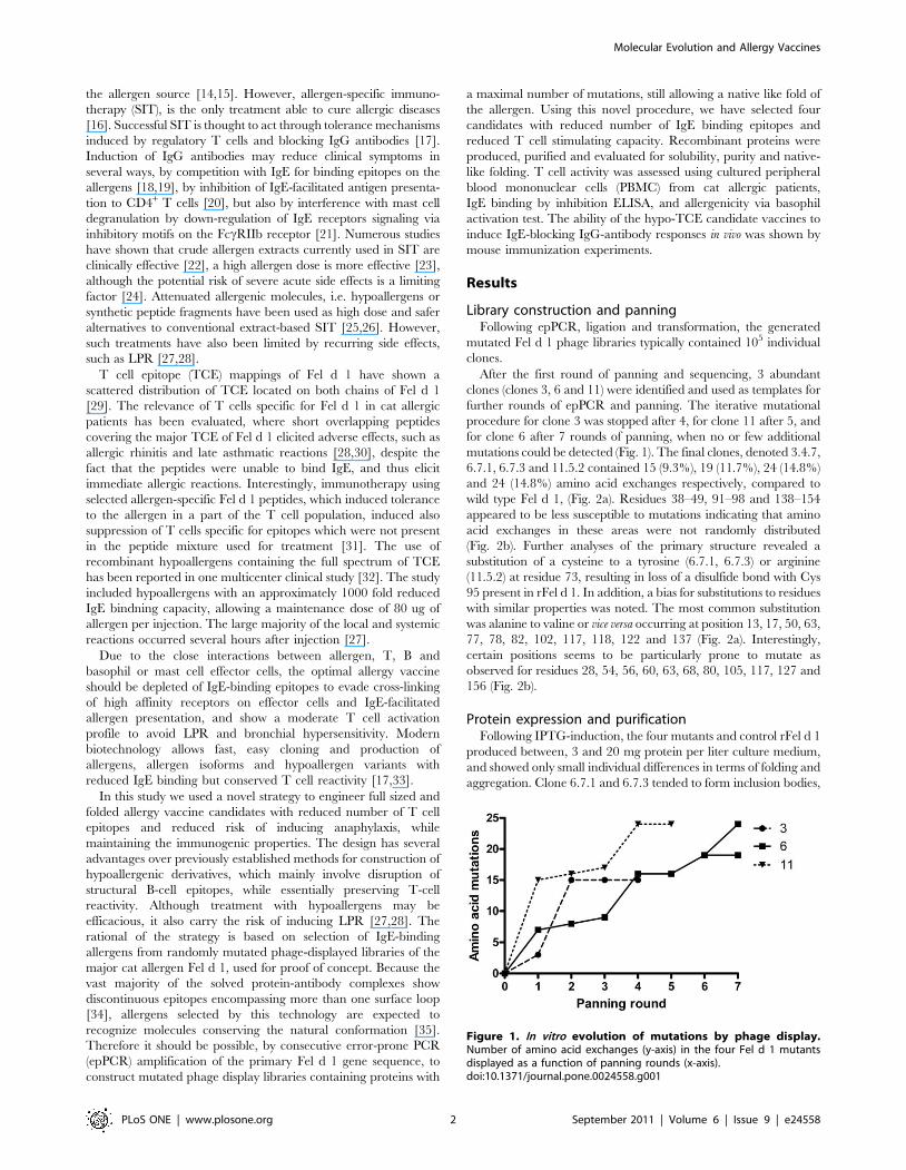
After the first round of panning and sequencing, 3 abundant
clones (clones 3, 6 and 11) were identified and used as templates for
further rounds of epPCR and panning. The iterative mutational
procedure for clone 3 was stopped after 4, for clone 11 after 5, and
for clone 6 after 7 rounds of panning, when no or few additional
mutations could be detected (Fig. 1). The final clones, denoted 3.4.7,
6.7.1, 6.7.3 and 11.5.2 contained 15 (9.3%), 19 (11.7%), 24 (14.8%)
and 24 (14.8%) amino acid exchanges respectively, compared to
wild type Fel d 1, (Fig. 2a). Residues 38–49, 91–98 and 138–154
appeared to be less susceptible to mutations indicating that amino
acid exchanges in these areas were not randomly distributed
(Fig. 2b). Further analyses of the primary structure revealed a
substitution of a cysteine to a tyrosine (6.7.1, 6.7.3) or arginine
(11.5.2) at residue 73, resulting in loss of a disulfide bond with Cys
95 present in rFel d 1. In addition, a bias for substitutions to residues
with similar properties was noted. The most common substitution
was alanine to valine or vice versa occurring at position 13, 17, 50, 63,
77, 78, 82, 102, 117, 118, 122 and 137 (Fig. 2a). Interestingly,
certain positions seems to be particularly prone to mutate as
observed for residues 28, 54, 56, 60, 63, 68, 80, 105, 117, 127 and
156 (Fig. 2b).
Protein expression and purificationFollowing IPTG-induction, the four mutants and control rFel d 1
produced between, 3 and 20 mg protein per liter culture medium,
and showed only small individual differences in terms of folding and
aggregation. Clone 6.7.1 and 6.7.3 tended to form inclusion bodies,
Figure 1. In vitro evolution of mutations by phage display.Number of amino acid exchanges (y-axis) in the four Fel d 1 mutantsdisplayed as a function of panning rounds (x-axis).doi:10.1371/journal.pone.0024558.g001
Molecular Evolution and Allergy Vaccines
PLoS ONE | www.plosone.org 2 September 2011 | Volume 6 | Issue 9 | e24558
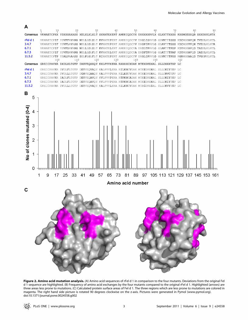
Figure 2. Amino acid mutation analysis. (A) Amino acid sequences of rFel d 1 in comparison to the four mutants. Deviations from the original Feld 1 sequence are highlighted. (B) Frequency of amino acid exchanges by the four mutants compared to the original rFel d 1. Highlighted (arrows) arethree areas less prone to mutations. (C) Calculated protein surface areas of Fel d 1. The three regions which are less prone to mutations are colored inmagenta. The right hand side picture is rotated 90 degrees clockwise on the z-axis. Pictures were generated in Pymol (www.pymol.org).doi:10.1371/journal.pone.0024558.g002
Molecular Evolution and Allergy Vaccines
PLoS ONE | www.plosone.org 3 September 2011 | Volume 6 | Issue 9 | e24558
but were soluble after the refolding process, whereas clone 3.4.7 and
11.5.2 were produced as soluble proteins in the E. coli cytoplasm.
Final purity of the recombinant proteins was estimated to be .95%
by Coomassie-stained SDS PAGE (Fig. S1a). The LPS content of
the purified proteins varied between 15 and 40 ng/mg protein.
Protein characterizationThe behavior of the purified proteins in solution was analyzed by
size exclusion chromatography. As reported previously for rFel d 1
[7], the proteins eluted as symmetrical peaks at approximately
30 kDa with only minor differences in elution volume (Fig. S1b) and
mobility in SDS PAGE (Fig. S1a). Far-UV CD spectra recorded for
rFel d 1 and the 3.4.7, 6.7.3, and 11.5.2 mutants were indicative of
folded proteins with high helical content as evidenced by a double
minima at 208 and 222 nm and a maximum at 190 nm (Fig. 3).
The spectra of the mutants were very similar indicating an overall
similar secondary structure content. Clone 6.7.1, however,
displayed a less intense minimum at 222 nm and more intense
and long wavelength shifted minimum at about 210 nm and a less
intense maximum near 190 nm.
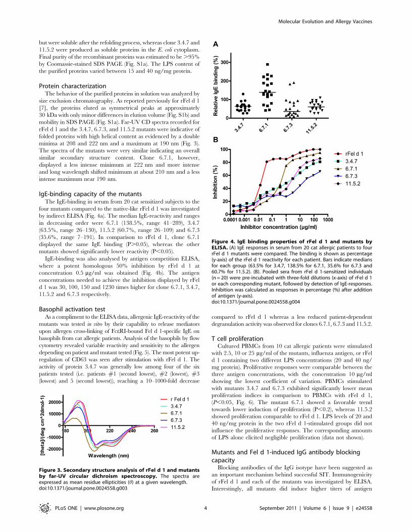
IgE-binding capacity of the mutantsThe IgE-binding in serum from 20 cat sensitized subjects to the
four mutants compared to the native-like rFel d 1 was investigated
by indirect ELISA (Fig. 4a). The median IgE-reactivity and ranges
in decreasing order were 6.7.1 (138.5%, range 41–289), 3.4.7
(63.5%, range 26–130), 11.5.2 (60.7%, range 26–109) and 6.7.3
(35.6%, range 7–191). In comparison to rFel d 1, clone 6.7.1
displayed the same IgE binding (P.0.05), whereas the other
mutants showed significantly lower reactivity (P,0.05).
IgE-binding was also analysed by antigen competition ELISA,
where a potent homologous 50% inhibition by rFel d 1 at
concentration 0.5 mg/ml was obtained (Fig. 4b). The antigen
concentrations needed to achieve the inhibition displayed by rFel
d 1 was 30, 100, 150 and 1230 times higher for clone 6.7.1, 3.4.7,
11.5.2 and 6.7.3 respectively.
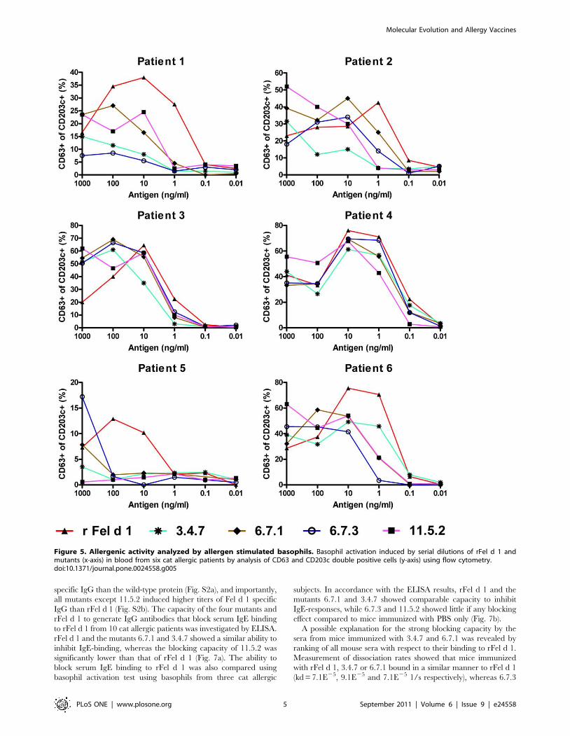
Basophil activation testAs a compliment to the ELISA data, allergenic IgE-reactivity of the
mutants was tested in vitro by their capability to release mediators
upon allergen cross-linking of FceRI-bound Fel d 1-specific IgE on
basophils from cat allergic patients. Analysis of the basophils by flow
cytometry revealed variable reactivity and sensitivity to the allergen
depending on patient and mutant tested (Fig. 5). The most potent up-
regulation of CD63 was seen after stimulation with rFel d 1. The
activity of protein 3.4.7 was generally low among four of the six
patients tested (i.e. patients #1 (second lowest), #2 (lowest), #3
(lowest) and 5 (second lowest)), reaching a 10–1000-fold decrease
compared to rFel d 1 whereas a less reduced patient-dependent
degranulation activity was observed for clones 6.7.1, 6.7.3 and 11.5.2.
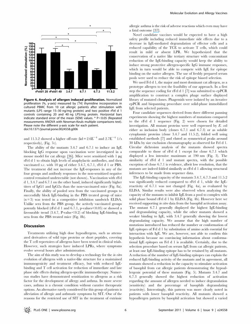
T cell proliferationCultured PBMCs from 10 cat allergic patients were stimulated
with 2.5, 10 or 25 mg/ml of the mutants, influenza antigen, or rFel
d 1 containing two different LPS concentrations (20 and 40 ng/
mg protein). Proliferative responses were comparable between the
three antigen concentrations, with the concentration 10 mg/ml
showing the lowest coefficient of variation. PBMCs stimulated
with mutants 3.4.7 and 6.7.3 exhibited significantly lower mean
proliferation indices in comparison to PBMCs with rFel d 1,
(P,0.05, Fig. 6). The mutant 6.7.1 showed a favorable trend
towards lower induction of proliferation (P,0.2), whereas 11.5.2
showed proliferation comparable to rFel d 1. LPS levels of 20 and
40 ng/mg protein in the two rFel d 1-stimulated groups did not
influence the proliferative responses. The corresponding amounts
of LPS alone elicited negligible proliferation (data not shown).
Mutants and Fel d 1-induced IgG antibody blockingcapacity
Blocking antibodies of the IgG isotype have been suggested as
an important mechanism behind successful SIT. Immunogenicity
of rFel d 1 and each of the mutants was investigated by ELISA.
Interestingly, all mutants did induce higher titers of antigen
Figure 3. Secondary structure analysis of rFel d 1 and mutantsby far-UV circular dichroism spectroscopy. The spectra areexpressed as mean residue ellipticities (h) at a given wavelength.doi:10.1371/journal.pone.0024558.g003
Figure 4. IgE binding properties of rFel d 1 and mutants byELISA. (A) IgE responses in serum from 20 cat allergic patients to fourrFel d 1 mutants were compared. The binding is shown as percentage(y-axis) of the rFel d 1 reactivity for each patient. Bars indicate mediansfor each group (63.5% for 3.4.7, 138.5% for 6.7.1, 35.6% for 6.7.3 and60.7% for 11.5.2). (B). Pooled sera from rFel d 1-sensitized individuals(n = 20) were pre-incubated with three-fold dilutions (x-axis) of rFel d 1or each corresponding mutant, followed by detection of IgE-responses.Inhibition was calculated as responses in percentage (%) after additionof antigen (y-axis).doi:10.1371/journal.pone.0024558.g004
Molecular Evolution and Allergy Vaccines
PLoS ONE | www.plosone.org 4 September 2011 | Volume 6 | Issue 9 | e24558
specific IgG than the wild-type protein (Fig. S2a), and importantly,
all mutants except 11.5.2 induced higher titers of Fel d 1 specific
IgG than rFel d 1 (Fig. S2b). The capacity of the four mutants and
rFel d 1 to generate IgG antibodies that block serum IgE binding
to rFel d 1 from 10 cat allergic patients was investigated by ELISA.
rFel d 1 and the mutants 6.7.1 and 3.4.7 showed a similar ability to
inhibit IgE-binding, whereas the blocking capacity of 11.5.2 was
significantly lower than that of rFel d 1 (Fig. 7a). The ability to
block serum IgE binding to rFel d 1 was also compared using
basophil activation test using basophils from three cat allergic
subjects. In accordance with the ELISA results, rFel d 1 and the
mutants 6.7.1 and 3.4.7 showed comparable capacity to inhibit
IgE-responses, while 6.7.3 and 11.5.2 showed little if any blocking
effect compared to mice immunized with PBS only (Fig. 7b).
A possible explanation for the strong blocking capacity by the
sera from mice immunized with 3.4.7 and 6.7.1 was revealed by
ranking of all mouse sera with respect to their binding to rFel d 1.
Measurement of dissociation rates showed that mice immunized
with rFel d 1, 3.4.7 or 6.7.1 bound in a similar manner to rFel d 1
(kd = 7.1E25, 9.1E25 and 7.1E25 1/s respectively), whereas 6.7.3
Figure 5. Allergenic activity analyzed by allergen stimulated basophils. Basophil activation induced by serial dilutions of rFel d 1 andmutants (x-axis) in blood from six cat allergic patients by analysis of CD63 and CD203c double positive cells (y-axis) using flow cytometry.doi:10.1371/journal.pone.0024558.g005
Molecular Evolution and Allergy Vaccines
PLoS ONE | www.plosone.org 5 September 2011 | Volume 6 | Issue 9 | e24558
and 11.5.2 showed a higher off-rate (kd = 2.6E24 and 2.7E24 1/s
respectively), (Fig. 7c).
The ability of the mutants 3.4.7 and 6.7.1 to induce an IgE
blocking IgG response upon vaccination were investigated in a
mouse model for cat allergy [36]. Mice were sensitized with 1 mg
rFel d 1 to obtain high levels of anaphylactic antibodies, and then
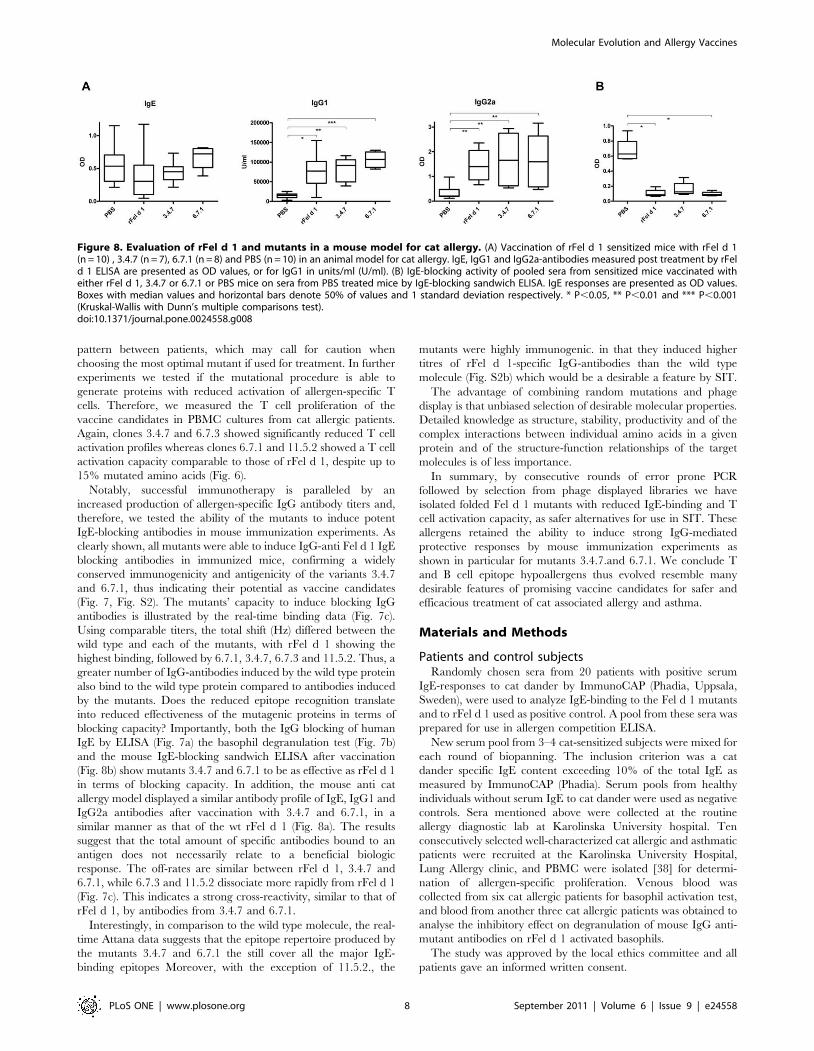
vaccinated s.c. with 10 mg of either 3.4.7, 6.7.1, rFel d 1 or PBS.
The treatment did not increase the IgE-responses in any of the
four groups and antibody responses in the non-sensitized negative
control remained undetectable (not shown).. Vaccination with rFel
d 1, 3.4.7 and 6.7.1 on the other hand, induced significantly higher
titers of IgG1 and IgG2a than the non-vaccinated mice (Fig. 8a).
Finally, the ability of pooled sera from the vaccinated groups to
successfully block IgE-binding in the PBS treated group of mice
(n = 5) was tested in a competitive inhibition sandwich ELISA.
Unlike sera from the PBS group, the actively vaccinated groups
potently blocked (rFel d 1 and 6.7.1, p-values,0.05) or showed a
favourable trend (3.4.7, P-value,0.2) of blocking IgE-binding in
sera from the PBS treated mice (Fig. 8b).
Discussion
Treatments utilizing high dose hypoallergens, such as attenu-
ated derivatives of wild type proteins or short peptides, covering
the T cell repertoires of allergens have been tested in clinical trials.
However, such strategies have induced LPRs, where symptoms
evolve several hours after administration.
The aim of this study was to develop a technology for the in vitro
evolution of allergens with a native-like structure for a maintained
immunogenicity and treatment efficacy, but with reduced IgE-
binding and T cell activation for reduction of immediate and late
phase side effects during allergen-specific immunotherapy. Numer-
ous studies have demonstrated sensitization to allergens as a risk
factor for the development of allergy and asthma. In more severe
cases, asthma is a chronic condition without curative therapeutic
options. An alternative rarely considered for this group of patients is
alleviation of allergic and asthmatic symptoms by SIT. One of the
reasons for the restricted use of SIT in the treatment of extrinsic
allergic asthma is the risk of adverse reactions which even may have
a fatal outcome [37].
Novel candidate vaccines would be expected to have a high
safety profile including reduced immediate side effects due to a
decrease in IgE-mediated degranulation of effector cells and a
reduced capability of the TCE to activate T cells, which could
result in mild or absent LPR. We hypothesized that the
conservation of a native like tertiary structure with concomitant
reduction of the IgE-binding capacity would keep the ability to
induce strong protective allergen-specific IgG immune responses,
which in turn would be able to compete with IgE for epitope
binding on the native allergen. The use of freshly prepared serum
pools were used to reduce the risk of epitope biased selection.
We used Fel d 1, the major and most dominant cat allergen, as a
prototype allergen to test the feasibility of our approach. In a first
step the sequence coding for rFel d 1 [7] was submitted to epPCR
amplification to construct a complex phage surface displayed
library of mutated clones. Phagemids were isolated by an iterative
epPCR and biopanning procedure over solid-phase immobilized
IgE from selected patients.
Four candidate sequences derived from three different panning
experiments showing the highest numbers of mutations compared
to the rFel d 1 sequence (Fig. 2) were chosen for detailed
investigation. All mutant proteins were well expressed by E. coli
either as inclusion body (clones 6.7.1 and 6.7.3) or as soluble
cytoplasmic proteins (clone 3.4.7 and 11.5.2), folded well using
established methods [7] and eluted as symmetrical peaks around
30 kDa by size exclusion chromatography as observed for Fel d 1.
Circular dichroism analysis of the mutants showed spectra
comparable to those of rFel d 1 except for clone 6.7.1 which
displayed a less intensive maximum at 190 nm (Fig. 3). The
similarity of rFel d 1 and mutant spectra, with the possible
exception of clone 6.7.1 is evidence, albeit low resolution, that the
mutants are indeed folded similarily as rFel d 1 allowing structural
inferences to be made from sequence data.
The IgE-binding capacity of the mutants 3.4.7, 6.7.3 and 11.5.2
was significantly reduced compared to rFel d 1, while the IgE-
reactivity of 6.7.1 was not changed (Fig. 4a), as evaluated by
ELISA. Similar results were also observed when analyzing the
capacity of the mutants to inhibit IgE from a pool to complex with
solid phase bound rFel d 1 by ELISA (Fig. 4b). However here we
received supporting in vitro data from the basophil activation assay.
The mutant 6.7.1 generally displayed the highest IgE-binding
and degranulating capacity, while the other mutants showed a
weaker binding to IgE, with 3.4.7 generally showing the lowest
degranulating capacity. We assume that the high number of
mutations introduced has reduced the number or conformation of
IgE epitopes of Fel d 1 by substitution of amino acids essential for
interaction with IgE. We are, however, not able to confirm this
hypothesis because no convincing information about conforma-
tional IgE epitopes on Fel d 1 is available. Certainly, due to the
selection procedure based on serum IgE from cat allergic patients,
at least one IgE-binding epitope has to be retained by all mutants.
A reduction of the number of IgE-binding epitopes can explain the
reduced IgE-binding activity of the mutants and in agreement, all
mutants showed a reduction in the capacity to stimulate activation
of basophil from cat allergic patients demonstrating the hypoal-
lergenic potential of these mutants (Fig. 5). Mutants 3.4.7 and
6.7.3 generally showed the highest reduction of activation
regarding the amount of allergen needed to induce degranulation
(sensitivity) and the percentage of basophils degranulating
(reactivity). Interestingly, this pattern was more clearly noted in
patients with lower basophil reactivity. All mutants showed a
hypoallergen pattern by basophil activation but showed a varied
Figure 6. Analysis of allergen induced proliferation. Normalizedproliferation (%, y-axis) measured by [3H] thymidine incorporation incultured PBMC from 10 cat allergic patients after stimulation withmutants (LPS range 15–30 ng/mg protein) and two positive rFel d 1controls containing 20 and 40 ng LPS/mg protein. Horizontal barsindicate standard error of the mean (SEM) values. * P,0.05 (Repeatedmeasurements ANOVA with Newman-Keuls multiple comparisons test).Please note the different y-axis scale for each patient.doi:10.1371/journal.pone.0024558.g006
Molecular Evolution and Allergy Vaccines
PLoS ONE | www.plosone.org 6 September 2011 | Volume 6 | Issue 9 | e24558
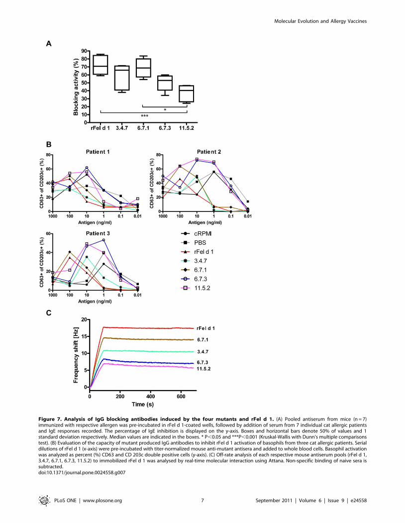
Figure 7. Analysis of IgG blocking antibodies induced by the four mutants and rFel d 1. (A) Pooled antiserum from mice (n = 7)immunized with respective allergen was pre-incubated in rFel d 1-coated wells, followed by addition of serum from 7 individual cat allergic patientsand IgE responses recorded. The percentage of IgE inhibition is displayed on the y-axis. Boxes and horizontal bars denote 50% of values and 1standard deviation respectively. Median values are indicated in the boxes. * P,0.05 and ***P,0.001 (Kruskal-Wallis with Dunn’s multiple comparisonstest). (B) Evaluation of the capacity of mutant produced IgG antibodies to inhibit rFel d 1 activation of basophils from three cat allergic patients. Serialdilutions of rFel d 1 (x-axis) were pre-incubated with titer-normalized mouse anti-mutant antisera and added to whole blood cells. Basophil activationwas analyzed as percent (%) CD63 and CD 203c double positive cells (y-axis). (C) Off-rate analysis of each respective mouse antiserum pools (rFel d 1,3.4.7, 6.7.1, 6.7.3, 11.5.2) to immobilized rFel d 1 was analysed by real-time molecular interaction using Attana. Non-specific binding of naive sera issubtracted.doi:10.1371/journal.pone.0024558.g007
Molecular Evolution and Allergy Vaccines
PLoS ONE | www.plosone.org 7 September 2011 | Volume 6 | Issue 9 | e24558
pattern between patients, which may call for caution when
choosing the most optimal mutant if used for treatment. In further
experiments we tested if the mutational procedure is able to
generate proteins with reduced activation of allergen-specific T
cells. Therefore, we measured the T cell proliferation of the
vaccine candidates in PBMC cultures from cat allergic patients.
Again, clones 3.4.7 and 6.7.3 showed significantly reduced T cell
activation profiles whereas clones 6.7.1 and 11.5.2 showed a T cell
activation capacity comparable to those of rFel d 1, despite up to
15% mutated amino acids (Fig. 6).
Notably, successful immunotherapy is paralleled by an
increased production of allergen-specific IgG antibody titers and,
therefore, we tested the ability of the mutants to induce potent
IgE-blocking antibodies in mouse immunization experiments. As
clearly shown, all mutants were able to induce IgG-anti Fel d 1 IgE
blocking antibodies in immunized mice, confirming a widely
conserved immunogenicity and antigenicity of the variants 3.4.7
and 6.7.1, thus indicating their potential as vaccine candidates
(Fig. 7, Fig. S2). The mutants’ capacity to induce blocking IgG
antibodies is illustrated by the real-time binding data (Fig. 7c).
Using comparable titers, the total shift (Hz) differed between the
wild type and each of the mutants, with rFel d 1 showing the
highest binding, followed by 6.7.1, 3.4.7, 6.7.3 and 11.5.2. Thus, a
greater number of IgG-antibodies induced by the wild type protein
also bind to the wild type protein compared to antibodies induced
by the mutants. Does the reduced epitope recognition translate
into reduced effectiveness of the mutagenic proteins in terms of
blocking capacity? Importantly, both the IgG blocking of human
IgE by ELISA (Fig. 7a) the basophil degranulation test (Fig. 7b)
and the mouse IgE-blocking sandwich ELISA after vaccination
(Fig. 8b) show mutants 3.4.7 and 6.7.1 to be as effective as rFel d 1
in terms of blocking capacity. In addition, the mouse anti cat
allergy model displayed a similar antibody profile of IgE, IgG1 and
IgG2a antibodies after vaccination with 3.4.7 and 6.7.1, in a
similar manner as that of the wt rFel d 1 (Fig. 8a). The results
suggest that the total amount of specific antibodies bound to an
antigen does not necessarily relate to a beneficial biologic
response. The off-rates are similar between rFel d 1, 3.4.7 and
6.7.1, while 6.7.3 and 11.5.2 dissociate more rapidly from rFel d 1
(Fig. 7c). This indicates a strong cross-reactivity, similar to that of
rFel d 1, by antibodies from 3.4.7 and 6.7.1.
Interestingly, in comparison to the wild type molecule, the real-
time Attana data suggests that the epitope repertoire produced by
the mutants 3.4.7 and 6.7.1 the still cover all the major IgE-
binding epitopes Moreover, with the exception of 11.5.2., the
mutants were highly immunogenic. in that they induced higher
titres of rFel d 1-specific IgG-antibodies than the wild type
molecule (Fig. S2b) which would be a desirable a feature by SIT.
The advantage of combining random mutations and phage
display is that unbiased selection of desirable molecular properties.
Detailed knowledge as structure, stability, productivity and of the
complex interactions between individual amino acids in a given
protein and of the structure-function relationships of the target
molecules is of less importance.
In summary, by consecutive rounds of error prone PCR
followed by selection from phage displayed libraries we have
isolated folded Fel d 1 mutants with reduced IgE-binding and T
cell activation capacity, as safer alternatives for use in SIT. These
allergens retained the ability to induce strong IgG-mediated
protective responses by mouse immunization experiments as
shown in particular for mutants 3.4.7.and 6.7.1. We conclude T
and B cell epitope hypoallergens thus evolved resemble many
desirable features of promising vaccine candidates for safer and
efficacious treatment of cat associated allergy and asthma.
Materials and Methods
Patients and control subjectsRandomly chosen sera from 20 patients with positive serum
IgE-responses to cat dander by ImmunoCAP (Phadia, Uppsala,
Sweden), were used to analyze IgE-binding to the Fel d 1 mutants
and to rFel d 1 used as positive control. A pool from these sera was
prepared for use in allergen competition ELISA.
New serum pool from 3–4 cat-sensitized subjects were mixed for
each round of biopanning. The inclusion criterion was a cat
dander specific IgE content exceeding 10% of the total IgE as
measured by ImmunoCAP (Phadia). Serum pools from healthy
individuals without serum IgE to cat dander were used as negative
controls. Sera mentioned above were collected at the routine
allergy diagnostic lab at Karolinska University hospital. Ten
consecutively selected well-characterized cat allergic and asthmatic
patients were recruited at the Karolinska University Hospital,
Lung Allergy clinic, and PBMC were isolated [38] for determi-
nation of allergen-specific proliferation. Venous blood was
collected from six cat allergic patients for basophil activation test,
and blood from another three cat allergic patients was obtained to
analyse the inhibitory effect on degranulation of mouse IgG anti-
mutant antibodies on rFel d 1 activated basophils.
The study was approved by the local ethics committee and all
patients gave an informed written consent.
Figure 8. Evaluation of rFel d 1 and mutants in a mouse model for cat allergy. (A) Vaccination of rFel d 1 sensitized mice with rFel d 1(n = 10) , 3.4.7 (n = 7), 6.7.1 (n = 8) and PBS (n = 10) in an animal model for cat allergy. IgE, IgG1 and IgG2a-antibodies measured post treatment by rFeld 1 ELISA are presented as OD values, or for IgG1 in units/ml (U/ml). (B) IgE-blocking activity of pooled sera from sensitized mice vaccinated witheither rFel d 1, 3.4.7 or 6.7.1 or PBS mice on sera from PBS treated mice by IgE-blocking sandwich ELISA. IgE responses are presented as OD values.Boxes with median values and horizontal bars denote 50% of values and 1 standard deviation respectively. * P,0.05, ** P,0.01 and *** P,0.001(Kruskal-Wallis with Dunn’s multiple comparisons test).doi:10.1371/journal.pone.0024558.g008
Molecular Evolution and Allergy Vaccines
PLoS ONE | www.plosone.org 8 September 2011 | Volume 6 | Issue 9 | e24558
ELISA and inhibition ELISASolid-phase ELISA measurements were carried out essentially
as described [7]. Briefly, antigen or antibodies was coated to
microtiter wells (Nunc, Roskilde, Denmark). Sera from patients,
IgE-myeloma containing 2000 kU/L IgE serving as negative
control and phages were incubated in serial dilution followed by
detection of bound IgE-antibodies [39]. A reference curve was
included in each run for quantification of IgE [40].
Allergen inhibition ELISA was performed as described above,
but with a prior over night incubation at +4uC of serum from a
pool of cat sensitized patients (n = 20) mixed with 3-fold serial
dilutions (range 170 pg/ml–270 mg/ml) of mutants or rFel d 1 as
competing antigen.
Phage display librariesLibraries containing random mutations were created by epPCR
using dNTP-Mutagenesis Kit (Jena Bioscience, Jena, Germany).
The rFel d 1 [7] template used in the first round was amplified
with the rFel d 1-specific primers 59 caccggaatt cgttaaaatg
gctgaaacct gc and 59 cgcgccgctc gaggcacagc gggg, with overhangs
designed for the Eco RI/Xho I cloning sites of phagemid pJuFo
[41]. The purified PCR amplicons were inserted into the rescue
vector pT7-Blue (Novagen Inc., Madison, WI, USA), using pT7
Blue Perfectly Blunt Cloning Kit (Novagen), transformed into
Nova Blue Singles cells (Novagen), vectors isolated (Plasmid
Miniprep Spin Kit, Genomed, Lohne, Germany) and fragments
excised using Eco RI and Xho I restriction sites, subsequently
ligated into pJuFo using T4 ligase (New England Biolabs, Ipswich,
MA, USA).
Phagemids carrying the mutated Fel d 1 coding sequences were
transformed into XL1-Blue MRF’ Electroporation-Competent
Cells (Stratagene, La Jolla, CA, USA) by electroporation (Bio-Rad,
Herts, United Kingdom), immediately added to 3 ml SOC
medium for 1 h at 37uC followed by titration. Transformed cells
were added to 12 ml 26YT medium containing ampicillin
(100 mg/L) and tetracycline (10 mg/L) and incubated for 2 h
(250 rpm, at 37uC) before addition of helper phage (VCSM13,
Stratagene) at a multiplicity of infection (moi) of 10. The medium
was supplemented with kanamycin (25 mg/L) after an additional
2 h and the culture incubated over night. The steps described
above were repeated 4–7 times depending on the clone.
Selection of clonesPhage-infected XL1-Blue cell were centrifuged (J2-21 Beckman
Coulter, Palo Alto, CA) at 50006 g, 15 min, 4uC. The
supernatant was collected and phagemids precipitated with 5%
PEG-8000, 4% NaCl final concentration, incubated on ice for 2 h
followed by centrifugation (186006 g, 15 min, 4uC). Phages were
resuspended in ice cold Tris-buffered saline (TBS) centrifuged
(161006g, 10 min, 4uC) to eliminate cell debris and supernatants
stored at 4uC.
100 ml, 2 mg/ml of monoclonal anti-human IgE (a kind gift from
Phadia, Uppsala, Sweden) was coated to 96-well microtiter plates
(Nunc). 100 ml of a patient serum pool (n = 3) containing Fel d 1-
specific IgE, Fel d 1 negative serum, or TBS as negative control was
added with shaking at 100 rpm for 2 h at 37uC. After washing,
phage library (100 ml, around 105 cfu) was added and incubated
with shaking at 100 rpm for 2 h at 37uC. The supernatant
containing unbound phages was discarded and the wells washed
using TBS with 0,1% Tween with increasing stringency (5, 10 and
15 times), to isolate clones with the highest IgE- binding capacity.
Bound phages were eluted with 0.1 M glycin buffer, pH 2.2,
neutralized with 0.5 M Tris-HCl pH 10.9, and added to 5 ml of
exponentially growing XL1-Blue, optical density (OD) 1.0 at
600 nm. Number of phages before (input) and after biopanning
(output) were calculated by titrations counting single colony forming
units on agar plates. To prepare phage for the next round of
panning ampicillin (100 mg/L) and tetracycline (10 mg/L) were
added to the infected XL1-Blue cells. Helper phage (moi of 10) and
kanamycin (25 mg/L) were added after 2 h and the cultures
incubated over night at 37uC with shaking at 250 rpm.
SequencingThe inserts of twelve randomly picked colonies from the input
and output of each panning round were amplified by colony PCR
with pJuFo sequencing primers 59 ccgaaatcgc gaacctgc and 59
aacgacggcc agtgaattg. The PCR products were purified (PCR
Product Purification Spin Kit/250, Genomed) and used as
templates for sequencing (BigDye Terminator v1.1 Cycle
Sequencing Kit and ABI PRISM 310 Genetic Analyzer, Applied
Biosystems, CA, USA). The repetitive biopanning process was
terminated when no new dominant clones could be amplified, as
confirmed by sequencing of 12 randomly picked clones.
Expression of recombinant allergensFinally selected clones were PCR-amplified using rFel d 1
primers, cleaved by XhoI and NdeI restriction endonucleases and
ligated into the expression vector pET20b (Novagen). The
constructs were transformed into BL21(DE3)pLysS (Novagen),
plated on LB plates containing 100 mg/L ampicillin and 35 mg/L
chloramphenicol and incubated overnight at 37uC. Single colonies
were picked, cultured in 15 ml LB medium containing ampicillin
(100 mg/L) and chloramphenicol (35 mg/L) overnight at 37uCand 250 rpm and used to inoculate two liters of LB medium
containing ampicillin (100 mg/L) and chloramphenicol (35 mg/L)
for protein production. Cells at log phase were induced using
isopropyl thiogalactoside at a final concentration of 0.4 mM, with
simultaneous addition of ampicillin to 100 mg/l final concentra-
tion. After 3 h incubation at 37uC, the cells were collected by
centrifugation (186006g, 10 min), resuspended in 30 ml PBS and
frozen at 220uC.
Protein purificationThawed pellets were adjusted to 6 M guanidine-HCl, sonicated
(Soniprep 150 ultrasonic disintegrator, Sanyo Gallenkamp, Ux-
bridge, UK) by 5 bursts610 sec (10 ma) on ice and centrifuged at
186006 g for 10 min. Buffer was exchanged on Sephadex G-25
equilibrated in 5 mM Tris-HCl containing 0.5 M NaCl, 20 mM
imidazol and 6 M guanidine-HCl, loaded on a 5-ml Ni2+-HiTrap
IMAC column (GE Healthcare, Uppsala, Sweden) followed by
elution with 5 mM Tris-HCl containing 0.5 M NaCl, 0.5 M
imidazol and 6 M guanidine-HCl. The guanidine was removed by
dialysis in 20 mM Tris-HCl, pH 7.4. Proteins were concentrated to
3 ml in a 10 ml Amicon cell (Millipore, Billerica, MA, USA) prior to
size exclusion chromatography on a 16/60 Superdex 200 pg column
equilibrated in PBS pH 7.4 using FPLC system (GE Healthcare).
The peaks corresponding to monomeric proteins (30 kDa) were
diluted 1:1 in 20 mM Tris-HCl pH 8.7, and loaded on 10 ml anion
exchange Q-Sepharose HP columns (GE Healthcare) equilibrated in
20 mM Tris-HCl pH 8.7. Proteins were fractioned with a 15 column
volumes linear gradient using 20 mM Tris-HCl, 1 M NaCl, pH 7.4,
aliquoted and stored at 280uC. Protein concentration was
determined using the BCA protein assay (Thermo scientific, Chicago,
USA) using bovine serum albumin as standard. The purity was
estimated by SDS-PAGE under reducing and non-reducing
conditions, and by analytical SEC using a Superdex 200 32/300
column (GE Healthcare) equilibrated in PBS at a flow rate of 40 ml/
min using AKTA Purifier (GE Healthcare).
Molecular Evolution and Allergy Vaccines
PLoS ONE | www.plosone.org 9 September 2011 | Volume 6 | Issue 9 | e24558
For control of allergen-specific T cell proliferation, two samples of
purified rFel d 1 containing 20 and 40 ng lipopolysaccharide (LPS)/
mg protein, respectively, were used. The LPS content was determined
with the limulus amebocyte lysate endochrome assay according to the
instructions (Charles River Endosafe, Charleston, USA).
Protein characterizationSDS-PAGE was performed under reducing conditions as
described [7]. Analytical SEC was performed using AKTA Purifier
(GE Healthcare) and a Superdex 200 PC 3.2/30 column,
equilibrated in PBS pH 7.4. The secondary structure of the purified
mutants and rFel d 1 was analyzed by far-UV circular dichroism
(CD) spectra measured at 25uC in phosphate buffer (10 mM
NaHPO4, pH 7.4). Data were collected at 1 nm intervals with time
averaging for 16 seconds using a bandwidth of 1.5 nm in a 0.01 cm
quartz cuvette (Hellma, Mullheim, Germany) using an Aviv 202 DS
spectropolarimeter (Aviv Biomedical, Lakewood NJ, USA).
Basophil activation test (BAT)Allergen-specific basophil degranulation was analyzed by
monitoring the basophil activation markers CD203c and CD63
using flow cytometry [42]. Briefly, 10-fold serial dilutions of
antigen (1000 ng/ml to 0.01 ng/ml), medium (negative control)
and anti-IgE (positive control) was added to venous blood samples
from three cat allergic patients and incubated with anti-CD63
and anti-CD203c (Immunotech, Marseille, France). From each
patient, allergen-activated basophils were identified by gating for
CD203c+ cells using FACS Calibur (BD Biosciences, San Jose,
CA, USA). The magnitude of reactivity (y-axis, Fig. 5) was
reported as percentage of CD63+ cells among the gated basophils.
Sensitivity was analyzed at the right hand side of the bell-shaped
BAT curve, where positive reactivity was measured post
stimulation with the wild type protein. Comparison of the rFel d
1 mutants with the wild type protein was analyzed at 50% of
reactivity with rFel d 1 within the sensitive range.
T cell proliferationAllergen-specific proliferation of cultured PBMC from 10 cat
allergic patients was analyzed in triplicates with 26105 cells/well
using 96-well plates [38]. Cells were stimulated with either 2.5, 10
or 25 mg/ml of the Fel d 1 mutants and rFel d 1 containing 20 or
40 ng LPS/mg protein and influenza antigen, (VaxigripH vaccine,
Aventis Pasteur, Lyon, France) 30 ng/ml LPS in cRPMI and
cRPMI alone as controls. Cells were incubated in 5% CO2 for five
days at 37uC when 1 mCi/well [3H] thymidine was added followed
by 18 h of incubation. Counts per minute (cpm) obtained from the
cell cultures were divided by cpm values from unstimulated wells
and expressed as a stimulation index (SI). SI values $2.0 were
considered to be positive. The proliferation varied between
patients (SI range, 5–80) and was normalized for purposes of
comparison. For each patient, a mean proliferation value (MPV,
cpm for tested antigens/number of wells) was calculated. This
value was set to 100%. The stimulation for single antigens is
presented as percentages in relation to the MPV.
Assessment of IgE-blocking mutant induced IgGantibodies
Six to eight week old female BALB/c mice were obtained from
Charles River (Sulzfeld, Germany) and housed with food and
water ad libitum. Animal experiments were approved by the
Swedish local ethics committee for animal welfare.
The ability of the four mutants to generate IgE-blocking IgG
antibodies was tested. Groups of mice (n = 4) were immunized
subcutaneously in the neck with 10 mg of the mutant proteins and
as positive control rFel d 1, each protein adsorbed to 1 mg
aluminium hydroxide (Sigma-Aldrich, Steinheim, Germany).
Injections were given on day 0, 14, 28 and 42. On day 47, mice
were sacrificed and blood collected. Sera from mice immunized
with a distinct protein were pooled and a serum pool from naıve
mice was used as a negative control.
In a first set of experiments titers were normalized between the
five different mouse immune serum pools by 5-fold serial dilutions
from 1/50 to 1/781250 using the respective antigen by ELISA as
described (Fig. S2a) [7].
The ability of mouse IgG-antibodies to block binding of
patient’s serum IgE to allergen was determined by ELISA. Plates
were coated with rFel d 1 and blocked with BSA. The normalized
mouse serum pools were added and incubated over night at 4uC.
The following day, patient serum containing IgE to rFel d 1
diluted to 0.85 kUA/L, and serum from an IgE-myeloma of
2000 kU/L as negative control was added, followed by IgE
detection as described.
To assess the biological relevance of anti- mutant IgG, the
ability to inhibit Fel d 1-mediated degranulation of basophils was
investigated. Briefly, 10-fold serial dilutions of rFel d 1 (1000 ng/
ml to 0.01 ng/ml) or medium was pre-incubated over night with
the four normalized mouse anti-mutant IgG serum pools and the
rFel d 1 pool as control. The mixes of antigen and serum pool
were then added to fresh venous blood from three cat allergic
patients as described above.
For real-time analysis of the different mouse antisera with
respect to binding properties to rFel d 1 an Attana A200 system
(Attana AB, Sweden) was used. rFel d 1 (5 mg/mL in 10 mM
sodium actetate pH 4.5) was immobilized on a LNB sensor chip
by amine coupling according to the manufacturer’s instructions.
All experiments were performed in running buffer, HBS-T
(10 mM HEPES buffered saline with 0.005% Tween 20), at
37uC. Dissociation rate constants for the 5 different antisera were
determined at 25 mL/min using the Attester Evaluation off-rate
screening tool, between time points 150 and 450 seconds. All sera
were normalized as determined by ELISA, and diluted 1:100 in
HBS-T. The surface was regenerated using a 41 s pulse of 10 mM
glycine pH 1.5. Non-specific binding to the activated and
deactivated LNB carboxyl surface was recorded using sera from
mice immunized with PBS.
Vaccination using mutants in a mouse model for catallergy
Animals were obtained and housed as described above. Mice
were sensitized s.c. with 1 mg, rFel d 1 adsorbed to 1 mg alum in
200 ml PBS or PBS only (non-sensitized negative control) on day 0,
14 and 28 to obtain high titers of anaphylactic antibodies. Groups
of mice (n = 7–10) were then vaccinated s.c. with 10 mg of rFel d 1,
3.4.7) or 6.7.1 respectively in 200 ml PBS or 200 ml PBS alone
(control) on day 30, 32 and 34. The mice were sacrificed on day 42
and peripheral blood was collected and stored at 280uCLevels of Fel d 1-specific IgE, IgG1 and IgG2a was analyzed by
ELISA. For analysis of IgE, 96-well plates (Nunc) were coated with
0.2 mg/100 ml rat anti-mouse IgE (BD Biosciences), and IgG1 and
IgG2a plates were coated with 0.5 mg rFel d 1. Sera were diluted
1:20 for IgE measurement, 1:25000, 1:100000 or 1:1000000 for
IgG1 and 1:1000 for IgG2a. Levels of IgG 1 anti rFel d 1 was
analysed using an in-house reference curve and presented in units/
ml, U/ml. Bound IgE levels was measured using 0.2 mg/100 ml
and well of biotinylated rFel d 1 followed by alkaline phosphatase
(AP)-conjugated streptavidine (diluted 1:3000, Sigma-Aldrich).
IgG1 and IgG2a levels were detected using goat anti-mouse AP-
Molecular Evolution and Allergy Vaccines
PLoS ONE | www.plosone.org 10 September 2011 | Volume 6 | Issue 9 | e24558
conjugated IgG1 (1:5000) and IgG2a (1:1000) respectively
(Southern Biotech, Birmingham, AL, USA), followed by OD
measurements at 405 nm.
The IgE blocking capability of the antibodies obtained after
treatment was investigated using a competitive inhibition sandwich
ELISA as described above. Pooled sera from the group of mice
treated with PBS only were selected and added to wells coated
with rat anti-mouse IgE. Fresh serum pools from the respective
groups were prepared, diluted 1:10 and pre-incubated over night
with biotinylated rFel d 1 protein to a final concentration of 2 mg/
ml. The pre-incubated protein mixes were then added to wells and
detection of bound biotinylated rFel d 1 was carried out.
Statistical analysisProliferation data was analysed by ANOVA with Newman-
Keuls multiple comparisons test for pair wise analysis and the
ELISA and mouse study analysed by Kruskal-Wallis with Dunn’s
multiple comparisons test. All statistics were performed using
Graphpad Prism 5.02 software (Graphpad Software Inc., San
Diego, CA, USA). P-values,0.05 were considered significant.
Ethics StatementAll animal research has been approved by the local ethics
committee at Karolinska University Hospital, ‘‘Stockholms norra
djurforsoksetiska namnd’’, with the approval ID [DNR: N245/
08]. All human research has been approved by the local ethics
committee at Karolinska Institutet, ‘‘Regionala etikprovnings-
namnden i Stockholm’’. Written consent was obtained from all
participants involved in the present study.
Supporting Information
Figure S1 Size and purity of rFel d 1 and mutants. (A)
SDS-PAGE of purified rFel d 1 mutants under reducing conditions
with comassie blue staining. Molecular weight markers (lane 1,7),
rFel d 1 (lane 2), 3.4.7 (lane 3), 6.7.1 (lane 4), 6.7.3 (lane 5), 11.5.2
(lane 6). (B) Analytical size exclusion chromatography of rFel d 1
and mutants. The dots denote bovine serum albumin (67 kDa),
ovalbumin (43 kDa), and chymotrypsinogen A (25 kDa) used as
molecular weight markers.
(TIF)
Figure S2 Comparison of immunogenicity of rFel d 1and mutants. Serial dilutions (x-axis) of pooled mouse sera from
mice immunized with either rFel d 1 or each respective mutant.
Pre-diluted sera were added to 96-well microtiter plates coated
with either (a) respective allergen or rFel d 1 only (b), and IgG
responses (y-axis) measured by ELISA.
(TIF)
Acknowledgments
We acknowledge Lise-Lotte Kaiser and Attana AB for assistance with the
real-time antibody binding experiments.
Author Contributions
Conceived and designed the experiments: OBN JA RC HG. Performed the
experiments: OBN JA CR TNA JG KDB. Analyzed the data: OBN JA CR
TNA KDB RC HG JG. Contributed reagents/materials/analysis tools:
OBN JA CR TNA JG KDB RC HG. Wrote the paper: OBN CR RC HG.
References
1. Plaschke P, Janson C, Norrman E, Bjornsson E, Lundback B, et al. (1996) Skin
prick tests and specific IgE in adults from three different areas of Sweden.
Allergy 51: 461–472.
2. Plaschke P, Janson C, Norrman E, Bjornsson E, Ellbjar S, et al. (1999)Association between atopic sensitization and asthma and bronchial hyperre-
sponsiveness in swedish adults: pets, and not mites, are the most importantallergens. J Allergy Clin Immunol 104: 58–65.
3. van Ree R, van Leeuwen WA, Bulder I, Bond J, Aalberse RC (1999) Purifiednatural and recombinant Fel d 1 and cat albumin in in vitro diagnostics for cat
allergy. J Allergy Clin Immunol 104: 1223–1230.
4. Partti-Pellinen K, Marttila O, Makinen-Kiljunen S, Haahtela T (2000)Occurrence of dog, cat, and mite allergens in public transport vehicles. Allergy
55: 65–68.
5. Lau S, Illi S, Platts-Mills TA, Riposo D, Nickel R, et al. (2005) Longitudinal
study on the relationship between cat allergen and endotoxin exposure,sensitization, cat-specific IgG and development of asthma in childhood–report
of the German Multicentre Allergy Study (MAS 90). Allergy 60: 766–773.
6. Kaiser L, Gronlund H, Sandalova T, Ljunggren HG, van Hage-Hamsten M,
et al. (2003) The crystal structure of the major cat allergen Fel d 1, a member ofthe secretoglobin family. J Biol Chem 278: 37730–37735.
7. Gronlund H, Bergman T, Sandstrom K, Alvelius G, Reininger R, et al. (2003)
Formation of disulfide bonds and homodimers of the major cat allergen Fel d 1equivalent to the natural allergen by expression in Escherichia coli. J Biol Chem
278: 40144–40151.
8. Zhu D, Kepley CL, Zhang K, Terada T, Yamada T, et al. (2005) A chimeric
human-cat fusion protein blocks cat-induced allergy. Nat Med 11: 446–449.
9. Schmitz N, Dietmeier K, Bauer M, Maudrich M, Utzinger S, et al. (2009)Displaying Fel d1 on virus-like particles prevents reactogenicity despite greatly
enhanced immunogenicity: a novel therapy for cat allergy. J Exp Med 206:1941–1955.
10. Williams GS, Oxenius A, Hengartner H, Benoist C, Mathis D (1998) CD4+ Tcell responses in mice lacking MHC class II molecules specifically on B cells.
Eur J Immunol 28: 3763–3772.
11. Jardetzky TS, Brown JH, Gorga JC, Stern LJ, Urban RG, et al. (1996)Crystallographic analysis of endogenous peptides associated with HLA-DR1
suggests a common, polyproline II-like conformation for bound peptides. Proc
Natl Acad Sci U S A 93: 734–738.
12. Infuhr D, Crameri R, Lamers R, Achatz G (2005) Molecular and cellular targetsof anti-IgE antibodies. Allergy 60: 977–985.
13. Ali FR, Kay AB, Larche M (2007) Airway hyperresponsiveness and bronchial
mucosal inflammation in T cell peptide-induced asthmatic reactions in atopic
subjects. Thorax 62: 750–757.
14. Morgan WJ, Crain EF, Gruchalla RS, O’Connor GT, Kattan M, et al. (2004)
Results of a home-based environmental intervention among urban children with
asthma. N Engl J Med 351: 1068–1080.
15. Kattan M, Stearns SC, Crain EF, Stout JW, Gergen PJ, et al. (2005) Cost-
effectiveness of a home-based environmental intervention for inner-city children
with asthma. J Allergy Clin Immunol 116: 1058–1063.
16. Crameri R (2007) Allergy vaccines: dreams and reality. Expert Rev Vaccines 6:
991–999.
17. Larche M, Akdis CA, Valenta R (2006) Immunological mechanisms of allergen-
specific immunotherapy. Nat Rev Immunol 6: 761–771.
18. Gehlhar K, Schlaak M, Becker WM, Bufe A (1999) Monitoring allergen
immunotherapy of pollen-allergic patients: the ratio of allergen-specific IgG4 to
IgG1 correlates with clinical outcome. Clin Exp Allergy 29: 497–506.
19. Ball T, Fuchs T, Sperr WR, Valent P, Vangelista L, et al. (1999) B cell epitopes
of the major timothy grass pollen allergen, phl p 1, revealed by gene
fragmentation as candidates for immunotherapy. FASEB J 13: 1277–1290.
20. van Neerven RJ, Wikborg T, Lund G, Jacobsen B, Brinch-Nielsen A, et al.
(1999) Blocking antibodies induced by specific allergy vaccination prevent the
activation of CD4+ T cells by inhibiting serum-IgE-facilitated allergen
presentation. J Immunol 163: 2944–2952.
21. Strait RT, Morris SC, Finkelman FD (2006) IgG-blocking antibodies inhibit
IgE-mediated anaphylaxis in vivo through both antigen interception and Fc
gamma RIIb cross-linking. J Clin Invest 116: 833–841.
22. Bousquet J, Lockey R, Malling HJ (1998) Allergen immunotherapy: therapeutic
vaccines for allergic diseases. A WHO position paper. J Allergy Clin Immunol
102: 558–562.
23. Werfel T, Breuer K, Rueff F, Przybilla B, Worm M, et al. (2006) Usefulness of
specific immunotherapy in patients with atopic dermatitis and allergic
sensitization to house dust mites: a multi-centre, randomized, dose-response
study. Allergy 61: 202–205.
24. Mellerup MT, Hahn GW, Poulsen LK, Malling H (2000) Safety of allergen-
specific immunotherapy. Relation between dosage regimen, allergen extract,
disease and systemic side-effects during induction treatment. Clin Exp Allergy
30: 1423–1429.
25. Valenta R, Niederberger V (2007) Recombinant allergens for immunotherapy.
J Allergy Clin Immunol 119: 826–830.
26. Larche M (2007) Update on the current status of peptide immunotherapy.
J Allergy Clin Immunol 119: 906–909.
27. Purohit A, Niederberger V, Kronqvist M, Horak F, Gronneberg R, et al. (2008)
Clinical effects of immunotherapy with genetically modified recombinant birch
pollen Bet v 1 derivatives. Clin Exp Allergy 38: 1514–1525.
Molecular Evolution and Allergy Vaccines
PLoS ONE | www.plosone.org 11 September 2011 | Volume 6 | Issue 9 | e24558
28. Haselden BM, Kay AB, Larche M (1999) Immunoglobulin E-independent major
histocompatibility complex-restricted T cell peptide epitope-induced lateasthmatic reactions. J Exp Med 189: 1885–1894.
29. Counsell CM, Bond JF, Ohman JL, Jr., Greenstein JL, Garman RD (1996)
Definition of the human T-cell epitopes of Fel d 1, the major allergen of thedomestic cat. J Allergy Clin Immunol 98: 884–894.
30. Oldfield WL, Larche M, Kay AB (2002) Effect of T-cell peptides derived fromFel d 1 on allergic reactions and cytokine production in patients sensitive to cats:
a randomised controlled trial. Lancet 360: 47–53.
31. Campbell JD, Buckland KF, McMillan SJ, Kearley J, Oldfield WL, et al. (2009)Peptide immunotherapy in allergic asthma generates IL-10-dependent immunological
tolerance associated with linked epitope suppression. J Exp Med 206: 1535–1547.32. Niederberger V, Horak F, Vrtala S, Spitzauer S, Krauth MT, et al. (2004)
Vaccination with genetically engineered allergens prevents progression ofallergic disease. Proc Natl Acad Sci U S A 101 Suppl 2: 14677–14682.
33. Engel E, Richter K, Obermeyer G, Briza P, Kungl AJ, et al. (1997)
Immunological and biological properties of Bet v 4, a novel birch pollenallergen with two EF-hand calcium-binding domains. J Biol Chem 272:
28630–28637.34. Niemi M, Jylha S, Laukkanen ML, Soderlund H, Makinen-Kiljunen S, et al.
(2007) Molecular interactions between a recombinant IgE antibody and the
beta-lactoglobulin allergen. Structure 15: 1413–1421.35. Laver WG, Air GM, Webster RG, Smith-Gill SJ (1990) Epitopes on protein
antigens: misconceptions and realities. Cell 61: 553–556.
36. Neimert-Andersson T, Thunberg S, Swedin L, Wiedermann U, Jacobsson-
Ekman G, et al. (2008) Carbohydrate-based particles reduce allergic inflamma-
tion in a mouse model for cat allergy. Allergy 63: 518–526.
37. Frew AJ (1993) Allergen immunotherapy. J Allergy Clin Immunol 125:
S306–313.
38. Gafvelin G, Thunberg S, Kronqvist M, Gronlund H, Gronneberg R, et al.
(2005) Cytokine and antibody responses in birch-pollen-allergic patients treated
with genetically modified derivatives of the major birch pollen allergen Bet v 1.
Int Arch Allergy Immunol 138: 59–66.
39. Madhurantakam C, Nilsson OB, Uchtenhagen H, Konradsen J, Saarne T, et al.
(2001) Crystal Structure of the Dog Lipocalin Allergen Can f 2: Implications for
Cross-reactivity to the Cat Allergen Fel d 4. J Mol Biol.
40. Saarne T, Gronlund H, Kull I, Almqvist C, Wickman M, et al. (2009) Cat
sensitization identified by recombinant Fel d 1 several years before symptoms -
results from the BAMSE cohort. Pediatr Allergy Immunol.
41. Crameri R, Hemmann S, Blaser K (1996) PJuFo: a phagemid for display of
cDNA libraries on phage surface suitable for selective isolation of clones
expressing allergens. Adv Exp Med Biol 409: 103–110.
42. Saarne T, Kaiser L, Rasool O, Huecas S, van Hage-Hamsten M, et al. (2003)
Cloning and characterisation of two IgE-binding proteins, homologous to
tropomyosin and alpha-tubulin, from the mite Lepidoglyphus destructor. Int
Arch Allergy Immunol 130: 258–265.
Molecular Evolution and Allergy Vaccines
PLoS ONE | www.plosone.org 12 September 2011 | Volume 6 | Issue 9 | e24558