In vitro and in vivo studies of the immunomodulatory effect of Echinacea purpurea on dendritic cells
8
In vitro and in vivo studies of the immunomodulatory effect of Echinacea purpurea on dendritic cells N.E. El-Ashmawy a , E.A. El-Zamarany b , M.L. Salem c , H.A. El-Bahrawy a , G.M. Al-Ashmawy a, * a Department of Biochemistry, Faculty of Pharmacy, Tanta University, Egypt b Department of Clinical Pathology, Faculty of Medicine, Tanta University, Egypt c Department of Immunology, Faculty of Science, Tanta University, Egypt Received 18 January 2015; revised 18 April 2015; accepted 15 May 2015 KEYWORDS Dendritic cells; Echinacea purpurea; CD83; CD11c; Murine splenic cells Abstract Background: Extracts of Echinacea have been used traditionally for the treatment of diverse types of infections and wounds. They have become very familiar immunostimulant herbal medicine. However, the specific immunomodulatory effect of Echinacea remains to be elucidated. Aim: In our study, the effect of Echinacea purpurea extract on the generation of immature DCs from monocytes was described, as well as its effect on DC differentiation. In addition, an in vivo experiment was conducted to investigate whether treatment of mice with extracts derived from E. purpurea has immunomodulatory effect on murine splenic DCs. Methods: Immature DCs were generated by incubating peripheral blood monocytes with cytokine cocktail (GM-CSF + IL-4) and matured by tumor necrosis factor-a (TNF-a). The cells were randomized to 5 groups to investigate E. purpurea effect in different stages. Phenotypic analysis of cell marker CD83-expressed on DCs was performed by flow cytometry. Mice were randomly divided into 3 groups; control, E. purpurea treated and E. purpurea-TNF-a treated group. The murine splenic DCs were isolated and phenotyped for CD83 and CD11c by flow cytometry. Results: Treatment of monocytes with E. purpurea prior to addition of the maturation factor TNF-a resulted in a significant decrease in the yield of DC expressing CD83. On the other hand, immature DCs generated in the culture in the presence of GM-CSF and IL-4, when treated simultaneously with E. purpurea and TNF-a, exhibited an insignificant change in the yield of CD83-expressing DCs compared with untreated control. The in vivo experiments showed that * Corresponding author. E-mail addresses: [email protected] (N.E. El-Ashmawy), [email protected] (E.A. El-Zamarany), [email protected] (M.L. Salem), helbahrawy@ yahoo.com (H.A. El-Bahrawy), [email protected] (G.M. Al-Ashmawy). Peer review under responsibility of National Research Center, Egypt. Journal of Genetic Engineering and Biotechnology (2015) xxx, xxx–xxx HOSTED BY Academy of Scientific Research & Technology and National Research Center, Egypt Journal of Genetic Engineering and Biotechnology www.elsevier.com/locate/jgeb http://dx.doi.org/10.1016/j.jgeb.2015.05.002 1687-157X ª 2015 Production and hosting by Elsevier B.V. on behalf of Academy of Scientific Research & Technology. Please cite this article in press as: N.E. El-Ashmawy et al., Journal of Genetic Engineering and Biotechnology (2015), http://dx.doi.org/10.1016/j.jgeb.2015.05.002
Transcript of In vitro and in vivo studies of the immunomodulatory effect of Echinacea purpurea on dendritic cells

Journal of Genetic Engineering and Biotechnology (2015) xxx, xxx–xxx
HO ST E D BY
Academy of Scientific Research & Technology andNational Research Center, Egypt
Journal of Genetic Engineering and Biotechnology
www.elsevier.com/locate/jgeb
In vitro and in vivo studies of the immunomodulatory
effect of Echinacea purpurea on dendritic cells
* Corresponding author.
E-mail addresses: [email protected] (N.E. El-Ashmawy),
[email protected] (E.A. El-Zamarany),
[email protected] (M.L. Salem), helbahrawy@
yahoo.com (H.A. El-Bahrawy), [email protected]
(G.M. Al-Ashmawy).
Peer review under responsibility of National Research Center, Egypt.
http://dx.doi.org/10.1016/j.jgeb.2015.05.0021687-157X ª 2015 Production and hosting by Elsevier B.V. on behalf of Academy of Scientific Research & Technology.
Please cite this article in press as: N.E. El-Ashmawy et al., Journal of Genetic Engineering and Biotechnology (2015), http://dx.doi.org/10.1016/j.jgeb.2015
N.E. El-Ashmawy a, E.A. El-Zamarany b, M.L. Salem c, H.A. El-Bahrawy a,
G.M. Al-Ashmawy a,*
a Department of Biochemistry, Faculty of Pharmacy, Tanta University, Egyptb Department of Clinical Pathology, Faculty of Medicine, Tanta University, Egyptc Department of Immunology, Faculty of Science, Tanta University, Egypt
Received 18 January 2015; revised 18 April 2015; accepted 15 May 2015
KEYWORDS
Dendritic cells;
Echinacea purpurea;
CD83;
CD11c;
Murine splenic cells
Abstract Background: Extracts of Echinacea have been used traditionally for the treatment of
diverse types of infections and wounds. They have become very familiar immunostimulant
herbal medicine. However, the specific immunomodulatory effect of Echinacea remains to be
elucidated.
Aim: In our study, the effect of Echinacea purpurea extract on the generation of immature DCs
from monocytes was described, as well as its effect on DC differentiation. In addition, an in vivo
experiment was conducted to investigate whether treatment of mice with extracts derived from E.
purpurea has immunomodulatory effect on murine splenic DCs.
Methods: Immature DCs were generated by incubating peripheral blood monocytes with
cytokine cocktail (GM-CSF + IL-4) and matured by tumor necrosis factor-a (TNF-a). The cells
were randomized to 5 groups to investigate E. purpurea effect in different stages. Phenotypic
analysis of cell marker CD83-expressed on DCs was performed by flow cytometry. Mice were
randomly divided into 3 groups; control, E. purpurea treated and E. purpurea-TNF-a treated
group. The murine splenic DCs were isolated and phenotyped for CD83 and CD11c by flow
cytometry.
Results: Treatment of monocytes with E. purpurea prior to addition of the maturation factor
TNF-a resulted in a significant decrease in the yield of DC expressing CD83. On the other hand,
immature DCs generated in the culture in the presence of GM-CSF and IL-4, when treated
simultaneously with E. purpurea and TNF-a, exhibited an insignificant change in the yield of
CD83-expressing DCs compared with untreated control. The in vivo experiments showed that
.05.002

2 N.E. El-Ashmawy et al.
Please cite this article in press as: N.E. El-As
splenic DCs obtained from mice treated with E. purpurea with or without TNF-a did not exhibit
significant changes in CD83 or CD11c compared with those obtained from control mice.
Conclusion: Our findings suggest that the immunomodulatory mechanisms of E. purpurea
impact generation fate of DCs rather than differentiation stages. The results obtained in the
in vivo study utilizing murine splenic DCs supported those observed in vitro.
ª 2015 Production and hosting by Elsevier B.V. on behalf of Academy of Scientific Research &
Technology.
1. Introduction
Immunotherapy is the treatment of a disease by producing,improving or overcoming an immune response.Immunotherapies, produced to obtain or augment an immune
response, are classified as immunostimulants. On the otherhand, immunotherapies prepared to reduce or suppress, areassorted as immunosuppressants [14].
Cell based immunotherapies are shown to be useful forsome cancers. Immune effector cells such as lymphocytes,macrophages, dendritic cells, natural killer cells (NKs) and
cytotoxic T lymphocytes (CTL), operate concurrently to guardthe body toward cancer by marking unusual antigens repre-sented on the surface of the malignant cells due to mutation
[8].The immunomodulating characteristics of plants are being
examined widely to achieve the desirable effects on diseaseprevention. Consequently, herbal remedies have been utilized
for centuries for safety, effectiveness, minor side effect andcultural acceptability. Therefore plant and its products areharmless and so, there is continuous application of plant
product as an optional way to cure the patients and thisapproach is in practice from the ancient times [29].
Echinacea is considered one of the most popular herbal sup-
plements used to attenuate colds, sore throats, coughs andother respiratory infections [25]. Three of the nine species ofEchinacea possess medical uses: Echinacea angustifolia,Echinacea pallida and Echinacea purpurea, which are the most
commonly consumed species in the United States [4].Echinacea includes a diversity of medically essential materialsthat perform a role in its therapeutic effects. They involve alky-
lamides, caffeic acid derivatives, glycoproteins, polysaccha-rides, polyacetylenes, phenolic mixtures, cinnamic acids,essential oils and flavonoids [25].
E. purpurea has numerous advantageous characteristics,especially activation of the immune system, by triggering thealternating complement pathway as well as raising the number
of distributing white blood cells, stimulating phagocytosis,T-cell production, lymphocytic activity, cytokine production,cellular respiration and enzyme secretion [11].
Dendritic cells (DCs) are the common effective to the most
potent and professional antigen-presenting cells of the immunesystem required in both enhancing immune responses andmaintaining tolerance [33]. In response to infections, DCs
advance the production of effector CD4+ T helper 1 (Th1)and CD8+ T cell-dominated immune replies. Furthermore tothese effector responses, DCs can be directed to become tolero-
genic and increase regulatory T cells (Tregs), that control effec-tor T cell responses, a process that is important forpreservation of immune homeostasis and control of autoim-
mune diseases and hypersensitivities [6].
hmawy et al., Journal of Genetic Eng
DCs can exist in two main states, steady state immature
dendritic cells and fully mature DCs. The difference betweenimmature and mature DCs is notably based on changes occur-ring on a phenotypic level and functional level [7]. Immature
dendritic cells exhibited characteristics of primary cells, definedby expression of classical dendritic cell surface markersCD11c, CD11b and major histocompatibility complex class
II (MHC-II). Phenotypic maturation is achieved when DCsup-regulate surface maturation markers such as CD80,CD83, and CD86 [16].
An important biological function of DCs relies on the con-
tinuous sampling of their tissue environment, responding tostress, danger signals and transducing the collected molecularinformation to other cell types of the immune system [20].
DCs are equipped with unique sets of phylogenetically con-served Pattern Recognition Receptors (PRRs), which are spe-cialized to recognize Microbe Associated Molecular Patterns
(MAMPs) and Danger Associated Molecular Patterns(DAMPs) [28].
Activation of DCs by MAMPs and DAMPS results in the
rapid, chemokine-mediated translocation of DCs to draininglymph nodes where they have the chance to contact antigen-specific T-lymphocytes to initiate adaptive immune responses[24]. This process ensures the transfer of molecular informa-
tion collected in the periphery toward other cell types of bothinnate and adaptive immunity such as neutrophils, granulo-cytes, NKs, killer T cells, T- and B-lymphocytes [2].
Because of the powerful immuno-regulatory functions ofDCs, there has been high attention in identifying theimmunomodulating drugs that manage the various features
of these cells to finally recognize means to manage the functionof DCs for the intelligent pattern of DC-based immune-interventions. The aim of this research was to examine theimmunomodulatory effect of ethanolic extract derived from
E. purpurea on DC generation from human monocytes andto examine its effect on DCs maturation via series of in vitroexperiments. We also studied whether treatment with extracts
derived from E. purpurea has immunomodulatory effect onsplenic murine DCs.
2. Materials and methods
2.1. Materials and reagents
Dulbecco’s modified Eagle medium (DMEM) and phosphatebuffered saline-Ca2+ Mg2+ free (PBS) were obtained from
Biowhittaker�, Belgium. Granulocyte macrophage-colonystimulating factor (GM-CSF), phycoerythrin (PE) anti-human CD83 monoclonal antibody (cat# 12-0839, clone:
HB15e), fluorescence-activated cell sorting (FACS) buffer
ineering and Biotechnology (2015), http://dx.doi.org/10.1016/j.jgeb.2015.05.002

Immunomodulatory effect of Echinacea purpurea on dendritic cells 3
and FACS lyse solution were obtained from eBioscience�,Austria. Phycoerythrin hamster anti-mouse CD11c mono-clonal antibody (cat# MCA1369T, clone: N418) and fluores-
cein isothiocyanate (FITC) rat anti-mouse CD83 monoclonalantibody (cat# MCA2747F, clone: 3D11) were obtained fromAbD Serotec�, USA. IL-4 was obtained from Sigma
Aldrich�, USA. TNF-a was obtained from R&D Systems�,USA. Penicillin–streptomycin solution (Pen/Strep) wasobtained from Euro-lone�, Europe. Amphotericin B
(Fungizone�) was obtained from Gibco�, USA. Biocoll sepa-rating solution, density 1.077 g/mL, was obtained fromBiochrom�, Germany. E. purpurea dried whole plantwas obtained from Haraz Co., Egypt. Heparin calcium
(Cal-heparin�) 5000 U was obtained from AmounPharmaceutical Co., Egypt. Human albumin (Zenalb�) 20%was obtained from Bio Products Laboratory Limited, United
Kingdom. All other chemicals and reagents were of high ana-lytical grade.
2.2. Extraction of E. purpurea
Dried whole plant was powdered and soaked in absolutemethanol for one day. After one day, methanolic extraction
was separated by filtration and evaporated using Rota Vapinstrument (Heidolph�, Germany). Then, the extract contain-ing active constituents was divided into aliquots, weighed andstored at �20 �C [23].
2.3. In vitro experiments for culture of DCs
2.3.1. Isolation of monocytes
The present study was approved by the Research EthicsCommittee of Tanta Faculty of Pharmacy. Heparinized
human peripheral blood was collected and mononuclear cellswere isolated by Biocoll density gradient centrifugation.Peripheral blood monocytes (PBMCs) were then placed in
the incubator (Shel lab�, USA) and allowed to adhere for2 h at 37 �C in 5% CO2. Non-adherent cells of peripheralblood lymphocytes (PBLs) were gently removed [10].
2.4. Generation of human monocyte-derived DCs
DCs were generated according to Rubinstein et al. [26]. Briefly,purified human monocytes were cultured (106 cells/mL) on day
zero in DMEM medium supplemented with 100 lL ampho-tericin B (1 lg/mL), 100 lL Pen/Strep, IL-4 (10 ng/mL) andGM-CSF (10 ng/mL) for 9 days in order to obtain immature
DCs. The cells were further fed on day 3 with fresh DMEMmedium (half the original medium volume containing the sameconcentration of cytokine cocktail). Maturation was achieved
by addition of TNF-a (10 ng/mL) dissolved in PBS on day 7and kept for 48 h. Mature DCs were harvested and analyzedon day 9 [34].
2.5. Cell culture grouping
Regarding the above mentioned preparation of DCs, the cellswere randomized to 5 groups to investigate E. purpurea effect
at different stages: Group A, control immature DCs; Group B,E. purpurea added on day 7; Group C, TNF-a (10 ng/mL)
Please cite this article in press as: N.E. El-Ashmawy et al., Journal of Genetic Eng
added on day 7 + E. purpurea on day 8; Group D, E. purpureaadded on day 7 + TNF-a (10 ng/mL) on day 8; and Group E,E. purpurea added on day 0 + TNF-a (10 ng/mL) on day 7.
The prepared concentrated extract of E. purpurea was dilutedin absolute ethanol to prepare 500 lg/mL solution. 0.5% ofE. purpurea ethanolic solution, equivalent to 25 lg concen-
trated E. purpurea extract, was added to the cell culture onthe time specified for each group [4]. Morphological changesof cells were examined on days 0, 7, and 9 using inverted
microscope Carl Zeiss�, Germany.
2.6. Flow cytometry analysis for CD83 expression in DCs
Approximately 106 cells were harvested and stained with anti-CD83-PE for 30 min at 4 �C. After incubation, the cells werewashed with FACS buffer and finally suspended in 500 lLFACS buffer. Harvested cells were injected using a BD
FACSCalibur�, USA and data were analyzed using BDCellQuest� software. Stained cells with fluorescent-labeledantibody exhibiting a higher fluorescence intensity value than
exhibited by unstained control cells were considered positive[30].
2.7. In vivo experiments investigating E. purpurea effect onmurine splenic DCs
2.7.1. Experimental animals
Male mice were purchased from the animal house of GeizaInstitute of Ophthalmology, Cairo, Egypt. Animal care andexperiments were conducted in accordance with institutional
guidelines and with the approval of the Research EthicsCommittee of Tanta Faculty of Pharmacy. All mice were6–8 weeks old with an average weight between 20 and 25 g.
Mice were maintained on normal balanced diet and housedin wire cages for one week under identical environmental con-ditions for adaptation.
2.7.2. Experimental design
Mice were randomly divided into three major groups (n = 10)according to the received treatment: Group A, B and C.
2.7.2.1. Group A. This group involved 10 mice representingcontrol normal, which received PBS orally daily for 5 consec-
utive days.
2.7.2.2. Group B. This group involved 10 mice representing E.purpurea treated group. Each mouse was given E. purpurea at a
dose of 30 mg/kg/day, suspended in 1.0 mL of PBS and gav-aged to each animal for 5 consecutive days [1].
2.7.2.3. Group C. This group involved 10 mice representing E.purpurea +TNF-a treated group. Each mouse was given E.purpurea as in group B, and then the animal was injected with
TNF-a at a dose of 5 lg/kg, i.p. one hour after the last dose ofE. purpurea [12].
2.8. Isolation of spleen cells
Spleen cell suspension was prepared according to Nakanoet al. [17]. Freshly acquired spleens were immediately pushed
ineering and Biotechnology (2015), http://dx.doi.org/10.1016/j.jgeb.2015.05.002
4 N.E. El-Ashmawy et al.
through a 40 lm sieve by mechanical squeezing using rubberrod. The cells were resuspended in PBS, then cell suspensionswere centrifuged at 250 g for 10 min at 8 �C and supernatants
were discarded. Then, 100 lL of cell suspension was taken andincubated for 20 min in BD FACS lysing solution for redblood cell lysis. The cells were rinsed twice in FACS buffer
by centrifugation at 400 g for 10 min at room temperature.
2.9. Flow cytometry of murine splenic DCs
The spleen cells were stained with 10 lL anti-CD83-FITC and10 lL anti-CD11c-PE. Following 30 min of incubation at 4 �C,the cells were washed twice in FACS buffer [30]. The samples
were injected in flow cytometer BD FACSCalibur�, USA.Data were analyzed using BD CellQuest� software to deter-mine the percentage of each of CD11c�CD83+ andCD11c+CD83+ expressing DCs.
2.10. Statistical analysis
All statistical analyses were accomplished by using Microsoft
Office Excel 2007. Differences between groups were statisti-cally analyzed using one way analysis of variance (ANOVA).The results were expressed as mean ± standard error (SE). P
values less than 0.05 were considered statistically significant.
3. Results
3.1. Generation of DCs from PBMCs
PBMCs were cultured in DMEM medium (day 0), allowed totransform to immature DCs (day 7), which were then differen-tiated into mature DCs in the presence of TNF-a (day 9)(Fig. 1).
Figure 1 Morphological changes during generation and differ-
entiation of control untreated DCs (40·). (A): Day 0: adherent
monocytes. (B): Day 7: immature DCs. (C): Day 9: mature DCs.
Figure 2 Echinacea purpurea effect on DCs morphology on day 9 (100
treatment). (B): DCs in E. purpurea treated culture, there were no ob
control.
Please cite this article in press as: N.E. El-Ashmawy et al., Journal of Genetic Eng
3.2. Changes in cell morphology
Microscopically, DCs in various groups reached nearly fullgrowth on day 7 and completely covered the culture plateson day 9, where local cell clusters were noted. On day 9,
DCs appeared to be loosely adherent and have branched pro-jections (Fig. 2A). All culture groups treated with E. purpureashowed no notable changes in the morphology of DCs com-pared with control untreated cultures (Fig. 2B).
3.3. Phenotypic characterization of DCs and effect of E.
purpurea
The surface expression of the important DC maturation mar-ker CD83 was assessed by flow cytometry. The percentage ofDCs that expressed CD83 was 27.25 ± 0.76% in the control
culture of group A, 25.33 ± 0.88% in culture of group B,31.66 ± 2.19% in culture of group C, 30.66 ± 2.73% inculture of group D and 17 ± 2.52% in culture of group E
(Fig. 3A and B).E. purpurea added on day 0 (group E) significantly
decreased the expression of CD83 compared to other groups(Fig. 3B). The CD83 expression was 27.25% in control group
A, and decreased to 17% in E. purpurea treated group E, witha percent decreased of about 62.4%.
Group B (E. purpurea on day 7 with no TNF-a) and group
D (E. purpurea on day 7 + TNF-a on day 8) showed aninsignificant change in expression of CD83 compared to con-trol group A. Group C (TNF-a on day 7 + E. purpurea on
day 8) exhibited the greatest increase in CD83 (31.66%)although it was insignificant compared to control culturegroup A (Fig. 3B).
3.4. In vivo study
In vivo experiment was conducted to investigate whether treat-ment of mice with E. purpurea extract has immunomodulatory
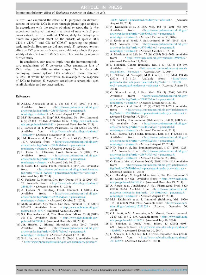
effect on murine splenic DCs. We analyzed CD11c-CD83+and CD11c+CD83+ splenic DCs in E. purpurea-treated miceby flow cytometry (Fig. 4).
The percentage of each of CD11c�CD83+ andCD11c+CD83+ expressing DCs was determined by flowcytometry. The percentage of CD11c�CD83+ DCs was
38.75 ± 2.73% in the control group A, 38.00 ± 5.19% ingroup B and 36.66 ± 3.78% in group C. The percentage ofCD11c+CD83+ DCs was 28.00 ± 3.43% in the control group
·). (A): DCs nucleated with branched projections (control without
servable changes in all E. purpurea treated groups compared with
ineering and Biotechnology (2015), http://dx.doi.org/10.1016/j.jgeb.2015.05.002
Figure 3 Effect of E. purpurea on CD83 expression in various
cell cultures. (A): Flow cytometry showing the expression of
CD83. Monocyte-derived DCs were generated in presence or
absence of E. purpurea. (Group A) control group; (Group B) E.
purpurea (500 lg/ml) on day 7; (Group C) TNF-a (10 ng/ml) on
day 7 + E. purpurea (500 lg/ml) on day 8; (Group D) E. purpurea
(500 lg/ml) on day 7 + TNF-a (10 ng/ml) on day 8; (Group E) E.
purpurea (500 lg/ml) on day 0 + TNF-a (10 ng/ml) on day 7. (B):
Percentage of DCs expressing CD83 in different groups.
Expression of CD83 cell marker is measured by BD
FACSCalibur�. Each value is the mean of 3 experiments ± SE.*P < 0.05.
Immunomodulatory effect of Echinacea purpurea on dendritic cells 5
A, 23.66 ± 5.37% in group B and 23.66 ± 4.45% in group C
(Fig. 5).No changes in expression of the accessory molecules were
observed in splenic cells obtained from different animal
groups. E. purpurea-treated mice did not produce any signifi-cant change in phenotypic analysis of splenic DCs (group Band group C). Furthermore, the injection of stimulation agentTNF-a into mice of group C had no effect on the above-
mentioned markers (Fig. 5).
4. Discussion
Dendritic cells (DCs) are professional antigen presenting cells(APCs) that act at the interface of the innate and adaptive
Please cite this article in press as: N.E. El-Ashmawy et al., Journal of Genetic Eng
branches of the immune system. DCs provoke and controlimmunity against pathogens and tolerance against self-antigens and commensal microorganisms [27].
Delivery of adjuvant to DCs may be a promising strategy totreat autoimmune disorders. But, to produce immunity ratherthan tolerance, it is crucial to provide the DCs with an activa-
tion signal [5]. Although a limited number of surface moleculesare preferentially expressed by DCs, high-density CD83expression is serving as an excellent marker for DC character-
ization [21].In the present study, PBMCs were cultured and allowed to
generate DCs, which became mature after addition of TNF-ato the culture. Mature DCs were identified by microscopical
examination and flow cytometry to detect the expression ofthe cell specific marker CD83.
DCs undergo several changes during maturation including
morphological changes, loss of endocytic and phagocyticreceptors, chemokine secretion, upregulation of costimulatorymolecules, translocation of MHC–II biomarkers to the cell
surface and cytokine secretion [31]. Ultimately these changesprepare DCs to interact successfully with T cells in the contextof antigen. Activation of the antigen-specific T cell results in
clonal expansion and differentiation, leading to adaptiveimmunity [19].
E. purpurea has been shown to have immunostimulatoryeffects on monocytes, macrophages, natural killer cells and T
cells in vitro [9], but few studies have attempted to elucidatethe immunomodulatory effect of E. purpurea on DCs.
In our work, we set out to define morphological variations
and CD83 phenotypic analysis of DCs after exposure ofcultured peripheral blood monocytes to E. purpurea extractat different time intervals. In addition, we carried out
phenotypic analysis of splenic DCs obtained from mice treatedwith E. purpurea daily for 5 days.
Our results showed that addition of 0.5% of 500 lg/mL E.
purpurea ethanolic extract on day 0 of culture (group E) pro-duced a significant reduction of the expression of immunosup-pressant cell marker CD83 by 62.4% in cultured DCs whenexamined on day 9 compared to control cultures. These results
were confirmed by the work of Wang et al. [32], who reportedthat treatment of cultured DCs with E. purpurea extracts at500 lg/mL greatly decreased the expression of CD83 by 61%
in comparison with negative control DCs.Because the addition of alcoholic extract of E. purpurea on
day 0 to PBMC cultures decreased the expression of CD83
analyzed on day 9, we thought to analyze the expression of thismolecule at multiple time days. Our results showed that theexpression of CD83 was not significantly changed followingtreatment of generated DCs with E. purpurea extract added
on day 7 or day 8 either in the presence or absence ofTNF-a (group B, C and D). We inferred that the timing ofaddition of E. purpurea extract is critical. We suggest that
E. purpurea extract impacts on DC precursors not on differen-tiated DCs. This observation is important since this extract canbe used as an immunomodulator, which marked with reduced
expression of CD83 in group E.Depending on phenotypic and functional states, DCs may
promote immunogenic or tolerogenic responses. Although var-
ious Toll-like receptors, such as TLR3, TLR4, TLR5, TLR7and TLR8, induce immune activation, others can silenceimmune responses through tolerance induction in DCs.TLR2 activation induces IL-10 production, inhibits
ineering and Biotechnology (2015), http://dx.doi.org/10.1016/j.jgeb.2015.05.002
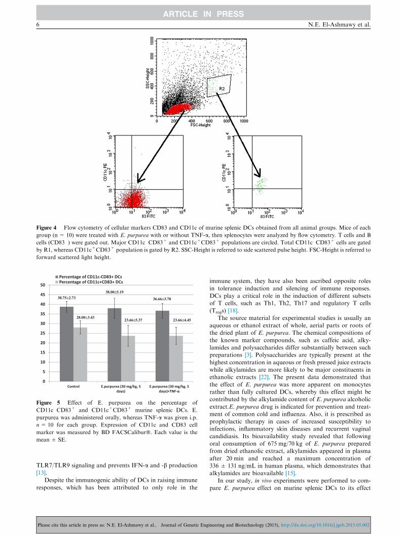
Figure 4 Flow cytometry of cellular markers CD83 and CD11c of murine splenic DCs obtained from all animal groups. Mice of each
group (n = 10) were treated with E. purpurea with or without TNF-a, then splenocytes were analyzed by flow cytometry. T cells and B
cells (CD83�) were gated out. Major CD11c�CD83+ and CD11c+CD83+ populations are circled. Total CD11c�CD83+ cells are gated
by R1, whereas CD11c+CD83+ population is gated by R2. SSC-Height is referred to side scattered pulse height. FSC-Height is referred to
forward scattered light height.
38.75±2.7338.00±5.19
36.66±3.78
28.00±3.4323.66±5.37 23.66±4.45
Figure 5 Effect of E. purpurea on the percentage of
CD11c�CD83+ and CD11c+CD83+ murine splenic DCs. E.
purpurea was administered orally, whereas TNF-a was given i.p.
n= 10 for each group. Expression of CD11c and CD83 cell
marker was measured by BD FACSCalibur�. Each value is the
mean ± SE.
6 N.E. El-Ashmawy et al.
TLR7/TLR9 signaling and prevents IFN-a and -b production[13].
Despite the immunogenic ability of DCs in raising immune
responses, which has been attributed to only role in the
Please cite this article in press as: N.E. El-Ashmawy et al., Journal of Genetic Eng
immune system, they have also been ascribed opposite roles
in tolerance induction and silencing of immune responses.DCs play a critical role in the induction of different subsetsof T cells, such as Th1, Th2, Th17 and regulatory T cells
(Tregs) [18].The source material for experimental studies is usually an
aqueous or ethanol extract of whole, aerial parts or roots of
the dried plant of E. purpurea. The chemical compositions ofthe known marker compounds, such as caffeic acid, alky-lamides and polysaccharides differ substantially between suchpreparations [3]. Polysaccharides are typically present at the
highest concentration in aqueous or fresh pressed juice extractswhile alkylamides are more likely to be major constituents inethanolic extracts [22]. The present data demonstrated that
the effect of E. purpurea was more apparent on monocytesrather than fully cultured DCs, whereby this effect might becontributed by the alkylamide content of E. purpurea alcoholic
extract.E. purpurea drug is indicated for prevention and treat-ment of common cold and influenza. Also, it is prescribed asprophylactic therapy in cases of increased susceptibility toinfections, inflammatory skin diseases and recurrent vaginal
candidiasis. Its bioavailability study revealed that followingoral consumption of 675 mg/70 kg of E. purpurea preparedfrom dried ethanolic extract, alkylamides appeared in plasma
after 20 min and reached a maximum concentration of336 ± 131 ng/mL in human plasma, which demonstrates thatalkylamides are bioavailable [15].
In our study, in vivo experiments were performed to com-pare E. purpurea effect on murine splenic DCs to its effect
ineering and Biotechnology (2015), http://dx.doi.org/10.1016/j.jgeb.2015.05.002
Immunomodulatory effect of Echinacea purpurea on dendritic cells 7
in vitro. We examined the effect of E. purpurea on differentsubsets of splenic DCs in mice through phenotypic analysis.In accordance with the results obtained in vitro, the in vivo
experiment indicated that oral treatment of mice with E. pur-purea extract, with or without TNF-a, daily for 5 days pro-duced no significant effect on the subsets of splenic DCs
CD11c�CD83+ and CD11c+CD83+ regarding the pheno-typic analysis. Because we did not study E. purpurea extracteffect on DC precursors in vivo, we could not exclude the pos-
sibility of its effect on PBMCs in vivo. This possibility requiresfurther study.
In conclusion, our results imply that the immunomodula-tory mechanisms of E. purpurea affect generation fate of
DCs rather than differentiation stages. The in vivo studyemploying murine splenic DCs confirmed those observedin vitro. It would be worthwhile to investigate the response
of DCs to isolated E. purpurea constituents separately, suchas alkylamides and polysaccharides.
References
[1] A.M.K. Abouelella et al, J. Vet. Sci. 8 (4) (2007) 341–351.
Available from: <http://www.pubmedcentral.nih.gov/
articlerender.fcgi?artid=2868149&tool=pmcentrez&
rendertype=abstract> (Accessed August 17, 2014).
[2] M.F. Bachmann, M. Kopf, B.J. Marsland, Nat. Rev. Immunol.
6 (2) (2006) 159–164. Available from: <http://www.ncbi.nlm.
nih.gov/pubmed/16491140> (Accessed December 30, 2014).
[3] J. Barnes et al, J. Pharm. Pharmacol. 57 (8) (2005) 929–954.
Available from: <http://www.ncbi.nlm.nih.gov/pubmed/
16102249> (Accessed November 26, 2014).
[4] J.M. Benson et al, Food Chem. Toxicol. 48 (5) (2010) 1170–
1177. Available from: <http://www.pubmedcentral.nih.gov/
articlerender.fcgi?artid=2883451&tool=pmcentrez&
rendertype=abstract> (Accessed August 23, 2014).
[5] L. Cohn, L. Delamarre, Front. Immunol. 5 (2014) 255.
Available from: <http://www.pubmedcentral.nih.gov/
articlerender.fcgi?artid=4039009&tool=pmcentrez&
rendertype=abstract> (Accessed July 10, 2014).
[6] B. Everts, E.J. Pearce, Front. Immunol. 5 (2014) 203. Available
from: <http://www.pubmedcentral.nih.gov/articlerender.
fcgi?artid=4021118&tool=pmcentrez&rendertype=abstract>
(Accessed July 18, 2014).
[7] G. Ferlazzo, L. Moretta, Crit. Rev. Oncog. 19 (1–2) (2014) 67–
75. Available from: <http://www.ncbi.nlm.nih.gov/pubmed/
24941374> (Accessed October 31, 2014).
[8] A. Gallois, N. Bhardwaj, Front. Immunol. 4 (2013) 436.
Available from: <http://www.pubmedcentral.nih.gov/
articlerender.fcgi?artid=3857536&tool=pmcentrez&
rendertype=abstract> (Accessed October 31, 2014).
[9] M.H. Goldrosen, S.E. Straus, Nat. Rev. Immunol. 4 (11) (2004)
912–921. Available from: <http://www.ncbi.nlm.nih.gov/
pubmed/15516970> (Accessed August 23, 2014).
[10] S.S. Hashimdeen et al, Clin. Hemorheol. Micro. 55 (4) (2013)
501–512. Available from: <http://www.ncbi.nlm.nih.gov/
pubmed/24099989> (Accessed December 20, 2014).
[11] J.B. Hudson, J. Biomed. Biotechnol. 2012 (2012) 769896.
Available from: <http://www.pubmedcentral.nih.gov/
articlerender.fcgi?artid=3205674&tool=pmcentrez&
rendertype=abstract> (Accessed August 17, 2014).
[12] S.-F. Jiao et al, J. Biomed. Sci. 21 (2014) 1. Available from:
<http://www.pubmedcentral.nih.gov/articlerender.fcgi?artid=
Please cite this article in press as: N.E. El-Ashmawy et al., Journal of Genetic Eng
3902418&tool=pmcentrez&rendertype=abstract> (Accessed
August 18, 2014).
[13] N. Kadowaki et al, J. Exp. Med. 194 (6) (2001) 863–869.
Available from: <http://www.pubmedcentral.nih.gov/
articlerender.fcgi?artid=2195968&tool=pmcentrez&
rendertype=abstract> (Accessed December 30, 2014).
[14] S. Koido et al, World J. Gastroenterol. 19 (46) (2013) 8531–
8542. Available from: <http://www.pubmedcentral.nih.gov/
articlerender.fcgi?artid=3870498&tool=pmcentrez&
rendertype=abstract> (Accessed October 31, 2014).
[15] A. Matthias et al, Life Sci. 77 (16) (2005) 2018–2029. Available
from: <http://www.ncbi.nlm.nih.gov/pubmed/15919096>
(Accessed December 17, 2014).
[16] I. Mellman, Cancer Immunol. Res. 1 (3) (2013) 145–149.
Available from: <http://cancerimmunolres.aacrjournals.
org/content/1/3/145.full> (Accessed July 17, 2014).
[17] H. Nakano, M. Yanagita, M.D. Gunn, J. Exp. Med. 194 (8)
(2001) 1171–1178. Available from: <http://www.
pubmedcentral.nih.gov/articlerender.fcgi?artid=2193516&
tool=pmcentrez&rendertype=abstract> (Accessed August 18,
2014).
[18] C. Ohnmacht et al, J. Exp. Med. 206 (3) (2009) 549–559.
Available from: <http://www.pubmedcentral.nih.gov/
articlerender.fcgi?artid=2699126&tool=pmcentrez&
rendertype=abstract> (Accessed December 5, 2014).
[19] B. Piqueras et al, Blood 107 (7) (2006) 2613–2618. Available
from: <http://www.pubmedcentral.nih.gov/articlerender.
fcgi?artid=1895384&tool=pmcentrez&rendertype=abstract>
(Accessed December 19, 2014).
[20] D.S. Pisetsky, Clin. Immunol. (Orlando, Fla.) 144 (1) (2012) 32–
40. Available from: <http://www.pubmedcentral.nih.gov/
articlerender.fcgi?artid=3724456&tool=pmcentrez&
rendertype=abstract> (Accessed December 4, 2014).
[21] C.M. Prazma, T.F. Tedder, Immunol. Lett. 115 (1) (2008) 1–8.
Available from: <http://www.pubmedcentral.nih.gov/
articlerender.fcgi?artid=2699889&tool=pmcentrez&
rendertype=abstract> (Accessed August 17, 2014).
[22] N.D. Pugh et al, Int. Immunopharmacol. 8 (7) (2008) 1023–
1032. Available from: <http://www.pubmedcentral.nih.gov/
articlerender.fcgi?artid=2467439&tool=pmcentrez&
rendertype=abstract> (Accessed December 20, 2014).
[23] G. Ragupathi et al, Vaccine 26 (37) (2008) 4860–4865. Available
from: <http://www.pubmedcentral.nih.gov/articlerender.
fcgi?artid=2565601&tool=pmcentrez&rendertype=abstract>
(Accessed August 17, 2014).
[24] G.J. Randolph, V. Angeli, M.A. Swartz, Nat. Rev. Immunol. 5
(8) (2005) 617–628. Available from: <http://www.ncbi.nlm.
nih.gov/pubmed/16056255> (Accessed December 19, 2014).
[25] A. Rezaie et al, Jundishapur J. Nat. Pharmaceut. Prod. 8 (2)
(2013) 60–64. Available from: <http://www.pubmedcentral.
nih.gov/articlerender.fcgi?artid=3941908&tool=pmcentrez&
rendertype=abstract> (Accessed August 17, 2014).
[26] M.P. Rubinstein et al, J. Immunol. (Baltimore, Md.: 1950)
169 (9) (2002) 4928–4935. Available from: <http://www.ncbi.
nlm.nih.gov/pubmed/12391205> (Accessed December 20,
2014).
[27] C.L. Scott, A.M. Aumeunier, A.M. Mowat, Trends Immunol.
32 (9) (2011) 412–419. Available from: <http://www.ncbi.nlm.
nih.gov/pubmed/21816673> (Accessed July 16, 2014).
[28] K. Shimizu, S. Fujii, Front. Biosci. 13 (2008) 6193–
6201. Available from: <http://www.ncbi.nlm.nih.gov/pubmed/
18508653> (Accessed December 30, 2014).
[29] G. Shrestha, L.L. St Clair, K.L. O’Neill, Phytother. Res. (2014).
Available from: <http://www.ncbi.nlm.nih.gov/pubmed/
25339289> (Accessed October 31, 2014).
ineering and Biotechnology (2015), http://dx.doi.org/10.1016/j.jgeb.2015.05.002

8 N.E. El-Ashmawy et al.
[30] M. Stetler-Stevenson, R.C. Braylan, Semin. Hematol. 38 (2)
(2001) 111–123. Available from: <http://www.ncbi.nlm.
nih.gov/pubmed/11309693> (Accessed December 20, 2014).
[31] E.S. Trombetta, I. Mellman, Annu. Rev. Immunol. 23 (2005)
975–1028. Available from: <http://www.ncbi.nlm.nih.gov/
pubmed/15771591> (Accessed July 9, 2014).
[32] C.-Y. Wang et al, Genomics 88 (6) (2006) 801–808. Available
from: <http://www.ncbi.nlm.nih.gov/pubmed/17011161>
(Accessed August 23, 2014).
Please cite this article in press as: N.E. El-Ashmawy et al., Journal of Genetic Eng
[33] K. Yamada et al, J. Ethnopharmacol. 137 (1) (2011) 231–235.
Available from: <http://www.ncbi.nlm.nih.gov/pubmed/
21619924> (Accessed August 8, 2014).
[34] Z. Ye et al, Cancer Biotherap. Radiopharmaceut. 21 (6) (2006)
613–622. Available from: <http://www.ncbi.nlm.nih.gov/
pubmed/17257077> (Accessed December 20, 2014).
ineering and Biotechnology (2015), http://dx.doi.org/10.1016/j.jgeb.2015.05.002