The IL-12 family and dendritic cells
21
Cover Page The handle http://hdl.handle.net/1887/28326 holds various files of this Leiden University dissertation Author: Dixon, Karen Title: The IL-12 family and dendritic cells : key regulators of immunity and immunological tolerance Issue Date: 2014-08-28
-
Upload
khangminh22 -
Category
Documents
-
view
1 -
download
0
Transcript of The IL-12 family and dendritic cells

Cover Page
The handle http://hdl.handle.net/1887/28326 holds various files of this Leiden University dissertation Author: Dixon, Karen Title: The IL-12 family and dendritic cells : key regulators of immunity and immunological tolerance Issue Date: 2014-08-28

Chapter
Human tolerogenic Dendritic cells produce IL-35 in the absence of other IL-12 family members.
Karen O. Dixon*, Sandra W. van der Kooij*, Dario A.A. Vignali†, Cees van Kooten*.
*Leiden University Medical Center, Dept of Nephrology, Leiden, The Netherlands
†Department of Immunology, St Jude Children’s Research Hospital, Memphis, USA
Under revision
2

34 • Chapter 2
AbstractIL-35 is a novel cytokine of the IL-12 family, existing as a heterodimer of IL-12p35 and EBV-induced gene 3 (Ebi3). IL-35 has anti-inflammatory properties and has been described to be produced by regulatory T cells in humans and mice, where it is required for optimal suppression. Distinct from other IL-12 family members, expression of IL-35 has not been clearly described in antigen presenting cells. In view of its immune-regulatory properties, we investigated the expression, regulation and function of IL-12p35 and Ebi3 in human monocyte derived dendritic cells (DCs) and tolerogenic DCs (tolDCs), generated in the presence of dexamethasone. These tolDCs did not produce detectable levels of bioactive IL-12p70 or the homodimer IL-12p40. In line with this, we demonstrate by Q-PCR that tolDCs completely lack transcriptional expression of IL-12p40. However, tolDCs maintain mRNA expression of IL-12p35 and Ebi3. Using intracellular flow cytometry and western blot we show that tolDCs are able to produce Ebi3 and IL-12p35, and both can be further enhanced upon stimulation with IFNγ, LPS or CD40 ligation. Supernatant of tolDCs has the capacity to suppress T cell activation. Using IL-12A silencing we demonstrate that IL-12p35 is required for tolDCs to reach their full suppressive potential. Taken together, our results indicate that tolDCs produce IL-35, providing an additional novel mechanism by which these cells elicit their tolerogenic potential.
IntroductionDendritic cells (DCs) represent a heterogeneous family of professional APCs that have many functions in both the initiation and maintenance of immunity and immunological tolerance1,2. Several factors, including IL-10, TGF-β, Vitamin D3 and corticosteroids have been shown to modulate DC function in vitro and in vivo, and lead to the generation of tolerogenic DCs (tolDCs)3-6. These tolDCs represent an altered status in DC differentiation and are currently considered in the design of therapeutic strategies to treat patients suffering from autoimmune disease7,8 and those undergoing transplantation9,10. Impaired expression of IL-12 in tolDCs is thought to be a crucial requirement for these cells to elicit their tolerogenic potential, and indeed many tolDC populations have been shown to lack IL-123,5. However the IL-12 family is ever expanding and little is known about the regulation and expression of other IL-12 family members in tolDCs.The IL-12 family is composed of four heterodimeric complexes each consisting of an alpha chain (IL-12p35, IL-23p19 and IL-27p28) and a beta chain (Ebi3 and IL-12p40)11. The IL-12p40 subunit can pair with either IL-12p35 or IL-23p19 to yield IL-12 and IL-23 respectively12,13. The second beta chain subunit Ebi3, can pair with 2 other alpha chain subunits, IL-27p28 and IL-12p35, to yield IL-27 and IL-35 respectively. Although there is a large degree of chain sharing within

TolDCs require IL-12p35 for their full tolerogenic potential • 35
2
this family, each cytokine possesses a unique and distinct function. IL-12 and IL-23 have primarily pro-inflammatory properties and play an important role in the development of Th1 and Th17 T helper cell subsets respectively. IL-27 possess dual roles playing a protective role in tumour immunity14,15, but as an immunoregulatory cytokine can induce Tr1 cells16,17 and lead to the generation of a tolerogenic DC expressing elevated levels of CD3918. The most recently characterised member of this family is IL-35, a potent anti-inflammatory cytokine, which has been shown to be required for the optimal suppressive activity of regulatory T cell populations in both mice and man19-22. DCs are known producers of three family members, IL-12, IL-23 and IL-27. In tolDCs little is known about the expression of these family members aside from the characteristic loss of IL-12 production and limited transcriptional data on IL-23 and IL-2723. For IL-35, production by myeloid cells in human or mouse has not yet been demonstrated.In this study we addressed the expression profile and regulation of all IL-12 family members in human DCs and tolDCs with a particular emphasis on the subunits which combined make up IL-35. We show that tolDCs did not produce detectable levels of bioactive IL-12p70, the homodimer IL-12p40 or express IL-12p40 transcripts. However tolDCs maintain mRNA and protein expression of IL-12p35 and Ebi3. We show that supernatants of tolDCs are sufficient to suppress T cell proliferation, and that supernatants of cells silenced for IL-12p35 are hampered in their suppressive potential, thereby implying a role for IL-35 in tolDC function.
Materials and Methods
Generation of human monocyte derived DCsHuman monocytes were isolated from buffy coats obtained from healthy donors using Ficoll density gradient centrifugation followed by positive selection using anti-CD14 MACS microbeads (Miltenyi Biotech GmBH, Bergisch Gladbach, Germany). DCs were generated and cultured in RPMI supplemented with 10% heat inactivated FCS, 90 U/ml penicillin and 90 U/ml streptomycin (Gibco/ Life Technologies, Bleiswijk, The Netherlands), supplemented with 5 ng/ml GM-CSF and 10 ng/ml IL-4 (Invitrogen/ Life Technologies, Bleiswijk, The Netherlands) as described before3. TolDCs were generated by the addition of Dexamethasone (10-6M Dex) (Pharmacy, LUMC, Leiden, The Netherlands), only at the start of culture (day 0). Cultures were refreshed with medium containing cytokines on day 3. For differentiation into mature DCs, cells were harvested, washed and stimulated on day 6 with 200 ng/mL LPS (E.Coli 0127:B8 Sigma-Aldrich, Zwijndrecht), 20µg/ml Poly I:C, 10 µg/ml PGN (InvivoGen) or 100 ng/ml IFNγ (Peprotech, Germany). CD40L-activation was performed with CD40L transfected L cells (L-CD40L) in a DC: L cell ratio of 5:1. Non-transfected L cells served as control cells (L-Orient).
DC-T cell co cultureTotal CD4+ lymphocytes were negatively selected from PBMCs using CD4+ T cell isolation kit II (Miltenyi Biotech GmBH, Bergisch Gladbach, Germany), and subsequently labelled with carboxyfluorescein diacetate succinamidyl ester (CFSE) (Molecular Probes, Europe BV Leiden, The Netherlands). In short, T cells were

36 • Chapter 2
suspended in PBS and incubated for 15 minutes at 37°C with 5 μM CFSE. The reaction was quenched by washing the cells in medium containing 10% FCS before resuspending at 1×10⁶/ml in total RPMI culture medium. Labelled CD4+ T cells were co-cultured with allogeneic DCs at a 10:1 T cell/DC ratio for 5 days. The T cells were then harvested and proliferation was assessed by CFSE dilution.
Surface and Intracellular flow cytometryFor cell surface flow cytometric analysis, cells were stimulated overnight, harvested, washed, and stained for 30 minutes at 4°C in FACS buffer (PBS, 0.5% heat inactivated NHS, 1% BSA, 0.02% NaN3) with anti-CD86, anti-CD14 MΦ P9 (BD biosciences, San Diego, CA, USA), anti-B7H1, anti-B7DC, anti-ILT3 (Biolegend), anti-HLADR (B8.11.2, ATCC), anti-Mannose Receptor (clone D547, msIgG1 in house) or anti-DC-SIGN (R&D Systems, Wiesbaden, Germany). Nonconjugated antibodies were detected with PE-conjugated goat-anti-mouse Ig (Dako, Glostrup, Denmark). Isotype matched control antibodies were used to determine the level of background staining. For intracellular staining cells were stimulated overnight, followed by incubation in the presence of Brefeldin-A (Sigma-Aldrich, Zwijndrecht) for a further 5 hours. The cells were then harvested, washed and fixed in PBS containing 4% formaldehyde and 1% heat inactivated FCS, washed with PBS containing 10% heat inactivated FCS and permeabilised with perm buffer (PBS, 0.5% saponin, 1% heat inactivated FCS) for 10 minutes. Cells were stained with anti-Ebi3-APC or anti-IL-12p35-APC for 30 minutes at 4°C in perm buffer. APC-conjugated isotype matched control antibodies were used to determine the level of background staining. The fluorescence was measured on a FACS Calibur flow cytometer, and data were analysed with Cell Quest Software (BD Biosciences, San Diego, CA, USA) and FlowJo Software (Tree star, USA).
mRNA isolation, cDNA synthesis, and real-time PCRDay 6 DCs were plated in a 12-well plate at a density of 1x10⁶/ml and harvested after indicated time points. mRNA was isolated using an RNeasy kit (Qiagen, Hilden, Germany) following the manufacturer’s instructions. Digestion of genomic DNA was performed by using the on-column RNase-free DNase set. Reverse transcriptase system kit (Promega, Leiden, The Netherlands) was used to synthesise cDNA conforming to the manufacture’s instruction and was stored at -20ºC for further analysis. Specific primers (Table I) for human IL12A, IL12B, IL23A, IL27A, IL27B, IL10 and GAPDH were designed using the computer software Oligo explorer and synthesised at Biolegio. Quantitative PCR was performed using SYBR Green qPCR master mix (Bio-Rad, Veenendaal, The Netherlands). GAPDH was used as the endogenous reference gene. Data analysis was performed using Bio-Rad CFX Manager Software (Bio-Rad). For each sample, the relative abundance of target mRNA was calculated from the obtained Ct values for the target gene and expressed relative to the endogenous reference gene GAPDH.
Table I: Real Time PCR Oligonucleotide sequences
TolDCs require IL-12p35 for their full tolerogenic potential • 37
2
Cytokine productionDCs were plated at 1x10⁶/ml and stimulated accordingly. Cell culture supernatants were harvested after 48 hours and frozen at −80°C until analysis. Subsequently they were tested for the presence of IL-12p70, IL-12p40, (Biolegend) IL-10 (Sanquin, Amsterdam, The Netherlands) and IL-27 (eBioscience) by ELISA according to manufacturer’s instructions. IFNγ levels were determined in the cellular supernatants of T cells from indicated time points (eBioscience).
Western BlottingDCs were plated at 1×10⁶ /ml, stimulated for 16 hours, followed by the addition of Brefeldin A for a further 5 hours. Cells were harvested, washed in ice-cold phosphate-buffered saline and lysed in medium stringency lysis buffer for 30 minutes. The lysates were centrifuged at 13,000 rpm, the supernatants harvested and protein concentration was determined by a Pierce assay. The samples were diluted in 1x SDS loading buffer, boiled for 5 minutes in reducing conditions, followed by separation on 10% SDS-PAGE gel. Gels were transferred to nitrocellulose membranes, blocked with TBS plus 0.1% Tween-20 (TBS-T) and 3% BSA or milk and probed with indicated primary antibodies; purified anti-IL-12p40, anti-IL-12p35 (Santa Cruz Heidelberg, Germany) or purified anti-Ebi3 (Abnova, Huissen The Netherlands). After incubation with HRP-conjugated secondary antibody, proteins were detected with super signal ECL system. Blots were stripped and β-actin was used as a loading control using HRP conjugated anti-β actin (Abcam, Cambridge, UK).
Immunofluorescence microscopy Cells were cultured on 8 well chamber slides (NUNC) in the presence of stimuli and Brefeldin-A. The cells were subsequently washed and fixed in PBS containing 4% formaldehyde, 1% heat inactivated FCS followed by washing with PBS containing 10% heat inactivated FCS and permeabilised with perm buffer (PBS, 0.5% saponin, 1% heat inactivated FCS) for 10 minutes. The DCs were then stained using IL-12p35-APC or purified anti-Ebi3 (Abnova), followed by goat anti-mouse-Alexa488 (Molecular Probes). Hoechst was used for nuclear staining.
RNA interferenceDCs were transfected with 50 nM siRNA through the use of the transfection reagent Lipofectamine 2000 (Life Technologies) and were used for experiments 24 hours after transfection. The following SMARTpool siRNAs were used (Dharmacon, Fisher Scientific, Netherlands): IL-12A and nontargeting siRNA as a control. Silencing of expression was verified by real-time PCR in tolDC and IL-12p40/p70 ELISAs in DC.
T cell proliferation assaysNaïve CD4+ T cells were isolated by negative selection using naïve CD4+ T cell isolation kit II (Miltenyi Biotech), from cord blood derived PBMCs. Cells were stimulated as described previously20. Briefly cells were plated at a density of 25,000/well in 96 well round bottom plates in the presence of αCD3/28 coated beads. Supernatants from stimulated tolDCs were harvested and incubated with the T cells at a dilution of 1:2. T cell proliferation was assessed by the addition of 3[H]-thymidine (0,5 µCi/well) for the last 8 hours. Percentage suppression was calculated by 1-(cpm T cells + tolDC sup / cpm T cells alone) times 100.
Statistical analysisStatistical analysis was performed with Graph Pad Prism (Graph Pad Software, San Diego, CA) using a one-tailed t-test. P-values ≤ 0.05 were considered statistically significant.

38 • Chapter 2
Results
DCs generated in the presence of dexamethasone display typical features of tolerogenic DCs with low expression of CD86 but high B7H1 and B7DC.We have previously demonstrated that Dex treated DCs display reduced levels of co-stimulatory molecules3. To further investigate the phenotypical characteristics of DCs and tolDCs, freshly isolated monocytes were cultured in the presence of GM-CSF and IL-4, with the addition of Dexamethasone to generate tolDCs. Both cell types expressed characteristic markers of myeloid DCs including DC-SIGN (CD209) and Mannose Receptor (CD206), while tolDCs preserved the expression of CD14 upon differentiation from monocyte to immature DC (data not shown). In an immature state both DCs expressed comparable levels of CD86 and immunoregulatory molecules B7H1 (CD274) and ILT3 (CD85k), while HLA-DR and B7DC (CD273) were more abundantly expressed on tolDCs (Fig.1). Stimulation using IFNγ, LPS or PGN resulted in the generation of mature DCs as shown by increased expression of co-stimulatory molecules including CD86 and HLA-DR (Fig.1A,B). tolDCs were significantly impeded in their ability to upregulate CD86 or HLA-DR upon stimulation with any agents used (Fig.1A, B). In contrast tolDC upregulated B7H1 (Fig.1C) and to a lesser degree B7DC and ILT3 (Fig.1D,E). To fully ascertain the allostimulatory capabilities of tolDCs, immature and mature DC/tolDCs were co-cultured with allogenic CD4+ T lymphocytes for 5 days, after which the T cells were analysed for CFSE dilution (Fig.1F). DCs were potent inducers of T cell proliferation which was further enhanced when the DCs were first matured for 24 hours with LPS or PGN. Conversely tolDCs were hampered in their ability to stimulate allogenic T cells regardless of maturation stimulus used (Fig.1G). In line with the proliferation data DCs induced a strong IFNγ production by the T cells, whereas tolDCs induced almost negligible amounts of IFNγ production (Fig.1G). In summary the tolDCs used in this study display typical characteristics of myeloid tolerogenic DCs.
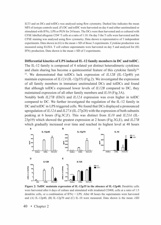
TolDC maintain expression of IL-12p35 in the absence of IL-12p40.A key characteristic of tolDCs is their ability to preserve production of immunoregulatory cytokines while maintaining low to absent production of inflammatory cytokines. Upon activation, DCs showed strong production of IL-12p40 and IL-12p70, whereas these cytokines were completely absent in supernatants of activated tolDCs (Fig.2A,B). In contrast, tolDCs produced significantly higher levels of IL-10 upon stimulation as compared to DC (Fig.2C). To further explore the expression of IL-12 in DC and tolDC we assessed by RT-PCR the expression of IL12A (IL-12p35) and IL12B (IL-12p40), together making IL-12p70. In line with our ELISA data DCs showed a strong IL12B

TolDCs require IL-12p35 for their full tolerogenic potential • 39
2
expression upon stimulation while tolDCs were greatly inhibited in this ability (Fig.2D). However, tolDCs maintained their expression of IL12A and expressed significantly higher basal levels compared to DC (Fig.2E). Upon stimulation, where IL12B remains absent (Fig.2D), IL12A expression is significantly increased in tolDCs to comparable and even higher levels than DC (Fig.2E).
Figure 1: DCs generated in the presence of dexamethasone display a tolerogenic phenotype and have a reduced allostimulatory capacity. Monocytes were isolated from PBMCs and cultured for 6 days in the presence of IL-4 and GM-CSF to obtain immature DC. tolDC were generated by the addition of 10-6M Dexamethasone to the moDC cultures at day 0. Cells were activated overnight with IFNγ, LPS or PGN. The expression of (A) CD86, (B) HLA-DR, (C) B7H1, (D) B7DC and (E)
40 • Chapter 2
Differential kinetics of LPS induced IL-12 family members in DC and tolDC.The IL-12 family is composed of 4 related yet distinct heterodimeric cytokines and chain sharing has become a quintessential feature of this cytokine family24,
25. We demonstrated that tolDCs lack expression of IL12B (IL-12p40) yet maintain expression of IL12A (IL-12p35) (Fig.2). We investigated the expression of all family members in immature unstimulated DCs and tolDCs and found that although tolDCs expressed lower levels of IL12B compared to DC, they maintained expression of all other family members and IL10 (Fig.3A). Notably both IL27B (Ebi3) and IL12A expression was even higher in tolDC compared to DC. We further investigated the regulation of the IL-12 family in DC and tolDC in LPS triggered cells. We found that DCs displayed a pronounced upregulation of IL12A and IL27A (IL-27p28) with the expression of both subunits peaking at 6 hours (Fig.3C,F). This was distinct from IL10 and IL23A (IL-23p19) which showed the greatest expression at 2 hours (Fig.3G,E), and IL27B which gradually increased over time and reached its highest level at 48 hours
ILT3 and on DCs and tolDCs was analysed using flow cytometry. Dashed line indicates the mean MFI of isotype controls used. (F) DC and tolDC were harvested on day 6 and either unstimulated or stimulated with IFNγ, LPS or PGN for 24 hours. The DCs were then harvested and co cultured with CFSE labelled allogenic CD4+ T cells at a ratio of 1:10. On day 5 the T cells were harvested and the CFSE staining was analysed using flow cytometry. Data shown is representative of 3 independent experiments. Data shown in (G) is the mean ± SD of those 3 experiments. Cytokine production was measured using ELISA. T cell culture supernatants were harvested on day 5 and analysed for (H) IFNγ production. Data shown is the mean ± SD of 3 experiments.
Figure 2: TolDC maintain expression of IL-12p35 in the absence of IL-12p40. Dendritic cells were harvested after 6 days of culture and stimulated with irradiated CD40L cells at a ratio of 1:5 dendritic cells, or a combination of IFNγ + LPS. After 48 hours the supernatants were harvested and (A) IL-12p40, (B) IL-12p70 and (C) IL-10 were measured. Data shown is the mean ±SD

TolDCs require IL-12p35 for their full tolerogenic potential • 41
2(Fig.3D). While IL12B was rapidly induced in DCs, expression remained very low in tolDCs (Fig.3B). tolDCs demonstrated only a minor increase in IL12A and IL27B despite possessing up to 100 fold higher basal level of expression compared to DC (Fig.3C,D). IL23A expression was also higher in tolDC at time 0 but gradually decreased over time before returning to the initial expression level (Fig.3E). IL27A expression was comparable between the two cell types up until 6 hours, after which a drop off was noted for tolDC (Fig.3F). Finally although IL10 started off with a higher level of expression in tolDC the pattern of upregulation
of 6 independent experiments.Cells were stimulated for 6 hours after which mRNA was isolated followed by cDNA synthesis. The transcript levels of (D) IL12B (E) IL12A were determined by RT-PCR. GAPDH mRNA expression from the same samples was used as an endogenous reference gene (relative mRNA expression).Data shown is mean ± SD of 4 independent experiments. Untransfected L cells were used as a control for CD40L cells and yielded results in line with medium conditions (data not shown).
Figure 3: Kinetic expression of IL-12 family members and IL-10 mRNA in LPS-treated DCs and tolDCs. Cells were harvested on day 6, mRNA was isolated followed by cDNA synthesis. The transcript levels of (A) IL12B, IL12A, IL27B, IL23A, IL27A and IL10 were determined by RT-PCR in unstimulated cells. Cells were stimulated with LPS for indicated time points; mRNA was isolated followed by cDNA synthesis. The transcript levels of (B) IL12B, (C) IL12A, (D) IL27B (E) IL23A, (F) IL27A and (G) IL10 were determined. GAPDH mRNA expression from the same samples was used as an endogenous reference gene (relative mRNA expression). Data shown is mean ± SD of 2 independent experiments.

42 • Chapter 2
was comparable between the two cell types with a transient upregulation at 2 hours followed by a gradual decrease over time (Fig.3G). The relative abundance of both IL27B and IL12A transcripts in unstimulated tolDC compared to DC, in contrast to other IL-12 family subunits, is intriguing and may indicate some inherent regulation of these subunits within tolerogenic DCs.
Stimulation of tolDCs favours upregulation of the immunoregulatory chains IL-12p35, Ebi3 and IL-27p28.Although tolDCs specifically maintained the expression of subunits involved in immunoregulatory processes (Fig.3), they remained relatively refractory to LPS stimulation in terms of IL-12 family expression. We investigated if this profile was intrinsic for these cells or was activation dependent. We observed that although DCs expressed relatively abundant levels of IL12B, particularly upon stimulation with LPS, IFNγ + LPS or Poly I:C, transcripts of this subunit were significantly reduced in tolDC (Fig.4A). We demonstrate that IL12A is maintained in tolDCs and expressed comparably to DC across the different stimuli used. Notably treatment with a combination of IFNγ + LPS yielded the highest IL12A expression in tolDCs (Fig.4B). IFNγ, and a combination of IFNγ +LPS also displayed the greatest increase in IL27B expression when comparing DC to tolDCs (Fig.4C), with CD40 ligation showing the next highest expression in tolDC. IL27A levels were comparable between both DCs but IFNγ yielded the strongest induction of IL27A transcripts in tolDCs (Fig.4D). In summary tolDCs do not express IL12B but do maintain expression of IL12A, IL27A and IL27B which can be further induced upon activation with IFNγ, IFNγ + LPS and CD40 ligation.
Figure 4: Stimulation of tolDCs favours upregulation of the immunoregulatory chains IL-12p35, Ebi3 and IL-27p28. Dendritic cells were stimulated for 6 hours with IFNγ, LPS, PGN,

TolDCs require IL-12p35 for their full tolerogenic potential • 43
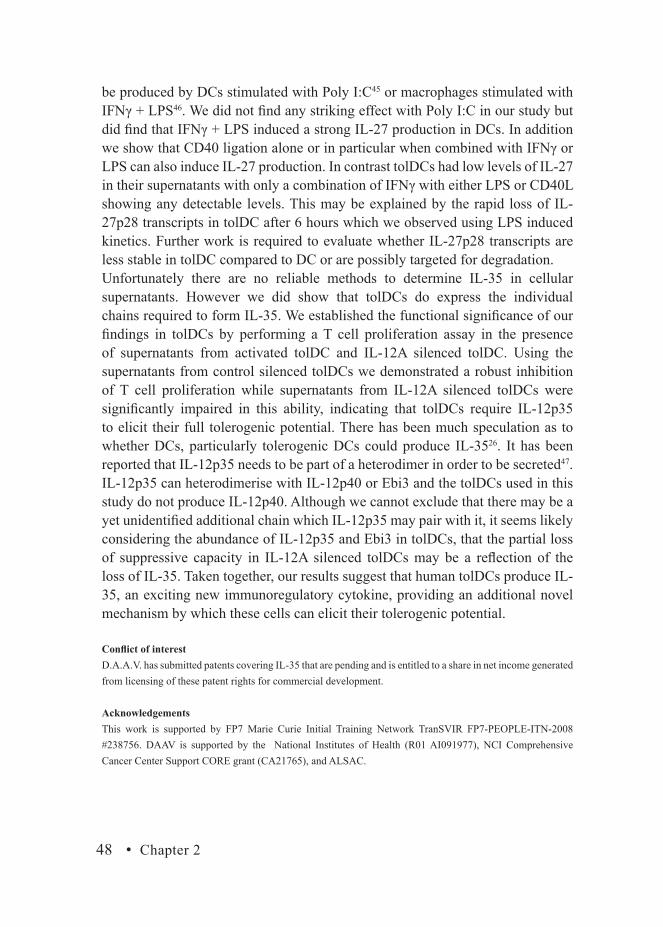
2TolDC produce low levels of IL-27 but maintain production of IL-35 related chains.We investigated if our observations regarding transcriptional expression of IL-27p28, Ebi3 and IL-12p35 were translated into protein by assessing production in DC and tolDCs. Using an IL-27 ELISA we found that DCs, but not tolDCs, produced IL-27 upon IFNγ + LPS stimulation, although we did find considerable variation across the 6 donors analysed (Fig.5A). Notably a combination of IFNγ + LPS or CD40L with either IFNγ or LPS, were the best inducers of IL-27 production in DCs while the individual stimuli alone were poor inducers (Fig.5B). Under all conditions tolDCs were significantly hampered in their IL-27 production (Fig.5B). Ebi3 and IL-12p35 can dimerise to form IL-35; however there are no reliable methods for the detection of IL-35 in human cellular supernatants. We established by intracellular fluorescent microscopy that tolDCs do express the individual chains, Ebi3 and IL-12p35, and that production of both can be increased upon stimulation with IFNγ or IFNγ + LPS (Fig.5C). To quantify this, we measured Ebi3 and IL-12p35 by intracellular flow cytometric staining in both DC and tolDC. We found that although tolDCs had lower basal levels of Ebi3 compared to DC, they showed a stronger induction upon stimulation (Fig.5D). The relative increase in Ebi3 expression upon stimulation with IFNγ or IFNγ + LPS in tolDCs was 6 fold or higher (Fig.5D). Although tolDC have a lower basal level of IL-12p35 compared to DC, they increase expression comparably to DC upon stimulation with IFNγ or IFNγ + LPS (Fig.5E). To further reinstate our findings we performed a western blot for IL-12p35 and Ebi3 on whole cell lysates. In line with our flow cytometry and microscopy data, tolDCs do express both IL-12p35 and Ebi3, and both appear to increase upon stimulation. Importantly, in contrast to DC, tolDC do not express IL-12p40 using any stimuli assessed (Fig.5F).
TolDCs require expression of IL-12p35 to elicit their full tolerogenic potential.To determine the functional significance of IL-12p35 and Ebi3 produced by tolDC, we investigated the suppressive activity of tolDC supernatants on αCD3/CD28 activated T cells. Supernatants from CD40 ligated tolDCs demonstrated a pronounced inhibition of T cell proliferation which was not observed with mock activated tolDCs (Fig.6A). Using different tolDC and T cell donors, we found
Poly (I: C) or a combination of both IFN γ + LPS. Irradiated CD40L cells were added at a ratio of 1:5. The transcript levels of (A) IL12B, (B) IL12A, (C) IL27B, (D) IL27A were determined by RT-PCR. GAPDH expression from the same samples was used as an endogenous reference gene (relative mRNA expression). Data shown is mean ± SD of 3-5 independent experiments. Untransfected L cells were used as a control for CD40L cells and yielded results in line with medium conditions (data not shown).

44 • Chapter 2
Figure 5: TolDC maintain and can enhance their protein expression of IL-12p35 and Ebi3 in the absence of IL-12p40. (A) DCs and tolDCs were harvested on day 6, replated and stimulated with IFNγ + LPS; IL-27 levels were assessed by ELISA. (B) Cells were stimulated with IFNγ + LPS, IFNγ+CD40L, LPS+CD40L or CD40L for 48 hours followed by detection of IL-27 by ELISA. Data shown is mean ± SD of 3-6 independent experiments. (C) tolDCs were cultured on 8 well chamber slides and either untreated, or stimulated with IFNγ or IFNγ + LPS followed by overnight incubation with Brefeldin A. The cells were then fixed and permeabilised, followed by incubation with purified anti-huEbi3 or IL-12p35-APC. Ebi3 was detected using GaM-Alexa488. For flow cytometry DCs and tolDCs were either untreated or stimulated for 16 hours with IFNγ alone or IFNγ + LPS, followed by culture for a further 5 hours in the presence of 10µg/ml Brefeldin A. The cells were then harvested, fixed and permeabilised, followed by incubation with (D) Ebi3-APC or (E) IL-12p35-APC. Data shown is representative of 3 independent experiments, with graphed data showing the mean ± SD of those 3 experiments. (F) DCs were treated similarly for the generation of cellular lysates. Samples were loaded on 10% SDS gel followed by transfer to

TolDCs require IL-12p35 for their full tolerogenic potential • 45
2that supernatant from CD40L activated tolDC consistently induced suppression ranging from 30-80% (median 60%) (Fig.6B).To establish the role of IL-35 production by tolDC in T cell suppression, we performed RNA interference of IL12A. By mRNA analysis we found at least a tenfold inhibition of IL12A expression in activated tolDCs (Fig.6C). Importantly IL27B and IL10 remained unaffected by IL12A silencing, and use of a non-specific siRNA did not affect IL12A expression (Fig.6C).
nitrocellulose membranes. Blots were probed with indicated primary antibodies and detected with super signal ECL system. (data not shown).
Figure 6: TolDCs require IL-12p35 in order to elicit their full tolerogenic potential. tolDCs were harvested on day 6 and stimulated with irradiated CD40L or mock transfected cells at a ratio of 1:5 for 48 hours. Supernatants were harvested and incubated with αCD3/28 stimulated naïve CD4+ T cells at a dilution of 1:2 for 72 hours. 3H incorporation was determined during the last 8-12 hours of culture. Results shown in (A) are from a representative experiment (mean ± SD of CPM from triplicate cultures). Data shown in (B) is the mean ± SEM percentage suppression from 14 independent experiments. (C) IL12A was silenced in tolDCs demonstrating up to 90% interference in mRNA transcripts of IL12A. IL-12A silenced or control treated tolDCs were harvested and stimulated as described above for 48 hours. The supernatants were then incubation with αCD3/28 stimulated naïve CD4+ T cells at a dilution of 1:2 for 72 hours. 3H incorporation was determined during the last 8-12 hours of culture. CPM shown in (D) are from a representative experiment of

46 • Chapter 2
Using supernatants from CD40L stimulated tolDCs we found that IL12A silenced supernatants were significantly impaired in their suppressive capacity (Fig 6D, 6E), thereby indicating that IL-12p35 is required by tolDCs to elicit their full tolerogenic potential. To confirm that the IL12A siRNA directly targeted IL-12p35 protein we demonstrated, in activated control DCs, that IL-12p70 was up to 60% inhibited while IL-12p40 was unaffected (Fig 6F). As a consequence, the capacity of DC supernatant to induce IFNγ production in activated T cells was significantly reduced (Fig 6G).
Discussion.The IL-12 family is gaining increasing attention as being a key regulator of immunity and immunological tolerance25,26. A common feature of tolerogenic DCs is the lack of IL-12, however little is known about the expression, regulation or function of the other family members in human tolDCs. In this study we focused on the IL-12/IL-35 axis and found that tolDCs maintain, and can increase expression of Ebi3 and IL-12p35, and that IL-12p35 is required for their full tolerogenic potential.Despite the remarkable degree of chain sharing between the four members of the IL-12 family, each one has its own unique biological function. IL-12 and IL-23 are pro-inflammatory cytokines crucial for the development of Th1 and memory Th17 cells respectively27,28. On the other hand IL-27 and IL-35 are inhibitory and immunoregulatory cytokines20,29-31. IL-35 although a relatively new player within this family has already been shown to induce proliferation of iTr35, a regulatory T cell subset which possesses a profound anti-inflammatory phenotype with the ability to reduce inflammation in animal models of IBD and EAE20. With the striking immunoregulatory functions of IL-35 it became evident to us that the possible secretion of IL-35 from tolerogenic DCs could be a new previously unexplored mechanism into how these cells can suppress T cell responses. To study this we used a well characterised human tolDC and first established that they displayed typical features associated with regulatory DC populations -low CD86, high B7H132 and poor ability to stimulate allogenic T cells8,33. In addition we observed that these cells express high levels of the endocytic receptor CD206 as well as the immunoregulatory molecules B7DC34,35 and ILT336. Our focus was
3 independent experiments performed. Percentage inhibition shown in (E) is the mean ± SD of those 3 experiments. DCs were harvested on day 6 and were either mock (-), IL-12A, or Non-target siRNA treated, followed by stimulation with IFNγ + LPS. Cell supernatants were harvested and assessed for production of (F) IL-12p70 and IL-12p40 by ELISA, data shown is mean ± SD of 4 independent experiments. siRNA treated DCs were stimulated with CD40L, supernatants were harvested after 48 hours and incubated with αCD3/28 activated total CD4+ T cells for 3 days. (G) IFNγ levels were determined in the T cell supernatants by ELISA.

TolDCs require IL-12p35 for their full tolerogenic potential • 47
2
on the cytokine repertoire of these cells and one of the more intriguing qualities of tolDCs is the profound lack of IL-12p40/70 production regardless of stimulation used. We have shown that tolDCs completely lack IL-12p40 expression yet preserve their transcriptional expression of the other IL-12 family members, in particular those who, upon dimerisation are associated with anti-inflammatory properties, namely Ebi3, IL-12p35 and IL-27p28. Analysing LPS triggered DCs, we observed that IL-12p35/p40 were transient and superimposable with both subunits reaching their highest level at 6 hours in DCs. A similar peak in expression was noted for IL-27p28, though the increase in expression appears to occur more rapidly than for p40/p35. Conversely Ebi3 does not peak until 24-48 hours post activation in DCs. This differential pattern of expression has been likewise noted in previous studies37,38 and has fuelled speculation that Ebi3 may have a unique biological function independent of its typical pairing subunits. Recent evidence using murine cells showed that type 2 anti-inflammatory macrophages express high level of Ebi339. We have also observed elevated Ebi3 levels in M-CSF generated human M2 compared to GM-CSF generated M1 macrophages (unpublished data). Intriguingly, the authors show that intracellular Ebi3 expression can block LPS induced M1 to M2 transition and IL-12p70 production39, suggesting that Ebi3 may play a role in maintaining the regulatory functions of M2 macrophages. Further work is needed to fully extrapolate this data to human macrophage subsets.In our study with dendritic cells we not only observed consistently higher levels of Ebi3 in tolDCs, but also IL-12p35. We observed minimal upregulation when the tolDCs were stimulated with LPS so we addressed whether this was an intrinsic characteristic of these cells or was activation dependent. Notably IFNγ seemed to have the greatest effect on tolDC in terms of Ebi3/p35/p28 expression with just a moderate increase noted in DCs. This may be explained by the finding that tolDCs express higher levels of IFNγR1/2 and STAT1 compared to DC (data not shown). Although IFNγ is well acknowledged as an inflammatory mediator, in recent years it has also been appreciated as a key coordinator of immunity with many anti-inflammatory molecules also under its control, including B7H140 and IDO41,42. More recently IFNγ has even been demonstrated to lead to the generation of a tolerogenic DC population in vitro43. In fact, Ebi3 and IFNγ have been demonstrated to be important to limit allograft rejection in mouse, whereby IFNγ upregulation of Ebi3 in autologous tolerogenic DCs was found to be a crucial mechanism in graft maintainance44. It is intriguing that in our current study, using human cells, Ebi3 was also strongly upregulated by IFNγ in tolDCs.As described earlier Ebi3 can dimerise with either IL-27p28 or IL-12p35 to yield IL-27 or IL-35 respectively. In view of the expression of IL-27p28 in tolDCs we investigated IL-27 production in both DC and tolDC. IL-27 has been shown to
48 • Chapter 2
be produced by DCs stimulated with Poly I:C45 or macrophages stimulated with IFNγ + LPS46. We did not find any striking effect with Poly I:C in our study but did find that IFNγ + LPS induced a strong IL-27 production in DCs. In addition we show that CD40 ligation alone or in particular when combined with IFNγ or LPS can also induce IL-27 production. In contrast tolDCs had low levels of IL-27 in their supernatants with only a combination of IFNγ with either LPS or CD40L showing any detectable levels. This may be explained by the rapid loss of IL-27p28 transcripts in tolDC after 6 hours which we observed using LPS induced kinetics. Further work is required to evaluate whether IL-27p28 transcripts are less stable in tolDC compared to DC or are possibly targeted for degradation.Unfortunately there are no reliable methods to determine IL-35 in cellular supernatants. However we did show that tolDCs do express the individual chains required to form IL-35. We established the functional significance of our findings in tolDCs by performing a T cell proliferation assay in the presence of supernatants from activated tolDC and IL-12A silenced tolDC. Using the supernatants from control silenced tolDCs we demonstrated a robust inhibition of T cell proliferation while supernatants from IL-12A silenced tolDCs were significantly impaired in this ability, indicating that tolDCs require IL-12p35 to elicit their full tolerogenic potential. There has been much speculation as to whether DCs, particularly tolerogenic DCs could produce IL-3526. It has been reported that IL-12p35 needs to be part of a heterodimer in order to be secreted47. IL-12p35 can heterodimerise with IL-12p40 or Ebi3 and the tolDCs used in this study do not produce IL-12p40. Although we cannot exclude that there may be a yet unidentified additional chain which IL-12p35 may pair with it, it seems likely considering the abundance of IL-12p35 and Ebi3 in tolDCs, that the partial loss of suppressive capacity in IL-12A silenced tolDCs may be a reflection of the loss of IL-35. Taken together, our results suggest that human tolDCs produce IL-35, an exciting new immunoregulatory cytokine, providing an additional novel mechanism by which these cells can elicit their tolerogenic potential.
Conflict of interest D.A.A.V. has submitted patents covering IL-35 that are pending and is entitled to a share in net income generated from licensing of these patent rights for commercial development.
AcknowledgementsThis work is supported by FP7 Marie Curie Initial Training Network TranSVIR FP7-PEOPLE-ITN-2008 #238756. DAAV is supported by the National Institutes of Health (R01 AI091977), NCI Comprehensive Cancer Center Support CORE grant (CA21765), and ALSAC.
TolDCs require IL-12p35 for their full tolerogenic potential • 49
2
References
1. Reis e Sousa C. 2006. Dendritic cells in a mature age. Nat Rev Immunol 6: 476-832. Steinman RM, Hawiger D, Nussenzweig MC. 2003. Tolerogenic dendritic cells. Annu Rev Immunol 21: 685-7113. Woltman AM, de Fijter JW, Kamerling SW, Paul LC, Daha MR, van Kooten C. 2000. The effect of calcineurin inhibi-
tors and corticosteroids on the differentiation of human dendritic cells. Eur J Immunol 30: 1807-124. Woltman AM, van Kooten C. 2003. Functional modulation of dendritic cells to suppress adaptive immune responses.
J Leukoc Biol 73: 428-415. Torres-Aguilar H, Aguilar-Ruiz SR, Gonzalez-Perez G, Munguia R, Bajana S, Meraz-Rios MA, Sanchez-Torres C.
2010. Tolerogenic dendritic cells generated with different immunosuppressive cytokines induce antigen-specific an-ergy and regulatory properties in memory CD4+ T cells. J Immunol 184: 1765-75
6. Boks MA, Kager-Groenland JR, Haasjes MS, Zwaginga JJ, van Ham SM, ten Brinke A. 2012. IL-10-generated tolero-genic dendritic cells are optimal for functional regulatory T cell induction--a comparative study of human clinical-applicable DC. Clin Immunol 142: 332-42
7. Zhao Y, Zhang A, Du H, Guo S, Ning B, Yang S. 2012. Tolerogenic dendritic cells and rheumatoid arthritis: current status and perspectives. Rheumatol Int 32: 837-44
8. Torres-Aguilar H, Blank M, Jara LJ, Shoenfeld Y. 2010. Tolerogenic dendritic cells in autoimmune diseases: crucial players in induction and prevention of autoimmunity. Autoimmun Rev 10: 8-17
9. Morelli AE, Thomson AW. 2007. Tolerogenic dendritic cells and the quest for transplant tolerance. Nat Rev Immunol 7: 610-21
10. van Kooten C, Lombardi G, Gelderman KA, Sagoo P, Buckland M, Lechler R, Cuturi MC. 2011. Dendritic cells as a tool to induce transplantation tolerance: obstacles and opportunities. Transplantation 91: 2-7
11. Collison LW, Vignali DA. 2008. Interleukin-35: odd one out or part of the family? Immunol Rev 226: 248-6212. Oppmann B, Lesley R, Blom B, Timans JC, Xu Y, Hunte B, Vega F, Yu N, Wang J, Singh K, Zonin F, Vaisberg E,
Churakova T, Liu M, Gorman D, Wagner J, Zurawski S, Liu Y, Abrams JS, Moore KW, Rennick D, de Waal-Malefyt R, Hannum C, Bazan JF, Kastelein RA. 2000. Novel p19 protein engages IL-12p40 to form a cytokine, IL-23, with biological activities similar as well as distinct from IL-12. Immunity 13: 715-25
13. Brombacher F, Kastelein RA, Alber G. 2003. Novel IL-12 family members shed light on the orchestration of Th1 responses. Trends Immunol 24: 207-12
14. Morishima N, Owaki T, Asakawa M, Kamiya S, Mizuguchi J, Yoshimoto T. 2005. Augmentation of effector CD8+ T cell generation with enhanced granzyme B expression by IL-27. J Immunol 175: 1686-93
15. Schneider R, Yaneva T, Beauseigle D, El-Khoury L, Arbour N. 2011. IL-27 increases the proliferation and effector functions of human naive CD8+ T lymphocytes and promotes their development into Tc1 cells. Eur J Immunol 41: 47-59
16. Pot C, Jin H, Awasthi A, Liu SM, Lai CY, Madan R, Sharpe AH, Karp CL, Miaw SC, Ho IC, Kuchroo VK. 2009. Cutting edge: IL-27 induces the transcription factor c-Maf, cytokine IL-21, and the costimulatory receptor ICOS that coordinately act together to promote differentiation of IL-10-producing Tr1 cells. J Immunol 183: 797-801
17. Pot C, Apetoh L, Awasthi A, Kuchroo VK. 2011. Induction of regulatory Tr1 cells and inhibition of T(H)17 cells by IL-27. Semin Immunol 23: 438-45
18. Mascanfroni ID, Yeste A, Vieira SM, Burns EJ, Patel B, Sloma I, Wu Y, Mayo L, Ben-Hamo R, Efroni S, Kuchroo VK, Robson SC, Quintana FJ. 2013. IL-27 acts on DCs to suppress the T cell response and autoimmunity by inducing expression of the immunoregulatory molecule CD39. Nat Immunol 14: 1054-63
19. Collison LW, Workman CJ, Kuo TT, Boyd K, Wang Y, Vignali KM, Cross R, Sehy D, Blumberg RS, Vignali DA. 2007. The inhibitory cytokine IL-35 contributes to regulatory T-cell function. Nature 450: 566-9
20. Collison LW, Chaturvedi V, Henderson AL, Giacomin PR, Guy C, Bankoti J, Finkelstein D, Forbes K, Workman CJ, Brown SA, Rehg JE, Jones ML, Ni HT, Artis D, Turk MJ, Vignali DA. 2010. IL-35-mediated induction of a potent regulatory T cell population. Nat Immunol 11: 1093-101
21. Seyerl M, Kirchberger S, Majdic O, Seipelt J, Jindra C, Schrauf C, Stockl J. 2010. Human rhinoviruses induce IL-35-producing Treg via induction of B7-H1 (CD274) and sialoadhesin (CD169) on DC. Eur J Immunol 40: 321-9
22. Whitehead GS, Wilson RH, Nakano K, Burch LH, Nakano H, Cook DN. 2012. IL-35 production by inducible costimu-lator (ICOS)-positive regulatory T cells reverses established IL-17-dependent allergic airways disease. J Allergy Clin Immunol 129: 207-15 e1-5
23. Chamorro S, Garcia-Vallejo JJ, Unger WW, Fernandes RJ, Bruijns SC, Laban S, Roep BO, t Hart BA, van Kooyk Y. 2009. TLR triggering on tolerogenic dendritic cells results in TLR2 up-regulation and a reduced proinflammatory im-mune program. J Immunol 183: 2984-94
24. Garbers C, Hermanns HM, Schaper F, Muller-Newen G, Grotzinger J, Rose-John S, Scheller J. 2012. Plasticity and cross-talk of interleukin 6-type cytokines. Cytokine Growth Factor Rev 23: 85-97
25. Vignali DA, Kuchroo VK. 2012. IL-12 family cytokines: immunological playmakers. Nat Immunol 13: 72226. Banchereau J, Pascual V, O’Garra A. 2012. From IL-2 to IL-37: the expanding spectrum of anti-inflammatory cyto-
kines. Nat Immunol 13: 925-3127. Trinchieri G. 1993. Interleukin-12 and its role in the generation of TH1 cells. Immunol Today 14: 335-828. Awasthi A, Riol-Blanco L, Jager A, Korn T, Pot C, Galileos G, Bettelli E, Kuchroo VK, Oukka M. 2009. Cutting edge:
50 • Chapter 2
IL-23 receptor gfp reporter mice reveal distinct populations of IL-17-producing cells. J Immunol 182: 5904-829. Apetoh L, Quintana FJ, Pot C, Joller N, Xiao S, Kumar D, Burns EJ, Sherr DH, Weiner HL, Kuchroo VK. 2010. The
aryl hydrocarbon receptor interacts with c-Maf to promote the differentiation of type 1 regulatory T cells induced by IL-27. Nat Immunol 11: 854-61
30. Collison LW, Pillai MR, Chaturvedi V, Vignali DA. 2009. Regulatory T cell suppression is potentiated by target T cells in a cell contact, IL-35- and IL-10-dependent manner. J Immunol 182: 6121-8
31. Sweeney CM, Lonergan R, Basdeo SA, Kinsella K, Dungan LS, Higgins SC, Kelly PJ, Costelloe L, Tubridy N, Mills KH, Fletcher JM. 2011. IL-27 mediates the response to IFN-beta therapy in multiple sclerosis patients by inhibiting Th17 cells. Brain Behav Immun 25: 1170-81
32. Unger WW, Laban S, Kleijwegt FS, van der Slik AR, Roep BO. 2009. Induction of Treg by monocyte-derived DC modulated by vitamin D3 or dexamethasone: differential role for PD-L1. Eur J Immunol 39: 3147-59
33. Woltman AM, van der Kooij SW, de Fijter JW, van Kooten C. 2006. Maturation-resistant dendritic cells induce hypo-responsiveness in alloreactive CD45RA+ and CD45RO+ T-cell populations. Am J Transplant 6: 2580-91
34. Pfistershammer K, Klauser C, Pickl WF, Stockl J, Leitner J, Zlabinger G, Majdic O, Steinberger P. 2006. No evidence for dualism in function and receptors: PD-L2/B7-DC is an inhibitory regulator of human T cell activation. Eur J Im-munol 36: 1104-13
35. Zhang Y, Chung Y, Bishop C, Daugherty B, Chute H, Holst P, Kurahara C, Lott F, Sun N, Welcher AA, Dong C. 2006. Regulation of T cell activation and tolerance by PDL2. Proc Natl Acad Sci U S A 103: 11695-700
36. Manavalan JS, Rossi PC, Vlad G, Piazza F, Yarilina A, Cortesini R, Mancini D, Suciu-Foca N. 2003. High expression of ILT3 and ILT4 is a general feature of tolerogenic dendritic cells. Transpl Immunol 11: 245-58
37. Schuetze N, Schoeneberger S, Mueller U, Freudenberg MA, Alber G, Straubinger RK. 2005. IL-12 family members: differential kinetics of their TLR4-mediated induction by Salmonella enteritidis and the impact of IL-10 in bone marrow-derived macrophages. Int Immunol 17: 649-59
38. Molle C, Nguyen M, Flamand V, Renneson J, Trottein F, De Wit D, Willems F, Goldman M, Goriely S. 2007. IL-27 synthesis induced by TLR ligation critically depends on IFN regulatory factor 3. J Immunol 178: 7607-15
39. Zheng XF, Hong YX, Feng GJ, Zhang GF, Rogers H, Lewis MA, Williams DW, Xia ZF, Song B, Wei XQ. 2013. Lipopolysaccharide-induced M2 to M1 macrophage transformation for IL-12p70 production is blocked by Candida albicans mediated up-regulation of EBI3 expression. PLoS One 8: e63967
40. Schoop R, Wahl P, Le Hir M, Heemann U, Wang M, Wuthrich RP. 2004. Suppressed T-cell activation by IFN-gamma-induced expression of PD-L1 on renal tubular epithelial cells. Nephrol Dial Transplant 19: 2713-20
41. Hwu P, Du MX, Lapointe R, Do M, Taylor MW, Young HA. 2000. Indoleamine 2,3-dioxygenase production by human dendritic cells results in the inhibition of T cell proliferation. J Immunol 164: 3596-9
42. Jurgens B, Hainz U, Fuchs D, Felzmann T, Heitger A. 2009. Interferon-gamma-triggered indoleamine 2,3-dioxygenase competence in human monocyte-derived dendritic cells induces regulatory activity in allogeneic T cells. Blood 114: 3235-43
43. Rojas-Canales D, Krishnan R, Jessup CF, Coates PT. 2012. Early exposure of interferon-gamma inhibits signal trans-ducer and activator of transcription-6 signalling and nuclear factor kappaB activation in a short-term monocyte-de-rived dendritic cell culture promoting ‘FAST’ regulatory dendritic cells. Clin Exp Immunol 167: 447-58
44. Hill M, Thebault P, Segovia M, Louvet C, Beriou G, Tilly G, Merieau E, Anegon I, Chiffoleau E, Cuturi MC. 2011. Cell therapy with autologous tolerogenic dendritic cells induces allograft tolerance through interferon-gamma and epstein-barr virus-induced gene 3. Am J Transplant 11: 2036-45
45. de Groot R, van Beelen AJ, Bakdash G, Taanman-Kueter EW, de Jong EC, Kapsenberg ML. 2012. Viral dsRNA-acti-vated human dendritic cells produce IL-27, which selectively promotes cytotoxicity in naive CD8+ T cells. J Leukoc Biol
46. Liu J, Guan X, Ma X. 2007. Regulation of IL-27 p28 gene expression in macrophages through MyD88- and interferon-gamma-mediated pathways. J Exp Med 204: 141-52
47. Trinchieri G, Pflanz S, Kastelein RA. 2003. The IL-12 family of heterodimeric cytokines: new players in the regulation of T cell responses. Immunity 19: 641-4

TolDCs require IL-12p35 for their full tolerogenic potential • 51
2