In the absence of aminopeptidase ERAAP, MHC class I molecules present many unstable and highly...
8
In the absence of aminopeptidase ERAAP, MHC class I molecules present many unstable and highly immunogenic peptides Gianna Elena Hammer, Federico Gonzalez, Edward James, Hector Nolla & Nilabh Shastri Immunosurveillance by cytotoxic T cells requires that cells generate a diverse spectrum of peptides for presentation by major histocompatibility complex (MHC) class I molecules. Those peptides are generated by proteolysis, which begins in the cytoplasm and continues in the endoplasmic reticulum by the unique aminopeptidase ERAAP. The overall extent to which trimming by ERAAP modifies the peptide pool and the immunological consequences of ERAAP deficiency are unknown. Here we show that the peptide-MHC repertoire of ERAAP-deficient mice was missing many peptides. Furthermore, ERAAP- deficient cells presented many unstable and structurally unique peptide-MHC complexes, which elicited potent CD8 + T cell and B cell responses. Thus, ERAAP is a ‘quintessential editor’ of the peptide-MHC repertoire and, paradoxically, its absence enhances immunogenicity. The peptide–major histocompatibility complex class I (pMHC I) repertoire, containing thousands of different eight- to ten-residue peptides, is generated by the antigen-processing pathway in almost all vertebrate cells 1,2 . Because the pMHC I complexes serve as ligands for the CD8 + T cell receptor, antigen processing is essential for immune surveillance of intracellular viruses, bacteria or mutant proteins in tumor cells. The peptides are generated by different proteases in two distinct subcellular compartments 3 . Beginning in the cytoplasm, the proteasome and possibly other proteases fragment intracellular pro- teins to cut them at the precise C terminus of the final peptide and generate a mixture of N-terminally extended intermediates 4–6 . These intermediates are then transported by TAP, the transporter associated with antigen processing, into the endoplasmic reticulum 7 . There, ERAAP (the endoplasmic reticulum aminopeptidase associated with antigen processing) trims the extra N-terminal residues to generate final peptides of the correct length, which are bound by MHC class I molecules 8,9 . Those peptides are eventually presented as pMHC I complexes on the cell surface. Involvement of ERAAP in regulating pMHC I expression has been demonstrated in cell lines 8,9 and in ERAAP-deficient mice 10,11 . Those studies have shown that although some peptides are unaffected by ERAAP deficiency, the overall pMHC I repertoire is altered. Some pMHC I complexes are not detected in ERAAP-deficient cells and others are substantially upregulated. Furthermore, biochemical analy- sis of processed peptides has shown that those alterations in the pMHC I repertoire are due to the inability to trim N-terminal residues in the endoplasmic reticulum without ERAAP 10 . However, to what extent ERAAP deficiency alters the total peptide repertoire and its immunological consequences remain unknown. Here we investigate the extent to which N-terminal trimming by ERAAP shapes the overall pMHC I repertoire and the fate of untrimmed precursors of the final peptides. We found that an absence of ERAAP activity depleted the pMHC I repertoire of many peptides and concomitantly generated a large volume of new pMHC I com- plexes on the cell surface. Those unique pMHC I complexes expressed by ERAAP-deficient cells were structurally distinct from conventional pMHC I complexes and elicited potent CD8 + T cell and B cell responses in MHC-matched wild-type mice. RESULTS ERAAP-deficient CD8 + T cells respond to wild-type cells In addition to having altered expression of endogenous peptides, cell surface pMHC I is expressed less abundantly by ERAAP-deficient mice than by wild-type mice 10 . Although those changes do not affect the total number of CD8 + T cells, antigen-specific responses of female ERAAP-deficient mice to pMHC I encoded by the Y chromosome (HY) are lower. To further investigate why CD8 + T cell anti-HY responses are low, we immunized female ERAAP-deficient or wild- type mice with cells from wild-type C57BL/6J mice. Two peptides processed from the protein products of Uty (‘ubiquitously transcribed tetratricopeptide repeat gene, Y chromosome’) and Jarid1d (‘jumonji, AT-rich interactive domain 1D’; also called Smcy) were presented by H-2D b MHC class I on cells from male C57BL/6J mice and, as reported before, the average CD8 + T cell response of ERAAP-deficient Received 5 September; accepted 13 October; published online 26 November 2006; doi:10.1038/ni1409 Division of Immunology, Department of Molecular and Cell Biology, University of California, Berkeley, California 94720, USA. Correspondence should be addressed to N.S. ([email protected]). NATURE IMMUNOLOGY VOLUME 8 NUMBER 1 JANUARY 2007 101 ARTICLES © 2007 Nature Publishing Group http://www.nature.com/natureimmunology
-
Upload
independent -
Category
Documents
-
view
1 -
download
0
Transcript of In the absence of aminopeptidase ERAAP, MHC class I molecules present many unstable and highly...

In the absence of aminopeptidase ERAAP, MHC class Imolecules present many unstable and highlyimmunogenic peptides
Gianna Elena Hammer, Federico Gonzalez, Edward James, Hector Nolla & Nilabh Shastri
Immunosurveillance by cytotoxic T cells requires that cells generate a diverse spectrum of peptides for presentation by
major histocompatibility complex (MHC) class I molecules. Those peptides are generated by proteolysis, which begins in
the cytoplasm and continues in the endoplasmic reticulum by the unique aminopeptidase ERAAP. The overall extent to which
trimming by ERAAP modifies the peptide pool and the immunological consequences of ERAAP deficiency are unknown.
Here we show that the peptide-MHC repertoire of ERAAP-deficient mice was missing many peptides. Furthermore, ERAAP-
deficient cells presented many unstable and structurally unique peptide-MHC complexes, which elicited potent CD8+ T cell
and B cell responses. Thus, ERAAP is a ‘quintessential editor’ of the peptide-MHC repertoire and, paradoxically, its absence
enhances immunogenicity.
The peptide–major histocompatibility complex class I (pMHC I)repertoire, containing thousands of different eight- to ten-residuepeptides, is generated by the antigen-processing pathway in almost allvertebrate cells1,2. Because the pMHC I complexes serve as ligands forthe CD8+ T cell receptor, antigen processing is essential for immunesurveillance of intracellular viruses, bacteria or mutant proteins intumor cells. The peptides are generated by different proteases in twodistinct subcellular compartments3. Beginning in the cytoplasm, theproteasome and possibly other proteases fragment intracellular pro-teins to cut them at the precise C terminus of the final peptide andgenerate a mixture of N-terminally extended intermediates4–6. Theseintermediates are then transported by TAP, the transporter associatedwith antigen processing, into the endoplasmic reticulum7. There,ERAAP (the endoplasmic reticulum aminopeptidase associated withantigen processing) trims the extra N-terminal residues to generatefinal peptides of the correct length, which are bound by MHC class Imolecules8,9. Those peptides are eventually presented as pMHC Icomplexes on the cell surface.
Involvement of ERAAP in regulating pMHC I expression has beendemonstrated in cell lines8,9 and in ERAAP-deficient mice10,11. Thosestudies have shown that although some peptides are unaffected byERAAP deficiency, the overall pMHC I repertoire is altered. SomepMHC I complexes are not detected in ERAAP-deficient cells andothers are substantially upregulated. Furthermore, biochemical analy-sis of processed peptides has shown that those alterations in thepMHC I repertoire are due to the inability to trim N-terminal residuesin the endoplasmic reticulum without ERAAP10. However, to what
extent ERAAP deficiency alters the total peptide repertoire and itsimmunological consequences remain unknown.
Here we investigate the extent to which N-terminal trimming byERAAP shapes the overall pMHC I repertoire and the fate ofuntrimmed precursors of the final peptides. We found that an absenceof ERAAP activity depleted the pMHC I repertoire of many peptidesand concomitantly generated a large volume of new pMHC I com-plexes on the cell surface. Those unique pMHC I complexes expressedby ERAAP-deficient cells were structurally distinct from conventionalpMHC I complexes and elicited potent CD8+ T cell and B cellresponses in MHC-matched wild-type mice.
RESULTS
ERAAP-deficient CD8+ T cells respond to wild-type cells
In addition to having altered expression of endogenous peptides, cellsurface pMHC I is expressed less abundantly by ERAAP-deficient micethan by wild-type mice10. Although those changes do not affect thetotal number of CD8+ T cells, antigen-specific responses of femaleERAAP-deficient mice to pMHC I encoded by the Y chromosome(HY) are lower. To further investigate why CD8+ T cell anti-HYresponses are low, we immunized female ERAAP-deficient or wild-type mice with cells from wild-type C57BL/6J mice. Two peptidesprocessed from the protein products of Uty (‘ubiquitously transcribedtetratricopeptide repeat gene, Y chromosome’) and Jarid1d (‘jumonji,AT-rich interactive domain 1D’; also called Smcy) were presented byH-2Db MHC class I on cells from male C57BL/6J mice and, asreported before, the average CD8+ T cell response of ERAAP-deficient
Received 5 September; accepted 13 October; published online 26 November 2006; doi:10.1038/ni1409
Division of Immunology, Department of Molecular and Cell Biology, University of California, Berkeley, California 94720, USA. Correspondence should be addressed toN.S. ([email protected]).
NATURE IMMUNOLOGY VOLUME 8 NUMBER 1 JANUARY 2007 101
A R T I C L E S©
2007
Nat
ure
Pub
lishi
ng G
roup
ht
tp://
ww
w.n
atur
e.co
m/n
atur
eim
mun
olog
y

mice to those peptides was only 20% that of wild-type mice (Fig. 1a).We also measured interferon-g (IFN-g) produced by thoseT cells when directly stimulated by antigen-presenting cells (APCs)from male wild-type mice expressing HY antigen. When culturedtogether with APCs from male wild-type mice, about 8% (average,5%) of wild-type CD8+ T cells produced IFN-g, but did not respondto self APCs from female wild-type mice (Fig. 1b,c). Notably, IFN-gproduction induced by APCs from male wild-type mice was some-what lower than the actual number of T cells that responded tosynthetic HY peptides, probably because of differences in liganddensity achieved with synthetic peptides compared with physiologicaldensity. In contrast, not only was the fraction of ERAAP-deficientCD8+ T cells producing IFN-g in response to cells from male wild-type mice 500% higher (average, 25% CD8+IFN-g+), but also a similarnumber responded to cells from female wild-type mice (Fig. 1b,c).That strong response to autosomal antigens probably ‘diluted out’ theHY-specific response. We conclude that unlike female wild-type mice,which responded only to Y chromosome–linked antigens of male cells,the ERAAP-deficient mice elicited CD8+ T cell responses to a differentset of autosomal antigens.
ERAAP-deficient mice lack a large set of pMHC I complexes
Biochemical analysis of the pMHC I repertoire of ERAAP-deficientmice has shown that a subset of peptides is absent from the cell surfaceand is also not detected in cell extracts10. Because CD8+ T cells aretolerant to self but respond with exquisite specificity to non–selfpeptide–MHC class I complexes, we reasoned that if many peptideswere truly absent from ERAAP-deficient mice, the CD8+ T cell responseto wild-type cells described above could have been a consequence ofgross disruption of the pMHC I repertoire. To test that hypothesis, weimmunized female ERAAP-deficient mice with cells from male wild-
type mice, but after immunization, restimu-lated the T cells with spleen cells from femalewild-type mice. After 5 d in vitro, almost 38%(average, 35%) of the CD8+ T cells fromimmunized mice responded specifically tothe wild-type APCs (Fig. 2a,b), whereas theirresponse to ERAAP-deficient self APCs re-mained less than 1%. The same CD8+ T cellsdid not respond to C57BL/6J mice lackingH-2Kb and H-2Db MHC class I molecules(called ‘H2-Kb–H-2Db–knockout’ here)12,indicating that expression of their ligandswas dependent on classical MHC I molecules.Furthermore, antibodies specific for the MHCclass I molecules H-2Kb and H-2Db but notthose to the MHC class II molecule H-2Ab
inhibited the IFN-g response of the CD8+
T cells by approximately 50% (Fig. 2c,d).Thus, the ERAAP-deficient CD8+ T cellsresponding to wild-type cells were specificfor peptides presented by either H-2Kb orH-2Db MHC class I. The lack of tolerance ofERAAP-deficient mice to pMHC I presentedon wild-type cells and the magnitude of theCD8+ T cell response showed that the absenceof ERAAP caused substantial loss of peptidesnormally present in the pMHC I repertoire.We have called these ‘ERAAP-dependent pep-tides’ (Supplementary Fig. 1 online).
ERAAP-deficient cells are immunogenic in wild-type mice
If the only consequence of ERAAP deficiency was loss of ERAAP-dependent peptides and other peptides expressed in the pMHC Irepertoire were unchanged or were increased relative to those in wild-type mice10,11, then wild-type mice should be tolerant to ERAAP-deficient cells, because wild-type cells express all those peptides as selfpeptide–MHC class I complexes. Unexpectedly, when we immunizedwild-type mice with ERAAP-deficient splenocytes, they elicited a veryrobust CD8+ T cell response (Fig. 3a,b). Over 43% (average, 35%)of the wild-type CD8+ T cells produced IFN-g in response toERAAP-deficient APCs, compared with 1.5% producing IFN-g inresponse to wild-type self APCs. The ligands recognized by theCD8+ T cells required that the APCs be completely deficient inERAAP, as cells from mice heterozygous for the locus encodingERAAP (called ‘ERAAP-heterozygous’ here) failed to stimulateIFN-g production. Notably, unlike the response to normal complexesof ERAAP-dependent peptide and MHC class I (Fig. 2c,d), which wasinhibited by both antibody to H-2Kb (anti-H-2Kb) and anti-H-2Db,neither those antibodies nor anti–H-2 Qa-1 (MHC class Ib) inhibitedthe response to the ligands expressed by the ERAAP-deficient cells(Fig. 3c,d and data not shown). ERAAP-deficient cells do expresspMHC I detectable with conventional MHC antibodies, at approxi-mately 70% the expression of wild-type cells10,11. The wild-typeCD8+ T cells producing IFN-g were heterogeneous, as they expressedseveral different T cell receptor variable region-b segments and weresimilar to conventional CD8+ T cells in that they expressed CD8a,CD8b and CD3 and lacked the natural killer cell marker NK1.1(data not shown). Moreover, the magnitude of the wild-typeCD8+ T cell response to ERAAP-deficient cells was similar to the‘alloresponse’ (CD8+ T cell response to MHC-mismatched cells) toC57BL/6J.C-H2bm1 (bm1) cells (about 43% CD8+IFN-g+), which
APC: WT
WT ERAAP-KOERAAP-KOWTT cell:
b
c
103
102
101
100
IFN-γC
D8
WT
ERAAP-KO
0.0
5.5
11.0
WT0
20
40a Uty-H-2Db Jarid1d-H-2Db
0.0
17.5
35.0
ERAAP-KOWT ERAAP-KO
CD
8+IF
N-γ
+ (
%)
103
102
101
100
10–1 100 101 102 10310–1 100 101 102 103
WT
CD
8+IF
N-γ
+ (
%)
+
WT APCWT APC +
0.2%
23.0%
8.7%
25.0%
Figure 1 ERAAP-deficient mice elicit a CD8+ T cell response to autosomal antigens expressed on
wild-type cells. (a) Intracellular cytokine staining to detect the IFN-g responses of T cells from female
wild-type mice (WT) and ERAAP-deficient mice (ERAAP-KO). Female mice were immunized and
restimulated in vitro with cells from male wild-type mice; T cells were incubated with peptides derived
from Uty and Jarid1d. Each symbol represents one mouse; horizontal lines, average. Data are
representative of two independent experiments. (b) IFN-g production by CD8+ T cells cultured together
with APCs, assayed as described in a. Splenocyte samples from male wild-type mice (left), which
express HY antigens, or from female wild-type mice (right), were depleted of CD8 and were used as
APCs to restimulate the T cells from a. Numbers above boxed areas indicate percent CD8+IFN-g+ cells
among all CD8+ cells. Data are representative of responses from eight mice of either genotype.
(c) Responses of immunized mice to APCs. Each symbol represents one mouse; horizontal lines,
average. Numbers reported are above the background response of T cells alone. Data are representative
of two separate experiments with at least three mice each.
102 VOLUME 8 NUMBER 1 JANUARY 2007 NATURE IMMUNOLOGY
A R T I C L E S©
2007
Nat
ure
Pub
lishi
ng G
roup
ht
tp://
ww
w.n
atur
e.co
m/n
atur
eim
mun
olog
y

express a mutant H-2Kb MHC class I (Supplementary Fig. 2 online)and have been shown to activate 1–10% of all naive CD8+ T cells inwild-type mice13. We called this population of CD8+ T cells ‘BEkoT cells’ because they were elicited in C57BL/6J mice by ERAAP-knockout cells (Supplementary Fig. 1).
BEko T cell ligands require TAP and H-2b
We were puzzled by the inability of antibodies to MHC to inhibitBEko T cell responses and considered the possibility that unlike theERAAP-deficient CD8+ T cell response to wild-type cells, the BEkoT cells might instead be specific for other potential ligands, such asnonclassical MHC class I molecules14. We therefore characterized theBEko T cell ligands on ERAAP-deficient cells by assaying T cellresponses to APCs from mice of different genetic backgrounds. Incontrast to the robust IFN-g response to ERAAP-deficient cells, BEkoT cells did not respond to APCs doubly deficient in both ERAAPand TAP1 or singly deficient in TAP1 (Fig. 4a). Likewise, BEko T cellswere not activated by several other APCs, including those lackingclassical MHC I (H2-Kb–H-2Db–knockout), ERAAP-heterozygousAPCs or APCs lacking ERAAP but expressing different H-2d MHCmolecules (Fig. 4b).
To further confirm that the generation of BEko T cell ligands wasa direct consequence of ERAAP deficiency, we used a fibroblast cellline derived from ERAAP-deficient C57BL/6J mice10. We firsttreated ERAAP-deficient fibroblasts stably transfected with cDNAencoding ERAAP or vector control with IFN-g to upregulate MHCclass I molecules and then used those as APCs for BEko T cells.The ERAAP-deficient fibroblasts activated 37% of BEko T cells toproduce IFN-g, which is approximately one half the response tosplenic APCs (Fig. 4a), indicating that ligand expression was notrestricted to spleen cells alone (Fig. 4c). Most notably, when we stablytransfected the cDNA encoding ERAAP into the fibroblasts, those cellsfailed to activate the BEko T cells, directly demonstrating an inverserelationship between ligand expression and the presence of functionalERAAP. Our results thus showed that the BEko T cell ligandsexpressed by ERAAP-deficient cells required both TAP and H-2b
MHC. Thus, despite their inability to engage antibodies to MHCclass I, the BEko T cell ligands shared similarities to conventionalpMHC I. The ligands contained peptides of cytoplasmic origin,which were transported by TAP and were presented by the H-2b
MHC I molecules on the cell surface, but only in the complete absenceof ERAAP.
102
101
100
0.2% 37.8% 0.3% 36.9% 19.8% 15.2%
WT APC + α-Ab WT APC + α-Kb WT APC + α-Db
ERAAP-KO
WT
0
35
70ba WT H-2Kb–H-2Db–KOERAAP-KO
CD
8
IFN-γ
CD
8
IFN-γ
Res
pons
e (%
of m
ax)d
α-Ab
0
50
100
+ WT APC
c
α-Kb
α-Db
100 101 102 103
102
101
100CD
8+IF
N-γ
+ (
%)
100 101 102 103 100 101 102 103 100 101 102 103 100 101 102 103 100 101 102 103
H-2Kb –H
-2Db –
KO
Figure 2 ERAAP-deficient mice lack peptides normally expressed in the pMHC I repertoire of wild-type cells. (a) Responses
of T cells to coculture with APCs. ERAAP-deficient mice were immunized with wild-type spleen cells, their spleen cells were
restimulated for 5 d in vitro with wild-type spleen cells, and then the restimulated T cells were cultured together with APCs
from ERAAP-knockout, wild-type or H2-Kb–H-2Db–knockout C57BL/6J mice and their responses were assessed as IFN-gproduction. Rectangles enclose CD8+IFN-g+ cells; numbers above indicate percent of total CD8+ cells responding above the
background of T cell–only cultures. (b) Percent CD8+ IFN-g-producing cells detected in a from each immunized ERAAP-knockoutmouse in response to various APCs (horizontal axis). (c) T cell responses, analyzed as described in a, for cocultures of T cells and
wild-type APCs incubated with blocking antibodies to MHC class II H-2Ab (a-Ab) or to MHC class I H-2Kb (a-Kb) or H-2Db (a-Db),
included to assess the MHC restriction of the responding CD8+ T cells. (d) Normalization of the CD8+ IFN-g responses of three mice analyzed in c to the
response to APCs in medium alone (arithmetic mean + s.d.). max, maximum. At least three immunized mice were used for each experiment and data are
representative of three separate experiments.
102
101
100
102
101
100
a b
ERAAP-KOWT
d
c
Res
pons
e (%
of m
ax)
α-Ab α-Kb α-Db0
50
100
CD
8
IFN-γ
CD
8
IFN-γERAAP-het
0
25
50 + α-Ab + α-Kb + α-Db
ERAAP-KO APC
WT ERAAP-KO
100 101 102 103 100 101 102 103 100 101 102 103 10–1 100 101 102 103 10–1 100 101 102 103
CD
8+IF
N-γ
+ (
%)
1.5% 43.8% 45.9%49.6%48.6%
Figure 3 C57BL/6J wild-type mice elicit a robust CD8+ T cell response to ERAAP-deficient cells. (a) Responses of CD8+
T cells to coculture with APCs. Wild-type mice were immunized with ERAAP-deficient spleen cells, their spleen cells were
restimulated for 5 d in vitro with ERAAP-deficient spleen cells, and then the restimulated T cells were cultured together
with wild-type, ERAAP-deficient or ERAAP-heterozygous (ERAAP-het) APCs and their responses were detected by assay of
IFN-g production. (b) Percent CD8+ IFN-g-producing cells detected in a from each immunized wild-type mouse in response to
various APCs (horizontal axis). Each symbol represents one mouse; horizontal lines, arithmetic mean. (c) T cell responses,
analyzed as described in a, for cocultures of T cells and wild-type APCs incubated with blocking antibodies to MHC class II
H-2Ab or to MHC class I H-2Kb or H-2Db. (d) Normalization of the CD8+ IFN-g responses of three mice analyzed in c to the
response to APCs in medium alone (arithmetic mean + s.d.). Data are representative of three separate experiments with at
least three mice each.
NATURE IMMUNOLOGY VOLUME 8 NUMBER 1 JANUARY 2007 103
A R T I C L E S©
2007
Nat
ure
Pub
lishi
ng G
roup
ht
tp://
ww
w.n
atur
e.co
m/n
atur
eim
mun
olog
y

BEko T cell ligands are relatively unstable
ERAAP deficiency is expected to result in a higher abundance ofN-terminally extended peptides in the endoplasmic reticulum, whichare normally trimmed in that compartment. We reasoned that if suchpeptides bound MHC class I molecules and the complexes actuallymade it to the cell surface, they would be unique to ERAAP-deficientcells and could conceivably serve as ligands for BEko T cells. ThoseMHC class I molecules assembled with N-terminally extended pep-tides would also be expected to be less stable than conventional pMHCI, as MHC class I molecules bind best to peptides of the appropriatelength and consensus motifs15,16. To test the stability of BEko T cellligands, we assessed their expression on cells treated with brefeldinA17. Brefeldin A inhibits egress from the endoplasmic reticulum andthus prevents newly assembled pMHC I from reaching the cell surface.The persistence of previously generated pMHC I on the surface of cellstreated with brefeldin A therefore reflects their stability. When wetreated wild-type cells with brefeldin A for 2 or 4 h, there was nodecrease in the number of ERAAP-deficient CD8+ T cells producingIFN-g in response to the conventional ERAAP-dependent peptide–MHC I ligands recognized by those cells (Fig. 5a,b). In contrast, the
BEko T cell response dropped precipitously (by 80%) after only 2 h ofculture of the ERAAP-deficient APCs in brefeldin A, indicating thatthe ligands were rapidly lost from the cell surface (Fig. 5c,d). Thedisparate stability of the T cell ligands did not correlate with theoverall expression of conventional pMHC I, which, as assayed by flowcytometry, decreased by approximately 50% on both wild-type andERAAP-deficient cells after 4 h of treatment with brefeldin A10. Thus,the ERAAP-dependent peptide–MHC I ligands recognized by ERAAP-deficient T cells and those recognized by BEko T cells could bedistinguished not only by different antibody inhibition but also bytheir instability on the cell surface.
BEko T cell ligands arise in ERAAP-inhibited wild-type cells
ERAAP functions in the endoplasmic reticulum and is not expected toinfluence the cytoplasmic peptide pool before TAP transport. Theprecursor peptides that enter the endoplasmic reticulum shouldtherefore be identical in both wild-type and ERAAP-deficient cells.Only the lack of N-terminal trimming by ERAAP should thereforeaccount for the ligands expressed on the surface of ERAAP-deficientcells recognized by BEko T cells. To independently establish that the
a
b c
103
102
101
100
103
102
101
100
0
10
2040506070
ERAAP-KO
WT
TAP-KO
ERAAP-KO fibroblasts
CD
8
IFN-γ
CD
8
IFN-γ100 101 102 103 100 101 102 103 100 101 102 103 100 101 102 103
100 101 102 103 100 101 102 103
ERAAP-het
H-2Kb –H
-2Db –KO
ERAAP-TAP-D
KO
H-2d W
T
H-2d E
RAAP-KO
CD
8+IF
N-γ
+ (
%)
37.6% 0.0%
+ vector + hERAAP cDNA
72.0% 1.6% 1.2%0.1%
ERAAP-KO ERAAP-TAP-DKO TAP-KOERAAP-het
a b c
d
103
102
101
100
103
102
101
100
2 h 4 h0
25
50
75
100
125
+ BfA
Med.
2 h 4 h0
25
50
75
100
+ BfA
CD
8
IFN-γ
CD
8
IFN-γ
Res
pons
e (%
of m
ax)
Res
pons
e (%
of m
ax)
Med.
0.0% 79.1%66.0% 0.0%
ERAAP-KO APC + Medium + BfA 2 h WT APC
7.1%50.0%
+ Medium + BfA 2 h
ERAAP-KO APCsWT APCs
100 101 102 103 100 101 102 103 100 101 102 103
Figure 5 Ligands for BEko T cells but not those for wild-type-specific ERAAP-deficient CD8+ T cells are unstable.
(a) IFN-g production of T cells from ERAAP-deficient mice, immunized and restimulated as described in Figure 2a,
in response to culture together with APCs (genotype, above plots) incubated in medium alone or with 8 mg/ml of
brefeldin A (BfA) for 2 h before coculture. Numbers above boxed areas indicate percent CD8+IFN-g+ cells among all
CD8+ cells. (b) Responses of ERAAP-deficient CD8+IFN-g+ T cells (mean percent and s.d.) to brefeldin A–treated APCs
relative to the response to control APCs treated with medium only (Med). (c,d) Response of BEko T cells to culture with
APCs, analyzed as described in a,b. Data are representative of two separate experiments with three mice each.
Figure 4 Wild-type CD8+ T cells responding to
ERAAP-deficient cells (BEko T cells) are specific
for ligands expressed on ERAAP-deficient cells
in a TAP- and H-2b-dependent way. (a) IFN-gproduction of CD8+ T cells from wild-type C57BL/
6J mice, immunized and restimulated twice
in vitro as described in Figure 3a, in response to
APCs from ERAAP-heterozygous mice,ERAAP-deficient mice, mice doubly deficient in
ERAAP and TAP (ERAAP-TAP-DKO) or mice
singly deficient in TAP (TAP-KO). (b) Percent
CD8+ IFN-g+ cells detected after stimulation of
BEko T cells with APCs from C57BL/6J mice
(genotype, horizontal axis). (c) Elimination of
BEko ligands from ERAAP-deficient fibroblasts by
expression of ERAAP-encoding cDNA. Fibroblasts
derived from ERAAP-deficient mice stably
expressing either empty vector (+ vector) or cDNA
encoding human ERAAP (+ hERAAP cDNA) were
treated with IFN-g and were used as APCs for
BEko T cells. Numbers above boxed areas
(a,c) indicate percent CD8+IFN-g+ cells among all
CD8+ cells. Data are representative of two
separate experiments with two immunized mice.
104 VOLUME 8 NUMBER 1 JANUARY 2007 NATURE IMMUNOLOGY
A R T I C L E S©
2007
Nat
ure
Pub
lishi
ng G
roup
ht
tp://
ww
w.n
atur
e.co
m/n
atur
eim
mun
olog
y

dearth of ERAAP enzymatic activity was responsible for the generationof the unique BEko T cell ligands, we treated bone marrow–deriveddendritic cells (BMDCs) with leucinethiol, a potent inhibitor ofaminopeptidases, including ERAAP18. The BEko T cells producedIFN-g in response to untreated ERAAP-deficient BMDCs or toERAAP-deficient BMDCs treated with dithiothreitol (required tokeep the leucinethiol inhibitor reduced and active) but not to similarlytreated BMDCs from wild-type C57BL/6J or H2-Kb–H-2Db–knockoutmice (Fig. 6). Notably, after 5 h of treatment of BMDCs withleucinethiol, the wild-type BMDCs expressed BEko T cell ligandsand stimulated IFN-g responses similar to those of ERAAP-deficientBMDCs (Fig. 6). Leucinethiol treatment induced only a marginalchange in the response to ERAAP-knockout BMDCs, indicating thatinduced expression of BEko ligands on wild-type cells was due mainlyto inhibition of ERAAP and that other leucinethiol-sensitive amino-peptidases had at best a minor function. Notably, leucinethiol-treated
BMDCs lacking both H-2Kb and H-2Db MHC class I molecules hadno expression of BEko ligands, as those APCs stimulated only 3% ofthe BEko T cells. That result independently confirmed the idea thatBEko T cells were specific for peptides presented by H-2Kb or H-2Db
MHC class I and showed that the unique ‘non–self peptide–MHCclass I complex’ expressed on ERAAP-deficient cells could also havearisen from the normal intracellular pool of ‘self ’ peptides in wild-typeAPCs when N-terminal trimming by ERAAP was inhibited. We havetherefore called those peptides ‘ERAAP-unedited, novel’ peptides(Supplementary Fig. 1).
ERAAP-deficient cells express structurally unique pMHC I
Because the ERAAP-unedited, novel peptides are not trimmed byERAAP in the endoplasmic reticulum, we reasoned that they wouldretain their N-terminal extensions and would therefore be presentedby MHC class I molecules on the cell surface. Accommodating longerpeptides in the peptide-binding groove of MHC class I wouldprobably cause structural alterations that could be used to distinguishcomplexes of ERAAP-unedited, novel peptide and MHC class I fromconventional pMHC I complexes. We therefore investigated whetherERAAP-deficient cells would elicit antibody responses in wild-typemice. We immunized wild-type mice with ERAAP-deficient spleencells and assayed their serum for antibodies by flow cytometry. Serumfrom each of the two immunized mice contained antibodies thatspecifically stained ERAAP-deficient cells with a mean fluorescenceintensity more than twice the staining of wild-type cells (Fig. 7a). Tofurther characterize the specificity of the antibody response of wild-type mice to ERAAP-deficient cells, we generated antibody-secretinghybridomas using spleen cells from the immunized mice. One of themonoclonal antibodies, which we called ‘WEko.70’ (for ‘wild-typeanti-ERAAP-knockout’), specifically stained ERAAP-deficient cellswith a mean fluorescence intensity about ten times greater than that
Medium + DTT + DTT + LeuSH
ER
AA
P-K
O
BM
DC
sH
-2K
b –H
-2D
b –K
O
BM
DC
sW
TB
MD
Cs
57.8% 57.1% 63.5%
3.2%0.5%0.9%
60.5%0.6% 1.0%
103
102
101
100
CD
8
IFN-γ
103
102
101
100
103
102
101
100
100 101 102 103100 101 102 103100 101 102 103
c
0
20
40
60
80
WEko.70-biotin
ERAAP-KO
ERAAP-TAP-DKO
TAP-KO
100
100 101 102 10310–1
Rel
ativ
e ce
ll nu
mbe
r
Fluorescence intensity
WEko.70
ERAAP-KO
WT
H-2Kb–H-2Db–KO
0
20
40
60
80
Antiseruma
ERAAP-KO
WT
2°
100
100 101 10210–1
Rel
ativ
e ce
ll nu
mbe
r
Fluorescence intensity
103 100 101 10210–1 103 100 101 10210–1 103
Anti-Kb
ERAAP-KO
WT
H-2Kb–H-2Db–KO
+/–
–/–
+/+
Anti-Kb WEko.70
0
4
8102030
Mea
n flu
ores
cenc
e in
tens
ityb
+/–
–/–
+/+
Figure 7 ERAAP-deficient cells express a large volume of structurally unique pMHC I complexes. (a) Fluorescent intensity of wild-type (dashed black lines)
or ERAAP-deficient (solid red lines) spleen cells (samples depleted of B cells) stained with antibodies and/or serum obtained from wild-type mice immunized
and boosted five times with ERAAP-deficient spleen cells; relative cell number is presented as a function of fluorescent intensity. Spleen cells from
H2-Kb–H-2Db–knockout mice serve as a negative control (middle, right). Left, staining with fluorescent secondary antibody alone (21; shaded histogram)
or with antiserum plus secondary antibody. Middle, staining with WEko.70. Right, conventional H-2Kb MHC class I expression (detected by staining with
monoclonal antibody 28.13.3S; Anti-Kb). Data are representative of three experiments. (b) Staining of ERAAP-deficient cells (–/–), ERAAP-heterozygous cells
(+/–) and wild-type cells (+/+) with anti-H-2Kb and WEko.70 s (mean fluorescent intensity and s.d.). Cells from at least three different mice of each genotype
were used for staining; data are representative of three separate experiments. (c) Staining of whole spleen cell suspensions (nondepleted) from ERAAP-
deficient mice (solid red line), mice singly deficient in TAP (solid black line) or mice doubly deficient in ERAAP and TAP (dashed red line) with biotinylated
WEko.70 and fluorescent strepavidin as the secondary antibody (shaded histogram). Data are representative of six mice for each genotype.
Figure 6 BEko T cells are specific for precursors of self peptides that are
N-terminally extended and presented by H-2Kb or H-2Db MHC class I
molecules. BMDCs were cultured for 5 h in medium alone, with the
reducing agent dithiothreitol (+ DTT) or with dithiothreitol and leucinethiol
(+ DTT + LeuSH), then were used as APCs for BEko T cells, whose IFN-gresponses were then analyzed as described in Figure 3a. Numbers above
boxed areas indicate percent CD8+IFN-g+ cells among all CD8+ cells. Data
are representative of three separate experiments.
NATURE IMMUNOLOGY VOLUME 8 NUMBER 1 JANUARY 2007 105
A R T I C L E S©
2007
Nat
ure
Pub
lishi
ng G
roup
ht
tp://
ww
w.n
atur
e.co
m/n
atur
eim
mun
olog
y
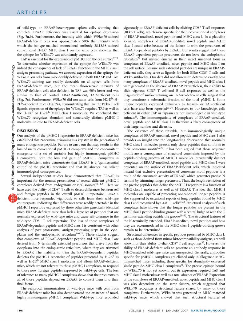
of wild-type or ERAAP-heterozygous spleen cells, showing thatcomplete ERAAP deficiency was essential for epitope expression(Fig. 7a,b). Furthermore, the intensity with which WEko.70 stainedERAAP-deficient cells was approximately 50% the intensity withwhich the isotype-matched monoclonal antibody 28.13.3S stainedconventional H-2Kb MHC class I on the same cells, showing thatthe epitope for WEko.70 was abundantly expressed.
TAP is essential for the expression of pMHC I on the cell surface7,19.To determine whether expression of the epitope for WEko.70 wasindeed the consequence of lack of ERAAP function in the MHC class Iantigen-processing pathway, we assessed expression of the epitope forWEko.70 on cells from mice doubly deficient in both ERAAP and TAP.WEko.70 staining was readily detectable on all spleen cells fromERAAP-deficient mice, but the mean fluorescence intensity ofERAAP-deficient cells also deficient in TAP was 90% lower and wassimilar to that of control ERAAP-sufficient, TAP-deficient cells(Fig. 7c). Furthermore, WEko.70 did not stain cells from H2-Kb–H-2Db–knockout mice (Fig. 7a), demonstrating that like the BEko T cellligands, expression of the epitope for WEko.70 required TAP as well asthe H-2Kb or H-2Db MHC class I molecules. We concluded thatWEko.70 recognizes abundant and structurally distinct pMHC Imolecules unique to ERAAP-deficient cells.
DISCUSSION
Our analysis of the pMHC I repertoire in ERAAP-deficient mice hasestablished that N-terminal trimming is a key step in the generation ofmany endogenous peptides. Failure to carry out that step results in theloss of many conventional pMHC I complexes and the concomitantemergence of a set of unstable but highly immunogenic pMHCI complexes. Both the loss and gain of pMHC I complexes inERAAP-deficient mice demonstrate that ERAAP is a ‘quintessentialeditor’ of the pMHC repertoire and that its absence has salientimmunological consequences.
Several independent studies have demonstrated that ERAAP isimportant for the normal presentation of several different pMHC Icomplexes derived from endogenous or viral sources10,11,20. Here wehave used the ability of CD8+ T cells to detect differences between selfand foreign peptides in the overall pMHC I repertoire. ERAAP-deficient mice responded vigorously to cells from their wild-typecounterparts, indicating that differences were readily detectable in thepMHC I repertoire expressed by those otherwise genetically identicalmice. ERAAP-deficient mice thus lack a large set of peptides that arenormally expressed by wild-type mice and cause self-tolerance in thewild-type CD8+ T cell repertoire. The loss of those complexes ofERAAP-dependent peptide and MHC class I is consistent with otheranalyses of post-proteasomal antigen-processing steps in the cyto-plasm and the endoplasmic reticulum4–6,21. Those studies suggestthat complexes of ERAAP-dependent peptide and MHC class I arederived from N-terminally extended precursors that arrive from thecytoplasm into the endoplasmic reticulum, where they are trimmedby ERAAP. The inability to trim the ERAAP-dependent peptidesdepletes the pMHC I repertoire of peptides presented by H-2Kb aswell as H-2Db MHC class I molecules and allows ERAAP-deficientmice, which are not tolerant to those pMHC I complexes, to respondto those now ‘foreign’ peptides expressed by wild-type cells. The lossof tolerance to many pMHC I complexes shows that the precursors toall of those peptides depend on ERAAP to convert them into theirfinal forms.
The reciprocal immunization of wild-type mice with cells fromERAAP-deficient mice has also demonstrated the existence of novel,highly immunogenic pMHC I complexes. Wild-type mice responded
vigorously to ERAAP-deficient cells by eliciting CD8+ T cell responses(BEko T cells), which were specific for the unconventional complexesof ERAAP-unedited, novel peptide and MHC class I. In a plausiblescenario, complexes of ERAAP-unedited, novel peptide and MHCclass I could arise because of the failure to trim the precursors ofERAAP-dependent peptides by ERAAP. Our results suggest that thoseERAAP-dependent peptide precursors do not leave the endoplasmicreticulum22 but instead emerge in their intact unedited form ascomplexes of ERAAP-unedited, novel peptide and MHC class I onthe cell surface. Because such extended peptides are unique in ERAAP-deficient cells, they serve as ligands for both BEko CD8+ T cells andWEko antibodies. Our data did not allow us to determine exactly howmany complexes of ERAAP-unedited, novel peptide and MHC class Iwere generated in the absence of ERAAP. Nevertheless, their ability toelicit vigorous CD8+ T cell and B cell responses as well as themagnitude of surface staining on ERAAP-deficient cells suggest thatthey constitute a substantial fraction of the total pMHC I. Someunique peptides expressed exclusively by tapasin- or TAP-deficientcells have also been reported23,24. However, to our knowledge, cellsdeficient in either TAP or tapasin are not immunogenic in wild-typeanimals25. The immunogenicity of complexes of ERAAP-unedited,novel peptide and MHC class I is therefore a likely consequence oftheir large number and diversity.
The existence of these unstable, but immunologically uniquecomplexes of ERAAP-unedited, novel peptide and MHC class I alsoprovides an insight into the longstanding observation that normallyMHC class I molecules present only those peptides that conform totheir consensus motifs16,26. It has been argued that those sequencemotifs are a consequence of structural constraints imposed by thepeptide-binding grooves of MHC I molecules. Structurally distinctcomplexes of ERAAP-unedited, novel peptide and MHC class I werepresented on the surface of ERAAP-deficient cells, which indicatedinstead that exclusive presentation of consensus motif peptides is aresult of the enzymatic activity of ERAAP, which generates precise Ntermini by trimming longer precursors. Thus, the length restriction ofthe precise peptides that define the pMHC I repertoire is a function ofMHC class I molecules as well as of ERAAP. The idea that MHC Imolecules are capable of presenting noncanonical longer peptides isalso supported by occasional reports of long peptides bound by MHCclass I and recognized by CD8+ T cells27,28. Structural analyses of suchcomplexes have shown that the longer peptides are present in theMHC class I peptide-binding groove with a central bulge or with the Cterminus extending outside the groove29–32. The structural features ofthe N-terminally extended, ERAAP-unedited, novel peptides and howthey are accommodated in the MHC class I peptide-binding grooveremain to be determined.
Structural differences in specific peptides presented by MHC class I,such as those derived from minor histocompatibility antigens, are wellknown for their ability to elicit CD8+ T cell responses33. However, theability of ERAAP-deficient cells to generate an antibody response inMHC-matched wild-type mice was unexpected. Generally, antibodiesspecific for pMHC I complexes are elicited only in allogeneic MHC-mismatched mice, including those specific for abundantly expressedsingle peptide–MHC class I complexes34. The precise epitope boundby WEko.70 is not yet known, but its expression required TAP andMHC class I molecules as well as a total absence of ERAAP. Expressionof the complexes of ERAAP-unedited, novel peptide and MHC class Iwas also dependent on the same factors, which suggested thatWEko.70 recognizes a structural feature shared by many of thosecomplexes. Furthermore, WEko.70 was generated in MHC-matchedwild-type mice, which showed that such structural features of
106 VOLUME 8 NUMBER 1 JANUARY 2007 NATURE IMMUNOLOGY
A R T I C L E S©
2007
Nat
ure
Pub
lishi
ng G
roup
ht
tp://
ww
w.n
atur
e.co
m/n
atur
eim
mun
olog
y

complexes of ERAAP-unedited, novel peptide and MHC class I mustbe unique and abundant to account for their immunogenicity. Thus,the antibody response of wild-type mice to ERAAP-deficient cells isanother immunological consequence of ERAAP deficiency.
Finally, we note the paradox that inhibiting a key component of theantigen-processing pathway actually enhanced the immunogenicity ofcells similar to that of allogeneic cells with mismatched MHC.Notably, the enhancement of immunogenicity as a consequence ofERAAP inhibition did not require mismatched MHC, polymorphicdifferences in minor histocompatibility genes or mutations in genesencoding normal proteins. Thus, it may be possible to enhance theimmunogenicity of tumors by inhibiting ERAAP or, conversely, loss ofERAAP function in self tissues could result in autoimmunity.
METHODSMice, cell lines, immunization and CD8+ T cell responses. Use of all mice was
with the approval of the Animal Care and Use Committee of the University of
California at Berkeley. ERAAP-deficient mice and fibroblast cell lines derived
from those mice have been described10. ERAAP-deficient mice were back-
crossed nine times to the C57BL/6J (H-2b) background, and ERAAP-
heterozygous mice were their littermates. Mice deficient in both ERAAP
and TAP, mice expressing H-2d MHC, and H2-Kb–H-2Db–knockout
mice have been described10,12. Wild-type C57BL/6J, TAP-deficient, wild-type
B10.D2 (H-2d) and C57BL/6J.C-H2bm1 mice were purchased from the
Jackson Laboratory.
Female ERAAP-deficient and wild-type mice were immunized intraperito-
neally with 20 � 106 spleen cells from male wild-type or ERAAP-deficient mice,
respectively. Then, 10 d after immunization, whole spleens were restimulated
for 5 d in vitro in cultures containing 20 U/ml of interleukin 2 (BD Biosciences)
and irradiated spleen cells from female mice of the same genotype used for
immunization. Subsequent restimulations were done similarly with 50 U/ml of
interleukin 2 every 7 d, after removal of dead cells by Ficoll fractionation (GE
Healthcare). CD8+ T cell HY peptide responses were assayed as described10.
The CD8+ T cell responses of the immunized mice were measured by assay for
inducible IFN-g production by those cells in response to APCs from female
mice. Unless otherwise indicated, APCs were splenocytes from female mice and
were incubated overnight in 200 ng/ml of lipopolysaccharide (Sigma) and
samples were depleted of CD8+ cells by Dynabeads conjugated to anti-CD8
(Dynal Biotech). Restimulated CD8+ T cells and APCs were cultured together
for 4 h in the presence of brefeldin A before being stained for surface CD8 and
intracellular IFN-g (BD Biosciences). Cells were then analyzed on a FACScan
(Coulter) and data were analyzed with FlowJo Software (Treestar). Numbers are
reported as above the background response of T cells alone.
Antibody blocking and treatment of APCs with brefeldin A and IFN-c.
For inhibition of presentation by MHC class I molecules, APCs were incubated
for 20 min at 4 1C with supernatant from hybridoma 5F1 (anti-H-2Kb),
B22.249 (anti-H-2Db) or M5/1114 (anti-H-2Ab) before being cultured together
with CD8+ T cells as described above. The coculture medium also contained
50% of the same hybridoma supernatant used during the 4 1C incubation. For
analysis of the cell surface stability of the MHC class I ligands, APCs were
preincubated for 2 or 4 h at 37 1C in medium containing 8 mg/ml of brefeldin A
(Sigma). CD8+ T cells producing IFN-g were counted as described above.
ERAAP-deficient fibroblasts expressing either vector or cDNA encoding human
ERAAP were generated as described10 and were treated for 2 d with 500 U/ml
of recombinant mouse IFN-g (Biosource) before being used as APCs for
BEko T cells.
BMDCs and leucinethiol treatment. Bone marrow was extracted from female
ERAAP-deficient, wild-type and H2-Kb–H-2Db–knockout mice and cells were
cultured for 5–7 d in 20 ng/ml of granulocyte-macrophage colony-stimulating
factor to enrich the growth of DCs. Nonadherent cells were collected and were
replated in medium only or in medium containing 0.5 mM dithiothreitol alone
or dithiothreitol plus 30 mM leucinethiol (Sigma) as described18. After 5 h,
nonadherent cells were used as APCs for BEko T cells.
Antibody responses of wild-type mice to ERAAP-deficient cells and genera-
tion of WEko.70. Female wild-type C57BL/6J mice were immunized intra-
peritoneally with 20 � 106 spleen cells from male ERAAP-deficient mice.
Mice were boosted every 3 weeks and antiserum and spleens were collected
after five boosts. Spleens were fused with the P3X63-Ag8.653 myeloma fusion
partner according to the standard protocol for generating antibody-secreting
hybridomas. For screening for positive cells and for analysis of antibody
specificity, supernatants from terminal cultures or antiserum were used to
stain ERAAP-deficient, wild-type and H2-Kb–H-2Db–knockout spleen cell
samples depleted of B cells with Dynabeads conjugated to anti–mouse
immunoglobulin G. The specificity of the antibody for ERAAP-deficient cells
was demonstrated by comparison of the mean fluorescence intensity of the
staining of ERAAP-deficient cells to that of cells doubly deficient for ERAAP
and TAP, ERAAP-heterozygous cells or wild-type cells. Ascites fluid from mice
injected with 28.13.3S (immunoglobulin M) was used to stain conventional
H-2Kb MHC class I. The secondary antibody was fluorescein isothiocyanate–
conjugated goat anti–mouse immunoglobulin G (Cappel). Supernatant from
the WEko.70 hybridoma was purified with a protein L column (Pierce) and was
biotinylated with EZ-Link NHS-PEO solid-phase biotinylation reagents
(Pierce). Biotinylated WEko.70 was used to stain whole spleen cell suspensions,
and phycoerythrin-conjugated strepavidin (Caltag) was used as the secondary
staining reagent.
Statistical methods. Mean values and standard deviations were calculated with
GraphPad Prism (GraphPad Software).
Note: Supplementary information is available on the Nature Immunology website.
ACKNOWLEDGMENTSSupported by the US National Institutes of Health (N.S.).
AUTHOR CONTRIBUTIONSG.E.H. contributed to the experimental design, immunized mice, didexperiments, screened hybridomas, analyzed data and cowrote the manuscript;F.G. generated and backcrossed ERAAP-deficient mice and generated the WEkohybridomas and the biotinylated derivative of WEko.70; E.J. contributed to theexperimental design and did skin grafts; H.N. sorted cells; and N.S. establishedthe initial scientific questions, provided guidance and cowrote the manuscript.
COMPETING INTERESTS STATEMENTThe authors declare that they have no competing financial interests.
Published online at http://www.nature.com/natureimmunology/
Reprints and permissions information is available online at http://npg.nature.com/
reprintsandpermissions/
1. Shastri, N., Schwab, S. & Serwold, T. Producing nature’s gene-chips. The generation ofpeptides for display by MHC class I molecules. Annu. Rev. Immunol. 20, 463–493(2002).
2. Cresswell, P., Ackerman, A.L., Giodini, A., Peaper, D.R. & Wearsch, P.A. Mechanisms ofMHC class I-restricted antigen processing and cross-presentation. Immunol. Rev. 207,145–157 (2005).
3. Shastri, N., Cardinaud, S., Schwab, S.R., Serwold, T. & Kunisawa, J. All the peptidesthat fit: the beginning, the middle, and the end of the MHC class I antigen-processingpathway. Immunol. Rev. 207, 31–41 (2005).
4. Kunisawa, J. & Shastri, N. The group II chaperonin TRiC protects proteolytic inter-mediates from degradation in the MHC class I antigen processing pathway. Mol. Cell12, 565–576 (2003).
5. Kunisawa, J. & Shastri, N. Hsp90a chaperones large proteolytic intermediates in theMHC class I antigen processing pathway. Immunity 24, 523–534 (2006).
6. Rock, K.L., York, I.A. & Goldberg, A.L. Post-proteasomal antigen processing formajor histocompatibility complex class I presentation. Nat. Immunol. 5, 670–677(2004).
7. Van Kaer, L., Ashton-Rickardt, P.G., Ploegh, H.L. & Tonegawa, S. TAP1 mutant mice aredeficient in antigen presentation, surface class I molecules, and CD4–8+ T cells. Cell71, 1205–1214 (1992).
8. Serwold, T., Gonzalez, F., Kim, J., Jacob, R. & Shastri, N. ERAAP customizes peptidesfor MHC class I molecules in the endoplasmic reticulum. Nature 419, 480–483(2002).
9. York, I.A. et al. The ER aminopeptidase ERAP1 enhances or limits antigen presentationby trimming epitopes to 8–9 residues. Nat. Immunol. 3, 1177–1184 (2002).
10. Hammer, G.E., Gonzalez, F., Champsaur, M., Cado, D. & Shastri, N. The aminopepti-dase ERAAP shapes the peptide repertoire displayed by major histocompatibilitycomplex class I molecules. Nat. Immunol. 7, 103–112 (2006).
NATURE IMMUNOLOGY VOLUME 8 NUMBER 1 JANUARY 2007 107
A R T I C L E S©
2007
Nat
ure
Pub
lishi
ng G
roup
ht
tp://
ww
w.n
atur
e.co
m/n
atur
eim
mun
olog
y

11. Yan, J. et al. In vivo role of ER-associated peptidase activity in tailoring peptides forpresentation by MHC class Ia and class Ib molecules. J. Exp. Med. 203, 647–659(2006).
12. Perarnau, B. et al. Single H2Kb, H2Db and double H2KbDb knockout mice: peripheralCD8+ T cell repertoire and anti-lymphocytic choriomeningitis virus cytolytic responses.Eur. J. Immunol. 29, 1243–1252 (1999).
13. Sherman, L.A. & Chattopadhyay, S. The molecular basis of allorecognition. Annu. Rev.Immunol. 11, 385–402 (1993).
14. Loveland, B., Wang, C-R., Yonekawa, H., Hermel, E. & Lindahl, K.F. Maternallytransmitted histocompatibility antigen of mice: A hydrophobic peptide of a mitochond-rially encoded protein. Cell 60, 971–980 (1990).
15. Madden, D.R., Gorga, J.C., Strominger, J.L. & Wiley, D.C. The structure of HLA-B27reveals nonamer self-peptides bound in an extended conformation. Nature 353, 321–325 (1991).
16. Rammensee, H.G., Bachmann, J. & Stevanovic, S. in MHC Ligands and Peptide Motifs1–457 (Landes Bioscience, Austin, Texas, 1997).
17. Yewdell, J.W. & Bennink, J.R. Brefeldin A specifically inhibits presentation of proteinantigens to cytotoxic T lymphocytes. Science 244, 1072–1075 (1989).
18. Serwold, T., Gaw, S. & Shastri, N. ER aminopeptidases generate a unique pool ofpeptides for MHC class I molecules. Nat. Immunol. 2, 644–651 (2001).
19. Cresswell, P., Bangia, N., Dick, T. & Diedrich, G. The nature of the MHC class I peptideloading complex. Immunol. Rev. 172, 21–28 (1999).
20. York, I.A., Brehm, M.A., Zendzian, S., Towne, C.F. & Rock, K.L. Endoplasmic reticulumaminopeptidase 1 (ERAP1) trims MHC class I-presented peptides in vivo and plays animportant role in immunodominance. Proc. Natl. Acad. Sci. USA 103, 9202–9207(2006).
21. Saveanu, L., Fruci, D. & van Endert, P. Beyond the proteasome: trimming, degradationand generation of MHC class I ligands by auxiliary proteases. Mol. Immunol. 39, 203–215 (2002).
22. Koopmann, J-O. et al. Export of antigenic peptides from the endoplasmic reticulumintersects with retrograde protein translocation through the Sec61p channel. Immunity13, 117–127 (2000).
23. van Hall, T. et al. Selective cytotoxic T-lymphocyte targeting of tumor immune escapevariants. Nat. Med. 12, 417–424 (2006).
24. Purcell, A.W. et al. Quantitative and qualitative influences of tapasin on the class Ipeptide repertoire. J. Immunol. 166, 1016–1027 (2001).
25. Wolpert, E.Z. et al. Generation of CD8+ T cells specific for transporter associated withantigen processing deficient cells. Proc. Natl. Acad. Sci. USA 94, 11496–11501(1997).
26. Falk, K., Rotzschke, O., Stevanovic, S., Jung, G. & Rammensee, H-G. Allele-specificmotifs revealed by sequencing of self-peptides eluted from MHC molecules. Nature351, 290–296 (1991).
27. Probst-Kepper, M. et al. An alternative open reading frame of the human macrophagecolony-stimulating factor gene is independently translated and codes for an antigenicpeptide of 14 amino acids recognized by tumor-infiltrating CD8 T lymphocytes. J. Exp.Med. 193, 1189–1198 (2001).
28. Green, K.J. et al. Potent T cell response to a class I-binding 13-mer viral epitope andthe influence of HLA micropolymorphism in controlling epitope length. Eur. J.Immunol. 34, 2510–2519 (2004).
29. Guo, H-C. et al. Different length peptides bind to HLA-Aw68 similarly at their ends butbulge out in the middle. Nature 360, 364–366 (1992).
30. Probst-Kepper, M. et al. Conformational restraints and flexibility of 14-meric peptidesin complex with HLA-B*3501. J. Immunol. 173, 5610–5616 (2004).
31. Tynan, F.E. et al. T cell receptor recognition of a ‘super-bulged’ major histo-compatibility complex class I-bound peptide. Nat. Immunol. 6, 1114–1122(2005).
32. Collins, E.J., Garboczi, D.N. & Wiley, D.C. Three-dimensional structure of apeptide extending from one end of a class I MHC binding site. Nature 371,626–629 (1994).
33. Roopenian, D., Choi, E.Y. & Brown, A. The immunogenomics of minor histocompat-ibility antigens. Immunol. Rev. 190, 86–94 (2002).
34. Porgador, A., Yewdell, J.W., Deng, Y., Bennink, J.R. & Germain, R.N. Localization,quantitation, and in situ detection of specific peptide-MHC class I complexes using amonoclonal antibody. Immunity 6, 715–726 (1997).
108 VOLUME 8 NUMBER 1 JANUARY 2007 NATURE IMMUNOLOGY
A R T I C L E S©
2007
Nat
ure
Pub
lishi
ng G
roup
ht
tp://
ww
w.n
atur
e.co
m/n
atur
eim
mun
olog
y