Human adipose-derived stromal cells for cell-based therapies in the treatment of systemic sclerosis
Upload
independentCategory
view
0download
0
RESEARCH ARTICLE
In Situ Normoxia Enhances Survival andProliferation Rate of Human AdiposeTissue-Derived Stromal Cells withoutIncreasing the Risk of TumourigenesisJane Ru Choi1, Belinda Pingguan-Murphy1, Wan Abu Bakar Wan Abas1, Kar Wey Yong1,Chi Tat Poon1, Mat Adenan Noor Azmi2, Siti Zawiah Omar2, Kien Hui Chua3, Feng Xu4,5,Wan Kamarul ZamanWan Safwani1*
1Department of Biomedical Engineering, Faculty of Engineering, University of Malaya, Lembah Pantai,50603 Kuala Lumpur, Malaysia, 2Department of Obstetrics and Gynaecology, Faculty of Medicine,University of Malaya, Lembah Pantai, 50603 Kuala Lumpur, Malaysia, 3 Department of Physiology, Facultyof Medicine, Universiti Kebangsaan Malaysia, Jalan Raja Muda Abdul Aziz, 50300 Kuala Lumpur, Malaysia,4 The Key Library of Biomedical Information Engineering of Ministry of Education, School of Life Science andTechnology, Xi’an Jiaotong University, Xi’an 710049, P.R. China, 5 Bioinspired Engineering andBiomechanics Center (BEBC), Xi’an Jiaotong University, Xi’an 710049, P.R. China
AbstractAdipose tissue-derived stromal cells (ASCs) natively reside in a relatively low-oxygen ten-
sion (i.e., hypoxic) microenvironment in human body. Low oxygen tension (i.e., in situ nor-
moxia), has been known to enhance the growth and survival rate of ASCs, which, however,
may lead to the risk of tumourigenesis. Here, we investigated the tumourigenic potential of
ASCs under their physiological condition to ensure their safe use in regenerative therapy.
Human ASCs isolated from subcutaneous fat were cultured in atmospheric O2 concentra-
tion (21% O2) or in situ normoxia (2% O2). We found that ASCs retained their surface mark-
ers, tri-lineage differentiation potential, and self-renewal properties under in situ normoxia
without altering their morphology. In situ normoxia displayed a higher proliferation and via-
bility of ASCs with less DNA damage as compared to atmospheric O2 concentration. More-
over, low oxygen tension significantly up-regulated VEGF and bFGF mRNA expression and
protein secretion while reducing the expression level of tumour suppressor genes p16, p21,
p53, and pRb. However, there were no significant differences in ASCs telomere length and
their relative telomerase activity when cultured at different oxygen concentrations. Collec-
tively, even with high proliferation and survival rate, ASCs have a low tendency of develop-
ing tumour under in situ normoxia. These results suggest 2% O2 as an ideal culture
condition for expanding ASCs efficiently while maintaining their characteristics.
PLOS ONE | DOI:10.1371/journal.pone.0115034 January 23, 2015 1 / 19
OPEN ACCESS
Citation: Choi JR, Pingguan-Murphy B, Wan AbasWAB, Yong KW, Poon CT, Noor Azmi MA, et al.(2015) In Situ Normoxia Enhances Survival andProliferation Rate of Human Adipose Tissue-DerivedStromal Cells without Increasing the Risk ofTumourigenesis. PLoS ONE 10(1): e0115034.doi:10.1371/journal.pone.0115034
Academic Editor: Zoran Ivanovic, French BloodInstitute, FRANCE
Received: April 5, 2014
Accepted: November 18, 2014
Published: January 23, 2015
Copyright: © 2015 Choi et al. This is an openaccess article distributed under the terms of theCreative Commons Attribution License, which permitsunrestricted use, distribution, and reproduction in anymedium, provided the original author and source arecredited.
Funding: This work was financially supported by theUniversity of Malaya, High Impact Research Grant(UM.C/HIR/MOHE/ENG/44) from the Ministry ofHigher Education Malaysia. FX was partiallysupported by the Major International Joint ResearchProgram of China (11120101002), the NationalNatural Science Foundation of China (11372243),and the National 111 Project of China (B06024). Thefunders had no role in study design, data collectionand analysis, decision to publish, or preparation ofthe manuscript.
IntroductionStem cells have attracted significant interest as a cell source for regenerative medicine and celltherapy due to their differentiation and self-renewal capacity [1–3]. Among various types ofstem cells, adipose tissue-derived stromal cells (ASCs) have recently become particularly attrac-tive due to the relative abundance and accessibility of adipose tissue [4–6]. Interestingly, de-spite the fact that adipose tissue is highly vascularised [7], they have been found residing in anative hypoxic microenvironment [8, 9], e.g., 3% oxygen concentration in mouse adipose tissue[10] and<4% O2 in human adipose tissue [11, 12]. However, oxygen tension is often over-looked when performing in vitro study of stem cells [13, 14], where 21% oxygen concentrationis normally used for culture of ASCs. Therefore, it is essential to understand the response ofASCs to in situ normoxia for developing cell-based therapy strategies.
As large numbers of ASCs are required for clinical application, many studies have empha-sized the effect of in situ normoxia on cell expansion. Most studies have demonstrated thathuman ASCs displayed a higher proliferation and survival rate at low oxygen concentration ascompared to normal oxygen concentration [15–17]. It has been reported that the release ofhypoxia inducible factor-1 alpha (HIF-1α) under low oxygen tension increased the expressionof growth factors, such as vascular endothelial growth factor (VEGF), basic fibroblast growthfactor (bFGF), epidermal growth factor (EGF), transforming growth factors (TGF), insulin-likegrowth factor (IGF), hepatocyte growth factor (HGF) and platelet-derived growth factor(PDGF) [12, 18–20]. These growth factors might in turn promote the growth and survival ofthe cells [21]. However, a high rate of proliferation and an anti-apoptotic characteristic resem-bles the behaviour of cancerous cells, suggesting the potential of malignant transformation andcancer development [22, 23]. Conversely, it has been suggested that culture of ASCs at low oxy-gen concentration may enhance their genetic stability [15]. To our knowledge, the tumouri-genic potential of ASCs under in situ normoxia has not yet been well investigated.
Notably, 2% O2 was most frequently reported to enhance the expansion and survival rateof ASCs [16, 17, 24–26]. Thus, a biosafety assessment should be performed prior to clinicaluse. There has been controversy in research findings regarding the tumorigenic potential ofMSCs. Some groups have reported that human MSCs may develop tumourigenesis afterlong-term culture (eg. beyond 5 weeks [27–30]). However, recent studies have shown thatMSCs can be expanded over multiple cell doublings (3–6 months) without chromosomal al-terations, demonstrating a low risk of tumourigenesis [31–33]. In this study, we aimed to de-termine the tumourigenic potential of ASCs cultured at 2% oxygen tension at early passage.We first characterized the ASCs by examining their morphology, differentiation capabilities,surface markers expression, and self-renewal properties under both atmospheric O2 concen-tration (21% O2) and in situ normoxia (2% O2). We then investigated the influence of in situnormoxia on viability and proliferation rate of ASCs, followed by determining the HIF-1α ex-pression level and the production of growth factors under this physiological condition. There-after, we evaluated the tumourigenic potential of ASCs in terms of tumour suppressor geneexpression level, alteration in telomere length and telomerase activity as well as the degree ofDNA damage.
Materials and Methods
Ethics StatementThis study was approved by the Medical Ethics Committee of University Malaya Medical Cen-tre (UMMC). Subcutaneous adipose tissue was collected from female donors aged 25 to 45 un-dergoing caesarean section with the informed written consent under a protocol approved by
Tumourigenic Potential of Human ASCs under In Situ Normoxia
PLOS ONE | DOI:10.1371/journal.pone.0115034 January 23, 2015 2 / 19
Competing Interests: The authors have declaredthat no competing interests exist.
the Medical Ethics Committee of UMMC (reference no. 996.46). Consent was obtained fromeach donor before collecting the samples.
Isolation and culture of human adipose tissue-derived stromal cellsThe adipose tissue was washed with phosphate buffered saline (PBS) (Sigma-Aldrich, St Louis,MO, USA), minced and digested with 0.3% collagenase I (Worthington, Freehold, NJ, USA).The digested tissue was washed with PBS and centrifuged. The pellet was cultured with Dulbec-co’s Modified Eagle’s Medium (DMEM)/Ham F-12 growth medium containing 10% FBS, 1%vitamin C, 1% glutamax, and 1% antibiotic-antimycotic solution (Gibco, Grand Island, NY,USA) in culture flasks. As a normal control group, the cells were cultured with 21% O2 and 5%CO2 at 37°C. The culture media was then replaced every 2–3 days. ASCs at passage 3 were usedfor all the tests unless otherwise stated.
In situ normoxiaASCs were cultured at 37°C in an O2 incubator (Galaxy 170 R, New Brunswick Scientific,USA). The incubator was supplied with 2% O2 and 5% CO2 whereby low O2 concentration wasmaintained with nitrogen gas (N2) supply. The oxygen level was confirmed with Jenway 970portable dissolved oxygen meter (Bibby Scientific Limited, Staffordshire, UK). Similar to thecontrols (cells cultured under atmospheric O2 concentration), the culture media under in situnormoxia was replaced every 2–3 days. ASCs at passage 3 were tested immediately after theyhad been taken out from the incubator.
ASC characterizationTo make sure that the cells used in this experiment are mesenchymal stem cells (MSCs), we fol-lowed the three standard criteria suggested by Dominici et al. [34], stating that, firstly MSCsmust be plastic-adherent. Secondly, they must express their surface markers CD73, CD90 andCD105 while showing negative for haematopoetic markers CD14, CD34 and CD45, B cellmarker CD 19, and histocompatibility class II cell surface receptor HLA-DR, DP, DQ whichare found only on antigen-presenting cells and B cells. Additional histocompatibility class I cellsurface receptor, HLA-ABC was also tested in this study which is typically expressed by MSCs.Thirdly, MSCs must possess the potential to differentiate into adipocytes, osteoblasts andchondroblasts [34].
In this study, the morphology of the cells was examined, followed by determining the ex-pression of their surface markers and their capability of differentiation. Flow cytometry wasperformed to identify the ASCs surface markers under in situ normoxia and atmosphericO2 concentration. ASCs from both groups were harvested, washed and immuno-stained withFITC-conjugated antibodies against CD105, CD90, CD45, CD34, HLA-ABC and HLA-DR,DP, DQ and PE-conjugated antibodies against CD73, CD14 and CD19 (Becton Dickinson,San Jose, CA, USA). Negative control staining was done using FITC-conjugated mouse IgG1& IgG2 isotypes, and PE-conjugated mouse IgG1 & IgG2 isotypes (Becton Dickinson). Flowcytometry was conducted using BD FACSCanto II (Becton Dickinson). A minimum of 10000events for each sample were acquired and analysed with FlowJo analysis software (Treestar,Ashland, OR, USA).
For adipogenic and osteogenic differentiation, ASCs at passage 3 were initially grown inpetri dishes with growth medium which was then replaced with their corresponding inductionmedium when they reached confluence. Adipogenic induction medium contained high glucoseDMEM supplemented with 10% FBS (Gibco), 0.5 μM isobutyl-1-methyl xanthine, 1 μM dexa-methasone and 200 μM indomethacin (Sigma-Aldrich). Osteogenic induction medium
Tumourigenic Potential of Human ASCs under In Situ Normoxia
PLOS ONE | DOI:10.1371/journal.pone.0115034 January 23, 2015 3 / 19
contained high glucose DMEM with 10% FBS (Gibco), 0.05 mM ascorbic acid-2-phosphate,10 mM b-glycerophosphate and 100 nM dexamethasone (Sigma-Aldrich). Differentiation me-dium was changed every 3 days for up to 21 days, with the cells cultured in both atmosphericO2 concentration and in situ normoxia. Adipogenic differentiation was confirmed by detectingthe presence of lipid droplets stained with Oil red O (Sigma-Aldrich), whereas osteogenenicdifferentiation was assessed by staining of calcium deposits using Alizarin red (Sigma-Aldrich).For chondrogenic differentiation, ASCs at passage 3 were pelleted and cultured in chondro-genic induction medium composed of DMEM/F12 supplemented with 1% FBS, 1% glutamax,1% vitamin C, 1% antibiotic-antimycotic (Gibco), ITS premix (Becton Dickinson), 40μg/mlL-proline, 100nM dexamethasone, 50 μg/ml ascorbate- 2-phophate (Sigma-Aldrich), 50 ng/mlIGF-1 and 10 ng/ml TGF-β1 (Peprotech, Rocky Hill, NJ, USA). Culture medium was changedevery 3 days for 21 days. For histological examination, briefly, pellets were fixed overnight in10% formalin (Sigma-Aldrich) and processed according to the standard procedures forsample processing, embedding and sectioning. Proteoglycans accumulation was assessed usingalcian blue staining (Sigma-Aldrich). The stained sections were mounted with mounting medi-um (DPX) (Sigma-Aldrich) and visualized with a light microscope (Eclipse TS100, Nikon,NY, USA).
Colony-forming unit-fibroblast (CFU-F) assaysASCs at P3 were plated at a density of 400 cells/ cm2 in 100-mm culture dishes under in situnormoxia or atmospheric O2 concentration. The medium was changed every 3 days for a peri-od of 14 days. The culture dishes were rinsed with PBS, fixed with 10% formalin solution andstained for 20 min with Giemsa stain (Sigma-Aldrich). After rinsing with PBS, colonies consistof more than 50 stained cells were counted. Total colony numbers was determined.
Cell proliferation assaysCell proliferation was assessed by Resazurin reduction assay and population doubling time.Briefly, about 140 mg Resazurin sodium salts (Sigma-Aldrich) was dissolved in 1 L PBS(Sigma-Aldrich, USA) to prepare Resazurin stock solution. Resazurin 10% working solutionwas then prepared from the stock solution. ASCs were seeded onto a 24-well plate at 5 × 10⁴
cells per well with growth media on day 0. Resazurin assay was performed on day 1, 3, 7, 10,14, 17 and 21. The absorbance signals were quantified at excitation wavelength of 570 nm andemission wavelength of 595nm, using a FLUOstar Optima microplate reader (BMG Labtech,Offenburg, Germany). Viable cells were quantified as percentage of Resazurin reduction. Atthe same time, cell suspensions were serially diluted and seeded into a 24-well plate. After24 hours of incubation, the percentage of Resazurin reduction was determined following thesteps explicitly mentioned above. The relationship between Resazurin reduction (%) and cellnumbers was determined by plotting a standard curve. A growth curve was then generated byplotting a graph of cell number versus time to compare the growth rate between both groups.
The growth kinetic was evaluated to further determine the proliferation ability of ASCs atdifferent conditions. This was determined by population doubling time (PDT, time required fora culture to double in number). Cells were cultured under two different conditions until passage3 and the number of cells was determined at the beginning and the end of each passage. Num-ber of cell doublings were calculated based on the formula n = (log10 Nh- log10 Ni) /log10 2,where Nh is the cell number at the end of passage and Ni is the cell number at initial seeding.Population doubling time was calculated as a ratio of number of days in culture divided by thenumber of cell doublings at each passage. Mean PDT of 3 passages was then determined.
Tumourigenic Potential of Human ASCs under In Situ Normoxia
PLOS ONE | DOI:10.1371/journal.pone.0115034 January 23, 2015 4 / 19

Cell cycle analysisSamples for cell cycle analysis were prepared using CycleTEST Plus DNA Reagent Kit (BectonDickinson) in accordance with the manufacturer’s instruction. Flow cytometry was performedusing BD Accuri C6 (Becton Dickinson). The percentage of cells in G0/G1, S and G2/M phasesof cell cycle was determined using ModFit LT 4.0 software (Verity Software House, Topsham,ME, USA).
Apoptosis assayThe apoptosis assay was performed using Annexin V: FITC Apoptosis Detection Kit I (BectonDickinson) following the manufacturer’s instructions. Flow cytometry analysis was carried outusing BD Accuri C6 (Becton Dickinson).
Alkaline comet assayThe comet assay was conducted using alkaline conditions according to the manufacturer’sinstruction of Comet Assay Kit (Trevigen, Gaitherburg, MD, USA). After staining withethidium bromide (Invitrogen, Carlsbad, CA, USA), images (magnification 100x) were ob-served and captured using inverted fluorescence microscope (ECLIPSE TI-S, Nikon, NY,USA) connected to a digital camera. About 100 nuclei per sample were analyzed usingComet Assay IV 4.3 software (Perspective Instrument, Haverhill, UK). The three parameters:percentage of DNA in the tail, tail length and tail moment were used to evaluate the DNAdamage.
RNA extraction, cDNA synthesis and quantitative real-time polymerasechain reaction (qPCR)RNA extraction was performed using TRI reagent (Ambion, Austin, TX, USA), followed byphase separation with chloroform (Fisher Scientific, Loughborough, UK) and RNA precipitationwith isopropanol (Sigma-Aldrich). The high capacity RNA-to-cDNA kit (Applied Biosystems,Foster City, CA, USA) was used to synthesize cDNAs. Then qPCR was conducted with TaqMangene expression assays (Applied Biosystems) using StepOnePlus Real-Time PCR system (Ap-plied Biosystem). The genes tested include HIF-1a (Hs00153153_m1), TP53 (Hs00153349_m1),CDKN1A (Hs00355782_m1), CDKN2A (Hs00923894_ml), TERT (Hs00972656_ml), RB1(Hs01078066_ml), VEGFA (Hs00173626_ml), PDGFA (Hs00234994_ml), FGF2 (Hs00266-645_ml), HGF (Hs00300159_ml), IGF1 (Hs01547656_ml), EGF (Hs01099999_ml), TGFB1(Hs00171257_ml) and GAPDH (Hs99999905_ml). GAPDH was used as housekeeping gene fornormalization. The thermal cycling conditions include incubation for 20 s at 95°C to activateAmpliTaq Fast DNA polymerase, followed by 40 cycles of denaturation for 1 s at 95°C, annealingand elongation for 20 s at 60°C. The gene expression of control group (atmospheric O2 concen-tration) was normalized to 1. The results were expressed as fold changes in gene expression rela-tive to the control.
ELISAAs VEGF and bFGF gene expression level were significantly higher under in situ normoxiathan atmospheric O2 concentration as indicated by qRT-PCR, their production were quanti-fied using human Quantikine ELISA kit (R&D Systems, Minneapolis, MN, USA) following themanufacturer’s instructions.
Tumourigenic Potential of Human ASCs under In Situ Normoxia
PLOS ONE | DOI:10.1371/journal.pone.0115034 January 23, 2015 5 / 19

Telomerase assayTelomerase activity of ASCs was determined using the TeloTAGGG PCR ELISA PLUS kit(Roche, Mannheim, Germany), based on telomere repeat amplification protocol (TRAP) ac-cording to the manufacturer’s recommendations. The relative telomerase activities (RTA) weredetermined using the formula provided by the manufacturer.
Telomere length analysisGenomic DNA was extracted from ASCs cultured in two different conditions using thePureLink Genomic DNAMini Kit (Invitrogen). Telomere length analysis was carried out usingTeloTAGGG Telomere Length Assay kit (Roche) in accordance with the manufacturer’s instruc-tions. The chemiluminescence was detected by gel documentation imaging system (Fusion FX5,Vilber Lourmat, France). By comparing the signal relative to a molecular weight standard, themean telomere restriction fragment (TRF) length was quantified.
Statistical analysisStudent’s t test was used to compare the normally distributed data between in situ normoxiaand atmospheric O2 concentration. Data were expressed as mean ± standard error of the mean(SEM) of six different experiments with six different donors (n = 6). p< 0.05 was reported asstatistically significant.
Results
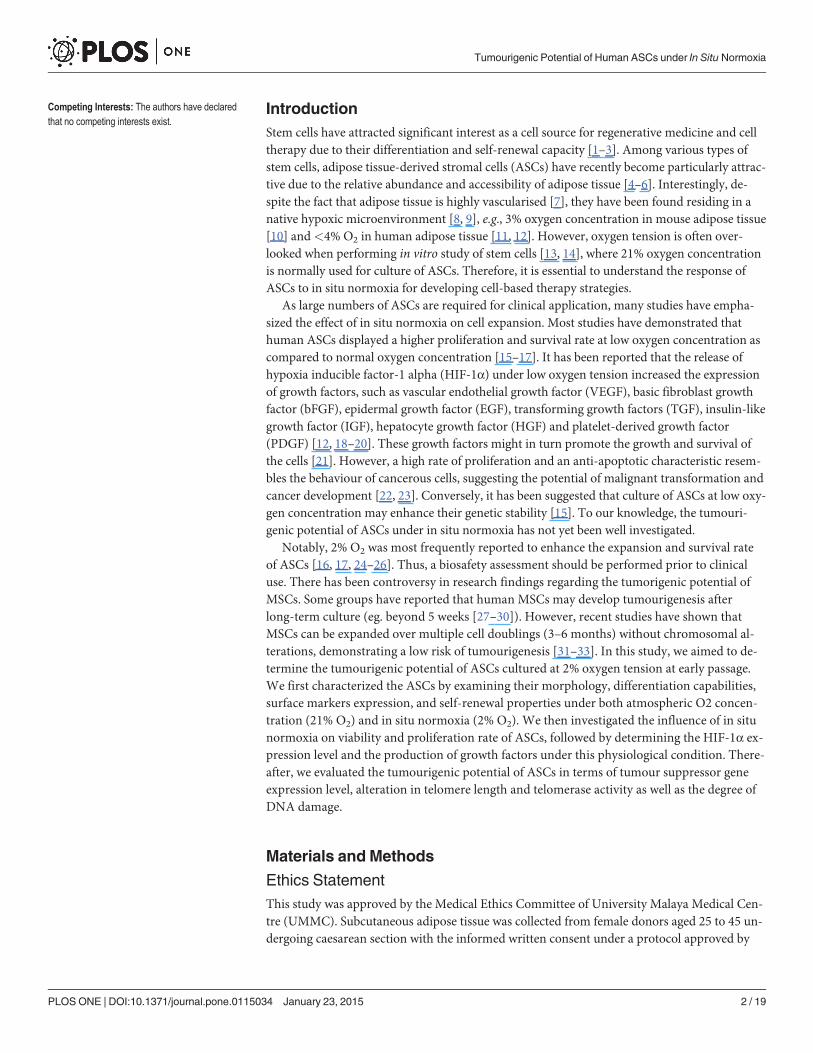
ASCs retain their characteristics under in situ normoxiaTo determine if ASCs are able to maintain their characteristics under in situ normoxia, theirmorphological feature was first observed. Under both in situ normoxia and atmospheric O2concentration, ASCs displayed adherent fibroblast-like features. There were no morphologicalchanges from day 1 to day 10 of the culture (Fig. 1A). In addition to morphological examina-tion, ASCs were further characterized through the expression of their surface markers andtheir differentiation capabilities. The result of flow cytometry analysis showed no significantdifferences in ASCs surface marker expression at different oxygen tensions. They were positivefor the stem cell surface markers CD73, CD90, CD105 and HLA-ABC, and negative for HLA-DR DP DQ, CD14, CD19, CD34 and CD45, indicating that in situ normoxia did not alter thephenotype of ASCs (S1 Fig.). Moreover, like atmospheric O2 concentration, in situ normoxiaalso maintained the adipogenic, osteogenic and chondrogenic differentiation abilities of ASCs,as indicated by the positive staining (S2 Fig.). Additionally, to show the presence of cells exhib-iting the functional properties of MSC, we performed colony-forming unit-fibroblast (CFU-F)assay. Our result showed the formation of single colonies of ASC, representing the cell popula-tion with self-renewal properties. There was no significant difference in the number of coloniesin both groups, indicating the maintenance of the stemness properties of ASC under in situnormoxia (Fig. 1B).
In situ normoxia enhances the expansion rate of ASCsTo determine if the cells grow more rapidly in their physiological condition, we measured theproliferation rate of ASCs at different oxygen tensions. Even though there was no alteration intheir morphology, we observed a remarkable increase in cell density under in situ normoxiafrom day 7 to day 10 as compared to atmospheric O2 concentration (Fig. 1A). This indicatesan increased proliferation rate of ASCs under in situ normoxia. To further quantify the growthrate of cells, Resazurin reduction assay and population doublings time were assessed. We
Tumourigenic Potential of Human ASCs under In Situ Normoxia
PLOS ONE | DOI:10.1371/journal.pone.0115034 January 23, 2015 6 / 19
Figure 1. In situ normoxia enhanced the expansion rate of ASCs without changing their morphology and self-renewal properties. Cells maintainedfibroblast-like appearance on day 3, 7 and 10 of culture. The density of the cells was remarkably increased under in situ normoxia, magnification 100x (A). Insitu normoxia maintained the number of CFU-F. Macroscopic examination showed the single colonies of ASCs, representing the population which exhibitsthe self-renewal properties of MSC (B). Growth curve of ASCs (C) showed an increased rate of cell proliferation under in situ normoxia. Population doublingtime (D) of ASCs was significantly lower under in situ normoxia than in atmospheric O2 concentration. Cell cycle analysis showed the percentage of ASCs inS-phase was higher under in situ normoxia as compared to atmospheric O2 concentration (E). Data shown represent as mean ± SEM. * indicates p< 0.05relative to atmospheric O2 concentration.
doi:10.1371/journal.pone.0115034.g001
Tumourigenic Potential of Human ASCs under In Situ Normoxia
PLOS ONE | DOI:10.1371/journal.pone.0115034 January 23, 2015 7 / 19
found an increased number of ASCs under in situ normoxia as compared to atmospheric O2concentration from day 7 of the culture. Specifically, at day 10, ASCs cultured under in situnormoxia had a 1.8-fold greater cell number (13.75 × 10⁴ ± 0.09 × 10⁴) than atmospheric O2concentration (7.5 × 10⁴ ± 0.039 × 10⁴) (Fig. 1C). Additionally, at 2% O2, ASCs displayed alower population doubling time as compared to atmospheric O2 concentration with the meanpopulation doubling time of 3.97 ± 0.31 days and 6.93 ± 0.75 days, respectively, p< 0.05(Fig. 1D).
To confirm the result of cell proliferation, cell cycle analysis was carried out by flowcytometry. The result showed that the average percentage of cells in G0/G1 phase was signifi-cantly higher in atmospheric O2 concentration (87.59 ± 1.27%) than under in situ normoxia(71.23 ± 0.095%), whereas the fraction of cells in S phase was higher under in situ normoxia(10.32 ± 0.14%) than atmospheric O2 concentration (1.38 ± 0.25%), p< 0.05 (Fig. 1E).
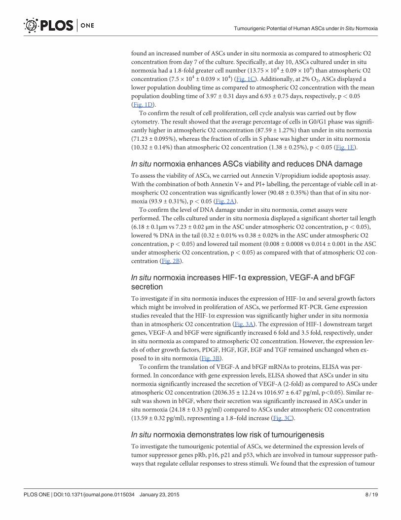
In situ normoxia enhances ASCs viability and reduces DNA damageTo assess the viability of ASCs, we carried out Annexin V/propidium iodide apoptosis assay.With the combination of both Annexin V+ and PI+ labelling, the percentage of viable cell in at-mospheric O2 concentration was significantly lower (90.48 ± 0.35%) than that of in situ nor-moxia (93.9 ± 0.31%), p< 0.05 (Fig. 2A).
To confirm the level of DNA damage under in situ normoxia, comet assays wereperformed. The cells cultured under in situ normoxia displayed a significant shorter tail length(6.18 ± 0.1μm vs 7.23 ± 0.02 μm in the ASC under atmospheric O2 concentration, p< 0.05),lowered % DNA in the tail (0.32 ± 0.01% vs 0.38 ± 0.02% in the ASC under atmospheric O2concentration, p< 0.05) and lowered tail moment (0.008 ± 0.0008 vs 0.014 ± 0.001 in the ASCunder atmospheric O2 concentration, p< 0.05) as compared with that of atmospheric O2 con-centration (Fig. 2B).
In situ normoxia increases HIF-1α expression, VEGF-A and bFGFsecretionTo investigate if in situ normoxia induces the expression of HIF-1α and several growth factorswhich might be involved in proliferation of ASCs, we performed RT-PCR. Gene expressionstudies revealed that the HIF-1α expression was significantly higher under in situ normoxiathan in atmospheric O2 concentration (Fig. 3A). The expression of HIF-1 downstream targetgenes, VEGF-A and bFGF were significantly increased 6 fold and 3.5 fold, respectively, underin situ normoxia as compared to atmospheric O2 concentration. However, the expression lev-els of other growth factors, PDGF, HGF, IGF, EGF and TGF remained unchanged when ex-posed to in situ normoxia (Fig. 3B).
To confirm the translation of VEGF-A and bFGF mRNAs to proteins, ELISA was per-formed. In concordance with gene expression levels, ELISA showed that ASCs under in situnormoxia significantly increased the secretion of VEGF-A (2-fold) as compared to ASCs underatmospheric O2 concentration (2036.35 ± 12.24 vs 1016.97 ± 6.47 pg/ml, p<0.05). Similar re-sult was shown in bFGF, where their secretion was significantly increased in ASCs under insitu normoxia (24.18 ± 0.33 pg/ml) compared to ASCs under atmospheric O2 concentration(13.59 ± 0.32 pg/ml), representing a 1.8–fold increase (Fig. 3C).
In situ normoxia demonstrates low risk of tumourigenesisTo investigate the tumourigenic potential of ASCs, we determined the expression levels oftumor suppressor genes pRb, p16, p21 and p53, which are involved in tumour suppressor path-ways that regulate cellular responses to stress stimuli. We found that the expression of tumour
Tumourigenic Potential of Human ASCs under In Situ Normoxia
PLOS ONE | DOI:10.1371/journal.pone.0115034 January 23, 2015 8 / 19
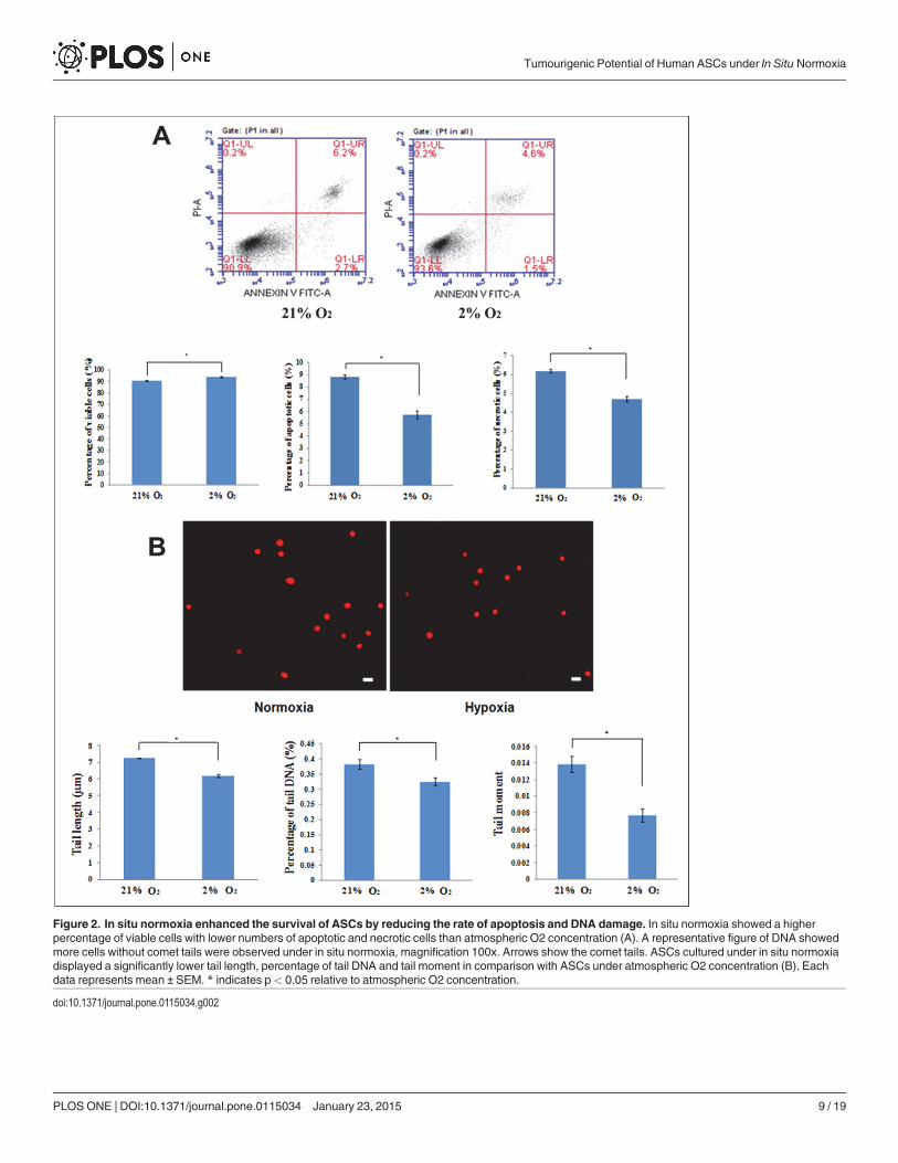
Figure 2. In situ normoxia enhanced the survival of ASCs by reducing the rate of apoptosis and DNA damage. In situ normoxia showed a higherpercentage of viable cells with lower numbers of apoptotic and necrotic cells than atmospheric O2 concentration (A). A representative figure of DNA showedmore cells without comet tails were observed under in situ normoxia, magnification 100x. Arrows show the comet tails. ASCs cultured under in situ normoxiadisplayed a significantly lower tail length, percentage of tail DNA and tail moment in comparison with ASCs under atmospheric O2 concentration (B). Eachdata represents mean ± SEM. * indicates p< 0.05 relative to atmospheric O2 concentration.
doi:10.1371/journal.pone.0115034.g002
Tumourigenic Potential of Human ASCs under In Situ Normoxia
PLOS ONE | DOI:10.1371/journal.pone.0115034 January 23, 2015 9 / 19
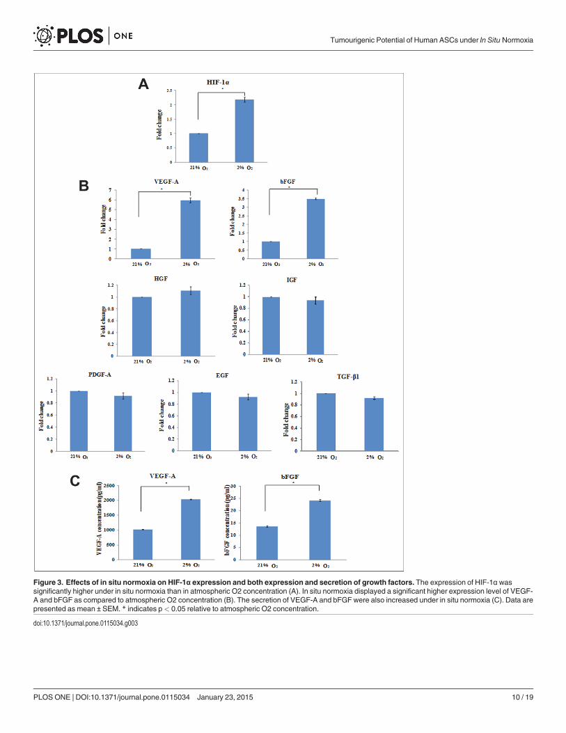
Figure 3. Effects of in situ normoxia on HIF-1α expression and both expression and secretion of growth factors. The expression of HIF-1α wassignificantly higher under in situ normoxia than in atmospheric O2 concentration (A). In situ normoxia displayed a significant higher expression level of VEGF-A and bFGF as compared to atmospheric O2 concentration (B). The secretion of VEGF-A and bFGF were also increased under in situ normoxia (C). Data arepresented as mean ± SEM. * indicates p< 0.05 relative to atmospheric O2 concentration.
doi:10.1371/journal.pone.0115034.g003
Tumourigenic Potential of Human ASCs under In Situ Normoxia
PLOS ONE | DOI:10.1371/journal.pone.0115034 January 23, 2015 10 / 19
suppressor genes pRb, p16, p21 and p53 were significantly lower under in situ normoxia thanatmospheric O2 concentration, suggesting that in situ normoxia down-regulates tumour sup-pressor activity (Fig. 4A).
To further assess the risk of tumourigenesis, the telomerase activity of ASCs and the changes intelomere length were determined. The telomerase activity of ASCs was first assessed by the expres-sion level of TERT using quantitative RT-PCR. hTERT was undetected in both culture conditions.In telomerase assay, we observed that there was no significant difference in relative telomerase ac-tivity of ASCs when cultured in both in situ normoxia (226.59 ± 3.92) and atmospheric O2 con-centration (223.67 ± 3.51) (Fig. 4B). Furthermore, no significant difference in mean TRF length
Figure 4. In situ normoxia lowered the expression of tumour suppressor genes andmaintained bothtelomerase activity and TRF length of ASCs. In situ normoxia displayed lower expression levels of p53,p21, p16 and pRb (A) as compared to atmospheric O2 concentration. There were no significant differencebetween both groups on their telomerase activity (B) and mean TRF length (C). Data are presented as mean± SEM. * indicates p< 0.05 relative to atmospheric O2 concentration.
doi:10.1371/journal.pone.0115034.g004
Tumourigenic Potential of Human ASCs under In Situ Normoxia
PLOS ONE | DOI:10.1371/journal.pone.0115034 January 23, 2015 11 / 19
was noted between ASCs cultured under both in situ normoxia (11.26 ± 0.88 kbp) and atmo-spheric O2 concentration (10.64 ± 0.31 kbp) (Fig. 4C).
DiscussionASCs at passage 3 were used in this study due to their homogeneity at this stage. Cells at earlierpassage have been shown to be more heterogeneous, which contain multiple types cell such asfibroblasts, pre-adipocytes, endothelial cells and other cell types, as indicated by low expressionlevels of MSCs major surface markers with high expression level of haematopoetic stem cellmarker CD 34 [35, 36]. To prevent haematopoetic cell contamination, which normally presentin early passage and might affect the result, most studies utilized the ASCs at their homoge-neous stage (eg. passage 3–6) [37–40].
Initially, characterization of ASCs was performed under both in situ normoxia and atmo-spheric O2 concentration to confirm that the cells used in our study were MSCs [41]. Our re-sults demonstrated that under both conditions, ASCs displayed adherent fibroblast-likemorphology. They were also able to express the major surface markers of MSCs and displayadipogenic, osteogenic and chondrogenic differentiation potential, which met the minimalcriteria of characterizing the human MSCs as suggested by Dominici et al. [34]. Additionally,the cells cultured under both conditions have also been proven to exhibit MSC functionalproperties (i.e., self-renewal properties) by CFU-F assay, as described by other studies[42–44].
Microscopic examination revealed that in situ normoxia (2% O2) was able to maintain themorphology of the cells. Remarkably, there was an increased density of cells under in situ nor-moxia as compared to atmospheric O2 concentration, suggesting that in situ normoxia in-creased the proliferation rate of ASCs. Moreover, ASCs culture at 2% O2 had an overall higherresazurin reduction and shorter population doubling time than ASCs at 21% O2, indicatingthat the cells cultured under in situ normoxia expanded more rapidly than that of atmosphericO2 concentration. Additionally, a higher cell percentage in S phase and lower in G0/G1 phaseof cell cycle suggests that a larger number of cells were in the stage of DNA replication under insitu normoxia, which is in agreement with the result on cell proliferation. Our results were con-sistent with many studies which have shown an increased proliferation rate of ASCs at 2% O2
[16, 17, 24, 25]. The positive proliferative effect of ASCs under in situ normoxia suggests thatthis physiologically relevant condition provides a more preferable environment for theirgrowth. It has been reported that the promotion of ASCs growth under their physiological con-dition might be due to the better cell adhesion and extracellular matrix formation [45] in thepresence of other factors (e.g. growth factors) [46, 47], rather than the change in self-renewalproperties of stem cells [48, 49].
Our data also demonstrate that the percentage of viable cells under in situ normoxia washigher than that of atmospheric O2 concentration with lower apoptosis rate, suggesting that insitu normoxia enhanced the survival of ASCs. Apoptosis represents a process of programmedcell death which normally occurs in response to multiple oncogenic stresses including DNAdamage, chromatin changes, telomeres dysfunction and mitogenic stimulation [50]. This pro-cess is important to prevent the uncontrolled cell division that might cause neoplastic transfor-mation [51]. The result of comet assay suggested that in situ normoxia caused less DNAdamage of ASCs in comparison to atmospheric O2 concentration. This further supports the re-sult of lower DNA damage-induced apoptosis under in situ normoxia.
Most of the studies revealed that in situ normoxia increased the viability of ASCs [16, 52,53]. Mohyeldin et al. [54] demonstrated that the cells exposed to normal oxygen level are moresusceptible to DNA damage than the cells cultured under in situ normoxia due to reactive
Tumourigenic Potential of Human ASCs under In Situ Normoxia
PLOS ONE | DOI:10.1371/journal.pone.0115034 January 23, 2015 12 / 19

oxygen species (ROS) produced by oxidative stress. Stubbs et al. [55] suggested that hypoxicpreconditioning (<1% O2) increased cell viability and reduced apoptosis, whereas Valoraniet al. [16] provided the evidence that the numbers of apoptotic cells were greater in atmospher-ic O2 concentration compared to that of in situ normoxia. Taken together, low oxygen tensionmay reduce apoptosis in response to low DNA damage.
In line with other studies [56–58], we found that HIF-1α expression was up-regulatedunder in situ normoxia. It has been known that when oxygen tension falls below a certainthreshold, several cellular processes are regulated by HIF-1α, especially the promotion of cellproliferation and viability. This result further supports the notion that in situ normoxia pro-motes the survival and expansion rate of ASCs. Furthermore, we cannot exclude the possibilitythat growth factors might play an important role in promoting cell proliferation. In response toin situ normoxia, HIF-1α activates the expression of VEGF-A which has a very potent mito-genic characteristic [47]. FGF2 or bFGF, a member of FGF family, has also been shown to in-volve in cell proliferation [59]. In addition to cell proliferation, these growth factors also play acrucial role in regulating cell survival [60, 61]. Our results revealed both mRNA expression andprotein secretion of VEGF-A and bFGF were significantly increased at 2% oxygen level, sug-gesting that these growth factors may contribute to the enhanced proliferation and viability ofASCs. Similarly, Lee et al. [25] suggested that in situ normoxia increased the growth rate ofASCs partly by up-regulation of VEGF and bFGF secretion. It has also been reported thatunder in situ normoxia, these growth factors secretion enhanced survival of the stem cells [21].Under both culture conditions, we found that there were no significant differences in the ex-pression of other growth factors, such as IGF, EGF, TGF, PDGF and HGF, suggesting thattheir production were too low, and may have little impact on cell growth and survival. Takentogether, this evidence suggested that both VEGF and bFGF secreted by ASCs under in situnormoxia may play an essential role in promoting their survival and proliferation.
At low oxygen tension, the increase of ASCs proliferation ability indicates that they mightundergo uncontrollable proliferation which represents one of the hallmarks of tumourigenesis[22]. Determination of tumour suppressor genes retinoblastoma (pRb), p16, p21 and p53 ex-pression levels is a way to evaluate the tumourigenic potential of ASCs [62]. Generally, p53-p21-pRb and p16-pRb pathways are two main tumour suppressor pathways that regulate theresponses of cells to oncogenic stimuli. Both pathways play a central role in regulating the pro-gression of cell cycle and cellular senescence [63]. PRb can be activated through either or bothp53-p21-pRb and p16-pRb pathways concurrently depending on the severity of stress condi-tion. P53 tumour suppressor, a crucial regulator of cell survival, can be triggered by oxidativestress, which in turn activates its downstream target, p21. P16, another tumour suppressor, canalso be induced by the same way. Up-regulation of these tumour suppressors, maintains pRb inits hypophosphorylated state, which results in cell cycle arrest [64]. It has been reported thatupon DNA damage, p53 is induced to accumulate in nucleus, resulting in a higher expressionlevel of p53. The activation of p53 in turn, would induce cell cycle arrest or apoptosis to repairdamaged DNA [65].
Generally, over-expression of tumour suppressor genes, especially p53 was detected in vari-ous types of tumour cells [66, 67]. Here, we found that the expression of tumour suppressorgenes pRb, p53, p21 and p16 were lower under in situ normoxia. The presence of low levels oftumor suppressor genes might be due to the low level of DNA damage which reduced apopto-sis [68]. This is in agreement with other findings, suggesting that in situ normoxia reduced ap-optosis in MSCs through down-regulation of p21 [69] and p16 [70]. Notably, DNA damagerepresents a key feature that distinguishes tumour or cancerous cells from normal cells [71].Therefore, the low level of DNA damage observed under in situ normoxia may not raise thepotential of tumourigenesis.
Tumourigenic Potential of Human ASCs under In Situ Normoxia
PLOS ONE | DOI:10.1371/journal.pone.0115034 January 23, 2015 13 / 19
The tumourigenic potential of ASCs was further investigated by telomerase activity andtelomere length analysis [62]. Telomerase is an enzyme involves in the maintenance of telo-mere length which regulates the cellular proliferative capacity [72]. One of its catalytic sub-units, hTERT, acts as a template for the addition of DNA sequence to chromosomal ends [73].During tumourigenesis, telomerase activity is activated and telomere length is stabilized, result-ing in indefinite cellular proliferation [74]. Conversely, telomeres shortening might cause cellu-lar senescence [75]. In the present study, we found that the expression of hTERT wasundetected in both culture conditions, which might be due to the extremely low level ofhTERT mRNA in the cells. It has been known that activation of tumour suppressor genes suchas pRb, p53, p21 and p16 suppresses the expression of hTERT, whereas, the loss of those genesaccelerate the telomerase activity [76, 77]. Our data show that low level of tumour suppressorgenes pRb, p53, p21 and p16 under in situ normoxia did not affect the telomerase activity ofthe cells. We suggest that under low oxygen tension, the level of tumour suppressor genes maystill adequate for hTERT suppression to maintain the basal telomerase activity of ASCs. Themaintenance of low hTERT level under in situ normoxia may exclude the possibility of tumorformation. Murofushi et al. [78] reported that hTERT transcripts were easily detected in tu-mour cells but were undetectable in normal human cell lines.
In fact, there has been controversy in several research findings regarding the hypoxic effecton telomere length and telomerase activity of ASCs. Dos Santos et al. [79] found that expand-ing bone marrow mesenchymal stem cells (BMMCs) under low oxygen tension showed a great-er shortening of telomere length in relation to their high proliferation rate with undeterminedtelomerase activity. In contrast, Yanada et al. [80], suggested that with the presence of bFGF,MSCs enhance their proliferation without affecting their telomerase activity while reducingtheir telomere shortening rate. However, the mechanism involved has yet to be determined.Our data indicate that under in situ normoxia, the telomerase activity of ASCs was maintainedwith normal telomere shortening rate despite their high proliferative ability. This result wasconsistent with the findings of Buchheiser et al. [81], suggesting that under low oxygen tension,MSCs was able to proliferate rapidly without up-regulating their telomerase activity. Their telo-mere shortening rate was maintained with an undefined mechanism. Taken together, the unaf-fected telomere length and telomerase activity by low oxygen tension suggests that in situnormoxia does not increase the potential of developing tumour.
ConclusionIn this paper, we have studied the tumourigenic potential of human ASCs under both in situnormoxia and atmospheric O2 concentration. Our findings clearly showed that expansion ofASCs under 2% O2 did not increase the risk of tumourigenesis even they possess a high prolif-erative ability and survival rate. These characteristics might be correlated to the upregulation ofHIF-1α and its downstream targets, VEGF-A and bFGF. We suggest that the rapid growth ofASCs under this oxygen tension is kept under control without reaching the stage of tumouri-genesis. Since there is low risk of tumourigenicity in this study, further studies involvingtumourigenic assessment on long term expansion of ASCs at their physiological condition mayoffer greater insight into the safe use of ASCs in clinical therapy. Given the urgent need for theproduction of large quantities of high quality cells in the clinical field, growing ASCs under thisphysiological condition may prove to be an important option for clinical therapy.
Supporting InformationS1 Fig. In situ normoxia did not alter the phenotype of ASCs. Representative histograms byflow cytometry analysis for both atmospheric O2 concentration (A) and in situ normoxia
Tumourigenic Potential of Human ASCs under In Situ Normoxia
PLOS ONE | DOI:10.1371/journal.pone.0115034 January 23, 2015 14 / 19

(B). Graph shows the percentage of expression of ASCs surface markers (C). In situnormoxia maintained the ASCs surface markers expression CD73, CD90, CD105 and HLAABC.(TIF)
S2 Fig. ASCs maintained their differentiation abilities under in situ normoxia. Adipogenicdifferentiation of ASCs was confirmed by Oil Red O staining, magnification 400x. Arrowsshow the formation of lipid droplets (A). Osteogenic differentiation of ASCs was detected byAlizarin Red staining, magnification 100x. Arrows show the formation of calcium deposits (B).Alcian blue staining was used to confirm the chondrogenic differentiation of ASCs, magnifica-tion 40x. The accumulations of proteoglycans are indicated by arrows (C).(TIF)
Author ContributionsConceived and designed the experiments: JRC BPM KHCWKZWS. Performed the experi-ments: JRC KWY CTP. Analyzed the data: JRC BPM FXWKZWS. Contributed reagents/mate-rials/analysis tools: BPMWABWAMANA SZO FXWKZWS. Wrote the paper: JRC.
References1. Smith A (2006) A glossary for stem-cell biology. Nature 441: 1060–1060. doi: 10.1038/nature04954
2. Doulatov S, Daley GQ (2013) A Stem Cell Perspective on Cellular Engineering. Science 342:700–702. doi: 10.1126/science.1238363 PMID: 24202165
3. Wagers AJ (2012) The Stem Cell Niche in Regenerative Medicine. Cell Stem Cell 10: 362–369. doi:10.1016/j.stem.2012.02.018 PMID: 22482502
4. Rubin JP, Marra KG (2013) Adipose stem cell therapy for soft tissue reconstruction. Lancet 382:1077–1079. doi: 10.1016/S0140-6736(13)61744-4 PMID: 24075037
5. Kolle SF, Fischer-Nielsen A, Mathiasen AB, Elberg JJ, Oliveri RS, et al. (2013) Enrichment of autolo-gous fat grafts with ex-vivo expanded adipose tissue-derived stem cells for graft survival: a randomisedplacebo-controlled trial. Lancet 382: 1113–1120. doi: 10.1016/S0140-6736(13)61410-5 PMID:24075051
6. Schäffler A, Büchler C (2007) Concise Review: Adipose Tissue-Derived Stromal Cells-Basic and Clini-cal Implications for Novel Cell-Based Therapies. Stem Cells 25: 818–827. doi: 10.1634/stemcells.2006-0589 PMID: 17420225
7. Crisan M, Casteilla L, Lehr L, Carmona M, Paoloni-Giacobino A, et al. (2008) A Reservoir of Brown Adi-pocyte Progenitors in Human Skeletal Muscle. Stem Cells 26: 2425–2433. doi: 10.1634/stemcells.2008-0325 PMID: 18617684
8. Kim JH, Park SH, Park SG, Choi JS, Xia Y, et al. (2011) The pivotal role of reactive oxygen species gen-eration in the hypoxia-induced stimulation of adipose-derived stem cells. Stem Cells Dev 20:1753–1761. doi: 10.1089/scd.2010.0469 PMID: 21265612
9. Traktuev DO, Merfeld-Clauss S, Li J, Kolonin M, ArapW, et al. (2008) A Population of MultipotentCD34-Positive Adipose Stromal Cells Share Pericyte and Mesenchymal Surface Markers, Reside in aPeriendothelial Location, and Stabilize Endothelial Networks. Circulation Research 102: 77–85. doi:10.1161/CIRCRESAHA.107.159475 PMID: 17967785
10. Matsumoto A, Matsumoto S, Sowers AL, Koscielniak JW, Trigg NJ, et al. (2005) Absolute oxygen ten-sion (pO2) in murine fatty and muscle tissue as determined by EPR. Magnetic Resonance in Medicine54: 1530–1535. doi: 10.1002/mrm.20714 PMID: 16276490
11. Pasarica M, Sereda OR, Redman LM, Albarado DC, Hymel DT, et al. (2008) Reduced Adipose TissueOxygenation in Human Obesity: Evidence for Rarefaction, Macrophage Chemotaxis, and InflammationWithout an Angiogenic Response. Diabetes 58: 718–725. doi: 10.2337/db08-1098 PMID: 19074987
12. Chung H-M, Won C-H, Sung J-H (2009) Responses of adipose-derived stem cells during hypoxia- en-hanced skin-regenerative potential. Expert Opin Biol Ther 9: 1499–1508. doi: 10.1517/14712590903307362 PMID: 19780713
13. Scadden DT (2006) The stem-cell niche as an entity of action. Nature 441: 1075–1079. doi: 10.1038/nature04957 PMID: 16810242
Tumourigenic Potential of Human ASCs under In Situ Normoxia
PLOS ONE | DOI:10.1371/journal.pone.0115034 January 23, 2015 15 / 19

14. Ivanovic Z (2009) Hypoxia or in situ normoxia: The stem cell paradigm. J Cell Physiol 219: 271–275.doi: 10.1002/jcp.21690 PMID: 19160417
15. Estrada JC, Albo C, Benguría A, Dopazo A, López-Romero P, et al. (2011) Culture of humanmesen-chymal stem cells at low oxygen tension improves growth and genetic stability by activating glycolysis.Cell Death and Differentiation 19: 743–755. doi: 10.1038/cdd.2011.172 PMID: 22139129
16. Valorani MG, Montelatici E, Germani A, Biddle A, D0Alessandro D, et al. (2012) Pre-culturing human ad-ipose tissue mesenchymal stem cells under hypoxia increases their adipogenic and osteogenic differ-entiation potentials. Cell Proliferation 45: 225–238. doi: 10.1111/j.1365-2184.2012.00817.x PMID:22507457
17. Xu Y, Malladi P, Chiou M, Bekerman E, Giaccia AJ, et al. (2007) In vitro expansion of adipose-derivedadult stromal cells in hypoxia enhances early chondrogenesis. Tissue Eng 13: 2981–2993. doi: 10.1089/ten.2007.0050 PMID: 17916040
18. Hung SP, Yang MH, Tseng KF, Lee OK (2013) Hypoxia-induced secretion of TGF-beta1 in mesenchy-mal stem cell promotes breast cancer cell progression. Cell Transplant 22: 1869–1882. doi: 10.3727/096368912X657954 PMID: 23067574
19. Thangarajah H, Vial IN, Chang E, El-Ftesi S, Januszyk M, et al. (2009) IFATS Collection: Adipose Stro-mal Cells Adopt a Proangiogenic Phenotype Under the Influence of Hypoxia. Stem Cells 27: 266–274.doi: 10.1634/stemcells.2008-0276 PMID: 18974212
20. Kim S, Chaudhry A, Lee I, Frank JA (2013) Effects of Long-Term Hypoxia and Pro-Survival Cocktail inBone Marrow-Derived Stromal Cell Survival. Stem Cells Dev 20: 20.
21. Crisostomo PR, Wang Y, Markel TA, Wang M, Lahm T, et al. (2008) Human mesenchymal stem cellsstimulated by TNF-α, LPS, or hypoxia produce growth factors by an NFκB- but not JNK-dependentmechanism. American Journal of Physiology—Cell Physiology 294: C675–C682. doi: 10.1152/ajpcell.00437.2007 PMID: 18234850
22. Stoeltzing O, McCarty MF, Wey JS, Fan F, Liu W, et al. (2004) Role of Hypoxia-Inducible Factor 1 inGastric Cancer Cell Growth, Angiogenesis, and Vessel Maturation. JNCI Journal of the National Can-cer Institute 96: 946–956. doi: 10.1093/jnci/djh168
23. Hanahan D, Weinberg RA (2011) Hallmarks of Cancer: The Next Generation. Cell 144: 646–674. doi:10.1016/j.cell.2011.02.013 PMID: 21376230
24. Valorani MG, Germani A, Otto WR, Harper L, Biddle A, et al. (2010) Hypoxia increases Sca-1/CD44 co-expression in murine mesenchymal stem cells and enhances their adipogenic differentiation potential.Cell Tissue Res 341: 111–120. doi: 10.1007/s00441-010-0982-8 PMID: 20496083
25. Lee EY, Xia Y, KimWS, Kim MH, Kim TH, et al. (2009) Hypoxia-enhanced wound-healing function ofadipose-derived stem cells: increase in stem cell proliferation and up-regulation of VEGF and bFGF.Wound Repair Regen 17: 540–547. doi: 10.1111/j.1524-475X.2009.00499.x PMID: 19614919
26. Choi JR, Pingguan-Murphy B, Wan AbasWAB, Noor Azmi MA, Omar SZ, et al. (2014) Impact of low ox-ygen tension on stemness, proliferation and differentiation potential of human adipose-derived stemcells. Biochemical and Biophysical Research Communications 448: 218–224. doi: 10.1016/j.bbrc.2014.04.096 PMID: 24785372
27. Rosland GV, Svendsen A, Torsvik A, Sobala E, McCormack E, et al. (2009) Long-term cultures of bonemarrow-derived humanmesenchymal stem cells frequently undergo spontaneous malignant transfor-mation. Cancer Res 69: 5331–5339. doi: 10.1158/0008-5472.CAN-08-4630 PMID: 19509230
28. Rubio D, Garcia-Castro J, Martin MC, de la Fuente R, Cigudosa JC, et al. (2005) Spontaneous humanadult stem cell transformation. Cancer Res 65: 3035–3039. PMID: 15833829
29. Barkholt L, Flory E, Jekerle V, Lucas-Samuel S, Ahnert P, et al. (2013) Risk of tumorigenicity in mesen-chymal stromal cell–based therapies—Bridging scientific observations and regulatory viewpoints.Cytotherapy 15: 753–759. doi: 10.1016/j.jcyt.2013.03.005 PMID: 23602595
30. Pan Q, Fouraschen SM, de Ruiter PE, DinjensWN, Kwekkeboom J, et al. (2013) Detection of sponta-neous tumorigenic transformation during culture expansion of human mesenchymal stromal cells. Ex-perimental Biology and Medicine: 1535370213506802.
31. Ben-David U, Mayshar Y, Benvenisty N (2011) Large-scale analysis reveals acquisition of lineage-specific chromosomal aberrations in human adult stem cells. Cell Stem Cell 9: 97–102. doi: 10.1016/j.stem.2011.06.013 PMID: 21816361
32. Prockop DJ, Keating A (2012) Relearning the Lessons of Genomic Stability of Human Cells During Ex-pansion in Culture: Implications for Clinical Research. STEM CELLS 30: 1051–1052. doi: 10.1002/stem.1103 PMID: 22495826
33. Wan Kamarul ZamanWS, Makpol S, Sathapan S, Chua KH (2014) Long-term in vitro expansion ofhuman adipose-derived stem cells showed low risk of tumourigenicity. Journal of Tissue Engineeringand Regenerative Medicine 8: 67–76. doi: 10.1002/term.1501
Tumourigenic Potential of Human ASCs under In Situ Normoxia
PLOS ONE | DOI:10.1371/journal.pone.0115034 January 23, 2015 16 / 19
34. Dominici M, Le Blanc K, Mueller I, Slaper-Cortenbach I, Marini F, et al. (2006) Minimal criteria for defin-ing multipotent mesenchymal stromal cells. The International Society for Cellular Therapy positionstatement. Cytotherapy 8: 315–317.
35. Mitchell JB, McIntosh K, Zvonic S, Garrett S, Floyd ZE, et al. (2006) Immunophenotype of HumanAdipose-Derived Cells: Temporal Changes in Stromal-Associated and Stem Cell-Associated Markers.Stem Cells 24: 376–85. doi: 10.1634/stemcells.2005-0234
36. Sidney LE, Branch MJ, Dunphy SE, Dua HS, Hopkinson A (2014) Concise Review: Evidence for CD34as a CommonMarker for Diverse Progenitors. Stem Cells 32: 1380–1389. doi: 10.1002/stem.1661PMID: 24497003
37. Hsiao ST, Lokmic Z, Peshavariya H, Abberton KM, Dusting GJ, et al. (2013) Hypoxic conditioning en-hances the angiogenic paracrine activity of human adipose-derived stem cells. Stem cells and develop-ment 22: 1614–1623. doi: 10.1089/scd.2012.0602 PMID: 23282141
38. Sheehy EJ, Buckley CT, Kelly DJ (2012) Oxygen tension regulates the osteogenic, chondrogenic andendochondral phenotype of bone marrow derived mesenchymal stem cells. Biochemical and biophysi-cal research communications 417: 305–310. doi: 10.1016/j.bbrc.2011.11.105 PMID: 22155244
39. Dos Santos F, Andrade PZ, Boura JS, Abecasis MM, Da Silva CL, et al. (2010) Ex vivo expansion ofhuman mesenchymal stem cells: a more effective cell proliferation kinetics and metabolism under hyp-oxia. Journal of cellular physiology 223: 27–35. PMID: 20020504
40. Malladi P, Xu Y, Chiou M, Giaccia AJ, Longaker MT (2006) Effect of reduced oxygen tension on chon-drogenesis and osteogenesis in adipose-derived mesenchymal cells. American Journal of Physiology-Cell Physiology 290: C1139–C1146. doi: 10.1152/ajpcell.00415.2005 PMID: 16291817
41. Charbord P, Casteilla L (2011) [Human mesenchymal stem cell biology]. Med Sci (Paris) 27: 261–267.doi: 10.1051/medsci/2011273261
42. WageggM, Gaber T, Lohanatha FL, Hahne M, Strehl C, et al. (2012) Hypoxia promotes osteogenesisbut suppresses adipogenesis of human mesenchymal stromal cells in a hypoxia-inducible factor-1 de-pendent manner. PLoS One 7: e46483. doi: 10.1371/journal.pone.0046483 PMID: 23029528
43. Berniakovich I, Giorgio M (2013) Low Oxygen Tension Maintains Multipotency, Whereas Normoxia In-creases Differentiation of Mouse Bone Marrow Stromal Cells. International Journal of MolecularSciences 14: 2119–2134. doi: 10.3390/ijms14012119 PMID: 23340651
44. Wagner W, Wein F, Seckinger A, Frankhauser M, Wirkner U, et al. (2005) Comparative characteristicsof mesenchymal stem cells from human bone marrow, adipose tissue, and umbilical cord blood.Exp Hematol 33: 1402–1416. doi: 10.1016/j.exphem.2005.07.003 PMID: 16263424
45. GraysonWL, Zhao F, Bunnell B, Ma T (2007) Hypoxia enhances proliferation and tissue formation ofhuman mesenchymal stem cells. Biochemical and Biophysical Research Communications 358:948–953. doi: 10.1016/j.bbrc.2007.05.054 PMID: 17521616
46. Ahn HJ, LeeWJ, Kwack K, Kwon YD (2009) FGF2 stimulates the proliferation of humanmesenchymalstem cells through the transient activation of JNK signaling. FEBS Lett 583: 2922–2926. doi: 10.1016/j.febslet.2009.07.056 PMID: 19664626
47. Chen G, Shi X, Sun C, Li M, Zhou Q, et al. (2013) VEGF-mediated proliferation of human adipose tis-sue-derived stem cells. PLoS One 8: e73673. doi: 10.1371/journal.pone.0073673 PMID: 24098328
48. Tamama K, Kawasaki H, Kerpedjieva SS, Guan J, Ganju RK, et al. (2011) Differential roles of hypoxiainducible factor subunits in multipotential stromal cells under hypoxic condition. J Cell Biochem 112:804–817. doi: 10.1002/jcb.22961 PMID: 21328454
49. Sheehy EJ, Buckley CT, Kelly DJ (2012) Oxygen tension regulates the osteogenic, chondrogenic andendochondral phenotype of bone marrow derived mesenchymal stem cells. Biochem Biophys ResCommun 417: 305–310. doi: 10.1016/j.bbrc.2011.11.105 PMID: 22155244
50. RoosWP, Kaina B (2006) DNA damage-induced cell death by apoptosis. Trends Mol Med 12:440–450. doi: 10.1016/j.molmed.2006.07.007 PMID: 16899408
51. Plotkin JB, Nowak MA (2002) The Different Effects of Apoptosis and DNA Repair on Tumorigenesis.Journal of Theoretical Biology 214: 453–467. doi: 10.1006/jtbi.2001.2471 PMID: 11846602
52. Pilgaard L, Lund P, Duroux M, Lockstone H, Taylor J, et al. (2009) Transcriptional signature of humanadipose tissue-derived stem cells (hASCs) preconditioned for chondrogenesis in hypoxic conditions.Experimental Cell Research 315: 1937–1952. doi: 10.1016/j.yexcr.2009.01.020 PMID: 19331821
53. Rehman J (2004) Secretion of Angiogenic and Antiapoptotic Factors by Human Adipose Stromal Cells.Circulation 109: 1292–1298. doi: 10.1161/01.CIR.0000121425.42966.F1 PMID: 14993122
54. Mohyeldin A, Garzón-Muvdi T, Quiñones-Hinojosa A (2010) Oxygen in Stem Cell Biology: A CriticalComponent of the Stem Cell Niche. Cell Stem Cell 7: 150–161. doi: 10.1016/j.stem.2010.07.007PMID: 20682444
Tumourigenic Potential of Human ASCs under In Situ Normoxia
PLOS ONE | DOI:10.1371/journal.pone.0115034 January 23, 2015 17 / 19
55. Stubbs SL, Hsiao ST-F, Peshavariya HM, Lim SY, Dusting GJ, et al. (2012) Hypoxic PreconditioningEnhances Survival of Human Adipose-Derived Stem Cells and Conditions Endothelial Cells In Vitro.Stem Cells and Development 21: 1887–1896. doi: 10.1089/scd.2011.0289 PMID: 22165914
56. Shih-Chieh H, Radhika RP, Shu-Ching H, Cecelia S, Sy-Chi C, et al. (2007) Short-Term Exposure ofMultipotent Stromal Cells to Low Oxygen Increases Their Expression of CX3CR1 and CXCR4 andTheir Engraftment In Vivo. PLoS ONE 2.
57. Yang D-C, Yang M-H, Tsai C-C, Huang T-F, Chen Y-H, et al. (2011) Hypoxia Inhibits Osteogenesis inHuman Mesenchymal Stem Cells through Direct Regulation of RUNX2 by TWIST. PLoS ONE 6:e23965. doi: 10.1371/journal.pone.0023965 PMID: 21931630
58. WageggM, Gaber T, Lohanatha FL, Hahne M, Strehl C, et al. (2012) Hypoxia Promotes Osteogenesisbut Suppresses Adipogenesis of Human Mesenchymal Stromal Cells in a Hypoxia-Inducible Factor-1Dependent Manner. PLoS ONE 7: e46483. doi: 10.1371/journal.pone.0046483 PMID: 23029528
59. Ahn H-J, LeeW-J, Kwack K, Kwon YD (2009) FGF2 stimulates the proliferation of humanmesenchy-mal stem cells through the transient activation of JNK signaling. FEBS Letters 583: 2922–2926. doi:10.1016/j.febslet.2009.07.056 PMID: 19664626
60. Byrne AM, Bouchier-Hayes DJ, Harmey JH (2005) Angiogenic and cell survival functions of vascularendothelial growth factor (VEGF). J Cell Mol Med 9: 777–794. doi: 10.1111/j.1582-4934.2005.tb00379.x PMID: 16364190
61. Eiselleova L, Matulka K, Kriz V, Kunova M, Schmidtova Z, et al. (2009) A complex role for FGF-2 in self-renewal, survival, and adhesion of human embryonic stem cells. Stem Cells 27: 1847–1857. doi: 10.1002/stem.128 PMID: 19544431
62. Wan Kamarul ZamanWS, Makpol S, Sathapan S, Chua KH (2012) Long-term in vitro expansion ofhuman adipose-derived stem cells showed low risk of tumourigenicity. Journal of Tissue Engineeringand Regenerative Medicine 8: 67–76. doi: 10.1002/term.1501
63. Pelicci PG (2004) Do tumor-suppressive mechanisms contribute to organism aging by inducing stemcell senescence? J Clin Invest 113: 4–7. doi: 10.1172/JCI200420750 PMID: 14702099
64. Lilian C-N, Magda Carolina S-C, Sandra Rocío R-C (2010) The Dual Role of Senescence in Tumori-genesis. Sociedad Chilena de Anatomía 28.
65. Solozobova V, Rolletschek A, Blattner C (2009) Nuclear accumulation and activation of p53 in embry-onic stem cells after DNA damage. BMCCell Biology 10: 46. doi: 10.1186/1471-2121-10-46 PMID:19534768
66. Inoue K, Kurabayashi A, Shuin T, Ohtsuki Y, Furihata M (2012) Overexpression of p53 protein inhuman tumors. Medical Molecular Morphology 45: 115–123. doi: 10.1007/s00795-012-0575-6 PMID:23001293
67. Kikuchi S, Nishimura R, Osako T, Okumura Y, Nishiyama Y, et al. (2013) Definition of p53 overexpres-sion and its association with the clinicopathological features in luminal/HER2-negative breast cancer.Anticancer Res 33: 3891–3897. PMID: 24023325
68. Offer H, Erez N, Zurer I, Tang X, Milyavsky M, et al. (2002) The onset of p53-dependent DNA repair orapoptosis is determined by the level of accumulated damaged DNA. Carcinogenesis 23: 1025–1032.doi: 10.1093/carcin/23.6.1025 PMID: 12082025
69. Tsai CC, Chen YJ, Yew TL, Chen LL, Wang JY, et al. (2010) Hypoxia inhibits senescence and main-tains mesenchymal stem cell properties through down-regulation of E2A-p21 by HIF-TWIST. Blood117: 459–469. doi: 10.1182/blood-2010-05-287508 PMID: 20952688
70. Jin Y, Kato T, Furu M, Nasu A, Kajita Y, et al. (2010) Mesenchymal stem cells cultured under hypoxiaescape from senescence via down-regulation of p16 and extracellular signal regulated kinase.Biochem Biophys Res Commun 391: 1471–1476. doi: 10.1016/j.bbrc.2009.12.096 PMID: 20034468
71. Gorgoulis VG, Vassiliou LV, Karakaidos P, Zacharatos P, Kotsinas A, et al. (2005) Activation of theDNA damage checkpoint and genomic instability in human precancerous lesions. Nature 434:907–913. doi: 10.1038/nature03485 PMID: 15829965
72. Gomez DE, Armando RG, Farina HG, Menna PL, Cerrudo CS, et al. (2012) Telomere structure and tel-omerase in health and disease (review). Int J Oncol 41: 1561–1569. doi: 10.3892/ijo.2012.1611 PMID:22941386
73. Blasco MaA (2003) Telomeres and cancer: a tale with many endings. Current Opinion in Genetics & De-velopment 13: 70–76. doi: 10.1016/S0959-437X(02)00011-4
74. Artandi SE, DePinho RA (2010) Telomeres and telomerase in cancer. Carcinogenesis 31: 9–18. doi:10.1093/carcin/bgp268 PMID: 19887512
75. Stewart SA, Weinberg RA (2002) Senescence: does it all happen at the ends? Oncogene 21:627–630. doi: 10.1038/sj.onc.1205062 PMID: 11850788
Tumourigenic Potential of Human ASCs under In Situ Normoxia
PLOS ONE | DOI:10.1371/journal.pone.0115034 January 23, 2015 18 / 19
76. Stampfer MR, Garbe J, Nijjar T, Wigington D, Swisshelm K, et al. (2003) Loss of p53 function acceler-ates acquisition of telomerase activity in indefinite lifespan human mammary epithelial cell lines. Onco-gene 22: 5238–5251. doi: 10.1038/sj.onc.1206667 PMID: 12917625
77. Cong YS, Wright WE, Shay JW (2002) Human telomerase and its regulation. Microbiol Mol Biol Rev66: 407–425. doi: 10.1128/MMBR.66.3.407-425.2002 PMID: 12208997
78. Murofushi Y, Nagano S, Kamizono J, Takahashi T, Fujiwara H, et al. (2006) Cell cycle-specific changesin hTERT promoter activity in normal and cancerous cells in adenoviral gene therapy: a promising impli-cation of telomerase-dependent targeted cancer gene therapy. Int J Oncol 29: 681–688. PMID:16865285
79. Dos Santos F, Andrade PZ, Boura JS, Abecasis MM, da Silva CL, et al. (2010) Ex vivo expansion ofhuman mesenchymal stem cells: a more effective cell proliferation kinetics and metabolism under hyp-oxia. J Cell Physiol 223: 27–35. PMID: 20020504
80. Yanada S, Ochi M, Kojima K, Sharman P, Yasunaga Y, et al. (2006) Possibility of selection of chondro-genic progenitor cells by telomere length in FGF-2-expanded mesenchymal stromal cells. Cell Prolif39: 575–584. doi: 10.1111/j.1365-2184.2006.00397.x PMID: 17109640
81. Buchheiser A, Houben AP, Bosch J, Marbach J, Liedtke S, et al. (2012) Oxygen tension modifies the‘stemness’ of human cord blood-derived stem cells. Cytotherapy 14: 967–982. doi: 10.3109/14653249.2012.671518 PMID: 22494073
Tumourigenic Potential of Human ASCs under In Situ Normoxia
PLOS ONE | DOI:10.1371/journal.pone.0115034 January 23, 2015 19 / 19
Copyright © 2022 FDOKUMEN