
In silico modeling predicts drug sensitivity of patient-derived cancer cells
13
RESEARCH Open Access In silico modeling predicts drug sensitivity of patient-derived cancer cells Sandeep C Pingle 1† , Zeba Sultana 2† , Sandra Pastorino 1 , Pengfei Jiang 1 , Rajesh Mukthavaram 1 , Ying Chao 1 , Ila Sri Bharati 1 , Natsuko Nomura 1 , Milan Makale 1 , Taher Abbasi 3 , Shweta Kapoor 2 , Ansu Kumar 2 , Shahabuddin Usmani 2 , Ashish Agrawal 2 , Shireen Vali 2,3 and Santosh Kesari 1,4* Abstract Background: Glioblastoma (GBM) is an aggressive disease associated with poor survival. It is essential to account for the complexity of GBM biology to improve diagnostic and therapeutic strategies. This complexity is best represented by the increasing amounts of profiling (“omics”) data available due to advances in biotechnology. The challenge of integrating these vast genomic and proteomic data can be addressed by a comprehensive systems modeling approach. Methods: Here, we present an in silico model, where we simulate GBM tumor cells using genomic profiling data. We use this in silico tumor model to predict responses of cancer cells to targeted drugs. Initially, we probed the results from a recent hypothesis-independent, empirical study by Garnett and co-workers that analyzed the sensitivity of hundreds of profiled cancer cell lines to 130 different anticancer agents. We then used the tumor model to predict sensitivity of patient-derived GBM cell lines to different targeted therapeutic agents. Results: Among the drug-mutation associations reported in the Garnett study, our in silico model accurately predicted ~85% of the associations. While testing the model in a prospective manner using simulations of patient-derived GBM cell lines, we compared our simulation predictions with experimental data using the same cells in vitro. This analysis yielded a ~75% agreement of in silico drug sensitivity with in vitro experimental findings. Conclusions: These results demonstrate a strong predictability of our simulation approach using the in silico tumor model presented here. Our ultimate goal is to use this model to stratify patients for clinical trials. By accurately predicting responses of cancer cells to targeted agents a priori, this in silico tumor model provides an innovative approach to personalizing therapy and promises to improve clinical management of cancer. Keywords: Glioblastoma, Cancer, In Silico modeling, Deterministic model, Virtual tumor technology, Tumor profiling, Personalized therapy, Targeted therapy Introduction Cancer remains a major unmet clinical need despite ad- vances in clinical medicine and cancer biology. Glioblastoma (GBM) is the most common type of primary adult brain cancer, characterized by infiltrative cellular proliferation, angiogenesis, resistance to apoptosis, and widespread gen- omic aberrations. GBM patients have poor prognosis, with a median survival of 15 months [1]. Molecular profiling and genome-wide analyses have revealed the remarkable gen- omic heterogeneity of GBM [2,3]. Based on tumor profiles, GBM has been classified into four distinct molecular sub- types [4]. However, even with existing molecular classifica- tions, the high intertumoral heterogeneity of GBM makes it difficult to predict drug responses a priori. This is even more evident when trying to predict cellular responses to multiple signals following combination therapy. Our ration- ale is that a systems-driven computational approach will help decipher pathways and networks involved in treatment responsiveness and resistance. Though computational models are frequently used in biology to examine cellular phenomena, they are not * Correspondence: [email protected] † Equal contributors 1 Translational Neuro-Oncology Laboratories, Moores Cancer Center, UC San Diego, La Jolla, CA 92093, USA 4 Department of Neurosciences, UC San Diego, La Jolla, CA 92093, USA Full list of author information is available at the end of the article © 2014 Pingle et al.; licensee BioMed Central Ltd. This is an Open Access article distributed under the terms of the Creative Commons Attribution License (http://creativecommons.org/licenses/by/2.0), which permits unrestricted use, distribution, and reproduction in any medium, provided the original work is properly credited. The Creative Commons Public Domain Dedication waiver (http://creativecommons.org/publicdomain/zero/1.0/) applies to the data made available in this article, unless otherwise stated. Pingle et al. Journal of Translational Medicine 2014, 12:128 http://www.translational-medicine.com/content/12/1/128
Transcript of In silico modeling predicts drug sensitivity of patient-derived cancer cells

Pingle et al. Journal of Translational Medicine 2014, 12:128http://www.translational-medicine.com/content/12/1/128
RESEARCH Open Access
In silico modeling predicts drug sensitivity ofpatient-derived cancer cellsSandeep C Pingle1†, Zeba Sultana2†, Sandra Pastorino1, Pengfei Jiang1, Rajesh Mukthavaram1, Ying Chao1,Ila Sri Bharati1, Natsuko Nomura1, Milan Makale1, Taher Abbasi3, Shweta Kapoor2, Ansu Kumar2,Shahabuddin Usmani2, Ashish Agrawal2, Shireen Vali2,3 and Santosh Kesari1,4*
Abstract
Background: Glioblastoma (GBM) is an aggressive disease associated with poor survival. It is essential to accountfor the complexity of GBM biology to improve diagnostic and therapeutic strategies. This complexity is bestrepresented by the increasing amounts of profiling (“omics”) data available due to advances in biotechnology. Thechallenge of integrating these vast genomic and proteomic data can be addressed by a comprehensive systemsmodeling approach.
Methods: Here, we present an in silico model, where we simulate GBM tumor cells using genomic profiling data.We use this in silico tumor model to predict responses of cancer cells to targeted drugs. Initially, we probed theresults from a recent hypothesis-independent, empirical study by Garnett and co-workers that analyzed the sensitivityof hundreds of profiled cancer cell lines to 130 different anticancer agents. We then used the tumor model to predictsensitivity of patient-derived GBM cell lines to different targeted therapeutic agents.
Results: Among the drug-mutation associations reported in the Garnett study, our in silico model accurately predicted~85% of the associations. While testing the model in a prospective manner using simulations of patient-derived GBMcell lines, we compared our simulation predictions with experimental data using the same cells in vitro. This analysisyielded a ~75% agreement of in silico drug sensitivity with in vitro experimental findings.
Conclusions: These results demonstrate a strong predictability of our simulation approach using the in silico tumormodel presented here. Our ultimate goal is to use this model to stratify patients for clinical trials. By accuratelypredicting responses of cancer cells to targeted agents a priori, this in silico tumor model provides an innovativeapproach to personalizing therapy and promises to improve clinical management of cancer.
Keywords: Glioblastoma, Cancer, In Silico modeling, Deterministic model, Virtual tumor technology, Tumor profiling,Personalized therapy, Targeted therapy
IntroductionCancer remains a major unmet clinical need despite ad-vances in clinical medicine and cancer biology. Glioblastoma(GBM) is the most common type of primary adult braincancer, characterized by infiltrative cellular proliferation,angiogenesis, resistance to apoptosis, and widespread gen-omic aberrations. GBM patients have poor prognosis, with amedian survival of 15 months [1]. Molecular profiling and
* Correspondence: [email protected]†Equal contributors1Translational Neuro-Oncology Laboratories, Moores Cancer Center, UC SanDiego, La Jolla, CA 92093, USA4Department of Neurosciences, UC San Diego, La Jolla, CA 92093, USAFull list of author information is available at the end of the article
© 2014 Pingle et al.; licensee BioMed Central LCommons Attribution License (http://creativecreproduction in any medium, provided the orDedication waiver (http://creativecommons.orunless otherwise stated.
genome-wide analyses have revealed the remarkable gen-omic heterogeneity of GBM [2,3]. Based on tumor profiles,GBM has been classified into four distinct molecular sub-types [4]. However, even with existing molecular classifica-tions, the high intertumoral heterogeneity of GBM makes itdifficult to predict drug responses a priori. This is evenmore evident when trying to predict cellular responses tomultiple signals following combination therapy. Our ration-ale is that a systems-driven computational approach willhelp decipher pathways and networks involved in treatmentresponsiveness and resistance.Though computational models are frequently used in
biology to examine cellular phenomena, they are not
td. This is an Open Access article distributed under the terms of the Creativeommons.org/licenses/by/2.0), which permits unrestricted use, distribution, andiginal work is properly credited. The Creative Commons Public Domaing/publicdomain/zero/1.0/) applies to the data made available in this article,

Pingle et al. Journal of Translational Medicine 2014, 12:128 Page 2 of 13http://www.translational-medicine.com/content/12/1/128
common in cancers, particularly brain cancers [5,6].However, models have previously been used to estimatetumor infiltration following surgery [7] or changes intumor density following chemotherapy in brain cancers[8]. More recently, brain tumor models have been usedto determine the effects of conventional therapies in-cluding chemotherapy and radiation [5]. Brain tumorshave also been studied using an agent-based modelingapproach [9]. Multiscale models that integrate hierarch-ies in different scales are being developed for applicationin clinical settings [10]. Unfortunately, none of these modelshave been successfully translated into the clinic so far. It isclear that innovative models are required to translate datainvolving biological networks and genomics/proteomics intooptimal therapeutic regimens. To this end, we present a de-terministic in silico tumor model that can accurately predictsensitivity of patient-derived tumor cells to various targetedagents.
MethodsDescription of In Silico model (Version 7.3 Cellworks)We performed simulation experiments and analyses usingthe predictive tumor model – a comprehensive and dy-namic representation of signaling and metabolic pathwaysin the context of cancer physiology. This in silico modelincludes representation of important signaling pathwaysimplicated in cancer such as growth factors such as EGFR,PDGFR, FGFR, c-MET, VEGFR and IGF-1R; cytokine andchemokines such as IL1, IL4, IL6, IL12, TNF; GPCR medi-ated signaling pathways; mTOR signaling; cell cycleregulations, tumor metabolism, oxidative and ER stress,representation of autophagy and proteosomal degradation,DNA damage repair, p53 signaling and apoptotic cascade.The current version of this model includes more than4,700 intracellular biological entities and ~6,500 reactionsrepresenting their interactions, regulated by ~25,000kinetic parameters. This comprises a comprehensiveand extensive coverage of the kinome, transcriptome,proteome and metabolome. Currently, we have 142 kinasesand 102 transcription factors modeled in the system.
Model developmentWe built the basic model by manually curating data fromthe literature and aggregating functional relationships be-tween proteins. The detailed procedure for model devel-opment is explained in Additional file 1 (Section 2) usingthe example of the epidermal growth factor receptor(EGFR) pathway block (Additional file 1: Figure S1 andFigure S2). We have also presented examples of howthe kinetic parameters are derived from experimentaldata, in Additional file 1: (Section 2). We have validatedthe simulation model prospectively and retrospectively, atphenotype and biomarker levels using extensive in vitroand in vivo studies [11-20].
Disease phenotype definitionsDisease phenotype indices are defined in the tumormodel as functions of biomarkers involved. ProliferationIndex is an average function of the active CDK-Cyclincomplexes that define cell cycle check-points and arecritical for regulating overall tumor proliferation poten-tial. The biomarkers included in calculating this indexare: CDK4-CCND1, CDK2-CCNE, CDK2-CCNA andCDK1-CCNB1. These biomarkers are weighted and theirpermutations provide an index definition that gives max-imum correlation with experimentally reported trend forcellular proliferation (based on literature).We also generate a Viability Index based on 2 sub-
indices: Survival Index and Apoptosis Index. The bio-markers constituting the Survival Index include: AKT1,BCL2, MCL1, BIRC5, BIRC2 and XIAP. These biomarkerssupport tumor survival. The Apoptosis Index comprises:BAX, CASP3, NOXA and CASP8. The overall ViabilityIndex of a cell is calculated as a ratio of Survival Index/Apoptosis Index. The weightage of each biomarker isadjusted so as to achieve a maximum correlation withthe experimental trends for the endpoints (based onliterature).In order to correlate the results from experiments
such as MTT Assay, which are a measure of metabolic-ally active cells, we have a ‘Relative Growth’ Index that isan average of the Survival and Proliferation Indices.The percent change seen in these indices following a
therapeutic intervention helps assess the impact of thatparticular therapy on the tumor cell. A cell line in whichthe Proliferation/Viability Index decreases by <20% fromthe baseline is considered resistant to that particulartherapy.
Creation of cancer cell line and its variantsTo create a cancer-specific simulation model, we startwith a representative non-transformed epithelial cell ascontrol. This cell is triggered to transition into a neo-plastic state, with genetic perturbations like mutationand copy number variation (CNV) known for that spe-cific cancer model. We also created in silico variants forcancer cell lines, to test the effect of various mutationson drug responsiveness. We created these variants byadding or removing specific mutations from the cell linedefinition. For example, DU145 prostate cancer cells nor-mally have RB1 deletion. To generate a variant of DU145with wild-type RB1 (WT), we retained the rest of its muta-tion definition except for the RB1 deletion, which wasconverted to WT RB1 (Additional file 1).
Simulation of drug effectTo simulate the effect of a drug in the in silico tumor model,the targets and mechanisms of action of the drug are deter-mined from published literature. The drug concentration is

Pingle et al. Journal of Translational Medicine 2014, 12:128 Page 3 of 13http://www.translational-medicine.com/content/12/1/128
assumed to be post-ADME (Absorption, Distribution, Me-tabolism and Excretion).
Creation of simulation avatars of patient-derived GBM celllinesTo predict drug sensitivity in patient-derived GBM celllines, we created simulation avatars (in silico profiles) foreach cell line as illustrated in Figure 1B. First, we simu-lated the network dynamics of GBM cells by using ex-perimentally determined expression data (Additional file1: Table S1; Additional file 1: Section 7). Next, we over-lay tumor-specific genetic perturbations on the controlnetwork, in order to dynamically generate the simulationavatar. For instance, the patient-derived cell line SK987is characterized by overexpression of AKT1, EGFR, IL6, andPI3K among other proteins and knockdown of CDKN2A,CDKN2B, RUNX3, etc. (Additional file 1: Table S1). Afteradding this information to the model, we further optimizedthe magnitude of the genetic perturbations, based onthe responses of this simulation avatar to three mo-lecularly targeted agents: erlotinib, sorafenib and dasa-tinib. The response of the cells to these drugs (fromin vitro experimental data) was used as an “alignmentdata set”. In this manner, we used “alignment drugs”(erlotinib, sorafenib, and dasatinib) to optimize themagnitude of genetic perturbation in the trigger filesand their impact on key pathways targeted by thesedrugs. For example, most GBM cell lines demonstrated
Figure 1 Simulation workflow for in silico tumor model. A, This illustraanalysis of gene mutation-drug sensitivity association reported in the GarUntriggered state is simulated for 50,000 seconds to allow the biologicamutation data is introduced and simulated for an additional 1,25,000 secdrug into the system by perturbing the target reaction nodes and simulpercent change in the indices for cell survival. B, This schematic demonstratespatient-derived GBM cell line profiles in silico. The key steps involved in develoInput profiling data reporting relative expression of the different proteins in thexperimental data on drugs used for alignment of the network (erlotinib, soraand in vitro testing.
dominance of EGFR signaling as they had gains in copynumber of EGFR gene. Hence the effect of EGFR in-hibitor would be a good indicator for the relative dom-inance of this signaling pathway. This is illustrated infurther details in Additional file 1 using an example oftwo cell line profiles that have EGFR over-expressionbut differential response to EGFR inhibitor. Similarly, so-rafenib helped determine and align with MEK/ERK activa-tion, while dasatinib with activation of SRC signaling.
Simulation protocolThe simulation protocol included 3 states:
1. Control State – The in silico model was simulatedfor 50,000 seconds, during which the differentbiological entities (called species) attain asteady-state concentration. This concentrationdepends on the balance between the rate ofreaction nodes producing the species and thereaction nodes utilizing/degrading the species.This is an untriggered system and is representative ofa non-transformed epithelial cell.
2. Disease State – At 50,000 seconds simulation time,we introduced the mutation data (specific topatient-derived GBM cell lines to be created) andsimulated for an additional 1,25,000 seconds. Duringthis time, the system attained a new steady state thataligns to the network dynamics of the cell line.
tion depicts a representative simulation protocol used for retrospectivenett study. The simulation protocol included 3 states: Control orl entities to attain a steady-state concentration. At 50,000 seconds,onds to attain Disease state. For Drug-treated state, we introduce aate the model for 2,00,000 seconds. At the end of this state, we calculatethe simulation workflow for creation, optimization and testing ofping the simulation avatars of the patient-derived GBM cell lines include:e cell lines; Iterative testing and alignment of simulation avatars to matchfenib and dasatinib); Locking the simulation avatars; In silico predictions

Pingle et al. Journal of Translational Medicine 2014, 12:128 Page 4 of 13http://www.translational-medicine.com/content/12/1/128
3. Disease State + Drug Treatment – Following thesimulation run time of 1,25,000 seconds for DiseaseState, we introduced the drug into the system byperturbing the target reaction nodes as explainedabove. We then simulated the model further for2,00,000 seconds (drug treatment). A percent changein the indices for cell survival (described earlier)indicates the therapeutic potential of the drug.Iterative simulations with varying concentrations ofthe drug generate dose-response curves from whichIC50 values can be determined.
Figure 1A is a schematic of the representative simula-tion protocol that we used for the retrospective analysisof gene mutations-drug effects reported in the study byGarnett and co-workers. Figure 1B illustrates the work-flow for simulation studies on patient-derived GBM celllines. For the patient-derived GBM cell line predictions,we prospectively compared in silico responses to experi-mentally obtained results (in vitro data from patient-derived GBM cell lines) and determined corroborationbetween in silico and in vitro data. As per the dose-response plots generated by in silico predictions, a cellline was considered sensitive to a drug if it demon-strated >20% decrease in relative growth. The 20% thresh-old was used for both in silico predictions and for in vitroexperimental data.
Patient-derived glioblastoma cell linesFresh human glioblastoma samples were acquired frombrain tumor patients undergoing clinically indicated sur-gery (University of California San Diego Human SubjectsProtocol) and cultured as previously reported [21,22].GBM4 and 8 cells were a kind gift from C. David James(University of California San Francisco). Briefly, the disso-ciated tissue was washed, filtered through a 30 μm meshand plated onto ultra-low adherence flasks at a concentra-tion of 500,000 to 1,500,000 viable cells/ml. The stem cellisolation medium included human recombinant EGF(20 ng/ml), human bFGF (10 ng/ml) and heparin(2 μg/ml). Sphere cultures were passaged by dissoci-ation using Acutase (Sigma), washed, resuspended inneural stem cell culture medium (#05750, Stemcell Tech-nologies), and plated on ultra low-adherence 96 well platesat 2000 cells per well for all subsequent drug testing. Wecharacterized all patient-derived glioblastoma lines usinghistopathologic and integrated genomic analyses. Theglioblastoma lines were profiled using the AffymetrixGene Chip Human Gene 1.0 ST Array.
Drug screeningDrug screens were performed on patient-derived GBMcell lines plated at 2000 cell per well in 96-well microtiterplates, incubated overnight. After 72 hours of incubation
with drugs, cell viability was quantified by the Alamar Blueassay. Briefly, after incubation, Alamar Blue (#BUF012B,AbDSerotec) was added directly to the culture medium,and the fluorescence measured at 560/90 to determine thenumber of viable cells (Infinite M200, Tecan Group Ltd.).
ResultsOur study involved a retrospective component where wepredicted gene mutations – drug sensitivity associationsdefined in a recent hypothesis-independent study [23].In addition, we predicted sensitivity of our profiledpatient-derived GBM cell lines to targeted agents andcompared these in silico predictions to in vitro experi-mental data.
Retrospective validation of in Silico tumor modelIn the first part of the study, we evaluated the ability ofthe in silico tumor model to predict drug responses thatwere reported in the study by Garnett and colleagues[23]. A comparison of our predictions with the associa-tions reported in the Garnett study indicated the pre-dictive capability of our in silico tumor model.Our modeling library has definitions for 45 of the 639
cell lines used in this study (Additional file 1: Table S2)and supports 70 of the 130 drugs studied (Additional file 1:Table S3). Further, we can represent 51 of the 84 genesscreened for mutations (Additional file 1: Table S4). Of the448 significant gene mutation-drug response associationsreported, our in silico model was able to accurately predict22 of the 25 testable associations from the Garnett study(>85% agreement; Additional file 1: Table S5). The genemutation–drug response correlations from the Garnettstudy that are currently not supported by the system arelisted in Additional file 1: Table S6. From the 25 gene mu-tation–drug response associations tested from the Garnettstudy (Additional file 1: Table S5), a few examples of thecorrelations are explained below. Figure 1A depicts a rep-resentative schematic of this retrospective analysis usingthe simulation (in silico tumor model).
BRAF Mutations and Drug SensitivityThe Garnett study showed that cells with BRAF mutationwere sensitive to the MEK1/2 inhibitor AZD2644 [23]. Toexamine this association, we modeled cancer cell variantswith wild-type BRAF in silico. Modeling data showed thatcells with wild-type BRAF were resistant to AZD6244,when compared to the parent tumor cells with mutantBRAF. Thus, BRAF mutation conferred sensitivity to theMEK1/2 inhibitor in silico; this prediction validates thefinding reported in the Garnett study (Figure 1A). 40-60%melanoma patients carry BRAF mutations that activateMAPK signaling [24,25] and this association could havetherapeutic implications for the treatment of patients withBRAF mutant melanoma.

Pingle et al. Journal of Translational Medicine 2014, 12:128 Page 5 of 13http://www.translational-medicine.com/content/12/1/128
Effect of different mutations on sensitivity to tyrosineKinase inhibitorsThe Garnett study showed that cells with BRAF muta-tion were sensitive to the MEK1/2 inhibitor AZD2644[23]. To examine this association, we created cancer cellvariants with wild-type BRAF in the in silico model.Simulation data showed that cells with wild-type BRAFwere resistant to AZD6244, when compared to cells withmutant BRAF. Thus, BRAF mutation conferred sensitivityto the MEK1/2 inhibitor; this validates the finding re-ported in the Garnett study (Figure 2A). 40-60% melan-oma patients carry BRAF mutations that activate MAPKsignaling [24,25]. This association tested in Figure 2A mayhave therapeutic implications for the treatment of patientswith BRAF mutant melanoma.ERBB2 (HER2) amplification is a biomarker for sensi-
tivity to EGFR-family inhibitors [26]. In the in silico
Figure 2 Retrospective analysis tests in silico predictions of gene mutreported in the Garnett study were tested in a blinded manner using our ireported in the Garnett study. A, We created wild-type BRAF variants of fouand compared the effect of MEK1/2 inhibitor AZD2644 on these cell lines ademonstrated that BRAF mutation increases sensitivity to AZD6244. B, Wemutant BRAF and tested for sensitivity to the EGFR2 family kinase inhibitoC, Similarly, when four cell lines (AGS, H1437, MKN1 and MKN45) were teincreases sensitivity to lapatinib. D, We generated cell lines with wild-typAGS, H358 and HT29 cell lines). MET over-expression increases sensitivity
model, we tested for sensitivity to EGFR2 family inhibi-tors, lapatinib and BIBW2992. Specifically, we examinedsensitivity of cancer cells in the presence of mutationsand/or over-expression of BRAF, CDH1, ERBB2, CCND1and MET. These predictions from simulations were com-pared with results obtained in the Garnett study and thepredictive capability of our model was determined.In silico predictions indicate that BRAF mutation de-
creases sensitivity of cells to lapatinib (Figure 2B), whereasCDH1 mutant lines demonstrated higher sensitivity tolapatinib when compared to variants with wild-typeCDH1 (Figure 2C). Further, cMET over-expression showedincreased sensitivity to lapatinib, as indicated by decreasein viability in cells with cMET over-expression (Figure 2D).Additionally, ERBB2 and CCND1 over-expression cor-related positively with lapatinib sensitivity (Additionalfile 1: Table S5). In all these simulation experiments
ations and sensitivity to EGFR family inhibitors. Associationsn silico model and predictions obtained were compared to resultsr cancer cell lines – COLO205, HT29, MDAMB231 and U266 in silicond on corresponding parent lines expressing mutant BRAF. Our datasimulated three cell lines – H1650, H1975 and SW48 with wild-type orr, lapatinib. BRAF mutation decreases sensitivity of cells to lapatinib.sted for sensitivity to lapatinib, we observed that CDH1 mutatione or MET over-expression and tested the effect of lapatinib (A549,to lapatinib.
Pingle et al. Journal of Translational Medicine 2014, 12:128 Page 6 of 13http://www.translational-medicine.com/content/12/1/128
testing sensitivity to lapatinib, our in silico predictionscorroborated with associations reported in the Garnettstudy.
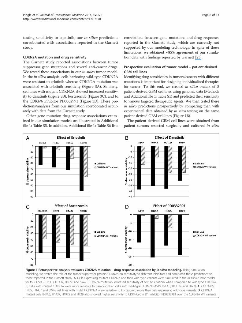
CDKN2A mutation and drug sensitivityThe Garnett study reported associations between tumorsuppressor gene mutations and several anti-cancer drugs.We tested these associations in our in silico tumor model.In the in silico analysis, cells harboring wild-type CDKN2Awere resistant to erlotinib whereas CDKN2A mutation wasassociated with erlotinib sensitivity (Figure 3A). Similarly,cell lines with mutant CDKN2A showed increased sensitiv-ity to dasatinib (Figure 3B), bortezomib (Figure 3C), and tothe CDK4/6 inhibitor PD0332991 (Figure 3D). These pre-dictions/analyses from our simulation corroborated accur-ately with data from the Garnett study.Other gene mutation-drug response associations exam-
ined in our simulation models are illustrated in Additionalfile 1: Table S5. In addition, Additional file 1: Table S6 lists
Figure 3 Retrospective analysis evaluates CDKN2A mutation – drug rmodeling, we tested the role of the tumor-suppressor protein CDKN2A onthose reported in the Garnett study. A, Cells expressing mutant CDKN2A anfor four lines – BxPC3, H1437, H1650 and SW48. CDKN2A mutation increaseB, Cells with mutant CDKN2A were more sensitive to dasatinib than cells wHT29, H1437 and SW48 cell lines with mutant CDKN2A were sensitive to bmutant cells BxPC3, H1437, H1975 and HT29 also showed higher sensitivity
correlations between gene mutations and drug responsesreported in the Garnett study, which are currently notsupported by our modeling technology. In spite of theselimitations, we obtained ~85% agreement of our simula-tion data with findings reported by Garnett [23].
Prospective evaluation of tumor model – patient-derivedGBM cell linesIdentifying drug sensitivities in tumors/cancers with differentmutations is important for designing individualized therapiesfor cancer. To this end, we created in silico avatars of 8patient-derived GBM cell lines using genomic data (Methodsand Additional file 1: Table S1) and predicted their sensitivityto various targeted therapeutic agents. We then tested thesein silico predictions prospectively by comparing then withexperimental data obtained by in vitro testing on the samepatient-derived GBM cell lines (Figure 1B).The patient-derived GBM cell lines were obtained from
patient tumors resected surgically and cultured in vitro
esponse association by in silico modeling. Using simulationsensitivity to different inhibitors and compared these predictions tod their wild-type variants were simulated in the in silico tumor modeld sensitivity of cells to erlotinib when compared to wild-type CDKN2A.ith wild-type CDKN2A (A549, BxPC3, HCT116 and H460). C, COLO205,ortezomib more than cells expressing wild-type variants. D, CDKN2Ato CDK4-Cyclin D1 inhibitor PD0332991 over the CDKN2A WT variants.

Pingle et al. Journal of Translational Medicine 2014, 12:128 Page 7 of 13http://www.translational-medicine.com/content/12/1/128
(details in Methods). We have profiled these lines usingAffymetrix Gene Chip Human Gene 1.0 ST Array. Usingwhole-exome sequencing, we recently tested the validityof these cells (maintained in cultures) for developmentand testing of personalized targeted therapies, based ontheir accurate representation of the original tumor profiles[27]. We have designated the different patient-derived
Figure 4 In silico modeling analysis and experimental in vitro data forresponse data for erlotinib with percent change in viability. Cells showing ddrug. B, In vitro experimental results for effect of 1 μM erlotinib on viabilityusing Alamar Blue assay. C, D, Predictive and experimental data for sorafentested in vitro at 1 μM. Dose-response curves for in silico data demonstratsorafenib and sunitinib on the viability of profiled patient-derived GBM c
GBM cell lines as: GBM4, GBM8, SK102, SK262, SK429,SK748, SK987 and SK1035.After generating in silico profiles of these cells, we opti-
mized these simulation avatars in terms of strength of func-tional effect of the mutation on key pathways such as EGFR,RAS and Src/PI3K. The rationale for this optimization isthat expression data on these cells does not provide an
drug responsiveness to 3 alignment drugs. A, Predictive doseecrease in viability of 20% or greater are considered sensitive to thein patient-derived GBM cell lines; viability was determined at 72 hib. E, F, Predictive and experimental data for dasatinib. All drugs weree the effects of increasing concentrations of the drugs – erlotinib,ell lines in the simulation model.

Figure 5 (See legend on next page.)
Pingle et al. Journal of Translational Medicine 2014, 12:128 Page 8 of 13http://www.translational-medicine.com/content/12/1/128

(See figure on previous page.)Figure 5 In silico modeling and experimental in vitro data for drug responsiveness to tyrosine kinase inhibitors. This figuredemonstrates in silico predictions of sensitivity and in vitro viability (respectively) in response to treatment with tyrosine kinase inhibitors:A, B, lapatinib, C, D, nilotinib, E, F, Imatinib and G, H, Sunitinib. Cells were exposed in vitro to 1 μM tyrosine kinase inhibitors for 72 h andviability determined using Alamar Blue assay. The dose-response for in silico predictions is generated by iterative simulations with increasingconcentrations of the drug in the model and the viability index is calculated. Cells showing decrease in viability of 20% or greater are consideredsensitive to the drug.
Pingle et al. Journal of Translational Medicine 2014, 12:128 Page 9 of 13http://www.translational-medicine.com/content/12/1/128
accurate measure of the dominance of different intracellularpathways. In order to interrogate this information on thepathways that play a dominant role in each tumor line (suchas EGFR, RAS, PI3K, etc.), we used 3 anti-cancer agents (er-lotinib, sorafenib and dasatinib) targeting these pathways.This will achieve “alignment” and train the simulation ava-tars for further analyses (details in Additional file 1). Thealignment for these 3 drugs could be best achieved in thefollowing cell lines: GBM8, SK262, SK429, SK748, andSK1035. In cell lines GBM4 and SK987, there was a mis-match for sorafenib where the predictive trends were re-versed. GBM4 was sensitive to sorafenib experimentally butour in silico predictions showed it to be resistant; SK987was resistant experimentally but sensitive in predictive re-sults. Similarly, the experimental trend for SK102 resistanceto dasatinib could not be met predictively. Correlation ofpredictive trends with alignment drugs is shown in Figure 4A-F.Predictions obtained by simulation modeling are pre-
sented as dose-response plots for viability; decrease inviability of >20% was considered as sensitive. Experimen-tally, viability was determined by Alamar blue assay, inresponse to 1 μM concentration of respective inhibitorsat 72 h. These data represent viability as mean valuesfrom triplicate samples.We tested ten anti-cancer drugs in silico on the simu-
lation avatars of the 8 patient-derived GBM cell lines ina blinded prospective study. These simulations generatedpredictions that we compared with in vitro experimentaldata (Additional file 1: Table S7A-D). Of the 80 in silicopredictions, 61 (76.25%) predictions showed agreementwith in vitro experimental results. Analysis of drugsensitivity correlation for all 8 GBM patient-derivedcell lines, for all the 13 drugs is summarized in Additionalfile 1: Table S7. Figures 5A-H and 6A-H show a drug-wise comparison of in silico predictions (dose-responsecurves) and in vitro experimental results generatedwith testing 1 μM concentration of each drug on thesecell lines.
Effect of tyrosine kinase inhibitors on patient-derivedGBM cellsFor the EGFR family inhibitor lapatinib, simulation stud-ies predicted SK429, SK748 and SK1035 to be resistant,which were confirmed by in vitro data. Similarly, model-ing predicted GBM8, SK102, SK262 and SK987 to be
sensitive and these predictions were in agreement withexperimental data (Figure 5A and B). However, modelingpredicted GBM4 to be resistant to lapatinib while in vitrodata showed GBM4 to be highly sensitive to lapatinib(Figure 5B). For the tyrosine kinase inhibitor nilotinib,the model predicted GBM8 to be sensitive while all theother profiles to be resistant (Figure 5C). In vitro stud-ies demonstrated that GBM8 was indeed sensitive tonilotinib as predicted, but there was a mismatch withthe experimental results for two lines – SK262 andSK1035. Experimentally, SK262 was found to be sensi-tive, whereas SK1035 was on the borderline of sensitiv-ity and resistance (Figure 5D). For imatinib, simulationpredicted that all GBM lines except GBM8 were resist-ant (Figure 5E). The experimental results corroboratedwith this in silico prediction (Figure 5F). Sunitinib wasthe other multi-tyrosine kinase inhibitor tested. Oursimulation predicted GBM8, SK102 and SK987 to besensitive to sunitinib; however, only GBM8 was foundto be sensitive in vitro. SK262 was predicted to be re-sistant to sunitinib but in vitro data found it to be moder-ately sensitive. On the other hand, GBM4, SK429, SK748and SK1035 were found to be resistant in both simulationand experimental data (Figure 5G-H).
Effect of other drugs on patient-derived GBM cellsBesides the tyrosine kinase inhibitors, correlation be-tween in silico predictions and experimental results forthe 8 patient-derived GBM cell lines was also tested fordrugs such as pitavastatin (HMG CoA reductase inhibi-tor), everolimus (mTOR inhibitor), celecoxib (COX2 in-hibitor) and bortezomib (proteasome inhibitor) (Figure 6A-H). For bortezomib, all profiles were predicted to besensitive and these predictions matched with in vitro ex-perimental results (Figure 6A and B). For everolimus,in vitro results were in agreement with simulation pre-dictions for all lines except SK429 (Figure 6C and D).Our in silico model predicted GBM4, SK262, SK429,SK748 and SK1035 to be resistant to celecoxib; these pre-dictions matched with in vitro results. However, GBM8,SK102 and SK987 were predicted to show moderate sensi-tivity to celecoxib, but were found to be resistant in vitro(Figure 6E and F). For pitavastatin, the simulation pre-dicted 5 patient-derived GBM cell lines to be sensitive(GBM8, GBM4, SK102, SK262 and SK987), of whichSK987 was found to be resistant in vitro. On the other

Figure 6 (See legend on next page.)
Pingle et al. Journal of Translational Medicine 2014, 12:128 Page 10 of 13http://www.translational-medicine.com/content/12/1/128

(See figure on previous page.)Figure 6 In silico modeling and experimental in vitro data for drug responsiveness to different drugs. This figure demonstrates in silicopredictions of sensitivity and in vitro viability in response to treatment of patient-derived GBM cell lines with A, B, bortezomib, C, D, everolimus,E, F, celecoxib, and G, H, pitavastatin. All drugs were tested in vitro at 1 μM for 72 h and viability was assayed using Alamar Blue assay. Cellsshowing decrease in viability of 20% or greater are considered sensitive to the drug.
Pingle et al. Journal of Translational Medicine 2014, 12:128 Page 11 of 13http://www.translational-medicine.com/content/12/1/128
hand, of the cell lines predicted to be resistant (SK429,SK748 and SK1035), SK1035 was sensitive in vitro and didnot match with the prediction (Figure 6G and H).These data demonstrate a 76.25% agreement between
in silico predictions of drug response and in vitro experi-mental data in patient-derived GBM cell lines.
DiscussionDeveloping an in silico model that takes into accountthe complex genotypes/phenotypes of cancer to accur-ately predict drug response will help personalize therapywith more efficiency. In this study, we developed andvalidated a virtual tumor model by retrospectively test-ing it against a dataset from a recent screening study[23]; we obtained a corroboration of ~85% between ourpredictions and the results from this study. Followingthis retrospective validation, we generated in silico pre-dictions to prospectively test the sensitivity of patient-derived GBM cell lines to targeted agents. These analysesalso demonstrated a high degree of agreement (>75%) be-tween in vitro experimental findings and in silico predic-tions. These studies validate our in silico tumor modeland the simulation-based approach and provide criticalproof-of-concept of a priori prediction of responses to tar-geted therapies. Thus, this model provides an effectiveplatform for testing and developing personalized thera-peutic regimens for cancer patients.The genomic inputs that we used to create simulation
avatars for patient-derived GBM cell lines were copynumber variation data. A more comprehensive and ac-curate profile would require additional data (gene muta-tions, methylation status etc. along with copy numbervariation); this would help us develop a more representa-tive avatar and would likely improve the accuracy of ourdrug response predictions and provide higher correlationwith experimental data.Genotypes of cancer cell lines have traditionally been
used to correlate with drug sensitivity [28,29]. A similarrecent study makes efficient use of gene expression pro-files to categorize colorectal cancers into different mo-lecular and clinically actionable subtypes [30]. Moreover,it is clear that using molecular tumor profiles to stratifypatients for therapy affects response and progression-free survival [31]. However, increasing amounts of datafrom genomic, proteomic, transcriptomic and metabolo-mic profiling will likely require integration of these var-ied datasets and development of predictive systems
modeling, which may hold the key to effective cancertherapy.Rapid screening of patient samples in real time with
models such as the one we have developed can drive crit-ical therapeutic decision-making. Although our currentmodel makes only cell-intrinsic predictions, we have beenable to achieve a high rate of agreement between in silicopredictions and in vitro findings. Future versions of thismodel are being refined to incorporate tumor microenvir-onment including aspects of angiogenesis, hypoxia, andtumor-associated inflammation. We believe that incorpor-ating these features into our model would more accuratelyrepresent the tumor in a patient. Importantly, this will fur-ther help improve our predictions for designing thera-peutic regimens for GBM patients. This model can also beadapted to identify potential mechanisms of resistance apriori and to design rational drug combinations that pre-vent emergence of resistance and development of escapepathways.Our in silico model aligns with NCI guidelines that
emphasize evaluation of similar predictor models to de-termine their accuracy [12,15,32,33]. We intend to testthis model in clinical trials and utilize it as a tool to ex-pedite clinical decision-making and determine drugs/combinations most likely to benefit a patient. Addition-ally, models such as these will play important roles intesting new biological hypotheses. This is critical to thediscovery of molecular drivers and critical networks incancer pathophysiology and the development of betterdiagnostics and effective therapeutics.
Additional file
Additional file 1: Supplementary Information.
Competing interestsThe following authors are employed by Cellworks, Inc.: Zeba Sultana, TaherAbbasi, Shweta Kapoor, Ansu Kumar, Shahabuddin Usmani, Ashish Agrawal,and Shireen Vali. The other authors report no competing financial interests.
Authors’ contributionsSCP – designed study, performed research, analyzed data, wrote manuscript;ZS – executed simulation studies, analyzed data, wrote manuscript; SP – designedstudy, performed research, analyzed data, wrote manuscript; PJ – performedresearch, analyzed data; RM – performed research, analyzed data; YC – performedresearch, analyzed data; ISB – performed research; NN – performedresearch; MM – performed research, analyzed data; TA – analyzed data,developed analytics, wrote manuscript; SK – developed predictivesimulation-based tumor cell technology; AK – developed predictivesimulation-based tumor cell technology; SU – executed simulation studies,developed predictive simulation-based tumor cell technology; AA – developedpredictive simulation-based tumor cell technology; SV – analyzed data,

Pingle et al. Journal of Translational Medicine 2014, 12:128 Page 12 of 13http://www.translational-medicine.com/content/12/1/128
developed predictive simulation-based tumor cell technology, developedanalytics, wrote manuscript; SK (Santosh Kesari) – designed study,planned and directed research, analyzed data, wrote manuscript. All authorsread and approved the final manuscript.
AcknowledgmentsThis work was supported in part by grants from National Brain TumorSociety (NBTS) David Cook Chair of Research, Barbara and Joseph Ajello trustfund, Tuttleman Family Foundation, MCJ Amelior Foundation, and BostonFire Department/Kenney Foundation to S. Kesari.
Author details1Translational Neuro-Oncology Laboratories, Moores Cancer Center, UC SanDiego, La Jolla, CA 92093, USA. 2Cellworks Research India Ltd, Bangalore 560066, India. 3Cellworks Group Inc, Saratoga, CA 95070, USA. 4Department ofNeurosciences, UC San Diego, La Jolla, CA 92093, USA.
Received: 14 January 2014 Accepted: 23 April 2014Published: 21 May 2014
References1. Wen PY, Kesari S: Malignant gliomas in adults. N Engl J Med 2008,
359:492–507.2. Mischel PS, Shai R, Shi T, Horvath S, Lu KV, Choe G, Seligson D, Kremen TJ,
Palotie A, Liau LM, Cloughesy TF, Nelson SF: Identification of molecularsubtypes of glioblastoma by gene expression profiling. Oncogene 2003,22:2361–2373.
3. TCGA: Comprehensive genomic characterization defines humanglioblastoma genes and core pathways. Nature 2008, 455:1061–1068.
4. Verhaak RG, Hoadley KA, Purdom E, Wang V, Qi Y, Wilkerson MD, Miller CR,Ding L, Golub T, Mesirov JP, Alexe G, Lawrence M, O'Kelly M, Tamayo P,Weir BA, Gabriel S, Winckler W, Gupta S, Jakkula L, Feiler HS, Hodgson JG,James CD, Sarkaria JN, Brennan C, Kahn A, Spellman PT, Wilson RK, Speed TP,Gray JW, Meyerson M, et al: Integrated genomic analysis identifies clinicallyrelevant subtypes of glioblastoma characterized by abnormalities inPDGFRA, IDH1, EGFR, and NF1. Cancer Cell 2010, 17:98–110.
5. Deisboeck TS: Personalizing medicine: a systems biology perspective. MolSyst Biol 2009, 5:249.
6. Deisboeck TS, Zhang L, Yoon J, Costa J: In silico cancer modeling: is itready for prime time? Nat Clin Pract Oncol 2009, 6:34–42.
7. Woodward DE, Cook J, Tracqui P, Cruywagen GC, Murray JD, Alvord ECJr: A mathematical model of glioma growth: the effect of extent ofsurgical resection. Cell Prolif 1996, 29:269–288.
8. Tracqui P, Cruywagen GC, Woodward DE, Bartoo GT, Murray JD, Alvord ECJr: A mathematical model of glioma growth: the effect of chemotherapyon spatio-temporal growth. Cell Prolif 1995, 28:17–31.
9. Zhang L, Wang Z, Sagotsky JA, Deisboeck TS: Multiscale agent-based cancermodeling. J Math Biol 2009, 58:545–559.
10. Deisboeck TS, Wang Z, Macklin P, Cristini V: Multiscale cancer modeling.Annu Rev Biomed Eng 2011, 13:127–155.
11. Almine JF, Wise SG, Hiob M, Singh NK, Tiwari KK, Vali S, Abbasi T, Weiss AS:Elastin sequences trigger transient proinflammatory responses byhuman dermal fibroblasts. FASEB J 2013, 27:3455–3465.
12. Barve A, Gupta A, Solapure SM, Kumar A, Ramachandran V, Seshadri K, Vali S,Datta S: A kinetic platform for in silico modeling of the metabolic dynamicsin Escherichia coli. Adv Appl Bioinform Chem 2010, 3:97–110.
13. Cirstea D, Hideshima T, Rodig S, Santo L, Pozzi S, Vallet S, Ikeda H, Perrone G,Gorgun G, Patel K, Desai N, Sportelli P, Kapoor S, Vali S, Mukherjee S, Munshi NC,Anderson KC, Raje N: Dual inhibition of akt/mammalian target of rapamycinpathway by nanoparticle albumin-bound-rapamycin and perifosine inducesantitumor activity in multiple myeloma. Mol Cancer Ther 2010, 9:963–975.
14. Equils O, Nambiar P, Hobel CJ, Smith R, Simmons CF, Vali S: A computersimulation of progesterone and Cox2 inhibitor treatment for pretermlabor. PLoS One 2010, 5:e8502.
15. Harvey LE, Kohlgraf KG, Mehalick LA, Raina M, Recker EN, Radhakrishnan S,Prasad SA, Vidva R, Progulske-Fox A, Cavanaugh JE, Vali S, Brogden KA:Defensin DEFB103 bidirectionally regulates chemokine and cytokineresponses to a pro-inflammatory stimulus. Sci Rep 2013, 3:1232.
16. Kannaiyan R, Hay HS, Rajendran P, Li F, Shanmugam MK, Vali S, Abbasi T,Kapoor S, Sharma A, Kumar AP, Chng WJ, Sethi G: Celastrol inhibitsproliferation and induces chemosensitization through down-regulation
of NF-kappaB and STAT3 regulated gene products in multiple myelomacells. Br J Pharmacol 2011, 164:1506–1521.
17. Kaushik P, Gorin F, Vali S: Dynamics of tyrosine hydroxylasemediated regulation of dopamine synthesis. J Comput Neurosci2007, 22:147–160.
18. Tandon R, Kapoor S, Vali S, Senthil V, Nithya D, Venkataramanan R, Sharma A,Talwadkar A, Ray A, Bhatnagar PK, Dastidar SG: Dual epidermal growth factorreceptor (EGFR)/insulin-like growth factor-1 receptor (IGF-1R) inhibitor: anovel approach for overcoming resistance in anticancer treatment. Eur JPharmacol 2011, 667:56–65.
19. Vali S, Mythri RB, Jagatha B, Padiadpu J, Ramanujan KS, Andersen JK, GorinF, Bharath MM: Integrating glutathione metabolism and mitochondrialdysfunction with implications for Parkinson's disease: a dynamic model.Neuroscience 2007, 149:917–930.
20. Vali S, Pallavi R, Kapoor S, Tatu U: Virtual prototyping study showsincreased ATPase activity of Hsp90 to be the key determinant of cancerphenotype. Syst Synth Biol 2010, 4:25–33.
21. Galli R, Binda E, Orfanelli U, Cipelletti B, Gritti A, De Vitis S, Fiocco R, ForoniC, Dimeco F, Vescovi A: Isolation and characterization of tumorigenic,stem-like neural precursors from human glioblastoma. Cancer Res 2004,64:7011–7021.
22. Lee J, Kotliarova S, Kotliarov Y, Li A, Su Q, Donin NM, Pastorino S, Purow BW,Christopher N, Zhang W, Park JK, Fine HA: Tumor stem cells derived fromglioblastomas cultured in bFGF and EGF more closely mirror thephenotype and genotype of primary tumors than do serum-cultured celllines. Cancer Cell 2006, 9:391–403.
23. Garnett MJ, Edelman EJ, Heidorn SJ, Greenman CD, Dastur A, Lau KW,Greninger P, Thompson IR, Luo X, Soares J, Liu Q, Iorio F, Surdez D, Chen L,Milano RJ, Bignell GR, Tam AT, Davies H, Stevenson JA, Barthorpe S, Lutz SR,Kogera F, Lawrence K, McLaren-Douglas A, Mitropoulos X, Mironenko T, Thi H,Richardson L, Zhou W, Jewitt F, et al: Systematic identification of genomicmarkers of drug sensitivity in cancer cells. Nature 2012, 483:570–575.
24. Curtin JA, Fridlyand J, Kageshita T, Patel HN, Busam KJ, Kutzner H, Cho KH,Aiba S, Brocker EB, LeBoit PE, Pinkel D, Bastian BC: Distinct sets of geneticalterations in melanoma. N Engl J Med 2005, 353:2135–2147.
25. Davies H, Bignell GR, Cox C, Stephens P, Edkins S, Clegg S, Teague J,Woffendin H, Garnett MJ, Bottomley W, Davis N, Dicks E, Ewing R, Floyd Y,Gray K, Hall S, Hawes R, Hughes J, Kosmidou V, Menzies A, Mould C, Parker A,Stevens C, Watt S, Hooper S, Wilson R, Jayatilake H, Gusterson BA, Cooper C,Shipley J, et al: Mutations of the BRAF gene in human cancer. Nature 2002,417:949–954.
26. Konecny GE, Pegram MD, Venkatesan N, Finn R, Yang G, Rahmeh M, Untch M,Rusnak DW, Spehar G, Mullin RJ, Keith BR, Gilmer TM, Berger M, Podratz KC,Slamon DJ: Activity of the dual kinase inhibitor lapatinib (GW572016)against HER-2-overexpressing and trastuzumab-treated breast cancer cells.Cancer Res 2006, 66:1630–1639.
27. Yost SE, Pastorino S, Rozenzhak S, Smith EN, Chao YS, Jiang P, Kesari S,Frazer KA, Harismendy O: High-resolution mutational profiling suggeststhe genetic validity of glioblastoma patient-derived pre-clinical models.PLoS One 2013, 8:e56185.
28. Shoemaker RH, Monks A, Alley MC, Scudiero DA, Fine DL, McLemore TL,Abbott BJ, Paull KD, Mayo JG, Boyd MR: Development of human tumorcell line panels for use in disease-oriented drug screening. Prog Clin BiolRes 1988, 276:265–286.
29. Weinstein JN, Myers TG, O'Connor PM, Friend SH, Fornace AJ Jr, Kohn KW,Fojo T, Bates SE, Rubinstein LV, Anderson NL, Buolamwini JK, van Osdol WW,Monks AP, Scudiero DA, Sausville EA, Zaharevitz DW, Bunow B,Viswanadhan VN, Johnson GS, Wittes RE, Paull KD: An information-intensiveapproach to the molecular pharmacology of cancer. Science 1997,275:343–349.
30. Sadanandam A, Lyssiotis CA, Homicsko K, Collisson EA, Gibb WJ,Wullschleger S, Ostos LC, Lannon WA, Grotzinger C, Del Rio M, Lhermitte B,Olshen AB, Wiedenmann B, Cantley LC, Gray JW, Hanahan D: A colorectalcancer classification system that associates cellular phenotype andresponses to therapy. Nat Med 2013, 19:619–625.
31. Von Hoff DD, Stephenson JJ Jr, Rosen P, Loesch DM, Borad MJ, Anthony S,Jameson G, Brown S, Cantafio N, Richards DA, Fitch TR, Wasserman E,Fernandez C, Green S, Sutherland W, Bittner M, Alarcon A, Mallery D, Penny R:Pilot study using molecular profiling of patients’ tumors to find potentialtargets and select treatments for their refractory cancers. J Clin Oncol 2010,28:4877–4883.

Pingle et al. Journal of Translational Medicine 2014, 12:128 Page 13 of 13http://www.translational-medicine.com/content/12/1/128
32. Rajendran P, Ong TH, Chen L, Li F, Shanmugam MK, Vali S, Abbasi T, Kapoor S,Sharma A, Kumar AP, Hui KM, Sethi G: Suppression of signal transducer andactivator of transcription 3 activation by butein inhibits growth of humanhepatocellular carcinoma in vivo. Clin Cancer Res 2011, 17:1425–1439.
33. Sultana Z, Paleologou KE, Al-Mansoori KM, Ardah MT, Singh N, Usmani S,Jiao H, Martin FL, Bharath MM, Vali S, El-Agnaf OM: Dynamic modelingof alpha-synuclein aggregation in dopaminergic neuronal systemindicates points of neuroprotective intervention: experimentalvalidation with implications for Parkinson's therapy. Neuroscience2011, 199:303–317.
doi:10.1186/1479-5876-12-128Cite this article as: Pingle et al.: In silico modeling predicts drugsensitivity of patient-derived cancer cells. Journal of Translational Medicine2014 12:128.
Submit your next manuscript to BioMed Centraland take full advantage of:
• Convenient online submission
• Thorough peer review
• No space constraints or color figure charges
• Immediate publication on acceptance
• Inclusion in PubMed, CAS, Scopus and Google Scholar
• Research which is freely available for redistribution
Submit your manuscript at www.biomedcentral.com/submit


![in silico (BFA thesis), 2002 [español]](https://static.fdokumen.com/doc/165x107/631f4913dbf756400702aca8/in-silico-bfa-thesis-2002-espanol.jpg)