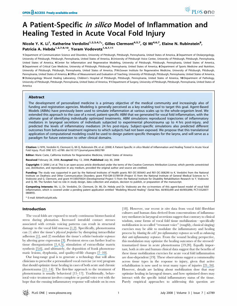
A Patient-Specific in silico Model of Inflammation and Healing Tested in Acute Vocal Fold Injury
11
A Patient-Specific in silico Model of Inflammation and Healing Tested in Acute Vocal Fold Injury Nicole Y. K. Li 1 , Katherine Verdolini 1,2,3,4,7 *, Gilles Clermont 4,5,7 , Qi Mi 4,6,7 , Elaine N. Rubinstein 8 , Patricia A. Hebda 1,2,7,9,10 , Yoram Vodovotz 1,4,7,11 1 Department of Communication Science and Disorders, University of Pittsburgh, Pittsburgh, Pennsylvania, United States of America, 2 Department of Otolaryngology, University of Pittsburgh, Pittsburgh, Pennsylvania, United States of America, 3 University of Pittsburgh Voice Center, University of Pittsburgh, Pittsburgh, Pennsylvania, United States of America, 4 Center for Inflammation and Regenerative Modeling, University of Pittsburgh, Pittsburgh, Pennsylvania, United States of America, 5 Department of Critical Care Medicine, University of Pittsburgh, Pittsburgh, Pennsylvania, United States of America, 6 Department of Sports Medicine and Nutrition, University of Pittsburgh, Pittsburgh, Pennsylvania, United States of America, 7 McGowan Institute for Regenerative Medicine, University of Pittsburgh, Pittsburgh, Pennsylvania, United States of America, 8 Office of Measurement and Evaluation of Teaching, University of Pittsburgh, Pittsburgh, Pennsylvania, United States of America, 9 Otolaryngology Wound Healing Laboratory, Children’s Hospital of Pittsburgh, Pittsburgh, Pennsylvania, United States of America, 10 Department of Pathology, University of Pittsburgh, Pittsburgh, Pennsylvania, United States of America, 11 Department of Surgery, University of Pittsburgh, Pittsburgh, Pennsylvania, United States of America Abstract The development of personalized medicine is a primary objective of the medical community and increasingly also of funding and registration agencies. Modeling is generally perceived as a key enabling tool to target this goal. Agent-Based Models (ABMs) have previously been used to simulate inflammation at various scales up to the whole-organism level. We extended this approach to the case of a novel, patient-specific ABM that we generated for vocal fold inflammation, with the ultimate goal of identifying individually optimized treatments. ABM simulations reproduced trajectories of inflammatory mediators in laryngeal secretions of individuals subjected to experimental phonotrauma up to 4 hrs post-injury, and predicted the levels of inflammatory mediators 24 hrs post-injury. Subject-specific simulations also predicted different outcomes from behavioral treatment regimens to which subjects had not been exposed. We propose that this translational application of computational modeling could be used to design patient-specific therapies for the larynx, and will serve as a paradigm for future extension to other clinical domains. Citation: Li NYK, Verdolini K, Clermont G, Mi Q, Rubinstein EN, et al. (2008) A Patient-Specific in silico Model of Inflammation and Healing Tested in Acute Vocal Fold Injury. PLoS ONE 3(7): e2789. doi:10.1371/journal.pone.0002789 Editor: Marie Csete, California Institute for Regenerative Medicine, United States of America Received February 28, 2008; Accepted May 12, 2008; Published July 30, 2008 Copyright: ß 2008 Li et al. This is an open-access article distributed under the terms of the Creative Commons Attribution License, which permits unrestricted use, distribution, and reproduction in any medium, provided the original author and source are credited. Funding: The study was supported in part by the National Institutes of Health grants R01-DC-005643 and R01-DC-008290 to K. Verdolini from the National Institute on Deafness and Other Communication Disorders; grant P50-GM-53789-09 (Project V) from the National Institute of General Medical Sciences to Y. Vodovotz and G. Clermont, and grant H133E070024 (Developmental Project 1) from the National Institute for Disability Rehabilitation Research to Y. Vodovotz and Q. Mi. The funders had no role in study design, data collection and analysis, decision to publish, or preparation of the manuscript. Competing Interests: Ms. Li, Dr. Verdolini, Dr. Clermont, Dr. Mi, Dr. Hebda and Dr. Vodovotz are the co-inventors of this agent-based model of vocal fold inflammation, which is covered under a pending patent application entitled ‘‘Modeling Wound Healing’’ (Serial Nos. 60/850,690 and 60/850,896; PCT/US2007/ 080893). * E-mail: [email protected] Introduction The vocal folds are exposed to nearly continuous biomechanical stress during phonation. Increased intrafold contact stresses associated with certain voicing patterns can result in structural damage to the vocal fold mucosa [1,2]. Specifically, phonotrauma can (1) alter the tissue’s physical properties by disrupting intracellular adhesion [1], and (2) modulate the tissue’s cellular/molecular responses by altering gene expression [3]. Persistent stress can further lead to tissue disorganization [1,4,5], stimulation of extracellular matrix synthesis [3,6], and ultimately, the deposition of frank phonotrau- matic lesions, dysphonia, and quality-of-life changes [7–10]. Our long-range goal is to generate a technology that will allow clinicians to prescribe a personalized vocal exercise (or rest) program that should optimize tissue healing in cases of both acute and chronic phonotrauma [11–14]. The first-line approach to the treatment of phonotrauma is usually behavioral [15–17]. Traditionally, behav- ioral voice treatment involves complete or partial voice rest with the hope that the ensuing inflammatory response will subside on its own [18]. However, our recent in vitro data from vocal fold fibroblast cultures and human data derived from concentrations of inflamma- tory mediators in laryngeal secretions suggest that contrary to clinical wisdom, some forms of vocal fold tissue mobilization—specifically mobilization in so-called ‘‘resonant voice’’ (roughly, classical singing) exercises may be able to modulate the inflammatory and healing process by blunting the cells’ pro-inflammatory responses as well as enhancing their anti-inflammatory responses. From the wound healing perspective, this modulation may optimize the healing outcomes of the stressed/ traumatized tissue in acute phonotrauma [19,20]. Equally impor- tant, both in vitro and human clinical data suggest that the benefits of these tissue mobilization exercises for acute vocal fold inflammation are dose-dependent [19]. These observations suggest a commonality across tissue types in the response to injury, given that active rehabilitation is now used to treat many types of injuries [21–34]. However, details are lacking about mobilization dose that may optimize healing in laryngeal tissues, and how optimized doses may interact with the specific initial inflammatory status of the tissue. Purely empirical approaches to addressing this question are PLoS ONE | www.plosone.org 1 July 2008 | Volume 3 | Issue 7 | e2789
-
Upload
independent -
Category
Documents
-
view
2 -
download
0
Transcript of A Patient-Specific in silico Model of Inflammation and Healing Tested in Acute Vocal Fold Injury
A Patient-Specific in silico Model of Inflammation andHealing Tested in Acute Vocal Fold InjuryNicole Y. K. Li1, Katherine Verdolini1,2,3,4,7*, Gilles Clermont4,5,7, Qi Mi4,6,7, Elaine N. Rubinstein8,
Patricia A. Hebda1,2,7,9,10, Yoram Vodovotz1,4,7,11
1 Department of Communication Science and Disorders, University of Pittsburgh, Pittsburgh, Pennsylvania, United States of America, 2 Department of Otolaryngology,
University of Pittsburgh, Pittsburgh, Pennsylvania, United States of America, 3 University of Pittsburgh Voice Center, University of Pittsburgh, Pittsburgh, Pennsylvania,
United States of America, 4 Center for Inflammation and Regenerative Modeling, University of Pittsburgh, Pittsburgh, Pennsylvania, United States of America,
5 Department of Critical Care Medicine, University of Pittsburgh, Pittsburgh, Pennsylvania, United States of America, 6 Department of Sports Medicine and Nutrition,
University of Pittsburgh, Pittsburgh, Pennsylvania, United States of America, 7 McGowan Institute for Regenerative Medicine, University of Pittsburgh, Pittsburgh,
Pennsylvania, United States of America, 8 Office of Measurement and Evaluation of Teaching, University of Pittsburgh, Pittsburgh, Pennsylvania, United States of America,
9 Otolaryngology Wound Healing Laboratory, Children’s Hospital of Pittsburgh, Pittsburgh, Pennsylvania, United States of America, 10 Department of Pathology,
University of Pittsburgh, Pittsburgh, Pennsylvania, United States of America, 11 Department of Surgery, University of Pittsburgh, Pittsburgh, Pennsylvania, United States of
America
Abstract
The development of personalized medicine is a primary objective of the medical community and increasingly also offunding and registration agencies. Modeling is generally perceived as a key enabling tool to target this goal. Agent-BasedModels (ABMs) have previously been used to simulate inflammation at various scales up to the whole-organism level. Weextended this approach to the case of a novel, patient-specific ABM that we generated for vocal fold inflammation, with theultimate goal of identifying individually optimized treatments. ABM simulations reproduced trajectories of inflammatorymediators in laryngeal secretions of individuals subjected to experimental phonotrauma up to 4 hrs post-injury, andpredicted the levels of inflammatory mediators 24 hrs post-injury. Subject-specific simulations also predicted differentoutcomes from behavioral treatment regimens to which subjects had not been exposed. We propose that this translationalapplication of computational modeling could be used to design patient-specific therapies for the larynx, and will serve as aparadigm for future extension to other clinical domains.
Citation: Li NYK, Verdolini K, Clermont G, Mi Q, Rubinstein EN, et al. (2008) A Patient-Specific in silico Model of Inflammation and Healing Tested in Acute VocalFold Injury. PLoS ONE 3(7): e2789. doi:10.1371/journal.pone.0002789
Editor: Marie Csete, California Institute for Regenerative Medicine, United States of America
Received February 28, 2008; Accepted May 12, 2008; Published July 30, 2008
Copyright: � 2008 Li et al. This is an open-access article distributed under the terms of the Creative Commons Attribution License, which permits unrestricteduse, distribution, and reproduction in any medium, provided the original author and source are credited.
Funding: The study was supported in part by the National Institutes of Health grants R01-DC-005643 and R01-DC-008290 to K. Verdolini from the NationalInstitute on Deafness and Other Communication Disorders; grant P50-GM-53789-09 (Project V) from the National Institute of General Medical Sciences to Y.Vodovotz and G. Clermont, and grant H133E070024 (Developmental Project 1) from the National Institute for Disability Rehabilitation Research to Y. Vodovotzand Q. Mi. The funders had no role in study design, data collection and analysis, decision to publish, or preparation of the manuscript.
Competing Interests: Ms. Li, Dr. Verdolini, Dr. Clermont, Dr. Mi, Dr. Hebda and Dr. Vodovotz are the co-inventors of this agent-based model of vocal foldinflammation, which is covered under a pending patent application entitled ‘‘Modeling Wound Healing’’ (Serial Nos. 60/850,690 and 60/850,896; PCT/US2007/080893).
* E-mail: [email protected]
Introduction
The vocal folds are exposed to nearly continuous biomechanical
stress during phonation. Increased intrafold contact stresses
associated with certain voicing patterns can result in structural
damage to the vocal fold mucosa [1,2]. Specifically, phonotrauma
can (1) alter the tissue’s physical properties by disrupting intracellular
adhesion [1], and (2) modulate the tissue’s cellular/molecular responses
by altering gene expression [3]. Persistent stress can further lead to
tissue disorganization [1,4,5], stimulation of extracellular matrix
synthesis [3,6], and ultimately, the deposition of frank phonotrau-
matic lesions, dysphonia, and quality-of-life changes [7–10].
Our long-range goal is to generate a technology that will allow
clinicians to prescribe a personalized vocal exercise (or rest) program
that should optimize tissue healing in cases of both acute and chronic
phonotrauma [11–14]. The first-line approach to the treatment of
phonotrauma is usually behavioral [15–17]. Traditionally, behav-
ioral voice treatment involves complete or partial voice rest with the
hope that the ensuing inflammatory response will subside on its own
[18]. However, our recent in vitro data from vocal fold fibroblast
cultures and human data derived from concentrations of inflamma-
tory mediators in laryngeal secretions suggest that contrary to clinical
wisdom, some forms of vocal fold tissue mobilization—specifically
mobilization in so-called ‘‘resonant voice’’ (roughly, classical singing)
exercises may be able to modulate the inflammatory and healing
process by blunting the cells’ pro-inflammatory responses as well as enhancing
their anti-inflammatory responses. From the wound healing perspective,
this modulation may optimize the healing outcomes of the stressed/
traumatized tissue in acute phonotrauma [19,20]. Equally impor-
tant, both in vitro and human clinical data suggest that the benefits of
these tissue mobilization exercises for acute vocal fold inflammation
are dose-dependent [19]. These observations suggest a commonality
across tissue types in the response to injury, given that active
rehabilitation is now used to treat many types of injuries [21–34].
However, details are lacking about mobilization dose that may
optimize healing in laryngeal tissues, and how optimized doses may
interact with the specific initial inflammatory status of the tissue.
Purely empirical approaches to addressing this question are
PLoS ONE | www.plosone.org 1 July 2008 | Volume 3 | Issue 7 | e2789
unattractive because of the relatively invasive and expensive nature
of the research protocols [35]. The cumbersome nature of data
collection also complicates the potential for biologically oriented
clinical trials on the value of therapeutic interventions for
phonotrauma in humans.
Inflammation and Healing in PhonotraumaPhonotrauma, like all other forms of trauma, is a highly complex
process induced by a variety of stimuli, modulated by numerous cells
and their products, and affecting different tissues in diverse ways. At
the heart of the response to phonotrauma is the intertwined process
of inflammation and wound healing [36–42]. Inflammation is the
earliest and necessary response for the subsequent phase of wound
healing [43]. The whole inflammatory process can be understood as
an information system that processes and controls the biochemical
signals induced by injury and/or infection. These signals include: (1)
‘‘Go’’ signals to initiate the inflammatory reactions, (2) ‘‘Stop’’ signals
to temper the inflammation, and (3) ‘‘Switch’’ signals to convert a
tissue-damaging mode to a healing mode [44]. Also, these signals are
actively monitored and regulated by a number of checkpoints within
the system. If any of the aforementioned signals is missing or one of
the checkpoints malfunctions, the normal inflammatory process is
altered possibly resulting in persistent inflammation, abnormal
healing and tissue distortion. A prompt transition from the
inflammatory phase to the healing phase is a key determinant of
good wound healing, that is the replacement of traumatized tissue by
healthy tissue [45].
Wound healing outcomes depend on the original insult to a
certain degree, and are also likely influenced by individual
genetics. Under ideal conditions, the inflammatory response
would be limited by the antagonistic interactions among the
various pro- and anti-inflammatory agents, followed by a
transition to the later healing process in an orderly fashion.
However, if repeated injuries occur over a period of time, the
normal healing process would be disrupted. Two possible healing
processes can occur in that case, namely, reparative regeneration and
constructive repair. Reparative regeneration is scarless wound healing,
in which traumatized tissue is completely restored in normal
architecture and function. On the other hand, constructive repair
is more common in humans. The lost tissue is replaced by
granulation tissue which matures into a scar. Depending on the
amount of scarring, scarred tissue usually does not properly restore
the architecture and perform the pre-morbid function of the
injured tissue. The vocal folds are generally capable of
withstanding phonatory stresses and have the reparative capability
of resolving microscopically phonotraumatic damage incurred
during daily voice use. However, when injury exceeds a critical
threshold, inflammation is usually clinically evident and construc-
tive repair is involved. As a result, macroscopic vocal fold lesions
may develop, with prolonged dysfunction in vocal fold vibratory
function and voice output characteristics [46,47].
Our research team has published four studies on the
development of a novel method for obtaining quantitative
information about the inflammatory status of the larynx from
laryngeal secretions. Our prior studies [48,49] demonstrate the
presence of various mediators (interleukin-1 beta [IL-1b], tumor
necrosis factor alpha [TNF-a]), prostaglandin E2 [PGE-2], and
matrix metalloproteinase 8 [MMP-8] ) in vocal fold surface
secretions of human subjects in response to phonotrauma. Our
first study showed that pre- to post-vocal loading shifts in mediator
concentrations were clearly evident at 10 and 20 min post-loading
for IL-b, TNF-a, and MMP-8, reflecting the presence of acute
phonotrauma. In contrast, concentration shifts were not shown for
TGF-b1 or PGE-2 [49]. Another, intraoperative human study
confirmed that IL-1b was an indicator of acute inflammation,
whereas PGE-2 characterized chronic wounds [48]. A third study
showed that IL-1b was an early indicator of inflammation, and
PGE-2 was a later indicator of wound healing in rabbits subjected
to surgical trauma, with IL-1b returning to baseline by Day 7 post-
injury and PGE2 remaining elevated until the final time-point of
three weeks post-injury [50]. Finally, a fourth study in rabbits
subjected to surgical trauma assessed the degree to which assays of
laryngeal secretions may reflect wound healing processes deep to
the epithelium [51]. That study showed that the time point
associated with spikes in IL-1b (24 hours) corresponded to the
presence of fibrinous clot. The time point associated with
maximum PGE-2 levels (7 days) was associated with the presence
of mature collagen. Massive cellular infiltration and complete
epithelial coverage were found at intermediate time points. Taken
together, these studies provide robust evidence that secretions from
the laryngeal surfaces can provide a quantitative reflection of the
current inflammatory and wound healing state of vocal fold tissue.
The attractiveness of this marginally invasive technology is that it
can be readily used in human subjects—although not without
some difficulties on the part of both subjects and examiners–and
thus the data gain external validity over data obtained from more
invasive technologies involving animal subjects. Our research team
has also examined the potential of mathematical modeling to
elucidate apparently contradictory and unpredictable behavior
emerging from the plethora of interactions among biologic
pathways involved in the acute inflammatory response [52–54].
We have recently coined the term ‘‘translational systems biology’’
to refer to the process of creating, calibrating, and validating
computational simulations in settings of complex diseases,
simulations that are designed a priori for the purpose of modifying
clinical treatment, carrying out in silico clinical trials, generating
novel therapies, and refining diagnoses [55,56].
The objective of the present study was to use empirical data from
human subjects to develop a mechanistic computational simulation
for the biological dynamics of vocal fold inflammation and wound
healing following acute phonotrauma. The modeling approach
employed in the present study involves Agent-Based Models (ABMs).
In such models, individual components of a given complex system
interact based on rules whose outcomes are partially determined by
stochastic processes [57]. More specifically, ABM involves discrete
event simulation to study the behavior of complex systems. ABM is
the most direct initial approach to simulate the temporal evolution of
a complex system and to encode complicated time-dependent
cellular and molecular events that occur during inflammation and
wound healing [58–61]. Our results suggest that patient-specific,
mechanistic, and predictive computational simulations of inflam-
mation can be generated, raising the possibility that similar methods
could be utilized in other complex diseases.
Results
OverviewThe premise of mathematical models involves experimental
validation and feedback between the models and experiments. The
details of our model calibration and validation are described in
Materials and Methods. In brief, we first calibrated the ABM using
data from a base cohort (Subjects 1, 2 and 3), using their mediator
levels in laryngeal fluid at baseline, immediately after phonotrauma
induction, and following a 4-hr treatment that involved either voice
rest, ‘‘resonant voice’’ exercises, or spontaneous speech) (Figures 1–2,
dark circles) [35]. The calibrated ABM was run 10 times for the full
cohort of 7 subjects, up to 5 simulated days post baseline under the
condition of (1) each subject’s actual treatment group and (2)
Patient-Specific Simulation
PLoS ONE | www.plosone.org 2 July 2008 | Volume 3 | Issue 7 | e2789
hypothetical randomization to either of the other two treatment
groups, i.e., hypothetical treatments, using each subject’s baseline
mediator profile. Thus, a large simulation data set of subject-specific
mediator trajectories (3 treatment conditions610 runs67 sub-
jects = 210 runs) was generated for evaluating benefits of behavioral
treatments for acute phonotrauma.
Quantitative validation of the ABM was carried out by
comparing the predicted mediator levels with the empirical
mediator levels at 24 hr (1) for the base cohort (Subjects 1–3), as
well as (2) for Subjects 4–7 whose data had never been used for the
model calibration, under the actual treatment condition. The
validation results showed that predicted mediator levels generally
matched empirical mediator levels for the ABM at 24 hr post
baseline. Also, results from the simulation data showed that the
predicted mediator trajectories varied with a function of treatment
and initial mediator profile.
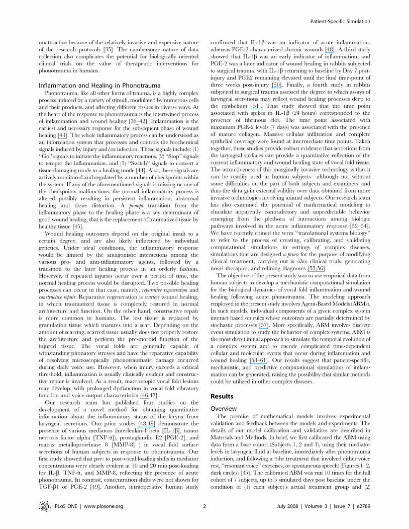
Predicted Trajectories of Inflammatory MediatorsThe ABM reproduced and predicted subject-specific mediator
trajectories (Figures 1 and 2). In the base cohort (Subjects 1–3), the
ABM predicted 24-hr mediator values in 80% (12/15; p,0.05) of
instances (+9 mediator measurements from 3 between-group subjects,
each with one of the 3 different treatments; +9 mediator
measurements from one within-group subject—Subject #3– who
received each of the treatments; -3 mediator measurements to
account for double-counting the ‘‘spontaneous speech’’ treatment for
Subject #3 in both between- and within-group measurements; hence
15 different mediator measurements in total). For Subjects 4–7, z-tests
(model data vs. empirical data) indicated that the ABM predicted
empirically obtained 24-hr mediator values 67% (2/3; p,0.05) of the
times for markers that were considered ‘‘valid,’’ as described shortly,
and 44% (4/9; p,0.05) of the time for markers that were considered
‘‘pre-inflamed and/or non-responsive’’ (see Materials and Methods).
For the single within-group subject (Subject 3; Figure 1), both
empirical data and simulation results showed that the concentrations
of pro-inflammatory mediators (IL-1b and TNF-a) spiked immedi-
ately following 1 hr of vocal loading, whereas the anti-inflammatory
mediator (interleukin-10 [IL-10]) showed a more protracted course.
The model predicted that following the 4-hr treatment, inflamma-
tory mediators would have distinctive temporal and quantitative
expression patterns across treatment assignments. For the sponta-
neous speech condition, the ABM predicted that the inflammatory
Figure 1. Empirical and model-predicted inflammatory and wound healing responses to acute phonotrauma in a single humansubject (Subject 3) following spontaneous speech (Panels A–C), voice rest (Panels D–F) and resonant voice treatment conditions(Panels G–I). Panels A, D and G display empirical and predicted trajectories of IL-1b. Panels B, E and H show empirical and predicted trajectories ofTNF-a. Panels C, F and I show empirical and predicted trajectories of IL-10. Inflammatory marker concentrations are in pg/ml. The grey bars representthe mean of the simulated data, and the error bars represent standard deviations in the simulated data. The dark circles represent the input data forthe first three time-points (baseline, post-loading, 4-hr post treatment onset), obtained from human laryngeal secretion data. The empty circlesrepresent the validation data at the 24-hr time point from the human laryngeal secretion data. B: baseline; PL: post vocal loading; 4hrPRx: following a4-hr treatment. Note that human validation data for Days 2–5 have not yet been generated.doi:10.1371/journal.pone.0002789.g001
Patient-Specific Simulation
PLoS ONE | www.plosone.org 3 July 2008 | Volume 3 | Issue 7 | e2789
response would be further escalated, i.e., would involve markedly
increased secretion of both pro- and anti-inflammatory mediators
following the 4-hr treatment. The concentrations of pro-inflamma-
tory mediators (IL-1b and TNF-a) reached their peaks at Day 1 post-
injury and resolved to baseline concentrations around Day 2–3 post-
injury. The model also predicted that the anti-inflammatory
mediator, IL-10, would be secreted in great quantities by wound
macrophages during the first 5 days post-injury. On the other hand,
under conditions of voice rest and ‘‘resonant voice’’ exercises, the
predicted concentrations of the pro-inflammatory mediators
dropped rapidly after the 4-hour treatment and then remained low
at the end of simulation, i.e., Day 5. The anti-inflammatory mediator
IL-10 was predicted to be secreted rapidly after the 4-hr treatment
and remain elevated up to Day 3 post-injury. Similar mediator
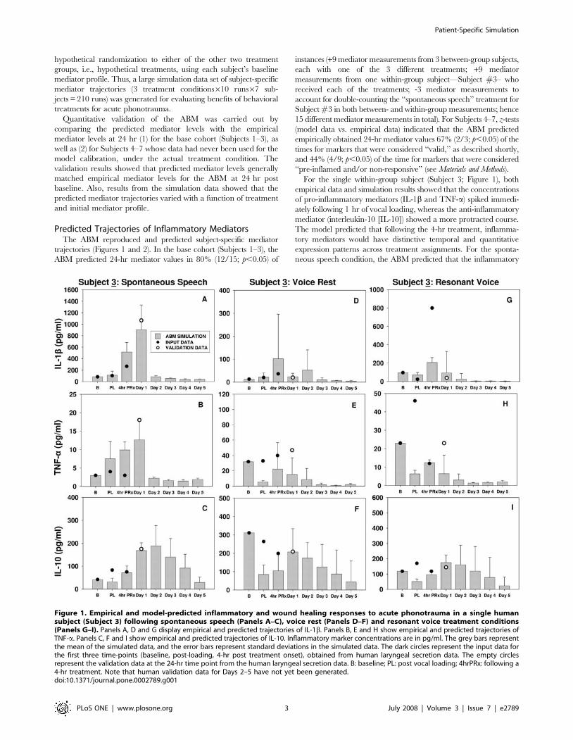
patterns were found in the larger dataset from the between-group
subjects (Figure 2).
Predicted Outcomes of In Silico TherapiesIn order to demonstrate the application of ABM for running in
silico (‘‘virtual’’) clinical studies, we ran a series of simulations for the
full cohort of subjects under the conditions of (1) actual treatment
received and (2) hypothetical treatments. The simulation results
showed theoretical individual-specific trajectories of mediator levels
across the treatments. For between-group comparisons, linear
mixed models showed significant treatment effects on the predicted
levels of mediators (IL-1b: F (2, 1260) = 223.17, p,0.001; TNF-a: F
(2, 1260) = 12.27, p,0.001; IL-10: F (2, 1260) = 215.65, p,0.001).
Post-hoc pairwise comparisons with Bonferroni adjustments showed
that among the three treatment groups, the predicted level of the
pro-inflammatory marker IL-1b ensuing from spontaneous speech was
the highest (p,0.001), whereas concentrations of the same marker
in the resonant voice condition was the lowest (p,0.001). At the same
time, among the three treatment groups, the predicted level of the
anti-inflammatory marker IL-10 ensuing from voice rest was the
lowest (p,0.001), whereas the level for the resonant voice condition
was the highest (p,0.001).
Discussion
We asked several experimental questions in the current study.
First, we sought to determine if an ABM could offer an
individualized prediction of biological readouts presumed to be
closely related to tissue status. Second, we wished to know if the
Figure 2. Empirical and model-predicted inflammatory and wound healing responses to acute phonotrauma in three subjectsfollowing spontaneous speech (Subject 3; Panels A–C), voice rest (Subject 1; Panels D–F) and resonant voice treatment conditions(Subject 2; Panels G–I). Panels A, D and G display empirical and predicted trajectories of IL-1b. Panels B, E and H show empirical and predictedtrajectories of TNF-a. Panels C, F and I show empirical and predicted trajectories of IL-10. Inflammatory marker concentrations are in pg/ml. The greybars represent the means from the simulated data, and the error bars represent the standard deviation from the simulated data. The dark circlesrepresent the input data for the first three time-points (baseline, post-loading, 4-hr post treatment onset) from the human laryngeal secretion data.The empty circles represent the validation data at the 24-hr time point from the human laryngeal secretion data. B: baseline; PL: post vocal loading;4hrPRx: following a 4-hr treatment. Note that human validation data for Days 2–5 have not yet been generated.doi:10.1371/journal.pone.0002789.g002
Patient-Specific Simulation
PLoS ONE | www.plosone.org 4 July 2008 | Volume 3 | Issue 7 | e2789
simulated wound healing response would vary specifically as a
function of the initial settings of mediator level and treatment
prescribed. Positive answers to these experimental questions would
strengthen confidence in the eventual utility of mathematical
modeling in general, and of ABM in particular, for understanding
the complex vocal fold wound healing system and for predicting
the healing outcome after phonotrauma and varied treatment
approaches.
The current study describes the development of an ABM that
reproduced diverse trajectories of inflammatory mediators in the
laryngeal secretions of different human subjects at early time-points
up to 4 hrs post-phonotrauma, and furthermore was capable of
predicting the levels of these mediators at 24 hrs. The subject-
specific ABMs were further used to explore the effects of treatment
regimens to which subjects had not been exposed and the predicted
levels of mediators under each condition were compared. The
models predicted that the wound healing outcomes as informed by
the mediator trajectories would be dramatically different given
variations in the initial mediator profile, presumably due to some
combination of pre-existing vocal fold conditions and the treatment
prescribed following phonotrauma.
Contemporary therapeutic interventions in phonotrauma are
oriented towards modulating the inflammatory and healing
processes to promote reparative healing of the traumatized vocal
folds. A plausible approach is to both blunt the inflammatory
response and activate the healing program. Upon mechanical
challenge, an acute inflammatory cascade is immediately activated
in damaged or stressed tissues [62,63]. The inflammatory cells
infiltrate the area of injury to remove damaged and dead cells and
tissue debris. This inflammatory reaction contributes to additional
tissue damage and cell death, which exacerbate the initial tissue
damage and amplify the signals for scarring. Theoretically,
inflammation-blocking interventions may reduce the ‘‘secondary’’
tissue damage and the possibility for fibrosis and scarring. At the
same time, this approach may potentially reduce the supply of
growth factors and cytokines from the inflammatory cells that
facilitate tissue repair. Thus, a therapeutic balance between the
need to limit inflammation causing tissue damage and the need for
inflammation to initiate tissue repair is important to optimize the
quality of healing outcomes and the recovery of physiological
functions [62–66]. In addition to in vitro and human data from our
laboratory, the current in silico study also suggests that modified
tissue mobilization exercise in the form of ‘‘resonant voice
production’’ may have the effect of blunting the cells’ pro-
inflammatory responses (e.g. IL-1b and TNF-a) but enhancing
their anti-inflammatory responses (e.g. IL-10), which may
ultimately promote tissue regeneration [6,20]. At this point, only
the ABMs’ molecular outputs were calibrated with experimental
data, whereas the ABMs’ tissue-level outputs are yet to be
experimentally validated. We have embarked on the work of
evaluating the ‘‘net effect’’ of the complex interactions among
these inflammatory mediators at the tissue level. The success of this
work will lead us to predict actual vocal fold tissue status following
injury, which ultimately may advance understanding of inflam-
mation and healing in a clinically useful way.
Our ABM predicted that the secretion of pro-inflammatory
mediators would be both prolonged and elevated subsequent to
spontaneous speech following an episode of phonotraumatic
injury. This prolongation would be due to a positive feedback
loop involving ‘‘Pro-inflammation R Damage R Pro-inflamma-
tion,’’ [67] thereby delaying the transition to the subsequent
healing process. Clinically, visible vocal fold inflammation would
be expected. On the other hand, under conditions of either voice
rest or resonant voice, the ABM predicted that the pro-
inflammatory response would be attenuated. It also predicted that
the anti-inflammatory response may be escalated in response to
the tissue mobilization involved in resonant voice—and the pro-
inflammatory response attenuated by the limitation of impact
stress—thereby further prompting rapid healing [6]. These
predictions suggest that the repair process would bypass the
‘‘Pro-inflammation R Damage R Pro-inflammation’’ positive
feedback loop and avoid a full-scale inflammatory and repair
response. As a result, a reparative regeneration (as opposed to
constructive repair) of mucosal structure and function would be
observed clinically and the probability of scar formation would be
minimized in the long run.
As additional data are acquired, the current ABM is under
continuous revision and augmentation. Our ultimate, long-term
goal is to generate in silico models that can be queried to identify
biomechanical treatments that will optimize the wound healing
process in the vocal folds, as a function of patient-specific
inflammatory profiles. Although these results are encouraging in
terms of the potential translational utility of ABM in the setting of
vocal fold inflammation, at least five limitations can be noted in
our study.
First, the current ABM mainly simulated (1) inflammation, (2)
proliferation and (3) collagen formation. The model did not
account for a final phase of the wound healing process, which
involves extracellular matrix (ECM) reorganization. According to
the literature on dermal wound healing, ECM reorganization is
initiated once a neo-matrix involving materials such as collagen is
deposited at the wound site [68–70]. Collagen is indeed a core
component of the ECM, and undergoes remodeling that is
dependent on both continued collagen synthesis and compensa-
tory collagen degradation. The degradation of wound collagen is
controlled by collagenases and other proteolytic enzymes, and the
net increase in wound collagen is determined by the balance of
these opposing mechanisms. Compared to the large body of
literature on dermal wound healing, research on ECM reorgani-
zation in vocal fold wound healing is sparse. No in vivo
measurement of collagen remodeling in human vocal folds is
currently available. Thus, in its use of empirical data obtained
from non-destructive methodologies only, the current model did
not incorporate aspects of collagen remodeling that might prove to
be important.
A second limitation in the current study is that healing outcomes
in this ABM were primarily informed by interactions among
inflammatory mediators and cells. However, a growing literature
supports the idea that several ECM components such as
fibronectin, hyaluronic acid, and decorin could also be involved
in regulating the wound healing process. Studies to date have
shown that aberrant scarring/fibrosis is at least partly due to the
response of fibroblasts in the wound to both inflammatory
mediators and extracellular matrix components [3,71–74], some
of which are known to constitute alarm/danger signals (e.g.
fragments of hyaluronic acid); [75] and therefore may be currently
abstracted under the ‘‘damage’’ variable in our model. Future
iterations of the ABM could be augmented with rules developed
around the interactions among inflammatory mediators, cells and
ECM components to yield more precise predictions.
Third, our model assumes that biomechanical stresses during
phonation cause mucosal damage. However, the current biolog-
ically-based ABM lacks the ability to receive input from physical
models of phonation, because data are lacking regarding the link
between the output of physical models—i.e. distributed tissue
stresses [76–79] and the biological consequences of those physical
stresses. Although biochemical networks may be reasonably
modeled by using stochastic simulations, many cell biology
Patient-Specific Simulation
PLoS ONE | www.plosone.org 5 July 2008 | Volume 3 | Issue 7 | e2789

phenomena relating to wound healing require the calculation of
biophysical processes, such as tissue deformation and disruption of
intracellular adhesion [1]. Ideally, our synthesized biochemical
networks should be coupled with these biophysical processes to
yield a more complete picture of vocal fold wound healing in
response to biomechanical stresses during phonation.
A fourth limitation relates to the inclusion of the multi-functional
anti-inflammatory mediator TGF-b1 in our model, although our
earlier work on assaying biochemical markers of vocal fold wound
healing failed to detect this mediator in laryngeal secretions pre- or
post-traumatically [48]. TGF-b1 is known to be involved in the
regulation of cell proliferation, cell differentiation and extracellular
matrix formation in all phases of inflammation and wound healing
[69,80]. This mediator exerts both anti-inflammatory and pro-
fibrotic effects that could convert an active site of inflammation into a
site dominated by subsequent tissue repair [81]. We suspect that
TGF-b1 might be highly cell-associated [82–84], and that this
property might have led to our inability to detect this growth factor
in vocal fold secretions [48]. To determine if TGF-b1 is necessary for
a correct simulation of inflammation and wound healing in the vocal
folds, a qualitative validation procedure was carried out to determine
what the simulated data would show in the presence or absence of
TGF-b1. In the latter case, the ABM predicted that no cellular or
molecular events would be triggered for any ranges of initial damage
(data not shown). In the presence of TGF-b1, the ABM predicted
different inflammatory and wound healing curves that vary with
initial mediator profile and treatment modalities following phono-
trauma (Figures 1 and 2). These results indicated that TGF-b1 (or a
qualitatively similar cytokine) is essential for the wound healing
process and should be included in the ABM structure.
The fifth, and perhaps most important limitation concerns the
validation of the ABM predictions with regard to the different
treatment modalities. We have in this study shown data from seven
humans and the capacity of our ABM to predict mid-term (24-hr)
inflammation based on earlier short-term assays. The subject
population in this study was relatively homogeneous in terms of
age. The subjects were all relatively young (21–46 years) and it is
known that tissue regenerative processes usually decline with aging.
We have carried out preliminary mathematical modeling of the
effects of aging on the acute inflammatory response [85]. We have
not, however, verified in a large cohort of patients the validity of
ABM predictions with regard to treatment outcome. It should be
noted that such studies in humans are complex from regulatory,
practical, and ethical points of view. We have embarked on a larger
clinical study to validate the ABM described herein. Indeed, clinical
management of phonotrauma remains a challenge to clinicians.
Large clinical trials are needed to establish optimized patient-specific
treatment interventions. In addition to validating the ABM with
further experimental studies, a parallel development of ordinary
differential equation-based models (EBMs) has been pursued in the
interest of cross-platform comparison of results [86]. This ‘‘model
docking’’ is a well-vetted validation strategy based on a comparison
of predictions of different models across an array of user input data.
The finding of similar predictions in ABMs and EBMs would
increase confidence in the underlying assumptions in the current
ABMs. Also, the EBMs alone allow formal mathematical analysis of
the simulation results, such as, bifurcation and stability analysis, that
will facilitate capture of the systems dynamics of the inflammatory
and healing responses [60].
Finally, for the approach ultimately to be useful in a real-life
clinical setting, the time lag introduced by ELISAs will have to be
addressed, or an alternate methodology will have to be identified
that can yield biological profiles of materials in a shorter period of
time. In the meantime, there is encouragement that models such
as ABMs and EBMs have potential to illuminate important
information about individualized wound healing processes in the
larynx.
More globally, we suggest that a systems biology approach that
involves modeling is integral to sorting through the perplexing
array of factors that dictate success or failure of clinical trials for
complex diseases [87]. Ultimately, this process would be
augmented by the inclusion of genetic variability in inflammatory
and wound healing components, typically mediated via single-
nucleotide gene polymorphisms in relevant genes [88].
In summary, this study suggests for the first time that patient-
specific, individualized models of inflammation and healing are
possible. This demonstration extends the power of translational
simulations of acute inflammation beyond the responses of
idealized organisms [89–91], quantitative prediction of inflamma-
tion occurring in experimental animals [52], and simulations of
populations (clinical trials) [88,92,93]. It is hoped that this work
will point the way to addressing other complex disease processes.
Materials and Methods
Experimental Protocol for Acute Phonotrauma inDifferent Treatment Modalities
The study was approved by the Institutional Review Board at
the University of Pittsburgh. A total of nine subjects participated in
the study; six females (21–46 years) and three males (21–29 years).
Eight of nine subjects participated in a between-subjects study
design, which involved exposure to one ‘‘treatment’’ condition
(voice rest, ‘‘resonant voice’’ exercises or spontaneous speech)
following a vocal loading task. One female subject (Subject 3) was
involved in a within-subjects design, and received all three
treatments, randomly ordered without replacement on different
pairs of days separated by intervals ranging from 1–6 months.
Prior to subjects’ participation in the experimental part of the
protocol, written informed consent was obtained by an investigator
or research coordinator and then subjects received a screening in
the clinic for gag response and nasal patency. Exclusion criteria for
the study included gag reflex with tooth-brushing or history of
exaggerated gag reflex, deviated septum (based on the otolaryn-
gology exam), current or recent voice problems (within 1 year),
current or any history of speech or language deficits, current use of
drugs that may influence the voice (e.g., diuretics, decongestants),
and allergy to local anesthetics (especially lidocaine).
In this experimental protocol, a vocal loading task aimed to
induce an acute phonotrauma or acute laryngeal inflammation.
The protocol for vocal loading entailed three consecutive cycles,
each involving 15 minutes of loud phonation (,75–90 dB @
15 cm microphone-to-mouth distance) followed by 5 minutes of
silence, for a total 60 minutes. Then, the subjects were randomly
assigned to one of three treatment groups: voice rest, ‘‘resonant
voice’’ exercises or spontaneous speech for 4 hours in the clinic,
under the careful supervision of a voice trainer, who-for the
majority of subjects-was blinded to the experimental hypotheses.
These three treatment modalities can be considered on a
continuum of tissue mobilization magnitude and as important,
intra-vocal fold impact stress magnitude: no mobilization or
impact stress (voice rest), normal-to-large magnitude but relatively
low impact stress (‘‘resonant voice’’ exercise; [94,95]) and normal
to larger magnitude mobilization but potentially greater impact
stress, depending on the mode of phonation (spontaneous speech;
[94,95]). The ‘‘resonant voice’’ exercise condition involved cycles
of 4 minutes of voice exercise using ‘‘resonant voice,’’ defined as
‘‘easy’’ voice associated with perceptible anterior oral vibrations
[96]followed by 16 minutes of rest, whereas the spontaneous
Patient-Specific Simulation
PLoS ONE | www.plosone.org 6 July 2008 | Volume 3 | Issue 7 | e2789
speech condition involved repeating cycles of 16 minutes of
conversational speech followed by 4 minutes of silence. After a 4-
hour treatment period monitored in the clinic, participants were
discharged to home with instructions to continue their corre-
sponding treatments (in somewhat less intense cycles for resonant
voice and spontaneous speech conditions). The following morning,
participants were required to observe complete voice rest until
their arrival at the clinic between 7:30 and 8:30 a.m.
Laryngeal Secretion Procedure and Assessment ofInflammatory Analytes
A total of 4 secretion specimens were collected from each subject
at 4 different times per treatment condition: at baseline, immediately
after vocal loading, following the 4-hr treatment and 24 hours post-
baseline. For baseline secretion collection, an otolaryngologist first
examined the subject’s oral cavity, oropharynx, and nasal cavity and
placed a cotton pledget (a flat absorbent pad) soaked with lidocaine
and decongestant into the subject’s more patent nasal cavity.
CetacaineH was sprayed into the oropharynx. Rigid laryngeal
stroboscopy was performed to obtain a baseline stroboscopic
evaluation of the patient. Then 4% lidocaine was dripped onto the
endolarynx through the working channel of a chip-tip flexible
laryngoscope. After approximately 5 minutes, subsequent to verifi-
cation of anesthesia to light touch, a one millimeter plastic cannula
was passed through the working channel of the scope and guided
down to the free edge and superior surface of the vocal folds while
suction was applied to the catheter. That procedure allowed for the
collection of a small amount of vocal fold secretions (about 100 ml),
while minimizing contact of the scope with the vocal folds bilaterally.
Secretions were captured in a modified sinus trap and then
transferred into a 0.2 ml microfuge tube using a 1cc syringe. The
tubes were labeled using codes that could not be traced to the subject
or the subject’s condition—except by way of a secret list retained by
one investigator who was not involved with secretion data analysis–
and the tubes were placed on dry ice. Tubes were then stored at
280uC until analysis.
All secretion analyses were carried out by an investigator who
was blinded to subjects’ conditions (time point and treatment
condition). For the analyses, a known volume was aliquoted for
analysis and served as the dilution factor. The appropriate volume
of sterile saline was added to the tube to bring the total volume up
to 2.0 ml. Standard enzyme-linked immunosorbent assays
(ELISAs) were performed for IL-1b, IL-6, IL-8, TNF-a, matrix
metalloproteinase (MMP)-8, and IL-10 utilizing the manufactur-
er’s recommended protocol (R&D Systems, Minneapolis, MN). All
samples were run on the same kit to avoid inter-kit variability.
Inspection of the laryngeal secretion data was carried out in order
to identify the cleanest data for the development of ABM. To do so,
the secretion data were sorted into three main categories for each
subject and inflammatory marker: (1) data showing high baseline
concentrations of pro-inflammatory markers ($1 standard deviation
from the sample mean; designated as ‘‘pre-inflamed’’ data); (2) data
showing normal baseline concentrations of markers (,1 standard
deviation from the sample mean), but paradoxically decreasing post
loading (‘‘non-responsive’’ data); and (3) data showing normal
baseline concentrations of markers (,1 standard deviation from the
sample mean) and increase after loading (‘‘responsive’’ data) [35]. In
addition, data from two subjects (Subject 8 and Subject 9) were
considered as invalid due to thick secretions that compromised
interpretation of ELISA results. Therefore, their data were not used
in the development of the ABM.
As a result of this process, a base data set was identified using
data from three subjects (Subject 1—voice rest, Subject 2—
resonant voice, and Subject 3—spontaneous speech), whose
mediator data were considered ‘‘responsive’’ and ‘‘not pre-
inflamed’’. Of note, Subject 3 was the one who participated in
both the between- and within-groups design. The first three time
points of the base data set were used for ABM calibration. Also,
mediator data at 24 hr for (1) Subjects 1, 2 and 3 as well as (2)
Subjects 4–7, whose inflammatory mediator patterns were
anomalous in some fashion, was used for model validation.
ABM DevelopmentThe ABM of inflammation and tissue damage/healing was a
modification of one we developed previously to address the
prototypical wound healing scenario, namely skin healing [97].
The freeware Netlogo (Center for Connected Learning and
Computer-Based Modeling, Northwestern University, Evanston,
IL) was used as the platform for model building and simulation.
This first-generation ABM aimed to reproduce the basic and
generally-accepted mechanisms of wound healing. Thus, detailed
literature on inflammation and wound healing was reviewed to
identify the essential components and rules for this ABM [36,68–
70,98]. Then, experimental measures of inflammatory mediators
in human laryngeal secretions [35] were used to adapt the model
to the setting of vocal fold injury.
Simply put, our ABM of phonotrauma represents processes
thought to occur in the vocal fold mucosal tissue and to simulate
the mucosal repair response to biomechanical damage during
phonation. The model consists of platelets, inflammatory cells
(neutrophils, macrophages, and fibroblasts), mediators that involve
in inflammation and wound healing (IL-1b, TNF-a, IL-10, and
TGF-b1), a representative component of the extracellular matrix
(collagen), and, perhaps most important, a tissue damage function
functionally analogous to alarm/danger signals [99] that produces
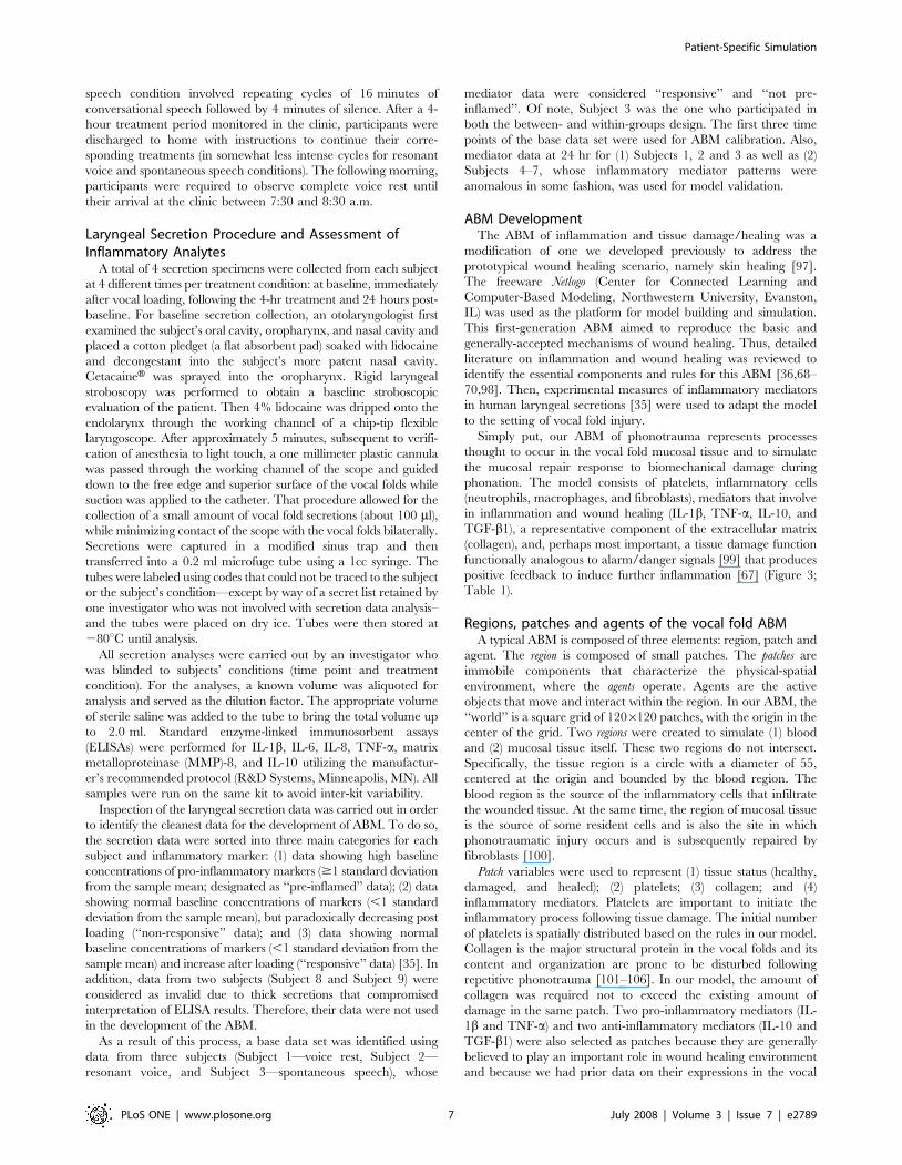
positive feedback to induce further inflammation [67] (Figure 3;
Table 1).
Regions, patches and agents of the vocal fold ABMA typical ABM is composed of three elements: region, patch and
agent. The region is composed of small patches. The patches are
immobile components that characterize the physical-spatial
environment, where the agents operate. Agents are the active
objects that move and interact within the region. In our ABM, the
‘‘world’’ is a square grid of 1206120 patches, with the origin in the
center of the grid. Two regions were created to simulate (1) blood
and (2) mucosal tissue itself. These two regions do not intersect.
Specifically, the tissue region is a circle with a diameter of 55,
centered at the origin and bounded by the blood region. The
blood region is the source of the inflammatory cells that infiltrate
the wounded tissue. At the same time, the region of mucosal tissue
is the source of some resident cells and is also the site in which
phonotraumatic injury occurs and is subsequently repaired by
fibroblasts [100].
Patch variables were used to represent (1) tissue status (healthy,
damaged, and healed); (2) platelets; (3) collagen; and (4)
inflammatory mediators. Platelets are important to initiate the
inflammatory process following tissue damage. The initial number
of platelets is spatially distributed based on the rules in our model.
Collagen is the major structural protein in the vocal folds and its
content and organization are prone to be disturbed following
repetitive phonotrauma [101–106]. In our model, the amount of
collagen was required not to exceed the existing amount of
damage in the same patch. Two pro-inflammatory mediators (IL-
1b and TNF-a) and two anti-inflammatory mediators (IL-10 and
TGF-b1) were also selected as patches because they are generally
believed to play an important role in wound healing environment
and because we had prior data on their expressions in the vocal
Patient-Specific Simulation
PLoS ONE | www.plosone.org 7 July 2008 | Volume 3 | Issue 7 | e2789
folds [48–50]. The concentrations of the inflammatory mediators
on each patch are controlled by formulae for mediator synthesis,
mediator degradation and mediator diffusion.
Agent variables were used to represent (1) tissue damage and (2)
cells. Tissue damage is induced by the initial injury and the
subsequent inflammatory response of the pro-inflammatory medi-
ators (IL-1b and TNF-a). Tissue damage also acts as a stimulus for
further inflammation. Another class of agent is cells, namely,
neutrophils, macrophages and fibroblasts. In our model, cells have
three states: resting, activated or dead. Cells are represented as
agents because they can be organized based on common behavioral
rules, and because the response of a particular cell type to various
mediators is readily characterized in the literature [92]. Cell
behavior was governed by rules based on existing wound healing
literature [36,68–70,98]. Depending on the cell type, the cellular
responses included activation, migration, proliferation, cell death,
secretion of inflammatory mediators, tissue debridement, and
collagen generation. The complete rules of the ABM with
explanations are attached in Supporting Information (Table S1).
Simulation of acute phonotraumaFor each simulation, the user can define the initial levels of IL-
1b, TNF-a and IL-10, add a phonotraumatic event, and then a 4-
hr treatment event (voice rest, ‘‘resonant voice’’ exercises or
spontaneous speech). We assumed in the model that one step of
simulated time represents 0.1 days or approximately 2.4 hours.
The changes in temporal concentration of inflammatory cells,
mediators, tissue damage and collagen were plotted and refolded
into the model at each time step.
Initially, some resting neutrophils are in the blood region,
whereas some macrophages and fibroblasts are present with a
random distribution in both blood and tissue regions. Simulated
phonatory stresses traumatized the mucosal tissue in the middle of
region, triggering platelet degranulation. Shortly afterwards, a
chemoattractant gradient is created that stimulates the infiltration
and activation of neutrophils and macrophages. Later on,
fibroblasts are activated by tissue damage and TGF-b1. Fibro-
blasts secrete collagen to repair both the initial and the
inflammation-induced damage. Last, additional mechanical stress-
es are applied to the traumatized tissue based on the treatment
selected (voice rest: no additional mechanical stress; resonant
voice: low mechanical stress; spontaneous speech: high mechanical
stress).
Model Calibration and ValidationStandard procedures to evaluate the fit of ABM to empirical
data have not been established in the literature. In the present
study, pattern-oriented analysis [107] was used to estimate the
conformity of simulation-generated data curves with the inflam-
matory and wound healing patterns reported in the literature as
well as the empirical data sets around acute phonotrauma
(Table 2).
Using this approach, the user-defined initial magnitude of
mucosal injury (range 0–40 in arbitrary units of damage) was set at
Figure 3. An overall flowchart of the model. The model assumes that biomechanical stress during phonation causes mucosal damage andactivates platelets, neutrophils and macrophages. Platelets produce TGF-b1, which chemoattracts both neutrophils and macrophages. Activatedneutrophils and macrophages secrete pro-inflammatory mediators, which in turn induce anti-inflammatory mediator release. Pro-inflammatorymediators also induce neutrophils and macrophages to produce free radicals that damage tissue. In our model, the activity of free radicals wassubsumed in the actions of TNF-a. Anti-inflammatory mediators contribute to fibroblast activation. Activated fibroblasts secrete collagen thatmediates tissue repair. In the model, collagen accumulation is considered as the surrogate for healing outcome following phonotrauma. Collagen isan important ECM protein involving both structural and biomechanical functions in the vocal folds (Gray & Titze, 1988; Gray et al., 2000).doi:10.1371/journal.pone.0002789.g003
Patient-Specific Simulation
PLoS ONE | www.plosone.org 8 July 2008 | Volume 3 | Issue 7 | e2789
a value of 20 as a ‘‘comparison condition’’, because that setting
resulted in realistic predictions of mucosal damage and healing
when compared with the general consensus around laryngeal
wound healing documented in the literature [68–70,98]. When the
qualitative behavior of the simulation appeared satisfactory, we
proceeded to calibrate the model by adjusting parameter values
not found in the literature to fit the quantity and time-course of
measured vocal fold mediators. We calibrated the ABM using data
from the base cohort (Subjects 1, 2 and 3) using their baseline
mediator levels in laryngeal fluid, immediately after phonotrauma
induction, and following a 4-hr treatment (voice rest, ‘‘resonant
voice’’ exercises, or spontaneous speech) (Figures 1–2, dark circles)
[35]. Subsequently, we validated the ABM quantitatively by
comparing the predicted mediator levels with the empirical
mediator levels at 24 hr (1) for Subjects 1, 2 and 3, as well as (2)
Subjects 4–7 whose data had never been used for the model.
Due to the inherent stochasticity of the ABM framework, we
performed ten runs of the calibrated ABM for each subject up to
five simulated days under the condition of (1) each subject’s actual
treatment group and (2) hypothetical randomization to either of
the other two treatment groups for all subjects, i.e., hypothetical
treatments. The means and standard deviations of model variables
(concentrations of inflammatory cells, mediators, and tissue
damage) were computed at each time point for subsequent
analysis. Hypothetical treatments were included for simulation to
demonstrate the application of ABM for running individual-
specific in silico clinical trials.
Statistical AnalysisZ-tests using the p-value approach to testing statistical significance
were carried out to compare the human empirical data and the data
predicted by the model for IL-1b, TNF-a and IL-10 at the 24-hr
time point for (1) the base cohort (Subjects 1–3) and (2) Subjects 4–7.
Also, linear mixed models with fixed and random effects were used to
compare the predicted levels of IL-1b, TNF-a, and IL-10. Fixed
factors included: (1) patient, (2) treatment condition (spontaneous speech,
voice rest and ‘‘resonant voice’’ exercises) and (3) time point
(immediately after phonotrauma induction, following a 4-hr
treatment and 24-hr post baseline). The random factor is the number
of simulation runs and is nested within the patient factor.
Supporting Information
Table S1 ABM RULES.
Found at: doi:10.1371/journal.pone.0002789.s001 (0.19 MB RTF)
Acknowledgments
The authors would like to acknowledge the following clinicians and
investigators: Dr. Clark Rosen, Dr. Kim Steinhauer, Dr. Ryan Branski and
Dr. Elizabeth U. Grillo, for their contributions to this work. We would also
like to thank Dr. Gary An for his valuable inputs to the computer model.
Author Contributions
Conceived and designed the experiments: NYKL KV. Performed the
experiments: NYKL KV. Analyzed the data: NYKL KV GC PAH YV.
Contributed reagents/materials/analysis tools: NYKL KV GC QM PAH
YV. Wrote the paper: NYKL KV GC QM PAH YV. Statistical
consultation: ENR. Agent-based model construction: NYL GC QM YV.
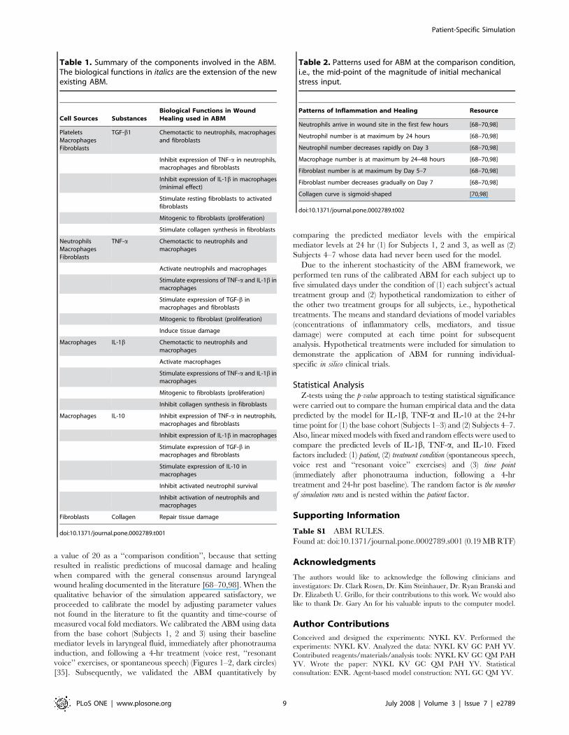
Table 1. Summary of the components involved in the ABM.The biological functions in italics are the extension of the newexisting ABM.
Cell Sources SubstancesBiological Functions in WoundHealing used in ABM
PlateletsMacrophagesFibroblasts
TGF-b1 Chemotactic to neutrophils, macrophagesand fibroblasts
Inhibit expression of TNF-a in neutrophils,macrophages and fibroblasts
Inhibit expression of IL-1b in macrophages(minimal effect)
Stimulate resting fibroblasts to activatedfibroblasts
Mitogenic to fibroblasts (proliferation)
Stimulate collagen synthesis in fibroblasts
NeutrophilsMacrophagesFibroblasts
TNF-a Chemotactic to neutrophils andmacrophages
Activate neutrophils and macrophages
Stimulate expressions of TNF-a and IL-1b inmacrophages
Stimulate expression of TGF-b inmacrophages and fibroblasts
Mitogenic to fibroblast (proliferation)
Induce tissue damage
Macrophages IL-1b Chemotactic to neutrophils andmacrophages
Activate macrophages
Stimulate expressions of TNF-a and IL-1b inmacrophages
Mitogenic to fibroblasts (proliferation)
Inhibit collagen synthesis in fibroblasts
Macrophages IL-10 Inhibit expression of TNF-a in neutrophils,macrophages and fibroblasts
Inhibit expression of IL-1b in macrophages
Stimulate expression of TGF-b inmacrophages and fibroblasts
Stimulate expression of IL-10 inmacrophages
Inhibit activated neutrophil survival
Inhibit activation of neutrophils andmacrophages
Fibroblasts Collagen Repair tissue damage
doi:10.1371/journal.pone.0002789.t001
Table 2. Patterns used for ABM at the comparison condition,i.e., the mid-point of the magnitude of initial mechanicalstress input.
Patterns of Inflammation and Healing Resource
Neutrophils arrive in wound site in the first few hours [68–70,98]
Neutrophil number is at maximum by 24 hours [68–70,98]
Neutrophil number decreases rapidly on Day 3 [68–70,98]
Macrophage number is at maximum by 24–48 hours [68–70,98]
Fibroblast number is at maximum by Day 5–7 [68–70,98]
Fibroblast number decreases gradually on Day 7 [68–70,98]
Collagen curve is sigmoid-shaped [70,98]
doi:10.1371/journal.pone.0002789.t002
Patient-Specific Simulation
PLoS ONE | www.plosone.org 9 July 2008 | Volume 3 | Issue 7 | e2789
References
1. Gray SD, Titze IR (1988) Histologic investigation of hyperphonated canine
vocal cords. Ann Otol Rhinol Laryngol 97: 381–388.
2. Gray SD, Titze IR, Alipour F, Hammond TH (2000) Biomechanical andhistologic observations of vocal fold fibrous proteins. Ann Otol Rhinol Laryngol
109: 77–85.
3. Titze IR, Hitchcock RW, Broadhead K, Webb K, Li W, et al. (2004) Designand validation of a bioreactor for engineering vocal fold tissues under combined
tensile and vibrational stresses. J Biomech 37: 1521–1529.
4. Gray SD (1991) Basement membrane zone injury in vocal nodules; Gauffin J,Hammarberg B, eds. San Diego: Singular Press. pp 1–35.
5. Gray SD, Titze IR, Lusk RP (1987) Electron microscopy of hyperphonated
canine vocal cords. Journal of Voice 1: 109–115.
6. Branski RC, Perera P, Verdolini K, Rosen CA, Hebda PA, et al. (2006)Dynamic Biomechanical Strain Inhibits IL-1beta-induced Inflammation in
Vocal Fold Fibroblasts. J Voice.
7. Jacobson BH, Johnson A, Grywalski C, Silbergleit A, Jacobson G,Benninger MS, Newman CW (1997) The voice handicap index (VHI):
Development and validation. American Journal of Speech-Language Pathology6: 66–70.
8. Ma EP, Yiu EM (2001) Voice activity and participation profile: assessing the
impact of voice disorders on daily activities. J Speech Lang Hear Res 44:511–524.
9. Raaijmakers MF, Dekker J, Dejonckere PH (1998) Diagnostic assessment and
treatment goals in logopedics: impairments, disabilities and handicaps FoliaPhoniatr Logop 50: 71–79.
10. Smith E, Verdolini K, Gray SD, Nichols S, Lemke J (1996) Effect of voice
disorders on quality of life. Journal of Medical SpeechLanguage Pathology 4:223–244.
11. Goldring SR (2003) Inflammatory mediators as essential elements in bone
remodeling. Calcified Tissue International 73: 97–100.
12. Hart J (2002) Inflammation. 1: Its role in the healing of acute wounds. J WoundCare 11: 205–209.
13. Hart J (2002) Inflammation. 2: Its role in the healing of chronic wounds.
J Wound Care 11: 245–249.
14. Redd MJ, Cooper L, Wood W, Stramer B, Martin P (2004) Wound healingand inflammation: embryos reveal the way to perfect repair. Philos Trans R Soc
Lond B Biol Sci 359: 777–784.
15. Morrison M, Rammage L (1994) The Management of Voice Disorders. SanDiego: Singular Publishing Group Inc.
16. Stemple JC, Lee L, D’Amico B, Pickup B (1994) Efficacy of vocal function
exercises as a method of improving voice production. Journal of Voice 8:271–278.
17. Verdolini K (2000) Case Study: Resonant Voice Therapy; Stemple J, ed. San
Diego: Singular Publishing Group Inc. pp 46–62.
18. Sataloff RT (1997) Voice rest; Sataloff RT, ed. San Diego: Singular PublishingGroup, Inc. pp 453–456.
19. Branski RC (2005) Vocal fold fibroblast response to mechanical stress [doctoral
dissertation]. Pittsburgh: University of Pittsburgh. pp 1–123.20. Verdolini K, Li NYK, Branski RC, Rosen CA, Urban EG, et al. (in
preparation) The effect of targeted vocal exercise on recovery from acute
inflammation.21. Burroughs P, Dahners LE (1990) The effect of enforced exercise on the healing
of ligament injuries. Am J Sports Med 18: 376–378.
22. Eiff MP, Smith AT, Smith GE (1994) Early mobilization versus immobilizationin the treatment of lateral ankle sprains. Am J Sports Med 22: 83–88.
23. Kerkhoffs GM, Struijs PA, Marti RK, Assendelft WJ, Blankevoort L, et al.
(2002) Different functional treatment strategies for acute lateral ankle ligamentinjuries in adults. Cochrane Database Syst Rev. pp CD002938.
24. Kerkhoffs GM, Struijs PA, Marti RK, Blankevoort L, Assendelft WJ, et al.
(2003) Functional treatments for acute ruptures of the lateral ankle ligament: a
systematic review. Acta Orthop Scand 74: 69–77.25. Kvist J (2004) Rehabilitation following anterior cruciate ligament injury:
current recommendations for sports participation. Sports Med 34: 269–280.
26. Pijnenburg AC, Van Dijk CN, Bossuyt PM, Marti RK (2000) Treatment ofruptures of the lateral ankle ligaments: a meta-analysis. J Bone Joint Surg Am
82: 761–773.
27. Mulligan B (1995) Manual Therapy ‘‘NAGS,’’ ‘‘SNAGS,’’ ‘‘MWMSs’’ etc.Wellington, New Zealand: Plane View Service. pp 78–88.
28. Paungmali A, O’Leary S, Sowvlis T, Vicenzino B (2003) Hypoalgesic and
sympathoexcitatory effects of mobilization with movement for lateralepicodylalgia. Physical Therapy 83: 374–383.
29. Salter RB (1994) The physiologic basis of continuous passive motion for
articular cartilage healing and regeneration. Hand Clinics 10: 211.
30. Salter RB (1996) History of rest and motion and the scientific basis for earlycontinuous passive motion. Hand Clinics 12: 1–11.
31. Thornton GM, Shrive NG, Frank CB (2003) Healing ligaments have decreased
cyclic modulus compared to normal ligaments and immobilization furthercompromises healing ligament response to cyclic loading. Journal of
Orthopaedic Research 21: 716–722.
32. Threlkeld AJ (1992) Manual Therapy: An American Physical TherapyAssociation Monograph. Physical Therapy. pp 59–68.
33. Visser NA, de Koning MH, Lammi MJ, Hakkinen T, Tammi M, et al. (1998)
Increase of decorin content in articular cartilage following running. ConnectTissue Res 37: 295–302.
34. Williams JM, Moran M, Thonar EJ, Salter RB (1994) Continuous passive
motion stimulates repair of rabbit knee articular cartilage after matrixproteoglycan loss. Clin Orthop 304: 252–262.
35. Verdolini K, Li NYK, Rosen CA, Grillo E, Branski RC, et al. (in preparation
[b]) Effect of resonant voice excercises on the biological recovery from acutephonotrauma.
36. Clark RAF (1998) Wound Repair. Overview and General Considerations. In:
Clark RAF, ed. The Molecular and Cellular Biology of Wound Repair. NewYork: Plenum Press.
37. Clark RAF (1988) Cutaneous wound repair: Molecular and cellular controls.
Prog Dermatol 22: 1–12.
38. Clark RAF, ed (1998) The Molecular and Cellular Biology of Wound Repair.New York: Plenum Press.
39. Calvin M (1998) Cutaneous wound repair. Wounds 10: 12–32.
40. Mast BA, Schultz GS (1996) Interactions of cytokines, growth factors, andproteases in acute and chronic wounds. Wound Repair Regen 4: 411–420.
41. Werner S, Grose R (2003) Regulation of wound healing by growth factors and
cytokines. Physiol Rev 83: 835–870.
42. Gillitzer R, Goebeler M (2001) Chemokines in cutaneous wound healing.J Leukoc Biol 69: 513–521.
43. Hardy MA (1989) The biology of scar formation. Phys Ther 69: 1014–1024.
44. Nathan C (2002) Points of control in inflammation. Nature 420: 846–852.
45. Walter JB, Israel MS, eds (1987) General Pathology. Churchill: Livingstone.
46. Catten M, Gray SD, Hammond TH, Zhou R, Hammond EH (1998) Analysisof cellular location and concentration in vocal fold lamina propria.
Otolaryngology Head and Neck Surgery 118: 663–667.
47. Gray SD (2000) Cellular Physiology of the Vocal Folds. In: Rosen CA,Murry T, eds. The Otolaryngologic Clinics of North America. Philadelphia:
W.B. Saunders Company.
48. Branski RC, Verdolini K, Rosen CA, Hebda PA (2004) Markers of woundhealing in vocal fold secretions from patients with laryngeal pathology. Annals
of Otology, Rhinology, and Laryngology 113: 23–29.
49. Verdolini K, Rosen CA, Branski RC, Hebda PA (2003) Shifts in biochemicalmarkers associated with wound healing in laryngeal secretions following
phonotrauma: A preliminary study. Annals of Otology, Rhinology, &Laryngology 112: 1021–1025.
50. Branski RC, Rosen CA, Verdolini K, Hebda PA (2005) Biochemical markers
associated with acute vocal fold wound healing: a rabbit model. J Voice 19:283–289.
51. Branski RC, Rosen CA, Verdolini K, Hebda PA (2005) Acute vocal fold wound
healing in a rabbit model. Ann Otol Rhinol Laryngol 114: 19–24.
52. Chow CC, Clermont G, Kumar R, Lagoa C, Tawadrous Z, et al. (2005) Theacute inflammatory response in diverse shock states. Shock 24: 74–84.
53. Day J, Rubin J, Vodovotz Y, Chow CC, Reynolds A, et al. (2006) A reduced
mathematical model of the acute inflammatory response II. Capturingscenarios of repeated endotoxin administration. J Theor Biol.
54. Prince JM, Levy RM, Bartels J, Baratt A, Kane JM 3rd, et al. (2006) In silico
and in vivo approach to elucidate the inflammatory complexity of CD14-deficient mice. Mol Med 12: 88–96.
55. An G, Faeder J, Vodovotz Y (2008) Translational systems biology: introduction
of an engineering approach to the pathophysiology of the burn patient. J BurnCare Res 29: 277–285.
56. Vodovotz Y, Csete M, Bartels J, Chang S, An G (2008) Translational systems
biology of inflammation. PLoS Comput Biol 4: e1000014.
57. Ermentrout GB, Edelstein-Keshet L (1993) Cellular automata approaches tobiological modeling. J Theor Biol 160: 97–133.
58. An G (2005) Mathematical modeling in medicine: a means, not an end. Crit
Care Med 33: 253–254.59. Smallwood RH, Holcombe WML, Walker DC (2004) Development and
validation of computational models of cellular interaction. Journal of Molecular
Histology 35: 659–665.60. Vodovotz Y, Clermont G, Chow C, An G (2004) Mathematical models of the
acute inflammatory response. Current Opinion in Critical Care 10: 383–390.
61. Walker DC, Hill G, Wood SM, Smallwood RH, Southgate J (2004) Agent-based computational modeling of wounded epiethelial cell monolayers. IEEE
Transactions on Nonobioscience 3: 153–163.
62. Butterfield TA, Best TM, Merrick MA (2006) The dual roles of neutrophils andmacrophages in inflammation: a critical balance between tissue damage and
repair. J Athl Train 41: 457–465.
63. Toumi H, F’Guyer S, Best TM (2006) The role of neutrophils in injury andrepair following muscle stretch. J Anat 208: 459–470.
64. Tidball JG (1995) Inflammatory cell response to acute muscle injury. Med Sci
Sports Exerc 27: 1022–1032.
65. Eming SA, Krieg T, Davidson JM (2007) Inflammation in wound repair:molecular and cellular mechanisms. J Invest Dermatol 127: 514–525.
66. Stramer BM, Mori R, Martin P (2007) The inflammation-fibrosis link? A Jekyll
and Hyde role for blood cells during wound repair. J Invest Dermatol 127:1009–1017.
Patient-Specific Simulation
PLoS ONE | www.plosone.org 10 July 2008 | Volume 3 | Issue 7 | e2789
67. Vodovotz Y (2006) Deciphering the complexity of acute inflammation using
mathematical models. Immunol Res 36: 237–245.68. Cockbill S (2002) Wound: the healing process. Hospital Pharmacist 9: 255–260.
69. Martin P (1997) Wound Healing-aiming for perfect skin regeneration. Science
276: 75–81.70. Witte MB, Barbul A (1997) General principles of wound healing. Surgical
Clinics of North America 77: 509–528.71. Hirschi SD, Gray SD, Thibeault SL (2002) Fibronectin:an interesting vocal fold
protein. J Voice 16: 310–316.
72. Thibeault SL, Bless DM, Gray SD (2003) Interstitial protein alterations inrabbit vocal fold with scar. J Voice 17: 377–383.
73. Thibeault SL, Gray SD, Bless DM, Chan RW, Ford CN (2002) Histologic andRheologic characterization of vocal fold scarring. Journal of Voice 16: 96–104.
74. Thibeault SL, Gray SD, Li W, Ford CN, Smith ME, et al. (2002) Genotypicand phenotypic expression of vocal fold polyps and Reinke’s edema: a
preliminary study. Ann Otol Rhinol Laryngol 111: 302–309.
75. Taylor KR, Trowbridge JM, Rudisill JA, Termeer CC, Simon JC, et al. (2004)Hyaluronan fragments stimulate endothelial recognition of injury through
TLR4. J Biol Chem 279: 17079–17084.76. Gunter HE (2003) A mechanical model of vocal-fold collision with high spatial
and temporal resolution. J Acoust Soc Am 113: 994–1000.
77. Gunter HE (2004) Modeling mechanical stresses as a factor in the etiology ofbenign vocal fold lesions. Journal of Biomechanics 37: 1119–1124.
78. Jiang JJ, Diaz CE, Hanson DG (1998) Finite modeling of vocal fold vibratioinin normal phonation and hyperfunctional dysphonia: implications for the
pathogenesis of vocal nodules. Ann Otol Rhinol Laryngol 107: 603–610.79. Thomson SL, Mongeau L, Frankel SH (2005) Aerodynamic transfer of energy
to the vocal folds. J Acoust Soc Am 118: 1689–1700.
80. Roberts AB, Sporn MB (1996) Transforming growth factor-b. In: Clark RAF,ed. The Molecular and Cellular Biology of Wound Repair. New York: Plenum
Press. pp 275–308.81. O’Kane S, Ferfuson MW (1997) Transforming growth factor betas and wound
healing. International Journal of Biochemistry & Cell Biology 29: 63–78.
82. Barcellos-Hoff MH, Ehrhart EJ, Kalia M, Jirtle R, Flanders K, et al. (1995)Immunohistochemical detection of active transforming growth factor-b in situ
using engineered tissue. American Journal of Pathology 147: 1228–1237.83. Chong H, Vodovotz Y, Cox GW, Barcellos-Hoff MH (1999) Immunocyto-
chemical detection of latent TGF-b activation in cultured macrophages.Journal of Cellular Physiology 178: 275–283.
84. Vodovotz Y, Chesler L, Chong H, Kim SJ, Simpson JT, et al. (1999)
Regulation of transforming growth factor beta1 by nitric oxide. Cancer Res 59:2142–2149.
85. Vodovotz Y, Hoffman R, Baratt A, Wei J, Lagoa C, et al. (2006) In silico and invivo studies modeling the aged acute inflammatory response. Shock 25: 39–40
(Abstract).
86. Li NYK, Verdolini K, Clermont G, Mi Q, Hebda PA, et al. Simulating acutephonotrauma: a comparison of agent-based and equation-based models; 2006;
Tokyo, Japan.87. Whitcomb DC, Aoun E, Vodovotz Y, Clermont G, Barmada MM (2005)
Evaluating disorders with a complex genetics basis. the future roles of meta-analysis and systems biology. Dig Dis Sci 50: 2195–2202.
88. Clermont G, Bartels J, Kumar R, Constantine G, Vodovotz Y, et al. (2004) In
silico design of clinical trials: a method coming of age. Crit Care Med 32:2061–2070.
89. An G (2001) Agent-based computer simulation and sirs: building a bridge
between basic science and clinical trials. Shock 16: 266–273.
90. Kumar R, Clermont G, Vodovotz Y, Chow CC (2004) The dynamics of acute
inflammation. J Theor Biol 230: 145–155.
91. Reynolds A, Rubin J, Clermont G, Day J, Vodovotz Y, et al. (2006) A reduced
mathematical model of the acute inflammatory response: I. Derivation of
model and analysis of anti-inflammation. J Theor Biol.
92. An G (2004) In silico experiments of existing and hypothetical cytokine-directed
clinical trials using agent-based modeling. Critical Care Medicine 32:
2050–2060.
93. Kumar R, Chow CC, Bartels J, Clermont G, Vodovotz Y (In Press) Amathematical simulation of the inflammatory response to anthraz infection.
Shock.
94. Peterson KL, Verdolini-Marston K, Barkmeir JM, Hoffman HT (1994)
Comparison of aerodynamic and electroglottographic parameters in evaluating
clinically relevant voicing patterns. Ann Otol Rhinol Laryngol 103: 335–346.
95. Verdolini K, Druker DG, Palmer PM, Samawi H (1998) Laryngeal adduction
in resonant voice. J Voice 12: 315–327.
96. Verdolini-Marston K, Burke MK, Lessac A, Glaze L, Caldwell E (1995)
Preliminary study of two methods of treatment for laryngeal nodules. Journal of
Voice 9: 74–85.
97. Mi Q, Riviere B, Clermont G, Steed DL, Vodovotz Y (2007) Agent-based
modeling of inflammation and wound healing: insights into diabetic foot ulcer
pathology and the role of transforming growth factor-beta 1. Wound Repair
and Regeneration 15: 671–682.
98. Robson MC, Steed DL, Franz MG (2001) Wound healing: biologic featuresand approaches to maximize healing trajectories. Curr Probl Surg 38: 72–140.
99. Matzinger P (2002) The danger model: a renewed sense of self. Science 296:
301–305.
100. Mi Q, Riviere B, Clermont G, Steed DL, Vodovotz Y (in press) Agent-basedmodeling of inflammation and wound healing: insights into diabetic foot ulcer
pathology and the role of transforming growth factor-beta 1. Wound Repair
and Regeneration.
101. Hahn MS, Kobler JB, Zeitels SM, Langer R (2006) Quantitative and
comparative studies of the vocal fold extracellular matrix II: collagen. Ann Otol
Rhinol Laryngol 115: 225–232.
102. Madruga de Melo EC, Lemos M, Aragao Ximenes Filho J, Sennes LU,
Nascimento Saldiva PH, et al. (2003) Distribution of collagen in the lamina
propria of the human vocal fold. Laryngoscope 113: 2187–2191.
103. Tateya T, Tateya I, Bless DM (2006) Collagen subtypes in human vocal folds.
Ann Otol Rhinol Laryngol 115: 469–476.
104. Courey MS, Shohet JA, Scott MA, Ossoff RH (1996) Immunohistochemical
characterization of benign laryngeal lesions. Ann Otol Rhinol Laryngol 195:
525–531.
105. Gray SD, Hammond EH, Hanson D (1995) Benign pathologic response of thelarynx. Ann Otol Rhinol Laryngol 104: 8–13.
106. Neves BMJ, Neto JG, Pontes P (2004) Histopathological and immunohisto-
chemical differentiation of epithelial alterations in vocal nodule comparing to
polyps and to laryngeal edema. Brazillian Journal of Otolaryngology 70:
439–448.
107. Railback SF (2001) Getting ‘‘results’’: the pattern-oriented approach to analyze
natural systems with individual-based models. Natural Resource Modeling 14:
465–474.
Patient-Specific Simulation
PLoS ONE | www.plosone.org 11 July 2008 | Volume 3 | Issue 7 | e2789