The Spectrin cytoskeleton regulates the Hippo signalling pathway
Importance of Non-Selective Cation Channel TRPV4Interaction with Cytoskeleton and Their ReciprocalRegulations in Cultured CellsChandan Goswami1,2*, Julia Kuhn1, Paul A. Heppenstall3¤, Tim Hucho1
1 Signal Transduction in Pain and Mental Retardation, Department for Molecular Human Genetics Max Planck Institute for Molecular Genetics, Berlin, Germany, 2 National
Institute of Science Education and Research, Bhubaneswar, India, 3 Klinik fur Anaesthesiologie und Operative Intensivmedizin, Charite Universitatsmedizin Berlin, Campus
Benjamin Franklin, Berlin, Germany
Abstract
Background: TRPV4 and the cellular cytoskeleton have each been reported to influence cellular mechanosensitiveprocesses as well as the development of mechanical hyperalgesia. If and how TRPV4 interacts with the microtubule andactin cytoskeleton at a molecular and functional level is not known.
Methodology and Principal Findings: We investigated the interaction of TRPV4 with cytoskeletal componentsbiochemically, cell biologically by observing morphological changes of DRG-neurons and DRG-neuron-derived F-11 cells,as well as functionally with calcium imaging. We find that TRPV4 physically interacts with tubulin, actin and neurofilamentproteins as well as the nociceptive molecules PKCe and CamKII. The C-terminus of TRPV4 is sufficient for the directinteraction with tubulin and actin, both with their soluble and their polymeric forms. Actin and tubulin compete for binding.The interaction with TRPV4 stabilizes microtubules even under depolymerizing conditions in vitro. Accordingly, in cellularsystems TRPV4 colocalizes with actin and microtubules enriched structures at submembranous regions. Both expressionand activation of TRPV4 induces striking morphological changes affecting lamellipodial, filopodial, growth cone, and neuritestructures in non-neuronal cells, in DRG-neuron derived F11 cells, and also in IB4-positive DRG neurons. The functionalinteraction of TRPV4 and the cytoskeleton is mutual as Taxol, a microtubule stabilizer, reduces the Ca2+-influx via TRPV4.
Conclusions and Significance: TRPV4 acts as a regulator for both, the microtubule and the actin. In turn, we describe thatmicrotubule dynamics are an important regulator of TRPV4 activity. TRPV4 forms a supra-molecular complex containingcytoskeletal proteins and regulatory kinases. Thereby it can integrate signaling of various intracellular second messengersand signaling cascades, as well as cytoskeletal dynamics. This study points out the existence of cross-talks between non-selective cation channels and cytoskeleton at multiple levels. These cross talks may help us to understand the molecularbasis of the Taxol-induced neuropathic pain development commonly observed in cancer patients.
Citation: Goswami C, Kuhn J, Heppenstall PA, Hucho T (2010) Importance of Non-Selective Cation Channel TRPV4 Interaction with Cytoskeleton and TheirReciprocal Regulations in Cultured Cells. PLoS ONE 5(7): e11654. doi:10.1371/journal.pone.0011654
Editor: Hiroaki Matsunami, Duke University, United States of America
Received December 3, 2009; Accepted June 15, 2010; Published July 19, 2010
Copyright: � 2010 Goswami et al. This is an open-access article distributed under the terms of the Creative Commons Attribution License, which permitsunrestricted use, distribution, and reproduction in any medium, provided the original author and source are credited.
Funding: Funding from Max Planck Institute for Molecular Genetics (Berlin, Germany) is gratefully acknowledged. CG is now supported by National Institute ofScience Education and Research, India. The authors declare that the funders had no role in study design, data collection and analysis, decision to publish, orpreparation of the manuscript.
Competing Interests: The authors have declared that no competing interests exist.
* E-mail: [email protected]
¤ Current address: EMBL Monterotondo, Adriano Buzzati-Traverso Campus, Monterotondo, Italy
Introduction
Transient receptor potential vanilloid sub type 4 (TRPV4) is a
member of TRP super family of Ca2+-permeable non-selective
cation channels. This polymodal receptor is involved in cellular
processes such as mechanosensation, osmosensation and thermo-
sensation [1–5]. Some of these sensory functions are well conserved
in different species. For example, mammalian TRPV4 can rescue
mechanosensitive defects observed in C.elegans OSM-9 mutants [5].
In higher organisms TRPV4 is endogenously expressed in
nociceptive dorsal root ganglion (DRG) neurons but also in many
non-neuronal tissues and cells such as skin, kidney corneal epithelial
cells [6], cerebral microvascular endothelial cells [7], cortical
astrocytes [8], tracheal epithelial cells [9], keratinocyte cell lines [10]
and in other cells. The widespread distribution of TRPV4 is
indicative of its involvement in various physiological functions.
Indeed, TRPV4 is of importance in shear stress-induced vasodila-
tion [11] as well as in auditory functions [12–13]. Recently TRPV4
gained importance as it has been linked with the development of
different pathophysiological conditions such as neuropathic pain,
cystic fibrosis, brachyolmia and cancer [14–18].
From several reports, the involvement of cytoskeleton can be
correlated with the localization and function of TRPV4. For
example, TRPV4 is found in structures like cilia in various tissues
and cells [9,19–21] and in lamellipodia, where it regulates the
dynamics of cytoskeleton [22–23]. Many cellular functions
involving TRPV4 are known to require active participation of
the cytoskeleton. For example, TRPV4 activity is central to
PLoS ONE | www.plosone.org 1 July 2010 | Volume 5 | Issue 7 | e11654

cytoskeleton-dependent/mediated regulatory volume decrease of
cells [6,10,24], a process where actin-binding proteins contribute
to cell volume regulatory ion channel activation [24–26]. In
addition, TRPV4 has a conserved role in mechanotransduction, a
complex process that involves both actin and microtubule
cytoskeletal components [27–29]. The interplay of TRPV4 with
microtubule cytoskeleton also appears on a behavioural level,
where alteration of microtubule dynamics by Taxol induces a
TRPV4-dependent painful peripheral neuropathy [30]. While all
these cellular and behavioural studies strongly suggest that TRPV4
shares a functional relation with the cytoskeleton, so far a direct
link of TRPV4 with the cytoskeleton has not been demonstrated.
Thus, a molecular mechanism for the role of TRPV4 and the
cytoskeleton in pain, mechanosensation as well as other cellular
functions remains elusive.
Recently, we have established a functional interplay between
TRPV1, a close homologue of TRPV4, and the microtubule
cytoskeleton [31–35]. We demonstrated the physical interaction of
microtubule cytoskeleton with TRPV1 via two novel tubulin-
binding motifs [36–37]. Based on our previous experiments done
on TRPV1 and the sequence homology between TRPV1 and
TRPV4, we predicted that TRPV4 might interact with tubulin via
its C-terminal domain. Therefore, in this work we set out to
explore if TRPV4 physically and functionally interacts with actin
and microtubule cytoskeletal components.
Results
TRPV4 interacts with endogenous actin and tubulinIn order to test if TRPV4 interacts with cytoskeletal proteins like
tubulin and actin, we performed co-immunoprecipitation experi-
ments with affinity purified TRPV4 antibodies. CHO-KI-TRPV4
stable cell lines were used, which express low levels of TRPV4. In
immunoblot analysis, we observed that TRPV4 antibodies
precipitated TRPV4 together with actin and tubulin proteins
(Fig. 1a). Presence of tubulin and actin was not observed when a
similar co-immunoprecipitation was performed from the same cell
extract using an antibody, which was not raised against TRPV4. To
confirm further that the tubulin interaction is occurring even in
endogenous tissues, we isolate DRG neurons from rat and
performed similar co-immunoprecipitation experiments with affin-
ity purified TRPV4 antibodies. We observed that tubulin co-
immunoprecipitated with TRPV4 even from DRG neurons (1b).
The C-terminus of TRPV4 is sufficient for interaction withactin and tubulin
To identify, which part of the TRPV4 interacts with actin and/
or tubulin proteins, we performed a pull down experiment using
maltose-binding-protein (MBP)-fused to the N- and C-termini of
TRPV4. According to our prediction [37], at least one tubulin-
binding site is located within the C-terminus of TRPV4 (Fig. S1)
Figure 1. Interaction of soluble tubulin and actin with TRPV4. a. Co-immunoprecipitation of actin and tubulin with TRPV4. Cell extracts fromCHO-KI cells stably expressing TRPV4 (lane 1) was immunoprecipitated by TRPV4 antibody (lane 2) or by a non-specific antibody (lane 3). Blots wereprobed for TRPV4 (left side), tubulin (middle) and actin (right side). b. Co-immunoprecipitation of tubulin with TRPV4. Extracts from DRG (lane 1) wasimmunoprecipitated by TRPV4 antibody (lane 2) or by a non-specific antibody (lane 3). Blots were probed for TRPV4 (upper panel) and tubulin (lowerside). c. MBP-TRPV4-Ct (lane 2-3) but not MBP-LacZ (lane 4–5) forms specific complexes when incubated with mammalian brain extract (lane 1), bothin presence (lane 2 and 4) or absence (lane 3 and 5) of Ca2+ (1 mM). Presence of PKCe, actin and tubulin are observed only in lane 2 and 3.Neurofilament in the pull down samples is visible only after exposing for a prolonged time. Presence of CamKII is noted only in the presence of Ca2+
(lane 2). Note that the amount of MBP-LacZ used, as a negative control for the pull down experiment is much more than MBP-TRPV4-Ct. d. Tubulininteracts with TRPV4-Ct directly. MBP-LacZ (lane 1–2) or MBP-TRPV4-Ct (lane 3–4) was incubated with buffer only (lane 1 and 3) or with purifiedtubulin (lane 2 and 4). Pulled down samples were probed for different isotype-specific and different post-translationally modified tubulins. e. Actininteracts directly with TRPV4-Ct. MBP-TRPV4-Ct (lane 1–2) or MBP-LacZ (lane 3–4) was incubated with purified actin (lane 1–4) either in the presence(lane 1-and 3) or absence (lane 2 and 4) of Ca2+ (1 mM) and subsequently probed for bound actin. f. Soluble tubulin and actin competes for the C-terminal cytoplasmic fragment of TRPV4. MBP-TRPV4-Ct was incubated with only tubulin (lane 1), with only actin (lane 2), or both tubulin and actin ina sequential manner (lane 3–4). Prior incubation of tubulin inhibits further binding of actin (lane 3). Similarly, prior incubation of actin significantlyreduces the further binding of tubulin (lane 4).doi:10.1371/journal.pone.0011654.g001
TRPV4 Regulates Cytoskeleton
PLoS ONE | www.plosone.org 2 July 2010 | Volume 5 | Issue 7 | e11654
and as the expression of the N-terminal cytoplasmic fragment
remained poor, we restricted our study to the C-terminus of
TRPV4 only. Pull down was performed from adult porcine brain
homogenate as well as from F11 cell lysate, a fusion-cell of rat
DRG neurons and mouse neuroblastoma cells [38]. In immuno-
blot analysis, we observed MBP-TRPV4-C-terminus fusion
protein (MBP-TRPV4-Ct) but not the control fusion protein
MBP-LacZ to pull down soluble tubulin and actin (Fig. 1c, S2).
When probed for the presence of another cytoskeletal element,
namely soluble neurofilament proteins, only minimal amounts of
NF116 kDa and no NF200 kDa were detected.
As activation of TRPV4 results in high Ca2+-influx, we tested if
higher concentration of Ca2+ could modulate these interactions.
However, we observed that tubulin and actin interaction with
TRPV4 do not depend on Ca2+ (Fig. 1c, S2).
TRPV4-Ct forms a Ca2+-sensitive supra-molecularsignaling complex made of actin, tubulin as well asnociceptive signaling components PKCe and CamKII
Previously we have demonstrated that mechanical hyperalgesia
induced agonists of novel estrogen receptor GPR30 strongly
correlates with the translocation of PKCe in IB4 (+) neurons [39].
Moreover, mechanical hyperalgesia can be induced by activation of
the epsilon isoform of PKC (PKCe), a well-described pro-
nociceptive signaling molecule [39–43]. In addition, calmodulin,
and CamKII function has been linked with the chronic inflamma-
tory pain [44–46]. Thus, we tested if the TRPV4-actin/tubulin
complex also contains PKCe and/or CamKII. We found CamKII
to be present in the eluates of the pull-down material both from
soluble brain and F11 cell extract, but only in the presence of Ca2+
(Fig. 1c, Fig. S1). In addition, we also detected PKCe in the MBP-
TRPV4-Ct complex, both in presence and absence of Ca2+.
Presence of PKCe or CamKII was not observed with the control
protein MBP-LacZ. Thus, using two different biological sources,
our results indicate that TRPV4-Ct can form supra-molecular
complexes consisting of structural and signaling proteins.
MBP-TRPV4-Ct interacts directly with soluble tubulin andactin
To test if TRPV4-Ct interacts directly with tubulin and actin, we
performed pull down experiments with the purified MBP-TRPV4-
Ct, tubulin, and actin. As expected, MBP-TRPV4-Ct but not MBP-
LacZ pulls down tubulin (Fig. 1d). Tubulin is subjected to different
types of post-translational modification, which regulate the process
of microtubule stabilization, destabilization and maturation. Thus,
we tested for the presence of various post-translationally modified
tubulins in the complex formed with MBP-TRPV4-Ct. Indeed we
found a large number of these post-translationally modified tubulins
and neuron-specific b-III tubulin (Fig. 1d).
Next we tested if also soluble actin interacts directly with MBP-
TRPV4-Ct. In immunoblot analysis, we observed that the MBP-
TRPV4-Ct pulls down purified actin, while under the same
conditions MBP-LacZ was unable to pull down any actin (Fig. 1e).
Again, we observed no influence of Ca2+ on this interaction.
Tubulin and actin compete for binding to MBP-TRPV4-CtTo understand if tubulin binding affects actin binding and vice
versa, we performed a competition experiment between soluble
tubulin and soluble actin for the binding to MBP-TRPV4-Ct. Soluble
tubulin and actin were added to MBP-TRPV4-Ct sequentially before
being analysed for their binding to TRPV4 (Fig. 1f). As control either
soluble tubulin or soluble actin were used alone and washed in the
same manner. We observed that the amount of bound tubulin was
strongly reduced if MBP-TRPV4-Ct was initially incubated with actin
(Fig. 1f). Inversely, the amount of bound actin is very low if MBP-
TRPV4-Ct is initially incubated with tubulin. These results indicate
that both actin and tubulin compete for binding to MBP-TRPV4-Ct.
MBP-TRPV4-Ct interacts with polymerized actin andtubulin filaments and favours formation of stablemicrotubules
Next we addressed whether TRPV4 can also bind to polymerized
filaments. We observed that MBP-TRPV4-Ct co-sedimented with
polymerized actin and thus appeared in the pellet fraction (Fig. 2a).
Under the same conditions, MBP only showed no interaction with
polymerized actin filaments.
Here we probed if the MBP-TRPV4-Ct can also interact with
polymerized microtubules. We observed that MBP-TRPV4-Ct but
not MBP alone co-sedimented with Taxol-stabilized polymerized
microtubules (Fig. 2b, left side). In addition, we observed that if
MBP-TRPV4-Ct was added to saturated tubulin dimer solution
during GTP-induced microtubule formation, co-sedimentation of
MBP-TRPV4-Ct with polymerized microtubules was also observed
(Fig. 2b, right side). Again, only MBP, the control protein failed to
bind polymerized microtubules. These results confirm that MBP-
TRPV4-Ct directly interacts with filamentous microtubules.
We tested if MBP-TRPV4-Ct can change the physico-chemical
properties of the microtubules. For that purpose, we analysed the
stability of the microtubules by comparing the amount of
microtubules formed under depolymerization conditions, both in
the presence or absence of MBP-TRPV4-Ct. We observed that the
presence of MBP-TRPV4-Ct favours microtubule formation even
in presence of Nocodazole, Ca2+, or both (Fig. 2c). In contrast, in
absence of MBP-TRPV4-Ct much lower amount of microtubules
was formed. MBP as a control protein does not interact with the
polymerised microtubules and thus fails to provide stability to the
microtubules. This result suggests a stabilization effect of MBP-
TRPV4-Ct on microtubules.
TRPV4 localizes to actin- and microtubule-enrichedregions in F11 cell
Based on a RT-PCR analysis demonstrating the amplification of
small mRNA fragment, endogenous expression of TRPV4 in F11
cell has been proposed [47]. However, by immunofluorescence
analysis two different polyclonal antibodies which recognise the C-
terminal region of TRPV4, we could not observe any specific
endogenous expression of TRPV4 in F11 cells. To investigate if
the biochemical interaction under in vitro conditions also occurs in
vivo, we expressed TRPV4 in F11 cells as well as in other non-
neuronal cells and performed co-localization experiments. The
purpose of TRPV4 overexpression in F-11 cells is to mimic the
physiological situation in DRG neurons.
Using fluorescent labelled Phalloidin, we observed co-localiza-
tion of TRPV4 with actin at various actin cytoskeleton enriched
regions, such as the actin ribs of the cortical membranous regions,
and at filopodial and lamellipodial structures (Fig. S3).
As some of these structures are very dynamic and sensitive to
environmental changes such as temperature drops or media
composition, we attempted to confirm that the observed co-localization
in these structures was not a fixation artefact. Thus, we performed live
cell imaging by expressing TRPV4-GFP and RFP-actin in F11 cells
and we observed co-localization at filopodia, at lamellipodia, and also
at cortical actin-rich structures (Fig. 3a-c). We also noted co-localization
at actin-rich structures, which resemble focal adhesion points (Fig. 3b).
This is in agreement with the fact that focal adhesion points are
important for cellular mechanosensory functions [48].
TRPV4 Regulates Cytoskeleton
PLoS ONE | www.plosone.org 3 July 2010 | Volume 5 | Issue 7 | e11654

Next, we addressed if TRPV4 similarly co-localizes with
microtubules. We observed co-localization between TRPV4 and
microtubules in fixed F11 cells all along neurite-like structures. Co-
localization was observed at the plasma membrane and at
membrane ruffles that are enriched in TRPV4 (Fig. 3d-e). In
addition, we noted the presence of numerous microtubule ends at
the submembranous regions. Often these microtubule ends
extended to the plasma membrane and seemed to be stabilized at
the membranous regions containing TRPV4. Co-localization of
tubulin and TRPV4 was also observed in filopodial structures that
were developed from growth cones, from neurite-like structures as
well as from cell bodies. This kind of submembranous tubulin
accumulation was not observed in non-transfected cells (Fig. 3f),
implying that this effect is primarily due to the presence of TRPV4.
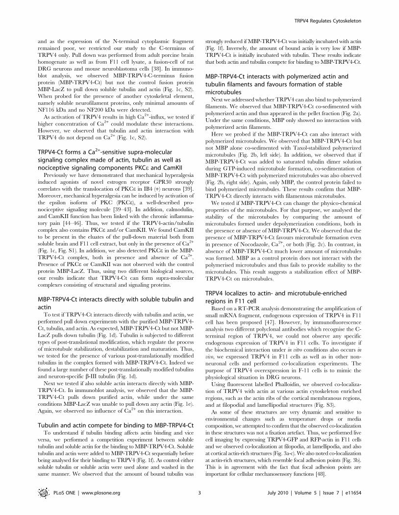
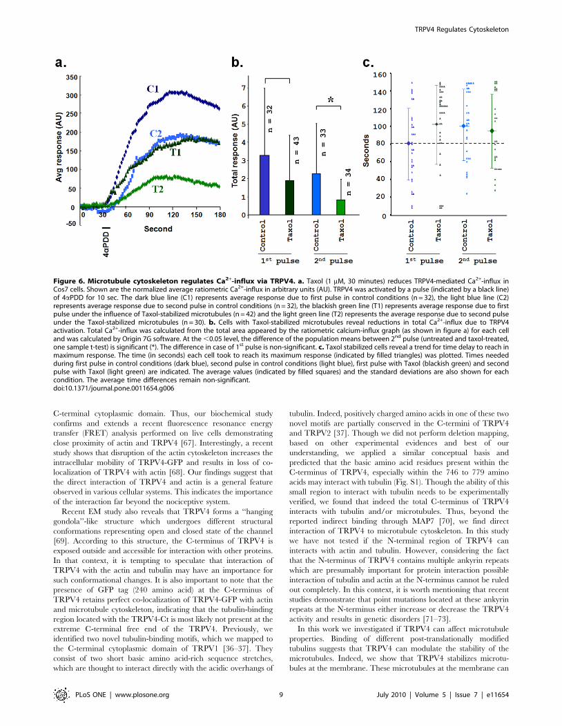
Activation of TRPV4 results in fast retraction of growthcones in F11 cell
TRPV4 localizes to growth cones when expressed in F11 cells.
So we tested if TRPV4 activation can alter the morphology and
movement of growth cones. For that purpose we expressed
TRPV4-GFP along with RFP-actin and performed live cell
imaging. We observed that in response to 4a-phorbol-didecanoate
(4aPDD, 1 mM), a TRPV4-specific agonist [22], neurites from
TRPV4-GFP expressing F11 cells show rapid change in its
morphology and induce multiple varicosities (Fig. S4). In response
to 4aPDD, TRPV4-GFP containing growth cones retract quickly
(Fig. 4a). Under the same conditions growth cones developing
from non-transfected cells do not show any retraction, thus
assuring specificity of the pharmacological treatment. These results
strongly suggest that TRPV4 can regulate the growth cone
motility.
Long-term exposure to a TRPV4 agonist restricts neuriteoutgrowth in a subset of primary DRG neurons
To validate the effect of TRPV4 activation on growth cones in a
system with endogenous TRPV4 expression, we used primary
neurons and tested the effect of TRPV4 activation. DRG neurons
from adult male rat were cultured for 5 days and treated with
4aPDD (1 mM) for short-term (20 minutes). Live cell-imaging
revealed indications of slow retraction of neurites in response to
Figure 2. TRPV4 interacts directly with polymerized actin and microtubule filaments. MBP and MBP-TRPV4-Ct were centrifuged at70000 g/30 min/4uC and only soluble proteins present in the supernatant were used for all co-sedimentation experiments. a. MBP-TRPV4-Ct co-sediments with polymerized actin filaments. Actin was polymerized either in presence of MBP-TRPV4-Ct (lane 1 and 4), in presence of MBP only (lane2 and 5) or in buffer only (lane 3 and 6). Polymerized actin filaments and associated proteins were isolated from remaining soluble actin and unboundproteins by centrifugal separation of pellets (P, lane 1–3) from corresponding supernatants (S, lane 4–6). The entire amount of MBP remains in thesupernatant (lane 5) while a significant amount of MBP-TRPV4-Ct appears in the pellet (lane 1). Arrows indicate the position of respective proteins. b.MBP-TRPV4-Ct co-sediments with microtubules. Taxol-stabilized microtubules (left panel) were incubated with MBP (lane 1–2), MBP-TRPV4-Ct (lane 3–4) or with buffer only (lane 5–6) followed by the centrifugal separation of pellet (P) consisting MT and bound proteins from supernatant (S) consistingof soluble tubulin and other unbound proteins (left side panel). In right side panel, soluble tubulin and GTP was incubated with MBP (lane 1–2), MBP-TRPV4-Ct (lane 3–4) or buffer only (lane 5–6) followed by separation of pellet (P) and supernatant (S). Note the specific presence of MBP-TRPV4-Ct inthe pellet in both cases (in lane 4). c. MBP-TRPV4-Ct stabilizes microtubules against depolymerizing factors. Microtubules was formed form solubletubulin in buffer (lane 1), along with MBP (lane 2) or along with MBP-TRPV4-Ct (lane 3) in control condition (left most panel), in presence ofNocodazole (middle left panel), in presence of Ca2+ (middle right side) or in presence of both Nocodazole and Ca2+ (right most). Microtubules andbound proteins present in the pellet fraction (P) were isolated from unpolymerized tubulin and unbound proteins remaining in the supernatant (S)by centrifugal separation. Note the enhancement of polymerized microtubules (represented by tubulin present in lane 3, P fraction in everyconditions) due to the presence of MBP-TRPV4-Ct.doi:10.1371/journal.pone.0011654.g002
TRPV4 Regulates Cytoskeleton
PLoS ONE | www.plosone.org 4 July 2010 | Volume 5 | Issue 7 | e11654

Figure 3. TRPV4 co-localizes with actin and microtubule cytoskeleton. a–c. Shown are the live-cell confocal images of F11 cells expressingTRV4-GFP (green) and RFP-actin (red). Presences of TRPV4-GFP specifically in actin-enriched structures are shown. a. Enlarged view of lamellipodiaand at the tip of the actin filaments are shown. b–c. Enlarged view of focal adhesion point-like structures (b) and cell cortex with actin ribs (c) areshown. Arrows indicate the localization of TRPV4-GFP at filopodial tips. d–f. TRPV4 co-localizes with microtubule cytoskeleton. Shown are theconfocal images of F11 cells immunostained for TRPV4 (green) and tyrosinated tubulin (red). Arrows indicate presence and acumulation ofmicrotubules in thin filopodial structures (d, upper panel) and thin lamellipodial structures (e, middle panel). The status of the microtubules in thenon-transfected cells are shown in below (f, lower panel).doi:10.1371/journal.pone.0011654.g003
TRPV4 Regulates Cytoskeleton
PLoS ONE | www.plosone.org 5 July 2010 | Volume 5 | Issue 7 | e11654
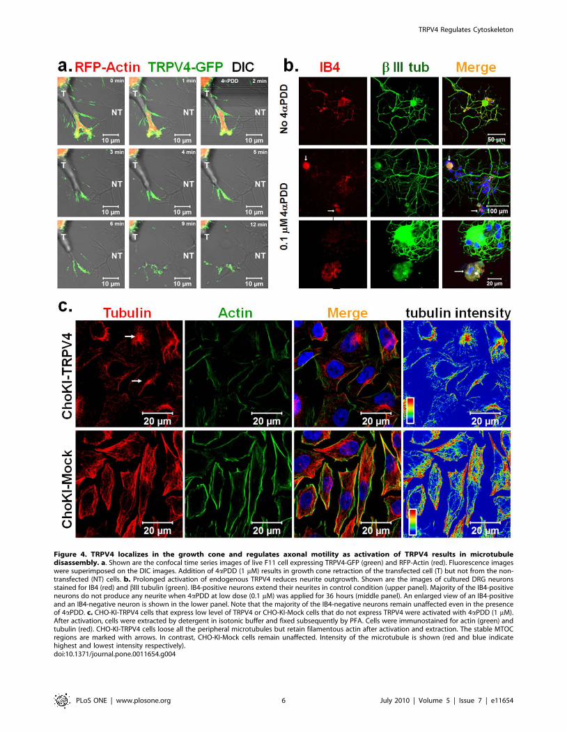
Figure 4. TRPV4 localizes in the growth cone and regulates axonal motility as activation of TRPV4 results in microtubuledisassembly. a. Shown are the confocal time series images of live F11 cell expressing TRPV4-GFP (green) and RFP-Actin (red). Fluorescence imageswere superimposed on the DIC images. Addition of 4aPDD (1 mM) results in growth cone retraction of the transfected cell (T) but not from the non-transfected (NT) cells. b. Prolonged activation of endogenous TRPV4 reduces neurite outgrowth. Shown are the images of cultured DRG neuronsstained for IB4 (red) and bIII tubulin (green). IB4-positive neurons extend their neurites in control condition (upper panel). Majority of the IB4-positiveneurons do not produce any neurite when 4aPDD at low dose (0.1 mM) was applied for 36 hours (middle panel). An enlarged view of an IB4-positiveand an IB4-negative neuron is shown in the lower panel. Note that the majority of the IB4-negative neurons remain unaffected even in the presenceof 4aPDD. c. CHO-KI-TRPV4 cells that express low level of TRPV4 or CHO-KI-Mock cells that do not express TRPV4 were activated with 4aPDD (1 mM).After activation, cells were extracted by detergent in isotonic buffer and fixed subsequently by PFA. Cells were immunostained for actin (green) andtubulin (red). CHO-KI-TRPV4 cells loose all the peripheral microtubules but retain filamentous actin after activation and extraction. The stable MTOCregions are marked with arrows. In contrast, CHO-KI-Mock cells remain unaffected. Intensity of the microtubule is shown (red and blue indicatehighest and lowest intensity respectively).doi:10.1371/journal.pone.0011654.g004
TRPV4 Regulates Cytoskeleton
PLoS ONE | www.plosone.org 6 July 2010 | Volume 5 | Issue 7 | e11654

4aPDD in some neurites, but did not show immediate retraction
or varicosity formation (data not shown). Thus we tested if long-
term exposure to a TRPV4 agonist but a low-level activation of
TRPV4 can regulate the neurites. For that purpose, DRG neurons
were cultured for 36 hours and then treated with a low dose of
4aPDD (0.1 mM) for an additional 36 hours. Neurons were
visualized for neuron-specific bIII tubulin. Immunodetection of
TRPV4-positive neurons was not conclusive due to the very low
endogenous expression of TRPV4 in DRG neurons in this stage.
Thus, we categorized the DRG neurons by staining with IB4
lectin, which mark a subsection of nociceptive neurons [49]. We
observed that due to long-term exposure of 4aPDD, majority of
the IB4-positive neurons developed much shorter neurites if at all
(Fig. 4b). In contrast, under the same condition, IB4-negative
neurons revealed long neurites similar to non-treated neurons
(Fig. 4b). This confirmed that long-term exposure to a TRPV4
agonist resulted in growth cone and neurite retraction, and
demonstrates that this effect is restricted to a subpopulation of
nociceptors.
Activation of TRPV4 results in disassembly ofmicrotubules
Previously it has been shown that activation of Ca2+ channels
causes rapid reorganization of the cytoskeleton [31,50–51]. Also,
rapid disassembly of microtubules causes growth cone retraction
[32,34,52]. So, we tested if in contrast to a stabilizing function of
TRVP4 at resting stage, activation of this channel leads to
destabilization of microtubules. To better differentiate between
soluble and polymerized cytoskeleton components, soluble pro-
teins were removed by Digitonin-extraction leaving behind only
the insoluble intact cytoskeleton. Extraction of unstimulated
TRPV4 expressing F11 cells showed fine filamentous microtubules
(Fig. S5a). But application of 4aPDD (1 mM) resulted in loss of
microtubules from the majority of TRPV4 expressing F11 cells. In
contrast, cells, which did not express TRPV4 were unaffected by
4aPDD (Fig. S5a).
We observed that the cytoskeleton rearrangement by TRPV4
was not restricted to the neuronal cells only. In TRPV4 expressing
CHO-KI cells 4aPDD treatment induced loss of almost all
microtubules whereas non-transfected cells retained all their
microtubules (Fig. S5b). Similarly, we observed loss of all
peripheral microtubules by application of 4aPDD in CHO-KI-
TRPV4 cells, which express low level of TRPV4 stably (Fig. 4c).
Under the same conditions, CHO-KI mock-transfected cells
retained all microtubules. All these results confirm that activation
of TRPV4 results in microtubules disassembly and this effect is
independent of TRPV4 expression level in whatever cellular
context chosen.
Activation of TRPV4 results in formation of elongatedfilopodia
We also explored if TRPV4, being present at the filopodia can
alter the actin cytoskeletal dynamics upon activation. For that
purpose, we expressed TRPV4-GFP and actin-red in F11 cells and
monitored the filopodial structures. We observed that activation of
TRPV4 by 4aPDD results in rapid change of lamellipodia to
filopodia and/or further elongation of filopodial structures (Fig. 5).
These changes were observed at filopodial structures emanating
from the cell body as well from neurites. We observed that during
this change, several lamellipodial actin ribs fused into single
filopodial structures and TRPV4-GFP localized at the filopodial
tips (Fig. 5b). At the same time, lamellipodial region located
between two actin-ribs retract.
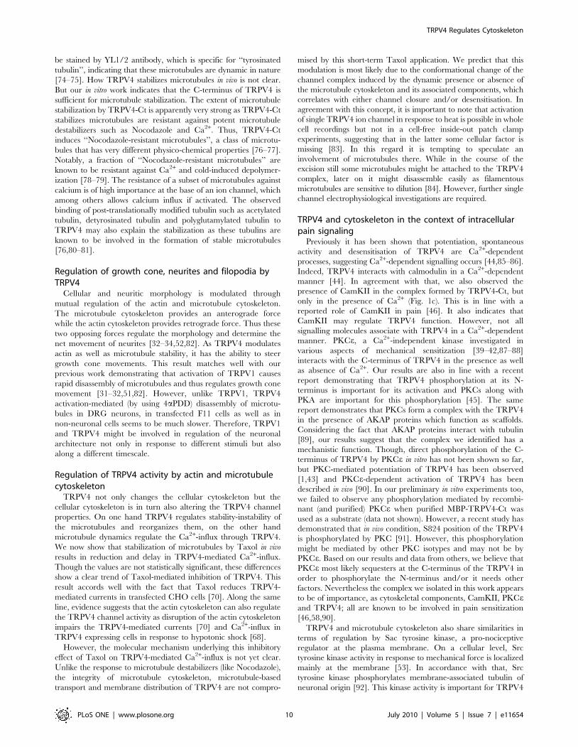
Stabilization of microtubules by Taxol reduces Ca2+-influxthrough TRPV4 activation
In contrast to a unidirectional effect of TRPV4 activation on
microtubule integrity, we explored if microtubule dynamics can
also affect the TRPV4 channel properties. We measured Ca2+-
influx via TRPV4 as an indicator of TRPV4 channel opening.
We compared the Ca2+-influx via TRPV4 in response to 4aPDD
in the presence and absence of Taxol. We observed that the
normalized average Ca2+-influx due to TRPV4 activation was
strongly diminished (but not abolished) if the cells were treated
with Taxol (Fig. 6a). This suggests a partial inhibition of TRPV4
channel activity due to stabilization of microtubules. This
reduction due to Taxol treatment (30 min) was also observed
when cells were activated by 4aPDD for a second time (Fig. 6a).
To understand the extent of reduction, we compared the total
Ca2+-influx (Fig. 6b). We observed 21.9% reduction in the case of
the 1st pulse in presence of Taxol when compared to Taxol-free
control condition. However, this difference remain non-signifi-
cant at (P.0.05) (Fig. 6b). However, this difference was even
higher in the case of the 2nd pulse where a major reduction
(61.79%) was observed in the presence of Taxol (compared with
Taxol-free 2nd pulse condition). At the level of P,0.05, this
difference is significant. To confirm this inhibitory effect of
microtubule stabilization on TRPV4 channel opening, we
analysed by another parameter, i.e. the time each cell took to
reach its maximum response. We observed that Taxol-treatment
resulted in a delay of the cells to reach their maximum response
level (Fig. 6c). Though this time difference indicates a clear trend
of inhibition, the differences remain non-significant (Fig. 6c). By
immunofluorescence analysis (Fig. S6) and surface-biotinylation
experiments (Fig. S7), we confirmed that the presence of
microtubules and TRPV4 at the cell membrane or nearby
regions was also not altered by Taxol application. Taxol did not
alter the distribution of TRPV4 in the cell surface also. Taken
together, these results suggest that microtubule dynamics can
regulate TRPV4 channel properties.
Discussion
A substantial amount of work has been done to identify
endogenous and exogenous sensitizing stimuli, their target proteins
and elucidate their molecular effects on nociceptive neurons. Yet,
the detailed cellular and molecular mechanisms underlying
sensitization against mechanical stimuli and the specific molecular
components associated with these signaling events are not well
understood. A priori, as there is no force without counterforce,
scaffold structures like the cytoskeleton and/or the extracellular
matrix are intuitively likely to be required for stimulus detection
and execution of the responses. In nociception, so far the
cytoskeleton has been described as an important regulator of
sensitization only on a behavioural level [29,42,53–58]. In
contrast, in systems other than nociception, genetic, biochemical,
cellular as well as behavioural data are indicative of the
importance of the cytoskeleton for mechanosensation in general
[29,59–60]. Thus, contrasting the wealth of data in non-
nociceptive context, the nearly complete lack of molecular data
about an involvement of the microtubule and actin cytoskeleton in
pain sensitization is surprising. Though, recent results demon-
strated the expression of TRPV4 in Merkel cells and linked
TRPV4 function with the mechanotransduction [61,62], in the
context of nociceptive and non-nociceptive mechanosensitivity, a
direct physical interaction of actin or microtubule cytoskeleton
with ion channels has not been reported. Therefore, we now
provide the first evidence that both actin and microtubule
TRPV4 Regulates Cytoskeleton
PLoS ONE | www.plosone.org 7 July 2010 | Volume 5 | Issue 7 | e11654

cytoskeleton components interact directly and functionally with a
mechanosensitive ion channel, namely TRPV4.
The C-terminus of TRPV4 interacts directly with actin andmicrotubule cytoskeletal components
The C-terminus of various TRP channels has been shown to be
of importance. For example, a recent study reported that the
deletion of the C-terminal cytoplasmic domain of TRPV4 strongly
inhibits membrane localization [63]. And also the C-terminus of
TRPC4 interacts with another cytoskeletal component, spectrin,
which is essential for the surface expression of the receptor [64]. In
this work we now demonstrate that TRPV4-Ct is sufficient for the
interaction with tubulin and actin as well as signaling-components.
The interaction of MBP-TRPV4-Ct to tubulin and actin appears
to be very strong as these interactions withstand the presence of
high salt concentrations (500 mM NaCl, data not shown). If the N-
terminal cytoplasmic domain of TRPV4 can also interact with the
cytoskeleton has not been established yet.
We observed that TRPV4 co-localized with actin-enriched
structures, such as focal adhesion points, filopodial and lamellipo-
dial structures. These results fit well with the previously described
subcellular localizations of TRPV4 in actin cytoskeleton-enriched
structures like dendritic spines [65]. This also accords well with the
reported presence of a2 integrin, an actin binding protein in the
TRPV4 complex [66]. We show that TRPV4 physically interacts
with soluble actin as well as with polymerised actin filaments via its
Figure 5. Activation of TRPV4 results in reorganization of actin cytoskeleton. Shown are the confocal images of live F11 cells expressingTRPV4-GFP (green) and Actin-RFP (red). a. Activation of TRPV4 results in merging of several actin-ribs at the tips and further transition of lamellipodialstructures to filopodial structures. The arrow indicates the region and direction of the cell retraction at the same time. b. Activation of TRPV4 resultsin lateral initiation of filopodial structures from neurites and further elongation of them.doi:10.1371/journal.pone.0011654.g005
TRPV4 Regulates Cytoskeleton
PLoS ONE | www.plosone.org 8 July 2010 | Volume 5 | Issue 7 | e11654
C-terminal cytoplasmic domain. Thus, our biochemical study
confirms and extends a recent fluorescence resonance energy
transfer (FRET) analysis performed on live cells demonstrating
close proximity of actin and TRPV4 [67]. Interestingly, a recent
study shows that disruption of the actin cytoskeleton increases the
intracellular mobility of TRPV4-GFP and results in loss of co-
localization of TRPV4 with actin [68]. Our findings suggest that
the direct interaction of TRPV4 and actin is a general feature
observed in various cellular systems. This indicates the importance
of the interaction far beyond the nociceptive system.
Recent EM study also reveals that TRPV4 forms a ‘‘hanging
gondola’’-like structure which undergoes different structural
conformations representing open and closed state of the channel
[69]. According to this structure, the C-terminus of TRPV4 is
exposed outside and accessible for interaction with other proteins.
In that context, it is tempting to speculate that interaction of
TRPV4 with the actin and tubulin may have an importance for
such conformational changes. It is also important to note that the
presence of GFP tag (240 amino acid) at the C-terminus of
TRPV4 retains perfect co-localization of TRPV4-GFP with actin
and microtubule cytoskeleton, indicating that the tubulin-binding
region located with the TRPV4-Ct is most likely not present at the
extreme C-terminal free end of the TRPV4. Previously, we
identified two novel tubulin-binding motifs, which we mapped to
the C-terminal cytoplasmic domain of TRPV1 [36–37]. They
consist of two short basic amino acid-rich sequence stretches,
which are thought to interact directly with the acidic overhangs of
tubulin. Indeed, positively charged amino acids in one of these two
novel motifs are partially conserved in the C-termini of TRPV4
and TRPV2 [37]. Though we did not perform deletion mapping,
based on other experimental evidences and best of our
understanding, we applied a similar conceptual basis and
predicted that the basic amino acid residues present within the
C-terminus of TRPV4, especially within the 746 to 779 amino
acids may interact with tubulin (Fig. S1). Though the ability of this
small region to interact with tubulin needs to be experimentally
verified, we found that indeed the total C-terminus of TRPV4
interacts with tubulin and/or microtubules. Thus, beyond the
reported indirect binding through MAP7 [70], we find direct
interaction of TRPV4 to microtubule cytoskeleton. In this study
we have not tested if the N-terminal region of TRPV4 can
interacts with actin and tubulin. However, considering the fact
that the N-terminus of TRPV4 contains multiple ankyrin repeats
which are presumably important for protein interaction possible
interaction of tubulin and actin at the N-terminus cannot be ruled
out completely. In this context, it is worth mentioning that recent
studies demonstrate that point mutations located at these ankyrin
repeats at the N-terminus either increase or decrease the TRPV4
activity and results in genetic disorders [71–73].
In this work we investigated if TRPV4 can affect microtubule
properties. Binding of different post-translationally modified
tubulins suggests that TRPV4 can modulate the stability of the
microtubules. Indeed, we show that TRPV4 stabilizes microtu-
bules at the membrane. These microtubules at the membrane can
Figure 6. Microtubule cytoskeleton regulates Ca2+-influx via TRPV4. a. Taxol (1 mM, 30 minutes) reduces TRPV4-mediated Ca2+-influx inCos7 cells. Shown are the normalized average ratiometric Ca2+-influx in arbitrary units (AU). TRPV4 was activated by a pulse (indicated by a black line)of 4aPDD for 10 sec. The dark blue line (C1) represents average response due to first pulse in control conditions (n = 32), the light blue line (C2)represents average response due to second pulse in control conditions (n = 32), the blackish green line (T1) represents average response due to firstpulse under the influence of Taxol-stabilized microtubules (n = 42) and the light green line (T2) represents the average response due to second pulseunder the Taxol-stabilized microtubules (n = 30). b. Cells with Taxol-stabilized microtubules reveal reductions in total Ca2+-influx due to TRPV4activation. Total Ca2+-influx was calculated from the total area appeared by the ratiometric calcium-influx graph (as shown in figure a) for each celland was calculated by Origin 7G software. At the ,0.05 level, the difference of the population means between 2nd pulse (untreated and taxol-treated,one sample t-test) is significant (*). The difference in case of 1st pulse is non-significant. c. Taxol stabilized cells reveal a trend for time delay to reach inmaximum response. The time (in seconds) each cell took to reach its maximum response (indicated by filled triangles) was plotted. Times neededduring first pulse in control conditions (dark blue), second pulse in control conditions (light blue), first pulse with Taxol (blackish green) and secondpulse with Taxol (light green) are indicated. The average values (indicated by filled squares) and the standard deviations are also shown for eachcondition. The average time differences remain non-significant.doi:10.1371/journal.pone.0011654.g006
TRPV4 Regulates Cytoskeleton
PLoS ONE | www.plosone.org 9 July 2010 | Volume 5 | Issue 7 | e11654
be stained by YL1/2 antibody, which is specific for ‘‘tyrosinated
tubulin’’, indicating that these microtubules are dynamic in nature
[74–75]. How TRPV4 stabilizes microtubules in vivo is not clear.
But our in vitro work indicates that the C-terminus of TRPV4 is
sufficient for microtubule stabilization. The extent of microtubule
stabilization by TRPV4-Ct is apparently very strong as TRPV4-Ct
stabilizes microtubules are resistant against potent microtubule
destabilizers such as Nocodazole and Ca2+. Thus, TRPV4-Ct
induces ‘‘Nocodazole-resistant microtubules’’, a class of microtu-
bules that has very different physico-chemical properties [76–77].
Notably, a fraction of ‘‘Nocodazole-resistant microtubules’’ are
known to be resistant against Ca2+ and cold-induced depolymer-
ization [78–79]. The resistance of a subset of microtubules against
calcium is of high importance at the base of an ion channel, which
among others allows calcium influx if activated. The observed
binding of post-translationally modified tubulin such as acetylated
tubulin, detyrosinated tubulin and polyglutamylated tubulin to
TRPV4 may also explain the stabilization as these tubulins are
known to be involved in the formation of stable microtubules
[76,80–81].
Regulation of growth cone, neurites and filopodia byTRPV4
Cellular and neuritic morphology is modulated through
mutual regulation of the actin and microtubule cytoskeleton.
The microtubule cytoskeleton provides an anterograde force
while the actin cytoskeleton provides retrograde force. Thus these
two opposing forces regulate the morphology and determine the
net movement of neurites [32–34,52,82]. As TRPV4 modulates
actin as well as microtubule stability, it has the ability to steer
growth cone movements. This result matches well with our
previous work demonstrating that activation of TRPV1 causes
rapid disassembly of microtubules and thus regulates growth cone
movement [31–32,51,82]. However, unlike TRPV1, TRPV4
activation-mediated (by using 4aPDD) disassembly of microtu-
bules in DRG neurons, in transfected F11 cells as well as in
non-neuronal cells seems to be much slower. Therefore, TRPV1
and TRPV4 might be involved in regulation of the neuronal
architecture not only in response to different stimuli but also
along a different timescale.
Regulation of TRPV4 activity by actin and microtubulecytoskeleton
TRPV4 not only changes the cellular cytoskeleton but the
cellular cytoskeleton is in turn also altering the TRPV4 channel
properties. On one hand TRPV4 regulates stability-instability of
the microtubules and reorganizes them, on the other hand
microtubule dynamics regulate the Ca2+-influx through TRPV4.
We now show that stabilization of microtubules by Taxol in vivo
results in reduction and delay in TRPV4-mediated Ca2+-influx.
Though the values are not statistically significant, these differences
show a clear trend of Taxol-mediated inhibition of TRPV4. This
result accords well with the fact that Taxol reduces TRPV4-
mediated currents in transfected CHO cells [70]. Along the same
line, evidence suggests that the actin cytoskeleton can also regulate
the TRPV4 channel activity as disruption of the actin cytoskeleton
impairs the TRPV4-mediated currents [70] and Ca2+-influx in
TRPV4 expressing cells in response to hypotonic shock [68].
However, the molecular mechanism underlying this inhibitory
effect of Taxol on TRPV4-mediated Ca2+-influx is not yet clear.
Unlike the response to microtubule destabilizers (like Nocodazole),
the integrity of microtubule cytoskeleton, microtubule-based
transport and membrane distribution of TRPV4 are not compro-
mised by this short-term Taxol application. We predict that this
modulation is most likely due to the conformational change of the
channel complex induced by the dynamic presence or absence of
the microtubule cytoskeleton and its associated components, which
correlates with either channel closure and/or desensitisation. In
agreement with this concept, it is important to note that activation
of single TRPV4 ion channel in response to heat is possible in whole
cell recordings but not in a cell-free inside-out patch clamp
experiments, suggesting that in the latter some cellular factor is
missing [83]. In this regard it is tempting to speculate an
involvement of microtubules there. While in the course of the
excision still some microtubules might be attached to the TRPV4
complex, later on it might disassemble easily as filamentous
microtubules are sensitive to dilution [84]. However, further single
channel electrophysiological investigations are required.
TRPV4 and cytoskeleton in the context of intracellularpain signaling
Previously it has been shown that potentiation, spontaneous
activity and desensitisation of TRPV4 are Ca2+-dependent
processes, suggesting Ca2+-dependent signalling occurs [44,85–86].
Indeed, TRPV4 interacts with calmodulin in a Ca2+-dependent
manner [44]. In agreement with that, we also observed the
presence of CamKII in the complex formed by TRPV4-Ct, but
only in the presence of Ca2+ (Fig. 1c). This is in line with a
reported role of CamKII in pain [46]. It also indicates that
CamKII may regulate TRPV4 function. However, not all
signalling molecules associate with TRPV4 in a Ca2+-dependent
manner. PKCe, a Ca2+-independent kinase investigated in
various aspects of mechanical sensitization [39–42,87–88]
interacts with the C-terminus of TRPV4 in the presence as well
as absence of Ca2+. Our results are also in line with a recent
report demonstrating that TRPV4 phosphorylation at its N-
terminus is important for its activation and PKCs along with
PKA are important for this phosphorylation [45]. The same
report demonstrates that PKCs form a complex with the TRPV4
in the presence of AKAP proteins which function as scaffolds.
Considering the fact that AKAP proteins interact with tubulin
[89], our results suggest that the complex we identified has a
mechanistic function. Though, direct phosphorylation of the C-
terminus of TRPV4 by PKCe in vitro has not been shown so far,
but PKC-mediated potentiation of TRPV4 has been observed
[1,43] and PKCe-dependent activation of TRPV4 has been
described in vivo [90]. In our preliminary in vitro experiments too,
we failed to observe any phosphorylation mediated by recombi-
nant (and purified) PKCe when purified MBP-TRPV4-Ct was
used as a substrate (data not shown). However, a recent study has
demonstrated that in vivo condition, S824 position of the TRPV4
is phosphorylated by PKC [91]. However, this phosphorylation
might be mediated by other PKC isotypes and may not be by
PKCe. Based on our results and data from others, we believe that
PKCe most likely sequesters at the C-terminus of the TRPV4 in
order to phosphorylate the N-terminus and/or it needs other
factors. Nevertheless the complex we isolated in this work appears
to be of importance, as cytoskeletal components, CamKII, PKCeand TRPV4; all are known to be involved in pain sensitization
[46,58,90].
TRPV4 and microtubule cytoskeleton also share similarities in
terms of regulation by Sac tyrosine kinase, a pro-nociceptive
regulator at the plasma membrane. On a cellular level, Src
tyrosine kinase activity in response to mechanical force is localized
mainly at the membrane [53]. In accordance with that, Src
tyrosine kinase phosphorylates membrane-associated tubulin of
neuronal origin [92]. This kinase activity is important for TRPV4
TRPV4 Regulates Cytoskeleton
PLoS ONE | www.plosone.org 10 July 2010 | Volume 5 | Issue 7 | e11654
function [66] as it phosphorylates TRPV4 [43,93]. Thus
membrane-associated tubulin-TRPV4 complex seems to be
important for stimuli that activate Src-tyrosine kinase.
Microtubules and TRPV4 in pain sensitizationInvolvement of cytoskeleton in pain sensitisation has been
anticipated for a long time, as microtubule-based chemothera-
peutics against cancer are known to induce severe long-lasting
neuropathic pain [94]. But, how these chemotherapeutics, like
Taxol and Vincristine alter pain sensitivity is not well understood.
Microtubules and mitochondrial structures develop abnormalities
in nociceptive neurons due to prolonged and systemic application
of these chemotherapeutics [95–97]. Even stabilization of
microtubules by short-term application of Taxol (30 min) exerts
an impact on pain sensitization [58]. It is important to mention
that disruption of microtubules by short-term (30 min) application
of Nocodazole abolishes/reduces pain sensitization [58]. These
suggest that the integrity of microtubules can indeed modulate
pain sensitivity [98]. TRPV4 has recently been considered as
essential for chemotherapy-induced neuropathic pain as mechan-
ical hyperalgesia induced by Taxol and Vincristine was strongly
reduced in trpv4 2/2 mice [30,66].
We established a direct physical, cell-biological and functional
relationship between cytoskeletal components and TRPV4, which
plays an important role in inflammatory, neuropathic and
chemotherapeutic-induced pain [30,58,66]. This gives a mecha-
nistic base to further study the role of the apparent dynamic
signalling complex formed by TRPV4 and its functional
implications in various physiological scenarios.
Materials and Methods
Reagents and antibodiesTaxol, Nocodazole, 4aPDD, bovine actin, tetramethylrhodamine
isothiocyanate-labelled IB4 from Griffonia simplicifolia, antibodies
against a-tubulin (clone DM1A), b-tubulin (clone D66), tyrosinated
tubulin (clone TUB1A2), polyglutamylated tubulin (clone B3),
acetylated tubulin (clone 611-B-1), phospho-serine (Clone PSR-45),
b-tubulin sub type III (clone SDL.3D10), neurofilament 116 kDa
(clone NN18) and the affinity purified rabbit polyclonal antibody
against C-terminal cytoplasmic domain of TRPV4 were purchased
from Sigma-Aldrich (Taufkirchen, Germany). Antibodies against
neurofilament 200 kDa (clone RT97) and detyrosinated tubulin were
purchased from Chemicon (Chandlers Ford, UK). The antibody
against actin (clone JLA20) was purchased from Oncogene (Cam-
bridge, MA, USA). Antibody against maltose-binding protein (MBP)
and the amylose resin were purchased from New England Biolab
(Beverly, MD, USA). Purified bovine muscle actin was purchased
from Sigma-Aldrich (Taufkirchen, Germany). Protein G-agarose was
purchased from Amersham Pharmacia Biotech (Munich, Germany).
For some experiments, another affinity purified rabbit polyclonal
antibody raised against TRPV4 (kind gift from Jon D. Levine) was
used [3]. Anti CamKII antibody was purchased from BD
(Heidelberg, Germany). Anti PKCe antibody (KP4) was a kind gift
from Dr Robert Messing, University of California San Francisco. All
secondary IgG antibodies (alexa-488-labelled anti-mouse, alexa-488-
labelled anti-rabbit, alexa-594-labelled anti-rabbit, alexa-594-labelled
anti-rat, Fura-2/AM, alexa-488- and alexa-594-labelled Phalloitoxin
were purchased from Invitrogen (Karlsruhe, Germany).
ConstructsFull-length TRPV4 (NCBI accession number AF263521) in
pCDNA3.1 vector was a kind gift from Prof. Jon D Levine [3]. For
expression of C-terminal cytoplasmic domain (amino acid residue
718 to 871) of TRPV4-fused with MBP, the corresponding cDNA
region of TRPV4 (NCBI accession number AF263521) was
amplified by using forward primer (59TGACGAATTCATG-
GGTGAGACCGTGGGCCA39) and reverse primer (59TGA-
CAAGCTTCTACAGTGGTGCGTCCTCCG39). The ampli-
fied cDNA was sub cloned into the EcoRI and HindIII restriction
site of the pMALc2x vector (NEB, Beverly, MD, USA) and
verified by automated nucleotide sequencing. TRPV4-GFP
construct (cloned in pEGFPN3 vector) used in this study was
kindly provided by Prof. J. Berreiter-Hahn [10]. Tubulin-cherry
construct was a kind gift from Prof. R. Y. Tsien [99]. RFP-actin
construct was purchased from Clontech (Heidelberg, Germany).
Cells and transfectionF11 cells [38] were cultured in Ham’s F12 medium (Sigma
Aldrich) supplemented with 20% fetal bovine serum (Sigma
Aldrich). Cos7 [100] and HaCat [101] cells were maintained in
Dulbecco’s modified Eagle’s medium (Sigma Aldrich) with 10%
fetal calf serum. CHO-K1 cells [3] stably transfected with TRPV4
(CHO-K1-TRPV4) and the negative control cell line (CHO-K1-
MOCK) [3] was cultured in F12 Ham’s (Sigma Aldrich) medium
supplemented with 5% fetal bovine serum (FBS) and L-glutamine.
These two cell lines were kindly provided by Prof. J. D. Levine [3].
All media were supplemented with streptomycin (100 mg/ml) and
penicillin (100 mg/ml) (Invitrogen). Cells were maintained in a
humidified atmosphere at 5% CO2 and 37uC. For transient
transfection, Lipofectamine (Invitrogen) was used according to the
manufacturer’s instructions.
Ethics StatementCell biological experiments were performed in cultured DRG
neurons from male Sprague-Dawley rats (200–300 g; Harlan
Winkelmann, Borchen, Germany). Care and use of animals were
in accordance with the European Communities Council Directive
of 24 November 1986 (86/609/EEC) and were approved by the
LaGeSo, Berlin. All efforts were made to minimize the number of
animals used and their suffering.
DRG neuronal culture and neurite out growthDissociated rat DRG neurones were prepared from adult male
rats (200–300 g) as recently described [32]. Neurons were grown
in 24-well plates at 37uC in 5% CO2 and were grown for a total of
72 h. At that time most untreated neurons grow numerous and
long neurites. In some experiments neurons were treated with
4aPDD (0.1 mM for the last 36 hour) before they were fixed with
2% PFA. Subsequently the neurons were visualized for bIII-
tubulin (antibody dilution 1:1000) and IB4-lectin (1:15000).
ImmunoprecipitationFor immunoprecipitation, approximately 50 ml of 50% protein
G beads-slurry equilibrated with IP buffer (1% sodium dodecyl
maltoside, PIPES 50 mM (pH 6.8), 100 mM NaCl, 1 mM EGTA,
0.2 mM MgCl2 and complete protease inhibitors (Roche) was used
for each IP. Affinity purified rabbit polyclonal TRPV4 antibody
(Sigma Aldrich) (7 mg per IP condition, antibody obtained from
Sigma-Aldrich) was used. In control, total serum IgG from rabbit
(7 ml which is equivalent to 20 mg of antibody) was used as non-
specific antibody. For co-immunoprecipitation, CHO-KI-TRPV4
cells grown for 2 days and then scraped off from the dishes. Cells
were harvested by centrifuging (Hettich Rotanta/T, 5 min at
100 g). Cells were washed with PBS once and centrifuged again at
the same conditions. Cells were resuspended in 2 ml of IP buffer
and homogenized (10 strokes) with a glass-Teflon homogenizer
TRPV4 Regulates Cytoskeleton
PLoS ONE | www.plosone.org 11 July 2010 | Volume 5 | Issue 7 | e11654
(Kontes glass, Germany). Cell extract was further centrifuged at
16 K (25000 g) for 30 minute at 4uC and the clear supernatant
was used for IP. Similarly, DRG tissues (L1-L6) from 2 male rats
were isolated, homogenized in IP buffer. The lysate was
centrifuged at 25.000 g, 30 min at 4uC and the clear extract was
used for the further IP. Clear cell extract (800 ml equivalent to
1 mg of protein) and the corresponding antibody were applied to
the equilibrated protein G beads. The mixture was incubated at
25uC for 4 hours on a shaker. After that the beads were washed 3
times, each time with 400 ml of IP buffer. A Hamilton syringe was
used for all washing steps. Finally the beads were taken in 100 ml of
IP buffer and 50 ml of 5x Laemmli sample buffer was added. The
samples were boiled and used for western blot analysis.
Expression and purification of MBP-fusion proteinsExpression and purification of MBP-TRPV4-Ct (C-terminal
cytoplasmic domain of TRPV4 fused with MBP) as well as of
MBP-LacZ (LacZ fused with MBP) was based on a protocol
described previously [36]. In short, the expression constructs were
introduced into the Escherichia coli (E. coli) strain BL21DE3 by
transformation heat shock method. Fusion protein expression was
induced by addition of isopropyl thiogalactoside (IPTG) for 2 h.
Cells were lysed by repeated freeze-thaw cycles in lysis buffer
(20 mM Tris–HCl, pH 7.4, 150 mM NaCl, 0.1% Tween 20,
lysozyme, benzonase and protease inhibitor cocktail). The lysed
extracts were cleared by centrifugation (100000 g in a TFT 45
rotor for 2 h) and applied to amylose resin. The resins with bound
proteins were washed thoroughly and finally the proteins were
eluted with 10 mm maltose in elution buffer (50 mM PIPES,
pH 6.8, 100 mM NaCl, 1 mM EGTA and 0.2 mM MgCl2).
Pull down assaysMBP-LacZ and MBP-TRPV4-Ct constructs were expressed in
E. coli; the cleared cell lysates were applied to amylose resin (NEB),
and incubated for 1 hour at RT (25uC) followed by washing. The
amylose resin with bound proteins was resuspended in PEM-S
buffer (50 mM PIPES, pH 6.8, 100 mM NaCl, 1 mM EGTA and
0.2 mM MgCl2). Approximately 50 ml of amylose resin per tube
with the bound fusion protein was used for pull-down experiments.
Depending on the respective experiment, the resin with coupled
fusion protein was incubated with either 50 ml of soluble tubulin
(1 mg/ml protein), 20 ml of soluble actin (0.1 mg/ml), 500 ml of
soluble brain extract (1 mg/ml), or 500 ml of soluble F11 extract
(1 mg/ml) for 1 hour at RT in the presence or absence of Ca2+
(2 mM). This was followed by three washes with 200 ml PEM-S
buffer. The proteins were eluted by 10 mm maltose in 100 ml
solution. Eluted samples were analysed by 10% sodium dodecyl
sulphate polyacrylamide gel electrophoresis (SDS–PAGE).
Co-sedimentation assay with polymerised actin, Taxol-stabilized microtubules (MT) and analysis for microtubulestability
For experiments aimed at understanding the association of
TRPV4-Ct with polymerised actin filaments, purified MBP-
TRPV4-Ct alone was subjected to centrifugation at 70000 g/
30 min/4uC to exclude any aggregates. The soluble MBP-
TRPV4-Ct was further processed for all co-sedimentation assays.
Either 5 mg of purified MBP-TRPV4-Ct or MBP alone in total
50 ml of PEM-S buffer or only 50 ml of PEM-S buffer was added
to a saturated actin solution (50 mg actin in total 50 ml) in
centrifuge tubes. ATP at a final concentration of 2.5 mM was
added to each tube. The reaction tubes were incubated at 37uCfor 30 minutes followed by centrifugal separation of pellet
(polymerised actin filaments) and supernatant (free dimer) at
70000 g/30 min/37uC.
Approximately 100 mg of purified ab-tubulin dimer in a total
volume of 100 mL were incubated in modified PEM buffer
(20 mm PIPES, pH 6.8, 0.2 mm MgCl2 and 1 mm EGTA
supplemented by 1 mm Taxol and 5 mm GTP) for 30 min at
37uC, to form MTs. After that the MT were isolated by
centrifugation at 70 000 g/30 min/37uC. Purified MBP-TRPV4-
Ct or MBP only (each 5 mg) were incubated with Taxol-stabilized
MT for 40 min at 37uC followed by centrifugal separation of pellet
(MT) and supernatant (free dimer) at 70 000 g/30 min/37uC.
In another experiment, MT was formed under Taxol-free
conditions. Tubulin dimer (100 mg) with purified MBP-TRPV4-Ct
or MBP only (5 mg each) were taken in total 100 ml PEM-S buffer
with 5 mm GTP in the absence of Taxol and incubated for
30 min at 37uC. After that the MT and bound proteins were
separated by centrifugal separation of pellet (MT) and supernatant
(free dimer) at 70000 g/30 min/37uC.
For experiments aimed at understanding the effect of TRPV4-
Ct on MT stabilization, either 5 mg of purified MBP-TRPV4-Ct
or MBP alone in total 50 ml of PEM-S buffer or only 50 ml of
PEM-S buffer was added to a tubulin solution (5 mg tubulin in
total 50 ml) in centrifuge tubes. Depending on the experimental
conditions, MT-depolymerizing agent Nocodazole (final concen-
tration 10 mM), Ca2+ (final concentration 0.1 mM) or both
Nocodazole and Ca2+ was added to the tubes. GTP at a final
concentration of 2.5 mM was added to each tube. The reaction
tubes were incubated at 37uC for 30 minute s followed by
centrifugal separation of pellet (MT) and supernatant (free dimer)
at 70000 g/30 min/37uC.
Purification of tubulinab-tubulin dimers were purified from porcine brain as described
previously [36]. In brief, from soluble brain extract tubulin was
enriched by two cycles of polymerization in the presence of
glycerol and GTP and depolymerization by cold temperature (ice
cold), which were then followed by chromatography on phospho-
cellulose.
ImmunocytochemistryFor immunocytochemical analysis, cells were washed once with
PBS and fixed with 2% PFA/PBS if not mentioned otherwise.
Alternatively, where mentioned, an equal volume of 4% PFA was
carefully added to the medium without disturbing the cell culture
within the incubator to avoid morphological changes due to the
PBS wash. The cells were fixed with PFA for 5 minutes at room
temperature (25uC). For experiments aimed to explore the
integrity of the cytoskeleton after TRPV4 activation, soluble
components were removed by digitonin extraction as described
before [31]. Briefly, after activation the cells were extracted with
an isotonic cell membrane-permeabilization buffer (digitonin
(50 mg/ml), PIPES (50 mM pH 6.8), EGTA (1 mM), MgCl2(0.2 mM) and complete protease inhibitors (Roche) that perforates
the cell membrane and allow all the soluble cellular proteins to
diffuse out while insoluble cytoskeleton and cell morphology
remain intact. After extraction the cells were quickly fixed with 2%
PFA. Fixed cells were permeabilized with 0.1% Triton X-100 in
PBS (5 min) and subsequently blocked with 5% normal goat
serum or 5% BSA.
For co-immunostaining of TRPV4 and tubulin, a rabbit
polyclonal anti TRPV4 antibody (Sigma Aldrich) and a rat
monoclonal anti-tyrosinated tubulin antibody (clone YL1/2) were
used. For staining of actin filaments, either alexa-594 or alexa-488
labelled Phalloidin (molecular probe) was used. Alexa 594-labelled
TRPV4 Regulates Cytoskeleton
PLoS ONE | www.plosone.org 12 July 2010 | Volume 5 | Issue 7 | e11654

anti rat antibody and/or alexa 488 labelled anti rabbit antibody
were used as secondary antibodies. For labeling IB4-positive
neurons, tetramethylrhodamine isothiocyanate-labelled IB4 from
Griffonia simplicifolia was used at 1:15000 dilutions in PBST
supplemented with 0.2 mM CaCl2, 0.2 mM MgCl2 and 0.2 mM
MnCl2 and incubated for 30 minutes. All images were taken on a
confocal laser-scanning microscope (Zeiss Axiovert 100 M) with a
63x-objective and analyzed with the Zeiss LSM image examiner
software.
TRPV4 activation and live cell imagingFor visualizing the effect of TRPV4 activation, F11 cells were
seeded on glass cover slips. Two days after transfection with
TRPV4, the cells in either complete medium or in HBSS buffer
were imaged at RT. Where indicated, TRPV4-GFP was co-
expressed with either RFP-Actin or Tubulin-cherry. In experi-
ments aiming to activated TRPV4, its agonist 4 a-phorbol 12,13-
didecanoate (4aPDD) was used at a final concentration of 1 mm, if
not stated otherwise [22].
Calcium imagingFor measuring the TRPV4 channel activity; Ca2+-imaging was
performed using Cos7 cells at room temperature (25uC). Cos7 cells
plated on glass cover slips and transiently transfected with TRPV4
and a yellow fluorescent protein (YFP) reporter plasmid. Cells
transfected with only YFP reporter plasmid were used as controls.
The transfected cells were used for the experiments at around
48 hour later transfection. Cells were loaded with 3 mM Fura-2
AM and placed in a recording chamber that allowed direct
application of buffer at controlled temperature (27 to 30uC).
Loading of Fura-2 AM dye and the fluorescence measurements
were done in calcium imaging buffer (140 mM NaCl, 4 mM KCl,
2 mM CaCl2, 1 mM MgCl2, 5 mM Glucose, 10 mM HEPES,
pH 7.4). Cells were activated by 4aPDD (1 mM) for 10 seconds
after first 30 seconds of imaging. Images were captured in every 2
seconds for a total of 3 minutes. 10 minutes after the 1st round of
activation (1st pulse of 4aPDD) and imaging, cells on the same
cover slips were imaged again for the next round (2nd pulse of
4aPDD). In experiments aimed to analyze the effect of
microtubule stabilization, Taxol (1 mM) was maintained through-
out the experiment in all solutions used, i.e. during the dye
loading, during activation by 4aPDD and in between when
running calcium-imaging buffer was applied. Ratiometric calcium
imaging was performed and analyzed using Tillvision software
(Till Photonics, Germany). Fluorescence was measured at
excitation wavelengths alternating between 340 nm and 380 nm.
Following subtraction of background fluorescence the ratio of
fluorescence at 340 nm and 380 nm was calculated. For
determination of total Ca2+-influx, we measured the total area of
the graph each cell formed after stimulation. Microsoft Excel and
Origin software were used to calculate and plot the graphs. All
significance tests were performed at the ,0.05 level (one sample t-
test).
Supporting Information
Figure S1 Prediction of tubulin-binding region located within
the C-terminus of TRPV4. a. Identification of amino acid
stretches located within the TRPV4-Ct with high isoelectric point
(pI) values. The C-terminal overhanging region of tubulin contains
highly acidic residues and most of the tubulin-binding proteins
contain amino acid stretches which have high pI due to the
presence of basic residues. Therefore, the C-terminus of TRPV4
(amino acid residues 718 to 871) was analyzed for its isoelectric
points. Similarly, different fragments of TRPV4-Ct, each shorter
by 10 amino acids from the extreme C-terminal end were also
analyzed. This analysis indicates that there are two areas which
contain positively charged residues (indicated by shaded area). The
corresponding pI of each fragment is mentioned in the right most
side. In below, the secondary structure prediction is provided (red
indicates helical structure, green arrow indicates beta strand and
black line indicates unstructured coil region. Note that the arrows
do not indicate the directionality of the strands). All theoretical pI
values of the fragments were calculated by using available software
(http://www.expasy.org/tools/pi_tool.html). All secondary struc-
ture prediction was done by using Advanced Protein Secondary
Structure Prediction Server available at http://imtech.res.in/
raghava/apssp/. b. Amino acid residues 746 to 779 contain
multiple positively charged amino acids which can potentially
form an alpha-helix. The PyMol programme was used to build this
peptide structure. Blue and red colours indicate the positive and
negative charges at the surface respectively. Notably all the
positively charged amino acids (indicated by arrows) are located in
one side of the helix and can be important for interaction with the
tubulin/microtubule (which contain negative charges at the
surface). c. These predicted and important basic charges (indicated
by blue) are conserved throughout the evolution. The NCBI
accession numbers are indicated. TRPV4, Homo sapiens (NP_
067638.3); TRPV4, Canis lupus familiaris (XP_543434.2); TRPV4,
Bos taurus (XP_001253150.1); TRPV4, Mus musculus (NP_071300.1);
TRPV4, Rattus norvegicus (NP_076460.1); TRPV4, Gallus gallus
(NP_990023.1); TRPV4, Danio rerio (NP_001036195.1); Putative
uncharacterized protein, Ailuropoda melanoleuca (PANDA_020862);
Putative uncharacterized protein, Ailuropoda melanoleuca (PAN-
DA_018294), Chromosome 12 SCAF14999, Tetraodon nigroviridis
(GSTENG00029525001).
Found at: doi:10.1371/journal.pone.0011654.s001 (7.95 MB TIF)
Figure S2 TRPV4 pulls down cytoskeletal components and
nociceptive regulator kinases from F11 cell extract. MBP-TRPV4-
Ct (lane 2–3) but not MBP-LacZ (lane 4–5) forms specific complex
when incubated with soluble F11 extract (lane 1), both in presence
(lane 2 and 4) or absence (lane 3 and 5) of Ca2+ (1 mM). Presence
of actin, tubulin and PKCe are observed only in lane 2 and 3
whereas CamKII is present only in lane 2.
Found at: doi:10.1371/journal.pone.0011654.s002 (8.31 MB TIF)
Figure S3 TRPV4 co-localizes with actin cytoskeleton. Shown
are the confocal images of neuronal and non-neuronal cells
expressing TRPV4. Cells were stained for TRPV4 (green) and
actin (red). Presences of TRPV4 specifically in actin-enriched
structures (indicated by arrows) are shown. Enlarged views of
filopodial structures developed from HaCat cell (upper panel) and
F11 (lower panel) cell are shown.
Found at: doi:10.1371/journal.pone.0011654.s003 (0.92 MB TIF)
Figure S4 TRPV4 activation can influence the neuritic
morphology. Shown are the time-series confocal images of a
neurite developed from a F11 cell expressing TRPV4-GFP (green)
and Tubulin-cherry (red). This neurite produces varicosities
(indicated by arrows) after adding 4aPDD due to disassembly of
MT. Note that all the varicosities are formed simultaneously (at
5th minute in this case), indicating a global disassembly of
microtubules all over this neurite. Tubulin Cherry construct was a
kind gift from Prof. R. Y. Tsien (Shaner et al. 2004).
Found at: doi:10.1371/journal.pone.0011654.s004 (0.85 MB TIF)
Figure S5 Activation of TRPV4 results in microtubule disas-
sembly. a. F11 cells were mock activated with buffer or activated
with 4aPDD (1 mM) followed by detergent extraction with isotonic
TRPV4 Regulates Cytoskeleton
PLoS ONE | www.plosone.org 13 July 2010 | Volume 5 | Issue 7 | e11654

buffer and fixed. Cells were immunostained for TRPV4 (green)
and tubulin (red). In absence of activation, TRPV4 expressing cells
retains all the microtubules (upper panel, i-iii), but lost all the distal
microtubules if activated with 4aPDD (middle panel, iv-vi). The
stable MTOC region is marked with an arrow. Note that non-
transfected cells retain all the microtubules even after addition of
4aPDD (lower panel). b. CHO-KI cells expressing TRPV4 (green)
were activated with 4aPDD (1 mM) and were further extracted
with detergent in isotonic buffer and fixed. Under this condition,
nontransfected cells show microtubules all over the cells, while
transfected cells contain very little microtubules.
Found at: doi:10.1371/journal.pone.0011654.s005 (1.67 MB TIF)
Figure S6 TRPV4 localization at membrane is not affected by
Taxol. Shown are the confocal images depicting the distribution
and co-localization of TRPV4 (green) and tubulin (red) in Cos7
cells. Cells expressing TRPV4 were fixed under control condition
(i), fixed after incubation with Taxol for 30 minute (ii) or
incubation with Nocodazole for 30 minutes (iii). Cells were
immunostained for TRPV4 (green) and tubulin (red). The white
line indicates the periphery of the cell. All these images were
acquired at an identical laser power and gain value.
Found at: doi:10.1371/journal.pone.0011654.s006 (1.62 MB TIF)
Figure S7 Microtubule stabilization by Taxol does not alter the
surface distribution of TRPV4. To confirm that Taxol-treated cells
still contain TRPV4 at the cell surface, we performed surface
biotinylation of TRPV4-GFP expressing Cos7 cells that were
either treated with Taxol (1 mM, 30 minutes) or left untreated. We
isolated TRPV4-GFP by immunoprecipitation and found that
TRPV4-GFP is similarly labelled with biotin in both conditions.
These results confirm that short-term Taxol application does not
alter the distribution of TRPV4 in the membrane. Total cell
extract in control condition (lane 1) or under Taxol-stabilized
condition (lane 2), TRPV4 immunoprecipitates from control
condition (lane 3) or from Taxol-stabilized condition (lane 4) were
blotted with TRPV4 antibody (upper panel) and with HRP-Avidin
(lower panel). Arrow indicates the position of biotinylated TRPV4.
Found at: doi:10.1371/journal.pone.0011654.s007 (1.19 MB TIF)
Acknowledgments
We thank Prof. J.D Levine, Prof. J. Berreiter-Hahn, Prof. F. Hucho, Prof.
R.Y. Tsien and Dr. Franck Perez for their generous help with reagents. We
thank Prof. H.H. Ropers for supporting this work. We thank Dr. Luna
Goswami and Dr. K.V.S. Badireenath for their help with numerical data
analysis and bioinformatics.
Author Contributions
Conceived and designed the experiments: CG PH TH. Performed the
experiments: CG JK PH. Analyzed the data: CG PH TH. Contributed
reagents/materials/analysis tools: CG PH TH. Wrote the paper: CG TH.
Performed all experiments except figure 1b: CG. Performed experiments
required for Figure 6 jointly: CG PH. Contributed in preparation of
figure 1b: JK. Majority of the work was done in TH’s lab.
References
1. Gao X, Wu L, O’Neil RG (2003) Temperature-modulated diversity of TRPV4
channel gating: activation by physical stresses and phorbol ester derivatives
through protein kinase C-dependent and -independent pathways. J Biol Chem
278: 27129–37.
2. Mizuno A, Matsumoto N, Imai M, Suzuki M (2003) Impaired osmotic
sensation in mice lacking TRPV4. Am J Physiol Cell Physiol 285: C96–101.
3. Alessandri-Haber N, Yeh JJ, Boyd AE, Parada CA, Chen X, et al. (2003)
Hypotonicity induces TRPV4-mediated nociception in rat. Neuron 39:
497–511.
4. Strotmann R, Harteneck C, Nunnenmacher K, Schultz G, Plant TD (2000)
OTRPC4, a nonselective cation channel that confers sensitivity to extracellular
osmolarity. Nat Cell Biol 2: 695–702.
5. Liedtke W, Tobin DM, Bargmann CI, Friedman JM (2003) Mammalian
TRPV4 (VR-OAC) directs behavioural responses to osmotic and mechanical
stimuli in Caenorhabditis elegans. Proc Natl Acad Sci U S A 100:
14531–14536.
6. Pan Z, Yang H, Mergler S, Liu H, Tachado SD, et al. (2008) Dependence of
regulatory volume decrease on transient receptor potential vanilloid 4 (TRPV4)
expression in human corneal epithelial cells. Cell Calcium 44: 374–385.
7. Ma YY, Huo HR, Li CH, Zhao BS, Li LF, et al. (2008) Effects of
cinnamaldehyde on PGE2 release and TRPV4 expression in mouse cerebral
microvascular endothelial cells induced by interleukin-1beta. Biol Pharm Bull
31: 426–430.
8. Benfenati V, Amiry-Moghaddam M, Caprini M, Mylonakou MN,
Rapisarda C, et al. (2007) Expression and functional characterization of
transient receptor potential vanilloid-related channel 4 (TRPV4) in rat cortical
astrocytes. Neuroscience 148: 876–892.
9. Lorenzo IM, Liedtke W, Sanderson MJ, Valverde MA (2008) TRPV4 channel
participates in receptor-operated calcium entry and ciliary beat frequency
regulation in mouse airway epithelial cells. Proc Natl Acad Sci U S A 105:
12611–12616.
10. Becker D, Blase C, Bereiter-Hahn J, Jendrach M (2005) TRPV4 exhibits a
functional role in cell-volume regulation. J Cell Sci 118: 2435–2440.
11. Kohler R, Heyken WT, Heinau P, Schubert R, Si H, et al. (2006) Evidence for
a functional role of endothelial transient receptor potential V4 in shear stress-
induced vasodilatation. Arterioscler Thromb Vasc Biol 26: 1495–1502.
12. Cuajungco MP, Grimm C, Heller S (2007) TRP channels as candidates for
hearing and balance abnormalities in vertebrates. Biochim Biophys Acta 1772:
1022–1027.
13. Tabuchi K, Suzuki M, Mizuno A, Hara A (2005) Hearing impairment in
TRPV4 knockout mice. Neurosci Lett 382: 304–308.
14. Alessandri-Haber N, Joseph E, Dina OA, Liedtke W, Levine JD (2005) TRPV4
mediates pain-related behavior induced by mild hypertonic stimuli in the
presence of inflammatory mediator. Pain 118: 70–79.
15. Levine JD, Alessandri-Haber N (2007) TRP channels: targets for the relief of
pain. Biochem Biophys Acta 1772: 989–1003.
16. Kwan HY, Huang Y, Yao X (2007) TRP channels in endothelial function and
dysfunction. Biochim Biophys Acta 1772: 907–914.
17. Rock MJ, Prenen J, Funari VA, Funari TL, Merriman B, et al. (2008) Gain-of-
function mutations in TRPV4 cause autosomal dominant brachyolmia. Nat
Genet 40: 999–1003.
18. Arniges M, Vazquez E, Fernandez-Fernandez JM, Valverde MA (2004)
Swelling-activated Ca2+ entry via TRPV4 channel is defective in cystic fibrosis
airway epithelia. J Biol Chem 279: 54062–54068.
19. Gradilone SA, Masyuk AI, Splinter PL, Banales JM, Huang BQ, et al. (2007)
Cholangiocyte cilia express TRPV4 and detect changes in luminal tonicity
inducing bicarbonate secretion. Proc Natl Acad Sci U S A 104: 19138–
19143.
20. Teilmann SC, Byskov AG, Pedersen PA, Wheatley DN, Pazour GJ, et al.
(2005) Localization of transient receptor potential ion channels in primary and
motile cilia of the female murine reproductive organs. Mol Reprod Dev 71:
444–452.
21. Andrade YN, Fernandes J, Vazquez E, Fernandez-Fernandez JM, Arniges M,
et al. (2005) TRPV4 channel is involved in the coupling of fluid viscosity
changes to epithelial ciliary activity. J Cell Biol 168: 869–874.
22. Watanabe H, Davis JB, Smart D, Jerman JC, Smith GD, et al. (2002)
Activation of TRPV4 channels (hVRL-2/mTRP12) by phorbol derivatives.
J Biol Chem 277: 13569–13577.
23. Waning J, Vriens J, Owsianik G, Stuwe L, Mally S, et al. (2007) A novel
function of capsaicin-sensitive TRPV1 channels: involvement in cell migration.
Cell Calcium 42: 17–25.
24. Liu X, Bandyopadhyay B, Nakamoto T, Singh B, Liedtke W, et al. (2006) A
role for AQP5 in activation of TRPV4 by hypotonicity: concerted involvement
of AQP5 and TRPV4 in regulation of cell volume recovery. J Biol Chem 281:
15485–15495.
25. Cantiello HF, Prat AG, Bonventre JV, Cunningham CC, Hartwig JH, et al.
(1993) Actin-binding protein contributes to cell volume regulatory ion channel
activation in melanoma cells. J Biol Chem 268: 4596–4599.
26. Blase C, Becker D, Kappel S, Bereiter-Hahn J (2009) Microfilament dynamics
during HaCaT cell volume regulation. Eur J Cell Biol 88: 131–139.
27. Alenghat FJ, Nauli SM, Kolb R, Zhou J, Ingber DE (2004) Global cytoskeletal
control of mechanotransduction in kidney epithelial cells. Exp Cell Res 301:
23–30.
28. Kaazempur Mofrad MR, Abdul-Rahim NA, Karcher H, Mack PJ, Yap B,
et al. (2005) Exploring the molecular basis for mechanosensation, signal
transduction, and cytoskeletal remodeling. Acta Biomater 1: 281–293.
29. Chen CS (2008) Mechanotransduction - a field pulling together? J Cell Sci 121:
3285–3292.
TRPV4 Regulates Cytoskeleton
PLoS ONE | www.plosone.org 14 July 2010 | Volume 5 | Issue 7 | e11654
30. Alessandri-Haber N, Dina OA, Yeh JJ, Parada CA, Reichling DB, et al. (2004)Transient receptor potential vanilloid 4 is essential in chemotherapy-induced
neuropathic pain in the rat. J Neurosci 24: 4444–4452.
31. Goswami C, Dreger M, Otto H, Schwappach B, Hucho F (2006) Rapid
disassembly of dynamic microtubules upon activation of the capsaicin receptorTRPV1. J Neurochem 96: 254–266.
32. Goswami C, Schmidt H, Hucho F (2007) TRPV1 at nerve endings regulatesgrowth cone morphology and movement through cytoskeleton reorganization.
FEBS J 274: 760–772.
33. Goswami C, Hucho T (2007) TRPV1 expression-dependent initiation and
regulation of filopodia. J Neurochem 103: 1319–1333.
34. Goswami C, Hucho T (2008) Submembraneous microtubule cytoskeleton:
biochemical and functional interplay of TRP channels with the cytoskeleton.FEBS J 275: 4684–4699.
35. Goswami C, Hucho T (2008) Novel aspects of the submembraneousmicrotubule cytoskeleton. FEBS J 275: 4653.
36. Goswami C, Dreger M, Jahnel R, Bogen O, Gillen C, et al. (2004)Identification and characterization of a Ca2+ -sensitive interaction of the
vanilloid receptor TRPV1 with tubulin. J Neurochem 91: 1092–1103.
37. Goswami C, Hucho TB, Hucho F (2007) Identification and characterisation of
novel tubulin-binding motifs located within the C-terminus of TRPV1.J Neurochem 101: 250–262.
38. Platika D, Boulos MH, Baizer L, Fishman MC (1985) Neuronal traits of clonalcell lines derived by fusion of dorsal root ganglia neurons with neuroblastoma
cells. Proc Natl Acad Sci U S A 82: 3499–3503.
39. Kuhn J, Dina OA, Goswami C, Suckow V, Levine JD, et al. (2008) GPR30
estrogen receptor agonists induce mechanical hyperalgesia in the rat.Eur J Neurosci 27: 1700–1709.
40. Khasar SG, Lin YH, Martin A, Dadgar J, McMahon T, et al. (1999) A novelnociceptor signaling pathway revealed in protein kinase C epsilon mutant mice.
Neuron 24: 253–260.
41. Hucho TB, Dina OA, Levine JD (2005) Epac mediates a cAMP-to-PKC
signaling in inflammatory pain: an isolectin B4(+) neuron-specific mechanism.
J Neurosci 25: 6119–6126.
42. Hucho T, Levine JD (2007) Signaling pathways in sensitization: toward anociceptor cell biology. Neuron 55: 365–376.
43. Xu F, Satoh E, Iijima T (2003) Protein kinase C-mediated Ca2+ entry in HEK293 cells transiently expressing human TRPV4. Br J Pharmacol 140: 413–421.
44. Strotmann R, Schultz G, Plant TD (2003) Ca2+-dependent potentiation of thenonselective cation channel TRPV4 is mediated by a C-terminal calmodulin
binding site. J Biol Chem 278: 26541–26549.
45. Fan HC, Zhang X, McNaughton PA (2009) Activation of the TRPV4 ion
channel is enhanced by phosphorylation. J Biol Chem 284: 27884–91.
46. Luo F, Yang C, Chen Y, Shukla P, Tang L, et al. (2008) Reversal of chronic
inflammatory pain by acute inhibition of Ca2+/calmodulin-dependent proteinkinase II. J Pharmacol Exp Ther 325: 267–275.
47. Bender FL, Mederos Y, Schnitzler M, Li Y, Ji A, et al. (2005) The temperature-sensitive ion channel TRPV2 is endogenously expressed and functional in the
primary sensory cell line F-11. Cell Physiol Biochem 15: 183–94.
48. Bershadsky AD, Ballestrem C, Carramusa L, Zilberman Y, Gilquin B, et al.
(2006) Assembly and mechanosensory function of focal adhesions: experiments
and models. Eur J Cell Biol 85: 165–173.
49. Drew LJ, Wood JN, Cesare P (2002) Distinct mechanosensitive properties ofcapsaicin-sensitive and -insensitive sensory neurons. J Neurosci 22: RC228.
50. Lieuvin A, Labbe JC, Doree M, Job D (1994) Intrinsic microtubule stability ininterphase cells. J Cell Biol 124: 985–996.
51. Han P, McDonald HA, Bianchi BR, Kouhen RE, Vos MH, et al. (2007)Capsaicin causes protein synthesis inhibition and microtubule disassembly
through TRPV1 activities both on the plasma membrane and intracellular
membranes. Biochem Pharmacol 73: 1635–45.
52. Baas PW, Ahmad FJ (2001) Force generation by cytoskeletal motor proteins asa regulator of axonal elongation and retraction. Trends Cell Biol 11: 244–249.
53. Wang Y, Botvinick EL, Zhao Y, Berns MW, Usami S, et al. (2005) Visualizingthe mechanical activation of Src. Nature 434: 1040–1045.
54. Martinez-Salgado C, Benckendorff AG, Chiang LY, Wang R, Milenkovic N,et al. (2007) Stomatin and sensory neuron mechanotransduction. J Neurophysiol
98: 3802–3808.
55. McCarter GC, Levine JD (2006) Ionic basis of a mechanotransduction current
in adult rat dorsal root ganglion neurons. Mol Pain 2: 28.
56. McCarter GC, Reichling DB, Levine JD (1999) Mechanical transduction by rat
dorsal root ganglion neurons in vitro. Neurosci Lett 273: 179–182.
57. Lauritzen I, Chemin J, Honore E, Jodar M, Guy N, et al. (2005) Cross-talk
between the mechano-gated K2P channel TREK-1 and the actin cytoskeleton.EMBO Rep 6: 642–648.
58. Dina OA, McCarter GC, de Coupade C, Levine JD (2003) Role of the sensoryneuron cytoskeleton in second messenger signaling for inflammatory pain.
Neuron 39: 613–624.
59. Huang M, Gu G, Ferguson EL, Chalfie M (1995) A stomatin-like protein
necessary for mechanosensation in C. elegans. Nature 378: 292–295.
60. Wetzel C, Hu J, Riethmacher D, Benckendorff A, Harder L, et al. (2007) A
stomatin-domain protein essential for touch sensation in the mouse. Nature445: 206–209.
61. Boulais N, Pennec JP, Lebonvallet N, Pereira U, Rougier N, et al. (2009) RatMerkel cells are mechanoreceptors and osmoreceptors. PLoS One 4: e7759.
62. Boulais N, Pereira U, Lebonvallet N, Gobin E, Dorange G, et al. (2009) Merkel
cells as putative regulatory cells in skin disorders: an in vitro study. PLoS One
4: e6528.
63. Becker D, Muller M, Leuner K, Jendrach M (2008) The C-terminal domain of
TRPV4 is essential for plasma membrane localization. Mol Membr Biol 25:
139–151.
64. Odell AF, Van Helden DF, Scott JL (2008) The spectrin cytoskeleton influences
the surface expression and activation of human transient receptor potential
channel 4 channels. J Biol Chem 283: 4395–4407.
65. Shibasaki K, Suzuki M, Mizuno A, Tominaga M (2007) Effects of body
temperature on neural activity in the hippocampus: regulation of resting
membrane potentials by transient receptor potential vanilloid 4. J Neurosci 27:
1566–1575.
66. Alessandri-Haber N, Dina OA, Joseph EK, Reichling DB, Levine JD (2008)
Interaction of transient receptor potential vanilloid 4, integrin, and SRC
tyrosine kinase in mechanical hyperalgesia. J Neurosci 28: 1046–1057.
67. Ramadass R, Becker D, Jendrach M, Bereiter-Hahn J (2007) Spectrally and
spatially resolved fluorescence lifetime imaging in living cells: TRPV4-
microfilament interactions. Arch Biochem Biophys 463: 27–36.
68. Becker D, Bereiter-Hahn J, Jendrach M (2009) Functional interaction of the
cation channel transient receptor potential vanilloid 4 (TRPV4) and actin in
volume regulation. Eur J Cell Biol 88: 141–152.
69. Shigematsu H, Sokabe T, Danev R, Tominaga M, Nagayama K (2010) A 3.5-
nm structure of rat TRPV4 cation channel revealed by zernike phase-contrast
cryo-EM. J Biol Chem 285: 11210–8.
70. Suzuki M, Hirao A, Mizuno A (2003) Microtubule-associated [corrected]
protein 7 increases the membrane expression of transient receptor potential
vanilloid 4 (TRPV4). J Biol Chem 278: 51448–53.
71. Deng HX, Klein CJ, Yan J, Shi Y, Wu Y, et al. (2010) Scapuloperoneal spinal
muscular atrophy and CMT2C are allelic disorders caused by alterations in
TRPV4. Nat Genet 42: 165–9.
72. Landoure G, Zdebik AA, Martinez TL, Burnett BG, Stanescu HC, et al. (2010)
Mutations in TRPV4 cause Charcot-Marie-Tooth disease type 2C. Nat Genet
42: 170–4.
73. Nilius B, Owsianik G (2010) Channelopathies converge on TRPV4. Nat Genet
42: 98–100.
74. Beltramo DM, Arce CA, Barra HS (1987) Tyrosination of microtubules and
non-assembled tubulin in brain slices. Eur J Biochem 162: 137–141.
75. Rodriguez JA, Borisy GG (1979) Tyrosination state of free tubulin subunits and
tubulin disassembled from microtubules of rat brain tissue. Biochem Biophys
Res Commun 89: 893–899.
76. Piperno G, LeDizet M, Chang XJ (1987) Microtubules containing acetylated
alpha-tubulin in mammalian cells in culture. J Cell Biol 104: 289–302.
77. Kreis TE (1987) Microtubules containing detyrosinated tubulin are less
dynamic. EMBO J 6: 2597–2606.
78. Belmadani S, Pous C, Fischmeister R, Mery PF (2004) Post-translational
modifications of tubulin and microtubule stability in adult rat ventricular
myocytes and immortalized HL-1 cardiomyocytes. Mol Cell Biochem 258:
35–48.
79. Webster DR (1997) Neonatal rat cardiomyocytes possess a large population of
stable microtubules that is enriched in post-translationally modified subunits.
J Mol Cell Cardiol 29: 2813–2824.
80. Khawaja S, Gundersen GG, Bulinski JC (1988) Enhanced stability of
microtubules enriched in detyrosinated tubulin is not a direct function of
detyrosination level. J Cell Biol 106: 141–149.
81. LeDizet M, Piperno G (1986) Cytoplasmic microtubules containing acetylated
alpha-tubulin in Chlamydomonas reinhardtii: spatial arrangement and
properties. J Cell Biol 103: 13–22.
82. Ahmad FJ, Hughey J, Wittmann T, Hyman A, Greaser M, Baas PW (2000)
Motor proteins regulate force interactions between microtubules and
microfilaments in the axon. Nat Cell Biol 2: 276–280.
83. Watanabe H, Vriens J, Suh SH, Benham CD, Droogmans G, et al. (2002b)
Heat-evoked activation of TRPV4 channels in a HEK293 cell expression
system and in native mouse aorta endothelial cells. J Biol Chem 277:
47044–47051.
84. Voter WA, O’Brien ET, Erickson HP (1991) Dilution-induced disassembly of
microtubules: relation to dynamic instability and the GTP cap. Cell Motil
Cytoskeleton 18: 55–62.
85. Watanabe H, Vriens J, Janssens A, Wondergem R, Droogmans G, et al. (2003)
Modulation of TRPV4 gating by intra- and extracellular Ca2+. Cell Calcium
33: 489–495.
86. Earley S, Heppner TJ, Nelson MT, Brayden JE (2005) TRPV4 forms a novel
Ca2+ signaling complex with ryanodine receptors and BKCa channels. Circ
Res 97: 1270–1279.
87. Hucho TB, Dina OA, Kuhn J, Levine JD (2006) Estrogen controls
PKCepsilon-dependent mechanical hyperalgesia through direct action on
nociceptive neurons. Eur J Neurosci 24: 527–534.
88. Dina OA, Chen X, Reichling D, Levine JD (2001) Role of protein kinase
Cepsilon and protein kinase A in a model of paclitaxel-induced painful
peripheral neuropathy in the rat. Neuroscience 108: 507–515.
89. Cardone L, de Cristofaro T, Affaitati A, Garbi C, Ginsberg MD, et al. (2002)
A-kinase anchor protein 84/121 are targeted to mitochondria and mitotic
spindles by overlapping amino-terminal motifs. J Mol Biol 320: 663–75.
TRPV4 Regulates Cytoskeleton
PLoS ONE | www.plosone.org 15 July 2010 | Volume 5 | Issue 7 | e11654
90. Alessandri-Haber N, Dina OA, Joseph EK, Reichling D, Levine JD (2006) A
transient receptor potential vanilloid 4-dependent mechanism of hyperalgesia isengaged by concerted action of inflammatory mediators. J Neurosci 26:
3864–3874.
91. Peng H, Lewandrowski U, Muller B, Sickmann A, Walz G, et al. (2010)Identification of a Protein Kinase C-dependent phosphorylation site involved in
sensitization of TRPV4 channel. Biochem Biophys Res Commun 391: 1721–5.92. Maness PF, Matten WT (1990) Tyrosine phosphorylation of membrane-
associated tubulin in nerve growth cones enriched in pp60c-src. Ciba Found
Symp 150: 57–69.93. Wegierski T, Lewandrowski U, Muller B, Sickmann A, Walz G (2009) Tyrosine
phosphorylation modulates the activity of TRPV4 in response to definedstimuli. J Biol Chem 284: 2923–2933.
94. Malik B, Stillman M (2008) Chemotherapy-induced peripheral neuropathy.Curr Neurol Neurosci Rep 8: 56–65.
95. Tanner KD, Levine JD, Topp KS (1998) Microtubule disorientation and
axonal swelling in unmyelinated sensory axons during vincristine-inducedpainful neuropathy in rat. J Comp Neurol 395: 481–492.
96. Topp KS, Tanner KD, Levine JD (2000) Damage to the cytoskeleton of large
diameter sensory neurons and myelinated axons in vincristine-induced painfulperipheral neuropathy in the rat. J Comp Neurol 424: 563–576.
97. Jin HW, Flatters SJ, Xiao WH, Mulhern HL, Bennett GJ (2008) Prevention of
paclitaxel-evoked painful peripheral neuropathy by acetyl-L-carnitine: effectson axonal mitochondria, sensory nerve fiber terminal arbors, and cutaneous
Langerhans cells. Exp Neurol 210: 229–237.98. Bhave G, Gereau RW, 4th (2003) Growing pains: the cytoskeleton as a critical
regulator of pain plasticity. Neuron 39: 577–579.
99. Shaner NC, Campbell RE, Steinbach PA, Giepmans BN, Palmer AE, et al.(2004) Improved monomeric red, orange and yellow fluorescent proteins
derived from Discosoma sp. red fluorescent protein. Nat Biotechnol 22:1567–1572.
100. Gluzman Y (1981) SV40-transformed simian cells support the replication ofearly SV40 mutants. Cell 23: 175–82.
101. Boukamp P, Petrussevska RT, Breitkreutz D, Hornung J, Markham A,
Fusenig NE (1988) Normal keratinization in a spontaneously immortalizedaneuploid human keratinocyte cell line. J Cell Biol 106: 761–71.
TRPV4 Regulates Cytoskeleton
PLoS ONE | www.plosone.org 16 July 2010 | Volume 5 | Issue 7 | e11654
Copyright © 2022 FDOKUMEN