Asymmetric Somatic Plant Hybridization: Status and Applications
Upload
kuleuven-kortrijkCategory
view
0download
0
Biosensors and Bioelectronics 24 (2008) 72–77
Contents lists available at ScienceDirect
Biosensors and Bioelectronics
journa l homepage: www.e lsev ier .com/ locate /b ios
Impact of spacers on the hybridization efficiency of mixedself-assembled DNA/alkanethiol films
Sara Peetersa,b,∗, Tim Stakenborga,c, Gunter Reekmansa, Wim Laureyna,Liesbet Lagaea, Arthur Van Aerschotd, Marc Van Ranstb
a IMEC vzw, Nano Engineered Component Science Research, B-3000 Leuven, Belgiumb KULeuven, Department of Medical Diagnostic Sciences, B-3000 Leuven, Belgiumc Veterinary Research Institute (VAR), B-1180 Brussels, Belgium
d KULeuven, Department of Pharmaceutical Sciences, Rega Institute, B-3000 Leuven, BelgiumA stranet
ategyspec
evalurodu
pacer)glyco
wellsing trtherondi
a r t i c l e i n f o
Article history:Received 25 October 2007Received in revised form 26 February 2008Accepted 17 March 2008Available online 22 March 2008
Keywords:BiosensorSurface chemistryAlkanethiolSPRImmobilizationDNA hybridization
a b s t r a c t
The immobilization of DNassembled mixed DNA/alksurface. Although this strthe distance between thegoal of this work was tothe sensitivity and the repmercapto-undecyl (C11) s(TEG) and hexa(ethyleneimmobilization degree asresonance (SPR). When umore densely packed. Futhe sensitivity, the corresp
immobilization pathways, i.e. fltion efficiency. These data sugghybridization events. In conclubiosensor performance but also1. Introduction
The discovery of numerous genetic loci related to complexdiseases, the rapid spreading of infectious agents, the increasingquality standards for food production and consumption and thenecessity to detect genetic-modified organisms are just a few exam-ples of what has led to complete new challenges in moleculardiagnostics (Chen et al., 2004; Castillo et al., 2004; Deisingh andThompson, 2004; Andreotti et al., 2003; Bashir, 2001). As a result,the interest and research in new and sensitive diagnostic tools,such as DNA biosensors, has increased dramatically (Soper et al.,2006; Hahn et al., 2005). Within this growing field, the immobi-lization of single-stranded DNA (ssDNA) probes and the subsequent
∗ Corresponding author at: IMEC vzw, Nano Engineered Component ScienceResearch, NEXT, Kapeldreef 75, B-3001 Leuven, Belgium. Tel.: +32 16 288516;fax: +32 16 281097.
E-mail address: [email protected] (S. Peeters).
0956-5663/$ – see front matter © 2008 Elsevier B.V. All rights reserved.doi:10.1016/j.bios.2008.03.012
ands is an essential step in the development of any DNA biosensor. Self-hiol films are often used for coupling DNA probes covalently to the sensoris well accepted, the effect of introducing a spacer molecule to increaseific DNA sequence and the surface has rarely been assessed. The majorate a number of such spacers and to assess their impact on for examplecibility. Besides the commonly used mercaptohexyl (C6) spacer, a longerwas selected. The combination of both spacers with tri(ethylene)glycoll (HEG) was studied as well. The effect of the different spacers on theas on the consecutive hybridization was studied using surface plasmonhe longer C11 spacer the mixed DNA/alkanethiol films were found to behybridization studies have indicated that C11 modified probes improveng detection limit as well as the reproducibility. In addition two differentow vs. diffusion controlled, were compared with respect to the hybridiza-est that a flow-assisted approach is beneficial for DNA immobilization andsion, this work demonstrates the considerable impact of spacers on theshows the importance of a flow-assisted immobilization approach.
© 2008 Elsevier B.V. All rights reserved.
hybridization with their target sequences have proven to directlyimpact the biosensor performance (Mannelli et al., 2005; Yao andTan, 2004; Wang, 2000). Therefore, it is important to further studyand optimize both aspects into detail.
Thiolated ssDNA oligonucleotides are commonly used to immo-bilize DNA onto gold substrates via self-assembly (Herne andTarlov, 1997). This immobilization approach is most often per-formed in combination with alkanethiol “backfilling” molecules(e.g. 6-mercapto-1-hexanol or 11-mercapto-1-undecanol). Theselatter molecules reduce the overall density of the immobilized DNAlayer by displacing weakly bound thiolated ssDNA probes in a time-dependent manner (Gong et al., 2006; Satjapipat et al., 2001; Steelet al., 2000; Levicky et al., 1998). This results in a better accessibil-ity of the surface-confined probes towards their target. Despite theextensive study of this mixed DNA/alkanethiol approach, little isknown about the impact of spacers on the biosensor performance.It is generally accepted that the further an immobilized moleculeis away from the surface the closer it is to the solution state and themore likely it is to react freely with dissolved molecules (Ricci et al.,

nd Bio
S. Peeters et al. / Biosensors a2007; Halperin and Buhot, 2006; Wong et al., 2005; Shchepinov etal., 1997). Therefore, in this work, the alkane chain length was var-ied beyond the commonly used C6 spacer. Besides the impact of thelength, the influence of adding supplementary poly(ethylene)glycol(PEG) units in the spacer was examined as well. These moleculesare highly hydrophilic and ensure additional chain flexibility. At thesame time a flow-assisted immobilization method was comparedto a more diffusion-controlled approach.
The in-depth study on the effect of spacers was performed usinga surface plasmon resonance (SPR) system (Jonsson et al., 1991).To allow a more quantitative estimation of the surface coverage,additional methods based on quartz crystal microbalance (QCM)and fluorescence were used as well (Castelino et al., 2005; Cho etal., 2004; Wang et al., 2004; Demers et al., 2000).
2. Materials and methods
2.1. Materials
6-Mercapto-1-hexanol (MCH; 97% purity), 11-mercapto-1-undecanol (MCU; 97%), dithiothreitol (DTT) and sodium chloride(NaCl) were all purchased from Sigma–Aldrich (St. Louis, Mo, USA).Tris(hydroxymethyl)aminomethane hydrochloride (Tris–HCl) wasobtained from Merck (Rahway, NJ, USA). Ethylenediaminete-traacetic acid (EDTA) was from Fluka (Buchs, Switzerland). Ethanolwas purchased from Honeywell (Brussels, Belgium). All other prod-ucts were supplied by Air products (Brussels, Belgium), unlessmentioned otherwise.
2.2. DNA probe sequences and spacers
The 5′ thiol-modified 25-base pair capture probe (25 bp) wasselected from the shiga toxin type 2 gene (stx2) of enterohaem-orrhagic Escherichia coli (Genbank accession number: AF461171)(Fraser et al., 2004). This probe, its complement (25 cp) and a non-specific probe (25 np) have the following sequence:
25 bp: 5′-TTCACAGGTACTGGATTTGATTGTG-3′
25 cp: 5′-CACAATCAAATCCAGTACCTGTGAA-3′
25 np: 5′-GTGCTCAGTTGACAGGAATGACTGT-3′
A mercaptohexyl (C6) or mercapto-undecyl (C11) spacer was usedto link a thiol moiety at the 5′ end of a specific DNA sequence via a
phosphodiester bond.In case of the C6 spacer, the linkage is commonly performedstarting from a symmetric disulfide. Hereto, one hydroxyl end isprotected via the classic dimethoxytrityl ether and the other end isfunctionalized as a phosphoramidite allowing automatic assemblyon column. Subsequently, the C6 spacer can be attached via a phos-phodiester bond to supplementary hydrophilic PEG units under theform of triethylene glycol (TEG) or hexaethylene glycol (HEG). ThesePEG moieties are being introduced like regular phosphoramiditesafter monoprotection of the symmetric diol and the subsequentphosphitylation. All thiolated 25 bp probes bearing a C6 spacer werepurchased from Eurogentec (Wavre, Belgium). A 5′-thiolated probewith C6-TEG spacer and a 3′Alexa594 fluo label was ordered at DNATechnology A/S (Aarhus, Denmark).
In case of the C11 spacer a new synthesis strategy based on thedouble phosphitylation of the symmetric disulfide was used (VanAerschot and Rozenski, 2006). This approach is preferable as theuse of a symmetric diol is a low yielding reaction. First the disul-fide is coupled to solid-supported oligonucleotides. By cleavingthis coupled disulfide during deprotection the desired oligonu-cleotide constructs are obtained. Likewise upon reaction of two
electronics 24 (2008) 72–77 73
oligonucleotide molecules with a single spacer, the dithiothreitolin the deprotection cocktail (1:1 mixture of 30% aq. ammonia:40%aq. methylamine) should provide the desired product. Indeed,after coupling the in-house synthesized bis-(11-hydroxyundecyl)-disulfide to solid-supported oligonucleotides it was cleaved duringdeprotection resulting in the desired thiolated 25 bp with C11spacer. Similar as for the C6 spacer, this longer C11 spacer was sup-plemented with TEG and HEG moieties. Since the attachment ofthe TEG and HEG units generates additional phosphodiester bonds,a new spacer, combining both the lipophilic and hydrophilic partin one molecule, was prepared. Hereto, the 20-hydroxy-12,15,18-trioxa-eicosylsulfide was synthesized, dimerised to its disulfide andsubjected to a double phosphitylation reaction. However, no correctproduct could be isolated. As a consequence, the thiolated 25 bpprobes with C11-TEG and C11-HEG spacers were constructed alikethe C6-modified probes.
Both the complete experimental preparation and the obtainedmass spectrometric data of the C11 spacer prepared via dou-ble phosphitylation reaction are provided in the SupplementaryInformation. The mass spectrometric analysis of the oligonu-cleotides is included as well. All six spacers used within the scopeof this paper, are schematically represented in Fig. 1 .
2.3. Comparative study of the different spacers using SPR
The gold substrates were prepared by depositing 2 nm Ti and50 nm Au on glass using electron beam evaporation. Before use,they were cleaned for 15 min using a homemade UV/O3 devicecontaining an ozone producing Mercury Grid Lamp (BHK Inc., Clare-mont, CA, USA). Afterwards, they were mounted onto a plasticsupport and docked into the SPR instrument (BiacoreTM 2000,GE Healthcare, UK) according to the instructions of the manufac-turer. The temperature during the SPR experiments was kept at25 ◦C.
For immobilization, 1 �M of the thiolated 25 bp probe was dis-solved in immobilization buffer (1M KH2PO4; pH 3.8) and injectedduring 1 h at a constant flow rate of 5 �l/min. Following immo-bilization, the surface was rinsed with running buffer (1M NaCl,10 mM Tris–HCl, 2 mM EDTA, pH 7.0) and incubated for 30 minwith either 1 mM of MCH or MCU, corresponding to the length ofthe incorporated C6 or C11 spacer. Before starting the hybridizationexperiments the surface was rinsed again with running buffer.
All hybridizations were carried out at a flow rate of 5 �l/min.To accurately investigate the effect of the spacers, different con-
centrations of 25 cp were hybridized for 10 min to the immobilizedthiolated 25 bp probes. Concentrations ranging from 10 to 320 nMand from 0.625 to 320 nM were used for the C6 and C11 probes,respectively. For each spacer three independent experiments wererun. In order to hybridize the whole concentration range on thesame mixed DNA/alkanethiol film, each hybridization cycle wasfollowed by the regeneration of the surface using 2.5 mM HCl dur-ing 5 min. This regeneration procedure was followed by a rinsingstep with running buffer. Fig. 2a shows a schematic representa-tion of the whole experimental process as described above. AllSPR signals (in RU) vs. concentration of 25 cp (in nM) were plottedusing a dose–response curve fit. The differences in signal expressedin resonance units or RU, before and after each step, were calcu-lated from the sensograms and were used to estimate the degree ofimmobilization (Fig. 2b) and hybridization (Fig. 2c). A SPR signal of1000 RU was reported to correspond to 100 ng/cm2, as estimatedwith a radio labeling-based calibration method (Bunimovich et al.,2006). The surface density of the immobilized 25 bp probes andthe number of hybridized DNA molecules were calculated usingthis conversion factor. To estimate the sensitivity these curves werepartially fitted linearly. The corresponding slope is taken as a mea-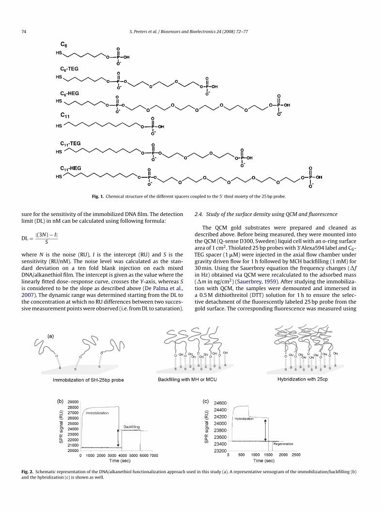
74 S. Peeters et al. / Biosensors and Bioelectronics 24 (2008) 72–77
Fig. 1. Chemical structure of the different spacers co
sure for the sensitivity of the immobilized DNA film. The detectionlimit (DL) in nM can be calculated using following formula:
DL = |(3N) − I|S
where N is the noise (RU), I is the intercept (RU) and S is thesensitivity (RU/nM). The noise level was calculated as the stan-dard deviation on a ten fold blank injection on each mixedDNA/alkanethiol film. The intercept is given as the value where thelinearly fitted dose–response curve, crosses the Y-axis, whereas Sis considered to be the slope as described above (De Palma et al.,2007). The dynamic range was determined starting from the DL tothe concentration at which no RU differences between two succes-sive measurement points were observed (i.e. from DL to saturation).
T
Fig. 2. Schematic representation of the DNA/alkanethiol functionalization approach usedand the hybridization (c) is shown as well.
upled to the 5′ thiol moiety of the 25 bp probe.
2.4. Study of the surface density using QCM and fluorescence
The QCM gold substrates were prepared and cleaned asdescribed above. Before being measured, they were mounted intothe QCM (Q-sense D300, Sweden) liquid cell with an o-ring surfacearea of 1 cm2. Thiolated 25 bp probes with 3′Alexa594 label and C6-EG spacer (1 �M) were injected in the axial flow chamber under
gravity driven flow for 1 h followed by MCH backfilling (1 mM) for30 min. Using the Sauerbrey equation the frequency changes (�fin Hz) obtained via QCM were recalculated to the adsorbed mass(�m in ng/cm2) (Sauerbrey, 1959). After studying the immobiliza-tion with QCM, the samples were demounted and immersed ina 0.5 M dithiothreitol (DTT) solution for 1 h to ensure the selec-tive detachment of the fluorescently labeled 25 bp probe from thegold surface. The corresponding fluorescence was measured using
in this study (a). A representative sensogram of the immobilization/backfilling (b)

nd Bioelectronics 24 (2008) 72–77 75
S. Peeters et al. / Biosensors aa spectrophotometer (RF-5301PC, Shimadzu, Japan) and convertedto a surface coverage (i.e. molecules/cm2) based on a previouslyprepared standard curve.
2.5. Flow-assisted vs. diffusion-controlled immobilization of DNAprobes
To study the influence of flow on the consecutive hybridizationreactions, two different immobilization pathways were evalu-ated and compared. For the flow-assisted approach, samples werecleaned, functionalized with the thiolated 25 bp probes and back-filled as described above. For the diffusion-controlled approachthe immobilization was performed overnight at 25 ◦C undercontinuous shaking using an Eppendorf thermomixer (450 rpm,Thermomixer R, Eppendorf, Germany). Afterwards, the sampleswere rinsed with distilled water, dried under a flow of N2 anddocked in the SPR device. For both approaches the subsequent back-filling and hybridization of the 25 cp were performed within the SPRdevice as described before. To compare the difference between bothapproaches two different spacers (i.e. C6 and C11) were selected.Again three independent experiments were run to assess the repro-ducibility.
3. Results
3.1. Comparative study of the different spacers using SPR
The immobilization grade was carefully evaluated for the thio-lated 25 bp probes modified with the six different spacers (Table 1).All surfaces functionalized with probes containing the C11 spacer(including TEG and HEG) had a clearly higher surface density com-pared to surfaces immobilized with probes containing the shorter
C6 spacer.The hybridization efficiency was calculated for each spacerbased on the surface density and maximum hybridization sig-nals (i.e. 320 nM) (Table 1). The C6-TEG spacer was found toshow the highest hybridization efficiency (i.e. 31.0%), whereasthe C6 spacer (i.e. 23.5%) and C11-HEG spacer (i.e. 23.3%) showedthe lowest efficiencies. Since these efficiencies only take intoaccount the maximum hybridization signals, it is impossibleto fully assess the impact of spacers on the biosensor per-formance. Therefore, the sensitivity, detection limit, dynamicrange and reproducibility were also determined using theconcentration-dependent hybridization curves as shown in Fig. 3.The obtained results for all these parameters are summarized inTable 2.
3.1.1. SensitivityThe lowest sensitivity was observed for the C6 spacer (i.e.
3.6 RU/nM). When introducing supplementary PEG units the sen-sitivity more than doubled. Moreover, a tremendous increase (>25times) was observed when using the longer C11 spacer. This wasalso very clear from the C11 dose–response hybridization curves
Table 1Overview of the surface density, the maximum hybridization level of the 25 cp andthe corresponding hybridization efficiency as calculated for the six different spacers
Spacer Surface density(×1013 molecules/cm2)
Hybridization(×1012 molecules/cm2)
Hybridizationefficiency (%)
C6 2.0 ± 0.02 4.8 ± 0.19 23.5C6-TEG 1.3 ± 0.30 4.2 ± 0.27 31.0C6-HEG 1.7 ± 0.06 4.8 ± 0.09 28.6C11 2.3 ± 0.02 5.8 ± 0.02 25.3C11-TEG 2.0 ± 0.15 5.4 ± 0.13 26.9C11-HEG 2.7 ± 0.03 6.3 ± 0.03 23.3
Fig. 3. Dose–response hybridization curves after immobilization of probes with C6
spacers ((a) C6, �; C6-TEG, �; and C6-HEG, �); and C11 spacers ((b) C11, �; C11,TEG, �; and C11-HEG, �) are expressed as the SPR response (RU) in function ofthe concentration of 25 cp (nM). The solid lines represent the hybridization curvesafter a flow-assisted immobilization, while the dashed line represents a diffusion-controlled immobilization of the probes. The error bars represent the standarddeviation on three independent experiments.
that clearly shifted to lower concentrations (Fig. 3b). In contrastto the C6 spacer, the sensitivity decreased when PEG units wereattached (Table 2).
3.1.2. Detection limitBesides the low sensitivity, the C6 spacer showed to have the
highest and thus the least desired detection limit. The lowest detec-tion limit (i.e. 0.3 nM) was obtained for the C11 spacer. As noted forthe sensitivity, the detection limit improved when TEG and HEG
Table 2Summary of the sensitivity, the detection limit and the dynamic range, calculated forboth the flow-assisted immobilization of thiolated 25 bp probes and the diffusion-controlled immobilization pathway (indicated by ‘no flow’)
Spacer Sensitivitya (RU/nM) Detection limita (nM) Dynamic range (nM)
C6 3.6 ± 2.1 21.6 ± 1.9 21.6–160C6-TEG 7.1 ± 0.9 11.5 ± 2.9 11.5–80C6-HEG 9.6 ± 0.6 5.7 ± 1.3 5.7–80C11 99.2 ± 13.7 0.3 ± 0.2 0.3–20C11-TEG 70.6 ± 6.7 1.2 ± 0.3 1.2–20C11-HEG 69.0 ± 3.5 2.3 ± 0.7 2.3–20C6 no flow 2.3 ± 0.9 12.9 ± 31.0 12.9–40C11 no flow 4.7 ± 0.2 8.4 ± 0.71 8.4–40
a The standard deviations were determined based on the average of the individualexperiments.

nd Bio
76 S. Peeters et al. / Biosensors aTable 3QCM responses and corresponding surface densities calculated via the Sauerbreyequation and via fluorescence
QCM response(�f/3 in Hz)
Surface density(×1013 molecules/cm2)
Fluorescence(×1013 molecules/cm2)
48.9 6.5 0.9844.0 5.8 0.9942.0 5.6 1.2
units were introduced to the C6 spacer. Also for the C11 spacer mod-ified with PEG moieties an improvement was observed, albeit lesspronounced (Table 2).
3.1.3. Dynamic rangeThe dynamic range of each spacer was determined based on
the hybridization curves. The broadest dynamic range (∼2 decades)was observed for probes modified with the C11 spacer. The dynamicrange for all other probes was found to be much narrower, especiallyfor probes with C6, C6-TEG and C6-HEG spacer (Table 2).
3.1.4. Reproducibility and specificityThe short C6 spacer was found to be the least reproducible,
particularly in the linear range (Fig. 3a). When attaching sup-plementary PEG units to this spacer, the reproducibility betweenindividual experiments improved over the whole concentrationrange. The introduction of a C11 spacer was shown to be even moreeffective. When attaching TEG or HEG moieties to this spacer, thereproducibility hardly seemed to improve any further (Fig. 3b).Hybridization of the 25 np sequence on all six thiolated 25 bpprobes modified with the different spacers resulted in no or verylow hybridization signals (0–5 RU) (data not shown).
3.2. Study of the surface density using QCM and fluorescence
To determine the amount of immobilized probes in an alter-native method and to estimate the extent of discrepancy betweenthe different techniques (i.e. SPR, QCM and fluorescence) the thi-olated 25 bp probe with 3′Alexa594 label and C6-TEG spacer wasimmobilized on a gold substrate. The corresponding immobiliza-tion curve is provided in the Supplementary Information. Table 3summarizes for each technique the results of three independentexperiments. By recalculation of the QCM data using the Sauerbreyequation an almost six times higher surface density was observed
compared to SPR. The surface densities determined by fluorescenceshowed a high similarity with the densities obtained via SPR (i.e.1.1 ± 0.1 × 10¹³ molecules/cm2).3.3. Study of the immobilization approach: flow vs. diffusioncontrolled
In order to assess the impact of using an integrated flow sys-tem, such as the one included in the SPR instrument, two differentimmobilization approaches (i.e. flow vs. diffusion) were compared.When immobilizing the probes in a diffusion-controlled mannerthe observed differences between the C6 and C11 spacer decreased.For the C6 spacer a sensitivity of 2.25 RU/nM and a correspond-ing DL of 12.9 nM were noted. Hybridization on the C11 spacerresulted in a sensitivity of 4.7 RU/nM and a DL of 8.4 nM. Theseresults are summarized in Table 2. For both spacers the dynamicrange was negatively affected and the maximum hybridization sig-nal dropped for more than 70% (Fig. 3). Despite the negative impactof the diffusion-controlled immobilization on most parameters, thereproducibility particularly within the linear range improved (datanot shown).
electronics 24 (2008) 72–77
4. Discussion
In this study, thiolated DNA probes with different spacers wereimmobilized and compared mainly using SPR as a characterizationtechnique. When comparing the length (C6 vs. C11) of the spacermolecule, a significant increase in surface density for thiolated25 bp probes having a C11 spacer was observed. These higher sur-face densities are most likely the result of the increase in the vander Waals forces. This will result in more ordered DNA films withmost probes orientated perpendicular to the surface (Porter et al.,1987). In contrast, thiolated 25 bp probes with the shorter C6 spacermay bind non-specifically on the gold surface leading to a strandorientation parallel to the surface (Wolf et al., 2004). Addition of abackfilling molecule (i.e. MCH or MCU) will reorganize the immobi-lized DNA layer by displacing some of the probes (Herne and Tarlov,1997). It is apparent that the surface density will be affected by thisconformational change.
Besides SPR, the surface densities were characterized using flu-orescence and QCM. As proven by our QCM data, as well as byothers, the use of the Sauerbrey equation led to an overestima-tion of the actual surface density (Voinova et al., 2002; Hook etal., 2001). This overestimation can be explained by assuming thathydration layers are being formed around the immobilized DNAstrands (Su et al., 2005; Cho et al., 2004; Ha et al., 2004). This hydra-tion effect was found to be sensitive to the buffer conditions used(Cho et al., 2004). For buffers with a low pH the overestimationwas found to be more pronounced as the immobilized DNA layerlikely becomes less rigid, changing its viscoelestic properties (Choet al., 2004). Within this report the thiolated DNA probes wereimmobilized using a 1 M KH2PO4 buffer at pH 3.8. Consequently,QCM data used to study DNA immobilization and hybridizationevents should be interpreted with care and further profound stud-ies through combined frequency and dissipation monitoring (i.e.QCM-D) are desired (Hook et al., 2001; Okahata et al., 1998).
As the hybridization of the 25 cp is directly related to the surfacedensity, the impact of the spacers on the hybridization efficiencywas assessed as well. The overall low hybridization efficiencies(23–31%), as reported in this study and observed by others (Gonget al., 2006; Steel et al., 2000), are likely caused by the high sur-face densities. High surface densities are expected to introducesteric effects that will reduce the accessibility of the immobilizedprobes (Ricci et al., 2007). Consequently, the introduction of longeralkane chain spacers will lead to lower hybridization efficiencies.Despite the limited hybridization efficiencies, the sensitivities and
detection limits obtained with thiolated 25 bp probes bearing solelythe longer C11 spacer were shown to be superior over those hav-ing the shorter spacers. Indeed, the chance of a low concentratedtarget DNA strand to bind with its surface immobilized comple-ment increases at higher probe densities. However, as mentionedbefore, high surface densities will still promote steric hindrance.To increase the accessibility and flexibility of the immobilizedDNA probes, spacers containing PEG molecules may be introduced(Biswal and Gast, 2003). In our case, the linking of PEG moietiesto the C11 spacer negatively affected both the sensitivity and thedetection limit, while the opposite was observed for the shorterC6 spacer. Apparently, there is a trade-off between the beneficialeffect of the alkane chain length and the introduction of PEG units.In addition, the individual impact of these two parameters is dif-ficult to assess as the surface density differs for each of the sixstudied probes. Nonetheless, both modifications, i.e. the length ofthe alkane chain and the presence of PEG units are important as theydetermine the quality of the immobilized DNA/alkanethiol film toa great extent. In addition, PEG units are expected to improve thespecificity as well. When performing assays in more complex bio-logical media the combination of mixtures of thiolated DNA and
nd Bio
S. Peeters et al. / Biosensors aalkanethiol “backfilling” molecules (e.g. MCH or MU) was shownnot to be sufficient (Lee et al., 2007). Therefore, spacers attached toPEG units, as studied in this report, are expected to further reducethese non-specific interactions (Unsworth et al., 2005; Saerens etal., 2005; Ostuni et al., 2001; Chapman et al., 2000).
Besides possessing high sensitivities and low detection limits,DNA biosensors need to be highly reproducible and stable as well.The data obtained within this study show an enormous increase inreproducibility when using thiolated 25 bp probes with C11 spacers(i.e. C11, C11-TEG and C11-HEG). In addition, sensors immobilizedwith the longer C11 spacers have shown improved stability over theshorter C6 spacer (Lai et al., 2006). This indicates that both factorsare strongly related to the length of the spacer and the associatedincrease in van der Waals forces.
The comparison between the two immobilization pathways(i.e. flow vs. diffusion) confirmed the need for an integratedflow-system. Solid-phase based immobilization and hybridizationreactions, like performed within this report, are poorly under-stood and rather complex (Erickson et al., 2003). It is assumed thatflow-systems are beneficial as they accelerate both reactions bythe introduction of convection. This was already shown by oth-ers as well (Kim et al., 2006). When using diffusion-controlledsystems fewer probes will take part in the immobilization anddepletion zones are expected to be introduced during the subse-quent hybridization process.
5. Conclusions
In this work, the impact of six different spacers used to link athiol moiety to the 5′ end of a specific DNA sequence, was stud-ied with respect to the immobilization degree and hybridizationperformance. To assess the effect of these spacers on the biosen-sor performance, different parameters such as the sensitivity, thedetection limit and the reproducibility were determined using SPR.To our knowledge, this is the first report presenting such a study.From the obtained results it was shown that varying the spacercan be advantageous for the design of sensitive and reliable DNAbiosensors. The alkane chain length was found to be an importantfactor. The sensitivities and detection limits significantly improvedwhen using probes with longer C11 spacers (i.e. C11, C11-TEG andC11-HEG). The effect of introducing PEG units, on the other hand,was probably masked by the high overall surface densities. PEGmoieties may, however, still be beneficial for real-world applica-
tions because of their high protein repelling capacities. Althoughthe C6 spacer is generally used to link thiols to specific DNAsequences, the findings presented in this report show that longeralkane chain spacers are highly recommended especially for appli-cations where the detection of low target concentrations is desired.Furthermore, we have demonstrated the clear impact of the immo-bilization approach (flow vs. static) on the biosensor performance.Acknowledgments
The authors would like to thank all current and former membersof the NEXT/NS research group for their valuable scientific input,Dr. A. Standaert for his help with the statistical analysis of the dataand Dr. F. Frederix for synthesizing the C11 precursor molecule. SaraPeeters is grateful to the Institute for the Promotion and Innovationthrough Science and Technology (IWT-Vlaanderen) for their fund-ing. This work was also partially financed by a grant of the BelgianFederal Service of Public Health, Food Chain Safety and Environ-ment (Grant no. S-6167) and by the Commission of the EuropeanCommunities in the context of IST project 027652 MASCOT andIST-NMP project 016817 SmartHEALTH.
electronics 24 (2008) 72–77 77
Appendix A. Supplementary data
Supplementary data associated with this article can be found,in the online version, at doi:10.1016/j.bios.2008.03.012.
References
Andreotti, P.E., Ludwig, G.V., Peruski, A.H., Tuite, J.J., Morse, S.S., Peruski, L.F., 2003.Biotechniques 35, 850–859.
Bashir, R., 2001. Superlattices and Microstructures 29, 1–16.Biswal, S.L., Gast, A.P., 2003. Physical Review E 68, 021402-1–021402-9.Bunimovich, Y.L., Shin, Y.S., Yeo, W.S., Amori, M., Kwong, G., Heath, J.R., 2006. Journal
of the American Chemical Society 128, 16323–16331.Castelino, K., Kannan, B., Majumdar, A., 2005. Langmuir 21, 1956–1961.Castillo, J., Gaspar, S., Leth, S., Niculescu, M., Mortari, A., Bontidean, I., Soukharev, V.,
Dorneanu, S.A., Ryabov, A.D., Csoregi, E., 2004. Sensors and Actuators B-Chemical102, 179–194.
Chapman, R.G., Ostuni, E., Yan, L., Whitesides, G.M., 2000. Langmuir 16, 6927–6936.Chen, J.R., Miao, Y.Q., He, N.Y., Wu, X.H., Li, S.J., 2004. Biotechnology Advances 22,
505–518.Cho, Y.K., Kim, S., Kim, Y.A., Lim, H.K., Lee, K., Yoon, D.S., Lim, G., Pak, Y.E., Ha, T.H.,
Kim, K., 2004. Journal of Colloid and Interface Science 278, 44–52.De Palma, R., Reekmans, G., Laureyn, W., Borghs, G., Maes, G., 2007. Analytical Chem-
istry 79, 7540–7548.Deisingh, A.K., Thompson, M., 2004. Canadian Journal of Microbiology 50, 69–77.Demers, L.M., Mirkin, C.A., Mucic, R.C., Reynolds, R.A., Letsinger, R.L., Elghanian, R.,
Viswanadham, G., 2000. Analytical Chemistry 72, 5535–5541.Erickson, D., Li, D., Krull, U.J., 2003. Analytical Biochemistry 317, 186–200.Fraser, M.E., Fujinaga, M., Cherney, M.M., Melton-Celsa, A.R., Twiddy, E.M., O’Brien,
A.D., James, M.N.G., 2004. Journal of Biological Chemistry 279, 27511–27517.Gong, P., Lee, C.H., Gamble, J.L., Castner, D.G., Grainger, D.W., 2006. Analytical Chem-
istry 78, 3326–3334.Ha, T.H., Kim, S., Lim, G., Kim, K., 2004. Biosensors & Bioelectronics 20, 378–389.Hahn, S., Mergenthaler, S., Zimmermann, B., Holzgreve, W., 2005. Bioelectrochem-
istry 67, 151–154.Halperin, A., Buhot, A., 2006. Langmuir 22, 11290–11304.Herne, T.M., Tarlov, M.J., 1997. Journal of the Chemical Society 119, 8916–8920.Hook, F., Ray, A., Norden, B., Kasemo, B., 2001. Langmuir, 8305–8312.Jonsson, U., Fagerstam, L., Ivarsson, B., Johnsson, B., Karlsson, R., Lundh, K., Lofas,
S., Persson, B., Roos, H., Ronnberg, I., Sjolander, S., Stenberg, E., Stahlberg, R.,Urbaniczky, C., Ostlin, H., Malmqvist, M., 1991. Biotechniques 11, 620–627.
Kim, H.S., Marafie, A., Jia, X.Y., Zoval, J.V., Madou, J., 2006. Sensors and ActuatorsB-Chemical 113, 281–289.
Lai, R.Y., Seferos, D.S., Heeger, A.J., Bazan, G.C., Plaxco, K.W., 2006. Langmuir 22,10796–10800.
Lee, C.Y., Nguyen, T.P., Grainger, D.W., Gamble, L.J., Castner, D.G., 2007. AnalyticalChemistry 79, 4390–4400.
Levicky, R., Herne, T.M., Tarlov, M.J., Satija, S.K., 1998. Journal of the American Chem-ical Society 120, 9787–9792.
Mannelli, I., Minunni, M., Tombelli, S., Wang, R., Spiriti, M., Mascini, M., 2005. Bio-electrochemistry 66, 129–138.
Okahata, Y., Kawese, M., Niikura, K., Ohtake, F., Furusawa, H., Ebara, Y., 1998. Analyt-ical Chemistry 70, 1288–1296.
Ostuni, E., Chapman, R.G., Holmlin, R.E., Takayama, S., Whitesides, G.M., 2001. Lang-muir 17, 5605–5620.
Porter, M.D., Bright, T.B., Allara, D.L., Chidsey, C.E., 1987. Journal of the AmericanChemical Society 109, 3559–3567.
Ricci, F., Lai, R.Y., Heeger, A.J., Plaxco, K.W., Sumner, J.J., 2007. Langmuir 23,6827–6834.
Saerens, D., Frederix, F., Reekmans, G., Conrath, K., Jans, K., Brys, L., Huang, L.,Bosmans, E., Maes, G., Borghs, G., Muyldermans, S., 2005. Analytical Chemistry77, 7547–7555.
Satjapipat, M., Sanedrin, R., Zhou, F.M., 2001. Langmuir 17, 7637–7644.Sauerbrey, G., 1959. Zeitschrift fur Physik 155, 206–222.Shchepinov, M.S., Case-Green, S.C., Southern, E.M., 1997. Nucleic Acids Research 25,
1155–1161.Soper, S.A., Brown, K., Ellington, A., Frazier, B., Garcia-Manero, G., Gau, V., Gutman, S.I.,
Hayes, D.F., Korte, B., Landers, J.L., Larson, D., Ligler, F., Majumdar, A., Mascini, M.,Nolte, D., Rosenzweig, Z., Wang, J., Wilson, D., 2006. Biosensors & Bioelectronics21, 1932–1942.
Steel, A.B., Levicky, R.L., Herne, T.M., Tarlov, M.J., 2000. Biophysical Journal 79,975–981.
Su, X., Wu, Y.J., Knoll, W., 2005. Biosensors & Bioelectronics 15, 719–726.Unsworth, L.D., Sheardown, H., Brash, J.L., 2005. Langmuir 21, 1036–1041.Van Aerschot, A., Rozenski, J., 2006. Journal of the American Society for Mass Spec-
trometry 17, 1396–1400.Voinova, M.V., Jonson, M., Kasemo, B., 2002. Biosensors & Bioelectronics 17, 835–841.Wang, J., 2000. Nucleic Acids Research 28, 3011–3016.Wang, R., Muninni, M., Tombelli, S., Mascini, M., 2004. Biosensors & Bioelectronics
20, 598–605.Wolf, L.K., Gao, Y., Georgiadis, R.M., 2004. Langmuir 20, 3357–3361.Wong, E.L., Chow, E., Gooding, J., 2005. Langmuir 21, 6957–6965.Yao, G., Tan, W., 2004. Analytical Biochemistry 331, 216–223.
Copyright © 2022 FDOKUMEN