In vitro investigation of pig cells for resistance to human antibody-mediated rejection
Upload
independentCategory
view
4download
0
Immunoproteasome beta subunit 10 is increased inchronic antibody-mediated rejectionJoanna Ashton-Chess1,2,3,8, Hoa Le Mai1,2,3,8, Vojislav Jovanovic1,2,3,8, Karine Renaudin4,Yohann Foucher1,2,3, Magali Giral1,2,3, Anne Moreau4, Emilie Dugast1,2,3, Michael Mengel5,Maud Racape1,2,3, Richard Danger1,2,3, Claire Usal1,2,3, Helga Smit1,2,3, Marina Guillet6, Wilfried Gwinner7,Ludmilla Le Berre1,2,3, Jacques Dantal1,2,3, Jean-Paul Soulillou1,2,3,8 and Sophie Brouard1,2,3,8
1INSERM, U643, Nantes, France; 2CHU Nantes, Institut de Transplantation et de Recherche en Transplantation, ITERT, Nantes,France; 3Faculte de Medecine, Universite de Nantes, Nantes, France; 4CHU Nantes, Service d’Anatomie Pathologique, Nantes,France; 5Department of Laboratory Medicine and Pathology, University of Alberta, Edmonton, Canada; 6TcLand Expression,Nantes, France and 7Abteilung fur Nephrologie, Medizinische Hochschule Hannover, Hannover, Germany
Chronic active antibody-mediated rejection is a form of late
rejection with a poor prognosis. To identify specific markers
of this, we analyzed several microarray studies in the
literature and performed mRNA profiling of 65 biopsies and
165 blood samples of a large cohort of renal transplant
patients with precisely characterized pathologies.
Immunoproteasome beta subunit 10 was found to be
specifically increased in the graft and blood samples during
chronic active antibody-mediated rejection and was also
significantly increased in rat cardiac allografts undergoing
acute rejection as well as chronic active antibody-mediated
rejection. This syndrome is characterized by chronic
transplant vasculopathy associated with diffuse C4d staining
and circulating donor-specific antibodies. Using this animal
model, we found that administration of the proteasome
inhibitor, Bortezomib, delayed acute rejection and
attenuated the humoral response in both the acute phase
and established state of this syndrome in a dose-dependent
manner. Following treatment with this reagent, donor-
specific antibodies and C4d deposition were reduced.
These studies highlight the role of the proteasome in chronic
rejection and identify this molecule as a marker of this
syndrome.
Kidney International (2010) 77, 880–890; doi:10.1038/ki.2010.15;
published online 24 February 2010
KEYWORDS: chronic allograft rejection; gene transcription; kidney
transplantation
Long-term graft loss remains the bane of kidney transplanta-tion. More recently, much attention has been paid to the‘humoral’ theory of chronic allograft rejection.1 Evidence forthe involvement of a humoral arm of the immune responseto allografts has come from studies analyzing the impact ofanti-human leukocyte antigen (HLA) antibodies on graftoutcome,2,3 and evidence of complement cascade activationwithin kidney grafts diagnosed by intragraft deposition of thecomplement split product C4d.4,5 These data were recentlyreinforced when the definition of chronic active antibody-mediated rejection (CAMR) was introduced into the Banffclassification of kidney graft injury as an association ofspecific histological lesions associated with diffuse C4ddeposition in peritubular capillaries and circulating donor-specific anti-HLA (DSA) antibodies.6
Current therapies to combat the acute form of antibody-mediated rejection aim to reduce antibody titers throughthe use of intravenous immunoglobulin,7 plasmapheresis,8 orB-cell-targeting antibodies such as Rituximab,9 but thesehave not gained notoriety in the chronic form. Moreover,these strategies do not seem to have an effect on plasma cells,the source of antibodies. Thus, the development of moreeffective strategies would benefit patients suffering from thistype of late graft rejection with such a poor prognosis.10 Theidentification of molecular markers associated withCAMR would not only facilitate its diagnosis, but could alsohelp to understand the pathophysiology of CAMR and thusaid in the design of new therapeutic strategies. Currently,CAMR diagnosis depends on the triad of graft lesionstogether with intragraft C4d and DSA mentioned above.Neither one of these alone can diagnose CAMR; DSA arepredictive of graft loss but have not yet been shown totightly correlate with intragraft lesions, and C4d hasrecently been shown to be absent in up to 50% of CAMRcases.11 Here, using a gene-set comparison approach, wedescribe the identification of a well-known molecule, theimmunoproteasome-b subunit 10 (PSMB10, also known asMECL-1) as a potential marker of CAMR in humans.
o r i g i n a l a r t i c l e http://www.kidney-international.org
& 2010 International Society of Nephrology
Received 19 July 2009; revised 12 November 2009; accepted
8 December 2009; published online 24 February 2010
Correspondence: Jean-Paul Soulillou, INSERM U643, 30 Building Jean
Monnet, Nantes Cedex 44093, France.
E-mail: [email protected]
8These authors contributed equally to this work.
880 Kidney International (2010) 77, 880–890

Moreover, we obtained concordant results in a rat heartallograft model that we have recently described as apertinent model of CAMR (histological lesions of chronicvascular damage, persisting antidonor antibodies and diffuseC4d deposits on the graft vasculature).12 Here we show that,in this model, inhibition of the proteasome significantlyprolongs allograft survival by preventing acute rejection, andalso attenuates established humoral immune responses(decreases DSA and C4d deposition) in both short- andlong-term surviving recipients. Our data suggest theimplication of the immunoproteasome in CAMR withPSMB10 as a potentially useful marker and point towardproteasome inhibition as a means of treating both acuterejection and CAMR.
RESULTSIdentification of the inducible member of theimmunoproteasome, PSMB10, as a potential intragraftbiomarker of chronic graft injury in human kidneytransplantation
In order to identify potential diagnostic markers of chronicgraft injury in humans, we compared several microarray datasets published in the literature in the context of humankidney transplant biopsies with chronic lesions (referred to asCAN or chronic rejection in the studies in question; Table 1).In total, less than 20 molecules were found to be commonbetween at least two data sets (not shown). Among them,PSMB10 was found to be upregulated in chronic rejection ina study by Donauer et al.13 and in Banff grade 3 versus Banffgrade 0 in a study by Flechner et al.14 PSMB10 was chosenfrom among the others because of its function as aninstrumental member of the immunoproteasome and theavailability of reagents to test it as a potential therapeutictarget. In fact, the immunoproteasome was shown to beinvolved in antigen processing for presentation,15 andproteasome inhibition was shown to blunt antibodyresponses in mice.16 In healthy individuals, PSMB10 isexpressed constitutively by the immune system (the spleen,peripheral blood, and lymph nodes; SupplementaryFigure S1A) and by lymphocytes and monocytes, where itsexpression is regulated by cell activation (SupplementaryFigure S1A and B). It can also be induced through exposureto interferon-g.17
PSMB10 is upregulated in CAMR in human kidney transplantrecipients
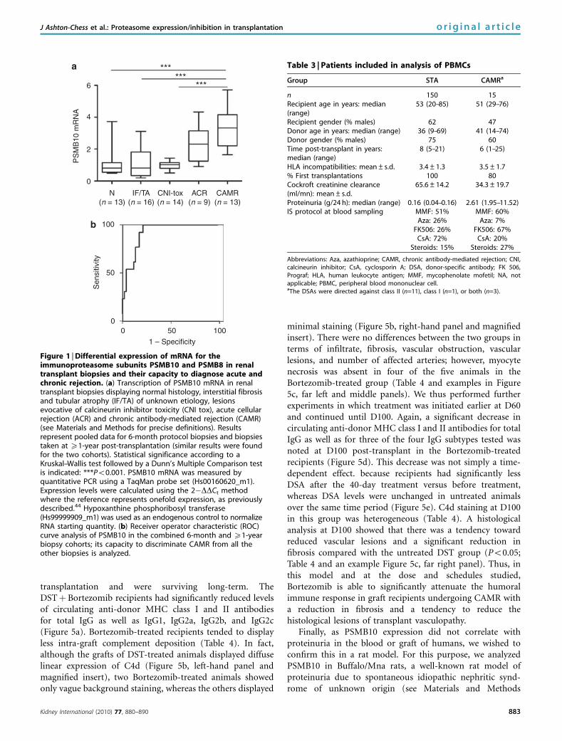
We analyzed PSMB10 in graft biopsies with differenthistological diagnoses (Table 2). As shown in Figure 1a,PSMB10 mRNA was specifically upregulated in biopsies withCAMR (Po0.001). Of note, PSMB10 expression was notcorrelated with proteinuria (r¼�0.16; P¼ 0.63). Receiveroperator characteristic (ROC) curve analysis revealed thatPSMB10 mRNA had an excellent capacity to discriminateCAMR from the other histological diagnoses, with an areaunder the curve of 0.92 (Po0.0001, 95% confidence intervalof 0.84–0.97). At a cut-off of 1.95, there was a sensitivity of0.85 and a specificity of 0.83 (Figure 1b). Thus, PSMB10shows potential as an intragraft marker of CAMR in humans.
Given that ACR is becoming a rare phenomenon, we nextfocused on CAMR, to determine whether the specificregulation of PSMB10 was reflected in the peripheral blood.PSMB10 was analyzed in 150 kidney transplant recipientswith stable graft function and 15 patients with CAMR(Table 3). As shown in Figure 2, patients with CAMR hadsignificantly higher levels of PSMB10 than those with stablegraft function (Po0.01). Again, PSMB10 expression was notcorrelated with proteinuria (r¼�0.01; P¼ 0.98).
To assess whether PSMB10 could potentially serve as aminimally invasive clinical decision-making tool, ROC curveanalysis was performed. The results (Figure 2b) showed thatthis molecule analyzed in recipient blood could stilldiscriminate CAMR from the other groups of patients well,albeit less than in the biopsies, with an area under the curveof 0.72 (Po0.01; 95% confidence interval of 0.57–0.84). At acut-off of 0.96, there was a sensitivity of 0.67 and a specificityof 0.64.
Potential confounding factors influencing the expression ofPSMB10
We next performed a multivariate analysis on the 150 stablepatients to evaluate the potential impact of clinical anddemographic factors on the expression of PSMB10 in theperipheral blood (Table 3). Of all the parameters tested(legend to Figure 2), the statistically significant parametersidentified as being associated with PSMB10 mRNA expre-ssion were recipient gender (Po0.05) and time post-transplant (Po0.01). Thus, creatinine clearance, proteinuria,
Table 1 | Studies used to identify common biomarkers of late graft injury
First author, journal/year Subject Sample Microarray platform Gene list
Hotchkiss, Transplantation/2006 ‘Chronic allograftnephropathy’ (CAN)
Biopsies Affymetrix Selected upregulated genes in CAN (gene listpublished)
Flechner, Am J Transplant/2004 Banff grade 3 vs Banff 1 Biopsies Affymetrix Genes upregulated in Banff 3 vs Banff 01 (gene listavailable on the authors’ website)
Scherer, Transplantation/2003 ‘Chronic Rejection’ (CR) Biopsies Affymetrix Genes upregulated in biopsies at 6 months whichwent on to develop CR at 1-year vs biopsies at6 months with no CR at 1-year (exhaustive genelist kindly provided by the authors)
Donauer, Transplantation/2003 ‘Chronic Rejection’ (CR) Biopsies ‘Homemade’ accordingto Stanford protocol
Upregulated in CR vs normal and polycystickidneys (gene list published)
Kidney International (2010) 77, 880–890 881
J Ashton-Chess et al.: Proteasome expression/inhibition in transplantation o r i g i n a l a r t i c l e

donor and recipient age, number of HLA incompatibilities(AþBþDR), and maintenance immunosuppression werenot confounders, neither were presence or absence of anti-HLA. Thus, PSMB10 is not simply a reflection of presence ofanti-HLA antibodies. These data are modeled in Figure 2c,where predicted values of PSMB10 are expressed according totime post-transplant and recipient gender. PSMB10 was thussignificantly higher in the peripheral blood mononuclear cells(PBMCs) of male recipients compared with female recipients(Po0.05) and displayed a distinctive inverse bell-shapedrelationship with time post-transplant. However, thesepotentially confounding factors could not explain thedifference in PSMB10 between stable (STA) and CAMR,because there was no difference in time post-transplant orrecipient gender between the STA and CAMR groups(P¼not significant). Thus, overall these data show thatPSMB10 may also be a peripheral blood biomarker of CAMRin humans, but some potential confounding factors may existand need to be taken into consideration.
PSMB10 as an intragraft and peripheral blood marker ofacute rejection as well as CAMR in a rat cardiac allograftmodel
To determine whether we could reproduce the above data in arodent model, PSMB10 mRNA was analyzed in the grafts andPBMCs of rat recipients of cardiac allografts undergoingacute rejection or CAMR. We found that PSMB10 wassignificantly increased in the cardiac allografts during bothacute rejection (at day (D)7 post-transplant) and CAMR(analyzed at D100 post-transplant) compared with syngeneiccontrols (Figure 3a; Po0.01 and Po0.0001, respectively).A different expression profile was observed in the PBMCs,where PSMB10 displayed no change during acute rejection,
suggesting an effect of DST priming on PBMCs. Moreover,similar to in humans, a significant increase was observedduring CAMR at D100 post-transplant (Figure 3b; Po0.05).Thus, PSMB10 shows potential as an intragraft andperipheral blood marker of CAMR in rats and humans.
The proteasome as a therapeutic target for AR: prolongedsurvival and attenuation of the humoral response during ARin a rat cardiac allograft model upon immunoproteasomeinhibition with Bortezomib
Given this upregulation of PSMB10 in acute rejection andCAMR in this rodent model, we set out to determine whetherproteasome inhibition could influence graft outcome. In thecontext of acute rejection, recipients of major histocompa-tibility complex (MHC)-mismatched cardiac allografts weretreated with Bortezomib every other day from D0–D20. Thedata presented in Figure 4a show that Bortezomib dose-dependently prolonged cardiac allograft survival with anoptimal effect at 0.1 mg/kg, giving a mean survival of 31.7days (n¼ 6) (versus 6.3 days in untreated controls (n¼ 4);Po0.01). Moreover, at D7, there were significantly lowercirculating anti-donor MHC class I and II antibodies for totalimmunoglobulin G (IgG) (Po0.01) as well as IgG1(Po0.01), and IgG2c (Po0.05), with no reduction forIgG2a or IgG2b (Figure 4b).
The proteasome as a therapeutic target for CAMR:attenuation of the humoral response during CAMR in a ratcardiac allograft model upon immunoproteasome inhibitionwith Bortezomib
In the context of CAMR, Bortezomib was initially adminis-tered every other day from D80 to D100 at 0.1 mg/kg in ratrecipients of cardiac allografts that had received DST before
Table 2 | Patients included in analysis of biopsies (Nantes and Hanover biopsies pooled; see Materials and Methods section).
Group N IF/TA CNI tox CAMRa ACRb
n 13 16 14 13 9Recipient age, years: median (range) 40.0 (18–69) 49.5 (24–66) 51.5 (29–66) 48 (24–71) 45 (25–61)Recipient gender ratio (M/F) 9:4 9:7 8:6 7:6 5:4Donor age, years: median (range) 35.0 (12–69) 51.0 (23–77) 41.5 (16–56) 34 (14–75) 34 (5–72)Time post-transplant years: median (range)c 1 2 (1–3) 5 (1–11) 11 (1–18) 0.67 (0.08–5)Donor gender ratio (M/F) 8:5 10:6 8:6 5:6 (n=2 NA) 8:1HLA incompatibilities: mean±s.d. 2.5±1.7 2.5±1.2 2.4±1.4 3.5±1.4 3.1±1.1% First transplant 100 100 79 92 78Cockroft creatinine clearance (ml/min): mean±s.d. 75.9±26.4 49.9±17.0 52.4±24.1 25.8±16.7 32.6±16.2Proteinuria (g/24 h): median (range) 0.10 (0.03–0.43) 0.11 (0.01–6.37) 0.09 (0.03–4.47) 1.94 (0.80–4.00) 0.46 (0.21–0.49)Banff c grade: mean±s.d. 0.00±0.00 1.25±0.58 0.80±1.03 2.27±0.79 1.5±0.53
Immunosuppression protocol (% of patients) MMF: 69% MMF: 63% MMF: 64% MMF:46% MMF: 78%at the time of biopsy Aza: 8% Aza: 13% Aza: 14% Aza: 15% Aza: 0%
FK506: 62% FK506: 63% FK506: 29% FK506:38% FK506: 67%CsA: 23% CsA: 13% CsA: 71% CsA: 54% CsA: 0%Rapa: 0% Rapa: 13% Rapa: 7% Rapa: 0% Rapa: 22%
Steroids: 54% Steroids: 69% Steroids: 57% Steroids: 23% Steroids: 33%
Abbreviations: ACR, acute cellular rejection; Aza, azathioprine; CAMR, chronic antibody-mediated rejection; CNI tox, calcineurin inhibitor toxicity; CsA, cyclosporin A;FK 506, tacrolimus; DSA, donor-specific anti-HLA; HLA, human leukocyte antigen; IF/TA, interstitial fibrosis and tubular atrophy; MMF, mycophenolate mofetil; N, normalhistology; TG, transplant glomerulopathy.aThe DSA were directed against class II (n=6), class I (n=2), or both (n=5).bThese were grade Ia (n=3), Ib (n=2), and grade IIa (n=4 with moderate intimal arteritis-vi) – all were C4d-negative and DSA-negative.cBiopsies for cause only.
882 Kidney International (2010) 77, 880–890
o r i g i n a l a r t i c l e J Ashton-Chess et al.: Proteasome expression/inhibition in transplantation
transplantation and were surviving long-term. TheDSTþBortezomib recipients had significantly reduced levelsof circulating anti-donor MHC class I and II antibodiesfor total IgG as well as IgG1, IgG2a, IgG2b, and IgG2c(Figure 5a). Bortezomib-treated recipients tended to displayless intra-graft complement deposition (Table 4). In fact,although the grafts of DST-treated animals displayed diffuselinear expression of C4d (Figure 5b, left-hand panel andmagnified insert), two Bortezomib-treated animals showedonly vague background staining, whereas the others displayed
minimal staining (Figure 5b, right-hand panel and magnifiedinsert). There were no differences between the two groups interms of infiltrate, fibrosis, vascular obstruction, vascularlesions, and number of affected arteries; however, myocytenecrosis was absent in four of the five animals in theBortezomib-treated group (Table 4 and examples in Figure5c, far left and middle panels). We thus performed furtherexperiments in which treatment was initiated earlier at D60and continued until D100. Again, a significant decrease incirculating anti-donor MHC class I and II antibodies for totalIgG as well as for three of the four IgG subtypes tested wasnoted at D100 post-transplant in the Bortezomib-treatedrecipients (Figure 5d). This decrease was not simply a time-dependent effect. because recipients had significantly lessDSA after the 40-day treatment versus before treatment,whereas DSA levels were unchanged in untreated animalsover the same time period (Figure 5e). C4d staining at D100in this group was heterogeneous (Table 4). A histologicalanalysis at D100 showed that there was a tendency towardreduced vascular lesions and a significant reduction infibrosis compared with the untreated DST group (Po0.05;Table 4 and an example Figure 5c, far right panel). Thus, inthis model and at the dose and schedules studied,Bortezomib is able to significantly attenuate the humoralimmune response in graft recipients undergoing CAMR witha reduction in fibrosis and a tendency to reduce thehistological lesions of transplant vasculopathy.
Finally, as PSMB10 expression did not correlate withproteinuria in the blood or graft of humans, we wished toconfirm this in a rat model. For this purpose, we analyzedPSMB10 in Buffalo/Mna rats, a well-known rat model ofproteinuria due to spontaneous idiopathic nephritic synd-rome of unknown origin (see Materials and Methods
PS
MB
10 m
RN
A6
4
2
0
N(n = 13)
IF/TA(n = 16)
CNI-tox(n = 14)
ACR(n = 9)
CAMR(n = 13)
******
***
100
50
0
Sen
sitiv
ity
0 50 100
1 – Specificity
a
b
Figure 1 | Differential expression of mRNA for theimmunoproteasome subunits PSMB10 and PSMB8 in renaltransplant biopsies and their capacity to diagnose acute andchronic rejection. (a) Transcription of PSMB10 mRNA in renaltransplant biopsies displaying normal histology, interstitial fibrosisand tubular atrophy (IF/TA) of unknown etiology, lesionsevocative of calcineurin inhibitor toxicity (CNI tox), acute cellularrejection (ACR) and chronic antibody-mediated rejection (CAMR)(see Materials and Methods for precise definitions). Resultsrepresent pooled data for 6-month protocol biopsies and biopsiestaken at X1-year post-transplantation (similar results were foundfor the two cohorts). Statistical significance according to aKruskal–Wallis test followed by a Dunn’s Multiple Comparison testis indicated: ***Po0.001. PSMB10 mRNA was measured byquantitative PCR using a TaqMan probe set (Hs00160620_m1).Expression levels were calculated using the 2�DDCt methodwhere the reference represents onefold expression, as previouslydescribed.44 Hypoxanthine phosphoribosyl transferase(Hs99999909_m1) was used as an endogenous control to normalizeRNA starting quantity. (b) Receiver operator characteristic (ROC)curve analysis of PSMB10 in the combined 6-month and X1-yearbiopsy cohorts; its capacity to discriminate CAMR from all theother biopsies is analyzed.
Table 3 | Patients included in analysis of PBMCs
Group STA CAMRa
n 150 15Recipient age in years: median(range)
53 (20–85) 51 (29–76)
Recipient gender (% males) 62 47Donor age in years: median (range) 36 (9–69) 41 (14–74)Donor gender (% males) 75 60Time post-transplant in years:median (range)
8 (5–21) 6 (1–25)
HLA incompatibilities: mean±s.d. 3.4±1.3 3.5±1.7% First transplantations 100 80Cockroft creatinine clearance(ml/mn): mean±s.d.
65.6±14.2 34.3±19.7
Proteinuria (g/24 h): median (range) 0.16 (0.04–0.16) 2.61 (1.95–11.52)IS protocol at blood sampling MMF: 51% MMF: 60%
Aza: 26% Aza: 7%FK506: 26% FK506: 67%
CsA: 72% CsA: 20%Steroids: 15% Steroids: 27%
Abbreviations: Aza, azathioprine; CAMR, chronic antibody-mediated rejection; CNI,calcineurin inhibitor; CsA, cyclosporin A; DSA, donor-specific antibody; FK 506,Prograf; HLA, human leukocyte antigen; MMF, mycophenolate mofetil; NA, notapplicable; PBMC, peripheral blood mononuclear cell.aThe DSAs were directed against class II (n=11), class I (n=1), or both (n=3).
Kidney International (2010) 77, 880–890 883
J Ashton-Chess et al.: Proteasome expression/inhibition in transplantation o r i g i n a l a r t i c l e

section for details). As shown in the Figure 6, PSMB10 wasnot significantly differentially expressed between rats with orwithout proteinuria, despite radical differences in proteinuria.
DISCUSSION
Here we used a literature gene-set comparison approach andidentified PSMB10 as a potential biomarker of chronic graftinjury. Further profiling by quantitative PCR in biopsies andPBMCs of renal transplant recipients revealed this moleculeto be specifically increased in the graft and blood in CAMR.PSMB10 was also significantly increased in rat cardiacallograft models with acute or chronic rejection. In theacute model, administration of the proteasome inhibitor
PS
MB
10 m
RN
A
2.5
2.0
1.5
1.0
0.5
0.0
**
STA CAMR
Sen
sitiv
ity
100
50
0
1 – Specificity0 50 100
0.0
–0.1
–0.2
–0.3
Post-transplantation time (months)
100 150 200 250
Pre
dict
ed lo
g-P
SM
B10
Female recipientsMale recipients
Figure 2 | Differential expression of PSMB10 mRNA in theperipheral blood mononuclear cells of renal transplantpatients, its capacity to diagnose chronic rejection and itsinteraction with confounding factors. (a) PSMB10 mRNAexpression in patients with stable graft function under standardimmunosuppression (STA; n¼ 150) and chronic antibody-mediatedrejection (CAMR; n¼ 15) (see Materials and Methods section forprecise definitions). Statistical significance according to aMann–Whitney test is indicated: **Po0.01. PSMB10 mRNA wasmeasured by quantitative PCR as described in the legend toFigure 1. (b) Receiver operator characteristic (ROC) curve analysis ofPSMB10 in the PBMCs of renal transplant patients. The capacity ofPSBM10 to discriminate CAMR from the other transplant groups isanalyzed. (c) Results of a multivariate linear regression analysis onlog-transformed PSMB10 values. Statistical modeling of therelationship between PSMB10 mRNA values, time post-transplantand donor gender is illustrated. Parameters analyzed includeddonor and recipient age and gender, time post-transplant,creatinine clearance, proteinuria, number of HLA incompatibilities(Aþ BþDR), presence or absence of anti-HLA, and maintenanceimmunosuppression (presence/absence of steroids, high versuslow trough level CsA (o125 versus X125 ng/ml) versus high andlow trough level FK-506 (o5 versus X5 ng/ml).
Graft
PBMC
PS
MB
10 m
RN
AP
SM
B10
mR
NA
2
1
0Syn D5(n = 6)
DST D5(n = 6)
AR(n = 6)
Syn D5(n = 6)
DST D5(n = 5)
AR(n = 4)
Syn D100(n = 7)
DST D100(n = 8)
Syn D100(n = 4)
DST D100(n = 4)
*****
*
*25
20
15
10
5
0
a
b
Figure 3 | PSMB10 mRNA profiles in the allografts and PBMCsof rat recipients of MHC-mismatched heart grafts displayingacute and chronic rejection. PSMB10 mRNA transcription in thegrafts and PBMCs of rat recipients of MHC-mismatched andDST-treated or syngeneic cardiac transplants. Analyses wereperformed at D5 and D100 post-transplant as well as during acuterejection (mean 6.3 days post-transplant). PSMB10 was measuredby quantitative PCR as described in the legend to Figure 1 usingTaqMan probes for rat PSMB10 (Rn01432424_g1) and rat HPRT(Rn01527838_g1). Statistical significance according to aKruskal–Wallis test followed by a Dunn’s Multiple Comparison testis indicated: ***Po0.001; **Po0.01 and *Po0.05. In this model,DST treatment induces long-term survival but with histologiclesions of chronic transplant vasculopathy40 associated withdeposition of the complement split product C4d and circulatingdonor-specific antibodies.12
884 Kidney International (2010) 77, 880–890
o r i g i n a l a r t i c l e J Ashton-Chess et al.: Proteasome expression/inhibition in transplantation

Bortezomib not only dose-dependently delayed acute rejec-tion but also attenuated the humoral response. In the chronicmodel with established CAMR, Bortezomib treatmentdecreased DSA and C4d deposition and improved someaspects of the chronic tissue injury. These data thus suggestPSMB10 as a potential marker and the proteasome as atherapeutic target for CAMR.
Proteasomes are large protease complexes located incytoplasm and nuclei that degrade cellular proteins in aubiquitin-dependent and adenosine triphosphate-dependentmanner present in immune cells.18 In response to interferon-g,the three catalytic subunits are replaced by their homologoussubunits, PSMB 8, 9, and 10, to form the so-calledimmunoproteasome, which is essential for processing anti-genic peptides for presentation through the class I MHCcomplex.15 Immunoproteasomes have additional effects thatare independent to class I processing, for example, theyinhibit T-cell proliferation, as T cells lacking immunoprotea-some subunits hyperproliferate in vitro and in vivo, and KOmice have higher numbers of central memory CD8þ cells.19
Experiments in KO mice have revealed that PSMB10 isinvolved in controlling homeostatic equilibrium betweenT-cell subsets,20 thereby controlling the T-cell repertoire.21
Given the expression of PSMB10 by lymphocytes andmonocytes, it is possible that the increase in PSMB10 in thegraft in CAMR may be the result of specific infiltration ofthese cells. B cells in particular may be involved in thisbecause patients with chronic rejection undergo lymphoidneogenesis with the development of intragraft B-cell germinalcenters.22 Likewise, increased PSMB10 expression may comefrom locally present activated monocytes or mature dendriticcells, as the immunoproteasome is known to be increased inthese cells.23 Given that the proteasome decreases during latephase plasma cell differentiation,24 it is unlikely that theincrease in PSMB10 in the peripheral blood stems from anincrease in plasma cells in chronic rejection. ROC curveanalysis suggested that the level of PSMB10 in the blood ofgraft recipients could be useful to diagnose CAMR, althoughit may not be sufficient as a stand-alone marker and could becombined with other markers to improve its diagnosticperformance. Moreover, we found PSMB10 levels to beinfluenced by recipient gender and time post-transplant (butnot presence of anti-HLA or renal function), highlighting theneed to take confounding factors into account in biomarkeranalyses. Thus, PSMB10 is not simply a reflection of DSA orpoor renal function, but rather of histological lesionstogether with C4d and DSA. Future analyses on simultaneousbiopsy blood samples, which were not possible here becauseof lack of material, could be considered to directly compareintragraft and peripheral blood diagnostic performance.
The intragraft upregulation of PSMB10 we observed inCAMR in both humans and rats prompted us to test theproteasome inhibitor Bortezomib in the rat models of bothacute rejection and CAMR. Currently, Bortezomib is used inthe clinic for multiple myeloma and solid tumors.25 However,a limited number of studies have analyzed the effect of thisdrug on acute rejection in murine models of transplantation,showing a preventative effect on both heart26 and islet27
allograft acute rejection. On the basis of blood biochemistry,Bortezomib was also associated with side effects, but thesewere reversible upon cessation of treatment.27 Our data in arat model of acute heart rejection are similar to thosereported in the mouse, with a similar survival prolongationobserved in both models. In the chronic model, diminutionof some aspects of chronic injury was observed. Thepreventative effect on acute rejection is compatible with thehypothesis that proteasome inhibition reduces alloantigeni-city by reducing class I presentation. Moreover, we observed asignificant decrease in antibody production, suggesting anattenuating effect on the humoral response. Very recent datashowed that proteasome inhibition specifically induces deathof activated T cells,28 and suppresses essential immunefunction of CD4þ T cells upon activation by allogeneicdendritic cells.29 Moreover, proteasome inhibition has beenshown to modulate toll-like receptor4-induced dendritic cellactivation,30 which could have a role here, as we recently
Per
cent
sur
viva
l
100
75
50
25
0
Bortezomib Days post transplant
0 10 20 30 40 120
** ** **
3000
2000
1000
0D7-R D7-T D7-R D7-T D7-R D7-T D7-R D7-T D7-R D7-T
MF
I
P=NS
P=NS
Total IgG IgG2a IgG2b IgG2CIgG1
**
*
**
Figure 4 | Induction treatment in rat recipients ofMHC-mismatched cardiac allografts with Bortezomib.(a) Kaplan–Maier survival analysis of cardiac allograft recipientsuntreated (K) or treated every other day for 20 days withBortezomib at 0.025 (’), 0.05 (m), or 0.1 (.) mg/kg. Statisticalsignificance is indicated **Po0.01. (b) Measurement (as describedin the Materials and Methods section) of anti-donor MHC class Iand II IgG1, IgG2a, IgG2b, and IgG2c subtypes in the serum ofMHC-mismatched cardiac allograft recipients at D7 post-transplant treated with Bortezomib (D7-T) or without Bortezomibtreatment and thus undergoing rejection (D7-R). Results areexpressed as mean fluorescence intensity (MFI). Statisticalsignificance is indicated *Po0.05, **Po0.01.
Kidney International (2010) 77, 880–890 885
J Ashton-Chess et al.: Proteasome expression/inhibition in transplantation o r i g i n a l a r t i c l e

found toll-like receptor 4 to be increased in the graft andblood of CAMR patients.31
We also found that in a rat model of CAMR, Bortezomibdecreased the humoral immune response by significantlyreducing DSA. These data are in accordance with a recentreport that proteasome inhibition blunts Ab responsesfollowing in vivo B-cell activation in mice, by promotingapoptosis.16 In the latter study, immunoproteasome sub-components decreased during B-cell differentiation, inparallel with increased immunoglobulin synthesis. Morerecently, Bortezomib was shown to rapidly and efficientlydeplete both short- and long-lived plasma cells in the spleens
and bone marrow in a murine model of lupus-like disease,leading to reduced autoantibody development.32 Moreover,Bortezomib has recently been shown to induce apoptosis ofplasma cells, to block anti-HLA Ab secretion in vitro, and totransiently decrease plasma cells in vivo in renal transplantrecipients.33,34 However, in the latter studies in humans,Bortezomib was used in combination with other immuno-suppressors and plasma exchange making it difficult topinpoint the specific role of Bortezomib among the treatmentarsenal. Moreover, its effect on graft histology has yet to bedetermined. In our model, we show a clear-cut effect ofBortezomib monotherapy on DSA and C4d, significantly
50,000
40,000
30,000
20,000
10,000
0
40,000
30,000
20,000
10,000
0
MF
I
Total IgG IgG1 IgG2a IgG2b IgG2C Total IgG IgG1 IgG2a IgG2b IgG2C
**
**
**
**
**
**
*
*
*P=0.08
D100DST
D100DST
D100DST
D100DST
D100DST
D100DST-T
D80–D100
D100DST-T
D80–D100
D100DST-T
D80–D100
D100DST-T
D80–D100
D100DST-T
D80–D100
D100DST
D100DST
D100DST
D100DST
D100DST
D100DST-T
D60–D100
D100DST-T
D60–D100
D100DST-T
D60–D100
D100DST-T
D60–D100
D100DST-T
D60–D100
20,000
15,000
10,000
5000
0D60 D100 D60 D100
No treatment Bortezomib
P = NS *
MF
IDST D100 DST-T D80–D100 DST-T D80–D100
Figure 5 | Maintenance treatment in rat recipients of MHC-mismatched cardiac allografts with Bortezomib. (a) Measurement(as described in the Materials and Methods section) of anti-donor MHC class I and II total IgG and IgG1, IgG2a, IgG2b, and IgG2c subtypes inthe serum of MHC-mismatched cardiac allograft recipients treated with DST or with DST followed by Bortezomib from D80–D800, at D100post-transplantation. Results are expressed as mean fluorescence intensity (MFI). Statistical significance is indicated: *Po0.05. (b)Immunoperoxidase staining of C4d at D100 post-transplantation in recipients of MHC-mismatched cardiac allografts treated with DSTalone (left-hand panel and insert) or with DST followed by Bortezomib from D80 to D100 (right-hand panel and insert) (see Materialsand Methods section for details). (c) Histology (hematoxylin and eosin) at D100 post-transplantation in recipients of MHC-mismatchedcardiac allografts treated with DST alone (far left panel) or with DST followed by Bortezomib from D80 to D100 (middle panel) or fromD60 to D100 (far right panel) at � 100 magnification. (d) Measurement (as described in the Materials and Methods section) of anti-donor MHC class I and II total IgG and IgG1, IgG2a, IgG2b, and IgG2c subtypes in the serum of MHC-mismatched cardiac allograft recipientstreated with DST or with DST followed by Bortezomib from D60 to D100, at D100 post-transplantation. (e) Paired measurement of anti-donor MHC class I and II total IgG at D60 and D100 in untreated recipients and recipients treated with Bortezomib from D60 to D100.
886 Kidney International (2010) 77, 880–890
o r i g i n a l a r t i c l e J Ashton-Chess et al.: Proteasome expression/inhibition in transplantation

reduced allograft fibrosis and a tendency toward reducedvascular lesions. Therefore, early treatment with Bortezomibmay be pivotal to prevent irreversible chronic lesions. Furtheranalyses in a rodent model of kidney transplantation wouldalso be interesting, although for the moment there are notrue models of CAMR in the kidney. Indeed, in the Fischer toLewis kidney transplant model of chronic allograft nephro-pathy (reviewed by Marco35), our own experience revealedchronic pyelonephritis with no vascular lesions (data notshown).
On the whole, our data point toward PSMB10 as apotentially useful intragraft and peripheral blood marker for
CAMR, with immunoproteasome inhibition as a valuabletreatment strategy for antibody-mediated rejection.
MATERIALS AND METHODSGene-set comparison
With the aim of identifying relevant biomarkers of late graftinjury, we compared the gene sets from four microarraystudies of late kidney graft injury published in theliterature13,14,36,37 (see Table 1). To overcome the problemof a single gene having numerous denominations, accessionnumbers, and so on, all genes were converted into a singlecommon identifier, the ‘gene ID,’ using the gene IDconversion tool available on the DAVID BioinformaticResources 2006 website (http://www.david.abcc.ncifcrf.gov/home.jsp). Gene IDs from the different studies representingwere sorted in Microsoft Excel and repeats of the same IDidentified.
Human kidney transplant recipients
Study groups. All patients who participated in this studygave informed consent, and the study was approved by theUniversity Hospital Ethical Committee and the Committeefor the Protection of Patients from Biological Risks. Thestudy was performed on 65 biopsies and 165 blood samples.Samples were included prospectively on the basis ofhistological or clinical criteria.(a) Biopsies were collected at two centers, Nantes, France
(n¼ 45) and Hanover, Germany (n¼ 20). Of the total
Table 4 | Histological analysis of DST-treated animals (D7 and D14 before transplantation) subsequently untreated or treatedevery other day from D80 to D100 or from D60 to D100 (CAMR lesions already established) with Velcade at 0.1 mg/kg
Rat GroupGraft
infiltrate Fibrosis NecrosisVascularlesions
Vascularobstruction
No. of affectedarteries (art/section) C4d
1 DST 2 3* + 2 2 3 (28) +++2 DST 2 3 + 0 0 0 +++3 DST 3 4* + 2 4 13 (16) +++4 DST 2–3 2 + 2 1 1 (21) +++5 DST 2–3 3 + 1 1 3 (19) +++
1 DST+Bortezomib D80–D100 2–3 3–4* + 1–3 4 13 (33) +2 DST+Bortezomib D80–D100 2 3 � 2 4 8 (48) �3 DST+Bortezomib D80–D100 1–2 3–4 � 0 0 0 �4 DST+Bortezomib D80–D100 1–2 3 � 2 4 7 (38) ++5 DST+Bortezomib D80–D100 1–2 3* � 1 1 2 (16) ++
1 DST+Bortezomib D60–D100 2 1* + 0 0 0 +++2 DST+Bortezomib D60–D100 2–3 2* + 0 0 0 ++3 DST+Bortezomib D60–D100 1–2 3 Focal 3 3 7 (31) +++4 DST+Bortezomib D60–D100 2 1 Focal 1 1 3 (8) +++5 DST+Bortezomib D60–D100 1–2 2 � 0 0 0 �6 DST+Bortezomib D60–D100 2 2 Focal 0 0 0. �
P=NS Po0.05 P=NS P=NS P=NS
Abbreviations: CAMR, chronic antibody-mediated rejection; DST, donor-specific antibody; NS, not significant.Infiltrate was graded from 0 to 4 (0, absence; 1, minimum; 2, discrete; 3, moderate; 4, abundant). Fibrosis was graded from 0 to 4 (0, absence; 1, focal; 2, diffuse-minimal; 3,diffuse-moderate; 4, diffuse-abundant; *, edema). Vascular obstruction was graded from 0 to 4 (0, 0; 1, o20%; 2, 20–50%; 3, 50–80%; 4, 480%). Vascular lesions were gradedfrom 0 to 3 (0, normal; 1, leuco-intimal adhesion; 2, inflammatory endarteritis; 3, fibrous endarteritis). C4d was graded from – to +++ (�, o20%; +, 20–50%; ++, 50–75%, and+++, 475%). Necrosis was graded as presence (+) or absence (�) of myocyte damage. Statistical significance was measured using a non-parametric Kruskal–Wallis test withDunn’s Multiple Comparison test.
2.5
2.0
1.5
1.0
0.5
0.01-month 6-month 1-month 6-month
0.25
0.20
0.15
0.10
0.05
0.00
Proteinuria PSMB10
P = 0.01 P = NS
Figure 6 | PSMB10 and proteinuria in a rat model of idiopathicnephritic syndrome. Proteinuria (left-hand graph) andPSMB10 expression (right-hand graph) in Buffalo/Mna ratsat 1-month of age and 6 months of age (see Materials andMethods section for details).
Kidney International (2010) 77, 880–890 887
J Ashton-Chess et al.: Proteasome expression/inhibition in transplantation o r i g i n a l a r t i c l e

65 biopsies, 29 were protocol biopsies at 6 months or1 year and the remaining 36 were biopsies for cause.All were classified according to the updated Banffclassification criteria,6 as displaying normal histology(N- C4d-negative and DSA-negative; n¼ 13) or lesionsof interstitial fibrosis and tubular atrophy of unknownetiology (IF/TA-C4d-negative and DSA-negative; n¼ 16)or lesions of calcineurin inhibitor toxicity (tox-C4d-negative and DSA-negative; n¼ 14), or lesions of acutecellular rejection (ACR; n¼ 9) or CAMR (defined by thepresence of circulating DSA associated with transplantglomerulopathy and deposition of C4d in peritubularcapillaries (n¼ 13). C4d staining was performed onfrozen sections with 450% staining being considered aspositive. The demographic and clinical data for all ofthese patients are provided in Table 2.
(b) Peripheral blood mononuclear cells (PBMCs) were preparedfrom the blood of 165 kidney transplant patients (largelyindependent from those included in the biopsy analysis)whose statuses were defined on a histological and/orclinical basis: Stable graft function (patients understandard immunosuppression with stable graft function(creatinemia o150mmol/l and proteinuria o1 g/24 h forat least 3 years including over the two previous 6-monthfollow-up appointments with o20% change between thetwo time points: STA; n¼ 150), or CAMR as definedabove (n¼ 15). The demographic and clinical data forthese patients are provided in Table 3.
Rodent transplant models
Study groups. Inbred male adult rats (200–250 g) of theLEW.1A (RT1a) and LEW.1W (RT1u) MHC-incompatiblecongeneic strains were purchased from Janvier (Le Genest-Saint-Isle, France) and maintained in an animal facility understandard conditions according to the European and Institu-tional Guidelines.(a) Acute rejection of heart allografts. LEW.1A rats received
heterotopic heart allografts from LEW.1W donors aspreviously described38 and received no immunosuppres-sive treatment. Rejection (cessation of heart beatingassessed by daily abdominal palpation) occurs at a meanof 6.3 days post-transplant.
(b) Chronic antibody-mediated rejection of heart allografts.LEW.1A rat recipients were transfused intravenouslywith 1 ml of blood from a LEW.1W donor 14 and 7 daysbefore heterotopic LEW.1W cardiac transplantation asdescribed.39 Graft function was evaluated as above. Inthis strain combination, donor-specific transfusioninduces long-term graft survival but does not inhibitthe indirect pathway of allorecognition, resulting inhistological lesions of chronic transplant vasculopathy40
associated with deposition of the complement splitproduct C4d and circulating DSAs.12
(c) Bortezomib treatment for the prevention of acute rejection.Heart allograft recipients received Bortezomib (Velcade,
Millennium Pharmaceuticals, Cambridge, MA, USA)intraperitoneally from the day of transplantation andevery other day until day (D) 20. Three different doseswere tested: 0.025, 0.05, and 0.1 mg/kg.
(d) Bortezomib treatment of CAMR in the context of DST-induced long-term allograft survival. Heart allograftrecipients were treated with DST as described aboveand received Bortezomib intraperitoneally at 0.1 mg/kgevery other day either from D80 until D100 or from D60until D100. At this time, all allografts present vascularlesions with diffuse C4d deposits and circulatinganti-DSA.
Rodent proteinuria model
The Buffalo/Mna rat strain maintained in our laboratory wasprovided by Dr Saito (Central Experimental Institute,Nokawa, Kawasaki, Japan). These rats have spontaneousidiopathic nephritic syndrome of unknown origin aspreviously described.41 They do not have proteinuria at1 month of age but develop significant proteinuria by 6months of age. Animal care was in accordance with ourinstitutional guidelines. The rats were placed in metaboliccages for 24 h and total urinary protein concentration(g/l) was measured by a colorimetric method using a Hitachiautoanalyzer (Boehringer, Reims, France). Proteinuriawas expressed according to the formula proteinuria(g/mmol)¼ urinary proteins (g/l)/urinary creatinine (mmol/l),and it was considered to be abnormal at values 40.2 g/mmol.
Sample preparation
Human kidney transplant biopsies were processed as de-scribed.42 Rat heart transplant or kidney tissue was processedas described.40 Human and rat peripheral blood was collectedin EDTA vacutainers and PBMCs separated by densitycentrifugation using Lymphosep, lymphocyte separation media(Bio West, Nuaille, France). PBMCs were stored in TRIzol(Invitrogen, Cergy Pontoise, France) at �801C until use.
RNA extraction and reverse transcription. Human biopsieswere processed as described in detail.42 Rat heart allograftsand rat and human PBMCs were processed using the TRIzolmethod (Invitrogen) according to the manufacturer’s in-structions. RNA quality and quantity was determined usingan Agilent 2100 BioAnalyzer (Palo Alto, CA, USA). GenomicDNA was removed by DNase treatment (Roche, Indianapolis,IN, USA). RNA was reverse transcribed into cDNA usingpolydT oligonucleotide and Maloney leukemia virus reversetranscription (Invitrogen).
Real-time quantitative PCR. Real-time quantitativePCR was performed in an Applied Biosystems GenAmp7700 sequence detection system (Applied Biosystems,Foster City, CA, USA) as described in detail,42 usingcommercially available primer and probe sets (AppliedBiosystems).
Rat cardiac allograft histological analyses and C4d immuno-
staining. Heart allografts were prepared as described by
888 Kidney International (2010) 77, 880–890
o r i g i n a l a r t i c l e J Ashton-Chess et al.: Proteasome expression/inhibition in transplantation

Ballet et al.12 and analyzed by a pathologist (KR) (see alsoTable 4 and legend). C4d deposition was localized asdescribed by Ballet et al.12 using an affinity-purifiedpolyclonal rabbit antibody to C4d kindly provided byBaldwin and group43 (department of pathology, JohnHopkins Medical Institutions, Baltimore, MD, USA).
Measurement of circulating anti-donor antibodies in rat
recipients of cardiac allografts. Anti-donor class I and II totalIgG and subtypes in the sera of untreated or Bortezomib-treated recipients at D7 post-transplantation, and in DST-treated and DSTþ Bortezomib-treated animals at D60 andD100 post-transplantation were measured as described byBallet et al.12
Statistical analyses
The non-parametric Mann–Whitney test was used forcomparison between two groups. Note that there was animbalance in sample size between the CAMR and STApatients (n¼ 15 versus 150); this was because we preferred touse all available STA samples to better reflect the relativelylow prevalence of CAMR and to use all samples that wereused for the other analyses (multivariate analyses andROC). Such an imbalance is authorized within the contextof the non-parametric Mann–Whitney test. The non-para-metric Kruskal–Wallis test was performed for comparison ofthree or more groups, followed by Dunn’s multiplecomparisons test. ROC curve analysis was performed todetermine the cut-off point of PSMB10 in biopsies and/orblood that yielded the highest combined sensitivity andspecificity in diagnosing CAMR (see Figure 1 legend forexplanation). Note that the n¼ 15 CAMR were comparedwith a large number of STA patients (n¼ 150) in order tobetter reflect the relatively low prevalence of CAMR in thisdiagnostic test. The effect of the different clinical anddemographic parameters on the expression of PSMB10 inpatients with stable graft function, a multivariate linearregression analysis, was performed following log transforma-tion of the data taking into account the various parameters(see legend to Figure 2). The statistical software used wasGraphPad prism 5.
DISCLOSURESeveral authors hold a patent for PSMB10 expression as a biomarkerof CAMR. JA-C and MG are currently employees of TcLand Expression,Nantes France, a company developing blood biomarkers intransplantation.
ACKNOWLEDGMENTSThis research was funded by a grant from the ‘Foundation Progreffe’(Nantes, France). The 164 stable patients were recruited in Nantesin the context of the Genhomme PHRC. This work is also part of aFrench Transplantation Research Network (RTRS) supported bythe ‘Fondation de Cooperation Scientifique’ CENTAURE. Wethank Dr W Baldwin (Cleveland Clinic, OH, USA) for kindlyproviding the anti-rat C4d antibody.
SUPPLEMENTARY MATERIALFigure S1. PSMB10 mRNA expression within the immune system andvarious cell subtypes of healthy individuals.
Supplementary material is linked to the online version of the paper athttp://www.nature.com/ki
REFERENCES1. Terasaki PI. Humoral theory of transplantation. Am J Transplant 2003; 3:
665–673.2. Terasaki PI, Ozawa M. Predicting kidney graft failure by HLA antibodies:
a prospective trial. Am J Transplant 2004; 4: 438–443.3. Terasaki PI, Ozawa M. Predictive value of HLA antibodies and serum
creatinine in chronic rejection: results of a 2-year prospective trial.Transplantation 2005; 80: 1194–1197.
4. Mauiyyedi S, Pelle PD, Saidman S et al. Chronic humoral rejection:identification of antibody-mediated chronic renal allograft rejection byC4d deposits in peritubular capillaries. J Am Soc Nephrol 2001; 12:574–582.
5. Regele H, Bohmig GA, Habicht A et al. Capillary deposition ofcomplement split product C4d in renal allografts is associated withbasement membrane injury in peritubular and glomerular capillaries: acontribution of humoral immunity to chronic allograft rejection. J Am SocNephrol 2002; 13: 2371–2380.
6. Solez K, Colvin RB, Racusen LC et al. Banff’05 Meeting report: DifferentialDiagnosis of Chronic Allograft Injury and Elimination of Chronic AllograftNephropathy (‘CAN’). Am J Transplant 2007; 7: 518–526.
7. Jordan SC, Vo AA, Toyoda M et al. Post-transplant therapy with high-doseintravenous gammaglobulin: applications to treatment of antibody-mediated rejection. Pediatr Transplant 2005; 9: 155–161.
8. Akalin E, Dinavahi R, Friedlander R et al. Addition of plasmapheresisdecreases the incidence of acute antibody-mediated rejection insensitized patients with strong donor-specific antibodies. Clin J Am SocNephrol 2008; 3: 1160–1167.
9. Faguer S, Kamar N, Guilbeaud-Frugier C et al. Rituximab therapy for acutehumoral rejection after kidney transplantation. Transplantation 2007; 83:1277–1280.
10. David-Neto E, Prado E, Beutel A et al. C4d-positive chronic rejection: afrequent entity with a poor outcome. Transplantation 2007; 84:1391–1398.
11. Sis B, Jhangri GS, Bunnag S et al. Endothelial gene expressionin kidney transplants with alloantibody indicates antibody-mediateddamage despite lack of C4d staining. Am J Transplant 2009; 9:2312–2323.
12. Ballet C, Renaudin K, Degauque N et al. Indirect CD4+ TH1 response,antidonor antibodies and diffuse C4d graft deposits in long-termrecipients conditioned by donor antigens priming. Am J Transplant 2009;9: 697–708.
13. Donauer J, Rumberger B, Klein M et al. Expression profiling on chronicallyrejected transplant kidneys. Transplantation 2003; 76: 539–547.
14. Flechner SM, Kurian SM, Solez K et al. De novo kidney transplantationwithout use of calcineurin inhibitors preserves renal structure andfunction at two years. Am J Transplant 2004; 4: 1776–1785.
15. Tanaka K, Kasahara M. The MHC class I ligand-generating system: roles ofimmunoproteasomes and the interferon-gamma-inducible proteasomeactivator PA28. Immunol Rev 1998; 163: 161–176.
16. Cascio P, Oliva L, Cerruti F et al. Dampening Ab responses usingproteasome inhibitors following in vivo B cell activation. Eur J Immunol2008; 38: 658–667.
17. Van den Eynde BJ, Morel S. Differential processing of class-I-restrictedepitopes by the standard proteasome and the immunoproteasome. CurrOpin Immunol 2001; 13: 147–153.
18. Adams J. The proteasome: structure, function, and role in the cell. CancerTreat Rev 2003; 29(Suppl 1): 3–9.
19. Caudill CM, Jayarapu K, Elenich L et al. T cells lacking immunoproteasomesubunits MECL-1 and LMP7 hyperproliferate in response to polyclonalmitogens. J Immunol 2006; 176: 4075–4082.
20. Zaiss DM, de Graaf N, Sijts AJ. The proteasome immunosubunitmulticatalytic endopeptidase complex-like 1 is a T-cell-intrinsicfactor influencing homeostatic expansion. Infect Immun 2008; 76:1207–1213.
21. Basler M, Moebius J, Elenich L et al. An altered T cell repertoire in MECL-1-deficient mice. J Immunol 2006; 176: 6665–6672.
22. Thaunat O, Field AC, Dai J et al. Lymphoid neogenesis in chronicrejection: evidence for a local humoral alloimmune response. Proc NatlAcad Sci USA 2005; 102: 14723–14728.
23. Whiteside TL, Stanson J, Shurin MR et al. Antigen-processing machinery inhuman dendritic cells: up-regulation by maturation and down-regulationby tumor cells. J Immunol 2004; 173: 1526–1534.
Kidney International (2010) 77, 880–890 889
J Ashton-Chess et al.: Proteasome expression/inhibition in transplantation o r i g i n a l a r t i c l e

24. Cenci S, Mezghrani A, Cascio P et al. Progressively impaired proteasomalcapacity during terminal plasma cell differentiation. EMBO J 2006; 25:1104–1113.
25. Adams J, Kauffman M. Development of the proteasome inhibitor Velcade(Bortezomib). Cancer Invest 2004; 22: 304–311.
26. Luo H, Wu Y, Qi S et al. A proteasome inhibitor effectivelyprevents mouse heart allograft rejection. Transplantation 2001; 72:196–202.
27. Wu Y, Han B, Luo H et al. Dipeptide boronic acid, a novel proteasomeinhibitor, prevents islet-allograft rejection. Transplantation 2004; 78:360–366.
28. Wang X, Luo H, Chen H et al. Role of proteasomes in T cell activation andproliferation. J Immunol 1998; 160: 788–801.
29. Berges C, Haberstock H, Fuchs D et al. Proteasome inhibition suppressesessential immune functions of human CD4+ T cells. Immunology 2008;124: 234–246.
30. Nencioni A, Schwarzenberg K, Brauer KM et al. Proteasome inhibitorbortezomib modulates TLR4-induced dendritic cell activation. Blood2006; 108: 551–558.
31. Braudeau C, Ashton-Chess J, Giral M et al. Contrasted blood and intragrafttoll-like receptor 4 mRNA profiles in operational tolerance versus chronicrejection in kidney transplant recipients. Transplantation 2008; 86:130–136.
32. Neubert K, Meister S, Moser K et al. The proteasome inhibitor bortezomibdepletes plasma cells and protects mice with lupus-like disease fromnephritis. Nat Med 2008; 14: 748–755.
33. Perry DK, Burns JM, Pollinger HS et al. Proteasome inhibition causesapoptosis of normal human plasma cells preventing alloantibodyproduction. Am J Transplant 2009; 9: 201–209.
34. Trivedi HL, Terasaki PI, Feroz A et al. Abrogation of anti-HLA antibodiesvia proteasome inhibition. Transplantation 2009; 87: 1555–1561.
35. Marco ML. The Fischer-Lewis model of chronic allograft rejection – asummary. Nephrol Dial Transplant 2006; 21: 3082–3086.
36. Hotchkiss H, Chu TT, Hancock WW et al. Differential expression ofprofibrotic and growth factors in chronic allograft nephropathy.Transplantation 2006; 81: 342–349.
37. Scherer A, Krause A, Walker JR et al. Early prognosis of the developmentof renal chronic allograft rejection by gene expression profiling of humanprotocol biopsies. Transplantation 2003; 75: 1323–1330.
38. Ono K, Lindsey ES. Improved technique of heart transplantation in rats.J Thorac Cardiovasc Surg 1969; 57: 225–229.
39. Soulillou JP, Blandin F, Gunther E et al. Genetics of the blood transfusioneffect on heart allografts in rats. Transplantation 1984; 38: 63–67.
40. Heslan JM, Renaudin K, Thebault P et al. New evidence for a role ofallograft accommodation in long-term tolerance. Transplantation 2006;82: 1185–1193.
41. Le Berre L, Godfrin Y, Gunther E et al. Extrarenal effects on thepathogenesis and relapse of idiopathic nephrotic syndrome in Buffalo/Mna rats. J Clin Invest 2002; 109: 491–498.
42. Ashton-Chess J, Giral M, Mengel M et al. Tribbles-1 as a novel biomarkerof chronic antibody-mediated rejection. J Am Soc Nephrol 2008; 19:1116–1127.
43. Minami K, Murata K, Lee CY et al. C4d deposition and clearance in cardiactransplants correlates with alloantibody levels and rejection in rats. Am JTransplant 2006; 6(5 Part 1): 923–932.
44. Livak KJ, Schmittgen TD. Analysis of relative gene expression data usingreal-time quantitative PCR and the 2(�Delta Delta C(T)) Method. Methods2001; 25: 402–408.
890 Kidney International (2010) 77, 880–890
o r i g i n a l a r t i c l e J Ashton-Chess et al.: Proteasome expression/inhibition in transplantation
Copyright © 2022 FDOKUMEN