In vitro investigation of pig cells for resistance to human antibody-mediated rejection
12
ORIGINAL ARTICLE In vitro investigation of pig cells for resistance to human antibody-mediated rejection Hidetaka Hara, 1 Cassandra Long, 1 Yih Jyh Lin, 1 Hao-Chih Tai, 1 Mohamed Ezzelarab, 1 David Ayares 2 and David K. C. Cooper 1 1 Thomas E. Starzl Transplantation Institute, Department of Surgery, University of Pittsburgh Medical Center, Pittsburgh, PA, USA 2 Revivicor, Inc., Blacksburg, VA, USA Introduction The availability of pigs homozygous for a1,3-galactosyl- transferase gene-knockout (GTKO) [1,2] has enabled pig- to-baboon organ transplantation to be carried out in the absence of Gala1,3Gal (Gal) epitopes that are known to be important targets for primate anti-pig antibodies [3–6]. Transplantation of hearts [7] and kidneys [8] from GTKO pig into immunosuppressed baboons was followed by relatively prolonged graft survival. Heart graft survival is currently limited by the development of a thrombotic microangiopathy that may be a form of delayed antibody- mediated rejection. However, it is clear that anti-non-Gal antibodies can be associated with the rejection or injury of GTKO organs in baboons [9]. The incidence and complement- dependent cytotoxicity (CDC) of preformed antibodies to GTKO pig peripheral blood mononuclear cells (PBMC) are significantly less than those to wild-type (WT) in humans [10], baboons [11], and monkeys [12]. However, approximately 50% of primates had cytotoxic antibodies to GTKO PBMC. These may be associated with early rejection of GTKO organs [9,13]. Further genetic modification of the organ-source pig would be advantageous to reduce antibody binding and/or CDC. Keywords anti-pig antibodies, complement-regulatory protein, cytotoxicity, Gala1,3Gal, pig, xenotransplantation, a1,3- galactosyltransferase gene-knockout. Correspondence Hidetaka Hara MD, PhD, Thomas E. Starzl Transplantation Institute, University of Pittsburgh Medical Center, Thomas E. Starzl Biomedical Science Tower, W1546, 200 Lothrop Street, Pittsburgh, PA 15261, USA. Tel.: 412 383 6960; fax: 412 624 6666; e-mail: [email protected] Received: 15 April 2008 Revision requested: 13 May 2008 Accepted: 24 June 2008 doi:10.1111/j.1432-2277.2008.00736.x Summary Although human complement-dependent cytotoxicity (CDC) of a1,3-galacto- syltransferase gene-knockout (GTKO) pig cells is significantly weaker than that of wild-type (WT) cells, successful xenotransplantation will require pigs with multiple genetic modifications. Sera from healthy humans were tested by (i) flow cytometry for binding of IgM/IgG, and (ii) CDC assay against peripheral blood mononuclear cells and porcine aortic endothelial cells from five types of pig – WT, GTKO, GTKO transgenic for H-transferase (GTKO/HT), WT trans- genic for human complement regulatory protein CD46 (CD46) and GTKO/ CD46. There was significantly higher mean IgM/IgG binding to WT and CD46 cells than to GTKO, GTKO/HT, and GTKO/CD46, but no difference between GTKO, GTKO/HT, and GTKO/CD46 cells. There was significantly higher mean CDC to WT than to GTKO, GTKO/HT, CD46, and GTKO/CD46 cells, but no difference between GTKO and GTKO/HT. Lysis of GTKO/CD46 cells was sig- nificantly lower than that of GTKO or CD46 cells. CD46 expression provided partial protection against serum from a baboon sensitized to a GTKO pig heart. GTKO/CD46 cells were significantly resistant to lysis by human serum and sensitized baboon serum. In conclusion, the greatest protection from CDC was obtained by the combination of an absence of Gal expression and the pres- ence of CD46 expression, but the expression of HT appeared to offer no advantage over GTKO. Organs from GTKO/CD46 pigs are likely to be signifi- cantly less susceptible to CDC. Transplant International ISSN 0934-0874 ª 2008 The Authors Journal compilation ª 2008 European Society for Organ Transplantation 21 (2008) 1163–1174 1163
-
Upload
pittsburgh -
Category
Documents
-
view
1 -
download
0
Transcript of In vitro investigation of pig cells for resistance to human antibody-mediated rejection
ORIGINAL ARTICLE
In vitro investigation of pig cells for resistance to humanantibody-mediated rejectionHidetaka Hara,1 Cassandra Long,1 Yih Jyh Lin,1 Hao-Chih Tai,1 Mohamed Ezzelarab,1 David Ayares2
and David K. C. Cooper1
1 Thomas E. Starzl Transplantation Institute, Department of Surgery, University of Pittsburgh Medical Center, Pittsburgh, PA, USA
2 Revivicor, Inc., Blacksburg, VA, USA
Introduction
The availability of pigs homozygous for a1,3-galactosyl-
transferase gene-knockout (GTKO) [1,2] has enabled pig-
to-baboon organ transplantation to be carried out in the
absence of Gala1,3Gal (Gal) epitopes that are known to
be important targets for primate anti-pig antibodies
[3–6]. Transplantation of hearts [7] and kidneys [8] from
GTKO pig into immunosuppressed baboons was followed
by relatively prolonged graft survival. Heart graft survival
is currently limited by the development of a thrombotic
microangiopathy that may be a form of delayed antibody-
mediated rejection.
However, it is clear that anti-non-Gal antibodies can
be associated with the rejection or injury of GTKO
organs in baboons [9]. The incidence and complement-
dependent cytotoxicity (CDC) of preformed antibodies
to GTKO pig peripheral blood mononuclear cells
(PBMC) are significantly less than those to wild-type
(WT) in humans [10], baboons [11], and monkeys
[12]. However, approximately 50% of primates had
cytotoxic antibodies to GTKO PBMC. These may be
associated with early rejection of GTKO organs [9,13].
Further genetic modification of the organ-source pig
would be advantageous to reduce antibody binding
and/or CDC.
Keywords
anti-pig antibodies, complement-regulatory
protein, cytotoxicity, Gala1,3Gal, pig,
xenotransplantation, a1,3-
galactosyltransferase gene-knockout.
Correspondence
Hidetaka Hara MD, PhD, Thomas E. Starzl
Transplantation Institute, University of
Pittsburgh Medical Center, Thomas E. Starzl
Biomedical Science Tower, W1546, 200
Lothrop Street, Pittsburgh, PA 15261, USA.
Tel.: 412 383 6960; fax: 412 624 6666;
e-mail: [email protected]
Received: 15 April 2008
Revision requested: 13 May 2008
Accepted: 24 June 2008
doi:10.1111/j.1432-2277.2008.00736.x
Summary
Although human complement-dependent cytotoxicity (CDC) of a1,3-galacto-
syltransferase gene-knockout (GTKO) pig cells is significantly weaker than that
of wild-type (WT) cells, successful xenotransplantation will require pigs with
multiple genetic modifications. Sera from healthy humans were tested by (i)
flow cytometry for binding of IgM/IgG, and (ii) CDC assay against peripheral
blood mononuclear cells and porcine aortic endothelial cells from five types of
pig – WT, GTKO, GTKO transgenic for H-transferase (GTKO/HT), WT trans-
genic for human complement regulatory protein CD46 (CD46) and GTKO/
CD46. There was significantly higher mean IgM/IgG binding to WT and CD46
cells than to GTKO, GTKO/HT, and GTKO/CD46, but no difference between
GTKO, GTKO/HT, and GTKO/CD46 cells. There was significantly higher mean
CDC to WT than to GTKO, GTKO/HT, CD46, and GTKO/CD46 cells, but no
difference between GTKO and GTKO/HT. Lysis of GTKO/CD46 cells was sig-
nificantly lower than that of GTKO or CD46 cells. CD46 expression provided
partial protection against serum from a baboon sensitized to a GTKO pig
heart. GTKO/CD46 cells were significantly resistant to lysis by human serum
and sensitized baboon serum. In conclusion, the greatest protection from CDC
was obtained by the combination of an absence of Gal expression and the pres-
ence of CD46 expression, but the expression of HT appeared to offer no
advantage over GTKO. Organs from GTKO/CD46 pigs are likely to be signifi-
cantly less susceptible to CDC.
Transplant International ISSN 0934-0874
ª 2008 The Authors
Journal compilation ª 2008 European Society for Organ Transplantation 21 (2008) 1163–1174 1163
The addition of the H-transferase gene to GTKO cells
(GTKO/HT), increasing expression of the H(O) antigen
(the universal human donor antigen), may increase pro-
tection by reducing antibody-binding and CDC [14–17].
Alternatively, the expression of a complement-regulatory
protein, such as CD46, is known to protect pig cells from
CDC [18–21].
During transplantation, an organ is subjected to vari-
ous insults, such as ischemia and reperfusion, that result
in the activation of the endothelium [22]. Studying acti-
vated pig aortic endothelial cell (PAEC) provides addi-
tional information that is perhaps more indicative of the
in vivo situation, especially in the acute phase.
We investigated antibody binding and CDC of human
sera to WT, GTKO, GTKO/HT, CD46, and GTKO/CD46
PBMC and PAEC. Furthermore, we investigated the effect
of activation of the PAEC on IgM/IgG binding and CDC.
Methods
Human serum and PBMC donors
Serum was collected from 16 healthy human volunteers
of all ABO blood types who had no history suggesting
previous exposure to pig antigens or to alloantigens (i.e.,
no previous pregnancies, blood transfusions, or organ
allotransplants). Pooled healthy human sera (including all
ABO blood types) were also used. The sera were stored at
)80 �C. Decomplementation was carried out by heat-
inactivation for 30 min at 56 �C. PBMC were obtained
from three healthy unrelated human volunteers who were
of blood type O. Participants gave informed consent as
per the guidelines of the Institutional Review Board of
the University of Pittsburgh.
Sensitized baboon serum
Serum from one sensitized baboon was also used for
IgM/IgG binding and CDC. This baboon had previously
received a heart transplant from a GTKO pig without
immunosuppression. The heart was electively excised after
150 min and remnants of the pig aorta and pulmonary
artery were left in situ for 10 weeks [11].
Pig cell sources
Peripheral blood mononuclear cells or PAEC were col-
lected from WT, GTKO, GTKO/HT, CD46, and GTKO/
CD46 pigs (all provided by Revivicor, Inc., Blacksburg,
VA, USA). Two to three pigs from each type were used
for these experiments. They were all of blood type non-
A(O). The WT and GTKO pigs were of Large White/
Landrace/Duroc cross-breed, but were not from identical
clones. The GTKO/HT, CD46, and GTKO/CD46 pigs
were derived from cross-breeding between different herds
of Large White pigs.
All animal care procedures were in accordance with the
Principles of Laboratory Animal Care formulated by the
National Society for Medical Research and the Guide for
the Care and Use of Laboratory Animals prepared by the
Institute of Laboratory Animal Resources and published
by the National Institutes of Health (NIH publication No.
86-23, revised 1985).
Isolation of PBMC
Peripheral blood mononuclear cell from pigs or humans
were isolated, as previously described [10]. PBMC were
resuspended in FACS buffer (PBS containing 1% BSA and
0.1% NaN3) for flow cytometry or in cytotoxicity medium
(RPMI culture medium; Invitrogen, Carlsbad, CA, USA)
containing 10% controlled process serum replacement-
type 3 (CPSR-3, Sigma, St. Louis, MO, USA), 1% HEPES
buffer (Invitrogen), and 100 IU/ml penicillin–100 lg/ml
streptomycin (Invitrogen) for CDC assay.
Vascular endothelial cells
Pig aortic endothelial cell were obtained from freshly har-
vested porcine aortas by treatment with 0.05% collagenase
B (Roche Applied Science, Indianapolis, IN, USA). The
cells were collected and washed with washing medium
[RPMI containing 10% heat-inactivated bovine serum
(Invitrogen) to inactivate the collagenase], and then were
cultured in PAEC culture medium (medium 199, Invitro-
gen) containing 10% heat-inactivated FBS (Sigma) and
antibiotic–antimycotic (Invitrogen) and endothelial growth
factor (30 lg/ml, BD Biosciences, San Jose, CA, USA).
Human aortic endothelial cells (HAEC), purchased from
Cambrex (Walkersville, MD, USA), were cultured with
endothelial growth medium-2 (Cambrex). Both PAEC and
HAEC were grown to confluence into a collagen I-coated
25 cm2 or 75 cm2 tissue culture flask (BD) and used for
experiments between passages 2–6. Activation of subconflu-
ent PAEC was carried out by culture in recombinant porcine
IFN-c (400 unit/ml; Serotec, Raleigh, NC, USA) for 48 h.
Cell staining for identification of antigens (Gal, H)
and CD46, CD31, and SLA Class II
The PBMC, PAEC, and HAEC were diluted to 105 cells per
tube in FACS buffer. Surface expression of the Gal and H
antigens and CD46 was measured by direct immunofluo-
rescence using FITC-conjugated isolectin B4 from Bandei-
raea simplicifolia (BS-IB4, Sigma), FITC-conjugated lectin
from Ulex europaeus (UEA-I, Sigma), FITC-conjugated
mouse anti-human CD46 mAb (clone MEM-258; Serotec),
Resistance of genetically engineered pig cells Hara et al.
ª 2008 The Authors
1164 Journal compilation ª 2008 European Society for Organ Transplantation 21 (2008) 1163–1174
and FITC-conjugated mouse IgG1 isotype control (clone
W3/25; Serotec). Cells were incubated for 30 min at 4 �C.
Isotype-matched mAb or staining buffer alone (for BS-IB4,
UEA-1) were used as negative controls. To confirm activa-
tion of endothelial cells, PAEC were stained with purified
anti-pig swine leukocyte antigen-DR (SLA-DR) mAb
(clone 1053h2-18-1; BD) or purified mouse IgG2aj isotype
control (clone G155-178; BD), followed by staining with
FITC-conjugated anti-mouse IgG2a/2b mAbs (clone R2-40;
BD). Purity of PAEC was confirmed to be >90% by stain-
ing with PE-conjugated mouse anti-rat CD31 mAbs
(clone;TLD-3A12; BD) or PE-conjugated mouse IgG1j iso-
type control (clone X40; BD).
Binding of IgM and IgG to pig PBMC or PAEC
Binding of xenoreactive antibody to pig cells was measured
as previously described [10]. Briefly, 20% or serial diluted
heat-inactivated human serum or FACS buffer (control)
was incubated with 105 target cells for 30 min at 4 �C. To
prevent nonspecific binding, 10% goat serum was added
after washing twice. Detection of IgM or IgG binding was
performed by further incubating with FITC-conjugated
goat anti-human IgM (l chain-specific) and IgG (c chain-
specific) (Invitrogen) for 30 min at 4 �C. Data acquisition
was performed with BD� LSR II flow cytometer (BD).
Binding of IgM and IgG was assessed using relative mean
fluorescence intensity (MFI) which was obtained as follows:
Relative MFI ¼ ðactual MFI valueÞ=ðMFI obtained using secondary antibody
only, in the absence of serumÞ
Complement-dependent cytotoxicity assay using
chromium51
The CDC assay was carried out as previously described
[23]. Briefly, target cells were prepared from PBMC or
PAEC, and incubated with 51Cr (50 lCi for every 1 · 106
PBMC or 100 lCi for every 1 · 106 PAEC) for 60 min at
37 �C. Labeled cells were washed twice. On a 96-well
round-bottom plate (Corning, Corning, NY, USA), 104
51Cr-labeled cells suspended in cytotoxicity medium were
loaded into each well, and incubated with heat-inactivated
human or sensitized baboon serum at various dilutions
for 30 min at 37 �C. After incubation, the cells were fur-
ther incubated with 5% rabbit HLA-ABC serum (Sigma),
as a source of complement, for 45 min at 37 �C.
Cell killing was calculated as follows: % cytotoxic-
ity = ([A ) C]/[B ) C]) · 100, where A represents the
experimental release (cpm in the supernatant from target
cells incubated with serum and complement), B is the
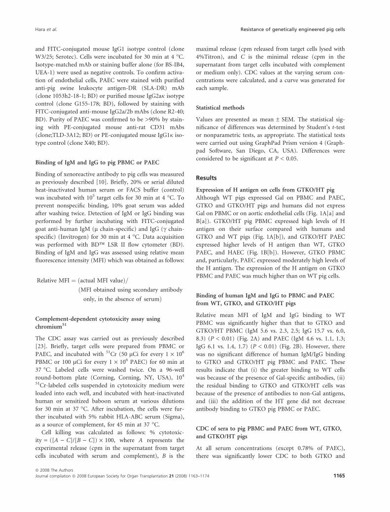
maximal release (cpm released from target cells lysed with
4%Titron), and C is the minimal release (cpm in the
supernatant from target cells incubated with complement
or medium only). CDC values at the varying serum con-
centrations were calculated, and a curve was generated for
each sample.
Statistical methods
Values are presented as mean ± SEM. The statistical sig-
nificance of differences was determined by Student’s t-test
or nonparametric tests, as appropriate. The statistical tests
were carried out using GraphPad Prism version 4 (Graph-
pad Software, San Diego, CA, USA). Differences were
considered to be significant at P < 0.05.
Results
Expression of H antigen on cells from GTKO/HT pig
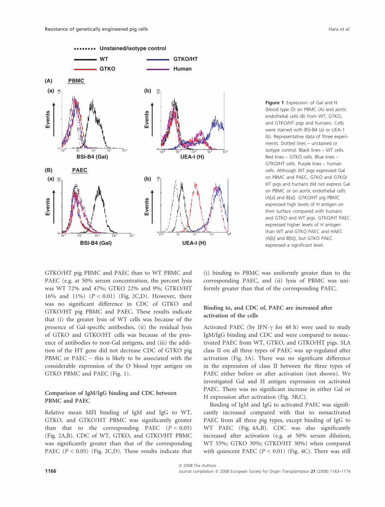
Although WT pigs expressed Gal on PBMC and PAEC,
GTKO and GTKO/HT pigs and humans did not express
Gal on PBMC or on aortic endothelial cells (Fig. 1A[a] and
B[a]). GTKO/HT pig PBMC expressed high levels of H
antigen on their surface compared with humans and
GTKO and WT pigs (Fig. 1A[b]), and GTKO/HT PAEC
expressed higher levels of H antigen than WT, GTKO
PAEC, and HAEC (Fig. 1B[b]). However, GTKO PBMC
and, particularly, PAEC expressed moderately high levels of
the H antigen. The expression of the H antigen on GTKO
PBMC and PAEC was much higher than on WT pig cells.
Binding of human IgM and IgG to PBMC and PAEC
from WT, GTKO, and GTKO/HT pigs
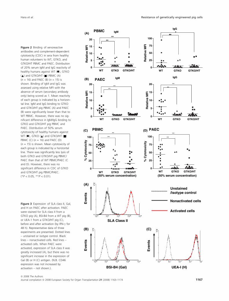
Relative mean MFI of IgM and IgG binding to WT
PBMC was significantly higher than that to GTKO and
GTKO/HT PBMC (IgM 5.6 vs. 2.3, 2.5; IgG 15.7 vs. 6.0,
8.3) (P < 0.01) (Fig. 2A) and PAEC (IgM 4.6 vs. 1.1, 1.3;
IgG 6.1 vs. 1.4, 1.7) (P < 0.01) (Fig. 2B). However, there
was no significant difference of human IgM/IgG binding
to GTKO and GTKO/HT pig PBMC and PAEC. These
results indicate that (i) the greater binding to WT cells
was because of the presence of Gal-specific antibodies, (ii)
the residual binding to GTKO and GTKO/HT cells was
because of the presence of antibodies to non-Gal antigens,
and (iii) the addition of the HT gene did not decrease
antibody binding to GTKO pig PBMC or PAEC.
CDC of sera to pig PBMC and PAEC from WT, GTKO,
and GTKO/HT pigs
At all serum concentrations (except 0.78% of PAEC),
there was significantly lower CDC to both GTKO and
Hara et al. Resistance of genetically engineered pig cells
ª 2008 The Authors
Journal compilation ª 2008 European Society for Organ Transplantation 21 (2008) 1163–1174 1165
GTKO/HT pig PBMC and PAEC than to WT PBMC and
PAEC (e.g. at 50% serum concentration, the percent lysis
was WT 72% and 47%; GTKO 22% and 9%; GTKO/HT
16% and 11%) (P < 0.01) (Fig. 2C,D). However, there
was no significant difference in CDC of GTKO and
GTKO/HT pig PBMC and PAEC. These results indicate
that (i) the greater lysis of WT cells was because of the
presence of Gal-specific antibodies, (ii) the residual lysis
of GTKO and GTKO/HT cells was because of the pres-
ence of antibodies to non-Gal antigens, and (iii) the addi-
tion of the HT gene did not decrease CDC of GTKO pig
PBMC or PAEC – this is likely to be associated with the
considerable expression of the O blood type antigen on
GTKO PBMC and PAEC (Fig. 1).
Comparison of IgM/IgG binding and CDC between
PBMC and PAEC
Relative mean MFI binding of IgM and IgG to WT,
GTKO, and GTKO/HT PBMC was significantly greater
than that to the corresponding PAEC (P < 0.05)
(Fig. 2A,B). CDC of WT, GTKO, and GTKO/HT PBMC
was significantly greater than that of the corresponding
PAEC (P < 0.05) (Fig. 2C,D). These results indicate that
(i) binding to PBMC was uniformly greater than to the
corresponding PAEC, and (ii) lysis of PBMC was uni-
formly greater than that of the corresponding PAEC.
Binding to, and CDC of, PAEC are increased after
activation of the cells
Activated PAEC (by IFN-c for 48 h) were used to study
IgM/IgG binding and CDC and were compared to nonac-
tivated PAEC from WT, GTKO, and GTKO/HT pigs. SLA
class II on all three types of PAEC was up-regulated after
activation (Fig. 3A). There was no significant difference
in the expression of class II between the three types of
PAEC either before or after activation (not shown). We
investigated Gal and H antigen expression on activated
PAEC. There was no significant increase in either Gal or
H expression after activation (Fig. 3B,C).
Binding of IgM and IgG to activated PAEC was signifi-
cantly increased compared with that to nonactivated
PAEC from all three pig types, except binding of IgG to
WT PAEC (Fig. 4A,B). CDC was also significantly
increased after activation (e.g. at 50% serum dilution,
WT 55%; GTKO 30%; GTKO/HT 30%) when compared
with quiescent PAEC (P < 0.01) (Fig. 4C). There was still
PBMC (A)
(a) (b)
(a) (b)
(B) PAEC
WT
GTKO
Unstained/isotype control
Human
GTKO/HT
BSI-B4 (Gal) UEA-I (H)
BSI-B4 (Gal)
Eve
nts
UEA-I (H)
Eve
nts
Eve
nts
Eve
nts
Figure 1 Expression of Gal and H
(blood type O) on PBMC (A) and aortic
endothelial cells (B) from WT, GTKO,
and GTKO/HT pigs and humans. Cells
were stained with BSI-B4 (a) or UEA-1
(b). Representative data of three experi-
ments. Dotted lines – unstained or
isotype control. Black lines – WT cells.
Red lines – GTKO cells. Blue lines –
GTKO/HT cells. Purple lines – human
cells. Although WT pigs expressed Gal
on PBMC and PAEC, GTKO and GTKO/
HT pigs and humans did not express Gal
on PBMC or on aortic endothelial cells
(A[a] and B[a]). GTKO/HT pig PBMC
expressed high levels of H antigen on
their surface compared with humans
and GTKO and WT pigs. GTKO/HT PAEC
expressed higher levels of H antigen
than WT and GTKO PAEC and HAEC
(A[b] and B[b]), but GTKO PAEC
expressed a significant level.
Resistance of genetically engineered pig cells Hara et al.
ª 2008 The Authors
1166 Journal compilation ª 2008 European Society for Organ Transplantation 21 (2008) 1163–1174
IgMIgG
***PBMC
10
15
20
(A)
(B)
(C) (D)
50
75
100**
****
0
5
0
25Rel
ativ
e M
FI
WT
Rel
ativ
e M
FI
PAEC
15
20
15
20**
**
IgM IgG
****
0
5
10
Rel
ativ
e M
FI
0
5
10
Rel
ativ
e M
FI
100**
**
PAECPBMC
100**
**
25
50
75
25
50
75
0
% C
yto
toxi
city
(50% serum concentration)
0
% C
yto
toxi
city
(50% serum concentration)
GTKO GTKO/HTWT GTKO GTKO/HT
WT GTKO GTKO/HTWT GTKO GTKO/HT
WT GTKO GTKO/HTWT GTKO GTKO/HT
Figure 2 Binding of xenoreactive
antibodies and complement-dependent
cytotoxicity (CDC) in sera from healthy
human volunteers to WT, GTKO, and
GTKO/HT PBMC and PAEC. Distribution
of 20% serum IgM and IgG reactivity of
healthy humans against WT (d), GTKO
( ) and GTKO/HT ( ) PBMC (A)
(n = 16) and PAEC (B) (n = 15) is
shown. Binding of IgM and IgG was
assessed using relative MFI with the
absence of serum (secondary antibody
only) being scored as 1. Mean reactivity
of each group is indicated by a horizon-
tal line. IgM and IgG binding to GTKO
and GTKO/HT pig PBMC (A) and PAEC
(B) were significantly lower than that to
WT PBMC. However, there was no sig-
nificant difference in IgM/IgG binding to
GTKO and GTKO/HT pig PBMC and
PAEC. Distribution of 50% serum
cytotoxicity of healthy humans against
WT (d), GTKO ( ) and GTKO/HT ( )
PBMC (C) (n = 16) and PAEC (D)
(n = 15) is shown. Mean cytotoxicity of
each group is indicated by a horizontal
line. There was significantly less lysis of
both GTKO and GTKO/HT pig PBMC/
PAEC than that of WT PBMC/PAEC (C
and D). However, there was no
significant difference in CDC of GTKO
and GTKO/HT pig PBMC/PAEC.
(*P < 0.05, **P < 0.01).
Eve
nts
BSI-B4 (Gal)
Eve
nts
UEA-I (H)
Nonactivated cells
Activated cells
Unstained/isotype control
SLA Class II
(A)
(B) (C)
Eve
nts
Figure 3 Expression of SLA class II, Gal,
and H on PAEC after activation. PAEC
were stained for SLA class II from a
GTKO pig (A), BSI-B4 from a WT pig (B),
or UEA-1 from a GTKO/HT pig (C),
before and after activation (by IFN-c for
48 h). Representative data of three
experiments are presented. Dotted lines
– unstained or isotype control. Black
lines – nonactivated cells. Red lines –
activated cells. When PAEC were
activated, expression of SLA class II was
greatly increased (A), but there was no
significant increase in the expression of
Gal (B) or H (C) antigen. (N.B. CD46
expression was not increased by
activation – not shown.).
Hara et al. Resistance of genetically engineered pig cells
ª 2008 The Authors
Journal compilation ª 2008 European Society for Organ Transplantation 21 (2008) 1163–1174 1167
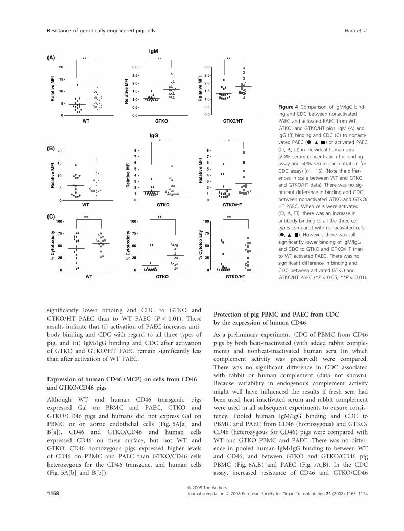
significantly lower binding and CDC to GTKO and
GTKO/HT PAEC than to WT PAEC (P < 0.01). These
results indicate that (i) activation of PAEC increases anti-
body binding and CDC with regard to all three types of
pig, and (ii) IgM/IgG binding and CDC after activation
of GTKO and GTKO/HT PAEC remain significantly less
than after activation of WT PAEC.
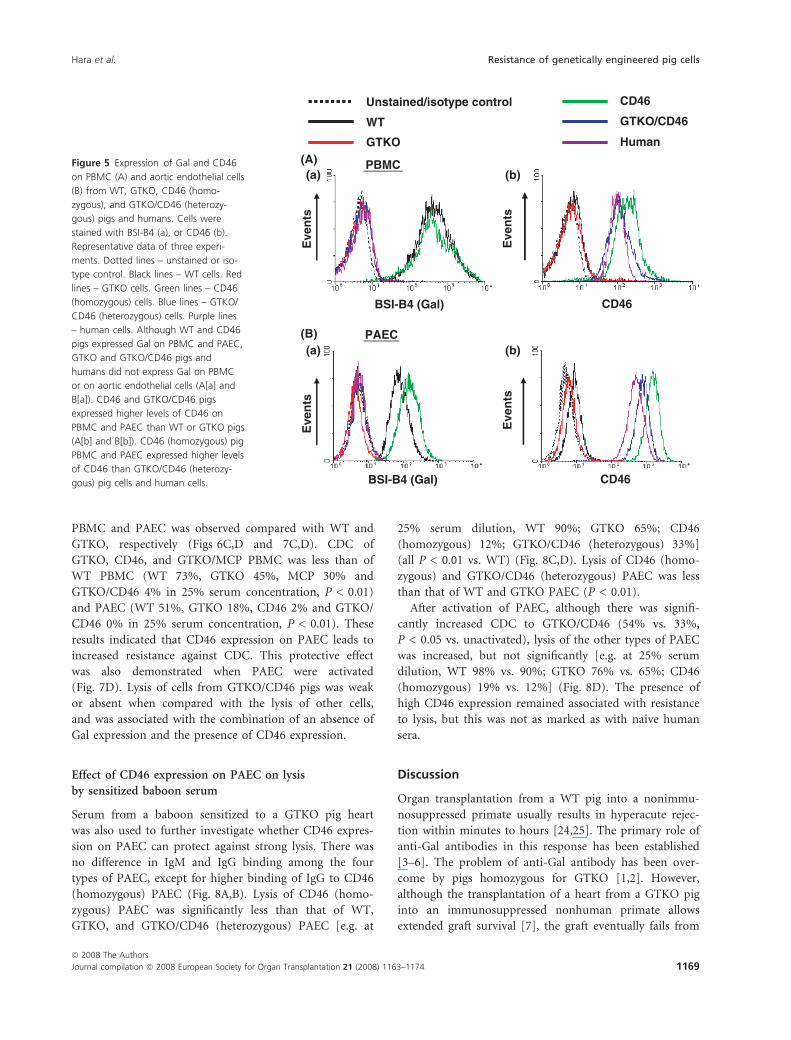
Expression of human CD46 (MCP) on cells from CD46
and GTKO/CD46 pigs
Although WT and human CD46 transgenic pigs
expressed Gal on PBMC and PAEC, GTKO and
GTKO/CD46 pigs and humans did not express Gal on
PBMC or on aortic endothelial cells (Fig. 5A[a] and
B[a]). CD46 and GTKO/CD46 and human cells
expressed CD46 on their surface, but not WT and
GTKO. CD46 homozygous pigs expressed higher levels
of CD46 on PBMC and PAEC than GTKO/CD46 cells
heterozygous for the CD46 transgene, and human cells
(Fig. 5A[b] and B[b]).
Protection of pig PBMC and PAEC from CDC
by the expression of human CD46
As a preliminary experiment, CDC of PBMC from CD46
pigs by both heat-inactivated (with added rabbit comple-
ment) and nonheat-inactivated human sera (in which
complement activity was preserved) were compared.
There was no significant difference in CDC associated
with rabbit or human complement (data not shown).
Because variability in endogenous complement activity
might well have influenced the results if fresh sera had
been used, heat-inactivated serum and rabbit complement
were used in all subsequent experiments to ensure consis-
tency. Pooled human IgM/IgG binding and CDC to
PBMC and PAEC from CD46 (homozygous) and GTKO/
CD46 (heterozygous for CD46) pigs were compared with
WT and GTKO PBMC and PAEC. There was no differ-
ence in pooled human IgM/IgG binding to between WT
and CD46, and between GTKO and GTKO/CD46 pig
PBMC (Fig. 6A,B) and PAEC (Fig. 7A,B). In the CDC
assay, increased resistance of CD46 and GTKO/CD46
IgM
10
15
20
(A)
(B)
(C)
1.5
2.0
2.5
3.0
1.5
2.0
2.5
3.0
** ****
WT GTKO GTKO/HT0
5
0.0
0.5
1.0
0.0
0.5
1.0
15
20
6
7
8
6
7
8
**IgG
Rel
ativ
e M
FI
Rel
ativ
e M
FI
Rel
ativ
e M
FI
Rel
ativ
e M
FI
Rel
ativ
e M
FI
Rel
ativ
e M
FI
5
10
1
2
3
4
5
1
2
3
4
5
WT GTKO GTKO/HT0 0 0
100 100 100** ****
25
50
75
25
50
75
25
50
75
0
% C
yto
toxi
city
% C
yto
toxi
city
% C
yto
toxi
city
0 0
GTKO/HTGTKOWT
Figure 4 Comparison of IgM/IgG bind-
ing and CDC between nonactivated
PAEC and activated PAEC from WT,
GTKO, and GTKO/HT pigs. IgM (A) and
IgG (B) binding and CDC (C) to nonacti-
vated PAEC (d, , ) or activated PAEC
(s, D, h) in individual human sera
(20% serum concentration for binding
assay and 50% serum concentration for
CDC assay) (n = 15). (Note the differ-
ences in scale between WT and GTKO
and GTKO/HT data). There was no sig-
nificant difference in binding and CDC
between nonactivated GTKO and GTKO/
HT PAEC. When cells were activated
(s, D, h), there was an increase in
antibody binding to all the three cell
types compared with nonactivated cells
(d, , ). However, there was still
significantly lower binding of IgM/IgG
and CDC to GTKO and GTKO/HT than
to WT activated PAEC. There was no
significant difference in binding and
CDC between activated GTKO and
GTKO/HT PAEC (*P < 0.05, **P < 0.01).
Resistance of genetically engineered pig cells Hara et al.
ª 2008 The Authors
1168 Journal compilation ª 2008 European Society for Organ Transplantation 21 (2008) 1163–1174
PBMC and PAEC was observed compared with WT and
GTKO, respectively (Figs 6C,D and 7C,D). CDC of
GTKO, CD46, and GTKO/MCP PBMC was less than of
WT PBMC (WT 73%, GTKO 45%, MCP 30% and
GTKO/CD46 4% in 25% serum concentration, P < 0.01)
and PAEC (WT 51%, GTKO 18%, CD46 2% and GTKO/
CD46 0% in 25% serum concentration, P < 0.01). These
results indicated that CD46 expression on PAEC leads to
increased resistance against CDC. This protective effect
was also demonstrated when PAEC were activated
(Fig. 7D). Lysis of cells from GTKO/CD46 pigs was weak
or absent when compared with the lysis of other cells,
and was associated with the combination of an absence of
Gal expression and the presence of CD46 expression.
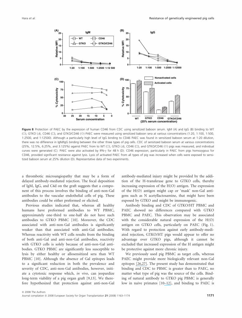
Effect of CD46 expression on PAEC on lysis
by sensitized baboon serum
Serum from a baboon sensitized to a GTKO pig heart
was also used to further investigate whether CD46 expres-
sion on PAEC can protect against strong lysis. There was
no difference in IgM and IgG binding among the four
types of PAEC, except for higher binding of IgG to CD46
(homozygous) PAEC (Fig. 8A,B). Lysis of CD46 (homo-
zygous) PAEC was significantly less than that of WT,
GTKO, and GTKO/CD46 (heterozygous) PAEC [e.g. at
25% serum dilution, WT 90%; GTKO 65%; CD46
(homozygous) 12%; GTKO/CD46 (heterozygous) 33%]
(all P < 0.01 vs. WT) (Fig. 8C,D). Lysis of CD46 (homo-
zygous) and GTKO/CD46 (heterozygous) PAEC was less
than that of WT and GTKO PAEC (P < 0.01).
After activation of PAEC, although there was signifi-
cantly increased CDC to GTKO/CD46 (54% vs. 33%,
P < 0.05 vs. unactivated), lysis of the other types of PAEC
was increased, but not significantly [e.g. at 25% serum
dilution, WT 98% vs. 90%; GTKO 76% vs. 65%; CD46
(homozygous) 19% vs. 12%] (Fig. 8D). The presence of
high CD46 expression remained associated with resistance
to lysis, but this was not as marked as with naive human
sera.
Discussion
Organ transplantation from a WT pig into a nonimmu-
nosuppressed primate usually results in hyperacute rejec-
tion within minutes to hours [24,25]. The primary role of
anti-Gal antibodies in this response has been established
[3–6]. The problem of anti-Gal antibody has been over-
come by pigs homozygous for GTKO [1,2]. However,
although the transplantation of a heart from a GTKO pig
into an immunosuppressed nonhuman primate allows
extended graft survival [7], the graft eventually fails from
WT
Unstained/isotype control
GTKO
CD46
Human
GTKO/CD46
Eve
nts
Eve
nts
BSI-B4 (Gal) CD46
PBMC(A)
(B)
(a) (b)
(a) (b)
Eve
nts
Eve
nts
BSI-B4 (Gal) CD46
PAEC
Figure 5 Expression of Gal and CD46
on PBMC (A) and aortic endothelial cells
(B) from WT, GTKO, CD46 (homo-
zygous), and GTKO/CD46 (heterozy-
gous) pigs and humans. Cells were
stained with BSI-B4 (a), or CD46 (b).
Representative data of three experi-
ments. Dotted lines – unstained or iso-
type control. Black lines – WT cells. Red
lines – GTKO cells. Green lines – CD46
(homozygous) cells. Blue lines – GTKO/
CD46 (heterozygous) cells. Purple lines
– human cells. Although WT and CD46
pigs expressed Gal on PBMC and PAEC,
GTKO and GTKO/CD46 pigs and
humans did not express Gal on PBMC
or on aortic endothelial cells (A[a] and
B[a]). CD46 and GTKO/CD46 pigs
expressed higher levels of CD46 on
PBMC and PAEC than WT or GTKO pigs
(A[b] and B[b]). CD46 (homozygous) pig
PBMC and PAEC expressed higher levels
of CD46 than GTKO/CD46 (heterozy-
gous) pig cells and human cells.
Hara et al. Resistance of genetically engineered pig cells
ª 2008 The Authors
Journal compilation ª 2008 European Society for Organ Transplantation 21 (2008) 1163–1174 1169
2525
IgM(A) (B)
(D)(C)
IgG
5
10
15
20
Rel
ativ
e M
FI
5
10
15
20
Rel
ativ
e M
FI
00
1:20
WT CD46
100 100**
**** *
25
50
75
% C
yto
toxi
city
25
50
75
% C
yto
toxi
city
*
GTKO GTKO/CD46
GTKO GTKO/CD46
0510152025300 0
(25% serum concentration)Serum concentrationCD46WT
1:100 1:500 1:2500 1:12 500 1:20 1:100 1:500 1:2500 1:12 500
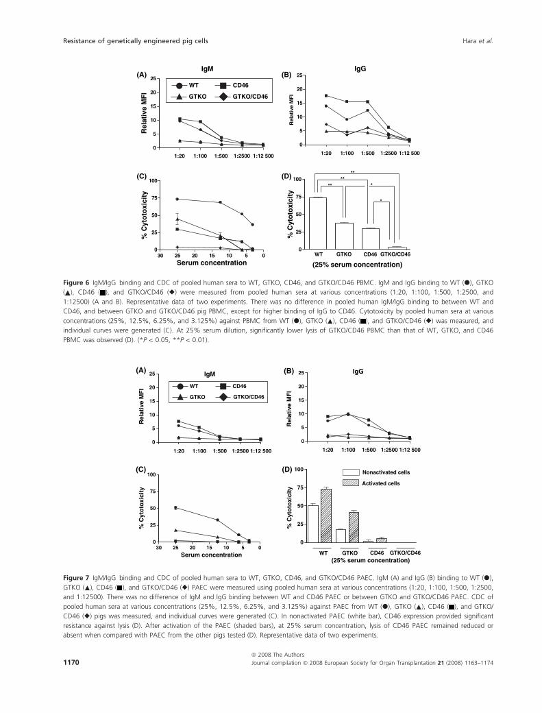
Figure 6 IgM/IgG binding and CDC of pooled human sera to WT, GTKO, CD46, and GTKO/CD46 PBMC. IgM and IgG binding to WT (d), GTKO
( ), CD46 ( ), and GTKO/CD46 (r) were measured from pooled human sera at various concentrations (1:20, 1:100, 1:500, 1:2500, and
1:12500) (A and B). Representative data of two experiments. There was no difference in pooled human IgM/IgG binding to between WT and
CD46, and between GTKO and GTKO/CD46 pig PBMC, except for higher binding of IgG to CD46. Cytotoxicity by pooled human sera at various
concentrations (25%, 12.5%, 6.25%, and 3.125%) against PBMC from WT (d), GTKO ( ), CD46 ( ), and GTKO/CD46 (r) was measured, and
individual curves were generated (C). At 25% serum dilution, significantly lower lysis of GTKO/CD46 PBMC than that of WT, GTKO, and CD46
PBMC was observed (D). (*P < 0.05, **P < 0.01).
20
25 IgM
20
25 IgG
0
5
10
15
0
5
10
15
Rel
ativ
e M
FI
Rel
ativ
e M
FI WT CD46
1:1001:20
75
100100
25
50
% C
yto
toxi
city
25
50
75
% C
yto
toxi
city
Nonactivated cells
Activated cells
WT
0
(25% serum concentration)
0510152025300
Serum concentration GTKO CD46 GTKO/CD46
1:500 1:2500 1:12 5001:1001:20 1:500 1:2500 1:12 500
GTKO
(A) (B)
(C) (D)
GTKO/CD46
Figure 7 IgM/IgG binding and CDC of pooled human sera to WT, GTKO, CD46, and GTKO/CD46 PAEC. IgM (A) and IgG (B) binding to WT (d),
GTKO ( ), CD46 ( ), and GTKO/CD46 (r) PAEC were measured using pooled human sera at various concentrations (1:20, 1:100, 1:500, 1:2500,
and 1:12500). There was no difference of IgM and IgG binding between WT and CD46 PAEC or between GTKO and GTKO/CD46 PAEC. CDC of
pooled human sera at various concentrations (25%, 12.5%, 6.25%, and 3.125%) against PAEC from WT (d), GTKO ( ), CD46 ( ), and GTKO/
CD46 (r) pigs was measured, and individual curves were generated (C). In nonactivated PAEC (white bar), CD46 expression provided significant
resistance against lysis (D). After activation of the PAEC (shaded bars), at 25% serum concentration, lysis of CD46 PAEC remained reduced or
absent when compared with PAEC from the other pigs tested (D). Representative data of two experiments.
Resistance of genetically engineered pig cells Hara et al.
ª 2008 The Authors
1170 Journal compilation ª 2008 European Society for Organ Transplantation 21 (2008) 1163–1174
a thrombotic microangiopathy that may be a form of
delayed antibody-mediated rejection. The focal deposition
of IgM, IgG, and C4d on the graft suggests that a compo-
nent of this process involves the binding of anti-non-Gal
antibodies to the vascular endothelial cells of pig. These
antibodies could be either preformed or elicited.
Previous studies indicated that, whereas all healthy
humans have preformed antibodies to WT PBMC,
approximately one-third to one-half do not have such
antibodies to GTKO PBMC [10]. Moreover, the CDC
associated with anti-non-Gal antibodies is significantly
weaker than that associated with anti-Gal antibodies.
Whereas reactivity with WT cells results from the binding
of both anti-Gal and anti-non-Gal antibodies, reactivity
with GTKO cells is solely because of anti-non-Gal anti-
bodies. GTKO PBMC are significantly less susceptible to
lysis by either healthy or allosensitized sera than WT
PBMC [10]. Although the absence of Gal epitopes leads
to a significant reduction in both the prevalence and
severity of CDC, anti-non-Gal antibodies, however, initi-
ate a cytotoxic response which, in vivo, can jeopardize
long-term viability of a pig organ graft [9,13]. We there-
fore hypothesized that protection against anti-non-Gal
antibody-mediated injury might be provided by the addi-
tion of the H-transferase gene to GTKO cells, thereby
increasing expression of the H(O) antigen. The expression
of the H(O) antigen might cap or ‘mask’ non-Gal anti-
gens such as N acetyllactosamine, that might have been
exposed by GTKO and might be immunogenic.
Antibody binding and CDC of GTKO/HT PBMC and
PAEC showed no differences compared with GTKO
PBMC and PAEC. This observation may be associated
with the considerable natural expression of the H(O)
antigen on GTKO cells, particularly on PAEC (Fig. 1).
With regard to protection against early antibody-medi-
ated rejection, GTKO/HT pigs would appear to offer no
advantage over GTKO pigs, although it cannot be
excluded that increased expression of the H antigen might
be protective against more chronic injury.
We previously used pig PBMC as target cells, whereas
PAEC might provide more biologically relevant non-Gal
epitopes [26,27]. The present study has demonstrated that
binding and CDC to PBMC is greater than to PAEC, no
matter what type of pig was the source of the cells. Bind-
ing of natural antibody to GTKO pig PBMC is generally
low in naive primates [10–12], and binding to PAEC is
20
25 IgM(A) (B)
(C) (D)
20
25 IgG
5
10
15
Rel
ativ
e M
FI
5
10
15
Rel
ativ
e M
FI
WT CD46
01:1001:20
0
100100
25
50
75
% C
yto
toxi
city
25
50
75
% C
yto
toxi
city
GTKO GTKO/CD46
0510152025300
Nonactivated cells
0
WT(25% serum concentration)
Serum concentration
Activated cells
1:500 1:2500 1:12 500 1:1001:20 1:500 1:2500 1:12500
GTKO CD46 GTKO/CD46
Figure 8 Protection of PAEC by the expression of human CD46 from CDC using sensitized baboon serum. IgM (A) and IgG (B) binding to WT
(s), GTKO (D), CD46 (h), and GTKO/CD46 (e) PAEC were measured using sensitized baboon sera at various concentrations (1:20, 1:100, 1:500,
1:2500, and 1:12500). Although a particularly high level of IgG binding to CD46 PAEC was found in sensitized baboon serum at 1:20 dilution,
there was no difference in IgM/IgG binding between the other three types of pig cells. CDC of sensitized baboon serum at various concentrations
(25%, 12.5%, 6.25%, and 3.125%) against PAEC from to WT (s), GTKO (D), CD46 (h), and GTKO/CD46 (e) pigs was measured, and individual
curves were generated (C). PAEC were also activated by IFN-c for 48 h (D). CD46 expression, particularly in PAEC from pigs homozygous for
CD46, provided significant resistance against lysis. Lysis of activated PAEC from all types of pig was increased when cells were exposed to sensi-
tized baboon serum at 25% dilution (D). Representative data of two experiments.
Hara et al. Resistance of genetically engineered pig cells
ª 2008 The Authors
Journal compilation ª 2008 European Society for Organ Transplantation 21 (2008) 1163–1174 1171
even lower. PBMC rather than PAEC might therefore
prove preferable target cells to test antibody binding and
CDC.
Although CD59 limits the formation of the terminal
membrane attack complex [28], CD55 and CD46 may be
the most ‘useful’’ transgenes for xenotransplantation.
CD55 is generally considered to control the classical com-
plement activation pathway more effectively, and CD46
the alternative activation pathway. However, CD46 is
effective in protecting cells against CDC by both the clas-
sical and alternative pathways [29,30]. Its ability to con-
trol alternative pathway activation suggests that, as innate
immune responses might be particularly important, it
may be preferred over CD55. Alternatively, CD46 should
be used in combination with CD55 [31].
The PBMC and PAEC from CD46 pigs that were used
in the present study expressed higher levels of CD46 than
humans and other pigs. PAEC from pigs homozygous for
CD46 showed a greater resistance to CDC than did WT
PAEC, and the level of resistance was equivalent to that
demonstrated by GTKO and GTKO/HT PAEC, even
though the binding of human IgM and IgG to PAEC
from CD46 pigs was significantly higher than to GTKO
and GTKO/HT PAEC, likely because of anti-Gal antibody
binding to the CD46 cells. The expression of CD46 in
GTKO pigs is therefore likely to provide further protec-
tion from CDC.
We found less lysis by human sera of GTKO/CD46
(heterozygous for CD46) pig PBMC and PAEC than by
that of WT, GTKO, and CD46 (homozygous) cells. How-
ever, GTKO/CD46 (heterozygous) PAEC were less resis-
tant to lysis compared with CD46 (homozygous) PAEC
when serum from a baboon sensitized to non-Gal anti-
gens (after exposure to a GTKO pig heart) was tested.
This is almost certainly related to the increased expression
of CD46 in the homozygous animal (Fig. 5A[b] and
B[b]). A possible alternative explanation is that GTKO
cells may express more non-Gal antigens than do WT
cells, leading to increased antibody binding and lysis to
these specific antigens that were present in greater num-
bers on GTKO/CD46 cells than on CD46 cells. Further-
more, the GTKO pigs were from a different founder herd
than the MCP pigs; the GTKO/CD46 pigs were a mixture
of these two herds. As SLA class I and II expression is
likely to be different between the GTKO and CD46
strains, sensitization to a GTKO pig heart could result in
a specific spectrum of anti-SLA antibodies that might be
more reactive to the GTKO/CD46 (heterozygous) cells
than to the CD46 (homozygous) cells. However, lysis of
GTKO/CD46 PAEC by sensitized baboon serum was still
less than that of GTKO PAEC. GTKO pigs homozygous
for CD46 are likely to be more resistant to the effects of
sensitized serum.
We have previously demonstrated that antibody bind-
ing and CDC to GTKO PBMC in naive baboon sera are
similar to naı̈ve human sera [10,11]. In the present study,
binding and CDC associated with sensitized baboon
serum were documented. Although expression of CD46
on GTKO pig cells only partially protected the cells from
CDC caused by sensitized baboon serum, we would antic-
ipate that this genetic combination would be significantly
more protective in the presence of naive baboon serum.
Activation of PAEC (by IFN-c) resulted in increased
antibody binding and CDC of cells from all pig types.
This would increase immune injury of the graft. However,
GTKO, GTKO/HT, CD46, and GTKO/CD46 PAEC
remained at an advantage over WT PAEC as IgM/IgG
binding and CDC, although increased over nonactivated
state, remained significantly less compared with WT
PAEC.
Our study suggests that GTKO pigs transgenic for
human CD46 are likely to be less susceptible to human
serum CDC as a result of both reduced binding of anti-
body and increased resistance to complement-mediated
injury. However, if an elicited antibody response devel-
ops, even a graft from GTKO/CD46 pig may not be pro-
tected from CDC. Our ongoing in vivo studies using
organs from GTKO/CD46 pigs in baboons indicate that,
although completely resistant to hyperacute rejection
(even in the absence of agents such as cobra venom
factor), the grafts develop thrombotic microangiopathy
that is associated with the development of a consumptive
coagulopathy in the recipient nonhuman primate
(M. Ezzelarab, unpublished data). Therefore, additional
genes need to be expressed in the organ-source pig. An
‘anticoagulant/anti-thrombotic’ gene, such as human
tissue factor pathway inhibitor or CD39, is likely to be
beneficial [32,33].
Authorship
HH: designed the study, performed the research, collected
and analyzed the data, and wrote the paper. CL, YJL, H-
CT and ME: collaborated with HH in performing the
research, and have read the paper and agreed with its
conclusions. DA: contributed ideas for the research and
provided samples, such as blood, from the genetically
modified pigs that have been generated by his group. He
also contributed to the writing of the manuscript. DKC:
contributed ideas for the research, and supervised the
experiments and the writing of the manuscript.
Acknowledgements
H. Hara, M.D., Ph.D. was a recipient of a grant from
Uehara Memorial Foundation, Japan and Kawasaki
Resistance of genetically engineered pig cells Hara et al.
ª 2008 The Authors
1172 Journal compilation ª 2008 European Society for Organ Transplantation 21 (2008) 1163–1174
Medical School Alumni Association Foundation, Japan.
Work in our laboratory was supported in part by NIH
grants #U01AI068642 and R21-A1074844 and by a Spon-
sored Research Agreement between the University of
Pittsburgh and Revivicor, Inc., Blacksburg, VA, USA.
References
1. Phelps CJ, Koike C, Vaught TD, et al. Production of alpha
1,3-galactosyltransferase-deficient pigs. Science 2003; 299:
411.
2. Kolber-Simonds D, Lai L, Watt SR, et al. Production of
alpha-1,3-galactosyltransferase null pigs by means of
nuclear transfer with fibroblasts bearing loss of heterozy-
gosity mutations. Proc Natl Acad Sci U S A 2004; 101:
7335.
3. Galili U, Shohet SB, Kobrin E, Stults CL, Macher BA.
Man, apes, and Old World monkeys differ from other
mammals in the expression of alpha-galactosyl epitopes on
nucleated cells. J Biol Chem 1988; 263: 17755.
4. Good AH, Cooper DK, Malcolm AJ, et al. Identification of
carbohydrate structures that bind human antiporcine anti-
bodies: implications for discordant xenografting in
humans. Transplant Proc 1992; 24: 559.
5. Cooper DK, Good AH, Koren E, et al. Identification of
alpha-galactosyl and other carbohydrate epitopes that are
bound by human anti-pig antibodies: relevance to dis-
cordant xenografting in man. Transpl Immunol 1993; 1:
198.
6. Collins BH, Cotterell AH, McCurry KR, et al. Cardiac
xenografts between primate species provide evidence
for the importance of the alpha-galactosyl determinant in
hyperacute rejection. J Immunol 1995; 154: 5500.
7. Kuwaki K, Tseng YL, Dor FJ, et al. Heart transplantation
in baboons using alpha1,3-galactosyltransferase gene-
knockout pigs as donors: initial experience. Nat Med 2005;
11: 29.
8. Yamada K, Yazawa K, Shimizu A, et al. Marked prolonga-
tion of porcine renal xenograft survival in baboons
through the use of alpha1,3-galactosyltransferase gene-
knockout donors and the cotransplantation of vascularized
thymic tissue. Nat Med 2005; 11: 32.
9. Chen G, Qian H, Starzl T, et al. Acute rejection is associ-
ated with antibodies to non-Gal antigens in baboons using
Gal-knockout pig kidneys. Nat Med 2005; 11: 1295.
10. Hara H, Ezzelarab M, Rood PP, et al. Allosensitized
humans are at no greater risk of humoral rejection of
GT-KO pig organs than other humans. Xenotransplanta-
tion 2006; 13: 357.
11. Ezzelarab M, Hara H, Busch J, et al. Antibodies directed to
pig non-Gal antigens in naive and sensitized baboons.
Xenotransplantation 2006; 13: 400.
12. Rood PP, Hara H, Busch JL, et al. Incidence and cytotox-
icity of antibodies in cynomolgus monkeys directed to
nonGal antigens, and their relevance for experimental
models. Transpl Int 2006; 19: 158.
13. Ezzelarab M, Garcia B, Azimzadeh A, et al. Innate immune
mechanisms predominate in GT-KO pig organ graft failure
in baboons. Xenotransplantation 2007; 14: 403 (Abstract).
14. Koike C, Kannagi R, Takuma Y, et al. Introduction of
[alpha](1,2)-fucosyltransferase and its effect on [alpha]-Gal
epitopes in transgenic pig. Xenotransplantation 1996; 3: 81.
15. Sharma A, Okabe J, Birch P, et al. Reduction in the level
of Gal(alpha1,3)Gal in transgenic mice and pigs by the
expression of an alpha(1,2)fucosyltransferase. Proc Natl
Acad Sci U S A 1996; 93: 7190.
16. Chen CG, Salvaris EJ, Romanella M, et al. Transgenic
expression of human alpha1,2-fucosyltransferase (H-trans-
ferase) prolongs mouse heart survival in an ex vivo model
of xenograft rejection. Transplantation 1998; 65: 832.
17. Costa C, Zhao L, Burton WV, et al. Expression of the
human alpha1,2-fucosyltransferase in transgenic pigs mod-
ifies the cell surface carbohydrate phenotype and confers
resistance to human serum-mediated cytolysis. FASEB J
1999; 13: 1762.
18. Diamond LE, Quinn CM, Martin MJ, Lawson J, Platt JL,
Logan JS. A human CD46 transgenic pig model system for
the study of discordant xenotransplantation. Transplanta-
tion 2001; 71: 132.
19. Adams DH, Kadner A, Chen RH, Farivar RS. Human
membrane cofactor protein (MCP, CD 46) protects trans-
genic pig hearts from hyperacute rejection in primates.
Xenotransplantation 2001; 8: 36.
20. Huang J, Gou D, Zhen C, et al. Protection of xenogeneic
cells from human complement-mediated lysis by the
expression of human DAF, CD59 and MCP. FEMS Immu-
nol Med Microbiol 2001; 31: 203.
21. Loveland BE, Milland J, Kyriakou P, et al. Characterization
of a CD46 transgenic pig and protection of transgenic
kidneys against hyperacute rejection in non-immunosup-
pressed baboons. Xenotransplantation 2004; 11: 171.
22. Banz Y, Rieben R. Endothelial cell protection in xenotrans-
plantation: looking after a key player in rejection.
Xenotransplantation 2006; 13: 19.
23. Rood PP, Tai HC, Hara H, et al. Late onset of develop-
ment of natural anti-nonGal antibodies in infant humans
and baboons: implications for xenotransplantation in
infants. Transpl Int 2007; 20: 1050.
24. Cooper DK, Human PA, Lexer G, et al. Effects of cyclo-
sporine and antibody adsorption on pig cardiac xenograft
survival in the baboon. J Heart Transplant 1988; 7: 238.
25. Alexandre GP. From ABO-incompatible human kidney
transplantation to xenotransplantation. Xenotransplantation
2004; 11: 233.
26. Baumann BC, Stussi G, Huggel K, Rieben R, Seebach JD.
Reactivity of human natural antibodies to endothelial cells
from Galalpha(1,3)Gal-deficient pigs. Transplantation
2007; 83: 193.
Hara et al. Resistance of genetically engineered pig cells
ª 2008 The Authors
Journal compilation ª 2008 European Society for Organ Transplantation 21 (2008) 1163–1174 1173
27. Chen G, Sun H, Yang H, et al. The role of anti-non-Gal
antibodies in the development of acute humoral xenograft
rejection of hDAF transgenic porcine kidneys in baboons
receiving anti-Gal antibody neutralization therapy. Trans-
plantation 2006; 81: 273.
28. Morgan BP. Regulation of the complement membrane
attack pathway. Crit Rev Immunol 1999; 19: 173.
29. Loveland BE, Johnstone RW, Russell SM, Thorley BR,
McKenzie IF. Different membrane cofactor protein (CD46)
isoforms protect transfected cells against antibody and
complement mediated lysis. Transpl Immunol 1993; 1: 101.
30. Christiansen D, Milland J, Thorley BR, McKenzie IF,
Loveland BE. A functional analysis of recombinant soluble
CD46 in vivo and a comparison with recombinant soluble
forms of CD55 and CD35 in vitro. Eur J Immunol 1996;
26: 578.
31. Brodbeck WG, Mold C, Atkinson JP, Medof ME.
Cooperation between decay-accelerating factor and
membrane cofactor protein in protecting cells from
autologous complement attack. J Immunol 2000; 165:
3999.
32. Crikis S, Cowan PJ, d’Apice AJ. Intravascular thrombosis
in discordant xenotransplantation. Transplantation 2006;
82: 1119.
33. Cooper DK, Dorling A, Pierson III RN, et al.
Alpha1,3-galactosyltransferase gene-knockout pigs for
xenotransplantation: where do we go from here?
Transplantation 2007; 84: 1.
Resistance of genetically engineered pig cells Hara et al.
ª 2008 The Authors
1174 Journal compilation ª 2008 European Society for Organ Transplantation 21 (2008) 1163–1174