Immune response to influenza A(H1N1)v in HIV-infected patients
12
Original Article Immune response to influenza A(H1N1)v in HIV-infected patients Paola Sansonetti 1 , Michela Sali 1 , Massimiliano Fabbiani 2 , Matteo Morandi 1 , Rosa Martucci 1 , Ali Danesh 3 , Giovanni Delogu 1 , Jesus F. Bermejo-Martin 4 , Maurizio Sanguinetti 1 , David Kelvin 3,5,6 , Roberto Cauda 2 , Giovanni Fadda 1 , Salvatore Rubino 5 1 Institute of Microbiology, Catholic University of Sacred Heart, Rome, Italy 2 Institute of Clinical Infectious Diseases, Catholic University of Sacred Heart, Rome, Italy 3 Division of Immunology, International Institute of Infection and Immunity, Shantou University Medical College, Shantou, Guangdong, China 4 Infection and Immunity Medical Investigation Unit (IMI), Microbiology and Immunology Service, Hospital Clínico Universitario-IECSCYL, Valladolid, Spain 5 Department of Biomedical Sciences, University of Sassari, Sassari, Italy 6 Division of Experimental Therapeutics, University Health Network, Toronto, Ontario, Canada Abstract Introduction: HIV infection is considered a risk factor for severe outcomes of influenza A(H1N1)v infection. However, data on immune response against influenza A(H1N1)v virus in HIV-infected patients are lacking. Methodology: Data from seven HIV-positive and 14 HIV-negative patients infected with A(H1N1)v and from 23 HIV-positive and six HIV- negative asymptomatic controls were analyzed to evaluate the clinical picture, A(H1N1)v viral shedding, and the immune response against the virus. Results: Patients displayed mainly upper respiratory tract diseases (57.1%), while pneumonia was diagnosed only in HIV-negative patients (23.8% of subjects, of which 4.8% required intensive care unit admission). At day seven, 29% of HIV-infected patients were still positive for A(H1N1)v by RT-PCR on nasopharyngeal swabs. Interestingly, a persistence of CXCL10 secretion at high level and lower IL-6 levels was observed in HIV-positive subjects. The geometric mean haemagglutination inhibition titer (HI-GMT) and anti-influenza IgM levels were lower in HIV-positive individuals while anti-influenza IgG levels remained similar in the two groups. Conclusions: The immune impairment due to HIV infection could affect A(H1N1)v clearance and could lead to a lower antibody response and a persistent secretion of CXCL10 at high levels. However, the lower IL-6 secretion and treatment with highly active antiretroviral therapy (HAART) could result in a milder clinical picture. Key words: HIV; influenza A(H1N1)v 2009; cytokines; chemokines; antibody response J Infect Dev Ctries 2014; 8(1):101-109. doi:10.3855/jidc.3147 (Received 26 November 2012 – Accepted 13 October 2013) Copyright © 2014 Sansonetti et al. This is an open-access article distributed under the Creative Commons Attribution License, which permits unrestricted use, distribution, and reproduction in any medium, provided the original work is properly cited. Introduction On 11 June 2009, the World Health Organization (WHO) gave a pandemic alert in response to the global spread of a novel influenza virus. Since the first human case of infection, the novel pandemic influenza A (H1N1) virus, then named influenza A (H1N1)v virus, rapidly spread in US and Mexico, leading to global transmission through Europe [1]. The new virus was the result of a triple reassortment that combined gene segments from the North America and Eurasian swine H1N1 lineage; its historical origin can be located in 1918, when a virus of avian origin infected humans, causing the Spanish flu outbreak [2]. The distinctive combination of gene segments gives (H1N1)v antigenic features that make it dissimilar from seasonal influenza A [3]. The function of host immune response to eliminating (H1N1)v or the role of host immune response in contributing to respiratory illness of (H1N1)v infections is not clear. Studies on SARS CoV and influenza A (H5N1) have previously identified specific host immune signatures [4]. These studies also included (H1N1)v and showed that in the early phase of the infection, most of the mild outcomes are characterized by the expression of systemic levels of chemokines, such as CXCL10, that are associated with the innate antiviral response [5]. Although (H1N1)v generally leads to mild infection symptoms, thousands of deaths have been
-
Upload
independent -
Category
Documents
-
view
7 -
download
0
Transcript of Immune response to influenza A(H1N1)v in HIV-infected patients
Original Article
Immune response to influenza A(H1N1)v in HIV-infected patients Paola Sansonetti1, Michela Sali1, Massimiliano Fabbiani2, Matteo Morandi1, Rosa Martucci1, Ali Danesh3, Giovanni Delogu1, Jesus F. Bermejo-Martin4, Maurizio Sanguinetti1, David Kelvin3,5,6, Roberto Cauda2, Giovanni Fadda1, Salvatore Rubino5 1 Institute of Microbiology, Catholic University of Sacred Heart, Rome, Italy
2 Institute of Clinical Infectious Diseases, Catholic University of Sacred Heart, Rome, Italy
3 Division of Immunology, International Institute of Infection and Immunity, Shantou University Medical College,
Shantou, Guangdong, China 4 Infection and Immunity Medical Investigation Unit (IMI), Microbiology and Immunology Service, Hospital Clínico
Universitario-IECSCYL, Valladolid, Spain 5 Department of Biomedical Sciences, University of Sassari, Sassari, Italy
6 Division of Experimental Therapeutics, University Health Network, Toronto, Ontario, Canada
Abstract Introduction: HIV infection is considered a risk factor for severe outcomes of influenza A(H1N1)v infection. However, data on immune
response against influenza A(H1N1)v virus in HIV-infected patients are lacking.
Methodology: Data from seven HIV-positive and 14 HIV-negative patients infected with A(H1N1)v and from 23 HIV-positive and six HIV-
negative asymptomatic controls were analyzed to evaluate the clinical picture, A(H1N1)v viral shedding, and the immune response against
the virus.
Results: Patients displayed mainly upper respiratory tract diseases (57.1%), while pneumonia was diagnosed only in HIV-negative patients
(23.8% of subjects, of which 4.8% required intensive care unit admission). At day seven, 29% of HIV-infected patients were still positive for
A(H1N1)v by RT-PCR on nasopharyngeal swabs. Interestingly, a persistence of CXCL10 secretion at high level and lower IL-6 levels was
observed in HIV-positive subjects. The geometric mean haemagglutination inhibition titer (HI-GMT) and anti-influenza IgM levels were
lower in HIV-positive individuals while anti-influenza IgG levels remained similar in the two groups.
Conclusions: The immune impairment due to HIV infection could affect A(H1N1)v clearance and could lead to a lower antibody response
and a persistent secretion of CXCL10 at high levels. However, the lower IL-6 secretion and treatment with highly active antiretroviral
therapy (HAART) could result in a milder clinical picture.
Key words: HIV; influenza A(H1N1)v 2009; cytokines; chemokines; antibody response J Infect Dev Ctries 2014; 8(1):101-109. doi:10.3855/jidc.3147
(Received 26 November 2012 – Accepted 13 October 2013)
Copyright © 2014 Sansonetti et al. This is an open-access article distributed under the Creative Commons Attribution License, which permits unrestricted use,
distribution, and reproduction in any medium, provided the original work is properly cited.
Introduction On 11 June 2009, the World Health Organization
(WHO) gave a pandemic alert in response to the
global spread of a novel influenza virus. Since the first
human case of infection, the novel pandemic influenza
A (H1N1) virus, then named influenza A (H1N1)v
virus, rapidly spread in US and Mexico, leading to
global transmission through Europe [1]. The new virus
was the result of a triple reassortment that combined
gene segments from the North America and Eurasian
swine H1N1 lineage; its historical origin can be
located in 1918, when a virus of avian origin infected
humans, causing the Spanish flu outbreak [2]. The
distinctive combination of gene segments gives
(H1N1)v antigenic features that make it dissimilar
from seasonal influenza A [3].
The function of host immune response to
eliminating (H1N1)v or the role of host immune
response in contributing to respiratory illness of
(H1N1)v infections is not clear. Studies on SARS CoV
and influenza A (H5N1) have previously identified
specific host immune signatures [4]. These studies also
included (H1N1)v and showed that in the early phase
of the infection, most of the mild outcomes are
characterized by the expression of systemic levels of
chemokines, such as CXCL10, that are associated with
the innate antiviral response [5].
Although (H1N1)v generally leads to mild
infection symptoms, thousands of deaths have been
Sansonetti et al. – Influenza A(H1N1)v in HIV-infected patients J Infect Dev Ctries 2014; 8(1):101-109.
102
reported from the first confirmed case [1], mostly
associated with patients with underlying risk factors
that are known to contribute to a severe outcome, such
as metabolic dysfunctions, pregnancy, and
immunodeficiency [6]. Indeed, reports from Africa
have shown that a high proportion of fatal cases of
(H1N1)v occurred in HIV-infected people with
advanced disease and none under highly active
antiretroviral therapy (HAART) [7]. Hence, HIV
infection is a possible risk factor for severe (H1N1)v
outcome because it is associated with deficiencies in
humoral and cell-mediated immunity that is reflected
in an impaired development and maintenance of
serological memory [8]. Moreover, as a result of HIV
infection, T cell depletion might aggravate the clinical
outcome, resulting in an extended length of influenza
infection [9].
Even if influenza is a common cause of respiratory
illness among the HIV-positive population, limited
data are available on the clinical course of influenza in
this group of people; in this context, the 2009 (H1N1)v
pandemic provided a unique opportunity for
investigation. To determine if HIV-infected patients
are able to clear the (H1N1)v infection, a cohort of
HIV-positive patients infected with the 2009 pandemic
influenza A virus was monitored; the study focused on
the impact of infection on the innate and humoral
immune responses. The hemagglutination inhibition
assay (HI) was used to evaluate patients’ ability to
generate antibodies against the (H1N1)v, and
characterization of the innate immune signatures was
assessed by measuring cytokine and chemokine
secretions associated with the development of the
adaptive immunity in relation to HIV infection. The
results of this study identified a specific hallmark
characterizing the host immune response of HIV-
infected patients against (H1N1)v.
Methodology Patient selection
During the 2009-2010 season, a prospective
observational study was performed at the Agostino
Gemelli Hospital, a reference University Hospital in
Rome, Italy. All adults and adolescents seeking
medical care for influenza-like illnesses (ILI) were
enrolled. The main aim of the study was to describe
clinical features of influenza A (H1N1)v virus
infection (detailed study design and clinical results are
published elsewhere) [10]. At the time of first medical
evaluation (day 0), nasopharyngeal swabs and serum
samples were obtained from each subject and sent for
virological analyses. For the subgroups of HIV-
positive and HIV-negative patients testing positive for
influenza A(H1N1)v virus by real time reverse
transcriptase polymerase chain reaction (RT-PCR),
nasopharyngeal swabs and serum samples were
collected at seven and 14 days after the first sample
was collected. Nasopharyngeal swabs and serum
samples were also collected from a control group of
asymptomatic HIV-positive and HIV-negative patients
who did not have a referred episode of ILI in the
previous month.
For all patients, demographic and clinical data
were collected using a pre-defined form. The evolution
of symptoms was then recorded until recovery by
reviewing medical charts (for hospitalized patients)
and by outpatient visits or telephone medical
interviews (for discharged patients).
Viral diagnosis
Nasopharyngeal swabs were immediately sent for
microbiological analysis. A 400-µL aliquot of the
specimen was used for automated RNA extraction
with an EZ1 viral kit (QIAGEN, Hilden, Germany)
and then subjected to real time RTPCR. Primers and
probes for the H1 gene (swH1) and M gene (InfA)
used in this work were recommended by the WHO
[11] and were synthesized by Applied Biosystems
(Forest City, USA). RT-PCR was performed in a
25 μL reaction volume that contained 5 μl of the RNA
dilution, 12.5 μl 2x AgPath-ID One-Step RT-PCR
buffer, 1 μL enzyme mix, 0.5 μL assay mix in a
fluorometric PCR instrument (ABI 7300). Thermal
cycling conditions were 30 minutes at 50°C followed
by 10 minutes at 95°C and a subsequent 45 cycle
amplification (95°C for 15 seconds, 55°C for 30
seconds; fluorescence was collected at 55°C).
Cytokine and chemokine quantification
Serum samples obtained at each time point were
stored at -80°C until analyses were performed. To
evaluate the immune response profile during (H1N1)v
infection, samples obtained at each time point (day 0,
7, and 14) from HIV-positive or HIV-negative
(H1N1)v-infected patients and from their
corresponding controls (HIV-positive or HIV-negative
(H1N1)v-uninfected patients) were tested to determine
levels of cytokines and chemokines (CXCL10, CCL-2,
CXCL9, CXCL8, IL-17, IFN-γ, IL-10, IL-6, IL-4, IL-
2,IL-12p70, TNF α, IL-1β). Serum chemokine and
cytokine levels were evaluated by flow cytometry
using BD Cytometric Bead Array (CBA) human
inflammatory cytokines and Th1/Th2/Th17 human
cytokines kit.
Sansonetti et al. – Influenza A(H1N1)v in HIV-infected patients J Infect Dev Ctries 2014; 8(1):101-109.
103
Hemagglutination inhibition assay (HI)
To evaluate antibody response against (H1N1)v,
hemagglutination inhibition (HI) assay was performed
on sera collected at day 0 and 14. Sera were treated
with receptor-destroying enzyme (RDE-Sigma) of V.
cholera by diluting one part serum with four parts of
the enzyme and were incubated overnight in a 37°C
water bath. The enzyme was inactivated by a 30-
minute incubation at 56°C followed by the addition of
three volumes of sodium citrate 2.5 % for a final
dilution of 1/10. HI assays were performed in V-
bottom 96-well microtiter plates with 0.5% chicken
erythrocytes, as previously described, [12] using
inactivated pandemic influenza A/California/07/2009
antigens.
ELISA
To further investigate the expression of class-
specific IgM and IgG antibodies during (H1N1)v
infection, a modified enzyme-linked immunosorbent
assay (ELISA) protocol was performed as described
previously [13]. Briefly, BPL-California/07/09 H1N1
virus (adjusted to 20 hemagglutination units/well in
PBS) was used as a coating antigen. Plates were
blocked with PBS containing 1% BSA and incubated
for 1 hour at 37°C. Serial dilutions of each human
serum sample in PBS were added to the plates and
incubated for two hours at room temperature. Bound
antibodies were detected with goat anti-human IgG or
IgM, and conjugated with horseradish peroxides
(AbCam). Plates were stained with o-
Phenylenediaminedihydrochloride (OPD, Sigma
Aldrich, Saint Louis, USA) in 0.05 M citrate-
phosphate buffer pH 5 (Sigma) as a substrate, and the
absorbance was measured (wavelength, 492 nm) after
stopping the reaction with 3 M H2SO4 solution.
Statistical analysis
Data analysis was performed using SPSS version
15.0. Categorical variables were compared between
groups using the Chi-square test or Fisher’s exact test,
as appropriate; for continuous variables, comparisons
were based on the non-parametric Mann-Whitney U
test. GMT was compared by means of Student’s t test
on the log10-transformed titers. A two-sided p value
of less than 0.05 was considered statistically
significant.
Table 1: Patient cohort description
Total patients (N=21)
Etiology
Hiv-positive
(n=7) vs
Hiv-negative
(n=14) p
b
Male sex 10 (47.6) 3 (42.9)
7 (50) 1.000
Age (years)a 44 (38-49) 40 (31-49)
44 (41-53) 0.263
Smokers 2 (9.5) 1 (14.3)
1 (7.1) 1.000
Co-morbidities
Cardiac Disease 3 (14.3) 0 (0)
3 (21.4) 0.521
Diabetes 2 (9.5) 0 (0)
2 (14.3) 0.533
Hypertension 3 (14.3) 0 (0)
3 (21.4) 0.521
Obesity 1 (4.8) 1 (14.3)
0 (0) 0.333
Epidemiological contact with confirmed or suspected
Influenza A(H1N1)v case 2 (9.5) 2 (28.6)
0 (0) 0.100
Receipt of seasonal trivalent influenza vaccine 3 (14.3) 1 (14.3)
2 (14.3) 1.000
Diagnosis:
Upper respiratory tract infection 12 (57.1) 5 (71.4)
7 (50) 0.642
Bronchitis 4 (19.0) 2 (28.6)
2 (14.3) 574
Pneumonia 5 (23.8) 0 (0)
5 (35.7) 0.123
Complications:
Hospitalization 7 (33.3) 1 (14.3)
6 (42.9) 0.337
Lenght of hospitalization (days)a 7 (4-14) 7 (7-7)
9 (3-14) 1.000
Intensive Care Unit admission 1 (4.8) 0 (0)
1 (7.1) 1.000
Oxygen supplementation requirement 1 (4.8) 0 (0)
1 (7.1) 1.000
Therapy:
Oseltamivir 15 (71.4) 7 (100)
8 (57.1) 0.061
Antibiotics 13 (61.9) 3 (42.9) 10 (71.4) 0.346
Notes: values are expressed as n (%) except for a median (interquartile range); bcomparisons are based on Chi square test or Student’s t test.
Abbreviations: COPD, chronic obstructive pulmonary disease; C-PAP, continuous positive airway pressure.
None of the subjects received Influenza A (H1N1) 2009 monovalent Vaccine.
Sansonetti et al. – Influenza A(H1N1)v in HIV-infected patients J Infect Dev Ctries 2014; 8(1):101-109.
104
Results Description of patient cohort and (H1N1)v infection
Complete clinical data, nasopharyngeal swabs, and
serum samples at each time point (day 0, 7, and 14)
were available for a total of 21 (H1N1)v-infected (7
HIV-positive and 14 HIV-negative) patients. All
subjects were enrolled during the second wave of the
(H1N1)v pandemic in the fall between October and
December of 2009. The main demographical and
clinical characteristics are shown in Table 1. Subjects
were mainly young adults with classic ILI symptoms.
All patients (HIV positive and HIV negative) were
mainly affected by upper respiratory tract diseases
(57.1%), while pneumonia was diagnosed only in
HIV-negative individuals (23.8% of subjects, of which
4.8% required intensive care unit admission and four
of five were treated with oseltamivir). Hospitalization
occurred in 33.3% of patients; the median length of
hospitalization was seven days (interquartile range
(IQR) 4-14). HIV-positive patients had a median value
for CD4 and CD8 cells count of 637 cell/mm3 (IQR
373-757) and 775 cell/mm3 (IQR 637-1115),
respectively; six of seven (86%) were enrolled in
combined antiretroviral therapy, and all seven (100%)
had a HIV RNA < 50 copies/mL. No statistically
significant differences in the main demographical and
clinical characteristics were observed between HIV-
infected and uninfected patients (Table 1).
Seven days following the first diagnosis, as
assessed by RT-PCR, all HIV-uninfected patients were
negative for (H1N1)v, while 29% (n = 2) of HIV-
infected patients were still positive for (H1N1)v (p =
0.100). At day 14, all patients (HIV-positive and
negative) tested negative for (H1N1)v and were free of
any disease symptoms.
To compare the basal immune profile in the
absence of (H1N1)v infection, a control group of 29
asymptomatic (H1N1)v-uninfected patients (23 HIV
positive and six HIV negative) with available serum
samples was also included in the study.
Comparison of immune markers expression between
(H1N1)v-infected and uninfected patients
The levels of the immune markers, cytokines, and
chemokines were evaluated in serum samples by flow
cytometry analysis using the CBA assay as a standard
method.
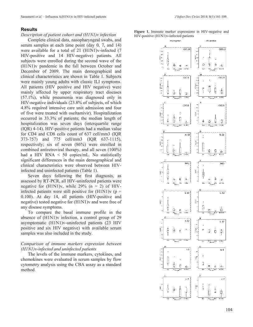
Figure 1. Immune marker expressions in HIV-negative and
HIV-positive (H1N1)v-infected patients

Sansonetti et al. – Influenza A(H1N1)v in HIV-infected patients J Infect Dev Ctries 2014; 8(1):101-109.
105
In HIV-negative patients, an increase of the
chemokines CXCL10 (p = 0.003), CXCL8 (p = 0.005)
and the cytokines IFNγ (p = 0.01), IL-10 (p = 0.002),
IL-6 (p = 0.002), and IL-1β (p = 0.01) was observed
during the acute phase of (H1N1)v infection (day 0)
when compared to healthy controls (HIV-negative,
(H1N1)v-uninfected). Moreover, whereas CXCL10 (p
= 0.2), IFNγ (p = 0.3), and IL-10 (p = 0.1) returned to
a basal level at day 7, CXCL8 (day 7, p - = 0.01; day
14, p - 0.004), IL-6 (day 7, p = 0.01; day 14, p = 0.02)
and IL-1β (day 7, p = 0.003; day 14, p = 0.02)
remained high later on (day 14), when compared to
healthy controls (Figure 1A to I) (Table
Supplementary 1).
In HIV-positive (H1N1)v-infected patients,
significantly higher levels of CXCL10 (p = 0.02),
CXCL9 (p = 0.01), IFNγ (p = 0.0004), IL-10 (p =
0.01), IL-6 (p = 0.003), IL-1β (p = 0.03), IL-17 (p =
0.01), and IL-4 (p = 0.04) were observed compared to
their corresponding controls (HIV-positive and
(H1N1)v-uninfected) at day 0. While most of these
cytokines returned to a basal level seven days
following the first diagnosis, CXCL10 (p = 0.02) and
(CXCL9 p = 0.03) still remained at higher levels in
(H1N1)v-infected patients compared to their
corresponding controls. In the late phase of infection
(day 14) CXCL9 (p = 0.2) returned to a basal level,
whereas CXCL10 (p = 0.02) remained at higher levels
in the (H1N1)v-infected patients (Figure 1 L to T)
(Table Supplementary 1).
Comparison of immune markers expression between
HIV-positive and HIV-negative (H1N1)v-infected
patients
A comparative analysis was performed to evaluate
the differential secretion of cytokine and chemokine
levels in HIV-positive and HIV-negative (H1N1)v-
infected individuals.
During the acute phase (day 0) of infection, the
serum cytokines levels appeared to be elevated in both
HIV-positive and HIV-negative cohorts compared to
their corresponding (H1N1)v negative controls (Figure
1 L to T and A to I), though significant differences in
the expression levels of immune mediators among
these two groups of (H1N1)v-infected patients were
not observed (Table Supplementary 1).
Conversely, 7 and 14 days following the first
diagnosis, significantly higher levels of CXCL10 were
observed in the HIV-positive group compared to the
HIV-negative cohort (day 7, p = 0.04; day 14, p =
0.03) (Figure 2). Higher levels of CXCL10 observed
in HIV-positive patients infected with (H1N1)v seems
not to be ascribable to oseltamivir treatment, as
demonstrated by the analysis comparingCXCL10
levels in oseltamivir treated HIV-negative against
HIV-positive patients (Figure Supplementary 1). Such
analysis confirmed that increased CXCL10 expression
by HIV-positive (H1N1)v-infected patients was not
due to oseltamivir treatment, since HIV-positive
patients maintained high levels of CXCL10
expression, while the CLCL10 levels in HIV-
negative/(H1N1)v patients treated with oseltamivir
decreased after 7 days of treatment. IL-6 expression
was further analyzed comparing HIV-negative and
HIV-positive patients treated with oseltamivir,
confirming that antiviral therapy does not statistically
influence immune mediators levels in HIV-negative
patients treated with oseltamivir. Oseltamivir
treatment lead to a strong decrease of the immune
mediators in the HIV-negative group 7 days after
administration. Conversely, in the HIV-positive
patients, the level of CXCL10 remained higher.
Although there was a decrease in statistical
significance at day 7 (p = 0.18) when only the
oseltamivir treated patients were compared, the HIV-
positive cohort had a delay in the normalization of
CXCL10 levels compared to the HIV-negative cohort
as the higher levels at day 14 show (p < 0.05) (Figure
Supplementary 1).
Assessment of differential basal expression of immune
mediators in HIV-positive and HIV-negative (H1N1)v-
uninfected control subjects
To determine whether the differential basal levels
of cytokine expression observed were the results of
HIV infection, serum samples from HIV-negative and
HIV-positive asymptomatic (H1N1)v-uninfected
patients were analyzed. Most of the cytokines tested
Figure 2. Level of CXCL10 in HIV-positive and HIV-negative
patients

Sansonetti et al. – Influenza A(H1N1)v in HIV-infected patients J Infect Dev Ctries 2014; 8(1):101-109.
106
were found at similar levels in the two groups, though
in HIV-positive patients, higher levels of IL-6 (p =
0.02), IL-1β (p = 0.04), and CXCL8 (p = 0.02) were
detected compared to the HIV-negative individuals
(Figure 3) (Table Supplementary 2). To determine
whether this observation could have been influenced
by oseltamivir treatment, oseltamivir-treated HIV-
negative/(H1N1)v patients were compared to the HIV-
positive/(H1N1)v cohort (Figure Supplementary 2).
This analysis showed that the higher IL-6 basal level
was not influenced by oseltamivir treatment.
The low influence of oseltamivir treatment on this
analysis was further confirmed by the analysis of HIV-
negative/(H1N1)v treated or untreated patients with
the antiviral-treated patients (Figure 2). As shown by
bar graphs, none of the cytokines analyzed were
significantly different between the treated and
untreated groups. Only a difference on day 0 was seen
for CXCL10 and IL-6 in HIV-negative/(H1N1)v
patients who were then treated with oseltamivir
because they developed strong symptoms and the
antiviral therapy was then supplied.
Finally, in order to exclude that these observations
could have been influenced by comorbidities in the
HIV-negative/(H1N1)v cohort, an analysis was
performed, excluding from this group the patients with
other diseases specified in Table 1. Such analysis
confirmed that, even excluding comorbidities, the
difference between CXCL10 and IL-6 expression in
HIV-positive and HIV-negative (H1N1)v-infected
patients remains statistically significant (Figure 3).
Comparison of antibody response between HIV-
infected and uninfected patients
To evaluate antibody response against (H1N1)v,
an HI assay was performed on sera obtained from
HIV-positive and HIV-negative (H1N1)v-infected
individuals at day 0 and day 14. In HIV-negative
patients, the geometric mean titer (GMT) significantly
increased over time from 22.08 at day 0 (95%
confidence intervals (CI) 10.60-45.99) to 204.9 at day
14 (95% confidence intervals (CI) 112.5-373; p =
0.006) and in the same time frame also in HIV-
positive patients from 14.96 at day 0 (95% confidence
intervals (CI) 7.183-30.74) to 107.7 at day 14 (95%
confidence intervals (CI) 47.63-243.4; p = 0.02).
While an increase in IgM secretion was observed
in both HIV-positive and HIV-negative patients, at day
14, a significantly higher level of IgM was observed in
HIV-negative patients (p = 0.004) (Figure 4A). A
similar pattern was observed for IgG, though HIV-
negative subjects showed a significantly higher level
of IgG at day 0 compared to their HIV-positive
counterparts (p = 0.045) (Figure 4B).
Discussion
The pandemic spread of the novel influenza virus
has raised concerns on the possible increased risk of
severe infection in immune-suppressed populations.
As it has already been shown, the 2009 (H1N1)v
infection seems to be associated with mild clinical
outcomes. Nevertheless, experiments conducted in
animal models indicated that (H1N1)v infection may
result in more severe lung lesions in humans compared
to mice, ferrets, and non-human primates infected with
the seasonal human H1N1 virus [14], suggesting that
(H1N1)v can cause distinctive clinical effects,
especially in patients with a compromised immune
system [15]. HIV infection, which is associated with
anomalous humoral and T-cell–mediated immune
responses, could result in increased susceptibility to
viral respiratory infections [16]. However, recent
studies have reported that the susceptibility of HIV-
infected patients without advanced disease to 2009
H1N1v influenza may not be increased [17].
Figure 4. IgG and IgM response between HIV-infected and
HIV-uninfected patients
Figure 3. Cytokine levels in HIV-negative and HIV-positive
(H1N1)v-uninfected patients
Sansonetti et al. – Influenza A(H1N1)v in HIV-infected patients J Infect Dev Ctries 2014; 8(1):101-109.
107
Here, we investigated the clinical outcomes
following (H1N1)v infection in HIV-positive and
HIV-negative patients and attempted to identify the
immune signatures associated with the two groups. We
found that (H1N1)v infection presents different
clinical outcomes in HIV-positive compared to HIV-
negative patients, and that this relates to a different
immune response in these two groups.
In this study, the clinical picture following
(H1N1)v-infection in HIV-positive patients appeared
milder overall compared to what was observed in
HIV-negative subjects. In fact, cases of severe
complications with onset of pneumonia that led to
hospitalization and that, in one case, required oxygen
supplementation, were detected only in HIV-negative
patients. This is in apparent contrast with previous
findings, where no remarkable differences in clinical
outcomes were observed in HIV-positive patients
compared to HIV-negative subjects [18,19]. In a study
by Peters et al., 911 subjects were analyzed in three
separate cohorts, which included HIV+ patients with
CD4 counts below 200, with half taking ART. The
results of the study showed that receiving ART
approximates significance in terms of being admitted
to an ICU and/or death. The majority of the patients
receiving ART therapy were HIV-infected patients
with a CD4 cell count <200 cells/µL, while in the
present study, the median value for CD4 counts was
more than 600; this could explain the differences in
the findings [20].
However, other studies have shown that influenza
infection in HIV-positive subjects usually has a milder
outcome, probably as a result of the immune
impairment due to CD4-deficiency [21]. Indeed, it has
been shown that complications following influenza
infection result from an inflammatory burst triggered
by a cytokine and chemokine storm induced by viral
replication in the lung parenchyma, which directly
involves CD4 T cells [22].
To determine whether the different clinical
outcomes observed among HIV-positive and HIV-
negative patients were dependent upon a diverse
immune response, we dissected the immune response
elicited following (H1N1)v infection using the human
beads cytokines array assay (CBA, BD). With this
method we were able to detect and quantify different
chemokines, cytokines, and immune mediators
through flow cytometry analysis in the serum of
(H1N1)v infected patients and controls.
Interestingly, HIV-positive patients did not show
remarkable differences in the cytokine and chemokine
patterns in response to H1N1 infection compared to
HIV-negative patients, although the chemokine
CXCL10 appeared elevated along the time of the
infection in the HIV-positive group, showing a delay
in the long-term normalization of its level compared to
what was reported in HIV-negative patients. CXCL10
or IP-10 is an IFN-γ-inducible protein that plays an
important role in the host response to a diversity of
viral infections [23]; it is expressed in a variety of cells
in response to IFN-γ stimulation [24]. High levels of
CXCL10 have been associated with a poor control of
viral infection by the immune system [25,4].Moreover,
increased levels of CXCL10 on plasma have been
already detected by Kamat et al. in HIV patients on
HAART [26]. Hence, the immune dysfunction during
HIV infection mainly reflected in the CD4 T cell
impairment could lead to a persistent secretion of
CXCL10 at high levels, as observed in HIV-positive
patients.
Another interesting observation comes from the
analysis of IL-6 that was found to be lower in HIV-
positive compared to HIV-negative patients. The
higher secretion of IL-6, which is considered an
activator of the acute phase responses [27] and also a
marker of critical illness during influenza infection
[28,5], may also explain the more severe clinical
outcomes reported in HIV-negative patients,
supporting the hypothesis that clinical complications
following influenza infection may result from an
inflammatory burst. It was shown that higher levels of
IL-6 in pre-HAART HIV patients correlate with
higher morbidity and mortality rates [29]. These data
suggest that IL-6 can effectively be considered as a
valuable marker of severe infection.
Protection and viral clearance during influenza
infection relies on the humoral arm of the immune
response. To determine the H1N1v serum antibody
levels, the GMT level in HIV-positive and negative
patients at day 0 and day 14 of the H1N1 infection
were analyzed. While HIV-positive patients were able
to mount an effective antibody response, they showed
a significantly lower GMT at day 14 compared to
HIV-negative patients. Interestingly, the difference in
GMT observed was dependent on the impairment of
HIV-positive subjects to mount a solid IgM response,
since IgG levels were similar among the two groups.
While the immunological basis of the difference
between IgG and IgM response remains unknown, it is
of interest to note that IgMs are known to be highly
neutralizing and may contribute to viral clearance. The
reduced viral clearance observed in the HIV-positive
group may result from the impaired ability to mount
an IgM response.
Sansonetti et al. – Influenza A(H1N1)v in HIV-infected patients J Infect Dev Ctries 2014; 8(1):101-109.
108
However, our data consider HIV-infected patients
to be in a no-advance immune suppression, as the
CD4+ and CD8+ cell counts show (CD4+ > 200
cells/mm3 and CD8+ < 1000 cells/mm3), and under
HAART, which reduces the incidence of opportunistic
infection; this suggests that influenza severity could be
different in a situation of advanced HIV disease or in
the presence of comorbidities. Furthermore, it should
be taken into account that HAART is not accessible to
HIV-infected people in many parts of the world,
especially in developing countries, where pulmonary
complications of HIV infection are major causes of
morbidity and mortality [9].
In summary, HIV-infected patients under HAART
show a distinctive feature in the immune response
against 2009 pandemic influenza, characterized by a
persistent secretion of high levels of CXCL10 and
lower antibody responses. It may be hypothesized that
the immune impairment due to the HIV infection in
patients under HAART could lead to a higher
CXCL10 secretion at long term, and to a delay in
normalization of its levels during recovery. Moreover,
the poorer response of specific antibodies thus
potentially affects virus clearance. Nonetheless, the
concomitant lower IL-6 secretion, which has been
described to be associated with critical illness during
influenza infection [27,30], could explain the milder
symptom manifestation observed in HIV-positive
patients. Acknowledgements This work was supported by the IDR, Li KaShing
Foundation, CIHR (grant to DK).
References 1. Giamarellos-Bourboulis EJ, Raftogiannis M, Antonopoulou
A, Baziaka F, Koutoukas P, Savva A, Kanni T, Georgitsi M,
Pistiki A, Tsaganos T, Pelekanos N, Athanassia S, Galani L,
Giannitsioti E, Kavatha D, Kontopidou F, Mouktaroudi M,
Poulakou G, Sakka V, Panagopoulos P, Papadopoulos A,
Kanellakopoulou K, Giamarellou H (2009) Effect of the novel
influenza A (H1N1) virus in the human immune system.
PLoS One 4: e8393.
2. Greenbaum JA, Kotturi MF, Kim Y, Oseroff C, Vaughan K,
Salimi N, Vita R, Ponomarenko J, Scheuermann RH, Sette A,
Peters B (2009) Pre-existing immunity against swine-origin
H1N1 influenza viruses in the general human population.
Proc Natl Acad Sci U S A 106: 20365-20370.
3. Mamun MM, Huda AK (2011) Origins and evolutionary
genomics of the novel swine-origin influenza A (H1N1) virus
in humans--past and present perspectives. Yakugaku Zasshi
131: 553-562.
4. Cameron CM, Cameron MJ, Bermejo-Martin JF, Ran L, Xu
L, Turner PV, Ran R, Danesh A, Fang Y, Chan PK, Mytle N,
Sullivan TJ, Collins TL, Johnson MG, Medina JC, Rowe T,
Kelvin DJ (2008) Gene expression analysis of host innate
immune responses during Lethal H5N1 infection in ferrets. J
Virol 82: 11308-1317.
5. Bermejo-Martin JF, Ortiz de Lejarazu R, Pumarola T, Rello J,
Almansa R, Ramirez P, Martin-Loeches I, Varillas D,
Gallegos MC, Seron C, Micheloud D, Gomez JM, Tenorio-
Abreu A, Ramos MJ, Molina ML, Huidobro S, Sanchez E,
Gordon M, Fernandez V, Del Castillo A, Marcos MA,
Villanueva B, Lopez CJ, Rodriguez-Dominguez M, Galan JC,
Canton R, Lietor A, Rojo S, Eiros JM, Hinojosa C, Gonzalez
I, Torner N, Banner D, Leon A, Cuesta P, Rowe T, Kelvin DJ
(2009) Th1 and Th17 hypercytokinemia as early host
response signature in severe pandemic influenza. Crit Care
13: R201.
6. Archer B, Cohen C, Naidoo D, Thomas J, Makunga C,
Blumberg L, Venter M, Timothy G, Puren A, McAnerney J,
Cengimbo A, Schoub B (2009) Interim report on pandemic
H1N1 influenza virus infections in South Africa, April to
October 2009: epidemiology and factors associated with fatal
cases. Euro Surveill 14: pii: 19369.
7. Baker M, Kelly H, Wilson N (2009) Pandemic H1N1
influenza lessons from the southern hemisphere. Euro Surveill
14: pii 19370
8. Janoff EN, Hardy WD, Smith PD, Wahl SM (1991) Humoral
recall responses in HIV infection. Levels, specificity, and
affinity of antigen-specific IgG. J Immunol 147: 2130-2135.
9. Cagigi A, Nilsson A, Pensieroso S, Chiodi F (2010)
Dysfunctional B-cell responses during HIV-1 infection:
implication for influenza vaccination and highly active
antiretroviral therapy. Lancet Infect Dis 10: 499-503.
10. Fabbiani M, Sali M, Di C, V, Pignataro G, Prete V, Farina S,
D'Avino A, Manzara S, Dal Verme LZ, Silveri NG, Cauda R,
Delogu G, Fadda G, Di GS (2011) Prospective evaluation of
epidemiological, clinical, and microbiological features of
pandemic influenza A (H1N1) virus infection in Italy. J Med
Virol 83: 2057-2065.
11. Meurs E, Chong K, Galabru J, Thomas NS, Kerr IM,
Williams BR, Hovanessian AG (1990) Molecular cloning and
characterization of the human double-stranded RNA-activated
protein kinase induced by interferon. Cell 62: 379-390.
12. Kendal AP, Skehel JJ, Pereira MS (1982) Concepts and
procedures for laboratory-based influenza surveillance. U.S.
Department of Health and Human Services, Centers for
Disease Control, Atlanta, USA. Available at:
http://stacks.cdc.gov/view/cdc/12251
13. Ferko B, Kittel C, Romanova J, Sereinig S, Katinger H,
Egorov A (2006) Live attenuated influenza virus expressing
human interleukin-2 reveals increased immunogenic potential
in young and aged hosts. J Virol 80: 11621-11627.
14. Itoh Y, Shinya K, Kiso M, Watanabe T, Sakoda Y, Hatta M,
Muramoto Y, Tamura D, Sakai-Tagawa Y, Noda T, Sakabe S,
Imai M, Hatta Y, Watanabe S, Li C, Yamada S, Fujii K,
Murakami S, Imai H, Kakugawa S, Ito M, Takano R,
Iwatsuki-Horimoto K, Shimojima M, Horimoto T, Goto H,
Takahashi K, Makino A, Ishigaki H, Nakayama M, Okamatsu
M, Warshauer D, Shult PA, Saito R, Suzuki H, Furuta Y,
Yamashita M, Mitamura K, Nakano K, Nakamura M,
Brockman-Schneider R, Mitamura H, Yamazaki M, Sugaya
N, Suresh M, Ozawa M, Neumann G, Gern J, Kida H,
Ogasawara K, Kawaoka Y (2009) In vitro and in vivo
characterization of new swine-origin H1N1 influenza viruses.
Nature 460: 1021-1025.
Sansonetti et al. – Influenza A(H1N1)v in HIV-infected patients J Infect Dev Ctries 2014; 8(1):101-109.
109
15. Lin JC, Nichol KL (2001) Excess mortality due to pneumonia
or influenza during influenza seasons among persons with
acquired immunodeficiency syndrome. Arch Intern Med 161:
441-446.
16. Beck JM, Rosen MJ, Peavy HH (2001) Pulmonary
complications of HIV infection. Report of the Fourth NHLBI
Workshop. Am J Respir Crit Care Med 164: 2120-2126.
17. Kok J, Tudo K, Blyth CC, Foo H, Hueston L, Dwyer DE
(2011) Pandemic (H1N1) 2009 influenza virus
seroconversion rates in HIV-infected individuals. J Acquir
Immune Defic Syndr 56: 91-94.
18. Perez CM, Dominguez MI, Ceballos ME, Moreno C, Labarca
JA, Rabagliati R, Vasquez P, Lasso M, Serri M (2010)
Pandemic influenza A (H1N1) in HIV-1-infected patients.
AIDS 24: 2867-2869.
19. Sheth AN, Patel P, Peters PJ (2011) Influenza and HIV:
lessons from the 2009 H1N1 influenza pandemic. Curr
HIV/AIDS Rep 8: 181-191.
20. Peters PJ, Skarbinski J, Louie JK, Jain S, Roland M, Jani SG,
Finelli L, Brooks JT (2011) HIV-infected hospitalized
patients with 2009 pandemic influenza A (pH1N1)--United
States, spring and summer 2009. Clin Infect Dis 52 Suppl 1:
S183-S188.
21. Isais F, Lye D, Llorin R, Dimatatac F, Go CJ, Leo YS, Chow
A (2010) Pandemic (H1N1) 2009 influenza in HIV-infected
adults: clinical features, severity, and outcome. J Infect 61:
437-440.
22. Nathan C (2002) Points of control in inflammation. Nature
420: 846-852.
23. Sumino KC, Walter MJ, Mikols CL, Thompson SA,
Gaudreault-Keener M, Arens MQ, Agapov E, Hormozdi D,
Gaynor AM, Holtzman MJ, Storch GA (2010) Detection of
respiratory viruses and the associated chemokine responses in
serious acute respiratory illness. Thorax 65: 639-644.
24. Luster AD, Ravetch JV (1987) Biochemical characterization
of a gamma interferon-inducible cytokine (IP-10). J Exp Med
166: 1084-1097.
25. de Jong MD, Simmons CP, Thanh TT, Hien VM, Smith GJ,
Chau TN, Hoang DM, Chau NV, Khanh TH, Dong VC, Qui
PT, Cam BV, Ha do Q, Guan Y, Peiris JS, Chinh NT, Hien
TT, Farrar J (2006) Fatal outcome of human influenza A
(H5N1) is associated with high viral load and
hypercytokinemia. Nat Med 12: 1203-1207.
26. Kamat A, Misra V, Cassol E, Ancuta P, Yan Z, Li C,
Morgello S, Gabuzda D (2012) A plasma biomarker signature
of immune activation in HIV patients on antiretroviral
therapy. PLoS One 7: e30881.
27. Kishimoto T, Akira S, Narazaki M, Taga T (1995)
Interleukin-6 family of cytokines and gp130. Blood 86: 1243-
1254.
28. Lee N, Wong CK, Chan PK, Chan MC, Wong RY, Lun SW,
Ngai KL, Lui GC, Wong BC, Lee SK, Choi KW, Hui DS
(2011) Cytokine response patterns in severe pandemic 2009
H1N1 and seasonal influenza among hospitalized adults.
PLoS One 6: e26050.
29. Boulware DR, Hullsiek KH, Puronen CE, Rupert A, Baker
JV, French MA, Bohjanen PR, Novak RM, Neaton JD, Sereti
I (2011) Higher levels of CRP, D-dimer, IL-6, and hyaluronic
acid before initiation of antiretroviral therapy (ART) are
associated with increased risk of AIDS or death. J Infect Dis
203: 1637-1646.
30. Lee H, Yoon TJ, Weissleder R (2009) Ultrasensitive detection
of bacteria using core-shell nanoparticles and an NMR-filter
system. Angew Chem Int Ed Engl 48: 5657-5660.
Corresponding author Michela Sali
Institute of Microbiology, Università Cattolica del Sacro Cuore
Largo A. Gemelli 8, 00168 Rome, Italy
Phone: +390630154964
Fax +39063051152
Email: [email protected]
Conflict of interests: No conflict of interests is declared.

Sansonetti et al. – Influenza A(H1N1)v in HIV-infected patients J Infect Dev Ctries 2014; 8(1):101-109.
Supplementary Items
Table Supplementary 1: Median and interquartile range for chemokines and cytokines in HIV-infected and HIV-uninfected
(H1N1)v-positive patients HIV-infected HIV-uninfected
Median Range
Median Range
CXCL10
Day 0 712,35 2.386
CXCL10
Day 0 1860,5 4.796
Day 7 1050,8 4.201 Day 7 533 2.451
Day 14 780,7 491 Day 14 365 2.168
CCL2
Day 0 809 839
CCL2
Day 0 486 2.565
Day 7 764 1.352 Day 7 477 836
Day 14 375 582 Day 14 479 692
CXCL9
Day 0 567,25 1.952
CXCL9
Day 0 403 1.107
Day 7 693,1 1.234 Day 7 231 2.023
Day 14 418 739 Day 14 235 2.688
IL-17
Day 0 12,9 11
IL-17
Day 0 10 10
Day 7 11 4 Day 7 10 29
Day 14 9 7 Day 14 10 7
INFɣ
Day 0 15,4 20
INFɣ
Day 0 16,85 352
Day 7 4 5 Day 7 4 15
Day 14 4 4 Day 14 4 17
IL-10
Day 0 3,7 24
IL-10
Day 0 4,8 54
Day 7 2 24 Day 7 4 7
Day 14 0 2 Day 14 3 15
IL-4
Day 0 2,9 4
IL-4
Day 0 2 4
Day 7 2 4 Day 7 2 13
Day 14 2 3 Day 14 2 3
IL-2
Day 0 4 6
IL-2
Day 0 4 5
Day 7 4 1 Day 7 4 9
Day 14 4 1 Day 14 3 2
IL-12p70
Day 0 3 37
IL-12p70
Day 0 2 48
Day 7 3 32 Day 7 2 40
Day 14 3 77 Day 14 2 198
TNFα
Day 0 0 1
TNFα
Day 0 0 4
Day 7 0 2 Day 7 0 2
Day 14 1 3 Day 14 0 9
IL-6
Day 0 9,9 23
IL-6
Day 0 25,2 171
Day 7 5 15 Day 7 4,95 51
Day 14 4 33 Day 14 5,8 104
IL1-β
Day 0 11,08 92
IL1-β
Day 0 20,2 114
Day 7 9 17 Day 7 11,8 61
Day 14 6 48 Day 14 11,6 73
CXCL8
Day 0 36 41
CXCL8
Day 0 29,6 556
Day 7 31 41 Day 7 23,45 223
Day 14 31 134 Day 14 42,6 141
Sansonetti et al. – Influenza A(H1N1)v in HIV-infected patients J Infect Dev Ctries 2014; 8(1):101-109.
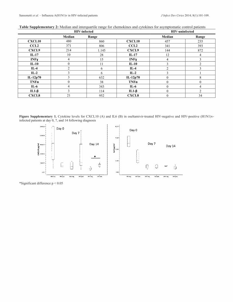
Table Supplementary 2: Median and interquartile range for chemokines and cytokines for asymptomatic control patients
HIV-infected HIV-uninfected
Median Range
Median Range
CXCL10 480 860 CXCL10 457 235
CCL2 371 806 CCL2 341 393
CXCL9 214 1.145 CXCL9 144 872
IL-17 10 28 IL-17 12 4
INFɣ 4 15 INFɣ 4 3
IL-10 0 11 IL-10 3 2
IL-4 2 6 IL-4 2 3
IL-2 3 6 IL-2 3 1
IL-12p70 3 632 IL-12p70 0 8
TNFα 0 38 TNFα 0 0
IL-6 4 343 IL-6 0 4
IL1-β 3 114 IL1-β 0 2
CXCL8 21 952 CXCL8 0 34
Figure Supplementary 1. Cytokine levels for CXCL10 (A) and IL6 (B) in oseltamivir-treated HIV-negative and HIV-positive (H1N1)v-
infected patients at day 0, 7, and 14 following diagnosis
*Significant difference p < 0.05

Sansonetti et al. – Influenza A(H1N1)v in HIV-infected patients J Infect Dev Ctries 2014; 8(1):101-109.
Figure Supplementary 2. Cytokine levels in oseltamivir-treated (-) and oseltamivir-untreated (-) HIV-negative (H1N1)v-infected patients at
day 0, 7, and 14 following diagnosis
Figure Supplementary 3. Cytokine levels excluding HIV-negative patients with underlying comorbidities, for CXCL10 (A) and IL6 (C) in
HIV-positive and HIV-negative (H1N1)v-infected patients at day 0, 7 and 14 following diagnosis
*Significant difference p < 0.05