Clinical and microbiological characteristics of dogs in sepsis ...
Upload
independentCategory
view
0download
0
CLINICAL PRESENTATION, MICROBIOLOGICAL FEATURES AND CORRELATES
OF DISEASE SEVERITY OF 2009 PANDEMIC INFLUENZA A (H1N1) INFECTION
Simona Di Giambenedetto1, Lorenzo Zileri Dal Verme
2, Michela Sali
3, Salvatore Farina
1, Valentina
Di Cristo1, Stefania Manzara
3, Andrea De Luca
1,4, Giulia Pignataro
2, Mattia Prosperi
1, Aldo Di
Franco3, Nicolò Gentiloni Silveri
2, Giovanni Delogu
3, Roberto Cauda
1, Massimiliano Fabbiani
1*,
Giovanni Fadda3.
1Institute of Clinical Infectious Diseases, Catholic University of Sacred Heart, Largo F. Vito1,
00168 Rome, Italy;
2Department of Emergency Medicine, Catholic University of Sacred Heart, Largo F. Vito1, 00168
Rome, Italy;
3Institute of Microbiology, Catholic University of Sacred Heart, Largo F. Vito1, 00168 Rome, Italy;
4Unit of Infectious Diseases, Siena University Hospital, Viale Bracci 16, 53100 Siena, Italy.
Corresponding author:
*Dr. Fabbiani Massimiliano, Istituto di Clinica delle Malattie Infettive, Università Cattolica del
Sacro Cuore, Largo Francesco Vito 1, 00168 Rome, Italy. Phone: +390630154945. Fax:
+39063054519. E-mail: [email protected]
ABSTRACT
Purpose: To describe epidemiological, clinical and microbiological characteristics of confirmed
novel influenza A (H1N1) infection, investigating factors associated to disease severity.
Methods: We retrospectively selected patients seeking care for respiratory symptoms in two
periods (May-August and September-November 2009) with different epidemiological
characteristics. Only patients with confirmed pandemic influenza A (H1N1) were enrolled in this
study.
Results: A total of 104 patients with H1N1 infection were evaluated, mostly referring classic
influenza symptoms; in addition, diarrhea and vomiting were often referred. Clinical signs,
symptoms and respiratory complications were different in the two periods. In the total, 18 patients
(17%) had pneumonia. Patients older than 50 years showed a lower probability of pneumonia
diagnosis when compared to children aged 0-13 (p=0.049); a longer duration of symptoms before
medical care was associated with a higher probability of pneumonia (p=0.026). Phylogenetic
analysis showed a low variability both in hemagglutinin and neuraminidase genes. In addition, no
neuraminidase mutation associated to antiviral resistance was detected.
Conclusions: A detailed description of respiratory diseases associated with H1N1 infection was
provided and factors associated with its severity were investigated, thus contributing to the insight
into epidemiological, clinical and microbiological knowledge of the disease.
Keywords: Influenza A (H1N1), swine flu, respiratory tract infection, pneumonia, phylogenesis,
Italy.
INTRODUCTION
Influenza A H1N1 virus is a subtype of influenza A virus, the most common cause of influenza
(flu) in humans. Some strains of H1N1 are endemic in humans and cause a small fraction of all
seasonal influenza or influenza-like illnesses. H1N1 strains caused a tiny percent of all human flu
infections in 2004–2005 but other H1N1 strains are endemic in pigs (swine influenza) and in birds
(avian influenza) [1].
The influenza pandemic the world was waiting for might have arrived on April 2009. Mexico
was the first country where a sharp increase in reports of patients with pneumonia caused by
influenza A H1N1 was observed [2]. In June 2009, World Health Organization (WHO) declared
that flu due to a new strain of swine-origin H1N1 was responsible for the 2009 influenza pandemic.
According to the Centers for Disease Control and Prevention (CDC), in humans the symptoms of
the 2009 "swine flu" H1N1 virus were similar to those of seasonal influenza and influenza-like
illness in general, including fever, cough, sore throat, headache, chills and fatigue. Preliminary
reports have shown that, when compared to previous influenza seasons, the 2009 outbreak showed
an increased percentage of patients reporting diarrhea and vomiting [3]. The most common cause of
death was respiratory failure. Other reported causes of death included pneumonia, high fever
(leading to neurological problems), dehydration (from excessive vomiting and diarrhea) and
electrolyte imbalance. Severe cases were most frequent in middle-aged patients, often with
comorbidities [4]. However, to date the virulence of H1N1 strains appears to be no greater than that
of seasonal influenza.
The aim of this study was to describe epidemiological, clinical and microbiological
characteristics of all confirmed novel influenza A H1N1 cases observed in a reference hospital in
Rome from May to November 2009, focusing on the impact that seasonal and epidemiological
factors could have on clinical features of the disease.
MATERIALS AND METHODS
Patients
Patients admitted with respiratory symptoms and /or fever (temperature >38.5°C) at the
Emergency Room of the Catholic University of Sacred Heart in Rome, in two different periods of
the year (May-August and September-November 2009) were retrospectively selected. The
separation of the cases in the two periods was justified by epidemiological factors. During the first
period H1N1 pandemic virus was not epidemic in the country and the Italian Ministry of Health
adopted a policy aimed at containing its spread from travellers and their contacts, recommending
testing for the new influenza virus in all probable cases of influenza A H1N1 (defined as those
showing typical symptoms and having had recent contact with proven cases of H1N1, or returning
from travel to areas where influenza A H1N1 was epidemic or working in a laboratory) observed in
reference hospitals. In the second period, when the virus was endemic in Italy, the definition of
probable cases was less stringent, without the inclusion of epidemiological criteria; probable cases
included patients with fever greater than or equal to 38°C associated with at least one constitutional
symptom (headache, asthenia, tremors, weakness, abdominal pain and diarrhea) and accompanied
by at least one respiratory symptom (cough, sore throat, nasal congestion). However, in this period
H1N1 testing was recommended only for subjects with more severe disease or requiring
hospitalization.
Only patients with pandemic influenza A (H1N1) virus detected from nasal or oropharingeal
swabs by real-time RT PCR assay were considered for this analysis.
Real-Time RT PCR and viral sequencing
A 400-µl aliquot of the specimen was used for automated RNA extraction with a EZ1 viral kit
(QIAGEN, Hilden, Germany) for use in Real Time RT PCR. The primers and probes for the H1
gene (swH1) and M gene (InfA) used in this work were recommended by WHO [5] and synthesized
by the Applied Biosystems (Forest City, CA USA) company. RT-PCR was performed in a 25 μl
reaction volume that contained 5 μl of the RNA dilution, 12.5 μl 2x AgPath-ID™ One-Step RT-
PCR buffer, 1 μl enzyme mix, 0.5 μl assay mix in a fluorometric PCR instrument (ABI 7300).
Thermal cycling conditions were 30 min at 50°C followed by 10 min at 95°C and a subsequent 45
cycles amplification (95 C for 15 s; 55°C for 30 s, fluorescence was collected at 55°C).
In all patients with H1N1 infection confirmed by RT-PCR on respiratory specimens, samples
stored at -80°C were then grown in MDCK cell cultures for viral isolation and subsequent HA and
NA genes sequencing. Amplicons for sequencing were generated by reverse transcription, followed
by PCR amplification to generate overlapping double-stranded DNA amplicons covering HA and
NA segments of the influenza virus genome [3], the products were purified with QIAquick columns
(Qiagen) and sequenced by using an ABI Prism BigDye Terminator (version 1.1) cycle sequencing
kit in an ABI Prism 3100- genetic analyzer on a 50-cm array. The sequencing reaction was
performed according to the manufacturer's protocol in a final volume of
10 µl with approximately 2
ng of amplified product, BigDye Terminator (version 1.1) premix, and 0.3 pmol of the primer.
Unincorporated nucleotides were purified by using a BigDye XTerminator purification kit (ABI).
The data were collected by using ABI software (version 2.0) [6]. The sequences
were analyzed by
using Seqman software (Lasergene package).
Phylogenetic Analysis
Sequenced HA and NA genes were used for a BLAST search in the NCBI genbank and an
additional set of background sequences with high similarity was collected, adding a reference for
H1N1 and an outgroup of H2N2 strain. Sequences were aligned using Opal software [7]. Maximum
likelihood (ML) phylogenetic analysis was performed using parallel computational methods, setting
up Akaike Information Criterion (AIC)-based model selection, nearest-neighbor-interchange tree
search, 4 gamma categories, and 100 bootstrap [8]. Sequence variability was annotated using
entropy plots.
Statistical analysis
Characteristics of the patients who presented in the two periods May-August and September-
November were compared using Fisher’s exact test or chi-square test for categorical variables and
the Student’s T test for continuous variables. Denominators that were used to calculate proportions
varied according to the number of patients with available data. Factors associated with pneumonia
diagnosis were analyzed by univariable and multivariable logistic regression. A two-sided P value
of less than 0.05 was considered statistically significant. All analyses were performed with SPSS
version 18.0 (SPSS Inc, Chicago, IL).
RESULTS
Patient’s characteristics
A total of 104 patients [52% males, median age 15 years (interquartile range, IQR 13-25)] with
H1N1 infection were evaluated. Of these, 77 (74%) were admitted to the Emergency ward during
May-August/2009 and 27 (26%) during September-November 2009. Overall, 42 (40%) patients
reported travelling in European countries (outside Italy), 14 (14%) in the USA and 6 (6%) in other
geographical areas, while 42 (40%) had never left Italy. As expected, in the second period a
significant increase in the number of patients not reporting travelling outside Italy was observed
(96% versus 19%, p<0.001). Patients characteristics according to the subgroups are summarized in
table 1.
Clinical presentation: symptoms, signs and laboratory parameters
A description of symptoms, signs and laboratory parameters according to subgroups is
summarized in table 2.
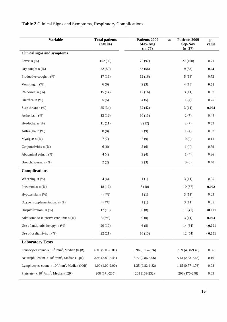
In our series, fever was reported by 102 (98%) patients, dry cough by 52 (50%) and productive
cough by 17 (16%), vomiting by 6 (6%) and diarrhea by 5 (5%). Other symptoms reported by
patients included sore throat (34%), asthenia (11%), headache (11%), arthralgia (8%), myalgia
(7%), conjunctivitis (6%), abdominal pain (4%), wheezing (4%) and cyanosis (2%). A total of 17
patients (16%) required hospitalization, two had severe respiratory failure and required admission to
the intensive care unit. In particular, among hospitalized patients 4 (23%) were subjects under the
age of 14 years, 7 (42%) between 15-39 years and 6 (35%) older than 40 years. When
hospitalization rate was analyzed for each age group, we found that it was higher in patients older
than 40 years when compared to those aged 15-39 (13% versus 46%, p=0.008) or to those under 14
years (11% versus 46%, p=0.006). None of the patients died. Oseltamivir (at a dosage of 75 mg bid
for 5 days) was administered within 48 hours after the onset of symptoms in 22 (22%) patients.
Thirteen (12%) subjects had received outpatient antimicrobial therapy while 20 (20%) were
prescribed antibiotics after medical care in the Emergency room. Three (3%) patients had pre-
existing predisposing disorders (one patient had asthma and heart disease, two patients
immunosuppression).
Blood tests were performed in 89 (86%) individuals. Of these, 9 (10%) patients had leukocytosis, 7
(8%) leukopenia, 36 (40%) lymphopenia and 4 (4%) lymphocytosis.
Clinical signs and symptoms on admission and the respiratory complications significantly
differed from the first to the second period with dry cough declining from 56% to 33% (p=0.04),
sore throat declining from 42% to 11% (p=0.004), vomiting increasing from 3% to 15% (p=0.01).
Pneumonia diagnosis was more frequent in the second period (37% versus 10%, p=0.002).
Hospitalization rate increased from 8% to 41% (p<0.001) and admission to intensive care, which
was absent in the first period, occurred in 11% of the cases observed during the second period
(p=0.003). Prescription of antibiotic therapy increased from 8% to 64% (p<0.001) and use of
oseltamivir from 13% to 54% (p<0.001).
Clinical complications
In total, 18 cases (17%) had pneumonia confirmed by chest X-ray. Twelve cases (67%) showed
focal infiltrates involving a single lung and 6 (33%) showed a bilateral interstitial-alveolar pattern.
In all these patients blood cultures and culture of respiratory specimens were negative. Among
patients with pneumonia, 4 (27%) had leukocytosis (leukocyte count, >9,800 per cubic millimeter)
and 3 (20%) lymphopenia (absolute lymphocyte count, <1,680 per cubic millimeter). Five (28%)
patients started early antimicrobial therapy before access in the Emergency Department. Overall,
antibiotics were administered to 9 (50%) subjects. Oral levofloxacin and amoxicillin-clavulanic acid
were the most frequently prescribed drugs.
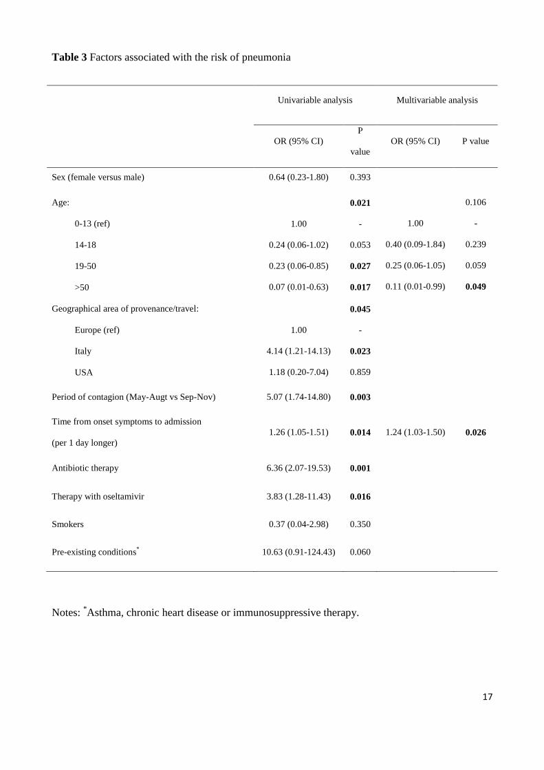
At univariable analysis, older age, a negative epidemiology for recent foreign travel,
emergency admission between September-November 2009, longer time from onset symptoms to
emergency admission, treatment with antibiotic therapy and oseltamivir were all associated with
higher odds of pneumonia. No specific symptoms, signs or laboratory parameters were associated
with the occurrence of pneumonia. Due to the small sample size, a full multivariable analysis
(including all variables that showed an association with pneumonia at univariate analysis) could not
be performed since it would have exhibited a low statistical power. We thus executed single sets of
bivariable logistic regression analyses, including only variables considered clinically relevant and
excluding variables such as the use of antimicrobial therapy and oseltamivir administration since
these were constant in the clinical management of pneumonia and rather a consequence than a cause
of pneumonia (see table 3). At multivariate analysis, patients older than 50 years showed a lower
probability of pneumonia diagnosis when compared to children aged 0-13 (p=0.049); a longer
duration of symptoms before medical care was associated with a higher probability of pneumonia
(p=0.026).
Sequence and phylogenetic analysis
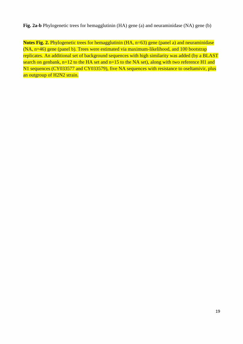
Overall, 63 and 46 sequences could be obtained for phylogenetic analysis of HA and NA genes,
respectively. By a BLAST search on genbank data base, keeping the first best-scoring sequences for
each one of our sequences and eliminating duplicates, an additional set of 12 and 15 sequences was
added to HA and NA sets. The reference H1 and N1 sequences (CY033577 and CY033579) from
Puerto Rico were added, as well as the outgroup references H2 and N2 (NC_007374 and
NC_007382) from Korea. Five NA sequences with reported resistance to oseltamivir were
eventually added to the NA set. In the HA gene, BLAST search reported that the best-scoring
sequences were distributed in several countries all over the world (Russia, Turkey, Germany, China,
Canada, Sweden, Taiwan, Netherlands, Thailand). The same was when considering the NA gene
(Argentina, California, Canada, Italy, Russia, Malaysia, USA) and 2 sequences were from Italy
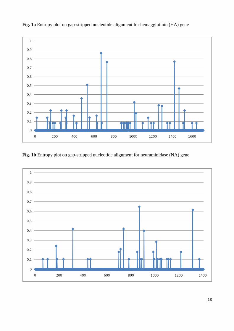
(Ancona province). After multiple alignment and gap-stripping, entropy analysis on the nucleotide
alignment showed very low variation (Figures 1A and 1B): the average (stdev) entropy value for
HA was 0.005 (0.04), the median (IQR) was 0 (0-0); the average (stdev) entropy value for NA was
0.004 (0.03), the median (IQR) was 0 (0-0). The analysis of the deduced aminoacid sequences
revealed no changes associated to oseltamivir or zanamivir resistance (E119V, H274Y, R292K,
N294S) in the NA gene. The bootstrapped ML phylogenetic analysis, highly affected by the
extreme sequence identity, revealed no evidence of clustering in the HA gene (selected model via
AIC was general time reversible), and the same held in the NA gene (selected model via AIC was
transversional model). Figures 2A and 2B depict the relative phylogenetic trees.
DISCUSSION
The global and rapid spread of pandemic influenza A H1N1 raised concerns for the serious
complications potentially associated with infection. Reports from different countries describing
clinical features of H1N1 infection have been published [9-11]; however, several aspects (such as
differences in epidemiological trends and in clinical presentation between different countries or
different seasons) are still unexplored. As a consequence, further surveillance is needed.
In this study, patients with pandemic 2009 H1N1 virus infection diagnosed in Italy during two
periods (May-August and September-November 2009) characterized by different epidemiological
features were enrolled. Regarding clinical characteristics, influenza A H1N1 infected patients
mostly referred the classic influenza symptoms; in addition, H1N1 patients often had diarrhea and
vomiting, which are not usually seen in seasonal influenza [3]. In our series, hospitalization was
required by 16% of patients, the majority of which was aged less than 40 years, confirming that
children and young adults were the age category mostly affected by the epidemic [12], probably
resulting from the higher susceptibility of the youngest to the new virus due to the lack of H1N1-
specific antibodies or memory immune response [13]. This is in contrast with epidemiological
features of seasonal influenza, for which hospitalizations are more common among persons 65 years
of age or older and those under the age of 15 years. However, hospitalization rate was higher in
patients older than 40 years suggesting that, when H1N1 disease occurs in this age group, it can be
more severe.
Risk factors for acquiring the infection or its complications significantly varied between the
two explored periods. In particular, a history of travel in a country where endemic transmission of
H1N1 virus occurred was most frequently reported in the first period, while pre-existing conditions
predisposing to clinical severity where prevalent in patients observed in the second period.
Moreover, several complications, such as pneumonia, wheezing, hospitalization or admission to an
intensive care unit, were more frequently observed in the second period. These observations may
also reflect the change in health policies when H1N1 strains became endemic in Italy, with
authorities recommending influenza testing only for patients with more severe respiratory disease.
However, the influence of several other factors (such as the role of cold climate in favoring
complications, an higher diffusion of the virus in age groups with risk factors for complications or a
increased virulence of circulating strains) cannot be fully excluded.
Few studies analyzed factors predisposing to the development of clinical complications and
further investigation is required. In our series, we observed that children and those with a longer
duration of symptoms before medical care showed an higher risk of pneumonia and, as a
consequence, should be carefully evaluated for the possible development of such complication.
Phylogenetic analysis is a useful tool to investigate whether differences in clinical presentation
between different seasons or different countries could be attributable to viral evolution and to define
and understand dynamics of the evolution of viral epidemics. In our study population, sequence
analysis showed that the nucleotide and amino acidic variability was low both in the HA and the
NA genes. In addition, we did not detect any virus showing mutations in the NA gene that are
associated to antiviral resistance. This is not unexpected, given the generally low antiviral uptake in
Italy and Europe. During the 2008-2009 seasonal influenza, there was a dramatic increase in the
oseltamivir-resistant strain circulation in some countries [14]. In addition, phylogenetic analysis did
not show any clear clustering, although a differentiation from the original Puerto Rico Strain
occurred. The BLAST search on the genbank database reported that the sequences showing the best
associations with those observed in this study were widely distributed in the world, in agreement
with the epidemiological information for which many of the diagnosed cases were subjects who
travelled outside the country. This may suggest that the epidemic in the country was probably the
result of multiple entry points. Notwithstanding, it must be considered that the evidence of no-
clustering suggests that the evolution of H1N1 probably acts at a higher epidemic level and across
the whole viral genome. In fact, a recent study showed that H1N1 is evolving into two distinct
clusters [15]. Another paper, investigating viral phylogeography, highlighted that the H1N1 drift
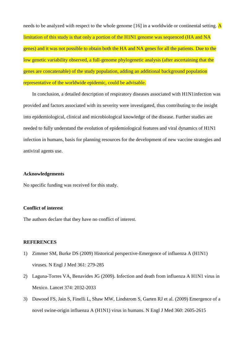
needs to be analyzed with respect to the whole genome [16] in a worldwide or continental setting. A
limitation of this study is that only a portion of the H1N1 genome was sequenced (HA and NA
genes) and it was not possible to obtain both the HA and NA genes for all the patients. Due to the
low genetic variability observed, a full-genome phylogenetic analysis (after ascertaining that the
genes are concatenable) of the study population, adding an additional background population
representative of the worldwide epidemic, could be advisable.
In conclusion, a detailed description of respiratory diseases associated with H1N1infection was
provided and factors associated with its severity were investigated, thus contributing to the insight
into epidemiological, clinical and microbiological knowledge of the disease. Further studies are
needed to fully understand the evolution of epidemiological features and viral dynamics of H1N1
infection in humans, basis for planning resources for the development of new vaccine strategies and
antiviral agents use.
Acknowledgements
No specific funding was received for this study.
Conflict of interest
The authors declare that they have no conflict of interest.
REFERENCES
1) Zimmer SM, Burke DS (2009) Historical perspective-Emergence of influenza A (H1N1)
viruses. N Engl J Med 361: 279-285
2) Laguna-Torres VA, Benavides JG (2009). Infection and death from influenza A H1N1 virus in
Mexico. Lancet 374: 2032-2033
3) Dawood FS, Jain S, Finelli L, Shaw MW, Lindstrom S, Garten RJ et al. (2009) Emergence of a
novel swine-origin influenza A (H1N1) virus in humans. N Engl J Med 360: 2605-2615
4) Estenssoro E, Ríos FG, Apezteguía C, Reina R, Neira J, Ceraso DH et al. (2010) Pandemic
2009 Influenza A (H1N1) in Argentina: a study of 337 patients on mechanical ventilation. Am
J Respir Crit Care Med 182: 41-8
5) World Health Organization (2009). Protocol of real-time RT-PCR for influenza A(H1N1), 28
April 2009. Available at:
http://www.who.int/csr/resources/publications/swineflu/CDCRealtimeRTPCR_Swine H1
Assay-2009, 20090430.pdf
6) Pabbaraju K, Wong S, Wong AA, Appleyard GD, Chui L, Pang XL et al. (2009) Design and
validation of real-time reverse transcription-PCR assays for detection of pandemic (H1N1)
2009 virus. J Clin Microbiol 47: 3454-3460
7) Wheeler T and Kececioglu J (2007) Multiple alignment by aligning alignments. Proceedings of
the 15th ISCB Conference on Intelligent Systems for Molecular Biology. Bioinformatics 23:
i559-i568
8) Keane TM, Naughton TJ, McInerney JO (2007) A high-throughput phylogenomics webserver
using distributed computing. Nucleic Acids Research 35: W33-W37
9) Jain S, Kamimoto L, Bramley AM, Schmitz AM, Benoit SR, Louie J et al. (2009) Hospitalized
patients with 2009 H1N1 influenza in the United States, April-June 2009. N Engl J Med 361:
1935-1944
10) Dominguez-Cherit G, Lapinsky SE, Macias AE, Pinto R, Espinosa-Perez L, De la Torre A et
al. (2009) Critically ill patients with 2009 influenza A (H1N1) in Mexico. JAMA 302: 1880-
1887
11) Cao B, Li XW, Mao Y, Wang J, Lu HZ, Chen YS et al. (2009) Clinical features of the initial
cases of 2009 pandemic influenza A (H1N1) virus infection in China. N Engl J Med 361: 2507-
2517
12) Chowell G, Bertozzi SM, Colchero MA, Lopez-Gatell H, Alpuche-Aranda C, Hernandez M et
al. (2009) Severe respiratory diseases concurrent with the circulation of H1N1 influenza. N
Engl J Med 361: 674-679
13) Hancock K, Veguilla V, Lu X, Zhong W, Butler EN, Sun H et al. (2009) Cross-reactive
antibody responses to the 2009 pandemic H1N1 influenza virus. N Engl J Med 361: 1945-1952.
14) Baranovich T, Saito R, Suzuki Y, Zaraket H, Dapat C, Caperig-Dapat I et al. (2010) Emergence
of H274Y oseltamivir-resistant A(H1N1) influenza viruses in Japan during the 2008-2009
season. J Clin Virol 47: 23-28
15) Fereidouni SR, Beer M, Vahlenkamp T, Starick E (2009) Differentiation of two distinct
clusters among currently circulating influenza A(H1N1)v viruses, March-September 2009.
Euro Surveill 14 (46) pii: 19409
16) Parks D, Macdonald N, Beiko R (2009) Tracking the evolution and geographic spread of
Influenza A. PLoS Curr Influenza 27: RRN1014
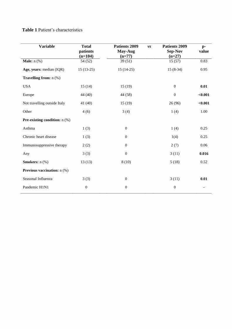
Table 1 Patient’s characteristics
Variable Total
patients
(n=104)
Patients 2009
May-Aug
(n=77)
vs Patients 2009
Sep-Nov
(n=27)
p-
value
Male: n (%) 54 (52) 39 (51) 15 (57) 0.83
Age, years: median (IQR) 15 (13-25) 15 (14-25) 15 (8-34) 0.95
Travelling from: n (%)
USA
Europe
Not travelling outside Italy
Other
15 (14)
44 (40)
41 (40)
4 (6)
15 (19)
44 (58)
15 (19)
3 (4)
0
0
26 (96)
1 (4)
0.01
<0.001
<0.001
1.00
Pre-existing condition: n (%)
Asthma
Chronic heart disease
Immunosuppressive therapy
Any
1 (3)
1 (3)
2 (2)
3 (3)
0
0
0
0
1 (4)
1(4)
2 (7)
3 (11)
0.25
0.25
0.06
0.016
Smokers: n (%) 13 (13) 8 (10) 5 (18) 0.52
Previous vaccination: n (%)
Seasonal Influenza
Pandemic H1N1
3 (3)
0
0
0
3 (11)
0
0.01
-
16
Table 2 Clinical Signs and Symptoms, Respiratory Complications
Variable Total patients
(n=104)
Patients 2009
May-Aug
(n=77)
vs Patients 2009
Sep-Nov
(n=27)
p-
value
Clinical signs and symptoms
Fever: n (%) 102 (98) 75 (97) 27 (100) 0.71
Dry cough: n (%) 52 (50) 43 (56) 9 (33) 0.04
Productive cough: n (%) 17 (16) 12 (16) 5 (18) 0.72
Vomiting: n (%) 6 (6) 2 (3) 4 (15) 0.01
Rhinorrea: n (%) 15 (14) 12 (16) 3 (11) 0.57
Diarrhea: n (%) 5 (5) 4 (5) 1 (4) 0.75
Sore throat: n (%) 35 (34) 32 (42) 3 (11) 0.004
Asthenia: n (%) 12 (12) 10 (13) 2 (7) 0.44
Headache: n (%) 11 (11) 9 (12) 2 (7) 0.53
Arthralgia: n (%) 8 (8) 7 (9) 1 (4) 0.37
Myalgia: n (%) 7 (7) 7 (9) 0 (0) 0.11
Conjunctivitis: n (%) 6 (6) 5 (6) 1 (4) 0.59
Abdominal pain: n (%) 4 (4) 3 (4) 1 (4) 0.96
Bronchospasm: n (%) 2 (2) 2 (3) 0 (0) 0.40
Complications
Wheezing: n (%) 4 (4) 1 (1) 3 (11) 0.05
Pneumonia: n (%) 18 (17) 8 (10) 10 (37) 0.002
Hypoxemia: n (%) 4 (4%) 1 (1) 3 (11) 0.05
Oxygen supplementation: n (%) 4 (4%) 1 (1) 3 (11) 0.05
Hospitalization : n (%) 17 (16) 6 (8) 11 (41) <0.001
Admission to intensive care unit: n (%) 3 (3%) 0 (0) 3 (11) 0.003
Use of antibiotic therapy: n (%) 20 (19) 6 (8) 14 (64) <0.001
Use of oseltamivir: n (%) 22 (21) 10 (13) 12 (54) <0.001
Laboratory Tests
Leucocytes count–x 103 /mm3, Median (IQR) 6.00 (5.00-8.00) 5.96 (5.15-7.36) 7.09 (4.58-9.48) 0.06
Neutrophil count–x 103 /mm3, Median (IQR) 3.96 (2.80-5.45) 3.77 (2.86-5.06) 5.43 (2.63-7.48) 0.10
Lymphocytes count–x 103 /mm3, Median (IQR) 1.00 (1.00-2.00) 1.25 (0.82-1.82) 1.15 (0.77-1.76) 0.98
Platelets– x 103 /mm3, Median (IQR) 208 (171-235) 208 (169-232) 208 (175-248) 0.83
17
Table 3 Factors associated with the risk of pneumonia
Univariable analysis Multivariable analysis
OR (95% CI)
P
value
OR (95% CI) P value
Sex (female versus male) 0.64 (0.23-1.80) 0.393
Age:
0-13 (ref)
14-18
19-50
>50
1.00
0.24 (0.06-1.02)
0.23 (0.06-0.85)
0.07 (0.01-0.63)
0.021
-
0.053
0.027
0.017
1.00
0.40 (0.09-1.84)
0.25 (0.06-1.05)
0.11 (0.01-0.99)
0.106
-
0.239
0.059
0.049
Geographical area of provenance/travel:
Europe (ref)
Italy
USA
1.00
4.14 (1.21-14.13)
1.18 (0.20-7.04)
0.045
-
0.023
0.859
Period of contagion (May-Augt vs Sep-Nov) 5.07 (1.74-14.80) 0.003
Time from onset symptoms to admission
(per 1 day longer)
1.26 (1.05-1.51) 0.014 1.24 (1.03-1.50) 0.026
Antibiotic therapy 6.36 (2.07-19.53) 0.001
Therapy with oseltamivir 3.83 (1.28-11.43) 0.016
Smokers 0.37 (0.04-2.98) 0.350
Pre-existing conditions* 10.63 (0.91-124.43) 0.060
Notes: *Asthma, chronic heart disease or immunosuppressive therapy.
18
Fig. 1a Entropy plot on gap-stripped nucleotide alignment for hemagglutinin (HA) gene
Fig. 1b Entropy plot on gap-stripped nucleotide alignment for neuraminidase (NA) gene
19
Fig. 2a-b Phylogenetic trees for hemagglutinin (HA) gene (a) and neuraminidase (NA) gene (b)
Notes Fig. 2. Phylogenetic trees for hemagglutinin (HA, n=63) gene (panel a) and neuraminidase
(NA, n=46) gene (panel b). Trees were estimated via maximum-likelihood, and 100 bootstrap
replicates. An additional set of background sequences with high similarity was added (by a BLAST
search on genbank, n=12 to the HA set and n=15 to the NA set), along with two reference H1 and
N1 sequences (CY033577 and CY033579), five NA sequences with resistance to oseltamivir, plus
an outgroup of H2N2 strain.
Copyright © 2022 FDOKUMEN